Please cite this article in press as: S. Singh, et al., Anhydrobiosis and programmed cell death in plants: Commonalities and Differences, Curr. Plant Biol. (2015), http://dx.doi.org/10.1016/j.cpb.2014.12.001 ARTICLE IN PRESS G Model CPB-13; No. of Pages 9 Current Plant Biology xxx (2015) xxx.e1–xxx.e9 Contents lists available at ScienceDirect Current Plant Biology jo ur nal home page: www.elsevier.com/locate/cpb Anhydrobiosis and programmed cell death in plants: Commonalities and Differences Samer Singh a , Vivek Ambastha b , Alex Levine c , Sudhir Kumar Sopory b , Pramod Kumar Yadava b , Baishnab Charan Tripathy b , Budhi Sagar Tiwari b,∗ a Department of Microbial Biotechnology, Panjab University, Chandigarh 160014, India b School of Life Sciences, Jawaharlal Nehru University, New Delhi 110067, India c Department of Plant and Environmental Sciences, The Hebrew University of Jerusalem, Givat-Ram, Jerusalem, Israel a r t i c l e i n f o Article history: Received 18 July 2014 Received in revised form 4 December 2014 Accepted 28 December 2014 Keywords: Anhydrobiosis Desiccation ROS PCD Trehalose Polyamines ABA LEA proteins a b s t r a c t Anhydrobiosis is an adaptive strategy of certain organisms or specialised propagules to survive in the absence of water while programmed cell death (PCD) is a finely tuned cellular process of the selective elimination of targeted cell during developmental programme and perturbed biotic and abiotic condi- tions. Particularly during water stress both the strategies serve single purpose i.e., survival indicating PCD may also function as an adaptive process under certain conditions. During stress conditions PCD cause targeted cells death in order to keep the homeostatic balance required for the organism survival, whereas anhydrobiosis suspends cellular metabolic functions mimicking a state similar to death until reestablishment of the favourable conditions. Anhydrobiosis is commonly observed among organisms that have ability to revive their metabolism on rehydration after removal of all or almost all cellular water without damage. This feature is widely represented in terrestrial cyanobacteria and bryophytes where it is very common in both vegetative and reproductive stages of life-cycle. In the course of evolution, with the development of advanced vascular system in higher plants, anhydrobiosis was gradually lost from the vegetative phase of life-cycle. Though it is retained in resurrection plants that primarily belong to thallophytes and a small group of vascular angiosperm, it can be mostly found restricted in orthodox seeds of higher plants. On the contrary, PCD is a common process in all eukaryotes from unicellular to multicellular organisms including higher plants and mammals. In this review we discuss physiological and biochemical commonalities and differences between anhydrobiosis and PCD. © 2015 The Authors. Published by Elsevier B.V. This is an open access article under the CC BY-NC-ND license (http://creativecommons.org/licenses/by-nc-nd/4.0/). Contents 1. Introduction . . . . . . . . . . . . . . . . . . . . . . . . . . . . . . . . . . . . . . . . . . . . . . . . . . . . . . . . . . . . . . . . . . . . . . . . . . . . . . . . . . . . . . . . . . . . . . . . . . . . . . . . . . . . . . . . . . . . . . . . . . . . . . . . . . . . . . . . . . 00 2. Origin and evolution of desiccation tolerance and programmed cell death (PCD) . . . . . . . . . . . . . . . . . . . . . . . . . . . . . . . . . . . . . . . . . . . . . . . . . . . . . . . . . . . . . . . . . . 00 3. Distribution of anhydrobiosis and PCD in photosynthetic organisms . . . . . . . . . . . . . . . . . . . . . . . . . . . . . . . . . . . . . . . . . . . . . . . . . . . . . . . . . . . . . . . . . . . . . . . . . . . . . . . 00 4. Mechanisms of desiccation tolerance . . . . . . . . . . . . . . . . . . . . . . . . . . . . . . . . . . . . . . . . . . . . . . . . . . . . . . . . . . . . . . . . . . . . . . . . . . . . . . . . . . . . . . . . . . . . . . . . . . . . . . . . . . . . . . . . 00 4.1. Cellular water and desiccation tolerance . . . . . . . . . . . . . . . . . . . . . . . . . . . . . . . . . . . . . . . . . . . . . . . . . . . . . . . . . . . . . . . . . . . . . . . . . . . . . . . . . . . . . . . . . . . . . . . . . . . . . 00 4.2. Reactive oxygen species (ROS) and desiccation tolerance . . . . . . . . . . . . . . . . . . . . . . . . . . . . . . . . . . . . . . . . . . . . . . . . . . . . . . . . . . . . . . . . . . . . . . . . . . . . . . . . . . . 00 4.3. Metabolic changes and desiccation tolerance . . . . . . . . . . . . . . . . . . . . . . . . . . . . . . . . . . . . . . . . . . . . . . . . . . . . . . . . . . . . . . . . . . . . . . . . . . . . . . . . . . . . . . . . . . . . . . . . 00 5. Programmed cell death (PCD) in plants: its representation across kingdoms . . . . . . . . . . . . . . . . . . . . . . . . . . . . . . . . . . . . . . . . . . . . . . . . . . . . . . . . . . . . . . . . . . . . . . . 00 6. Commonality and differences between anhydrobiosis and PCD, based on their function . . . . . . . . . . . . . . . . . . . . . . . . . . . . . . . . . . . . . . . . . . . . . . . . . . . . . . . . . . 00 7. Common regulators of programmed cell death and anhydrobiosis . . . . . . . . . . . . . . . . . . . . . . . . . . . . . . . . . . . . . . . . . . . . . . . . . . . . . . . . . . . . . . . . . . . . . . . . . . . . . . . . . 00 7.1. Abscisic acid . . . . . . . . . . . . . . . . . . . . . . . . . . . . . . . . . . . . . . . . . . . . . . . . . . . . . . . . . . . . . . . . . . . . . . . . . . . . . . . . . . . . . . . . . . . . . . . . . . . . . . . . . . . . . . . . . . . . . . . . . . . . . . . . . . . 00 7.2. Polyamines . . . . . . . . . . . . . . . . . . . . . . . . . . . . . . . . . . . . . . . . . . . . . . . . . . . . . . . . . . . . . . . . . . . . . . . . . . . . . . . . . . . . . . . . . . . . . . . . . . . . . . . . . . . . . . . . . . . . . . . . . . . . . . . . . . . . 00 8. Conclusion and future perspectives . . . . . . . . . . . . . . . . . . . . . . . . . . . . . . . . . . . . . . . . . . . . . . . . . . . . . . . . . . . . . . . . . . . . . . . . . . . . . . . . . . . . . . . . . . . . . . . . . . . . . . . . . . . . . . . . . . 00 ∗ Corresponding author. Tel.: +91 9711020658; fax: +91 11 6187338. E-mail address: [email protected] (B.S. Tiwari). http://dx.doi.org/10.1016/j.cpb.2014.12.001 2214-6628/© 2015 The Authors. Published by Elsevier B.V. This is an open access article under the CC BY-NC-ND license (http://creativecommons.org/licenses/by-nc-nd/4.0/).

Welcome message from author
This document is posted to help you gain knowledge. Please leave a comment to let me know what you think about it! Share it to your friends and learn new things together.
Transcript
C
Aa
SPa
b
c
a
ARRA
KADRPTPAL
C
h2
ARTICLE IN PRESSG ModelPB-13; No. of Pages 9
Current Plant Biology xxx (2015) xxx.e1–xxx.e9
Contents lists available at ScienceDirect
Current Plant Biology
jo ur nal home page: www.elsev ier .com/ locate /cpb
nhydrobiosis and programmed cell death in plants: Commonalitiesnd Differences
amer Singha, Vivek Ambasthab, Alex Levinec, Sudhir Kumar Soporyb,ramod Kumar Yadavab, Baishnab Charan Tripathyb, Budhi Sagar Tiwarib,∗
Department of Microbial Biotechnology, Panjab University, Chandigarh 160014, IndiaSchool of Life Sciences, Jawaharlal Nehru University, New Delhi 110067, IndiaDepartment of Plant and Environmental Sciences, The Hebrew University of Jerusalem, Givat-Ram, Jerusalem, Israel
r t i c l e i n f o
rticle history:eceived 18 July 2014eceived in revised form 4 December 2014ccepted 28 December 2014
eywords:nhydrobiosisesiccationOSCDrehaloseolyaminesBAEA proteins
a b s t r a c t
Anhydrobiosis is an adaptive strategy of certain organisms or specialised propagules to survive in theabsence of water while programmed cell death (PCD) is a finely tuned cellular process of the selectiveelimination of targeted cell during developmental programme and perturbed biotic and abiotic condi-tions. Particularly during water stress both the strategies serve single purpose i.e., survival indicatingPCD may also function as an adaptive process under certain conditions. During stress conditions PCDcause targeted cells death in order to keep the homeostatic balance required for the organism survival,whereas anhydrobiosis suspends cellular metabolic functions mimicking a state similar to death untilreestablishment of the favourable conditions. Anhydrobiosis is commonly observed among organismsthat have ability to revive their metabolism on rehydration after removal of all or almost all cellular waterwithout damage. This feature is widely represented in terrestrial cyanobacteria and bryophytes whereit is very common in both vegetative and reproductive stages of life-cycle. In the course of evolution,with the development of advanced vascular system in higher plants, anhydrobiosis was gradually lost
from the vegetative phase of life-cycle. Though it is retained in resurrection plants that primarily belongto thallophytes and a small group of vascular angiosperm, it can be mostly found restricted in orthodoxseeds of higher plants. On the contrary, PCD is a common process in all eukaryotes from unicellular tomulticellular organisms including higher plants and mammals. In this review we discuss physiologicaland biochemical commonalities and differences between anhydrobiosis and PCD.© 2015 The Authors. Published by Elsevier B.V. This is an open access article under the CC BY-NC-ND
license (http://creativecommons.org/licenses/by-nc-nd/4.0/).ontents
1. Introduction . . . . . . . . . . . . . . . . . . . . . . . . . . . . . . . . . . . . . . . . . . . . . . . . . . . . . . . . . . . . . . . . . . . . . . . . . . . . . . . . . . . . . . . . . . . . . . . . . . . . . . . . . . . . . . . . . . . . . . . . . . . . . . . . . . . . . . . . . . 002. Origin and evolution of desiccation tolerance and programmed cell death (PCD) . . . . . . . . . . . . . . . . . . . . . . . . . . . . . . . . . . . . . . . . . . . . . . . . . . . . . . . . . . . . . . . . . . 003. Distribution of anhydrobiosis and PCD in photosynthetic organisms . . . . . . . . . . . . . . . . . . . . . . . . . . . . . . . . . . . . . . . . . . . . . . . . . . . . . . . . . . . . . . . . . . . . . . . . . . . . . . . 004. Mechanisms of desiccation tolerance . . . . . . . . . . . . . . . . . . . . . . . . . . . . . . . . . . . . . . . . . . . . . . . . . . . . . . . . . . . . . . . . . . . . . . . . . . . . . . . . . . . . . . . . . . . . . . . . . . . . . . . . . . . . . . . . 00
4.1. Cellular water and desiccation tolerance . . . . . . . . . . . . . . . . . . . . . . . . . . . . . . . . . . . . . . . . . . . . . . . . . . . . . . . . . . . . . . . . . . . . . . . . . . . . . . . . . . . . . . . . . . . . . . . . . . . . . 004.2. Reactive oxygen species (ROS) and desiccation tolerance . . . . . . . . . . . . . . . . . . . . . . . . . . . . . . . . . . . . . . . . . . . . . . . . . . . . . . . . . . . . . . . . . . . . . . . . . . . . . . . . . . . 004.3. Metabolic changes and desiccation tolerance . . . . . . . . . . . . . . . . . . . . . . . . . . . . . . . . . . . . . . . . . . . . . . . . . . . . . . . . . . . . . . . . . . . . . . . . . . . . . . . . . . . . . . . . . . . . . . . . 00
5. Programmed cell death (PCD) in plants: its representation across kingdoms. . . . . . . . . . . . . . . . . . . . . . . . . . . . . . . . . . . . . . . . . . . . . . . . . . . . . . . . . . . . . . . . . . . . . . . 006. Commonality and differences between anhydrobiosis and PCD, based on their function . . . . . . . . . . . . . . . . . . . . . . . . . . . . . . . . . . . . . . . . . . . . . . . . . . . . . . . . . . 007. Common regulators of programmed cell death and anhydrobiosis . . . . . . . . . . . . . . . . . . . . . . . . . . . . . . . . . . . . . . . . . . . . . . . . . . . . . . . . . . . . . . . . . . . . . . . . . . . . . . . . . 00
Please cite this article in press as: S. Singh, et al., Anhydrobiosis and pCurr. Plant Biol. (2015), http://dx.doi.org/10.1016/j.cpb.2014.12.001
7.1. Abscisic acid . . . . . . . . . . . . . . . . . . . . . . . . . . . . . . . . . . . . . . . . . . . . . . . . . . . . . . . . .7.2. Polyamines . . . . . . . . . . . . . . . . . . . . . . . . . . . . . . . . . . . . . . . . . . . . . . . . . . . . . . . . . .
8. Conclusion and future perspectives . . . . . . . . . . . . . . . . . . . . . . . . . . . . . . . . . . . . . . . .
∗ Corresponding author. Tel.: +91 9711020658; fax: +91 11 6187338.E-mail address: [email protected] (B.S. Tiwari).
ttp://dx.doi.org/10.1016/j.cpb.2014.12.001214-6628/© 2015 The Authors. Published by Elsevier B.V. This is an open access article un
rogrammed cell death in plants: Commonalities and Differences,
. . . . . . . . . . . . . . . . . . . . . . . . . . . . . . . . . . . . . . . . . . . . . . . . . . . . . . . . . . . . . . . . . . . . . . . . . . 00 . . . . . . . . . . . . . . . . . . . . . . . . . . . . . . . . . . . . . . . . . . . . . . . . . . . . . . . . . . . . . . . . . . . . . . . . . . 00
. . . . . . . . . . . . . . . . . . . . . . . . . . . . . . . . . . . . . . . . . . . . . . . . . . . . . . . . . . . . . . . . . . . . . . . . . . 00
der the CC BY-NC-ND license (http://creativecommons.org/licenses/by-nc-nd/4.0/).
ARTICLE IN PRESSG ModelCPB-13; No. of Pages 9
xxx.e2 S. Singh et al. / Current Plant Biology xxx (2015) xxx.e1–xxx.e9
Acknowledgement . . . . . . . . . . . . . . . . . . . . . . . . . . . . . . . . . . . . . . . . . . . . . . . . . . . . . . . . . . . . . . . . . . . . . . . . . . . . . . . . . . . . . . . . . . . . . . . . . . . . . . . . . . . . . . . . . . . . . . . . . . . . . . . . . . . 00 . . . . . .
1
ih(raprdobti−[hcwms
uriseiiddsspui
ipstbsrstttipsPsfef
wir
Desiccation tolerance is observed in a wide range of taxa, includ-
References . . . . . . . . . . . . . . . . . . . . . . . . . . . . . . . . . . . . . . . . . . . . . . . . . . . . . . . . . . . .
. Introduction
Life forms on earth encompass a wide diversity that inhab-ts different climatic regions, ranging from cold ice caps to theot springs and dry environment (e.g., rocks, dessert) to wet onese.g., ponds, lakes) in different geographical regions. This diversityeflects the adaptability of inhabitants at physiological, biochemicalnd genetic levels to cope with the prevailing environment. Occu-ying extreme environmental niche, certain organisms can surviveemoval of almost all of their cellular water without irreversibleamage; such organisms are referred to as desiccation tolerantr anhydrobiotes [1–4] and the phenomenon itself as anhydro-iosis. Measurements of water potential by Gaff group indicatedhat even when plants are equilibrated at 50% relative humid-ty at 28 ◦C, they experience a water deficit equivalent to that of100 MPa pressure which is lethal for the majority of angiosperms
5]. Desiccation or drought tolerant organisms on the other handave the ability to survive dehydration, to the point where moistureontent in the cytoplasm has no free water, i.e., ∼0.3 g H2O/g dryeight, a condition where most of the cellular water is bound withacro-molecules. Resumption of normal life after rehydration is a
ignificant feature of desiccation tolerance [6].In contrast to anhydrobiosis, programmed cell death (PCD)
biquitously occurs throughout all eukaryotic lineages. Though aegulatory process, PCD also act as one of the survival mechanismsn planta during certain instances of biotic and abiotic stressesuch as disease, water stress, salt and heat stress [7]. PCD gen-rally involves targeted killing of unwanted or diseased cells ands used to control cell number in the given tissue, thus maintain-ng homeostasis. Additionally, PCD is also observed during certainevelopmental processes as well where defined cells die and theead cells take over their assigned function such as tracheary cells,clerenchyma fibres and cork cells in planta [8]. Considering sen-itivity of all the major crop plants to drought, understanding therocess of anhydrobiosis and that of PCD has the potential to openp the possibility of introducing the drought resistance character
n crop plants which could solve the global food security problem.Anhydrobiosis is more prevalent in lower plants, especially
n thallophytes, although involvement of anhydrobiosis in higherlants is not ruled out, the phenomenon is mostly restricted toome reproductive propagules like seeds. During evolution, withhe development of water conducting system in higher plants PCDecame one of the prominent strategies for the cellular homeosta-is while anhydrobiosis progressively became restricted to certaineproductive structures as a mechanism to tide over the watertress conditions such as unfavourable dry weather or dissemina-ion of propagules over longer distances where they were proneo be exposed to low water availability. There are certain specieshat manifest both the survival strategies. The higher plants bear-ng orthodox seeds are the ideal examples – manifesting bothhenomena at successive developmental stages, i.e., anhydrobio-is and PCD in the endosperm during seed maturation phase, whileCD in aleurone layer cells during seed germination. Existence ofuch examples in nature opens up the possibility of incorporatingeatures to regulate PCD and promote vegetative desiccation tol-rance at least up to certain extent in crop plants that lack thisaculty.
In this review we have attempted to show that anhydrobiosis as
Please cite this article in press as: S. Singh, et al., Anhydrobiosis and pCurr. Plant Biol. (2015), http://dx.doi.org/10.1016/j.cpb.2014.12.001
ell as PCD are survival strategies that have evolved independentlyn plants as a means of adaptation to their frequently changing envi-onment. We have endeavoured to sketch a parallelism between
. . . . . . . . . . . . . . . . . . . . . . . . . . . . . . . . . . . . . . . . . . . . . . . . . . . . . . . . . . . . . . . . . . . . . . . . . . 00
these two processes and highlight a possibility to explore thephenomenon of anhydrobiosis for the acquisition of desiccation-tolerance in higher crop plants.
2. Origin and evolution of desiccation tolerance andprogrammed cell death (PCD)
Desiccation tolerance is a primitive trait that evolved whenorganisms originated in water took over terrestrial habitats [9–11].Migration to land exposed the organisms primarily adapted toaquatic life-style to frequent desiccations due to heat, sunlightand wind. Thus, in order to thrive (i.e., colonize and survive) interrestrial habitat, aquatic plants acquired tolerance for dry con-ditions [12]. As the primitive architecture of early aquatic plantscould not prevent the water loss on exposure to the frequent dry-ing, the early land invaders developed intrinsic mechanisms thatresisted harsh and frequent fluctuations in terrestrial environment.For example, desiccation tolerant lichens and bryophytes have abil-ity to rehydrate within 15 min, resume net photosynthesis in lessthan an hour and resume full photosynthetic functions in about24 h [13–15]. Apparently, to survive desiccation, the early plants(e.g., bryophytes and lichens) would have acquired the ability todehydrate slowly and rehydrate quickly. The acquisition of the slowdehydration characteristics during low water condition could havebeen the key to successful development of desiccation toleranceas exemplified by an aquatic moss Fontinalis where slow dehydra-tion protected cells against desiccation induced damages throughreduced production of ROS and oxidative bursts [16]. Most of theearly land plants were tolerant to desiccation in their vegetative, aswell as reproductive phase of life, but the loss of desiccation toler-ance in vegetative phase of higher plants occurred during evolutionof water transport system, such as tracheid and xylem vessels [10].Programmed cell death (PCD) is also a trait which is believed to haveoriginated and evolved with the origin of the first cell [17]. Althoughsupposed to be diverse in nature with respect to means, execution-ers and phenotypes, PCD invariably functions as a regulated celldeath as a means to make other members fitter to survive in a givenenvironment. In case of unicellular organism the display of PCD isgenerally ‘altruistic’ in nature to help other members of the colonysurvive in limiting growth conditions (light, nutrients). In bacteriaPCD acts as interesting toxin/antitoxin ‘addiction modules’ to attaina kind of enforced symbiosis, where their disruption could resultin death of ‘host cell’. The functions of PCD in multicellular orga-nisms has evolved and elaborated further to involve/control thedevelopment and tissue homeostasis including protection from thediseases. These aspects have been comprehensively reviewed else-where [17]. Although origin of PCD could have been the culminationof multiple processes/mechanisms, in its simplest form it could besummed as the result of unavoidable stochastic ‘self-destruct’ ten-dency of most of the cell effectors/processes when their activity isbeyond the control of cell survival factors as beautifully put forwardby ‘original sin’ hypothesis [17].
3. Distribution of anhydrobiosis and PCD in photosyntheticorganisms
rogrammed cell death in plants: Commonalities and Differences,
ing bacteria, algae and higher plants [18–23]. It is a primitive traitand represented in cyanobacteria, bryophytes and pteridophytes,in both vegetative and reproductive structures, whereas in higher
ING ModelC
Biolog
pp
osrctyartvs
bilwtbpsbffgv
natd
4
4
acmswepihoaetaatrnra
cdtod
ARTICLEPB-13; No. of Pages 9
S. Singh et al. / Current Plant
lants it is mostly restricted to the orthodox seeds and resurrectionlants [9].
Cyanobacteria (blue green algae) represent a class of prokary-tes with photosynthetic activity and ability to mobilize atmo-pheric nitrogen through N2 fixation. They are distributed in aange of environmental settings such as deserts, hot springs, iceaps [24]. A specimen of Nostoc commune – a terrestrial cyanobac-erium, which was stored in desiccated state for more than 100ears, retained its ability to grow on rehydration [25,26]. In tropicalnd sub-tropical countries, blackish brown patches of cyanobacte-ia are very common on the surfaces of old buildings and bark ofrees [19,27,28]. These cyanobacteria have been reported to sur-ive extreme desiccation along with surface temperature whichometimes reaches up to 68 ◦C during summer season.
Among thallophytes – the most primitive plants, lichens andryophytes have unique ability to survive from months up to years
n the desiccated state. Anhydrobiosis is very common among thal-ophyta which are capable of restoring photosynthesis process
ithin 15 min to 1 h after rehydration [29–32]. The desiccationolerance at vegetative level is also typical for pteridophytes [33]ut it is mostly lost in the higher plants, where it can be foundrimarily restricted to orthodox seeds and pollen [34]. Althoughporadically represented, desiccation-tolerant vascular plants haveeen reported in 13 families belonging to monocots and ferns. Onlyew desiccation-tolerant dicots exist, and all are distributed amongamilies Gesneriaceae, Myrothamnaceae and Scrophulariaceae. Alto-ether, about 330 species of vascular plants are known to showegetative desiccation tolerance [35].
Contrary to anhydrobiosis, the PCD has been reported in all orga-isms evaluated to date from taxa belonging to groups of bacteria,lgae and higher plants [36]. It is a trait displayed in both vegeta-ive and reproductive structures suggesting its key role in growth,evelopment and survival.
. Mechanisms of desiccation tolerance
.1. Cellular water and desiccation tolerance
The anhydrobiotic organisms suspend almost all metabolicctivities during desiccation without irreversible damage, indi-ating that anhydrobiotic adaptations act at the membrane andacromolecular levels [37]. Studies on desiccation-tolerance of
ub-aerial cyanobacterium Scytonema geitleri [21,22], showed thathen dried microbial mats were allowed to take up water from
nvironment or rehydrate, the water molecules show degree ofreference for specific binding sites on macromolecules. The bind-
ng usually starts with charged regions or ionic sites followed byydrogen binding and Van-der Waal’s interaction sites. The numberf strong binding sites directly correlated with desiccation toler-nce. When analyzed for the resumption of physiological activities,nergy generating reactions like photochemical reactions of pho-osynthesis resumed first, followed by increase in ATP level. At
later stage of hydration, the energy consuming activities, suchs carbon and nitrogen fixation resumed [22]. During dehydra-ion, the sequence of rehydration steps/reactions are followed ineverse order. This observation indicates that anhydrobiotic orga-isms have the ability to programme both suspension as well asesumption of physiological processes to withstand dehydrationnd resume life at the onset of favourable conditions.
The survival at extreme desiccation is a direct reflection of theapacity to retain water and its slow release during the period of
Please cite this article in press as: S. Singh, et al., Anhydrobiosis and pCurr. Plant Biol. (2015), http://dx.doi.org/10.1016/j.cpb.2014.12.001
esiccation. In most of the cyanobacteria shown to be desiccationolerant, there is a slime layer on the surface of cells composedf exo-polysaccharides that reduce the cellular water loss duringehydration [38]. In some cyanobacterial species like Gloeothece
PRESSy xxx (2015) xxx.e1–xxx.e9 xxx.e3
ATCC 27152, water content of polysaccharides has been reportedto be even higher than that of a cell interior [39]. Formation ofspores (akinetes) in cyanobacteria is also purported to be part ofthe survival mechanism that protects it against the extreme envi-ronmental conditions such as desiccation. Interestingly, severaldesiccation tolerant filamentous cyanobacteria such as Scytonema,Calothrix, and Lyngbya, do not form spores [40], consistent withthe fact that during evolution organisms would have evolved andperfected different strategies to achieve anhydrobiosis and desic-cation tolerance to tide over water-limiting conditions regularlyencountered in terrestrial habitats.
Cytological investigations made in desiccation tolerant mossPhyscomitrella patens revealed that desiccation resulted intoappearance of several small vesicles which were generated due tobreakage of central large vacuole, denser cytoplasm and remark-able shrinkage of cell without the loss of plasma membraneintegrity [41]. Changes in the shape of chloroplasts from ellipsoidalto spherical and disappearance of starch grains from chloroplastshave been also observed. Interestingly except vacuole and chloro-plasts other sub-cellular organelles were not much disrupted [41].Most of the cytological changes have been reported to preventplasmolysis during desiccation [42]. The cytological changes inchloroplast have been argued as a meticulously developed protec-tion strategy by anhydrobiotic organisms to facilitate their survivalin desiccated state [41].
During evolution, when the early plants, i.e., mosses and ferns,started to colonize the terrestrial habitats, some of them wouldhave speciated and acquired evolutionary adaptations that ensuredtheir survival in terrestrial habitats experiencing drastic changesin water potential such as development of conducting vesselswith thick cell walls, presence of cuticle or waxy layer on epider-mis, modifications that are directly involved in curtailing waterloss from the cell, and development of strategies to minimizedependence on water for sexual reproduction and seed dispersalas observed in modern terrestrial plants [43]
4.2. Reactive oxygen species (ROS) and desiccation tolerance
ROS produced during cellular metabolism are responsible for themost of the damage to lipids, proteins and nucleic acids [44,45].It is particularly destructive during desiccation of the photosyn-thetic tissues when carbon fixation is reduced as a result of waterlimiting conditions but the chlorophyll retains its ability to trans-fer electron after photo-excitation. Under such conditions, there isa continuous flow of electrons from photo-excited chlorophyll pig-ments to the ground state oxygen (3O2), which generates singletoxygen (1O2) species. In addition, there is continuous generationof superoxide (O2−), hydrogen peroxides (H2O2) and the highlytoxic hydroxyl radical (* OH) from photosystem II [46]. Plants havedeveloped several protective mechanisms to reduce ROS accu-mulation/generation, such as dissipation of excess energy vianon-photochemical quenching (NPQ) by xanthophyll cycle [47],induction of antioxidants production (e.g., ascorbate, tocopheroland glutathione) and increased production of ROS-detoxifyingenzymes (e.g., catalases, superoxide dismutase and peroxidases)[48,49]. As a protective mechanism, the cells of a desiccationtolerant cyanobacterium N. commune were shown to deactivatetheir photosynthetic systems activity on sensing water loss, andrecover the photosystems I and II once favourable conditions returni.e., rehydration [50]. Additionally, as indicated above the anhy-drobiotic organisms have also developed elaborate mechanismsto orchestrate slow dehydration during desiccation to effectively
rogrammed cell death in plants: Commonalities and Differences,
reduce the generation of cellular ROS and suppress the oxida-tive burst to reduce cellular damage during the process [16]. Inorder to protect cellular compartments from photo-oxidative dam-age during dry period, desiccation tolerant photoautotrophs have
ING ModelC
x Biolo
iesfppmi
tsswaiiedb([
4
umctieoapl[oiwwffibtief[
ddAttaieaEetipDc
ARTICLEPB-13; No. of Pages 9
xx.e4 S. Singh et al. / Current Plant
nstituted mechanisms for the conservation and dissipation of lightnergy in photosynthesis by two different mechanisms. In onetrategy energy dissipation in the antenna of photosystem (PS) II isacilitated. The rate of dissipation is even faster than energy capturerocess by the active reaction centre. In such scenarios where thishotoprotection mechanism is insufficient, a second mechanismay become operational wherein energy dissipation is permitted
n the reaction centre itself [51].ROS mediated protein carbonylation is a type of modifica-
ion suggested as one of the indicators of plant vigour undertressful conditions [52,53]. In a recent report on recalcitranteeds of Antiaris toxicaria, the pretreatment of seeds with NOas found to overcome the inhibitory effect of desiccation and
llowed the seeds to germinate. This inhibitory effect of des-ccation on seed germination has been explained as increasen the activity of antioxidant ascorbate–glutathione pathwaynzymes and metabolites on NO pretreatment which in turniminished H2O2 production. This observation indicates a possi-ility of cross talk between ROS and Reactive Nitrogen SpeciesRNS) in the acquisition of adaptation during severe water scarcity53,54]
.3. Metabolic changes and desiccation tolerance
During the period of dehydration, anhydrobiotic organismndergoes metabolic changes particularly associated with sugaretabolism and/or synthesis of stress specific proteins [55]. These
hanges are thought to be brought about to provide protectiono cellular membranes, macro-molecules and maintain structuralntegrity in the desiccated state of organisms. One of the widelymployed strategy to survive desiccation involves overproductionf osmolytes, e.g., trehalose, sucrose, myo-inositol, fructans, aminocids (proline), quaternary ammonium compound (glycine betain),olyols (e.g., sorbitol, mannitol and d-pinnitol) that protect cel-
ular membranes and macro-molecules in low water conditions56]. Trehalose has been suggested to replace structural waterf membrane and macromolecules, thus protecting its structuralntegrity [37,57,58] in low water conditions. Upon rehydration,
ater replaces osmolytes/osmoprotectants and the cells recoverithout damage. In extreme desiccation of anhydrobiotic cells,
ormation of a biologically inert glossy layer of osmolytes (vitri-cation) that acts as an inert protective matrix for the cells hadeen reported [59–62]. The osmoprotectant sugars were showno interact with polar head groups of lipid membranes, keep-ng them liquid under drought conditions. Vitrification is alsossential for protecting adjacent cell membrane bilayers fromusion to avoid/minimize the desiccation induced cell damage37].
The elucidation of the molecular mechanism of the regulation ofesiccation tolerance has highlighted the importance of inherentlyisordered hydrophilic proteins (IDPs), such as Late Embryogenesisbundant (LEA) proteins, in the plant anhydrobiosis. Members of
he LEA proteins are ubiquitous across the plant kingdom. LEA pro-eins are unstructured in solution and rich in glycine that amountslmost 6% of the total amino acid pool and have hydrophilicityndex greater than 1 [63]. As the name suggests LEA are generallyxpressed during late stage of embryogenesis, i.e., seed maturation,nd in vegetative organs during exposure to water deficit condition.ctopic expression of LEA has been shown to confer desiccation tol-rance to a number of plants while absence of LEA has been showno make them osmo-sensitive [64]. There are evidences suggest-
Please cite this article in press as: S. Singh, et al., Anhydrobiosis and pCurr. Plant Biol. (2015), http://dx.doi.org/10.1016/j.cpb.2014.12.001
ng that LEA proteins during water loss could help protect otherroteins from aggregation [65–68], stabilize membranes, bind toNA, bind to a variety of metal ions – effectively protecting theells against ROS [66,69,70].
PRESSgy xxx (2015) xxx.e1–xxx.e9
The repertoire of functions that IDPs may perform in cells haskept expanding. For example, the study of expression of another IDPnamed ‘Anhydrin’ in anhydrobiotic nematode Aphelenchus avenaehas shown to make cells tolerant to desiccation-induced cell-damage by inhibition of the intracellular proteins’ aggregation byacting as a chaperone [71]. Interestingly, ‘anhydrin’ was found tobe able to act as ‘endonuclease’ that may act on supercoiled, lin-ear, as well as chromatin linker DNA. This endonuclease functionof ‘anhydrin’ is hypothesized to have a role in the repair of thedesiccation-induced DNA damage or alternatively take part in theprocess of apoptosis or necrosis. Since, IDP members are scantlystudied in general including that from plants, their overall role inthe process of anhydrobiosis and the possibility of them playinga role in the execution of the PCD process is largely unexplored.Thus, there remains a distinct possibility that IDPs may have a rolein both the processes.
5. Programmed cell death (PCD) in plants: itsrepresentation across kingdoms
PCD is a process that occurs in all eukaryotic organisms to sac-rifice targeted cells in order to control cell number and removeunwanted or damaged cells, thus maintaining cellular homeosta-sis [72–74]. In Planta, PCD modulates several developmental andphysiological processes like embryogenesis, xylem development,flowering and senescence [75–78]. Apart from regulating devel-opmental programmes, PCD has been reported to play importantrole during biotic and environmental interactions ([79] and refer-ences therein). PCD can be induced via extrinsic pathway whereinthe signal is perceived by membrane receptor or intrinsic path-way wherein it is triggered by release of pro-apoptotic proteinsfrom mitochondria. In Plants, many of the executioners involved inanimal apoptosis have not been identified yet. Some of features,which plants share with animal apoptosis, include cell shrink-age, chromatin condensation, and DNA fragmentation, release ofcytochrome c from mitochondria and retention of some genesinvolved in autophagy. Differences include absence of classical cas-pases and formation of apoptotic bodies [80].
PCD has been reported from all kingdoms of life. It has beenreported in a number of bacterium and slime moulds as well.Best studied example of bacterial PCD is toxin–antitoxin mod-ule (mazEF) located on bacterial chromosomes [81–83]. In slimemoulds such as Dictyostelium discoideum that spend most of theirlives as unicellular amoebae, during starvation the individual cellsaggregate into a distinct ‘slug’ and form a fungus like structureconsisting of both a stalk and spores. The spores from the fruitingstructure disperse for a better hospitable environment, whereasthe stalk cells undergo PCD [84,85]. Among algae, PCD has beenreported from a number of species. Some well studied exam-ples include Dunaliella tertiolecta during light deprivation [86],Micrasterias denticulate upon H2O2 induction [87], Chlamydomonasreinhardtii following UV radiation [88], Chlorella saccharophila dur-ing heat stress [89] and Peridinium gatunense in response to nutrientand light limitation [90]. PCD has been also studied in the buddingyeast Saccharomyces cerevisiae, leading to the discovery of metacas-pases in execution of PCD (Madeo et al., 2009). Lately, metacaspaseshave been also shown to play an important function in the execu-tion of PCD in Arabidopsis thaliana [91,92].
The PCD mechanism in bryophytes is similar to those foundin flowering plants. It is characterized by accumulation of ROS,induction of defence-related genes, such as PAL, LOX, CHS and PR-
rogrammed cell death in plants: Commonalities and Differences,
1, and activation of programmed cell death [93]. Interestingly,in mosses and ferns the neck canal cells lying above oogoniumundergo coordinated autolysis process [94]. In gymnosperm seed,usually multiple embryos are formed, but due to competition for
ING ModelC
Biolog
teNPbctsAogartr
6a
tpidea
blPtcwaocPtmdioa[
mdeTacItNeoabwrcmtie
ARTICLEPB-13; No. of Pages 9
S. Singh et al. / Current Plant
he nutritional resources most embryos undergo PCD, except onembryo that survives and represents second generation [95]. Inorway spruce (Picea abies), there are two successive waves ofCD during somatic embryogenesis: the first wave is responsi-le for degradation of the most of the proembryogenic mass ofells that give rise to embryos, while the second wave eliminateserminally differentiated embryo-suspensor cells [96]. PCD in thisystem also requires activation of type II metacaspase mcII-Pa [97].dditionally, the PCD also plays an important role in other devel-pmental processes of higher plants, such as seed maturation andermination [76,98,99], xylem differentiation [78,100–103], leafnd organ senescence [77,104,105]. Plants also activate PCD inesponse to various environmental cues and during biotic interac-ions. Nonetheless, in contrast to animal apoptosis, the plant PCDemains poorly characterized [106].
. Commonality and differences between anhydrobiosisnd PCD, based on their function
Although PCD and anhydrobiosis are independent mechanismshat were adopted by plants during evolution, they share a commonurpose that is survival during adverse conditions. Anhydrobiosis
s supposed to primarily function as survival mechanism duringesiccation, while PCD is supposed to act as a mechanism of celllimination during different developmental stages and in biotic andbiotic stresses.
Some shared hallmarks and key executioners with their role inoth PCD and anhydrobiosis of plants are described below. Cyto-
ogical observations indicate that during both anhydrobiosis andCD process while a dramatic shrinkage in cell size is observed,he integrity of plasma membrane is maintained [41,107]. In desic-ated cells, central large vacuole breaks down into several vesicleshile during PCD the vacuole becomes enlarged occupying almost
ll cellular volume and the sub-cellular components are pushedutside towards periphery. Nucleus remains intact with a block ofondensed chromatin during anhydrobiosis while cells undergoingCD initially display polylobal nucleus with chromatin condensa-ion that later falls apart [31,32,41]. While minimal ultra-structural
odifications have been reported in chloroplast and mitochondriauring progressive desiccation of cells except appearance of spher-
cal chloroplasts with a loss of starch granules and appearancef plastoglobuli, cells during PCD display swelled mitochondriand normal chloroplast that has lost the membrane integrity31,32,41].
Although the studies unravelling the details of the molecularechanisms and executioners of PCD that may be involved in anhy-
robiosis as well remains scanty, some interesting details havemerged about this connection from the studies of Boris et al. [108].he study had tried to identify the set of genes that may positivelyffect the survival of S. cerevisiae using single gene null mutantollection from EUROSCARF on rehydration following dehydration.nterestingly, the genes that encoded mitochondrial components ofhe cell death machinery, i.e., apoptosis-inducing factor or AIF [109],UC1-the major mitochondrial nuclease that has RNAse and DNAndo- and exonucleolytic activities [110], CPR3 – a yeast homologuef cyclophilin D [111] and QCR7 – a protein essential to respiratoryctivity [112,113], were found to negatively affect the anhydro-iosis or revival on rehydration. Although these PCD executionersere shown to definitely have a role in anhydrobiosis their exact
ole and molecular mechanism of action needs to be further elu-idated as preliminary investigation entails previously unknown
Please cite this article in press as: S. Singh, et al., Anhydrobiosis and pCurr. Plant Biol. (2015), http://dx.doi.org/10.1016/j.cpb.2014.12.001
echanism of action or functions. For example, the QCR7 appearedo enhance the death of cells following anhydrobiosis in a mannerndependent of the respiratory capacity, quite opposite to its wellstablished respiratory capacity dependenct role in PCD.
PRESSy xxx (2015) xxx.e1–xxx.e9 xxx.e5
7. Common regulators of programmed cell death andanhydrobiosis
7.1. Abscisic acid
Anhydrobiosis has been shown to be regulated in a number ofplants by plant hormone abscisic acid (ABA) that functions in des-iccation tolerance through modulation of the expression of genesinvolved in seed maturation [114–118]. In desiccation tolerant cal-lus of resurrection plant Craterostigma plantagenium, ABA inducedexpression of a major gene involved in a stress signalling pathwaynamed Craterostigma desiccation tolerance (CDT-1) [119]. A similarABA-dependent pathway has been found in non-seed plants likealgae and mosses such as P. patens [120,121]. One of the threehomologues of the ABA transcription regulator ‘ABA insensitive 3(ABI3) found in P. patens had been shown to partially complementthe Arabidopsis abi3-6 mutant [122]; indicating that ABA-inducedprotection against desiccation is a conserved mechanism acrossplant kingdom. In a recent report, accumulation of ABA triggered by�-Aminobeuteric acid (BABA) treatment has been shown to act asnon-hydraulic root signal resulting in stomatal closure, reductionin ROS production and higher anti-oxidant defence enzymes thusincreasing the desiccation-tolerance in wheat cultivar [123].
Interestingly ABA has been reported to down regulate PCD inbarley aleurone cells [124] and promote senescence in Arabidop-sis [125]. In barley aleurone cells, Gibberelic acid (GA) inducesPCD while ABA inhibits the process [126]. Furthermore, the ectopicexpression of an ABA-induced protein HVA22 in aleurone cells hadbeen found to inhibit the formation of large lytic vacuole, a charac-teristic feature of GA-induced PCD [126]. In Iris petals, ABA acts as amodulator of the senescence process in ethylene independent man-ner. In this system ABA activates protein phosphatase 2C (PP2C)which then regulates the downstream components essential for thecell death through dephosphorylation of an interacting componentessential for ABA signalling [127].
7.2. Polyamines
Polyamines (PAs) are low molecular weight polycationiccompounds involved in the regulation of various growth and devel-opmental processes and their metabolism is shown to alter duringvarious environmental cues such as chilling, drought, osmoticstress and salinity [128]. Due to polycationic nature, polyamineshave strong ability to bind the negatively charged molecules likeDNA, protein and membrane phospholipids [129], enabling cellsthe capacity to withstand stress induced damage. Polyamines areshown to be involved in desiccation tolerance. A study comparingmosses Pseudevernia furfuracea and Ramalina farinacea that dis-play different extent of desiccation tolerance has underlined thekey role of polyamines in conferring differential desiccation toler-ance [130]. Moreover this report also indicated that polyaminesmodulate desiccation tolerance in combination of ABA indicat-ing a positive regulator role of polyamines in anhydrobiosis. Oncontrary, PAs have been reported to modulate PCD through theirmetabolic derivatives [131]. The indication for the protective roleof Polyamines in inhibiting PCD comes from a number of stud-ies. A study by Papadakis and Roubelakis-Angelakis [132], showedthat polyamines inhibit NADPH oxidase mediated super-oxide gen-eration while a diamine named ‘Puetrescine’ inhibited PCD byattenuating the generation of H2O2 by polyamine catabolism viapolyamine oxidases or PAOs. This study indicated that polyamineshave the ability to reduce cellular ROS level which is required for
rogrammed cell death in plants: Commonalities and Differences,
the inhibition of ROS mediated PCD [132,132]. This is further sup-ported by another report wherein polyamine catabolism by PAOhad been demonstrated to be one of the key elements of oxida-tive burst which induces PCD in tobacco cells [133]. A link between
ARTICLE IN PRESSG ModelCPB-13; No. of Pages 9
xxx.e6 S. Singh et al. / Current Plant Biology xxx (2015) xxx.e1–xxx.e9
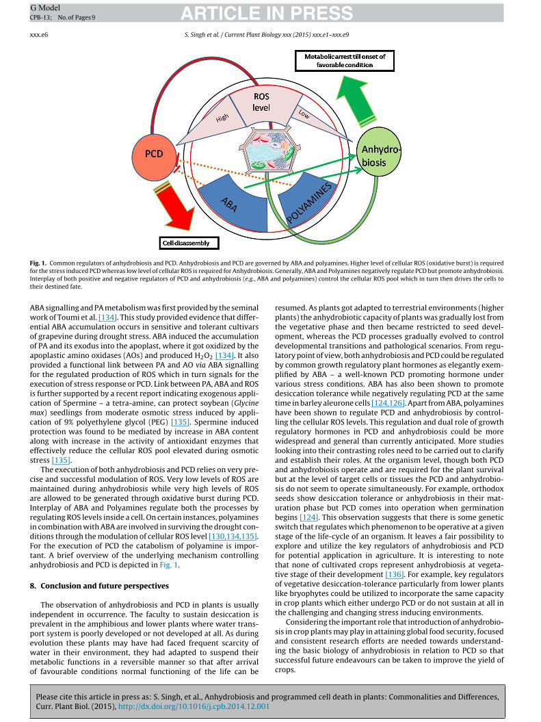
Fig. 1. Common regulators of anhydrobiosis and PCD. Anhydrobiosis and PCD are governed by ABA and polyamines. Higher level of cellular ROS (oxidative burst) is requiredf iosis.
I BA ant
Aweooapfeicmcpaes
cmaIridFta
8
ippewmo
or the stress induced PCD whereas low level of cellular ROS is required for Anhydrobnterplay of both positive and negative regulators of PCD and anhydrobiosis (e.g., Aheir destined fate.
BA signalling and PA metabolism was first provided by the seminalork of Toumi et al. [134]. This study provided evidence that differ-
ntial ABA accumulation occurs in sensitive and tolerant cultivarsf grapevine during drought stress. ABA induced the accumulationf PA and its exodus into the apoplast, where it got oxidized by thepoplastic amino oxidases (AOs) and produced H2O2 [134]. It alsorovided a functional link between PA and AO via ABA signallingor the regulated production of ROS which in turn signals for thexecution of stress response or PCD. Link between PA, ABA and ROSs further supported by a recent report indicating exogenous appli-ation of Spermine – a tetra-amine, can protect soybean (Glycineax) seedlings from moderate osmotic stress induced by appli-
ation of 9% polyethylene glycol (PEG) [135]. Spermine inducedrotection was found to be mediated by increase in ABA contentlong with increase in the activity of antioxidant enzymes thatffectively reduce the cellular ROS pool elevated during osmotictress [135].
The execution of both anhydrobiosis and PCD relies on very pre-ise and successful modulation of ROS. Very low levels of ROS areaintained during anhydrobiosis while very high levels of ROS
re allowed to be generated through oxidative burst during PCD.nterplay of ABA and Polyamines regulate both the processes byegulating ROS levels inside a cell. On certain instances, polyaminesn combination with ABA are involved in surviving the drought con-itions through the modulation of cellular ROS level [130,134,135].or the execution of PCD the catabolism of polyamine is impor-ant. A brief overview of the underlying mechanism controllingnhydrobiosis and PCD is depicted in Fig. 1.
. Conclusion and future perspectives
The observation of anhydrobiosis and PCD in plants is usuallyndependent in occurrence. The faculty to sustain desiccation isrevalent in the amphibious and lower plants where water trans-ort system is poorly developed or not developed at all. As during
Please cite this article in press as: S. Singh, et al., Anhydrobiosis and pCurr. Plant Biol. (2015), http://dx.doi.org/10.1016/j.cpb.2014.12.001
volution these plants may have had faced frequent scarcity ofater in their environment, they had adapted to suspend theiretabolic functions in a reversible manner so that after arrival
f favourable conditions normal functioning of the life can be
Generally, ABA and Polyamines negatively regulate PCD but promote anhydrobiosis.d polyamines) control the cellular ROS pool which in turn then drives the cells to
resumed. As plants got adapted to terrestrial environments (higherplants) the anhydrobiotic capacity of plants was gradually lost fromthe vegetative phase and then became restricted to seed devel-opment, whereas the PCD processes gradually evolved to controldevelopmental transitions and pathological scenarios. From regu-latory point of view, both anhydrobiosis and PCD could be regulatedby common growth regulatory plant hormones as elegantly exem-plified by ABA – a well-known PCD promoting hormone undervarious stress conditions. ABA has also been shown to promotedesiccation tolerance while negatively regulating PCD at the sametime in barley aleurone cells [124,126]. Apart from ABA, polyamineshave been shown to regulate PCD and anhydrobiosis by control-ling the cellular ROS levels. This regulation and dual role of growthregulatory hormones in PCD and anhydrobiosis could be morewidespread and general than currently anticipated. More studieslooking into their contrasting roles need to be carried out to clarifyand establish their roles. At the organism level, though both PCDand anhydrobiosis operate and are required for the plant survivalbut at the level of target cells or tissues the PCD and anhydrobio-sis do not seem to operate simultaneously. For example, orthodoxseeds show desiccation tolerance or anhydrobiosis in their mat-uration phase but PCD comes into operation when germinationbegins [124]. This observation suggests that there is some geneticswitch that regulates which phenomenon to be operative at a givenstage of the life-cycle of an organism. It leaves a fair possibility toexplore and utilize the key regulators of anhydrobiosis and PCDfor potential application in agriculture. It is interesting to notethat none of cultivated crops represent anhydrobiosis at vegeta-tive stage of their development [136]. For example, key regulatorsof vegetative desiccation-tolerance particularly from lower plantslike bryophytes could be utilized to incorporate the same capacityin crop plants which either undergo PCD or do not sustain at all inthe challenging and changing stress inducing environments.
Considering the important role that introduction of anhydrobio-sis in crop plants may play in attaining global food security, focused
rogrammed cell death in plants: Commonalities and Differences,
and consistent research efforts are needed towards understand-ing the basic biology of anhydrobiosis in relation to PCD so thatsuccessful future endeavours can be taken to improve the yield ofcrops.
ING ModelC
Biolog
A
Bt
R
ARTICLEPB-13; No. of Pages 9
S. Singh et al. / Current Plant
cknowledgement
This work has been supported by a grant from Department ofiotechnology, Govt. India, as a part of Ramalingaswami fellowshipo BST.
eferences
[1] J.R. Phillips, M.J. Oliver, D. Bartels, Molecular genetics of desiccation and tol-erant systems, in: M. Black, H.W. Pritchard (Eds.), Desiccation and Survival inPlants: Drying Without Dying, CAB International, Wallingford, UK, 2002, pp.319–341.
[2] M. Watanabe, T. Sakashita, A. Fujita, T. Kikawada, D.D. Horikawa, Y.Nakahara, S. Wada, T. Funayama, N. Hamada, Y. Kobayashi, T. Okuda,Biological effects of anhydrobiosis in an African chironomid, Polypedilum van-derplanki on radiation tolerance, Int. J. Radiat. Biol. 82 (2006) 587–592.
[3] L. Rebecchi, T. Altiero, R. Guidetti, Anhydrobiosis: the extreme limit of desic-cation tolerance, ISJ 4 (2007) 65–81.
[4] J. Maia, B.J. Dekkers, N.J. Provart, W. Ligterink, H.W. Hilhorst, The re-establishment of desiccation tolerance in germinated Arabidopsis thalianaseeds and its associated transcriptome, PLoS ONE 6 (2011) e29123.
[5] D.F. Gaff, Mechanisms of desiccation tolerance in resurrection vascular plants,in: A.S. Basra (Ed.), Mechanisms of Environmental Stress Resistance in Plants,Harwood Academic Publishers, Amsterdam, 1997, pp. 43–58.
[6] F.A. Hoekstra, E.A. Golovina, J. Buitink, Mechanisms of plant desiccation tol-erance, Trends Plant Sci. 6 (2001) 431–438.
[7] A.I. Tuzhikov, B.B. Vartapetian, A.B. Vartapetian, N.V. Chichkova, Abioticstress-induced programmed cell death in plants: a phytaspase connection, in:A. Shanker (Ed.), Abiotic Stress Response in Plants – Physiological, Biochemicaland Genetic Perspectives, 2011, pp. 183–196.
[8] K.V. Krishnamurthy, R. Krishnaraj, R. Chozhavendan, F.S. Christopher, The pro-gramme of cell death in plants and animals – a comparison, Curr. Sci. 79 (2000)1169–1181.
[9] M.J. Oliver, Z. Tuba, B.D. Mishler, The evolution of vegetative desiccation tol-erance in land plants, Plant Ecol. 151 (2000) 85–100.
[10] M.J. Oliver, J. Velten, B.D. Mishler, Desiccation tolerance in bryophytes: areflection of the primitive strategy for plant survival in dehydrating habitats?Integr. Comp. Biol. 45 (2005) 788–799.
[11] O. Toldi, Z.N. Tuba, P. Scott, Vegetative desiccation tolerance: is it a goldminefor bioengineering crops? Plant Sci. 176 (2009) 187–199.
[12] J.M. Farrant, J.P. Moore, Programming desiccation-tolerance: from plants toseeds to resurrection plants, Curr. Opin. Plant Biol. 14 (2011) 340–345.
[13] P.C. Alpert, W. Oechel, Comparative patterns of net photosynthesis in anassemblage of mosses with contrasting microdistributions, Am. J. Bot. 74(1987) 1787–1796.
[14] Z. Csintalan, Z. Takacs, M.C.F. Proctor, H.K. Lichtenthaler, Z. Tuba, Desiccationand rehydration responses of desiccation tolerant moss and lichen speciesfrom a temperate semidesert grassland, J. Hattori Bot. Lab. (1998) 71–80.
[15] Z. Csintalan, M.C.F. Proctor, Z. Tuba, Chlorophyll fluorescence during dry-ing and rehydration in the mosses Rhytidiadelphus loreus (Hedw.) Warnst.,Anomodon viticulosus (Hedw.) Hook. & Tayl. and Grimmia pulvinata (Hedw.),Sm. Ann. Bot. 84 (1999) 235–245.
[16] R. Cruz de Carvalho, M. Catala, J. Marques da Silva, C. Branquinho, E.Barreno, The impact of dehydration rate on the production and cellular loca-tion of reactive oxygen species in an aquatic moss, Ann. Bot. 110 (2012)1007–1016.
[17] J.C. Ameisen, The Origin and Evolution of Programmed Cell Death. In eLS, JohnWiley & Sons, 2009.
[18] A.C. Leopold, Membranes, Metabolism and Dry Organisms, Cornell UniversityPress, Ithaca, New York, 1986.
[19] S.N. Tripathi, B.S. Tiwari, E.R.S. Talpasayi, Growth of cyanobacteria on urbanbuildings, Energy Build. 15–16 (1991) 499–505.
[20] S.N. Tripathi, B.S. Tiwari, Photofixation of 14CO2 and photosynthetic electrontransport in a roof top cyanobacterium Scytonema geitleri, in: N. Murata (Ed.),Researches in Photosynthesis IV, vol. IV, Kluwer Academic Publishers, TheNetherland, 1992, pp. 267–270.
[21] B.S. Tiwari, S.N. Tripathi, Effect of hydration and dehydration on initiation anddynamics of some physiological reactions in desiccation tolerant cyanobac-terium Scytonema geitleri, Indian J. Biochem. Biophys. 35 (1998) 172–178.
[22] B.S. Tiwari, S.N. Tripathi, Water binding in sub-aerial cyanobacteria, Indian J.Biochem. Biophys. 35 (1998) 52–61.
[23] T.S. Gechev, C. Dinakar, M. Benina, V. Toneva, D. Bartels, Molecular mecha-nisms of desiccation tolerance in resurrection plants, Cell. Mol. Life Sci. 69(2012) 3175–3186.
[24] B.A. Whitton, The Ecology of Cyanobacteria II: Their Diversity in Space andTime, Springer, 2012.
[25] C.B. Lipman, The successful revival of Nostoc commune from a herbariumspecimen eighty-seven years old, Bull. Torrey Bot. Club. 68 (1941) 664–666.
Please cite this article in press as: S. Singh, et al., Anhydrobiosis and pCurr. Plant Biol. (2015), http://dx.doi.org/10.1016/j.cpb.2014.12.001
[26] R.E. Cameron, Species of Nostoc vaucher Occurring in the Sonoran Desert inArizona, Transact. Am. Microsc. Soc. 81 (1962) 379–384.
[27] P. Tripathy, A. Roy, N. Anand, S.P. Adhikari, Blue-green algal flora on therock surface of temples and monuments of India, Feddes Repert. 110 (1999)133–144.
PRESSy xxx (2015) xxx.e1–xxx.e9 xxx.e7
[28] N. Keshari, S.P. Adhikary, Characterization of cyanobacteria isolated frombiofilms on stone monuments at Santiniketan, India, Biofouling 29 (2013)525–536.
[29] Z. Tuba, Z. Cintalan, M.C.F. Proctor, Photosynthetic responses of a moss, Tortularuraliss sp. ruralis, and the lichens Cladonia convolute and C. jurcata to waterdeficit and short periods of desicication, and their ecophysiological signifi-cance: a baseline study at present-day CO2 concentration, New Phytoli 133(1996) 353–361.
[30] M. Jensen, S. Chakir, G.B. Feige, Osmotic and atmospheric dehydration effectsin the lichens Hypogymniaphysodes, Lob ariapulmonaria, and Peltigera phtosa:an in vivo study of the chlorophyll fluorescence induction, Photosynthetica37 (1999) 393–404.
[31] M.C. Proctor, R. Ligrone, J.G. Duckett, Desiccation tolerance in the mossPolytrichum formosum: physiological and fine-structural changes during des-iccation and recovery, Ann. Bot. 99 (2007) 75–93.
[32] M.C.F. Proctor, M.J. Oliver, A.J. Wood, P. Alpert, L.R. Stark, N.L. Cleavitt, B.D.Mishler, Desiccation tolerance in bryophytes: a review, Bryologist 110 (2007)595–621.
[33] J.M. Farrant, A. Lehner, K. Cooper, S. Wiswedel, Desiccation tolerance in thevegetative tissues of the fern Mohria caffrorum is seasonally regulated, PlantJ. 57 (2009) 65–79.
[34] G.G. Franchi, B. Piotto, M. Nepi, C.C. Baskin, J.M. Baskin, E. Pacini, Pollen andseed desiccation tolerance in relation to degree of developmental arrest, dis-persal, and survival, J. Exp. Bot. 62 (2011) 5267–5281.
[35] S. Porembski, W. Barthlott, Granitic and gneissic outcrops (inselbergs) as cen-ter of diversity for desiccation-tolerant vascular plants, Plant Ecol. 151 (2000)19–28.
[36] J.C. Ameisen, On the origin, evolution, and nature of programmed cell death:a timeline of four billion years, Cell Death Differ. 9 (2002) 367–393.
[37] J.H. Crowe, Trehalose as a chemical chaperone: fact and fantasy, Adv. Exp.Med. Biol. 594 (2007) 143–158.
[38] M. Potts, Desiccation tolerance of prokaryotes, Microbiol. Rev. 58 (1994)755–805.
[39] B.E. Tease, R.W. Walker, Comparative composition of the sheath of thecyanobacterium Gloeothece ATCC27152 cultured with and without combinednitrogen, J. Gen. Microbiol. 133 (1987) 3331–3339.
[40] K. Hosi, J. Okamoto, Y. Tanji, H. Unno, Sedimentation and germination prop-erties of Anabaena akinates, Biochem. Eng. J. 14 (2003) 67–73.
[41] X.Q. Wang, P.F. Yang, Z. Liu, W.Z. Liu, Y. Hu, H. Chen, T.Y. Kuang, Z.M. Pei,S.H. Shen, Y.K. He, Exploring the mechanism of Physcomitrella patens des-iccation tolerance through a proteomic strategy, Plant Physiol. 149 (2009)1739–1750.
[42] J.M. Farrant, A comparison of mechanisms of desiccation tolerance amongthree angiosperm resurrection plant species, Plant Ecol. 151 (2000) 29–39.
[43] D.F. Gaff, M. Oliver, The evolution of desiccation tolerance in angiospermplants: a rare yet common phenomenon, Funct. Plant Biol. (2013) 315–328.
[44] K. Brawn, I. Fridovich, DNA strand scission by enzymically generated oxygenradicals, Arch. Biochem. Biophys. 206 (1981) 414–419.
[45] N. Smirnoff, The role of active oxygen in the response of plants to water deficitand desiccation, New Phytol. 125 (1993) 27–58.
[46] B.D. McKersie, Y.Y. Leshem, Stress and Stress Coping in Cultivated Plants,Kluwer Academic Publishers, Dordrecht, The Netherlands, 1994.
[47] A.M. Gilmore, Mechanistic aspects of xanthophyll cycle-dependent photo-protection in higher plant chloroplasts and leaves, Physiol. Plant. 99 (1997)197–209.
[48] I. Kranner, F. Lutzoni, Evolutionary consequences of transition to a lichen sym-biotic state and physiological adaptation to oxidative damage associated withpoikilohydry, in: H.R. Lerner (Ed.), Plant Response to Environmental Stress:From Phytohormones to Genome Reorganisation, Marcel Dekker, New York,1999, pp. 591–628.
[49] I. Kranner, S. Birtic, A modulating role for antioxidants in desiccation toler-ance, Integr. Comp. Biol. 45 (2005) 734–740.
[50] K. Satoh, M. Hirai, J. Nishio, T. Yamaji, Y. Kashino, H. Koike, Recoveryof photosynthetic systems during rewetting is quite rapid in a terrestrialcyanobacterium, Nostoc commune, Plant Cell Physiol. 43 (2002) 170–176.
[51] U. Heber, Conservation and dissipation of light energy in desiccation-tolerantphotoautotrophs, two sides of the same coin, Photosynth. Res. 113 (2012)5–13.
[52] G. Tanou, C. Job, L. Rajjou, E. Arc, M. Belghazi, G. Diamantidis, A. Molassiotis,D. Job, Proteomics reveals the overlapping roles of hydrogen peroxide andnitric oxide in the acclimation of citrus plants to salinity, Plant J. 60 (2009)795–804.
[53] G. Tanou, C. Job, M. Belghazi, A. Molassiotis, G. Diamantidis, D. Job, Proteomicsignatures uncover hydrogen peroxide and nitric oxide cross-talk signalingnetwork in citrus plants, J. Proteome Res. 9 (2010) 5994–6006.
[54] X. Bai, L. Yang, M. Tian, J. Chen, J. Shi, Y. Yang, X. Hu, Nitric oxide enhancesdesiccation tolerance of recalcitrant Antiaris toxicaria seeds via protein S-nitrosylation and carbonylation, PLoS ONE 6 (2011) e20714.
[55] D. Bartels, F. Salamini, Desiccation tolerance in the resurrection plantCraterostigma plantagineum. A contribution to the study of drought toleranceat the molecular level, Plant Physiol. 127 (2001) 1346–1353.
rogrammed cell death in plants: Commonalities and Differences,
[56] D.K. Hincha, M. Hagemann, Stabilization of model membranes during dryingby compatible solutes involved in the stress tolerance of plants and microor-ganisms, Biochem. J. 383 (2004) 277–283.
[57] J.S. Clegg, The physical properties and metabolic status of Artemia cysts atlow water contents: the water replacement hypothesis, in: A. Leopold (Ed.),
ING ModelC
x Biolo
ARTICLEPB-13; No. of Pages 9
xx.e8 S. Singh et al. / Current Plant
Membranes, Metabolism and Dry Organisms, Cornell University Press, NewYork, 1986, pp. 169–187.
[58] J.H. Crowe, J.F. Carpenter, L.M. Crowe, The role of vitrification in anhydrobiosis,Annu. Rev. Physiol. 60 (1998) 73–103.
[59] J. Buitink, O. Leprince, Intracellular glasses and seed survival in the dry state,C. R. Biol. 331 (2008) 788–795.
[60] M. Sakurai, T. Furuki, K. Akao, D. Tanaka, Y. Nakahara, T. Kikawada, M.Watanabe, T. Okuda, Vitrification is essential for anhydrobiosis in an Africanchironomid, Polypedilum vanderplanki, Proc. Natl. Acad. Sci. U. S. A. 105 (2008)5093–5098.
[61] S. Hengherr, A.G. Heyer, F. Brummer, R.O. Schill, Trehalose and vitreous states:desiccation tolerance of dormant stages of the crustaceans Triops and Daphnia,Physiol. Biochem. Zool. 84 (2011) 147–153.
[62] S. Hengherr, R.O. Schill, J.S. Clegg, Mechanisms associated with cellular desic-cation tolerance in the animal extremophile artemia, Physiol. Biochem. Zool.84 (2011) 249–257.
[63] M. Battaglia, Y. Olvera-Carrillo, A. Garciarrubio, F. Campos, A.A. Covarrubias,The enigmatic LEA proteins and other hydrophilins, Plant Physiol. 148 (2008)6–24.
[64] Y. Liu, L. Wang, X. Xing, L. Sun, J. Pan, X. Kong, M. Zhang, D. Li, ZmLEA3, amultifunctional group 3 LEA protein from maize (Zea mays L.), is involved inbiotic and abiotic stresses, Plant Cell Physiol. 54 (2013) 944–959.
[65] K. Goyal, L.J. Walton, A. Tunnacliffe, LEA proteins prevent protein aggregationdue to water stress, Biochem. J. 388 (2005) 151–157.
[66] A. Tunnacliffe, M.J. Wise, The continuing conundrum of the LEA proteins,Naturwissenschaften 94 (2007) 791–812.
[67] G. Iturriaga, The LEA proteins and trehalose loving couple: a step forward inanhydrobiotic engineering, Biochem. J. 410 (2008) e1–e2.
[68] C. Wang, M.A. Grohme, B. Mali, R.O. Schill, M. Frohme, Towards decryptingcryptobiosis – analyzing anhydrobiosis in the tardigrade Milnesium tardi-gradum using transcriptome sequencing, PLoS ONE 9 (2014) e92663.
[69] A. Tunnacliffe, D. Hincha, O. Leprince, D. Macherel, LEA proteins: versatility ofform and function, in: E. Lubzens, J. Cerda, M. Clark (Eds.), Sleeping Beauties –Dormancy and Resistance in Harsh Environments, Springer, Berlin, 2010, pp.91–108.
[70] D.K. Hincha, A. Thalhammer, LEA proteins: IDPs with versatile functions incellular dehydration tolerance, Biochem. Soc. Trans. 40 (2012) 1000–1003.
[71] S. Chakrabortee, F. Meersman, G.S. Kaminski Schierle, C.W. Bertoncini, B.McGee, C.F. Kaminski, A. Tunnacliffe, Catalytic and chaperone-like functionsin an intrinsically disordered protein associated with desiccation tolerance,Proc. Natl. Acad. Sci. U. S. A. 107 (2010) 16084–16089.
[72] R.A. Lockshin, Z. Zakeri, Apoptosis, autophagy, and more, Int. J. Biochem. CellBiol. 36 (2004) 2405–2419.
[73] R.R. Buss, W. Sun, R.W. Oppenheim, Adaptive roles of programmed cell deathduring nervous system development, Annu. Rev. Neurosci. 29 (2006) 1–35.
[74] E. Lam, Programmed cell death in plants: orchestrating an intrinsic suicideprogram within walls, Crit. Rev. Plant Sci. 27 (2008) 413–423.
[75] M.C. Drew, C.J. He, P.W. Morgan, Programmed cell death and aerenchymaformation in roots, Trends Plant Sci. 5 (2000) 123–127.
[76] A. Fath, P. Bethke, J. Lonsdale, R. Meza-Romero, R. Jones, Programmed celldeath in cereal aleurone, Plant Mol. Biol. 44 (2000) 255–266.
[77] F.A. Hoeberichts, A. de Jong, E.J. Woltering, Apoptotic-like cell death marks theearly stages of gypsophila (Gypsophila paniculata) petal senescence, Posthar-vest Biol. Technol. 35 (2004) 229–236.
[78] B. Bollhoner, J. Prestele, H. Tuominen, Xylem cell death: emerging under-standing of regulation and function, J. Exp. Bot. 63 (2012) 1081–1094.
[79] R. Mittler, Oxidative stress, antioxidants and stress tolerance, Trends PlantSci. 7 (2002) 405–410.
[80] M.B. Dickman, R. Fluhr, Centrality of host cell death in plant–microbe inter-actions, Annu. Rev. Phytopathol. 51 (2013) 543–570.
[81] E. Aizenman, H. Engelberg-Kulka, G. Glaser, An Escherichia coli chro-mosomal addiction module regulated by guanosine [corrected] 3′ ,5′-bispyrophosphate: a model for programmed bacterial cell death, Proc. Natl.Acad. Sci. U. S. A. 93 (1996) 6059–6063.
[82] H. Engelberg-Kulka, G. Glaser, Addiction modules and programmed cell deathand antideath in bacterial cultures, Annu. Rev. Microbiol. 53 (1999) 43–70.
[83] H. Engelberg-Kulka, S. Amitai, I. Kolodkin-Gal, R. Hazan, Bacterial pro-grammed cell death and multicellular behavior in bacteria, PLoS Genet. 2(2006) e135.
[84] C. Giusti, E. Tresse, M.F. Luciani, P. Golstein, Autophagic cell death: analysis inDictyostelium, Biochim. Biophys. Acta 1793 (2009) 1422–1431.
[85] J. Calvo-Garrido, S. Carilla-Latorre, Y. Kubohara, N. Santos-Rodrigo, A.Mesquita, T. Soldati, P. Golstein, R. Escalante, Autophagy in Dictyostelium:genes and pathways, cell death and infection, Autophagy 6 (2010) 686–701.
[86] M. Segovia, L. Haramaty, J.A. Berges, P.G. Falkowski, Cell death in the uni-cellular chlorophyte Dunaliella tertiolecta. A hypothesis on the evolution ofapoptosis in higher plants and metazoans, Plant Physiol. 132 (2003) 99–105.
[87] A. Darehshouri, M. Affenzeller, U. Lutz-Meindl, Cell death upon H2O2 induc-tion in the unicellular green alga Micrasterias, Plant Biol. (Stuttg.) 10 (2008)732–745.
[88] S. Moharikar, J.S. D’Souza, A.B. Kulkarni, J. Basuthkar, B.J. Rao, Apoptotic-like
Please cite this article in press as: S. Singh, et al., Anhydrobiosis and pCurr. Plant Biol. (2015), http://dx.doi.org/10.1016/j.cpb.2014.12.001
cell death pathway is induced in unicellular chlorophyte Chlamydomonasreinhardtii (Chlorophyceae) cells following UV irradiation: detection andfunctional analyses, J. Phycol. 42 (2006) 423–433.
[89] A. Zuppini, C. Andreoli, B. Baldan, Heat stress: an inducer of programmed celldeath in Chlorella saccharophila, Plant Cell Physiol. 48 (2007) 1000–1009.
PRESSgy xxx (2015) xxx.e1–xxx.e9
[90] J.A. Berges, G.F. Paul, Physiological stress and cell death in marine phytoplank-ton: induction of proteases in response to nitrogen or light limitation, Limnol.Oceanogr. 43 (1998) 129–135.
[91] B. Belenghi, M.C. Romero-Puertas, D. Vercammen, A. Brackenier, D. Inze, M.Delledonne, F. Van Breusegem, Metacaspase activity of Arabidopsis thaliana isregulated by S-nitrosylation of a critical cysteine residue, J. Biol. Chem. 282(2007) 1352–1358.
[92] X. Wang, H. Feng, C. Tang, P. Bai, G. Wei, L. Huang, Z. Kang, TaMCA4, a novelwheat metacaspase gene functions in programmed cell death induced by thefungal pathogen Puccinia striiformis f. sp. tritici, Mol. Plant Microbe Interact.25 (2012) 755–764.
[93] I.P. de León, The MOSS Physcomitrella patens as a model system to study inter-actions between plants and phytopathogenic fungi and oomycetes, J. Pathog.2011 (2011) 1–6.
[94] K. Landberg, E.R. Pederson, T. Viaene, B. Bozorg, J. Friml, H. Jonsson, M.Thelander, E. Sundberg, The MOSS Physcomitrella patens reproductive organdevelopment is highly organized, affected by the two SHI/STY genes and bythe level of active auxin in the SHI/STY expression domain, Plant Physiol. 162(2013) 1406–1419.
[95] L.H. Filonova, S. von Arnold, G. Daniel, P.V. Bozhkov, Programmed cell deatheliminates all but one embryo in a polyembryonic plant seed, Cell Death Differ.9 (2002) 1057–1062.
[96] L.H. Filonova, P.V. Bozhkov, V.B. Brukhin, G. Daniel, B. Zhivotovsky, S. vonArnold, Two waves of programmed cell death occur during formation anddevelopment of somatic embryos in the gymnosperm, Norway spruce, J. CellSci. 113 (Pt. 24) (2000) 4399–4411.
[97] P.V. Bozhkov, M.F. Suarez, L.H. Filonova, G. Daniel, A.A. Zamyatnin Jr., S.Rodriguez-Nieto, B. Zhivotovsky, A. Smertenko, Cysteine protease mcII-Paexecutes programmed cell death during plant embryogenesis, Proc. Natl.Acad. Sci. U. S. A. 102 (2005) 14463–14468.
[98] T.E. Young, D.R. Gallie, Regulation of programmed cell death in maizeendosperm by abscisic acid, Plant Mol. Biol. 42 (2000) 397–414.
[99] T.E. Young, D.R. Gallie, Programmed cell death during endosperm develop-ment, Plant Mol. Biol. 44 (2000) 283–301.
[100] H. Fukuda, Programmed cell death of tracheary elements as a paradigm inplants, Plant Mol. Biol. 44 (2000) 245–253.
[101] H. Kuriyama, H. Fukuda, Developmental programmed cell death in plants,Curr. Opin. Plant Biol. 5 (2002) 568–573.
[102] J. Cao, X.Q. He, Y.Q. Wang, Programmed cell death during secondaryxylem differentiation in Eucommia ulmoides, Acta Bot. Sin. 45 (2003)1465–1474.
[103] U. Avci, H.E. Petzold, I.O. Ismail, E.P. Beers, C.H. Haigler, Cysteine proteasesXCP1 and XCP2 aid micro-autolysis within the intact central vacuole duringxylogenesis in Arabidopsis roots, Plant J. 56 (2008) 303–315.
[104] B. Uzelac, D. Janosevic, S. Budimir, In situ detection of programmed cell deathin Nicotiana tabacum leaves during senescence, J. Microsc. 230 (2008) 1–3.
[105] T. Yamada, K. Ichimura, M. Kanekatsu, W.G. van Doorn, Homologs of genesassociated with programmed cell death in animal cells are differentiallyexpressed during senescence of Ipomoea nil petals, Plant Cell Physiol. 50(2009) 610–625.
[106] W.G. van Doorn, E.P. Beers, J.L. Dangl, V.E. Franklin-Tong, P. Gallois, I. Hara-Nishimura, A.M. Jones, M. Kawai-Yamada, E. Lam, J. Mundy, L.A. Mur, M.Petersen, A. Smertenko, M. Taliansky, F. Van Breusegem, T. Wolpert, E. Wolter-ing, B. Zhivotovsky, P.V. Bozhkov, Morphological classification of plant celldeaths, Cell Death Differ. 18 (2011) 1241–1246.
[107] J.D. Bewley, Physiological aspects of desiccation tolerance, Ann. Rev. PlantPhysiol. 30 (1979) 195–238.
[108] B. Rodriguez-Porrata, D. Carmona-Gutierrez, G. Lopez-Matinez, A. Reisenbich-ler, M. Bauer, F. Madeo, C.-O. Ricardo, Yeast cell death during the drying andrehydration process, in: I. Schmid (Ed.), Flow Cytometry – Recent Perspec-tives, 2012.
[109] S. Wissing, P. Ludovico, E. Herker, S. Buttner, S.M. Engelhardt, T. Decker, A.Link, A. Proksch, F. Rodrigues, M. Corte-Real, K.U. Frohlich, J. Manns, C. Cande,S.J. Sigrist, G. Kroemer, F. Madeo, An AIF orthologue regulates apoptosis inyeast, J. Cell Biol. 166 (2004) 969–974.
[110] S. Buttner, T. Eisenberg, D. Carmona-Gutierrez, D. Ruli, H. Knauer, C. Ruck-enstuhl, C. Sigrist, S. Wissing, M. Kollroser, K.U. Frohlich, S. Sigrist, F. Madeo,Endonuclease G regulates budding yeast life and death, Mol. Cell 25 (2007)233–246.
[111] K. Dolinski, S. Muir, M. Cardenas, J. Heitman, All cyclophilins and FK506binding proteins are, individually and collectively, dispensable for viabil-ity in Saccharomyces cerevisiae, Proc. Natl. Acad. Sci. U. S. A. 94 (1997)13093–13098.
[112] S.Y. Lee, C. Hunte, S. Malaney, B.H. Robinson, The N-terminus of the Qcr7protein of the cytochrome bc(1) complex in S. cerevisiae may be involved infacilitating stability of the subcomplex with the Qcr8 protein and cytochromeb, Arch. Biochem. Biophys. 393 (2001) 215–221.
[113] V. Zara, I. Palmisano, L. Conte, B.L. Trumpower, Further insights into theassembly of the yeast cytochrome bc1 complex based on analysis of single anddouble deletion mutants lacking supernumerary subunits and cytochrome b,Eur. J. Biochem. 271 (2004) 1209–1218.
rogrammed cell death in plants: Commonalities and Differences,
[114] R.W. King, Abscisic acid in developing wheat grains and its relationship tograin growth and maturation, Planta 132 (1976) 43–51.
[115] R.S. Quatrano, Regulation of gene expression by abscisic acid duringangiosperm embryo development, Oxf. Surv. Plant Mol. Cell Biol. 3 (1986)467–476.
ING ModelC
Biolog
[
[
[
[
[
[
[
[
[
[
[
ARTICLEPB-13; No. of Pages 9
S. Singh et al. / Current Plant
116] M. Black, Involvement of ABA in the physiology of developing and matureseeds, in: W.J. Daviesa, H.G. Jones (Eds.), Abscisic Acid: Physiology and Bio-chemistry, Bios Scientific Publishers, Oxford, UK, 1991, pp. 99–124.
117] P.M. Chandler, M. Robertson, Gene expression regulated by abscisic acid andits relation to stress tolerance, Annu. Rev. Plant Physiol. Plant Mol. Biol. 45(1994) 113–141.
118] T.J. Close, Dehydrins: emergence of a biochemical role of a family of plantdehydration proteins, Physiol. Plant. 97 (2006) 795–803.
119] A. Furini, C. Koncz, F. Salamini, D. Bartels, High level transcription of a memberof a repeated gene family confers dehydration tolerance to callus tissue ofCraterostigma plantagineum, EMBO J. 16 (1997) 3599–3608.
120] M.M. Johri, Hormonal regulation in green plant lineage families, Physiol. Mol.Biol. Plants 14 (2008) 23–38.
121] A. Khandelwal, S.H. Cho, H. Marella, Y. Sakata, P.F. Perroud, A. Pan, R.S. Qua-trano, Role of ABA and ABI3 in desiccation tolerance, Science 327 (2010)546.
122] H.H. Marella, Y. Sakata, R.S. Quatrano, Characterization and functional analysisof ABSCISIC ACID INSENSITIVE3-like genes from Physcomitrella patens, PlantJ. 46 (2006) 1032–1044.
123] Y.L. Du, Z.Y. Wang, J.W. Fan, N.C. Turner, T. Wang, F.M. Li, Beta-aminobutyricacid increases abscisic acid accumulation and desiccation tolerance anddecreases water use but fails to improve grain yield in two spring wheatcultivars under soil drying, J. Exp. Bot. 63 (2012) 4849–4860.
124] P.C. Bethke, J.E. Lonsdale, A. Fath, R.L. Jones, Hormonally regulated pro-grammed cell death in barley aleurone cells, Plant Cell 11 (1999) 1033–1046.
Please cite this article in press as: S. Singh, et al., Anhydrobiosis and pCurr. Plant Biol. (2015), http://dx.doi.org/10.1016/j.cpb.2014.12.001
125] A.B. Bleecker, S.E. Patterson, Last exit: senescence, abscission, and meristemarrest in Arabidopsis, Plant Cell 9 (1997) 1169–1179.
126] W.J. Guo, T.H. Ho, An abscisic acid-induced protein, HVA22, inhibitsgibberellin-mediated programmed cell death in cereal aleurone cells, PlantPhysiol. 147 (2008) 1710–1722.
PRESSy xxx (2015) xxx.e1–xxx.e9 xxx.e9
[127] Y. Zhong, C. Ciafré, Role of ABA in ethylene-independent Iris flower senes-cence, in: 2011 Int. Conf. Food Eng. Biotechnol. IPCBEE, vol. 9, IACSIT Press,2011, pp. 261–266.
[128] J. Martin-Tanguy, Metabolism and function of polyamines in plant: recentdevelopment (NewApproaches), Plant Growth Reg. 34 (2001) 135–148.
[129] K. Liu, H. Fu, Q. Bei, S. Luan, Inward potassium channel in guard cells as atarget for polyamine regulation of stomatal movements, Plant Physiol. 124(2000) 1315–1326.
[130] D. Unal, A. Senkardesler, A. Sukatar, Abscisic acid and polyamine contentsin the Lichens Pseudevernia furfuracea and Ramalina farinacea, Russ. J. PlantPhysiol. 55 (2008) 115–118.
[131] P.N. Moschou, K.A. Roubelakis-Angelakis, Polyamines and programmed celldeath, J. Exp. Bot. (2013).
[132] A.K. Papadakis, K.A. Roubelakis-Angelakis, Polyamines inhibit NADPHoxidase-mediated superoxide generation and putrescine prevents pro-grammed cell death induced by polyamine oxidase-generated hydrogenperoxide, Planta 220 (2005) 826–837.
[133] H. Yoda, Y. Hiroi, H. Sano, Polyamine oxidase is one of the key elements foroxidative burst to induce programmed cell death in tobacco cultured cells,Plant Physiol. 142 (2006) 193–206.
[134] I. Toumi, P.N. Moschou, K.A. Paschalidis, B. Bouamama, A. Ben Salem-Fnayou,A.W. Ghorbel, A. Mliki, K.A. Roubelakis-Angelakis, Abscisic acid signalsreorientation of polyamine metabolism to orchestrate stress responses viathe polyamine exodus pathway in grapevine, J. Plant Physiol. 167 (2010)519–525.
rogrammed cell death in plants: Commonalities and Differences,
[135] R. Radhakrishnan, I-J. Lee, Spermine promotes acclimation to osmotic stressby modifying antioxidant, abscisic acid, and jasmonic acid signals in soybean,J. Plant Growth Regul. 32 (2013) 22–30.
[136] P. Alpert, The limits and frontiers of desiccation-tolerant life, Integr. Comp.Biol. 46 (2005) 685–695.
Related Documents