Angular (Gothic) aortic arch leads to enhanced systolic wave reflection, central aortic stiffness, and increased left ventricular mass late after aortic coarctation repair: Evaluation with magnetic resonance flow mapping Phalla Ou, MD, a,b David S. Celermajer, MBBS, DSc, FRACP, c Olivier Raisky, MD, d Odile Jolivet, PhD, a Fanny Buyens, MS, a Alain Herment, PhD, a Daniel Sidi, MD, PhD, e Damien Bonnet, MD, PhD, e and Elie Mousseaux, MD, PhD a,f Objective: We sought to investigate the mechanism whereby a particular deformity of the aortic arch, an angulated Gothic shape, might lead to hypertension late after anatomically successful repair of aortic coarctation. Methods: Fifty-five normotensive patients with anatomically successful repair of aortic coarctation and either a Gothic (angulated) or a Romanesque (smooth and rounded) arch were studied with magnetic resonance angiography and flow mapping in both the ascending and descending aortas. Systolic waveforms, central aortic stiffness, and pulse velocity were measured. We hypothesized that arch angulation would result in enhanced systolic wave reflection with loss of energy across the aortic arch, as well as increased central aortic stiffness. Results: Twenty patients were found to have a Gothic, and 35 a Romanesque, arch. Patients with a Gothic arch showed markedly augmented systolic wave reflection (12 6 vs 5 0.3 mL, P .001) and greater loss of systolic wave height in the distal aorta (30% 16% vs 22% 12%, P .01) compared with that of subjects with a Romanesque arch. Pulse wave velocity was also increased with a Gothic arch (5.6 1.1 vs 4.1 1 m/s, P .0001), as well as left ventricular mass index (85 15 vs 77 20 g/m 2 ). Patients with a Romanesque arch had increased aortic stiffness compared with that of control subjects (stiffness -index, 3.9 0.9 vs 2.9 1; P .03). Conclusions: Angulated Gothic aortic arch is associated with increased systolic wave reflection, as well as increased central aortic stiffness and left ventricular mass index. These findings explain (at least in part) the association between this pattern of arch geometry and late hypertension at rest and on exercise in subjects after coarctation repair. D espite early and anatomically successful repair, patients with coarctation of the aorta (CoA) still remain at high risk for cardiovascular events. The major adverse prognostic factor includes late hypertension, which occurs in 20% to 40% of survivors of anatomically successful repair. 1-5 The pathophysiology of abnormal blood pressure in this setting is still unclear. Different mechanisms have been proposed, including, for example, mild residual stenosis at the coarctectomy site, hyperreactivity of the renin-angiotensin system, impairment of the baroreflex system, impairment of peripheral vascular reactivity, and increased aortic stiffness. 4 Recently, we have highlighted the association between abnormal aortic arch geometry and late hypertension in patients with anatomically successful CoA repair From INSERM UMR_S678, a CHU la Pitié- Salpétrière, Paris, France; the Departments of Pediatric Radiology, b Pediatric Cardio- vascular Surgery, d and Pediatric Cardiolo- gy, e UFR Necker-Enfants Malades, Univer- sity Rene Descartes Paris V, AP-HP, Paris, France; and the Department of Medicine, c University of Sydney, Sydney, Australia; and the Department of Cardiovascular Ra- diology, f Hôpital Européen Georges Pom- pidou, University Rene Descartes Paris V, AP-HP, Paris, France. Supported by a grant from the Fondation Wyeth pour la Santé de l’Enfant et de l’Adolescent (France) and from the Fédéra- tion Française de Cardiologie (France). Received for publication Sept 28, 2006; revisions received March 14, 2007; ac- cepted for publication March 29, 2007. Address for reprints: Phalla Ou, MD, De- partment of Pediatric Radiology, Hôpital Necker-Enfants Malades, 149, rue de Sèvres 75743 Paris Cedex 15, France (E-mail: [email protected]). J Thorac Cardiovasc Surg 2008;135:62-8 0022-5223/$34.00 Copyright © 2008 by The American Asso- ciation for Thoracic Surgery doi:10.1016/j.jtcvs.2007.03.059 Surgery for Congenital Heart Disease Ou et al 62 The Journal of Thoracic and Cardiovascular Surgery ● January 2008 CHD

Welcome message from author
This document is posted to help you gain knowledge. Please leave a comment to let me know what you think about it! Share it to your friends and learn new things together.
Transcript
AwvEPFa
Surgery for Congenital Heart Disease Ou et al
6
CHD
ngular (Gothic) aortic arch leads to enhanced systolicave reflection, central aortic stiffness, and increased left
entricular mass late after aortic coarctation repair:valuation with magnetic resonance flow mapping
halla Ou, MD,a,b David S. Celermajer, MBBS, DSc, FRACP,c Olivier Raisky, MD,d Odile Jolivet, PhD,a
anny Buyens, MS,a Alain Herment, PhD,a Daniel Sidi, MD, PhD,e Damien Bonnet, MD, PhD,e
nd Elie Mousseaux, MD, PhDa,f
Ooa
Mariswa
RP(dwa(s2
Cwioc
D2
Dsia
From INSERM UMR_S678,a CHU la Pitié-Salpétrière, Paris, France; the Departmentsof Pediatric Radiology,b Pediatric Cardio-vascular Surgery,d and Pediatric Cardiolo-gy,e UFR Necker-Enfants Malades, Univer-sity Rene Descartes Paris V, AP-HP, Paris,France; and the Department of Medicine,c
University of Sydney, Sydney, Australia;and the Department of Cardiovascular Ra-diology,f Hôpital Européen Georges Pom-pidou, University Rene Descartes Paris V,AP-HP, Paris, France.
Supported by a grant from the FondationWyeth pour la Santé de l’Enfant et del’Adolescent (France) and from the Fédéra-tion Française de Cardiologie (France).
Received for publication Sept 28, 2006;revisions received March 14, 2007; ac-cepted for publication March 29, 2007.
Address for reprints: Phalla Ou, MD, De-partment of Pediatric Radiology, HôpitalNecker-Enfants Malades, 149, rue deSèvres 75743 Paris Cedex 15, France(E-mail: [email protected]).
J Thorac Cardiovasc Surg 2008;135:62-8
0022-5223/$34.00
Copyright © 2008 by The American Asso-ciation for Thoracic Surgery
gdoi:10.1016/j.jtcvs.2007.03.059
2 The Journal of Thoracic and Cardiov
bjective: We sought to investigate the mechanism whereby a particular deformityf the aortic arch, an angulated Gothic shape, might lead to hypertension late afternatomically successful repair of aortic coarctation.
ethods: Fifty-five normotensive patients with anatomically successful repair ofortic coarctation and either a Gothic (angulated) or a Romanesque (smooth andounded) arch were studied with magnetic resonance angiography and flow mappingn both the ascending and descending aortas. Systolic waveforms, central aortictiffness, and pulse velocity were measured. We hypothesized that arch angulationould result in enhanced systolic wave reflection with loss of energy across the
ortic arch, as well as increased central aortic stiffness.
esults: Twenty patients were found to have a Gothic, and 35 a Romanesque, arch.atients with a Gothic arch showed markedly augmented systolic wave reflection12 � 6 vs 5 � 0.3 mL, P � .001) and greater loss of systolic wave height in theistal aorta (30% � 16% vs 22% � 12%, P � .01) compared with that of subjectsith a Romanesque arch. Pulse wave velocity was also increased with a Gothic
rch (5.6 � 1.1 vs 4.1 � 1 m/s, P � .0001), as well as left ventricular mass index85 � 15 vs 77 � 20 g/m2). Patients with a Romanesque arch had increased aortictiffness compared with that of control subjects (stiffness �-index, 3.9 � 0.9 vs.9 � 1; P � .03).
onclusions: Angulated Gothic aortic arch is associated with increased systolicave reflection, as well as increased central aortic stiffness and left ventricular mass
ndex. These findings explain (at least in part) the association between this patternf arch geometry and late hypertension at rest and on exercise in subjects afteroarctation repair.
espite early and anatomically successful repair, patients with coarctation ofthe aorta (CoA) still remain at high risk for cardiovascular events. Themajor adverse prognostic factor includes late hypertension, which occurs in
0% to 40% of survivors of anatomically successful repair.1-5
The pathophysiology of abnormal blood pressure in this setting is still unclear.ifferent mechanisms have been proposed, including, for example, mild residual
tenosis at the coarctectomy site, hyperreactivity of the renin-angiotensin system,mpairment of the baroreflex system, impairment of peripheral vascular reactivity,nd increased aortic stiffness.4
Recently, we have highlighted the association between abnormal aortic arch
eometry and late hypertension in patients with anatomically successful CoA repairascular Surgery ● January 2008
(stGrda
wuattuwo
MSWafpaCwa
jbBspiaeesaaoadpo
maog
MAsWtdav2(wtonpgitpas2cwnomTse1
matabp
I
tboGdsdwl
Ou et al Surgery for Congenital Heart Disease
CHD
no residual obstruction). In two previous studies we havehown that an angulated Gothic arch, so-called because ofhe typical triangular form of the aortic arch, as seen inothic architecture (Figure 1, A), is a strong predictor of both
esting5 and exercise-induced6 hypertension in patients whoid not have any significant anatomic stenosis along theortic arch or at the site of repair.
In this study we have sought to elucidate the mechanismshereby a Gothic arch might predispose to hypertensionsing magnetic resonance imaging (MRI) to study centralortic biomechanical properties and systolic wave propaga-ion. We hypothesized that Gothic arch geometry might leado enhanced systolic wave reflection (generated by the un-sual arch angulation) and increased central aortic stiffness,hich are both well-known contributors to the developmentf hypertension.7-9
aterials and Methodstudy Subjectse studied 60 consecutively presenting eligible subjects who
ttended our institution for routine clinical follow-up after success-ul CoA repair. All patients had been operated on by means of leftosterolateral thoracotomy before 6 months of age. End-to-endnastomosis (n � 32, 53.3%) was performed in case of simpleoA, whereas extended end-to-end anastomosis (n � 28, 46.7%)as performed when CoA was associated with hypoplasia of the
ortic arch, as previously described.10
Criteria for inclusion were as follows: (1) asymptomatic sub-ects taking no cardioactive medication; (2) systolic and diastoliclood pressure at rest defined according to the Task Force on Highlood Pressure in Children and Adolescents11 for children or
ystolic blood pressure of less than 140 mm Hg and diastolic bloodressure of less than 90 mm Hg for adults; (3) absence of signif-cant associated cardiac anomalies, including bicuspid aortic valvend aortic regurgitation grade of greater than 1/4 on Dopplerchocardiography; and (4) absence of clinical and Doppler flowvidence of structural restenosis in the aorta, with a right arm-legystolic blood pressure gradient at rest of less than 15 mm Hg andsystolic peak flow velocity of less than 3 m/s in the descending
orta with no evidence of diastolic runoff on Doppler echocardi-graphy. Consistent with this, all subjects had an unobstructedortic arch on MRI (�30% narrowing at the minimum lumeniameter, see below). We excluded patients who had had anyrevious treatment for recoarctation (balloon angioplasty, stenting,
Abbreviations and AcronymsCoA � coarctation of the aortaFIESTA � fast imaging employing steady-state
acquisitionLV � left ventricularMRI � magnetic resonance imagingPWV � pulse wave velocity
r surgical intervention). d
The Journal of Thorac
We investigated, using an identical protocol, 20 healthy age-atched volunteers (outpatients found to have innocent murmurs
nd children of hospital employees). The study was approved byur institutional ethics review committee, and all patients or theiruardians provided informed consent.
RI Acquisitionll studies were performed by using a 1.5-T magnetic resonance
ystem (Signa LX; General Electric Medical Systems, Milwaukee,is) with electrocardiographic gating with fiberoptic leads and a
horacic phased-array surface coil for radiofrequency signaletection. After acquisition of axial images of the thorax, long-nd short-axis image series of the heart, including the leftentricle, were obtained by using an electrocardiography-gated-dimensional fast imaging employing steady-state acquisitionFIESTA) pulse sequence completed during 24 or fewer heartbeatsithin a single breath hold. To visualize every segment of the
horacic aorta, the entire aorta was imaged in the transverse andblique sagittal planes by first using cine-segmented FIESTA andext by using conventional gadolinium-enhanced MRI angiogra-hy in an oblique sagittal plane. Before gadolinium injection, aradient echo pulse sequence with velocity encoding was appliedn the direction of the section-selection gradient, perpendicular tohe aorta, and proximal to the bifurcation of the pulmonary artery,roviding area and velocities at, respectively, the central aorta (orscending aorta) and the descending aorta. The gradient echo pulseequence was applied with a 7.3- to 7.8-ms repetition time and a.7- to 2.9-ms echo time. Maximal velocity encoding was routinelyhosen at 150 cm/s and was increased to a maximum of 200 cm/shen aliasing occurred. Other parameters of the magnetic reso-ance sequence were as follows: flip angle, 40°; view per segment,nly 1; cardiac phase, 18 to 30; slice thickness, 8 mm; imagingatrix, 256 � 160 or 256 � 256; and field of view, 20 to 32 cm.he spatial resolution was 1.20 to 1.68 mm in all cases. This pulseequence generates phase-related pairs of modulus and velocity-ncoded images with a temporal resolution of between 15 and6 ms.
In all subjects systolic and diastolic blood pressures wereeasured at 2-minute intervals during the MRI scan by using an
utomated brachial artery sphygmomanometer cuff placed aroundhe right arm (Accutor 4; Datascope Corp, Montvale, NJ). Theverage blood pressure of the measurement obtained immediatelyefore and immediately after the aortic MRI was used for thehysiologic calculations, as described below.
mage Analysis and CalculationsAortic arch geometry. As previously described,5,6 we charac-
erized the postcoarctectomy aortic arch shape in 3 categoriesased on the global geometry of the aortic arch in the left anteriorblique projection: Gothic, crenel, and Romanesque. Briefly, aothic arch had a triangular form, a crenel arch had an interme-iate rectangular form, and a Romanesque arch had a smoothemicircular form. As previously reported,5 we also measured theegree of residual stenosis as follows: 100 � (1 � [Øm/ØD]),here Øm is the smallest diameter at the anastomosis or other
ocation in the transverse arch and ØD is the diameter of the
escending aorta 10 cm distal to the anastomosis. To investigateic and Cardiovascular Surgery ● Volume 135, Number 1 63

Surgery for Congenital Heart Disease Ou et al
6
CHD
Figure 1. Relation between aortic archgeometry and fluid dynamics. A, Gothic(left) and Romanesque (right) arch. B,Color technique applied in a section ofthe ascending aorta, which separatesthe forward flow (red) and backwardflow (blue) during a cardiac cycle.Please note the skewed systolic back-ward flow (white arrow) in the Gothicaorta, whereas there is no backwardflow seen during systole in a Ro-manesque aorta. C, Flow waveformsduring a cardiac cycle illustrating phe-nomenon described in panel B. Thereis an early and important systolicbackward flow in the Gothic aortacompared with the Romanesque aorta.D, Systolic anterograde wave dissipa-tion across the aortic arch. Please notethe important loss of systolic wave am-plitude in the Gothic arch comparedwith the Romanesque arch.
4 The Journal of Thoracic and Cardiovascular Surgery ● January 2008

encta
ps
a
wAaeEah
(Actva
istsl�seaostivBaaniwlw
b
rr0
aswdF(weabfirs
asaa
vduec
(
SA1tDmusbdgot
RCSdsas(P
Ou et al Surgery for Congenital Heart Disease
CHD
specially the effect of a Gothic arch and in view of the smallumbers of subjects with the crenel (intermediate) form, we ex-luded the 5 subjects with a crenel arch, and this report focuses onhose 55 subjects with Gothic (n � 20) or Romanesque (n � 35)rch anatomy.
Assessment of aortic stiffness. We calculated the followingarameters to assess compliance/stiffness of the central and de-cending aorta in each subject12,13:
Distensibility ��A
Adiast � �P, (1)
Compliance ��A
�P, (2)
nd
Aortic stif fness�-index �Adiast � log�P
�A, (3)
here �A (in millimeters squared) is defined as Asyst minus Adiast,
syst is the systolic aortic area and Adiast is the diastolic aortic area,nd �P (in millimeters of mercury) is the pulse pressure (�Pquals systolic blood pressure minus diastolic blood pressure).xcellent interobserver agreement and reproducibility of systolicnd diastolic aortic area measurements by using this procedureave been reported previously.12
Extraction of flow wave curves. Flow image analysis softwareMedis, Leiden, the Netherlands) was used to analyze images.ortic contours were derived from the modulus images of all
ardiac phases. Ascending and descending aorta flow curves werehen obtained by using the area of the modulus image and thealues of the corresponding encoded velocity image, according to previously described procedure.12
Assessment of aortic pulse wave velocity. Pulse wave veloc-ty (PWV) was calculated by using the transit time (�t, ineconds) of the systolic flow curves across the aortic arch andhe distance (�d, in meters) between the locations of the mea-urements at both the ascending and descending aortas (at theevel of the pulmonary artery bifurcation): PWV �m ⁄ s� � d ⁄ � t . 11 The distance � d was calculated from the oblique
agittal image of the central aorta. Transit time (�t) was gen-rated automatically from the normalized flow curves of thescending and descending aortas by using an algorithm basedn the �2 minimization technique written on Matlab version 7.1oftware (Mathworks, Sherborn, Mass). In brief, we first ex-racted the flow waveforms of both the ascending and descend-ng aortas by using the areas on the modulus images and theelocity values of the corresponding velocity-encoded images.ecause the flow waveforms were different with regard to theirmplitude in the y-axis (the flow waveform was higher in thescending aorta compared with the descending aorta), they wereormalized so that their maximum was equal to 1. Then thenterval of time separating the 2 waveforms was determinedith a �2 minimization technique, which automatically calcu-
ated the minimal time (corresponding to the transit time) forhich the resemblance between the 2 waveforms was optimal.
Sixteen scans were measured independently by 2 observers
linded to the clinical details of the patients scanned to assess the rThe Journal of Thorac
eliability of these measurements. An excellent interobserver cor-elation was found in PWVs, with a mean difference of �0.08 �.2 m/s and an interclass correlation coefficient of 0.93.
Assessment of systolic backward flow and energy dissipationcross the aortic arch. Forward and backward flows across theections of the ascending and descending aortas were visualizedith a color technique on MRI, which separated the 2 flows inifferent colors (analogous to that in color Doppler scanning).orward (or anterograde) flow was visualized in red, and backwardor retrograde) flow was visualized in blue (Figure 1, B). Flowaveforms in both the ascending and descending aortas were then
xtracted, and the volume of backward flow (corresponding to therea under the backward flow curve), as well as the time of theackward flow’s appearance in the ascending aorta, was quanti-ed. Backward flow during systole corresponds to systolic waveeflection, and systolic volume is higher in cases in which theystolic wave reflection is enhanced (Figure 1, C).
Finally, estimation of the hemodynamic obstruction across theortic arch was based on the difference in the amplitude of theystolic peak between the ascending and descending waveformsnd expressed as the percentage of systolic anterograde wave losscross the arch (Figure 1, D).
Assessment of left ventricular mass. Calculation of the leftentricular (LV) mass on MRI was performed by using the stan-ard disc-summation technique.14 The diastolic and systolic vol-mes of the left ventricle were measured after determination ofpicardial and endocardial borders. LV mass was automaticallyomputed as follows:
Epicardial � Endocardial volume) �
Myocardial density (1.05 g ⁄ mL).
tatistical Analysisll data were stored and analyzed with the R software Version.7.0 (R Fondation for Statistical Computing, Vienna, Austria) andhe JMP software Version 5.0.1a (SAS Institute, Inc, Cary, NC).ata are presented as the mean value � standard deviation oredian and range where appropriate. Simple linear regression was
sed to determine the relationships between systolic blood pres-ure, wave reflection, PWV, and LV mass index. Differencesetween 2 groups were compared by using the 2-sample (indepen-ent) Student t test. For assessment of differences between the 3roups (Gothic, Romanesque, and control), we performed analysisf variance, followed by post-hoc pairwise testing with the Scheffeest.
esultslinical Characteristicsubjects with CoA were aged 16 � 5 years, and theiremographic characteristics are shown in Table 1. Theubjects with CoA and control subjects were comparable inge, weight, and height. There was so difference in residualtenosis between the Gothic and Romanaesque groupsGothic, 14.5% � 11%; Romanesque, 13.6% � 9%;
� .2). Patients with a Gothic arch were normotensive at
est, but they had a higher resting blood pressure and aic and Cardiovascular Surgery ● Volume 135, Number 1 65
ha
AFowjbwpthvc(rbwam1
ibj
AIppacawpsP3dtji1
T
MAAFA
B
T group
T
DCSPSSL
L
T
Surgery for Congenital Heart Disease Ou et al
6
CHD
igher pulse pressure compared with those of patients with Romanesque arch and control subjects (Table 1).
ortic Fluid Dynamicsluid dynamics were markedly different in the central aortaf patients with a Gothic compared with dynamics of thoseith a Romanesque arch and compared with control sub-
ects (Table 2). As shown in Figure 1, B and C, systolicackward flow appeared much earlier in systole in subjectsith a Gothic arch (ie, an early return of the incident waveressure; 22 � 16 vs 34 � 10 ms, P � .0001). Furthermore,he volume of this systolic backward flow was significantlyigher in subjects with a Gothic arch (ie, an enhancedolume, as well as earlier timing of systolic wave reflection)ompared with that of subjects with a Romanesque archvolume 12 � 6 vs 5 � 0.3 mL, P � .0001). Systolic waveeflection volume correlated significantly with systoliclood pressure (r2 � 0.7, P � .001). Consistent with greaterave reflection, there was greater loss of systolic wave
mplitude in patients with a Gothic compared with a Ro-anesque arch (systolic wave amplitude loss of 30% �
6% vs 22% � 12%, P � .01). There were no differences
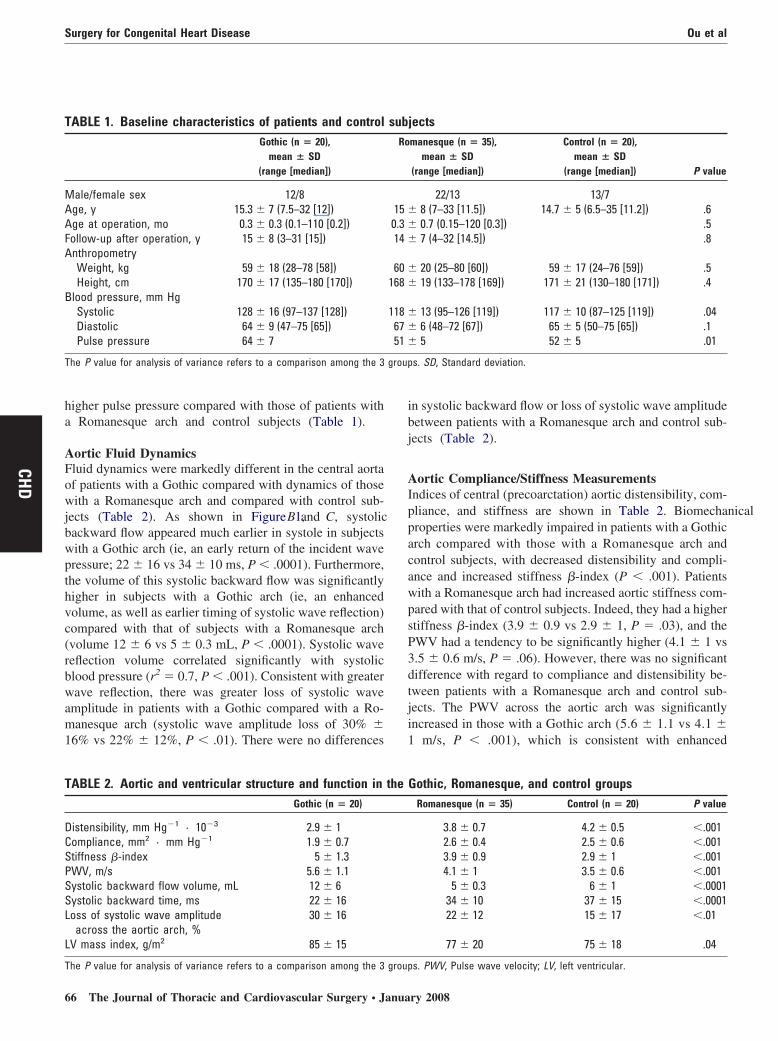
ABLE 1. Baseline characteristics of patients and controlGothic (n � 20),
mean � SD(range [median])
ale/female sex 12/8ge, y 15.3 � 7 (7.5–32 [12])ge at operation, mo 0.3 � 0.3 (0.1–110 [0.2])ollow-up after operation, y 15 � 8 (3–31 [15])nthropometryWeight, kg 59 � 18 (28–78 [58])Height, cm 170 � 17 (135–180 [170])
lood pressure, mm HgSystolic 128 � 16 (97–137 [128])Diastolic 64 � 9 (47–75 [65])Pulse pressure 64 � 7
he P value for analysis of variance refers to a comparison among the 3
ABLE 2. Aortic and ventricular structure and function inGothic (n � 20)
istensibility, mm Hg�1 · 10�3 2.9 � 1ompliance, mm2 · mm Hg�1 1.9 � 0.7tiffness �-index 5 � 1.3WV, m/s 5.6 � 1.1ystolic backward flow volume, mL 12 � 6ystolic backward time, ms 22 � 16oss of systolic wave amplitude
across the aortic arch, %30 � 16
V mass index, g/m2 85 � 15
he P value for analysis of variance refers to a comparison among the 3 group
6 The Journal of Thoracic and Cardiovascular Surgery ● Janua
n systolic backward flow or loss of systolic wave amplitudeetween patients with a Romanesque arch and control sub-ects (Table 2).
ortic Compliance/Stiffness Measurementsndices of central (precoarctation) aortic distensibility, com-liance, and stiffness are shown in Table 2. Biomechanicalroperties were markedly impaired in patients with a Gothicrch compared with those with a Romanesque arch andontrol subjects, with decreased distensibility and compli-nce and increased stiffness �-index (P � .001). Patientsith a Romanesque arch had increased aortic stiffness com-ared with that of control subjects. Indeed, they had a highertiffness �-index (3.9 � 0.9 vs 2.9 � 1, P � .03), and theWV had a tendency to be significantly higher (4.1 � 1 vs.5 � 0.6 m/s, P � .06). However, there was no significantifference with regard to compliance and distensibility be-ween patients with a Romanesque arch and control sub-ects. The PWV across the aortic arch was significantlyncreased in those with a Gothic arch (5.6 � 1.1 vs 4.1 �
m/s, P � .001), which is consistent with enhanced
ectsmanesque (n � 35),
mean � SD(range [median])
Control (n � 20),mean � SD
(range [median]) P value
22/13 13/78 (7–33 [11.5]) 14.7 � 5 (6.5–35 [11.2]) .60.7 (0.15–120 [0.3]) .57 (4–32 [14.5]) .8
20 (25–80 [60]) 59 � 17 (24–76 [59]) .519 (133–178 [169]) 171 � 21 (130–180 [171]) .4
13 (95–126 [119]) 117 � 10 (87–125 [119]) .046 (48–72 [67]) 65 � 5 (50–75 [65]) .15 52 � 5 .01
s. SD, Standard deviation.
othic, Romanesque, and control groupsRomanesque (n � 35) Control (n � 20) P value
3.8 � 0.7 4.2 � 0.5 �.0012.6 � 0.4 2.5 � 0.6 �.0013.9 � 0.9 2.9 � 1 �.0014.1 � 1 3.5 � 0.6 �.001
5 � 0.3 6 � 1 �.000134 � 10 37 � 15 �.000122 � 12 15 � 17 �.01
77 � 20 75 � 18 .04
subjRo
15 �0.3 �14 �
60 �168 �
118 �67 �51 �
the G
s. PWV, Pulse wave velocity; LV, left ventricular.
ry 2008
rmb
LLGmpmidm
DTnmac(nwtsbsprh
otaoyups
fr(imdc
Goswc
inLpTItmrcssdPbm
psrcnchltapsm
wwpsarsNGaiyceaao
ait
Ou et al Surgery for Congenital Heart Disease
CHD
igidity of the central aorta with an angular arch. Further-ore, there was a positive correlation between systolic
lood pressure and PWV (r2 � 0.7, P � .001).
V MassV mass index was significantly higher in patients with aothic arch compared with the index of those with a Ro-anesque arch and control subjects (Table 2). There was a
ositive correlation between systolic blood pressure and LVass index (r2 � 0.7, P � .0001) and between LV mass
ndex and PWV (r2 � 0.8, P � .0001). There was noifference in LV mass index between patients with a Ro-anesque arch and control subjects.
iscussionhe major findings of the present study are that fluid dy-amics and aortic biomechanics of the central aorta arearkedly abnormal in young normotensive survivors of
ortic coarctation repair with an angulated (Gothic) archompared with those of patients with a smooth roundedRomanesque) arch. Furthermore, LV mass index was sig-ificantly increased in patients with a Gothic arch comparedith LV mass index of those with a Romanesque arch, and
he LV mass index correlated positively with indices ofystolic wave reflection, central aortic stiffness, and systoliclood pressure. Thus our results demonstrate important re-idual pathophysiologic abnormalities in the central aorta inatients with a Gothic arch, even after “successful” CoAepair, which might be related to the pathogenesis of lateypertension in this clinical setting.
Several previous reports have documented abnormalitiesf the central aorta, both structural and functional, in pa-ients after anatomically successful CoA repair.15-19 Thesebnormalities are likely related to the well-recognized riskf premature cardiovascular morbidity and mortality inoung adults after CoA repair.4 Nevertheless, the factorsnderlying the appearance of resting or effort-related hy-ertension in some (approximately 20%-40%) but not alluch subjects have not been identified until recently.
With the advent of MRI and its use in all routinelyollowed survivors of CoA repair at our institution, weecently noted a particular association between an angulatedGothic-type) arch on MRI and both resting and exercise-nduced hypertension.5,6 We then sought to understand the
echanisms whereby an angulated arch might change fluidynamics and structure in the central aorta in an attempt tolarify the pathogenesis of hypertension in this setting.
Our hypothesis was that the shaped aortic angle in aothic arch would lead to early and large-volume reflectionf the systolic wave, which would then lead to higherystolic pressure (by addition of the incident and reflectedaves). In addition, enhanced wave reflection would in-
rease wall stress and thus likely induce structural changes a
The Journal of Thorac
n the aortic wall (leading, for example, to increased stiff-ess and decreased compliance) and also potentially lead toV hypertrophy (related to both the higher ascending aorticressure and stiffer aortic mechanical properties).20-22
hese findings were all confirmed in the current study.nterestingly, our study suggests that geometric abnormali-ies in the aortic arch shape do not entirely explain impair-ents in aortic elasticity properties. Indeed, patients with a
ounded Romanesque arch had increased aortic stiffnessompared with that of control subjects. The difference wasignificant between these two groups with regard to thetiffness �-index. However, we did not find any significantifference when using the other parameters, includingWV, compliance, and distensibility. It might be explainedy the small number of subjects in both subgroups (Ro-anesque, n � 35; control, n � 20).We were especially careful to exclude subjects with
otential confounders of aortic biomechanical properties,uch as established hypertension, bicuspid aortic valve, oresidual distal stenosis, to allow us to study in isolation thehanges caused by a Gothic-type arch versus the moreormal-shaped Romanesque-type arch. We speculate thatases of Gothic arch that have already been complicated byypertension (excluded from the current article) wouldikely have even greater wave reflection abnormalities, andhis requires further study. In patients with a Gothic archnd normotension, it is possible that homeostatic bloodressure-regulating mechanisms (eg, the renin-angiotensinystem and central baroreceptors) might be activated toaintain blood pressure within a normal range.The pathogenesis of Gothic arch is uncertain, although
e have recently found that it is not particularly associatedith either preoperative aortic arch hypoplasia or with anyarticular type of surgical repair technique.6 We hypothe-ized that a short aortic isthmus might predispose to anngulated arch after repair. Whether identification of theisks associated with a Gothic arch will inform changes inurgical technique will require further experimentation.evertheless, the identification of (noninvasively imaged)othic arch geometry as a risk factor for aortic stiffness,
bnormal wave reflection, and both resting and exercise-nduced hypertension serves to identify a high-risk group ofoung adults. Such subjects require close surveillance andonsideration of early medical intervention in view of recentvidence linking aortic stiffness and LV hypertrophy todverse outcome,23,24 as well as suggestions that centralortic stiffness might be treatable, with beneficial effects onutcomes (Conduit Artery Function Evaluation study25).
In summary, we have found that young adult survivors ofortic CoA repair who have an angulated Gothic aortic archn follow-up have early and enhanced systolic wave reflec-ion and greater aortic stiffness compared with those char-
cteristics in those whose aortic arch has a more normalic and Cardiovascular Surgery ● Volume 135, Number 1 67

sctcr
R
1
1
1
1
1
1
1
1
1
1
2
2
2
2
2
2
Surgery for Congenital Heart Disease Ou et al
6
CHD
mooth and rounded appearance. These abnormalities ofentral aortic fluid dynamics and biomechanics predisposeo hypertension, LV hypertrophy, and, most likely, adverseardiovascular outcomes in this group of young adults withepaired congenital heart disease.
eferences
1. Cohen M, Fuster V, Steele PM, Driscoll D, McGoon DC. Coarctationof the aorta. Long-term follow-up and prediction of outcome aftersurgical correction. Circulation. 1989;80:840-5.
2. O’Sullivan JJ, Derrick G, Darnell R. Prevalence of hypertension inchildren after early repair of coarctation of the aorta: a cohort studyusing casual and 24 hour blood pressure measurement. Heart. 2002;88:163-6.
3. Celermajer DS, Greaves K. Survivors of coarctation repair: fixed butnot cured. Heart. 2002;88:113-4.
4. de Divitiis M, Rubba P, Calabro R. Arterial hypertension and cardio-vascular prognosis after successful repair of aortic coarctation: aclinical model for the study of vascular function. Nutr Metab Cardio-vasc Dis. 2005;15:382-94.
5. Ou P, Bonnet D, Auriacombe L, Pedroni E, Balleux F, Sidi D, et al.Late systemic hypertension and aortic arch geometry after successfulrepair of coarctation of the aorta. Eur Heart J. 2004;25:1853-9.
6. Ou P, Mousseaux E, Celermajer DS, Pedroni E, Vouhe P, Sidi D, et al.Aortic arch deformation following coarctation surgery: effect on bloodpressure response. J Thorac Cardiovasc Surg. 2006;132:1105-11.
7. London GM. Large artery function and alterations in hypertension.J Hypertens Suppl. 1995;13:S35-8.
8. Dernellis J, Panaretou M. Aortic stiffness is an independent predictorof progression to hypertension in nonhypertensive subjects. Hyperten-sion. 2005;45:426-31.
9. Willum-Hansen T, Staessen JA, Torp-Pedersen C, Rasmussen S, ThijsL, Ibsen H, et al. Prognostic value of aortic pulse wave velocity asindex of arterial stiffness in the general population. Circulation. 2006;113:664-70.
0. Vouhe PR, Trinquet F, Lecompte Y, Vernant F, Roux PM, Touati G,et al. Aortic coarctation with hypoplastic aortic arch. Results of ex-tended end-to-end aortic arch anastomosis. J Thorac Cardiovasc Surg.1988;96:557-63.
1. National High Blood Pressure Education Program Working Group onHypertension Control in Children and Adolescents. Update on the1987 task force report on high blood pressure in children and adoles-cents: a working group report from the National High Blood PressureEducation Program. Pediatrics. 1996;98:649-58.
2. Groenink M, de Roos A, Mulder BJ, Verbeeten B Jr, Timmermans J,
Zwinderman AH, et al. Biophysical properties of the normal-sized8 The Journal of Thoracic and Cardiovascular Surgery ● Janua
aorta in patients with Marfan syndrome: evaluation with MR flowmapping. Radiology. 2001;219:535-40.
3. O’Rourke MF, Staessen JA, Vlachopoulos C, Duprez D, Plante GE.Clinical applications of arterial stiffness; definitions and referencevalues. Am J Hypertens. 2002;15:426-44.
4. Salton CJ, Chuang ML, O’Donnell CJ, Kupka MJ, Larson MG, Kiss-inger KV, et al. Gender differences and normal left ventricular anat-omy in an adult population free of hypertension. A cardiovascularmagnetic resonance study of the Framingham Heart Study Offspringcohort. J Am Coll Cardiol. 2002;39:1055-60.
5. Sehested J, Baandrup U, Mikkelsen E. Different reactivity and struc-ture of the prestenotic and poststenotic aorta in human coarctation.Implications for baroreceptor function. Circulation. 1982;65:1060-5.
6. Xu J, Shiota T, Omoto R, Zhou X, Kyo S, Ishii M, et al. Intravascularultrasound assessment of regional aortic wall stiffness, distensibility,and compliance in patients with coarctation of the aorta. Am Heart J.1997;134:93-8.
7. Ong CM, Canter CE, Gutierrez FR, Sekarski DR, Goldring DR.Increased stiffness and persistent narrowing of the aorta after success-ful repair of coarctation of the aorta: relationship to left ventricularmass and blood pressure at rest and with exercise. Am Heart J.1992;123:1594-600.
8. Vogt M, Kuhn A, Baumgartner D, Baumgartner C, Busch R, KostolnyM, et al. Impaired elastic properties of the ascending aorta in newbornsbefore and early after successful coarctation repair: proof of a systemicvascular disease of the prestenotic arteries? Circulation. 2005;111:3269-73.
9. Murakami T, Takeda A. Enhanced aortic pressure wave reflection inpatients after repair of aortic coarctation. Ann Thorac Surg. 2005;80:995-9.
0. O’Rourke M. Arterial stiffening and vascular/ventricular interaction. JHum Hypertens. 1994;8(suppl):S9-15.
1. London GM, Guerin A. Influence of arterial pulse and reflective waveson systolic blood pressure and cardiac function. J Hypertens Suppl.1999;17:S3-6.
2. London GM. Role of arterial wall properties in the pathogenesis ofsystolic hypertension. Am J Hypertens. 2005;18(suppl):19S-22S.
3. Roman MJ, Ganau A, Saba PS, Pini R, Pickering TG, Devereux RB.Impact of arterial stiffening on left ventricular structure. Hypertension.2000;36:489-94.
4. Sutton-Tyrrell K, Najjar SS, Boudreau RM, Venkitachalam L, Kupe-lian V, Simonsick EM, et al. Elevated aortic pulse wave velocity, amarker of arterial stiffness, predicts cardiovascular events in well-functioning older adults. Circulation. 2005;111:3384-90.
5. Williams B, Lacy PS, Thom SM, Cruickshank K, Stanton A, Collier D,et al. Differential impact of blood pressure-lowering drugs on centralaortic pressure and clinical outcomes: principal results of the ConduitArtery Function Evaluation (CAFE) study. Circulation. 2006;113:
1213-25.ry 2008
Related Documents