S1 Available online http://ccforum.com/supplements/13/S1 Critical Care Volume 13 Suppl 1, 2009 29th International Symposium on Intensive Care and Emergency Medicine Brussels, Belgium, 24–27 March 2009 Published online: 13 March 2009 These abstracts are available online at http://ccforum.com/supplements/13/S1 © 2009 BioMed Central Ltd P1 Time course of dyspnea evolution in the emergency department: results from the URGENT dyspnea survey M Tavares 1 , P Pang 2 , S Laribi 3 , A Mebazaa 3 , M Gheorghiade 2 1 Hospital de Santo Antonio-CHP, Porto, Portugal; 2 Northwestern University, Chicago, IL, USA; 3 Hôpital Lariboisière, Paris, France Critical Care 2009, 13(Suppl 1):P1 (doi: 10.1186/cc7165) Introduction There is considerable uncertainty about the repro- ducibility of the various instruments used to measure dyspnea, their ability to reflect changes in symptoms, whether they accurately reflect the patient’s experience and if its evolution is similar between acute heart failure syndrome patients and nonacute heart failure syndrome patients. URGENT was a prospective multicenter trial designed to address these issues. Methods Patients were interviewed within 1 hour of first physician evaluation, in the emergency department or acute care setting, with dyspnea assessed by the patient using both a five-point Likert scale and a 10-point visual analog scale (VAS) in the sitting (60º) and then supine (20º) position if dyspnea had not been considered severe or very severe by the sitting versus decubitus dyspnea measurement. Results Very good agreements were found between the five-point Likert and VAS at baseline (0.891, P <0.0001) and between changes (from baseline to hour 6) in the five-point Likert and in VAS (0.800, P <0.0001) in acute heart failure (AHF) patients. Lower agreements were found when changes from baseline to H6 measured by Likert or VAS were compared with the seven-point comparative Likert (0.512 and 0.500 respectively) in AHF patients. The worse the dyspnea at admission, the greater the amplitude of improvement in the first 6 hours; this relationship is stronger when dyspnea is measured with VAS (Spearman’s rho coefficient = 0.672) than with the five-point Likert (0.272) (both P <0.0001) in AHF patients. By the five-point Likert, only nine patients (3% (1% to 5%)) reported an improvement in their dyspnea, 177 (51% (46% to 57%)) had no change, and 159 (46% (41% to 52%)) reported worse dyspnea supine compared with sitting up in AHF patients. The PDA test with VAS was markedly different between AHF and non-AHF patients. Conclusions Both clinical tools five-point Likert and VAS showed very good agreement at baseline and between changes from baseline to tests performed 6 hours later in AHF patients. The PDA test with VAS was markedly different between AHF and non-AHF patients. Dyspnea is improved within 6 hours in more than three- quarters of the patients regardless of the tool used to measure the change in dyspnea. The greater the dyspnea at admission, the greater the amplitude of improvement in the first 6 hours. P2 Endotracheal intubation in the emergency room: a multicenter questionnaire SB Bélorgey 1 , L Montésino 2 1 CHSF Gilles de Corbeil, Corbeil Essonnes, France; 2 CHG, Longjumeau, France Critical Care 2009, 13(Suppl 1):P2 (doi: 10.1186/cc7166) Introduction Endotracheal intubation (ETI) engages the patient’s life and demands a good experience. A preliminary prospective study has shown in one hospital that emergency physicians (EPs) rarely performed ETI. Do the EPs in Ile de France (Paris region) have sufficient experience and regular training to realise this procedure safely in the emergency room (ER)? Methods We conducted a descriptive telephone-based question- naire study to assess EPs’ endotracheal intubation skills through all ERs in Ile de France public hospitals. A questionnaire was completed by the investigator during a 10-minute telephone call with at least one EP in each ER. The structure of hospitals, number of ETIs performed, devices and personnel available and the existence of protocols were collected. Their usual practice of sedation and intubation, training and proposals for changes were noted. Results The study was made through all of the 64 public hospitals of Ile de France. Fifty-six hospitals have an ICU, 37 a mobile ICU. We questioned 96 EPs; that is, 10% of EPs from our region. All of the 96 EPs called responded. These physicians were certified emergency physicians (CAMU) for 90% of them. The median of ETI declared was 24.5/year per ER. Thirty-eight percent of EPs performed less than five ETIs during the past 2 years. The success rate reported was 85%. In 94% of ERs, metallic blades and Eischmann mandrin were available and about two nurses can help during the procedure. Predictive criteria for difficult ETI cited the most were: short neck, obesity, small mouth opening and otorhinolaryngology disease or previous history of cervical radiotherapy. Seventy-six percent of EPs followed the recommendations for preoxygenation and 91% performed rapid sequence induction. The vast majority (76%) of ERs did not have standardized procedures for airway management. Theoretical training was acquired for 46% of EPs by the CAMU, practical training occurring in the operating room for 71%. Among the EPs interviewed, 87% believe that they should remain the principal actor for ETI – although as high as 89% of them consider that they were insufficiently trained in ETI management and only 41% pursued continuing medical education on that theme. Seventy- seven percent proposed to spend time in the operating room to improve their practice of ETI. Conclusions ETI is rarely performed in the ER. It should be part of the EP curricula and written procedures should be made.

Welcome message from author
This document is posted to help you gain knowledge. Please leave a comment to let me know what you think about it! Share it to your friends and learn new things together.
Transcript

S1
Available online http://ccforum.com/supplements/13/S1
Critical Care Volume 13 Suppl 1, 200929th International Symposium on Intensive Care and EmergencyMedicineBrussels, Belgium, 24–27 March 2009
Published online: 13 March 2009These abstracts are available online at http://ccforum.com/supplements/13/S1© 2009 BioMed Central Ltd
P1Time course of dyspnea evolution in the emergencydepartment: results from the URGENT dyspnea survey
M Tavares1, P Pang2, S Laribi3, A Mebazaa3, M Gheorghiade2
1Hospital de Santo Antonio-CHP, Porto, Portugal; 2NorthwesternUniversity, Chicago, IL, USA; 3Hôpital Lariboisière, Paris, FranceCritical Care 2009, 13(Suppl 1):P1 (doi: 10.1186/cc7165)
Introduction There is considerable uncertainty about the repro-ducibility of the various instruments used to measure dyspnea, theirability to reflect changes in symptoms, whether they accuratelyreflect the patient’s experience and if its evolution is similarbetween acute heart failure syndrome patients and nonacute heartfailure syndrome patients. URGENT was a prospective multicentertrial designed to address these issues.Methods Patients were interviewed within 1 hour of first physicianevaluation, in the emergency department or acute care setting, withdyspnea assessed by the patient using both a five-point Likertscale and a 10-point visual analog scale (VAS) in the sitting (60º)and then supine (20º) position if dyspnea had not been consideredsevere or very severe by the sitting versus decubitus dyspneameasurement.Results Very good agreements were found between the five-pointLikert and VAS at baseline (0.891, P <0.0001) and betweenchanges (from baseline to hour 6) in the five-point Likert and inVAS (0.800, P <0.0001) in acute heart failure (AHF) patients.Lower agreements were found when changes from baseline to H6measured by Likert or VAS were compared with the seven-pointcomparative Likert (0.512 and 0.500 respectively) in AHF patients.The worse the dyspnea at admission, the greater the amplitude ofimprovement in the first 6 hours; this relationship is stronger whendyspnea is measured with VAS (Spearman’s rho coefficient =0.672) than with the five-point Likert (0.272) (both P <0.0001) inAHF patients. By the five-point Likert, only nine patients (3% (1%to 5%)) reported an improvement in their dyspnea, 177 (51%(46% to 57%)) had no change, and 159 (46% (41% to 52%))reported worse dyspnea supine compared with sitting up in AHFpatients. The PDA test with VAS was markedly different betweenAHF and non-AHF patients.Conclusions Both clinical tools five-point Likert and VAS showedvery good agreement at baseline and between changes frombaseline to tests performed 6 hours later in AHF patients. The PDAtest with VAS was markedly different between AHF and non-AHFpatients. Dyspnea is improved within 6 hours in more than three-quarters of the patients regardless of the tool used to measure thechange in dyspnea. The greater the dyspnea at admission, thegreater the amplitude of improvement in the first 6 hours.
P2Endotracheal intubation in the emergency room: a multicenter questionnaire
SB Bélorgey1, L Montésino2
1CHSF Gilles de Corbeil, Corbeil Essonnes, France; 2CHG,Longjumeau, FranceCritical Care 2009, 13(Suppl 1):P2 (doi: 10.1186/cc7166)
Introduction Endotracheal intubation (ETI) engages the patient’slife and demands a good experience. A preliminary prospectivestudy has shown in one hospital that emergency physicians (EPs)rarely performed ETI. Do the EPs in Ile de France (Paris region)have sufficient experience and regular training to realise thisprocedure safely in the emergency room (ER)?Methods We conducted a descriptive telephone-based question-naire study to assess EPs’ endotracheal intubation skills through allERs in Ile de France public hospitals. A questionnaire wascompleted by the investigator during a 10-minute telephone callwith at least one EP in each ER. The structure of hospitals, numberof ETIs performed, devices and personnel available and theexistence of protocols were collected. Their usual practice ofsedation and intubation, training and proposals for changes werenoted.Results The study was made through all of the 64 public hospitalsof Ile de France. Fifty-six hospitals have an ICU, 37 a mobile ICU.We questioned 96 EPs; that is, 10% of EPs from our region. All ofthe 96 EPs called responded. These physicians were certifiedemergency physicians (CAMU) for 90% of them. The median ofETI declared was 24.5/year per ER. Thirty-eight percent of EPsperformed less than five ETIs during the past 2 years. The successrate reported was 85%. In 94% of ERs, metallic blades andEischmann mandrin were available and about two nurses can helpduring the procedure. Predictive criteria for difficult ETI cited themost were: short neck, obesity, small mouth opening andotorhinolaryngology disease or previous history of cervicalradiotherapy. Seventy-six percent of EPs followed therecommendations for preoxygenation and 91% performed rapidsequence induction. The vast majority (76%) of ERs did not havestandardized procedures for airway management. Theoreticaltraining was acquired for 46% of EPs by the CAMU, practicaltraining occurring in the operating room for 71%. Among the EPsinterviewed, 87% believe that they should remain the principalactor for ETI – although as high as 89% of them consider that theywere insufficiently trained in ETI management and only 41%pursued continuing medical education on that theme. Seventy-seven percent proposed to spend time in the operating room toimprove their practice of ETI.Conclusions ETI is rarely performed in the ER. It should be part ofthe EP curricula and written procedures should be made.

S2
Critical Care March 2009 Vol 13 Suppl 1 29th International Symposium on Intensive Care and Emergency Medicine
P3Radiological validation of endotracheal tube insertiondepth in prehospital emergency patients
DM Maybauer1, MO Maybauer1, H Wolff2, E Pfenninger2, W Geisser3
1The University of Texas Medical Branch and Shriners BurnsHospital, Galveston, TX, USA; 2University Hospital, Ulm, Germany;3County Clinics, Dillingen, GermanyCritical Care 2009, 13(Suppl 1):P3 (doi: 10.1186/cc7167)
Introduction Incorrect positioning of the endotracheal tube (ETT)within the airway after emergent intubation can result in seriouscomplications. Accidental mainstem bronchus intubation isassociated with contralateral atelectasis, tension pneumothorax,hypotension, and decreased survival. Conversely, failure to placethe tube several centimeters beyond the vocal cords may result ininadvertent extubation, aspiration, pneumonia, or laryngeal spasm[1]. The aim of this study was to investigate the occurrence of ETTmalpositioning after emergency intubation in the out-of-hospitalsetting.Methods A retrospective study of a 5-year time period, usingrecords of 1,081 patients admitted to the trauma emergency room(ER) at a university hospital. Within 30 minutes after admission, achest X-ray or whole-body CT scan was routinely performed inintubated patients to determine the tube–tip–carina relationship.Results Sixteen out of 1,081 patients died immediately afteradmission to the trauma ER and were not further radiologicallydiagnosed. Of the surviving 1,065 patients, 346 (32.5%) werefemale and 719 (67.5%) male. In the group of 488 intubatedpatients, 346 (70.9%) were correctly intubated, 89 (18.2%) werenot correctly intubated – herein were 64 patients (14.7%)intubated with tip–carina distance <2 cm, and 25 patients (5.7%)were endobronchially intubated. Chest X-ray scans were notavailable for 53 patients (10.9%). Detailed data on ETT placementwere available in 435 patients; 346 (79.5%) with correct ETTplacement, 89 (20.5%) with incorrect ETT placement. None of thepatients displayed an esophageal or pharyngeal intubation (0%).Of 435 patients, 324 had been intubated preclinically on scene –254 (78.4%) were correctly intubated, 70 (21.6%) were notcorrectly intubated.Conclusions This study clearly shows that ETT misplacement inemergency patients is still a serious problem with an incidence of21.6% in our study, of which 5.7% were endobronchiallyintubated. We conclude that the skill level of the operator may bekey in determining efficacy of endotracheal intubation. Based onour findings, all efforts should be made to verify the tube positionwith immediate radiographic confirmation after admission to theER.Reference1. Owen RL, Cheney FW: Endobronchial intubation: a pre-
ventable complication. Anesthesiology 1987, 67:255-257.
P4Does bedside chest ultrasound in the ICU improve earlydiagnosis and quick resolution of pleural effusion?
L Tutino1, R Spina2, A Di Filippo1, S Batacchi2, G Cianchi2, A Peris2
1University of Florence, Italy; 2Careggi Teaching Hospital, Florence,ItalyCritical Care 2009, 13(Suppl 1):P4 (doi: 10.1186/cc7168)
Introduction A bedside chest ultrasound (bCUS) programmeperformed by intensivists after 18 months of training was
introduced on a regular basis in a 10-bed emergency ICU fromApril to November 2008, in order to check its effectiveness in theearly diagnosis and treatment of pleural effusion (PE). The resultshave been compared with those of a control group, when bCUSwas not part of the ICU procedures.Methods The procedure was performed within the first 24 hoursafter admittance. All of the 92 patients were examined supine, withthe probe perpendicular to the chest wall, using all of theintercostal spaces as the acoustic window. With this technique,once we identified the lung’s base, we looked for signs of PEaccording to the following criteria: (a) space between the twopleural layers; (b) variation in the interpleural distance of this spaceduring breathing. For each patient the following data werecollected: age, sex, weight, height, SAPS II, number of chestdrains, number of ultrasound scans performed, number ofsignificant PE (at least 2 cm of width between the two pleurae),amount of ultrasound-guided drainage actually performed withinthe first 24 hours, timing of resolution of PE. Data were comparedwith those of a group of patients admitted to the ICU from Januaryto March 2008, when bCUS was not part of the daily proceduresand only chest CT and X-ray scans were used as evidence of PE.We considered P <0.05 statistically significant.Results A total of 103 bCUS were performed on the first day inthe control group, against the 12 bCUS performed in the studygroup. A total of 59 PEs for which drainage proved to be usefulwere found. An amount of 27 pleural drainages were performedwithin the first 24 hours. We have no evidence of complications.All of the positive cases for PE have been successfully treated. Alldrainage was performed within the first 24 hours or at least withinthe first 48 hours.Conclusions Compared with the control group (25.9%), in thestudy group 45% of drainage performed was done within the first24 hours thanks to the skill of intensivists. As far as PE isconcerned, the introduction of bCUS performed by intensivists inthe ICU daily routine determines an increase of early diagnosis andtreatment. However, the increase in the number of the first-daytreatments was not significant since this procedure is now turningfrom a purely diagnostic approach into an operative one. This willnecessarily need time.
P5Portable chest radiography in mechanically ventilated ICUpatients: does synchronizing with end-inspiration improvethe quality of films?
A Cheng1, K Tang1, H Yip1, W Kwan1, Y Lee2, A Lee2, K Lee2, K Wong1, C Gomersall21North District Hospital, Sheung Shui, Hong Kong; 2Prince ofWales Hospital, The Chinese University of Hong Kong, Shatin,Hong KongCritical Care 2009, 13(Suppl 1):P5 (doi: 10.1186/cc7169)
Introduction The quality of portable chest radiographs (CXRs)taken in the ICU is inferior to films taken in the radiologydepartment. The inability of sedated, ventilated patients to holdtheir breath during CXR will also affect the degree of lung inflationand contribute to lack of correlation between serial CXR changesand clinical status [1]. We studied the effect on CXR quality bymanually synchronizing the ventilator to end-inspiration inmechanically ventilated ICU patients.Methods A pair of CXRs was taken after recruiting intubated,ventilated patients within 24 hours of emergency ICU admission.Intubated post-elective surgical patients were excluded due to thehigh likelihood of normal lungs. The control film was taken in the

S3
Available online http://ccforum.com/supplements/13/S1
usual way, at a random phase of the ventilator cycle. For thesynchronized film, the investigator wore a lead apron anddosimeter, stood 1 to 1.5 meters away from the patient, andpressed the inspiratory hold button. The sequence of the pairedfilms was computer-randomized. The ventilator model, settings,patient position and portable X-ray machine settings were keptconstant between films. Patients served as their own controls.Films were independently scored (1 = not clear/poorly inflated, 5 =very clear/well inflated) by two specialist radiologists based on fivecriteria: (i) clarity of lines and tubes, (ii) definition of pulmonaryvasculature, (iii) visibility of mediastinum, (iv) definition of thediaphragm and (v) degree of lung inflation. Linear regression,taking two radiologists’ scores of each patient into account, wasused to examine whether there were any differences in the criteriaratings between random and synchronized films. Radiologists andstatistician were blinded.Results We recruited 110 patients; there were no complicationsfrom the breath-hold maneuver. Dosimeter readings werenegligible. Synchronized films had higher total scores and meanscores for criteria (ii) to (v), 95% confidence interval. P values werestatistically significant: for total score, P <0.001; and for criteria (ii),P = 0.001; (iii), P <0.001; (iv), P <0.01; and (v), P <0.001.Conclusions Synchronizing the CXR to end-inspiration improvesthe quality of the film and is safe.Reference1. Langevin PB, Hellein V, Harms SM, Tharp WK, Cheung-Seekit
C, Lampotang S: Synchronization of radiograph film expo-sure with the inspiratory pause. Effect on the appearanceof bedside chest radiographs in mechanically ventilatedpatients. Am J Respir Crit Care Med 1999, 160:2067-2071.
P6Quality assurance report on the use of continuous positiveairway pressure and end-tidal carbon dioxide duringrespiratory distress in field emergency care
D Lain1, S Bourn2
1Oridion Capnography, Needham, MA, USA; 2AMR, Aurora, IL, USACritical Care 2009, 13(Suppl 1):P6 (doi: 10.1186/cc7170)
Introduction The use of continuous positive airway pressure(CPAP) is beneficial in the hospital and home care environment. Itis used to support ventilation during neurological disease,ventilatory defects, congestive heart failure and obstructive sleepapnea. Field emergency medicine has inherent complications forthe delivery and monitoring of patients receiving CPAP. Wecompleted an internal quality audit to determine whether CPAPhad benefit and whether capnography could be comfortably usedin parallel with a CPAP device to monitor ventilation.Methods The data collection was completed on patients withrespiratory distress. Data were collected pre-CPAP and post-CPAP. Patients were monitored with capnography and pulseoximetry. Emergency Medical Services and Emergency Depart-ment staff evaluated acceptance and ease of use of the equipment.A one-tailed paired test and descriptive statistics were completed.Results Eighteen respiratory distress patients received CPAP:eight female, nine male and one patient had missing data (sex entrywas blank). Mean age was 79 years. Statistical significance wasdetermined at P <0.05. There was no significant difference in heartrate: mean pre-CPAP = 116, mean post-CPAP = 114, P = 0.19.There was a significant improvement in arterial oxygen saturation per-centage: pre-CPAP = 81.5, mean post-CPAP = 90.2, P = 0.0003.There was a significant improvement in end-tidal carbon dioxide(etCO2) (ventilation parameter): mean pre-CPAP = 40.1, mean
post-CPAP = 35.1, P = 0.038. There was a significant decrease inrespiratory rate: pre-CPAP = 36, post-CPAP = 33, P = 0.031.Using a Borg scale for severity of respiratory distress, there was asignificant improvement after CPAP: pre-CPAP = 8.06, post-CPAP = 5.7, P = 0.0001. The emergency medical techniciansfound the devices, CPAP, mask, and etCO2, easy to use, and 16patients ranked it comfortable. Two patients were uncomfortablewith CPAP. The most comfortable score was 10; the populationscored the overall comfort of CPAP with etCO2 at 7.8.Conclusions CPAP in field emergency medicine can be easilyapplied, is well tolerated, and results can be monitored bycapnography. Capnographic measurements indicated improvedventilation by a decrease in carbon dioxide. CPAP and etCO2 canbe used in field emergencies to support and monitor ventilationduring respiratory distress.
P7Cardiogenic oscillations extracted from spontaneousbreathing airway pressure and flow signal are related tochest wall motility and continuous positive airway pressure
S Schumann1, F Messmer1, M Lichtwarck-Aschoff2, C Haberthuer3, K Moeller4, J Guttmann1
1University Medical Center, Freiburg, Germany; 2UniversityHospital, Uppsala, Sweden; 3Kantonsspital, Luzern, Switzerland;4University of Applied Sciences, Furtwangen, GermanyCritical Care 2009, 13(Suppl 1):P7 (doi: 10.1186/cc7171)
Introduction During mechanical ventilation, signal pulses withinpressure and flow curves can be observed that are related to theactivity of the beating heart. From a signal-processing view, thesecardiogenic oscillations (COS) can be understood as repeatedpulses that are transferred via the lungs and airways to the airwayopening. It was demonstrated earlier that COS, achieved duringbreath-holding maneuvers, were influenced by changes inmechanical properties of the thorax [1]. We hypothesized thatthese COS can be extracted from the airway pressure and flowsignal during spontaneous breathing. Furthermore, we hypo-thesized that these isolated signals contain information about themechanical properties of the respiratory system.Methods Fifteen healthy volunteers breathed spontaneouslyagainst continuous positive airway pressure (CPAP) of 0 to9 cmH2O at normal thorax or at thorax bending, limiting motility to90% of the normal circumference. Airway pressure and flow aswell as an electrocardiogram were recorded at a sample frequencyof 200 Hz. To isolate the signals that are related to the activity ofthe heart, pressure and flow data were aligned in time to the R-wave of the QRS complex and averaged.Results Highly characteristic pressure and flow oscillations couldbe extracted from the spontaneous breathing signals. Both signalswere closely related (r2 = 0.97 ± 0.02, each P <0.0001). Thepressure amplitude of the COS was influenced by CPAP(P = 0.049) and thorax motility (P <0.001); in contrast, the flowamplitude was influenced only by thorax motility (P <0.001).Conclusions COS can be extracted from the airway pressure andflow signal during spontaneous breathing. They contain informationabout the mechanical properties of the respiratory system. Afterfurther investigations, our new method potentially allows anestimation of compliance of the respiratory system duringspontaneous breathing.Reference1. Bijaoui E, Baconnier PF, Bates JH: Mechanical output
impedance of the lung determined from cardiogenic oscil-lations. J Appl Physiol 2001, 91:859-865.

S4
P8Noninvasive positive pressure ventilation in infants withrespiratory failure
I Lazar, Y Cavari, S SoferSoroka Medical Center and Faculty of Health Sciences, Ben-Gurion University of the Negev, Beer Sheva, IsraelCritical Care 2009, 13(Suppl 1):P8 (doi: 10.1186/cc7172)
Introduction Respiratory failure is a common indication foradmission to a pediatric intensive care unit (PICU). Trachealintubation and invasive ventilation carries some risk and cancontribute to morbidity and possible mortality. Noninvasive positivepressure ventilation (NIPPV) is a mode in which ventilation isapplied without tracheal intubation but via nasal prongs or a facemask. We hypothesized that using NIPPV in infants with pendingrespiratory failure may improve their outcome.Methods In this prospective study, we enrolled infants admittedwith pending respiratory failure to the PICU. NIPPV was deliveredusing silicone INCA nasal prongs (Ackrad Labs, Trumbull, CT,USA) connected to an INFANT STAR 950 standard ventilator. TheNIPPV mode of support (continuous positive airway pressure vs.synchronized nasal intermittent positive pressure ventilation) wasset to meet patient needs. Vital signs, ventilator settings andlaboratory results were recorded electronically. The primaryoutcome was prevention of invasive ventilation. Secondaryoutcomes were seeking a physiological marker for NIPPV failureand complications from NIPPV.Results Between December 2007 and November 2008, 18patients (15 infants, three enrolled twice) were eligible to receiveNIPPV based on the attending physician’s decision. Median agewas 3 months (15 days to 17 months). Eleven patients had apneaand seven patients had respiratory distress. Among them, fivepatients had bronchiolitis, two had central hypotonia, two hadPertussis and six patients had other miscellaneous respiratoryconditions needing support. Fourteen infants (78%) wereimproved and weaned off NIPPV while four (22%) infants had tobe intubated. The mean length of NIPPV was 47 hours. Nocomplication secondary to the NIPPV was recorded. No single vitalsign or laboratory test predicted whether a patient would improveor fail NIPPV, although the improvement (when happened) wasnoticed within hours of NIPPV. All four failures were due tocontinuum of apnea.Conclusions Our study shows that NIPPV is a safe and successfulmethod to support infants suffering from pending respiratoryfailure. Lacking a physiological or biological marker that coulddistinguish between so-called responders and nonresponders toNIPPV, and the high rate of success in preventing the need forinvasive ventilation, supports our recommendation to trial any infantin pending respiratory failure with NIPPV.
P9Functional respiratory effects of noninvasive ventilation inacute hypercapnic patients with chronic obstructivepulmonary disease
AG Graziani1, B Carenzi1, F Morgagni1, B Praticò2, P Casalini1,GF Stefanini11Ospedale per Gli Infermi, Faenza, Italy; 2Ospedale Bufalini,Cesena, ItalyCritical Care 2009, 13(Suppl 1):P9 (doi: 10.1186/cc7173)
Introduction Noninvasive ventilation (NIV) is useful for improvingoxygenation, decreasing hypercapnia, and avoiding invasivemechanical ventilation in patients with chronic obstructive
pulmonary disease (COPD) exacerbations and acute respiratoryfailure [1,2]. During the hospitalization, the treatment intensity isquite variable on the basis of the patient’s clinical condition. Often,after the acute phase, patients need only periods of NIV followedby oxygenotherapy. NIV reduces the work of breathing andprobably improves the respiratory muscular strength even whenthe ventilation is ended [3]. The aim of this study is to evaluate thiseffect in COPD submitted to NIV.Methods We studied 10 patients (seven male; three female; age74.2 ± 5.3 years) admitted to our hospital with a COPDexacerbation and acute respiratory failure. After a period of clinicalstabilization, patients underwent spirometry and arterial blood gasas basal examinations. For each patient, NIV was applied for2 hours and than spirometry and arterial blood gas were repeated.Results are presented as mean ± SD. Data were evaluated bypaired t test and a value of P <0.05 was taken as statisticallysignificant.Results At the end of NIV, the forced ventilatory capacity (FVC)and the forced expiratory volume at the first second (FEV1)/FVCratio were significantly increased compared with the basal value:FVC 47.9 ± 11.0% basal, 55.3 ± 15.6% after NIV, P = 0.02;FEV1/FVC 68.5 ± 17.1 basal, 59.3 ± 15.2 after NIV, P = 0.007.Compared with the basal value, arterial carbon dioxide pCO2 wassignificantly decreased: 68.0 ± 2.5 mmHg (basal), 55.3 ± 4.5 mmHg(after NIV), P <0.02; and arterial oxygen was increased:50.0 ± 14.2 mmHg (basal), 56.3 ± 15.3 mmHg (after NIV).Conclusions The results of this study demonstrate that, in COPDexacerbation, NIV determines a decrease of the pulmonaryhyperinflaction. This effect could improve the clinical status and thework of breathing.References1. West JB: COPD. In Pulmonary Physiology and Pathophysiol-
ogy. Baltimore, MD: Lippincott Williams & Wilkins; 2001:33-53.
2. Brochard L: Mechanical ventilation: invasive versus noninvasive. Eur Respir J 2003, 22(Suppl 47):31s-37s.
3. Lightowler JV, et al.: Non-invasive positive pressure ventila-tion to treat respiratory failure resulting from exacerbationof COPD disease: Cochrane systematic review and meta-analysis. BMJ 2003, 326:185-187.
P10Clinical application of noninvasive ventilation in acuterespiratory failure in a general ICU
T Correa, L Morais, P Sanches, H Feitosa, C Azevedo, T Figueiredo, C Taniguchi, R Caserta, C BarbasHospital Israelita Albert Einstein, São Paulo, BrazilCritical Care 2009, 13(Suppl 1):P10 (doi: 10.1186/cc7174)
Introduction Noninvasive ventilation (NIV) is an option in patientswith acute respiratory failure [1-3]. However, NIV complicationsand failure are not yet completely understood [1,2]. Our objectivewas to evaluate the indications, complications and failure of NIV inan adult general ICU.Methods From 1 August to 27 October we analyzed prospectivelyall patients admitted to a 40-bed clinical–surgical ICU of a tertiarycare hospital. From those patients, we included the ones whoreceived NIV (total face mask coupled to a BIPAP Vision® orSynchrony®) and evaluated the indications, causes of failure andthe complications of this ventilatory support.Results During the study period, 465 patients were admitted tothe ICU; 111 patients (23.9%) received NIV. The main indicationsfor NIV were: hypoxemic respiratory failure in 22 patients (19.8%),respiratory infection in 19 (17.1%), acute COPD in 14 (12.6%), as
Critical Care March 2009 Vol 13 Suppl 1 29th International Symposium on Intensive Care and Emergency Medicine

S5
part of a weaning strategy in 16 (14.4%), cardiogenic pulmonaryedema in 15 (13.5%), ALI/ARDS in nine (8.1%), palliative care insix (5.4%), neuromuscular disease in one (0.9%) and others in ninepatients (8.1%). NIV did not avoid intubation in 31 patients(27.9%). The main reasons for failure were: progressive acuterespiratory failure in 23 patients (71.9%) and neurologicaldeterioration in five patients (15.6%). NIV was used after extu-bation in 16 patients, and in five of them (31%) it was necessaryreintubation. The only complication observed was gastricinsufflation in six patients (5.4%).Conclusions NIV is frequently used in a general ICU and the mainindication is acute hypoxemic respiratory failure. The NIV failureincidence was significant but similar to the medical literature.References1. Demoule A, et al.: Benefits and risk of success or failure of
noninvasive ventilation. Intensive Care Med 2006, 32:1756-1765.
2. Celikel T, et al.: Comparison of noninvasive positive pres-sure ventilation with standard medical therapy in hyper-capnic acute respiratory failure. Chest 1998, 114:1636-1642.
3. Wysocki M, et al.: Noninvasive pressure support ventilationin patients with acute respiratory failure. A randomizedcomparison with conventional therapy. Chest 1995, 107:761-768.
P11Relative effectiveness of two nasal continuous positiveairway pressure devices in VLBW infants: first report froma multicenter, randomized, controlled trial
K Bober1, J Swietlinski2, J Zejda1, K Kornacka3, D Pawlik4, J Behrendt1, E Gajewska5, M Czyzewska5, P Korbal6, J Witalis7,W Walas8, A Turzanska9, M Wilinska10, G Zielinski11, B Czeszynska12, T Bachman13
1Medical University of Silesia, Katowice, Poland; 2The Children’sMemorial Heath Institute, Warsaw, Poland; 3Medical UniversityWarsaw, Poland; 4 Medical College Jagiellonian University ofCracow, Poland; 5Medical University Wroclaw, Poland; 6RegionalHospital Bydgoszcz, Poland; 7 Regional Hospital Rzeszow, Poland;8Regional Hospital Opole, Poland; 9Center of MedicalPostgraduate Education Warsaw, Poland; 10Regional HospitalLomza, Poland; 11Regional Hospital Czestochowa, Poland;12Pomeranian Medical University Szczecin, Poland; 13CaliforniaState University, San Bernardino, CA, USACritical Care 2009, 13(Suppl 1):P11 (doi: 10.1186/cc7175)
Introduction Nasal continuous positive airway pressure (nCPAP)is accepted as an effective and relatively complication-free methodof respiratory support of premature infants [1,2]. We intended tocompare the effectiveness of two nCPAP devices/approaches(Hudson prongs/bubble (H), and Infant Flow (IF)), in differentgroups of very low birth weight infants in a large trial.Methods Infants weighing 750 to 1,500 g, gestational age<32 weeks, were enrolled from April 2006 to July 2008 at 12centers. The newborns, categorized into three study groups, wererandomly assigned to one of the nCPAP devices in the first 6 hoursof life. Study group A (n = 119) were neonates placed immediatelyon nCPAP. Group B (n = 157) were placed on nCPAP afterreceiving surfactant. Group C (n = 56) were treated with conven-tional ventilation and nCPAP was used as the method of weaningfrom mechanical ventilation.Results There were no statistically significant differences betweenthe two devices with regard to treatment success, pneumothoraxor bronchopulmonary dysplasia. The incidence of severe nasal
complications was lower in the infants treated with Infant Flow:0.8% (IF), 6.6.% (H) (P = 0.01) in group A; 0.6% (IF), 5.1% (H)(P = 0.01) in group B; and 0% (IF), 5.3% (H) (P = 0.1) in group C.The incidence of necrotizing enterocolitis was also lower in thegroup A infants treated with Infant Flow: 2.5% (IF), 8.3% (H)(P = 0.03).Conclusions (1) The two nCPAP methods are comparable withregard to the incidence of pulmonary complications and primaryeffectiveness. (2) The Infant Flow method resulted in fewer severenasal complications.References1. Morley CJ, Davis PG, Doyle LW, Brion LP, Hascoet JM, Carlin
JB, COIN Trial Investigators: Nasal CPAP or intubation atbirth for very preterm infants. N Engl J Med 2008, 358:700-708.
2. Swietlinski J, Bober K, Gajewska E, Helwich E, Lauterbach R,Manowska M, et al., Polish Noninvasive Respiratory SupportProgram Study Group: Introduction of Infant Flow nasalcontinuous airway pressure as the standard of practice inPoland: the initial 2-year experience. Pediatr Crit Care Med2007, 8:109-114.
P12Contribution of noninvasive ventilation in the precociousextubation in the medical ICU
B Charra, A Hachimi, A Benslama, S MotaouakkilHôpital Ibn Rochd, Casablanca, MoroccoCritical Care 2009, 13(Suppl 1):P12 (doi: 10.1186/cc7176)
Introduction During the past decade, noninvasive ventilation (NIV)has imposed itself as an alternative to endotracheal intubation.Several recent studies let us believe that this technique could bealso beneficial at the precocious extubation. The purpose of oursurvey is to value the place of NIV at the precocious extubation inventilated patients.Methods A prospective, randomized and controlled survey hasbeen driven in a medical resuscitation unit during 6 months (Juneto December 2007). After 48 hours of mechanical ventilation (MV),if the patients have no fever, no neurological anomalies norhemodynamic instability and SaO2 >90% with 40% FiO2, aspontaneous breathing trial (T-piece) is performed. If after theT-piece test the clinical status, blood gas and hemodynamic datawere good, the patient was extubated. If these criteria were notfilled, the MV was continued and a daily assessment performed.On the other hand, if the patient was anxious, agitated, withpolypnea >35/minute, PaO2 <50 mmHg under 40% FiO2, heartrate >145/minute, systolic arterial pressure >170 mmHg or<70 mmHg or arrhythmia, the patient is randomized for one of thetwo protocols: in the first, the patient was extubated and NIV(pressure support – positive end-expiratory pressure) via a facialmask was performed; in the second, a classic weaning wasperformed with pressure support ventilation. The quantitativevariables are expressed as the average or median ± standardderivation, and the qualitative variables by as the percentage. Theunivariate analysis was based on the chi-squared test or Fisher testfor the qualitative variables and the Student test for the quantitativeones. P <0.05 is considered significant. The statistic analysis wasbased on SPSS 11.0 for Windows.Results Twenty-four patients (13 men and 11 women) wereenrolled (12 in each group: NIV group and control group). Themean age was 42 ± 2 years. The length of hospitalization for theNIV group is less than that for the control group (P = 0.001). Theweaning of MV was more precocious in the NIV group than in thecontrol group (P = 0.001). Also, nosocomial pneumonia occurred
Available online http://ccforum.com/supplements/13/S1

S6
less in the NIV group than in the control group (P = 0.04). No caseof mortality was noticed.Conclusions It seems that NIV permits one to shorten the durationof MV and length of stay in the ICU at the precocious extubation.
P13Survey of current intubation practices in Polish neonataland pediatric ICUs
J Swietlinski1, J Zejda2, G Brozek2, E Musialik-Swietlinska2, M Migdal1, K Bober2
1The Childrens Memorial Heath Institute, Warsaw, Poland;2Medical University of Silesia, Katowice, PolandCritical Care 2009, 13(Suppl 1):P13 (doi: 10.1186/cc7177)
Introduction Guidelines pertaining to the details of intubationpractices in neonates and children are not well established. Wesought to describe the current practices with regard to theintubation of newborns and children.Methods The study was performed in 2007. Anonymous question-naires were sent out to all Polish neonatal (n = 418) and pediatric(n = 45) ICUs. The overall response rate was 65%. The responserate in level III of neonatal care was 86.7% and in the pediatric ICU(PICU) was 65.1%. The responders were asked to provideinformation regarding the frequency of specific practices. Dataanalysis (the difference between neonatal units and PICUs) wasperformed by means of procedures available in SAS software.Results Seventy-four percent of neonatal units and 89.3% ofPICUs have a policy for elective intubation. Only a part of the unitshas a written policy (from 48% in level III neonatal ICUs to 19.4%in level I neonatal units). Unplanned extubation was found animportant problem in 3.0% of neonatal units and 7.1% of PICUs(P = 0.05). A written protocol for difficult airways intubation wasavailable in 48.1% of PICUs and 3.0% of neonatal units(P = 0.0001). In total, 92.2% of PICUs have regular sedativepractices for elective intubation. Only 44.6% of neonatal units havesuch a policy (P = 0.0001). Numerous combinations of eithersedatives or muscle relaxants were found to be used by units.However the most common policy in neonatal units (69.0%) was asingle dose of midazolam. Combination of thiopental or midazolam,muscle relaxant and/or atropine was used frequently (79.4%) inPICUs. Only 74% of PICUs and 37.5% of neonatal ICUs have apolicy for elective extubation (P = 0.0001).Conclusions When compared with similar studies, a lower numberof Polish neonatal and pediatric ICUs have a written policy forelective intubation. Only a minority of PICUs fail to provide anysedation prior to elective intubation. More than one-half of neonatalunits have no such policy despite strong evidence of physiologicand practical benefits. This phenomenon was found also by others.A lack of written guidelines for the extubation procedure is anotherfinding for a future educational programme.
P14Incidence of post-intubation hemodynamic instabilityassociated with emergent endotracheal intubations: asystematic review
R Green1, B Hutton2, L McIntyre2, D Fergusson2
1Dalhousie University, Halifax, NS, Canada; 2Ottawa HealthResearch Institute, Ottawa, ON, CanadaCritical Care 2009, 13(Suppl 1):P14 (doi: 10.1186/cc7178)
Introduction The development of hemodynamic instabilityfollowing emergent endotracheal intubation and the initiation ofpositive pressure ventilation is a potentially life-threatening adverse
event. Unfortunately, the incidence of post-intubation hemo-dynamic instability (PIHI) is relatively unknown. The objective of thisstudy is to estimate the risk of PIHI in adult patients who requireemergent intubation and to identify factors contributing to thelikelihood of this adverse event.Methods This is a systematic review of published adult, inhospitalstudies of emergent endotracheal intubation. A systematic searchof Medline (1950 to November 2008) and relevant bibliographieswas completed. No restrictions were placed on the language ofpublication, patient diagnosis, indication for intubation, orintubation method employed. One author independently reviewedall citations, and two authors reviewed all candidate articles duringthe process of final selection. Data were independently retrievedon a standardized data abstraction form by two authors. Random-effects meta-analysis was used to estimate the pooled prevalenceof PIHI across studies.Results A total of 22 relevant studies were identified and includedin our analysis. One randomized controlled trial and 21 obser-vational studies met the eligibility criteria. Sample sizes rangedfrom 33 to 2,833 patients (median, 214). The prevalence of post-intubation hypotension ranged from 0% to 39%, with a random-effects, pooled estimate of 8.5% (95% CI, 4.8% to 14.5%).Studies that defined PIHI with a temporal relationship betweenblood pressure reduction and intubation had a PIHI prevalence of13.9% (95% CI, 8.8% to 21.2%) compared with a prevalence of5.0% (95% CI, 1.6% to 15.0%) in studies that did not.Heterogeneity between studies limits conclusions on the effect ofindication for intubation, intubator experience, medications utilizedto facilitate intubation, and management strategies used for PIHI.Conclusions Post-intubation hemodynamic instability occurscommonly after emergent intubations. Efforts are required toidentify risk factors, and potential preventative and therapeuticinterventions for PIHI.
P15A novel method to develop an elastic, thin-walled, leak-proof, inflatable tracheal tube cuff
M Cressoni1, A Zanella2, M Epp3, I Corti3, V Hoffmann3, P Cadringher1, T Kolobow3
1Policlinico IRCCS, Milano, Italy; 2Universita` Milano-Bicocca,Monza, Italy; 3National Institutes of Health, Bethesda, MD, USACritical Care 2009, 13(Suppl 1):P15 (doi: 10.1186/cc7179)
Introduction Commercially available endotracheal tube (ETT) cuffsare made of PVC polymer that shows little stretch upon inflation,resulting in a need for a cuff diameter larger than the trachea. Suchcuffs form folds, which became a ready passageway for bacteria-colonized subglottic secretions. We developed an elastic andsmooth balloon with no folds upon inflation.Methods In vitro study. Six different ETTs with the newly designedcuff were tested for leakage in a 20 mm internal diameter acrylictube at 20 cmH2O (1.96 kPa) inflation pressure, pouring 20 mlmethylene-blue colored water into the acrylic tracheal tube abovethe cuff to visualize any leak. We observed the ETT cuff for24 hours for possible leakage.In vivo study. Four Yukatan minipigs were intubated with the newETT cuff and mechanically ventilated for ~40 hours. Methylene-blue colored water was poured into the subglottic space through aline attached to the ETT. We looked for blue discoloration ofmucus retrieved during the tracheal suction at 5, 10 and30 minutes following instillation. At autopsy, tracheal mucosa wasexamined for possible cuff-related damage.Results The Lycra cuff was devoid of folds upon inflation, andtouched the wall of the mock trachea. There was no leakage of
Critical Care March 2009 Vol 13 Suppl 1 29th International Symposium on Intensive Care and Emergency Medicine
S7
methylene-blue colored water. Instead, the average leakage acrossthe Mallinkrodt Hi-Lo cuff was 1,182.2 ± 1,321.0 ml/hour (P <0.0001vs. the Lycra prototype); the average leakage across the Microcuffwas 12.2 ± 3.6 ml/hour (P <0.0001 vs. the Lycra prototype, andP <0.01 vs. the Microcuff).We observed no methylene-blue in tracheal secretions. The pigtrachea appeared normal at autopsy, with no signs of erosion.Conclusions We showed that a new concept, a smooth ultrathinelastic tracheal tube cuff, can perform better than presentcommercially available tracheal tubes.
P16Abstract withdrawn
P17Influence of tracheostomy on duration of weaning frommechanical ventilation
S Dubrov, F GlumcherNational O.O. Bogomolets Medical University, Kiev, UkraineCritical Care 2009, 13(Suppl 1):P17 (doi: 10.1186/cc7181)
Introduction There are several advantages of tracheostomy ifcompared with orotracheal intubation, such as a more comfortablestate for the patient, more effective aspiration of the sputum ofrespiratory ways, reduction of respiratory way resistance andavailability of peroral nutrition. These advantages also include moresafe maintenance of the passableness of respiratory ways,reducing the terms of carrying out respiratory support and thepatient staying in the ICU, decreasing the pneumonia developmentrisk. This study is designed to compare the duration of weaning inpatients with tracheostomy versus patients suffering orotrachealintubation.Methods For this observational prospective cohort study, multipletrauma patients requiring more than 72 hours of mechanicalventilation were prospectively selected. A group of patients thatunderwent early tracheostomy before weaning attempts (ET) werecompared with a group of patients that underwent weaningattempts while being orotracheally intubated. Tracheostomy wasperformed for these patients only in case there were severalunsuccessful attempts of weaning (forced tracheostomy) lastingmore than 7 days (FT). The general patient state was estimated bythe APACHE II score, traumatic damages have been estimated bythe injury severity score, and the degree of infringement ofconsciousness has been estimated by the GCS.Results Seventy-one patients meeting the inclusion criteria weresubject to our trial. The ET group included 41 patients thatunderwent tracheostomy lasting 2 to 5 days from the moment ofmechanical ventilation being started. The FT group consisted of 30patients and tracheostomy was performed for 60% of them. Bothgroups of patients were statistically comparable. The medianduration of mechanical ventilation was shorter in the ET group ofpatients (256 vs. 314 hours, P = 0.047). The median duration ofweaning was also shorter in the ET group of patients (61 vs.103 hours, P = 0.008) if compared with the FT group. In total,34.1% of patients of the ET group suffered pneumonia, while in theFT group the percentage was 50% (P = 0.032). The mortality ratewas almost the same in both groups (P = 0.944).Conclusions Results of this study show that early tracheostomy, ifperformed for patient’s suffering severe combined trauma, reducesthe duration of weaning. Early tracheostomy results in a decreaseof the frequency of complications such as ventilator-associatedpneumonia. The duration of tracheostomy did not affect themortality rate of the investigated groups of patients.
P18Safety of percutaneous tracheotomy in patients withcricoid cartilage not identified: report of 122 cases
T Adanir, A Sencan, M Aksun, A Atay, G Aran, N KarahanAtaturk Training and Research Hospital, Izmir, TurkeyCritical Care 2009, 13(Suppl 1):P18 (doi: 10.1186/cc7182)
Introduction Percutaneous tracheotomy (PT) is one of the mostfrequent procedures carried out in critically ill patients. However, itcarries contraindications in the patient group younger than16 years of age, in cases with known or anticipated difficultendotracheal intubations, in infection of surgical areas and in caseswith cricoid cartilage not identified. In this retrospective study, weevaluated the safety of performing the PT-associated bluntdilatational exploration with a right-angle clamp in patients havingcricoid cartilage not identified.Methods We reviewed retrospectively the data of 122 PTs inpatients having cricoid cartilage not identified due to obesity orpostural deformity, between January 2006 and October 2008. Thedata obtained from charts included age, sex, timing of PT, durationof the procedure, minor or major complications and mortality. All ofthe procedures were performed at the bedside in the ICU. After asmaximal a positioning of the patient as could be performed, localanesthetic infiltration was applied to 1 to 2 cm superior of thejugular notch of manibrium sterni (according to the structure ofpatient’s neck). After incision (~2 cm) of skin and subcutaneoustissue, all layers of subcutaneous tissue were passed through untilfeeling the trachea by finger using a right-angle clamp (bluntdilatational exploration). So, cricoid cartilage was directly palpatedby the tip of the finger, and the attempt (Griggs technique) wasperformed between the first and second tracheal cartilages belowthe cricoid cartilage.Results The patients were mechanically ventilated for an averageof 12.9 ± 2.6 days. They were 57 ± 14 (26 to 86) years old, and64 of them were female, 58 of them were male. The duration of thetechnique was 2.5 to 5 minutes. There was no death or cardiacarrest related to tracheotomy. There were 113 PTs (92.6%)documented as uncomplicated. There was no technically difficultprocedure, and none of the patients changed into a surgicalapproach during the procedure. However, major hemorrhagedeveloped during first 24 hours in eight patients. In one patient,pneumomediastinum was determined in the 48th hour after theprocedure. The overall complication ratio was established as 7.4%.Conclusions PT associated with using a right-angle clamp seemsto be safe; it could be performed in the patients having cricoidcartilage not identified.
P19Percutaneous dilational tracheostomy: early and latecomplications
RM Corso, E Fabbri, M Terzitta, P Gudenzi, J Chanis, M Baccanelli, G GambaleGB Morgagni Hospital, Forlì, ItalyCritical Care 2009, 13(Suppl 1):P19 (doi: 10.1186/cc7183)
Introduction Percutaneous dilational tracheostomy (PDT) is acommon procedure in ICU patients. In this study we evaluatedperioperative complications. Moreover we looked for late compli-cations by telephone interview together with a clinical evaluation inthe suspected cases.Methods We included 170 consecutive patients admitted to GBMorgagni Hospital ICU, between June 2005 and June 2007 whounderwent PDT. Demographic data, clinical data and severity
Available online http://ccforum.com/supplements/13/S1
S8
scores (SAPS II), data about the tracheostomy technique andtracheostomy major complications were collected. We used theCiaglia technique with endoscopic guidance throughout theprocedure. Twelve months after discharge, we traced andinterviewed our patients about possible late complicationsconnected with the tracheostomy. Symptomatic patients werereferred to the ENT specialist for fiberoptic laryngoscopy control.Results PDT was performed in 170 patients as a routineprocedure by intensivists. The main primary indications for PDTwere weaning failure (29%) and neurological dysfunction (71%).One hundred and five patients were male and 65 female, with anage average of 68 ± 15 years. The mean SAPS II was 53 ± 10points. The intubated time before PDT was 5 ± 2 days and thetime in the ICU after PDT was 14 ± 8 days. The ICU mortality was16%. Placement was successful in all cases. The total incidence ofmajor complications was 1.18%: a simple pneumothorax success-fully treated with chest tube insertion and one early (after 72 hours)cannula displacement evolved to cardiorespiratory arrest anddeath. We traced 38 patients alive 12 months after discharge; 22patients answered the telephone interview. None complained ofrespiratory symptoms, Four patients described symptoms thatwere considered worth further examination and were invited to anENT control. In two patients, swallowing uncoordination wasfound. In another two patients, a 20% tracheal stenosis was found.The stenosis was, however, asymptomatic.Conclusions In our experience PDT had an overall low rate ofmajor complications (1.8%). Only one patient had severe earlycomplication. We did not find severe late complication. In selectedpatients, PDT with endoscopic guidance guarantees a high safetystandard [1,2].References1. Gambale G, Cancellieri F, Baldini U, Vacchi Suzzi M,
Baroncini S, Ferrari F, Petrini F: Ciaglia percutaneous dila-tional tracheostomy. Early and late complications andfollow-up. Minerva Anestesiol 2003, 69:825-833.
2. Christenson TE, Artz GJ, Goldhammer JE, Spiegel JR, BoonMS: Tracheal stenosis after placement of percutaneousdilational tracheotomy. Laryngoscope 2008, 118:222-227.
P20Tracheostomy in the ICU: an analysis of 443 procedures
A Marbán, J LópezUniversity Hospital La Paz, Madrid, SpainCritical Care 2009, 13(Suppl 1):P20 (doi: 10.1186/cc7184)
Introduction The aim of this study is to analyse our experiencewith tracheostomies performed in the critical care unit of a tertiaryuniversity hospital.Methods A retrospective clinical records review of patients whounderwent this procedure in a 7-year period.Results From January 2001 to December 2007, 6,333 patientswere admitted to our unit; 1,528 needed mechanical ventilation(MV) for more than 48 hours and 443 underwent tracheostomy.The median age was 56 years (14 to 88 years); 66% were male.The median APACHE II score was 20 (4 to 44). The maindiagnoses were polytrauma including head injury in 24.2%, otherstructural neurological diseases in 21%, and prolonged weaning ofvarious aetiologies in 35%. The percutaneous dilational techniquewas used in the majority of cases (90%). The mean duration of MVprior to tracheostomy was 13.8 days (SD = 6.4). The overallcomplication rate was 6%. Intraprocedural complications wereatelectasis (0.4%) and bleeding (2%). Two of the patients neededsurgical control or transfusion (0.4%). Two stoma infectionsdeveloped in the open tracheostomy group. The most frequent
complication was tracheal stenosis, encountered in 15 patients(3%). The ICU mortality was 20.7%. Of the 351 patientsdischarged from the ICU, 45.8% were decannulated prior todischarge from the ICU and 31% in the ward; 23% of them couldnot be decannulated at any moment. Ward mortality in the group ofpatients decannulated in the ICU was 5%, 10% in the patientsdecannulated in the ward and 37% in those who faileddecannulation, for a total of 50 deaths before hospital discharge(11%). The main diagnoses of the patients who died on the wardwere: residual encephalopathy in 62% (postanoxic, posttraumaticor other causes), severe chronic respiratory failure in 10%, spinalcord injury in 6%, and neuromuscular disease in 4%.Conclusions We had a low rate of early complications, similar toother series, with no procedure-related deaths [1]. Our maincomplication was airway stenosis. As in other studies, patientswho needed a tracheostomy belonged to a group of patients with ahigh severity and mortality. Some of them do not recover asatisfactory neurological and functional status to be decannulatedand present a high ward mortality.Reference1. Díaz-Regañón G, Miñambres E, Ruiz A, González-Herrera S,
Holanda-Peña M, López-Espadas F: Safety and complica-tions of percutaneous tracheostomy in a cohort of 800mixed ICU patients. Anaesthesia 2008, 63:1198-1203.
P21Percutaneous dilational tracheostomy in neurointensivecare patients
M Ramamurthy, P NairWalton Neurosciences Centre, Liverpool, UKCritical Care 2009, 13(Suppl 1):P21 (doi: 10.1186/cc7185)
Introduction Neurointensive care patients often require electivetracheostomy for prolonged ventilatory support, control of intra-cranial pressure as sedation is weaned and for impaired pharyn-geal and laryngeal reflexes. The possibility of raised intracranialpressure, worsened by patient positioning and intraproceduraloccult hypercarbia, makes it a higher risk procedure [1]. There islittle information on the timing of percutaneous dilational tracheo-stomy (PDT) or periprocedural complications in neurointensivecare patients.Methods Out of 80 patients who underwent PDT over a period of1 year, information was obtained and analysed on 52 patients.Baseline demographical information collected included the date ofadmission, date of PDT, level of the operator, supervision, andperiprocedural complications. We also looked at the use of post-procedure chest radiography (CXR). Analysis was then carried outto determine the timing of PDT, the incidence of complications andthe use of CXR.Results Fifty-two patients were included, median age 56 years(range 20 to 79 years). The procedure was carried out either bytwo trainees with a consultant supervising (40%) or by a consul-tant and a trainee (57%). Two patients who had a difficultanatomical approach had the procedure done by two consultants.The timing of PDT ranged from 1 day to 22 days with a mean of7.69 days and SD of 4.29 days. There were only three reportedcomplications (5%), none of them major or involving raisedintracranial pressure. CXR was requested in 68% of cases; of the35 patients who did have CXR, only 51% had recorded reports inthe notes.Conclusions In spite of recommendations that CXR is not requiredfollowing uncomplicated PDT, most operators still request one, ahabit that leads to unnecessary patient and staff exposure toradiation. The majority of the PDTs was performed by trainees in
Critical Care March 2009 Vol 13 Suppl 1 29th International Symposium on Intensive Care and Emergency Medicine

S9
our unit, and the low complication rate proves that the technique issafe and easy. PDT in neurointensive care patients carries a higherrisk, but with proper patient selection and senior input theprocedure is as safe as in general intensive care patients.References1. Reilly PM, Anderson HL 3rd, Sing RF, Schwab CW, Bartlett
RH: Occult hypercarbia. An unrecognized phenomenonduring percutaneous endoscopic tracheostomy. Chest1995, 107:1760-1763.
P22Service evaluation of complications followingtracheostomy insertion in ICU patients
AJ Glossop, TC Meekings, SJ WebberSheffield Teaching Hospitals NHS Foundation Trust, Sheffield, UKCritical Care 2009, 13(Suppl 1):P22 (doi: 10.1186/cc7186)
Introduction We prospectively studied the tracheostomy compli-cation rate in ICU patients (four ICUs, 36 beds) over a 6-monthperiod. Quoted complication rates following tracheostomy varywidely [1,2].Methods We evaluated all tracheostomies sited in ICU patients inour trust between June and November 2008. Complications oninsertion, whilst cannulated and post (tracheal) decannulation wererecorded. Patients were followed up until 4 weeks post de-cannulation, hospital discharge or death.Results Sixty-four patients underwent tracheostomy (58 per-cutaneous, six surgical). The mean time with tracheostomy was24.8 days. Twenty-six insertion complications occurred in 20(31%) of 64 patients. Fifty-five patients received follow-up (fourtransferred to another hospital, one died, four lost to follow upbefore first visit). Eighteen complications occurred whilstcannulated in 12 (22%) of 55 patients. The number (%) ofinsertion complications were: all, 26 (41%); major, five (8%) –major bleed, four (6%); posterior tracheal wall injury, one (2%);minor – minor bleed, 12 (19%); abandonment/conversion tosurgical procedure, four (6%); tracheal cartilage fracture, three(5%); other, two (3%). The numbers (%) of complications whilstcannulated were: all, 18 (33%); major, seven (13%) – tracheo-stomy tube blockage/displacement, four (7%); loss of airway withsevere hypoxia, three (5%); minor – prolonged bleeding, two (4%);local infection, one (2%); surgical revision, two (4%); other, six(11%). Post decannulation, 38 patients were followed up. Therewere no major complications. The number (%) of post-decannu-lation complications were: all, 28 (74%); difficulty swallowing, eight(21%); regurgitation of liquid, eight (21%); voice change, six(16%); hoarseness, two (5%); regurgitation of solids, two (5%);altered cough, one (3%); abnormal breathing, one (3%). Onepatient had complications lasting >30 days post decannulation.Overall patient outcomes 30 days post decannulation (excluding15 patients transferred to other hospitals) were: 59% dischargedfrom hospital, 29% dead, 12% inpatient decannulated.Conclusions Although the tracheostomy complication rate in ourtrust was 41% at insertion (8% major), 33% whilst cannulated(13% major) and 74% post decannulation, only one post-decan-nulation complication persisted beyond 30 days and none wasmajor. Seventy-one per cent of patients undergoing tracheostomysurvived to 30 days post decannulation.References1. Silvester W, Goldsmith D, Uchino S, Bellomo R, Knight S,
Seevanayagam S, et al.: Percutaneous versus surgical tra-cheostomy: a randomized controlled study with long-termfollow-up. Crit Care Med 2006, 8:2145-2152.
2. Díaz-Regañón G, Miñambres E, Ruiz A, González-Herrera S,
Holanda-Peña M, López-Espadas F: Safety and complica-tions of percutaneous tracheostomy in a cohort of 800mixed ICU patients. Anaesthesia 2008, 63:1198-1203.
P23Risk factors for unplanned extubation in critically illpatients
RI De Groot, LP Aarts, MS ArbousLeiden University Medical Center, Leiden, the NetherlandsCritical Care 2009, 13(Suppl 1):P23 (doi: 10.1186/cc7187)
Introduction Unplanned extubation (UE) is a frequent complicationin ICU patients associated with increased morbidity, mortality,duration of mechanical ventilation, ICU stay and hospital stay.Although UE has been studied, still not much is known on theincidence, determinants and outcome. The aim of the study was toassess the incidence and determinants of UE in a tertiary-care ICU.Methods From 1 December 2005 to 1 June 2008 a prospectivecase–control study was undertaken. Cases were consecutive adultpatients in a 29-bed medical, surgical, neurosurgical, and thoracic-surgical ICU who experienced an UE. The UE was defined aspremature removal of the tube by the patient. For each case, fourcontrols were randomly selected. Controls were mechanicalventilated patients who did not experience an UE at the time acase occurred. Demographics and clinical characteristics wereobtained from the electronic medical records. Wilcoxon rank-sumand chi-squared tests were used as appropriate. To determineindependent risk factors for UE, univariate logistic regression wasused. Determinants significant in the univariate analysis wereincluded in the multivariate logistic regression. This model wastested for the clinically relevant interaction between determinants.Results In the study period, 74 UEs occurred and 296 controlswere collected. The incidence of UE is 2.1% for mechanicallyventilated patients and 0.4% per ventilation day. Cases andcontrols did not differ significantly with respect to age, type ofadmittance or diagnosis category. Forty-seven percent of thecases had to be reintubated, 77% did not experience anothercomplication. Cases had significantly lower median length ofintubation (5 vs. 7 days, P = 0.069), ICU mortality (18 vs. 27%,P = 0.096) and hospital mortality (19 vs. 34%, P = 0.028).Significant predictors of UE in the multivariate analysis wereadmittance to the thoracic surgery unit (OR = 2.63, 95% CI =1.06 to 6.53, P = 0.037) and a Ramsay sedation score of 1 (OR =30.57, 95% CI = 3.18 to 294.2, P = 0.003), 2 (OR = 25.47, 95%CI = 3.00 to 217.0, P = 0.003), and 3 (OR = 7.02, 95% CI = 0.78to 63.01, P = 0.082) compared with the most sedated score.Protective factors are female gender (OR = 0.55, 95% CI = 0.26to 1.19, P = 0.131), and use of midazolam at the time of UE (OR =0.44, 95% CI = 0.19 to 0.99, P = 0.048).Conclusions Although UE can be defined as a complication wecould not find a correlation with morbidity and mortality.
P24Protocol-driven weaning from mechanical ventilation: astudy into adherence and outcomes
R Rahman West, M Saidi, D DawsonSt Georges Hospital, London, UKCritical Care 2009, 13(Suppl 1):P24 (doi: 10.1186/cc7188)
Introduction Numerous studies have shown that ventilatorweaning protocols are likely to reduce the duration of mechanicalventilation and ICU stay. In 2001 a taskforce of pulmonary andcritical care experts developed guidelines for weaning and
Available online http://ccforum.com/supplements/13/S1

S10
discontinuation of mechanical ventilation that recommended thedevelopment and implementation of respiratory weaning protocolsfor nonphysician healthcare professionals in the ICU [1]. Ourweaning protocol was established in 2005 based on clinicalevidence and best-practice recommendation at the time. Theprimary aim of this audit is to ascertain the extent of protocoladherence in our unit. The secondary aims include correlation ofprotocol-driven weaning to outcome as defined by successfulextubation and reduction in the length of mechanical ventilation.Methods A prospective study of all patients who receivedmechanical ventilation over a 1-month period, excluding patientswho had tracheostomy insertion and patients who had theirtreatment withdrawn. We looked at the rate of compliance with ourweaning protocol, the reason for noncompliance and outcome.Results Fifty-two patients were included, 18 protocol-driven and34 nonprotocol-driven weaning. The most common reason fornonprotocol weaning was clinical decision (40.4%). In total, 19.2%of patients had an alternative spontaneous breathing trial from theprotocol and were counted as nonprotocol. There were nodifferences in the rate of successful extubation between patientswho were weaned from protocol versus nonprotocol, 94.4% vs.79.4% respectively (Fisher’s exact test P = 0.236). Duration ofventilation was also similar in the protocol and nonprotocol groups,mean ± SEM = 84.7 ± 16.6 vs. 76.4 ± 12.8 hours (unpaired t testP = 0.699). The overall success rate of extubation was 86.5%.Conclusions Our compliance rate is 34.6%, and the protocol-driven weaning trial does not improve outcome in our unit.However this could be due to the small sample size, the timing ofthe study and a nondiscriminatory protocol.Reference1. MacIntyre NR, et al.: Evidence-based guidelines for
weaning and discontinuing ventilatory support: a collec-tive task force facilitated by the American College ofChest Physicians; the American Association for Respira-tory Care; and the American College of Critical Care Medi-cine. Chest 2001, 120(6 Suppl):375S-395S.
P25SmartCare is faster than paper-protocol weaning
R Kleijn, M Van Spreuwel-Verheijen, B Kalkman, P Tangkau, L Dawson, S Sleeswijk Visser, I MeynaarReinier de Graaf Gasthuis, Delft, the NetherlandsCritical Care 2009, 13(Suppl 1):P25 (doi: 10.1186/cc7189)
Introduction We compared a computer-driven weaning protocol(SmartCare on Evita XL; Dräger, Lübeck, Germany) with our paper-based nurse-driven weaning protocol.Methods The ICU is a 10-bed intensivist-led unit in a 500-bedteaching hospital. We compared our paper-based nurse-drivenweaning protocol [1] with SmartCare in a prospective cohortstudy. All consecutive patients receiving mechanical ventilationbetween May and October 2008 and fulfilling the inclusion criteriawere included. Patients were included if the intensivist on his twicedaily rounds considered the patient ready for withdrawal of theventilator and if patients were on pressure support with no morethan 50% oxygen and no more than 8 mbar positive end-expiratorypressure, had no fever, had a normal pH, were arousable and hadno more than 5 μg/kg/min dopamine. Patients were excluded ifthey were ready for immediate extubation or if they had atracheostomy. For the first 3 months of the study, patients wereallocated to the paper-based nurse-driven weaning protocol, andfor the last 3 months to SmartCare.Results The results are presented in Tables 1 and 2. Normallydistributed data are presented as the mean and SD, nonparametric
data as the median and interquartile range. Thirty-two patientswere enrolled in the study. Baseline characteristics were similar inthe two groups. The main result was that the median weaning timewas significantly shorter in the SmartCare group: 4.5 hours vs.2.6 hours.Conclusions SmartCare reduces the weaning duration whencompared with a paper-based nurse-driven weaning protocol.Reference1. Wulff A, Kalkman B, Orsini M, Van der Hoeven M, Van der
Velden J, Tangkau P, et al.: The effect of a protocol on theduration of weaning. Intensive Care Med 2004, 30(Suppl1):S21.
P26Respiratory muscle oxygen saturation during weaning
X Borrat, J Mercadal, S Benito, R Adalia, E Zavala, J TerceroHospital Clinic Barcelona, SpainCritical Care 2009, 13(Suppl 1):P26 (doi: 10.1186/cc7190)
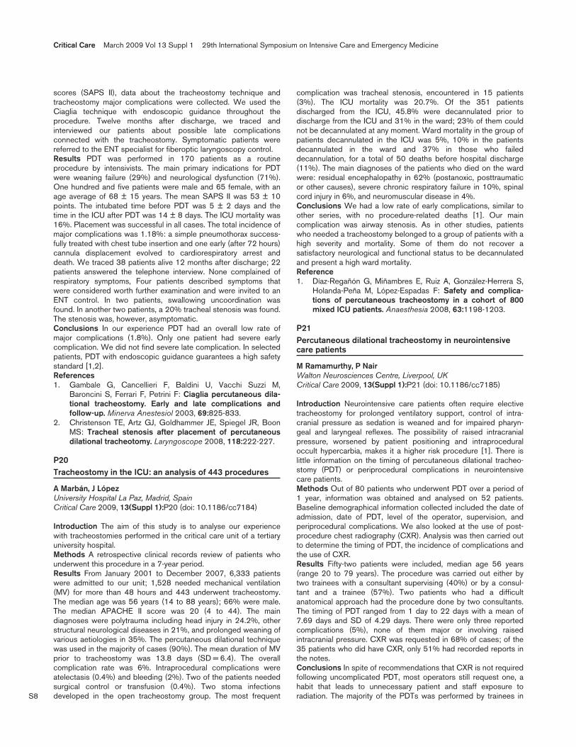
Introduction Unnecessary prolongation of mechanical ventilationis related to increased morbidity. On the contrary, early discon-tinuation of mechanical ventilation with reintubation is also relatedto bad prognosis. High respiratory rate, cardiac load and neuro-muscular dysfunction are known factors related to weaning failure.Oxygen tissue saturation (StO2) obtained by near-infrared spectro-scopy (NIRS) reflects the balance between oxygen delivery andconsumption at the muscle level. StO2 evolution during weaningmay have a role in assessing respiratory muscle performance andhelp us to predict patient readiness to be weaned. The studyobjective is to describe respiratory muscle StO2 during a T-tubetest.Methods A patient with mild head injury and pulmonary contusionwas submitted to a T-tube trial after obtaining stability on day 5.The NIRS signal from serratus anterior muscle was acquired byplacing an Inspectra device probe on the skin surface of the
Critical Care March 2009 Vol 13 Suppl 1 29th International Symposium on Intensive Care and Emergency Medicine
Table 1 (abstract P25)
Baseline characteristics
Paper SmartCare (n = 17) (n = 17) P value
Age 70.5 (9.4) 62.4 (13.8) 0.07 t test
APACHE II score 13.7 (5.2) 13.5 (4.4) 0.90 t test
Days ventilated 3 (2 to 4) 3 (2 to 4) 0.97 Mann–Whitney
U test
Table 2 (abstract P25)
Results
Paper SmartCare (n = 15) (n = 17) P value
Wean time (hours) 4.5 2.6 0.007 (2.8 to 21.3) (1.8 to 3.6) Mann–Whitney
U test
Reintubation 0/15 (0%) 2/17 (12%) 0.49 Fisher exact test
28-day mortality 1/15 (7%) 0/17 (0%) 0.47 Fisher exact test
S11
muscle [1]. Simultaneously, the respiratory rate, heart rate, meanarterial pressure and arterial oxygen saturation were recorded.Results After 5 minutes the patient failed on his first T-tube trial,showing profuse sweating, accessory muscle recruitment andincreasing respiratory rate and mean arterial pressure (Figure 1).The StO2 signal decreased during ventilatory failure until thepatient was on assistance again.Conclusions NIRS was sensitive to respiratory muscle fatigue butfurther research is in process to assess its predictive capability.Reference1. Moalla W, et al.: Respiratory muscle deoxygenation and
ventilatory threshold assessments using near infraredspectroscopy in children. Int J Sports Med 2005, 26:576-582.
P27Early and continuous weaning from mechanical ventilationwithout formal protocols in a university hospital
K Lam, J WalkerRoyal Liverpool University Hospital, Liverpool, UKCritical Care 2009, 13(Suppl 1):P27 (doi: 10.1186/cc7191)
Introduction There is evidence that formal weaning protocols canreduce the duration of mechanical ventilation (MV) and compli-cations of prolonged unnecessary ventilation [1]. In our ICU we donot employ a formal protocol, but have a standard practice wherepatients with spontaneous respiratory efforts on pressure-controlled ventilation (PCV) have a trial of assisted spontaneousbreathing (ASB). Patients on ASB with pressure support≤10 cmH2O will then have a trial of unassisted continuous positiveairway pressure (CPAP) with high-flow oxygen.Methods A retrospective audit was conducted on 47 patients in a13-bed general medical and surgical ICU of a university hospital.The length of time between first spontaneous breaths while onPCV and a trial of ASB was recorded. The length of time fromachieving pressure support ≤10 cmH2O on ASB to a trial ofunassisted CPAP was recorded. A retrograde step was defined asgoing back on to PCV from ASB, or to ASB from unassistedCPAP. If the trial of weaning was repeated within 12 hours of a
retrograde step, this was noted as a continuous attempt atweaning. Similar data were collected for subsequent retrogradesteps.Results The median duration of MV from initiation todiscontinuation was 85 hours (range 1 to 345 hours), with amedian time of 37 hours (range 1 to 245 hours) on PCV and32 hours (range 0 to 264 hours) on ASB. The median time fromrecorded spontaneous breaths on PCV to ASB was 1 hour (range0 to 34 hours). Twenty-nine (62%) patients on PCV progressed onASB without any retrograde steps. The median time from havingpressure support ≤10 cmH2O on ASB to unassisted CPAP was2 hours (range 0 to 41 hours). Thirty-one (66%) patients on ASBprogressed on CPAP without any retrograde steps. Forty-onepatients (87.2%) had a first attempt to wean from PCV to ASB and40 patients (85%) from ASB to CPAP within 10 hours of eligibility.Maximum delay in initiating first attempts to ASB was 34 hours,and 41 hours to CPAP. Reasons for retrograde steps includedrespiratory instability (n = 19), signs of poor tolerance or haemo-dynamic instability on ASB/unassisted CPAP (n = 18) and inter-ventions, for example bronchoscopy, imaging, theatre, and so forth(n = 13).Conclusions The median duration on MV in our unit comparesfavourably with a large randomised controlled trial with a similarpatient population [1]. Most patients had their first attempt to weanwithin 10 hours from eligibility. A significant number of patientsmay have weaned more quickly if a formal protocol was in place.Reference1. Marelich GP, Murin S, Battistella F, Inciardi J, Vierra T, Roby
M: Protocol weaning of mechanical ventilation in medicaland surgical patients by respiratory care practitioners andnurses: effect on weaning time and incidence of ventila-tor-associated pneumonia. Chest 2000, 118:459-467.
P28Ventilator dependency among morbidly obese in the ICU
CL Jessen, KM LarsenAarhus University Hospital, Aarhus C, DenmarkCritical Care 2009, 13(Suppl 1):P28 (doi: 10.1186/cc7192)
Introduction The purpose of this study was to evaluate thedependency for mechanical ventilation among morbidly obesepatients (MOP) defined by BMI ≥40 kg/m2, admitted to our ICU.Because of reduced functional residual capacity, increased risk ofatelectasis, increased work of breathing and decreasedcompliance of the lungs and chest wall [1], MOP are expected tohave a high dependency of mechanical ventilation. Early tracheo-tomy has a beneficial outcome in a medical population of patientsadmitted to the ICU [2], and one should assume benefits of earlytracheotomy in MOP because they are at high risk of pulmonarycomplication. A subject of debate as a study has shown morbidobesity associated with increased risk of complications [3].Methods All MOP admitted for more than 24 hours in a 12-bedmixed ICU at a Danish university hospital in the period of 2007 and2008 were retrospectively included. The ICU stay was registeredas well as airway management, length of mechanical ventilationand time for tracheotomy after intubation.Results Twenty-one morbidly obese patients were admitted.Fifteen patients (71.4%) needed mechanical ventilation. Three ofthese patients had a period of noninvasive ventilation. The medianduration of ventilation was 13 days (range 4 to 71 days) andmedian length of stay was 16 days (range 4 to 71 days). Elevenpatients were tracheotomised after a median 7 days (range 1 to11 days). Six patients had no need for mechanical ventilation. Theirmedian length of stay was 3 days (range 1 to 12 days). There was
Available online http://ccforum.com/supplements/13/S1
Figure 1 (abstract P26)
StO2 evolution during the T-tube trial. HR, heart rate; RR, respiratoryrate; MAP, mean arterial pressure.

S12
no difference in age and BMI between the two groups.Female/male ratio was 8/7 in the ventilated group versus 5/1 in thenonventilated group. Surgical/medical ratio was 11/4 in theventilated group versus 6/0 in the nonventilated group. Only onepatient died in the ICU.Conclusions A high proportion of MOP admitted to our ICUneeded mechanical ventilation (71.4%) and a very high proportionwas tracheotomised. Further studies are needed to evaluate thebeneficial effects of early tracheotomy in this patient group.References1. Marik P, et al.: The obese patient in the ICU. Chest 1998,
113:492-498.2. Rumbak MJ, et al.: A prospective, randomised, study com-
paring early percutaneous dilational trachetomy to pro-longed translaryngeal intubation (delayed tracheotomy) incritically ill medical patients. Crit Care Med 2004, 32:1689-1694.
3 Solh A, Jaafar W: A comparative study of the complicationsof surgical tracheostomy in morbidly obese critically illpatients. Crit Care 2007, 11:R3.
P29Comparison of a novel humidifier with two conventionalhumidifiers during high-frequency oscillatory ventilation
J Davies1, N Tiffin2, N MacIntyre1
1Duke University Medical Center, Durham, NC, USA; 2Hydrate,Inc., Midlothian, VA, USACritical Care 2009, 13(Suppl 1):P29 (doi: 10.1186/cc7193)
Introduction During high-frequency oscillatory ventilation (HFOV),drying of the airways and mucous plug formation can be compli-cations associated with inadequate humidification. This studycompares water vapor delivery of a standard passover humidifierand a conchatherm humidifier typically used during HFOV with anovel humidifier that employs the principle of capillary forcevaporization.Methods The Sensormedics 3100B oscillatory ventilator (CardinalHealth, Yorba Linda, CA, USA) was connected to a test lung at thefollowing settings: mean airway pressure, 30 cmH2O; power, 6.0;inspiratory time percentage, 33%; frequency of 6 Hz and bias flow30 l/min on room air. The 3100B was run at the above settings firstusing five different MR850 passover humidifiers (Fisher & Paykel,Auckland, New Zealand), followed by five different Hydrate OMNIs(Hydrate Inc., Midlothian, VA, USA) and then five differentConchaTherm Neptunes (Teleflex Medical, Research Triangle Park,NC, USA). The gas temperature and relative humidity wererecorded continuously using an electronic hygrometer/thermo-meter (SHT75; Sensirion, Staefa, Switzerland) in two circuitconfigurations: (1) between the test lung/patient wye (triangularplastic connector that connects the inspiratory and expiratory limbsof the circuit with the patient endotracheal tube) and (2) distal to acondensation tube placed between the test lung/patient wye. Thecondensation tube served to approximate the upper airways in our
lung model and collect the condensate. Water condensate wascollected over a 30-minute test period for each run. All humidifierswere set to 37°C.Results See Table 1.Conclusions In this model, the Hydrate OMNI provided thehighest absolute humidity during HFOV. The difference wasamplified at the end of the condensation tube. Further study onhumidification during HFOV is warranted.
P30A new in vitro model for force measurements at theisolated entire rat diaphragm
C Armbruster, K Gamerdinger, M Schneider, S Schumann, H Priebe, J GuttmannUniversity Medical Center, Freiburg, GermanyCritical Care 2009, 13(Suppl 1):P30 (doi: 10.1186/cc7194)
Introduction Several diseases of different origin as well as pro-longed mechanical ventilation result in diaphragmatic dysfunctionand atrophy. To date most in vitro experiments on the mechanics ofdiaphragm muscles are performed at isolated muscle strips. Sincethe improvement of the knowledge about the diaphragmfunctionality is lacking in view of the position and curvature, amodel of the whole diaphragm appears necessary. Here wepresent a new in vitro model of an isolated whole rat diaphragminside a bioreactor [1].Methods The bioreactor consists of a pressure chamber on whicha highly flexible membrane is attached. A rat diaphragm is fixed onthis membrane. By application of a gas volume into the pressurechamber, a defined deflection of the diaphragm is achieved. Thehighly flexible membrane adapts a given shape thus allowing thediaphragm to take up its in vivo profile. Electrical stimulation resultsin a contraction of the diaphragm. These diaphragm-twitchesgenerate pressure pulses inside the pressure chamber. Ratdiaphragms were excised rapidly and kept at room temperature inKrebs–Henseleit solution bubbled with oxygen. By application ofone of two different initial pressures (P1: 7 mbar; P2: 12 mbar) thediaphragms were set to a defined degree of deflection. Using aplatinum wire electrode, the diaphragms were electricallystimulated at impulses of 6V. The stimulation train duration was setto 500 ms or 1 second. The pulse duration was set to 50 ms at afrequency of 50 Hz. The pressure pulses resulting as the responseon muscle contraction were measured inside the pressurechamber.Results An increase of the initial pressure led to an increasedpressure caused by the muscle contraction. An enlarged muscleregeneration time led to an increase of the diaphragmatic twitch-induced pressure.Conclusions Our new in vitro model of an isolated whole ratdiaphragm allows the performance of mechanical and physiologicalinvestigations on the entire diaphragm. A dependency of pressuredevelopment of the diaphragm on its deflection state could bedemonstrated. Furthermore, we found effects of diaphragm musclefatigue. Our new model could be used in numerous types ofinvestigations, such as releasing factors of diaphragmatic dysfunc-tion or respiratory muscle fatigue.Reference1. Schumann S, Stahl CA, Möller K, Schneider M, Metzke R,
Wall WA, Priebe HJ, Guttmann J: Contact-free determina-tion of material characteristics using a newly developedpressure-operated strain-applying bioreactor. J BiomedMater Res B Appl Biomater 2008, 86B:483-492.
Critical Care March 2009 Vol 13 Suppl 1 29th International Symposium on Intensive Care and Emergency Medicine
Table 1 (abstract P29)
Wye Wye absolute Distal absolute temperature humidity (mg/l) humidity (mg/l)
MR850 34.4 ± 0.9 33.7 ± 1.3 17.2 ± 6.1
OMNI 34.4 ± 1.3 37.2 ± 2.6 26.8 ± 5.0
Neptune 32.0 ± 0.9 30.8 ± 1.7 17.7 ± 1.0
Data presented as mean ± SD.

S13
P31Adaptive support ventilation prevents ventilator-induceddiaphragmatic dysfunction: an in vivo piglet study
B Jung1, N Rossel1, C Le Goff1, N Claveiras1, M Wysocki2, S Matecki1, G Chanques1, S Jaber1
1Saint-Eloi Hospital, Montpellier, France; 2Hamilton Medical,Rhazuns, SwitzerlandCritical Care 2009, 13(Suppl 1):P31 (doi: 10.1186/cc7195)
Introduction Mechanical ventilation is a lifesaving supportivetherapy for patients with acute respiratory failure. However,prolonged mechanical ventilation results in the complete absenceof neural activation and mechanical activity of the diaphragm andhas been shown to induce ventilator-induced diaphragmaticdysfunction (VIDD) [1]. Few studies have shown that maintainingspontaneous ventilation could prevent VIDD in in vitro animalstudies [2]. Adaptive support ventilation (ASV) is an automaticventilation mode that allowed pressure-controlled breaths in activepatients able to trigger. The aim of our study was to compare ASVwith controlled ventilation (CV) without paralysing agents ondiaphragmatic contractile properties in an in vivo piglet study.Methods Two groups of six anesthetized piglets were ventilatedduring a 72-hour period. Piglets in the CV group (n = 6) wereventilated without any spontaneous ventilation and piglets in theASV group (n = 6) were ventilated with the possibility of triggeringspontaneous ventilation. The main endpoint was the transdiaphrag-matic pressure (Pdi) (after bilateral, supramaximal, transjugularstimulation of the two phrenic nerves), which represents the in vivocontractile function of the diaphragm. A force–frequency curvewas drawn after stimulation from 20 to 120 Hz of the phrenicnerves.Results The piglets in the ASV group were maintained withspontaneous ventilation during 80% of the study period instead ofless than 1% in the CV group. At 72 hours, Pdi was decreased by35% in the CV group although it was not modified in the ASVgroup (Figure 1).Conclusions Spontaneous breathing with ASV prevents VIDD incomparison with totally CV in an in vivo healthy piglet model.
References1. Sassoon CS: Ventilator-associated diaphragmatic dys-
function. Am J Respir Crit Care Med 2002, 166:1017-1018.2. Futier E, Constantin JM, Combaret L, Mosoni L, Roszyk L,
Sapin V, et al.: Pressure support ventilation attenuatesventilator-induced protein modifications in the diaphragm.Crit Care 2008, 12:R116.
P32Characterization of the mechanical ventilator adjustmentprocess
H Al-Otaibi, J Hardman, R MahajanThe University of Nottingham, UKCritical Care 2009, 13(Suppl 1):P32 (doi: 10.1186/cc7196)
Introduction The aim of the present study was to gain insight intothe mechanical ventilator adjustment process, particularly eventsthat prompt clinicians to adjust the ventilator, the indications,considerations, and methods used to assess the successfulness ofthe adjustment episode.Methods A prospective, observational, noninterventional studywas conducted in a 24-bed adult, medical and surgical intensivecare unit at a regional hospital. Patient demographics, ventilatoradjustment episodes, and clinical decisions related to these adjust-ments were collected. Clinicians were asked to complete aventilator flowsheet before and after any ventilator adjustmentepisode. Simultaneously, they were asked to complete an open-ended questionnaire related to the process of ventilator adjustmentepisode.Results A total of 168 ventilator adjustment episodes derived from26 mechanically ventilated patients were evaluated. Among theseepisodes, the ventilator mode was adjusted 33 times (20%), theminute volume 37 times (22%), the positive end-expiratorypressure 19 times (11%), the pressure support 21 times (12%),and the fraction of inspired oxygen 61 times (36%). Triggers whichstimulate the process of mechanical ventilator adjustment werecategorized into: routine ventilator checks (35%), routine arterialblood gases (ABG) (20%), weaning trials (13%), and calls fromnurses (9%). The most common indications to adjust the ventilatorwere hyperoxygenation (28%) and weaning trials (26%). Peripheraloxygen saturation, ABG, and level of consciousness were the mostcommon considered variables during the process of ventilatoradjustment. In 42% of the total adjustment episodes, clinicians didnot consider any technical, physiological or psychological variablewhile they adjusted ventilator parameters. Clinicians based theirdecisions to adjust ventilator settings on clinical experience (72%),trial and error (15%), protocols (11%), and scientific equations(2%). Clinicians assessed their decisions via ABG results (47%),peripheral oxygen saturation (23%), and general patientassessment (9%).Conclusions The process of ventilator adjustment is mainlystimulated by routine ventilator checks and ABG. ABG andweaning trials are the most common indications. Most ventilatoradjustment decisions are based on clinical experience, evaluatedvia ABG and peripheral oxygen saturation.
Available online http://ccforum.com/supplements/13/S1
Figure 1 (abstract P31)
Pdi evolution during the study. P <0.05 CV vs. ASV group at 48 hours.

S14
P33Impact of a dedicated ventilatory support team on howmechanical ventilation is employed in a tertiary-carehospital
F Saddy, A Thompson, R Serafim, F Gago, N Charris, J PantojaCopa D`Or, Rio de Janeiro, BrazilCritical Care 2009, 13(Suppl 1):P33 (doi: 10.1186/cc7197)
Introduction Advances in our knowledge of the pathophysiology ofrespiratory failure have forced major revisions of our approach toventilatory support. We describe how mechanical ventilation isemployed in four different ICUs (surgical, clinical, cardiac andneurological) of a tertiary-care Brazilian hospital where a ventilatorysupport team composed of intensivists is responsible for a daily-basis follow up.Methods A prospective observational study enrolled all invasivemechanically ventilated patients admitted to four ICUs from May2004 through June 2008. Daily recorded data included:demographics, diagnosis, modes of ventilation, tidal volume/kg(Vt), positive end-expiratory pressure (PEEP) level, peak inspiratorypressure, plateau pressure (Pplat), recruitment maneuvers, use ofsedation and neuromuscular blocking agents (NBA), tracheotomy,barotrauma, ventilation days, and length of stay (LOS) in the ICU.Results are expressed as the mean ± SD and percentage.Differences were assessed by one-way ANOVA followed by theTukey test. P <0.05 was considered significant.Results A total of 1,715 patients was studied. Diagnosis prevaileddepending on the ICU’s characteristics. Ventilatory data aredepicted in Table 1. Recruitment maneuvers were used in less than2% of patients. The most frequent type of ventilatory mode wasspontaneous (P <0.05). Barotrauma was similar and occurred inless than 0.63% (P >0.05). Intravenous sedation was administeredfor no more than 40% of the time on mechanical ventilation. NBAwas used for no more than 0.25% of patients. LOS and ventilationdays were different among ICUs (P <0.05).
Table 1 (abstract P33)
Ventilatory data
Vt (ml/kg) PEEP (cmH2O) Pplat (cmH2O)
Surgical 6.6 ± 2 8.9 ± 3.2 22 ± 6.3
Clinical 6.6 ± 1.7 8.6 ± 2.7 22 ± 5.9
Neuro 6.7 ± 2.1 8.3 ± 2.3 22.7 ± 4.4
Cardiac 6.8 ± 1.5 8.7 ± 2.1 21.3 ± 4
Conclusions Daily interaction of the ventilatory support team andthe ICU practitioners guaranteed a homogeneous and up-to-dateform of ventilatory support care to the patients in the differentICUs.
P34Hyperoxia in mechanically ventilated patients
S Bolton, E Pugh, A Hay, S McKechnieRoyal Infirmary Edinburgh, UKCritical Care 2009, 13(Suppl 1):P34 (doi: 10.1186/cc7198)
Introduction Supplemental oxygen is part of the supportivetreatment of hypoxaemic respiratory failure. Prolonged exposure tohigh fractions of inspired oxygen (FiO2) has been shown to beinjurious to the lung in vitro [1]. The optimal paO2 in critical illnesshas not been established but the clinical impression is that patientsare frequently exposed to a higher FiO2 than is necessary. The aim
of this study was to investigate current oxygen treatment inpatients undergoing prolonged mechanical ventilation anddetermine whether this resulted in exposure to a higher FiO2 thanthat required to adequately oxygenate haemoglobin (SaO2 >95%).Methods Indices of oxygenation were collected for 30 patientsrequiring mechanical ventilation >2 days. Data were collected4-hourly for 48 hours. The PaO2:FiO2 ratio was determined foreach data point and used to model the theoretical FiO2 required tomaintain an SaO2 of 95% (paO2 = 10.7 kPa).Results The results are shown in Figure 1. Data are expressed asmedians (IQR). The median observed FiO2 was 0.4 throughout thestudy whereas the median FiO2, calculated to maintain an SaO2 of95%, was 0.3.Conclusions Our data demonstrate that critically ill patients maybe exposed to a higher FiO2 than that required to maintainadequate oxygenation. Further, these results highlight an area ofICU care that has received little study, with no published clinicaltrials examining the effect of FiO2 on outcome.Reference1. McKechnie S: The effect of hyperoxia on alveolar epithelial
injury and repair. J Intensive Care Soc 2008, 9:94.
P35Are chest X-rays necessary after chest tube insertion intrauma emergencies?
MS MoengJohannesburg Hospital, Houton, Johannesburg, South AfricaCritical Care 2009, 13(Suppl 1):P35 (doi: 10.1186/cc7199)
Introduction Johannesburg Hospital is a level I trauma center inGauteng. We routinely do chest X-rays (CXRs) in indicated cases,if feasible, and prefer to perform a CXR after intercostal drain (ICD)insertion to maintain quality and direct further care [1].Methods A prospective data collection of patients who hadinjuries that required the insertion of an ICD over a period of8 months from 1 August 2006 to 30 March 2007. A questionnairewas developed and it included the patients’ demographics, mecha-nism of injury, reason for ICD insertion, findings of both the initialand post-ICD insertion CXR, change in management and anyacute complications noted.Results One hundred and forty patients were identified for thestudy, 129 (92.1%) were males and 11 (7.9%) were females. Theaverage age was 32 (range 11 to 64) years. Eighty-four (60%)
Critical Care March 2009 Vol 13 Suppl 1 29th International Symposium on Intensive Care and Emergency Medicine
Figure 1 (abstract P34)

S15
patients sustained stab wounds and 20 (14.3%) had gunshotwounds. The remaining 36 (25.7%) sustained blunt injury. Onehundred and four (74.3%) patients had an ICD inserted for bothradiological and clinical findings, while 19 (13.6%) patients haddrains inserted due to radiological findings and 14 (10%) patientson clinical grounds only. Three (2.1%) patients had drain insertionafter CT scan findings.In patients who had an initial CXR, clinical and/or radiological find-ings confirmed 47 (36.7%) patients with haemopneumothoraces,49 (38.3%) patients with pneumothoraces, 26 patients (20.3%)with haemothoraces and four (3.1%) patients had significantsurgical emphysema with fractures and two (1.6%) patients had noabnormalities on CXR. No acute complications to chest tubeinsertion were noted.In 89 (63.6%) patients, the post-ICD CXR showed good positionof the drain with improvement in pathology, in 31 (22.1%) patientsan inadequate ICD position was noted, in 17 (12.1%) patientssignificant retained haemothoraces were shown and in three(2.1%) patients poor lung expansion was detected. The post-ICDCXR contributed to change in management in 29 (20.7%) of thecases. Twenty-two (15.7%) patients required a change in positionof the tube, six (4.3%) had surgery performed, and one (0.7%)patient had their conservative treatment escalated. Four (2.9%)patients should have had their tubes adjusted.Conclusions Routine usage of post-ICD CXR contributes to achange in management in one out of five trauma patients.Reference1. Huber-Wagner S, Körner M, Ehrt A, Kay MV, Pfeifer KJ,
Mutschler W, Kanz KG: Emergency chest tube placementin trauma care – which approach is preferable? Resuscita-tion 2007, 72:226-233.
P36Bedside chest ultrasound reduces the rate of chest X-rayand CT examinations
R Spina1, L Tutino2, A Di Filippo2, L Perretta1, A Cecchi1, A Peris1
1Careggi Teaching Hospital, Florence, Italy; 2University of Florence,ItalyCritical Care 2009, 13(Suppl 1):P36 (doi: 10.1186/cc7200)
Introduction A bedside chest ultrasound (bCUS) programmeperformed by intensivists after 18 months of ultrasound trainingwas introduced in the ICU routine between April and November2008 in order to evaluate its effects on the number of chest X-ray(CXR) and CT scans. The setting was a 10-bed emergency ICU.Methods From April to November 2008 every patient hasundergone a bCUS within the first 48 hours since admittance, thenbetween the fourth and the sixth days of their stay. All of the 92patients were examined supine, with a convex probe perpendicularto the chest wall, using all the intercostal spaces as the acousticwindow. From the lung base, every intercostal space has beenexamined, looking for a pleural effusion, attaining to the followingcriteria: a space between the visceral and parietal pleura,movement of that space in agreement with the respiratory pattern.As for the volume esteem of the effusion, the distance (mm)between the posterior part of the lung and the posterior chest wallwas measured. For each patient the following data were collected:age, sex, weight, height, SAPS II, number of CXR and CT scansdone. Data were compared with those of a group of patientsadmitted to the ICU from January to March 2008, when bCUS wasnot part of the daily procedures. P <0.05 was consideredstatistically significant.
Results The two groups of patients were statisticallyhomogeneous. Comparing the control group with the study group,CXRs were reduced by 22.63% (from 433 to 335; P = not signi-ficant) and CT scans were reduced by 42.36% (from 144 to 83; P<0.05).Conclusions The use of daily routine bCUS is useful to reduce thetraditional radiology requests. The decrease of CT scans isstatistically relevant. As far as CXR requests are concerned, thedecrease does not appear significant since the CXR is relativelyeasy to perform and it is mandatory after radiopaque devicepositioning. These amount to 34% of the total CXRs performed.
P37Combination of variability with pressure support ventilationenhances lung protection and function in experimentalacute lung injury
M Gama de Abreu1, PM Spieth1, AR Carvalho1, P Pelosi2, T Koch1
1University Clinic Carl Gustav Carus, Dresden, Germany;2University of Insubria, Varese, ItalyCritical Care 2009, 13(Suppl 1):P37 (doi: 10.1186/cc7201)
Introduction Protective ventilation with low tidal volumes becamethe standard of care in acute lung injury (ALI). Although spon-taneous breathing activity may be beneficial even in early ALI,protective ventilation is usually performed as controlled ventilation.Theoretically, pressure support ventilation (PSV) may be advan-tageous over pressure-controlled ventilation (PCV), particularly ifcombined with variability in the form of noisy PSV [1,2].Methods Two protocols (A and B) were performed in pigs (20 to30 kg). Animals were anesthetized, intubated, mechanically venti-lated and ALI was induced by surfactant depletion. In protocol A,animals (n = 12) were randomly assigned to a sequence of PCV,PSV and noisy PSV, being ventilated for 1 hour in each mode(crossover design). The distribution of lung aeration and perfusionwere determined. In protocol B, animals (n = 24) were randomlyassigned to 6 hours of mechanical ventilation with PCV or PSV ornoisy PSV, and lungs were extracted for quantification ofinflammation and lung damage. In both protocols, gas exchangeand respiratory parameters were determined. Statistical analysiswas performed with univariate and multivariate tests, as appro-priate. Significance was accepted at P <0.05.Results Compared with PCV, arterial oxygenation, intrapulmonaryshunt, mean airway pressure and elastance of the respiratorysystem were improved by PSV and further improved by noisy PSV.Also, noisy PSV reduced the work of breathing and respiratorydrive compared with PSV. Lung damage and inflammation werereduced by assisted mechanical ventilation, but comparablebetween noisy PSV and PSV. The distribution of lung aeration didnot differ among the three modes, but PSV and noisy PSV wereassociated with more redistribution of pulmonary blood flowtowards better aerated ventral areas compared with PCV.Conclusions The combination of variability with PSV enhanceslung protection and function in experimental ALI. The mainmechanism of improvement of lung function by assistedmechanical ventilation using pressure support is not recruitment ofdorsal areas, but rather redistribution of pulmonary blood flow toventral zones.References1. Gama de Abreu M, Spieth PM, Pelosi P, Carvalho AR, Walter
C, Schreiber-Ferstl A, et al.: Noisy pressure support ventila-tion: a pilot study on a new assisted ventilation mode inexperimental lung injury. Crit Care Med 2008, 36:818-827.
2. Spieth et al.: Am J Respir Crit Care Med in press.
Available online http://ccforum.com/supplements/13/S1
S16
P38Measurement of the forearm to warrant low-tidal-volumeventilation in the acute respiratory distress syndrome
M Möller, J Völz, J NeuznerKlinikum Kassel, GermanyCritical Care 2009, 13(Suppl 1):P38 (doi: 10.1186/cc7202)
Introduction Acute respiratory distress syndrome (ARDS) is a life-threatening situation in patients on the ICU. Most patients have to beventilated mechanically to provide adequate oxygenation. Reductionof tidal volumes as low as 6 ml/kg adjusted bodyweight has beenconvincingly shown to reduce ARDS and mortality in the ARDSnettrial [1] and is now recommended in treating such patients. In theARDSnet trial, body weight has been calculated by a formulaimplementing the body height [1]. We suggest that in most patientson ICUs the correct height is not known or is at best estimated, butvery seldom correctly measured. We searched for both an easily ob-tainable and reproducible body mark to correctly predict body height.Anthropological and forensic data have shown a close correlationbetween the ulna length and body size. We prospectively measuredbody height and right and left ulna length in ventilated ICU patients.Methods In a 2-month period, 39 men and 42 women from fourICUs in a teaching hospital were included consecutively (mean age62 years, range 23 to 87 years). Body height was measuredfollowing a standardized protocol, ulna length was measured fromthe edge of the olecranon to the caput ulnae.Results Eighty-one patients were included. Thirty-nine men: height145 to 199 cm (mean 178.5 cm), right ulna 25 to 32 cm (mean28.3 cm), left ulna 24.5 to 32 cm (mean 28.3 cm). Forty-twowomen: height 142 to 185 cm (mean 165.7 cm), right ulna 21 to29 cm (mean 25.0 cm), left ulna 21 to 28 cm (mean 24.9 cm).Regression analyses were made in SAS 9.1 and showed asignificant correlation between the body height (men and women)and the length of the ulna. Regression analyses for men: bodyheight in cm = 3.9314 x (right ulna; in cm) + 67.059 cm(r2 = 53.25%; SD = 6.79) and body height in cm = 3.9786 x (leftulna; in cm) + 65.824 cm (r2 = 52.50%; SD = 6.84). Regressionanalyses for women: body height in cm = 4.88 x (right ulna; in cm)+ 43.76 cm (r2 = 55.53%; SD = 6.61) and body height in cm =5.41 x (left ulna; in cm) + 30.95 cm (r2 = 60.67%; SD = 6.21).Conclusions Ulna length is an easily obtainable estimate of totalbody height. It may aid in implementing low-tidal-volume ventilation.Anthropological data correlate reasonably with a contemporaryventilated ICU population. Whether ulna-derived estimates of bodyheight may improve adherence to established guidelines has to bestudied.Reference1. Acute Respiratory Distress Syndrome Network: Ventilation
with lower tidal volumes as compared with traditional tidalvolumes for acute lung injury and the acute respiratorydistress syndrome. N Engl J Med 2000, 342:1301-1308.
P39Effect of pleural effusion on gas exchange and responseto positive end-expiratory pressure in acute lunginjury/acute respiratory distress syndrome patients
D Chiumello, C Mietto, V Berto, M Cressoni, M Lazzerini, L GattinoniFondazione IRCCS, Ospedale Maggiore Policlinico, Milan, ItalyCritical Care 2009, 13(Suppl 1):P39 (doi: 10.1186/cc7203)
Introduction Pleural effusion is a common finding in acute lunginjury/acute respiratory distress syndrome (ALI/ARDS) patients.
However, the effect of pleural effusion on gas exchange, respira-tory mechanics and response to positive end-expiratory pressure(PEEP) in ALI/ARDS patients, during mechanical ventilation, hasnever been prospectively studied.Methods Patients with a diagnosis of ALI/ARDS, who underwent aCT scan at 5 cmH2O PEEP for clinical reasons, were included inthe study. Lung and pleural effusion were outlined separately; lungtotal weight and pleural effusion volume were computed withdedicated software. A PEEP test at 5 and 15 cmH2O withconstant minute ventilation was performed. Exclusion criteria were:age <18 years, hemodynamic instability, chronic obstructive pul-monary disease and evidence of barotrauma.Results We enrolled 11 ALI/ARDS patients (10 male). The meanclinical characteristics on admission to the ICU were: age 67.5 ± 9.9years, BMI 24.4 ± 2.1 kg/m2, PaO2/FiO2 203.1 ± 56.1 mmHg,PEEP 9.1 ± 2.8 cmH2O and pH 7.394 ± 0.048. The volume ofpleural effusion was not significantly related with the change inPaCO2 (r2 = 0.068; P = 0.437), PaO2 (r2 = 0.015; P = 0.722) anddead space (VD/VT) (r2 = 0.019; P = 0.682) going from PEEP 15to PEEP 5 cmH2O.Conclusions Pleural effusion does not seem to influence the gas-exchange response to PEEP.
P40CT-scan lung morphology predicts the response to arecruitment maneuver in acute respiratory distresssyndrome patients
J Constantin1, S Grasso2, JJ Rouby3, E Futier1, B Gallix4, B Jung4, JE Baazin1, S Jaber4
1Hotel-Dieu Hospital, Clermont-Ferrand, France; 2OspedalePoliclinico, Bari, Italy; 3Pitié Salpetriere Hospital, Paris, France;4Saint-Eloi Hospital, Montpellier, FranceCritical Care 2009, 13(Suppl 1):P40 (doi: 10.1186/cc7204)
Introduction CT-scan lung morphology (lobar or nonlobar) is themain determinant of positive end-expiratory pressure (PEEP)response. Repartition of gas and tissue probably influences theresponse to a recruitment maneuver (RM), but to date there is noproof. The aim of this study was to assess RM-induced changes inlung morphology and gas exchange during and after RM in acuterespiratory distress syndrome (ARDS) patients.Methods Nineteen patients with ARDS were included in the study.Patients were ventilated with volume control ventilation (Gallileo;Hamilton Medical, Bonaduz, Switzerland) with Vt = 6 ml/kg (idealbody weight) and respiratory rate to keep PaCO2 >55 mmHgwithout intrinsic PEEP. After a first CT scan in zero end-expiratorypressure conditions, a pressure–volume curve was performed andthe PEEP was set above the lower inflection point. After astabilisation period, a second CT scan (in PEEP) was performed.Then a RM was performed, using continuous positive airwaypressure 40 cmH2O for 40 seconds. At the end of the RM(between 35 and 39 s) a third CT scan was performed. Fiveminutes after the RM, a fourth CT scan was performed in PEEPconditions (same level of PEEP). Blood gas analysis was sampledat each step of the study. CT-scan analysis was performed usingspecific volumetric software (IRMA).Results Ten men and nine women, 63 ± 11 years old, wereincluded in the study. SAPS II was 44 ± 8. Nine presented anonlobar CT-scan attenuation and eight a focal loss of aeration. Allpatients presented early ARDS (onset between ARDS diagnosticand study inclusion was 18 ± 11 hours). Setting PEEP 2 cmH2Oabove the lower inflection point increased PaO2/FiO2 from167 ± 110 to 205 ± 72 mmHg (P <0.001), and 265 ± 80 mmHgafter the RM (P <0.005 vs. PEEP) and 273 ± 96, 5 minutes after
Critical Care March 2009 Vol 13 Suppl 1 29th International Symposium on Intensive Care and Emergency Medicine

S17
the RM. The RM-induced recruited volume at the same level ofPEEP was 8 ± 45 ml in the case of lobar CT-scan attenuationversus 96 ± 63 ml in nonlobar CT-scan attenuation (P <0.005).The overinflated lung volume after RM was 121 ± 170 ml in thecase of lobar attenuation versus 18 ± 22 ml in diffuse (P <0.005).During the RM, the overinflated lung volume was over 50% in thecase of lobar CT-scan attenuation.Conclusions As for PEEP, lung morphology predicts the responseto a RM. In the case of lobar loss of aeration, RM inducedoverinflation more than the recruited volume and should beavoided.
P41Alveolar wall disruption and lung inflammation associatedwith positive end-expiratory pressure and recruitmentmaneuver in pigs
A Ambrósio1, DT Fantoni1, CK Marumo2, D Otsuki2, C Gutierres3, Q Lu3, J Noel-Morgan2, JJ Rouby3, JO Auler Jr2
1Faculdade de Medicina Veterinária e Zootecnia da Universidadede São Paulo, Brazil; 2Faculdade de Medicina da Universidade deSão Paulo, Brazil; 3La Pitiè-Salpêtrière Hospital, AssistancePublique Hôpitaux de Paris, University Pierre et Marie Curie, Paris,FranceCritical Care 2009, 13(Suppl 1):P41 (doi: 10.1186/cc7205)
Introduction Different levels of positive end-expiratory pressure(PEEP) associated or not with alveolar recruitment maneuver(ARM) may have a significant impact on ventilator-induced lunginjury [1], but this issue has not been well addressed in a model oflung injury using inhaled hydrochloric acid. We aimed to evaluatethe effects of PEEP and ARM on respiratory mechanics and lungtissue in an inhaled hydrochloric acid acute lung injury (ALI) model.Methods Thirty-two pigs (22.9 ± 2.8 kg), were randomly allocatedinto one of four groups (I – Control-PEEP, II – Control-ARM, III –ALI-PEEP, IV – ALI-ARM). ALI was induced by intratrachealinstillation of hydrochloric acid. PEEP values were progressivelyincreased and decreased from 5, 10, 15 and 20 cmH2O in allgroups. Three alveolar recruitment maneuvers of 40 cmH2O for20 seconds were applied to the assigned groups at each intervalof PEEP level increase or decrease. Histological analysis wasmade to evaluate the presence of inflammatory infiltrates, alveolarwall thickening, atelectasis, hemorrhage, alveolar edema andalveolar disruption. Histomorphometrical analyses of the lungswere also performed to determine alveolar dimensions.Results Inflammation and alveolar disruption were statisticallygreater in group II when compared with group I. There were nostatistical differences between groups III and IV with respect tohemorrhage, alveolar edema, inflammation and alveolar disruption.Mean alveolar area and mean alveolar intercept were higher ingroup IV when compared with groups I and II (P <0.05). The meanalveolar area was significantly smaller in the diaphragmatic lobeswhen compared with other middle and upper pulmonary lobes, butno statistical difference was found among groups.Conclusions The association of PEEP with ARM promoted agreater degree of alveolar disruption.Acknowledgement Grants from LIM08-Anesthesia; FAPESP02/08621-1.Reference1. Halter JM, Steinberg JM, Schiller HJ, DaSilva M, Gatto LA,
Landas S, Nieman GF: Positive end-expiratory pressureafter a recruitment maneuver prevents both alveolar col-lapse and recruitment/derecruitment. Am J Respir CritCare Med 2003, 167:1620-1626.
P42Ventilation with high positive end-expiratory pressureimproves oxygenation after cardiac surgery independentlyof the mode of ventilation and of the use of nitric oxide
L Hajjar, F Galas, N Rossati, A Leme, R Kalil Filho, J AulerHeart Institute, São Paulo, BrazilCritical Care 2009, 13(Suppl 1):P42 (doi: 10.1186/cc7206)Introduction Postoperative pulmonary dysfunction in patients under-going cardiac surgery with cardiopulmonary bypass is a significantclinical problem and has long been recognized. Postoperative hypox-emia carries high morbidity leading to prolonged postoperativerecovery and hospital stays. We hypothesized that adding high posi-tive end-expiratory pressure (PEEP) would be effective for treatmentof postoperative hypoxemia after cardiac surgery with cardiopul-monary bypass, independently of the mode of ventilation or the useof nitric oxide.Methods During 2 years, 210 patients undergoing coronary arterybypass with pump surgery after diagnosis of hypoxemia (PO2/FiO2<200) were randomized into six groups after operation (35 in eachgroup): pressure-controlled ventilation (PCV) with inhaled nitricoxide and ideal PEEP (group 1), PCV without inhaled nitric oxideand ideal PEEP (group 2), PCV without inhaled nitric oxide andPEEP 5 cmH2O (group 3), volume-controlled ventilation (VCV)with inhaled nitric oxide and ideal PEEP (group 4), VCV withoutinhaled nitric oxide and ideal PEEP (group 5) and VCV withoutinhaled nitric oxide and PEEP 5 cmH2O (group 6). Arterial andmixed venous blood were drawn and analyzed before the interven-tions, 1, 2, 4 and 6 hours after the interventions in the ICU to deter-mine the PaO2/FiO2 ratio. Hemodynamic measurements wereanalyzed. The time to extubation was compared among groups,accordingly to the ICU weaning protocol.Results There was no significant difference among the groupsregarding hemodynamic measurements (mean arterial blood pres-sure, heart rate, central venous pressure and SVO2). Oxygenationwas higher in both high-PEEP groups (groups 1, 2, 4 and 5) thanin the PEEP 5 groups (groups 3 and 6) during the mechanical ven-tilation period (P <0.01). Also, the time to extubation was signifi-cantly lower in high PEEP groups than in the PEEP 5 groups (220min vs. 428 min, P <0.03).Conclusions Ventilation with high PEEP after cardiac surgery isassociated with improvement of oxygenation and less time ofmechanical ventilation independently of the mode of ventilation andof the use of nitric oxide.References1. Celebi S, Köner O, Menda F, Korkut K, Suzer K, Cakar N: The
pulmonary and hemodynamic effects of two differentrecruitment maneuvers after cardiac surgery. Anesth Analg2007, 104:384-390.
2. Rouby JJ, Ferrari F, Bouhemad B, Lu Q: Positive end-expira-tory pressure in acute respiratory distress syndrome:should the ‘open lung strategy’ be replaced by a ‘protec-tive lung strategy’? Crit Care 2007, 11:180.
P43Control system for automated titration of positiveend-expiratory pressure and tidal volume using dynamicnonlinear compliance as the setpointS Lozano-Zahonero1, A Wahl2, D Gottlieb2, J Arntz1, S Schumann2, J Guttmann2, K Möller1
1Furtwangen University, Villingen-Schwenningen, Germany;2University Hospital Freiburg, GermanyCritical Care 2009, 13(Suppl 1):P43 (doi: 10.1186/cc7207)
Introduction An automated respiratory mechanics control wasdeveloped to individually adapt the energy transfer from the
Available online http://ccforum.com/supplements/13/S1

S18
ventilator to the respiratory system. The controller titrates thepositive end-expiratory pressure (PEEP) and tidal volume (VT) toventilate the lung at its maximal compliance in order to avoidexcessive lung overinflation as well as underinflation.Methods The mechanics controller consists of a software programto set PEEP and VT and a user interface to observe the compli-ance and the controller state. The program has following structure:(1) Dynamic compliance is calculated breath by breath using theimplemented slice function [1]. This function divides VT into sixconsecutive volume slices of equal size. (2) For each volume slice,one value of dynamic compliance (CSLICE) is determined by least-squares fit using the linear resistance and compliance model withineach slice [2]. The six CSLICE values are plotted over thecorresponding volume, giving the compliance–volume curve. (3)The shape-compliance function of the controller identifies one outof six shape categories [3]. (4) The PEEP and VT-change functioncalculates the PEEP and VT titration depending on the shapecategory and sends a command to the ventilator for setting thenew PEEP and VT automatically.Results The system was tested with previously recorded patientdata (McRem) [4]. The compliance controller retrospectivelyanalysed the respiratory data and determined the shape categorydepending on the course of CSLICE. For shapes representing anintratidal increase of CSLICE, the controller increased the PEEP. Areduction of PEEP occurred when CSLICE decreased intratidally.PEEP was maintained when CSLICE was maximal and constant.Furthermore, for hybrid shape categories (one part in the linearregion and one part in the increasing and/or decreasing region) theVT was reduced.Conclusions The automated respiratory mechanics control systemtitrates PEEP and VT automatically until intratidal compliancereaches its maximal value within an appropriate VT.References1. Schumann S, et al.: Modellierung und Bestimmung der
nichtlinear volumenabhängigen Compliance der Lunge. InDreiländertagung der Deutschen, Österreichischen undSchweizerischen Gesellschaften für Biomedizinische Technik,Zürich; Proceedings V118; 2006.
2. Guttmann J, et al.: Determination of volume-dependentrespiratory system mechanics in mechanically ventilatedpatients using the new SLICE method. Technol HealthCare 1994, 2:175-191.
3. Mols G, et al.: Volume-dependent compliance in ARDS:proposal of a new diagnostic concept. Intensive Care Med1999, 25:1084-1091.
4. Stahl CA, et al.: Dynamic versus static respiratory mechan-ics in acute lung injury and acute respiratory distress syn-drome. Crit Care Med 2006, 34:2090-2098.
P44Effect of positive end-expiratory pressure on regionalventilation monitored by electrical impedance tomographyin mechanically ventilated ICU patients
D Gommers, IG Bikker, J BakkerErasmus Medical Center, Rotterdam, the NetherlandsCritical Care 2009, 13(Suppl 1):P44 (doi: 10.1186/cc7208)
Introduction The optimal positive end-expiratory pressure (PEEP)is a balance between the prevention of overdistention in thenondependent part and alveolar collapse in the dependent part.Electrical impedance tomography (EIT) has been introduced tomonitor regional change of ventilation at the bedside. Weevaluated the effect of changes in PEEP on regional ventilation inmechanically ventilated patients with or without lung disorders.
Methods Functional EIT (fEIT) images were obtained in 14patients on pressure-controlled ventilation with constant drivingpressure at four PEEP levels (15, 10, 5 and 0 cmH2O). fEITimages made before each reduction in PEEP were subtracted fromthose recorded after each PEEP step to evaluate the regionalincrease/decrease in tidal impedance in each EIT pixel.Results The response of regional tidal impedance to PEEPshowed a significant difference from 15 to 10 cmH2O (P = 0.002)and from 10 to 5 cmH2O (P = 0.001) between patients with andwithout lung disorders (Figure 1). During the decrease in PEEPfrom 15 to 10 cmH2O, tidal impedance increased in the ventralparts in both groups, but decreased markedly in the dorsal parts inthe patients with lung disorders. From PEEP 10 to 5 cmH2O, tidalimpedance increased in the ventral parts and decreased in thedorsal parts in patients without lung disorders, whereas in patientswith lung disorders tidal impedance decreased in both regions.Lowering the PEEP from 5 to 0 cmH2O decreased tidalimpedance in both regions in both groups.Conclusions During a decremental PEEP trial, EIT can visualizeimprovement or loss of ventilation in dependent and nondependentparts, indicating lung collapse or decreased overdistention.
P45Measurement of the end-expiratory lung volume withoutinterruption of mechanical ventilation in pediatric patients
IG Bikker, T Scohy, D GommersErasmus Medical Center, Rotterdam, the NetherlandsCritical Care 2009, 13(Suppl 1):P45 (doi: 10.1186/cc7209)
Introduction Monitoring the end-expiratory lung volume (EELV) is avaluable tool to optimize respiratory settings that could be ofparticular importance in mechanically ventilated pediatric patients.We evaluated the feasibility and precision of an ICU ventilator withan inbuilt nitrogen washout/washin technique in mechanicallyventilated pediatric patients.Methods Duplicate EELV measurements were performed in 26patients between 5 and 30 kg after cardiac surgery. Allmeasurements were taken during pressure-controlled ventilation at0 cmH2O positive end-expiratory pressure (PEEP).Results Linear regression between duplicate measurements wasexcellent (R2 = 0.99). Also, there was good agreement betweenduplicate measurements, bias –1.0% (–1.7 ml) ± 5.7% (15.5 ml)(Figure 1). The mean EELV was 18.9 ± 4.4 ml/kg at 0 cmH2OPEEP. The EELV correlated significantly with age (P <0.001,
Critical Care March 2009 Vol 13 Suppl 1 29th International Symposium on Intensive Care and Emergency Medicine
Figure 1 (abstract P44)

S19
r = 0.92, R2 = 0.79), body weight (P <0.001, r = 0.90, R2 = 0.77)and height (P <0.001, r = 0.90, R2 = 0.79).Conclusions This ICU ventilator with an inbuilt nitrogen washout/washin EELV technique can measure EELV with precision, and caneasily be used for mechanically ventilated pediatric patients.
P46Success of recruitment maneuvers during pneumoperitoneumis dependent on the intraabdominal pressure
H Runck, S Schumann, J Haberstroh, J GuttmannUniversity Medical Center Freiburg, GermanyCritical Care 2009, 13(Suppl 1):P46 (doi: 10.1186/cc7210)
Introduction Intraabdominal hypertension is a condition affectingrespiratory mechanics in many situations such as laparoscopy ormorbid obesity. We investigated the effects of different intra-abdominal pressures (IAP) at different positive end-expiratory pressure(PEEP) levels on nonlinear respiratory mechanics and the effects onhemodynamics and oxygenation in a rat model. The application of apneumoperitoneum served as a model for elevated IAP.Methods A helium pneumoperitoneum was established in 20anesthetized female Wistar rats that were randomly allocated toone of four PEEP levels (0, 3, 6 and 9 mbar). IAP of 9, 12, 15 and18 mbar was instilled in a random sequence in each rat, followedby respiratory mechanics measurement and blood gas analysis.From the low flow maneuvers’ pressure–volume loops, the lowerinflection point and the mathematical turning point within theexpiratory limb were detected and compared with the IAP appliedduring the measurement. For investigation of the lung’s intratidalcompliance, 10 consecutive breaths before and after low flowmaneuvers were analyzed. Intratidal nonlinear compliance wascalculated using a modified SLICE method [1].Results A higher IAP led to a decreased steepness of quasi-staticpressure–volume loops resulting in a right shift of the expiratorylower inflection point. The pressure at the mathematical turningpoint of the expiratory limb correlated well with the IAP (r = 0.97,P <0.001). Intratidal compliance decreased with increasing IAPbefore and after the low flow maneuvers. After execution of the lowflow maneuvers the compliance increased. Relative compliancegain caused by the low flow maneuvers was dependent on PEEP,IAP and slice (all P <0.001). The peak inspiratory pressureincreased with increasing IAP and was significantly smaller afterrecruitment maneuvers (P <0.001). The peak inspiratory pressurewas dependent on PEEP and IAP (P <0.001). PaO2 was dependent
on PEEP (P = 0.008) but not on IAP (P = 0.153). Arterial pressurewas not dependent on PEEP (P = 0.068) or IAP (P = 0.292).Conclusions Intraabdominal hypertension alters respiratorymechanics. IAP has no effect on oxygenation as long as PEEP isapplied. The examination of nonlinearity holds important informationfor the evaluation of respiratory mechanics. The success of recruit-ment maneuvers on healthy lungs depends strongly on the IAP.Reference1. Guttmann J, et al.: Determination of volume-dependent
respiratory system mechanics in mechanically ventilatedpatients using the new SLICE method. Technol HealthCare 1994, 2:175-191.
P47Correlation between lung sound distribution and functionalresidual capacity: preliminary findings
S Lev, YG Glickman, IK Kagan, JC Cohen, PS SingerRMC, Petach Tiqva, IsraelCritical Care 2009, 13(Suppl 1):P47 (doi: 10.1186/cc7211)
Introduction Vibration response imaging (VRI) is a bedside lungsound monitoring system. The VRI measurement has been provensensitive to changes in ventilator settings, including changes in themode of mechanical ventilation or in positive end-expiratorypressure (PEEP). In the present study, we correlate lung sounddistribution with functional residual capacity (FRC).Methods Thirty-nine lung sound measurements were performed onseven mechanically ventilated critically ill patients at different levelsof PEEP before and after recruitment maneuver. The FRC wasobtained for each measurement. Lung sound distribution wasmonitored using the sound distribution index (SDI), an indexrevealing the lung sound distribution at peak inspiration andcomputed from the percentages of lung sound distribution in fourlung segments (right lower (RL), left lower (LL), right upper (RU),left upper (LU)) as SDI = 100 – abs(RL + LL – RU – LU) / 2 –abs(RU + LU – RL – LL) / 2 (range 0% to 100%). P values wereobtained using the Wilcoxon two-sample test.Results Significantly increased mean (± SD) SDI was registered incases with higher FRC. In two out of five measurements with FRCabove 2.9 l, a low SDI was obtained (~70%).Conclusions Lung SDI significantly increased with FRC. At veryhigh volumes, a decreased SDI may indicate possible hyperinflation.
P48Clinical utility of functional residual capacity measurementbased on a modified nitrogen breath washout technique
F Turani1, R Barchetta1, F Mounajergi1, A Marinelli2, F Brunetti2,R Scaini1, C Di Corato1, M Falco1
1European Hospital, Rome, Italy; 2Aurelia Hospital, Rome, ItalyCritical Care 2009, 13(Suppl 1):P48 (doi: 10.1186/cc7212)
Introduction Improvement of oxygenation during acute lung injury(ALI) and acute respiratory distress syndrome (ARDS) requires ahigh level of positive end-expiratory pressure (PEEP) to recruitnonaerated lung zones and decrease pulmonary shunt. However,monitoring of alveolar recruitment at the bedside is difficult, asneither the PaO2/FiO2 ratio, thoracopulmonary compliance orgeneration of pressure curve are indices of alveolar recruitmentand avoidance of lung hyperinflation. Monitoring of the functionalresidual capacity (FRC) at the bedside may be useful to monitordirectly lung recruitment and to optimize the PEEP level [1]. Theaims of this study are to evaluate the FRC by a modified nitrogen
Available online http://ccforum.com/supplements/13/S1
Figure 1 (abstract P45)
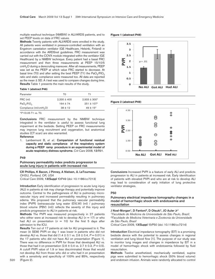
S20
multiple washout technique (NMBW) in ALI/ARDS patients, and toset PEEP levels on data of FRC values.Methods Twenty patients with ALI/ARDS were enrolled in the study.All patients were ventilated in pressure-controlled ventilation with anEngstrom carestation ventilator (GE Healthcare, Helsinki, Finland) inaccordance with the ARDSnet guidelines. FRC measurement wascarried out with the COVX module integrated within the ventilator (GEHealthcare) by a NMBW technique. Every patient had a basal FRCmeasurement and then three measurements at PEEP 15/10/5cmH2O during a derecruiting maneuver. After all measurements, PEEPwas set as the PEEP at which value FRC started to decrease. Atbasal time (T0) and after setting the best PEEP (T1) the PaO2/FIO2ratio and static compliance were measured too. All data are reportedas the mean ± SD. A t test was used to compare changes during time.Results Table 1 presents the main results of the study.
Table 1 (abstract P48)
Parameter T0 T1
FRC (ml) 2,330 ± 400 2,933 ± 300*
PaO2/FiO2 164 ± 74 251 ± 107*
Compliance (ml/cmH2O) 38 ± 12 49 ± 15*
*P <0.05 T1 vs. T0.
Conclusions FRC measurement by the NMBW techniqueintegrated in the ventilator is useful to assess functional lungimpairment at the bedside. Setting PEEP on FRC measurementsmay improve lung recruitment and oxygenation, but anatomicalstudies (CT scan) are also warranted.Reference1. Lambermont B, et al.: Comparision of functional residual
capacity and static compliance of the respiratory systemduring a PEEP ramp procedure in an experimental model ofacute respiratory distress syndrome. Crit Care 2008, 12:R91.
P49Pulmonary permeability index predicts progression toacute lung injury in patients with increased risk
CR Phillips, K Bacon, J Pinney, A Nielsen, JL LeTourneauOHSU, Portland, OR, USACritical Care 2009, 13(Suppl 1):P49 (doi: 10.1186/cc7213)
Introduction Early identification of progression to acute lung injury(ALI) in patients at risk may change therapy and potentially improveoutcome. Central to the pathogenesis of ALI is pulmonary micro-vascular injury and increased permeability resulting in pulmonaryedema. We proposed that the pulmonary vascular permeabilityindex (PVPI) (extravascular lung water (EVLW) (ml) / pulmonaryblood volume (PBV) (ml)) reflects the severity of this injury andpredicts progression to ALI in patients at risk.Methods The PVPI was measured prospectively in 27 patientswho either were at increased risk to develop ALI (n = 17) or whohad ALI on presentation (n = 10) for the first 5 days afteradmission to the ICU.Results Ten out of 17 patients at risk for ALI progressed to it. Themean (± SEM) PVPI on day 1 was lower in patients who did notdevelop ALI vs. those that did (1.4 ± 0.1 vs. 2.6 ± 0.4, P = 0.01) inthe 17 patients who did not have ALI on presentation (Figure 1).There was no difference in PVPI for those that developed ALI vs.those that had it on presentation (2.6 ± 0.4 vs. 2.7 ± 0.3, P = 0.5).A cutoff PVPI value of 1.9 or less discriminated those that wouldnot develop ALI from those who did or who had it on presentationwith a sensitivity and specificity of 100% and 85%, respectively(Figure 2).
Conclusions Increased PVPI is a feature of early ALI and predictsprogression to ALI in patients at increased risk. Early identificationof patients with elevated PVPI and who are at risk to develop ALImay lead to consideration of early initiation of lung protectiveventilator strategies.
P50Pulmonary electrical impedance tomography changes in amodel of hemorrhagic shock with endotoxemia andresuscitation
J Noel-Morgan1, D Fantoni2, D Otsuki1, JO Auler Jr1
1Faculdade de Medicina da Universidade de São Paulo, Brazil;2Faculdade de Medicina Veterinaria e Zootecnia da Universidadede São Paulo, BrazilCritical Care 2009, 13(Suppl 1):P50 (doi: 10.1186/cc7214)
Introduction Electrical impedance tomography (EIT) is a promisingbedside device with the potential to assess changes in regionalventilation and lung blood flow [1]. The purpose of our study wasto monitor lung images and changes in impedance by EIT in amodel of hemorrhagic shock with endotoxemia followed by fluidresuscitation.Methods Twelve anesthetized, mechanically ventilated, supinepigs were submitted to hemorrhagic shock (50% blood volume)and endotoxin infusion. Animals were randomly allocated to control
Critical Care March 2009 Vol 13 Suppl 1 29th International Symposium on Intensive Care and Emergency Medicine
Figure 1 (abstract P49)
Figure 2 (abstract P49)
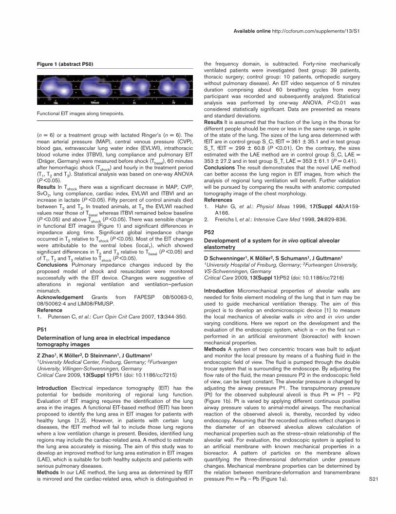
S21
(n = 6) or a treatment group with lactated Ringer’s (n = 6). Themean arterial pressure (MAP), central venous pressure (CVP),blood gas, extravascular lung water index (EVLWI), intrathoracicblood volume index (ITBVI), lung compliance and pulmonary EIT(Dräger, Germany) were measured before shock (Tbasal), 60 minutesafter hemorrhagic shock (Tshock) and hourly in the treatment period(T1, T2 and T3). Statistical analysis was based on one-way ANOVA(P <0.05).Results In Tshock there was a significant decrease in MAP, CVP,SvO2, lung compliance, cardiac index, EVLWI and ITBVI and anincrease in lactate (P <0.05). Fifty percent of control animals diedbetween T2 and T3. In treated animals, at T3 the EVLWI reachedvalues near those of Tbasal whereas ITBVI remained below baseline(P <0.05) and above Tshock (P <0.05). There was sensible changein functional EIT images (Figure 1) and significant differences inimpedance along time. Significant global impedance changeoccurred in T2 relative to Tshock (P <0.05). Most of the EIT changeswere attributable to the ventral lobes (local1), which showedsignificant differences in T2 and T3 relative to Tbasal (P <0.05) andof T1, T2 and T3 relative to Tshock (P <0.05).Conclusions Pulmonary impedance changes induced by theproposed model of shock and resuscitation were monitoredsuccessfully with the EIT device. Changes were suggestive ofalterations in regional ventilation and ventilation–perfusionmismatch.Acknowledgement Grants from FAPESP 08/50063-0,08/50062-4 and LIM08/FMUSP.Reference1. Putensen C, et al.: Curr Opin Crit Care 2007, 13:344-350.
P51Determination of lung area in electrical impedancetomography images
Z Zhao1, K Möller2, D Steinmann1, J Guttmann1
1University Medical Center, Freiburg, Germany; 2FurtwangenUniversity, Villingen-Schwenningen, GermanyCritical Care 2009, 13(Suppl 1):P51 (doi: 10.1186/cc7215)
Introduction Electrical impedance tomography (EIT) has thepotential for bedside monitoring of regional lung function.Evaluation of EIT imaging requires the identification of the lungarea in the images. A functional EIT-based method (fEIT) has beenproposed to identify the lung area in EIT images for patients withhealthy lungs [1,2]. However, in patients with certain lungdiseases, the fEIT method will fail to include those lung regionswhere a low ventilation change is present. Besides, identified lungregions may include the cardiac-related area. A method to estimatethe lung area accurately is missing. The aim of this study was todevelop an improved method for lung area estimation in EIT images(LAE), which is suitable for both healthy subjects and patients withserious pulmonary diseases.Methods In our LAE method, the lung area as determined by fEITis mirrored and the cardiac-related area, which is distinguished in
the frequency domain, is subtracted. Forty-nine mechanicallyventilated patients were investigated (test group: 39 patients,thoracic surgery; control group: 10 patients, orthopedic surgerywithout pulmonary disease). An EIT video sequence of 5 minutesduration comprising about 60 breathing cycles from everyparticipant was recorded and subsequently analyzed. Statisticalanalysis was performed by one-way ANOVA. P <0.01 wasconsidered statistically significant. Data are presented as meansand standard deviations.Results It is assumed that the fraction of the lung in the thorax fordifferent people should be more or less in the same range, in spiteof the state of the lung. The sizes of the lung area determined withfEIT are in control group S_C, fEIT = 361 ± 35.1 and in test groupS_T, fEIT = 299 ± 60.8 (P <0.01). On the contrary, the sizesestimated with the LAE method are in control group S_C, LAE =353 ± 27.2 and in test group S_T, LAE = 353 ± 61.1 (P = 0.41).Conclusions The result demonstrates that the novel LAE methodcan better access the lung region in EIT images, from which theanalysis of regional lung ventilation will benefit. Further validationwill be pursued by comparing the results with anatomic computedtomography image of the chest morphology.References1. Hahn G, et al.: Physiol Meas 1996, 17(Suppl 4A):A159-
A166.2. Frerichs I, et al.: Intensive Care Med 1998, 24:829-836.
P52Development of a system for in vivo optical alveolarelastometry
D Schwenninger1, K Möller2, S Schumann1, J Guttmann1
1University Hospital of Freiburg, Germany; 2Furtwangen University,VS-Schwenningen, GermanyCritical Care 2009, 13(Suppl 1):P52 (doi: 10.1186/cc7216)
Introduction Micromechanical properties of alveolar walls areneeded for finite element modeling of the lung that in turn may beused to guide mechanical ventilation therapy. The aim of thisproject is to develop an endomicroscopic device [1] to measurethe local mechanics of alveolar walls in vitro and in vivo undervarying conditions. Here we report on the development and theevaluation of the endoscopic system, which is – on the first run –performed in an artificial environment (bioreactor) with knownmechanical properties.Methods A system of two concentric trocars was built to adjustand monitor the local pressure by means of a flushing fluid in theendoscopic field of view. The fluid is pumped through the doubletrocar system that is surrounding the endoscope. By adjusting theflow rate of the fluid, the mean pressure P2 in the endoscopic fieldof view, can be kept constant. The alveolar pressure is changed byadjusting the airway pressure P1. The transpulmonary pressure(Pt) for the observed subpleural alveoli is thus Pt = P1 – P2(Figure 1b). Pt is varied by applying different continuous positiveairway pressure values to animal-model airways. The mechanicalreaction of the observed alveoli is, thereby, recorded by videoendoscopy. Assuming that the recorded outlines reflect changes inthe diameter of an observed alveolus allows calculation ofmechanical properties such as the stress–strain relationship of thealveolar wall. For evaluation, the endoscopic system is applied toan artificial membrane with known mechanical properties in abioreactor. A pattern of particles on the membrane allowsquantifying the three-dimensional deformation under pressurechanges. Mechanical membrane properties can be determined bythe relation between membrane-deformation and transmembranepressure Pm = Pa – Pb (Figure 1a).
Available online http://ccforum.com/supplements/13/S1
Figure 1 (abstract P50)
Functional EIT images along timepoints.

S22
Results Preliminary results were obtained in the bioreactor with apolymer membrane (polydimethylsiloxan) of 100 μm thickness.Graphite particles were used to produce a particle pattern.Deformation due to different Pt′ was observed and recordedsuccessfully.Conclusions The in vivo estimation of micromechanical propertiessuch as the stress–strain relationship of elastic walls is feasible.For that, the developed system has to be technically optimized.Reference1. Stahl CA, et al.: J Biomech 2006, 39:598.
P53Effects of inhaled iloprost on acute respiratory distresssyndrome in prone and supine positions
E Senturk1, N Cakar1, P Ergin Özcan1, A Basel1, T Sengül1, L Telci1, F Esen1, M Winterhalter2
1University of Istanbul, Turkey; 2Hannover Medical School,Hannover, GermanyCritical Care 2009, 13(Suppl 1):P53 (doi: 10.1186/cc7217)
Introduction In several studies it has been shown that inhaledpulmonary vasodilators (NO and iloprost) can decrease thepulmonary hypertension and also improve the oxygenation duringacute respiratory distress syndrome (ARDS) [1]. We investigatedthe effects of prone and supine positioning on the effects ofinhaled iloprost in an animal-ARDS model.Methods After approval of the animal ethics committee, 10 pigswere anesthetized and intubated. Invasive systemic and pulmonaryarterial catheterizations were performed (T1). ARDS was inducedin all animals with the infusion of oleic acid (0.15 to 0.30 ml/kg).The study design is shown in Figure 1. Hemodynamic andrespiratory parameters and ventilation parameters were measured;arterial and mixed venous blood samples were drawn; and wererecorded in T1 to T6. Pigs were ventilated in volume-controlledventilation mode with FiO2 100%, with 4 cmH2O positive end-expiratory pressure (PEEP) in the beginning and with 8 cmH2OPEEP after induction of ARDS. Statistical analysis was made withStudent’s t test, repeated measures of ANOVA (with Tukey as thepost-hoc test) and paired t tests. P <0.05 was significant.Results There was no significant difference between the sequences.Iloprost decreased the mean pulmonary arterial pressure in bothsupine (37 vs. 31 mmHg) and prone (38 vs. 29 mmHg) positionssignificantly, but there was no significant difference between bothpositions. Prone position was associated with an improvement inoxygenation compared with supine position both with or withoutiloprost application. There was no spillover effect of iloprost.Conclusions Iloprost decreased pulmonary arterial pressures inboth positions. On the other hand, the prone position improvedoxygenation. The decrease in pulmonary arterial pressures andimprovement in oxygenation was better in prone position +iloprost; however, these findings were not statistically significant.Reference1. Zwissler B, et al.: Inhaled prostacyclin (PGI2) versus
inhaled nitric oxide in adult respiratory distress syndrome.Am J Respir Crit Care Med 1996, 154:1671-1677.
P54Decreased vascular endothelial growth factor expressionin lung tissue during acute respiratory distress syndrome
L Azamfirei, S Gurzu, S Copotoiu, I Jung, R Copotoiu, K Branzaniuc, R SolomonUniversity of Medicine and Pharmacy, Targu Mures, RomaniaCritical Care 2009, 13(Suppl 1):P54 (doi: 10.1186/cc7218)
Introduction Endothelial injury is an important prognostic factor inacute respiratory distress syndrome (ARDS) [1,2]. Vascular endo-thelial growth factor (VEGF) plays a critical role in endothelialdestruction and angiogenesis [3]. The expression of VEGF inARDS varies, depending on epithelial and endothelial damage[4,5]. The objective of this study was to investigate the expressionof VEGF in lung tissue from ARDS patients.Methods Lung specimens were obtained by autopsy from 10patients with severe ARDS and were compared with a controlgroup of 10 non-ARDS patients autopsied. All lung samples werestained for standard histopathological analysis and for immuno-histochemical methods using a specific mouse monoclonalantibody.Results Compared with expression in non-ARDS controlindividuals, pulmonary expression of VEGF was significantlydecreased (P <0.001) in ARDS patients. Alveolar macrophageswere similarly immunopositive in both groups. No differences werenoted with regard to the individual patient’s characteristics (age,gender, period of ARDS condition, number of ICU days).Conclusions A decrease in alveolar type II cellularity, due toapoptosis, has been observed during ARDS that may reduce theproduction of VEGF in the alveolar space and may participate inthe decrease in lung perfusion.Acknowledgement Research Grant No. 136/IDEI from theNational Authority of Scientific Research, Romania.References1. Medford ARL, Millar AB: Vascular endothelial growth factor
(VEGF) in acute lung injury (ALI) and acute respiratory dis-tress syndrome (ARDS): paradox or paradigm? Thorax2006, 61:621.
2. Dvorak HF: Angiogenesis: update 2005. J Thromb Haemost
Critical Care March 2009 Vol 13 Suppl 1 29th International Symposium on Intensive Care and Emergency Medicine
Figure 1 (abstract P52) Figure 1 (abstract P53)
Ilop, iloporost; Pron, prone; Sup, supine.

S23
2005, 3:1835-1842.3. Gerber HP, Wu X, Yu L, Wiesmann C, et al.: Mice express-
ing a humanized form of VEGF-A may provide insightsinto the safety and efficacy of anti-VEGF antibodies. ProcNatl Acad Sci U S A 2007, 104:3478-3483.
4. Fehrenbach H: Development of the pulmonary surfactantsystem. Pneumologie 2007, 61:488.
5. Shibuya M: Differential roles of vascular endothelialgrowth factor receptor-1 and receptor-2 in angiogenesis. JBiochem Mol Biol 2006, 39:469-478.
P55The selective αα7 nicotinic acetylcholine receptor agonistGTS-21 attenuates ventilator-induced inflammation andlung injury
M Kox, JC Pompe, M Vaneker, LM Heunks, JG Van der Hoeven,CW Hoedemaekers, P PickkersRadboud University Nijmegen Medical Centre, Nijmegen, theNetherlandsCritical Care 2009, 13(Suppl 1):P55 (doi: 10.1186/cc7219)
Introduction Mechanical ventilation (MV) induces an inflammatoryresponse that contributes to lung injury such as in acute lung injuryor acute respiratory distress syndrome. The efferent vagus nervecan limit the inflammatory response via the α7 nicotinic acetyl-choline receptor (α7nAChR), the so-called cholinergic anti-inflam-matory pathway. The aim of this study was to evaluate the effect ofthe selective α7nAChR agonist GTS-21 on pulmonary andsystemic inflammation and lung injury induced by MV usingclinically relevant ventilator settings.Methods C57BL6 mice (n = 40) were intraperitoneally injectedwith 8 mg/kg GTS-21 or placebo, after which they weremechanically ventilated for 4 hours (tidal volume 8 ml/kg; positiveend-expiratory pressure 1.5 cm H2O; FiO2 0.45). Untreated, notmechanically ventilated mice were used as controls. Arterial bloodgases were obtained at the end of the experiment and TNFα, IL-6,IL-1α, IL-1β, keratinocyte-derived cytokine (IL-8 homologue) andIL-10 were determined in plasma and lung homogenates. LungTNFα and IL-10 mRNA expression was measured usingquantitative PCR.Results In GTS-21-treated mice, the alveolar–arterial gradient afterMV was significantly reduced compared with placebo (14.0 ± 0.76vs. 16.2 ± 0.59 kPa; P = 0.03). MV resulted in an increase of allcytokines in plasma and lung compared with control mice. TNFαwas significantly lower in plasma of GTS-21-treated animalscompared with placebo (196.2 ± 50.8 vs. 331.9 ± 31.9 pg/ml;P = 0.04). Similarly, in lung homogenates a distinct trend wasobserved towards lower TNFα levels in GTS-21-treated mice(53.9 ± 12.5 vs. 79.1 ± 5.6 pg/mg protein; P = 0.06). IL-10 levelswere unaffected by GTS-21. MV strongly increased TNFα mRNAexpression in lungs of placebo animals (21-fold compared withcontrols); this was significantly lower in GTS-21-treated mice (11-fold compared with controls; P = 0.02). IL-10 mRNA expressionwas similar in GTS-21-treated and placebo animals.Conclusions MV with clinically relevant ventilator settings resultsin activation of the immune system. GTS-21 inhibits proinflam-matory cytokine production while not affecting the anti-inflam-matory cytokine IL-10. The reduced alveolar–arterial gradient inGTS-21-treated animals indicates attenuation of lung injury. Inconclusion, limiting the inflammatory response appears to reducelung injury, and therefore the cholinergic anti-inflammatory pathwaymay represent new treatment options for MV-induced lung injury.
P56Usefulness of soluble E-selectin in the clinicopathologicassessment of acute lung injury/acute respiratory distresssyndrome
Y Kakihana, C Kuroki, H Murayama, T Ohryorji, N Kiyonaga, S Tashiro, T Imabayashi, T Yasuda, Y Kanmura, T Moriyama, A MatsunagaKagoshima University Hospital, Kagoshima, JapanCritical Care 2009, 13(Suppl 1):P56 (doi: 10.1186/cc7220)
Introduction A retrospective observational study was conductedto evaluate whether the plasma level of soluble E-selectin [1] mightbe a specific pathologic marker of acute lung injury/acuterespiratory distress syndrome (ALI/ARDS).Methods The data of 52 critically ill patients admitted to the ICUwith systemic inflammatory response syndrome and initiated onmechanical ventilation were retrospectively evaluated.Results The plasma levels of soluble E-selectin determined within24 hours of admission were significantly correlated with theSequential Organ Failure Assessment scores determined within24 hours of admission. Furthermore, the scores for both respiratoryfailure (evaluated by the PaO2/FiO2 ratio) and liver dysfunction(evaluated by the serum bilirubin value) in the Sequential OrganFailure Assessment scoring system were significantly correlatedwith plasma levels of soluble E-selectin. In relation to respiratoryfailure, the plasma level of soluble E-selectin was higher in patientswith ALI/ARDS than in those without (Figure 1), and receiveroperating characteristic analysis revealed that this parameter mightbe a specific marker of ALI/ARDS (Figure 2).Conclusions Soluble E-selectin might be specific and useful markerfor the clinicopathologic assessment of ALI/ARDS in critically illpatients with systemic inflammatory response syndrome. However,further investigation is clearly needed to determine whether solubleE-selectin can indeed predict the development of ALI/ARDS.Reference1. Okajima K, Harada N, Sakurai G, et al.: Rapid assay for
plasma soluble E-selectin predicts the development ofacute respiratory distress syndrome in patients with sys-temic inflammatory response syndrome. Transl Res 2006,148:295-300.
Available online http://ccforum.com/supplements/13/S1
Figure 1 (abstract P56)
Soluble E-selectin values in patients with non-ALI/ARDS, ALI or ARDS.

S24
P57Prognostic value of epithelial neutrophil activating peptide78 and monokine induced by IFNγγ in bronchoalveolarlavage fluid in critically ill pediatric patients with acuterespiratory distress syndrome
T Shahin, H IbrahimAin Shams University, Cairo, EgyptCritical Care 2009, 13(Suppl 1):P57 (doi: 10.1186/cc7221)
Introduction Diffuse alveolar damage (DAD) is the histopatho-logical hallmark of acute respiratory distress syndrome (ARDS).DAD is due, in part, to dysregulated angiogenesis, with over-expression of angiogenic and downregulation of angiostatic chemo-kines. The objective of this study was to measure the levels ofepithelial neutrophil activating peptide 78 (ENA78) as an angio-genic chemokine and monokine induced by IFNγ (MIG) as anangiostatic chemokine in bronchoalveolar lavage fluid of critically illchildren with ARDS to determine the prognostic value of theENA78/MIG ratio in assessing illness severity and patient outcome.Methods The study included 35 mechanically ventilated pediatricpatients with ARDS and 28 mechanically ventilated patients due tononpulmonary causes as a control group with mean age16.5 ± 10.2 and 29.1 ± 8.1 months, respectively. They weresubjected to fibroaptic bronchoscopic examination and broncho-alveolar lavage was done to estimate the ENA-78 and MIG levelsusing the ELISA technique.Results Both ENA-78 and MIG levels were found to besignificantly higher among patients versus controls (P <0.01)(390.3 ± 159.7 vs. 80.3 ± 17.6 pg/ml and 235.6 ± 92 vs. 91.0 ±18.9 pg/ml, respectively). ENA-78 was found to be significantlyhigher among nonsurvivors, while MIG was significantly loweramong nonsurvivors in comparison with the survivors (P <0.01)(455.3 ± 142 vs. 277.8 ± 29.1 pg/ml and 186.3 ± 43 vs. 359.0 ±56.4 pg/ml, respectively). Applying a receiver operating charac-
teristic curve indicated that ENA78/MIG ratio was the bestdiscriminator between survivors and nonsurvivors at cutoff value1.02 with 100% sensitivity and specificity.Conclusions Imbalance between angiogenic and angiostaticchemokines is the determining factor of ARDS patients’ outcome.Future studies to investigate therapeutic modalities to enhanceangiostatic factors or inhibit angiogenesis to delay diffuse alveolardamage and improve the outcome are recommended.
P58Cortisol-binding globulin cleavage at sites of inflammationin critically ill patients
M Williams, A Zhou, C Summers, D Halsall, D MenonUniversity of Cambridge, UKCritical Care 2009, 13(Suppl 1):P58 (doi: 10.1186/cc7222)
Introduction In vitro studies have shown that cortisol-bindingglobulin (CBG) can be cleaved by neutrophil elastase with aresulting reduction in affinity for cortisol [1]. Local elastaseproduction by activated neutrophils may trigger cortisol release byCBG cleavage, providing a potential mechanism for targeted deliveryof corticosteroids to inflamed tissues. We looked for evidence of thisprocess at sites of inflammation in critically ill patients.Methods Sera and bronchoalveolar lavage (BAL) fluid werecollected from six mechanically ventilated patients with a clinicaldiagnosis of ventilator-associated pneumonia or acute respiratorydistress syndrome (ARDS). These samples, along with sera fromsix healthy controls, were subjected to gel electrophoresis(SDS-PAGE) and immunoblotting for CBG. Densitometry wasused to quantify the proportion of cleaved CBG.Results CBG cleavage product was seen within the BAL but notthe sera of patients with acute inflammatory lung disease. Figure 1shows a typical western blot comparing control sera (C1 and C2),septic sera (S1 and S2) and BAL from the same septic patients (B1and B2, respectively). The mean percentage of CBG in the cleavedform was 63.3% ± 21.8 in BAL compared with 0.94% ± 1.73 insera from the same patients and 12.5% ± 11.4 from healthycontrols (see Figure 2; all comparisons statistically significant).Conclusions This study provides the first in vivo support for amechanism of enhanced cortisol delivery to sites of inflammationthat involves local CBG cleavage.Acknowledgement Study supported by the Intensive CareSociety (UK).Reference1. Pemberton PA, et al.: Nature 1988, 336:257-258.
Critical Care March 2009 Vol 13 Suppl 1 29th International Symposium on Intensive Care and Emergency Medicine
Figure 1 (abstract P58)
SDS-PAGE and immunoblotting of CBG. MW, molecular weights(arrows mark the place of the relevant molecular weight markers).
Figure 2 (abstract P56)
Receiver operating characteristic curves of soluble E-selectin. CRP,C-reactive protein; WBC, white blood cell count; E-XDP, elastasedigests of cross-linked fibrin.

S25
P59Vascular connexins have differing responses to tumornecrosis factor
Y OuelletteMayo Clinic, Rochester, MN, USACritical Care 2009, 13(Suppl 1):P59 (doi: 10.1186/cc7223)
Introduction Cell-to-cell communication via gap junctions hasbeen implicated in the control of vascular tone and may be alteredin sepsis. Endothelial cells express connexin (Cx) 37, Cx40 andCx43 and cytokines may modulate their function in sepsis resultingin altered gap junctional intercellular communication. Our hypothe-sis is that tumor necrosis factor (TNF) will decrease gap-junction-dependent cell-to-cell communication of vascular connexin.Methods Transformed HeLa cells expressing vascular Cx37, Cx40or Cx43 were used in these experiments. HeLa cells were treatedwith TNF (20 ng/ml) for up to 2 hours. In dye-transfer experiments,carboxyfluroscein (HeLaCx40 and HeLaCx43) or Alexa Fluor-480(HeLaCx37) was injected into one cell for 10 seconds and celltransfer was allowed to proceed for 10 minutes and the number oflabeled cells counted. Cell lysates were prepared in Triton X-100lysate buffer and detergent-soluble fractions collected. Cx37, Cx40and Cx43 were detected by western blot.Results After 1 hour, TNF treatment resulted in near total loss ofdye-coupling in HeLaCx37 and HeLaCx43 (P <0.02, n = 14 to 16)and remained constant up to 2 hours. Dye coupling in HeLaCx40cells remained unchanged after 1 hour and decreased after 2 hours(P <0.05, n = 10). Western blots indicated that TNF treatment didnot affect detergent solubility of Cx40 and Cx43. However, TNFcaused a significant increase in detergent solubility of Cx37.Conclusions These results suggest that inflammatory mediatorsaffect connexins differently. The loss of Cx37 function may be dueto the loss of detergent resistance, suggesting internalization ofCx37 in response to TNF.Acknowledgement Transformed HeLa cells were a gift from Dr K.Willecke.
P60Prehospital intubation for out-of-hospital cardiac arrest
D Young1, RM Lyon1, J Ferris2, DW McKeown1, A Oglesby1
1Royal Infirmary of Edinburgh, UK; 2Ninewells, Dundee, UKCritical Care 2009, 13(Suppl 1):P60 (doi: 10.1186/cc7224)
Introduction The most appropriate advanced airway intervention inout-of-hospital cardiac arrest (OHCA) remains unproven. Trainedambulance personnel may attempt endotracheal intubation in thefield for OHCA patients. This study aims to review prehospitalairway management in OHCA.Methods Observational, retrospective case review over a 4-yearperiod. All cases of OHCA brought to the Emergency Departmentof the Royal Infirmary of Edinburgh, Scotland were identified.Patient demographics, the airway management technique anddocumented complications were recorded. The primary endpointmeasure was survival to hospital admission.Results In total, 794 OHCA cases were identified. The aetiologyof cardiac arrest was medical in 95.2%, traumatic in 3.9% andunrecorded in 0.9% of cases. Prehospital endotracheal intubationwas attempted in 628 (79%) cases and was successful in 573(91.2%) cases. A significant complication (multiple attempts,displaced endotracheal tube or oesophageal intubation) occurredin 55 (8.8%) cases. In total, 165 (20.8%) patients survived tohospital admission, of whom 110 (17.5%) had undergone pre-hospital intubation. Fifty-five (33.1%) patients who did not undergoprehospital intubation survived to hospital admission.Conclusions Prehospital endotracheal intubation for out-of-hospitalcardiac arrest is associated with significant complications. For ambu-lance crews not routinely undertaking endotracheal intubation, a supra-glottic airway device may be more appropriate. Reliable methods ofconfirming endotracheal intubation in the field should be utilised.
P61Focused echocardiography and capnography duringresuscitation from pulseless electrical activity after out-of-hospital cardiac arrest
G Prosen1, Š Grmec1, D Kupnik1, M Krizmaric1, J Završnik1, R Gazmuri21Centre for Emergency Medicine, Maribor, Slovenia; 2RosalindFranklin University, Chicago, IL, USACritical Care 2009, 13(Suppl 1):P61 (doi: 10.1186/cc7225)
Introduction The study evaluated the ability of focused echo-cardiography (F-ECHO) and capnography to differentiate betweenpulseless electrical activity (PEA) and pseudo-PEA during resus-citation from out-of-hospital cardiac arrest (OHCA).Methods Patients in PEA, with stable values of end-tidal PCO2(PETCO2) during compression pauses, underwent subxiphoidF-ECHO examinations during pauses for carotid pulse evaluation,assessing for the presence or absence of cardiac kinetic activity(synchronous myocardial wall and valvular movement). Patientswith stable PETCO2 during the time of F-ECHO examinations whohad cardiac kinetic activity underwent a compression pause of15 seconds during which an additional 20 IU bolus of vasopressinand a 0.9% NaCl bolus were given. If pulselessness persisted afterthe 15-second pause, compressions were resumed. This groupwas denominated the F-ECHO group (November 2007 to October2008, n = 16) and was compared with a NON-ECHO group(November 2005 to October 2007, n = 48) who also had PEAwith stable PETCO2 and were managed according to 2005European Resuscitation Council guidelines without F-ECHO.Results There were no statistically significant differences betweengroups with regards to sex, suspected cause of arrest, initial cardiacrhythm, witnessed arrest, time elapsed before initiation ofcardiopulmonary resuscitation (CPR), and bystander CPR. Primaryoutcome (ICU admission: 88% vs. 50%; adjusted values: OR =22.4, 95% CI = 4.2 to 86.9, P <0.001) and secondary outcome(return of spontaneous circulation (carotid pulses palpable): 94% vs.54%, adjusted: OR = 28.4, 95% CI = 3.9 to 96.1, P <0.001; 24-
Available online http://ccforum.com/supplements/13/S1
Figure 2 (abstract P58)
Cleaved CBG as a percentage of total CBG. *P <0.05, **P <0.01.
S26
hour survival: 81% vs. 41%; adjusted: OR = 19.8, 95% CI = 3.1 to72.6, P <0.001; hospital survival: 56% vs. 15%; adjusted values:OR = 31.4, 95% CI = 2.9 to 85.7, P <0.001; neurological outcome– CPC 1 (good cerebral performance) to CPC 2 (moderate cerebraldisability): 50% vs. 8%; adjusted values: OR = 36.4, 95% CI = 4.8to 115.4, P <0.001) were significantly better in the F-ECHO group.In the NON-ECHO group, significantly higher doses of epinephrinewere needed (P = 0.009) and CPR lasted longer (P = 0.003).Conclusions F-ECHO and capnography during PEA in OHCAfacilitates return of spontaneous circulation, ICU admission, 24-hour survival, and hospital survival. This effect was attributed to theability to distinguish between PEA and pseudo-PEA and toinstitute appropriate treatment during CPR. The confirmation ofthese results in a large study is warranted.
P62Patients with an automatic external defibrillator applied bya bystander in a public setting have a strikingly higherfrequency of ventricular tachycardia/ventricular fibrillationthan observed cardiac arrests in the home
ML Weisfeldt1, C Sitlani2, T Rea2, D Atkins3, T Aufderheide4, S Brooks5, B Bigham5, C Foerster5, R Gray5, P Moran6, J Ornato7, J Powell2, L Van Ottingham2, LJ Morrison5
1John Hopkins University, Baltimore, MD, USA; 2University ofWashington, Seattle, WA, USA; 3University of Iowa, Iowa City, IA,USA; 4Medical College of Wisconsin, Milwaukee, WI, USA; 5St Michael’s Hospital, Keenan Research Centre, Li Ka ShingKnowledge Institute, Toronto, ON, Canada; 6Durham RegionalBase Hospital, ON, Canada; 7Medical College of Virgina,Richmond, VA, USACritical Care 2009, 13(Suppl 1):P62 (doi: 10.1186/cc7226)
Introduction The overall incidence of ventricular tachycardia/ventricular fibrillation (VT/VF) as the first recorded electrical rhythmin out-of-hospital cardiac arrest has declined from ~70% to ~25%over the past 30 years. This change has been attributed to primaryand secondary prevention of cardiovascular disease and VT/VF.We evaluated whether the incidence of VT/VF as first recordedrhythm differed by location among bystander-applied automaticexternal defibrillator (AED) patients and emergency medicalservices (EMS)-witnessed cardiac arrests.Methods A prospective cohort study of nontraumatic cardiacarrest from December 2005 to April 2007 in the ResuscitationOutcomes Consortium database from 10 US and Canadian sites.The incidence of an initial shockable rhythm on AED or documen-ted VT/VF was compared among bystander-applied AED patientsand EMS-witnessed arrests in public versus private settings.Results The first rhythm was known in 13,235 out of 14,059(94%) adult EMS-treated cardiac arrests. Of the 13,235 withknown rhythms, 3,436 (26%) had VT/VF. Among 1,115 EMS-witnessed arrests, 61/161 (38%) had VT/VF in public settings and224/954 (23%) in private settings. Similarly, for bystander AEDapplied in the private setting, 39/114 (34%) were shocked. But, incontrast, 125/159 (79%) (P <0.001 vs. all other) were shocked bythe AED in the public setting. Witnessed arrests in both the privatesetting (vs. public) and in EMS-witnessed cases (vs. bystanderAED applied) were more likely to occur in older subjects andfemales. After adjusting for age and gender via logistic regressionmodels, a significant difference in the odds of having a shockablerhythm in a public versus private location of arrest remained inEMS-witnessed arrests (P <0.005). The difference also remainedin bystander AED-applied arrests (P <0.001) after adjusting forage, gender, and bystander-witnessed status.
Conclusions The incidence of VT/VF is far greater in the publicsetting particularly for bystander-witnessed AED-applied arrests.Patients in the private home setting, even for EMS-observedarrests, are far less likely to benefit from AED application thanbystander-witnessed patients in the public setting. Cardiopulmo-nary resuscitation strategies may need to be tailored by arrest location.
P63Survival unchanged 5 months after implementing the 2005American Heart Association cardiopulmonary resuscitationand emergency cardiac care guidelines for out-of-hospitalcardiac arrest
B Bigham1, K Koprowicz2, A Kiss1, P Dorian1, S Emerson2, C Zhan1, T Rea2, TP Aufderheide3, J Powell2, S Cheskes1, D Davis4, J Stouffer5, J Perry6, LJ Morrison7
1University of Toronto, ON, Canada; 2University of Washington,Seattle, WA, USA; 3Medical College of Wisconsin, Milwaukee, WI,USA; 4University of California San Diego, CA, USA; 5Gresham Fire& Emergency Services, Gresham, OR, USA; 6University of Ottawa,ON, Canada; 7St Michael’s Hospital, Keenan Research Centre, LiKa Shing Knowledge Institute, Toronto, ON, CanadaCritical Care 2009, 13(Suppl 1):P63 (doi: 10.1186/cc7227)
Introduction To improve survival from out-of-hospital cardiacarrest, the American Heart Association released guidelines in2005. We examined the effect of these guidelines on survival inthe Resuscitation Outcomes Consortium (ROC) Epistry – CardiacArrest. We hypothesized that survival would increase afterguideline implementation.Methods One hundred and seventy-four emergency medicalservice (EMS) agencies from eight out of 10 ROC sites weresurveyed to determine 2005 American Heart Association guidelineimplementation, or crossover, date. Two sites with 2005 com-patible treatment algorithms prior to guideline release were notincluded. Patients with out-of-hospital cardiac arrest secondary toa noncardiac cause, EMS-witnessed events, patients <18 yearsold, and patients with do-not-resuscitate orders were excluded. Alinear mixed-effects model was applied for survival controlling fortime and agency. The crossover date was added to the model todetermine the effect of the 2005 guidelines.Results Of 174 agencies, 83 contributed cases to both cohortsduring the 18-month period between 1 December 2005 and 31May 2007. Of 7,403 cases, 4,897 occurred during the 13-month(median) interval before crossover and 2,506 occurred in the5-month (median) interval after crossover. The overall survival ratewas 5.9%. Our model estimated an overall increase in survival overtime (monthly OR = 1.02, 95% CI = 0.99 to 1.04, P = 0.23), adecrease in survival at crossover (OR = 0.92, 95% CI = 0.66 to1.26, P = 0.59), and a further increase in survival over time aftercrossover (monthly = OR 1.005, 95% CI = 0.96 to 1.05,P = 0.84).Conclusions The present study found a trend towards increasedsurvival over time and no statistically significant effect of the 2005guidelines early after implementation. This observed increase insurvival over time may be attributed to the Hawthorne effect orparticipation in the ROC or improved quality assurance. A delay inknowledge and skill acquisition amongst EMS providers and theneed to rechoreograph their cardiac arrest treatment may explainwhy no significant increase in survival was observed after imple-mentation. EMS providers may require more time to gain pro-ficiency in the guideline changes before the full potential of theguidelines can be realized. Further longitudinal study is needed todetermine the full impact of the guidelines on survival.
Critical Care March 2009 Vol 13 Suppl 1 29th International Symposium on Intensive Care and Emergency Medicine

S27
P64Outcomes following admission to ICU post cardiac arrest
E Casey, B MarshMater Misericordiae University Hospital, Dublin, IrelandCritical Care 2009, 13(Suppl 1):P64 (doi: 10.1186/cc7228)
Introduction ICU admission post cardiac arrest accounts for 6%of admissions to the ICU [1]. ICU survival post cardiac arrestranges from 25% to 35% [2]. We reviewed the records of bothout-of-hospital and inhospital cardiac arrest admissions to our ICUto audit their outcomes, the primary outcome variable beingsurvival to ICU and hospital discharge. Secondary objectives wereto determine the length of stay in the ICU and hospital of bothsurvivors and nonsurvivors.Methods We performed a retrospective review of all admissions toour ICU post cardiac arrest between January 2003 and December2006. Our data were sourced from the ICU access database, ICUdischarge summary and individual chart review. We recordeddemographics and data regarding each arrest.Results One hundred and forty-seven patients were admitted toour ICU during the 4-year period. The mean age was 59 years,ranging from 16 to 88 years. Out-of-hospital cardiac arrestaccounted for 51% (n = 75) of cases, inhospital cardiac arrest for49% (n = 72). Asystole was the first identifiable rhythm in 39%, ofwhich 21% survived to hospital discharge, 42% of whom had apoor neurological outcome. Ventricular fibrillation/ventriculartachycardia accounted for 32% of cases, of which 39% survived,all of whom had a good neurological outcome. Pulseless electricalactivity accounted for 29% of cases, of which 25% survived tohospital discharge, 10% of whom had a poor neurologicaloutcome. Overall survival was 27%, of which 15% had a poorneurological outcome. The mean ICU length of stay was 9.2 daysfor survivors and 6.8 days for nonsurvivors.Conclusions The high prevalence of asystole in both groups is notin keeping with previous audit series [3] in which ventricularfibrillation/ventricular tachycardia is the predominant arrest rhythmand may reflect a delayed response time. Our survival figures arecomparable with international data [3], which are limited. Thehigher male to female ratio is consistent with previous audit series[4], possibly reflecting the higher incidence of ischaemic heartdisease in males.References1. Nolan JP, et al.: A secondary analysis of the ICNARC case
mix programme database. J Intensive Care Soc 2007, 8:38.2 Denton R, Thomas AN: Cardiopulmonary resuscitation: a
retrospective review. Anaesthesia 1997, 52:324-327.3 Laver S, Farrow C, Turner D, Nolan J: Mode of death after
admission to our intensive care unit following cardiacarrest. Intensive Care Med 2004, 30:2126-2128.
4 Roberts H, Smithies M: Outcomes after ICU admission fol-lowing out of hospital cardiac arrest in a UK teaching hos-pital. Crit Care 2008, 12(Suppl 2):P366.
P65Clinical outcome in patients who experienced inhospitalcardiac arrest by underlying disease
Y Shin, C Lim, Y Koh, S HongAsan Medical Center, Seoul, Republic of KoreaCritical Care 2009, 13(Suppl 1):P65 (doi: 10.1186/cc7229)
Introduction Some clinical diagnoses such as sepsis, renal failure,metastatic cancer, house-bound lifestyle, and stroke are asso-ciated with worse prognosis after inhospital cardiac arrest (IHCA)
[1-3]. But there are a few reports about chronic liver disease, Asianpeople, and the incidence of IHCA is rarely reported in theliterature. The aim of this study was to evaluate the hospitalmortality of adult patients who had experienced IHCA byunderlying disease and the incidence of IHCA.Methods Between March and October 2008, 69 patients whoexperienced IHCA were prospectively enrolled in the study.There were 64,345 total admissions to the hospital in thisperiod. Patients who had cardiac arrests in the ICU, emergencyroom, and operating room were excluded from this study. Thehospital mortality compared group A (chronic liver disease, 10patients and cancer, 24 patients) with group B (chronic lungdisease and heart failure, chronic renal disease and diabetesmellitus).Results The incidence of IHCA was 1.07 events per 1,000hospital admissions. Of the 69 enrolled patients, 34 were assignedto group A and 35 to group B. The mean patient age of group Bwas higher than group A (66.7 vs. 56.5 years, P = 0.003). Therewas no difference of return of spontaneous circulation (ROSC)more than 20 minutes between the two groups (group A, 53% vs.group B, 63%, P = 0.40). The hospital mortalities of underlyingdisease were presented: chronic liver disease (90%), cancer(88%), chronic lung disease (20%), and heart failure (54%). Thehospital mortality was higher in group A than in group B (88.2% vs.45.7%, P <0.001). The hospital mortality was higher during thenight compared with during the day (80% vs. 50%, P = 0.01) andROSC was higher during the day than during the night (80% vs.41%, P = 0.001).Conclusions In this study, the chronic liver disease and cancergroup had poor prognosis compared with the other underlyingdiseases such as chronic lung disease or heart failure.References1. Sandroni C, et al.: Intensive Care Med 2007, 33:237-245.2. Peberdy MA, et al.: Resuscitation 2003, 58:297-308.3. Cooper S, et al.: Resuscitation 1997, 35:17-22.
P66Good outcome in octogenarians after ventricular fibrillationout-of-hospital cardiac arrest
M Busch, E SøreideStavanger University Hospital, Stavanger, NorwayCritical Care 2009, 13(Suppl 1):P66 (doi: 10.1186/cc7230)
Introduction Mild induced hypothermia (MIH) in comatosesurvivors of out-of-hospital cardiac arrest (OHCA) is highlyrecommended [1,2]. Still, there is great uncertainty when it comesto age limits for the therapy as the initial randomised studiesexcluded the majority of patients over 75 years of age.Methods We retrospectively studied 115 OHCA survivors thatwere treated with MIH in our ICU from 2002 to 2008 with regardto cerebral performance category (CPC) at hospital discharge.Inclusion criteria were the same as in the Hypothermia afterCardiac Arrest Study Group except for the age limits. Badoutcome was defined as severe disability (CPC3), vegetative state(CPC4) and death (CPC5).Results Bad outcome was significantly more frequent in patientsolder than 60 years (chi-square P = 0.003), but even in patientsolder than 80 years we had good outcome in more than 50% ofcases. The neurological outcome according to the different agegroups is displayed in Figure 1.Conclusions Although age seems to influence outcome, we founda surprisingly high incidence of good outcome even in the oldestcomatose survivors after ventricular fibrillation OHCA treated with
Available online http://ccforum.com/supplements/13/S1
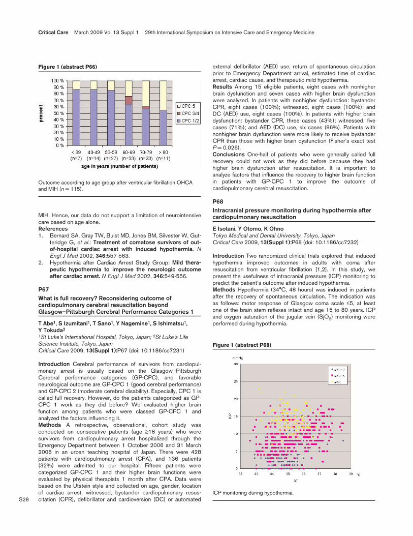
S28
MIH. Hence, our data do not support a limitation of neurointensivecare based on age alone.References1. Bernard SA, Gray TW, Buist MD, Jones BM, Silvester W, Gut-
teridge G, et al.: Treatment of comatose survivors of out-of-hospital cardiac arrest with induced hypothermia. NEngl J Med 2002, 346:557-563.
2. Hypothermia after Cardiac Arrest Study Group: Mild thera-peutic hypothermia to improve the neurologic outcomeafter cardiac arrest. N Engl J Med 2002, 346:549-556.
P67What is full recovery? Reconsidering outcome ofcardiopulmonary cerebral resuscitation beyondGlasgow–Pittsburgh Cerebral Performance Categories 1
T Abe1, S Izumitani1, T Sano1, Y Nagemine1, S Ishimatsu1, Y Tokuda2
1St Luke’s International Hospital, Tokyo, Japan; 2St Luke’s LifeScience Institute, Tokyo, JapanCritical Care 2009, 13(Suppl 1):P67 (doi: 10.1186/cc7231)
Introduction Cerebral performance of survivors from cardiopul-monary arrest is usually based on the Glasgow–PittsburghCerebral performance categories (GP-CPC), and favorableneurological outcome are GP-CPC 1 (good cerebral performance)and GP-CPC 2 (moderate cerebral disability). Especially, CPC 1 iscalled full recovery. However, do the patients categorized as GP-CPC 1 work as they did before? We evaluated higher brainfunction among patients who were classed GP-CPC 1 andanalyzed the factors influencing it.Methods A retrospective, observational, cohort study wasconducted on consecutive patients (age ≥18 years) who weresurvivors from cardiopulmonary arrest hospitalized through theEmergency Department between 1 October 2006 and 31 March2008 in an urban teaching hospital of Japan. There were 428patients with cardiopulmonary arrest (CPA), and 136 patients(32%) were admitted to our hospital. Fifteen patients werecategorized GP-CPC 1 and their higher brain functions wereevaluated by physical therapists 1 month after CPA. Data werebased on the Utstein style and collected on age, gender, locationof cardiac arrest, witnessed, bystander cardiopulmonary resus-citation (CPR), defibrillator and cardioversion (DC) or automated
external defibrillator (AED) use, return of spontaneous circulationprior to Emergency Department arrival, estimated time of cardiacarrest, cardiac cause, and therapeutic mild hypothermia.Results Among 15 eligible patients, eight cases with nonhigherbrain dysfunction and seven cases with higher brain dysfunctionwere analyzed. In patients with nonhigher dysfunction: bystanderCPR, eight cases (100%); witnessed, eight cases (100%); andDC (AED) use, eight cases (100%). In patients with higher braindysfunction: bystander CPR, three cases (43%); witnessed, fivecases (71%); and AED (DC) use, six cases (86%). Patients withnonhigher brain dysfunction were more likely to receive bystanderCPR than those with higher brain dysfunction (Fisher’s exact testP = 0.026).Conclusions One-half of patients who were generally called fullrecovery could not work as they did before because they hadhigher brain dysfunction after resuscitation. It is important toanalyze factors that influence the recovery to higher brain functionin patients with GP-CPC 1 to improve the outcome ofcardiopulmonary cerebral resuscitation.
P68Intracranial pressure monitoring during hypothermia aftercardiopulmonary resuscitation
E Isotani, Y Otomo, K OhnoTokyo Medical and Dental University, Tokyo, JapanCritical Care 2009, 13(Suppl 1):P68 (doi: 10.1186/cc7232)
Introduction Two randomized clinical trials explored that inducedhypothermia improved outcomes in adults with coma afterresuscitation from ventricular fibrillation [1,2]. In this study, wepresent the usefulness of intracranial pressure (ICP) monitoring topredict the patient’s outcome after induced hypothermia.Methods Hypothermia (34°C, 48 hours) was induced in patientsafter the recovery of spontaneous circulation. The indication wasas follows: motor response of Glasgow coma scale ≤5, at leastone of the brain stem reflexes intact and age 15 to 80 years. ICPand oxygen saturation of the jugular vein (SjO2) monitoring wereperformed during hypothermia.
Critical Care March 2009 Vol 13 Suppl 1 29th International Symposium on Intensive Care and Emergency Medicine
Figure 1 (abstract P66)
Outcome according to age group after ventricular fibrillation OHCAand MIH (n = 115).
Figure 1 (abstract P68)
ICP monitoring during hypothermia.

S29
Results Hypothermia was induced in 23 patients (55%) afterresuscitation. The outcome of nine patients (39%) was modifiedRankin Scale 0 to 2, 11 patients (48%) were 3 to 5 and threepatients (13%) were dead. ICP during hypothermia did notincrease beyond 10 mmHg in neurologically good outcomepatients (Figure 1). SjO2 tended to be <80% in neurologicallygood outcome patients (Figure 2).Conclusions ICP monitoring during hypothermia was extremelyuseful to predict the patient’s outcome. SjO2 monitoring duringhypothermia was also predictable about the patient’s outcome.References1. Bernard SA, et al.: N Engl J Med 2002, 346:557-563.2. Dixon SR, et al.: J Am Coll Cardiol 2002, 40:1928-1934.
P69Serum neuron-specific enolase as early predictor ofoutcome after inhospital cardiac arrest
G Abdel NabyAlex Faculty of Medicine, Alex, EgyptCritical Care 2009, 13(Suppl 1):P69 (doi: 10.1186/cc7233)
Introduction Cardiac arrest is a medical emergency that, in certaingroups of patients, is potentially reversible if treated early enough.Outcome after cardiac arrest is mostly determined by the degree ofhypoxic brain damage. Patients recovering from cardiopulmonaryresuscitation are at great risk of subsequent death or severe neuro-logical damage, including a persistent vegetative state. The earlydefinition of prognosis for these patients has ethical and economicimplications; it is estimated that the cost for the care of severelybrain-damaged survivors runs into billions of dollars each year.Methods The main purpose of this study was to investigate thevalue of serum neuron-specific enolase (NSE) in predictingoutcomes in patients early after inhospital cardiac arrest. This studywas carried on 30 patients who had inhospital cardiac arrestduring their admission to the critical care unit and for whom cardio-pulmonary resuscitation was performed according to the protocolof the European Resuscitation Guidelines, who survived for at least12 hours after the event and for whom informed consent was
obtained. We excluded patients with neoplastic diseases known toincrease NSE levels (small cell lung cancer, melanoma andmalignancy of the kidney and testicles, stroke (ischemic and/orhemorrhagic) or traumatic brain injury).Results As regards NSE, it was found that the NSE level at24 hours in patients with good recovery ranged between0.80 ng/ml and 10.20 ng/ml with a mean of 5.60 ± 3.26 ng/ml,and for those with severe disability the NSE level ranged between18.30 ng/ml and 20.50 ng/ml with a mean of 19.40 ± 1.56 ng/ml.Vegetative patients had a higher NSE level than others with a meanof 64.38 ± 26.16 ng/ml, and patients who died had a mean NSElevel of 22.61 ± 9.93 ng/ml. The difference was statisticallysignificant, showing a higher NSE level in patients with morehypoxic brain insult after arrest who died or remained in apersistent vegetative state. (P significant, P <0.001.)Conclusions The present study demonstrated that the NSE valueat 24 hours after return of spontaneous circulation was one of thebest predictors for neurological outcome as NSE levels, and ittended to increase in patients with a bad neurological outcome.The NSE level tended to decrease in those with a goodneurological outcome. If NSE concentrations increase by>15 ng/ml, prognosis tends to be bad.
P70Controlled reperfusion prevents neurologic injury afterglobal brain ischemia in a novel ischemic brain model
Y Ko, B Allen, Z Tan, S Sakhai, G BuckbergDavid Geffen School of Medicine at UCLA, Los Angeles, CA, USACritical Care 2009, 13(Suppl 1):P70 (doi: 10.1186/cc7234)
Introduction Recent investigations have revealed that the state ofpost-ischemic brain recirculation is of major importance in recoveryfrom sudden death, and we developed an isolated global ischemicbrain model that excludes the confounding variables of bypass,donor blood, and whole body damage to investigate the strategy ofcontrolled reperfusion. This study examines primary damage ofcerebral function and tissue with uncontrolled brain reperfusionfollowing 30 minutes of brain ischemia, and tests whethercontrolled brain reperfusion can attenuate the damage.Methods Sixteen pigs underwent 30 minutes of global brainischemia by clamping major neck vessels via a small suprasternalincision. Seven pigs then received uncontrolled reperfusion withnormal blood, while the other nine pigs received controlledreperfusion by infusing a modified (leukodepleted, hypocalcemic,hyperosmolar, alkalotic, normoglycemic, antioxidant enriched)warm blood solution into both carotid arteries for 20 minutes. Sixpigs underwent Sham operation. Brain oxygen uptake and venousconjugated dienes (CD) were measured during reperfusion. Theneurologic deficit score (NDS) (0 = normal, 500 = brain death)was determined at 24 hours after ischemia, and brain watercontents and cerebral infarction by 2,3,5-triphenyl tetrazoliumchloride staining were assessed post mortem.Results Sham pigs were neurologically normal at 24 hours.Uncontrolled reperfusion resulted in two early deaths with brainherniation, multiple seizures, low brain oxygen uptake*, high CDlevels (1.64 ± 0.03 A233 nm)*, and high NDS (244 ± 19 insurvivors)* indicating marked functional neurologic impairment.Postmortem analysis showed marked brain edema (84.3 ± 0.6%)*and extensive brain infarcts*. In contrast, pigs receiving controlledreperfusion had high brain oxygen uptake, low CD levels(1.3 ± 0.07 A233 nm), no post seizures, and low NDS (41 ± 11),with all pigs being able to sit and eat, and three showing completerecovery. Brain edema was reduced (81.8 ± 0.5%), and 2,3,5-
Available online http://ccforum.com/supplements/13/S1
Figure 2 (abstract P68)
SjO2 during hypothermia monitoring.

S30
triphenyl tetrazolium chloride staining showed no infarction.*P <0.05 uncontrolled vs. controlled, mean ± SEM.Conclusions Our novel ischemic brain model provides an effectivetool to study brain ischemia. More importantly, these data indicatethat controlled reperfusion attenuates reperfusion injury in the brainas it has been applied in other organs, and introduces the potentialof using controlled reperfusion as a new treatment for suddendeath and stoke.Reference1. Schaller B, et al.: J Cereb Blood Flow Metab 2004, 24:351-
371.
P71Effectiveness of an underbody forced warm-air blanket inpreventing postoperative hypothermia after coronary arterybypass graft surgery with normothermic cardiopulmonarybypass
JE Teodorczyk, JH Heijmans, WN Van Mook, DC Bergmans, PM RoekaertsMaastricht University Medical Centre, Maastricht, the NetherlandsCritical Care 2009, 13(Suppl 1):P71 (doi: 10.1186/cc7235)
Introduction Perioperative hypothermia in coronary artery bypassgraft (CABG) is associated with adverse outcomes [1,2]. Anunderbody forced-air warming blanket was developed for use incardiac surgery. The primary aim of this investigation was to studywhether this blanket could prevent postoperative hypothermia inroutine CABG.Methods Sixty low-risk patients who underwent elective CABGwere assigned into an intervention group that received the fullunderbody forced warm-air system (n = 30) and a control groupthat received standard thermal care (n = 30). Routine heat-conservation methods were applied in both groups includingdraping of the patient, fluid warming and normothermic cardio-pulmonary bypass (CPB) at core temperature ~36.5ºC. The forcedwarm-air system was set at 43ºC at the end of perfusion untildeparture from the operating room (OR). Bladder temperature wasmeasured at: T1 – end of perfusion, T2 – departure from the OR,T3 – arrival in the ICU, T4 – 1 hour after arrival in the ICU, and T5 –3 hours after arrival in the ICU.Results The number of patients arriving in the ICU with a bladdertemperature ≥36ºC was significantly higher in the interventiongroup than in the control group, respectively 27 patients (90%) vs.14 patients (46.7%) (P <0.001). Initial temperatures (mean ± SD)at T1 were similar in both groups: 36.7ºC ± 0.3ºC vs. 36.5ºC ±0.2ºC, respectively (P = 0.091). At time points T2, T3 and T4, thecore temperature was significantly lower in the control group ascompared with the intervention group, T2: 36.0ºC ± 0.3ºC vs.36.5ºC ± 0.3ºC, respectively (P <0.001); T3: 35.9ºC ± 0.4ºC vs.36.2ºC ± 0.3ºC (P <0.001); and T4: 36.0ºC ± 0.6ºC vs. 36.4ºC ±0.5ºC (P = 0.026). At T5, 3 hours after arrival in the ICU, bothgroups had similar bladder temperatures (37.3ºC ± 0.6ºC vs.37.2ºC ± 0.7ºC; P = 0.568). The temperature drop from the end ofCPB to arrival in the ICU was significantly less in the interventiongroup compared with the control group (0.4ºC ± 0.3ºC vs. 0.6ºC± 0.4ºC; P = 0.027).Conclusions The present study shows that additional warmthmanagement with a full underbody forced warm-air system, appliedin the OR to patients undergoing normothermic CABG, preventshypothermia with its deleterious effects in the early postoperativephase.References1. Insler SR, et al.: Anesth Analg 2008, 106:746-750.2. Sessler DI, et al.: Anesthesiology 2001, 95:531-543.
P72Good outcome in noncoronary out-of-hospital cardiacarrest treated with mild induced hypothermia
M BuschStavanger University Hospital, Stavanger, NorwayCritical Care 2009, 13(Suppl 1):P72 (doi: 10.1186/cc7236)
Introduction Mild induced hypothermia (MIH) has become astandard of care in comatose survivors of out-of-hospital cardiacarrest (OHCA) [1,2]. Even though the initial randomised trailsexcluded victims with noncoronary causes of OHCA, MIH may stillbe useful to attenuate ischemic brain damage in this group of patients.Methods We retrospectively studied 172 coronary and 32noncoronary OHCA survivors that were treated with MIH in ourICU from 2002 to 2008 with regard to cerebral performancecategory (CPC) at hospital discharge. Bad outcome was definedas severe disability (CPC3), vegetative state (CPC4) and death(CPC5).Results Bad outcome was significantly more frequent in patientswith noncoronary cause of OHCA (chi-square P <0.0001). Thesubgroups of noncoronary cardiac arrest differed substantially withregard to outcome. The outcome after coronary and noncoronaryOHCA treated with MIH is displayed in Figure 1.Conclusions No randomised controlled clinical trial supports theuse of MIH in noncoronary OHCA. Although noncoronary OHCAseems to influence outcome negatively, further studies arewarranted to examine the potential benefit of MIH in this categoryof patients.References1. Bernard SA, Gray TW, Buist MD, Jones BM, Silvester W, Gut-
teridge G, et al.: Treatment of comatose survivors of out-of-hospital cardiac arrest with induced hypothermia. NEngl J Med 2002, 346:557-563.
2. Hypothermia after Cardiac Arrest Study Group: Mild thera-peutic hypothermia to improve the neurologic outcomeafter cardiac arrest. N Engl J Med 2002, 346:549-556.
Critical Care March 2009 Vol 13 Suppl 1 29th International Symposium on Intensive Care and Emergency Medicine
Figure 1 (abstract P72)

S31
P73Nasopharyngeal cooling during resuscitation: randomizedstudy
F Taccone1, F Eichwede2, D Desruelles3, D De Longueville4, HJ Busch5, D Barbut6
1Erasme Hospital, Brussels, Belgium; 2Medizinisches ZentrumKreis Aachen, Würselen, Germany; 3UZ Gasthuisberg Leuven,Belgium; 4CHU Saint Pierre, Brussels, Belgium;5Universitätsklinikum Freiburg, Germany; 6BeneChill, San Diego,CA, USACritical Care 2009, 13(Suppl 1):P73 (doi: 10.1186/cc7237)
Introduction Nasopharyngeal cooling during cardiopulmonaryresuscitation has been shown to ease the resuscitation effort andto improve the resuscitation rate, survival and neurologic outcomein porcine models of both prolonged ventricular fibrillation andpulseless electrical activity arrest. The aim of this study was todetermine whether nasopharyngeal cooling initiated during resus-citation improves the resuscitation rate (return of spontaneouscirculation (ROSC)), survival and neurologic outcome.Methods The study is ongoing. Cooling was performed using anovel device (RhinoChill; BeneChill, Inc., San Diego, CA, USA)that sprays a volatile coolant into the nasal cavity. Patients wererandomized to nasopharyngeal cooling during resuscitation or nocooling in the field, followed by cooling for all patients in hospital.All patients with witnessed arrest and a downtime less than20 minutes deemed eligible for resuscitation were included. Naso-pharyngeal cooling was initiated either before or after defibrillationand was continued until systemic cooling could be initiated.Patients who had achieved ROSC were excluded. Resuscitationwas continued until ROSC was achieved or for 30 minutes.Results Five patients were randomized to treatment and six werecontrols. ROSC was achieved in five out of five (100%) treatedpatients but in only three out of six (50%) controls. All five (100%)treated patients survived to hospital admission as compared withone out of six (16.7%) controls. At 24 hours, three out of five(60%) treated patients were alive as compared with none of thecontrols. The first treated patient who completed the 1-weekevaluation was neurologically intact.Conclusions Nasopharyngeal cooling initiated during resuscitationmay improve the ROSC rate and survival to 24 hours. The impactof this treatment on long-term survival and neurologic outcomeremains to be determined.
P74Introduction of an external noninvasive cooling device foreffective implementation of Intensive Care Societystandards post cardiac arrest
J McGrath, K Williams, D Howell, J DownUniversity College Hospital London, UKCritical Care 2009, 13(Suppl 1):P74 (doi: 10.1186/cc7238)
Introduction Despite cardiac arrests accounting for 5.8% ofadmissions to intensive care, there is significant variation in themanagement and outcome of these patients in different units [1].Randomised controlled trials have demonstrated that activecooling to 32 to 34°C for 12 to 24 hours after return ofspontaneous circulation (ROSC) significantly improves theoutcome of patients who have an out-of-hospital ventricular fibril-lation arrest [2]. However there are a range of cooling techniquesemployed, and no trials have demonstrated that any particularsystem is superior [3].
Methods All adult patients admitted to critical care following out-of-hospital cardiac arrest with Glasgow coma scale <9 afterROSC were included. The initial audit included nine patientsadmitted between August 2006 and March 2007 who werecooled primarily with standard methods such as ice packs/coolingpads. Performance was re-audited between August 2007 andDecember 2007 following the introduction of CritiCool, andincluded nine patients. Data were collected using an audit form, acomputerised ICU patient database (CIMS) and clinical notes. Weassessed neurological outcome at ICU discharge by calculatingthe cerebral performance category (CPC). The CPC is scored on ascale (0 to 5) where higher scores indicate worse functionalimpairment.Results Following introduction of the CritiCool there wasimprovement in the speed of cooling to the target temperaturerange (patients effectively cooled within 4 hours: 33% vs. 57%).There was also improved ability to maintain patients within thetarget temperature range over the 24-hour period (57% vs. 70%).There was also a trend towards improvement in the mean CPCscore at ICU discharge from 3.4 to 2.3.Conclusions We have shown an improvement in the speed ofcooling and target temperature maintenance. Our study alsoshows that the introduction of CritiCool correlated with animprovement in CPC. We suggest that more widespread use ofnoninvasive cooling devices may improve implementation ofstandards, avoid risks associated with invasive cooling devices andpotentially improve neurological outcome.References1. Nolan JP: Intensive care society guidelines (draft); 2008.2. Nolan JP, Morley PT, et al.: Therapeutic hypothermia after
cardiac arrest: an advisory statement by the advanced lifesupport task force of the International Liaison Committeeon Resuscitation. Circulation 2003, 108:118-121.
3. Hay AW, Wann GS, Bell K, et al.: Therapeutic hypothermiain comatose patients after out-of-hospital cardiac arrest.Anaesthesia 2008, 63:15-19.
P75Therapeutic hypothermia for postcardiac arrest patients:physicians are warming up to the idea
B Bigham1, K Dainty2, D Scales3, LJ Morrison4, S Brooks4
1Institute of Medical Sciences, University of Toronto, ON, Canada;2Centre for Health Services Sciences, Sunnybrook HealthSciences Centre, Toronto, ON, Canada; 3InterdepartmentalDivision of Critical Care, University of Toronto, ON, Canada; 4StMichael’s Hospital, Keenan Research Centre, Li Ka ShingKnowledge Institute, Toronto, ON, CanadaCritical Care 2009, 13(Suppl 1):P75 (doi: 10.1186/cc7239)
Introduction Therapeutic hypothermia (TH) improves survival andneurologic recovery in resuscitated cardiac arrest patients.However, three published surveys with low response rates (<20%)reported most physicians have never used TH. We sought toevaluate current physician use, and barriers to use, of TH inCanada.Methods We developed a web-based questionnaire askingphysicians to self-report their experience with TH using thePathman framework of changing physician behavior. We surveyedall members of the Canadian Association of Emergency Physiciansand the Canadian Critical Care Forum using the Dillman surveymethod between 19 March and 21 May 2008. Adjusted oddsratios were generated from a multiple logistic regression modelthat included all reported predictor variables.
Available online http://ccforum.com/supplements/13/S1

S32
Results We surveyed 1,266 physicians, and 37% responded.Most (78%) respondents were emergency physicians, 54%worked at academic/tertiary-care hospitals, and 62% treated morethan 10 arrests annually. Almost all respondents were aware of TH(99%) and agreed that TH was beneficial (91%), but only two-thirds (68%) had used TH in clinical practice. Only one-half (50%)reported using standardized cooling protocols. Critical carephysicians were more likely to use TH than emergency physicians(93% vs. 61%, OR = 6.3, 95% CI = 2.5 to 16.0) and physicianswho worked at a facility with a cooling protocol were more likely touse TH than their colleagues at other facilities (86% vs. 43%,OR = 5.6, 95% CI = 3.1 to 10.0). Physicians ≤10 years postresidency were more likely to have used TH (73% vs. 63%, OR =2.0, 95% CI = 1.2 to 3.3) as were physicians who treated >10cardiac arrests annually (78% vs. 53%, OR = 2.6, 95% CI = 1.6 to4.1). The use of TH was similar comparing academic/tertiary-carehospitals with community hospitals (OR = 1.6, 95% CI = 0.92 to2.88). Common barriers reported by physicians included: lack ofawareness (31%), perceptions of futility or poor prognosis (25%),too much work required to cool (20%) and staffing shortages(20%).Conclusions Self-reported adoption of TH is higher thanpreviously reported at 68% among Canadian emergency andcritical care physicians. Adoption is more likely by critical carephysicians, those with protocols, and those treating >10 arrestsannually. Several barriers were reported; overcoming these mayimprove the adoption of TH.
P76Comparison between clinical tests and the Cerebral StateIndex or brain death determination
M Mahjoubifard, S Borjian Boroojeny, F NikbakhtZahedan University of Medical Sciences, Zahedan, IranCritical Care 2009, 13(Suppl 1):P76 (doi: 10.1186/cc7240)
Introduction Diagnosis of brain death is very important.Confirmatory tests have been used to corroborate brain death, butthey are expensive, nonattainable in all ICUs and in some instanceshave no correlation with clinical tests [1]. The Cerebral State Index(CSI) (Danmeter, Odense, Denmark) is a portable apparatus thathas been made for determination of the depth of anesthesia basedon brain waves determination and analysis [2].Methods This is a study on 65 head-injured patients and 72 alerthead-injured patients. Eighteen to 24 hours after confirmation ofbrain death by clinical tests applied by a neurologist, the CSI wasrecorded according to the company’s instruction. The CSI wasrecorded if the electromyography was zero on the screen and thequality index was above 70%.Results If the CSI score was ≤3 for detection of brain death andthe CSI score was ≥3 for detection of nonbrain-dead patients, theCSI can detect 100% of cases with brain death from nonbrain-dead patients. Furthermore, if burst suppression (BS%) >75% isfor detection of brain death and BS% between 0% and 75% is fordetection of nonbrain-dead patients, the CSI could detect 100%brain-dead patients from nonbrain-dead patients.Conclusions MRI, CT and electroencephalography are timeconsuming and expensive, and require specialized people. In aprevious study the Bispectral index scale (BIS) had been used forconfirmation of brain death [3], but was not successful becausethe range of EEG filtration of BIS is wider than that of CSI and thecalculation methods are different; moreover they did not considerBS% in addition to BIS. In the present study all of the clinicallydiagnosed brain-dead patients had CSI = 3 and BS% = 75%, andall of the clinically no-brain-death patients had CSI ≥3 and BS%
≤75. So the sensitivity and specificity of CSI for detection of braindeath is 100%.References1. Hammer MD, Crippen D: Brain death and withdrawal of
spport. Surg Clin N Am 2006, 86:1541-1551.2. Jensen EW, Litvan H, Revuelts M, Radriguez BE, Caminal P,
Martinez P, et al.: Cerebral state index during propofolanesthesia: a comparison with the bispectral index andthe A-line ARX index. Anesthesiology 2006, 105:28-36.
3. Escudero D, Otero J, Muniz G, Gonzalo JA, Calleja C, Gonza-lez A, et al.: The Bispectral index scale: its use in thedetection of brain death. Transplant Proc 2005, 37:3661-3663.
P77Effects of mechanical ventilation on Cushing’s triad
G Nimmo, A Howie, I GrantWestern General Hospital, Edinburgh, UKCritical Care 2009, 13(Suppl 1):P77 (doi: 10.1186/cc7241)
Introduction Cushing’s triad of bradycardia, hypertension andabnormal breathing due to critically raised intracranial pressurewas described in 1903 [1] and is generally accepted clinically. Werecognised that our patients undergoing uncal herniation and brainstem compression (whilst intubated and ventilated) did not appearto exhibit this classic response.Methods We identified suitable patients from the ScottishIntensive Care Society Wardwatcher audit database for our ICUadmissions from August 1999 to February 2007. We used thesearch terms subarachnoid haemorrhage and traumatic brain injuryboth linked to brain stem death testing. From the resultant 175patients, a consecutive sample of 50 patients was chosen for chartreview. There were 30 males, mean age 40.5 years, and 20females, mean age 50 years. Diagnoses were subarachnoidhaemorrhage n = 27, stroke n = 6, traumatic brain injury n = 5,subdural haematoma n = 3, intracerebral haemorrhage n = 6, othern = 3. The time of coning was identified by the development offixed dilated pupils and loss of cough reflex. Haemodynamics wererecorded 1 hour before coning occurred, at the time of coning and1 hour later. In 43 ventilated patients a hypertensive surge wasdocumented. One other patient underwent the blood pressuresurge breathing spontaneously and was not intubated. In sixpatients the haemodynamic surge had not been documented bythe bedside nurse. It was evident from chart review that coning hadoccurred in the hour between haemodynamic recordings.Results In 44 patients there was a significant rise in bloodpressure at the time of confirmation of uncal herniation with asubsequent fall. This was accompanied by a major tachycardia in38 patients and a normal heart rate in five patients. Only onepatient exhibited a bradycardia and had been extubated as part ofwithdrawal of therapy.Conclusions We have demonstrated that the haemodynamicresponse to critically raised intracranial pressure in a ventilatedpatient is not a bradycardia. We have used an approach akin tothat of Cushing but all of the six patients in Cushing’s originalpaper were breathing spontaneously. All but one of our patientswas ventilated. We suggest that by avoiding hypoxaemia andhypercapnic acidosis the cardiac response is altered. It isimportant for clinicians to be aware of this to aid recognition ofconing for diagnostic and prognostic reasons.Reference1. Cushing H: The blood pressure reaction of acute cerebral
compression, illustrated by cases of intracranial hemor-rhage. Am J Sci 1903, 125:1017-1044.
Critical Care March 2009 Vol 13 Suppl 1 29th International Symposium on Intensive Care and Emergency Medicine

S33
P78Losing potential organ donors in critical care units: datafrom the Donor Action Database
L Roels1, C Spaight1, J Smits2, B Cohen1
1Donor Action Foundation, Linden, Belgium; 2EurotransplantInternational Foundation, Leiden, the NetherlandsCritical Care 2009, 13(Suppl 1):P78 (doi: 10.1186/cc7242)
Introduction The aim was to analyze heart-beating organ donationpatterns in four countries using the Donor Action (DA) Program [1]nationally, to identify bottlenecks in their donation process andsuggest areas for improvement.Methods A retrospective medical record review (MRR) of allcritical care deaths between January 2006 and December 2007(n = 18,118) from 166 hospitals in 381 critical care units fromBelgium, Finland, France and Switzerland was made using theDA’s Diagnostic Review process. The upper age limit for medicalsuitability was 75 years. Data were entered into the DA SystemDatabase for analysis.Results From 6,561 patients (36.2% of all records) with noabsolute contraindications to donation, 2,973 (45.3%) metpreconditions for brain death (BD) diagnosis, 2,063 had signs ofsevere brain damage and 1,891 met criteria for formal BDdiagnosis (= potential donors). Belgium had the utmost number ofpatients with formal BD diagnosis (75.7%), and Switzerland(57.4%, P <0.0001) the lowest. Although donor identification rateswere higher in France (93.6%) with the lowest in Finland (47.7%,P <0.0001), Finland excelled in donor referral (93.9% of identifiedcases) vs. only 63.8% in Switzerland (P <0.0001), and excelled infamily approach rates (92.7%) vs. only 70.2% in France(P <0.0001). Consent rates as a percentage of familiesapproached were superior in Belgium and Finland (89.5%), withthe most inferior in France (65.7%, P <0.0001). Conversion ratesas a percentage of potential donors vs. actual donors were higherin France (43.1%) and Belgium (42.9%) and were significantlylower in Finland (34.9%) and Switzerland (33.5%) (P = 0.0187).Only Belgium had a nonheart-beating donation policy during thestudy period, resulting in 11.2% more donors added to thecountry’s donor pool.Conclusions The DA MRR proved to be an excellent tool toidentify areas of improvement within certain steps of the donationpathway, such as donor identification, BD diagnosis, donor referral,family approach and obtaining consent. Moreover, the DA MRRhas shown to be applicable in different countries and environmentsand should be considered a unique tool for comparing countries’donation performance.Reference1. Donor Action Program [www.donoraction.org]
P79Critical care staff attitudes to organ donation impact onnational donation rates: data from the Donor ActionDatabase
L Roels1, C Spaight1, J Smits2, B Cohen1
1Donor Action Foundation, Linden, Belgium; 2Eurotransplant,Leiden, the NetherlandsCritical Care 2009, 13(Suppl 1):P79 (doi: 10.1186/cc7243)
Introduction The aim was to investigate whether critical care (CC)staff attitudes to organ donation, the concept of brain death (BD)and self-reported skills in donation-related tasks impact on nationaldonation rates.
Methods Donor Action [1] Hospital Attitude Survey data werecollected from 19,537 CC staff (MDs: 3,422, nurses: 13,977,others: 2,138) in 11 countries (Australia, Belgium, Croatia, Finland,France, Israel, Italy, Japan, Norway, Poland, Switzerland) betweenNovember 2006 and October 2008. Medical and nursing staffattitudes from each country were correlated with each country’sdonors per million population rates in 2007 (Council of Europedata). Data examined included average support to donation,respondents’ willingness to donate their own, their children’s andrelatives’ organs, the acceptance of the BD concept and reportedconfidence levels with donation-related tasks.Results National donation rates significantly correlated withrespondents’ attitudes to donation (R = 0.783, P = 0.0029),acceptance of the BD concept (R = 0.651, P = 0.0279), explain-ing organ donation to family (R = 0.544, P = 0.0845), introducingorgan donation to family (R = 0.0182, P = 0.0182) and obtainingconsent for donation (R = 0.628, P = 0.0368).Conclusions Average CC staff attitudes and donation-relatedskills clearly influence their assessment and are a strong predictorof a country’s donation rate, as demonstrated in this study.Measures to improve donation rates should focus on guidance andeducation of CC staff so as to ensure that these practitioners havesufficient knowledge and confidence with donation-related issues.Reference1. Wight C, Cohen B, Roels l, Miranda B: Donor Action: a quality
assurance program for intensive care units that increasesorgan donation. J Intensive Care Med 2000, 15:104-114.
P80Effect of endotracheal suctioning on intracranial pressurein severe head-injured patients
S Gholamzadeh, M JavadiShiraz Medical University, Shiraz, IranCritical Care 2009, 13(Suppl 1):P80 (doi: 10.1186/cc7244)
Introduction Endotracheal suctioning (ETS) is a routine nursingprocedure used to decrease pulmonary complications; however, insevere head-injured patients it can result in a sudden increase inintracranial pressure (ICP) and may put the patient at risk forfurther cerebral damage [1-3]. The purpose of this study was toexamine the effect of ETS on ICP in severe head-injured patients.Methods Twenty-one patients with acute severe head injury(Glasgow coma score ≤8, range 4 to 8) were studied. Eachsubject received four passes of insertion of a standardized suctioncatheter and application of negative pressure limited to 10 to15 seconds in each procedure of suctioning. The ETS procedureconsisted of administration of 16 breaths at 135% of the patients’tidal volume, 100% FIO2 before and after suctioning with astandardized catheter (16 French) and duration between 10 and15 seconds. A repeated-measures model for ANOVA was used toexamine the changes in mean ICP 1 min before and during the first,second, third and fourth passes of catheter insertion.Results ICP significantly increased during suctioning (P <0.001).The change in ICP was significantly greater in the fourth pass ofcatheter insertion than in other passes.Conclusions Changes in ICP induced by ETS in severe head-injured patients are significant. Suction passes should be limited totwo to three per procedure. Repeated suctioning may increaseICP.References1. Kerr ME, Rudy EB, et al.: Effect of short-duration hyperven-
tilation during endotracheal suctioning on intracranialpressure in severe head-injured adults. Nurs Res 1997,46:195-201.
Available online http://ccforum.com/supplements/13/S1

S34
2. Gemma M, Tommasino C, et al.: Intracranial effects of endo-tracheal suctioning in the acute phase of head injury. JNeurosurg Anesthesiol 2002, 14:50-54.
3. Rudy EB, Turner BS, Baun M, et al.: Endotracheal suction-ing in adults with head injury. Heart Lung 1991, 20:667-674.
P81Elevated plasma ammonia concentration in patients withtraumatic hemorrhage
A Hagiwara, T SakamotoNational Defense Medical College, Tokorozawa, JapanCritical Care 2009, 13(Suppl 1):P81 (doi: 10.1186/cc7245)
Introduction The blood ammonia concentration has been reportedto be elevated by hemorrhagic shock in animal studies, but thisfinding has been reported in only one clinical study in a smallnumber of selected surgical patients. We therefore conducted astudy to determine whether plasma ammonia is elevated in traumapatients at hospital admission and can be used as a predictivefactor for serious hemorrhage.Methods The subjects were consequent trauma patients admittedto our level 1 trauma center between November 2006 andNovember 2007. Blood was sampled on admission from patientswho met the inclusion criteria to determine plasma ammonia andarterial lactate concentrations. Patients requiring blood transfusionor intervention for bleeding within 24 hours were classified into ablood transfusion and intervention group (BTI group). Patients notrequiring the transfusion or the interventions were classified into anon-BTI group. Logistic regression analysis was performed usingpatients requiring BTI as the dependent variable and ammoniaconcentration, lactate concentration, shock index on admission,systolic blood pressure on admission, and ISS as independentvariables.Results The subjects were 148 trauma patients. The mean agewas 44.8 ± 20.8 years, and the mean injury severity scale was17.7 ± 13.2. Patients showed a significant correlation betweenammonia and lactate concentrations (r = 0.46, P < 0.001). Inter-vention for arterial bleeding was required in 16 patients. Bloodtransfusion was required in 17 patients. The BTI group consistedof 21 patients. The BTI group had more hemodynamic instabilityand significantly higher ammonia and lactate concentrations thanthe non-BTI group. Logistic regression analysis shows that onlyplasma ammonia was a significant independent variable for BTI(P = 0.001). The odds ratio of requiring blood transfusion and/orintervention for arterial bleeding was 18.2 when ammonia was≥80 μg/dl.Conclusions Elevated ammonia concentration on admission willbe a predictive factor for traumatic hemorrhage requiring treatment.
P82Regulatory T cells are persistently enriched in theperipheral blood of head-injury patients
EJ Galtrey, AL Cox, DA Chatfield, AJ Coles, DK MenonUniversity of Cambridge, UKCritical Care 2009, 13(Suppl 1):P82 (doi: 10.1186/cc7246)
Introduction We have previously shown that a significantproportion of patients have increased numbers of lymphocytesproliferating in response to myelin basic protein 10 days posttraumatic brain injury (TBI); associated with a trend for morefavourable outcome [1]. This suggests that the adaptive immuneresponse may have a protective role following TBI, as demon-
strated in animal models [2]. In this study, we further explored thisprocess by examining the effect of TBI on regulatory T cell (Treg)numbers, and determining whether a change in Treg frequencywas long lived.Methods We recruited 12 patients with severe or moderate TBIwho required protocol-driven therapy to maintain cerebralperfusion and intracranial pressure. Blood samples were takenwithin 72 hours and 10 days post TBI. A separate group ofpatients was studied 6 months post TBI. Age-matched and sex-matched healthy volunteers were used as controls. Peripheralblood mononuclear cells (PBMCs) were isolated. Flow cytometrywas used to identify Tregs according to the surface expression ofmarkers (CD4/CD25high/CD45ROhigh/CD127low).Results When compared with controls, patients at 10 days and at6 months post TBI showed a greater proportion of PBMCs with aTreg phenotype (Mann–Whitney U test; P <0.01 and P <0.05,respectively). The proportion of Treg cells at 72 hours was higherthan controls, but this difference did not reach significance. SeeFigure 1.Conclusions We have shown that TBI upregulates CD4+ T cellswith a regulatory phenotype. This effect is sustained for at least6 months, and may modulate protective autoimmunity. Betterunderstanding of the functional capacity and specificity oflymphocytes identified as Tregs may provide therapeutic targets.References1. Cox AL, et al.: J Neuroimmunol 2006, 174:180-186.2. Hauben E, et al.: Lancet 2000, 354:286-287.
P83Alteration of cardiopulmonary function after severe headinjury
E Isotani, Y Otomo, K OhnoTokyo Medical and Dental University, Tokyo, JapanCritical Care 2009, 13(Suppl 1):P83 (doi: 10.1186/cc7247)
Introduction It is very hard to achieve optimal water balance insevere head injury (SHI) patients (Glasgow coma score ≤8).Cardiopulmonary complications are common after SHI: neurogenicpulmonary edema, cardiac failure, and so on [1-4]. In this study wepresent the alteration of cardiopulmonary function on pulse contour
Critical Care March 2009 Vol 13 Suppl 1 29th International Symposium on Intensive Care and Emergency Medicine
Figure 1 (abstract P82)
Treg frequency post TBI.

S35
analysis calibrated by transpulmonary thermodilution (PiCCO-plus)monitoring after SHI.Methods Plasma catecholamines, natriuretic polypeptides, throm-bomodulin and D-dimer of nine patients were measuredimmediately after SHI. The cardiopulmonary functions of nineconsecutive patients were monitored by PiCCO-plus daily during aweek after SHI.Results Noradrenalin, dopamine and brain natriuretic peptideconcentrations were significantly high during the entire studyperiod. Significantly higher elevations of plasma thrombomodulinand D-dimer concentrations were also observed after SHI. Theintrathoracic blood volume was maintained in spite of systemic
hypovolemia, and this fluid redistribution caused hydrostatic fluidretention in lung tissues on PiCCO-plus monitoring after SHI. SeeFigures 1 and 2.Conclusions Persistent catecholamine release and the differentsensitivity of blood vessels to catecholamine cause the bloodvolume redistribution: systemic hypovolemia and hydrostaticpulmonary edema. The excess cardiac preload due to catechol-amine release leads to brain natriuretic peptide release resulting innatriuresis.References1. Isotani E, et al.: Stroke 1994, 25:2198-2203.2. Isotani E, et al.: J Cardiovasc Pharmacol 1996, 28:639-644.3. Kubota Y, et al.: Vascul Pharmacol 2007, 47:90-98.4. Mizuno Y, et al.: Vascul Pharmacol 2008, 48:21-31.
P84Abstract withdrawn
P85The RESCUEicp decompressive craniectomy trial
P Hutchinson, I Timofeev, S Grainger, E CorteenUniversity of Cambridge, UKCritical Care 2009, 13(Suppl 1):P85 (doi: 10.1186/cc7249)
Introduction The RESCUEicp study (Randomised Evaluation ofSurgery with Craniectomy for Uncontrollable Elevation ofIntracranial Pressure) aims to provide Class 1 randomisedevidence as to whether decompressive craniectomy is effective forthe management of patients with raised and refractory intracranialpressure (ICP) following traumatic brain injury.Methods An international multicentre randomised trial comparingdecompressive craniectomy with medical management. Patients(n = 50 for the pilot phase, n = 600 for the main study) withtraumatic brain injury and raised ICP (>25 mmHg) refractory toinitial treatment measures are eligible for the study. Patients arerandomised to one of two arms: continuation of optimal medicalmanagement (including barbiturates) versus surgery (decompressivecraniectomy). The inclusion criteria are: traumatic brain injury, age10 to 65 years, abnormal CT scan and the exclusion criteria:bilateral fixed and dilated pupils, bleeding diathesis, devastatinginjury not expected to survive 24 hours. Outcome is assessedusing the extended Glasgow Outcome Score and the SF-36quality of life questionnaire at 6 months, 1 year and 2 years. SeeFigure 1.
Available online http://ccforum.com/supplements/13/S1
Figure 1 (abstract P83)
Volume management of SHI patients on PiCCO-plus monitoring. CVP,central venous pressure; SVV, stroke volume variation; PPV, pulsepressure variation.
Figure 2 (abstract P83)
Lung water content of SHI patients on PiCCO-plus. ELWI,extravascular lung water index; PVPI, pulmonary vascular permeabilityindex; EVLW, extravascular lung water; PBV, pulmonary vascularpermeability index.
Figure 1 (abstract P85)

S36
Results Over 170 patients have been recruited to date. At presentwe have achieved 97% data collection and 94% follow up. Thestudy is ongoing.Conclusions Randomising patients with traumatic brain injury todecompressive craniectomy versus optimal medical managementis feasible. Whether the operation is effective and safe remains tobe seen. We would welcome more centers’ participation.References1. Timofeev I, Hutchinson PJ: Outcome after surgical decom-
pression of severe traumatic brain injury. Injury 2006, 37:1125-1132.
2. Winter CD, et al.: The role of decompressive craniectomyin the management of traumatic brain injury: a criticalreview. J Clin Neurosci 2005, 12:619-623.
P86Drift analysis of a novel device for measurement ofregional cerebral blood flow
S Wolf1, L Schürer1, P Horn2, C Lumenta1
1Krankenhaus Bogenhausen, München, Germany; 2Charite, Berlin,GermanyCritical Care 2009, 13(Suppl 1):P86 (doi: 10.1186/cc7250)
Introduction Bedside measurement of regional cerebral bloodflow (rCBF) is a promising technology to detect vasospasm afteraneurysmal subarachnoid hemorrhage [1]. The Bowman® rCBFmonitor facilitates continuous and quantitative assessment of braintissue perfusion in ml/100 g tissue/minute with a thermodiffusionmicroprobe. The probe is self-calibrating at user-definable timeintervals from 1 to 120 minutes. To estimate the necessary recali-bration frequency, we looked at the measurement drift betweentwo calibration points.Methods In 32 patients with aneurysmal subarachnoid hemor-rhage, a Bowman® rCBF probe was implanted in the vascularterritory of the aneurysm-harboring vessel. According to previousexperience, we performed automatic recalibration of the rCBFdevice every 30 minutes, thus yielding measurement periods of25 minutes. CBF was recorded once per second. Data wereaveraged over 1 minute and all measurement cycles were pooled.Results The mean analyzed monitoring time per patient was6.9 days. The mean rCBF was 24.4 ml/100 g/min at the beginningand 25.6 ml/100 g/min at the end of the measurement cyclesbetween two recalibration periods, thus representing a mean driftof 1.15 ml/100 g/min per 25-minute measurement period(P <0.001). This drift was heterogeneous in different patients(range –3.67 to 12.0 ml/100 g/min). Patients number 24 to 32who were monitored more recently showed a significant upwarddrift of 7.57 ml/100 g/min, whereas data from patients number 1 to23 had a downward drift of –0.67 ml/100 g/min. Explanation for
this phenomenon is a revision of the monitor software by themanufacturer that was performed after patient 23. This finding wasverified with external data.Conclusions The current implementation of the Bowman® rCBFmonitor shows a severe upward measurement drift that is clinicallyrelevant. At present, the only solution is to perform recalibrations asfrequently as possible.Reference1. Vajkoczy P, et al.: Regional cerebral blood flow monitoring
in the diagnosis of delayed ischemia following aneurys-mal subarachnoid hemorrhage. J Neurosurg 2003, 98:1227-1234.
P87Continuous monitoring of carbon dioxide reactivity intraumatic brain injury
G De La Cerda, V VermaRoyal London Hospital, London, UKCritical Care 2009, 13(Suppl 1):P87 (doi: 10.1186/cc7251)
Introduction The objective is to study the relationship betweenend-tidal carbon dioxide (EtCO2) and cerebrovascular pressurereactivity in traumatic brain injury (TBI). Cerebrovascular pressurereactivity is the ability of cerebral vessels to respond to changes intransmural pressure. A cerebrovascular pressure reactivity index(PRx) can be determined as the moving correlation coefficientbetween the mean intracranial pressure (ICP) and mean arterialblood pressure. A negative or zero value reflects a normallyreactive vascular bed whereas positive values reflect passive, non-reactive vessels. This index correlates significantly with outcomeafter TBI [1]. Carbon dioxide affects the cerebral vascularresponse. Our aim is to identify the optimal EtCO2, at which PRxreaches its lowest value.Methods A prospective observational study of 20 patients withTBI at the Royal London Hospital. All patients were managedaccording to the local guidelines for the management of TBI. PRxwas determined by ICM+ (Cambridge University) software bycalculating the correlation coefficient between ICP and arterialblood pressure. A total of 965 hours of data were recordedincluding the mean arterial pressure, ICP, cerebral parfusionpressure, PRx and EtCO2.Results We plotted EtCO2 against PRx to identify the optimalEtCO2 and the range of carbon dioxide reactivity. The graph of PRxcompared with EtCO2 indicated a U-shaped curve, suggesting thattoo low or too high ETCO2 was associated with a disturbance inpressure reactivity (Figure 1). We found this pattern in 66% of thepatients. In the other patients (34%) there was no such correlation,so it was not possible to identify optimal EtCO2, probablyindicating loss of carbon dioxide reactivity.
Critical Care March 2009 Vol 13 Suppl 1 29th International Symposium on Intensive Care and Emergency Medicine
Figure 1 (abstract P87)

S37
Conclusions PRx allows the determination of the carbon dioxidelevel at which cerebrovascular pressure reactivity reaches itsoptimal value in individual patients. That would allow a dynamicapproach to the carbon dioxide target in TBI.Reference1. Zweifel C, et al.: Continuous monitoring of cerebrovascular
pressure reactivity in patients with head injury. NeurosurgFocus 2008, 25:E2.
P88Determining neurological prognosis in patients with severetraumatic brain injury: a survey of Canadian intensivists
A Turgeon1, F Lauzier1, K Burns2, D Fergusson3, M Meade4, D Zygun5, D Scales2, R Zarychanski3, L Moore1, S Kanji3, L McIntyre3, J Pagliarello3, P Hébert3, for the Canadian CriticalCare Trials Group1Université Laval, Quebec, QC, Canada; 2University of Toronto,ON, Canada; 3University of Ottawa, ON, Canada; 4McMasterUniversity, Hamilton, ON, Canada; 5University of Calgary, AB,CanadaCritical Care 2009, 13(Suppl 1):P88 (doi: 10.1186/cc7252)
Introduction Current prognostic information following severetraumatic brain injury (TBI) is of limited clinical utility. Wehypothesized that wide practice variation exists in determiningprognosis in this population. We conducted a survey of Canadianintensivists to better understand prognosis determination anddecisions regarding level of care following severe TBI.Methods Survey items were generated to assess the perceivedutilization and utility of different tests for prognosis determination,and the perception of prognosis and decision on the level of care.We used direct questions and scenario-based questions (five-point Likert scales). We pretested the questionnaire to assess itsclinical sensibility, and conducted test–retest reliability. Canadianintensivists were identified at all level I and level II trauma centers.The survey was administered electronically. Nonrespondents weresent a paper questionnaire.Results The response rate was 73% (180/215). Most respon-dents worked in teaching hospitals (95%), mixed medical/surgicalICUs (87%) with more than 40 severe TBI cases/year (65%). Poorneurological prognosis at 1 year was defined as Glasgow outcomescale 1, 2 or 3 for 69% of respondents. More than 60%considered that accurate prognosis determination would be mosthelpful within 7 days following severe TBI. Most respondents citedmonitoring (>70%), clinical examination (>85%) and CT scan(>90%) as being the most frequently used source of informationfor evaluating prognosis, as opposed to MRI (30%) orsomatosensory evoked potentials (15%). When asked if a 25-year-old male with severe TBI had a poor neurological prognosis at 1year, 40% of respondents disagreed/strongly disagreed, 30% hadno opinion and 30% agreed/strongly agreed. When asked howcomfortable they would be to recommend withdrawal of life-support measures, 82% reported being uncomfortable/veryuncomfortable, 9% had no opinion and 9% were comfortable/verycomfortable.Conclusions We observed significant variation in perceptions ofneurological prognosis and in decisions regarding level of careamong Canadian intensivists. Considering the importance of suchprognostic information in clinical practice, a better understandingof prognosis determination in TBI is warranted.
P89Severe brain trauma management analysis using a high-rate recording tool: better definition allows better analysisof practice
H Mehdaoui1, L Allart1, R Valentino1, I Elzein1, C Meunier1, B Sarrazin1, C Vilhelm2, D Zitouni2, P Ravaux2
1Fort de France University Hospital, Fort De France, Martinique;2Lille 2 University, Lille, FranceCritical Care 2009, 13(Suppl 1):P89 (doi: 10.1186/cc7253)
Introduction Review of practice is a way to enhance quality ofcare. We developed a high-rate recording tool able to store thedata of critical care patients [1]. We recorded 15 severely brain-injured patients and analyzed our team’s practice according tocommonly admitted recommendations.Methods Fifteen patients were recorded at a rate of one valueeach 2 seconds during a total of 750 hours. We analyzed the datato identify 5-minute or longer episodes of cerebral hypoperfusion(CHP) and intracranial hypertension (ICHT). The episodes wereelectronically detected among the signal files and manuallyvalidated using software allowing signal graph visualization andscrolling.Results Two hundred and forty-one episodes were detected: 135episodes of ICHT and 106 episodes of CHP. ICHT and CHPepisodes were grouped in 84 cases (34%). CHP episodes wereshorter than ICHT episodes (P <0.02). Medical reactions wereobserved in 128 cases (53%) and more often concerned episodeslasting more than 30 minutes (n = 88, 59%) than shorter episodes(n = 61, 41%). Electronic analysis of CHP showed that overallreactions are made to increase the mean arterial pressure (47%)rather than to lower intracranial pressure (35%). Adequacy ofmedical decisions for mean arterial pressure and intracranialpressure management was, respectively, 45% and 55%considering recommendations made for severe head traumamanagement.Conclusions Actual monitoring of severely injured patients missesshort episodes. Computers could help to better detect suchepisodes by adequate algorithms. This method will lower thehuman performance usually observed in complex managementprotocols and could help to improve decision-making ifimplemented at the bedside.Reference1. Allart L, Vilhelm C, Mehdaoui H, et al.: An architecture for
online comparison and validation of processing methodsand computerized guidelines in intensive care units.Comput Methods Programs Biomed 2008, 93:93-103.
P90Comparison of a new brain tissue oxygenation measuringprobe with the established standard
S Wolf1, L Schürer1, P Horn2, C Lumenta1
1Krankenhaus Bogenhausen, München, Germany; 2Charite, Berlin,GermanyCritical Care 2009, 13(Suppl 1):P90 (doi: 10.1186/cc7254)
Introduction Besides intracranial pressure (ICP) monitoring, braintissue oxygenation (pbtO2) monitoring with the Licox system(Integra Neuroscience, Germany) is on the verge of clinical routinein acute brain injury. Recently, a new pbtO2 probe by a differentmanufacturer (Raumedic AG, Germany) was introduced into themarket. As this new probe facilitates measurement of ICP as well,its use would reduce invasiveness of multimodal neuromonitoring.Therefore, we investigated the agreement of pbtO2 values of both
Available online http://ccforum.com/supplements/13/S1

S38
probes in patients with aneurysmal subarachnoid hemorrhagenecessitating ICP and pbtO2 monitoring.Methods Eight patients with pbtO2 monitoring probes of bothtypes implanted side by side in the same vascular territory wereinvestigated. Multimodal monitoring data were stored online withdedicated software. Data were analyzed using the methodproposed by Bland and Altman [1].Results The mean measurement time per patient was 8.6 days. Alldata pooled, the mean bias was –0.66 mmHg. The precision range(two standard deviations of the bias) was –32.9 to 32.6 mmHg.The Licox probe showed a tendency for higher values at highpbtO2, while the Raumedic probe showed higher values at lowpbtO2. Analysis of single patients revealed no discernible pattern inthe relationship of measurement values of both probes. Three ofthe new probes ceased to function prematurely.Conclusions Our data suggest that measurements of both pbtO2probes cannot be interchanged. No easy algorithm for conversionof measurement data from one system to the other is available.More rigorous bench testing is necessary before implementation ofthe new system in the clinical routine.Reference1. Bland JM, Altman DG: Statistical methods for assessing
agreement between two methods of clinical measure-ment. Lancet 1986, 1:307-310.
P91The brain is a major source of S100 increase in porcineendotoxemic shock
M Lipcsey1, M Olovsson1, E Larsson1, R Einarsson 2, GA Qadhr1, J Sjölin1, A Larsson1
1Uppsala University, Uppsala, Sweden; 2Fujirebio Diagnostics,Guthenburg, SwedenCritical Care 2009, 13(Suppl 1):P91 (doi: 10.1186/cc7255)
Introduction Cerebral dysfunction frequently complicates septicshock. A marker of cerebral dysfunction could be of significantvalue in managing sedated septic patients. Plasma S100 (S100B)proteins increase in sepsis. S100B is present in the brain, but alsoin other tissues. To date, the source of this protein has not beeninvestigated in sepsis. The aim of this study is to determinewhether the brain is the source of S100B in an experimental sepsismodel.
Methods Twenty-seven pigs were anesthetized and randomized toeither infusion of endotoxin at the rate of 1 μg/kg/hour (n = 19) orsaline (n = 8). Catheters were inserted into a cervical artery andthe superior sagittal sinus. Blood samples were collected fromboth sites and physiological data were registered before the startof the endotoxin infusion and hourly. After 6 hours, the animalswere terminated and brain tissue samples were taken from the lefthemisphere. S100B in plasma was measured by ELISA. Braintissue samples were stained with biotinylated S100B antibodies.Results S100B levels increased in plasma and expression ofS100B in cerebral tissue was higher in endotoxemic animalscompared with controls. Statistically higher sinus versus arterialS100B concentration was only found at 2 hours in theendotoxemic animals (Figure 1).Conclusions Although other sources exist, the brain is a majorsource of S100B in endotoxemia, making it a potential marker ofcerebral dysfunction in septic shock.
P92Anemia is associated with brain tissue hypoxia andmetabolic crisis after severe brain injury
P Kurtz, M Schmidt, J Claassen, E Carrera, L Fernandez, N Badjatia, S Mayer, K LeeColumbia University Medical Center, New York, USACritical Care 2009, 13(Suppl 1):P92 (doi: 10.1186/cc7256)
Introduction After severe brain injury, anemia can adversely affectcerebral oxygen delivery and brain tissue oxygen (PbtO2). How-ever, it is unclear whether low hemoglobin (Hb) contributes tobrain tissue hypoxia or compromises oxidative metabolism.Methods We studied 28 consecutive patients with severe braininjury (15 with subarachnoid hemorrhage, 8 with intracerebralhemorrhage, and 5 with traumatic brain injury) who underwentmultimodality intracranial pressure, PbtO2 and microdialysismonitoring. The relationship between Hb levels and the risk ofbrain tissue hypoxia (BTH), defined as PbtO2 ≤15 mmHg, andmetabolic crisis (MC), defined as lactate/pyruvate ratio ≥40 andglucose <0.7 mmol/l, was analyzed with general linear models oflogistic function for dichotomized outcomes utilizing generalizedestimating equations for model estimation.Results The mean age was 53 (20 to 83) years, 18 (64%) patientswere female, median Glasgow coma scale was 6 (IQR 4 to 8) and32% were dead at discharge. A total of 3,209 hours of monitoringand 297 Hb measurements were collected and analyzed. The
Critical Care March 2009 Vol 13 Suppl 1 29th International Symposium on Intensive Care and Emergency Medicine
Figure 1 (abstract P91)
Brain sinoarterial S100B concentration differences over time.Mean/SE. *P <0.05.
Figure 1 (abstract P92)
Relative frequency of metabolic crisis across hemoglobin ranges.

S39
mean Hb was 10.1 ± 1.5 mg/dl. Hb values were categorized intofour ranges: ≤8 mg/dl, 8.1 to 9 mg/dl, 9.1 to 10 mg/dl and>10 mg/dl. The range with lowest frequency of MC and BTH (9.1to 10 mg/dl) was defined as the reference. For every reduction inHb range below the reference, there was an increased risk of MC(adjOR = 1.7 (95% CI = 1 to 2.9), P = 0.048 for 1 to 9 mg/dl; andadjOR = 4.2 (1.6 to 11.4), P <0.01 for ≤8 mg/dl) (Figure 1). Areduction in Hb below 8 mg/dl was also associated with increasedrisk of BTH (adjOR = 2.5 (1 to 6.5), P = 0.04) (Figure 2). Thenumber of hours per day monitored spent in MC, but not with BTH,was associated with mortality at discharge (adjOR = 1.6 (1.1 to2.4), P = 0.02).Conclusions Mild reductions in blood Hb below 9 mg/dl areassociated with an increased risk of MC and BTH after severebrain injury. MC had a more pronounced relationship with anemiaand was associated with a greater risk of death.
P93Nonconvulsive seizures and renal failure after intracerebralhemorrhage
P Kurtz, L Fernandez, D Chong, L Hirsch, J Radhakrishnan, M Schmidt, K Lee, N Badjatia, S Mayer, J ClaassenColumbia University Medical Center, New York, USACritical Care 2009, 13(Suppl 1):P93 (doi: 10.1186/cc7257)
Introduction Nonconvulsive seizures (NCSZ) and periodic epi-leptiform discharges (PEDs) are common and associated withpoor outcome after intracerebral hemorrhage (ICH). Our objectiveis to describe the frequency of renal, liver, metabolic andthrombotic dysfunction after ICH and its association with non-convulsive status epilepticus (NCSE), NCSZ and PEDs.Methods We retrospectively identified all patients with spon-taneous ICH who underwent cEEG monitoring between 1998 and2006. Data assessed included admission creatinine and maximumvalues during the first 14 days for creatinine and bilirubin, andminimum values for bicarbonate, base excess and platelets. Acuterenal failure (ARF) was defined as an increase in creatinine >50%from baseline, and severity was assessed by the RIFLE criteria.Other continuous variables were dichotomized using the meanvalue as the cutoff point to define the presence of liver dysfunction,acidosis and thrombocytopenia. ICH characteristics were basedon CT scans. Univariate logistic regression was conducted toidentify associations between predictors and NCSE, NCSZ andPEDs. Significant (P <0.25) and clinically relevant variables were
then included in a multivariable logistic regression model to identifyindependent associations.Results A total of 102 patients were studied. The mean age was62 ± 17 years and 55 (56%) were male. Twenty-six (26%) patientswere comatose on admission and 15 (17%) had an increase inICH volume >30%. Seven (6%) patients developed NCSE, 18(18%) NCSZ and 17 presented PEDs. ARF developed in 30(29%) patients, liver dysfunction in 30 (29%), metabolic dys-function in 48 (47) and thrombocytopenia in 55 (54%). Patientswith NCSZ and PEDs more frequently developed ARF (56 vs.24%, P = 0.01 and 47 vs. 26%, P = 0.08, respectively). Afteradjusting for age, gender, coma at admission and increase in ICHvolume, patients with ARF were six times more likely to developNCSZ (OR = 5.9, 95% CI = 1.4 to 24.6, P = 0.01) as comparedwith those without ARF. Similarly, after adjusting for age, genderand coma/stupor at admission, each one-point increase in theRIFLE criteria doubles the odds of developing PEDs (OR = 2.0,95% CI = 1.1 to 3.6, P = 0.02). No significant associations werefound for NCSE.Conclusions ICH location within 1 mm from the cortex, anincrease in ICH volume, coma or stupor at admission and thepresence of ARF were independently associated with thedevelopment of NCSZ and PEDs after ICH.
P94S100 beta is a marker of brain insult caused by systemicinflammatory response syndrome
S Belyshev, A Levit, N DavydovaRegional Hospital, Ekaterinburg, Russian FederationCritical Care 2009, 13(Suppl 1):P94 (doi: 10.1186/cc7258)
Introduction Central nervous system (CNS) failure along withother systems is included in septic multiorgan failure; however, inour opinion, it is the less studied. Earlier, a S100β level rise wasdescribed for septic encephalopathy patients, but together withthis sites of a local ischemia or a bleed were discovered accordingto computer tomography in a brain [1]. The purpose of the presentstudy is to determine whether it is the functional or the organicCNS insult that defines septic encephalopathy and to study thedependence of septic encephalopathy on the severity of sepsis.Methods The prospective study involves 28 septic patientsaccording to sepsis criteria and ACCP/SCCM classifications. Theprimary site localization of infection in our study excluded the CNSaccording to computer tomography. After the approval of thehospital ethics committee we investigated the patients withoutneurological disturbances. The patients were divided into twogroups. The first group (n = 12) included the patients with variousdegrees of impairment of consciousness (<15 points of theGlasgow coma scale). The second group (n = 16) includedpatients without any disturbances of consciousness. There wereno differences between groups in age, gender, severity of sepsis,the cortisone level, lactate, and arterial and jugular bulb bloodsaturation.Results The S100β level in group 1 was reliably higher than ingroup 2, and on average it surpassed the normal rate by 2.6 times(group 2 showed the S100β level as not exceeding the normallevel of 0.15 units/ml). Venous blood saturation in the two groupsreliably did not differ and was within the norm. However, thesaturation of the jugularis blood in group 1 was equal to 57.64 ±13.3, which is below the norm. Venous blood lactate exceeded ingroup 2, while group 1 showed a normal rate both in venous andjugular blood (lactate jugular = 1.76). The 28-day mortality wasreliably higher in group 1. The S100β level correlated withmortality.
Available online http://ccforum.com/supplements/13/S1
Figure 2 (abstract P92)
Relative frequency of brain tissue hypoxia across hemoglobin ranges.

S40
Conclusions CNS failure under septic encephalopathy is oforganic character and relates to CNS cell necrosis that is not offocal character. The results obtained have suggested that theS100β level may be considered a marker of unfavorable outcomein patients with severe sepsis.Reference1. Nguyen DN, et al.: Crit Care Med 2006, 34:1967-1974.
P95Cerebral oxygenation monitoring in critical care patientswith traumatic brain injury
T Tokutomi, T Miyagi, H Katsuki, Y Takeuchi, M ShigemoriKurume University School of Medicine, Kurume, JapanCritical Care 2009, 13(Suppl 1):P95 (doi: 10.1186/cc7259)
Introduction One of the most controversial areas of traumatic braininjury (TBI) critical care is the management of cerebral perfusionpressure (CPP). Since optimal CPP levels depend on whethercerebral autoregulation is preserved, these levels must be determinedfor individual cases. The aim of this study was to investigate the role ofjugular venous saturation (SjO2) and brain tissue oxygen tension(PbrO2) monitoring in addition to CPP and intracranial pressure (ICP)monitoring in the acute management of patients with TBI.Methods Thirty-six severe TBI patients (ages 16 to 69 years, GCSscore 4 to 7) admitted to our neurosurgical critical care unit wereevaluated. ICP (Camino), CPP, and SjO2 (Abbott) werecontinuously monitored in the 36 patients, and PbrO2 (LICOX) wascontinuously monitored in the last 10 patients. The treatment goalwas aimed at keeping ICP <20 mmHg and CPP >60 mmHg.Patients with good recovery or moderate disability on the GlasgowOutcome Scale at 6 months after injury were regarded as havingfavorable outcomes, and those with severe disability, vegetativestate, or death were regarded as having unfavorable outcomes.Results Thirteen patients had favorable outcomes. PbrO2 valuesshowed a positive correlation with CPP (r = 0.380, P <0.0001).SjO2 tended to be abnormally high when the simultaneous PbrO2values were <15 or >45 mmHg. High SjO2 combined with lowCPP or low PbrO2 was associated with unfavorable outcome. Theoccurrence rate of CPP above 60 mmHg during the first 5 days ofmonitoring was 89.8% of the measurements in favorable outcomepatients vs. 72.7% of those in unfavorable outcome patients(likelihood ratio = 1.2). These proportions were altered to 70.3% vs.46.8% (likelihood ratio = 1.5) when the simultaneous SjO2 valueswere normal, and to 85.8% vs. 51.2% (likelihood ratio = 1.7) whenthe simultaneous PbrO2 values were more than 20 mmHg.Conclusions The present data emphasize the clinical significanceof brain oxygen monitoring in TBI and provide evidence for highSjO2 caused by cerebral hypoperfusion. SjO2 and PbrO2 measure-ments in combination with ICP and CPP should help in improving
the accuracy of monitoring intracranial pathophysiology andresponse to treatment.
P96Addition of new criteria to the Sequential Organ FailureAssessment for the patients with subarachnoid hemorrhage
S Macedo, B Oliveira, D Lima, A Da Silva, N Bastos, R De CarvalhoHospital São José do Avai, Itaperuna, BrazilCritical Care 2009, 13(Suppl 1):P96 (doi: 10.1186/cc7260)
Introduction The purpose of the treatment of subarachnoidhemorrhage (SAH) is to prevent or reverse ischemic disabilitiesthrough hemodynamic therapy (4H therapy) [1]. We add somecriteria to the index to assess patients with SAH.Methods Informed consent for each patient/family, APACHE IIscore, weekly Sequential Organ Failure Assessment, serumglucose, lactate, calcium, sodium and magnesium, measurement ofaxillary temperature and hourly diuresis as the additional prognosticindex Sequential Organ Failure Assessment were obtained. Thestudy enrolled 91 patients diagnosed with SAH, confirmed by CTin the ICU. The conduct to approach these new criteria was thesame in all patients. These patients were divided into two groupsaccording to their development in the ICU: Group I – patients whohad satisfactory development (out of the ICU), and Group II –patients who progressed to death.Results Among 91 patients, Group I had 55 patients (60.4%) andthe 36 remaining patients (39.6%) were classified as a Group II.See Table 1.Conclusions Regarding the criteria used to assess patients withSAH, we concluded the only criteria that showed statistical signifi-cance in the prediction of death was the serum sodium (P = 0.002).Reference1. Naval NS, et al.: Controversies in the management of
aneurysmal subarachnoid hemorrhage. Crit Care Med2006, 34:511-524.
P97Epidemiological analysis of patients with cerebral aneurysmsubmitted for an embolization at São José do Avaí Hospital
S Macedo, G Alves, T Souza, C Siqueira, S Siqueira, A Siqueira, L OliveiraHospital São José do Avai, Itaperuna, BrazilCritical Care 2009, 13(Suppl 1):P97 (doi: 10.1186/cc7261)
Introduction Cerebral aneurysms affect between 1% and 5% ofthe adult population and are responsible for significant rates ofmorbidity and mortality. The treatment of intracranial aneurysmshas evolved substantially since the introduction of endovascular
Critical Care March 2009 Vol 13 Suppl 1 29th International Symposium on Intensive Care and Emergency Medicine
Table 1 (abstract P96)
Results of the criteria examined in the study obtained in both groups
Group I (n = 55) Group II (n = 36)
Criterion Frequency (95% CI) OR Frequency (95% CI) OR P value
Arterial lactate 50.9 (37.1 to 64.6) 1.0 50.0 (32.9 to 67.1) 0.96 0.5Serum calcium 49.1 (35.4 to 62.9) 1.0 55.6 (38.1 to 72.1) 1.3 0.3Serum glucose 23.6 (13.2 to 37.0) 1.0 36.1 (20.8 to 53.8) 1.8 0.1Serum magnesium 10.9 (4.1 to 22.2) 1.0 0 (0 to 9.7) 0 0.04Serum sodium 23.6 (13.2 to 37.0) 1.0 55.6 (38.1 to 72.1) 4.0 0.002Hourly diuresis 78.2 (65.0 to 88.2) 1.0 72.2 (54.8 to 85.8) 0.7 0.3Axillary temperature 16.4 (7.8 to 28.8) 1.0 30.6 (16.3 to 48.1) 2.2 0.06

S41
neurosurgery by Guglielmi detachable coils in the 1990s. Theablation overtook clipping as the initial method in many centers,including Brazil, because of the safety and feasibility of this method.Methods This cohort retrospective study analyses the clinical andepidemiological variables. It was conducted from a database ofpatients submitted for an ablation in the neurosurgery departmentof São José do Avaí Hospital (Itaperuna – RJ, Brazil) in the periodDecember 2006 to May 2007.Results We studied 510 patients submitted for ablation: 406females (79.6%) and 104 males (20.4%). The average age ofpatients was 50.9 years (OR = 14). The total of patients studiedthat required hospitalization in the ICU was 176 (34.4%), stayingon average 6.1 days (OR = 5.0). Hunt–Hess scale prevalence: 1 –57.0%, 2 – 27.2%, 3 – 11.8%, 4 – 3.5%, 5 – 0.5%, and Fisher(tomography scale): 1 – 41.8%, 2 – 24.0%, 3 – 23.5%, 4 – 10.6%.We found risk factors involved in cerebral vascular accident.Conclusions We note the predominance of females for the occur-rence of cerebral vascular aneurysmatic accident. The average ageof patients was 50.9 years. Systemic hypertension and smokingshowed a strong association with the presence of intracranialaneurysms. The arteries of the previous segment were those thathad a higher incidence of aneurysms. The majority of patients didnot require hospitalization in the ICU. The mortality rate of cerebralvascular accident in patients with endovascular coiling was 11.3%.
P98Seizures and organ dysfunction after subarachnoidhemorrhage
P Kurtz, L Fernandez, D Chong, L Hirsch, J Radhakrishnan, M Schmidt, K Lee, N Badjatia, S Mayer, J ClaassenColumbia University Medical Center, New York, USACritical Care 2009, 13(Suppl 1):P98 (doi: 10.1186/cc7262)
Introduction Nonconvulsive status epilepticus (NCSE), non-convulsive seizures (NCSZ) and periodic epileptiform discharges(PEDs) are common and associated with poor outcome aftersubarachnoid hemorrhage (SAH). The objective of this study is todescribe the frequency of renal, liver, thrombotic and metabolicdysfunction after SAH and their association with NCSE, NCSZand PEDs.Methods We studied all patients with SAH who underwent cEEGmonitoring from 1997 to 2004. Data assessed included admissioncreatinine, maximum values during the first 14 days for creatinineand total bilirubin, and minimum bicarbonate, base excess andplatelets. Renal failure was defined either as a maximum creatinine>1.1 mg/dl or as an increase in creatinine >50% from baseline.Severity of renal dysfunction was assessed by the RIFLE criteria.The other continuous variables were dichotomized using the meanvalue as the cutoff point that defined liver dysfunction, metabolicacidosis and thrombocytopenia. Univariate logistic regression wasconducted to identify associations between predictor variableswith NCSE, NCSZ and PEDs. Significant (P <0.25) and clinicallymeaningful variables were then included in a multivariable logisticregression model to identify independent associations.Results A total of 116 patients were studied. The mean age was58 ± 16 years and 80 (69%) were female. Eighty-eight percent ofthe patients had a Hunt–Hess grade at admission of 3 or worse.Twelve patients (10%) developed NCSE, 17 (15%) NCSZ and 26(22%) PEDs. Acute renal failure (ARF) developed in 18 (15%)patients, liver dysfunction in 24 (21%), low bicarbonate in 37(32%), low base excess in 72 (62%) and thrombocytopenia in 55(47%). After adjusting for age, gender and Hunt–Hess grade,patients with severe metabolic acidosis were six times more likelyto develop NCSZ as compared with those without acidosis (OR =
5.9, 95% CI = 1.2 to 27.8, P = 0.03). Similarly, after adjusting forage, gender and Hunt–Hess grade, patients with renal dysfunctionwere six times more likely to develop NCSE as compared withthose without ARF (OR = 5.8, 95% CI = 1.5 to 21.8, P = 0.01).No association was found with PEDs.Conclusions Organ dysfunction is common after SAH. Metabolicacidosis and renal dysfunction were independently associated withthe development of NCSZ and NCSE, respectively.
P99Alteration of cardiopulmonary function after subarachnoidhemorrhage
E Isotani, Y Otomo, K OhnoTokyo Medical and Dental University, Tokyo, JapanCritical Care 2009, 13(Suppl 1):P99 (doi: 10.1186/cc7263)
Introduction Volume management is crucial in intensive care;however, in some patients it is very hard to achieve optimal waterbalance. The subarachnoid hemorrhage (SAH) patient is a repre-sentative example [1-3]. Cardiopulmonary complications arecommon after SAH: neurogenic pulmonary edema, cardiac failure,and so on. In this study we will present the time course ofcatecholamines and natriuretic polypeptides and fluid redistributionon pulse contour analysis calibrated by transpulmonary thermo-dilution PiCCO-plus monitoring after SAH.Methods Plasma catecholamines and natriuretic polypeptides of54 consecutive patients were measured every other day during2 weeks after SAH. The cardiopulmonary functions of 37 conse-cutive patients were monitored by PiCCO-plus daily during2 weeks after SAH.Results Noradrenalin, dopamine and brain natriuretic polypeptideconcentrations were significantly high during the entire studyperiod. Intrathoracic blood volume was maintained in spite ofsystemic hypovolemia, and this fluid redistribution causedhydrostatic fluid retention in lung tissues on PiCCO-plusmonitoring after SAH. See Figures 1 and 2.Conclusions Persistent catecholamine release and the differentsensitivity of blood vessels to catecholamine cause the bloodvolume redistribution: systemic hypovolemia and hydrostatic pul-monary edema. The excess cardiac preload due to catecholaminerelease leads to BNP release resulting in natriuresis.References1. Isotani E, et al.: Alterations in plasma concentrations of
natriuretic peptides and antidiuretic hormone after sub-arachnoid hemorrhage. Stroke 1994, 25:2198-2203.
2. Isotani E, et al.: Impaired endothelium-dependent relax-ation in rabbit pulmonary artery after subarachnoid hem-orrhage. J Cardiovasc Pharmacol 1996, 28:639-644.
3. Kubota Y, et al.: Alterations of intracellular calcium concen-tration and nitric oxide generation in pulmonary arteryendothelium after subarachnoid hemorrhage of the rabbit.Vasc Pharmacol 2007, 47:90-98.
P100Transfusion increases infection without affecting neurologicoutcome in spontaneous subarachnoid hemorrhage
K Matsushima, A Eastman, S Shafi, A Burris, T Tyner, H FrankelUniversity of Texas Southwestern Medical Center, Dallas, TX, USACritical Care 2009, 13(Suppl 1):P100 (doi: 10.1186/cc7264)
Introduction Liberal use of packed red blood cell (PRBC)transfusion to a predefined threshold has been shown to worsen
Available online http://ccforum.com/supplements/13/S1

S42
Critical Care March 2009 Vol 13 Suppl 1 29th International Symposium on Intensive Care and Emergency Medicine
Figure 2 (abstract P99)
PiCCO-plus monitoring after SAH. ELWI, extravascular lung water index; PVPI, pulmonary vascular permeability index; EVLW, extravascular lungwater; PBV, pulmonary blood volume.
Figure 1 (abstract P99)
Plasma catecholamine and natriuretic peptide concentrations after SAH. NOR, noradrenalin; DOA, dopamine.
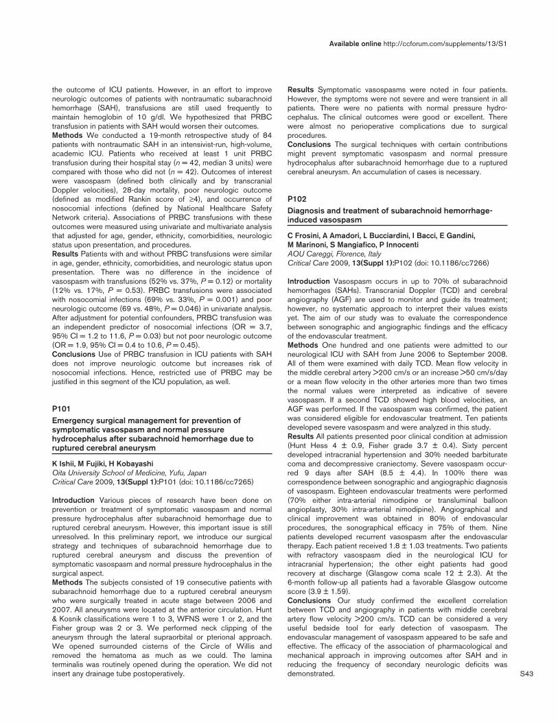
S43
the outcome of ICU patients. However, in an effort to improveneurologic outcomes of patients with nontraumatic subarachnoidhemorrhage (SAH), transfusions are still used frequently tomaintain hemoglobin of 10 g/dl. We hypothesized that PRBCtransfusion in patients with SAH would worsen their outcomes.Methods We conducted a 19-month retrospective study of 84patients with nontraumatic SAH in an intensivist-run, high-volume,academic ICU. Patients who received at least 1 unit PRBCtransfusion during their hospital stay (n = 42, median 3 units) werecompared with those who did not (n = 42). Outcomes of interestwere vasospasm (defined both clinically and by transcranialDoppler velocities), 28-day mortality, poor neurologic outcome(defined as modified Rankin score of ≥4), and occurrence ofnosocomial infections (defined by National Healthcare SafetyNetwork criteria). Associations of PRBC transfusions with theseoutcomes were measured using univariate and multivariate analysisthat adjusted for age, gender, ethnicity, comorbidities, neurologicstatus upon presentation, and procedures.Results Patients with and without PRBC transfusions were similarin age, gender, ethnicity, comorbidities, and neurologic status uponpresentation. There was no difference in the incidence ofvasospasm with transfusions (52% vs. 37%, P = 0.12) or mortality(12% vs. 17%, P = 0.53). PRBC transfusions were associatedwith nosocomial infections (69% vs. 33%, P = 0.001) and poorneurologic outcome (69 vs. 48%, P = 0.046) in univariate analysis.After adjustment for potential confounders, PRBC transfusion wasan independent predictor of nosocomial infections (OR = 3.7,95% CI = 1.2 to 11.6, P = 0.03) but not poor neurologic outcome(OR = 1.9, 95% CI = 0.4 to 10.6, P = 0.45).Conclusions Use of PRBC transfusion in ICU patients with SAHdoes not improve neurologic outcome but increases risk ofnosocomial infections. Hence, restricted use of PRBC may bejustified in this segment of the ICU population, as well.
P101Emergency surgical management for prevention ofsymptomatic vasospasm and normal pressurehydrocephalus after subarachnoid hemorrhage due toruptured cerebral aneurysm
K Ishii, M Fujiki, H KobayashiOita University School of Medicine, Yufu, JapanCritical Care 2009, 13(Suppl 1):P101 (doi: 10.1186/cc7265)
Introduction Various pieces of research have been done onprevention or treatment of symptomatic vasospasm and normalpressure hydrocephalus after subarachnoid hemorrhage due toruptured cerebral aneurysm. However, this important issue is stillunresolved. In this preliminary report, we introduce our surgicalstrategy and techniques of subarachnoid hemorrhage due toruptured cerebral aneurysm and discuss the prevention ofsymptomatic vasospasm and normal pressure hydrocephalus in thesurgical aspect.Methods The subjects consisted of 19 consecutive patients withsubarachnoid hemorrhage due to a ruptured cerebral aneurysmwho were surgically treated in acute stage between 2006 and2007. All aneurysms were located at the anterior circulation. Hunt& Kosnik classifications were 1 to 3, WFNS were 1 or 2, and theFisher group was 2 or 3. We performed neck clipping of theaneurysm through the lateral supraorbital or pterional approach.We opened surrounded cisterns of the Circle of Willis andremoved the hematoma as much as we could. The laminaterminalis was routinely opened during the operation. We did notinsert any drainage tube postoperatively.
Results Symptomatic vasospasms were noted in four patients.However, the symptoms were not severe and were transient in allpatients. There were no patients with normal pressure hydro-cephalus. The clinical outcomes were good or excellent. Therewere almost no perioperative complications due to surgicalprocedures.Conclusions The surgical techniques with certain contributionsmight prevent symptomatic vasospasm and normal pressurehydrocephalus after subarachnoid hemorrhage due to a rupturedcerebral aneurysm. An accumulation of cases is necessary.
P102Diagnosis and treatment of subarachnoid hemorrhage-induced vasospasm
C Frosini, A Amadori, L Bucciardini, I Bacci, E Gandini, M Marinoni, S Mangiafico, P InnocentiAOU Careggi, Florence, ItalyCritical Care 2009, 13(Suppl 1):P102 (doi: 10.1186/cc7266)
Introduction Vasospasm occurs in up to 70% of subarachnoidhemorrhages (SAHs). Transcranial Doppler (TCD) and cerebralangiography (AGF) are used to monitor and guide its treatment;however, no systematic approach to interpret their values existsyet. The aim of our study was to evaluate the correspondencebetween sonographic and angiographic findings and the efficacyof the endovascular treatment.Methods One hundred and one patients were admitted to ourneurological ICU with SAH from June 2006 to September 2008.All of them were examined with daily TCD. Mean flow velocity inthe middle cerebral artery >200 cm/s or an increase >50 cm/s/dayor a mean flow velocity in the other arteries more than two timesthe normal values were interpreted as indicative of severevasospasm. If a second TCD showed high blood velocities, anAGF was performed. If the vasospasm was confirmed, the patientwas considered eligible for endovascular treatment. Ten patientsdeveloped severe vasospasm and were analyzed in this study.Results All patients presented poor clinical condition at admission(Hunt Hess 4 ± 0.9, Fisher grade 3.7 ± 0.4). Sixty percentdeveloped intracranial hypertension and 30% needed barbituratecoma and decompressive craniectomy. Severe vasospasm occur-red 9 days after SAH (8.5 ± 4.4). In 100% there wascorrespondence between sonographic and angiographic diagnosisof vasospasm. Eighteen endovascular treatments were performed(70% either intra-arterial nimodipine or transluminal balloonangioplasty, 30% intra-arterial nimodipine). Angiographical andclinical improvement was obtained in 80% of endovascularprocedures, the sonographical efficacy in 75% of them. Ninepatients developed recurrent vasospasm after the endovasculartherapy. Each patient received 1.8 ± 1.03 treatments. Two patientswith refractory vasospasm died in the neurological ICU forintracranial hypertension; the other eight patients had goodrecovery at discharge (Glasgow coma scale 12 ± 2.3). At the6-month follow-up all patients had a favorable Glasgow outcomescore (3.9 ± 1.59).Conclusions Our study confirmed the excellent correlationbetween TCD and angiography in patients with middle cerebralartery flow velocity >200 cm/s. TCD can be considered a veryuseful bedside tool for early detection of vasospasm. Theendovascular management of vasospasm appeared to be safe andeffective. The efficacy of the association of pharmacological andmechanical approach in improving outcomes after SAH and inreducing the frequency of secondary neurologic deficits wasdemonstrated.
Available online http://ccforum.com/supplements/13/S1

S44
P103Effects of simvastatin in prevention of vasospasm innontraumatic subarachnoid hemorrhage: preliminary data
S Macedo, Y Bello, A Silva, C Siqueira, S Siqueira, L BritoHospital São José do Avai, Itaperuna, BrazilCritical Care 2009, 13(Suppl 1):P103 (doi: 10.1186/cc7267)
Introduction Vasospasm is the main cause of death and cognitivedeficits in patients with subarachnoid hemorrhage after rupture ofan aneurysm (aSAH). Some trials have shown that statins in theacute phase of aSAH reduce the incidence, morbidity and mortalityof cerebral vasospasm [1]. The purpose of this study is to evaluatethe potential of simvastatin (SVT) as prevention against vasospasm.Methods We realized a prospective, randomized, nonblind studywith the use of 80 mg SVT (night) in the first 72 hours of thebeginning of bleeding, and a control group that did not use SVT,for 21 days between January and December 2008. Informedconsent was obtained for all patients. CT scans were performed asa control and another CT scan was performed in patients withaltered neurological signals. In the presence of changessuggestive of vasospasm or correlation in clinical conditions andCT scans, the patients were taken for a cerebral arteriographyexamination followed by an angioplasty procedure if necessary.Liver and renal function and LDL cholesterol were evaluatedweekly, and total creatine kinase was evaluated every 3 days.Exclusion criteria: liver and renal disease, pregnant elevation ofserum transaminases (three times normal value), creatinine ≥2.5,rhabdomyolysis or total creatine kinase ≥1,000 U/l.Results We excluded two patients with bleeding for more than72 hours. There was no significant change in the levels of totalcreatine kinase, and renal or liver function. We included 21patients, 11 in the SVT group and 10 in the control group. Mortalitywas eight patients (38%), six patients in the control group and twoof the SVT group. Vasospasm was confirmed by cerebralarteriography examination in four patients in the control group andone patient in the SVT group. All the patients who died showedFisher scale IV.Conclusions SVT at a dose of 80 mg was effective in reducing themortality (18.1% against 60%) compared with the group that didnot use SVT, and also decreased the incidence of cerebralvasospasm despite the APACHE II score being higher in the groupthat used SVT (14.3 vs. 10.7). Less morbidity occurred in the SVTgroup, with an average Glasgow outcome score of 3.25 vs. 2.1.Reference1. Lynch JR, Wang H, et al.: Simvastatin reduces vasospasm
after aneurysmal subarachnoid hemorrhage: results of apilot randomized clinical trial. Stroke 2005, 36:2024-2026.
P104Magnesium use on prophylaxis of vasospasm morbidityand the mortality rate in subarachnoid hemorrhage
S Macedo, R Nuss, G Lubanco, R Lovatti, S Pereira, G Lima, C Siqueira, S Siqueira, D Lima, R TorresHospital São José do Avai, Itaperuna, BrazilCritical Care 2009, 13(Suppl 1):P104 (doi: 10.1186/cc7268)
Introduction We propose this study in order to reach two points: theclinical incidence of vasospasm morbidity, confirmed by CT; and themortality of subarachnoid hemorrhage (SAH) patients in 28 days [1].It shows the comparison of a group of patients that used magnesium(Mg) (intervention, Group 1) with those that did not use Mg(control, Group 2).Methods After institutional approval and informed consent, aprospective, randomized, nonblind study was carried out betweenFebruary and November 2008. The main goal of the study was toachieve a Mg serum concentration from 2.5 to 3.5 mg/dl, using asolution of Mg 2% (saline solution 5% 400 ml + MgSO4 10%100 ml/24 hours), during the first 14 days of aneurysm rupture.Admission criteria: patients diagnosed with SAH and ΔT <96 hours.Exclusion criteria: patients with SAH and ΔT >96 hours.Results In a previous study evaluation we analysed a total of 56patients with (n = 26 in Group 1 and n = 30 in Group 2) (Tables 1and 2). Main results: Group 1 – vasospasm frequency 26.9% (n = 7)and mortality 19.2% (n = 5) in 28 days; Group 2 – vasospasmfrequency 46.7% (n = 14) and mortality 33.3% (n = 10) in 28 days.Conclusions According to the outcome, we can conclude thatGroup 1 obtained greater protection on the vasospasm incidenceand decrease of mortality in comparison with Group 2. The P valuewas not significant due to a still small number of patients.Reference1. Schmid-Elsaesser R, et al.: Intravenous magnesium versus
nimodipine in the treatment of patients with aneurysmalsubarachnoid hemorrhage: a randomized study. Neuro-surgery 2006, 58:1054-1065.
P105Outcomes of ventilator-associated pneumonia inaneurysmal subarachnoid hemorrhage patients
R Lenhardt, O AkcaUniversity of Louisville, KY, USACritical Care 2009, 13(Suppl 1):P105 (doi: 10.1186/cc7269)
Introduction Subarachnoid hemorrhage (SAH) from rupture ofcerebral aneurysms is associated with significant mortality andmorbidity. About 10% to 25% die before reaching hospital, and of
Critical Care March 2009 Vol 13 Suppl 1 29th International Symposium on Intensive Care and Emergency Medicine
Table 1 (abstract P104)
Average age APACHE II score Average Mg level
Group I (n = 26) 52.3 8.2 2.32
Group II (n = 30) 50.3 15.6 1.9
Table 2 (abstract P104)
Group I (n = 26) Group II (n = 30)
Frequency 95% confidence Frequency 95% confidence (% (n)) interval Odds ratio (% (n)) interval Odds ratio P value
Vasospasm 26.9 (7) 11.6 to 47.8 0.4 46.7 (14) 28.3 to 65.7 1 0.1
Mortality in 28 days 19.2 (5) 6.6 to 39.4 0.5 33.3 (10) 17.3 to 52.8 1 0.2

S45
those who survive about 40% to 50% develop significantneurological deficits [1]. Ventilator-associated pneumonia (VAP) isdefined as pneumonia occurring more than 48 hours after initiationof mechanical ventilation. About 20% of post-aneurysmal SAHpatients are reported to experience VAP [2]. In this trial, we aimedto report the short-term outcomes of VAP. We performed asurveillance analysis on aneurysmal SAH patients who requiredmechanical ventilation for more than 48 hours.Methods After obtaining approval from the Human StudiesCommittee of the University of Louisville to retrospectively analyzethe prospectively collected patient data, we reviewed the elec-tronic records of our aneurismal SAH patients admitted between2004 and 2007. VAP was diagnosed and confirmed by theClinical Pulmonary Infection Score supported with culture resultson days 0 and 3. We analyzed host-specific and disease-specificand care-related risk factors. Categorical variables were comparedwith the chi-square test, and continuous data were analyzed withthe unpaired t and Kruskal–Wallis tests.Results Within 86 aneurysmal SAH patients admitted to the ICU,45 patients needed to be ventilated for more than 48 hours (52%),and 16 of them developed VAP (19%). More than 80% of patientswith SAH required either surgical or vascular procedure. Themajority of VAP were late-onset pneumonias (88%). The durationof mechanical ventilation was longer in the VAP patients. About20% of VAP group patients were also diagnosed with sepsis.However, the duration of ICU stay was neither influenced by VAPnor by sepsis. Four patients who did not experience any VAP werediagnosed with stroke during their ICU stay. All-cause mortalitywas not longer in patients with VAP.Conclusions In this preliminary report of a prospective cohort trial,it appeared that VAP did not contribute to additional morbidity ormortality. The majority of VAP occurred late in the course ofventilation. This supports the theory that VAP occurs primarily dueto the disease itself and that detailed and prolonged care isrequired for the management of aneurysmal SAH patients.References1. Schievink WI, et al.: Neurology 1995, 45:871-874.2. Frontera JA, et al.: Neurosurgery 2008, 62:80-87.
P106Outcomes from subarachnoid haemorrhage
I Whitehead, N Azam, S Bonner, J WrightJames Cook University Hospital, Middlesbrough, UKCritical Care 2009, 13(Suppl 1):P106 (doi: 10.1186/cc7270)
Introduction A retrospective assessment of the outcome ofpatients with poor-grade (World Federation of NeurosurgeonsGrades 4 and 5) subarachnoid haemorrhage (SAH) at a regionalneurosurgical centre. Previous studies have shown aggressivetreatment of patients with a poor clinical grade of SAH iswarranted as grading can improve following resuscitation anddrainage of cerebrospinal fluid [1,2]. Many units still withdrawtreatment if these patients do not improve neurologically in the first48 to 72 hours [3].Methods A retrospective analysis of the notes of 116 patients whowere recorded as having a diagnosis of SAH was performedbetween October 2002 and January 2006. Patients were excludedif they were World Federation of Neurosurgeons Grade 1, 2 or 3,had a traumatic SAH or been incorrectly classified as having SAH.Results Of 116 patients identified with a diagnosis of SAH, 12patients were excluded as they had a traumatic SAH, one patienthad a dissection, one patient a subdural haemorrhage and onepatient a basal ganglia haematoma. Thirteen patients had aGlasgow coma score >12. Eighty-eight patients were correctly
deemed as poor grade. All poor-grade patients were admitted tothe intensive therapy unit. Of these 88 patients, 34 (38.6%)survived. Twenty-four (70%) of the survivors were dischargedhome, eight (24%) to a care home, and two (6%) remained inhospital. Seventeen (50%) had a Glasgow Outcome Score of 4 or 5.Conclusions Outcomes from poor-grade SAH at James CookUniversity Hospital compare favourably with published data.Review of the literature gives a wide variation in outcome between3.2% and 42%. It is our hypothesis that a combination of moreaggressive critical care management combined with earlyintervention should result in a reevaluation of treatment plans inthose with a historically perceived poor outcome.References1. Bailes JE, et al.: Management morbidity and mortality of poor-
grade aneurysm patients. J Neurosurg 1990, 72:559-566.2. Hutchinson PJ, et al.: Outcome from poor grade aneurys-
mal subarachnoid haemorrhage – which poor grade sub-arachnoid haemorrhage patients benefit from aneurysmclipping? Br J Neurosurg 2000, 14:105-109.
3. Wilby M J, et al.: Cost effective outcome for treating poor-grade subarachnoid haemorrhage. Stroke 2003, 34:2508-2511.
P107Full Outline of Unresponsiveness compared with Glasgowcoma scale assessment and outcome prediction in coma
D Ledoux1, M Bruno2, S Jonlet1, P Choi1, C Schnakers2, F Damas1, B Lambermont1, P Damas1, S Laureys2
1Liege University Hospital, Liège, Belgium; 2Coma Science Group –University of Liege, BelgiumCritical Care 2009, 13(Suppl 1):P107 (doi: 10.1186/cc7271)
Introduction The most widely adopted scale to assessconsciousness in severely brain-damaged patients is the Glasgowcoma scale (GCS) [1]. Its major shortcomings are the failure toassess the verbal component in intubated patients, the inability totest brainstem reflexes and breathing patterns. In 2005, Wijdicksand colleagues proposed a new coma scale, the Full Outline ofUnresponsiveness (FOUR) scale [2], which consists of fourcomponents (eye, motor, brainstem, and respiration), each compo-nent having a maximal score of 4. Our objective was to validate theFrench version of the new FOUR coma scale in a general ICU andto assess its predictive value as compared with the GCS.Methods We performed FOUR and GCS evaluations inrandomized order in 176 acutely brain-injured patients (days frominsult to randomization <1 month). We assessed the associationbetween GCS and FOUR scores using the Spearman correlationcoefficient. A logistic regression analysis adjusted for age andetiology of coma was performed to assess the link between thestudied scores and the outcome based on the Glasgow outcomescales 3 months after injury (n = 63).Results The GCS and FOUR showed a significant correlation(r = 0.807). The GCS verbal component was scored 1 in 146patients; among these, 131 were intubated. The FOUR total scores(corrected for age) showed superior outcome prediction at 3 months(OR = 0.83; 95% CI = 0.70 to 0.98, P = 0.03) as compared withGCS total scores (OR = 0.85; 95% CI = 0.70 to 1.03, P = 0.09).Conclusions The FOUR scale does not need a verbal response,thus allowing complete testing in intubated patients (in our sample90% of patients showing a GCS V1 score were intubated). Mostimportantly, the FOUR scale demonstrated a better discriminationbetween the good (recovery of independent living) and poorneurological status at 3 months as compared with the GCS.
Available online http://ccforum.com/supplements/13/S1
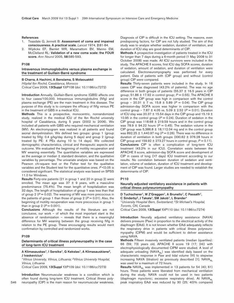
S46
References1. Teasdale G, Jennett B: Assessment of coma and impaired
consciousness. A practical scale. Lancet 1974, 2:81-84.2. Wijdicks EF, Bamlet WR, Maramattom BV, Manno EM,
McClelland RL: Validation of a new coma scale: the FOURscore. Ann Neurol 2005, 58:585-593.
P108Intravenous immunoglobulins versus plasma exchange inthe treatment of Guillain-Barré syndrome
B Charra, A Hachimi, A Benslama, S MotaouakkilHôpital Ibn Rochd, Casablanca, MoroccoCritical Care 2009, 13(Suppl 1):P108 (doi: 10.1186/cc7272)
Introduction Annually, Guillain-Barré syndrome (GBS) affects oneto four cases/100,000. Intravenous immunoglobulins (IVIg) andplasma exchange (PE) are the main treatment in this disease. Thepurpose of this study is to compare the efficacy of IVIg versus PEin the treatment of GBS in a medical ICU.Methods This is a prospective, monocentric, nonrandomizedstudy, realized in the medical ICU of the Ibn Rochd universityhospital of Casablanca, during 5 years (2002 to 2006). Weincluded all patients with GBS who required mechanical ventilation(MV). An electromyogram was realized in all patients and foundaxonal demyelinization. We defined two groups: group 1 (grouptreated by IVIg: 0.4 g/kg/day during 5 days) and group 2 (grouptreated by PE: 4 PEs during 10 to 14 days). We collecteddemographic characteristics, clinical and therapeutic aspects andoutcome. We evaluated the beginning of motility recuperation andMV weaning essentially. The quantitative variables are expressedas the average or median ± standard deviation, and the qualitativevariables by percentage. The univariate analysis was based on thePearson chi-square test or the Fisher test for the qualitativevariables and the Student test for the quantitative ones. P <0.05 isconsidered significant. The statistical analysis was based on SPSS11.0 for Windows.Results Forty-one patients (21 in group 1 and 20 in group 2) wereenrolled. The mean age was 37 ± 9 years, with a masculinepredominance (75.4%). The mean length of hospitalization was32 days. The length of hospitalization of group 1 was less than thatof group 2 (P = 0.02). The weaning of MV was more precocious inthe patients of group 1 than those of group 2 (P = 0.01). Also, thebeginning of motility recuperation was more precocious in group 1than in group 2 (P = 0.001).Conclusions Although the results of the literature are notconclusive, our work – of which the most important slant is theabsence of randomization – reveals that there is a meaningfuldifference for MV weaning between the group receiving IVIg inrelation to the PE group. These encouraging results would meritconfirmation by controlled and randomized works.
P109Determinants of critical illness polyneuropathy in the caseof long-term ICU treatment
A Klimasauskas1, I Sereike1, G Kekstas1, A Klimasauskiene2, J Ivaskevicius1
1Vilnius University, Vilnius, Lithuania; 2Vilnius University Hospital,Vilnius, LithuaniaCritical Care 2009, 13(Suppl 1):P109 (doi: 10.1186/cc7273)
Introduction Neuromuscular weakness is a condition which isoften found during long-term ICU treatment. Critical illness poly-neuropathy (CIP) is the main reason for neuromuscular weakness.
Diagnosis of CIP is difficult in the ICU setting. The reasons, evenpredisposing factors, for CIP are not fully studied. The aim of thisstudy was to analyze whether sedation, duration of ventilation, andduration of ICU stay are good determinants of CIP.Methods A prospective investigation of patients treated in the ICUfor longer than 7 days during a 6-month period (1 May 2008 to 31October 2008) was made. All ICU survivors were included in thestudy. The APACHE II scores, first ICU day SOFA scores, durationof sedation, amount of sedation, and duration of ventilation werecalculated. Electroneuromyography was performed for everypatient. Data of patients with (CIP group) and without (controlgroup) CIP were compared.Results Thirty-seven patients were included in the study. In 16cases CIP was diagnosed (43.2% of patients). The was no agedifference in both groups of patients (55.37 ± 16.5 years in CIPgroup; 51.86 ± 17.92 in control group; P = 0.55). The APACHE IIscore in the CIP group was high in comparison with the controlgroup – 20.31 ± 7 vs. 15.8 ± 5.89 (P = 0.04). The CIP groupadmission-day SOFA score was higher in comparison with thecontrol group – 7.87 ± 4.05 vs. 5.09 ± 2.52 (P = 0.01). Durationof ICU stay was 20.37 ± 15.54 days in the CIP group and 15.19 ±10.95 in the control group (P = 0.24). Duration of sedation in theCIP group was 118.68 ± 219.59 hours and in the control groupwas 78.9 ± 94.22 hours (P = 0.45). The sedation volume in theCIP group was 5,386.8 ± 18,112.04 mg and in the control groupwas 892.25 ± 1,440.97 mg (P = 0.26). There was no difference induration of ventilation in both groups (330.62 ± 376.28 hours inCIP group and 159.92 ± 273.27 hours in control group; P = 0.11).Conclusions CIP is often a complication of long-term ICUtreatment (43.2% in our ICU). Correlation exists between theAPACHE II score, admission-day SOFA score and development ofCIP in cases of long (>7 days) ICU treatment according to ourresults. No correlation between duration of sedation and venti-lation, volume of sedation, duration of ICU treatment and develop-ment of CIP was found. Larger studies are needed to establish thedeterminants of CIP.
P110Neurally adjusted ventilatory assistance in patients withcritical illness polyneuromyopathy
D Tuchscherer1, W Z’Graggen1, A Brunello1, C Passath1, C Sinderby2, J Takala1, SM Jakob1, L Brander1
1University Hospital Bern, Switzerland; 2St Michael’s Hospital,Toronto, ON, CanadaCritical Care 2009, 13(Suppl 1):P110 (doi: 10.1186/cc7274)
Introduction Neurally adjusted ventilatory assistance (NAVA)delivers pressure (Paw) in proportion to the electrical activity of thediaphragm (EAdi). It is not known whether EAdi adequately reflectsthe respiratory drive in patients with critical illness polyneuro-myopathy (CIPM) and would be sufficient to deliver assistanceusing NAVA.Methods Fifteen invasively ventilated patients (median (quartiles):66 (59; 73) years old, APACHE II score 19 (17; 24)) withelectrophysiologically documented CIPM were studied. A level ofadequate unloading (NAVAAL) was identified daily based on thecharacteristic response in Paw and tidal volume (Vt) to stepwiseincreasing NAVA (titration) as previously described [1]. NAVAALwas used for a maximum of 72 hours.Results NAVAAL was implemented in 13 patients for 54 (40; 61)hours. Three patients were liberated from mechanical ventilationduring the study. NAVA could not be used in two patients(diaphragm myoclonic; excessive respiratory drive). At NAVAALpeak inspiratory EAdi was reduced by 30 (25; 40)% compared
Critical Care March 2009 Vol 13 Suppl 1 29th International Symposium on Intensive Care and Emergency Medicine
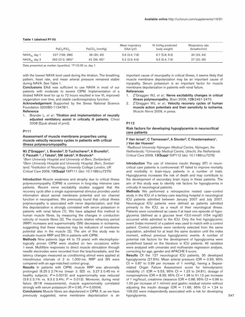
S47
with the lowest NAVA level used during the titration. The breathingpattern, heart rate, and mean arterial pressure remained stableduring NAVA. See Table 1.Conclusions EAdi was sufficient to use NAVA in most of ourpatients with moderate to severe CIPM. Implementation of atitrated NAVA level for up to 72 hours resulted in low Vt, improvedoxygenation over time, and stable cardiorespiratory function.Acknowledgement Supported by the Swiss National ScienceFoundation 3200B0-113478/1.Reference1. Brander L, et al.: Titration and implementation of neurally
adjusted ventilatory assist in critically ill patients. Chest2008 [Epub ahead of print].
P111Assessment of muscle membrane properties usingmuscle velocity recovery cycles in patients with criticalillness polyneuromyopathy
WJ Z’Graggen1, L Brander2, D Tuchscherer2, A Brunello2, C Passath 2, J Takala2, SM Jakob2, H Bostock3
1Bern University Hospital and University of Bern, Switzerland;2Bern University Hospital and University Hospital, Bern, Switzer-land; 3Institution of Neurology, University College London, UKCritical Care 2009, 13(Suppl 1):P111 (doi: 10.1186/cc7275)
Introduction Muscle weakness and atrophy due to critical illnesspolyneuromyopathy (CIPM) is common in long-stay intensive carepatients. Recent nerve excitability studies suggest that therecovery cycle after a single supramaximal stimulus provides usefulinformation about axonal membrane potential and ion channelfunction in neuropathies. We previously found that critical illnesspolyneuropathy is associated with nerve depolarization, and thatthis depolarization is strongly correlated with serum potassium inpatients with renal failure [1]. We have adapted this method tohuman muscle fibres, by measuring the changes in conductionvelocity of muscle fibres [2]. The muscle relative refractory period(RRP) increases and supernormality (SN) decreases in ischaemia,suggesting that these measures may be indicators of membranepotential also in the muscle [2]. The aim of this study was toevaluate muscle RRP and SN in patients with CIPM.Methods Nine patients (age 44 to 73 years) with electrophysio-logically proven CIPM were studied on two occasions within1 week. Multifibre responses to direct muscle stimulation throughneedle electrodes were recorded from the brachioradialis, and thelatency changes measured as conditioning stimuli were applied atinterstimulus intervals of 2 to 1,000 ms. RRP and SN werecompared with an age-matched control group.Results In patients with CIPM, muscle RRP was abnormallyprolonged (6.25 ± 2.74 ms (mean ± SD) vs. 3.27 ± 0.45 ms inhealthy subjects; P = 0.0015) and supernormality was reduced(3.6 ± 3.1% vs. 9.3 ± 3.4%; P = 0.013). Moreover, during renalfailure (8/18 measurements), muscle supernormality correlatedstrongly with serum potassium (R = 0.95, P = 0.0004).Conclusions Muscle fibres are depolarized in CIPM. If, as we havepreviously suggested, nerve membrane depolarization is an
important cause of neuropathy in critical illness, it seems likely thatmuscle membrane depolarization may be an important cause ofmyopathy. Serum potassium is an important factor for musclemembrane depolarization in patients with renal failure.References1. Z’Graggen WJ, et al.: Nerve excitability changes in critical
illness polyneuropathy. Brain 2006, 129:2461-2470.2. Z’Graggen WJ, et al.: Velocity recovery cycles of human
muscle action potentials and their sensitivity to ischemia.Muscle Nerve 2009, in press.
P112Risk factors for developing hypoglycemia in neurocriticalcare patients
F Van Iersel1, C Tiemessen2, A Slooter2, C Hoedemaekers1, J Van der Hoeven1
1Radboud University Nijmegen Medical Centre, Nijmegen, theNetherlands; 2University Medical Centre, Utrecht, the NetherlandsCritical Care 2009, 13(Suppl 1):P112 (doi: 10.1186/cc7276)
Introduction The use of intensive insulin therapy (IIT) in neuro-critical care patients is controversial: IIT failed to improve mortalityand morbidity in brain-injury patients in a number of trials.Hypoglycemia increases the risk of death and may contribute tothe development of secondary brain injury in these patients. Theaim of this study was to identify risk factors for hypoglycemia incritically ill neurological patients.Methods We performed a retrospective nested case–controlstudy in the ICU of a tertiary-care teaching hospital in neurologicalICU patients admitted between January 2007 and July 2007.Neurological ICU patients were defined as patients admittedprimarily to the ICU, as a result of their neurological illness.Patients were considered as cases if at least one episode of hypo-glycemia (defined as a glucose level <3.0 mmol/l (<54 mg/dl))occurred while admitted to the ICU. Only the first hypoglycemicevent (index moment) of a patient was used to match with a controlpatient. Control patients were randomly selected from the samepopulation, admitted for at least the same duration until the indexmoment, without previous hypoglycemic events. A number ofpotential risk factors for the development of hypoglycemia werepredefined based on the literature in ICU patients. All variableswere analyzed with univariate and multivariate regression analysis,correcting for age, gender and APACHE II score.Results Of the 127 neurological ICU patients, 35 developedhypoglycemia (27.6%). Mean arterial pressure (OR = 0.93; 95%CI = 0.87 to 0.99 per increase of 1 mmHg), having a Sepsis-related Organ Failure Assessment score for hemodynamicinstability ≥1 (OR = 5.53; 95% CI = 1.23 to 24.81), dosage ofnorepinephrine (OR = 8.39; 95% CI = 1.38 to 51.12 per increaseof 1 mg/hour), creatinine clearance (OR = 0.98; 95% CI = 0.96 to1.00 per increase of 1 ml/min) and gastric residual volume withoutadjusting the insulin dosage (OR = 11.66; 95% CI = 1.24 to109.20) were independently associated with a risk for developinghypoglycemia.
Available online http://ccforum.com/supplements/13/S1
Table 1 (abstract P110)
Mean inspiratory Vt (ml/kg predicted Respiratory rate PaO2/FiO2 PaCO2 (mmHg) EAdi (μV) body weight) (breaths/min)
NAVAAL day 1 227 (158; 286) 36 (30; 45) 5.6 (3.4; 7.8) 6.7 (5.8; 8.3) 30 (23; 34)
NAVAAL day 3 263 (212; 380)* 42 (39; 45)* 5.2 (2.9; 6.6) 6.5 (5.4; 7.6) 27 (22; 35)
Data presented as median (quartiles). *P <0.05 vs. day 1.
S48
Conclusions Hypoglycemia occurs in a significant proportion ofneurological ICU patients. We suggest more frequent control ofblood glucose values, especially in patients suffering fromhemodynamic instability, renal failure and gastroparesis.
P113Relationship between effective osmolality changes andneurological status during treatment for severe paediatricdiabetic ketoacidosis
S Tibby, A Durward, L Ferguson, H Bangalore, I MurdochEvelina Children’s Hospital, Guy’s & St Thomas’ NHS Trust,London, UKCritical Care 2009, 13(Suppl 1):P113 (doi: 10.1186/cc7277)
Introduction Cerebral oedema is a common, life-threateningcomplication of paediatric diabetic ketoacidosis (DKA). Several riskfactors at presentation are known, including urea, and pCO2. It hasrecently been suggested that changes in effective osmolalityduring treatment may also convey risk, but this has not beenconfirmed due to a lack of prospective data.Methods A prospective observational study over 24 hours of 50children admitted to a regional ICU with severe DKA (median pH atpresentation 6.90). Development of cerebral oedema was definedclinically as a deterioration in Glaser score of >2 points during the24 hours after presentation (equating to a Glasgow coma scale <8to 10). Changes in effective osmolality, uncorrected and correctedsodium were modelled using linear (random coefficients) mixedmodels, with adjustment for baseline covariates of urea and pCO2.Results Consistent with previous studies, baseline urea washigher and pCO2 lower in patients who subsequently showedneurological deterioration. All three osmolality variables (effectiveosmolality, uncorrected and corrected sodium) were similar atbaseline between the two groups. The best-fitting model utilizedcorrected sodium (lowest Akaike information criterion). Thisshowed a significant interaction effect (P = 0.01), in that correctedsodium increased over time at a rate of 0.35 mmol/l/hour inpatients who did not develop neurological symptoms, but did notchange in those who did.Conclusions Lack of change in corrected sodium is associated withneurological deterioration in the treatment of severe DKA in children.This variable may be useful if incorporated into treatment guidelines.
P114Continuous monitoring of blood parameters in intensivecare patients
A Weinstein1, O Herzenstein1, E Gabis1, I Kagan2, P Singer2
1Orsense Ltd, Nes Ziona, Israel; 2Rabin Medical Center, PetahTikva, IsraelCritical Care 2009, 13(Suppl 1):P114 (doi: 10.1186/cc7278)
Introduction Monitoring of blood parameters, such as glucose,hemoglobin and oxygen saturation, is essential in critically illpatients. The current invasive methods are not frequent enough forefficient tight glycemic control, and result in a high rate ofhypoglycemia. In addition, there is a growing need for a continuoushemoglobin measurement in postoperative care units and ICUs.The purpose of this study is to evaluate the feasibility of the fullynoninvasive blood monitor (NBM device; OrSense Ltd, Nes Ziona,Israel) for continuous monitoring of glucose, hemoglobin andoxygen saturation in critically ill patients.Methods The study was conducted on 14 patients (seven female,seven male, ages 34 to 92 years) in the ICU of the Rabin MedicalCenter, upon receipt of informed consent. The NBM probe was
placed on patients’ thumbs, where it performed measurements forup to 24 hours, with readings every 10 minutes. Patient compli-ance was good and no adverse effects were identified. The resultsobtained from the NBM device were compared with blood samplestaken through an arterial line every 30 to 60 minutes and wereanalyzed with a blood gas machine (ABL 700; Radiometer,Copenhagen, Denmark).Results A total of 208 paired data points were obtained in the trial.At each point, an algorithm based on a uniform model (withpersonal glucose calibration) was used to calculate the three bloodparameters. The reference glucose range was 62 to 369 mg/dl.The median relative absolute difference was 7.3%, and a Clarkeerror grid analysis showed that 95.2% of the measurements fellwithin zones A (74.5%) and B (20.7%). The reference range of thehemoglobin was 7 to 14.5 g/dl and the median absolute errorobtained was 1 g/dl. Oxygen saturation levels were tracked simul-taneously with a mean error of 2.5%.Conclusions The present study indicates the potential use of thenoninvasive NBM for continual, accurate, safe, and easy-to-usemultiparameter monitoring in critically ill patients. The device holdsthe promise of improving patient care and survival, as well asreducing staff workload.
P115Morning glucose correlates poorly with daily mean glucosein cardiac surgical ICU patients: impact of ultradianvariation of glucose
D Hagg, S Smith, K Oveson, M Slater, H Song, A AhmannOHSU, Portland, OR, USACritical Care 2009, 13(Suppl 1):P115 (doi: 10.1186/cc7279)
Introduction Treatment of hyperglycemia with an insulin infusionprotocol (IIP) has improved outcomes in ICU and cardiac surgerypatients [1]. Many studies have reported morning glucose valuesas representative of whole-day glucose control. We recentlydemonstrated that in a mixed ICU population this assumption doesnot hold [2]. Since it has been proposed that glucose control maybe especially important in cardiac surgery ICU patients, we askedwhether morning glucose accurately represented whole-dayglucose and determined whether glucose varied over the course ofthe day in this patient group.Methods A prospective, observational single-center study in thecardiac surgical ICU of a university tertiary-care hospital. Allglucose measurements in cardiac surgical ICU patients receivingan IIP targeting blood glucose 80 to 110 mg/dl were recordedfrom 1 June 2006 to 28 February 2007.Results We recorded 12,109 glucose measurements from 125patients on IIPs. The glucose measurements were widelydistributed with 1%, 7%, 47%, 33%, 9% and 2% in the ranges<60, 61 to 79, 80 to 110, 111 to 150, 151 to 200 and>200 mg/dl, respectively. After 8 hours of IIP, the proportion ofvalues in the target range increased to 49%. The 06:00-hourglucose values were lower than other values (mean ± SD: 105 ±24 mg/dl (n = 482) vs. 113 ± 33 mg/dl (n = 10,688); P <0.0001).The 06:00 values were poorly correlated with the average glucoserecorded for the remainder of the day (r2 = 0.029), which wasconfirmed by Bland–Altman analysis. The time-averaged glucosedata exhibited ultradian variation, peaking at 10:00 and 19:00hours. The insulin dose varied with a similar pattern.Conclusions In cardiothoracic ICU patients receiving an IIP,glucose exhibited an ultradian pattern with peaks at 10:00 and19:00 hours, and was lower in the early morning than during theremainder of the day. Consideration of this ultradian variation mayavoid hypoglycemic and hyperglycemic episodes and so facilitate
Critical Care March 2009 Vol 13 Suppl 1 29th International Symposium on Intensive Care and Emergency Medicine

S49
better glucose control with an IIP. Studies targeting control ofhyperglycemia should report mean blood glucose values for theentire day rather than early morning values.References1. Dellinger RP, et al.: Intensive Care Med 2004, 30:536-555.2. Smith SM, et al.: Diabetes Care 2007, 30:2503-2505.
P116Hypoglycaemia is associated with a higher mortality incritically ill patients
A Jose Pereira1, A Biasi Cavalcanti1, F Pereira Almeida1, T Correa1, J Telles2, M Lobato1, D Nishimura1, R Da HoraPassos1, E Silva1
1Hospital Israelita Albert Einstein, São Paulo, Brazil; 2HospitalPortugues, Salvador, BrazilCritical Care 2009, 13(Suppl 1):P116 (doi: 10.1186/cc7280)
Introduction Hypoglycaemia, a common complication of strictglucose control in critically ill patients, has controversial effects onmortality. The hyperglycaemic index (HGI) takes into account theunequal time distribution of blood glucose sampling and is a betterpredictor of death than other methods to quantify hyperglycaemia[1]. By analogy with the HGI, we defined the hypoglycaemic index(HGI-60) as the area above the glucose curve and lower than60 mg/dl divided by the length of ICU stay. The objective of thestudy was to evaluate the effects of hypoglycaemia on inhospitalmortality in critical care patients using a new method for quanti-fication of hypoglycaemia, the hypoglycaemic index (HGI-60).Methods A retrospective study performed in four mixed ICUs.From 2004 through 2006, patients treated with continuous insulintherapy were included. Admission type, sex, age, the occurrence ofhypoglycaemia <60 mg/dl, APACHE II score and outcome(hospital dead) were recorded. The HGI-60 is calculated aftersimple interpolation of all glucose values measured during the ICUstay. The relations between independent variables and hospitalmortality were determined by logistic regression analysis.Results One hundred and ninety-six patients were included. Themean age was 60.5 years and 56% were male. The meanAPACHE II score was 22.8 and the hospital mortality was 51.5%.The HGI-60 median was 0.02 mg/dl/hour (interquartile range 0 to0.13) in survivors versus 0.07 mg/dl/hour (interquartile range 0 to0.23) in nonsurvivors (P = 0.01). Results of logistic regressionanalysis show that HGI-60 was associated with higher hospitalmortality independently of the APACHE II score (OR = 7.2; 95%CI = 1.6 to 32.1; P = 0.009). The HGI-60 had a higher area underthe receiver operating characteristic curve (0.70) than theoccurrence of hypoglycaemia (0.67).Conclusions Hypoglycaemia during the ICU stay is a marker ofincreased mortality. The hypoglycaemic index (HGI-60) is a betterpredictor of mortality than the occurrence of hypoglycaemia.Reference1. Vogelzang M, et al.: Hyperglycaemic index as a tool to
assess glucose control: a retrospective study. Crit Care2004, 8:R122-R127.
P117Insulin pseudo-resistance from adsorption to burettes
R Nagappan, S Lingam, C Corallo, N WarriorBox Hill Hospital, Melbourne, AustraliaCritical Care 2009, 13(Suppl 1):P117 (doi: 10.1186/cc7281)
Introduction A postoperative diabetic patient, stabilized on 96units/day Actrapid by syringe pump, when changed to a burette
administration set required 1,200 units/day to achieve glycaemiccontrol. The problem resolved when reverting to a syringe. An exvivo experiment confirmed variable adsorption to syringes andburettes.Methods Postulating insulin adsorption to the infusion inlineburette as the cause of this excessively high insulin requirement,we confirmed by an in vitro experiment with (a) a Terumo® syringewith B-Braun extension tubing and (b) a B-Braun Dosifix® inlineburette system. Neutral insulin (Actrapid) with a concentration of1 unit in 1 ml isotonic sodium chloride was run at 10 units/hour.The insulin concentration at the point of exit from the infusionsystem was analysed periodically (Table 1).Results Immediately after priming, there was approximately a 56%loss of insulin from the solution being delivered from the burettesystem, compared with 37% from the syringe system. Thissyringe–burette variability persisted throughout our study. At9 hours, there had been an 81% loss of insulin from the burettesystem compared with 47% from the syringe. A second similarexperiment also confirmed this observation.Conclusions Data on stability of insulin solutions and infusionsystems are sparse. The insulin concentration is affected morewhen administered via inline burette infusion sets compared withsyringe pump infusion lines [1]. When managing hyperglycaemia,apart from endogenous causes and true insulin resistance asreasons for escalating insulin requirements, insulin adsorption tothe infusion systems should also be considered. More studies arerequired to elucidate this further.Reference1. Corallo C, et al.: Aus J Hosp Pharm 1995, 25:129-135.
P118High blood glucose variability in acute phase is one of themost important risk factors relating to the outcome inacutely ill severe patients with glucose intolerance
M Hoshino1, Y Haraguchi2, I Mizushima3, M Sakai4, S Kajiwara1, M Takagi11Shisei Hospital, Saitama, Japan; 2National Hospital DisasterMedical Center, Tokyo, Japan; 3Nippon Engineering College,Tokyo, Japan; 4Tokyo Women’s Medical University Hospital, Tokyo,JapanCritical Care 2009, 13(Suppl 1):P118 (doi: 10.1186/cc7282)
Introduction High blood glucose (BG) variability is considered toaffect the prognosis of acutely ill patients. However, the signifi-cance of BG variability is not clearly elucidated. We investigatedthe significance of BG variability with a bedside-type artificialpancreas (AP).Methods Strict BG control was performed by an AP, the STG22.Patients were evaluated at early (E) phase and late (L) phase(1 week after E phase). The number of patients was 83, amongwhich 67 patients with daily mean BG (BGm) below 200 mg/dlwere selected, because patients with BGm above 200 mg/dl had
Available online http://ccforum.com/supplements/13/S1
Table 1 (abstract P117)
Syringe concentration Burette concentration Time (min) (units/100 ml) (units/100 ml)
120 67.2 52.4
240 68.1 44
360 63.3 41.2
540 52.9 18.8

S50
extremely high mortality, or 56%. They were classified into twogroups, a group with high BG variability and a group without,based on the daily standard deviation of BG (BGsd). The twogroups were compared regarding the following items: (1) mortality;(2) BGm, daily maximal and minimal BG (BGmax, BGmin), anddaily BG difference (BGd); (3) demographic data; (4) administeredglucose and insulin; and (5) degree of organ dysfunction andSOFA score.Results (1) In the E phase, patients with BGsd above 14 mg/dl(group H, n = 26, mean BGsd 25 ± 11) had significantly highermortality (46% vs. 17%) as compared with those with BGsd below14 mg/dl (group N, n = 41, mean BGsd 9 ± 3). In the L phase,mortality was not significantly different between the groups withhigher BGsd and those with lower BGsd at any point. (2) Therewas no significant difference in BGm between group H and groupN (173 ± 24 vs. 173 ± 15). (3) BGmax and BGd in group H weresignificantly higher than those in group N (228 ± 42 vs. 191 ± 18,and 93 ± 39 vs. 37 ± 14, respectively). BGmin in group H wassignificantly lower than that in group N (132 ± 28 vs. 154 ± 16).(4) There was no significant difference in the abovementionedanother items between group H and group N.Conclusions High BG variability in the acute phase proved to beone of the highest risk factors. Therefore, strict BG controlfocusing on stabilization of BG variability especially in the acutephase was considered one of the important therapies. The AP wasmost reliable and therefore useful for clarifying the significance ofstrict BG control, and strict BG control using an AP should beconsidered in order to obtain the better outcome.
P119Effect of increasing intravenous glucose load in thepresence of normoglycemia on outcome and metabolismin critically ill rabbits
S Derde, I Vanhorebeek, E Ververs, V Darras, E Van Herck, G Van den BergheCatholic University Leuven, BelgiumCritical Care 2009, 13(Suppl 1):P119 (doi: 10.1186/cc7283)
Introduction Endocrine disturbances during critical illness lead toa feeding-resistant wasting syndrome, characterised by profoundprotein breakdown, promoting delayed recovery and pooroutcome. Parenteral nutrition failed to counteract the hypercata-bolic state, possibly due to aggravation of the detrimental hyper-glycemic response to critical illness. In our rabbit model ofprolonged critical illness we investigated the impact of varyingintravenous glucose load, while maintaining normoglycemia, onmortality, organ damage, and catabolism/anabolism.Methods Critically ill rabbits were randomised into a fasting group,a standard parenteral nutrition group, and two groups receivingeither an intermediate or high additional amount of intravenousglucose within the physiological range, all maintained normo-glycemic with insulin. These normoglycemic groups were com-pared with a hyperglycemic group (similar high glucose load as thelast normoglycemic group) and with healthy rabbits. Protein andlipid load was equal for all fed groups.Results Varying intravenous glucose load did not affect themortality or organ damage, provided normoglycemia was main-tained. Fasted critically ill rabbits lost weight, which was attenuatedby increasing intravenous glucose load. As compared with healthyrabbits, mRNA expression of several components of theubiquitin–proteasome pathway was elevated in skeletal muscle offasted critically ill rabbits, which was counteracted by intravenousfeeding. Except in the normoglycemic group with intermediateglucose load, circulating insulin-like growth factor 1 and thyroid
hormone levels decreased in all groups, most pronounced inhyperglycemic rabbits.Conclusions Provided normoglycemia is maintained, increasingintravenous glucose within the physiological range is safe for organfunction and survival of critically ill rabbits and reduces catabolismcompared with fasting.
P120Reduction of glucose and insulin concentrations during invitro incubation of whole blood
S Beitland, H Opdahl, T Aspelin, L Saetre, T LybergUllevaal University Hospital, Oslo, NorwayCritical Care 2009, 13(Suppl 1):P120 (doi: 10.1186/cc7284)
Introduction Incubation of whole blood has been used innumerous in vitro investigations. The purpose of the present studywas to test the hypothesis that glucose and insulin concentrationsdeclined during incubation.Methods Six young, healthy and fasting males donated blood.Aliquots containing heparin-anticoagulated whole blood wereadded to with different quantities of insulin (Actrapid; NovoNordisk, Bagsvaerd, Denmark) and bacterial endotoxin(Escherichia coli lipopolysaccharide serotype 026:B6; DifcoLaboratories, Detroit, MI, USA). Aliquots were incubated for 6hours at 37°C in an atmosphere of humidified 5% CO2 and 95%air. Concentrations of blood glucose were measured every hour,whereas insulin was measured at baseline and 6 hours. TheWilcoxon signed-rank test was used to compare medians.Results The glucose concentration in aliquots to which insulin30 nmol/l and lipopolysaccharide 1,000 ng/ml were addeddecreased linearly and approached zero after 6 hours (median 0.6vs. 4.75 mmol/l, P = 0.027; presented as a line chart of medianswith range in Figure 1). The insulin content in blood sampleswithout any additions was reduced by more than 50% duringincubation (median 22.5 vs. 48.0 pmol/l, P = 0.028; depicted asbox plots with median lines, 25th to 75th percentile boxes and10th to 90th percentile error bars in Figure 1). Similar glucose andinsulin measurements were performed during incubation of otherselected aliquots. The reductions of glucose and insulin contentwere analogous to the results above; the magnitude of reductionseemed to be independent of the addition of insulin and/orlipopolysaccharide.
Critical Care March 2009 Vol 13 Suppl 1 29th International Symposium on Intensive Care and Emergency Medicine
Figure 1 (abstract P120)
Glucose and insulin concentrations during incubation.

S51
Conclusions The concentration of glucose was reduced to almostzero during 6 hours of incubation; the decline was probably due toglycolysis in blood cells rather than the effects of insulin. Theresulting hypoglycaemia may affect cellular functions, and additionof glucose should therefore be performed during in vitroincubations. The decline in insulin content was unexpected, asinsulin is mostly degraded in the liver and kidney, but to someextent also in blood cells and extracellularly.
P121Surprising result after evaluating a nurse-driven guidelineon blood glucose management
R Schnabel, J Zwaveling, D BergmansMaastricht University Medical Center, Maastricht, the NetherlandsCritical Care 2009, 13(Suppl 1):P121 (doi: 10.1186/cc7285)
Introduction Whether blood glucose management is acornerstone in reducing ICU mortality is still under debate.Nevertheless, conscientious glucose regulation by intensive insulintherapy has become an indicator in the evaluation of the medicalquality of ICUs in the Netherlands. In recent years, however, therehave been several reports of increased hypoglycemic events andclinical harm due to stringent glucose regulation [1].The general ICU in our hospital consists of two units with acomparable patient population with separate nursing staff but thesame medical staff. Both units work with the same nurse-drivenguideline for the blood glucose management providing some liberty inthe decision-making process. The frequency of glucose measurementwith a point-of-care device is left to the nurses’ discretion. A glucoselevel of 4.5 to 7 mmol/l was set as the target value on both wards.Methods After 2 years the total number of tests done on eachward, the achieved average glucose level, and the number ofhypoglycemic events (glucose <2.5 mmol/l) and hyperglycemicevents (glucose >8 mmol/l) according to our hospital laboratorydatabase were evaluated.Results Unit 1 had a total of 700 patients with 5,717 ICU-daysand performed 27,218 tests according to an average of 4.8tests/patient/day. Unit 1 achieved an average glucose level of6.9 mmol/l with 0.6% hypoglycemia and 32% hyperglycemia. Unit2 had a total of 504 patients with 5,384 ICU-days and performed39,400 tests according to an average of 7.3 tests/patient/day. Unit2 achieved an average glucose level of 6.4 mmol/l with 0.9%hypoglycemia and 20% hyperglycemia.Conclusions Using a nurse-driven protocol, both units achievedcomparable and satisfying average glucose levels with acceptablenumbers of hypoglycemic events. There was a striking difference inthe number of tests performed between the two wards dependingon interpretation of the same guideline.Reference1. Wiener R, et al.: Benefits and risks of tight glucose control
in critically ill adults: a meta-analysis. JAMA 2008, 300:933-943.
P122Evaluation of nursing perceptions about three insulinprotocols for blood glucose control in critical care
T Correa, F Pereira de Almeida, A Biasi Cavalcanti, A Jose Pereira, E SilvaHospital Israelita Albert Einstein, São Paulo, BrazilCritical Care 2009, 13(Suppl 1):P122 (doi: 10.1186/cc7286)
Introduction To implement a tight glycemic control protocol in theICU, it is essential to obtain active nurse involvement [1,2]. Our
objective was to evaluate nurses’ perceptions about three differentblood glucose control protocols for critically ill patients.Methods As part of a randomized control trial comparing threeblood glucose control protocols in ICU patients, we issued aquestionnaire to all nurses who participated in the study toevaluate their perception on protocol efficacy, benefits, safety,risks, feasibility and a question asking which protocol the nurseswould like to be adopted in their ICU. The randomized control trialarms were: a computer-assisted insulin protocol (CAIP) withcontinuous insulin infusion to maintain blood glucose between 100and 130 mg/dl; a Leuven protocol with insulin infusion to maintainblood glucose between 80 and 110 mg/dl; and conventionaltreatment with subcutaneous insulin if glucose >150 mg/dl.Results Sixty nurses answered the questionnaires. The CAIP wasconsidered the most efficient by 57% of the nurses. About 58% ofthe nurses evaluated its performance as good or very good,compared with 22% for the Leuven protocol (P <0.001) and 40%for conventional treatment (P = 0.08). The CAIP was consideredeasier to use than the Leuven protocol (P <0.001) and as easy asconventional treatment (P = 0.78). Fifty-six percent of the nurseschose the CAIP as the protocol they would like to be adopted intheir institution.Conclusions The CAIP was more efficacious, safer and easier touse than the Leuven protocol. Compared with conventionaltreatment, the feasibility and safety of the CAIP were consideredsimilar. Most nurses chose the CAIP as the protocol they wouldlike to be adopted in their ICU.References1. Preston S, et al.: Introducing intensive insulin therapy: the
nursing perspective. Nurs Crit Care 2006, 11:75-79.2. Aragon D: Evaluation of nursing work effort and percep-
tions about blood glucose testing in tight glycemiccontrol. Am J Crit Care 2006, 15:370-377.
P123Impact of a simple computer alert on the quality of tightglycemic control
E Vander Stichele, W De Becker, P Wouters, D Cottem, G Van den Berghe, G MeyfroidtUZ Leuven, BelgiumCritical Care 2009, 13(Suppl 1):P123 (doi: 10.1186/cc7587)
Introduction Tight glycemic control (TGC) in the ICU is difficult,and is associated with an increased risk of hypoglycemia [1]. Weuse a nurse-wise insulin titration protocol for TGC in our ICU. Thepurpose of this study was to examine the impact of a simplecomputer alert on the quality of TGC.Methods An alert was created with the EventManager® ofMetaVision®. The nurses received a pop-up message on thebedside workstation, with a simple suggestion for the timing of thenext measurement and a nonspecific instruction to check caloricintake and insulin dose, at the following blood glucose (BG)thresholds: BG >180 mg/dl, >110 mg/dl, <80 mg/dl, <60 mg/dl.When BG was <40 mg/dl, an alert was sent to all workstations,and to both doctors and nurses. The alert was implemented on 1August 2007. We performed an observational cohort study,including all adults (>18 years) who were in our ICU between 31January and 31 July 2007 (control group, n = 731), and between31 August 2007 and 6 February 2008 (alert group, n = 654).StatView® was used for statistical analysis.Results The mean BG per patient, the glycemic penalty index(GPI) [2] and the hyperglycaemic index (HGI) [3] were significantlylower after implementation of the alert. There were fewer patientsin the alert group who experienced at least one episode of BG
Available online http://ccforum.com/supplements/13/S1

S52
<40 mg/dl. The amount of BG samples drawn per patient wassimilar in both groups (Table 1).Conclusions Even in an environment where TGC is performedwell, a simple computer alert can further improve BG level controland reduce the risk of hypoglycemia, without increasing the BGsample rate.References1. Van den Berghe G, et al.: Intensive insulin therapy in criti-
cally ill patients. N Engl J Med 2001, 345:1359-1367.2. Van Herpe T, et al.: Glycemic penalty index for adequately
assessing and comparing different blood glucose controlalgorithms. Crit Care 2008, 12:R24.
3. Vogelzang M, et al.: Hyperglycaemic index as a tool toassess glucose control: a retrospective study. Crit Care2004, 8:R122-R127.
P124Which variables affect strict glycaemic control withintensive insulin therapy in postoperative/post-traumaticcritically ill patients?
M Weiss1, M Kron2, B Hay2, M Taenzer1, M Huber-Lang1, P Radermacher1, M Georgieff1
1University Hospital, Ulm, Germany; 2Institute of Biometrics, Ulm,GermanyCritical Care 2009, 13(Suppl 1):P124 (doi: 10.1186/cc7288)
Introduction This study was performed to determine the effectvariables supposed to affect optimal blood glucose concentrationsbetween 80 and 150 mg/dl in postoperative/post-traumatic patients.Methods From January 2007 to December 2007, 826 post-operative/post-traumatic critically ill patients admitted to auniversity adult ICU performing intensive insulin therapy weresurveyed daily using computer assistance with respect to minimaland maximal daily blood glucose concentrations and insulintherapy. The variables age, sex, sepsis, neurosurgical patient,steroids, adrenaline and/or noradrenaline infusion rate, acute renalfailure, liver function assessed by the Model of Endstage LiverDisease score [1], organ dysfunctions reflected by the SequentialOrgan Failure Assessment score [2], and severity of disease by theSimplified Acute Physiology Score II [3] were monitored.Results Seven hundred and sixty-four patients with an ICU stay>48 hours were eligible for evaluation. In multiple logisticregression with backward elimination to determine the mostrelevant parameters, sepsis (OR = 1.2, with corresponding 95%CI = 1.1 to 1.4), neurosurgical patients (OR = 1.6, CI = 1.3 to1.9), steroids (OR = 1.5, CI = 1.2 to 1.9), noradrenaline infusion
(OR = 1.4, 95% CI = 1.2 to 1.6), and age (per year) (OR = 1.02,CI = 1.01 to 1.02) were associated with an increased risk not tolay within an optimal blood glucose range of 80 to 150 mg/dl(P <0.01).Conclusions Sepsis, neurosurgery, steroids, catecholamineinfusions and age may be associated with increased risk fordifficult blood glucose control in postoperative/post-traumaticpatients.References1. Kamath PS, Wiesner RH, Malinchoc M, et al.: A model to
predict survival in patients with end-stage liver disease.Hepatology 2001, 33:464-470.
2. Vincent JL, de Mendonca A, Cantraine F, et al.: Use of theSOFA score to assess the incidence of organ dysfunc-tion/failure in intensive care units: results of a multicenter,prospective study. Working group on ‘sepsis-related prob-lems’ of the European Society of Intensive Care Medicine.Crit Care Med 1998, 26:1793-1800.
3. Le-Gall JR, Lemeshow S, Saulnier F: A new Simplified AcutePhysiology Score (SAPS II) based on a European/NorthAmerican multicenter study. JAMA 1993, 270:2957-2963.
P125Comparison of conventional measures of glucose controlversus the area under the curve from a continuousglucose monitoring device in critical care patients
K Fitousis, M Sirimaturos, S Mannan, D Hamilton, S Hendricks,M LieblThe Methodist Hospital, Houston, TX, USACritical Care 2009, 13(Suppl 1):P125 (doi: 10.1186/cc7289)
Introduction In critical illness, hyperglycemia is a frequentcomplication resulting from metabolic and hormonal changes.Recent findings suggest increased glycemic variability may confera strong independent risk of mortality in the critically ill [1].Conventional measurements of glucose control include morningglucose, mean daily glucose, percentage of glucose readings inthe goal range, and the hyperglycemic index (HGI) [2]. Theobjective of this study was to determine the most appropriatemethod for assessing glycemic control in cardiovascular (CV)surgery patients.Methods Data were obtained from a continuous glucose monitor-ing system (Medtronic CGMS® System Gold™; MedtronicDiabetes, Northridge, CA, USA) database containing continuousglucose monitoring system, fingerstick, and morning laboratoryblood glucose values from adult CV ICU patients. A total of 23patients contributed to the dataset. The area under the curve every24 hours was calculated to determine the HGI. The mean andmedian daily glucose, percentage of glucose readings within andabove the goal range (80 to 115 mg/dl) and the mean morningglucose were calculated. Statistical analysis was performedutilizing Spearman’s rho correlation to determine whichmeasurement of glucose control correlates best with the HGI. TheHGI served as the comparator group, as it is a validated andcomprehensive method of assessing glucose control over time.Results See Table 1.Conclusions Mean glucose was the most reflective and practicalmethod for determining glycemic control in critically ill CV surgery
Critical Care March 2009 Vol 13 Suppl 1 29th International Symposium on Intensive Care and Emergency Medicine
Table 1 (abstract P125)
Mean glucose Median glucose % glucose 80 to 115 mg/dl % glucose >115 mg/dl Morning glucose
Spearman 0.735 (<0.001) 0.519 (<0.01) –0.578 (<0.001) 0.494 (<0.01) 0.196 (0.26)
Table 1 (abstract P123)
Control Alert P value
Mean BG 116 ± 17 114 ± 17 0.003
GPI 22 ± 11 21 ± 11 0.037
HGI (mg/dl) 14 ± 16 12 ± 13 0.005
Samples/patient 58 ± 93 57 ± 92 0.680
Patients, BG <40 mg/dl 6.6% (48) 4.0% (26) 0.032
Data presented as the mean ± SD or percentage (count).
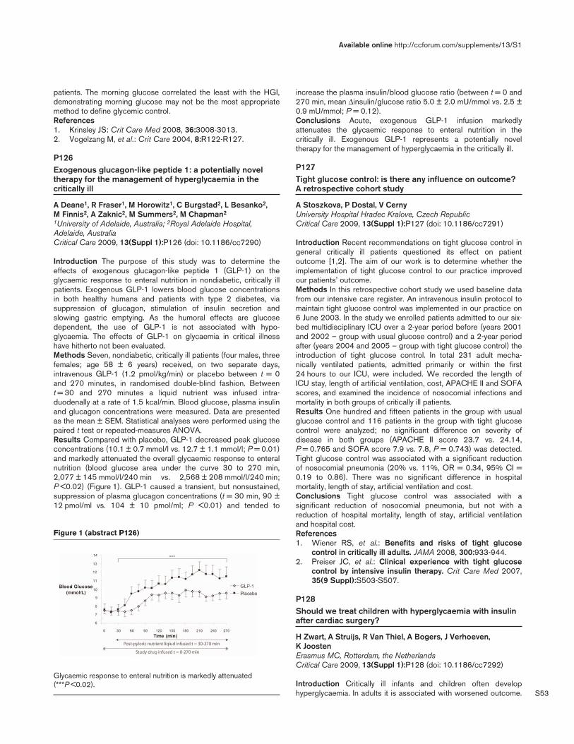
S53
patients. The morning glucose correlated the least with the HGI,demonstrating morning glucose may not be the most appropriatemethod to define glycemic control.References1. Krinsley JS: Crit Care Med 2008, 36:3008-3013.2. Vogelzang M, et al.: Crit Care 2004, 8:R122-R127.
P126Exogenous glucagon-like peptide 1: a potentially noveltherapy for the management of hyperglycaemia in thecritically ill
A Deane1, R Fraser1, M Horowitz1, C Burgstad2, L Besanko2, M Finnis2, A Zaknic2, M Summers2, M Chapman2
1University of Adelaide, Australia; 2Royal Adelaide Hospital,Adelaide, AustraliaCritical Care 2009, 13(Suppl 1):P126 (doi: 10.1186/cc7290)
Introduction The purpose of this study was to determine theeffects of exogenous glucagon-like peptide 1 (GLP-1) on theglycaemic response to enteral nutrition in nondiabetic, critically illpatients. Exogenous GLP-1 lowers blood glucose concentrationsin both healthy humans and patients with type 2 diabetes, viasuppression of glucagon, stimulation of insulin secretion andslowing gastric emptying. As the humoral effects are glucosedependent, the use of GLP-1 is not associated with hypo-glycaemia. The effects of GLP-1 on glycaemia in critical illnesshave hitherto not been evaluated.Methods Seven, nondiabetic, critically ill patients (four males, threefemales; age 58 ± 6 years) received, on two separate days,intravenous GLP-1 (1.2 pmol/kg/min) or placebo between t = 0and 270 minutes, in randomised double-blind fashion. Betweent = 30 and 270 minutes a liquid nutrient was infused intra-duodenally at a rate of 1.5 kcal/min. Blood glucose, plasma insulinand glucagon concentrations were measured. Data are presentedas the mean ± SEM. Statistical analyses were performed using thepaired t test or repeated-measures ANOVA.Results Compared with placebo, GLP-1 decreased peak glucoseconcentrations (10.1 ± 0.7 mmol/l vs. 12.7 ± 1.1 mmol/l; P = 0.01)and markedly attenuated the overall glycaemic response to enteralnutrition (blood glucose area under the curve 30 to 270 min,2,077 ± 145 mmol/l/240 min vs. 2,568 ± 208 mmol/l/240 min;P <0.02) (Figure 1). GLP-1 caused a transient, but nonsustained,suppression of plasma glucagon concentrations (t = 30 min, 90 ±12 pmol/ml vs. 104 ± 10 pmol/ml; P <0.01) and tended to
increase the plasma insulin/blood glucose ratio (between t = 0 and270 min, mean Δinsulin/glucose ratio 5.0 ± 2.0 mU/mmol vs. 2.5 ±0.9 mU/mmol; P = 0.12).Conclusions Acute, exogenous GLP-1 infusion markedlyattenuates the glycaemic response to enteral nutrition in thecritically ill. Exogenous GLP-1 represents a potentially noveltherapy for the management of hyperglycaemia in the critically ill.
P127Tight glucose control: is there any influence on outcome?A retrospective cohort study
A Stoszkova, P Dostal, V CernyUniversity Hospital Hradec Kralove, Czech RepublicCritical Care 2009, 13(Suppl 1):P127 (doi: 10.1186/cc7291)
Introduction Recent recommendations on tight glucose control ingeneral critically ill patients questioned its effect on patientoutcome [1,2]. The aim of our work is to determine whether theimplementation of tight glucose control to our practice improvedour patients’ outcome.Methods In this retrospective cohort study we used baseline datafrom our intensive care register. An intravenous insulin protocol tomaintain tight glucose control was implemented in our practice on6 June 2003. In the study we enrolled patients admitted to our six-bed multidisciplinary ICU over a 2-year period before (years 2001and 2002 – group with usual glucose control) and a 2-year periodafter (years 2004 and 2005 – group with tight glucose control) theintroduction of tight glucose control. In total 231 adult mecha-nically ventilated patients, admitted primarily or within the first24 hours to our ICU, were included. We recorded the length ofICU stay, length of artificial ventilation, cost, APACHE II and SOFAscores, and examined the incidence of nosocomial infections andmortality in both groups of critically ill patients.Results One hundred and fifteen patients in the group with usualglucose control and 116 patients in the group with tight glucosecontrol were analyzed; no significant difference on severity ofdisease in both groups (APACHE II score 23.7 vs. 24.14,P = 0.765 and SOFA score 7.9 vs. 7.8, P = 0.743) was detected.Tight glucose control was associated with a significant reductionof nosocomial pneumonia (20% vs. 11%, OR = 0.34, 95% CI =0.19 to 0.86). There was no significant difference in hospitalmortality, length of stay, artificial ventilation and cost.Conclusions Tight glucose control was associated with asignificant reduction of nosocomial pneumonia, but not with areduction of hospital mortality, length of stay, artificial ventilationand hospital cost.References1. Wiener RS, et al.: Benefits and risks of tight glucose
control in critically ill adults. JAMA 2008, 300:933-944.2. Preiser JC, et al.: Clinical experience with tight glucose
control by intensive insulin therapy. Crit Care Med 2007,35(9 Suppl):S503-S507.
P128Should we treat children with hyperglycaemia with insulinafter cardiac surgery?
H Zwart, A Struijs, R Van Thiel, A Bogers, J Verhoeven, K JoostenErasmus MC, Rotterdam, the NetherlandsCritical Care 2009, 13(Suppl 1):P128 (doi: 10.1186/cc7292)
Introduction Critically ill infants and children often develophyperglycaemia. In adults it is associated with worsened outcome.
Available online http://ccforum.com/supplements/13/S1
Figure 1 (abstract P126)
Glycaemic response to enteral nutrition is markedly attenuated(***P <0.02).

S54
No studies have so far investigated the feasibility and outcome of astandardized insulin/glucose protocol in children with congenitalheart disease after cardiac surgery.Methods We prospectively studied children with congenital heartdisease after cardiac surgery, for glucose intake, concomitantblood glucose values and results of insulin treatment. Results areexpressed as the median and range, P <0.05 consideredstatistically significant.Results Eighty-nine children were evaluated (male 56.2%), age0.9 years (1 day to 17.9 years), length of ICU stay 22 hours (2.5 to28.0 hours). All children survived. Fifty children were treated withand 39 without insulin. Overall, first blood glucose on admission tothe ICU was 5.7 mmol/l (3.1 to 21.6 mmol/l), after 6 hours was8.2 mmol/l (4.7 to 23.0 mmol/l), highest blood glucose was9.9 mmol/l (3.4 to 23.7 mmol/l), and 77.5% was hyperglycaemicduring admission (>8.0 mmol/l). Fifty out of the 70 children withhyperglycaemia were treated with insulin. Time to reach normo-glycaemia in insulin-treated children was 6.4 hours (0.3 to17.2 hours), total length of insulin treatment was 11.9 hours (2.3 to23.0 hours) and the length of hyperglycaemia was 6.7 hours (1.2to 17.0 hours) (Table 1). The duration of hyperglycaemia of treatedand untreated children was not significantly different. Hypogly-caemia (<4.0 mmol/l) occurred in 10 children (11.2%); none ofthem had severe hypoglycaemia (<2.2 mmol/l).Conclusions This study shows that a majority (77.5%) of childrenadmitted after cardiac surgery develops hyperglycaemia and72.5% were treated with insulin. The duration of hyperglycaemiawas not different between children with or without insulintreatment. It can be questioned whether beneficial effects of insulintherapy can be expected with the short duration of insulintreatment (11.9 hours), which was noticed in this study.
P129Effect of glucose–insulin–potassium infusion on mortalityin critically ill patients: a systematic review and meta-analysis
M Puskarich1, A Jones1, J Kline1, M Runyon1, S Trzeciak2
1Carolinas Medical Center, Charlotte, NC, USA; 2CooperUniversity Hospital, Camden, NJ, USACritical Care 2009, 13(Suppl 1):P129 (doi: 10.1186/cc7293)
Introduction Fifty years’ worth of published evidence has inferredbenefits of glucose–insulin–potassium (GIK) infusion to critically illpatients. We sought to measure the treatment effect of GIKinfusion on mortality in critically ill patients.Methods We conducted a systematic review of the CochraneLibrary, MEDLINE, EMBASE, CINAHL, conference proceedings,clinical practice guidelines, and other sources using acomprehensive strategy. We identified randomized controlled trialscomparing GIK treatment with standard care or placebo in criticallyill adult patients. The primary outcome variable was mortality. Twoauthors independently extracted data and assessed study qualityusing standardized instruments; consensus was reached by
conference. Preplanned subgroup analysis included studies ofhigh-quality methodology, septic shock or other circulatory shockpopulations. We used the chi-square test and the proportion oftotal variation in study estimates that is due to heterogeneity (I2) toassess for statistical heterogeneity (P <0.10, I2 >25%). Theprimary analysis was based on the random effects model toproduce pooled ORs with 95% CIs.Results The search yielded 1,720 potential publications; 23studies were included in the final analysis, providing a sample of22,525 patients. Included studies only contained populations ofacute myocardial infarction and cardiovascular surgery patients.The combined results demonstrate no statistically significantheterogeneity (P = 0.57, I2 = 0%) and no effect on mortality (OR =1.02; 95% CI = 0.93 to 1.11) with GIK treatment. Among the high-quality studies (n = 4) there was no effect on mortality (OR = 1.04;95% CI = 0.95 to 1.14). No experimental studies of shock orsepsis populations were identified.Conclusions This meta-analysis found that there is no mortalitybenefit to GIK infusion in critically ill patients; however, studypopulations were limited to acute myocardial infarction andcardiovascular surgery patients. No studies were identified utilizingGIK in patients with septic shock or other forms of circulatoryshock, providing an absence of evidence regarding the effect ofGIK as a therapy in patients with shock.
P130Incidence and risk factors of hypertriglyceridemia in theICU
MR Shams1, N Tavassoli2, N Tavassoli2, H Plicaud1, M Genestal11Purpan University Hospital, Toulouse, France; 2University ofPaul-Sabatier, Toulouse, FranceCritical Care 2009, 13(Suppl 1):P130 (doi: 10.1186/cc7294)
Introduction A linear correlation between serum triglyceride andrisk of intensive care mortality was identified. Various clinical andmetabolic situations such as sepsis, renal failure, hepatic failureand administration of certain drugs have been associated withhypertriglyceridemia (HTG). It is well known that HTG isassociated with acute pancreatitis. Development of systemicinflammatory response syndrome or the onset of organ systemdysfunction are the complications of severe acute pancreatitis thatis an episode of acute pancreatitis. The aim of this study was todetermine the incidence of HTG in patients receiving artificialnutrition in an ICU, to describe their characteristics and toestablish the relevance of risk factors associated with HTG.Methods A prospective, observational, cohort study performed in ageneral ICU of Toulouse University Hospital, Toulouse, France from1 May 2007 to 31 July 2008. We included consecutive intensivecare patients with an initial serum triglyceride level less than3 mmol/l who received at least 7 days of artificial nutrition and withat least three biochemical serum triglyceride analyses with a1-week interval. The chi-square test and a multivariate logistic
Critical Care March 2009 Vol 13 Suppl 1 29th International Symposium on Intensive Care and Emergency Medicine
Table 1 (abstract P128)
Low intake High intake Inadequate intake
Highest glucose Duration of Highest glucose Duration of Highest glucose Duration of (mmol/l) hyperglycaemia (hours) (mmol/l) hyperglycaemia (hours) (mmol/l) hyperglycaemia (hours)
Insulin 10.7 7.3 13.4 6.6 10.3 8.0
No insulin 7.5 2.7 10.3 3.1 8.1 4.8

S55
regression model were performed for statistical analysis. A total of17 clinical factors were studied as independent variables.Results A total of 107 patients were included in the study.Administration of lipid was 0.83 ± 0.36 g/kg/day. The incidence ofHTG was calculated as 17.9% per year. Multivariate analysisidentified three independent risk factors for HTG: age (P = 0.02;adjusted β-coefficient = –0.83), insulin dosage (P = 0.01; adjustedβ-coefficient = 0.83), hepatic failure (P = 0.04; adjusted β-coefficient = 1.27).Conclusions The present study showed that ICU-admittedpatients receiving artificial nutrition are prone to develop HTG.Hepatic failure and the insulin infusion rate were the mostimportant risk factors for HTG. Age had a protective effect. Ourresults raise three important matters: (1) serum triglyceridemeasurement is necessary in seriously ill patients receiving artificialnutrition; (2) elevated triglycerides reflect the degree of insulinresistance and severity of critical illness; and (3) the hyper-triglyceridemic profile of young patients admitted to the ICU is veryimportant to consider.
P131Abstract withdrawn
P132Should perioperative immune-modulating nutrition therapybe the standard of care? A systematic review
LB Weitzel1, R Dhaliwal2, J Drover2, G Schiel1, D Heyland2, W Mayles1, P Wischmeyer1
1University of Colorado, Aurora, CO, USA; 2Queens University,Kingston, ON, CanadaCritical Care 2009, 13(Suppl 1):P132 (doi: 10.1186/cc7296)
Introduction Major surgery carries a significant risk of post-operative infections, such as surgical site infections. An estimated500,000 surgical site infections occur annually at a cost of morethe $1 billion/year in the US alone. Surgical trauma leads to aninitial excessive inflammatory response, together with an almostimmediate and dramatic depression of cell-mediated immunity. Thisimmnosuppression may be due to a significant decrease in plasmaarginine levels observed following surgery. This arginine deficiencycan severely impair T-cell proliferation and key T-cell receptorfunction. Perioperative arginine administration can prevent argininedeficiency and restore cellular immunity. The purpose of this meta-analysis was to examine the relationship between immune-modulating enteral nutrition therapy (IMENT) containing arginineand infectious complications, length of stay, and mortality rates insurgical patients.Methods All prospective randomized controlled trials of arginine-containing IMENT versus standard enteral nutrition in surgical
patients conducted from 1990 to 2008 were identified frommultiple databases. Studies included in the analysis evaluatedinfectious complication, length of stay, and/or mortality rates.Methodological quality of individual studies was scored andnecessary data were abstracted in duplicate and independently.Results Thirty randomized trials with a total of 2,789 patientscompared the use of arginine-containing IMENT with standardenteral nutrition in surgical patients. Arginine-containing IMENTsignificantly decreased infectious complications (relative risk =0.58, 95% CI = 0.48 to 0.69, P <0.00001) and overall length ofstay (weighted mean difference = –2.09, 95% CI = –3.20 to–0.97, P = 0.0002) versus standard enteral nutrition. As expectedin a low-mortality surgical population, however, no effect wasobserved on mortality (relative risk = 1.06, 95% CI = 0.60 to 1.80,P = 0.84).Conclusions The cumulative results show that arginine-containingIMENT significantly reduces overall infections and length of stay insurgical patients. Based on this evidence, arginine-containingIMENT could soon become the standard of care in the surgicalpatient. This large treatment effect demands definitive evaluation ina large multicenter trial.
P133Arginine metabolism in a small animal model of sepsis andafter hemihepatectomy
B Van der Hoven1, T Teerlink2, S De Jong2, P Van Leeuwen2, J Bakker1, D Gommers1
1Erasmus University Medical Center, Rotterdam, the Netherlands;2VU University Medical Center, Amsterdam, the NetherlandsCritical Care 2009, 13(Suppl 1):P133 (doi: 10.1186/cc7297)
Introduction Asymmetric dimethylarginine (ADMA) is an inhibitorof the arginine–NO pathway. ADMA accumulates when degrada-tion in the liver by dimethylarginine dimethylaminohydrolase isimpaired. In theory, plasma citrulline, formed when arginine isconverted by NO synthase, and when ADMA is metabolized, wouldbe lowered and ornithine, formed by the degradation of arginine inthe urea cycle, would be potentially elevated when ADMAaccumulates as in sepsis and in liver failure [1].Methods Fourteen male Wistar rats were randomly allocated tolipopolysaccharide (LPS) or hemihepatectomy (HH). Plasma levelsof arginine, ADMA, citrulline and ornithine were measured beforeand 120 minutes after 5 mg/kg LPS and HH, respectively.Results See Table 1.Conclusions Plasma levels of arginine and derivatives should notbe interpreted as a reflection of metabolism at the tissue level. InHH, the elevated ADMA levels suggest dimethylarginine dimethyl-aminohydrolase activity depends on the liver tissue mass.Reference1. Wu G, et al.: Arginine metabolism: nitric oxide and beyond.
Biochem J 1998, 336:1-17.
Available online http://ccforum.com/supplements/13/S1
Table 1 (abstract P133)
Results
LPS HH
Parameter (μmol/l) t = 0 t = 120 t = 0 t = 120
Arginine 151.3 ± 28.7 99.1 ± 27.9* 138.0 ± 43.7 221.4 ± 57.1*
ADMA 1.14 ± 0.19 1.32 ± 0.24 0.59 ± 0.06 1.48 ± 0.27*
Citrulline 107.2 ± 19.3 129.1 ± 20.3* 83.5 ± 7.36 118.4 ± 18.5*
Ornithine 99.8 ± 29.2 129.6 ± 39.1* 66.4 ± 19.2 170.7 ± 50.6*
*Significant difference.
S56
P134Ginger extract dietary supplementation effects on delayedgastric emptying and ventilator-associated pneumonia inadult respiratory distress syndrome patients
M Mokhtari, Z ShariatpanahiSBU Medical Sciences, Tehran, IranCritical Care 2009, 13(Suppl 1):P134 (doi: 10.1186/cc7298)
Introduction Delayed gastric emptying is one of the major reasonsfor enteral feeding intolerance in ICU patients [1-3]. We studiedthe effect of ginger supplementation to the diet on development ofventilator-associated pneumonia (VAP) and adult respiratorydistress syndrome (ARDS) in ICU patients [2].Methods In a prospective, randomized, placebo-controlled fashion,32 ARDS mechanically ventilated patients who were fed entrallywere studied. Patients were randomized into two groups; onegroup had ginger added and the other had placebo added to theirenteral feeding. The amount of feeding tolerated in the first48 hours, the amount of feeding tolerated during the entire studyperiod, development of VAP, the number of ICU-free days, thenumber of ventilator-free days and morality were evaluated.Results Enteral feeding tolerated in the first 48 hours of study wassignificantly higher in patients with the ginger-supplemented diet(51% vs. 57%, P <0.005). However this was not different duringthe entire study period (92% vs. 93%, P = 0.42). VAP was seen in6.3% of the patients in the ginger group and in 31.3% of thecontrol group, which was not statistically significant (P = 0.08).The ICU mortality of 15.6% was similar in the two groups. Themedian number of ventilator-free days of 10 versus 7 days andICU-free days of 7 versus 4 days were significantly higher in theginger extract group, with respective P values of 0.02 and 0.04.Conclusions Supplementing the diet with ginger extract in ARDSpatients reduces the delayed gastric emptying risk and helpsreduce the duration of mechanical ventilation and increases ICU-free days.References1. Berne DJ, et al.: Erythromycin reduces delayed gastric
emptying in critically ill traumatic patients: a randomized,controlled trial. J Trauma 2002, 53:422-425.
2. Yavagal DR, Karnad DR, Oak JL: Metoclopramide for pre-venting pneumonia in critically ill patients receivingenteral tube feeding: a randomized controlled trial. CritCare Med 2000, 28:1408-1411.
3. Hoffman T: Ginger: an ancient remedy and modern miracledrug. J Environ Sci Health B 2008, 43:127-133.
P135Immunostimulated enterocytes activate extracellulararginase I which competes with enterocyte inducible nitricoxide synthase for arginine during inflammation
K Miki, R Delude, M KilleenUniversity of Pittsburgh Medical Center, Pittsburgh, PA, USACritical Care 2009, 13(Suppl 1):P135 (doi: 10.1186/cc7299)
Introduction We hypothesized that constitutively expressedarginase 1 (Arg-1) may be released from hepatocytes to the circu-latory compartment to play the role of extinguisher at the front andto serve a protective function by modulating inflammation. Weused a cell culture model of epithelial barrier dysfunction to deter-mine whether liver cytosolic proteins could decrease NO•
production and preserve enterocyte paracellular barrier function onthat basis.
Methods We exposed immunostimulated Caco 2BBe enterocyte-like cells to human liver cytosol (LC). Cytomix (IFNγ, TNFα, andIL-1β) was used to stimulate the cells. Arginase activity in cellsupernatants and murine serum was measured by following thegeneration of citrulline and urea from arginase. Tissue lysates andconditioned media were untreated or treated with the enzymeinhibitors (S)-(2-boronoethyl)-L-cysteine-HCl (BEC) and L-N(6)-(1-iminoethyl) lysine (L-NIL), which inhibit arginases and induciblenitric oxide synthase (iNOS), respectively.Results Cytomix increased paracellular permeability, and inducedthe expression of iNOS and release of NO•. LC protein(400 μg/ml) applied to the basal but not apical compartmentpreserved barrier function and completely blocked the release ofNO• but only slightly decreased the magnitude of iNOS proteinexpression in a dose-dependent and time-dependent manner.Ultrafiltration and ultracentrifugation demonstrated that microsomalArg-1 prepared from LC decreased iNOS-dependent NO•
production. BEC and anti-Arg-1 antibody inhibited the NO•
blocking ability of LC. Surprisingly, LC arginase activity requiredactivation by a cell-derived factor and its release could be blockedby treating cells with L-NIL. Increased consumption of arginase byactivated LC Arg-1 led to decreased iNOS dimerization, whichdecreased NO• production. In the serum from endotoxemic mice,arginase activity was significantly increased. Furthermore, iNOSexisted in ileal mucosa predominantly in the inactive monomericform at 18 hours after lipopolysaccharide injection, consistent withdecreased iNOS activity in the absence of arginase.Conclusions Arg-1 is one such liver-derived protein that increasesin serum during endotoxemia and other inflammatory states.Modulation of mucosal iNOS activity following activation ofcirculating Arg-1 may be protective because it would be expectedto decrease epithelial barrier dysfunction as a result of decreasedNO• production.
P136Role of parenteral glutamine supplementation on patientoutcome in the surgical ICU
R Aboelmagd, K Moez, W SalemNational Cancer Institute, Cairo, EgyptCritical Care 2009, 13(Suppl 1):P136 (doi: 10.1186/cc7300)
Introduction During the excessive organ/tissue demand ofglutamine (Gln) in episodes of stress following major surgery,endogenous Gln production may not be sufficient to meet theincreased requirements. The aim of the study was to evaluate theeffect of parenteral Gln supplementation on the outcome ofsurgical cancer patients after major surgery [1].Methods The study was performed on 40 adult patients admittedto the surgical ICU at the National Cancer Institute requiring totalparenteral nutrition (TPN) for at least 5 days. Patients wereassigned to two groups: control group, 20 patients receivednutritional support as the usual protocol; and Gln group, 20patients received nutritional support as the usual protocol +dipeptiven 300 mg/day for 7 days. Standard vitamins, traceelements, electrolytes and insulin therapy were supplied. The rateof infections, ICU and total hospital lengths of stay, severehyperglycemia and days of mechanical ventilation were recorded.Results There were no differences between groups according tothe demographic data, metabolic and nutritional parameters andthe numbers of diabetic patients. There was also no difference inthe duration of TPN. The incidence of pneumonia, surgical woundinfection, sepsis, urinary or intravenous catheter infection weresignificantly lower in the Gln group compared with the controlgroup. The days of mechanical ventilation among ventilated
Critical Care March 2009 Vol 13 Suppl 1 29th International Symposium on Intensive Care and Emergency Medicine

S57
patients and the ICU length of stay were significantly lower in theGln group compared with the control group. The nitrogen balancewas more negative in control patients than in Gln-supplementedpatients (the difference was insignificant). Hyperglycemia episodeswere significantly lower and easy controlled in the Gln group. Totalplasma amino acid concentrations and the Gln plasma levelincreased about 40% in patients receiving Gln supplementation.The number of adverse events per patient was significantly lower inthe Gln group (2.1 vs. 2.9, P <0.01).Conclusions Parenteral Gln-supplemented TPN reduces theclinical complications of surgical patients, mainly through a lowerincidence of pneumonia and better metabolic tolerance. This formsa strong rationale for the use of Gln-supplemented regimens forsurgical ICU patients.Reference1. De Sousa DA, Greene LJ: Intestinal permeability and sys-
temic infections in critically ill patients: effect of glutamine.Crit Care Med 2005, 33:1125-1135.
P137Plasma citrulline kinetics and prognostic value in thecritically ill patient
G Piton1, B Cypriani1, E Monnet1, JC Navellou1, C Manzon1, O Barbot1, F Carbonnel2, G Capellier1
1Hôpital Jean Minjoz, Besançon, France; 2Hôpital de Bicetre,Kremlin-Bicetre, FranceCritical Care 2009, 13(Suppl 1):P137 (doi: 10.1186/cc7301)
Introduction Multiple organ failure (MOF) is a frequent cause ofdeath in the critically ill patient. The gut could be the cornerstone ofMOF, the first step being an early splanchnic ischemia, inducingthe loss of barrier function, systemic infections and MOF [1].Plasma citrulline (normal 20 to 60 μmol/l) reflects a small bowelmass and is decreased in various small bowel diseases [2]. Theobjectives of the study were to study plasma citrulline kinetics andtheir prognostic value in adults hospitalized in the ICU.Methods A prospective monocentric observational study,including adults consecutively admitted to the ICU without smallbowel disease, and without chronic renal failure. We studied atonset, hour 12, hour 24, day 2, and day 7 of plasma citrulline, andclinical, biological, prognostic and therapeutic parameters. Theunivariate analysis of plasma citrulline (0 to 10 μmol/l, 11 to20 μmol/l, and >20 μmol/l) with other variables and multivariateanalysis.Results Sixty-seven patients were included, mean age 60 years,organ dysfunctions and/or infections model ODIN score of 2.4,IGS2 score (Simplified Acute Physiology Score) of 50, and a 28-day mortality of 34%. During the first day, mean plasma citrullinedecreased from 18.8 to 13.5 μmol/l, and it decreased more innonsurvivors than in survivors among patients without acute renalfailure (37% vs. 18%). A lower plasma citrulline at hour 24 wasassociated with higher plasma C-reactive protein, nosocomialinfection rate, and 28-day mortality (P = 0.006, P = 0.03 and P =0.03). A lower plasma citrulline at day 2 was associated with ahigher use of catecholamines, and a lower use of enteral feeding(P = 0.02 and P = 0.01). In multivariate analysis, plasma citrullineat hour 24 and ODIN score at admission ≥3 were associated with28-day mortality (P = 0.04 and P = 0.04).Conclusions A lower plasma citrulline at hour 24 was associatedwith higher plasma C-reactive protein, nosocomial infection rate,and 28-day mortality. A lower plasma citrulline at day 2 wasassociated with a higher use of catecholamines, and a lower use ofenteral feeding. Such results could reflect the systemic conse-quences of acute intestinal failure in patients hospitalized in the ICU.
References1. Deitch EA: Multiple organ failure. Pathophysiology and
potential future therapy. Ann Surg 1992, 216:117-134.2. Crenn P, Messing B, Cynober L: Citrulline as a biomarker of
intestinal failure due to enterocyte mass reduction. ClinNutr 2008, 27:328-339.
P138Modulation of lipid utilisation by parenteral administrationof a fish-oil-enriched new lipid formula (SMOFlipid®) insurgical ICU patients: comparison with a lipid emulsionbased on olive and soybean oil
S Piper1, T Schöllhorn1, I Schade2, R Beschmann2, K Röhm2
1Hospital of Frankenthal, Germany; 2Klinikum Ludwigshafen,GermanyCritical Care 2009, 13(Suppl 1):P138 (doi: 10.1186/cc7302)
Introduction Within the frame of postoperative total parenteralnutrition, a frequent consequence due to postaggression meta-bolism is an inhibition of lipoprotein lipase leading tohypertriglyceridemia [1]. The aim of this study was to investigatewhether the administration of a fish-oil-containing lipid emulsion(SMOF) compared with a lipid emulsion based on olive andsoybean oil led to a better utilisation of the lipids.Methods A prospective randomised study. After approval of theethical committee, 44 postoperative surgical patients with anindication for parenteral nutrition therapy were included in thestudy. Nonprotein calories were given as 60% glucose and 40%lipid emulsion. The total energy intake per day was 25 kcal/kg bodyweight. The sedation regimen was standardised (midazolam andfentanyl), propofol (a lipid emulsion) was avoided. Patients weredivided into two groups: group A (n = 22) received SMOF(SMOFlipid® 20%), and group B (n = 22) received an emulsionbased on olive and soybean oil (ClinOleic20%). Lipid emulsionswere administered for 5 days continuously. Trigylceride (TG) levelswere measured before the start of infusion (d0), at day 1 (d1), day 2(d2), and day 5 (d5) after the start of infusion. A pathological TGlevel was defined at 300 mg/dl and the significance level at P <0.05.Results There were no significant differences in TG levels atbaseline (d0: group A: 119 ± 35 vs. group B: 120 ± 45 mg /dl;P = 0.87), whereas at d2 (group A: 151 ± 52 vs. group B: 202 ±108 mg/dl; P < 0.03) and at d5 (group A: 163 ± 72 vs. group B:233 ± 94 mg/dl; P < 0.01) the TG levels in the SMOF group weresignificant lower than in the control group. At d5 the incidence ofpathological TG levels was significant lower in patients receivingSMOF (0%) compared with the control group (31.8%).Conclusions The administration of a fish-oil-containing lipidemulsion within a parenteral nutrition regimen led at d2 and d5 ofthe nutrition regimen to significantly reduced TG levels comparedwith a lipid emulsion based on olive and soybean oil, indicating abetter utilisation of the administered TGs.Reference1. Piper SN, et al.: Eur J Anaesthesiol 2008, 25:557-565.
P139Gastrointestinal function in critically ill trauma patientsusing motility capsule technology
S Rauch1, K Krueger2, N Roewer1
1University of Wuerzburg, Germany; 2University of Louisville, KY, USACritical Care 2009, 13(Suppl 1):P139 (doi: 10.1186/cc7303)
Introduction The aim of this study was to investigate the gastricemptying time and small bowel transit time, using a novel wireless
Available online http://ccforum.com/supplements/13/S1
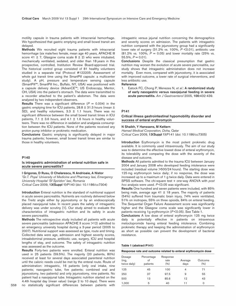
S58
motility capsule in trauma patients with intracranial hemorrhage.We hypothesized that gastric emptying and small bowel transit aredelayed.Methods We recruited eight trauma patients with intracranialhemorrhage (six male/two female, mean age 40 years, APACHE IIIscore 41 ± 7, Glasgow coma scale 8 ± 2) who were intubated,mechanically ventilated, sedated, and older than 18 years in thisprospective, controlled, Institution Review Board-approved trial.The historical control group consisted of 81 healthy volunteersstudied in a separate trial (Protocol #122205: Assessment ofwhole gut transit time using the SmartPill capsule: a multicenterstudy). A pH, pressure and temperature sensing capsule(SmartPill™; SmartPill Inc., Buffalo, NY, USA) was positioned witha capsule delivery device (AdvanCE™; US Endoscopy, Mentor,OH, USA) into the patient’s stomach. The data were transmitted toa recorder attached to the patient’s abdomen. The data wereanalyzed by two independent observers.Results There was a significant difference (P = 0.004) in thegastric emptying time for ICU patients, 28.8 ± 31.3 hours (mean ±SD), and healthy volunteers, 3.3 ± 1.1 hours. There was nosignificant difference between the small bowel transit times in ICUpatients, 7.1 ± 3.6 hours, and 4.1 ± 1.6 hours in healthy volun-teers. There was no difference in sedation and analgesia consump-tion between the ICU patients. None of the patients received anyproton pump inhibitor or prokinetic medication.Conclusions Gastric emptying is significantly delayed in majortrauma patients; however, small bowel transit times are similar tothose in healthy volunteers.
P140Is intragastric administration of enteral nutrition safe inacute severe pancreatitis?
I Grigoras, D Rusu, O Chelarescu, N Andrioaie, A Nistor'Gr.T. Popa' University of Medicine and Pharmacy Iasi, EmergencyUniversity Hospital ‘Sf Spiridon’ Iasi, RomaniaCritical Care 2009, 13(Suppl 1):P140 (doi: 10.1186/cc7304)
Introduction Enteral nutrition is the standard of nutritional supportin acute severe pancreatitis. Nutrients are routinely delivered belowthe Treitz angle either by jejunostomy or by an endoscopicallyplaced nasojejunal tube. In recent years the safety of intragastricdelivery was under scrutiny [1]. Our study aimed to evaluate thecharacteristics of intragastric nutrition and its safety in acutesevere pancreatitis.Methods The retrospective study included all patients with acutesevere pancreatitis (admission APACHE II score >12) admitted toan emergency university hospital during a 3-year period (2005 to2007). Nutritional support was assessed as type, route and timing.Collected data were age, admission and highest severity scores,intraabdominal pressure, antibiotic use, surgery, ICU and hospitallengths of stay, and outcome. The safety of intragastric nutritionwas assessed as the outcome.Results Forty-two patients were enrolled. Enteral nutrition wasused in 25 patients (59.5%). The majority (20 patients, 80%)received at least for several days associated parenteral nutritionuntil the caloric needs could be met by the enteral route. Route ofadministration: intragastric, 16 patients (only oral intake, ninepatients; nasogastric tube, five patients; combined oral andjejunostomy, two patients) and only jejunostomy, nine patients. Nopatient had a nasojejunal tube. Intragastric nutrition started on the4.4th hospital day (mean value) (range 2 to 10 days). There wereno statistically significant differences between patients with
intragastric versus jejunal nutrition concerning the demographicsand severity scores on admission. The patients with intragastricnutrition compared with the jejunostomy group had a significantlylower rate of surgery (31.2% vs. 100%, P <0.01), antibiotic use(81.2% vs. 100%, P = 0.05) and lower mortality rate (25% vs.55.5%, P = 0.01).Conclusions Despite the classical presumption that gastricnutrition may worsen the evolution of acute severe pancreatitis, ourstudy shows that intragastric administration does not increasemortality. Even more, compared with jejunostomy, it is associatedwith improved outcome, a lower rate of surgical interventions, andless antibiotic use.Reference1. Eatock FC, Chong P, Menezes N, et al.: A randomized study
of early nasogastric versus nasojejunal feeding in severeacute pancreatitis. Am J Gastroenterol 2005, 100:432-439.
P141Critical illness gastrointestinal hypomobility disorder andsuccess of enteral erythromycin
N Shaikh, Y Hanssens, M KetternHamad Medical Corporation, Doha, QatarCritical Care 2009, 13(Suppl 1):P141 (doi: 10.1186/cc7305)
Introduction Erythromycin is the most potent prokinetic drugavailable. It is commonly used intravenously. The aim of our studywas to determine the effective lowest dose of enteral erythromycin,its tolerability and comparing the dose with the severity of thedisease and outcome.Methods All patients admitted to the trauma ICU between January2004 and January 2008 who developed feeding intolerance wereincluded (residual volume >500/24 hours). The starting dose was125 mg erythromycin twice daily; if no response, the dose wasincreased up to a maximum of 1 g twice daily. Data were entered inSPSS software. The chi-square test + one-way ANOVA with posthoc analysis were used. P <0.05 was significant.Results One hundred and seven patients were included, with 85%being male, average age 41 ± 18 years. The majority of patients(54%) suffered from traumatic brain injury, 74% were ventilated,51% on inotropes, 55% on three opioids, 84% on enteral feeding.The Sequential Organ Failure Assessment score was significantlyhigher and the Glasgow coma scale was significantly lower inpatients receiving 1g erythromycin (P <0.05). See Table 1.Conclusions A low dose of enteral erythromycin 125 mg twicedaily is potentially effective in patients on intravenousmetoclopromide having enteral feeding intolerance. Combiningprokinetic therapy and keeping the administration of erythromycinas short as possible can prevent the development of bacterialresistance.
Critical Care March 2009 Vol 13 Suppl 1 29th International Symposium on Intensive Care and Emergency Medicine
Table 1 (abstract P141)
Response rate and outcome related to enteral erythromycin dose
Dosage Percentage Response (mg, of rate Average Outcome twice daily) patients (%) days (%)
125 45 100 4 71
250 37 97.5 9 55
500 13 85.6 12 43
1,000 5 40 11 0

S59
P142A randomised prospective trial to compare the efficacy ofbolus versus continuous nasogastric feeding in paediatricintensive care
P Kamath, J Longden, C Stack, A MayerSheffield Children’s Hospital, Sheffield, UKCritical Care 2009, 13(Suppl 1):P142 (doi: 10.1186/cc7306)
Introduction Failure to establish early nasogastric (NG) feeding iscommon in paediatric intensive care (PIC) and influences theoutcome. Decreased gastrointestinal motility is multifactorial inorigin. NG feeds may be administered continuously or byintermittent bolus; we hypothesise that bolus enteral feeds aremore physiological when compared with continuous NG feeds.We aimed to compare bolus versus continuous NG feeding in PIC.The outcome measures were the time to achieve maximalnutritional requirement by volume of feed and to identify thefrequency of adverse events.Methods Following ethical approval and informed consent, eligibleadmissions to a tertiary PIC from April 2006 to February 2008 wereprospectively randomised and enrolled into the study. Gastrostomyfeeding, use of motility drugs, gastroesophageal reflux andgastrointestinal surgery were the exclusion criteria. Patients wererandomised to receive either 3-hourly bolus feeds or continuousfeeding over 21 hours per day, for a 48-hour study period. NGtubes were aspirated 3-hourly and the gastric residual volume wasrecorded. The protocol was designed to give equal feed volume in a24-hour period. Intolerance was defined as gastric residual volumemore than 125% of feed administered. Data are expressed as themedian (interquartile range). Mann–Whitney’s test and Fisher’sexact test were used to test for associations.Results Seventy-six patients were enrolled (1.2:1, male:female),median age 11.6 (2.8 to 49.0) months and weight 10 (4.5 to 16.2)kg. Bolus group subjects achieved maximal feed potential by21 hours (95% CI = 18.6 to 25), the continuous group required27 hours (95% CI = 19.5 to 30, P = 0.035). The demographiccharacteristics, sedation, muscle relaxant or inotrope usagebetween groups were comparable. There was no statisticallysignificant difference in the incidence of adverse events (bolusn = 3 and continuous n = 4, P = 0.71).Conclusions This is the first study comparing enteral feedingtechniques in PIC patients. In PIC, bolus feeding methods may bemore beneficial, in achieving maximal nutritional requirementsearlier, when compared with continuous feeds. The risk of aspirationof aspiration or vomiting is low with both feeding techniques.
P143Nutrition therapy in the critical care setting: what is bestachievable practice? An international quality improvementproject
NE Jones, R Dhaliwal, X Jiang, DK HeylandQueen’s University, Kingston, ON, CanadaCritical Care 2009, 13(Suppl 1):P143 (doi: 10.1186/cc7307)
Introduction The purpose of this study was to describe currentnutrition practices in ICUs and to determine the best achievablepractice relative to the Critical Care Nutrition Clinical PracticeGuidelines (CPGs).Methods We conducted an international, prospective, obser-vational, cohort study. In January 2007, each ICU recorded data onnutrition practices on a consecutive cohort of 20 mechanicallyventilated adult patients that stayed in the ICU for at least72 hours. Data were collected from the time of admission to the
ICU to ICU discharge, or for a maximum of 12 days. Relative to theCPGs, we report average, best, and worst site performance on keynutrition practices.Results A total of 158 ICUs from 20 countries participated, andeach enrolled an average of 18.6 patients for a total of 2,946patients. Adherence to CPG recommendations was high for somerecommendations; namely, use of enteral nutrition (EN) inpreference to parenteral nutrition (PN) (site average 61.7% (range1 to 97.3%) of patients received EN alone), glycemic control (siteaverage 7.5 (range 3.5 to 10.4) mmol/l), lack of utilization ofarginine-enriched enteral formulas (site average 3.5% (range 0 to92.3%) of patients on EN), delivery of hypocaloric PN (site average16.8 (range 2.7 to 35.5) kcal/kg), and the presence of a feedingprotocol (79.7% of ICUs). However, significant practice gaps wereidentified for other recommendations. The average time to start ofEN was 46.5 hours (range 8.2 to 149.1 hours). The average use ofmotility agents and small bowel feeding in patients with highgastric residual volumes was 58.7% (range 0 to 100%) and14.7% (range 0 to 100%), respectively. There was poor compli-ance with recommendations for the use of enteral formulasenriched with fish oils, glutamine supplementation, timing ofsupplemental PN, and avoidance of soy-bean-oil-based parenterallipids. Average nutritional adequacy was 59% (range 20.5 to94.7%) for energy and 60.3% (range 18.6 to 153.5%) for protein.Conclusions Large gaps exist between the evidence-basedrecommendations and actual practice in ICUs, and consequentlynutrition therapy is suboptimal. We have identified best achievablepractice that can serve as targets for future quality improvementinitiatives.
P144Feeding enterally the hemodynamically unstable criticallyill patient: experience from a multicentre trial (theREDOXS© study)
R Dhaliwal, J Drover, J Muscedere, X Jiang, DK HeylandQueen’s University, Kingston, ON, CanadaCritical Care 2009, 13(Suppl 1):P144 (doi: 10.1186/cc7308)
Introduction The delivery of adequate enteral nutrition (EN) incritically ill patients with shock is problematic. The purpose of thisstudy is to describe how EN is delivered in patients with shockfrom the REDOXS© study, a multicentre, randomized controlledtrial of pharmaconutrition.Methods In 20 centres in Canada and Europe, we randomizedmechanically ventilated adults with two or more organ failures toone of four groups: (1) glutamine, (2) antioxidants, (3) glutamineplus antioxidants, (4) placebo. EN and parenteral nutrition wereinitiated and maintained independently as per the CanadianClinical Practice Guidelines. Shock was defined as the presenceof hypoperfusion requiring vasopressors for at least 2 hours. Dailydata including the timing of EN, volumes received, incidence ofhigh gastric residual volumes (hGRVs), use and timing of motilityagents and small bowel feeding were collected.Results From May 2007 to July 2008, 159 patients with shockwere randomized, 122 (77%) received EN only, and 13 (8%)patients received EN in combination with parenteral nutrition. ENwas started 20.2 hours (median, range 0 to 204.8 hours) after ICUadmission. The mean duration of EN was 9.2 days (median 6.8,range 0.1 to 30 days) and the mean volume of EN received was67% (range 2.5 to 199%) of that prescribed. In total 73/135(55%) had hGRVs >250 ml, and in these patients motility agentsand small bowel feeding were used in 78.1% and 41.1% ofpatients, respectively. Motility agents were started before onset ofhGRVs in 21% patients, on the same day as hGRVs in 40%
Available online http://ccforum.com/supplements/13/S1

S60
patients and on average 1.6 ± 0.9 days after hGRVs in 39%patients. The percentage mean prescribed volume of EN received24 hours before and after the start of motility agents was 35.1%vs. 55.9% (P = 0.009). Small bowel feeding was started beforeonset of hGRVs in 20% patients, on the same day as hGRVs in14% patients and was started on average 4.7 ± 3.5 (mean, SD)days after hGRVs in 67% patients. The percentage meanprescribed volume of EN received 24 hours before and after thestart of small bowel feeding was 54.2% vs. 67.3% (P = 0.36).Conclusions In critically ill patients with shock, EN can beprovided in the early phases of acute illness to the majority ofpatients. The delivery of EN in this population can be maximized bybetter adoption of motility agents and small bowel feeding.
P145Molecular adsorbent recirculating system: a clinicalexperience in acute or acute on chronic liver failure (133 sessions)
I Cardeau, L Lavayssiere, MB Nogier, O Cointault, L RostaingCHU Rangueil, Toulouse, FranceCritical Care 2009, 13(Suppl 1):P145 (doi: 10.1186/cc7309)
Introduction The molecular adsorbent recirculating system(MARS; Teraklin Industry, Rostock, Germany) is an extracorporealacute liver failure support system method using albumin-enricheddialysate to remove albumin-bound toxins.Methods Between 2004 and 2007, we performed 133 MARStreatments in 46 patients: Sequential Organ Failure Assessmentscore 11.8 ± 4.5. Indication for MARS included 16 cases of fulmi-nant hepatic failure, 21 cases of acute failure on cirrhosis, oneintractable pruritus, eight moderate or severe acute liver failure(Bernuau criteria). Among all these patients, 16 had acute renalfailure (hepatorenal syndrome). It was a retrospective study. All datawere recorded before (T0) and at the end of MARS treatment (T).Results Among 16 fulminant hepatic failure patients, we observeda significant decrease of encephalopathy (P = 0.04). On the otherhand we did not observe significant improvement of hemodynamicparameters (norepinephrine dose, mean arterial pressure), meta-bolic parameters (pH, lactate) or hepatic tests (aspartateaminotransferase, alanine aminotransferase, γ-glutamyltransferase,prothrombin time, factor V). In this subgroup, hospital mortality is31% at day 28. In the group treated for acute on chronic liverfailure, the results did not show a difference between grade ofencephalopathy before and after the session. We only observed atrend of improvement in hemodynamic and biologic parameters butthis was not statistically significant except for cholestaseparameters (γ-glutamyltransferase P = 0.022, bilirubin P = 0.002).Hospital mortality is 71% at day 28. Among the patients withhepatorenal syndrome, 62.5% were anuric. We observed asignificant increase of diuresis output (P <0.01). We did notobserve any significant adverse event.Conclusions Our results confirmed that nonbiologic hepaticsupport by MARS was safe. The results were disappointing aboveall in cirrhosis patients. Nevertheless, results in hepatorenal syn-drome were encouraging.
P146Comparison of two indwelling bowel catheters oneconomic impact by number of bedding and dressingchanges per day
Bowel Management Research Group, EC KonzHollister Incorporated, Libertyville, IL, USACritical Care 2009, 13(Suppl 1):P146 (doi: 10.1186/cc7310)
Introduction Fecal incontinence is prevalent in patients in theacute/ICU setting [1,2]. The primary objective of this study was toassess and compare the economic impact on fecal containmentwith use of catheter A or catheter B at 12 sites (A, seven sites; B,five sites) in the acute/ICU setting. Catheter A is Zassi BowelManagement System (Hollister Inc.) and catheter B is Flexi-SealFecal Management System (ConvaTec, Inc.).Methods An analysis of 146 patients (A, 76 patients; B, 70patients) on the number of bedding and dressing change visits perpatient-day (frequency of nursing visits per day spent changingbedding/dressings due to fecal contamination) can be used as anindirect economic measure of catheter leakage and containment.Routine daily bedding/dressing changes were not included, onlycatheter-related bedding/dressing changes were recorded.Results A nearly 30% reduction (1.20 vs. 1.71) in the rate ofbedding/dressing changes per patient-day were observed forcatheter A compared with catheter B (P = 0.0035). For catheter Asites, 735 bedding/dressing change visits occurred over 612patient-days; and for catheter B sites, 705 bedding/dressingchange visits occurred over 413 patient-days. Although non-significant, lower observed rates of leakage (A, 1.1; B, 1.4), reposi-tions due to leakage (A, 0.25; B, 0.39), and devices expelled (A,0.02; B, 0.07) may have contributed to the significant reduction inbedding/dressing changes associated with the use of catheter Acompared with catheter B.Conclusions The use of indwelling bowel management systems todivert, collect, and contain liquid stools may provide an economicadvantage in an acute/ICU setting for patients with fecalincontinence. These results suggest that catheter A may have agreater economic value compared with catheter B by decreasingthe number of nursing visits per patient-day.References1. Junkin J, Selekof J: Prevelance of incontinence and associ-
ated skin injury in the acute care patient. J Wound OstomyContinence Nurs 2007, 34:260-269.
2. Bliss DZ, Johnson S, Savik, Clabots CR, Gerding DN: Fecalincontinence in hospitalized patients who are acutely ill.Nurs Res 2000, 49:101-108.
P147Alterations of the immune system in acute pancreatitis andsystematic inflammatory response syndrome
V Mylona1, I Vaki2, K Lymberopoulou1, A Marioli1, A Georgopoulou1, M Lada1, E Giamarellos-Bourboulis2, G Koratzanis1
1Sismanogelion General Hospital, Athens, Greece; 2ATTIKONUniversity Hospital, Athens, GreeceCritical Care 2009, 13(Suppl 1):P147 (doi: 10.1186/cc7311)
Introduction A considerable body of evidence indicates thecontribution of the immune system to the mechanisms leading toacute pancreatitis [1]. Data focusing on the early course of eventsat the clinical ground of the process are lacking. The aim of thepresent study was to provide a complex evaluation of peripheralblood monocyte and subpopulations of lymphocytes in patients withacute pancreatitis and systemic inflammatory response syndrome.Methods Forty-three patients were enrolled in the study; 33 withacute pancreatitis and systemic inflammatory response syndrome,and 10 healthy subjects. Peripheral blood immune cells werestudied on days 1 and 4 by flow cytometry.Results Percentages of natural killer (NK) cells and of apoptosis ofCD4 lymphocytes upon diagnosis are presented in Table 1.Changes of NK cells and of monocyte apoptosis are presented inTable 2.
Critical Care March 2009 Vol 13 Suppl 1 29th International Symposium on Intensive Care and Emergency Medicine
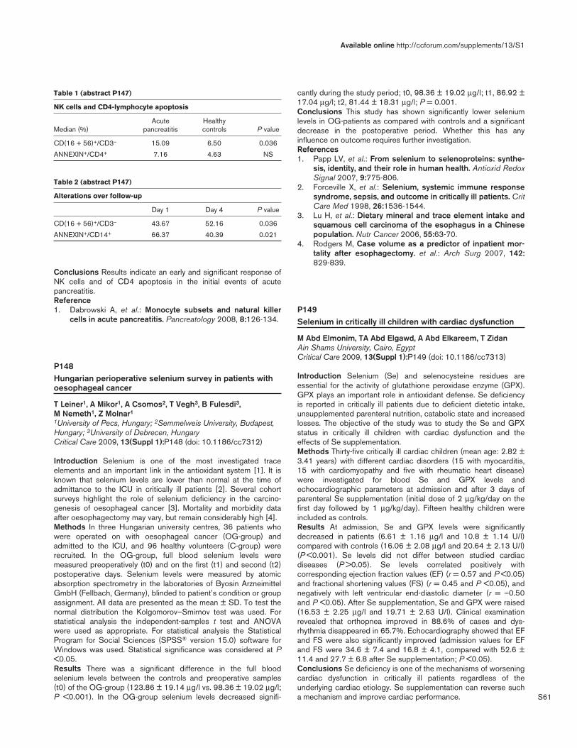
S61
Conclusions Results indicate an early and significant response ofNK cells and of CD4 apoptosis in the initial events of acutepancreatitis.Reference1. Dabrowski A, et al.: Monocyte subsets and natural killer
cells in acute pancreatitis. Pancreatology 2008, 8:126-134.
P148Hungarian perioperative selenium survey in patients withoesophageal cancer
T Leiner1, A Mikor1, A Csomos2, T Vegh3, B Fulesdi3, M Nemeth1, Z Molnar1
1University of Pecs, Hungary; 2Semmelweis University, Budapest,Hungary; 3University of Debrecen, HungaryCritical Care 2009, 13(Suppl 1):P148 (doi: 10.1186/cc7312)
Introduction Selenium is one of the most investigated traceelements and an important link in the antioxidant system [1]. It isknown that selenium levels are lower than normal at the time ofadmittance to the ICU in critically ill patients [2]. Several cohortsurveys highlight the role of selenium deficiency in the carcino-genesis of oesophageal cancer [3]. Mortality and morbidity dataafter oesophagectomy may vary, but remain considerably high [4].Methods In three Hungarian university centres, 36 patients whowere operated on with oesophageal cancer (OG-group) andadmitted to the ICU, and 96 healthy volunteers (C-group) wererecruited. In the OG-group, full blood selenium levels weremeasured preoperatively (t0) and on the first (t1) and second (t2)postoperative days. Selenium levels were measured by atomicabsorption spectrometry in the laboratories of Byosin ArzneimittelGmbH (Fellbach, Germany), blinded to patient’s condition or groupassignment. All data are presented as the mean ± SD. To test thenormal distribution the Kolgomorov–Smirnov test was used. Forstatistical analysis the independent-samples t test and ANOVAwere used as appropriate. For statistical analysis the StatisticalProgram for Social Sciences (SPSS® version 15.0) software forWindows was used. Statistical significance was considered at P<0.05.Results There was a significant difference in the full bloodselenium levels between the controls and preoperative samples(t0) of the OG-group (123.86 ± 19.14 μg/l vs. 98.36 ± 19.02 μg/l;P <0.001). In the OG-group selenium levels decreased signifi-
cantly during the study period; t0, 98.36 ± 19.02 μg/l; t1, 86.92 ±17.04 μg/l; t2, 81.44 ± 18.31 μg/l; P = 0.001.Conclusions This study has shown significantly lower seleniumlevels in OG-patients as compared with controls and a significantdecrease in the postoperative period. Whether this has anyinfluence on outcome requires further investigation.References1. Papp LV, et al.: From selenium to selenoproteins: synthe-
sis, identity, and their role in human health. Antioxid RedoxSignal 2007, 9:775-806.
2. Forceville X, et al.: Selenium, systemic immune responsesyndrome, sepsis, and outcome in critically ill patients. CritCare Med 1998, 26:1536-1544.
3. Lu H, et al.: Dietary mineral and trace element intake andsquamous cell carcinoma of the esophagus in a Chinesepopulation. Nutr Cancer 2006, 55:63-70.
4. Rodgers M, Case volume as a predictor of inpatient mor-tality after esophagectomy. et al.: Arch Surg 2007, 142:829-839.
P149Selenium in critically ill children with cardiac dysfunction
M Abd Elmonim, TA Abd Elgawd, A Abd Elkareem, T ZidanAin Shams University, Cairo, EgyptCritical Care 2009, 13(Suppl 1):P149 (doi: 10.1186/cc7313)
Introduction Selenium (Se) and selenocysteine residues areessential for the activity of glutathione peroxidase enzyme (GPX).GPX plays an important role in antioxidant defense. Se deficiencyis reported in critically ill patients due to deficient dietetic intake,unsupplemented parenteral nutrition, catabolic state and increasedlosses. The objective of the study was to study the Se and GPXstatus in critically ill children with cardiac dysfunction and theeffects of Se supplementation.Methods Thirty-five critically ill cardiac children (mean age: 2.82 ±3.41 years) with different cardiac disorders (15 with myocarditis,15 with cardiomyopathy and five with rheumatic heart disease)were investigated for blood Se and GPX levels andechocardiographic parameters at admission and after 3 days ofparenteral Se supplementation (initial dose of 2 μg/kg/day on thefirst day followed by 1 μg/kg/day). Fifteen healthy children wereincluded as controls.Results At admission, Se and GPX levels were significantlydecreased in patients (6.61 ± 1.16 μg/l and 10.8 ± 1.14 U/l)compared with controls (16.06 ± 2.08 μg/l and 20.64 ± 2.13 U/l)(P <0.001). Se levels did not differ between studied cardiacdiseases (P >0.05). Se levels correlated positively withcorresponding ejection fraction values (EF) (r = 0.57 and P <0.05)and fractional shortening values (FS) (r = 0.45 and P <0.05), andnegatively with left ventricular end-diastolic diameter (r = –0.50and P <0.05). After Se supplementation, Se and GPX were raised(16.53 ± 2.25 μg/l and 19.71 ± 2.63 U/l). Clinical examinationrevealed that orthopnea improved in 88.6% of cases and dys-rhythmia disappeared in 65.7%. Echocardiography showed that EFand FS were also significantly improved (admission values for EFand FS were 34.6 ± 7.4 and 16.8 ± 4.1, compared with 52.6 ±11.4 and 27.7 ± 6.8 after Se supplementation; P <0.05).Conclusions Se deficiency is one of the mechanisms of worseningcardiac dysfunction in critically ill patients regardless of theunderlying cardiac etiology. Se supplementation can reverse sucha mechanism and improve cardiac performance.
Available online http://ccforum.com/supplements/13/S1
Table 1 (abstract P147)
NK cells and CD4-lymphocyte apoptosis
Acute Healthy Median (%) pancreatitis controls P value
CD(16 + 56)+/CD3– 15.09 6.50 0.036
ANNEXIN+/CD4+ 7.16 4.63 NS
Table 2 (abstract P147)
Alterations over follow-up
Day 1 Day 4 P value
CD(16 + 56)+/CD3– 43.67 52.16 0.036
ANNEXIN+/CD14+ 66.37 40.39 0.021

S62
P150Effect of high-dose selenium substitution on selectedlaboratory parameters and prognosis in critically illpatients
H Brodska, A Kazda, J Valenta, J HendlUniversity Hospital, Prague, Czech RepublicCritical Care 2009, 13(Suppl 1):P150 (doi: 10.1186/cc7314)
Introduction High doses of selenium (Se) may improve thecondition of ICU patients with respect to its involvement inantioxidative protection and other tasks. In the previous work weconcluded, that the decrease of 28-day mortality is not significant.Now procalcitonin (PCT), C-reactive protein (CRP) and SequentialOrgan Failure Assessment (SOFA), as markers of the severity ofactual status, were monitored and evaluated in relation to theplasma Se concentration.Methods One hundred and forty patients were randomized intogroups A and B. Group A received standard Se substitution: 30 to75 μg/day NaSelenite intravenously; group B received Sesubstitution according to a protocol: 1,000 μg at day 1, followedwith 500 μg at days 2 to 14. These groups were divided into foursubgroups: systemic inflammatory response syndrome, sepsis,severe sepsis and septic shock. Plasma levels of Se, CRP andPCT were examined. SOFA and 28-day mortality were evaluatedas clinical markers. Relations between parameters were evaluatedstatistically. In both groups, transitions among subgroups duringtreatment were evaluated. All patients monitored less than threetimes were excluded.Results Negative correlations between Se x PCT, Se x SOFA andSe x CRP were found (Table 1). Correlation coefficients (r) werestatistically significantly higher in group B. Evaluation of transitionsamong subgroups during treatment showed there was a differencein subgroup septic shock between groups A and B. Into the lesssevere stage of sepsis moved 28% in group A (14 examinations)and 44% (16 examinations) in group B.
Table 1 (abstract P150)
Negative correlations
Parameter Group A Group B Significance
Se x PCT r = –0.24 r = –0.29 P < 0.05
Se x CRP r = –0.41 r = –0.53 P < 0.05
Se x SOFA r = –0.29 r = –0.03 NS
Conclusions There were more significant negative correlationsbetween plasmatic Se and parameters of inflammation in the groupof patients supplemented with high doses of Se. In accordancewas a finding of more frequent transition from septic shock to aless severe stage of sepsis. Even if a decrease of mortality wasagain not found, the presented results may perhaps indicate apositive metabolic influence in critical illness.
P151Dynamic generation of physiological model systems
J Kretschmer1, A Wahl2, J Guttmann2, K Möller1
1Furtwangen University, Villingen-Schwenningen, Germany;2University Hospital Freiburg, GermanyCritical Care 2009, 13(Suppl 1):P151 (doi: 10.1186/cc7315)
Introduction Mathematical models are widely used to simulatephysiological processes in the human body and can be exploitedfor diagnostic purpose or the automation of therapeutic measures
[1]. Usually these models focus on one single aspect of the humanphysiology. Complex models with interaction between differentphysiological processes usually do not consist of interchangeablesubmodels. We therefore designed a versatile software based onMatlab with dynamically exchangeable subsystems within the threemodel families of respiratory mechanics, gas exchange andcardiovascular dynamics.Methods For each submodel the parameters have been extractedfrom the corresponding literature. Common interfaces were definedfor each model family based on these parameters to ensureinterchangeability within the same model family. For simulation ofhuman body gas exchange we used a two-compartment modelwith oxygen and carbon dioxide dissociation curves. The modelfamily of cardiovascular dynamics consisted of a single-compartment model and a six-compartment model including theresponse to pleural pressure. The respiratory mechanics werebased on a first-order resistance-compliance model and a second-order resistance-compliance model. Model parameters were fit tohuman test data or to data taken from the literature. Simulation isexecuted using a dedicated caller program to combine theselected submodels and to execute them subsequently at eachtime step. Submodel selection and basic parameter specificationcan be done via a graphical user interface. The software wastested with different model combinations. Each combination wassupplied with alterations in ventilation frequency and positive end-expiratory pressure.Results Simulation based on parameters from the literature withthe variations described above showed plausible results. Altera-tions in ventilation frequency indicated a response time consistentwith data acquired by Jensen and colleagues [2].Conclusions The developed software is able to simulate differentcombinations of submodels at variable complexity. Simulationresults are consistent with experimental data. Interaction betweensubmodels can be seen in the simulation output.References1. Lozano S, Moller K, Brendle A, et al.: AUTOPILOT-BT: a
system for knowledge and model based mechanical venti-lation. Technol Health Care 2008, 16:1-11.
2. Jensen MC, Lozano S, Gottlieb D, et al.: An evaluation ofend-tidal CO2 change following alteration in ventilationfrequency [abstract]. In MBEC Antwerpen Conference;2008.
P152Intraaortic balloon pumping: why should we hurry up?
A Macas, A Mundinaite, G BaksyteKaunas University of Medicine, Kaunas, LithuaniaCritical Care 2009, 13(Suppl 1):P152 (doi: 10.1186/cc7316)
Introduction Application of the intraaortic balloon pump (IABP) forpatients with acute myocardial infarction complicated by cardio-genic shock is undoubted. But the time of IABC initiation isquestionable. The goal of the study was to evaluate the influence ofthe IABP initiation time and dose of dopamine for patient hemo-dynamic data and inhospital mortality.Methods Sixty-two consecutive acute myocardial infarctionpatients managed with IABP were included in the study. Theinitiation time of IABP and administered dopamine doses werecompared. Two subgroups of patients were separated: thosereceiving less than 10 μg/kg/minute dopamine, and others receiv-ing 10 μg/kg/minute dopamine or more. Standard hemodynamicindices of cardiac output (CO), cardiac index (CI), stroke volume(SV), stroke index (SI), pulmonary capillary wedge pressure(PCWP), mean pulmonary artery pressure (PAmean), cardiac
Critical Care March 2009 Vol 13 Suppl 1 29th International Symposium on Intensive Care and Emergency Medicine
S63
power (CP = CO x MAP / 451, where MAP is mean arterialpressure) and cardiac power index (CPI = CI x MAP / 451) weremeasured.Results CO in the first group was 3.58 ± 1.12 l/minute, incomparison with 2.54 ± 1.08 l/minute (P <0.0001) in the secondgroup. CI correspondingly was 1.9 ± 0.6 l/minute/m2 and 1.38 ±0.61 l/minute/m2 (P = 0.001), SV was 43.8 ± 26.14 ml and30.01 ± 13.75 ml (P = 0.012), SI was 23.93 ± 15.58 ml/m2 and16.28 ± 7.75 ml/m2 (P = 0.017), CP was 0.68 ± 0.25 W and0.4 ± 0.23 W (P <0.0001), CPI was 0.36 ± 0.14 W/m2 and0.22 ± 0.13 W/m2 (P <0.0001), PCWP was 21.32 ± 6.04 mmHgand 24.16 ± 7.42 mmHg (P = 0,104), PAmean was 28.74 ±8.53 mmHg and 30.23 ± 7.05 mmHg (P = 0.47). In the secondsubgroup, hemodynamic indices (CO, CI, SV, SI, CP and CPI)were statistically significantly lower (P <0.05) in comparison withthe data of patients receiving less than 10 μg/kg/minute, whilePCWP and CVP values differed insignificantly. Inhospital mortalitywas higher among patients receiving 10 μg/kg/minute dopamineor more. Among 31 patients receiving less than 10 μg/kg/minutedopamine 16 (51.6%) died, while among those 31 receiving10 μg/kg/minute or more 27 (87.1%) died. This was a significantdifference according to the Pearson χ2 criterion (χ2 = 9.182,P = 0.002).Conclusions Initiation of aortic counterpulsation should beconsidered as soon as possible, while the patient with acutemyocardial infarction is treated with low doses of vasopressors.
P153Study of risk factors for female patients with acutemyocardial infarction
H Fu1, Y Zhao2
1China Rehabilitation Research Center, Beijing, China; 2GeronticCardiovascular Disease Institution, Beijing, ChinaCritical Care 2009, 13(Suppl 1):P153 (doi: 10.1186/cc7317)
Introduction Acute myocardial infarction (AMI) is one of the mostcommon cardiovascular emergencies. Female patients havedifferent features to male patients. The objective of our study wasto analyze risk factors for female patients with AMI.Methods Five hundred and eighty female patients were comparedwith 2,058 male patients for age, occupation, positive familyhistory, type 2 diabetes mellitus, hypertension, and hyperlipemia.Then, according to age, patients with AMI were divided into fourgroups: <55 years, 55 to 64 years, 65 to 74 years, ≥75 years. Riskfactors were compared between female patients and male patientsin each group.Results Compared with male patients, female patients were older(P <0.01); The morbidity of type 2 diabetes mellitus and hyper-tension and the rate of physical labors were significantly higher infemale patients (P <0.01, P <0.01, P <0.01); the morbidity ofhyperlipemia and the rate of mental labors and positive familyhistory were significantly lower in female patients (P <0.01,P <0.01, P <0.05). In the <55 years group, the morbidity of type 2diabetes mellitus and hypertension and the rate of physical laborswere significantly higher in female patients (P <0.05, P <0.01,P <0.01); the rate of mental labors were significantly lower infemale patients (P <0.01). In the 55 to 64 years group, themorbidity of type 2 diabetes mellitus and hypertension and the rateof physical labors were significantly higher in female patients(P <0.01, P <0.05, P <0.01); the rate of mental labors was signifi-cantly lower in female patients (P <0.01). In the 65 to 74 yearsgroup, the morbidity of type 2 diabetes mellitus and hypertensionand the rate of physical labors were significantly higher in femalepatients (P <0.05, P <0.01, P <0.01); the rate of mental labors
was significantly lower in female patients (P <0.01). In the≥75 years group, the rate of physical labors was significantly higherin female patients (P <0.01); the rate of mental labors wassignificantly lower in female patients (P <0.01).Conclusions Compared with male patients, female patients withAMI were older, and type 2 diabetes mellitus and hypertensionprobably played more important roles in female patients. A higherrate of physical labors and a lower rate of mental labors in femalepatients probably contribute to prevent them from AMI.
P154Influence of arterial pressure on tissue perfusion in septicshock
E Tishkov, O BukaevMoscow State Medical University, Moscow, Russian FederationCritical Care 2009, 13(Suppl 1):P154 (doi: 10.1186/cc7318)
Introduction The aim of this study was to measure the effects ofincreasing mean arterial pressure (MAP) on systemic oxygenmetabolism and regional tissue perfusion in septic shock.Methods Twenty patients with the diagnosis of septic shock whorequired pressor agents to maintain a MAP ≥65 mmHg after fluidresuscitation to a pulmonary artery occlusion pressure (PAOP)≥12 mmHg were included. Norepinephrine was titrated to MAPs of65 mmHg, 75 mmHg and 85 mmHg in 20 patients with septicshock.Results At each level of MAP, hemodynamic parameters (heartrate, PAOP, cardiac index, left ventricular stroke work index, andsystemic vascular resistance index), metabolic parameters (oxygendelivery, oxygen consumption, blood lactate), and regionalperfusion parameters (gastric mucosal PCO2, skin capillary bloodflow and red blood cell velocity, urine output) were measured.Increasing the MAP from 65 to 85 mmHg with norepinephrineresulted in increases in cardiac index from 3.5 ± 0.4 l/min/m2 to5.0 ± 0.5 l/min/m2 (P < 0.03). Blood lactate was 3.5 ± 0.8 mEq/lat a MAP of 65 mmHg and 3.0 ± 0.8 mEq/l at 85 mmHg (P = NS).The gradient between arterial PCO2 and gastric intramucosalPCO2 was 11 ± 3 mmHg (1.5 ± 0.3 kPa) at a MAP of 65 mmHgand 15 ± 3 mmHg at 85 mmHg (2.0 ± 0.3 kPa, P = NS). Urineoutput at 65 mmHg was 40 ± 10 ml/hour and was 45 ± 12 ml/hourat 85 mmHg (P = NS). As the MAP was raised, there were nosignificant changes in skin capillary blood flow or red blood cellvelocity.Conclusions Increasing the MAP from 65 mmHg to 85 mmHgwith norepinephrine does not significantly affect systemic oxygenmetabolism, skin microcirculatory blood flow, urine output, orsplanchnic perfusion.
P155Echocardiographic assessment of the effects of acute leftventricular pacing on patients with severe congestive heartfailure and narrow QRS duration
K Hussein, H Elaassar, D Ragab, H Elattroush, R Soliman, H KhaledCairo University, Cairo, EgyptCritical Care 2009, 13(Suppl 1):P155 (doi: 10.1186/cc7319)
Introduction More than 20% of patients with congestive heartfailure (CHF) exhibit one form or another of mechanicaldyssynchrony, intraventricular conduction impairment, or bundlebranch block. The concept of dual-chamber pacing in refractoryheart failure was introduced to be followed later by the techniqueof biventricular pacing to restore cardiac synchrony in the failing
Available online http://ccforum.com/supplements/13/S1
S64
heart. The aim of the present study was to address the issue ofapplying the technique of left ventricular (LV) pacing to thatsubstrate of heart failure patients with a narrow rather than wideQRS complex, and with LV rather than biventricular pacing in orderto permit the use of an ordinary dual-chamber pacemaker.Methods We conducted an acute study on 20 patients (15 male,five female; mean age 43 years); all had CHF (12 ischemic andeight idiopathic) with normal QRS duration on ECG. All patientswere under maximal tolerated doses of antifailure treatment. Allpatients were subjected to M-mode and two-2D echocardiographyto measure: left ventricular end-diastolic dimension (LVEDD), leftventricular end-systolic dimension (LVESD), fractional shortening(FS), ejection fraction (EF), mitral regurge area and cardiac outputbefore and 15 minutes after LV pacing. All patients were subjectedto temporary dual-chamber right atrium, LV pacing; the LV leadwas passed retrogradely via the transaortic route. The pulmonarycapillary wedge pressure (PCWP) was measured using a trilumen,balloon-tipped thermodilution Swan–Ganz catheter. Patients weredivided into group I (PCWP >15 mmHg, 10 patients) and group II(PCWP <15 mmHg, 10 patients).Results Echocardiographic measurements after pacing in group Ishowed significantly lower LVEDD (5.12 vs. 6.53 cm, P <0.004),lower LVESD (4.01 vs. 4.65 cm, P <0.034), smaller mitral regurgearea (9.7 vs. 13.4 cm2, P <0.005), higher FS (18.9 vs. 17,P <0.04) and higher EF (37.9 vs. 35.5%, P <0.02). In contrast,following pacing in group II, the hemodynamics were notsignificantly different from pre-pacing values.Conclusions Single LV – rather than biventricular – pacing couldachieve remarkable hemodynamic beneficial effects in patients withCHF even with normal QRS, but only in that substrate of patientswith a high PCWP. Although this is an acute study, our findingsopen the scope for widespread application of the concept ofmultisite pacing.
P156Left ventricular pacing alone improves haemodynamicvariables
D RgabCairo University, Cairo, EgyptCritical Care 2009, 13(Suppl 1):P156 (doi: 10.1186/cc7320)
Introduction Despite advances in drug treatment, congestive heartfailure (CHF) remains a major healthcare problem associated witha poor quality of life and a high mortality rate. During the pastdecade, cardiac resynchronisation therapy (CRT) using biventri-cular (BIV) pacing emerged as a promising technique improvingthe quality of life, exercise tolerance and mortality in patients withsevere CHF. Left univentricular (LV) pacing is able to achieve thesame mechanical synchronisation as BIV pacing in experimentalstudies and in humans resulting in significant improvement infunctional class, quality of life and exercise tolerance at the sameextent as those observed with BIV stimulation. The aim was tostudy the acute hemodynamic effects of LV pacing in the categoryof patients with severe CHF and QRS duration <130 ms and todetermine whether pulmonary capillary wedge pressure (PCWP)>15 mmHg could have an impact on these changes in hemo-dynamics.Methods We conducted an acute study on 20 patients (15 male,five female; mean age 43 years); all had CHF (12 ischemic andeight idiopathic) with a QRS duration <130 ms. All patients had anejection fraction <40%. Group I comprised 10 patients withPCWP >15 mmHg, group II comprised 10 patients with PCWP<15 mmHg. Haemodynamics were measured using a Swan–Ganz
catheter at baseline and during LV VDD pacing. All patients wereunder maximal tolerated doses of antifailure treatment.Results After LV pacing, group I showed a significant decrease inright atrial pressure (P = 0.001), and PCWP also significantlydecreased (P = 0.0001), while the cardiac output ignificantlyincreased (P = 0.04). In group II none of these hemodynamicparameters showed any significant improvement. The improvementof hemodynamics in group I occurred despite the greater increasein QRS duration after LV pacing when compared with group II (24vs. 21%, respectively).Conclusions LV pacing acutely benefits CHF patients with PCWP>15 mmHg irrespective of the QRS duration. Although this is anacute study, our findings open the scope for revising the currentwide QRS duration as an indication for CRT and to consider theless expensive LV rather than BIV pacing when a patient isconsidered a candidate for resynchronization.
P157Preoperative left atrial dysfunction and new-onset atrialfibrillation in cardiac surgery patients
M Brouard, JJ Jimenez, J Iribarren, L Lorente, R Perez, L Lorenzo, S Palmero, L Raja, N Perez, R Martinez, ML MoraHospital Universitario de Canarias, La Laguna SC, Tenerife, SpainCritical Care 2009, 13(Suppl 1):P157 (doi: 10.1186/cc7321)
Introduction Postoperative atrial fibrillation is one of the mostfrequent complications after cardiopulmonary bypass (CPB). Theaim of the present study was to investigate the correlation betweenpreoperative left atrial dysfunction assessed by tissue Doppler andpostoperative new-onset atrial fibrillation (NOAF) after coronaryartery bypass grafting (CABG).Methods Preoperative transthoracic echocardiography Dopplerwas performed on elective cardiac surgery patients. Left atrialfunction was evaluated with tissue Doppler imaging of the mitralannulus.Results We studied 92 patients, 73 (79%) males and 19 (21%)females, mean age 67 ± 10 years, in preoperative sinus rhythmwho underwent elective CABG surgery under CPB. Nineteenpatients (20.6%) developed NOAF at 34 ± 12 postoperativehours. Patients with NOAF were older (71 ± 7 vs. 66 ± 10 years,P = 0.034), had a larger left atrial diameter (LAD), lower peak atrialsystolic mitral annular Doppler velocity (A′m) and higher E′/A′ ratioin a bivariate analysis. Stepwise logistic regression analysisshowed that LAD (OR = 2.23, 95% CI = 1.05 to 4.76; P = 0.033)and lower A′m (OR = 0.70, 95% CI = 0.55 to 0.99; P = 0.034)were independently associated with postoperative NOAF.Conclusions A preoperative left atrial dysfunction assessed bytissue Doppler imaging may identify the patients at risk of post-operative NOAF.
P158Right ventricular involvement in Takotsubocardiomyopathy
MM Moeller, J Voelz, C Lenz, J Wicke, R Gradaus, J Neuzner, J NeuznerKlinikum Kassel, GermanyCritical Care 2009, 13(Suppl 1):P158 (doi: 10.1186/cc7322)
Introduction Takotsubo syndrome (TS) is characterized by atransient apical ballooning of the left ventricle, reversible ST-T-segment abnormalities and mildly elevated troponin withoutcoronary artery stenosis, mimicking myocardial infarction (MI).Originally described as a disturbance of the left ventricle, recently
Critical Care March 2009 Vol 13 Suppl 1 29th International Symposium on Intensive Care and Emergency Medicine
S65
an involvement of the apex of the right ventricle (RV) has beenrecognized affecting approximately one-quarter of cases.Methods TS was diagnosed in 10 patients (nine female, one male;mean age 67 years) who displayed typical signs of acutemyocardial ischemia, showed typical ECG changes and slightlyelevated troponin I (0.8 ng/ml; n ± 3.5 ng/ml; normal <0.3 ng/ml).Echocardiography was obtained on admission in all patients. TSwas presumed by identification of the typical LV apical ballooningconfiguration (echo positive). In 10 patients left heart catheteri-zation was then performed immediately.Results All patients displayed LV apical ballooning on LV angio-graphy without coronary artery lesions. Nine patients were echopositive (90%; sensitivity 91%, specificity 100%); in the one echo-negative patient the LV angiogram showed only a very small apicalballooning area. In all of the nine patients classified as echopositive, involvement of the RV apex was identified as well.Additional hypokinesia of the middle part of the RV was seen intwo patients; however, the basal RV wall segments were nevercompromised. One patient showed dynamic LV outflow tractobstruction.Conclusions RV involvement was a common feature in TS,involving 90% of patients. In all cases, the apical portion of the RVwas compromised, the basal segments were never affected. Inmost patients the typical wall motion disturbance was readily seenon transthoracic echocardiogram. RV involvement was identified inall echo-positive patients. Probably, identification of RV apicalballooning may aid in differentiating TS from MI.
P159Possible cause of bradycardia developed due toαα2-sympothomimetic infusion
K Popugaev, I Savin, A Goriachev, A Oshorov, A Troitskiy, P KalininNeurosurgical Research Institute N.N. Burdenko, Moscow, RussianFederationCritical Care 2009, 13(Suppl 1):P159 (doi: 10.1186/cc7323)
Introduction Bradycardia is described as a side effect of α2-sympothomimetic infusion. It is supposed as a dose-dependentphenomenon. Cholinergic antagonists or β-sympathomimetics arerecommended for the bradycardia correction. We propose that theseverity of bradycardia associated with α2-sympothomimeticinfusion depends on the level of hypothyroidism. The aim of thisreport is to provide evidence for our point of view.Methods Fifteen patients after sellar region tumor surgery wereincluded in the study. Resistant arterial hypotension developed inthe early postoperative period in all patients and they weremonitored with a Swan–Ganz catheter for this reason. Thehormonal profile (triiodothyronine, thyroxine, free triiodothyronine,free thyroxine, and cortisol) was investigated daily during the wholeperiod of hemodynamic monitoring.Results The cause of resistant arterial hypotension in all caseswas decreased vascular tone (systemic vascular resistance index =1,503 ± 624 dyn·s·cm5·m2), and so phenylephrine as the α2-sympothomimetic was the drug of choice. Administration ofphenylephrine started with the mean dose of 2.9 μg/kg/minute,and the maximal mean dose during the period of arterialhypotension was 5 μg/kg/minute. Bradycardia developed in fourpatients during infusion of phenylephrine. All these patients hadclinical signs of hypothyroidism (hypothermia, dynamic ileus, etc.)and decreased levels of T3, T4, free T3 and free T4. In order tocorrect bradycardia, the infusion of phenylephrine was combinedwith cholinergic antagonists and β-sympathomimetics. Simul-taneously, dose administration of thyroid hormones was increased.
Heart rates reached the normal range, when clinical signs ofhypothyroidism were corrected, and the level of thyroid hormonestended to rise. In 11 patients without clinical signs of hypo-thyroidism and normal levels of thyroid hormones in plasma,bradycardia never developed even if they received high doses ofphenylephrine (5.5 μg/kg/min).Conclusions Bradycardia, associated with α2-sympothomimeticinfusion, is a consequence of hypothyroidism in patients after sellarregion tumor surgery. This phenomenon is not dose dependent.Patients with bradycardia, developed after beginning α2-sympotho-mimetic infusion, need to be screened for hypothyroidism. Ifhypothyroidism is confirmed, therapy with L-thyroxin should beadministrated immediately. In patients having received thyroidhormones, their doses need to be increased.
P160Automatic real-time detection of myocardial ischemia byepicardial accelerometer
P Halvorsen1, E Remme1, A Espinoza1, L Hoff2, H Skulstad1, T Edvardsen1, E Fosse1
1Rikshospitalet University Hospital, Oslo, Norway; 2VestfoldUniversity College, Tønsberg, NorwayCritical Care 2009, 13(Suppl 1):P160 (doi: 10.1186/cc7324)
Introduction Epicardial accelerometers have proved to detectmyocardial ischemia with high sensitivity [1]. In this combinedexperimental and clinical study using an epicardial accelerometer,we aimed to test two methods for real-time automated detection ofmyocardial ischemia.Methods One accelerometer (5 x 5 x 2 mm3) was sutured in theperfusion area of the left anterior descending artery (LAD).Epicardial acceleration was simultaneously recorded with ECG,and the ECG QRS complex was automatically detected for timingof systole. From the epicardial acceleration, signal circumferentialpeak velocity and displacement was automatically calculated withina time interval of 150 ms after peak R on the ECG. Experimentalmodel: in 10 open-chest pigs, regional left ventricular function wasreduced by temporary LAD occlusion and global myocardialfunction changed by esmolol infusion. The myocardial circum-ferential strain measured by echocardiography was used to confirmischemia. Clinical model: the accelerometer methods were testedin seven patients, receiving coronary artery bypass grafting. LADwas occluded for 3 minutes before grafting and the accelerometermeasurements were compared for hemodynamics, ECG ST-segment analysis and strain by transoesophageal echocardio-graphy.Results Accelerometer systolic displacement was superior tosystolic velocity to detect ischemia by automated analysis.Accelerometer systolic displacement demonstrated dyskinesiaduring LAD occlusion in pigs (11.5 ± 2.3 to –1.2 ± 2.8 mm,P <0.01), while hypokinesia was found in patients (12.8 ± 8.1 to3.5 ± 4.4 mm, P <0.01). Ischemia was confirmed by strain echo-cardiography in both models (P <0.01). No significant changes inhemodynamics and in the ECG ST segment were seen during LADocclusion in patients. In the experimental model, esmolol infusioninduced fewer changes in the automated accelerometermeasurements than LAD occlusion (P < 0.01) and ischemia wasdetected with a sensitivity of 100% and specificity of 95 to 100%.Conclusions Sensitive real-time detection of myocardial ischemiawas feasible by the use of automated analysis of continuousepicardial accelerometer signals. This technique may improve real-time detection of ischemia during and after cardiac surgery.Reference1. Halvorsen PS, et al.: Br J Anaesth 2009, 102:29-37.
Available online http://ccforum.com/supplements/13/S1

S66
P161Prognosis of acute myocardial infarction outcomes usingevaluation of cardiac power (product of cardiac output andmean arterial pressure)
A Macas, A Krisciukaitis, V Saferis, G Baksyte, A Mundinaite, V SemenaiteKaunas University of Medicine, Kaunas, LithuaniaCritical Care 2009, 13(Suppl 1):P161 (doi: 10.1186/cc7325)
Introduction Insufficient reliability and specificity of cardiac output(CO) as a widely used parameter for prognosis of acute myocardialinfarction (AMI) outcomes led to investigations and a search fornew methods and parameters. Cardiac power (CP) (a parameterproportional to the product of CO and mean arterial pressure) wasintroduced after studies mainly performed using the invasiveintermittent thermodilution (ITD) technique. The aim of this studywas to investigate the reliability and specificity of the newparameter mainly by means of noninvasive techniques such asimpedance cardiography (ICG).Methods CO and CP were evaluated by both ITD and ICGmethods in patients with AMI, admitted within 12 hours from theonset of pain. CP was evaluated using the suggested formula:CP = CO x MAP / 451, where MAP = mean arterial pressure.During the period of 2004 to 2008, 289 (196 men and 93 women)patients were investigated. The standard eight-electrode ICGregistration was used. The optimal binning method using theminimal description length principle was used to predict outcomesafter AMI: inhospital mortality, survival after 6 months and survivalafter 12 months.Results CP evaluated on the first day was found as the onlyvaluable prognostic parameter using the model entropy method inthe group of patients where noninvasive evaluation of CO wasused. Inhospital mortality was predicted with single cut point 0.65,sensitivity 100% and specificity 92.2%, while survival of 12 monthswas predicted with single cut point 0.90, sensitivity 88.9% andspecificity 73.0%. Only prediction of inhospital mortality waspossible in the group of patients where CO was evaluated usingthe ITD technique. The most significant criteria using minimizedentropy model were CP evaluated on the third day (single cut point0.79, sensitivity 84.6%, specificity 100.0%) and CO evaluatedalso on the third day (single cut point 4.00, sensitivity 84.6%,specificity 100.0%).Conclusions Cardiac power is a reliable predictor for inhospitalmortality and survival within the first year after acute myocardialinfarction. It could be evaluated using ITD and with sufficientaccuracy by means of a noninvasive method – ICG – as well.
P162Left ventricular torsion analysis using echocardiographicspeckle tracking in a canine model of dyssynchrony andcardiac resynchronization therapy predicts global cardiacperformance
B Lamia, M Tanabe, HK Kim, J Gorcsan, MR PinskyUniversity of Pittsburgh Medical Center, Pittsburgh, PA, USACritical Care 2009, 13(Suppl 1):P162 (doi: 10.1186/cc7326)
Introduction Left ventricular (LV) torsion is a primary mechanismused to eject blood during systole. We hypothesized that LVtorsion is impaired during dyssynchronous contractions andrestored with cardiac resynchronization therapy (CRT) inproportion to the degree that global LV performance improves.Methods Seven anesthetized open-chest dogs had high fidelitypressure and conductance volume catheters to assess LV
performance. Basal and apical Grayscale echo images wererecorded. Right atrial (RA) pacing served as the control. Rightventricular (RV) outflow tract pacing created a left bundle branchblock (LBBB) to simulate dyssynchrony. Simultaneous RV–LV freewall and RV–LV apex pacing were modeled CRT (CRTfw andCRTa). Torsion was estimated as the difference between apicaland basal rotation in degrees.Results Torsion during RA pacing was 7.0 ± 3.6°. RV pacingdecreased torsion (5.1 ± 3.6°, P <0.05 vs. control), reduced thestroke volume (SV), stroke work (SW), and dP/dtmax comparedwith RA (21 ± 5 vs. 17 ± 5 ml, 252 ± 61 vs.151 ± 64 mJ, and2,063 ± 456 vs. 1,603 ± 424 mmHg/s, P <0.05). CRTa improvedtorsion, SV, SW and dP/dtmax compared with RV pacing (7.7 ±4.7°, 23 ± 3 ml, 240 ± 50 mJ and 1,947 ± 647 mmHg/s, respec-tively, P <0.05), whereas CRTfw did not (5.1 ± 3.6°, 18 ± 5 ml,175 ± 48 mJ and 1,699 ± 432 mmHg/s, respectively, P <0.05)(Figure 1). Changes in torsion compared with RA covaried withchanges in SW during RV, CRTa and CRTfw.Conclusions LV torsion, as quantified by speckle trackingechocardiography in an acute canine model, was reduced bydyssynchrony contraction and restored by CRT in proportion to thedegree to which global measures of LV performance alsoimproved. Thus, torsion and global LV performance are linkedduring synchronous, dyssynchronous contractions and CRT.
P163Cardiac diseases during pregnancy and periperium: 13 years in the ICU
K Baccar, N Baffoun, C KaddourNational Institute Neurology, Tunis, TunisiaCritical Care 2009, 13(Suppl 1):P163 (doi: 10.1186/cc7327)
Introduction Pregnancy, labour and the postpartum periodconstitute major stresses on the cardiovascular system. Patientswith heart disease may decompensate due to the physiologicchanges that occur during pregnancy and may develop a cardiacevent with obstetric events.Methods A retrospective collection of data for all obstetric patientsadmitted to our ICU in a 13-year period (September 1995 toSeptember 2008). Data collected include the antepartum orperipartum heart status (abnormal finding on physical examination,ECG and echocardiography), reason for admission, stay on theICU prognostic scoring (APACHE II, Obstetrical SAPS) and theoutcome.Results During 13 years there were 42 obstetric admissions to ourICU. The reason for admission was due to pulmonary edema 16(38.1%) cases, hemorrhagic shock 11 (26.2%) cases, cardiogenicshock 8 (19%) cases, pulmonary emboli 4 (9.5%) cases, stroke 2(4.8%) cases and recent myocardic infarction 1 (2.4%) case.
Critical Care March 2009 Vol 13 Suppl 1 29th International Symposium on Intensive Care and Emergency Medicine
Figure 1 (abstract P162)

S67
Admission was unplanned for 36 (85.7%) parturients followingemergency caesarean section. Status of the heart was severeaortic or mitral stenosis 21 (50%) cases, mitral or aorticregurgitation with heart failure 5 (11.9%) cases, mechanicalprosthetic valve requiring anticoagulation 8 (19%) cases, myo-cardial infarction 1 (2.4%) case and peripartum cardiomyopathy 7(16.6%) cases. The median duration of stay was 4 days (range 1to 11 days), median Obstetrical SAPS was 19.3 (range 10 to 32)and APACHE II score was 13 (range 8 to 32). There were five(11.9%) maternal deaths, due to two cases PPCM, one cerebraldeath and two prothesis thrombus.Conclusions The majority of the cases of parturients wasunbooked for antenatal care; this leads us to acknowledge theexistence of risk factors related to pregnancy. Maternal functionalclass is an important predictor of outcome; a high index ofsuspicion for cardiac diseases is essential to identify risk [1], oncethese patients are referred for a cardiologic opinion there is aneeded for cardiologist to develop a systematic approach to theirevaluation. Ideally these considerations should be commencedduring prepregnancy consultations, but continued throughoutpregnancy and the postpartum period [1].Reference1. Siu, Colman JM: Cardiovascular problems and pregnancy:
an approach to management. Clev Clin J Med 2004, 71:977-985.
P164Endothelial nitric oxide synthase deficiency and induciblenitric oxide synthase inhibition in the setting of septiccardiomyopathy
A Van de Sandt1, R Windler1, A Gödecke2, J Ohlig1, S Becher1,E Van Faassen3, T Rassaf1, J Schrader2, M Kelm1, M Merx1
1Med. Clinic I, University Hospital RWTH Aachen, Germany;2University of Düsseldorf, Germany; 3Debye Institute ofNanomaterials Science, Utrecht, the NetherlandsCritical Care 2009, 13(Suppl 1):P164 (doi: 10.1186/cc7328)
Introduction Nitric oxide (NO) plays a central role in the patho-genesis of septic cardiomyopathy. However, the relative contri-bution of inducible nitric oxide synthase (iNOS) and endothelialnitric oxide synthase (eNOS) remains unclear. The aim of this studyis to elucidate the influence of eNOS and iNOS on cardiacfunction, NO production rate and survival in the clinically relevantpolymicrobial cecum ligation and puncture (CLP) model of sepsis.Methods B6/c57 wildtype (WT) and eNOS–/– mice were renderedseptic by CLP or were sham operated. Immediately, the selectiveiNOS inhibitor 1400W (6.6 mg/kg body weight intraperitoneallyand subcutaneously) or carrier were applied. At 12 hours aftersepsis induction, heart function was assessed by pressure–volumeloops (1.4 Fr Millar catheter). NOx levels and endogenous NOproduction (blood plasma/heart tissue) were measured via gas-phase chemiluminescence detection, high-performance liquidchromatography and electron paramagnetic resonance spectro-scopy. To evaluate apoptosis and inflammation, quantitativeimmunochemistry was applied. iNOS expression was analyzed viaRT-PCR.Results Cardiac function was significantly impaired solely in septicWT mice with diminished left ventricular developed pressure/dPdtmax(dPdtmax: WT CLP = 10,981 ± 1,100 mmHg/s vs. WT sham =13,408 ± 827 mmHg/s; P <0.01) and increased left-ventricularvolumes. Inhibition of iNOS in septic WT mice resulted inameliorated cardiovascular impairment, whereas no signs of septiccardiomyopathy were observed in septic eNOS–/–. In contrast toseptic eNOS–/– mice, septic WT mice developed a significant
increase in NO3–, NO2– and bioactive NO levels. eNOS deficiency
was associated with diminished apoptosis and modifiedinflammation. In eNOS–/– mice, a decreased iNOS expression wasobserved compared with septic WT mice. Both genetic eNOSdeficiency and pharmacologic iNOS inhibition was associated witha significant survival benefit. While eNOS–/– mice survived longest,additional iNOS inhibition in the latter diminished this benefitsignificantly.Conclusions In this clinically relevant model of sepsis, eNOSconstitutes an important factor mediating septic cardiomyopathy.To what extent the diminished inflammation, apoptosis, NOproduction rate, iNOS expression and prolonged survival ineNOS–/– mice contribute to the observed benefits remains to beclarified in future studies.
P165Use of levosimendan in myocardial dysfunction due tosepsis
J Vaitsis1, H Michalopoulou1, C Thomopoulos2, S Massias2, P Stamatis1
1‘Metaxa’ Hospital, Athens, Greece; 2‘Elena Venizelou’ Hospital,Athens, GreeceCritical Care 2009, 13(Suppl 1):P165 (doi: 10.1186/cc7329)
Introduction Myocardial dysfunction observed within the contextof sepsis is partially due to desensitization of the cardiac muscle tovasoactive agents. Levosimendan, a calcium sensitizer and a K+-ATP channel opener, plays a significant role in the treatment ofmyocardial depression although its use in sepsis has not yet beenestablished.Methods We studied 42 patients (66.1 ± 7.54 years, 24 male)who met the criteria for sepsis (infection by Gram-negative bacteriaand at least two SIRS criteria) and displayed severe heartdysfunction (cardiac index (CI) ≤2.2, ejection fraction (EF) ≤35%).The APACHE II score was 21.2 ± 4.9. Patients were randomizedto receive additionally to their standard treatment levosimendan(0.1 μg/kg/minute 24-hour infusion) (group A, n = 23) withoutloading dose or dobutamine (5 to 10 μg/kg/minute 24-hour infusion)(group B, n = 19). Noradrenaline was used to preserve the meanarterial pressure above 65 mmHg. The primary goal was mortalityat 7 and 30 days.Results Mortality at 7 and 30 days was 30% and 60% in group Aversus 36% and 68% in group B (P = 0.05, P = 0.03, respec-tively). The CI and EF significantly increased in group A (ΔCI:1.79 ± 0.16 vs. 1.4 ± 0.12, P = 0.027; ΔEF: 4.8 ± 0.2% vs. 3.5 ±0.7%, P = 0.04), and also SvO2 (P = 0.012) and mean arterialpressure (P = 0.035) were significantly increased.Conclusions Levosimendan compared with dobutamine improvesthe hemodynamic profile of septic patients with myocardial dys-function; and while there is evidence that it reduces mortality,further studies are needed to verify this.
P166Haemodynamic effects of levosimendan following cardiacsurgery
A Slezina, E Strike, M Bekers-AnchipolovskisPauls Stradins Clinical University Hospital, Riga, LatviaCritical Care 2009, 13(Suppl 1):P166 (doi: 10.1186/cc7330)
Introduction Myocardial contractile function following cardiacsurgery often requires inotropic support. Traditionally, phospho-diesterase inhibitors and catecholamines are used [1,2]. Levo-simendan is a novel inotropic agent–calcium sensitizer that
Available online http://ccforum.com/supplements/13/S1

S68
enhances myocardial contractility without increasing intracellularcalcium and myocardial oxygen demand [3]. The aim of our studywas to evaluate circulation following levosimendan infusion, to findout the efficiency of tissue oxygen supply and to evaluate otherinotrope dosage rates during levosimendan infusion.Methods Thirteen patients with acute heart failure followingelective cardiac surgery under cardiopulmonary bypass treatedwith levosimendan and other inotropes were enrolled.Haemodynamic parameters (mean arterial pressure, central venouspressure, mean pulmonary artery pressure, cardiac index), PaO2,SvO2, plasma lactate, cardiac troponin I and other inotropes rateswere obtained at baseline, 30 minutes, 1 hour, 6 hours and24 hours after the start of levosimendan infusion. Levosimendanwas administered at a rate of 0.1 μg/kg/minute (no bolus) with atotal 24-hour dose of 12.5 mg for each patient. Other inotropicagents used were epinephrine (range 0.03 to 0.15 μg/kg/min),norepinephrine (range 0.03 to 0.18 μg/kg/min), dobutamine (range3.5 to 7 μg/kg/min) and corotrope (range 0.3 to 0.6 μg/kg/min).Results Thirteen patients (nine female, four male) were investi-gated. Forty-six per cent of patients had III to IV (New York HeartAssociation) stage of congestive heart failure (ejection fraction<40%). Six patients died within 24 hours and were excluded.Maximal changes occurred 24 hours after levosimendan infusion.Mean arterial pressure increased, while the mean pulmonary arterypressure and central venous pressure decreased. The cardiacindex did not change considerably. Oxidative stress markersimproved. Mean infusion rates of epinephrine, dobutamine andcorotrope decreased, while the norepinephrine mean rate did notchange.Conclusions Levosimendan improves circulation by means of itspositive inotropic effect, increases mean arterial pressure, anddecreases pulmonary pressure. Levosimendan could be a drug ofchoice in myocardial hypoxia, probably combined with right heartfailure.References1. Hardy JF, et al.: J Cardiothorac Vasc Anesth 1993, 7(Suppl
2):33-39.2. Orime Y, et al.: Jpn Circ J 1999, 63:117-122.3. Raja SG, et al.: Ann Thorac Surg 2006, 81:1536-1546.
P167Effects of levosimendan started 24 hours beforecardiopulmonary bypass in patients admitted to the ICUwith an ejection fraction lower than 30%
J Duchateau1, H Vanden Eede2
1ZNA Middelheim, Antwerp, Belgium; 2HAGA Hospital, The Hague, the NetherlandsCritical Care 2009, 13(Suppl 1):P167 (doi: 10.1186/cc7331)
Introduction Patients with poor left ventricular function requireinotropic drug support after cardiopulmonary bypass (CPB). Theuse of levosimendan in these patients is associated with a betterpostoperative function [1]. A recent study has attributed thisphenomenon to preconditioning effects of levosimendan [2].However, a recent study showed no benefit in starting levo-simendan before CPB [3]. We hypothesized that admitting thepatient to intensive care 24 hours before CPB and starting levo-simendan preoperatively could reduce postoperative cardiacdamage.Methods Fifty patients with an ejection fraction less than 30%scheduled for elective cardiac surgery with CPB receivedanesthesia with propofol, cisatracurium and sufentanil. The patientswere randomly assigned to two protocols. Group A: levosimendan0.1 μg/kg/minute started before CPB after induction + dobutamine
5 μg/kg/minute started after the release of the Aox. Group B:levosimendan 0.1 μg/kg/minute started on the ICU 24 hoursbefore CPB + dobutamine 5 μg/kg/minute started after the releaseof the Aox. Data analysis: SV, dobutamine, noradrenaline time anddose, cardiac enzymes, global hemodynamic variables, ICU andhospital length of stay. Statistical significance was accepted atP 0.05.Results There was no difference in postoperative stroke volumeand troponin I. The incidence of postoperative atrial fibrillation inboth groups was very low. There was a significant difference inICU length of stay but not in hospital length of stay.Conclusions In cardiac surgery patients with a low preoperativeejection fraction there is no difference in cardiac function andpostoperative troponin I levels between the two differentlevosimendan treatment modalities. The incidence of postoperativeatrial fibrillation is low when levosimendan is used perioperatively.There is a significant reduction in the ICU length of stay, but not inthe hospital length of stay.References1. De Hert SG: Anesth Analg 2007, 104:766-773.2. Tritapepe L: Br J Anaesth 2006, 96:694-700.3. De Hert SG, et al.: J Cardiothorac Vasc Anesth 2008, 22:
699-705.
P168Utilization of levosimendan in the cardiac ICU: case series
D Filipescu, M Luchian, A Prodea, O Gheanu, A Calugareanu, S Marin, H Moldovan, A Iosifescu, O ChioncelIBCV, Bucharest, RomaniaCritical Care 2009, 13(Suppl 1):P168 (doi: 10.1186/cc7332)
Introduction Levosimendan, a novel calcium sensitizer, has beenshown to improve hemodynamic function in patients with acuteheart failure [1]. The aim of the study was to assess the hemo-dynamic effects of levosimendan as a rescue medication inaddition to conventional therapy in patients with low cardiac output(LCO) after cardiac surgery or myocardial infarction.Methods Forty-one patients with LCO admitted to the cardiac ICUbetween June 2004 and November 2008 were included in thisobservational hemodynamic study. Thirty-four patients wereadmitted after open heart surgery and seven patients for ischemicacute heart failure. Levosimendan (0.1 μg/kg/min x 24 hours,without bolus) was added to conventional inotropes and/or intra-aortic balloon pump (IABP) support. The measured parameterswere: cardiac output/index, pulmonary artery occlusion pressure(PaoP), left ventricular ejection fraction (LVEF), and mixed venousoxygen saturation (SvO2). Baseline data were collected beforelevosimendan administration and the following datasets wereobtained at 6, 24 and 48 hours. Length of stay (LOS) in thecardiac ICU and inhospital mortality were also registered. Datawere expressed as the mean ± SD. Fisher’s exact test and anonpaired t test were used when appropriate. P <0.05 wasconsidered significant.Results All patients were on dobutamine and epinephrine. IABPsupport was used in 28 (68%) of cases. The addition of levo-simendan significantly improved cardiac index (from 2.27 ± 0.9l/min/m2 to 3.05 ± 0.9 l/min/m2 at 6 hours (P <0.01); 2.93 ± 0.8l/min/m2 at 24 hours (P <0.02) and 2.93 ± 1.0 l/min/m2 at48 hours (P <0.05)). LVEF increased with 19.8% (from 30 ± 8%to 37 ± 9% at 48 hours (P <0.05)). SvO2 improved significantlyfrom 57 ± 13% to 69 ± 8% at 48 hours (P <0.01). PaoPdecreased significantly only in the first 6 hours from 19 ± 5 mmHgto 15 ± 4 mmHg (P <0.05). The mean LOS in the cardiac ICU was14 ± 13 days. Inhospital mortality was 24.4%.
Critical Care March 2009 Vol 13 Suppl 1 29th International Symposium on Intensive Care and Emergency Medicine

S69
Conclusions In our case series, addition of levosimendan followingineffective conventional therapy resulted in substantial hemo-dynamic improvement. These preliminary results support the use oflevosimendan in patients with LCO as a rescue medication withfavorable short-term effects.Reference1. Raja SG, et al.: Ann Thorac Surg 2006, 81:1536.
P169Effects of levosimendan and inhaled nitric oxide onmicrocirculation in septic shock
A Morelli1, A Donati2, C Ertmer3, S Rehberg3, B Bollen Pinto3, A Orecchioni1, H Van Aken3, P Pelaia2, P Pietropaoli1, M Westphal31University of Rome, Italy; 2Marche Polytechnique University,Ancona, Italy; 3University of Muenster, GermanyCritical Care 2009, 13(Suppl 1):P169 (doi: 10.1186/cc7333)
Introduction Microvascular resuscitation is a crucial therapeuticgoal in sepsis. The current study was performed to test thehypothesis that a combination of levosimendan and inhaled nitricoxide (INO) may improve microvascular perfusion in septic shock.Methods After initial hemodynamic stabilization (mean arterialpressure between 65 and 75 mmHg; mixed venous oxygensaturation ≥65%), seven patients with catecholamine-dependentseptic shock received intravenous levosimendan 0.2 μg/kg/minutefor 24 hours. At the end of the first 24 hours of the study period,inhaled nitric oxide (35 ppm) was added for another 12 hours.Sublingual microvascular perfusion was analyzed using the side-stream dark field method. The total vessel density (mm/mm2),perfused vessel density (mm/mm2), De Backer score (1/mm),microcirculatory flow index of small vessels (MFIs) and micro-circulatory flow index of medium vessels (MFIm) were obtained atbaseline and after 24 and 36 hours.Results Levosimendan significantly (P <0.05 vs. baseline)increased perfused vessel density from 11.3 mm/mm2 (10.7; 12.6)to 14.8 mm/mm2 (13.7; 16.1) and MFIs from 2 (1.9; 2.2) to 3 (2.8;3). Addition of INO further increased MFIm from 2.6 (2.5; 2.8) to 3(3; 3). Data are presented as median (25%; 75% range). Nostatistically significant differences were found in any of the otherinvestigated parameters.Conclusions The combination of Levosimendan and INO mayimprove microvascular perfusion in septic shock.
P170Effects of levosimendan on renal function in septic shock:a case–control study
A Morelli1, C Etmer2, S Rehberg2, A Orecchioni1, N Cannuovacciuolo1, B Bollen Pinto2, M Lange2, H Van Aken2,A Donati3, P Pietropaoli1, M Westphal21University of Rome, Italy; 2University of Muenster, Germany;3Marche Polytechnique University, Ancona, ItalyCritical Care 2009, 13(Suppl 1):P170 (doi: 10.1186/cc7334)
Introduction Nonhemodynamic mechanisms of cell injury mightplay a role in the loss of glomerular filtration rate during sepsis [1].We hypothesized that levosimendan may positively affect renalfunction by a combination of systemic and regional hemodynamic,anti-inflammatory and anti-apoptotic effects. We thereforeperformed a case–control study to investigate the effects of levo-simendan on creatinine clearance in patients with septic shock.Methods Ninety-nine septic shock patients received levosimendan(0.2 μg/kg/min for 24 hours) within the first 36 hours following
onset of septic shock. For each study patient, a control subjectfrom a group of patients with septic shock of an institutionaldatabase was matched for Simplified Acute Physiology Score II,baseline creatinine concentration, delay from shock onset, age,and gender. Serum creatinine concentrations were analyzed justbefore the start of the 24-hour period of levosimendan infusion(baseline) and 96 hours after levosimendan had been initiated. Theglomerular filtration rate was estimated by applying theCockcroft–Gault formula.Results Compared with the control cohort, levosimendansignificantly increased the glomerular filtration rate after 96 hours(62 ± 46 vs. 50 ± 33 ml/min, P < 0.05). In addition, the maximumserum creatinine concentration was lower in the levosimendangroup (2.2 ± 1.3 vs. 2.6 ± 2 mg/dl, P <0.05 vs. control) during the96-hour study period.Conclusions The present data suggest that levosimendan mayimprove renal function in patients with septic shock.Reference1. Wan L, et al.: Pathophysiology of septic acute kidney
injury: what do we really know? Crit Care Med 2008, 36:S198-S203.
P171Levosimendan versus dobutamine in septic shock
JA Alhashemi, Q AlotaibiKing Abdualziz University, Jeddah, Saudi ArabiaCritical Care 2009, 13(Suppl 1):P171 (doi: 10.1186/cc7335)
Introduction Levosimendan is a calcium sensitizer that increasescardiac contractility without increasing intracellular calcium levels.Its efficacy has been demonstrated in acute decompensated heartfailure but has not been evaluated in severe sepsis/septic shock.We hypothesized that levosimendan increases the cardiac indexsimilar to dobutamine in patients with severe sepsis/septic shock.Methods In a randomized, open-label trial, 42 patients admitted tothe ICU with severe sepsis/septic shock were randomized toreceive either levosimendan (group L) or dobutamine (group D) aspart of an early-goal directed therapy protocol [1]. Study drugswere titrated incrementally to an ScvO2 ≥70% or to a maximumdose, whichever was achieved first, and were continued for a totalof 24 hours only. Group L received levosimendan 0.05 μg/kg/minute intravenously that was increased by 0.05 μg/kg/minuteevery 30 minutes (maximum 0.2 μg/kg/min). Group D receiveddobutamine 5 μg/kg/minute intravenously which was increased by5 μg/kg/minute every 30 minutes (maximum 20 μg/kg/min). Rescuetherapy consisted of dobutamine 10 μg/kg/minute intravenouslytitrated to ScvO2 ≥70% or a maximum of 20 μg/kg/minute, which-ever was achieved first. Hypotension (mean arterial pressure(MAP) <65 mmHg) was treated with norepinephrine infusion,titrated to a MAP ≥65 mmHg. ScvO2 was recorded hourly. Cardiacoutput was measured continuously using the FloTrac™ device(Edwards Lifesciences, Irvine, CA, USA). Continuous data wereanalyzed using repeated-measures ANOVA, and proportions werecompared using Fisher’s exact test. Results are presented as themean ± SD unless otherwise indicated. Significance was definedas P <0.05.Results APACHE II scores were 21 ± 7 versus 27 ± 7 (P = 0.02),and the ICU mortality was 10 (48%) versus 13 (62%) (P = 0.35)for groups L and D, respectively. The cardiac index was lower ingroup L compared with group D (estimated marginal mean ± SEM:2.8 ± 0.1 vs. 3.2 ± 0.1, respectively, P <0.01) but the ScvO2changes over time did not differ between groups (P >0.5).Norepinephrine was administered to 17 (81%) patients in group Land 21 (100%) in group D (P = 0.04).
Available online http://ccforum.com/supplements/13/S1
S70
Conclusions Dobutamine increased the cardiac index more thandid levosimendan, although there was no difference between thetwo drugs with regard to their effects on ScvO2.Reference1. Rivers E, et al.: N Engl J Med 2001, 345:1368-1377.
P172Levosimendan therapy does not improve survival of post-resuscitation cardiogenic shock patients
P Soos1, D Becker1, G Barczi1, G Szabo1, E Zima2, G Fulop1, L Geller1, A Apor1, B Merkely1
1Heart Center, Budapest, Hungary; 2Semmelweis University,Budapest, HungaryCritical Care 2009, 13(Suppl 1):P172 (doi: 10.1186/cc7336)
Introduction The calcium sensitizer levosimendan enhancesmyocardial contractility, which could be advantageous in patientswith myocardial ischemia requiring inotropic support.Methods During 3 years 3,852 patients with high-risk acutecoronary syndrome (ACS) underwent percutaneous coronaryintervention in our department. In 106 cases ACS was compli-cated with cardiogenic shock (mean age: 68.6 ± 1.2 years); more-over, in 26 cases patients had to be resuscitated (cardiopulmonaryresuscitation (CPR)). Short-term and long-term effects oflevosimendan on cardiac functions and on survival of cardiogenicshock and post-CPR patients were analyzed. Levosimendan wasadministrated in 39 of 106 cases as add-on therapy for patientswith impaired left ventricular function, by extensive wall motionabnormality and by high blood cardiac enzyme concentration.Levosimendan therapy was started in most cases on the second orthird day and applied for 6 hours as a continuous infusion (0.1μg/kg/min). The mean time spent in the primary cardiac care center(inhospital time) was 6.0 ± 0.4 days, and the whole follow-up was204.6 ± 29.9 days long.Results In the post-CPR patient group there was no significantdifference in survival according to levosimendan treatment duringshort-term follow-up (36.5% vs. 40.0%, P = 0.790) and duringlong-term follow-up (15.6% vs. 15.0%, P = 0.754). On the otherhand, for nonresuscitated patients the survival rates were signifi-cantly higher in the levosimendan-treated patient group duringshort-term follow-up (84.4% vs. 57.9%, P <0.001) and duringlong-term follow-up (47.3% vs. 23.0%, P <0.001). The time inter-val between the onset of myocardial infarction and percutaneouscoronary intervention did not influence the effect of levosimendanon short-term and long-term mortality.Conclusions In summary, levosimendan may improve cardiacfunction and decrease short-term and even long-term mortality incardiogenic shock patients independently of the time interval ofmyocardial infarction. This positive effect of levosimendan may beabolished by post-CPR patients.
P173Study of hemodynamics in patients treated with landiololin the ICU
T Imabayashi, H Murayama, C Kuroki, N Kiyonaga, T Oryouji, S Tashiro, T Yasuda, Y Kakihana, A Matunaga, Y KanmuraKagoshima University Hospital, Kagoshima, JapanCritical Care 2009, 13(Suppl 1):P173 (doi: 10.1186/cc7337)
Introduction We experienced patients whose cardiac functionswere maintained or deteriorated during the treatment of a short-acting β-blocker, landiolol. We therefore investigated the effects oflandiolol on the cardiac function in patients.
Methods From January through December 2007, 21 patients wereselected if they had tachycardia (heart rate (HR) >100 beats/min)and they were treated with continuous infusion of landiolol.Hemodynamics were recorded using pulmonary artery catheters atfour time points (before administration of landiolol, 1 hour afterbeginning of administration, immediately before discontinuation ofadministration, and 1 hour after discontinuation of administration).Infusion of landiolol was discontinued if the HR fell below90 beats/min or the cardiac index (CI) fell below 2.0 l/min/m2. Thepaired Student t test was used to compare the differences.P <0.05 was statistically significant.Results Between the point before administration of landiolol (2.4 ±1.8 (mean ± SD) μg/kg/min) and 1 hour after beginning of adminis-tration, although the HR was decreased from 137 ± 20 beats/minto 109 ± 20 beats/min (P <0.01), the stroke index (SI) wasincreased from 20.4 ± 8.1 ml/beats/m2 to 22.5 ± 7.0 ml/beats/m2,and thus the CI was maintained (2.76 ± 1.05 l/min/m2 and 2.43 ±0.78 l/min/m2). Between the point before discontinuation ofadministration (1.4 ± 1.1 μg/kg/min) and 1 hour after discontinua-tion of administration, the HR was not increased significantly (93 ±18 beats/min and 100 ± 16 beats/min), however the SI wasincreased 21.5 ± 6.7 ml/beats/m2 to 25.7 ± 5.4 ml/beats/m2
(P <0.05), and therefore the CI was increased 1.93 ± 0.54l/min/m2 to 2.50 ± 0.40 l/min/m2 (P <0.01). Infusion of landiololwas discontinued because the HR fell below 90 beats/min in 12patients and the CI fell below 2.0 l/min/m2 in nine cases. Therewere no significant differences in the catecholamine index,pulmonary artery pressure, and central venous pressure.Conclusions We investigated hemodynamics in 21 patients whoused landiolol and recognized the effects of rate control anddepression of the SI and the CI; thus when we use landiolol, wehave to pay attention to the HR and cardiac function [1].Reference1. Goto K, Shingu C, Miyamoto S, et al.: The effect of landiolol on
hemodynamics and left ventricular function in patients withcoronary artery disease. J Clin Anesth 2003, 19:523-529.
P174Effects of intravenous diltiazem on a porcine model ofendotoxin-induced pulmonary hypertension
M Kyparissa, V Grosomanidis, K Kotzampassi, K Karakoulas, A Kolettas, B Fyntanidou, C SkourtisUniversity of Thessaloniki Medical School, Thessaloniki, GreeceCritical Care 2009, 13(Suppl 1):P174 (doi: 10.1186/cc7338)
Introduction Pulmonary hypertension (PAH) is a life-threateningdisease commonly seen in ICU septic patients. PAH is characterizedby alterations in the pulmonary circulation leading to right ventricularfailure and is associated with poor outcome in such patients. Variousagents such as nitric oxide, prostaglandins and phosphodiesteraseinhibitors have been used for its treatment. Drugs that inducepulmonary vasodilatation, increase contractility and maintain a stablehaemodynamic profile seem an attractive treatment option in acutePAH patients. However, there is lack of evidence-based guidelines inthe current medical literature. Although oral diltiazem has beenshown to improve haemodynamics in chronic PAH patients, it hasnot been used for the treatment of acute PAH. The aim of thepresent study was to evaluate the effects of intravenous diltiazem ona porcine model of acute PAH during sepsis.Methods PAH was induced in 16 anaesthetized, mechanicallyventilated pigs (25 kg) by intravenous infusion of 0.5 mg/kg LPS(Escherichia coli, 111: B4) in a period of 30 minutes. After LPSadministration, animals were randomly divided into two groups.Group A received intravenous diltiazem 280 to 400 μg/kg, and
Critical Care March 2009 Vol 13 Suppl 1 29th International Symposium on Intensive Care and Emergency Medicine
S71
Group B an equal dose of placebo (normal saline) and served asthe control. Haemodynamic measurements, including parametersof systemic and pulmonary circulation, were performed before andafter LPS administration and every 20 minutes for 2 hours.Results After LPS infusion, both systolic pulmonary pressure(PAPs) and diastolic pulmonary pressure (PAPd) exhibited astatistically significant increase (PAPs from 18 ± 1 to 48 ± 7 mmHg,P <0.01 and PAPd, from 7.7 ± 0.7 to 27 ± 4 mmHg, P <0.01) andremained elevated over time. Diltiazem administration reducedsignificantly both PAPs and PAPd in relation to placebo(P <0.001), but not to baseline levels. The heart rate, cardiacoutput and systemic haemodynamics exhibited no differencebetween groups throughout the time period.Conclusions Intravenous diltiazem was associated with areduction of pulmonary pressure without any systemic hypotensionin a porcine model of endotoxin-induced acute PAH. This mayrepresent a significant advance in the treatment of acute PAH.Potential clinical implications merit further study.
P175Acute liver failure induced by intravenous amiodarone inthe cardiac care unit: retrospective study of 3 years
E Zima, V Szabo, I Osztheimer, T Barany, D Becker, L Molnar, P Soos, L Geller, B MerkelySemmelweis University, Budapest, HungaryCritical Care 2009, 13(Suppl 1):P175 (doi: 10.1186/cc7339)
Introduction Amiodarone is the antiarrhythmic drug of choice intreatment of patients suffering from acute tachyarrhythmias andhaemodynamic instability due to impaired cardiac function. Theintravenous form of amiodarone hydrochloride (IvAm) has individualantiarrhythmic, rate-controlling efficacy, and the dosage isempirical. The main adverse effects are hypotension, severebradycardia, asystole, acute heart failure, and impaired liverfunction. Acute liver failure (ALF) is a known but rare complicationof IvAm that may be reversible by eliminating the infusion in mostcases. The few papers in the literature suppose ALF may becaused by polysorbate 80, the vehicle of IvAm. Oral administrationdoes not to have such an adverse effect, therefore IvAm can bechanged to oral form in any cases.Methods Our aim was to investigate retrospectively the incidenceof ALF and relation of IvAm and ALF in cardiac patients. Thehistory, treatment sheets and laboratory parameters of 11,722patients treated in the Heart Center between 2005 and 2007 wereanalyzed. Patients were considered severe ALF patients iftransaminase levels exceeded 80 x upper limit of normal (ULN)during their stay in our clinic. The cutoff point was determined todifferentiate ALF patients from heart failure and myocardial infarctpatients with elevated transaminase levels.Results According to the enzyme levels, 55 patients suffered fromsevere ALF during the 3 years; 26 of them had IvAm treatment. Onthe basis of treatment sheets, start and elimination of IvAm treatment,status of acute myocardial infarct and heart failure and transaminasekinetics, eight patients had ALF induced by IvAm. Indication foramiodarone was atrial fibrillation (n = 6) and ventricular tachycardia.Average multipliers of ULN were 379 ± 190 at aspartateaminotransferase, 191 ± 87 at alanine aminotransferase, 57 ± 22 atlactate dehydrogenase. The time from start of IvAm to detection ofALF was 17 ± 4.6 hours. One-quarter of these patients died in ALF.Liver enzymes decreased to 10 x ULN during 2.5 ± 0.6 days.Conclusions ALF is a rare but potentially life-threatening adverseeffect of IvAm. Authors suggest monitoring liver enzymes from thestart of IvAm treatment. Rapid elevation in liver enzyme levels indi-cates a hepatotoxic effect of IvAm. In these cases the immediate
cessation of IvAm administration and start of intensive care is life-saving.
P176Prevention of postoperative arrhythmias after pulmonaryresection: celiprolol versus magnesium
S Ouerghi1, K Moncer2, N Frikha2, M Ben Ammar2, M Mebazaa2
1Hospital A Mami, Ariana, Tunisia; 2Mongi Slim Hospital, La Marsa,TunisiaCritical Care 2009, 13(Suppl 1):P176 (doi: 10.1186/cc7340)
Introduction Incidence of arrhythmia after lobectomies is between10% and 20% and approaches 40% after pneumonectomy.Postoperative arrhythmia is associated with higher morbidity andmortality [1]. The objective of this study was to compare the effectof oral β-blocker (celiprolol) and intravenous magnesium (Mg) onthe frequency of supraventricular arrhythmia (SVA) after pulmonaryresection.Methods Twenty-six patients undergoing pneumonectomy orbilobectomy were randomised to receive either celiprolol (group 1,n = 13) or intravenous Mg (group 2, n = 13). Patients were excludedif they had a history of congestive heart failure, second-degree orgreater heart block, a history of SVA, or were receiving oral β-blocker,diltiazem, or verapamil. Patients of group 1 received oral celiprolol(100 mg every 8 hours) starting before operation and continued for10 days postoperatively. Group 2 received intravenously 2 g Mg atthe time of the thoracotomy, at 6 hours then every day for 3 days. Allpatients were followed clinically with at least four daily ECGs for up to10 days. Statistical analyses were performed using the statisticalpackage SPSS version 11.0. P <0.05 was considered significant.Results The mean age of the 26 patients was 53 ± 14 years. SVAdeveloped in 42.3% of the patients and atrial fibrillation (AF) in19.2%. The mean cardiac frequency was 82 ± 10 pulses/minute ingroup 1 versus 92 ± 10 pulses/minute in group 2. The incidenceof SVA was significantly higher in group 2 (30.4% vs. 11.4%,P = 0.047). However, the incidence of AF was similar in the twogroups (group 1: 3.8% vs. group 2: 15.2%, P = 0.135). The peakfor the occurrence of SVA was on postoperative day 2. Rightpneumonectomy and intrapericardial resection were not associatedwith increased development of postoperative SVA. No seriousadverse effects caused by celiprolol or magnesium were seen.Conclusions Terzi and colleagues [2] demonstrated a significantreduction in the incidence of AF using Mg. In our study Mg has norole in the prophylaxis of SVA in lung surgery. Perioperativeceliprolol can reduce the frequency of SVA without serious sideeffects. In fact, increased sympathetic activity is one of thepredominant factors in the cause of this complication.References1. Amar D: Perioperative atrial tachyarrhythmias. Anesthesiol-
ogy 2002, 97:1618-1623.2. Terzi A, et al.: Prevention of atrial tachyarrhythmias after
non-cardiac thoracic surgery by infusion of magnesiumsulfate. Thorac Cardiovasc Surg 1996, 44:300-303.
P177Low-dose dopamine is not useful in kidney transplantation
M Ciapetti, S Di Valvasone, M Bonizzoli, V Marcellino, A Di Filippo, A PerisCareggi Teaching Hospital, Florence, ItalyCritical Care 2009, 13(Suppl 1):P177 (doi: 10.1186/cc7341)
Introduction In kidney transplantation, low-dose dopamine (LDD)(0.5 to 2.5 μg/kg/min) is used to increase urine output and to
Available online http://ccforum.com/supplements/13/S1

S72
prevent arterial vasospasm and acute tubular necrosis, but there issome controversy about its use [1-4]. The aim of the study was toevaluate the effectiveness of LDD in the early post-transplantperiod.Methods Fifty kidney transplant recipients admitted to the ICU inthe early post-transplant period were allocated to two groups: A(n = 20) treated and B (n = 30) not treated with LDD. All patientsunderwent postoperative intensive monitoring, control of bloodsample and kidney functioning at 0, 6 and 12 hours in the ICU.Postoperative therapy was the same for all patients, except forLDD. The intravascular volume was kept effective by maintainingcentral venous pressure >5 mmHg and ScVO2 >70%. Wecollected donors’ and transplant kidney parameters (age, sex,death cause, ischaemia time), recipients’ parameters (age, sex,weight, height, BMI, duration of dialysis, end stage renal disease,Simplified Acute Physiology Score II), intraoperative parameters(metabolic, respiratory and hemodynamic), hemodynamic andkidney functioning parameters in the ICU (heart rate, mean arterialpressure, central venous pressure, ScVO2, lactate, diuresis, bloodurea nitrogen, creatinine, fluid balance), and outcome parameters(ICU length of stay, postoperative complications, acute rejection at28 days, mortality at 6 months).Results There were no significant difference in donors’ and graftdata, recipients’ data, intraoperative data, hemodynamic and kidneyfunctioning data and outcomes in both groups. The significantdifferences between the two groups of patients were: SimplifiedAcute Physiology Score II was higher in group B (A: 24.4 ± 12.9;B: 31.2 ± 9.4; P <0.05); heart rate was higher in group A at eachobservation time (at 6 hours A: 94.2 ± 17.03; B: 85.6 ± 12.2; at12 hours A: 92.7 ± 15.5; B: 82.6 ± 15.2; P <0.05); ICU length ofstay was shorter in group B (A: 28.9 ± 17.3; B: 20 ± 7.2; P <0.05).Conclusions LDD in kidney transplantation does not improvekidney function during the postoperative period, nor short-term andmedium-term outcome, but it increases the heart rate and ICUlength of stay.References1. Dalton R, et al.: Transplantation 2005, 79:1561-1567.2. Donmez A, et al.: Transplant Proc 1999, 31:3305-3306.3. Flancbaum L, et al.: Clin Transplant 1998, 12:256-259.4. Spicer ST, et al.: Clin Transplant 1999, 13:479-483.
P178Low-dose nitroglycerin improves microcirculation inhospitalized patients with acute heart failure
CA Den Uil1, WK Lagrand2, PE Spronk3, M Van der Ent1, L Jewbali1, JJ Brugts1, C Ince1, ML Simoons1
1Erasmus MC, Rotterdam, the Netherlands; 2LUMC, Leiden, theNetherlands; 3Gelre Hospitals, Apeldoorn, the NetherlandsCritical Care 2009, 13(Suppl 1):P178 (doi: 10.1186/cc7342)
Introduction Impaired tissue perfusion is often observed inpatients with acute heart failure. We tested whether low-dosenitroglycerin (NTG) improves microcirculatory blood flow inpatients admitted for acute heart failure.Methods In 20 acute heart failure patients, NTG was given asintravenous infusion at a fixed dose of 33 μg/minute. Usingsidestream dark field imaging, sublingual microvascular perfusionwas evaluated before (T0, average of two baseline measurements)and 15 minutes after initiation of NTG (T1). In a subgroup of sevenpatients, NTG was stopped for 20 minutes, whereupon sidestreamdark field measurements were repeated. Capillaries were definedas the microvessels with a diameter of <20 mm. The perfusedcapillary density (PCD) was determined as the parameter of tissue
perfusion. Values are expressed as the median and interquartilerange (P25; P75).Results The median age of the subjects was 60 (52; 73) yearsand 65% were male. Patients were in a stable condition beforestarting NTG. NTG decreased the central venous pressure (17(13; 19) mmHg at T0 vs. 16 (13; 17) mmHg at T1, P = 0.03) andpulmonary capillary wedge pressure (23 (18; 31) mmHg at T0 vs.19 (16; 25) mmHg at T1, P = 0.03). NTG increased PCD (10.7(9.9; 12.5) mm/mm2 at T0 vs. 12.4 (11.4; 13.6) mm/mm2 at T1,P = 0.01). After cessation of NTG, PCD returned to baselinevalues (P = 0.04).Conclusions Low-dose NTG significantly reduces cardiac fillingpressures and improves microvascular perfusion in patientsadmitted for acute heart failure.
P179Effects of intravenous nitroglycerin and noradrenaline ongastric microvascular perfusion in an experimental modelof gastric tube reconstruction
M Buise1, D Gommers2, J De Jonge2, M Van Genderen2, J Bakker2, J Van Bommel21Catharina Hospital, Eindhoven, the Netherlands; 2ErasmusMedical Center, Rotterdam, the NetherlandsCritical Care 2009, 13(Suppl 1):P179 (doi: 10.1186/cc7343)
Introduction Esophagectomy with gastric tube reconstruction isthe surgical treatment for cancer of the esophagus. The perfusionof the distal part of the gastric tube depends exclusively on themicrocirculation, making it susceptible to hypoperfusion andischemia. It is unknown whether an increased perfusion pressurecan exert a beneficial effect on gastric tissue perfusion.Methods For this purpose we performed a gastric tube recon-struction in 12 pigs, mean bodyweight 32 ± 1 kg (mean ± SE).Besides systemic hemodynamic parameters, the gastric micro-vascular blood flow (MBF) was assessed on pylorus, corpus andfundus with laser Doppler flowmetry and gastric microvascular HbO2saturation (mHbSO2) and hemoglobin concentration (mHbcon) withspectrophotometry. Animals were evenly randomized over twogroups: in both groups the mean arterial pressure was increasedfrom 50 to 110 mmHg with infusion of noradrenaline; however, in thenitroglycerin (NTG) group the central venous pressure wasmaintained below 10 mmHg throughout the entire experiment withintravenous NTG. In this way, we aimed to increase the perfusionpressure gradient over the gastric tube tissue.Results Central venous and pulmonary capillary wedge pressureswere lower in the NTG group. Although the systemic circulationtended to be more dynamic in the NTG group at baseline, allsystemic hemodynamic parameters were similar in both groupsthroughout the experiment. Baseline MBF was all-over higher in theNTG group. Following surgery, in both groups the MBF decreasedseverely especially in the upper gastric tube. At higher meanarterial pressure (MAP), MBFs tended to be higher than at thelowest MAP levels. Overall, MBFs were higher in the NTG group.mHbcon levels increased significantly with the initial decreases inflow and remained lower in the NTG group. mHbSO2 values wereno different between groups and did not change accordingly atdifferent MAP levels.Conclusions In our experimental model, tissue perfusion isseverely compromised following formation of the gastric tube; thiseffect is aggravated by systemic hypotension independent fromcardiac output. Venous congestion might contribute to this effectand can be prevented with continuous intravenous administrationof NTG. Clinical studies will have to demonstrate an effect onanastomotic healing and outcome.
Critical Care March 2009 Vol 13 Suppl 1 29th International Symposium on Intensive Care and Emergency Medicine

S73
P180Norepinephrine: more than blood pressure cosmetics?
L Hiltebrand, S Brandt, O Kimberger, E KoepfliUniversity Hospital Bern, SwitzerlandCritical Care 2009, 13(Suppl 1):P180 (doi: 10.1186/cc7344)
Introduction Norepinephrine (NE) is used to increase bloodpressure (mean arterial pressure (MAP)) if hypotension arises.Increasing the perfusion pressure may increase blood flow inregions at risk. But vasoconstriction might worsen microcirculatoryflow. We investigated the effects of NE on systemic, splanchnicand microcirculatory (microvascular blood flow (MBF)) blood flowin hypotensive pigs after major surgery.Methods Twenty-seven pigs (30 ± 3 kg) were anesthetized,ventilated and underwent laparotomy. They were randomized toone of the following treatments: Group Low received 3 ml/kg/hourRinger’s lactate (RL) throughout the study. Group H receivedhydroxyethyl starch (130/0.4) to maintain SvO2 ≥60%. Group NEreceived 3 ml/kg/hour RL and NE to increase blood pressure to 65and to 75 mmHg to match the MAP of Group H. Systemic,splanchnic, MBF blood flow and intestinal tissue oxygen tensionwere measured.Results Baseline MAP was similar in all groups. To increase MAPto 65 and 75 mmHg, 0.035 and 0.12 μg/kg/minute NE were
needed, respectively. The effects of NE on blood pressure, sys-temic, regional and MBF blood flow are shown in Figures 1 and 2.Conclusions NE increased MAP efficiently but had no beneficialeffects on regional or MBF in the splanchnic region. This suggeststhat administration of vasopressors such as NE could be an unsafeway to maintain MAP because it may leave intestinal hypoperfusionundetected.
P181Vasopressin in pediatric vasodilatory shock: a multicentredrandomized controlled trial
K Choong1, D Bohn2, D Fraser3
1McMaster University, Hamilton, ON, Canada; 2Hospital for SickChildren, Toronto, ON, Canada; 3Children’s Hospital of WesternOntario, London, ON, CanadaCritical Care 2009, 13(Suppl 1):P181 (doi: 10.1186/cc7345)
Introduction Vasopressin has been suggested as a useful vaso-active agent in the support of vasodilatory shock in adults;however, its effect in children is unclear. We hypothesized thatlow-dose vasopressin in this population, administered as anadjunctive vasoactive agent, would lead to more rapid reversal ofvasodilatory shock when compared with placebo.Methods In this multicenter, double-blind trial, children with clinicalevidence of vasodilatory shock were randomized to receive eitherlow-dose vasopressin (0.0005 to 0.002 U/kg/min) or placebo inaddition to open-label vasoactive agents. Vasoactive infusionswere titrated according to established guidelines to maintain targetmean arterial pressure and adequate perfusion. The primaryoutcome was the time to vasoactive-free hemodynamic stability.Secondary outcomes included mortality, organ-failure free days,length of stay, and adverse and serious adverse events.Results Sixty-five out of 69 children (94%) who were randomizedreceived the study drug (33 vasopressin, 32 placebo) and wereincluded in the analysis. There was no significant differencebetween the vasopressin and placebo groups in the time to vaso-active-free hemodynamic stability (49.7 vs. 47.1 hours, respec-tively, P = 0.85). There were 10 deaths (30%) in the vasopressingroup and five deaths (15.6%) in the placebo group (relative risk =1.94; 95% CI = 0.75 to 5.05, P = 0.24). There were no significantdifferences between the two groups with respect to organ-failurefree days (22 vs. 25.5 days, P = 0.11), ventilator-free days (16.5vs. 23 days, P = 0.15), length of pediatric critical care unit stay (8vs. 8.5 days, P = 0.93), or the adverse and serious adverse eventrate ratios (12.0%, 95% CI = –2.6 to 26.7, P = 0.15; and 3.2%,95% CI = –13.7 to 7.8, P = 0.55, respectively).Conclusions In this multicenter, randomized, placebo-controlledtrial in pediatric patients with vasodilatory shock, low-dosevasopressin did not demonstrate any beneficial effects but aconcerning trend in mortality.
P182Arginine vasopressin increases plasma levels of vonWillebrand factor in sheep
S Rehberg1, R Laporte2, P Enkhbaatar1, E La2, K Wisniewski2, L Traber1, P Riviere2, DL Traber1
1University of Texas Medical Branch, Galveston, TX, USA; 2Ferring,San Diego, CA, USACritical Care 2009, 13(Suppl 1):P182 (doi: 10.1186/cc7346)
Introduction The V1a/V2 receptor dual-agonist argininevasopressin (AVP) is increasingly used in catecholamine-resistantseptic shock [1]. While V1a receptor stimulation results in
Available online http://ccforum.com/supplements/13/S1
Figure 1 (abstract P180)
Target MAP 65 mmHg (mean ± SD).
Figure 2 (abstract P180)
Target MAP 75 mmHg (mean ± SD).

S74
vasoconstriction, V2 receptor stimulation promotes coagulation, atleast in part through an increase in plasma von Willebrand factorantigen (vWF:Ag) activity. We hypothesized that, at an intravenousinfusion rate representative of the requirements for the treatment ofsepsis-induced vasodilatory hypotension in sheep [2], AVPprovides procoagulant activities. We tested this hypothesis bymeasuring vWF:Ag activity in unanesthetized healthy sheep duringadministration of AVP in comparison with the vWF:Ag activityincrease induced by the selective V2 receptor agonist desmo-pressin (dDAVP).Methods After two to four measurements of vWF:Ag activity andhemoglobin (Hb) over a 1-hour baseline (BL) period, 13 femalesheep were randomly administered one of two treatments: an intra-venous bolus of dDAVP (1 nmol/kg; n = 7) or a 2-hour intravenousinfusion of AVP (3 pmol/kg/min; n = 6). vWF:Ag activity and Hbwere measured every 30 minutes over 2 hours from the time ofdDAVP administration or the initiation of AVP administration. Foreach sheep, vWF:Ag activity was corrected for plasma volume bycalculating the vWF:Ag activity/Hb ratio and was expressed aspercentage of the mean BL value. Data are expressed as the mean± SEM.Results Following dDAVP bolus injection and during AVP infusion,the vWF:Ag activity/Hb ratio increased to a maximum of 135 ± 4%(n = 7) and 135 ± 6% (n = 4) of mean BL value, respectively(P < 0.001 and P = 0.002 vs. BL, respectively). The vWF:Agactivity/Hb ratio did not increase beyond the maximal fluctuationrange of BL measurements in two out of the six sheep treated withAVP (maximum increases of 96% and 101%).Conclusions At an intravenous infusion rate representative of therequirements for the treatment of sepsis-induced vasodilatoryhypotension in sheep [2], the V1a/V2 receptor dual-agonist AVPincreased vWF:Ag activity to the same extent as the selective V2receptor agonist dDAVP. Because of its V2 receptor agonistactivity, the use of AVP may potentially amplify the microcirculationimpairment caused by sepsis-induced coagulopathy.References1. Russell JA, et al.: Vasopressin in septic shock. Crit Care
Med 2007, 35:609-615.2. Traber D: Selective V1a receptor agonists in experimental
septic shock [abstract]. Crit Care 2007, 11(Suppl 4):P51.
P183Selective V1-agonism and selective V2-antagonism aresuperior to arginine vasopressin in ovine septic shock
S Rehberg1, C Ertmer1, BB Pinto1, DL Traber2, H Van Aken1, F Su3, JL Vincent3, M Westphal11University of Muenster, Germany; 2University of Texas MedicalBranch, Galveston, TX, USA; 3Erasme Hospital, Université Libre deBruxelles, Brussels, BelgiumCritical Care 2009, 13(Suppl 1):P183 (doi: 10.1186/cc7347)
Introduction The present study was designed as a prospective,randomized, laboratory experiment to compare the effects of aselective V1-agonist (Phe2-Orn8-vasotocin), a selective V2-antagonist ((propionyl1-D-Tyr(Et)2-Val4-Abu6-Arg8,9)-vasopressin)and arginine vasopressin (AVP), when given as first-line therapy, onhemodynamics, metabolic changes, mesenteric blood flow (Qma)and mortality using a clinically relevant model of septic shock [1].Methods Fecal peritonitis was induced in 21 anesthetized,invasively monitored, mechanically ventilated sheep. A combinationof crystalloids and 6% hydroxyethyl starch was titrated to maintainconstant hematocrit. Following the shock time (ST), defined as
mean arterial pressure (MAP) <60 mmHg, sheep were randomlyassigned to receive either a continuous infusion of 1 μg/kg/hourV2-antagonist, 0.05 μg/kg/minute V1-agonist or 0.5 mU/kg/minuteAVP (n = 7 each). Norepinephrine was titrated up to a maximum of1 μg/kg/minute to maintain MAP at 70 ± 5 mmHg in all groups.Data are expressed as the mean ± SEM.Results No significant differences between groups were detectedat baseline and ST. The cardiac index and urine flow were similarbetween groups. The V1-agonist led to a higher Qma than in bothother groups from 5 to 8 hours after ST (P <0.05 each).Compared with AVP, selective V1-agonism stabilized MAP moreeffectively and allowed a reduction in cumulative norepinephrinerequirements from 4 to 8 hours. The V1-agonist and the V2-antagonist did not differ in these variables. V2-antagonismattenuated the decrease in base excess compared with both othergroups and was associated with increased fluid requirementscompared with V1-agonism (18 ± 1 vs. 14 ± 1 ml/kg/hour). Com-pared with AVP, the V1-agonist and the V2-antagonist reducedarterial lactate levels (3.5 ± 0.5 and 4.1 ± 0.3 mmol/l vs. 5.5 ± 0.2mmol/l, P <0.02 each) and prolonged survival (13 ± 1 hour and14 ± 1 hour vs. 10 ± 1 hour; P <0.01 each).Conclusions Whereas V2-antagonism reduced metabolicacidosis, V1-agonism stabilized hemodynamics more effectivelycompared with AVP. Because of the prolonged survival time,selective V1-agonism and V2-antagonism might be superior toAVP infusion in septic shock. Future studies are warranted toinvestigate the combination of these two therapeutic strategies.Reference1. Su F, et al.: Fluid resuscitation in severe sepsis and septic
shock: albumin, hydroxyethyl starch, gelatin or Ringer’slactate – does it really make a difference? Shock 2007, 27:520-526.
P184Adjunct terlipressin effect on vital organ perfusion duringadvanced life support in a porcine model of ventricularfibrillation
A Truhlar, V Cerny, Z Turek, D Kodejskova, J SuchankovaCharles University in Prague, University Hospital Hradec Kralove,Czech RepublicCritical Care 2009, 13(Suppl 1):P184 (doi: 10.1186/cc7348)
Introduction Drug administration is an integral part of advancedlife support (ALS). The coronary perfusion pressure (CorPP) duringcardiopulmonary resuscitation (CPR) predicts the probability ofreturn of spontaneous circulation, while the cerebral perfusionpressure (CPP) affects brain damage. Guidelines for cardiac arresttreatment recommend giving adrenaline (ADR) although there is noevidence showing long-term benefit from any medication. Vaso-pressin was probably the most effective alternative studied in bothexperimental and clinical trails. As vasopressin has never beenavailable in Europe, we evaluated the effect of its syntheticanalogue, terlipressin (TER), on vital organ perfusion in an animalmodel of ventricular fibrillation (VF) (Terlipressin in Cardiac Arrest(TERCA) study). The use of TER in VF has never been studiedbefore.Methods A prospective, experimental study in 14 domestic pigs(30 to 35 kg) randomly assigned into two groups: A (ADR + TER;n = 7) and B (ADR + NaCl; n = 7). CorPP and CPP werecalculated from right atrial, aortic and intracerebral pressures. VFwas induced using an intracardiac pacing electrode. After5 minutes of untreated arrest, chest compressions were startedusing the AutoPulse system (Zoll Circulation, Sunnyvale, CA, USA)
Critical Care March 2009 Vol 13 Suppl 1 29th International Symposium on Intensive Care and Emergency Medicine

S75
simulating hands-only CPR. At a time of 15 minutes after onset ofVF, ALS was started for the next 45 minutes. ADR 30 μg/kg andTER 30 μg/kg were administered intravenously 19 minutes afterinduction of VF in group A, while ADR and NaCl were given ingroup B. Equal doses of ADR were then repeated every 3 minutesin both groups. The design of the study reflected the real-life timeintervals achieved in clinical trials. The primary endpoint was tocompare CorPP and CPP between groups A and B. Data wereanalysed using SigmaStat. P <0.05 was considered statisticallysignificant.Results CorPP (mean ± SD) measured 35, 40 and 45 minutesafter the onset of VF was 12 ± 4, 13 ± 4 and 11 ± 6 mmHg ingroup A (with adjunct TER), and 6 ± 4, 2 ± 7 and 1 ± 5 mmHg incontrol group B (P <0.05, P <0.01 and P <0.01). CPP at thesame times was 23 ± 7, 24 ± 8 and 20 ± 7 mmHg in group A, and13 ± 7, 8 ± 6 and 6 ± 5 mmHg in control group B (P <0.05,P <0.01 and P <0.01).Conclusions Our results suggest a significant increase of CorPPand CPP after early adjunct treatment of VF with TER added tostandard doses of ADR during prolonged CPR in pigs.Acknowledgement Supported by research project MZO00179906.
P185Vasopressin versus norepinephrine infusion in patientswith vasoplegic shock after cardiac surgery
L Hajjar, A Roquim, R Kalil Filho, F Galas, T Ticom, J AulerHeart Institute, São Paulo, BrazilCritical Care 2009, 13(Suppl 1):P185 (doi: 10.1186/cc7349)
Introduction Vasopressin is commonly used as an adjunct tocatecholamines to support blood pressure in refractory septicshock. Its effect on vasoplegic shock after cardiac surgery isunknown. We hypothesized that low-dose vasopressin ascompared with norepinephrine would decrease the length of stayin the ICU in patients submitted to cardiac surgery with a pump.Methods We assigned patients who presented vasoplegic shockafter cardiac surgery in the first 24 hours after arrival in the ICU.During 6 months, out of 458 patients undergoing cardiac surgery,82 developed vasoplegia shock and participated in the study. Werandomly assigned patients to receive norepinephrine (initial dosesof 5 mg) or vasopressin (0.01 to 0.04 U/min). All vasopressorinfusions were titrated and tapered according to protocols tomaintain a target blood pressure. The primary endpoint was thelength of ICU stay. Secondary endpoints were mortality, timing ofneeding vasopressors, incidence of organ dysfunction and adverseeffects.Results A total of 82 patients were included in the analysis – 42patients received norepinephrine and 40 patients receivedvasopressin. There was a significant difference between thenorepinephrine and vasopressin groups in the length of ICU stay(4.2 days vs. 7.3 days, P <0.005). Also, the vasopressin group ofpatients presented a lower incidence of renal failure comparedwith the norepinephrine group (4.5% vs. 8.9%, P <0.001). Therewas no significant difference in the overall rates of adverse events,rates of mortality and timing of vasopressor therapy.Conclusions Low-dose vasopressin reduced the length of ICUstay and the incidence of renal failure in patients with vasoplegicshock after cardiac surgery compared with norepinephrine. Also,the vasopressin group did not present more adverse events.References1. Dünser MW, et al.: Circulation 2003, 107:2313-2319.2. Luckner G, et al.: Crit Care Med 2007, 35:2280-2285.
P186Arterial catheter-related infection according to the cathetersite
L Lorente, S Palmero, J Iribarren, J Jiménez, C Garcia, R Galván, J Castedo, J Martínez, M Brouard, M Martín, M MoraHospital Universitario de Canarias, La Laguna SC, Tenerife, SpainCritical Care 2009, 13(Suppl 1):P186 (doi: 10.1186/cc7350)
Introduction Although there are many studies on arterial catheter-related infection (ACRI), there are scarce data on such infectionaccording to the catheter access site. Which particular arterialcatheter site is associated with a higher risk of infection remainscontroversial. The guidelines of the Centers for Disease Controland Prevention make no recommendation about which site or sitesminimize the risk of catheter-related infection. In previous studies,we have found a higher incidence of ACRI in arterial femoral thanin radial access sites. In the present study, we increased thenumber of arterial catheters in order to increase the probability offinding significant differences in the incidence of ACRI betweenother arterial accesses. The objective of this study was to analyzethe incidence of ACRI according to different access sites.Methods We performed a prospective observational study of allconsecutive patients admitted to our 24-bed medical and surgicalICU of a 650-bed university hospital during 6 years (1 May 2000 to30 April 2006). ACRI included catheter-related local infection andcatheter-related bloodstream infection.Results A total of 1,085 arterial femoral catheters were insertedduring 6,497 days, 2,088 radial during 12,007 days, 174 dorsalispedis catheters during 1,050 days and 141 brachial during852 days. We detected 33 cases of ACRI (11 with bacteremiaand 22 with local infection) in femoral catheters, 12 cases of ACRI(three with bacteremia and nine with local infection) in radialcatheters, zero in dorsalis pedis catheters, and zero in brachialcatheters. The ACRI incidence per 1,000 arterial catheter dayswas significantly higher for femoral (5.08) than for radial (1.76)access (OR = 5.1, 95% CI = 2.56 to 10.81; P ≤0.001), dorsalispedis (0) access (OR = 7.6; 95% CI = 1.37 to infinite; P = 0.01)and brachial (0) access (OR = 6.2, 95% CI = 1.11 to infinite;P = 0.03). We did not find significant differences in the ACRIincidence per 1,000 arterial catheter days between radial anddorsalis pedis (OR = 1.5; 95% CI = 0.24 to infinite; P = 0.73); andbetween radial and brachial access (OR = 1.2; 95% CI = 0.20 toinfinite; P = 0.88).Conclusions Our results suggest that femoral arterial accessshould be avoided in order to minimize the risk of arterial catheter-related infection.
P187Inline filtration reduces the incidence of systemicinflammatory response syndrome in critically ill children
T Jack, M Boehne, BE Brent, A Wessel, M SasseMedical School Hannover, GermanyCritical Care 2009, 13(Suppl 1):P187 (doi: 10.1186/cc7351)
Introduction Particulate contamination of infusion solution impliesa potential health risk for intensive care patients with a backgroundof debilitation and impaired host responses. Particles have beenshown to induce thrombogenesis, deterioration of microcirculationand modulation of immunoresponse. We assessed the effect ofinline filtration on the reduction of major complications in critically illchildren (Clinical Trials.gov ID NCT 00209768).Methods In a randomised, prospective trial, paediatric patientsadmitted to the interdisciplinary pediatric ICU of a tertiary university
Available online http://ccforum.com/supplements/13/S1

S76
hospital were assigned to either a control or an interventionalgroup, the latter receiving inline filtration (infusion filter PallELD96LLCE/ NOE96E, Braun Intrapur Lipid/Intrapur Neonat Lipid)throughout infusion therapy. Prior to this study, the infusionregimen was optimised to prevent precipitation and incompati-bilities of solutions and drugs. Primary objectives were a reductionin the incidence of sepsis, thrombosis, systemic inflammatoryresponse syndrome (SIRS) or organ failure (liver, lung, kidney,circulation).Results Interim analysis of 398 children (171 female, 227 male,mean age 72 months) revealed a heterogeneous background ofunderlying diagnoses and a Gaussian distribution to either thecontrol group (208 patients) or inline filtration group (190patients). First analyses demonstrated a significant reduction in theincidence of SIRS for the interventional group (95% CI = 68 to112; P <0.035).Conclusions The occurrence of sepsis, SIRS, thrombosis or organfailure often complicates the course of disease in critically illpatients. Inline filtration is most effective reducing the incidence ofSIRS. Additional analyses are expected to confirm the preliminaryresults as well as to further identify the influence of inline filtrationon other complications.
P188Central venous catheter infection: incidence and riskfactors
G Lane, F Bell, M BellamyUniversity of Leeds, UKCritical Care 2009, 13(Suppl 1):P188 (doi: 10.1186/cc7352)
Introduction We performed a retrospective analysis of centralvenous catheter (CVC) infection over a 12-month period in atertiary referral teaching hospital general ICU to establishincidence of infection and clinical factors associated with CVCinfection.Methods We reviewed data from all patients admitted to the ICUover a 12-month period. CVC tips from surviving patients werecultured. Patient demographic data, diagnoses and therapeuticinterventions from our audit database were entered into a spread-sheet. Blood pressure, pulse, temperature and ventilatory datawere collected from ICU charts. Haematological and biochemicalvariables were accessed from the hospital results server. Otherpotential risk factors entered included the use of steroids,chemotherapy, diabetes, renal replacement therapy, tracheostomyor arterial catheter. Data were anonymised, and nested logic arrayswere used for processing and error checking. Risk factor analysiswas performed by logistic regression against the presence of apositive CVC culture.Results Of 865 patients, 191 had no CVC. Data were inadequatein a further 109 patients. In the remaining 565 patients, 836 CVCswere inserted. The median patient age was 61 years (IQR 49 to71 years) with 51.6% male. In total, 19.5% of CVCs were two-lumen vascular access catheters for dialysis, and 80.5% were four-lumen CVCs for monitoring and drug infusion. A total of 637central lines were inserted via the internal jugular route, 106femoral and 71 subclavian. The mean number of CVCs per personwas 1.47, median 1, range 1 to 15. One hundred and nineteencatheters (14.2%) became infected. The median duration of CVCwas 4 days (IQR 2 to 8 days). Logistic regression correctlypredicted CVC infection on 85.9% of occasions, with a specificityof 97.4%, but a relatively poor sensitivity of 18.6%. Independentlypredictive factors included subclavian placement (OR = 0.35),standard central line (v dialysis catheter, OR = 0.75); days of
catheterisation (1.31), peak temperature (1.42) and peak C-reactiveprotein (1.002).Conclusions In this study, subclavian placement was associatedwith a threefold reduction in risk of infection. There was a 31% perday increase in infection, and this was likely to be associated withpyrexia and a raised C-reactive protein. Interestingly, diseasefactors such as diabetes, chemotherapy and steroids wereunimportant. These data call into question the common practice ofpreferentially placing CVCs in the internal jugular vein.
P189Impact of bloodstream infections on ICU mortality
M Michalia, M Kompoti, G Kallitsi, I Vassiliadis, M Charitidi, P Clouva-MolyvdasThriassio General Hospital of Eleusis, Athens, GreeceCritical Care 2009, 13(Suppl 1):P189 (doi: 10.1186/cc7353)
Introduction Previous studies have investigated the impact ofdifferent types of bloodstream infection (BSI) (primary, secondary,catheter-related) on the outcome of critically ill patients, includingonly the first BSI episode in the analysis [1-3]. Our study aimed atevaluating the impact of different BSI types on ICU mortalityincluding the total number of each type of BSI in the analysis.Methods All patients admitted in the ICU during a 46-monthperiod were prospectively followed. Data recorded included:demographics, medical history, admission category (medical,elective surgical, emergency surgical, trauma), APACHE II score atadmission to the ICU, BSI episodes, isolated pathogens,continuous renal replacement therapy (CRRT) implementation,blood product transfusions, ICU length of stay (LOS) and ICUoutcome. BSIs were defined as primary (PBSI), secondary,catheter-related or mixed based on standard criteria. Data wereanalyzed with logistic regression, and the statistical significancelevel set at P <0.05.Results Four hundred and twenty-six consecutive patients (295males, 131 females) were included in the analysis. Age (mean ±SD) was 52.5 ± 19.4 years, APACHE II score at admission to theICU 18.3 ± 6.6. The BSI incidence density was 26.3 episodes per1,000 patient-days. ICU LOS was 21.6 ± 20.6 days. ICU mortalityrate was 17.8% (95% CI = 14.3 to 21.6). In univariable analysis,the APACHE II score, age, admission category, CRRT implemen-tation, PBSI episodes, and packed red blood cell (pRBC) unitstransfused during the ICU LOS were significantly associated withICU mortality. In multivariable analysis, age (OR = 1.2, P = 0.003),APACHE II score (OR = 1.1, P <0.001), infection at admission(OR = 2.9, P = 0.007), CRRT implementation (OR = 9.3, P<0.001), PBSI episodes (OR per episode = 2.7, P = 0.002) andpRBC transfusions (OR = 1.1, P = 0.003) were independentlyassociated with ICU mortality. None of the other BSI types showedassociation with ICU mortality.Conclusions In our patient sample, PBSI episodes during the ICULOS were independently associated with ICU mortality, each PBSIepisode conferring a 2.7-fold probability of ICU death afteradjustment for potential confounders. The other BSI types (secon-dary, catheter-related) showed no association with ICU mortality.References1. Renaud B, Brun-Buisson C: Outcomes of primary and
catheter-related bacteremia. A cohort and case-controlstudy in critically ill patients. Am J Respir Crit Care Med2001, 163:1584-1590.
2. DiGiovine B, Chenoweth C, Watts C, Higgins M: The attrib-utable mortality and costs of primary nosocomial blood-stream infections in the intensive care unit. Am J RespirCrit Care 1999, 160:976-981.
Critical Care March 2009 Vol 13 Suppl 1 29th International Symposium on Intensive Care and Emergency Medicine

S77
3. Garrouste-Orgeas M, Timsit JF, Tafflet M, Misset B, Zahar J-R,Soufir L: Excess risk of death from intensive care unit-acquired bloodstream infections: a reappraisal. Clin InfectDis 2006, 42:1118-1126.
P190Reduction of the catheter-related bloodstream infectionsin critically ill patients
R Peredo, C Sabatier, A Villagrá, D Suarez, J González, C Hernandez, F Pérez, J VallesHospital Parc Tauli. Sabadell, SpainCritical Care 2009, 13(Suppl 1):P190 (doi: 10.1186/cc7354)
Introduction The objective of our study was to determine the utilityof a multiple system intervention to reduce catheter-related blood-stream infections (CR-BSI) in an ICU.Methods We carried out a prospective cohort study in a medicaland surgical ICU. We determined the rate of CR-BSI per 1,000catheter-days during the application of an evidence-basedintervention used to decrease the CR-BSI in 2007 (March toDecember) compared with the rate during the same period in2006 in which we just applied conventional measures of preven-tion. During the intervention period we applied five measures:giving educational sessions about how to insert and maintaincentral catheters, cleaning the skin with chlorhexidine, filling in achecklist during the insertion of the catheter, using the subclavianvein as the preferred site and avoiding the femoral site if possible,and removing unnecessary catheters. CR-BSI were defined as therecovery of the same organism (same species, same antibioticsusceptibility profile) from the catheter tip and blood cultures.Results During the control and intervention periods we registered4,289 versus 4,174 patient-days and 3,572 versus 3,296 catheter-days, respectively. During the intervention period eight CR-BSIwere diagnosed compared with 24 CR-BSI in the control period.The mean incidence rate of CR-BSI was 6.7/1,000 catheter-daysin the control period and 2.4/1,000 catheter-days in the inter-vention period (RR = 0.3; 95% CI = 0.1 to 0.7; P = 0.03). Anursing intervention during the filling of the checklist was requiredin 17.7% of the insertions. The ratio of use of the catheter was81.5% during the control period and 80.6% in the interventionperiod without significant differences between periods.Conclusions The implementation of a multiple system interventionwith an evidence-based measure significantly reduces the CR-BSIin our ICU.
P191Definition of catheter-related bloodstream infection as aquality improvement measure in intensive care
C Meadows, B Creagh-Brown, T Nia, K Bonnici, S FinneyRoyal Brompton Hospital, London, UKCritical Care 2009, 13(Suppl 1):P191 (doi: 10.1186/cc7355)
Introduction Catheter-related bloodstream infection (CRBSI)accounts for 10% to 20% of hospital-acquired infections in the UKand is associated with both increased ICU stay and mortality.Rates of CRBSI may be modified by clinical care during insertionand utilisation of central venous catheters (CVCs) [1]. As such, theincidence of CRBSI has been proposed as a quality indicator.There is currently no consensus regarding the definition of CRBSI.Methods We applied two internationally recognised guidelines[1,2] to evaluate the CRBSI prevalence in a prospective study ofCVC replacements over 3 months on an adult cardiothoracic ICU.We utilised these criteria for confirmed CRBSI but considered
additionally CVC colonisation and possible CRBSI. CVC tips weresent for semiquantitative culture, and contemporaneous blood wasdrawn for qualitative culture from both the CVC and a peripheralvein. Possible CRBSI was defined as positive CVC blood cultureand tip in the absence of a peripheral blood culture; colonisedCVC was defined as a positive tip and negative blood culture.Results A total of 33 CVCs were replaced for: clinical suspicion ofa CRBSI (64%); duration of use (24%); incidental positive bloodculture (9%); and clinical evidence of catheter site infection (3%).Ninety-one per cent of CVC tips were sent for culture. Fifty-eightper cent of cases had paired blood cultures sent. The incidence ofantimicrobial therapy at the time of blood sampling was 85%. Onlya single CVC replacement (3%) was associated with a confirmedCRBSI. By contrast, the incidence of possible CRBSI was greater(45%).Conclusions Clinical suspicion of CRBSI was associated withinfrequent microbiological confirmation. The impact of antibiotictherapy on diagnostic sensitivity is unknown. The incidence ofpossible CRBSI was much greater; we believe this entity encom-passes unknown proportions of both colonised CVCs and CRBSI.The widely used definitions of CRBSI probably therefore under-estimate the true incidence: this limits their utility to describe aparameter that may benchmark different ICUs and drive qualityimprovement processes within an ICU. We propose that both con-firmed and possible CRBSI rates should be recorded as (different)surrogates for the true rate. Finally, this illustrates the shortcomingsof assessing outcomes (CRBSI rate). Measuring adherence toquality control processes (care bundles) may be a bettercomparator.References1. Pronovost P, et al.: An intervention to decrease catheter-
related bloodstream infections in the ICU. N Engl J Med2006, 355:2725-2732.
2. Pratt RJ, et al.: Epic2: national evidence-based guidelinesfor preventing healthcare-associated infections in NHShospitals in England. J Hosp Infect 2007, 65S:S1-S64.
P192Comparative study between conventional and antisepticimpregnated central venous catheters
S Macedo, J Molina Filho, G Lima, L BritoHospital São José do Avai, Itaperuna, BrazilCritical Care 2009, 13(Suppl 1):P192 (doi: 10.1186/cc7356)
Introduction Central venous catheters (CVCs) are very useful inthe management of patients hospitalized in the ICU, but are notdevoid of complications. Among the complications related to thepermanence of CVCs, infection stands out. This may increase themorbidity, mortality, costs and length of stay in the ICU. Thepurpose of this study was to compare the duration of standardCVCs with those impregnated with antiseptic: silver sulfadiazineand chlorhexidine.Methods A prospective randomized, alternate, nonblind study.Central venous access was taken, alternating the type of CVCused in each patient. Were recorded for each patient the sex, age,APACHE II score, Glasgow coma score, site of the puncture,reason for withdrawal of the catheter and the type of catheterused. The tip of the catheter was cultured (qualitative). The groupswere divided: group I (41 patients, 54 punctures) used the stan-dard CVC, and group II (38 patients, 54 punctures) used animpregnated CVC.Results See Table 1. We included 62 patients (48.38% female).We studied 108 periods of catheterization, of which 54 werestandard CVCs and 54 were impregnated CVCs. The average
Available online http://ccforum.com/supplements/13/S1

S78
length of stay was higher in impregnated CVCs (14.11 days)compared with standard CVCs (10.7 days). Excluding death inboth groups, the length of stay of the catheter in group I was 10.86days, compared with 15.43 days in group II. Adding all periods ofcatheterization for each group, group I had an amount of 578 daysand group II had 762 days. The total duration of group II was31.84% higher than group I. Regarding the reason for withdrawalof the CVC, the suspected infection predominated in 77.8% of thetime in standard CVCs and 49.1% of the time in impregnatedCVCs. The culture of the catheter’s tip was positive on 10occasions (18.5%) in standard CVCs, against eight occasions(15.1%) in the impregnated group. Most patients had Glasgowcoma score <9. The average APACHE II score was 17.97 inpatients of group I, compared with an average of 19.63 in group IIpatients. The predominant site of puncture in this study was thesubclavian vein (56.48%), and the catheters remained a long timeon this site when compared with others (jugular and femoral vein).But when we considered only group II (impregnated), the catheterslocated in the jugular vein remained longer. The impregnatedcatheter cost 40% more than the conventional.Conclusions The length of stay with the use of impregnated CVCswas higher (15.43 days) than the standard CVC (10.86 days). Therate of colonization was higher in the standard CVC. Patients whorequire a CVC for long periods have benefited with the use ofimpregnated CVCs, because they present a long use term, a lowerrate of colonization, avoiding complications related to theprocedure of successive punctures and related to the permanenceof the catheters. In view of the clinical benefits already mentioned,the benefit reached from the use of antiseptic-impregnatedcatheters compensated the initial expensive cost of 40%.
P193Impact of early catheter removal during treatment ofinvasive candidiasis: analysis from two phase 3 micafungintrials
M Nucci1, R Betts2, B Dupont3, C Wu4, D Buell4, L Kovanda4, O Lortholary3
1Hospital Universitário Clementino Fraga Filho, Rio de Janeiro,Brazil; 2University of Rochester, NY, USA; 3Hôpital Necker, Paris,France; 4Astellas Pharma Inc., Deerfield, IL, USACritical Care 2009, 13(Suppl 1):P193 (doi: 10.1186/cc7357)
Introduction Prompt central venous catheter (CVC) removal isoften recommended in candidemic patients [1,2]. To investigate
further, we evaluated relationships between CVC status and 28-day and 42-day survival post-treatment initiation, overall treatmentsuccess, and time to mycological eradication.Methods Data from two randomized, double-blind, phase 3 mica-fungin trials evaluating treatment of candidemia and invasivecandidiasis (n = 842) were analysed. Univariate analysis wasperformed to evaluate: associations of baseline CVC removalwithin 24 hours (CVC24) and 48 hours (CVC48) versus non-removal with each treatment outcome; and associations of poten-tial confounding factors with treatment outcomes. A final multi-variate analysis was performed on those associations with P ≤0.10.Results Univariate analysis revealed that CVC24 was associatedwith 28-day survival (P = 0.05) and 42-day survival (P = 0.05), andCVC48 with 28-day survival (P = 0.01), 42-day survival (P = 0.01),and overall treatment success (P = 0.02). Neither CVC24 norCVC28 were associated with time to mycological eradication.After controlling for confounding variables such as baselineCandida species, APACHE II score, underlying disease, and so on,multivariate analysis revealed that neither CVC24 nor CVC48 werestatistically significant prognostic indicators for 28-day and 42-daysurvival (Figure 1). In addition, CVC48 was not associated withoverall treatment success.Conclusions Early CVC removal (within 48 hours of treatmentstart) had no statistically significant effect on treatment success orsurvival during treatment of invasive candidiasis.References1. Pappas PG, et al.: Guidelines for treatment of candidiasis.
Clin Infect Dis 2004, 38:161-189.2. Nucci M, et al.: Should vascular catheters be removed
from all patients with candidemia? An evidence-basedreview. Clin Infect Dis 2002, 34:591-599.
Critical Care March 2009 Vol 13 Suppl 1 29th International Symposium on Intensive Care and Emergency Medicine
Table 1 (abstract P192)
Multivariate analysis
Standard CVC Impregnated CVC
Variable Value 95% CI Value 95% CI P value (OR*)
Age (average) 52.42 47.43 0.17
Sex (female) 50% 36.1 to 63.9 54.7% 40.4 to 68.4 0.3
Subclavian vein 46.3% 32.6 to 60.4 66.0% 51.7 to 78.5 0.03 (2.3)*
Glasgow coma score <8 59.3% 45.0 to 72.4 56.6% 42.3 to 70.2 0.5 (0.9)
APACHE II score (mean) 17.97 19.63 0.21
Site infection 77.8% 64.4 to 88.0 49.1% 35.1 to 63.1 0.002(0.3)*
Infection length 10.86 15.43 0.005*
Positive cultures 18.5% 9.3 to 31.4 15.1% 6.7 to 27.6 0.41 (1.3)
Figure 1 (abstract P193)
S79
P194Cholorhexidine, octenidine or povidone iodine for catheter-related infections: a randomised controlled trial
A Bilir, B Yelken, A ErkanOsmangazi University Medical Faculty, Eskisehir, TurkeyCritical Care 2009, 13(Suppl 1):P194 (doi: 10.1186/cc7358)
Introduction Protection of the catheter site by antimicrobialointments is one of the most important factors in the prevention ofinfection [1,2]. Povidone iodine, chlorhexidine gluconate andoctenidine hydrochloride are the most common used agents fordressing. The purpose of this study is to compare the effects ofpovidone iodine, chlorhexidine gluconate and octenidinehydrochloride in preventing catheter-related infections.Methods The study was performed in an adult ICU. Fifty-sevenpatients who have arterial, central venous catheterization wereeligible to be included in the study. Patients were randomized toreceive 4% chlorhexidine gluconate (group I, n = 19), 10%povidone iodine (group II, n = 19) or octenidine hydrochlorodine(group II, n = 19) for cutaneous antisepsis.Results The clinical characteristics of the patients and the riskfactors for infection were similar in the groups. There was astatistically significant difference between groups in catheter-related sepsis and colonization (P <0.001). The documentedcatheter-related sepsis rate was 10.5% in the povidone iodine andoctenidine hydrochlorodine groups. The catheter-related coloniza-tion rate was 26.3% in the povidone iodine group and 21.5% inthe octenidine hydrochlorodine group. In the chlorhexidine group,there was no catheter-related sepsis or colonization.Conclusions Chlorhexidine is an effective disinfectant agent. Theuse of 4% chlorhexidine rather than 10% povidone iodine oroctenidine hydrochlorodine for cutaneous disinfection beforeinsertion of an intravascular device and for postinsertion site carecan substantially reduce the incidence of catheter-related infection.References1. Chaiyakunapruk N, et al.: Chlorhexidine compared with
povidone-iodine solution for vascular catheter-site care: ametanalysis. Ann Intern Med 2002, 136:792-801.
2. Krau SD. Review: chlorhexidine gluconate is more effec-tive than povidone-iodine for preventing vascular catheterrelated bloodstream infection. Evid Based Nurs 2003, 6:18.
P195Review of central-line-related sepsis in neurointensivecare patients
E Doorley, P NairWalton Neurological Centre, Liverpool, UKCritical Care 2009, 13(Suppl 1):P195 (doi: 10.1186/cc7359)
Introduction Central venous catheterisation is a core componentof intensive care management. Despite advances in medicine,central venous catheters are associated with infection ratesranging between 2.5 and 70% [1,2]. There is, however, littleisolated information regarding rates of central line sepsis inneurointensive patients. Microbiology results from the 3 monthspreceding this study identified six line-related infections. Followingimplementation of a new evidence-based medicine technique forcentral lines, this study set out to review central line sepsis inpatients at Walton Neurological ITU.Methods Eighty-seven patients were identified by the study criteriaas suitable for inclusion, of which information on 65 patients wasobtained. Baseline demographical information was collected onpatient sex, age, catheter insertion site and procedural compli-
cations. Analysis was then carried out to review the rates of linesepsis in this neurologically compromised group of patients, alongwith identifying any obvious trends in central venous insertion.Results A total of 69.2% (n = 45) of the cohort of 65 patients hada central line in situ. Of these, 47% (n = 29) were femoral, 24%(n = 15) were internal jugular, 23% (n = 14) were subclavian whilst6% (n = 4) were unknown (site not mentioned in records). In total,4.8% (n = 3) had reported line infections, two of which werecoagulase-negative staphylococcus grown from femoral lines whileone patient was reported to have grown Candida albicans fromtheir internal jugular central line site.Conclusions Despite best practice evidence favouring cervicallines, the study found that femoral line insertion was moreappropriate in this subset of patients with unstable head injuries. Inaddition, this study highlighted a 50% reduction in central linesepsis in the 3 months following our central line insertion andmaintenance policies. Furthermore, early resiting on day 3 in thecase of femoral lines resulted in a reduction in contaminatedcentral lines.References1. Jones H: Focus on venous catheter related sepsis. Anaes-
thesia [http://www.frca.co.uk/article.aspx?articleid=100923]2. McKinley S, Mackenzie A, Ward R, Penford J: Incidence and
predictors of central venous catheter related infection inintensive care patients. Anaesthetic Intensive Care 1999,27:164-169.
P196Central venous catheterization: a randomized comparisonbetween external and internal jugular access
F Campos, A Jose Pereira, T Correa, A Biasi Cavalcanti, R Da Hora Passos, M Beller Ferri, C Alves Rosa, A Capone NetoHospital Israelita Albert Einstein, São Paulo, BrazilCritical Care 2009, 13(Suppl 1):P196 (doi: 10.1186/cc7360)
Introduction Central venous catheterization is a routine procedurein intensive care, and internal jugular access (IJA) is often used dueto its high success rates. However, complications can happen inup to 4.2% of internal jugular punctures and it is contraindicated inthe presence of coagulopathy. The external jugular access (EJA) isunderused, has low complications rates and is successful in up to90% of cases. So far, there has been no randomized, controlledtrial comparing both accesses. The objective of this study was todetermine the success and early complication rates of internal andexternal jugular vein access [1].Methods A prospective, randomized study, performed in two adultgeneral ICUs. Inclusion criteria were all patients who need centralvenous catheterization with a visible external jugular vein and nocontraindication for IJA. All included venous catheterizations wereperformed by the first-year and second-year critical care residents,supervised by a staff physician. Admission type, APACHE II scoreand outcomes were recorded.Results Sixty-nine patients were included. The mean APACHE IIscore was 22.8 (SD = 6.4). Thirty-three patients (47.8%) wererandomized to EJA and 36 (52.2%) to IJA. The percentage ofsuccess was 72.7% with EJA and 88.9% with IJA (risk ratio =0.82; 95% CI = 0.64 to 1.04; P = 0.09). Complications occurredin 2/33 (6%) EJA patients and in 1/33 (3%) IJA patients (riskratio = 2.1; 95% CI = 0.2 to 22.6; P = 0.28). Besides centralvenous catheterization failure, the only complications were carotidpuncture (one patient in IJA) and external hematoma (two patientsin EJA).
Available online http://ccforum.com/supplements/13/S1

S80
Conclusions External jugular venous access is a good alternativeto internal venous catheterization and it is associated with minorand low complications rates. Our results show a lower but notsignificant probability of success with the EJA. Considering thatthe procedure was done by physicians not familiar with thetechnique, however, we did not find definite evidence to indicatethat IJA is superior to EJA.Reference1. Blitt CD, et al.: Cardiovascular catheterization via the exter-
nal jugular vein. A technique employing the ‘J’ wire. JAMA1974, 229:817-818.
P197Ultrasound-guided positioning of totally implantableaccess port systems: a single-center experience
C Chelazzi, C Innocenti, C Pelagatti, AR De GaudioUniversity of Florence, ItalyCritical Care 2009, 13(Suppl 1):P197 (doi: 10.1186/cc7361)
Introduction Totally implantable access ports (TIAPs) are exten-sively used for long-lasting intravenous access. They can beassociated with early and late complications, which are related toan optimal site of insertion and tip positioning [1]. Despitefluoroscopic guidance being the gold standard, ultrasound can bea suitable and easier tool to guide venipuncture and catheterpositioning, provided a postprocedural chest X-ray is taken to ruleout tip malpositioning [2]. Our aim was to assess safety of thistechnique in terms of the best site of insertion, tip position and rateof complications.Methods A total of 360 TIAPs were implanted between December2007 and September 2008 in a dedicated surgical room at theICU of the University of Florence, Italy. Insertion was performed bytrained intensivists, using an ultrasound-guided technique. A chestX-ray scan was performed after the procedure to rule out acatheter’s tip malposition and pneumothorax. Early and latecomplications were noted and recorded. A Fisher test (P <0.05)was done to test for association between the site of insertion andeither tip position or occurrence of complications.Results From a total of 360 TIAPs implanted, the rate ofmalposition was seven catheters (1.9%). The site of insertion thatconsented the best rate of correct placement was the right internaljugular vein (P <0.05). The incidence of complications was 17 outof 360 procedures (4.7%). Early complications included fivearterial punctures and one pneumothorax, while late complicationsincluded four TIAP displacements, one pocket infection, threecatheter-related infections and three thromboses. A correlationwas found between occurrence of late complications and leftinsertion sites (P <0.05).Conclusions Ultrasound-guided venipuncture for TIAP implan-tation was safely conducted in the majority of patients. The rightinternal jugular vein was the best insertion site in terms of the bestrate of correct catheter tip position and lower incidence ofcomplications.References1. Ignatov A, Hoffman O, Smith B, Fahlke J, Peters B, Bischoff J,
Costa SD: An 11-year retrospective study of totallyimplanted central venous access ports: complications andpatient satisfaction. Eur J Surg Oncol 2008, in press. [Epubahead of print]
2. Brooks AJ, Alfredson M, Pettigrew B, Morris DL: Ultrasound-guided insertion of subclavian venous access ports. Ann RColl Surg Engl 2005, 1:25-27.
P198Ultrasound-guided catheterization of the subclavian vein: a prospective comparison with the landmark technique inICU patients
Y Alic, A Torgay, A PiratBaskent University School of Medicine, Ankara, TurkeyCritical Care 2009, 13(Suppl 1):P198 (doi: 10.1186/cc7362)
Introduction Ultrasound (US)-guided internal jugular veincatheterization has been recommended to increase the proceduralsuccess rate and to enhance patient safety. However, there arefew data on the potential advantages of the use of US guidance forsubclavian vein (SV) catheterization. The aim of this study was toevaluate whether US-guided catheterization of SV improves theprocedural success rate of the traditional landmark method in ICUpatients.Methods Ethics Committee approval and written informed consentfrom all patients or their next of kin were obtained. We prospec-tively and randomly evaluated an US-guided method in 35 patientsundergoing SV catheterization (Group US) and compared theresults with 35 patients in whom a landmark-guided technique wasused (Group LM). All procedures were performed by the samephysician, who was experienced in both techniques. The catheteri-zation success rate at the first attempt, the overall catheterizationsuccess rate, the number of attempts, the time to catheterization,and catheterization-related mechanical complications wererecorded. Bedside chest X-ray scans were used to evaluate theposition of the catheter tip, pneumothorax, and hemothorax.Results The groups were similar in terms of physicalcharacteristics, systemic disease, and risk factors for difficult SVcatheterization. Catheterization success rate at the first attempt(Group LM 63% and Group US 63%, P = 1.00) and overallsuccess rate (Group LM 94% and Group US 89%, P = 0.67)were similar in both groups. The number of attempts for SVcatheterization were no different in the groups (Group LM 1.6 ±1.0 attempts and Group US 1.7 ± 1.1 attempts, P = 0.61). Threepatients in Group LM developed six mechanical complicationswhile four patients in Group US had four such complications(P >0.05 for all). The time to catheterization was significantlylonger in Group US than Group LM (Group LM 178 ± 128 s vs.Group US 230 ± 127 s, P = 0.008).Conclusions Compared with the landmark technique, real-timetwo-dimensional US did not increase the overall or first attemptsuccess rate in subclavian vein catheterization in ICU patients. Thetime to catheterization was significantly longer with real-time two-dimensional US guidance than with landmark guidance.
P199Perioperative fluid administration in pancreatic surgery:comparison of three regimens
A Martini, N Menestrina, D Simion, L Filetici, V Schweiger, L GottinUniversity Hospital, Verona, ItalyCritical Care 2009, 13(Suppl 1):P199 (doi: 10.1186/cc7363)
Introduction Perioperative fluid administration represents animportant issue in perioperative medicine, because an incorrectstrategy is associated with increased morbidity and mortality. Theaim of this study was to compare three fluid administrationregimens in patients who have undergone pancreatic surgery.Methods A randomized prospective trial. Fifty-nine patients,American Society of Anesthesiologists class 1 to 3, were assignedto one of three perioperative fluid regimens (PFRs). Interventions:
Critical Care March 2009 Vol 13 Suppl 1 29th International Symposium on Intensive Care and Emergency Medicine
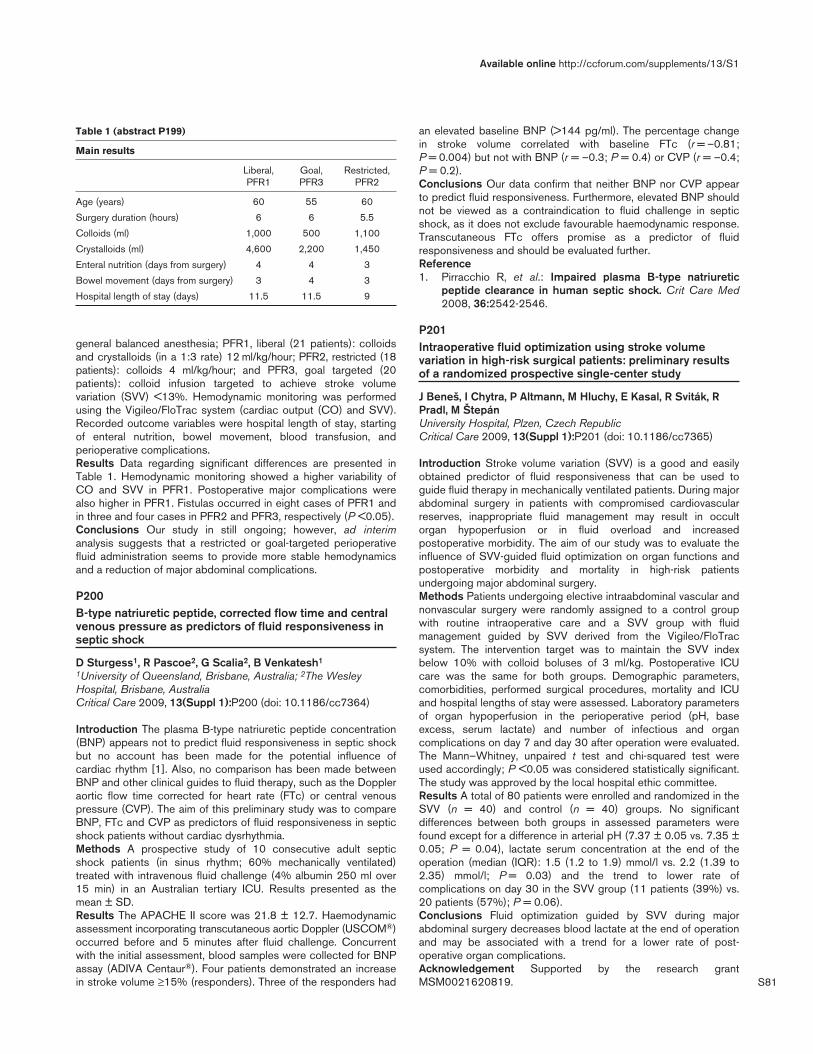
S81
general balanced anesthesia; PFR1, liberal (21 patients): colloidsand crystalloids (in a 1:3 rate) 12 ml/kg/hour; PFR2, restricted (18patients): colloids 4 ml/kg/hour; and PFR3, goal targeted (20patients): colloid infusion targeted to achieve stroke volumevariation (SVV) <13%. Hemodynamic monitoring was performedusing the Vigileo/FloTrac system (cardiac output (CO) and SVV).Recorded outcome variables were hospital length of stay, startingof enteral nutrition, bowel movement, blood transfusion, andperioperative complications.Results Data regarding significant differences are presented inTable 1. Hemodynamic monitoring showed a higher variability ofCO and SVV in PFR1. Postoperative major complications werealso higher in PFR1. Fistulas occurred in eight cases of PFR1 andin three and four cases in PFR2 and PFR3, respectively (P <0.05).Conclusions Our study in still ongoing; however, ad interimanalysis suggests that a restricted or goal-targeted perioperativefluid administration seems to provide more stable hemodynamicsand a reduction of major abdominal complications.
P200B-type natriuretic peptide, corrected flow time and centralvenous pressure as predictors of fluid responsiveness inseptic shock
D Sturgess1, R Pascoe2, G Scalia2, B Venkatesh1
1University of Queensland, Brisbane, Australia; 2The WesleyHospital, Brisbane, AustraliaCritical Care 2009, 13(Suppl 1):P200 (doi: 10.1186/cc7364)
Introduction The plasma B-type natriuretic peptide concentration(BNP) appears not to predict fluid responsiveness in septic shockbut no account has been made for the potential influence ofcardiac rhythm [1]. Also, no comparison has been made betweenBNP and other clinical guides to fluid therapy, such as the Doppleraortic flow time corrected for heart rate (FTc) or central venouspressure (CVP). The aim of this preliminary study was to compareBNP, FTc and CVP as predictors of fluid responsiveness in septicshock patients without cardiac dysrhythmia.Methods A prospective study of 10 consecutive adult septicshock patients (in sinus rhythm; 60% mechanically ventilated)treated with intravenous fluid challenge (4% albumin 250 ml over15 min) in an Australian tertiary ICU. Results presented as themean ± SD.Results The APACHE II score was 21.8 ± 12.7. Haemodynamicassessment incorporating transcutaneous aortic Doppler (USCOM®)occurred before and 5 minutes after fluid challenge. Concurrentwith the initial assessment, blood samples were collected for BNPassay (ADIVA Centaur®). Four patients demonstrated an increasein stroke volume ≥15% (responders). Three of the responders had
an elevated baseline BNP (>144 pg/ml). The percentage changein stroke volume correlated with baseline FTc (r = –0.81;P = 0.004) but not with BNP (r = –0.3; P = 0.4) or CVP (r = –0.4;P = 0.2).Conclusions Our data confirm that neither BNP nor CVP appearto predict fluid responsiveness. Furthermore, elevated BNP shouldnot be viewed as a contraindication to fluid challenge in septicshock, as it does not exclude favourable haemodynamic response.Transcutaneous FTc offers promise as a predictor of fluidresponsiveness and should be evaluated further.Reference1. Pirracchio R, et al.: Impaired plasma B-type natriuretic
peptide clearance in human septic shock. Crit Care Med2008, 36:2542-2546.
P201Intraoperative fluid optimization using stroke volumevariation in high-risk surgical patients: preliminary resultsof a randomized prospective single-center study
J Beneš, I Chytra, P Altmann, M Hluchy, E Kasal, R Sviták, RPradl, M ŠtepánUniversity Hospital, Plzen, Czech RepublicCritical Care 2009, 13(Suppl 1):P201 (doi: 10.1186/cc7365)
Introduction Stroke volume variation (SVV) is a good and easilyobtained predictor of fluid responsiveness that can be used toguide fluid therapy in mechanically ventilated patients. During majorabdominal surgery in patients with compromised cardiovascularreserves, inappropriate fluid management may result in occultorgan hypoperfusion or in fluid overload and increasedpostoperative morbidity. The aim of our study was to evaluate theinfluence of SVV-guided fluid optimization on organ functions andpostoperative morbidity and mortality in high-risk patientsundergoing major abdominal surgery.Methods Patients undergoing elective intraabdominal vascular andnonvascular surgery were randomly assigned to a control groupwith routine intraoperative care and a SVV group with fluidmanagement guided by SVV derived from the Vigileo/FloTracsystem. The intervention target was to maintain the SVV indexbelow 10% with colloid boluses of 3 ml/kg. Postoperative ICUcare was the same for both groups. Demographic parameters,comorbidities, performed surgical procedures, mortality and ICUand hospital lengths of stay were assessed. Laboratory parametersof organ hypoperfusion in the perioperative period (pH, baseexcess, serum lactate) and number of infectious and organcomplications on day 7 and day 30 after operation were evaluated.The Mann–Whitney, unpaired t test and chi-squared test wereused accordingly; P <0.05 was considered statistically significant.The study was approved by the local hospital ethic committee.Results A total of 80 patients were enrolled and randomized in theSVV (n = 40) and control (n = 40) groups. No significantdifferences between both groups in assessed parameters werefound except for a difference in arterial pH (7.37 ± 0.05 vs. 7.35 ±0.05; P = 0.04), lactate serum concentration at the end of theoperation (median (IQR): 1.5 (1.2 to 1.9) mmol/l vs. 2.2 (1.39 to2.35) mmol/l; P = 0.03) and the trend to lower rate ofcomplications on day 30 in the SVV group (11 patients (39%) vs.20 patients (57%); P = 0.06).Conclusions Fluid optimization guided by SVV during majorabdominal surgery decreases blood lactate at the end of operationand may be associated with a trend for a lower rate of post-operative organ complications.Acknowledgement Supported by the research grantMSM0021620819.
Available online http://ccforum.com/supplements/13/S1
Table 1 (abstract P199)
Main results
Liberal, Goal, Restricted, PFR1 PFR3 PFR2
Age (years) 60 55 60
Surgery duration (hours) 6 6 5.5
Colloids (ml) 1,000 500 1,100
Crystalloids (ml) 4,600 2,200 1,450
Enteral nutrition (days from surgery) 4 4 3
Bowel movement (days from surgery) 3 4 3
Hospital length of stay (days) 11.5 11.5 9
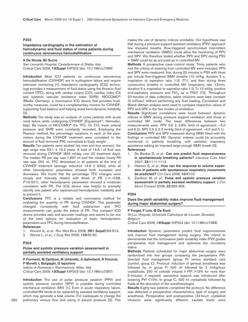
S82
P202Impedance cardiography in the estimation ofhemodynamic and fluid status of coma patients duringcontinuous venovenous hemodiafiltration
A De Nicola, MJ SucreSan Leonardo Hospital, Castellammare di Stabia, ItalyCritical Care 2009, 13(Suppl 1):P202 (doi: 10.1186/cc7366)
Introduction Most ICU patients on continuous venovenoushemodiafiltration (CVVHDF) are in multisystem failure and requireextensive monitoring [1]. Impedance cardiography (ICG) techno-logy provides a measurement of fluid status using the thoracic fluidcontent (TFC), along with cardiac output (CO), cardiac index (CI)and systemic vascular resistance index (SVRI). NICCOMO®
(Medis, Germany), a noninvasive ICG device that provides trust-worthy measures, could be a complementary monitor for CVVHDF,supporting fluid balance and helping avoid hemodynamic instability[2].Methods The study was an analysis of coma patients with acuterenal failure while undergoing CVVHDF (Equasmart®; Hemodec,Italy). By means of NICCOMO® the TFC, CI, CO, mean arterialpressure and SVRI were constantly recorded. Employing thePearson method, the percentage variations in each of the para-meters during the CVVHDF treatment were correlated to theamount of fluid removed (FR), normalized to body weight.Results Ten patients were studied (six men and four women); theage range was 53.1 ± 15.2 years. A total of 16.6 l of fluid wasremoved during CVVHDF (830 ml/day over 20 treatment days).The median FR per day was 1,837 ml and the median hourly FRrate was 252 ml. TFC diminished in all patients at the end ofCVVHDF treatment (average reduction 14.8 ± 9/kΩ), while allother hemodynamic parameters showed both increases anddecreases. We found that the percentage TFC changes wereclosely and inversely related with those of FR (r = –0.68,P <0.001); other hemodynamic parameters showed a moderatecorrelation with FR. The ICG device was helpful to promptlyidentify one patient who experienced hemodynamic instability andto prevent it.Conclusions TFC is a reliable and noninvasive method forevaluating the quantity of FR during CVVHDF. This parameterchanged consistently with fluid subtraction and TFCmeasurements can guide the extent of FR. This compact ICGdevice provides safe and accurate readings and seems to be oneof the best options for evaluation of basic hemodynamicparameters and TFC during hemodiafiltration.References1. Vincent JL, et al.: Rev Med Brux 2008, 29(1 Suppl):S9-S13.2. Wynne L, et al.: J Surg Res 2006, 133:55-60.
P203Pulse and systolic pressure variation assessment inpartially assisted ventilatory support
P Formenti, M Zaniboni, M Umbrello, A Galimberti, R Pinciroli,P Morelli, L Bolgiaghi, G IapichinoIstituto di Anestesia e Rianimazione, Milan, ItalyCritical Care 2009, 13(Suppl 1):P203 (doi: 10.1186/cc7367)
Introduction The use of pulse pressure variation (PPV) andsystolic pressure variation (SPV) is possible during controlledmechanical ventilation (MV) [1]. Even in acute respiratory failure,controlled MV tends to be replaced by assisted ventilatory support,which may generate a tidal volume (Tv) inadequate to change thepulmonary venous flow and swing in pleural pressure [2]. This
makes the use of dynamic indices unreliable. Our hypothesis wasthat during a pressure-support-assisted ventilatory (PSV) approachfew imposed breaths (flow-triggered synchronized intermittentmechanical ventilation (SIMV)) could allow the monitoring of PPVand SPV. We therefore tested whether PPV and SPV during PSV+ SIMV could be as accurate as in controlled MV.Methods A prospective case–control study. Thirty patients whomet the criteria of weaning from controlled MV were included. PPVand SPV were measured, first, during 20 minutes in PSV with threeper minute flow-triggered SIMV breaths (10 ml/kg, duration 5 s,inspiration to expiration ratio 1:3) (T1), and then during threeconsecutive breaths in controlled MV (respiratory rate 12/min,duration 5 s, inspiration to expiration ratio 1:3, Tv 10 ml/kg, positiveend-expiratory pressure and FiO2 as in PSV) (T2). Throughout20 minutes of data collection, saline infusions were kept constant(3 ml/hour) without performing any fluid loading. Correlation andBland–Altman analysis were used to compare respective values ofPPV and SPV in the two modes of ventilation.Results Significant correlations were found between dynamicindices in SIMV during pressure support ventilation and those incontrolled MV mode. The mean differences between twomeasurements were: PPV 0.6 ± 2.8% (limit of agreement: –5.0and 6.2), SPV 0.5 ± 2.3 mmHg (limit of agreement: –4.0 and 5.1).Conclusions PPV and SPV measured during SIMV fitted with thefindings in controlled MV. Dynamic indexes could be accuratelymonitored in patients breathing with assisted respiratoryassistance adding an imposed large enough SIMV breath [3].References1. De Backer D, et al.: Can one predict fluid responsiveness
in spontaneously breathing patients? Intensive Care Med2007, 33:1111-1113.
2. Heenen S, et al.: How can the response to volume expan-sion in patients with spontaneous respiratory movementsbe predicted? Crit Care 2006, 10:R102.
3. Zaniboni M, et al.: Pulse and systolic pressure variationassessment in partially assisted ventilatory support. J ClinMonit Comput 2008, 22:355-359.
P204Does the pleth variability index improve fluid managementduring major abdominal surgery?
P Forget, F Lois, M De KockSt-Luc Hospital, Université Catholique de Louvain, Brussels,BelgiumCritical Care 2009, 13(Suppl 1):P204 (doi: 10.1186/cc7368)
Introduction Dynamic parameters predict fluid responsivenessand improve fluid management during surgery. We intend todemonstrate that the noninvasive pleth variability index (PVI) guidesperoperative fluid management and optimizes the circulatorystatus.Methods Patients scheduled for major abdominal surgery wererandomized into two groups comparing the peroperative PVI-directed fluid management (group P) versus standard care(control, group C). Protocol: induction of general anesthesia wasfollowed by, in group P, 500 ml followed by 2 ml/kg/hourcrystalloids; 250 ml colloids infused if PVI >13% for more than5 minutes; if required, vasoactive support was introduced afterlowering PVI <10%. In group C, 500 ml crystalloids followed byfluids at the discretion of the anesthesiologist.Results Eighty-two patients completed the protocol. No differencewas detected in preoperative characteristics, type of surgery andanesthesia. Peroperative and postoperative (24-hour) crystalloidinfusions were significantly different. Lactate levels were
Critical Care March 2009 Vol 13 Suppl 1 29th International Symposium on Intensive Care and Emergency Medicine
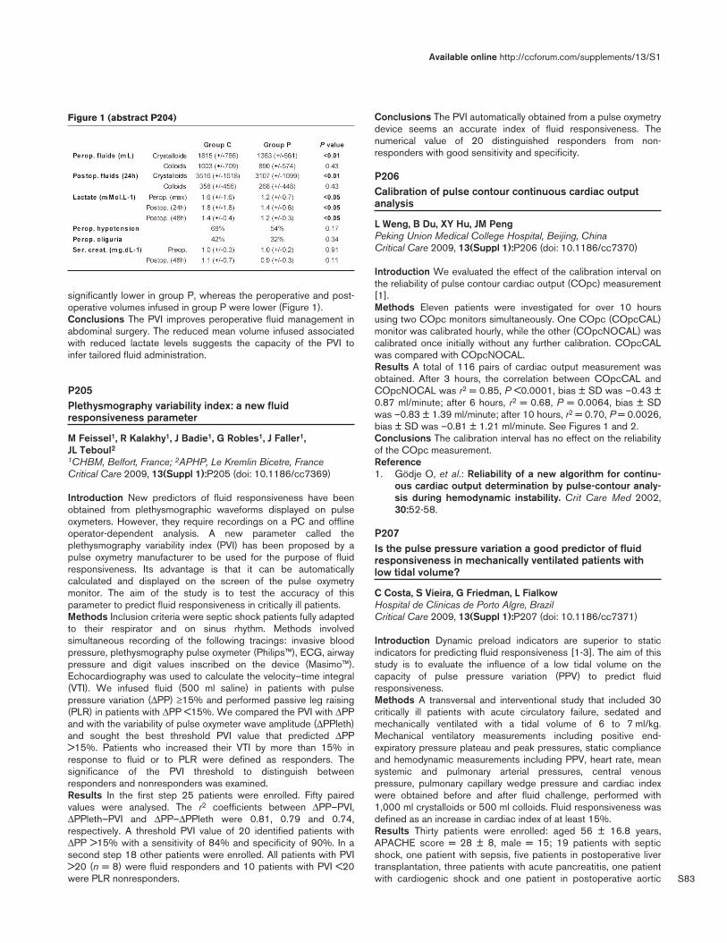
S83
significantly lower in group P, whereas the peroperative and post-operative volumes infused in group P were lower (Figure 1).Conclusions The PVI improves peroperative fluid management inabdominal surgery. The reduced mean volume infused associatedwith reduced lactate levels suggests the capacity of the PVI toinfer tailored fluid administration.
P205Plethysmography variability index: a new fluidresponsiveness parameter
M Feissel1, R Kalakhy1, J Badie1, G Robles1, J Faller1, JL Teboul21CHBM, Belfort, France; 2APHP, Le Kremlin Bicetre, FranceCritical Care 2009, 13(Suppl 1):P205 (doi: 10.1186/cc7369)
Introduction New predictors of fluid responsiveness have beenobtained from plethysmographic waveforms displayed on pulseoxymeters. However, they require recordings on a PC and offlineoperator-dependent analysis. A new parameter called theplethysmography variability index (PVI) has been proposed by apulse oxymetry manufacturer to be used for the purpose of fluidresponsiveness. Its advantage is that it can be automaticallycalculated and displayed on the screen of the pulse oxymetrymonitor. The aim of the study is to test the accuracy of thisparameter to predict fluid responsiveness in critically ill patients.Methods Inclusion criteria were septic shock patients fully adaptedto their respirator and on sinus rhythm. Methods involvedsimultaneous recording of the following tracings: invasive bloodpressure, plethysmography pulse oxymeter (Philips™), ECG, airwaypressure and digit values inscribed on the device (Masimo™).Echocardiography was used to calculate the velocity–time integral(VTI). We infused fluid (500 ml saline) in patients with pulsepressure variation (ΔPP) ≥15% and performed passive leg raising(PLR) in patients with ΔPP <15%. We compared the PVI with ΔPPand with the variability of pulse oxymeter wave amplitude (ΔPPleth)and sought the best threshold PVI value that predicted ΔPP>15%. Patients who increased their VTI by more than 15% inresponse to fluid or to PLR were defined as responders. Thesignificance of the PVI threshold to distinguish betweenresponders and nonresponders was examined.Results In the first step 25 patients were enrolled. Fifty pairedvalues were analysed. The r2 coefficients between ΔPP–PVI,ΔPPleth–PVI and ΔPP–ΔPPleth were 0.81, 0.79 and 0.74,respectively. A threshold PVI value of 20 identified patients withΔPP >15% with a sensitivity of 84% and specificity of 90%. In asecond step 18 other patients were enrolled. All patients with PVI>20 (n = 8) were fluid responders and 10 patients with PVI <20were PLR nonresponders.
Conclusions The PVI automatically obtained from a pulse oxymetrydevice seems an accurate index of fluid responsiveness. Thenumerical value of 20 distinguished responders from non-responders with good sensitivity and specificity.
P206Calibration of pulse contour continuous cardiac outputanalysis
L Weng, B Du, XY Hu, JM PengPeking Union Medical College Hospital, Beijing, ChinaCritical Care 2009, 13(Suppl 1):P206 (doi: 10.1186/cc7370)
Introduction We evaluated the effect of the calibration interval onthe reliability of pulse contour cardiac output (COpc) measurement[1].Methods Eleven patients were investigated for over 10 hoursusing two COpc monitors simultaneously. One COpc (COpcCAL)monitor was calibrated hourly, while the other (COpcNOCAL) wascalibrated once initially without any further calibration. COpcCALwas compared with COpcNOCAL.Results A total of 116 pairs of cardiac output measurement wasobtained. After 3 hours, the correlation between COpcCAL andCOpcNOCAL was r2 = 0.85, P <0.0001, bias ± SD was –0.43 ±0.87 ml/minute; after 6 hours, r2 = 0.68, P = 0.0064, bias ± SDwas –0.83 ± 1.39 ml/minute; after 10 hours, r2 = 0.70, P = 0.0026,bias ± SD was –0.81 ± 1.21 ml/minute. See Figures 1 and 2.Conclusions The calibration interval has no effect on the reliabilityof the COpc measurement.Reference1. Gödje O, et al.: Reliability of a new algorithm for continu-
ous cardiac output determination by pulse-contour analy-sis during hemodynamic instability. Crit Care Med 2002,30:52-58.
P207Is the pulse pressure variation a good predictor of fluidresponsiveness in mechanically ventilated patients withlow tidal volume?
C Costa, S Vieira, G Friedman, L FialkowHospital de Clinicas de Porto Algre, BrazilCritical Care 2009, 13(Suppl 1):P207 (doi: 10.1186/cc7371)
Introduction Dynamic preload indicators are superior to staticindicators for predicting fluid responsiveness [1-3]. The aim of thisstudy is to evaluate the influence of a low tidal volume on thecapacity of pulse pressure variation (PPV) to predict fluidresponsiveness.Methods A transversal and interventional study that included 30critically ill patients with acute circulatory failure, sedated andmechanically ventilated with a tidal volume of 6 to 7 ml/kg.Mechanical ventilatory measurements including positive end-expiratory pressure plateau and peak pressures, static complianceand hemodynamic measurements including PPV, heart rate, meansystemic and pulmonary arterial pressures, central venouspressure, pulmonary capillary wedge pressure and cardiac indexwere obtained before and after fluid challenge, performed with1,000 ml crystalloids or 500 ml colloids. Fluid responsiveness wasdefined as an increase in cardiac index of at least 15%.Results Thirty patients were enrolled: aged 56 ± 16.8 years,APACHE score = 28 ± 8, male = 15; 19 patients with septicshock, one patient with sepsis, five patients in postoperative livertransplantation, three patients with acute pancreatitis, one patientwith cardiogenic shock and one patient in postoperative aortic
Available online http://ccforum.com/supplements/13/S1
Figure 1 (abstract P204)
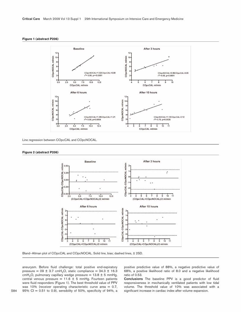
S84
aneurysm. Before fluid challenge: total positive end-expiratorypressure = 09 ± 3.7 cmH2O, static compliance = 34.3 ± 16.3cmH2O, pulmonary capillary wedge pressure = 13.8 ± 5 mmHg,central venous pressure = 11.6 ± 5 mmHg. Fourteen patientswere fluid responders (Figure 1). The best threshold value of PPVwas 10% (receiver operating characteristic curve area = 0.7,95% CI = 0.51 to 0.9), sensibility of 50%, specificity of 94%, a
positive predictive value of 88%, a negative predictive value of68%, a positive likelihood ratio of 8.0 and a negative likelihoodratio of 0.53.Conclusions The baseline PPV is a good predictor of fluidresponsiveness in mechanically ventilated patients with low tidalvolume. The threshold value of 10% was associated with asignificant increase in cardiac index after volume expansion.
Critical Care March 2009 Vol 13 Suppl 1 29th International Symposium on Intensive Care and Emergency Medicine
Figure 2 (abstract P206)
Bland–Altman plot of COpcCAL and COpcNOCAL. Solid line, bias; dashed lines, ± 2SD.
Figure 1 (abstract P206)
Line regression between COpcCAL and COpcNOCAL.
S85
References1. Teboul JL: Relation between respiratory changes in arterial
pulse pressure and fluid responsiveness in septic patientswith acute circulatory failure. Am J Respir Crit Care Med2000, 162:134-138.
2. De Backer D: Pulse pressure variations to predict fluidresponsiveness: influence of tidal volume. Intensive CareMed 2005, 31:517-523.
3. Chung-Chi H: Prediction of fluid resposiveness in acuterespiratory distress syndrome patients ventilated with lowtidal volume and high positive end-expiratory pressure.Crit Care Med 2008, 36:2810.
P208Proving the effectiveness of three dynamic indices topredict fluid responsiveness in septic mechanicallyventilated patients
P Wacharasint, A Lertamornpong, A Wathanathum, A WongsaPhramongkutklao Hospital, Bangkok, ThailandCritical Care 2009, 13(Suppl 1):P208 (doi: 10.1186/cc7372)
Introduction Fluid responsiveness is still a cornerstone inmanaging patients with severe sepsis and septic shock. Recentlynew technologies have been generated for facilitating the accuracyof predicting fluid responsiveness at the bedside, based oncardiopulmonary interaction [1,2]. We evaluated the effectivenessand accuracy of three dynamic indices, currently available inintensive care monitoring devices, which are pulse pressurevariation (PPV) [3], stroke volume variation (SVV) and pulseoximetry plethysmographic waveform variation (POPV) in septicmechanically ventilated patients [4].Methods A prospective clinical trial was conducted in 20 septicpatients 18 years of age and older who had invasive bloodpressure monitoring with an intraarterial cannula. PPV, SVV andPOPV (%) were calculated and compared with the percentagecardiac index (CI) change. Patients with a CI increase induced byvolume expansion >15% were classified as responders, and<15% as nonresponders. A parametric paired t test was used tocompare hemodynamic parameters at baseline and after volumeexpansion. Student’s t test was used to compare hemodynamicparameters in the responders and nonresponders groups. Receiveroperating characteristic curves were used to evaluate the
predictive value of various indices on fluid responsiveness.P <0.05 was considered significant.Results We found a strong correlation existed for PPV fordetection of a volume expansion-induced change in CI (r2 =0.794), followed by SVV (r2 = 0.667), and POPV (r2 = 0.633). Theareas under the receiver operating characteristic curves were 0.96for PPV (P <0.001), 0.92 for SVV (P = 0.001), and 0.85 for POPV(P = 0.008). Respiratory variation in POPV exceeding 14% (sensi-tivity of 72%, specificity of 90%), SVV exceeding 11% (sensitivity90%, specificity 92%), allowed detection of PPV exceeding 12%(sensitivity 84%, specificity 96%).Conclusions In the septic mechanically ventilated patients, PPV isthe most effective dynamic parameter for predicting fluidresponsiveness; all of PPV, SVV and POPV are well correlatedwith the percentage change of CI.References1. Michard F, Teboul JL: Predicting fluid responsiveness in
ICU patients: a critical analysis of the evidence. Chest2002, 121:2000-2008.
2. Bendjelid K, Romand JA: Fluid responsiveness in mechani-cally ventilated patients: a review of indices used in inten-sive care. Intensive Care Med 2003, 29:352-360.
3. Michard F, et al.: Relation between respiratory changes inarterial pulse pressure and fluid responsiveness in septicpatients with acute circulatory failure. Am J Respir CritCare Med 2000, 162:134-138.
4. Feissel M, et al.: Plethysmographic dynamic indices predictfluid responsiveness in septic ventilated patients. IntensiveCare Med 2007, 33:993-999.
P209Cross-comparison of the trending accuracy of continuouscardiac output measurement devices in postoperationpatients
HK Kim, M Hadian, D Severyn, MR PinskyUniversity of Pittsburgh Medical Center, Pittsburgh, PA, USACritical Care 2009, 13(Suppl 1):P209 (doi: 10.1186/cc7373)
Introduction Arterial pulse analysis estimates of cardiac output(CO) are less invasive than pulmonary artery catheter (PAC)-derived ones. Although PAC bolus thermodilution CO (COtd)values are the reference CO values for most clinical studies, thereis no defined gold standard. We therefore compared the co-variance of three commercially available arterial pulse analysisdevices (FloTrac, LiDCO, PiCCO) and PAC with COtd andcontinuous CO (CCO).Methods Seventeen postoperative cardiac surgery patients werestudied for the first 4 hours post ICU admission. CO wasmeasured simultaneously by FloTrac, LiDCO and PiCCO using thesame arterial waveform. LiDCO and PiCCO were calibrated to thefirst COtd measured on ICU admission. Values of CO werecompared before and after specific therapeutic interventions(volume, vasoactive or inotropic infusions). Absolute values for COacross all devices were compared by linear regression andBland–Altman analysis. Dynamic changes for CO across alldevices were compared by moment analysis.Results In 17 patients, 72 paired simultaneous CO measurementswere collected. By linear regression analysis, all devices correlatedwell with each other for absolute CO as r = 0.87 (PAC–LiDCO),0.70 (PAC–PiCCO), 0.51 (PAC–FloTrac), 0.80 (LiDCO–PiCCO),0.65 (LiDCO–FloTrac) and 0.50 (PiCCO–FloTrac) with P <0.001in all pairs. By Bland–Altman analysis, the mean bias between COmeasurements by each paired device was –0.18 l/min (PAC–LiDCO), 0.30 l/min (PAC–PiCCO), –0.43 l/min (PAC–FloTrac),
Available online http://ccforum.com/supplements/13/S1
Figure 1 (abstract P207)
Relationship between the PPV and the cardiac index variation.

S86
0.12 l/min (LiDCO–PiCCO), –0.63 l/min (LiDCO–FloTrac) and–0.73 l/min (PiCCO–FloTrac), with precision (1.96 SD, 95% CI) of±1.56, ±2.26, ±3.37, ±1.98, ±2.97 and ±3.45 l/min, respectively.By Pearson moment analysis, the dynamic change for CO was wellcorrelated between PAC–LiDCO, PAC–PiCCO, LiDCO–PiCCOand LiDCO–FloTrac (r = 0.78, 0.56. 0.65 and 0.55, respectively).However, FloTrac showed poor correlation with PAC and PiCCO(r = 0.28 and 0.33, respectively).Conclusions In postoperative cardiac surgery patients, theabsolute CO of arterial pulse analysis devices showed goodcorrelation with each other and PAC COtd and CCO. However,the dynamic change of CO was less correlated with each otherthan the absolute CO. Because each arterial pulse analysis deviceshowed different correlations with PAC CO, the CO data from onemonitoring system can be used carefully in concert with another todrive resuscitation protocols.
P210Comparison of three noninvasive cardiac output monitorsin patients undergoing therapeutic hypothermia aftercardiac arrest
GM Haslam, FE Kelly, WA English, A D’Agapeyeff, AJ Padkin,TM Cook, JP NolanRoyal United Hospital, Bath, UKCritical Care 2009, 13(Suppl 1):P210 (doi: 10.1186/cc7374)
Introduction We planned to observe the performance of theLiDCOplus (LiDCO Systems, UK), PiCCO (Pulsion MedicalSystems, Germany) and NICO (Novametrix-Respironics, USA)continuous cardiac output monitors during therapeutic hypo-thermia and rewarming post cardiac arrest. PiCCO calibrationfailure during hypothermia has been reported [1,2]. Use of theLiDCOplus and the NICO monitor during therapeutic hypothermiahas not been reported. The LiDCOplus pulse power analysis iscalibrated using a bolus of lithium chloride instead of
transpulmonary thermodilution. Function of the NICO is based on theFick principle and on partial rebreathing of carbon dioxide. LiDCOplusand NICO performance should not be affected by hypothermia.Methods Local research ethics committee approval was obtained.Patients admitted to our ICU for therapeutic hypothermia aftercardiac arrest were recruited to the study following assent from thenext of kin. Hypothermia was induced with 2 l crystalloid at 4°Cand maintained using an Alsius Coolguard Icy Catheter (AlsiusCorp., USA) inserted via the femoral vein. A 5 F PiCCO catheterwas inserted in the femoral artery on the opposite side andconnected to the PiCCO and LiDCOplus monitors. A NICOsystem was attached to the patient’s tracheal tube. Systems werecalibrated and recalibrated according to the manufacturer’sinstructions (PiCCO 8 hourly, LIDCOplus 24 hourly). The bladdertemperature was recorded and maintained at 33°C for 24 hours.The temperature, heart rate, blood pressure, and cardiac output onall three monitors were recorded hourly. After 24 hours, patientswere rewarmed at 0.25°C/hour to a target of 36.5°C.Results Six patients were recruited over 8 months. All patientswere at the target temperature of 33°C when cardiac output wasfirst recorded. No problems were encountered calibrating thePiCCO, LiDCOplus or NICO monitors at temperatures as low as33°C. All three monitors were observed to trend together(Figure 1). Statistical analysis of these trends will be available atthe time of presentation.Conclusions LiDCOplus, PiCCO and NICO perform comparablyin the temperature range of 33 to 36.5°C.References1. Ong T, Gillies MA, Bellomo R: Failure of continuous cardiac
output measurement using the PiCCO device duringinduced hypothermia: a case report. Crit Care Resusc2004, 6:99-101.
2. Sami A, Sami A, Rochdil N, Hatem K, Salah BL: PiCCO moni-toring accuracy in low body temperature. Am J Emerg Med2007, 25:845-846.
P211Head-up tilt and passive leg raising in healthy volunteersas a preclinical model for preload-induced stroke volumemodification
A Lima, E Klijn, J Bakker, C Ince, J Van BommelErasmus Medical Center, Rotterdam, the NetherlandsCritical Care 2009, 13(Suppl 1):P211 (doi: 10.1186/cc7375)
Introduction In clinical practice, fluid responsiveness is tested byinducing changes in the stroke volume (SV), as measured withinvasive monitoring techniques in often time-consuming proce-dures. To be able to investigate the effects of changes in SV onother hemodynamic parameters, there is the need for a preclinicalmodel that can be translated to clinical practice. For this purposewe studied the effect of the head-up tilt (HUT) test and passive legraising (PLR) test on SV in five healthy volunteers.Methods The tilt table test consisted of 3 minutes of supine restfollowed by a HUT of 70° on a manually operated tilt table andending with 3 minutes of supine rest. The PLR test consisted of3 minutes of rest in a semirecumbent position of 30°, followed by3 minutes PLR (lower limbs elevated at 30° and trunk in supineposition) and ending with 3 minutes of rest in a semirecumbentposition. Three HUTs and two PLR tests were performed. The SVwas measured continuously and noninvasively using a NICOM(Cheetah Medical, Tel Aviv, Israel), based on chest bioreactance,and a Finometer (TNO Biomedical Instrumentation, Amsterdam,the Netherlands), based on the Modelflow method. The Finometeralso measured the mean arterial pressure.
Critical Care March 2009 Vol 13 Suppl 1 29th International Symposium on Intensive Care and Emergency Medicine
Figure 1 (abstract P210)

S87
Results Both HUT and PLR induced significant changes in SV(Figure 1). In addition the cardiac output changed significantly, butnot during HUT when measured with the Finometer. Although bothdevices showed a similar response to the postural changes, thebaseline values and magnitude of these responses were notidentical. The mean arterial pressure remained constant duringthese manoeuvres. The heart rate significantly increased duringHUT but not during PLR.Conclusions The combination of noninvasive monitoring tech-niques with preload-induced SV modifications in healthy volunteersprovides an excellent preclinical model for the study of fluidresponsiveness. Although these hemodynamic monitors mightprovide different results, this model should be very suitable toassess the effect of SV variations on, for instance, parameters ofperipheral perfusion.
P212Global end-diastolic volume as a predictor of the need formassive transfusion in multiple-trauma patients withhemorrhagic shock
N Saito, Y Sakamoto, K MashikoChiba Hokusou Hospital, Nippon Medical School, Chiba, JapanCritical Care 2009, 13(Suppl 1):P212 (doi: 10.1186/cc7376)
Introduction The PiCCO system enables hemodynamic evaluationand monitoring by two different approaches: the transpulmonarythermal dilution technique and pulse counter analysis. Optimalmonitoring of cardiac preload is of paramount importance for thehemodynamic management of multiple-trauma patients withhemorrhagic shock. There have been only a few studies on the useof the PiCCO system (Pulsion, Germany) in multiple-traumapatients with hemorrhagic shock for hemodynamic monitoring. Wehypothesized that performing a cardiac adequate preloadevaluation with the PiCCO system would make it possible topredict latent hemorrhagic progress.Methods Data from 53 consecutive multiple-trauma patients (age51 ± 17.8 years, injury severity score 30 ± 12.9) with hemorrhagicshock at the scene of the injury or in the emergency room betweenJune 2007 and November 2008 were analyzed. All patientsunderwent a hemodynamic evaluation with the PiCCO system. Wedivided the patients into two groups according to whether theyreceived a massive transfusion (MT) (>2,000 ml packed red bloodcell transfusion after admission within 24 hours) and comparedtheir PiCCO data: cardiac output (CO), systemic vascular resis-tance (SVR), indexed global end-diastolic volume (GEDVi), andindexed extravascular lung water on admission to the ICU. The chi-square test and paired t test were used to perform the statisticalanalysis.
Results Twenty-seven patients required massive transfusion.Mortality was higher in the MT group (P = 0.05), and the CO,GEDVi and ITBVi values were significantly lower in the MT group(Table 1).Conclusions The parameters measured with the PiCCO systemenabled evaluation of the correct cardiac preload in the multiple-trauma patients with hemorrhagic shock. GEDVi was useful as apredictor of the need for MT.
P213Should pulse pressure variation be indexed to tidalvolume?
S Vistisen, J Koefoed-Nielsen, A LarssonAalborg Hospital/Aarhus University Hospitals, Aarhus C, DenmarkCritical Care 2009, 13(Suppl 1):P213 (doi: 10.1186/cc7377)
Introduction Pulse pressure variation (PPV) for prediction of fluidresponsiveness depends on the tidal volume (VT), and using VTsof the recommended 6 ml/kg makes PPV unreliable [1]. So far,nobody has suggested how to handle the VT when interpretingPPV or other dynamic parameters, but we hypothesise that PPV isproportional to VT and thus that PPV should be indexed to VT. Theaim was to investigate how three VT levels affected PPV at fourdifferent intravascular volumes.Methods The study was approved by the national animal ethicalcommittee. Eight anesthetised and ventilated pigs (23 to 27 kg)were bled 25% of the blood volume (hypovolemia). PPV wasmeasured at ventilation with VTs of 6, 9 and 12 ml/kg (VT6, VT9and VT12, respectively). Thereafter, depleted blood was replacedwith voluven (normovolemia) and PPV was again measured at thethree VT levels and subsequently at the +25% and +50%hypervolemic levels also generated with voluven infusion.PPV values were log-transformed and compared with a paired ttest. Comparisons were made at each intravascular volume levelbetween VT6 and VT9, VT9 and VT12, and VT6 and VT12.Because three comparisons were performed, P <0.05/3 = 0.017was considered significant.Results All comparisons for PPV at different VTs were significantlydifferent (P <0.001); see Table 1 for factorial increases in PPV. At–25% hypovolemia, PPV did not fully double with doubling of VT,whereas PPV slightly more than doubled at normovolemia andhypervolemia.
Available online http://ccforum.com/supplements/13/S1
Figure 1 (abstract P211) Table 1 (abstract P212)
MT group Non-MT group P value
CO (l/min) 4.88 ± 2.1 6.38 ± 2.8 0.034
SVR (dyn x s/cm5) 1,169 ± 364 1,452 ± 601 0.046
GEDVi (ml/m2) 554 ± 158 713 ± 238 0.007
Table 1 (abstract P213)
Comparisons of PPV at different VTs
Factorial increase in PPV
Volume VT6–VT9 VT9–VT12 VT6–VT12
–25% 1.28 to 1.34 1.17 to 1.28 1.51 to 1.70
0% 1.53 to 2.00 1.24 to 1.60 2.17 to 2.80
+25% 1.47 to 1.87 1.26 to 1.50 1.88 to 2.77
+50% 1.50 to 1.71 1.30 to 1.61 2.09 to 2.56
Data presented as the 95% CI.

S88
Conclusions Our study in pigs showed that PPV strongly dependson the VT and that PPV is nearly proportional to the VT, indicatingthat the clinical value of PPV may be improved by TV indexation.Reference1. De Backer D, et al.: Pulse pressure variations to predict
fluid responsiveness: influence of tidal volume. IntensiveCare Med 2005, 31:517-523.
P214Fluid responsiveness in patients following major surgery
M Tuccillo1, M Cecconi2, N Al-Subaie2, M Hamilton2, R Grounds2, G Della Rocca1, A Rhodes2
1Azienda Ospedaliera Universitaria S. Maria della Misericordia,Udine, Italy; 2St George’s Hospital, London, UKCritical Care 2009, 13(Suppl 1):P214 (doi: 10.1186/cc7378)
Introduction The aim of the study was to see how many patientswere fluid responders on arrival in the ICU and to evaluate theperformance of dynamic preload indices in predicting fluidresponsiveness in fully sedated and mechanically ventilatedpatients. The following indices were studied: pulse pressurevariation (PPV), stroke volume variation (SVV) and systolicpressure variation (SPV).Methods Patients were monitored with the LiDCOTMplus andreceived a fluid challenge (250 ml boluses of colloid in 5 min) toascertain fluid responsiveness. Patients in which a fluid challengeproduced a 10% increase in stroke volume were considered fluidresponders and thus the fluid challenge was repeated according tothe unit protocol [1]. In fully mechanically ventilated patients, thePPV, SVV and SPV were recorded and receiver operating charac-teristic (ROC) analysis was performed.Results Thirty-three patients (mean age 64 years; SD ± 13.80;mean BMI 27.34; 16 female and 17 male) were included; 23patients were on spontaneous ventilation and 10 were totally fullyventilated with a mean tidal volume of 8 ml/kg and positive end-expiratory pressure of 5 cmH2O. Thirteen patients (40%) wereresponders, of which five were mechanically ventilated. Areasunder the ROC curve (AUC) for dynamic predictors of fluidresponsiveness were examined for ventilated patients; for PPV,AUC = 0.65 with SD ± 0.14, P = 0.28 showing a sensitivity of67% and a specificity of 79% for a cutoff value of 14%; for SVV,AUC = 0.73 with SD ± 0.14, P = 0.1 showing a sensitivity of 67%and a specificity of 74% for a cutoff value of 9%; and for SPV,AUC = 0.74 with SD ± 0.12, P = 0.09 showing a sensitivity of67% and a specificity of 86% for a cutoff value of 8%.Conclusions A high percentage of patients were fluid responsiveon arrival in the ICU following major surgery (40%). The PPV, SVVand SPV have a potential to predict fluid responsiveness inmechanically ventilated patients.Reference1. Pearse RM, et al.: Early goal-directed therapy after major
surgery reduces complications and duration of hospital stay.A randomised, controlled trial. Crit Care 2005, 9:R687-R693.
P215Alternative echographic assessment of inferior vena cavadiameter variation in mechanically ventilated patients
T Leclerc1, N Libert1, JP Tourtier1, A Bannay2, S De Rudnicki11HIA Val-de-Grâce, Paris, France; 2CHU Nancy, FranceCritical Care 2009, 13(Suppl 1):P215 (doi: 10.1186/cc7379)
Introduction The echographic assessment of inferior vena cava(IVC) diameter (IVCD) variation allows for fluid-responsiveness
prediction in ventilated septic patients [1,2]. Its applicability insurgical patients is not established, partly due to the difficulty toacquire a subxiphoidian (SX) view after abdominal surgery. Thetranshepatic (TH) view of the IVC could provide an interestingalternative.Methods In this prospective, randomized, crossover, pilot study,IVCD variation was assessed in consecutive mechanicallyventilated (volume control) ICU patients, with SX and TH views, inrandom order by one operator. M-mode images were acquired at100 mm/s, 2 cm below the junction of the IVC with the right atrium.The minimal (Dmin) and maximal (Dmax) ICVD values were latermeasured on pooled images. IVCD variation [2], defined as dIVC =(Dmax – Dmin) / ((Dmax + Dmin) / 2), was compared betweenboth views (when available): dIVCsx and dIVCth for subxiphoidianand transhepatic views, respectively.Results Twenty-eight patients were included, 19 medical and ninesurgical. The TH view was obtained in all patients, the SX view inonly 22 of them with failures in four surgical and two morbidlyobese medical patients. Linear regression showed a strong corre-lation between dIVCth and dIVCsx (n = 22, r2 = 0.98, P <0.001).The agreement was satisfactory using Bland–Altman analysis(Figure 1): bias was 0.26%, limits of agreement were –8.4 to+7.9%. Results were similar for the IVC distensibility index [1].Conclusions Our results suggest that the SX view, whenunavailable, can be replaced by the TH view for the echographicassessment of IVCD variation. Further study is ongoing to formallyestablish its validity as a predictor of fluid responsiveness,especially in surgical ICU patients.References1. Barbier C, et al.: Intensive Care Med 2004, 30:1740-1746.2. Feissel M, et al.: Intensive Care Med 2004, 30:1834-1837.
P216Global end-diastolic volume and global end-diastolicvolume index are age dependent in awake, spontaneous-breathing patients after elective craniotomy
A Rieß, S Wolf, JF Landscheidt, CB Lumenta, P Friederich, L SchürerKlinikum Bogenhausen, München, GermanyCritical Care 2009, 13(Suppl 1):P216 (doi: 10.1186/cc7380)
Introduction Estimation of cardiac preload is an important pre-requisite for adequate volume resuscitation. The global end-diastolic volume (GEDV) and the global end-diastolic volume index(GEDVI) are surrogate parameters for preload and have beenhypothesized to be age dependent [1]. The current study wasperformed to assess the influence of age on the preloadparameters GEDV and GEDVI prospectively.
Critical Care March 2009 Vol 13 Suppl 1 29th International Symposium on Intensive Care and Emergency Medicine
Figure 1 (abstract P215)

S89
Methods One hundred and one patients (41 male, 60 female)scheduled for brain tumour surgery were investigated using thePiCCOplus device (Pulsion Medical Systems AG, Munich,Germany). On postoperative day 1, the cardiac output, GEDV andGEDVI were determined by transpulmonary thermodilution beforedischarge from the ICU. The influence of predefined age groups(21 to 40 years old, n = 12; 41 to 50 years old, n = 24; 51 to60 years old, n = 20; 61 to 70 years old, n = 26; 71 to 83 yearsold, n = 19) was tested using a Kruskal–Wallis test. The level ofsignificance was 5%.Results Age significantly influenced the GEDV (P = 0.0024) aswell as the GEDVI (P = 0.0007). Cardiac output (P = 0.3555),mean arterial pressure (P = 0.0764) and systemic vascular resis-tance (P = 0.1446) were not dependent on age.Conclusions The volumetric parameter GEDV is dependent onage in haemodynamically healthy and spontaneously breathingpatients. Indexing to body surface area does not remove agedependence. Targeting volume resuscitation using fixed ranges ofthe GEDVI acquired by transpulmonary thermodilution withoutreference to the patient’s age seems not to be appropriate.Reference1. Wolf S, et al.: ITBV and GEDV but not EVLW acquired by
transpulmonary thermodilution is age dependent in aseries of neurosurgical patients [poster]. Intensive CareMed 2007, 33(S2):P0273.
P217Recalibration of pulse contour cardiac output using thePiCCO-2 device: when to perform the next thermodilution?
W Huber, S Mair, T Oelfin, B Saugel, V Phillip, H Einwaechter, R SchmidKlinikum rechts der Isar der Technischen Universität München,GermanyCritical Care 2009, 13(Suppl 1):P217 (doi: 10.1186/cc7381)
Introduction Haemodynamic monitoring is a cornerstone of inten-sive care. In addition to parameters of preload and afterload, theassessment of cardiac output (CO) is of paramount importance.Thermodilution (TD) using the pulmonary artery catheter andtranspulmonary TD using the PiCCO device are the gold standardsfor CO determination. After calibration by TD, the PiCCO device isable to assess CO using pulse contour (PC) analysis. Despite anoverall good correlation of PC-CO and TD-CO in several studies,the manufacturer suggests recalibration by TD after 8 hours. Littleis known about the long-term accuracy of PC-CO. Therefore it wasthe aim of our prospective study to investigate the long-termaccuracy of PiCCO-2-derived PC-CO in the daily ICU routine.Methods In 10 consecutive patients (five male, five female, age65 ± 15 years) the PC-CO was recorded immediately beforerecalibration by TD-CO. One hundred and ninety-four measure-ments with a mean time-lag between two measurements of TD-COof 663.5 ± 371 min (100 to 2,700) were recorded. Mechanicalventilation, catecholamine and arrhythmia occurred in 60 (31%),132 (68%) and 154 (79%) of the measurements.
Results The 194 pairs of PC-CO and TD-CO showed a highlysignificant correlation (P <0.001; r = 0.875). There was no signifi-cant difference between PC-CO versus TD-CO (4.1 ± 1.6 vs.4.07 ± 1.4 l/min m2). Analysis according to Bland–Altmandemonstrated a mean bias of –0.036 ± 0.778 l/min m2 (lower andupper limits of agreement –1.56 and 1.49 l/min m2; percentageerror of 38%). The difference of PC-CO and TD-CO was notcorrelated to the time-lag to the last calibration (P = 0.257;r = –0.083 for uncorrected difference; P = 0.067; r = 0.134 forabsolute values of the difference). Further analysis demonstratedthat the absolute value of the differences correlated to TD-CO(P = 0.02, r = 0.226). Subgroup analysis of 160 measurementswith CO-TD <5.5 l/min m2 demonstrated an improved bias of0.085 ± 0.53 l/min m2 (lower/upper limits of agreement: –0.98and 1.12 l/min m2) and a percentage error of 28%.Conclusions PiCCO-2-derived PC-CO and TD-CO are highlysignificantly correlated. Accuracy is not influenced by the time-lagto the last calibration. Similar to previous data, PC-CO mightoverestimate very high CO, which usually does not influenceclinical practice. Recalibration should be considered in patientswith markedly increased PC-CO.
P218Transpulmonary lithium dilution technique: time torecalibration and calibration drift
R Stümpfle, M Cecconi, D Dawson, M Hamilton, RM Grounds, A RhodesSt George’s Hospital, London, UKCritical Care 2009, 13(Suppl 1):P218 (doi: 10.1186/cc7382)
Introduction We have previously demonstrated that an average ofat least two curves is necessary to improve the calibration of thelithium dilution technique of the LiDCO™plus. The precision of thenew calibration process is able to detect a least significant change(LSC) of 17% [1]. The primary aim of this study was to evaluatethe drift after an initial calibration with two lithium dilution curves.The second aim of the study was to evaluate the relationshipbetween the magnitude of the time to recalibration and themagnitude of the drift.Methods Patients requiring monitoring with the LiDCO™plusreceived an initial calibration plus a second calibration when clinicallyindicated. Data were downloaded from devices and analysed usingthe LiDCO™viewPRO v.1 program. Absent, abandoned or rejectedcalibration curves were excluded. Calibration factors from the firstand second calibrations were compared. All recalibrations in whichthe drift was higher than the LSC (17%) were considered usefulcalibrations. Regression analysis for the time to recalibration and driftwas performed. Receiver operating characteristic curve analysis wasperformed for the time free of calibration and the usefulness ofcalibrations (drift >17%).Results A total of 45 files from 45 patients with two calibrationpoints were identified. Patient mean age was 62 (39 to 88) years,mean height 1.69 (1.40 to 1.94) metres and mean weight 70.9 (40to 155) kg. Time to recalibration varied from 5 to 39 hours. In 23
Available online http://ccforum.com/supplements/13/S1
Table 1 (abstract P216)
GEDV in age groups
21 to 40 years old 41 to 50 years old 51 to 60 years old 61 to 70 years old 71 to 83 years old
GEDV (ml) 1,154 (1,442 to 980) 1,156 (1,295 to 975) 1,183 (1,352 to 1,074) 1,462 (1,630 to 1,182) 1,337 (1,567 to 1,157)
GEDVI (ml/m2) 612 (674 to 548) 598 (680 to 557) 652 (752 to 597) 756 (875 to 655) 734 (781 to 634)
Data presented as median (75th to 25th quartile).

S90
(51%) patients the drift was lower than 17% (unnecessarycalibration). In 22 (49%) patients the drift was higher than 17%(necessary calibration).Regression analysis did not show any significant relationshipbetween the time to recalibration and drift. This was confirmed byreceiver operating characteristic curve analysis.Conclusions The new implemented calibration process (using theaverage of two curves) does not show any drift depending on time.Despite this, 51% of the recalibrations are necessary (drift >17%).Further data analysis is necessary to identify when to recalibrate.Reference1. Cecconi M, Dawson D, Grounds R, Rhodes A: Lithium dilu-
tion cardiac output measurement in the critically ill patient:determination of precision of the technique. Online First[Epub ahead of print].
P219Individualized intraoperative patient optimization usinguncalibrated arterial pressure waveform analysis inhigh-risk patients undergoing major abdominal surgery
J Mayer, J Boldt, R Beschmann, A Stephan, S SuttnerKlinikum Ludwigshafen, GermanyCritical Care 2009, 13(Suppl 1):P219 (doi: 10.1186/cc7383)
Introduction Established methods to optimize cardiac functionand fluid balance based on flow-related variables are invasive orrequire considerable attentiveness. The purpose of this study wasto guide intraoperative fluid and catecholamine therapy in patientswith pre-existing cardiac disease undergoing major abdominalsurgery using a recently introduced less-invasive device withoutthe need for manual calibration (FloTrac/Vigileo™) and to deter-mine possible improvement in outcome by means of N-terminalpro-brain natriuretic peptide (NT-proBNP) plasma levels and theduration of hospital stay.Methods Forty American Society of Anesthesiologists III patientsscheduled for elective major abdominal surgery with pre-existingcardiac disease (coronary artery disease, myocardial infarction,cardiac surgery, heart failure, cardiomyopathy) were studied. Patientswere randomly allocated into a standard care group and anintervention group, where a stroke volume variation (SVV) andcardiac index (CI)-based protocol for volume and catecholaminetherapy was implemented until the end of surgery. In brief, CI≥2.5 l/min/m2 was aimed for, with a SVV threshold value for fluidchallenge of 12%. After the baseline (skin incision), haemodynamicdata and plasma NT-proBNP levels were obtained after 180 minutes,at the end of surgery, 5 hours post surgery, and on postoperativedays 1 and 2, and the ICU and hospital stays were recorded.Results Demographic data and Physiological and OperativeSeverity Score for the Enumeration of Mortality and Morbidityphysiology and operation score values were comparable betweenthe groups. The intervention group received significantly morecolloid volume replacement and more dobutamine; crystalloidvolume replacement and norepinephrine consumption did not differ.Plasma NT-proBNP levels were significantly higher in the standardcare group on postoperative days 1 and 2 (832 ± 675 vs. 1,633 ±690 pg/ml and 1,097 ± 827 vs. 2,085 ± 871 pg/ml). The meanhospital stay was reduced in the intervention group (14.8 ± 4.7days) versus 20.6 ± 8.1 days in the standard care group(P = 0.009), whereas the ICU stay did not differ significantly.Conclusions The use of uncalibrated arterial waveform analysis(FloTrac/Vigileo™) for intraoperative patient optimization in patientswith pre-existing cardiac disease undergoing major abdominalsurgery is associated with a reduction of hospital stay and lowerplasma NT-proBNP levels.
P220Evaluation of the effectiveness of different volumereplacement therapies in postoperative hypovolemicpatients using the PiCCO monitoring system
T Gondos1, Z Marjanek2, Z Ulakcsai3, Z Szabó4, L Bogár3, M Károlyi5, B Gartner6, J Futó7
1Semmelweis University, Budapest, Hungary; 2JÖK, Vác, Hungary;3PTE, Pécs, Hungary; 4Uzsoki Hospital, Budapest, Hungary;5Kenézy Hospital, Debrecen, Hungary; 6Petz Hospital, Györ,Hungary; 7St Imre Hospital, Budapest, HungaryCritical Care 2009, 13(Suppl 1):P220 (doi: 10.1186/cc7384)
Introduction We have no exact information about the hemo-dynamically relevant intravascular volume effect of different volumereplacements. The aim of the study was to describe the changes inintravascular volume status 120 minutes after the start of theinfusions using a transpulmonary thermodilution technique (PiCCOsystem; Pulsion).Methods The prospective, randomized, multicentre study of 11ICUs involved 200 mixed postoperative hypovolemic patients inHungary between 2005 and 2008. Ten millilitres per kilogram oflactated Ringer (LR) or 4% gelatin (GEL) or 6% hydroxyethyl-starch 130/0.4 (HES), or 5% human albumin (HA) was infused to50 patients in each group over 30 minutes. Hemodynamicmeasurements were taken at baseline, 30, 45, 60, 90 and120 minutes after the start of infusion.Results There were no significant differences in basicdemographic and laboratory data among the four groups.Differences between baseline and 120-minute values of the samesolution were significant only for the colloids: mean arterialpressure (MAP): HES 9%, HA 6%; central venous pressure (CVP):GEL 14%, HES 28%, HA 19%; cardiac index (CI): GEL 15%,HES 29%, HA 20%; global end-diastolic volume index (GEDVI):GEL 6%, HES 17%, HA 11%; stroke volume variation (SVV): GEL–12%, HES –15%, HA –25%; oxygen delivery index (DO2I): GEL10%, HES 15%, HA 12%; central venous oxygen saturation(ScvO2): GEL 5%, HES 6%, HA 3%. Significant intergroupdifferences at 120 minutes: MAP: HES–LR; CVP: HES–LR,HA–LR; CI: GEL–LR, HES–LR, HA–LR; GEDVI: GEL–LR,HES–LR, HA–LR, HES–GEL; SVV: HES–LR, HA–LR; DO2I:GEL–LR, HES–LR, HA–LR; ScvO2: GEL–LR, HES–LR, HA–LR.The ratio of patients with normalized baseline hypovolemiaparameters at 120 minutes: CVP: LR 0%, GEL 27%, HES 47%,HA 20%; CI: LR 0%, GEL 50%, HES 64%, HA 50%; GEDVI: LR0%, GEL 25%, HES 68%, HA 44%; SVV: LR 3%, GEL 20%, HES11%, HA 25%.Conclusions In postoperative hypovolemic patients, 10 ml/kg LRsolution has no demonstrable hemodynamic effect 120 minutes afterthe start of infusion. The colloid solutions have slightly differenthemodynamic effects. Ten millilitres per kilogram of colloids canimprove the hemodynamic parameters but their level is only 30%maximum comparing with the baseline at the end of the second hour.
P221Coupling of cardiac index and global end-diastolic volumeindex: is it mathematical or something else?
T Gondos1, Z Marjanek2, G Halász3
1Semmelweis University, Budapest, Hungary; 2JÖK, Vác, Hungary;3BME, Budapest, HungaryCritical Care 2009, 13(Suppl 1):P221 (doi: 10.1186/cc7385)
Introduction Our goal was to analyse the relationship between thecardiac index (CI) and the global end-diastolic volume index
Critical Care March 2009 Vol 13 Suppl 1 29th International Symposium on Intensive Care and Emergency Medicine

S91
(GEDVI), a volumetric preload parameter measured by the singletranspulmonary indicator dilution technique, in a mixed populationof intensive care patients. We hypothesized a close mathematicalconnection underlying observed clinical changes.Methods An observational study (OTKA T 046538) in themedical–surgical ICU of a teaching hospital. Hemodynamic datafrom 32 patients (altogether 122 datasets) were included in theanalysis using the PiCCO system (Pulsion, Germany). TheCI–GEDVI relationship was investigated using a regressionanalysis between the main components of the equation: GEDVI =CI x (MTt – DSt) (MTt = mean transit time, DSt = downslope time).To illustrate purely mathematical relationships, the theoreticalcorrelation lines were calculated and compared with the measureddata. To demonstrate the complex relationship among all threeparameters a three-dimensional (3D) presentation was applied.Results The 3D presentation resulted in a good fit of the measuredvalues onto the theoretical surface (r = 0.98). In GEDVI–CI andGEDVI–(MTt–DSt) there were aspects of the 3D surface where thecorrelation was weak (r = 0.35 and 0.34). However, classifying thedata according to the ranges of the third parameter, a positive linearregression was observable in each range with high correlationcoefficients. In the CI–(MTt–DSt) aspect of the 3D surface theregression was better (r = 0.76), supporting the role of hydro-dynamic rules, and the correlation curve crossed the theoretical iso-GEDVI lines, suggesting the effect of the Frank–Starling mechanism.Conclusions Our study has demonstrated that there are threemechanisms working at the same time in the relationship betweenGEDVI and CI. The basic mathematical coupling is modified byhydrodynamic rules and the final relationship is adjusted byphysiological factors. The complex analysis revealed that CI andGEDVI are related parameters even in clinical situations. Thephysiological influences can modify this relationship significantly,however, especially in low ranges of CI. Evaluating the GEDVI wehave to consider the CI values because the intravascular volumestatus depends on the relationship of these two parameters (highGEDVI with low CI – volume overload, the same GEDVI with highCI – normovolaemia).
P222Comparison of transpulmonary thermodilution measurementsof global end-diastolic volume index, extravascular lung waterindex and central venous pressure with radiographicestimation of these parameters using computed tomography
W Huber, B Saugel, RM SchmidKlinikum rechts der Isar der Technischen Universität München,GermanyCritical Care 2009, 13(Suppl 1):P222 (doi: 10.1186/cc7386)
Introduction In critical illness, optimization of the fluid status is ofcentral importance. This study aims to investigate whetherradiographic estimation of the global end-diastolic volume index(GEDI), the extravascular lung water index (ELWI) and the centralvenous pressure (CVP) using computed tomography (CT) is ableto contribute to an early, noninvasive evaluation of fluid status.Methods Thirty-two CT scans, 26 ICU patients. Estimation ofGEDI, ELWI and CVP using CT. Transpulmonary thermodilutionusing the PiCCO within at most 6 hours (mean 2.25 hours) beforeor after CT.Results The diagnostic accuracy of CT-based estimation of thevolume status (GEDI <680, 680 to 800, >800) was 25%.Sensitivity and specificity at diagnosis of hypovolemia were 0%and 100%, respectively. The positive predictive value (PPV) forhypovolemia was 0%. The negative predictive value (NPV) was74%. In prediction of a hypervolemia, radiographic estimation had
a sensitivity of 89% and a specificity of 62% (PPV 36%, NPV96%). CT-estimated and PiCCO-assessed GEDI values weresignificantly different (P < 0.005; overestimation of CT-estimatedGEDI in 91%). CT-based estimation of ELWI (<7 or >7 ml/kg) hada diagnostic accuracy of 72%. The sensitivity for the prediction ofelevated ELWI was 92% (specificity only 13%, PPV 76%, NPV33%). The ELWI estimated using CT and the PiCCO-assessedELWI were significantly different (P = 0.029; underestimation ofCT-estimated GEDI in only 6%). In prediction of CVP (1 to 9 or>9 mmHg) the estimation using CT had an accuracy of only 53%.Sensitivity for prediction of hypervolemia was only 48% (sensitivityof 80%, PPV 93%, NPV 22%).Conclusions Estimation of hemodynamic parameters using CT isdifficult. Estimation of the GEDI is not accurate, sensitive orspecific for prediction of hypovolemia. At prediction of hyper-volemia, radiographic estimation of the GEDI is suitable. Atassessment of elevated ELWI, the radiologist overestimates ELWIvalues (high sensitivity 92%, poor specificity, accuracy 72%). Atestimation of CVP, the radiographic estimation is not sufficientlyaccurate, sensitive or specific. Values for the GEDI and ELWIassessed using CT or PiCCO are significantly different. Diagnosticsensitivity/specificity of radiographic estimation probably can beimproved by interdisciplinary training.
P223Correlation of clinical evaluation and invasive monitoringevaluation in critically ill patients
V Wongsrichanalai, K Piyavechwiratana, W TiyanontPhramongkutklao Hospital, Bangkok, ThailandCritical Care 2009, 13(Suppl 1):P223 (doi: 10.1186/cc7387)
Introduction Shock is a critical condition. The knowledge and skills ofthe physician can improve the outcome of these patients. Wetherefore studied the factors that affect physician knowledge and skill.Methods We enrolled 12 shocked patients admitted to themedical ICU, their symptoms having been evaluated by the patient-care team for defining the type of shock [1,2]. Venous cathe-terization (central venous pressure) and arterial catheterization(A-line) had been performed for invasive monitoring data [3]. Afterthat either clinical evaluation data or invasive monitoring data werecollected for analysis [4].Results All 12 shock patients, seven men and five women, weredefined in four groups: hypovolemic, cardiogenic, obstructive anddistributive/septic shock in four cases, two cases, one case, andfive cases, respectively. Shock was defined by 38 volunteerphysicians, 27 men and 11 women. All physicians were studying inthe training program: 28 were in the residency program (first,second and third years – 12, eight and eight physicians,respectively), 10 in the fellowship training program (first and secondyears equally). We found that training physicians can define the typeof shock by clinical evaluation in 65.7% (residents vs. fellows64.29% vs. 80%, P <0.05), and fellowship physicians can definethe type of shock significantly better than residency physicians(P <0.05). Male physicians can define the type of shock significantlybetter than females (male vs. female 70.37% vs. 54.54%, P <0.05).In meta-analysis, clinical evaluating factors such as jugular venouspressure, capillary filling time and lung fine crepitation are correlatedsignificantly with invasive monitoring factors.Conclusions Physician experience is important for clinicalevaluation. It can be used for evaluating shocked patients nearly aswell as invasive monitoring. It can decrease procedure compli-cations and cost. Gender is an interesting factor that affectedevaluating abilities, it should be studied in greater numbers and in adifferent population for other significant statistics.
Available online http://ccforum.com/supplements/13/S1

S92
References1. Piyavechwiratana K: The Best Care of Shock: Diagnosis and
Goal Setting, Best Practices in Critical Care. PramongkutklaoHospital: 147-152.
2. Holmes CL, Walley KR: The evaluation and management ofshock. Clin Chest Med 2003, 24:775-789.
3. Cheatham ML: Hemodynamic Calculations I 2002 [www.sur-gicalcriticalcare.net/Lectures/PDF/hemodynamic calculations I.pdf]
4. Kress JP, et al.: Clinical examination reliability detectsintrinsic positive end-expiratory pressure in critically ill,mechanical ventilated patients. Am J Respir Crit Care Med1999, 159:290-294.
P224Influence of prone positioning on measurement ofextravascular lung water and pulmonary vascularpermeability indexes by transpulmonary thermodilution
SG Sakka, U Bruecken, U Gloeckner, F WapplerMedical Center Cologne–Merheim, Cologne, GermanyCritical Care 2009, 13(Suppl 1):P224 (doi: 10.1186/cc7388)
Introduction The transpulmonary thermodilution technique enablesmeasurement of the extravascular lung water index (EVLWI), whichhas been described in experimental and clinical studies to beaccurate when compared with the reference method (that is,transpulmonary double indicator technique) [1,2]. In this study, theinfluence of prone positioning on the reliability of the measurementof the EVLWI and the pulmonary vascular permeability index (PVPI) –as a measure of capillary function – by transpulmonary thermo-dilution was assessed.Methods We prospectively studied 12 consecutive critically illpatients (eight male, four female, age 20 to 64 years) receivingmechanical ventilation, who for clinical indication due to severe chesttrauma or acute respiratory distress syndrome underwent modifiedprone positioning and extended hemodynamic monitoring by thetranspulmonary thermodilution technique (PiCCO®; Pulsion MedicalSystems AG). In all patients, an arterial thermistor catheter (A.femoralis) was placed and connected to a monitor (PiCCO®plus,version 7.0 nonUS). Measurements of cardiac output, intrathoracicblood volume (ITBV), EVLWI and PVPI (five central venous bolusinjections, 15 ml NaCl, <8°C) were performed 10 minutes beforeand after turning the patients into a modified prone position (135°)from a supine position. No changes in respirator settings and fluidstatus were made. The statistical analysis was performed by linearregression and according to Bland–Altman.Results The range for the EVLWI was 5.0 to 21.0 and 5.0 to22.7 ml/kg at the two time points. Linear regression analysisrevealed r = 0.95 (mean bias 0.5 ml/kg, one standard deviation1.4 ml/kg). Furthermore, the PVPI ranged from 1.9 to 5.3 beforeand 1.7 to 5.8 after prone positioning. Correlation coefficient wasr = 0.96 (mean bias 0.04, one standard deviation 0.35). The linearregression analysis with respect to the ITBV at the two time pointsshowed r = 0.96, respectively.Conclusions Our results suggest that measurement ofextravascular lung water and pulmonary vascular permeabilityindexes by the transpulmonary thermodilution technique is notinfluenced by prone positioning.References1. Neumann P: Extravascular lung water and intrathoracic
blood volume: double versus single indicator dilutiontechnique. Intensive Care Med 1999, 25:216-219.
2. Sakka SG, et al.: Assessment of cardiac preload and extra-vascular lung water by single transpulmonary thermodilu-tion. Intensive Care Med 2000, 26:180-187.
P225Cardiac output by thermodilution and the MostCaresystem in patients on intra-aortic balloon pump
A Faltoni1, F Franchi1, E Bigio1, M Romano2, P Giomarelli1, B Biagioli1, S Scolletta1
1University of Siena, Italy; 2University of Florence, Firenze, ItalyCritical Care 2009, 13(Suppl 1):P225 (doi: 10.1186/cc7389)
Introduction The intra-aortic balloon pump (IABP) is a device used tohelp the heart pump. During the IABP assistance, significant changesin arterial pressure waveform occur. The so-called pulse contourmethods (PCMs) use proper algorithms to analyse the arterialpressure waveform and obtain the cardiac output (CO) [1]. As aconsequence, changes in pressure waveform in patients on IABPmay affect the reliability of different PCMs. The aim of this study wasto investigated the reliability of a new PCM, the MostCare poweredby the pressure recording analytical method (PRAM) (Vytech Health,Laboratoires Pharmaceutiques Vygon, Ecouen, France), bycomparing its CO values (PRAM-CO) with those determined bybolus thermodilution (ThD-CO) during aortic counterpulsation.Methods Eight patients requiring hemodynamic support with anIABP after coronary surgery were studied. A thermodilutionpulmonary artery catheter was inserted. To compute the CO fromthe analysis of the radial artery pressure waveform, the MostCarewas connected via a cable to the monitoring system (HewlettPackard, Andover, MA, USA). The comparisons of CO values bythe two techniques were carried out at four different IABP settings:1:1, 1:2; 1:3, and 1:4. Pearson’s correlation and Bland–Altmananalysis were applied. Percentages of error were also calculated.Results A total of 64 ThD-CO and PRAM-CO measurements wereevaluated. ThD-CO values ranged from 2.4 to 6.8 l/min; PRAM-COvalues ranged from 2.6 to 6.2 l/min. Mean ThD-CO values rangedfrom 4.0 ± 0.8 to 4.3 ± 0.9 l/min. Mean PRAM-CO values rangedfrom 3.9 ± 0.7 to 4.2 ± 0.8 l/min. At each IABP setting: (1)correlations between the two methods were 0.90, 0.87, 0.88, and0.86; (2) mean biases were 0.11, 0.05, 0.10, and 0.12; and (3)percentages of errors were 19%, 20%, 19%, and 18%.Conclusions Bolus thermodilution and MostCare showed a goodagreement at each time of the study. Although the IABP altered thearterial pressure wave profile this did not seem to affect the capabilityof the MostCare to adequately estimate the CO. This new deviceseemed to perform suitably in patients assisted with the IABP.Reference1. Romano SM, Pistolesi M: Assessment of cardiac output
from systemic arterial pressure in humans. Crit Care Med2002, 30:1834-1841.
P226Validation of the extravascular lung water by singletranspulmonary thermodilution in the clinical setting
T Tagami1, S Kushimoto2, T Masuno2, R Tosa1, K Yonezawa1, H Hirama1, Y Imazu3, K Matsuda4, Y Yamamoto2, M Kawai2, H Yokota2
1Aizu Chuo Hospital, Aizuwakamatsu, Fukushima, Japan; 2NipponMedical School, Tokyo, Japan; 3Saiseikai Chuo Hospital, Tokyo,Japan; 4Yamanashi Chuo Hospital, Koufu, JapanCritical Care 2009, 13(Suppl 1):P226 (doi: 10.1186/cc7390)
Introduction While it is known that extravascular lung water(EVLW) estimated by transpulmonary single thermodilutioncorrelates closely with gravimetric measurements of lungs inexperimental animal models, the correlation in human beings,especially in the clinical setting, is still unclear. The aim of our study
Critical Care March 2009 Vol 13 Suppl 1 29th International Symposium on Intensive Care and Emergency Medicine
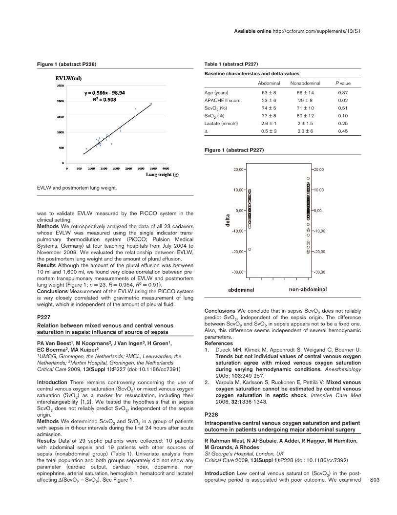
S93
was to validate EVLW measured by the PiCCO system in theclinical setting.Methods We retrospectively analyzed the data of all 23 cadaverswhose EVLW was measured using the single indicator trans-pulmonary thermodilution system (PiCCO; Pulsion MedicalSystems, Germany) at four teaching hospitals from July 2004 toNovember 2008. We evaluated the relationship between EVLW,the postmortem lung weight and the amount of plural effusion.Results Although the amount of the plural effusion was between10 ml and 1,600 ml, we found very close correlation between pre-mortem transpulmonary measurements of EVLW and postmortemlung weight (Figure 1; n = 23, R = 0.954, R2 = 0.91).Conclusions Measurement of the EVLW using the PiCCO systemis very closely correlated with gravimetric measurement of lungweight, which is independent of the amount of pleural fluid.
P227Relation between mixed venous and central venoussaturation in sepsis: influence of source of sepsis
PA Van Beest1, M Koopmans2, J Van Ingen3, H Groen1, EC Boerma2, MA Kuiper2
1UMCG, Groningen, the Netherlands; 2MCL, Leeuwarden, theNetherlands; 3Martini Hospital, Groningen, the NetherlandsCritical Care 2009, 13(Suppl 1):P227 (doi: 10.1186/cc7391)
Introduction There remains controversy concerning the use ofcentral venous oxygen saturation (ScvO2) or mixed venous oxygensaturation (SvO2) as a marker for resuscitation, including theirinterchangeability [1,2]. We tested the hypothesis that in sepsisScvO2 does not reliably predict SvO2, independent of the sepsisorigin.Methods We determined ScvO2 and SvO2 in a group of patientswith sepsis in 6-hour intervals during the first 24 hours after acuteadmission.Results Data of 29 septic patients were collected: 10 patientswith abdominal sepsis and 19 patients with other sources ofsepsis (nonabdominal group) (Table 1). Univariate analysis fromthe total population and both groups separately did not show anyparameter (cardiac output, cardiac index, dopamine, nor-epinephrine, arterial saturation, hemoglobin, hematocrit and lactate)affecting Δ(ScvO2 – SvO2). See Figure 1.
Conclusions We conclude that in sepsis ScvO2 does not reliablypredict SvO2, independent of the sepsis origin. The differencebetween ScvO2 and SvO2 in sepsis appears not to be a fixed one.Also, this difference seems independent of several hemodynamicparameters.References1. Dueck MH, Klimek M, Appenrodt S, Weigand C, Boerner U:
Trends but not individual values of central venous oxygensaturation agree with mixed venous oxygen saturationduring varying hemodynamic conditions. Anesthesiology2005; 103:249-257.
2. Varpula M, Karlsson S, Ruokonen E, Pettilä V: Mixed venousoxygen saturation cannot be estimated by central venousoxygen saturation in septic shock. Intensive Care Med2006, 32:1336-1343.
P228Intraoperative central venous oxygen saturation and patientoutcome in patients undergoing major abdominal surgery
R Rahman West, N Al-Subaie, A Addei, R Hagger, M Hamilton,M Grounds, A RhodesSt George’s Hospital, London, UKCritical Care 2009, 13(Suppl 1):P228 (doi: 10.1186/cc7392)
Introduction Low central venous saturation (ScvO2) in the post-operative period is associated with poor outcome. We examined
Available online http://ccforum.com/supplements/13/S1
Figure 1 (abstract P226)
EVLW and postmortem lung weight.
Figure 1 (abstract P227)
Table 1 (abstract P227)
Baseline characteristics and delta values
Abdominal Nonabdominal P value
Age (years) 63 ± 8 66 ± 14 0.37
APACHE II score 23 ± 6 29 ± 8 0.02
ScvO2 (%) 74 ± 5 71 ± 10 0.51
SvO2 (%) 77 ± 8 69 ± 12 0.10
Lactate (mmol/l) 2.6 ± 1 2 ± 1.5 0.25
Δ 0.5 ± 3 2.3 ± 6 0.45

S94
whether intraoperative ScvO2 is a predictor of outcome based onthe postoperative morbidity survey (POMS) and length of hospitalstay. The POMS is a method for describing complicationsassociated with major surgery [1]. It comprises a nine-point surveyand provides a generic measure of short-term postoperativeoutcome.Methods Prospective ScvO2 values were collected from patientsundergoing major abdominal surgery. This was done by intermittentsampling of blood obtained from the distal lumen of a centralvenous catheter placed in the jugular vein. The POMS was used toassess outcome on postoperative days 1, 3, 5, 8, 15 and 21 ifapplicable. To accommodate local practice, however, we excludedurinary catheter, pain and mobility from the POMS as it was not atrue reflection of organ dysfunction in our study population. Weevaluated other individual postsurgical complications as outlined inthe POMS to establish whether intraoperative median, minimumand ΔScvO2 (end of surgery value minus start of surgery value) arerelated to outcome.Results Fifty-two patients (average age 67.8, SD 12.0) wereincluded. The Portsmouth Physiological and Operative SeverityScore for the Enumeration of Mortality and Morbidity revealed apredicted morbidity of 54.2% and mortality of 3.3%. Thecommonest complications were gastrointestinal and infection(58.7% and 19.2%, respectively). We found no correlationbetween median, minimum and ΔScvO2 with postoperative compli-cation and length of hospital stay. However, ΔScvO2 for patientswho exhibited postoperative complication on day 3 was signifi-cantly different from the patients who did not (P = 0.035) but theactual difference was only 2%. Minimum ScvO2 correlated weaklywith high arterial lactate measured at the end of the procedure(Spearman r = 0.32, P = 0.025).Conclusions Intraoperative central venous saturation in patientsundergoing major abdominal surgery is not related to postoperativeoutcome or length of hospital stay.Reference1. Bennett-Guerrero E, et al.: The use of a postoperative mor-
bidity survey to evaluate patients with prolonged hospital-ization after routine, moderate-risk, elective surgery.Anesth Analg 1999, 89:514-519.
P229Bioelectrical impedance analysis in ICU patients
L Steinhilp1, F Bubser1, S Wiesener1, C Spies1, M Boschmann2,T Schütz1, S Weber-Carstens1
1Charite, Berlin, Germany; 2Max-Delbrueck-Centre, Berlin,GermanyCritical Care 2009, 13(Suppl 1):P229 (doi: 10.1186/cc7393)
Introduction Bioelectrical impedance analysis (BIA) is a widelyused method for calculating body compartments in healthysubjects and chronically ill patients. The resistance (R) – which iscorrelated with total body water – and the reactance (Xc) –depending on the capacitance of cell membranes – are measuredby BIA. The phase angle α, a mathematical relation between R andXc, has qualified as a prognostic marker in several severe diseases[1]. We aimed at monitoring body composition during the clinicalcourse of ICU patients and to investigate the impact of illnessseverity on BIA.Methods In this observational study we performed BIA testing atthe frequencies of 50 kHz unilaterally between the wrist and anklewith a body impedance analyzer. BIA of ICU patients took place onthree different measuring points: M1, M2 and M3 (days 2 to 3,days 10 to 11 and days 14 to 15 after admission). Severity ofillness was monitored by Simplified Acute Physiology Score
(SAPS) II. BIA results of each patient were matched with astandard collective according to sex, age, and body mass index.Spearman’s rho (rs) has been calculated to analyze correlationbetween α and SAPS II. Nonparametric analysis of longitudinaldata was carried out to analyze two groups differentiated by SAPSII (Group 1: ≤45 points, Group 2: >45 points).Results Forty-eight patients received one measurement at M1, 28patients were measured at all three measuring points. BIA of ourpatients differed considerably from the normal collective. Therewere significant differences between the patients grouped byprimary disease (abdominal cancer patients, nontumor patients,neurosurgical patients, multiple trauma and acute respiratorydistress syndrome). α is correlated with the ICU score SAPS II onadmission at all three measuring points (M1: n = 48, rs = –0.296,P <0.05; M4: n = 28, rs = –0.368, P <0.005; M5: n = 28, rs =–0.525, P <0.005). While we did not find a significant change of Rin our subsequent measurement, there was a decrease of Xc(P <0.05). Especially in the more serious ill patients (Group 2), thishas been observed (P <0.05).Conclusions BIA helps to estimate body composition at ICUadmission. It is associated with primary disease and severity ofillness. During the clinical course we observed a decrease of Xc,indicating loss of body cell mass.Reference1. Kyle UG, et al.: ESPEN Guidelines, bioelectrical impedance
analysis – part I: review of principles and methods. ClinNutr 2004, 23:1430-1453.
P230Estimated respiratory quotient and venoarterial pCO2difference are outcome markers in patients with leftventricular dysfunction submitted to coronary arterybypass surgery
L Hajjar, T Yamaguti, F Galas, R Kalil Filho, M Piccioni, J AulerHeart Institute, São Paulo, BrazilCritical Care 2009, 13(Suppl 1):P230 (doi: 10.1186/cc7394)
Introduction Experimental and clinical research has successfullyevaluated the performance of the respiratory quotient as a usefulmarker of anaerobic metabolism in shock from different causes.The aim of the present study was to evaluate the estimatedrespiratory quotient and venoarterial pCO2 difference as suitableanaerobic metabolism signs and outcome markers in patients withleft ventricular dysfunction undergoing coronary artery bypasssurgery.Methods A prospective study including 87 patients with leftventricular dysfunction undergoing coronary artery bypass surgerywith pump was performed from January 2006 to January 2008.Hemodynamic and metabolic parameters were obtained during fivemoments: after anesthesia induction and mechanical ventilationinitiation (T0), at end of surgery (T1), at admission to thepostsurgical ICU (T2), and 6 hours after (T3) and 12 hours afterICU admission (T4). The venoarterial carbon dioxide tensiondifference (ΔpCO2) and estimated respiratory quotient (eRQ) werealso calculated. Postoperative outcomes were compared regardingclinical events and mortality.Results In patients with unfavorable postoperative evolution, theeRQ values as venoarterial ΔpCO2 presented significantly highervalues at T4 compared with other groups (1.94 ± 0.9 vs. 1.43 ±0.65, P <0.05; 8.07 ± 3.24 vs. 5.66 ± 2.78, P <0.05). Patientswith unfavorable evolution had significant higher levels of arteriallactate concentration at 6 hours after ICU admission (4.30 ± 2.47vs. 2.72 ± 1.48, P <0.05). Stepwise logistic regression showedthat a higher age, higher estimated respiratory quotient at T4, and
Critical Care March 2009 Vol 13 Suppl 1 29th International Symposium on Intensive Care and Emergency Medicine
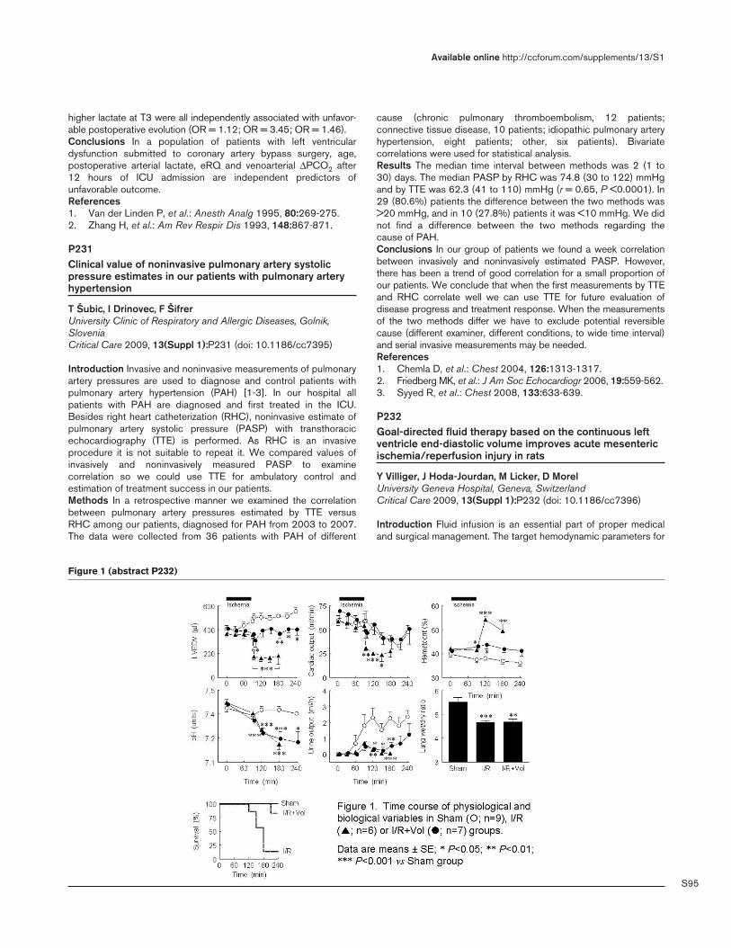
S95
higher lactate at T3 were all independently associated with unfavor-able postoperative evolution (OR = 1.12; OR = 3.45; OR = 1.46).Conclusions In a population of patients with left ventriculardysfunction submitted to coronary artery bypass surgery, age,postoperative arterial lactate, eRQ and venoarterial ΔPCO2 after12 hours of ICU admission are independent predictors ofunfavorable outcome.References1. Van der Linden P, et al.: Anesth Analg 1995, 80:269-275.2. Zhang H, et al.: Am Rev Respir Dis 1993, 148:867-871.
P231Clinical value of noninvasive pulmonary artery systolicpressure estimates in our patients with pulmonary arteryhypertension
T Šubic, I Drinovec, F ŠifrerUniversity Clinic of Respiratory and Allergic Diseases, Golnik,SloveniaCritical Care 2009, 13(Suppl 1):P231 (doi: 10.1186/cc7395)
Introduction Invasive and noninvasive measurements of pulmonaryartery pressures are used to diagnose and control patients withpulmonary artery hypertension (PAH) [1-3]. In our hospital allpatients with PAH are diagnosed and first treated in the ICU.Besides right heart catheterization (RHC), noninvasive estimate ofpulmonary artery systolic pressure (PASP) with transthoracicechocardiography (TTE) is performed. As RHC is an invasiveprocedure it is not suitable to repeat it. We compared values ofinvasively and noninvasively measured PASP to examinecorrelation so we could use TTE for ambulatory control andestimation of treatment success in our patients.Methods In a retrospective manner we examined the correlationbetween pulmonary artery pressures estimated by TTE versusRHC among our patients, diagnosed for PAH from 2003 to 2007.The data were collected from 36 patients with PAH of different
cause (chronic pulmonary thromboembolism, 12 patients;connective tissue disease, 10 patients; idiopathic pulmonary arteryhypertension, eight patients; other, six patients). Bivariatecorrelations were used for statistical analysis.Results The median time interval between methods was 2 (1 to30) days. The median PASP by RHC was 74.8 (30 to 122) mmHgand by TTE was 62.3 (41 to 110) mmHg (r = 0.65, P <0.0001). In29 (80.6%) patients the difference between the two methods was>20 mmHg, and in 10 (27.8%) patients it was <10 mmHg. We didnot find a difference between the two methods regarding thecause of PAH.Conclusions In our group of patients we found a week correlationbetween invasively and noninvasively estimated PASP. However,there has been a trend of good correlation for a small proportion ofour patients. We conclude that when the first measurements by TTEand RHC correlate well we can use TTE for future evaluation ofdisease progress and treatment response. When the measurementsof the two methods differ we have to exclude potential reversiblecause (different examiner, different conditions, to wide time interval)and serial invasive measurements may be needed.References1. Chemla D, et al.: Chest 2004, 126:1313-1317.2. Friedberg MK, et al.: J Am Soc Echocardiogr 2006, 19:559-562.3. Syyed R, et al.: Chest 2008, 133:633-639.
P232Goal-directed fluid therapy based on the continuous leftventricle end-diastolic volume improves acute mesentericischemia/reperfusion injury in rats
Y Villiger, J Hoda-Jourdan, M Licker, D MorelUniversity Geneva Hospital, Geneva, SwitzerlandCritical Care 2009, 13(Suppl 1):P232 (doi: 10.1186/cc7396)
Introduction Fluid infusion is an essential part of proper medicaland surgical management. The target hemodynamic parameters for
Available online http://ccforum.com/supplements/13/S1
Figure 1 (abstract P232)
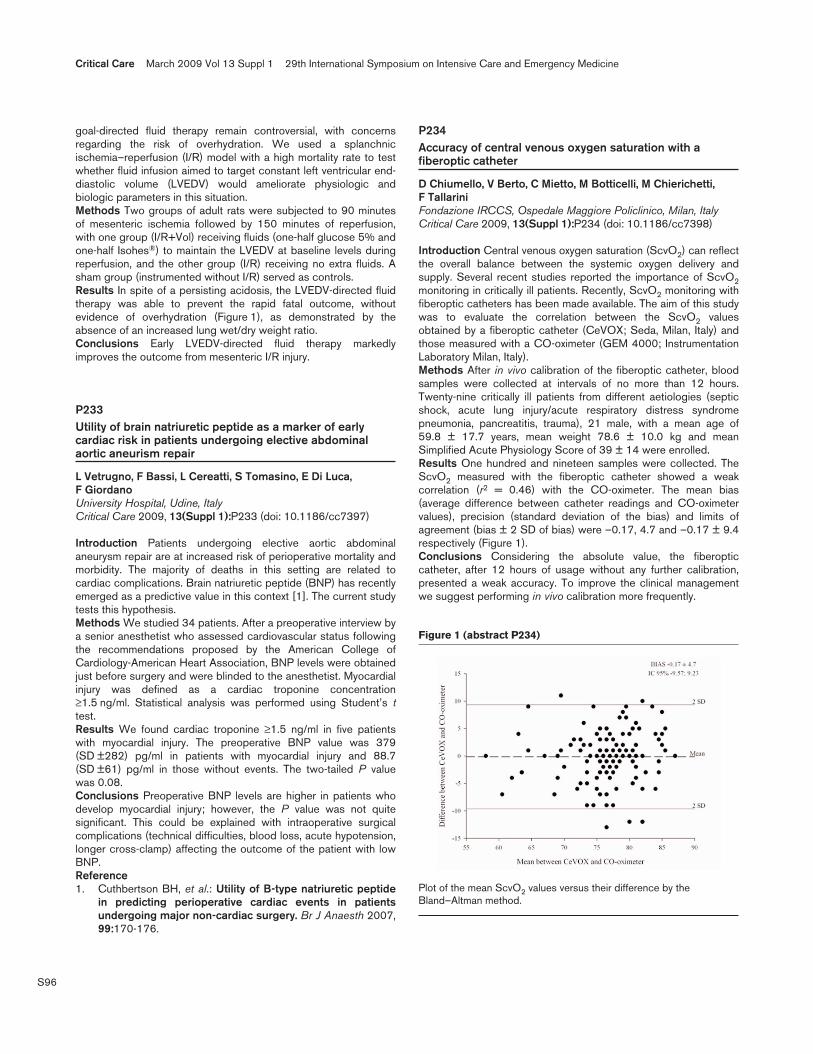
S96
goal-directed fluid therapy remain controversial, with concernsregarding the risk of overhydration. We used a splanchnicischemia–reperfusion (I/R) model with a high mortality rate to testwhether fluid infusion aimed to target constant left ventricular end-diastolic volume (LVEDV) would ameliorate physiologic andbiologic parameters in this situation.Methods Two groups of adult rats were subjected to 90 minutesof mesenteric ischemia followed by 150 minutes of reperfusion,with one group (I/R+Vol) receiving fluids (one-half glucose 5% andone-half Isohes®) to maintain the LVEDV at baseline levels duringreperfusion, and the other group (I/R) receiving no extra fluids. Asham group (instrumented without I/R) served as controls.Results In spite of a persisting acidosis, the LVEDV-directed fluidtherapy was able to prevent the rapid fatal outcome, withoutevidence of overhydration (Figure 1), as demonstrated by theabsence of an increased lung wet/dry weight ratio.Conclusions Early LVEDV-directed fluid therapy markedlyimproves the outcome from mesenteric I/R injury.
P233Utility of brain natriuretic peptide as a marker of earlycardiac risk in patients undergoing elective abdominalaortic aneurism repair
L Vetrugno, F Bassi, L Cereatti, S Tomasino, E Di Luca, F GiordanoUniversity Hospital, Udine, ItalyCritical Care 2009, 13(Suppl 1):P233 (doi: 10.1186/cc7397)
Introduction Patients undergoing elective aortic abdominalaneurysm repair are at increased risk of perioperative mortality andmorbidity. The majority of deaths in this setting are related tocardiac complications. Brain natriuretic peptide (BNP) has recentlyemerged as a predictive value in this context [1]. The current studytests this hypothesis.Methods We studied 34 patients. After a preoperative interview bya senior anesthetist who assessed cardiovascular status followingthe recommendations proposed by the American College ofCardiology-American Heart Association, BNP levels were obtainedjust before surgery and were blinded to the anesthetist. Myocardialinjury was defined as a cardiac troponine concentration≥1.5 ng/ml. Statistical analysis was performed using Student’s ttest.Results We found cardiac troponine ≥1.5 ng/ml in five patientswith myocardial injury. The preoperative BNP value was 379(SD ±282) pg/ml in patients with myocardial injury and 88.7(SD ±61) pg/ml in those without events. The two-tailed P valuewas 0.08.Conclusions Preoperative BNP levels are higher in patients whodevelop myocardial injury; however, the P value was not quitesignificant. This could be explained with intraoperative surgicalcomplications (technical difficulties, blood loss, acute hypotension,longer cross-clamp) affecting the outcome of the patient with lowBNP.Reference1. Cuthbertson BH, et al.: Utility of B-type natriuretic peptide
in predicting perioperative cardiac events in patientsundergoing major non-cardiac surgery. Br J Anaesth 2007,99:170-176.
P234Accuracy of central venous oxygen saturation with afiberoptic catheter
D Chiumello, V Berto, C Mietto, M Botticelli, M Chierichetti, F TallariniFondazione IRCCS, Ospedale Maggiore Policlinico, Milan, ItalyCritical Care 2009, 13(Suppl 1):P234 (doi: 10.1186/cc7398)
Introduction Central venous oxygen saturation (ScvO2) can reflectthe overall balance between the systemic oxygen delivery andsupply. Several recent studies reported the importance of ScvO2monitoring in critically ill patients. Recently, ScvO2 monitoring withfiberoptic catheters has been made available. The aim of this studywas to evaluate the correlation between the ScvO2 valuesobtained by a fiberoptic catheter (CeVOX; Seda, Milan, Italy) andthose measured with a CO-oximeter (GEM 4000; InstrumentationLaboratory Milan, Italy).Methods After in vivo calibration of the fiberoptic catheter, bloodsamples were collected at intervals of no more than 12 hours.Twenty-nine critically ill patients from different aetiologies (septicshock, acute lung injury/acute respiratory distress syndromepneumonia, pancreatitis, trauma), 21 male, with a mean age of59.8 ± 17.7 years, mean weight 78.6 ± 10.0 kg and meanSimplified Acute Physiology Score of 39 ± 14 were enrolled.Results One hundred and nineteen samples were collected. TheScvO2 measured with the fiberoptic catheter showed a weakcorrelation (r2 = 0.46) with the CO-oximeter. The mean bias(average difference between catheter readings and CO-oximetervalues), precision (standard deviation of the bias) and limits ofagreement (bias ± 2 SD of bias) were –0.17, 4.7 and –0.17 ± 9.4respectively (Figure 1).Conclusions Considering the absolute value, the fiberopticcatheter, after 12 hours of usage without any further calibration,presented a weak accuracy. To improve the clinical managementwe suggest performing in vivo calibration more frequently.
Critical Care March 2009 Vol 13 Suppl 1 29th International Symposium on Intensive Care and Emergency Medicine
Figure 1 (abstract P234)
Plot of the mean ScvO2 values versus their difference by theBland–Altman method.
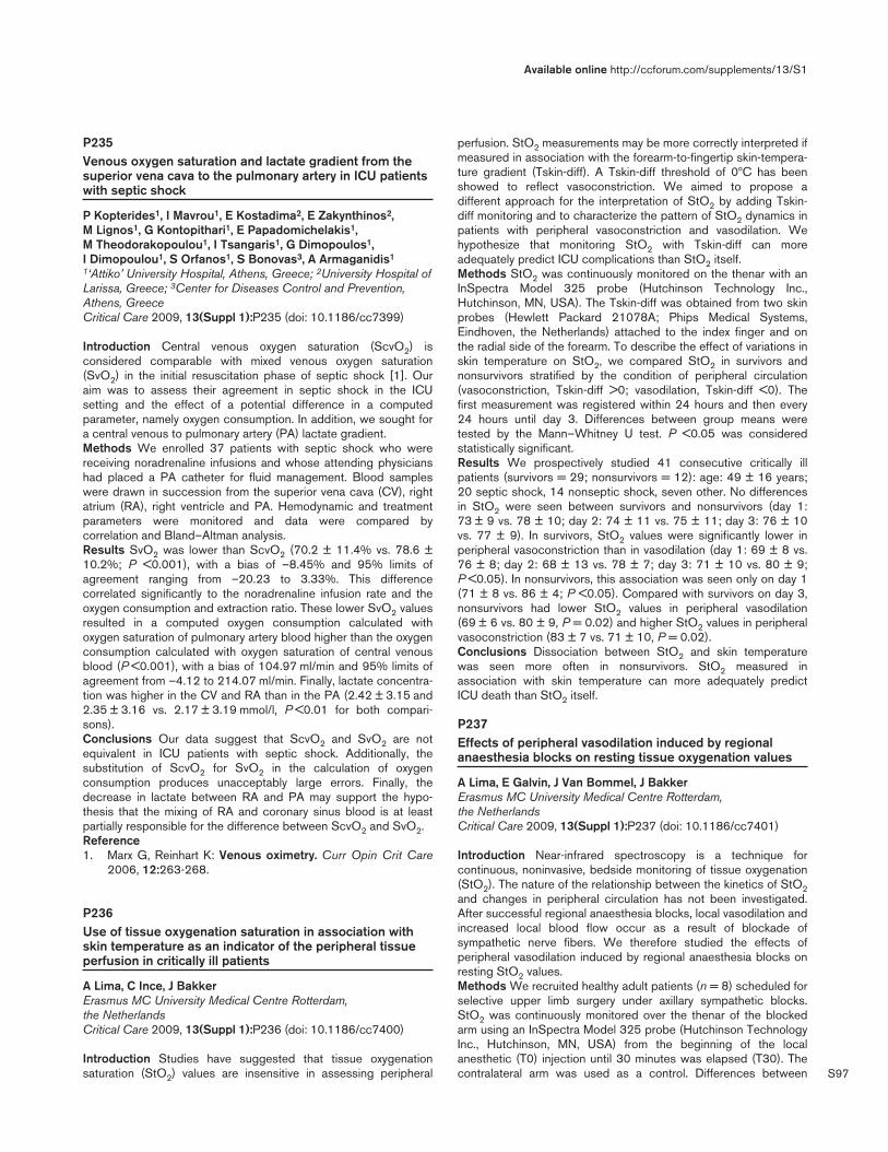
S97
P235Venous oxygen saturation and lactate gradient from thesuperior vena cava to the pulmonary artery in ICU patientswith septic shock
P Kopterides1, I Mavrou1, E Kostadima2, E Zakynthinos2, M Lignos1, G Kontopithari1, E Papadomichelakis1, M Theodorakopoulou1, I Tsangaris1, G Dimopoulos1, I Dimopoulou1, S Orfanos1, S Bonovas3, A Armaganidis1
1‘Attiko’ University Hospital, Athens, Greece; 2University Hospital ofLarissa, Greece; 3Center for Diseases Control and Prevention,Athens, GreeceCritical Care 2009, 13(Suppl 1):P235 (doi: 10.1186/cc7399)
Introduction Central venous oxygen saturation (ScvO2) isconsidered comparable with mixed venous oxygen saturation(SvO2) in the initial resuscitation phase of septic shock [1]. Ouraim was to assess their agreement in septic shock in the ICUsetting and the effect of a potential difference in a computedparameter, namely oxygen consumption. In addition, we sought fora central venous to pulmonary artery (PA) lactate gradient.Methods We enrolled 37 patients with septic shock who werereceiving noradrenaline infusions and whose attending physicianshad placed a PA catheter for fluid management. Blood sampleswere drawn in succession from the superior vena cava (CV), rightatrium (RA), right ventricle and PA. Hemodynamic and treatmentparameters were monitored and data were compared bycorrelation and Bland–Altman analysis.Results SvO2 was lower than ScvO2 (70.2 ± 11.4% vs. 78.6 ±10.2%; P <0.001), with a bias of –8.45% and 95% limits ofagreement ranging from –20.23 to 3.33%. This differencecorrelated significantly to the noradrenaline infusion rate and theoxygen consumption and extraction ratio. These lower SvO2 valuesresulted in a computed oxygen consumption calculated withoxygen saturation of pulmonary artery blood higher than the oxygenconsumption calculated with oxygen saturation of central venousblood (P <0.001), with a bias of 104.97 ml/min and 95% limits ofagreement from –4.12 to 214.07 ml/min. Finally, lactate concentra-tion was higher in the CV and RA than in the PA (2.42 ± 3.15 and2.35 ± 3.16 vs. 2.17 ± 3.19 mmol/l, P <0.01 for both compari-sons).Conclusions Our data suggest that ScvO2 and SvO2 are notequivalent in ICU patients with septic shock. Additionally, thesubstitution of ScvO2 for SvO2 in the calculation of oxygenconsumption produces unacceptably large errors. Finally, thedecrease in lactate between RA and PA may support the hypo-thesis that the mixing of RA and coronary sinus blood is at leastpartially responsible for the difference between ScvO2 and SvO2.Reference1. Marx G, Reinhart K: Venous oximetry. Curr Opin Crit Care
2006, 12:263-268.
P236Use of tissue oxygenation saturation in association withskin temperature as an indicator of the peripheral tissueperfusion in critically ill patients
A Lima, C Ince, J BakkerErasmus MC University Medical Centre Rotterdam, the NetherlandsCritical Care 2009, 13(Suppl 1):P236 (doi: 10.1186/cc7400)
Introduction Studies have suggested that tissue oxygenationsaturation (StO2) values are insensitive in assessing peripheral
perfusion. StO2 measurements may be more correctly interpreted ifmeasured in association with the forearm-to-fingertip skin-tempera-ture gradient (Tskin-diff). A Tskin-diff threshold of 0ºC has beenshowed to reflect vasoconstriction. We aimed to propose adifferent approach for the interpretation of StO2 by adding Tskin-diff monitoring and to characterize the pattern of StO2 dynamics inpatients with peripheral vasoconstriction and vasodilation. Wehypothesize that monitoring StO2 with Tskin-diff can moreadequately predict ICU complications than StO2 itself.Methods StO2 was continuously monitored on the thenar with anInSpectra Model 325 probe (Hutchinson Technology Inc.,Hutchinson, MN, USA). The Tskin-diff was obtained from two skinprobes (Hewlett Packard 21078A; Phips Medical Systems,Eindhoven, the Netherlands) attached to the index finger and onthe radial side of the forearm. To describe the effect of variations inskin temperature on StO2, we compared StO2 in survivors andnonsurvivors stratified by the condition of peripheral circulation(vasoconstriction, Tskin-diff >0; vasodilation, Tskin-diff <0). Thefirst measurement was registered within 24 hours and then every24 hours until day 3. Differences between group means weretested by the Mann–Whitney U test. P <0.05 was consideredstatistically significant.Results We prospectively studied 41 consecutive critically illpatients (survivors = 29; nonsurvivors = 12): age: 49 ± 16 years;20 septic shock, 14 nonseptic shock, seven other. No differencesin StO2 were seen between survivors and nonsurvivors (day 1:73 ± 9 vs. 78 ± 10; day 2: 74 ± 11 vs. 75 ± 11; day 3: 76 ± 10vs. 77 ± 9). In survivors, StO2 values were significantly lower inperipheral vasoconstriction than in vasodilation (day 1: 69 ± 8 vs.76 ± 8; day 2: 68 ± 13 vs. 78 ± 7; day 3: 71 ± 10 vs. 80 ± 9;P <0.05). In nonsurvivors, this association was seen only on day 1(71 ± 8 vs. 86 ± 4; P <0.05). Compared with survivors on day 3,nonsurvivors had lower StO2 values in peripheral vasodilation(69 ± 6 vs. 80 ± 9, P = 0.02) and higher StO2 values in peripheralvasoconstriction (83 ± 7 vs. 71 ± 10, P = 0.02).Conclusions Dissociation between StO2 and skin temperaturewas seen more often in nonsurvivors. StO2 measured inassociation with skin temperature can more adequately predictICU death than StO2 itself.
P237Effects of peripheral vasodilation induced by regionalanaesthesia blocks on resting tissue oxygenation values
A Lima, E Galvin, J Van Bommel, J BakkerErasmus MC University Medical Centre Rotterdam, the NetherlandsCritical Care 2009, 13(Suppl 1):P237 (doi: 10.1186/cc7401)
Introduction Near-infrared spectroscopy is a technique forcontinuous, noninvasive, bedside monitoring of tissue oxygenation(StO2). The nature of the relationship between the kinetics of StO2and changes in peripheral circulation has not been investigated.After successful regional anaesthesia blocks, local vasodilation andincreased local blood flow occur as a result of blockade ofsympathetic nerve fibers. We therefore studied the effects ofperipheral vasodilation induced by regional anaesthesia blocks onresting StO2 values.Methods We recruited healthy adult patients (n = 8) scheduled forselective upper limb surgery under axillary sympathetic blocks.StO2 was continuously monitored over the thenar of the blockedarm using an InSpectra Model 325 probe (Hutchinson TechnologyInc., Hutchinson, MN, USA) from the beginning of the localanesthetic (T0) injection until 30 minutes was elapsed (T30). Thecontralateral arm was used as a control. Differences between
Available online http://ccforum.com/supplements/13/S1

S98
group means were tested by Wilcoxon signed test. P <0.05 wasconsidered statistically significant.Results StO2 values in the blocked arm were significantly higher inall patients after the anaesthesia (Figure 1). T30 versus T0, 94 ± 2versus 82 ± 3; P = 0.002. StO2 did not increase in the controlarm.Conclusions Peripheral vasodilation increases StO2 in normalconditions.
P238Effect of anemia on tissue oxygenation saturation and thetissue deoxygenation rate during ischemia
M Meznar, R Pareznik, G VogaSB Celje, SloveniaCritical Care 2009, 13(Suppl 1):P238 (doi: 10.1186/cc7402)
Introduction The hypothesis was that anemia, independently ofhemodynamic stability, affects tissue oxygenation saturation (StO2)and the deoxygenation rate during stagnant ischemia. The bloodhemoglobin concentration is determinant of oxygen delivery. Inanemic patients, oxygen delivery decreases and oxygen extractionis increased. This leads to decreased venous hemoglobinsaturation and a lower tissue oxygen saturation. The rate of tissuedeoxygenation during ischemia is dependent on oxygen con-sumption and on the amount of oxygen available in the tissue [1].Methods In a prospective observational study we included 340patients in the medical emergency room. On admission, StO2 andthe tissue deoxygenation rate during ischemia were measured bynear-infrared spectroscopy. Patients were divided into four groupsaccording to hemoglobin concentration and hemodynamic (HD)stability: Group 1 (nonanemic, HD-stable patients), Group 2(anemic, HD-stable patients), Group 3 (nonanemic, HD-unstablepatients), Group 4 (anemic, HD-unstable patients). Differences inStO2 and the rate of tissue deoxygenation were analyzed.Results Anemic groups had a significantly lower hemoglobinconcentration compared with nonanemic groups (138 ± 16 vs.
76.5 ± 15 g/l, P <0.001). HD-unstable groups had significantlyhigher lactate levels compared with HD-stable groups (1.6 ± 0.6vs. 3.9 ± 1.6 mmol/l, P <0.001). The results are presented in Table1. StO2 in Groups 2 and 3 was comparable, while the difference inthe deoxygenation rate in these groups was significant(P = 0.007).Conclusions Anemia significantly affects StO2 and the deoxy-genation rate. It probably contributes to lower StO2 equally as HDinstability. In contrast to HD instability, anemia causes more rapiddeoxygenation during ischemia.Reference1. Creteur J: Muscle StO2 in critically ill patients. Curr Opin
Crit Care 2008, 14:361-366.
P239Near-infrared spectroscopy monitoring tissue oxygensaturation after cardiac surgery
R Kopp, S Rex, K Dommann, G Schälte, G Dohmen, G Marx, R RossaintUniversity Hospital Aachen, GermanyCritical Care 2009, 13(Suppl 1):P239 (doi: 10.1186/cc7403)
Introduction The aim of this study was to compare near-infraredspectroscopy with global parameters of tissue oxygenation aftercardiac surgery, such as cardiac output (CI), mixed venous oxygensaturation (SvO2) or lactate concentration. After cardiac surgery,the circulating blood volume and cardiac function is regularlyreduced on the ICU [1]. This results in reduced microperfusion andperipheral vasoconstriction. The noninvasive InSpectra StO2monitor (Hutchinson Technology Inc., Hutchinson, MN, USA)measures the oxygen saturation in the microcirculation of thethenar muscle [2].Methods Forty patients after cardiac surgery were monitored witha StO2 monitor and a Swan–Ganz catheter measuring CI andSvO2 on the ICU. Additionally intermittent lactate and blood gasanalysis was performed. ANOVA was used for statistics of results.Results The mean Euro-Score of the patients was 6.5 ± 3.7including 24 aortocoronary bypass, five heart valve, three ascen-ding aorta and eight combined cardiac operations. After admission
Critical Care March 2009 Vol 13 Suppl 1 29th International Symposium on Intensive Care and Emergency Medicine
Figure 1 (abstract P237)
Effect of peripheral blood flow variations in StO2 during regional block.
Table 1 (abstract P238)
Group 1, n = 251 Group 2, n = 30 P value Group 3, n = 48 Group 4, n = 10 P value
StO2 (%) 80.5 ± 7.8 76 ± 7.7 0.003 76.5 ± 11.9 68.2 ± 9.6 0.04
Deoxygenation rate (%/min) 16.5 ± 6.7 19.6 ± 6.6 0.017 14.8 ± 7.4 16.7 ± 5.7 NS
Figure 1 (abstract P239)
Course of StO2, cardiac output, SvO2 and lactate concentration.

S99
StO2, CI and SvO2 were significantly reduced (Figure 1). Thelactate concentration and noradrenaline dose were increased(P <0.05). On day 1 the noradrenaline dose dropped after fluidsubstitution, but only the StO2 value recovered.Conclusions After heart surgery StO2 indicated the reduction aswell as the recovery of microcirculation early, whereas the lactateconcentration and SvO2 seemed to demonstrate a delayedresponse especially of recovery. Management of postoperativefluid and catecholamine therapy by StO2 to optimize micro-circulation should be the subject of further studies.References1. Tschaikowsky K, et al.: Changes in circulating blood volume
after cardiac surgery measured by a novel method usinghydroxyethyl starch. Crit Care Med 2000, 28:336-334.
2. Myers DE, et al.: Noninvasive method for measuring localhemoglobin oxygen saturation in tissue using wide gapsecond derivative near-infrared spectroscopy. J BiomedOpt 2005, 10:034017.
P240Comparison of muscle tissue oxygenation responsecurves to two time-based vascular occlusion tests:evidence of diminishing returns?
SJ Thomson, N Al-Subaie, M Hamilton, ML Cowan, S Musa, M Grounds, TM RahmanSt George’s Hospital NHS Trust, London, UKCritical Care 2009, 13(Suppl 1):P240 (doi: 10.1186/cc7404)
Introduction Dynamic testing of muscle tissue oxygenation (StO2)with near-infrared spectroscopy and vascular occlusion (VOT) hasbeen used to study pathophysiological states, but there is apaucity of data for standardised techniques in normal subjects.Three-minute VOT is frequently described. We have collectedStO2 data for this technique and compared them with a shorter2-minute test.Methods Twenty-five subjects were studied using an InSpectra650 monitor and a 15 mm probe. VOT was applied at systolicblood pressure + 50 mmHg on opposite arms for 3-minute and2-minute time periods. StO2 data are reported for baseline,downslope (DS) and upslope (US)%/minute, overshoot%, areaover the ischaemic curve (AOC Isch) and area under the recoverycurve (AUC Rec)%/minute and recovery time (Rec Time) inminutes. Differences were analysed using paired t tests. Individualdifferences were then examined in relation to a predicted magni-tude of response based on the ischaemic load and occlusion time.Results Three-minute VOT versus 2 minutes: AOC Isch (–52.99vs. –25.79%/min, P <0.001), overshoot (14.96 vs. 13.2%,P = 0.004), AUC Rec (18.89 vs. 15.33%/min, P = 0.01), RecTime (2.37 vs. 2.05 min, P = 0.004). DS (–11.18 vs.–12.35%/min, P = 0.08) and US (212.99 vs. 201.24%/min,P = 0.365) were not significantly different. The 2-minute ischaemic
load was 0.5 times that of 3 minutes (AOC Isch –25.79/–52.99).The ratios of response parameters were compared with this valueusing one-sample t tests and were significantly greater: overshoot(0.89 ± 0.16, P <0.001), AUC Rec (0.86 ± 0.35, P <0.001), RecTime (0.88 ± 0.21, P <0.001).Conclusions StO2 VOT is a promising tool that is likely to advanceour knowledge of microvascular function under ischaemic stress.To standardise data interpretation it is imperative that similartechniques are used. We have reported data for two reproducibleVOTs in a healthy control group. Understandably, there aredifferences in hyperaemic response parameters that are dependenton the ischaemic time; however, the relationship is no linear. Ourdata suggest that although 2-minute VOT represents a halving ofthe ischaemic load versus 3 minutes, subjects demonstrate almost90% comparable hyperaemic responses. This suggests adiminishing return in hyperaemia with longer occlusive time. Furtherwork is required to evaluate the efficacy and practicality of differentVOTs.
P241Adaptability of muscle tissue oxygenation to repeatedvascular occlusion
SJ Thomson, N Al-Subaie, M Hamilton, ML Cowan, S Musa, RM Grounds, TM RahmanSt George’s Hospital NHS Trust, London, UKCritical Care 2009, 13(Suppl 1):P241 (doi: 10.1186/cc7405)
Introduction Dynamic testing of muscle tissue oxygenation (StO2)with near-infrared spectroscopy and vascular occlusion (VOT) is adeveloping area of research in critical care. VOT is often performedmultiple times during analysis. We report the impact of repeatedVOT on StO2 parameters.Methods Twenty-five healthy subjects were studied with theInSpectra 650 monitor and a 15 mm probe. VOT was set atsystolic blood pressure + 50 mmHg for 3 minutes in triplicate.Each VOT was separated by a 5-minute rest period. StO2 data arereported for baseline%, downslope (DS) and upslope (US)%/min,overshoot%, area over the ischaemic curve (AOC Isch) and areaunder the recovery curve (AUC Rec)%/min and recovery time (RecTime) in minutes. Data were compared by ANOVA.Results See Table 1.Conclusions It is important to understand the impact of serialVOT. We have observed sequential change in rates of StO2decline during ischaemia (reduced DS gradients, total drop andAOC Isch). Although the overshoot was unchanged, the AUC Recand Rec time demonstrated increases approaching significance.These patterns imply a preconditioned adaptation to repeated VOTand an exaggerated hyperaemic response. Further studies arerequired to determine acceptable rest periods between VOTs toameliorate these changes.
Available online http://ccforum.com/supplements/13/S1
Table 1 (abstract P241)
Population StO2 means (±SD) for consecutive VOT and significance
StO2 curve Baseline Total drop DS US AOC Isch Overshoot AUC Rec Rec Time parameter (%) (%) (%/min) (%/min) (%/min) (%) (%/min) (min)
VOT 1 78.7 (±4.2) 33.4 (±7) –11.2 (±2.5) 213 (±55.8) –53 (±11.5) 15 (±3.8) 18.9 (±6.6) 2.4 (±0.4)
VOT 2 78.3 (±4.2) 29.8 (±6.2) –10 (±2.3) 240 (±58.8) –48.2 (±10.7) 14.9 (±3.7) 21.7 (±7.5) 2.6 (±0.4)
VOT 3 77.9 (±4) 28.2 (±5.5) –9.4 (±1.8) 220 (±45.3) –44.5 (±8.7) 15.3 (±3.5) 23.4 (±7.3) 2.6 (±0.5)
P value 0.79 0.01 0.02 0.19 0.02 0.92 0.09 0.09

S100
P242Microcirculatory, leucocyte/endothelium interaction andsurvival time effects of dobutamine in nonhypotensiveendotoxemia
A Santos, E Furtado, N Villela, E BouskelaState University of Rio de Janeiro, BrazilCritical Care 2009, 13(Suppl 1):P242 (doi: 10.1186/cc7406)
Introduction Microcirculatory, leucocyte/endothelium interactionand survival time effects of dobutamine in the dose of 5 μg/kg/minute associated or not with volume resuscitation were studied inthe hamster window chamber model during resuscitation fromnonhypotensive endotoxemia [1,2].Methods Awake hamsters were submitted to endotoxemia withintravenous injection of lipopolysaccharide (LPS) in the dose of2 mg/kg. After 3 hours of LPS injection they were divided into fourgroups: LPS (n = 6): received no treatment; VR (n = 6):resuscitated with 40 ml/kg body weight of NaCl 0.9% in 1 hourfollowed by 20 ml/kg body weight during the remaining 4 hours;Dobuta (n = 6): received dobutamine infusion in the dose of5 μg/kg/minute for 3 hours; Dobuta/VR (n = 6): resuscitated with40 ml/kg body weight of NaCl 0.9% in the first hour followed by20 ml/kg body weight during the remaining 2 hours, the lastcombined with dobutamine in the dose of 5 μg/kg/minute. Thegroups were compared with Control (n = 7): no treatment. Arteriolarand venular diameters, functional capillary density (FCD), leukocyterolling and adhesion, and 72-hour survival time were evaluated.Results Dobuta had lower arteriolar diameter than Control (51 ± 10and 114 ± 10% from baseline). LPS and Dobuta had lower FCDthan Control and baseline values (18 ± 15; 16 ± 18; and 88 ± 6%from baseline, in LPS, Dobuta and Control, respectively). VR andVR/Dobuta restored FCD from baseline (382 ± 19 and 476 ± 30%from baseline in VR and VR/Dobuta, respectively). FCD in VR andVR/Dobuta were lower than Control. LPS and Dobuta had higherleucocytes adhesion than Control (42.2 ± 10; 32.2 ± 31; and 4.0 ±7.1 leucocytes/mm2 in LPS, Dobuta and Control groups, respec-tively). There was no significant difference in survival time betweenVR and Control, and VR/Dobuta and Control. Survival time wassignificantly lower in LPS and Dobuta than Control.Conclusions Dobutamine associated or not with volumeresuscitation did not improve microcirculatory parameters,leucocyte adhesion or survival time during resuscitation fromnonhypotensive endotoxemia while volume resuscitation restoredmicrocirculatory parameters and improved survival time.References1. Secchi A, et al.: Dobutamine maintains intestinal villus
blood flow during normotensive endotoxemia: an intravitalmicroscopic study in the rat. J Crit Care 1997, 12:137-141.
2. Hiltebrand LB, et al.: Effects of dopamine, dobutamine, anddopexamine on microcirculatory blood flow in the gas-trointestinal tract during sepsis and anesthesia. Anesthesi-ology 2004, 100:1188-1197.
P243Microcirculatory, leukocyte/endothelium interaction andsurvival time effects of recombinant C-reactive protein innonhypotensive endotoxemia in hamsters
M Silva, A Santos, E Furtado, N Villela, E BouskelaState University of Rio de Janeiro, BrazilCritical Care 2009, 13(Suppl 1):P243 (doi: 10.1186/cc7407)
Introduction Microcirculatory, leukocyte/endothelium interactionand 7-day survival time effects of human recombinant C-reactive
protein (rCRP) associated or not with volume resuscitation werestudied in the hamster window chamber model during resusci-tation from nonhypotensive endotoxemia [1,2].Methods Awake hamsters subjected to endotoxemia withintravenous injection of lipopolysaccharide (LPS) (2 mg/kg) weredivided, 1 hour after LPS injection, into four groups: LPS (n = 6):no treatment; VR (n = 6): received 40 ml/kg body weight of NaCl0.9% in 1 hour followed by 20 ml/kg body weight during theremaining 4 hours; rCRP (n = 6): received rCRP infusion in thedose of 24 μg/kg/hour during 5 hours; VR/rCRP (n = 6): received40 ml/kg body weight of NaCl 0.9% in the first hour followed by 20ml/kg body weight during the remaining 4 hours, and in the lastperiod it was combined with rCRP infusion in the dose of 24μg/kg/hour. Groups were compared with Control (n = 6): no LPS.Arteriolar and venular diameters, functional capillary density (FCD),venular leukocyte rolling and adhesion, and 7-day survival timewere evaluated.Results LPS reduced FCD so it was maintained lower thanControl and baseline even after 24 hours (13 ± 11 and 100 ± 7%from baseline in LPS and Control, respectively). Volumeresuscitation and rCRP restored FCD to baseline levels but it waslower than Control after 24 hours (36 ± 25 and 32 ± 20% frombaseline in VR and rCRP, respectively). VR/rCRP restored FCDand it was not different from Control (72 ± 26% from baseline inrCRP). Arteriolar and venular diameters were not different amonggroups. LPS increased the number of sticking leukocytes to thevenular wall (41.2 ± 13 and 1.1 ± 1 leucocytes/mm2 in LPS andControl, respectively). rCRP and VR/rCRP significantly reducedvenular leukocyte adhesion (12.6 ± 7.1 and 11.8 ± 9.5leucocytes/mm2 in rCRP and VR/rCRP, respectively). There wasno difference in rolling leukocytes among groups. The survivalcurve was not significantly different in rCRP, VR and VR/rCRPfrom Control.Conclusions rCRP associated or not with volume resuscitationimproved tissue perfusion, reduced the number of stickingleukocytes and increased the survival time during endotoxemia inthe hamster model.References1. Joyce DE, et al.: Leukocyte and endothelial cell interactions
in sepsis: relevance of the protein C pathway. Crit CareMed 2004, 32(5 Suppl):S280-S286.
2. Iba T, et al.: Activated protein C improves the visceralmicrocirculation by attenuating the leukocyte–endothelialinteraction in a rat lipopolysaccharide model. Crit CareMed 2005, 33:368-372.
P244Assessment of tissue oxygen saturation during a vascularocclusion test using near-infrared spectroscopy: role of theprobe spacing and measurement site studied in healthyvolunteers
R Bezemer1, A Lima2, E Klijn2, J Bakker2, C Ince1
1Academic Medical Center, Amsterdam, the Netherlands; 2ErasmusMC, Rotterdam, the NetherlandsCritical Care 2009, 13(Suppl 1):P244 (doi: 10.1186/cc7408)
Introduction To assess potential metabolic and microcirculatoryalterations in critically ill patients, near-infrared spectroscopy(NIRS) has been used, in combination with a vascular occlusiontest (VOT), for the noninvasive measurement of tissue oxygensaturation (StO2), oxygen consumption, and microvascular reper-fusion and reactivity. However, the methodologies for assessingStO2 are very inconsistent in the literature and results vary fromstudy to study. In this study we investigated the effects of the
Critical Care March 2009 Vol 13 Suppl 1 29th International Symposium on Intensive Care and Emergency Medicine

S101
probe spacing and measurement site, using both a 15 mm and a25 mm probe spacing on the thenar and the forearm, in healthyvolunteers.Methods StO2 was noninvasively measured in eight healthyvolunteers during 3-minute VOTs using two InSpectra TissueSpectrometers equipped with either a 15 mm or a 25 mm probe.VOT-derived StO2 traces were analyzed for baseline, ischemic,reperfusion, and hyperemic parameters.Results Although not apparent at baseline, the probe spacing andmeasurement site significantly influenced VOT-derived StO2parameters (Figures 1 and 2). StO2 parameters in the hyperemicphase of the VOT were shown to significantly correlate to theminimum StO2 value after 3 minutes of ischemia.Conclusions The present study showed that NIRS measurementsin combination with a VOT are measurement site and probedependent. Additionally, this study indicated that reactivehyperemia depends on the extent of ischemic hit and supports theuse of a target StO2 over the use of a fixed time of occlusion.
P245Effects of perfusion pressure on microcirculatory perfusionof gastric tubes in pigs
E Klijn, S Niehof, J De Jonge, J Bakker, C Ince, J Van BommelErasmus Medical Center, Rotterdam, the NetherlandsCritical Care 2009, 13(Suppl 1):P245 (doi: 10.1186/cc7409)
Introduction It is known that formation of a gastric tube afteresophagectomy impairs microcirculatory perfusion in the anasto-motic region. The impaired microcirculatory perfusion is caused bypoor arterial inflow and insufficient venous drainage, due to ligationof three of the four gastric arteries. We therefore hypothesized thatimproving venous drainage by giving nitroglycerin (NTG) andtitrating norepinephrine (NE) to increasing levels of mean arterialpressure (MAP) would improve microcirculatory perfusion of thedistal part of the gastric tube.Methods Five pigs, bodyweight 30.5 ± 0.5 kg (mean ± SD) wereanaesthesized and instrumented for continuous hemodynamicmonitoring. A median laparotomy was performed and a gastrictube was reconstructed. Laser speckle imaging was used tomeasure the microcirculatory blood flow (MBF) in the gastric tube.Laser speckle imaging is a laser-based technique that measuresMBF in a macroscopic field at a video frame rate. NTG was titratedto in order to achieve a central venous pressure below 10 mmHg.MBF was measured at the base and at the distal site of the gastrictube at MAP increasing stepwise from 50 to 110 mmHg. For thispurpose the MAP was decreased with infusion of propofol andincreased with infusion of NE. We determined the flow at the distalsite for each step compared with flow at the base of the gastrictube (fraction of flow) in percentages.Results Hemodynamic values: MAP was increased in steps of10 mmHg. The heart rate and cardiac output remained constant ataround 123 ± 28 bpm and 4.2 ± 1.0 l/min, respectively. Centralvenous pressure and pulmonary artery occlusion pressure did notchange throughout the experiment. MBF results: at MAP below70 mmHg the fraction of flow at the distal part was23.49 ± 3.47%. This increased significantly to 31.12 ± 9.15% atMAP between 70 and 90 mmHg. The fraction of flow for MAPabove 90 mmHg was 29.42 ± 6.84%. See Figure 1.Conclusions Hypotension impairs flow in the distal part comparedwith the base of the gastric tube. Normotension significantlyimproves flow in this region. MBF in the distal part does not benefitfrom increasing MAP to supranormal values.
Available online http://ccforum.com/supplements/13/S1
Figure 1 (abstract 244)
Mean ± SD StO2 values in the thenar during a 3-minute VOT. BSLN,baseline.
Figure 2 (abstract 244)
Mean ± SD StO2 values in the forearm during a 3-minute VOT. BSLN,baseline.
Figure 1 (abstract P245)
S102
P246Lack of autoregulatory blood flow escape in the skin afterinfusion of therapeutic levels of noradrenaline through amicrodialysis system in healthy volunteers
A Samuelsson, S Farnebo, E Zettersten, C Andersson, F SjöbergUniversity Hospital, Linkoping, SwedenCritical Care 2009, 13(Suppl 1):P246 (doi: 10.1186/cc7410)
Introduction In burn-injured patients we have previously shownthat skin has a decreased autoregulatory blood flow adjustment.Lack of such a function in skin would make uncontrolled use ofvasoconstrictors detrimental to this organ. If ischemia-likeconditions are produced in the skin, use could enhance theinflammatory response and increase the risk of organ dysfunction.We exposed local areas of the skin in healthy volunteers totherapeutic levels of noradrenaline (NA) by infusion through micro-dialysis (MD) intradermally, to investigate whether autoregulatoryescape is present in the skin during strong α1-adrenergicstimulation.Methods Five subjects received three MD catheters (CMA 70)intradermally in the lower arm. Catheters were perfused for a90-minute stabilizing period, Ringer solution with 20 mmol/l urea,for skin blood flow determinations (urea clearance). Thereafter NA0.5 or 5 μg/ml was perfused (2 μl/min) for 60 minutes followed bya buffer washout period. After this 0.5 mg/ml nitroglycerine wasperfused (2 μl/min) for 60 minutes. Samples were collected every10 minutes and analysed for urea, lactate, pyruvate and glucose.Results During perfusion with NA 5 and 0.5 μg/ml, skin blood flowdecreased significantly as indicated by changes in urea clearance;increased lactate pyruvate quotients and corresponding rapiddecreases in tissue glucose levels. These changes furtherincreased during the perfusion of pure buffer. Normalization wasfirst noted during infusion of nitroglycerine.Conclusions Both doses of NA, previously claimed to be physio-logical, induce a reproducible and severe vasoconstriction asindicated by effects on urea clearance, increases in lactatepyruvate quotients and decreases in tissue glucose. These datasuggest that skin lacks an autoregulatory escape function andischemia was induced in the skin of healthy volunteers by the NAinfusion. The ischemia situation further deteriorated until an activevasodilatation was started. In this skin vascular model, in healthyvolunteers, NA shows negative effects that may also be importantfor the critical care setting. Further studies are needed to validatethese findings.
P247Alterations of tissue-dependent microcirculation inpatients after successful resuscitation
H Busch, S Rahner, C Bode, T SchwabUniversity Hospital, Freiburg, GermanyCritical Care 2009, 13(Suppl 1):P247 (doi: 10.1186/cc7411)
Introduction The crucial role of the microcirculation in improvedneurological outcome in patients after successful resuscitation hasbeen discussed for many years. New noninvasive imagingtechniques enable the visualization and analysis of the micro-circulation in vivo. Our study utilised an orthogonal polarizationspectral (OPS) noninvasive imaging technique to testmicrocirculation in patients after successful resuscitation.Methods Between February and November 2008, 20 successfullyresuscitated patients were investigated in the medical intensive
care department of Albert Ludwigs University, Freiburg. OPSmeasurements were performed at the time of mild therapeutichypothermia and after rewarming. These results were comparedwith the results of a control group that consists of 15 healthypeople. OPS measurements were performed using the MicroScanfrom MicroVisionMedical. The recorded sublingual microcirculationimages were analysed utilising the AVA software with a focus onthe microcirculation parameters of total vessel density (TVD,mm/mm2) and perfused vessel density (PVD, mm/mm2).Results Patients after successful resuscitation showed asignificant decrease in the tissue-dependent microcirculationcompared with control (TVD: 5.33 ± 1.45 vs. 6.87 ± 1.07;P = 0.002) and PVD (3.48 ± 1.59 vs. 6.69 ± 1.03; P = 0.0002).Patients after successful resuscitation showed, during therapeutichypothermia, a significant increased microcirculation (TVD andPVD) compared with the rewarmed phase (TVD: 6.41 ± 1.53 vs.5.33 ± 1.45; P = 0.028; and PVD: 4.96 ± 2.3 vs. 3.48 ± 1.59;P = 0.023). These effects were independent of the use ofcatecholamines.Conclusions We could demonstrate significant alterations of thetissue-dependent microcirculation in patients after successfulresuscitation. Patients in the post-resuscitation phase showedduring therapeutic hypothermia an increased microcirculation,reflecting TVD and PVD, compared with the microcirculation afterrewarming, independent of noradrenaline or dobutamine.
P248Microcirculatory changes caused by magnesium sulphateinfusion in severe sepsis and septic shock
A Pranskunas, V PilvinisKaunas University of Medicine Hospital, Kaunas, LithuaniaCritical Care 2009, 13(Suppl 1):P248 (doi: 10.1186/cc7412)
Introduction Microcirculatory dysfunction is a key element in thepathogenesis of severe sepsis and septic shock and is related toendothelial dysfunction. Studies in vivo have shown that infusion ofmagnesium sulphate increased endothelium-dependent vasodila-tation in healthy people and patients with cardiac disorders, but theeffect on the septic patient’s vessels, especially small ones, isunknown. We hypothesized that magnesium sulphate infusion canimprove microcirculation in patients with severe sepsis and septicshock.Methods Six septic patients (mean age 56 ± 16 years), who hadalready been fluid resuscitated, underwent magnesium sulphateinfusion 2 g over 60 minutes with additional volume loading anduse of norepinephrine if required. Sublingual microcirculation wasevaluated using side dark-field videomicroscopy (MicroScan®;MicroVisionMedical). Each patient’s microcirculation was evaluatedby examining three to six different sublingual areas (10 to20 seconds/image). In all patients, measurements were obtained atbaseline and after 60 minutes. Images were analyzed bysemiquantitative scores of flow (mean flow index; proportion ofperfused vessels) and density (total vascular density; perfusedvascular density) of small vessels (<20 μm). Data are presented asmedian values (percentiles 25 to 75).Results Data of this study have shown that perfused vasculardensity increased from 9.7 (4.9 to 13.0) n/mm (at baseline) to 12.1(10.1 to 13.6) n/mm (at 60 min), proportion of perfused vesselsincreased from 70 (40 to 83)% to 77 (67 to 85)% but withoutstatistical significance, P = 0.068.Conclusions Magnesium sulphate has a tendency to improvemicrocirculation in severe sepsis and septic shock patients, butfurther studies are needed to obtain more detailed results.
Critical Care March 2009 Vol 13 Suppl 1 29th International Symposium on Intensive Care and Emergency Medicine

S103
P249Sepsis induces an early impairment of endothelialglycocalyx in glomerular capillaries
L Vitali, V Selmi, A Tani, M Margheri, M Miranda, C Innocenti, R De Gaudio, C AdembriUniversity of Florence, ItalyCritical Care 2009, 13(Suppl 1):P249 (doi: 10.1186/cc7413)
Introduction The increase in endothelial capillary permeabilityrepresents one of the early and significant hallmarks of sepsis. Aninflammatory-induced damage in endothelial glycocalyx has beenidentified as the mechanism involved in this increase in perme-ability in the condition of ischemia–reperfusion [1]. To date no dataare available for glycocalyx damage in sepsis. The aim of this studywas to evaluate whether sepsis-associated increase ofpermeability is due to glycocalyx alteration.Methods To induce sepsis, the cecal ligation and puncture (CLP),a clinically relevant model of polymicrobial infection that mimicshuman sepsis, was used [2]. Nine rats underwent CLP, two rats,receiving laparotomy only, served as control. Rats were thentranscardially perfused with PBS and PBS + paraformaldehyde4% at three different time points (3, 7 and 15 hours after CLP).Kidneys were collected and processed for light and confocalmicroscopy examination. Alterations in glycocalyx were studiedusing digoxigenin-labelled lectin maackia amurensis agglutinin(MMA) (to identify sialic acids, a large family of nine-carboxylatedsugars present on the cell surface) and mouse monoclonalantibody against Syndecan-1 (an integral membrane proteo-glycan).Results In control rats, the luminal surface of the glomerularendothelium appeared intensely stained for MMA lectin andSyndecan-1, indicating the presence of normal glycocalyx. Sinceits early phase (3 hours), sepsis induced a significant alteration inthe glomerular endothelial glycocalyx, which worsened at later timepoints (7 and 15 hours). At 15 hours, disruption of the glomerulararchitecture was also present.Conclusions Alterations in endothelial glycocalyx may representthe first step of sepsis-related changes in permeability. It is likelythat treatments aimed at preserving glycocalyx may also preventnot only the increase in permeability but possibly glomerulardisruption.References1. van den Berg, Vink H, Spaan JA: The endothelial glycocalyx
protects against myocardial edema. Circ Res 2003, 92:592-594.
2. Buras JA, Holzmann B, Sitkovsky M: Animal models ofsepsis: setting the stage. Nat Rev Drug Discov 2005, 10:854-865.
P250Electron microscopic renal tubular injury score in ovineendotoxemia
C Ertmer1, S Rehberg1, A Morelli2, M Lange1, G Kohler1, B Bollen Pinto1, H Van Aken1, M Westphal11University of Muenster, Germany; 2University of Rome ‘La Sapienza’, Rome, ItalyCritical Care 2009, 13(Suppl 1):P250 (doi: 10.1186/cc7414)
Introduction Sepsis-associated renal failure is a commoncomplication of septic shock. The current study investigateswhether an innovative score of electron microscopic tubular injury(EMTI) is correlated with plasma creatinine concentration in ovineendotoxemic shock.
Methods Twenty-nine awake healthy ewes received a continuousendotoxin infusion until the mean arterial pressure (MAP) fell below65 mmHg. Thereafter, sheep were optimally volume-resuscitatedand received norepinephrine to establish a MAP of 70 ± 5 mmHg.After 12 hours animals were anesthetized and killed. Kidney tissuesamples (10 per animal) were analyzed with standard transmissionelectron microscopy techniques. The EMTI score (0 to 12) wasdetermined as the sum of (1) vacuolar cell degeneration (0 to 3),(2) basal membrane dissociation (0 to 3), (3) epithelial cell injury (0to 3), and (4) luminal obstruction (0 to 3). The EMTI score wasaveraged as the mean value of the 10 kidney samples of eachanimal and was correlated with maximum plasma creatinineconcentrations by Pearson product moment correlation.Results The EMTI score (median 5.9; 25% to 75% range 4.1 to6.9) significantly correlated with the plasma creatinine concen-tration (1.19; 1.06 to 1.34) with a correlation coefficient of R =0.371 (P = 0.0476; Figure 1).Conclusions The EMTI score is useful to quantify alterations ofrenal tubular integrity and thereby reflects changes in renal functionin ovine endotoxemia. Scientific use of the EMTI score may providefurther insight into the pathophysiology of sepsis-associated renalfailure.
P251Inflammation-induced renal injury subsides whenendotoxin tolerance develops in humans as measured byurine proteomics
A Draisma1, S Heemskerk2, M Bouw1, C Laarakkers1, JG Van der Hoeven1, R Masereeuw2, P Pickkers1
1Radboud University Medical Center, Nijmegen, the Netherlands;2Nijmegen Centre for Molecular Life Sciences, Nijmegen, the NetherlandsCritical Care 2009, 13(Suppl 1):P251 (doi: 10.1186/cc7415)
Introduction A prominent role of inflammatory mediators such ascytokines is increasingly recognized to play a prominent role in thedevelopment of renal injury during sepsis. Because of its highmortality rates, early detection of renal injury is of the utmostimportance and the discovering of renal biomarkers seemspromising in achieving this goal.Methods We induced endotoxin tolerance to accomplish anattenuated proinflammatory state by intravenous injection of
Available online http://ccforum.com/supplements/13/S1
Figure 1 (abstract P250)
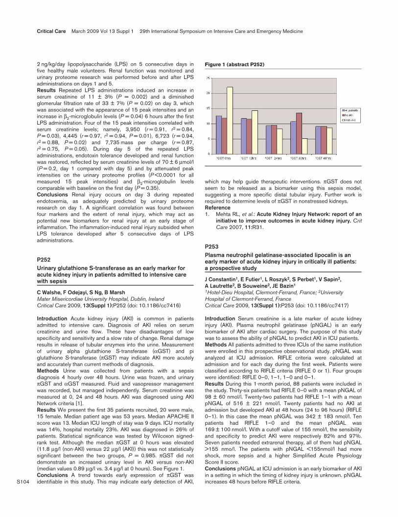
S104
2 ng/kg/day lipopolysaccharide (LPS) on 5 consecutive days infive healthy male volunteers. Renal function was monitored andurinary proteome research was performed before and after LPSadministrations on days 1 and 5.Results Repeated LPS administrations induced an increase inserum creatinine of 11 ± 3% (P = 0.002) and a diminishedglomerular filtration rate of 33 ± 7% (P = 0.02) on day 3, whichwas associated with the appearance of 15 peak intensities and anincrease in β2-microglobulin levels (P = 0.04) 6 hours after the firstLPS administration. Four of the 15 peak intensities correlated withserum creatinine levels; namely, 3,950 (r = 0.91, r2 = 0.84,P = 0.03), 4,445 (r = 0.97, r2 = 0.94, P = 0.01), 6,723 (r = 0.94,r2 = 0.88, P = 0.02) and 7,735 mass per charge (r = 0.87,r2 = 0.75, P = 0.05). During day 5 of the repeated LPSadministrations, endotoxin tolerance developed and renal functionwas restored, reflected by serum creatinine levels of 70 ± 6 μmol/l(P = 0.2, day 1 compared with day 5) and by attenuated peakintensities on the urinary proteome profiles (P <0.0001 for allmeasured 15 peak intensities) and β2-microglobulin levelscomparable with baseline on the first day (P = 0.35).Conclusions Renal injury occurs on day 3 during repeatedendotoxemia, as adequately predicted by urinary proteomeresearch on day 1. A significant correlation was found betweenfour markers and the extent of renal injury, which may act aspotential new biomarkers for renal injury at an early stage ofinflammation. The inflammation-induced renal injury subsided whenLPS tolerance developed after 5 consecutive days of LPSadministrations.
P252Urinary glutathione S-transferase as an early marker foracute kidney injury in patients admitted to intensive carewith sepsis
C Walshe, F Odejayi, S Ng, B MarshMater Misericordiae University Hospital, Dublin, IrelandCritical Care 2009, 13(Suppl 1):P252 (doi: 10.1186/cc7416)
Introduction Acute kidney injury (AKI) is common in patientsadmitted to intensive care. Diagnosis of AKI relies on serumcreatinine and urine flow. These have disadvantages of lowspecificity and sensitivity and a slow rate of change. Renal damageresults in release of tubular enzymes into the urine. Measurementof urinary alpha glutathione S-transferase (αGST) and piglutathione S-transferase (πGST) may indicate AKI more acutelyand accurately than current methods of diagnosis.Methods Urine was collected from patients with a sepsisdiagnosis 4 hourly over 48 hours. Urine was frozen, and urinaryπGST and αGST measured. Fluid and vasopressor managementwas recorded, but managed independently. Serum creatinine wasmeasured at 0, 24 and 48 hours. AKI was diagnosed using AKINetwork criteria [1].Results We present the first 35 patients recruited, 20 were male,15 female. Median patient age was 53 years. Median APACHE IIscore was 13. Median ICU length of stay was 9 days. ICU mortalitywas 14%, hospital mortality 23%. AKI was diagnosed in 26% ofpatients. Statistical significance was tested by Wilcoxon signed-rank test. Although the median πGST at 0 hours was elevated(11.8 μg/l (non-AKI) versus 22 μg/l (AKI)) this was not statisticallysignificant between the two groups, P = 0.985. πGST did notdemonstrate an increased urinary level in AKI versus non-AKI(median values 0.89 μg/l vs. 3.4 μg/l at 0 hours). See Figure 1.Conclusions A trend towards early expression of πGST wasidentifiable in this study. This may indicate early detection of AKI,
which may help guide therapeutic interventions. πGST does notseem to be released as a biomarker using this sepsis model,suggesting a more specific distal tubular injury. Further work isrequired to determine levels of πGST in nonstressed kidneys.Reference1. Mehta RL, et al.: Acute Kidney Injury Network: report of an
initiative to improve outcomes in acute kidney injury. CritCare 2007, 11:R31.
P253Plasma neutrophil gelatinase-associated lipocalin is anearly marker of acute kidney injury in critically ill patients: a prospective study
J Constantin1, E Futier1, L Roszyk2, S Perbet1, V Sapin2, A Lautrette2, B Souweine2, JE Bazin1
1Hotel-Dieu Hospital, Clermont-Ferrand, France; 2UniversityHospital of Clermont-Ferrand, FranceCritical Care 2009, 13(Suppl 1):P253 (doi: 10.1186/cc7417)
Introduction Serum creatinine is a late marker of acute kidneyinjury (AKI). Plasma neutrophil gelatinase (pNGAL) is an earlybiomarker of AKI after cardiac surgery. The purpose of this studywas to assess the ability of pNGAL to predict AKI in ICU patients.Methods All patients admitted to three ICUs of the same institutionwere enrolled in this prospective observational study. pNGAL wasanalyzed at ICU admission. RIFLE criteria were calculated atadmission and for each day during the first week. Patients wereclassified according to RIFLE criteria (RIFLE 0 or 1). Four groupswere identified: RIFLE 0–0, 1–1, 1–0 and 0–1.Results During this 1-month period, 88 patients were included inthe study. Thirty-six patients had RIFLE 0–0 with a mean pNGAL of98 ± 60 nmol/l. Twenty-two patients had RIFLE 1–1 with a meanpNGAL of 516 ± 221 nmol/l. Twenty patients had no AKI atadmission but developed AKI at 48 hours (24 to 96 hours) (RIFLE0–1). In this case the mean pNGAL was 342 ± 183 nmol/l. Tenpatients had RIFLE 1–0 and the mean pNGAL was169 ± 100 nmol/l. With a cutoff value of 155 nmol/l, the sensibilityand specificity to predict AKI were respectively 82% and 97%.Seven patients needed extrarenal therapy, all of them had pNGAL>155 nmo/l. The patients with pNGAL <155nmol/l had moreshock, more sepsis and a higher Simplified Acute PhysiologyScore II score.Conclusions pNGAL at ICU admission is an early biomarker of AKIin a setting in which the timing of kidney injury is unknown. pNGALincreases 48 hours before RIFLE criteria.
Critical Care March 2009 Vol 13 Suppl 1 29th International Symposium on Intensive Care and Emergency Medicine
Figure 1 (abstract P252)

S105
P254Plasma cholinesterase activity in patients during peritonealdialysis
L Popovic, A Pavicic, J Slavicek, J KernChildrens Hospital Zagreb, CroatiaCritical Care 2009, 13(Suppl 1):P254 (doi: 10.1186/cc7418)
Introduction Literature data suggest that the release of nitric oxide(NO) by endothelial NO synthase contributes to functionalalterations of the peritoneal membrane induced by acute perito-nitis. Experimental animal studies show that induced peritonitis wascharacterized by structural changes in the peritoneal membraneand increased permeability for urea and glucose, as well asincreased protein loss in dialysate [1]. Plasma cholinesterase(PChE, 3.1.1.8) is an enzyme synthesized in the liver, molecularmass from 345,000 to 371,000 Da. The aim of this study was toevaluate the functional characteristics of the peritoneum,determining PChE transfer from blood to dialysis fluid in peritonitis-free patients and in patients with peritonitis.Methods We measured PChE activity in plasma and peritonealdialysate in adult peritonitis-free patients (n = 20) and in patientswith peritonitis (n = 12). For determination of PChE activity, veinblood samples were collected and stored at –20°C until analysed.PChE activity was determined by the spectrophotometric methodof Ellman and colleagues [2], using butyryltiocholine as thesubstrate (Sigma Chemical Co., St Louis, MO, USA). Statisticalanalysis was made by Mann–Whitney U test and Wilcoxonmatched-pairs test.Results In both groups of examined patients we detected thesignificant passage of PChE from blood to dialysis fluid(P = 0.000089 for peritonitis-free patients and P = 0.002218 forpatients with peritonitis). The transperitoneal PChE passagedegree is statistically unchanged in patients with peritonitis inrelation to peritonitis-free patients (P = 0.119438).Conclusions In all patients we detected significant transperitonealpassage of PChE during peritoneal dialysis. During humanperitoneal dialysis there is no peritoneal permeability change formacromolecules such as PChE protein complex in patients withperitonitis.References1. Jie NI, et al.: J Am Soc Nephrol 2003, 14:3205-3216.2. Ellman GL, et al.: Biochem Pharmacol 1961, 7:88-95.
P255Early predictive value of neutrophil gelatinase-associatedlipocalin in adult ICU patients with acute kidney injury
H De Geus, J Le Noble, F Zijlstra, C Ince, J BakkerErasmus University Medical Center, Rotterdam, the NetherlandsCritical Care 2009, 13(Suppl 1):P255 (doi: 10.1186/cc7419)
Introduction We investigated the early predictive value of plasmaand urine neutrophil gelatinase-associated lipocalin (NGAL)compared with serum creatinine in patients with acute kidney injury(AKI) in a heterogeneous adult ICU population. Previous studiesdisplay the excellent predictive value of urine NGAL for AKI in apediatric ICU setting [1,2].Methods A prospective cohort study was conducted, including 63patients. After ICU admission, plasma and urine samples werecollected at eight time points. NGAL measurements were performedusing an ELISA. The difference between the increase in NGAL andthe increase in serum creatinine was calculated in relation to thetime point at which maximum AKI was reached (T = 0). AKI wasdefined according to Acute Kidney Injury Network definitions.
Results Mean age (±SD) in years and APACHE II (±SD) scores forAKI I (n = 22), II (n = 21) and III patients (n = 20) were respectively(56 ± 18, 21 ± 7), (58 ± 19, 25 ± 8) and (63 ± 13, 27 ± 7). In AKIIII patients, the difference between the increase in plasma NGALand the increase in serum creatinine expressed as a percentage(95% CI) at 72, 48 and 24 hours prior to maximum AKI was 203%(–169 to 574), –10% (–221 to 830) and –25% (–205 to 155). Forurine NGAL and serum creatinine the difference was respectively584% (–603 to 1,770), 756% (12 to 1,499) and 726% (240 to1,212). Figures 1 and 2 depict the increase in urine NGAL andserum creatinine separately and their difference in relation to T = 0.There was no positive difference in patients with AKI stages I and IIfor urine or plasma at any time point prior to maximum AKI.Conclusions NGAL is, compared with serum creatinine, an earlierpredictor of AKI III, in a heterogeneous adult ICU population. UrineNGAL provides a larger and more sustained window of earlyprediction than plasma NGAL in patients with developing AKI III.The use of urine NGAL as an early predictive biomarker for AKI III istherefore promising in an adult heterogeneous ICU setting.References1. Mishra J, et al.: NGAL as a biomarker for acute renal injury
after cardiac surgery. Lancet 2005, 365:1231-1238.2. Zappitelli M, et al.: Urine NGAL as an early marker of acute
kidney injury in critically ill children: a prospective cohortstudy. Crit Care 2007, 11:R84.
Available online http://ccforum.com/supplements/13/S1
Figure 1 (abstract P255)
Increase in urine NGAL and serum creatinine AKI III.
Figure 2 (abstract P255)
Increase difference between urine NGAL and serum creatinine AKI III.
S106
P256Novel time series analysis approach for prediction ofdialysis in critically ill patients using echo-state networks
T Verplancke1, S Van Looy2, K Steurbaut2, D Benoit1, F De Turck2, G De Moor1, J Decruyenaere1
1Ghent University Hospital, Ghent, Belgium; 2Ghent University,Ghent, BelgiumCritical Care 2009, 13(Suppl 1):P256 (doi: 10.1186/cc7420)
Introduction Echo-state networks are part of a group of reservoircomputing methods and are basically a form of recurrent artificialneural networks. These methods can perform classification taskson time-series data. The recurrent artificial neural network of anecho-state network has an echo-state characteristic. This echostate functions as a fading memory: samples that have beenintroduced into the network in a further past are faded away. Theecho-state approach for the training of recurrent neural networkswas first described by Jaeger [1]. In clinical medicine, until thismoment, no original research articles have been published toexamine the use of echo-state networks.Methods The present study examines the possibility of using anecho-state network for prediction of dialysis in the ICU. Therefore,diuresis values and creatinine levels of the first 3 days after ICUadmission were collected from 830 patients admitted to the ICUbetween 31 May 2003 and 17 November 2007. The outcomeparameter was the performance by the echo-state network inpredicting the need for dialysis between day 5 and day 10 of ICUadmission. Patients with an ICU length of stay <10 days orpatients that received dialysis in the first 5 days of ICU admissionwere excluded. Performance by the echo-state network was thencompared by means of the area under the receiver operatingcharacteristic curve (AUC) with results obtained by two other time-series analysis methods by means of a support vector machine anda naive Bayes algorithm.Results The AUCs in the three developed echo-state networkswere 0.822, 0.818, and 0.817. There were no statisticallysignificant differences at the 0.05 level with the results obtained bythe support vector machine and the naive Bayes algorithms.Conclusions This proof of concept study is the first to evaluate theperformance of echo-state networks in predicting the need fordialysis in an ICU population. The AUCs of the echo-statenetworks were good and comparable with the performance ofother classification algorithms.Reference1. Jaeger H: The ‘Echo State’ Approach to Analysing and Train-
ing Recurrent Neural Networks. Technical Report GMD 148.German National Research Institute for Computer Science;2001.
P257Contrast-induced nephropathy: a prospective analysis oflong-term outcome and persistence of renal impairment
W Huber, E Wohlleb, C Schilling, B Saugel, V Phillip, R SchmidKlinikum rechts der Isar der Technischen Universität München,GermanyCritical Care 2009, 13(Suppl 1):P257 (doi: 10.1186/cc7421)
Introduction Despite several prophylactic approaches such asacetylcysteine, theophylline and sodium-bicarbonate, contrast-induced nephropathy (CIN) remains a clinical problem. Regardingthe large number of studies on CIN, little is known about long-termfollow up of patients with CIN. The aim of our study was thereforeto analyse the outcome of patients with CIN included in eight
prospective studies on CIN conducted by our group, includingmore than 1,200 patients.Methods A systematic analysis of patients with CIN (defined asincrease in serum creatinine ≥0.5 mg/dl and/or ≥25% within48 hours after contrast-medium (CM)) using chart review and atelephone call to patients and to physicians involved in therapyafter CIN up to 1 year or up to death of the patient. Compositeprimary endpoint: requirement of renal replacement therapy (RRT),death, persistent increase in creatinine ≥0.3 mg/dl as comparedwith baseline value. Further endpoints: time course of creatinine upto 1 year after CM.Results Among 85 cases with CIN, complete datasets sufficientfor analysis of the above-mentioned endpoints could be recordedin 55 patients. Thirty-nine male and 16 female patients; age85.5 ± 13.1 years; amount of CM 274 ± 181 ml; intravenous CMapplication in 12 patients, intraarterial CM in 43 patients. The 28-day mortality was 8/55 (15%). At least four patients (7.3%) weretreated with RRT. A total of 24/55 patients (44%) fulfilled thecriteria of the composite endpoint (RRT or death or persistentincrease in creatinine ≥0.3 mg/dl). Compared with baselinecreatinine (1.8 ± 1.2 mg/dl), creatinine levels after 24 hours(2.3 ± 1.2 mg/dl; P <0.001), 48 hours (2.4 ± 1.4 mg/dl; P <0.001)and 1 week (2.5 M 1.9 mg/dl; P = 0.033) were significantlyelevated. In 37 of the surviving patients, creatinine after 1 year wasnot significantly higher than before CM (1.65 ± 1.05 vs.1.46 ± 0.76 mg/dl; P = 0.1). However, patients who died or wereon RRT were not included in this comparison. In two of our eightstudies CIN was significantly associated with mortality.Conclusions Our data indicate that CIN is associated with signifi-cant mortality, requirement of RRT and/or persistent renal impair-ment in nearly one-half of the patients. Since these data werecollected in patients included in studies aimed at prophylaxis ofCIN, the risk might be even more elevated in clinical routine. Prophy-laxis of CIN should therefore be a major issue in patients at risk.
P258Outcome of ICU patients requiring dialysis in an Africaninstitution
M Mer1, L Ezekiel1, S Naicker1, G Richards1, J Levin2
1University of the Witwatersrand, Johannesburg, South Africa;2Medical Research Council of South Africa, Pretoria, South AfricaCritical Care 2009, 13(Suppl 1):P258 (doi: 10.1186/cc7422)
Introduction Acute renal failure (ARF) is a common problem inICUs and is associated with a high mortality rate [1,2]. Early andaggressive management of renal dysfunction through intermittentor continuous renal replacement therapy (RRT) is now commonpractice. The objective of this study was to review the outcome ofpatients who received RRT in the multidisciplinary ICU of theJohannesburg Hospital, South Africa.Methods A review of ICU records between January 2003 andDecember 2004 of all patients requiring RRT in the ICU wasperformed. Demographic data, reason for ICU admission, presenceof comorbidity, APACHE II score, the modality and duration ofdialysis, as well as the outcome in the ICU were documented. Thisstudy was approved by the Human Research Ethics Committee ofthe University of the Witwatersrand, and statistical analysis carriedout in conjunction with the Biostatistics Unit of the MedicalResearch Council of South Africa using the Stata release 8.0statistical package.Results One hundred and fifty-six patients out of 2,200 admis-sions (January 2003 to December 2004) were initiated on RRT.One hundred and three patients were treated with intermittenthaemodialysis (IHD), 47 of whom died (45.6%). Twenty-three
Critical Care March 2009 Vol 13 Suppl 1 29th International Symposium on Intensive Care and Emergency Medicine

S107
patients underwent continuous venovenous haemodialysis, 20(87%) of whom died. Twenty-two patients underwent both IHDand continuous venovenous haemodialysis, 15 (68.2%) of whomdied. In eight patients there was no record of the mode of dialysisadministered. Multivariate logistic regression suggested that themain factor associated with mortality was dialysis. Omitting themode of dialysis, the presence of sepsis and the use of inotropeswere independent risk factors associated with mortality.Conclusions Continuous RRT allows renal support in patients whowould be unable to sustain IHD. The presence of sepsis and theuse of inotropic support, rather than the mode of dialysis, arepredictive of the outcome of ARF in the ICU. Of note, the overallmortality rate for ARF in this study appears to be lower thanpreviously reported.References1. de Mendonica A, Vincent JL, Suter PM, et al.: Acute renal
failure in the ICU: risk factors and outcome evaluation bySOFA score. Intensive Care Med 2000, 26:915-921.
2. Mehta RL: ARF in the ICU: lessons from the PICARD study.In ASN Proceedings 2004.
P259Early goal-directed therapy of septic patients coming fromthe ward does not protect the kidneys
HD Kiers1, A Litchfield2, S Reynolds2, D Griesdale2, RT Gibney3,P Pickkers1, D Chittock2, DD Sweet2
1Radboud University Nijmegen Medical Centre, Nijmegen, theNetherlands; 2Vancouver General Hospital, University of BritishColumbia, Vancouver, BC, Canada; 3University of Alberta,Edmonton, AB, CanadaCritical Care 2009, 13(Suppl 1):P259 (doi: 10.1186/cc7423)
Introduction Patients with severe sepsis or septic shock admittedto the emergency room appear to benefit from early goal-directedtherapy, while this is unknown for patients admitted to the ICU fromthe ward.Methods We documented achievement of mean arterial pressure>65 mmHg, central venous pressure >8 mmHg and centralvenous oxygen saturation >70% within the first 12 hours after ICUconsultation. Creatinine in the week prior to ICU admission,creatinine at ICU consultation and peak creatinine in the 2 weeksafter ICU admission were recorded.Results Eighty-five patients were included, of which 40% achievedall goals within 6 hours (early achievement), whereas 33% did notbut had all of them documented within the first 12 hours (noachievement), 27% had incomplete documentation of goals. Forty-four out of 85 patients (52%) developed acute kidney injury (AKI)according to the RIFLE criteria. Patients with incomplete docu-mentation, early or no achievement of goals were comparable atbaseline, APACHE II and interventions except for more use ofinotropes in the no achievement group. The course of creatinine inpatients was similar, regardless of achievement of goals: baseline –ICU consultation – peak, presented as median (IQR): earlyachievement group: 90 (61 to 119) – 118 (86 to 204) – 148 (86to 249); no achievement group: 76 (56 to 98) – 121 (78 to 159) –151 (88 to 205); incomplete documentation group: 81 (56 to 109) –95 (65 to 175) – 114 (77 to 223). The patients that developed AKIhad a significant increase in creatinine from baseline to the time ofICU consultation: baseline – ICU consultation – peak, presentedas median (IQR): no AKI: 75 (59 to 114) – 87 (64 to 123) – 92(72 to 129); AKI: 84 (61 to 105), P = 0.638 – 145 (102 to 205),P <0.001 – 196 (153 to 276), P <0.001. The development of AKIwas independent of achievement of physiologic goals ofresuscitation.
Conclusions In septic patients admitted from the ward there is noassociation between the development of AKI and the timeliness ofachievement of physiologic resuscitation goals. In view of thehigher creatinine values measured at ICU admission in the patientsthat eventually suffer from AKI, it appears that the insult hasoccurred prior to the time of ICU consultation and that physiologicresuscitation does not reverse it.
P260Is goal-directed therapy useful in kidney transplantation?
M Ciapetti, S Di Valvasone, M Bonizzoli, A Di Filippo, A PerisCareggi Teaching Hospital, Florence, ItalyCritical Care 2009, 13(Suppl 1):P260 (doi: 10.1186/cc7424)
Introduction Patients with end-stage renal disease (ESRD) areentitled to the kidney transplantation approach at surgery withvarious modifications of the volemic status. According to somestudies [1,2], acute tubular necrosis risk can be reduced bykeeping a correct intravascular volume before graft reperfusion.The aim of this study was to evaluate goal-directed therapy in theearly postoperative period in kidney transplantation.Methods We observed 50 kidney transplant recipients divided intotwo groups: 38 patients (Group A) underwent central oxygenvenous saturation (ScVO2) monitoring, and 12 patients (Group B)were controls. Continuous central venous pressure (CVP) andScVO2 were monitored the in ICU [3]. In Group A the volemicstatus by keeping CVP >5 mmHg and ScVO2 >70% wasoptimized. We collected donors’ and transplant patients’ kidneyparameters (age, sex, death cause, ischaemia time), recipients’parameters (age, sex, weight, height, BMI, duration of dialysis,ESRD, Simplified Acute Physiology Score II), intraoperativeparameters (metabolic, respiratory and hemodynamic), hemo-dynamic and kidney functioning parameters in the ICU (heart rate,mean arterial pressure, CVP, ScVO2, lactate, diuresis, blood ureanitrogen (BUN), creatinine, fluid balance), and outcome parameters(ICU length of stay, acute rejection at 28 days, mortality at6 months).Results At each observation ScVO2 was >70% in all patients ofGroup A. Diuresis was higher in Group A (at 6 hours, Group A:1,082.6 ± 1,000.7; Group B: 757.2 ± 462.7; at 12 hours, GroupA: 1,020.5 ± 921.5; Group B: 835.4 ± 517.8). The heart rate at 0hours was higher in Group B (Group A: 85.2 ± 13.2; Group B:97.7 ± 27.7; P <0.05). Creatinine (at 0 hours, Group A: 6.9 ± 2;Group B: 8.2 ± 3; at 12 hours, Group A: 7.2 ± 2.5; Group B: 9.3± 0.07) and BUN (at 0 hours, Group A: 0.9 ± 0.4; Group B: 1.0 ±0.3; at 12 hours, Group A: 1 ± 0.4; Group B: 1.1 ± 0.3) werehigher in Group B. The creatinine (Group A: –0.2 ± 1.2; Group B:0.4 ± 0.7) and BUN (Group A: 0.06 ± 0.1; Group B: 0.1 ± 0.04)reduction (12 to 0 hours) was higher in Group A. Diureticstimulation was reduced in 10 patients of Group A and zero ofGroup B (P <0.05) and was interrupted in eight patients of GroupA and two of Group B.Conclusions Postoperative intensive monitoring and optimizationof intravascular volume by CVP and ScVO2 grant fast recovery ofkidney functioning in transplant recipients, so reducing diureticstimulation, creatinine and BUN values.References1. De Gasperi A, et al.: Transplant Proc 2006, 38:807-809.2. Hadimioglu N, et al.: Transplant Proc 2006, 38:440-442.3. Rivers EP, et al.: Curr Opin Crit Care 2001, 7:204-211.
Available online http://ccforum.com/supplements/13/S1

S108
P261Clinical characteristics and outcomes of critically ill adultswith septic acute kidney injury in a general hospital inSingapore
J Koh, J Vijo Poulose, V PouloseChangi General Hospital, SingaporeCritical Care 2009, 13(Suppl 1):P261 (doi: 10.1186/cc7425)
Introduction The aim of this study was to determine the clinicalcharacteristics and outcomes of critically ill adults with septicacute kidney injury (AKI) stratified according to the AKI staging.The Acute Kidney Injury Network (AKIN) definition for AKI had beenshown to predict important clinical outcomes such as hospitalmortality [1]. Sepsis is the most common cause of AKI resulting inworse clinical outcomes when compared with other causes [2].Methods An observational study conducted in a medical ICU of ageneral hospital in Singapore over a 6-month period. Patients whowere admitted to the ICU with a diagnosis of sepsis and AKI (asdefined by the AKIN criteria) were prospectively enrolled. Theclinical characteristics and outcomes were determined andstratified according to the AKIN criteria.Results A total of 71 consecutive septic patients (60.6% Chinese,32.4% Malays, 2.8% Indians) were enrolled. The mean age was63.7 years with a male predominance of 67.6%. The medianSimplified Acute Physiology Score (SAPS) II score was 54. Themajority (60.6%) was AKI III, with 22.9% in stage II and 16.9% instage I. Overall hospital mortality was 39.4%. Patients who metseptic AKI III had significantly higher mortality compared with AKI Iand II (55.8% vs. 16.7% and 12.5%, respectively, P <0.001).There was a significant difference in the mean SAPS II scorebetween the dead and alive patients (70 vs. 46, P <0.001).Multiple logistic regression analysis showed that AKI III (OR =5.75, 95% CI = 1.2 to 25.5) and SAPS II score ≥65 (OR = 15.6,95% CI = 3.5 to 68.2) were found to be independent predictors ofhospital mortality.Conclusions In septic patients, AKI III appeared to be a strongpredictor of hospital mortality. This finding is similar to a previousstudy [3], which also showed that in patents with AKI only AKI IIIwas an independent risk factor for hospital death.References1. Barrantes F, et al.: Acute kidney injury criteria predict out-
comes of critically ill patients. Crit Care Med 2008, 36:1397-1403.
2. Bagshaw SM, et al.: Septic acute kidney injury in criticallyill patients: clinical characteristics and outcomes. Clin JAm Soc Nephrol 2007, 2:431-439.
3. Ostermann M, et al.: Correlation between the AKI classifi-cation and outcome. Crit Care 2008, 12:R144.
P262Incidence and outcome of acute renal failure necessitatingrenal replacement therapy after trauma
S Beitland1, H Moen1, I Os2
1Ullevaal University Hospital, Oslo, Norway; 2University of Oslo,NorwayCritical Care 2009, 13(Suppl 1):P262 (doi: 10.1186/cc7426)
Introduction Acute renal failure (ARF) requiring renal replacementtherapy (RRT) is uncommon in trauma patients. The aim of thisstudy was to assess incidence and outcome in this patient group.Methods Adult trauma patients with ARF treated with RRT atUllevaal University Hospital between 1 January 1996 and 31December 2007 were retrospectively reviewed. The hospital is the
regional trauma referral centre for approximately 1.93 million adult(>18 years) persons. Individuals were identified and data werecollected using several institutional registries in addition to theNorwegian renal registry. Patients were grouped according topresence of rhabdomyolysis based on peak serum creatine kinaselevels exceeding 10,000 U/l or not. Categorical data werecompared employing the two-sided Pearson chi-square test,whereas continuous data were compared utilizing the two-tailedMann–Whitney U test.Results There were 78,345 hospital admissions due to traumaduring the study period, 42 of these underwent RRT for ARF (afterexcluding one person due to low age, three with nonrenalindications for RRT and another three with chronic renal failure).The incidence rate of post-traumatic ARF requiring RRT was0.54‰, and was higher in males compared with females (33.72vs. 6.05 per million persons, P <0.01). The patients’ age wasmedian (range) 46.4 (18 to 84) years, and 85.7% were males.Mortality rates were 23.8% (ICU/hospital), 35.7% (3 months) and40.5% (1 year). Renal recovery, defined as independency fromRRT, occurred in all survivors after 3 months and 1 year.Rhabdomyolysis was present in 18 persons (42.9%) and was onlyregistered in males. Trauma patients with rhabdomyolysis weresignificantly younger (33.0 vs. 57.0 years, P = 0.01), needed RRTearlier (3.0 vs. 6.5 days, P = 0.02) and had lower 3-month (16.7vs. 50.0%, P = 0.03) and 1-year (22.2 vs. 54.2%, P = 0.04)mortality rates compared with nonrhabdomyolytic persons.Conclusions Post-traumatic ARF requiring RRT is rare and mainlyaffects males of young age. There is still a considerable mortalityrate in these patients, and it seems that rhabdomyolytic personshave a more favourable outcome compared with those withoutrhabdomyolysis. Among survivors, recovery of renal function isoften seen, and few become dependent on chronic RRT.
P263Early acute kidney injury in Northern Ireland ICUs
E Borthwick1, S Harris2, C Welch3, A Maxwell1, DF McAuley4, P Glover4, D Harrison3, K Rowan3
1Regional Nephrology Unit, Belfast, UK; 2UCL, London, UK;3Intensive Care National Audit and Research Centre, London, UK;4Royal Victoria Hospital, Belfast, UKCritical Care 2009, 13(Suppl 1):P263 (doi: 10.1186/cc7427)
Introduction There are limited data about the epidemiology ofacute kidney injury (AKI) in critically ill patients in Northern Ireland.The aim of this study was to examine AKI within 24 hours of ICUadmission and its relation to outcomes (ICU mortality and hospitalmortality).Methods A secondary analysis of prospectively collected data inthe Intensive Care National Audit and Research Centre Case MixProgramme Database. The Case Mix Programme Database wasinterrogated and data extracted from 22,313 admissions to eightICUs from 1999 to 2007. The presence of AKI was assessedwithin the first 24 hours after admission (early AKI) and classifiedaccording to the RIFLE criteria. Trends over time were describedfor the RIFLE categories, and outcomes of admissions in eachcategory were summarised. Where available, information on theuse of renal replacement therapy during the ICU stay wasanalysed.Results Trends in early AKI changed little over time: 35.5% ofpatients sustained AKI (risk 13.5%, injury 11.1%, failure 9.8%,end-stage 1.2%) and 9% of patients received renal replacementtherapy. Outcomes are presented in Table 1.Conclusions For the first time we have established the incidenceof early AKI using the RIFLE criteria in Northern Ireland ICUs. This
Critical Care March 2009 Vol 13 Suppl 1 29th International Symposium on Intensive Care and Emergency Medicine

S109
will inform renal service development in ICUs. The incidence ofsevere AKI is high relative to the rest of the UK [1] although it islow compared with the US [2]. Mortality increases with severity ofrenal injury.References1. Kolhe et al.: Crit Care 2008, 12(Suppl 1):S2.2. Hoste et al.: Crit Care 2006, 10:R73.
P264RIFLE classification can predict hospital mortality ofcritically ill patients
HY Xu1, JM Peng1, ZR Mao2, L Weng1, XY Hu1, B Du1
1Peking Union Medical College Hospital, Beijing, China; 2FirstAffiliated Hospital of Henan College of Traditional ChineseMedicine, Henan, ChinaCritical Care 2009, 13(Suppl 1):P264 (doi: 10.1186/cc7428)
Introduction The Acute Dialysis Quality Initiative group hasproposed the RIFLE (Risk–Injury–Failure–Loss–End-stage renaldisease) classification to assess acute kidney injury (AKI). Wesought to evaluate the incidence of AKI in critically ill patientsaccording to the RIFLE classification and the correlation betweenRIFLE class and hospital mortality.Methods We performed a retrospective cohort study applying theRIFLE classification on 1,138 patients admitted to the ICU during a2-year period.Results According to the RIFLE classification, 376 patients (33%)had AKI during their ICU stay. When assessing the maximumRIFLE class, 209 (18.3%) patients were classified Risk, 63 (5.5%)as Injury and 104 (6.3%) as Failure. Female (OR = 1.53; 95% CI =1.18 to 1.98; P = 0.001), nonsurgical admission (OR = 1.32; 95%CI = 1.01 to 1.70; P = 0.039), APACHE II on admission above 25(OR = 1.99; 95% CI = 1.06 to 3.76; P = 0.033), sepsis onadmission (OR = 2.20; 95% CI = 1.01 to 4.79; P = 0.046) andchronic organ dysfunction (OR = 1.65; 95% CI = 1.09 to 2.50;P = 0.017) were independent risk factors for AKI. Patients withprogressive RIFLE classification had increased hospital mortality,with Risk 13.9%, Injury 22.2% and Failure 47%, as compared with7.9% (P < 0.001) among patients without AKI. Furthermore, theRIFLE class Failure was an independent predictor of 3-monthhospital mortality (OR = 2.37; 95% CI = 1.23 to 4.54; P = 0.009)in addition to organ failure on admission (OR = 4.60; 95% CI =2.31 to 9.18; P <0.001), use of vasopressor (OR = 2.34; 95%CI = 1.09 to 5.00; P = 0.028), and APACHE II score on admission(OR = 1.160; 95% CI = 1.11 to 1.21; P < 0.001).Conclusions Patients with increasing RIFLE classification hadsignificant elevated hospital mortality. Maximum RIFLE class Failurewas independently associated with 3-month hospital mortality.References1. Bellomo R, et al.: Acute renal failure-definitions, outcome
measures, animal models, fluid therapy and informationtechnology needs: the Second International ConsensusConference of the Acute Dialysis Quality Initiative (ADQI)Group. Crit Care 2004, 8:R204-R212.
2. Hoste EA, et al.: RIFLE criteria for acute kidney injury areassociated with hospital mortality in critical ill patients: acohort analysis. Crit Care 2006, 10:R73-R83.
P265Assessment of acute kidney injury with modified RIFLEcriteria in critically ill pediatric burn patients
T Palmieri, A Lavrentieva, D GreenhalghShriners Hospital for Children, Sacramento, CA, USACritical Care 2009, 13(Suppl 1):P265 (doi: 10.1186/cc7429)
Introduction The objective of the present study was to evaluatethe incidence, risk factors and outcome associated with acutekidney injury (AKI) as defined by the modified pediatric version ofRIFLE criteria (pRIFLE) in children with severe burn injury.Methods The retrospective, descriptive cohort study included 123patients admitted for more than 24 hours to a burn pediatric ICUfrom 2006 to 2008. Burn injury severity was estimated using thetotal body surface area burn (TBSA%), severity of illness wasestimated using the Pediatric Risk of Mortality (PRISM) score. ThepRIFLE criteria were applied and the patients were assigned to theappropriate pRIFLE stratum (Risk, Injury, Failure) if they fulfilledeither estimated creatinine clearance, urine output criteria, or both.Results The incidence of AKI was 40.7%, maximum RIFLE classRisk, class Injury and class Failure occurred in 50%, 36% and18%, respectively. Patients with maximum RIFLE class Risk, Injuryand Failure had ICU mortality rates of 0%, 5.6% and 57.1%,respectively, compared with 1.4% for patients without AKI. Weobserved statistically significant differences between the patientswith AKI and those without AKI in the following parameters: TBSA(41.2 ± 17.7% vs. 24.2 ± 14.6%, P <0.001), admission PRISM(8.6 ± 6.4 vs. 4.8 ± 3.4, P <0.01), number of surgical procedures(3.7 ± 2.9 vs. 1.5 ± 1.5, P <0.001), occurrence of abdominalcompartment syndrome (18% vs. 0%, P <0.001), length ofmechanical ventilation (22.3 ± 27.6 days vs. 7.1 ± 11.4 days,P <0.001) and length of ICU stay (37 ± 30.1 days vs. 14.6 ± 13.9days, P <0.001). Logistic regression analysis indicated that PRISMscore (OR = 1.1, 95% CI = 1.0 to 1.2; P = 0.05) and TBSA(OR = 1.06, 95% CI = 1.0 to 1.1; P <0.001) were independentrisk factors for AKI in pediatric burn patients.Conclusions AKI estimated by pRIFLE criteria occurs in majorpediatric burns, and failure was associated with increasedmortality. Patients with AKI had higher admission burn and illnessseverity, increased incidence of abdominal compartment syn-drome, more operations, and had increased duration of mechanicalventilation and length of ICU stay. AKI is a marker of increasedresource utilization and risk for adverse outcomes after burn injuryin children.
Available online http://ccforum.com/supplements/13/S1
Table 1 (abstract P263)
Outcomes
No AKI (n = 14,385) Risk (n = 3,020) Injury (n = 2,446) Failure (n = 2,183) End-stage (n = 259)
ICU mortality 1,350 (9.4) 563 (18.6) 834 (33.8) 909 (41.6) 48 (18.5)
Hospital mortality 2,258 (16.8) 900 (31.9) 1,092 (47.2) 1,137 (55.9) 82 (32.7)
Data presented as n (%).
S110
P266Extracorporeal renal replacement therapy in patients withheparin-induced thrombocytopenia
M Bekers-Anchipolovskis, V Liguts, E Strike, N Porite, V Harlamovs, L Semcenko, M DauksteP. Stradina University Hospital, Riga, LatviaCritical Care 2009, 13(Suppl 1):P266 (doi: 10.1186/cc7430)
Introduction Evaluation of renal replacement therapy (RRT)efficacy in heparin-induced thrombocytopenia (HIT) patients, usingthe alternative anticoagulant bivalirudin. A decreased platelet count(PC) is often seen after cardiopulmonary bypass with PC reductionby about 30% to 50%, correlating with the duration of cardio-pulmonary bypass. However, only a few of these patients willdevelop HIT. Acute renal failure (ARF), followed by extracorporealRRT, was seen in 90% of the patients with critical PC reductionfound in the postoperative ICU. ARF has multiple etiology: baselinedisorders (congestive heart failure, acute heart failure), hypoxemia,drugs used during surgery, and so on. The significance of post-operative thrombocytopenia, including HIT, urges to differentiate itscauses and select appropriate patient care options, including useof alternative anticoagulants, namely bivalirudin. Bivalirudin (Angio-max™; The Medicines Company, Cambridge, MA, USA) is asynthetic hirudin analog, which reversibly binds thrombin.Bivalirudin is eliminated by enzymatic proteolysis (80%) and renalclearance (20%). The serum half-life is 25 minutes in patients withnormal renal function, while it is longer in those with renaldysfunction (3.5 hours).Methods A retrospective study in HIT patients (clinical scoring andlaboratory assays) who underwent RRT.Results Ten patients were studied, thereof four patients had lowPC, thrombotic complications, RRT and prolonged duration of stayin the ICU – 17 days on average. Two of the patients had positivetests (ranged from 0.45 to 1.62) on postoperative days 6 to 8 and11 to 12. All of the patients had their anticoagulant switched fromheparin to bivalirudin, despite implanted circulation assistingdevices and RRT. Coagulation system status was evaluated by theactivated partial thromboplastin time, of which target value wastwice the upper normal range. The bivalirudin infusion dose rangedfrom 50 μ/kg/hour to 100 μ/kg/hour, depending on the activatedpartial thromboplastin time value. Bivalirudin was found to be atleast as safe and effective an anticoagulant as heparin.Conclusions HIT patients must be switched from heparin toalternative anticoagulant. Bivalirudin is a safe and effective anti-coagulant for extracorporeal RRT; however, the dose should beadjusted individually. Bivalirudin has prolonged effect in ARFpatients and does not have antagonists.
P267Coagulopathy of liver disease does not increase the filterlife during continuous renal replacement therapy
B Agarwal, S Shaw, M HariRoyal Free Hospital, London, UKCritical Care 2009, 13(Suppl 1):P267 (doi: 10.1186/cc7431)
Introduction Clotting of haemofiltration circuits is a limiting factorin achieving efficient continuous renal replacement therapy(CRRT), yet systemic anticoagulation risks haemorrhage. Somepatients, such as those with liver failure, are traditionally managedwith no or minimal anticoagulation, because of abnormal clottingtests and therefore increased perceived risk of bleeding [1].Methods We retrospectively reviewed the CRRT circuit life inthree groups of liver failure patients (acute liver failure (ALF), acute
on chronic liver failure (ACLF) and postelective liver transplantation(LTx)), with two control groups (systemic sepsis (SS) and haema-tological malignancy (Haem)), admitted to the Royal Free HospitalICU – a tertiary referral centre for liver disease and transplantation –between 2003 and 2007. Ten consecutive patients in each of thefive groups were included in the study if they had renal failure andrequired continuous haemofiltration (CRRT) for more than48 hours.Results The mean CRRT circuit life was significantly greater in theHaem group, compared with the others; 28.5 ± 25.7 hours, versus11 ± 10.5 ALF, 11.6 ± 6.6 ACLF, 7.4 ± 5.1 LTx and 9.9 ± 5.9 SS,P <0.05, with the Haem group requiring fewest new CCRT circuitswithin 48 hours; 2.7 ± 1.5 versus 4.3 ± 1.3 ALF, 4.2 ± 2.1 ACLF,5.3 ± 1.5 LTx and 4.6 ± 1.5 SS, P <0.05 and least bloodtransfusions; 1.4 ± 1.3 versus 4.8 ± 4.2 ALF, 4.2 ± 4.1 ACLF,2.2 ± 2.1 LTx and 3.2 ± 1.2 SS. Transmembrane pressures werehigher in those CRRT circuits that clotted due to the filter,compared with other causes, such as access dysfunction (123 ±74 vs. 71.8 ± 29.3 mmHg, P = 0.009). In those patients whereanticoagulation was started due to repeated filter clotting, theCRRT circuit life improved from 5.6 ± 3.4 to 19 ± 12.7 hours,P <0.01.Conclusions Despite abnormal standard laboratory coagulationtests and thrombocytopenia, CRRT circuits clot frequently in liverfailure patients. Anticoagulation did improve CRRT circuit survivalwithout an obvious increase in bleeding or blood transfusionrequirement. Anticoagulation should therefore be considered inthese patients in cases of repeated circuit clotting.Reference1. Davenport A: CRRT in the management of patients with
liver disease. Semin Dial 1996, 9:78-84.
P268Optimal dose of renal replacement therapy in acute kidneyinjury: a meta-analysis
RJ Van Wert, DC Scales, JO Friedrich, R Wald, NK AdhikariUniversity of Toronto, ON, CanadaCritical Care 2009, 13(Suppl 1):P268 (doi: 10.1186/cc7432)
Introduction Acute kidney injury (AKI) requiring renal replacementtherapy (RRT) increases mortality in the critically ill patient. Ourobjective was to systematically review randomized controlled trials(RCTs) examining the effect of the RRT dose on mortality.Methods In duplicate, we searched Medline, EMBASE and theCochrane Central Register of Controlled Trials from inception toDecember 2008, using terms for all RRT modalities, AKI, andRCTs. Included RCTs compared different doses of RRT within agiven modality (effluent rate in continuous RRT (CRRT), number ofsessions per week in intermittent hemodialysis (IHD)) in patientswith AKI, and reported mortality or dialysis dependence. Weexcluded RCTs that evaluated very high volume hemofiltration(continuous effluent rates >60 ml/kg/hour). Risk ratios (RRs) with95% confidence intervals for mortality and dialysis dependenceamong survivors were calculated using random-effects models(RevMan 5). We investigated subgroup effects by modality (CRRTvs. IHD) and patient group (septic vs. nonseptic).Results Of 2,913 citations, seven RCTs (n = 2,255) met theinclusion criteria. Four RCTs used CRRT (35 to 48 vs.20 ml/kg/hour); two RCTs used IHD (daily vs. alternate day); andthe largest and only multicentre RCT (n = 1,124) integrated CRRTand IHD in high-dose and standard-dose arms. RCTs weregenerally of good quality. Meta-analysis did not demonstratereduced mortality with high-dose RRT (RR = 0.86, 0.70 to 1.06).The effect was similar in patients (1) treated with CRRT (RR =
Critical Care March 2009 Vol 13 Suppl 1 29th International Symposium on Intensive Care and Emergency Medicine

S111
0.87, 0.71 to 1.06, five trials, n = 1,552) and IHD (RR = 0.95, 0.59to 1.51, three trials, n = 703), and (2) with sepsis (RR = 1.08, 0.96to 1.22, n = 896) and without sepsis (RR = 0.88, 0.65 to 1.20,n = 959) in the four trials with these data. High-dose RRT did notdecrease dialysis dependence among all survivors (RR = 1.09,0.83 to 1.43, five trials, n = 774) or in the subgroup treated onlywith CRRT (RR = 1.27, 0.62 to 2.59, three trials, n = 233). Weestimate that if the ongoing multicentre RENAL RCT (plannedn = 1,500, ClinicalTrials.gov NCT00221013) of high-dose versusstandard-dose CRRT demonstrates the expected absolutemortality risk reduction (from 60% to 52%), the pooled mortalitybenefit would just reach statistical significance.Conclusions Current evidence does not demonstrate reducedmortality with high-dose RRT for AKI, even in the subgroup ofpatients with sepsis-associated AKI. Forthcoming results of theRENAL trial may contribute to more definitive results.
P269Removal of inflammatory mediators by continuous renalreplacement therapy in severe sepsis
E Tomas1, E Lafuente1, B Vera1, M Fernandes1, J Silva1, F Santos1, F Moura1, P Santos1, R Lopes1, P Calder2
1Centro Hospitalar Tamega e Sousa, Penafiel, Portugal; 2InstituteHuman Nutrition, Southampton, UKCritical Care 2009, 13(Suppl 1):P269 (doi: 10.1186/cc7433)
Introduction The aim of this study was to evaluate the efficacy ofcontinuous renal replacement therapy (CRRT) in systemic inflam-matory mediator removal in a group of patients with severesepsis/septic shock and related renal failure.Methods We conducted a prospective study, approved by theethics committee, enrolling 11 patients with severe sepsis/septicshock under CRRT. We measured the cytokines using theimmunoassay method. The cytokine measurement was done everymorning until the end of renal support. We analyzed the followingdata: age, severity scores (Simplified Acute Physiology Score(SAPS) II, Sequential Organ Failure Assessment (SOFA)),cytokines (TNF, IL-1β, IL-6 and IL-10) levels on pre-filter, post-filterand ultrafiltrate samples. We correlated these data with filterduration, considering 24 hours as the maximum theoretical limit foreffective cytokine removal.Results From the 11 patients enrolled, we collected 420 validsamples to measure the cytokine levels. The mean age and SAPSII were respectively 66 ± 10.7 years and 46 ± 27.1. The mean total
SOFA score at admission and at day 3 were 11.2 ± 4 and 12.7 ± 4respectively, with ΔSOFA = +1.3 points (P >0.05). The mean ICUhospitalization time was 25.9 ± 33.5 days. We have done876 hours of dialytic therapy; 33 AN69 membranes were used,during a mean time of 26.5 ± 25.6 hours. The mean total levelsfound for cytokines are presented in Figure 1 and the levelsdepending on the filter duration are in Figure 2. The ICU mortalitywas 54.6%.Conclusions The present study does not allow us to define aspecific pattern for the removal of inflammatory mediators relatedto CRRT. We observed a trend for cytokine reduction dependenton filter duration with the higher efficacy removal until 24 hours.The cytokine level found in the effluent does not reflect theabsolute reduction found in post-filter, and this effect could bedependent on adsorption, according to other studies [1,2].References1. Shoji H: Extracorporeal endotoxin removal for the treat-
ment of sepsis: endotoxin adsorption cartridge (toray-myxin). Ther Apher Dial 2003, 7:108-114.
2. Kellum J, Song M, Venkataraman R: Hemoadsorptionremoves tumor necrosis factor, interleukin-6, and inter-leukin-10, reduces nuclear factor-B DNA binding, andimproves short-term survival in lethal endotoxemia. CritCare Med 2004, 32:801-805.
P270Blood epuration of middle molecules in continuousvenovenous hemodiafiltration with regional citrateanticoagulation versus systemic heparinization
MB Nogier, L Lavayssiere, O Cointault, M Abbal, N Kamar, B Periquet, L Rostaing, D DurandCHU Rangueil, Toulouse, FranceCritical Care 2009, 13(Suppl 1):P270 (doi: 10.1186/cc7434)
Introduction Adequate anticoagulation is a precondition toimprove blood epuration. In a prospective crossover trial, wecompared the removal of middle molecules in continuous renalreplacement therapy, by two methods that used either trisodiumcitrate ACD (BRAUN Laboratories) or heparin as anticoagulation.Methods Fourteen critically ill patients were treated by continuousvenovenous hemodiafiltration. The dialysate and hemofiltrationflows were 1,500 ml/hour each. All patients received two sessionsof 36 hours successively. Patients 1 to 7 received systemicheparinization first, then regional anticoagulation by citrate.Conversely, Patients 8 to 14 received heparin and then citrate.Every 12 hours during each session of renal replacement (T0, T12,T24 and T36), blood epuration of small molecules (urea,creatinine) and middle molecules (β2-microglobulin, retinol bindingprotein) were evaluated from blood and effluent samples.Results At all time periods, there was no significant difference increatinine and urea clearances between citrate and heparintreatment, except in T12 where creatinine clearance was signifi-cantly higher when using citrate than when using heparin (38.5 ±4.0 ml/min vs. 34 ± 4.1 ml/min, P = 0.02). We did not observe asignificant decline of urea and creatinine clearance during thesession. The retinol binding protein blood and effluent rates werelimited and stable during the session. Between T0 and T12, resultsshowed a significant improvement of β2-microglobulin clearancewith using citrate or heparin. After T12, β2-microglobulin clearancedecreased with citrate treatment and stayed stable with heparin.Clearance seems to be higher with citrate than heparin, however,with only a significant difference in T12 (20.1 ± 4.0 vs. 16.3 ±3.1 ml/min, P = 0.02). Undergoing citrate treatment, we recordedsignificant metabolic alkalosis (P = 0.0025) but without hypernatremia.
Available online http://ccforum.com/supplements/13/S1
Figure 1 (abstract P269)
Total cytokine levels.
Figure 2 (abstract P269)
Removal of inflammatory mediators according to filter duration.
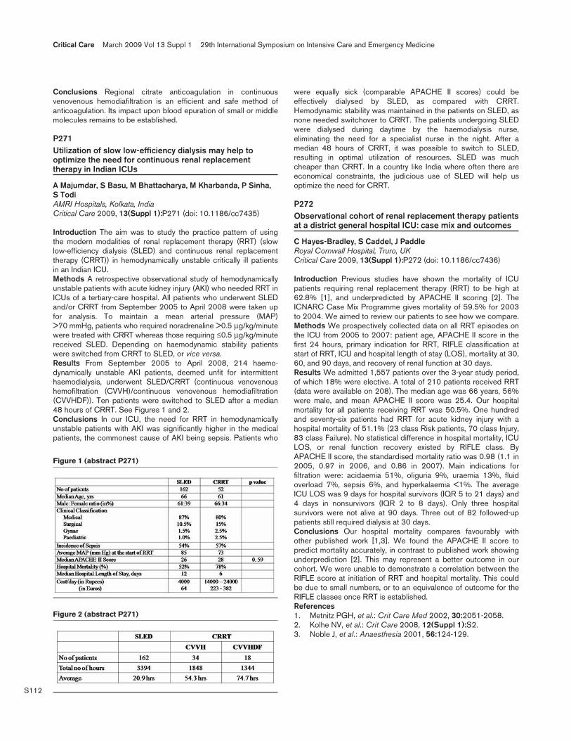
S112
Conclusions Regional citrate anticoagulation in continuousvenovenous hemodiafiltration is an efficient and safe method ofanticoagulation. Its impact upon blood epuration of small or middlemolecules remains to be established.
P271Utilization of slow low-efficiency dialysis may help tooptimize the need for continuous renal replacementtherapy in Indian ICUs
A Majumdar, S Basu, M Bhattacharya, M Kharbanda, P Sinha,S TodiAMRI Hospitals, Kolkata, IndiaCritical Care 2009, 13(Suppl 1):P271 (doi: 10.1186/cc7435)
Introduction The aim was to study the practice pattern of usingthe modern modalities of renal replacement therapy (RRT) (slowlow-efficiency dialysis (SLED) and continuous renal replacementtherapy (CRRT)) in hemodynamically unstable critically ill patientsin an Indian ICU.Methods A retrospective observational study of hemodynamicallyunstable patients with acute kidney injury (AKI) who needed RRT inICUs of a tertiary-care hospital. All patients who underwent SLEDand/or CRRT from September 2005 to April 2008 were taken upfor analysis. To maintain a mean arterial pressure (MAP)>70 mmHg, patients who required noradrenaline >0.5 μg/kg/minutewere treated with CRRT whereas those requiring ≤0.5 μg/kg/minutereceived SLED. Depending on haemodynamic stability patientswere switched from CRRT to SLED, or vice versa.Results From September 2005 to April 2008, 214 haemo-dynamically unstable AKI patients, deemed unfit for intermittenthaemodialysis, underwent SLED/CRRT (continuous venovenoushemofiltration (CVVH)/continuous venovenous hemodiafiltration(CVVHDF)). Ten patients were switched to SLED after a median48 hours of CRRT. See Figures 1 and 2.Conclusions In our ICU, the need for RRT in hemodynamicallyunstable patients with AKI was significantly higher in the medicalpatients, the commonest cause of AKI being sepsis. Patients who
were equally sick (comparable APACHE II scores) could beeffectively dialysed by SLED, as compared with CRRT.Hemodynamic stability was maintained in the patients on SLED, asnone needed switchover to CRRT. The patients undergoing SLEDwere dialysed during daytime by the haemodialysis nurse,eliminating the need for a specialist nurse in the night. After amedian 48 hours of CRRT, it was possible to switch to SLED,resulting in optimal utilization of resources. SLED was muchcheaper than CRRT. In a country like India where often there areeconomical constraints, the judicious use of SLED will help usoptimize the need for CRRT.
P272Observational cohort of renal replacement therapy patientsat a district general hospital ICU: case mix and outcomes
C Hayes-Bradley, S Caddel, J PaddleRoyal Cornwall Hospital, Truro, UKCritical Care 2009, 13(Suppl 1):P272 (doi: 10.1186/cc7436)
Introduction Previous studies have shown the mortality of ICUpatients requiring renal replacement therapy (RRT) to be high at62.8% [1], and underpredicted by APACHE II scoring [2]. TheICNARC Case Mix Programme gives mortality of 59.5% for 2003to 2004. We aimed to review our patients to see how we compare.Methods We prospectively collected data on all RRT episodes onthe ICU from 2005 to 2007: patient age, APACHE II score in thefirst 24 hours, primary indication for RRT, RIFLE classification atstart of RRT, ICU and hospital length of stay (LOS), mortality at 30,60, and 90 days, and recovery of renal function at 30 days.Results We admitted 1,557 patients over the 3-year study period,of which 18% were elective. A total of 210 patients received RRT(data were available on 208). The median age was 66 years, 56%were male, and mean APACHE II score was 25.4. Our hospitalmortality for all patients receiving RRT was 50.5%. One hundredand seventy-six patients had RRT for acute kidney injury with ahospital mortality of 51.1% (23 class Risk patients, 70 class Injury,83 class Failure). No statistical difference in hospital mortality, ICULOS, or renal function recovery existed by RIFLE class. ByAPACHE II score, the standardised mortality ratio was 0.98 (1.1 in2005, 0.97 in 2006, and 0.86 in 2007). Main indications forfiltration were: acidaemia 51%, oliguria 9%, uraemia 13%, fluidoverload 7%, sepsis 6%, and hyperkalaemia <1%. The averageICU LOS was 9 days for hospital survivors (IQR 5 to 21 days) and4 days in nonsurvivors (IQR 2 to 8 days). Only three hospitalsurvivors were not alive at 90 days. Three out of 82 followed-uppatients still required dialysis at 30 days.Conclusions Our hospital mortality compares favourably withother published work [1,3]. We found the APACHE II score topredict mortality accurately, in contrast to published work showingunderprediction [2]. This may represent a better outcome in ourcohort. We were unable to demonstrate a correlation between theRIFLE score at initiation of RRT and hospital mortality. This couldbe due to small numbers, or to an equivalence of outcome for theRIFLE classes once RRT is established.References1. Metnitz PGH, et al.: Crit Care Med 2002, 30:2051-2058.2. Kolhe NV, et al.: Crit Care 2008, 12(Suppl 1):S2.3. Noble J, et al.: Anaesthesia 2001, 56:124-129.
Critical Care March 2009 Vol 13 Suppl 1 29th International Symposium on Intensive Care and Emergency Medicine
Figure 1 (abstract P271)
Figure 2 (abstract P271)

S113
P273Effects of continuous hemofiltration on organ perfusion,energy metabolism, oxidative stress, endothelialdysfunction and inflammation
R Sykora, J Chvojka, A Krouzecky, J Radej, V Varnerova, T Karvunidis, I Novak, M MatejovicCharles University, Medical School and Teaching Hospital, Plzen,Czech RepublicCritical Care 2009, 13(Suppl 1):P273 (doi: 10.1186/cc7437)
Introduction Continuous renal replacement therapies (CRRT) arewidely used for treatment of acute kidney injury in critically illpatients. Little attention has been paid to the potential adverseeffects of CRRT related to extracorporeal circuit bioincompatibility.Limited available evidence suggests that intermittent dialysis maycompromise hepatosplanchnic perfusion in acute renal failurepatients. By contrast, there are no data on the bio(in)compatibilityindices in patients treated by CRRT. To investigate this issue, weutilized a long-term porcine model allowing a broad insight intoorgan hemodynamic, microvascular, metabolic and other pathwaysnot accessible in human medicine.Methods Measurements were performed in 11 healthy instru-mented animals. After baseline measurements, animals wererandomized to receive either no treatment (n = 6) or continuousvenovenous hemofiltration with a polysuphon membrane (CVVH,n = 5). Further data were collected at 6 and 10 hours after therandomization. During each time point the following data related to(1) organ perfusion (hepatosplanchnic and renal blood flows), (2)microcirculation (ileal mucosal and renal cortex laser Dopplerflowmetry and sidestream darkfield imaging), (3) energy balance(arterial and regional pH, lactate/pyruvate and ketone body ratios),(4) oxidative/nitrosative stress (thiobarbituric acid reactive species,nitrates + nitrites), (5) inflammation (TNFα, IL-6) and (6) endothelialdysfunction (von Willebrand factor, asymmetric dimethylarginine)were collected.Results Hemofiltration affected neither regional organ blood flowsnor ileal mucosal and renal cortex microvascular perfusion. Nochanges in liver and kidney oxygen exchange and energy balancewere detected during the 10-hour treatment. Similarly, CVVH didnot interfere with surrogate markers of inflammation, oxidative/nitrosative stress and endothelial dysfunction.Conclusions In our model CVVH utilizing a modern biocompatiblepolysulphon membrane did not induce any significant deleteriouseffects in various biological systems. The importance of ourfindings lies in the fact that any changes in the studied parameters,if observed in future studies, cannot be attributed to the effects ofputative extracorporeal circuit bioincompatibility. Finally, our datasupport the postulated concept of good biological tolerance ofCRRT.Acknowledgement Supported by MSM 0021620819 – Replace-ment of and support to some vital organs.
P274Influence of continuous venovenous hemofiltration ontranspulmonary thermodilution-derived parameters
V Neirynck, A Willems, D Peeters, N Van Regenmortel, I De laet, K Schoonheydt, H Dits, M MalbrainZNA Stuivenberg, Antwerp, BelgiumCritical Care 2009, 13(Suppl 1):P274 (doi: 10.1186/cc7438)
Introduction We studied the effects of continuous venovenoushemofiltration (CVVH) on transpulmonary hemodynamic para-meters in nine ventilated patients [1,2].Methods All together 32 calibrations were performed with andwithout CVVH treatment. For each calibration three consecutiveinjections of 20 ml cold saline were done via the central venousline (CVL), giving a total of 186 thermodilutions.Results Patient age was 72.3 ± 14, BMI 24.7 ± 3.8, SAPS II 56.2± 15.7. Regardless of the catheter position, CVVH increased theextravascular lung water index (EVLWi) from 11.3 ± 5 to 12.4 ±6.3 (P = NS), while the cardiac index (CI) and global end-diastolicvolume index (GEDVi) decreased from 5.1 ± 1.8 to 4.2 ± 1.3(P = 0.03) and from 1036 ± 298 to 885 ± 185 (P = 0.02),respectively. The results of a subanalysis comparing correctcatheter position (CVL placed in jugular or subclavian vein anddialysis catheter placed femorally) and faulty position (dialysiscatheter positioned between the thermodilution injection anddetection sites) are summarized in Table 1. In two patientscatheters were exchanged during the stay from the faulty to thecorrect position, and this resulted in a significant decrease in allparameters: CI dropped from 6.1 ± 0.9 to 5 ± 0.3 (P = 0.014),GEDVi from 1253 ± 165 to 829 ± 161 (P = 0.001) and EVLWifrom 16.1 ± 7.1 to 8.7 ± 1.2 (P = 0.03).Conclusions In critically ill patients treated with CVVH, thehemodynamic parameters obtained by PiCCO transpulmonarythermodilution can be influenced: EVLWi increases while CI andGEDVi drop. We hypothesize that this may be due to the positionof the CVL and dialysis catheters.References1. Martinez-Simon A: Crit Care 2006, 10:410.2. Sakka S, et al.: Anesth Analg 2007, 105:1079-1082.
P275Antibiotic dosing regimens for septic patients receivingcontinuous venovenous haemofiltration: do current studiessupply sufficient data?
A Li, C Gomersall, G Choi, Q Tian, G Joynt, J LipmanThe Chinese University of Hong Kong, NT, Hong KongCritical Care 2009, 13(Suppl 1):P275 (doi: 10.1186/cc7439)
Introduction The aim of this study was to establish the minimumdataset that needs to be specified when presenting pharmaco-kinetic data for critically ill patients with acute renal failure, and toreview the current literature to establish whether this minimum
Available online http://ccforum.com/supplements/13/S1
Table 1 (abstract P274)
Effect of CVVH on hemodynamic parameters in correct and faulty catheter positions
CVVH correct No CVVH correct CVVH faulty No CVVH faulty (n = 11) (n = 11) P value (n = 21) (n = 21) P value
CI (l/min/m2) 4.1 ± 0.9 4.5 ± 0.8 NS 4.3 ± 1.5 5.4 ± 2 0.04
GEDVi (ml/m2) 774.8 ± 83.8 810.6 ± 124.1 NS 942.1 ± 198.4 1,155.4 ± 294.6 0.009
EVLWi (ml/kg) 7.8 ± 2.2 7.8 ± 1.5 NS 14.8 ± 6.4 13.1 ± 5.3 NS

S114
dataset is indeed reported. Antibiotic dosing for septic patientswith acute renal failure receiving continuous renal replacementtherapy is complicated, and failure to correctly dose may result ineither drug toxicity, or treatment failure and development ofresistance [1].Methods A dataset was established of the minimal number ofparameters that need be reported when calculating a drug-dosingregimen. Patient demographics and markers of severity wereadded to allow for patient population comparisons. A Medlinesearch was performed of the relevant literature, producing 76studies from which completeness of the dataset was examined.Results None of the studies analysed presented the full datasetthat we established as necessary (Figure 1). Of concern, basicpharmacokinetic parameters such as the volume of distribution andclearance were absent in a significant number of studies effectivelyabrogating calculation of a meaningful dosing regimen.Conclusions A large proportion of current studies do not reportkey information necessary to devise a rational dosing regimen forpatients with acute renal failure receiving continuous renalreplacement therapy. We have presented a set of criteria webelieve are necessary to calculate an antibiotic-dosing regimen forthese patients and hope this will be a useful guide when reportingfuture pharmacokinetic data.Reference1. Roberts JA, et al.: Crit Care Med 2008, 36:2433-2440.
P276Comparing adsorption of gentamicin by polyacrylonitrileand polyamide hemofiltration filter in an in vitro continuousvenovenous hemofiltration model
P Lam, Q Tian, M Ip, C GomersallChinese University of Hong Kong, NT, Hong KongCritical Care 2009, 13(Suppl 1):P276 (doi: 10.1186/cc7440)
Introduction As a high proportion of patients who require con-tinuous renal replacement therapy will also be receiving antibiotics,the issue of whether significant amounts of antibiotic are adsorbed
by the haemofilter is relevant to critically ill patients. The aim of thestudy is to determine the time course of adsorption of gentamicinto a polyacrylonitrile filter (PAN) and a polyamide filter, respectively.Methods A unit of expired whole blood was mixed with heparinizedlactated Ringer’s solution to made up a total volume of 1,000 ml.Five hundred milliliters of this blood–crystalloid mixture was takento a glass chamber where it was agitated and heated. After theequilibration period, 20% of a standard dose of the drug wasinfused into the mixing chamber. The blood–crystalloid mixture wasthen circulated through an in vitro continuous venovenoushemofiltration model. The ultrafiltrate was returned to the mixingchamber and no replacement fluid was infused. As a result, anydecrease in drug concentration could only occur due to adsorp-tion. Samples were taken from the mixing chamber for measure-ment of the drug concentration. At 90 minutes, the remaining500 ml blood–crystalloid mixture will then be added to the mixingchamber. Samples were taken again after another hour. If the fall indrug concentration was less than those predicted from the dilutioneffect following the increase in blood–crystalloid volume, itindicates reversibility of adsorption. A second dose of drug wasthen added and samples were taken afterwards. Adsorption ofantibiotic before and after the second dose was compared. Twotypes of filter, namely a 0.6 m2 PAN hemofilter (Multiflow 100;Hospal) and a 0.6 m2 polyamide hemofilter (Hemofilter 6S;Gambro), were used in the study and four batches of tests wererepeated for each hemofilter.Results Drug adsorption by PAN hemofilters was significant,whereas drug adsorption by polyamide hemofilters was muchlower compared with that of PAN hemofilters. Gentamicinadsorption by PAN hemofilters was completed at 30 minutes and itwas irreversible.Conclusions The adsorption properties of gentamicin by the twohemofilters were markedly different.
Critical Care March 2009 Vol 13 Suppl 1 29th International Symposium on Intensive Care and Emergency Medicine
Figure 1 (abstract P275)
Reporting of required parameters for dose regimen calculation in the literature.

S115
P277Slow continuous ultrafiltration: how many fluids must betaken out?
G Guiotto, S Gligorova, F Paladino, F Schiraldi, S VerdeSan Paolo Hospital, Napoli, ItalyCritical Care 2009, 13(Suppl 1):P277 (doi: 10.1186/cc7441)
Introduction Ultrafiltration (UF) is effective and safe in treatingvolume-overloaded patients with acute decompensated heartfailure (ADHF) and diuretic resistance [1]. Accurate determinationof the amount of fluid to be removed and maintenance of thecirculating blood volume are critically important [2]. The inferiorvena cava diameter (IVCD) and its collapsibility index (IVCCI) arecompromised in ADHF patients due to high right atrial pressure[3]. We hypothesized that monitoring of IVCCI could be used tooptimize fluid removal rate during UF.Methods Twenty patients (nine male, 11 female; age 76 ± 4, NewYork Heart Association classes III to IV) admitted to our medicalICU for ADHF were treated with UF (Aquadex System 100; CHFSolutions, Minneapolis, MN, USA). The heart rate (HR), meanarterial pressure (MAP) and IVCD with M-mode subcostalechocardiography during spontaneous breathing were evaluatedbefore UF (T0), at 12 hours (T1) and at 24 hours (T2). The IVCCIwas calculated as follows: [(IVCDmax – IVCDmin) / IVCDmax] x 100.Results The mean UF time was 25.5 ± 5 hours with a mean volumeof 259 ml/hour and a total ultrafiltrate production of 6.6 ± 2 l.Differences between the T0 and T2 parameters are presented inTable 1. Hypotension was observed only in those patients (2/20)who reached IVCCI >35%. In all the other patients a significantincrease in IVCCI was obtained without any hemodynamic instability.
Table 1 (abstract P277)
T0 T2 P value
MAP (mmHg) 85 ± 9 86 ± 12 NSHR (bpm) 85 ± 6 85 ± 14 NSIVCD (mm) 30.2 ± 6.3 27.3 ± 5.9 <0.5IVCCI (%) 6.5 ± 1.7 33.3 ± 4.6 <0.001
Conclusions Inferior vena cava ultrasound is a rapid, simple andnoninvasive means for bedside monitoring of the intravascularvolume during UF.References1. Costanzo RM, et al.: Ultrafiltration versus intravenous
diuretics for patients hospitalized for acute decompen-sated heart failure. J Am Coll Cardiol 2007, 49:675-683.
2. Ronco C, et al.: Extracorporeal ultrafiltration for the treat-ment of overhydration and congestive heart failure. Cardi-ology 2001, 96:155-168.
3. Blehar DJ, et al.: Identification of congestive heart failurevia respiratory variation of inferior vena cava diameter. AmJ Emerg Med 2009, 27:71-75.
P278Plasmatic cytokines and intermittent hemodialysis withpolymethylmethacrylate membrane in septic shock patients
N Mayeur, L Lavayssiere, MB Nogier, O Cointault, O Fourcade,L RostaingCHU Rangueil, Toulouse, FranceCritical Care 2009, 13(Suppl 1):P278 (doi: 10.1186/cc7442)
Introduction Sepsis is mediated by many biologically activeinflammatory mediators, including interleukins. IL-6, IL-8, and IL-10
are correlated with increased mortality in septic shock acute renalfailure (ARF) [1]. ARF treatment requires renal replacement therapy(RRT). The cytokine plasmatic level during and after hemodialysis(HD) in septic ARF is partially described [2]. Polymethylmeth-acrylate (PMMA) hemodialyser membranes own high adsorptivecapacity [3]. In this prospective observational trial, we study theplasmatic level of IL-6, IL-8 and IL-10 during and after the first HDseance with PMMA membrane in septic shock patients with ARF.Methods Inclusion criteria: patients with septic shock <24 hoursas defined by the American College of Chest Physicians/Society ofCritical Care Medecine and requiring RRT (Injury in the RIFLEcriteria). The hemodialyser PMMA membrane was Filtrizer BK-1,6 F(TORAY Industrie, Tokyo, Japan). Data and blood samples werecollected at: start of HD (D0), every hour during HD (D1; D2), atthe end of HD (endD); and 30, 60, 90, 120 and 180 minutes afterHD (postD0.5; postD1; postD1.5; postD2; postD3, respectively).Solid-phase ELISA was used for cytokine measurements.Statistical analysis was by Kruskall–Wallis nonparametric test.Results Ten patients were included. At D0: Sequential OrganFailure Assessment (14.6 ± 0.8) and IGS 2 (Simplified AcutePhysiology Score II) (79.11 ± 4.73). At D0, IL-6, IL-8 and IL-10
Available online http://ccforum.com/supplements/13/S1
Figure 1 (abstract P278)
Relative IL-8 concentration versus baseline. *P <0.05.
Figure 2 (abstract P278)
Relative IL-10 concentration versus baseline. *P <0.05.

S116
concentration values were 767 ± 191.2, 724.4 ± 191.7 and168.5 ± 50.44 pg/ml, respectively. Relative serum IL-8 and IL-10concentrations versus D0 are shown in Figures 1 and 2 (mean ±SEM). The urea reduction between D0 and endD was 48.5%. Thenorepinephrine rate and mean arterial pressure did not changebetween D0 and endD (0.65 ± 0.12 vs. 0.57 ± 0.12 μg/kg/min,and 76.40 ± 4.554 vs. 83.60 ± 4.349 mmHg, respectively; P = NS).Conclusions PMMA membranes showed transient efficiency inIL-8 and IL-10 elimination by possible membrane saturation. TheIL-6 concentration was not modified. Three hours after HD, the IL-8and IL-10 concentrations were back to baseline. This fast increasecould be explained by plasmatic rebound and must be kept inmind. This rebound could be deleterious in this stage of sepsis.References1. Oberholzer A, et al.: Plasma cytokine measurements
augment prognostic scores as indicators of outcome inpatients with severe sepsis. Shock 2005, 23:488-493.
2. Haase M, et al.: Hemodialysis membrane with a high-mole-cular-weight cutoff and cytokine levels in sepsis compli-cated by acute renal failure: a phase 1 randomized trial.Am J Kidney Dis 2007, 50:296-304.
3. Hirasawa H, et al.: Continuous hemofiltration with cytokine-adsorbing hemofilter in the treatment of severe sepsisand septic shock. Contrib Nephrol 2007, 156:365-370.
P279Immunoadsorption in dilated cardiomyopathy
Y Wakabayashi1, A Baba1, M Akaishi1, T Yoshikawa2, T Monkawa2
1Kitasato Institute Hospital, Tokyo, Japan; 2Keio University Schoolof Medicine, Tokyo, JapanCritical Care 2009, 13(Suppl 1):P279 (doi: 10.1186/cc7443)
Introduction Removal of cardiodepressant autoantibodies byimmunoadsorption (IA) has been reported to induce early haemo-dynamic improvement in patients with dilated cardiomyopathy(DCM). The Immusorba TR-350 (Asahikasei-Kuraray Medical Co.Ltd, Japan) (TR) is an IA column currently used for Myastheniagravis or Guillain-Barré syndrome. This column, in whichtryptophan is immobilized as a ligand, has a property to have highaffinity to IgG subclass 3. With this property, imunoglobulinsubstitution is not usually required after the IA treatment. Sincecardiodepressant antibodies belong to IgG subclass 3, weinvestigated the effect of IA using this column on cardiac functionin patients with DCM.Methods Seventeen DCM patients (left ventricular ejectionfraction (LVEF) <30%) participated in the study. IA was conductedevery other day three to five times. Blood was drawn at the rate of80 to 100 ml/minute by direct venipuncture or from the bloodaccess catheter and it was first passed through the plasma-separating column. The separated plasma was then passedthrough the TR at the rate of 20 ml/minute. A total of 1,500 to2,000 ml plasma was processed per one session. Either heparin ornafamostat mesylate was used as an anticoagulant. The LVEF wasmeasured by quantitative gated single photon emission computedtomography. The cardiodepressant antibodies were assayed exvivo [1]. The β1-adrenergic and muscarinic M2-acetylcholinereceptor antibodies were measured by ELISA.Results After the three to five sessions of IA treatment,cardiodepressant antibodies were almost completely cleared fromthe circulation. Three months after the IA sessions, the LVEFincreased significantly from 18.7 ± 2.3 to 23.2 ± 2.6%, P <0.05.The average increase rate was 33.1 ± 12.6% from the baseline.Limited to the 10 patients who initially revealed the high titer of
cardiodepressant antibodies, the increase in LVEF was greater(54.5 ± 16.8% from the baseline). The cardiodepressant anti-bodies were not detected at this period except in one patient. Allpatients tolerated IA without any complication. Any cardiac eventor mortality did not take place over the period of 3 months.Conclusions The IA treatment with TR may bring benefit in leftventricular function in patients with DCM, particularly in those witha high titer of cardiodepressant antibodies. Further long-termfollow-up is, however, required before confirming its efficacy.Reference1. Baba A: Autoantigen estimation and simple screening assay
against cardiodepressant autoantibodies in patients withdilated cardiomyopathy. Ther Apher Dial 2008, 12:109-116.
P280Lipopolysaccharide adsorber in abdominal septic shock
T Ala-Kokko, J Koskenkari, J LaurilaOulu University Hospital, Oulu, FinlandCritical Care 2009, 13(Suppl 1):P280 (doi: 10.1186/cc7444)
Introduction Polymyxin-B hemoperfusion has been shown to lowermortality in sepsis [1]. The effects of a new endotoxin adsorber(Alteco LPS Adsorber; Alteco Medical AB, Lund, Sweden) on thelength of noradrenaline (NA) treatment and lipopolysaccharideblood levels in abdominal septic shock were evaluated.Methods Following consent a 2-hour hemoperfusion with LPSadsorber was began in five patients [2]. Sepsis guidelines werefollowed [3]. Two historical controls per case were selected.Results The mean total duration of NA infusion was 46 hoursshorter in the adsorber group compared with the control group(95% CI = –104 hours to 12 hours, P = 0.165) (Table 1). Theaverage length of NA infusion was 17.4 ± 6.8 hours (5.8 to23.8 hours) following the start of adsorption treatment. The level ofLPS decreased in all but one study patient and all were without NAat 24 hours. The mean Sequential Organ Failure Assessmentdecrease was 3.4 ± 1.7 from baseline to 24 hours post treatmentThe average length of hospital stay was 3.4 days shorter in theadsorber group (95% CI of the difference, –21.7 to 14.8 days,P = 0.881). All study patients were alive on day 28 and one controldied in the hospital.Conclusions Single 2-hour LPS hemoperfusion was associatedwith a rapid decrease in NA dose, reversal of septic shock, anddecrease in organ dysfunctions and LPS concentrations. The totalduration of NA infusion and hospital stay were shorter comparedwith historical controls, but the difference was not statisticallysignificant in this small study.References1. Cruz D, et al.: Effectiveness of polymyxin B-immobilized fiber
column in sepsis: a systematic review. Crit Care 2007, 11:R47.
Critical Care March 2009 Vol 13 Suppl 1 29th International Symposium on Intensive Care and Emergency Medicine
Table 1 (abstract P280)
Clinical characteristics of the study patients and the controls
Patients Controls (n = 5) (n = 10) P value
Age 64 ± 16 65 ± 13 NSMale/female 2/3 4/6 NSSimplified Acute Physiology Score II 38 ± 11 46 ± 11 NSSequential Organ Failure Assessment 9.6 ± 2 7.7 ± 2 NSICU length of stay 6.2 ± 3 6.4 ± 3 NSHospital length of stay 23 ± 13 27 ± 17 NSNA infusion (hours) 34 ± 12 81 ± 77 NS
Data presented as mean ± SD.

S117
2. Levy MM, et al.: 2001 SCCM/ESICM/ACCP/ATS/SIS Inter-national Sepsis Definitions Conference. Crit Care Med2001, 31:1250-1256.
3. Dellinger RP, et al.: Surviving Sepsis Campaign: interna-tional guidelines for management of severe sepsis andseptic shock: 2008. Crit Care Med 2008, 36:296-327.
P281Experience with the use of selective sorbents in complexintensive care of sepsis in patients after cardiac surgery
M Yaroustovsky, M Abramyan, Z Popok, E Nazarova, D Popov,O Stupchenko, M PlushchCentre Cardiovascular Surgery, Moscow, Russian FederationCritical Care 2009, 13(Suppl 1):P281 (doi: 10.1186/cc7445)
Introduction The aim of the study was to evaluate the firstexperience with endotoxin adsorption in complex intensive care ofcritically ill patients with sepsis after heart surgery.Methods Eleven adult patients were studied. Patients were dividedinto two groups: undergoing Alteco LPS adsorption procedures(n = 6), and undergoing hemoperfusion using Toraymyxin columns(n = 5). Intensive therapy of sepsis in both groups included anti-bacterial therapy, hemodynamic and respiratory support, preven-tion of thromboembolism, stress ulcer prophylaxis, nutritivesupport, and studied extracorporeal techniques to endotoxinremoval (two procedures for every patient).Results Endotoxin adsorption procedures were started on averageat day 8 after surgery in patients with systemic inflammatoryresponse syndrome and confirmed Gram-negative infection. As theresult of procedures, hemodynamic indices improved in bothgroups while the dose of inotropic support decreased. Also wenoted the improvement of oxygenating lung function. Positivedynamics of procalcitonin, endotoxin and inflammation mediatorconcentrations were noted. A favorable influence of studiedprocedures on the course of the infective process was confirmedby the dynamics of leukocyte count and the tendency to normali-zation of body temperature. Blood cultures taken several days afterthe procedures were negative. There was no deterioration ofhemodynamic indices during and after procedures. No cases ofthrombosis of the extracorporeal contour were noted. Data arepresented in Figure 1 as mean ± SEM.Conclusions Both studied procedures are safe. Clinical studiesshould be continued to define the place of endotoxin adsorption incomplex treatment of critically ill patients with sepsis.
P282Best choice of acute blood purification therapy based onthe severity score and blood lactic acid values in septicshock patients
Y Sakamoto1, K Mashiko1, H Matsumoto1, Y Hara1, N Kutsukata1, N Saito1, H Yokota2
1Chiba Hokusou Hospital, Nippon Medical School, Chiba, Japan;2Nippon Medical School, Tokyo, JapanCritical Care 2009, 13(Suppl 1):P282 (doi: 10.1186/cc7446)
Introduction Septic shock is a condition associated with diffusecoagulopathy and multiple organ failure, and frequently leads todeath. Direct hemoperfusion using a polymyxin B-immobilized fibercolumn (DHP-PMX) has been used for the treatment of septicshock [1]. On the other hand, there is another kind of acute bloodpurification therapy, but the optimal column of continuous veno-venous hemodiafiltration (CVVHDF) is still controversial.Methods We treated 88 septic shock patients by DHP-PMX. Thepatients were divided into two groups based on the survival outcomeand on the improvement in the circulatory dynamics immediately afterDHP-PMX (Group A: increase of systolic blood pressure (SBP) bymore than 30 mmHg (44 cases); Group B: increase of SBP by30 mmHg or less (44 cases)). In another study conducted to reviewthe best choice of acute blood purification therapy after DHP-PMX,the patients were divided into three groups: groups undergoingCVVHDF using a polymethylmethacrylate membrane hemofilter(PMMA) (28 cases), CVVHDF using a polyacrylonitrile membranehemofilter (26 cases), and no CVVHDF after DHP-PMX (34 cases).Results There were 48 survival cases and 40 expired cases. Theoverall survival rate was 54.5% (good outcome judging from theAPACHE II score), and outcome significantly related to APACHE IIscore, Sepsis-Related Organ Failure Assessment score and bloodlactic acid value before treatment (P <0.0001). The improvementrates of the blood pressure (increased by more than 30 mmHg)were 50.0% and significantly low blood lactic acid level in GroupA. For another examination, only the PMMA CVVHDF groupshowed a better outcome (survival rate of 78.6%) compared withthe other groups (P = 0.0190). In addition, only the PMMACVVHDF group showed significant improvements of the bloodlactic acid on day 3 (P = 0.0011).Conclusions Our study suggests that DHP-PMX treatment waseffective in the early phase of septic shock before critical increaseof the blood lactic acid levels. The optimal column for CVVHDF, asdetermined by improvement of the blood lactic acid levels,following DHP-PMX treatment is the PMMA column.Reference1. Cruz DN, Perazella MA, Bellomo R, et al.: Effectiveness of
polymyxin B-immobilized fiber column in sepsis: a sys-tematic review. Crit Care 2007, 11:R47.
P283Clinical effects of polymyxin B immobilised fiber withdirect hemoperfusion for the patients of severe sepsis orseptic shock caused by intra-abdominal infections
T Ikeda1, K Ikeda1, H Taniuchi1, S Suda1, Y Takahashi21Tokyo Medical University, Hachioji Medical Center, Tokyo, Japan;2Sannoudai Hospital, Ibaragi, JapanCritical Care 2009, 13(Suppl 1):P283 (doi: 10.1186/cc7447)
Introduction The endotoxin adsorption method polymyxin Bimmobilised fiber with direct hemoperfusion (PMX-DHP) has beenused for treatment of patients with severe sepsis and septic shockprimarily caused by Gram-negative infections in Japan [1]. One
Available online http://ccforum.com/supplements/13/S1
Figure 1 (abstract P281)
DHP-PMX, hemoperfusion with the use of Toraymyxin columns; MAP,mean arterial pressure; WBC, white blood cells; PLT, platelets; PCT,procalcitonin.
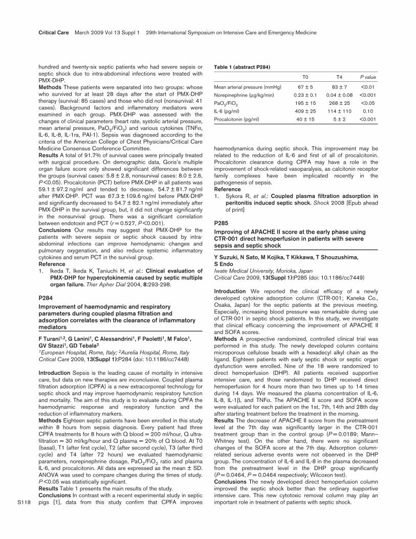
S118
hundred and twenty-six septic patients who had severe sepsis orseptic shock due to intra-abdominal infections were treated withPMX-DHP.Methods These patients were separated into two groups: whosewho survived for at least 28 days after the start of PMX-DHPtherapy (survival: 85 cases) and those who did not (nonsurvival: 41cases). Background factors and inflammatory mediators wereexamined in each group. PMX-DHP was assessed with thechanges of clinical parameters (heart rate, systolic arterial pressure,mean arterial pressure, PaO2/FiO2) and various cytokines (TNFα,IL-6, IL-8, IL-1ra, PAI-1). Sepsis was diagnosed according to thecriteria of the American College of Chest Physicians/Critical CareMedicine Consensus Conference Committee.Results A total of 91.7% of survival cases were principally treatedwith surgical procedure. On demographic data, Goris’s multipleorgan failure score only showed significant differences betweenthe groups (survival cases: 5.8 ± 2.8, nonsurvival cases: 8.0 ± 2.6,P <0.05). Procalcitonin (PCT) before PMX-DHP in all patients was59.1 ± 97.2 ng/ml and tended to decrease, 54.7 ± 81.7 ng/mlafter PMX-DHP. PCT was 67.3 ± 109.6 ng/ml before PMX-DHPand significantly decreased to 54.7 ± 82.1 ng/ml immediately afterPMX-DHP in the survival group, but, it did not change significantlyin the nonsurvival group. There was a significant correlationbetween endotoxin and PCT (r = 0.527, P <0.001).Conclusions Our results may suggest that PMX-DHP for thepatients with severe sepsis or septic shock caused by intra-abdominal infections can improve hemodynamic changes andpulmonary oxygenation, and also reduce systemic inflammatorycytokines and serum PCT in the survival group.Reference1. Ikeda T, Ikeda K, Taniuchi H, et al.: Clinical evaluation of
PMX-DHP for hypercytokinemia caused by septic multipleorgan failure. Ther Apher Dial 2004, 8:293-298.
P284Improvement of haemodynamic and respiratoryparameters during coupled plasma filtration andadsorption correlates with the clearance of inflammatorymediators
F Turani1,2, G Lanini1, C Alessandrini1, F Paoletti1, M Falco1, GV Stazzi1, GD Tebala2
1European Hospital, Rome, Italy; 2Aurelia Hospital, Rome, ItalyCritical Care 2009, 13(Suppl 1):P284 (doi: 10.1186/cc7448)
Introduction Sepsis is the leading cause of mortality in intensivecare, but data on new therapies are inconclusive. Coupled plasmafiltration adsorption (CPFA) is a new extracorporeal technology forseptic shock and may improve haemodynamic respiratory functionand mortality. The aim of this study is to evaluate during CPFA thehaemodynamic response and respiratory function and thereduction of inflammatory markers.Methods Eighteen septic patients have been enrolled in this studywithin 8 hours from sepsis diagnosis. Every patient had threeCPFA treatments for 8 hours with Q blood = 200 ml/hour, Q ultra-filtration = 30 ml/kg/hour and Q plasma = 20% of Q blood. At T0(basal), T1 (after first cycle), T2 (after second cycle), T3 (after thirdcycle) and T4 (after 72 hours) we evaluated haemodynamicparameters, norepinephrine dosage, PaO2/FiO2 ratio and plasmaIL-6, and procalcitonin. All data are expressed as the mean ± SD.ANOVA was used to compare changes during the times of study.P <0.05 was statistically significant.Results Table 1 presents the main results of the study.Conclusions In contrast with a recent experimental study in septicpigs [1], data from this study confirm that CPFA improves
haemodynamics during septic shock. This improvement may berelated to the reduction of IL-6 and first of all of procalcitonin.Procalcitonin clearance during CPFA may have a role in theimprovement of shock-related vasoparalysis, as calcitonin receptorfamily complexes have been implicated recently in thepathogenesis of sepsis.Reference1. Sykora R, et al.: Coupled plasma filtration adsorption in
peritonitis induced septic shock. Shock 2008 [Epub aheadof print]
P285Improving of APACHE II score at the early phase usingCTR-001 direct hemoperfusion in patients with severesepsis and septic shock
Y Suzuki, N Sato, M Kojika, T Kikkawa, T Shouzushima, S EndoIwate Medical University, Morioka, JapanCritical Care 2009, 13(Suppl 1):P285 (doi: 10.1186/cc7449)
Introduction We reported the clinical efficacy of a newlydeveloped cytokine adsorption column (CTR-001; Kaneka Co.,Osaka, Japan) for the septic patients at the previous meeting.Especially, increasing blood pressure was remarkable during useof CTR-001 in septic shock patients. In this study, we investigatethat clinical efficacy concerning the improvement of APACHE IIand SOFA scores.Methods A prospective randomized, controlled clinical trial wasperformed in this study. The newly developed column containsmicroporous cellulose beads with a hexadecyl alkyl chain as theligand. Eighteen patients with early septic shock or septic organdysfunction were enrolled. Nine of the 18 were randomized todirect hemoperfusion (DHP). All patients received supportiveintensive care, and those randomized to DHP received directhemoperfusion for 4 hours more than two times up to 14 timesduring 14 days. We measured the plasma concentration of IL-6,IL-8, IL-1β, and TNFα. The APACHE II score and SOFA scorewere evaluated for each patient on the 1st, 7th, 14th and 28th dayafter starting treatment before the treatment in the morning.Results The decrease of APACHE II score from the pretreatmentlevel at the 7th day was significantly larger in the CTR-001treatment group than in the control group (P = 0.0189; Mann–Whitney test). On the other hand, there were no significantchanges of the SOFA score at the 7th day. Adsorption column-related serious adverse events were not observed in the DHPgroup. The concentration of IL-6 and IL-8 in the plasma decreasedfrom the pretreatment level in the DHP group significantly(P = 0.0464, P = 0.0464 respectively; Wilcoxon test).Conclusions The newly developed direct hemoperfusion columnimproved the septic shock better than the ordinary supportiveintensive care. This new cytotoxic removal column may play animportant role in treatment of patients with septic shock.
Critical Care March 2009 Vol 13 Suppl 1 29th International Symposium on Intensive Care and Emergency Medicine
Table 1 (abstract P284)
T0 T4 P value
Mean arterial pressure (mmHg) 67 ± 5 83 ± 7 <0.01
Norepinephrine (μg/kg/min) 0.23 ± 0.1 0.04 ± 0.08 <0.001
PaO2/FiO2 195 ± 15 268 ± 25 <0.05
IL-6 (pg/ml) 409 ± 25 114 ± 110 0.10
Procalcitonin (pg/ml) 40 ± 15 5 ± 2 <0.001

S119
P286Beneficial effects of early hemoperfusion with a polymyxinB fibre column on septic shock
N Takeyama, H Noguchi, K Morino, T Obata, T Sakamoto, F Tamai, H Ishikura, Y Kase, M Kobayashi, Y TakahashiJapan Sepsis Study Group, Nagakute-cho, JapanCritical Care 2009, 13(Suppl 1):P286 (doi: 10.1186/cc7450)
Introduction The aim was to verify the hypothesis thatextracorporeal therapy with a polymyxin B (PMX) fibre column mayprevent septic shock-induced organ dysfunction and that earlytreatment of septic shock with PMX hemoperfusion may improvepatient outcome.Methods One hundred and sixteen patients with septic shock whowere admitted to the ICU of 35 hospitals were enrolled in thisstudy from April 2006 through March 2008. PMX treatment wasperformed immediately after septic shock was diagnosed. Allpatients were followed up for 28 days after enrollment in the study,and 28-day mortality was assessed. Arachidonylethanolamide, 2-arachidonoylglycerol, IL-6, lipopolysaccharide, lactic acid, and theSepsis-related Organ Failure Assessment score were determinedbefore PMX treatment and at 24, 72, and 168 hours after thetreatment.Results At the end of PMX treatment, the mean arterial pressureand plasma HCO3
– were significantly increased (P <0.01), and theplasma level of lactate was significantly decreased (P <0.05).Seven days after treatment, the PaO2/FiO2 ratio was significantlyincreased (P <0.05), and the plasma level of creatinine andarachidonylethanolamide were significantly decreased (P <0.05).Of the 57 surviving patients, 24 patients were treated with PMXwithin 6 hours after the diagnosis of the septic shock (early group)and 33 did not treat within 6 hours (late group). There was nosignificant difference between the early and late groups about 28-day mortality (41.5% and 37.7%, respectively; P = NS), whereasserum creatinine and the PaO2/FiO2 ratio were significantlyimproved (P <0.05). The ICU stay was shorter in the early groupthan the late group (10.6 vs. 16.4 days; P = 0.088).Conclusions Hemoperfusion with PMX was a safe and effectivetreatment for improvement of hypotension and hypoperfusion inseptic shock patients. PMX treatment within 6 hours afterdiagnosis of septic shock would be beneficial with respect tooxygenation and renal function.References1. Vincent JL, et al.: Shock 2005, 23:400-405.2. Shoji H: Ther Apher Dial 2003, 7:108-14.3. Nakamura T, et al.: ASAIO J 2004, 50:563-567.4. Enomoto N, et al.: Respirology 2008, 13:452-460.5. Kase Y, et al.: Ther Apher Dial 2008, 12:372-378.
P287Observational study for direct hemoperfusion therapy withpolymyxin-B immobilized fibers (PMX-DHP) in patientswith septic shock in Japan: PMX-DHP study group
Y Kase1, T Obata1, Y Takahashi21Jikei University School of Medicine, Tokyo, Japan; 2ShowaUniversity School of Medicine, Tokyo, JapanCritical Care 2009, 13(Suppl 1):P287 (doi: 10.1186/cc7451)
Introduction Direct hemoperfusion therapy with polymyxin-Bimmobilized fibers (PMX-DHP) has been widely applied as thetherapeutic method for the patients with septic shock in Japansince 1994. Optimal usage of PMX-DHP for the patients with
septic shock remains a matter of debate, mainly because of a lackof adequately designed clinical trials.Methods The observational study, carried out in 21 hospitals inJapan, was designed to investigate the effects of PMX-DHP forpatients with septic shock. A total of 36 patients with septic shockwere enrolled between April 2004 and January 2005.Results Forty-four percent (16/36) was acute peritonitis, whichrequired laparotomy and lavage of the peritoneum before PMX-DHP. The other 56% (20/36) was unable to remove the site ofinfection, such as urinary tract infection, respiratory infection andunknown cause of infections. Inhospital death rates were 6%(1/16) in patients with acute peritonitis and 55% (11/20) in thosewithout acute peritonitis (P = 0.0446). The early indication rates ofPMX-DHP, within 24 hours since the onset of septic shock, were61% (22/36). Among those 22 patients, the rate of achievingmean arterial pressure more than 65 mmHg within 6 hours of theonset of septic shock was 63% (14/22), and 18% (4/22) achieved65 mmHg within 24 hours.Conclusions In this group of septic patients, the better survivalrates of patients with acute peritonitis were associated with PMX-DHP. Early induction of PMX-DHP also contributed to recoveringadequate arterial blood pressure.
P288Investigation of type II phospholipase A2 values andeicosanoid values during polymyxin-B-immobilized fiberdirect hemoperfusion of septic shock patients
T Shouzushima, Y Suzuki, G Takahashi, S Shibata, N Sato, S EndoIwate Medical University, Morioka, JapanCritical Care 2009, 13(Suppl 1):P288 (doi: 10.1186/cc7452)
Introduction Type II phospholipase A2 (type II PLA2) is a rate-limiting enzyme in the eicosanoid cascade. We investigated type IIPLA2 and eicosanoids during polymyxin-B-immobilized fiber directhemoperfusion (PMX-DHP) of septic shock patients.Methods A highly sensitive nephelometry method was used tomeasure endotoxin. The 11 patients all had positive endotoxinlevels (>1.1 ng/ml). Their mean age was 69 years (range: 43 to85 years). The mean APACHE II score was 32 points, and themean SOFA score was 14 points. PMX-DHP was performed twicein every patient.Results The endotoxin level of every patient converted to negativeafter two PMX-DHP sessions, and they rapidly recovered fromshock. The patient’s TNFα values decreased significantly (from296 to 133 pg/ml) in response to the two PMX-DHP sessions, andtheir LBT4 values (from 114 to 77 pg/ml), PGF1α values (from 32to 19 pg/ml), and TXB2 values (from 79 to 57 pg/ml) also alldecreased significantly. The 28-day mortality rate was 9%, and the180-day mortality rate was 18%.Conclusions The results suggested that type II PLA2 produced inresponse to stimulation (for example, endotoxins or proinflam-matory cytokines) may further activate the arachidonic acidcascade, induce the production of various lipid mediators, and beinvolved in the formation of various pathological conditions. Itseemed that production of these humoral factors decreased as aresult of suppression of the inflammatory response by performingPMX-DHP and that their suppression was linked to theimprovement in pathology.
Available online http://ccforum.com/supplements/13/S1

S120
P289Lipid mediator adosorption with dialyser membrane inpatients with septic shock
Y Kase1, Y Sakamoto2, T Obata1
1Jikei University School of Medicine, Tokyo, Japan; 2NipponMedical School, Chiba, JapanCritical Care 2009, 13(Suppl 1):P289 (doi: 10.1186/cc7453)
Introduction Several studies have shown the effectiveness of non-renal indication of continuous renal replacement therapy (CRRT) orendotoxin-adosorbing fibers (direct hemoperfusion therapy withpolymyxin-B immobilized fibers (PMX-DHP)) for patients withseptic shock in improving unstable cardiovascular status. Thisnonrenal indication of CRRT might be the additional benefits thatresulted in adsorbing various bioactive lipid mediators with dialysermembranes. PMX-DHP is also known to adosorb various bioactivemediators except endotoxin. In this investigation, the additionalbenefits of CRRT and PMX-DHP were assessed in patients withseptic shock.Methods Polymethylmethacrylate (PMMA), polysulfone, polyacrylo-nitrile and polymyxin-B immobilized fiber (PMXBIF) were investi-gated after use in patients with septic shock. Various adosorbedbioactive lipid mediators, mainly arachidonylethanolamide and 2-arachidonylglycerol, in these fibers were measured with gaschromatography/mass spectrometry/selected-ion monitoring usingthe isotope dilution method.Results Bioactive lipid mediators, such as 2-arachidonylglyceroland arachidonylethanolamide, were adosorbed most in PMXBIFand then in PMMA, while polysulfone and polyacrylonitrileadosorbed a relatively low amount of lipid mediators.Conclusions The amount of adosorbed bioactive lipid mediatorswith PMXBIF and PMMA cannot be disregarded. It is necessary totake this result into consideration for selection of the dialysermembrane at the time of nonrenal indication of CRRT for patientswith septic shock.
P290Microbiologic contamination of ultrasound transducersutilized by anesthesiologists in the operating room and ICU
F Lytle, B Knoll, T ComfereMayo Clinic, Rochester, MN, USACritical Care 2009, 13(Suppl 1):P290 (doi: 10.1186/cc7454)
Introduction Ultrasound is increasingly used to facilitate centralvenous catheter and regional anesthetic block placement [1,2].Bacterial colonization of ultrasound probes has been demonstratedand the potential for cross-contamination between patients exists[3]. There are few, if any, studies investigating this for proceduresperformed by anesthesiologists, in the operating room and ICU.Methods Following Institutional Review Board approval, 18ultrasound probes utilized by anesthesiologists were sampled after1 week of average usage during 2 months in 2008. Standardmicrobiologic techniques were used [4]. Data were recorded,stored securely, and analyzed using appropriate statistics.Results Sixty-nine samples were obtained. Forty-nine percent ofsamples showed bacterial colonization. Coagulase-negative staphylo-coccus was identified in 42.6%. The incidence of Staphylococcusaureus (1.4%) and Gram-negative bacteria (4.4%) was low.Conclusions Ultrasound probes utilized in busy operating roomsand ICUs at a tertiary-care facility are a potential source forcontamination and cross-contamination. Further studies ofultrasound use, probe contamination with the potential to serve asa vector for pathogens, and cleaning protocols are indicated.
References1. Bodenham AR: Crit Care 2006, 10:175.2. Marhofer P, et al.: Anesth Analg 2007, 104:1265-1269.3. Mullaney PJ, et al.: Clin Radiol 2007, 62:694-698.4. Murray PR: Manual of Clinical Microbiology. 7th edition.
Washington, DC: ASM Press; 1999.
P291Assessment of the pathogen microorganisms andresistance patterns in patient with device-associatedinfections in the ICU
H Pampal, A Ozon, S Bilgin, E OzerMesa Hospital, Ankara, TurkeyCritical Care 2009, 13(Suppl 1):P291 (doi: 10.1186/cc7455)
Introduction Although several precautions are taken or studiesusing national guidelines are conducted for preventing device-associated infections (DAI), it is still a big problem with highmorbidity and mortality rates, related to frequent and long-termusage of invasive devices [1]. The aim of the study is to assess theincidence of DAI and to determine the microorganisms andresistance patterns of the pathogens.Methods All microbiological tests of DAI patients in the ICUbetween January 2007 and November 2008 were analyzed. Allmicroorganisms isolated from patients were recorded and theirresistance patterns were also ascertained.Results Sixty-nine patients were diagnosed as DAI using thecriteria of the Centers for Disease Control. Ventilator-associatedpneumonia (VIP) was the most frequent, with an incidence of49.3% (n = 35), followed by catheter-associated urinary tractinfection (CR-UTI) (n = 20) and central venous catheter-associatedbloodstream infection (CVC-BSI) (n = 15) with an incidence of28.9% and 21.8%, respectively. The most frequent pathogens inVIP cases were Pseudomonas aeruginosa in 57% of cases (80%of which were resistant to fluoroquinolones), enterobacteriaceaespecies in 22% of the cases (50% of which were resistant toceftriaxone) and Staphylococcus aureus in 8% of the cases(100% of which were methicillin resistant). The most frequentpathogens in CR-UTI cases were enterobactericeae in 60% ofcases (70% of which were resistant to ceftriaxone), enterococcispecies in 25% of the cases (none of the enterococci wasresistant to vancomycine) and P. aeruginosa in 15% of cases(100% of which were resistant to flouroquinolones). The mostfrequent pathogens in CVC-BSI cases were coagulase-negativestaphylococci in 66% of cases (75% of which were methicillinresistant), S. aureus in 20% of the cases (85% of which wereresistant to methicilline) and enterobacteriaceae species in 14% ofcases (30% of which were ceftriaxone).Conclusions DAI increase the mortality and morbidity rates in theICU. Therefore it is important to know the possible pathogenmicroorganisms and their resistance patterns for the success ofthe selection of the ampiric antibiotic treatment.Reference1. Rosenthal VD, et al.: Device-associated nasocomial infec-
tions in 55 intensive care units of 8 developing countries.Ann Intern Med 2006, 145:582-591.
Critical Care March 2009 Vol 13 Suppl 1 29th International Symposium on Intensive Care and Emergency Medicine
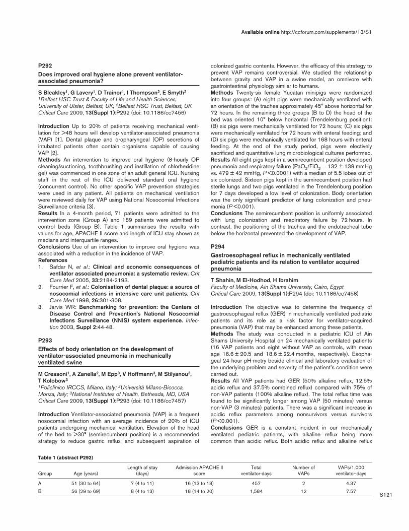
S121
P292Does improved oral hygiene alone prevent ventilator-associated pneumonia?
S Bleakley1, G Lavery1, D Trainor1, I Thompson2, E Smyth2
1Belfast HSC Trust & Faculty of Life and Health Sciences,University of Ulster, Belfast, UK; 2Belfast HSC Trust, Belfast, UKCritical Care 2009, 13(Suppl 1):P292 (doi: 10.1186/cc7456)
Introduction Up to 20% of patients receiving mechanical venti-lation for >48 hours will develop ventilator-associated pneumonia(VAP) [1]. Dental plaque and oropharyngeal (OP) secretions ofintubated patients often contain organisms capable of causingVAP [2].Methods An intervention to improve oral hygiene (8-hourly OPcleaning/suctioning, toothbrushing and instillation of chlorhexidinegel) was commenced in one zone of an adult general ICU. Nursingstaff in the rest of the ICU delivered standard oral hygiene(concurrent control). No other specific VAP prevention strategieswere used in any patient. All patients on mechanical ventilationwere reviewed daily for VAP using National Nosocomial InfectionsSurveillance criteria [3].Results In a 4-month period, 71 patients were admitted to theintervention zone (Group A) and 189 patients were admitted tocontrol beds (Group B). Table 1 summarises the results withvalues for age, APACHE II score and length of ICU stay shown asmedians and interquartile ranges.Conclusions Use of an intervention to improve oral hygiene wasassociated with a reduction in the incidence of VAP.References1. Safdar N, et al.: Clinical and economic consequences of
ventilator associated pneumonia: a systematic review. CritCare Med 2005, 33:2184-2193.
2. Fourrier F, et al.: Colonisation of dental plaque: a source ofnosocomial infections in intensive care unit patients. CritCare Med 1998, 26:301-308.
3. Jarvis WR: Benchmarking for prevention: the Centers ofDisease Control and Prevention’s National NosocomialInfections Surveillance (NNIS) system experience. Infec-tion 2003, Suppl 2:44-48.
P293Effects of body orientation on the development ofventilator-associated pneumonia in mechanicallyventilated swine
M Cressoni1, A Zanella2, M Epp3, V Hoffmann3, M Stilyanou3, T Kolobow3
1Policlinico IRCCS, Milano, Italy; 2Università Milano-Bicocca,Monza, Italy; 3National Institutes of Health, Bethesda, MD, USACritical Care 2009, 13(Suppl 1):P293 (doi: 10.1186/cc7457)
Introduction Ventilator-associated pneumonia (VAP) is a frequentnosocomial infection with an average incidence of 20% of ICUpatients undergoing mechanical ventilation. Elevation of the headof the bed to >30° (semirecumbent position) is a recommendedstrategy to reduce gastric reflux, and subsequent aspiration of
colonized gastric contents. However, the efficacy of this strategy toprevent VAP remains controversial. We studied the relationshipbetween gravity and VAP in a swine model, an omnivore withgastrointestinal physiology similar to humans.Methods Twenty-six female Yucatan minipigs were randomizedinto four groups: (A) eight pigs were mechanically ventilated withan orientation of the trachea approximately 45° above horizontal for72 hours. In the remaining three groups (B to D) the head of thebed was oriented 10° below horizontal (Trendelenburg position):(B) six pigs were mechanically ventilated for 72 hours; (C) six pigswere mechanically ventilated for 72 hours with enteral feeding; and(D) six pigs were mechanically ventilated for 168 hours with enteralfeeding. At the end of the study period, pigs were electivelysacrificed and quantitative lung microbiological cultures performed.Results All eight pigs kept in a semirecumbent position developedpneumonia and respiratory failure (PaO2/FiO2 = 132 ± 139 mmHgvs. 479 ± 42 mmHg, P <0.0001) with a median of 5.5 lobes out ofsix colonized. Sixteen pigs kept in the semirecumbent position hadsterile lungs and two pigs ventilated in the Trendelenburg positionfor 7 days developed a low level of colonization. Body orientationwas the only significant predictor of lung colonization and pneu-monia (P <0.001).Conclusions The semirecumbent position is uniformly associatedwith lung colonization and respiratory failure by 72 hours. Incontrast. the positioning of the trachea and the endotracheal tubebelow the horizontal prevented the development of VAP.
P294Gastroesophageal reflux in mechanically ventilatedpediatric patients and its relation to ventilator acquiredpneumonia
T Shahin, M El-Hodhod, H IbrahimFaculty of Medicine, Ain Shams University, Cairo, EgyptCritical Care 2009, 13(Suppl 1):P294 (doi: 10.1186/cc7458)
Introduction The objective was to determine the frequency ofgastroesophageal reflux (GER) in mechanically ventilated pediatricpatients and its role as a risk factor for ventilator-acquiredpneumonia (VAP) that may be enhanced among these patients.Methods The study was conducted in a pediatric ICU of AinShams University Hospital on 24 mechanically ventilated patients(16 VAP patients and eight without VAP as controls, with meanage 16.6 ± 20.5 and 18.6 ± 22.4 months, respectively). Esopha-geal 24 hour pH-metry beside clinical and laboratory evaluation ofthe underlying problem and severity of the patient’s condition werecarried out.Results All VAP patients had GER (50% alkaline reflux, 12.5%acidic reflux and 37.5% combined reflux) compared with 75% ofnon-VAP patients (100% alkaline reflux). The total reflux time wasfound to be significantly longer among VAP (50 minutes) versusnon-VAP (3 minutes) patients. There was a significant increase inacidic reflux parameters among nonsurvivors versus survivors(P <0.001).Conclusions GER is a constant incident in our mechanicallyventilated pediatric patients, with alkaline reflux being morecommon than acidic reflux. Both acidic reflux and alkaline reflux
Available online http://ccforum.com/supplements/13/S1
Table 1 (abstract P292)
Length of stay Admission APACHE II Total Number of VAPs/1,000 Group Age (years) (days) score ventilator-days VAPs ventilator-days
A 51 (30 to 64) 7 (4 to 11) 16 (13 to 18) 457 2 4.37
B 56 (29 to 69) 8 (4 to 13) 18 (14 to 20) 1,584 12 7.57

S122
were found to be associated with development of VAP and thetotal reflux time is found to be a reliable parameter to pick up VAPpatients. However, acidic reflux was found to be related to highmortality.
P295Incidence of ventilator-associated pneumonia in patientsundergoing elective tube exchange to LoTrachendotracheal tubes
A Fletcher, J Carter, M Blunt, P YoungQueen Elizabeth Hospital, Kings Lynn, UKCritical Care 2009, 13(Suppl 1):P295 (doi: 10.1186/cc7459)
Introduction The objective was to study a cohort of general ICUpatients electively reintubated with the LoTrach tracheal tube (tubeexchange) to determine safety and to audit postproceduralventilator-associated pneumonia (VAP). Emergency reintubationfollowing elective or unplanned extubation is a known risk factor forsubsequent VAP [1]; however, there are no published data on theeffects of protocol-based elective tube exchange. The LoTrachtube prevents the pulmonary aspiration that occurs withconventional cuffs [2] and allows subglottic secretion manage-ment, thereby directly influencing two key steps in the patho-genesis of VAP.Methods Sequential patients (53 patients in 14 months) receivingthe LoTrach tracheal tube and cuff pressure controller werestudied. Patients either underwent an elective tube exchange(using a bougie, preprocedural preoxygenation, gastric tubeaspiration, muscle relaxation and direct laryngoscopy to clearupper airway secretions) or were primary LoTrach intubations onthe ICU. Three clinicians independently examined the critical careelectronic patient record. VAP was identified by a fall in thePaO2/FaO2 ratio >25% or a clinical pulmonary infection score >5and the presence of a positive qualitative tracheal aspirate. Theinternational consensus criteria for VAP diagnosis were also used,as was the institution of antimicrobial therapy (if this was triggeredby a clinical suspicion of VAP, then for the purposes of this studyVAP was diagnosed).Results Forty-four (83%) patients underwent elective tubeexchange. No complications were noted associated with theprocedure. There were no episodes of VAP while the LoTrach wasin situ. On an intention-to-treat basis there was a 1.8% VAP ratebecause one patient who required emergency reintubationfollowing elective extubation received a conventional tube anddeveloped VAP 2 days later. No other patients had antimicrobialsbegun for chest infections.Conclusions There were no complications associated withelective tube exchange and no subsequent cases of VAP in thiscohort of patients who were reintubated with the LoTrach tube.Elective tracheal tube exchange can be safely performed in generalICU patients.References1. Torres A, et al.: Re-intubation increases the risk of nosoco-
mial pneumonia in patients needing mechanical ventila-tion. Am J Respir Crit Care Med 1995, 152:137-134.
2. Young PJ, et al.: A low-volume, low-pressure tracheal tubecuff reduces pulmonary aspiration. Crit Care Med 2006,34:632-639.
P296Reduction in ventilator-associated pneumonia followingthe introduction of subglottic suction endotracheal tubes
P Morgan, A Guyot, S Ranjan, M Eaton, M Carraretto, M ScottRoyal Surrey County Hospital, Guildford, UKCritical Care 2009, 13(Suppl 1):P296 (doi: 10.1186/cc7460)
Introduction Aspiration of subglottic secretions is recommendedby the American Thoracic Society, for the prevention of ventilator-associated pneumonia (VAP) [1], but has a poor implementationrate in some countries [2]. VAP is the most frequent infection inventilated patients, occurring in up to 27% of cases [1].Methods We studied 993 patients from September 2005, over a39-month period, following the introduction of our ventilation carebundle, which includes 30° head-up position, deep veinthrombosis and peptic ulcer prophylaxis, sedation holds and usingchlorhexidine for mouth care. VAP is difficult to diagnose anddefinitions vary. Our diagnoses were made prospectively by asingle consultant microbiologist using the validated ClinicalPulmonary Infection Score. After 18 months we introduced the Hi-Lo Evac/Lanz Mallinckrodt endotracheal tubes with suction appliedusing a 10 ml syringe 2-hourly, as our study intervention.Results The benchmark for European ICUs is a rate of 5% to 15%(HELICS; Hospital In Europe Link for Infection Control throughSurveillance) and in America is 3 to 20 cases per 1,000 ventilator-days (NNIS; National Nosocomial Infections Surveillance System).For the 18 months following the implementation of care bundles,our VAP rate was 5.39% (25/463 patients) with an incidence of14 cases per 1,000 ventilator-days. Following the study inter-vention the VAP rate fell to 1.5% (8/530 patients) and theincidence to 4.17 cases per 1,000 ventilator-days. Our cohort ofpatients over the period had no difference in APACHE II scores.The difference between the groups following the employment ofsubglottic secretion reached statistical significance with relativerisk 3.57 (95% CI = 1.63 to 7.85, P <0.001).Conclusions Our study confirms the benefit of subglotticsecretion clearance to reduce the occurrence of VAP.References1. American Thoracic Society: Am J Respir Crit Care Med
2005, 171:388-416.2. Sierra R, et al.: Chest 2005, 128:1667-1673.
P297Ventilator-associated pneumonia in a Greek ICU:prevalence and etiology
M Katsiari, E Apostolakou, C Nikolaou, E Pagouni, F Tsimpoukas, E Mainas, E Kounougeri, M Laskou, A MaguinaKonstantopoulion General Hospital, Nea Ionia, Athens, GreeceCritical Care 2009, 13(Suppl 1):P297 (doi: 10.1186/cc7461)
Introduction Nosocomial pneumonia is the leading cause of deathfrom hospital-acquired infections. Ventilator-associated pneumonia(VAP) is the most frequent ICU-acquired infection in mechanicallyventilated patients. The purpose of our study is to assess theprevalence and the etiologic pathogens of VAP in our ICU, as wellas its impact on morbidity and mortality.Methods A prospective observational study in a multidisciplinaryeight-bed ICU. During an 18-month period, 160 consecutivepatients with a length of stay (LOS) ≥48 hours were enrolled in thestudy. Data were collected using a specially designed softwareand included age, gender, APACHE II score on admission, days onmechanical ventilation, LOS and ICU outcome. Patients werestratified into two groups: Group A included patients who did not
Critical Care March 2009 Vol 13 Suppl 1 29th International Symposium on Intensive Care and Emergency Medicine
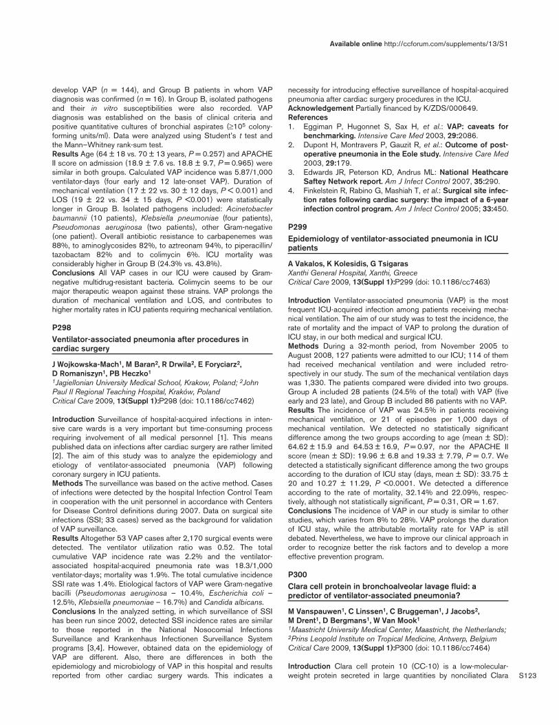
S123
develop VAP (n = 144), and Group B patients in whom VAPdiagnosis was confirmed (n = 16). In Group B, isolated pathogensand their in vitro susceptibilities were also recorded. VAPdiagnosis was established on the basis of clinical criteria andpositive quantitative cultures of bronchial aspirates (≥105 colony-forming units/ml). Data were analyzed using Student’s t test andthe Mann–Whitney rank-sum test.Results Age (64 ± 18 vs. 70 ± 13 years, P = 0.257) and APACHEII score on admission (18.9 ± 7.6 vs. 18.8 ± 9.7, P = 0.965) weresimilar in both groups. Calculated VAP incidence was 5.87/1,000ventilator-days (four early and 12 late-onset VAP). Duration ofmechanical ventilation (17 ± 22 vs. 30 ± 12 days, P < 0.001) andLOS (19 ± 22 vs. 34 ± 15 days, P <0.001) were statisticallylonger in Group B. Isolated pathogens included: Acinetobacterbaumannii (10 patients), Klebsiella pneumoniae (four patients),Pseudomonas aeruginosa (two patients), other Gram-negative(one patient). Overall antibiotic resistance to carbapenemes was88%, to aminoglycosides 82%, to aztreonam 94%, to piperacillin/tazobactam 82% and to colimycin 6%. ICU mortality wasconsiderably higher in Group B (24.3% vs. 43.8%).Conclusions All VAP cases in our ICU were caused by Gram-negative multidrug-resistant bacteria. Colimycin seems to be ourmajor therapeutic weapon against these strains. VAP prolongs theduration of mechanical ventilation and LOS, and contributes tohigher mortality rates in ICU patients requiring mechanical ventilation.
P298Ventilator-associated pneumonia after procedures incardiac surgery
J Wojkowska-Mach1, M Baran2, R Drwila2, E Foryciarz2, D Romaniszyn1, PB Heczko1
1Jagiellonian University Medical School, Krakow, Poland; 2JohnPaul II Regional Teaching Hospital, Kraków, PolandCritical Care 2009, 13(Suppl 1):P298 (doi: 10.1186/cc7462)
Introduction Surveillance of hospital-acquired infections in inten-sive care wards is a very important but time-consuming processrequiring involvement of all medical personnel [1]. This meanspublished data on infections after cardiac surgery are rather limited[2]. The aim of this study was to analyze the epidemiology andetiology of ventilator-associated pneumonia (VAP) followingcoronary surgery in ICU patients.Methods The surveillance was based on the active method. Casesof infections were detected by the hospital Infection Control Teamin cooperation with the unit personnel in accordance with Centersfor Disease Control definitions during 2007. Data on surgical siteinfections (SSI; 33 cases) served as the background for validationof VAP surveillance.Results Altogether 53 VAP cases after 2,170 surgical events weredetected. The ventilator utilization ratio was 0.52. The totalcumulative VAP incidence rate was 2.2% and the ventilator-associated hospital-acquired pneumonia rate was 18.3/1,000ventilator-days; mortality was 1.9%. The total cumulative incidenceSSI rate was 1.4%. Etiological factors of VAP were Gram-negativebacilli (Pseudomonas aeruginosa – 10.4%, Escherichia coli –12.5%, Klebsiella pneumoniae – 16.7%) and Candida albicans.Conclusions In the analyzed setting, in which surveillance of SSIhas been run since 2002, detected SSI incidence rates are similarto those reported in the National Nosocomial InfectionsSurveillance and Krankenhaus Infectionen Surveillance Systemprograms [3,4]. However, obtained data on the epidemiology ofVAP are different. Also, there are differences in both theepidemiology and microbiology of VAP in this hospital and resultsreported from other cardiac surgery wards. This indicates a
necessity for introducing effective surveillance of hospital-acquiredpneumonia after cardiac surgery procedures in the ICU.Acknowledgement Partially financed by K/ZDS/000649.References1. Eggiman P, Hugonnet S, Sax H, et al.: VAP: caveats for
benchmarking. Intensive Care Med 2003, 29:2086.2. Dupont H, Montravers P, Gauzit R, et al.: Outcome of post-
operative pneumonia in the Eole study. Intensive Care Med2003, 29:179.
3. Edwards JR, Peterson KD, Andrus ML: National HeathcareSaftey Network report. Am J Infect Control 2007, 35:290.
4. Finkelstein R, Rabino G, Mashiah T, et al.: Surgical site infec-tion rates following cardiac surgery: the impact of a 6-yearinfection control program. Am J Infect Control 2005; 33:450.
P299Epidemiology of ventilator-associated pneumonia in ICUpatients
A Vakalos, K Kolesidis, G TsigarasXanthi General Hospital, Xanthi, GreeceCritical Care 2009, 13(Suppl 1):P299 (doi: 10.1186/cc7463)
Introduction Ventilator-associated pneumonia (VAP) is the mostfrequent ICU-acquired infection among patients receiving mecha-nical ventilation. The aim of our study was to test the incidence, therate of mortality and the impact of VAP to prolong the duration ofICU stay, in our both medical and surgical ICU.Methods During a 32-month period, from November 2005 toAugust 2008, 127 patients were admitted to our ICU; 114 of themhad received mechanical ventilation and were included retro-spectively in our study. The sum of the mechanical ventilation dayswas 1,330. The patients compared were divided into two groups.Group A included 28 patients (24.5% of the total) with VAP (fiveearly and 23 late), and Group B included 86 patients with no VAP.Results The incidence of VAP was 24.5% in patients receivingmechanical ventilation, or 21 of episodes per 1,000 days ofmechanical ventilation. We detected no statistically significantdifference among the two groups according to age (mean ± SD):64.62 ± 15.9 and 64.53 ± 16.9, P = 0.97, nor the APACHE IIscore (mean ± SD): 19.96 ± 6.8 and 19.33 ± 7.79, P = 0.7. Wedetected a statistically significant difference among the two groupsaccording to the duration of ICU stay (days, mean ± SD): 33.75 ±20 and 10.27 ± 11.29, P <0.0001. We detected a differenceaccording to the rate of mortality, 32.14% and 22.09%, respec-tively, although not statistically significant, P = 0.31, OR = 1.67.Conclusions The incidence of VAP in our study is similar to otherstudies, which varies from 8% to 28%. VAP prolongs the durationof ICU stay, while the attributable mortality rate for VAP is stilldebated. Nevertheless, we have to improve our clinical approach inorder to recognize better the risk factors and to develop a moreeffective prevention program.
P300Clara cell protein in bronchoalveolar lavage fluid: apredictor of ventilator-associated pneumonia?
M Vanspauwen1, C Linssen1, C Bruggeman1, J Jacobs2, M Drent1, D Bergmans1, W Van Mook1
1Maastricht University Medical Center, Maastricht, the Netherlands;2Prins Leopold Institute on Tropical Medicine, Antwerp, BelgiumCritical Care 2009, 13(Suppl 1):P300 (doi: 10.1186/cc7464)
Introduction Clara cell protein 10 (CC-10) is a low-molecular-weight protein secreted in large quantities by nonciliated Clara
Available online http://ccforum.com/supplements/13/S1
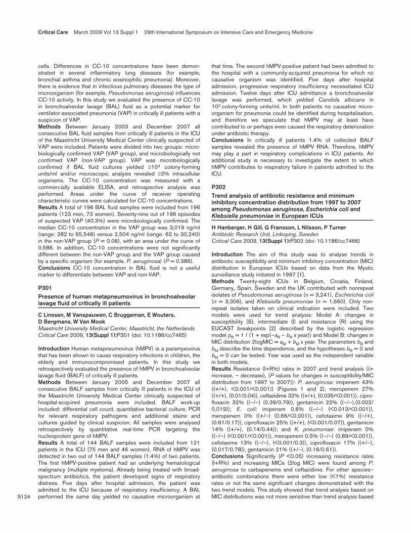
S124
cells. Differences in CC-10 concentrations have been demon-strated in several inflammatory lung diseases (for example,bronchial asthma and chronic eosinophilic pneumonia). Moreover,there is evidence that in infectious pulmonary diseases the type ofmicroorganism (for example, Pseudomonas aeruginosa) influencesCC-10 activity. In this study we evaluated the presence of CC-10in bronchoalveolar lavage (BAL) fluid as a potential marker forventilator-associated pneumonia (VAP) in critically ill patients with asuspicion of VAP.Methods Between January 2003 and December 2007 allconsecutive BAL fluid samples from critically ill patients in the ICUof the Maastricht University Medical Center clinically suspected ofVAP were included. Patients were divided into two groups: micro-biologically confirmed VAP (VAP group), and microbiologically notconfirmed VAP (non-VAP group). VAP was microbiologicallyconfirmed if BAL fluid cultures yielded ≥104 colony-formingunits/ml and/or microscopic analysis revealed ≥2% intracellularorganisms. The CC-10 concentration was measured with acommercially available ELISA, and retrospective analysis wasperformed. Areas under the curve of receiver operatingcharacteristic curves were calculated for CC-10 concentrations.Results A total of 196 BAL fluid samples were included from 196patients (123 men, 73 women). Seventy-nine out of 196 episodesof suspected VAP (40.3%) were microbiologically confirmed. Themedian CC-10 concentration in the VAP group was 3,019 ng/ml(range: 282 to 65,546) versus 2,504 ng/ml (range: 62 to 30,240)in the non-VAP group (P = 0.06), with an area under the curve of0.586. In addition, CC-10 concentrations were not significantlydifferent between the non-VAP group and the VAP group causedby a specific organism (for example, P. aeruginosa) (P = 0.386).Conclusions CC-10 concentration in BAL fluid is not a usefulmarker to differentiate between VAP and non-VAP.
P301Presence of human metapneumovirus in bronchoalveolarlavage fluid of critically ill patients
C Linssen, M Vanspauwen, C Bruggeman, E Wouters, D Bergmans, W Van MookMaastricht University Medical Center, Maastricht, the NetherlandsCritical Care 2009, 13(Suppl 1):P301 (doi: 10.1186/cc7465)
Introduction Human metapneumovirus (hMPV) is a paramyxovirusthat has been shown to cause respiratory infections in children, theelderly and immunocompromised patients. In this study weretrospectively evaluated the presence of hMPV in bronchoalveolarlavage fluid (BALF) of critically ill patients.Methods Between January 2005 and December 2007 allconsecutive BALF samples from critically ill patients in the ICU ofthe Maastricht University Medical Center clinically suspected ofhospital-acquired pneumonia were included. BALF work-upincluded: differential cell count, quantitative bacterial culture, PCRfor relevant respiratory pathogens and additional stains andcultures guided by clinical suspicion. All samples were analysedretrospectively by quantitative real-time PCR targeting thenucleoprotein gene of hMPV.Results A total of 144 BALF samples were included from 121patients in the ICU (75 men and 46 women). RNA of hMPV wasdetected in two out of 144 BALF samples (1.4%) of two patients.The first hMPV-positive patient had an underlying hematologicalmalignancy (multiple myeloma). Already being treated with broad-spectrum antibiotics, the patient developed signs of respiratorydistress. Five days after hospital admission, the patient wasadmitted to the ICU because of respiratory insufficiency. A BALperformed the same day yielded no causative microorganism at
that time. The second hMPV-positive patient had been admitted tothe hospital with a community-acquired pneumonia for which nocausative organism was identified. Five days after hospitaladmission, progressive respiratory insufficiency necessitated ICUadmission. Twelve days after ICU admittance a bronchoalveolarlavage was performed, which yielded Candida albicans in102 colony-forming units/ml. In both patients no causative micro-organism for pneumonia could be identified during hospitalisation,and therefore we speculate that hMPV may at least havecontributed to or perhaps even caused the respiratory deteriorationunder antibiotic therapy.Conclusions In critically ill patients 1.4% of collected BALFsamples revealed the presence of hMPV RNA. Therefore, hMPVmay play a part in respiratory complications in ICU patients. Anadditional study is necessary to investigate the extent to whichhMPV contributes to respiratory failure in patients admitted to theICU.
P302Trend analysis of antibiotic resistance and minimuminhibitory concentration distribution from 1997 to 2007among Pseudomonas aeruginosa, Escherichia coli andKlebsiella pneumoniae in European ICUs
H Hanberger, H Gill, G Fransson, L Nilsson, P TurnerAntibiotic Research Unit, Linkoping, SwedenCritical Care 2009, 13(Suppl 1):P302 (doi: 10.1186/cc7466)
Introduction The aim of this study was to analyse trends inantibiotic susceptibility and minimum inhibitory concentration (MIC)distribution in European ICUs based on data from the Mysticsurveillance study initiated in 1997 [1].Methods Twenty-eight ICUs in Belgium, Croatia, Finland,Germany, Spain, Sweden and the UK contributed with nonrepeatisolates of Pseudomonas aeruginosa (n = 3,241), Escherichia coli(n = 3,306), and Klebsiella pneumoniae (n = 1,660). Only non-repeat isolates taken on clinical indication were included. Twomodels were used for trend analysis: Model A: changes insusceptibility (S), intermediate (I) and resistance (R) using theEUCAST breakpoints [2] described by the logistic regressionmodel pR = 1 / (1 + exp(–aR – bR x year)) and Model B: changes inMIC distribution 2logMIC = aM + bM x year. The parameters bR andbM describe the time dependence, and the hypotheses bR = 0 andbM = 0 can be tested. Year was used as the independent variablein both models.Results Resistance (I+R%) rates in 2007 and trend analysis ((+increase, – decrease), (P values for changes in susceptibility/MICdistribution from 1997 to 2007)): P. aeruginosa: imipenem 43%((+/+), <0.001/<0.001)) (Figures 1 and 2), meropenem 27%((+/+), (0.01/0.04)), ceftazidime 32% ((+/+), (0.035/<0.001)), cipro-floxacin 32% ((–/–) (0.39/0.79)), gentamicin 22% ((–/–),(0.002/0,019)); E. coli: imipenem 0.6% ((–/–) (<0.013/<0.001)),meropenem 0% ((+/–) (0.66/<0.001)), cefotaxime 9% ((–/+),(0.61/0.17)), ciprofloxacin 25% ((+/+), (<0.001/0.07)), gentamicin14% ((+/+), (0.14/0.44)); and K. pneumoniae: imipenem 0%((–/–) (<0.001/<0.001)), meropenem 0.5% ((–/–) (0.89/<0.001)),cefotaxime 13% ((–/–), (<0.001/0.3)), ciprofloxacin 17% ((+/–),(0.017/0.78)), gentamicin 21% ((+/–), (0.16/0.61)).Conclusions Significantly (P <0.05) increasing resistance rates(I+R%) and increasing MICs (2log MIC) were found among P.aeruginosa to carbapenems and ceftazidime. For other species–antibiotic combinations there were either low (<1%) resistancerates or not the same significant changes demonstrated with thetwo trend models. This study showed that trend analysis based onMIC distributions was not more sensitive than trend analysis based
Critical Care March 2009 Vol 13 Suppl 1 29th International Symposium on Intensive Care and Emergency Medicine
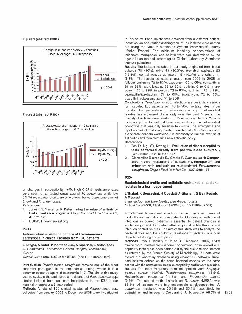
S125
on changes in susceptibility (I+R). High (>27%) resistance rateswere seen for all tested drugs against P. aeruginosa while low(<1%) resistance rates were only shown for carbapenems againstE. coli and K. pneumoniae.References1. Jones RN, Masterton R: Determining the value of antimicro-
bial surveillance programs. Diagn Microbiol Infect Dis 2001,41:171-175.
2. EUCAST [www.eucast.org]
P303Antimicrobial resistance pattern of Pseudomonasaeruginosa in clinical isolates from ICU patients
E Antypa, A Koteli, K Kontopoulou, A Kiparissi, E AntoniadouG. Gennimatas Thessaloniki General Hospital, Thessaloniki,GreeceCritical Care 2009, 13(Suppl 1):P303 (doi: 10.1186/cc7467)
Introduction Pseudomonas aeruginosa remains one of the mostimportant pathogens in the nosocomial setting, where it is acommon causative agent of bacteremia [1,2]. The aim of this studywas to evaluate the antimicrobial resistance of Pseudomonas spp.strains isolated from inpatients hospitalized in the ICU of ourhospital throughout a 3-year period.Methods A total of 175 clinical isolates of Pseudomonas spp.collected from January 2006 to December 2008 were investigated
in this study. Each isolate was obtained from a different patient.Identification and routine antibiograms of the isolates were carriedout using the Vitek 2 automated System (BioMerieux®, MarcyI’Etolie, France). The minimum inhibitory concentrations ofimipenem, meropenem and colistin were also determined by theagar dilution method according to Clinical Laboratory StandardsInstitute guidelines.Results The isolates included in our study originated from bloodcultures 70 (40%), urine 53 (30.3%), bronchial aspirates 23(13.1%), central venous catheters 18 (10.3%) and others 11(6.3%). The resistance rates changed from 2006 to 2008 asfollows: amikacin: 72 to 83%, aztreonam: 90 to 95%, ceftazidime:81 to 89%, ciprofloxacin: 79 to 85%, colistin: 0 to 0%, mero-penem: 72 to 83%, imipenem: 72 to 83%, netilmicin: 72 to 83%,piperacillin/tazobactam: 71 to 80%, tobramycin: 72 to 83%,ticarcillinlin/clavulanic acid: 71 to 80%.Conclusions Pseudomonas spp. infections are particularly seriousfor incubated ICU patients with 40 to 50% mortality rates. In ourhospital, the percentage of Pseudomonas spp. multiresistantisolates has increased dramatically over the past 3 years. Themajority of isolates were resistant to 15 or more antibiotics. What ismost worrying is the fact that there is a prevalence of a multiresistantphenotype that was only sensitive to colistin. The emergance andrapid spread of multidrug-resistant isolates of Pseudomonas spp.are of great concern worldwide. It is necessary to limit the overuse ofantibiotics and to implement a new antibiotic policy.References1. Tan TY, Ng LSY, Kwang LL: Evaluation of disc susceptibility
tests performed directly from positive blood cultures. JClin Pathol 2008, 61:343-346.
2. Giamarellos-Bourboulis EJ, Grecka P, Giamarellou H: Compar-ative in vitro interactions of ceftazidime, meropenem, andimipenem with amikacin on multiresistant Pseudomonasaeruginosa. Diagn Microbiol Infect Dis 1997. 29:81-86.
P304Bacteriological profile and antibiotic resistance of bacteriaisolates in a burn department
L Thabet, K Bousselmi, H Oueslati, A Ghanem, S Ben Redjeb,A MessadiTraumatology and Burn Center, Ben Arous, TunisiaCritical Care 2009, 13(Suppl 1):P304 (doi: 10.1186/cc7468)
Introduction Nosocomial infections remain the main cause ofmorbidity and mortality in burn patients. Ongoing surveillance ofinfections in burned patients is essential to detect changes inepidemiology and to guide better empirical antibiotherapy andinfection control policies. The aim of this study was to analyze thebacterial flora and the antibiotic resistance of isolates in a burndepartment during a 2-year period.Methods From 1 January 2005 to 31 December 2006, 1,268strains were isolated from different specimens. Antimicrobial sus-ceptibility testing has been carried out by the disk diffusion methodas referred by the French Society of Microbiology. All data werestored in a laboratory database using whonet 5.3 software. Dupli-cate isolates defined as the same bacterial species for the samepatient with the same antimicrobial susceptibility profile were excluded.Results The most frequently identified species were Staphylo-coccus aureus (19.8%), Pseudomonas aeruginosa (15.8%),Acinetobacter baumannii (11.8%), and Providencia stuarttii(9.5%). The rate of methicillin-resistant S. aureus (MRSA) was68.1%. All isolates were fully susceptible to glycopeptides. P.aeruginosa resistance was 35.6% and 35.4% respectively forceftazidime and imipenem. Concerning A. baumannii, 98.7% of
Available online http://ccforum.com/supplements/13/S1
Figure 1 (abstract P302)
Figure 2 (abstract P302)
S126
strains were resistant to ceftazidime, 59.5% to imipenem and87.5% to ciprofloxacin. In total, 77.3% of P. stuarttii isolates wereresistant to ceftazidime. The frequencies of resistance toceftazidime, ofloxacin and amikacin of Klebsiella pneumoniae wererespectively 60.9%, 25.4% and 47.1%. The survey of resistanceshowed a global decrease in 2006 versus 2005. The rate ofMRSA was 61% in 2005 versus 51.6% in 2006. The resistance ofceftazidime was 80.6% in 2005 versus 26.9% in 2006 in P.aeruginosa. The imipenem resistance showed also a decrease in2006 in A. baumannii and P. aeruginosa. The amelioration ofhygiene, particularly washing hands after introduction of hydro-alcoholic solutions, and the collaboration between microbiologistsand clinicians could explain the decrease of resistance showed inthe burn department.Conclusions Comparative to the previous years, S. aureus is stillthe commonest pathogen in the burn department. The incidence ofantimicrobial resistance has decreased during 2006 after a peak ofmultiresistance during 2005. The measures of prevention takenwere efficient and should be re-enforced.
P305Antibiotic resistance of Staphylococcus aureus from ICUsin the Netherlands 1996 to 2006
M Rijnders1, RH Deurenberg1, M Boumans1, M Hoogkamp-Korstanje2, P Beisser1, EE Stobberingh1
1University Hospital Maastricht, the Netherlands; 2SWAB, Utrecht,the NetherlandsCritical Care 2009, 13(Suppl 1):P305 (doi: 10.1186/cc7469)
Introduction Staphylococcus aureus is a potential pathogenicmicroorganism and a causative agent of ~25% of infections inintensive care patients. Optimal empiric therapy may reducemorbidity and mortality. Therefore, it is essential to provide theclinician with resistance data of the local circulating strains andpatients pathogens. A national surveillance program of the DutchAntibiotic Resistance Surveillance Group was started in theNetherlands in 1996 to gain insight into the emergence ofmicrobial resistance at local, regional and national levels. Thisstudy describes the findings of resistance development of S.aureus from ICUs of 14 large referral hospitals all over theNetherlands over a 10-year period from 1996 to 2006.Methods In the first 6 months of each year, the participatinghospitals collected clinical isolates from, among others, blood andrespiratory samples. In total 943 isolates were collected: 250 fromthree hospitals in the north, 187 from two in the east, 229 from five inthe west and 280 from four in the south. The susceptibility torelevant antibiotics was determined by micro broth dilution accordingto the Clinical and Laboratory Standards Institute guidelines.Results Resistance to penicillin fluctuated over time at ~75%;seven methicillin-resistant S. aureus were isolated (0.7%).Resistance to clarithromycin increased to 10% in 2003, butdecreased in 2006 to 6%, the level before 2003. Resistance toclindamycin fluctuated over time from 4 to 8%. Doxycyclin resis-tance varied between 2 and 10%. No resistance to vancomycin,teicoplanin and linezolid was demonstrated. Resistance togentamicin and rifampicin was sporadically found. The prevalenceof fluoroquinolone resistance was between 0 and 4% until 2002.In 2003, a peak in the prevalence of fluoroquinolone resistance(ciprofloxacin 14% and moxifloxacin 8%) was observed.Resistance to ciprofloxacin remained at this high level until 2005and decreased in 2006. Resistance to moxifloxacin decreasedimmediately. Regional differences were observed for ciprofloxacin,with the highest resistance in the western and southern parts andwith doxycyclin with the lowest resistance rate in the northern part.
Conclusions During the 10-year study period only an increase inresistance to ciprofloxacin was observed. The data presented stilljustify the empiric choice of flucloxacillin (with rifampicin orgentamicin depending on the indication) in case of an infectionprobably caused by S. aureus in ICU patients.
P306Clinical and microbiological efficacy of continuous versusintermittent administration of vancomycin in critical carepatients
M Štepán, I Chytra, P Pelnar, T Bergerová, E Kasal, A Zidkova,R PradlUniversity Hospital, Plzen, Czech RepublicCritical Care 2009, 13(Suppl 1):P306 (doi: 10.1186/cc7470)
Introduction Vancomycin is known to induce postantibiotic effectbut some data suggest that bactericidal activity is time dependent.Presently, the optimal dosing regimen for administration ofvancomycin remains unknown. The aim of this open prospectiverandomized study was to compare clinical and microbiologicalefficacy of continuous infusion versus intermittent administration ofvancomycin in critically ill patients.Methods Patients admitted to the interdisciplinary ICU sufferingfrom infection indicated to vancomycin administration withpredicted duration of treatment of at least 4 days were randomizedto receive either a 15 mg/kg intravenous loading dose ofvancomycin followed by a daily 15 mg/kg continuous infusion(continuous group) or intermittent administration of 15 mg/kgvancomycin intravenously every 12 hours (intermittent group).Antibiotic therapy was stopped at improvement of clinical state andlaboratory signs of subsidence of infection. Failure of antimicrobialtherapy was defined as persistence or progression of signs andsymptoms of infection, development of new clinical findingsconsistent with active infection or death from infection. The age,APACHE II score, type of infection, length of ICU stay, length ofmechanical ventilation, mortality, clinical and microbiological failureand length of vancomycin therapy and total dose of vancomycinwere assessed. The Sequential Organ Failure Assessment score,white blood count, C-reactive protein and renal function at thebeginning and at the end of therapy were evaluated. TheMann–Whitney test, unpaired t test and chi-squared test wereused accordingly; P <0.05 was considered statistically significant.Results A total of 65 patients were enrolled and randomized in thecontinuous (n = 33) and intermittent (n = 32) groups. Nosignificant differences between both groups in all assessedparameters were found. Clinically evaluated failure of therapy in thecontinuous versus intermittent groups was in 12 (36%) patientsversus eight (25%) patients, and microbiological failure in seven(21%) patients versus eight (25%) patients.Conclusions Continuous infusion and intermittent administrationof vancomycin in critically ill patients provided equivalent clinicaland microbiological outcome.Acknowledgement Supported by research grant MSM0021620819.
P307Over-increased creatinine renal clearance in septicpatients and implications for vancomycin optimization
J Baptista, P Casanova, P Martins, J PimentelCoimbra University Hospitals, Coimbra, PortugalCritical Care 2009, 13(Suppl 1):P307 (doi: 10.1186/cc7471)
Introduction The hyperdynamic stage occurring in sepsis may beresponsible for an increase in renal blood flow that may lead to
Critical Care March 2009 Vol 13 Suppl 1 29th International Symposium on Intensive Care and Emergency Medicine

S127
increased elimination of some drugs, namely vancomycin. The aimof this study was to evaluate the effects of 24-hour creatinineclearance (CrCL) higher than 130 ml/min/1.73 m2 on vancomycinserum levels, and to identify an accurate marker of this condition.Methods This study was carried out in a multipurpose ICU, on 120critical septic patients, 36% with septic shock (42 patients), 67men (72.5%), average age 56 ± 21 years, average APACHE IIscore and Simplified Acute Physiology Score (SAPS) II of 17.2and 42.2, respectively. We studied 120 consecutive vancomycintreatments (continuous perfusion) and we assessed therapeuticlevels (20 to 30 μg/ml) on days 1, 2 and 3 (V1, V2 and V3).Patients were divided into two groups according to CrCL: G1≤130 ml/min/1.73 m2 (n = 77); G2 >130 ml/min/1.73 m2 (n = 43).Both groups had similar vancomycin dosage on day 0 (47.7/46.6mg/kg; P = 0.26).Results Average age, APACHE II score and SAPS II were 62.6/44years, 18.8/14.2 and 45/36.9, respectively, for G1 and G2(P <0.05). Average CrCL in G1: 76.7 ml/min/1.73 m2; averageCrCL in G2: 176 ml/min/1.73 m2 (P <0.05). Serum proteins andalbumin were, respectively, 5.26/5.64 g/dl and 2.84/3.17 g/dl(P <0.05), and urinary creatinine (UCr), urinary urea and 24-hoururine output were 34.6/71 mg/dl, 437/625 mg/dl and 2,450/2,846 ml, respectively (P <0.05). V1, V2 and V3 were 20/14.3,24.3/17.6 and 26.5/20.3 μg/ml, respectively, for G1 and G2(P <0.05 on each day). The correlations between V1 and CrCLwere –0.47 (G1) and –0.33 (G2), both with P <0.05. The areaunder the curve (AUC) of the receiving operating curves of UCr,urinary urea and blood urea nitrogen (BUN) as markers of highclearance status (>130) were 0.85 (95% CI = 0.77 to 0.91), 0.72(95% CI = 0.63 to 0.80) and 0.66 (95% CI = 0.55 to 0.73),respectively, and this area was maximal for a subgroup of patients(76 patients) without shock (AUC: 0.9; 95% CI = 0.81 to 0.96 forUCr). Best cutoff point for UCr: > 58mg/dl; for BUN: <17 mg/dl.Conclusions Over-increased CrCL identifies a subgroup ofyounger patients, with lower severity scores and high incidence ofsubtherapeutic vancomycin concentrations on the first 3 days –especially on day 1 (39/43 patients; 90.7%). A UCr concentrationabove 58 mg/dl can be a sensitive (70%) and specific (87%)marker of this condition. The addition of BUN <17 mg/dl as asecond marker increases specificity to 97.4%.
P308Vancomycin-resistant enterococci colonisation in the ICUand control measures
K Katircioglu, M Ozkalkanli, S Gul Yurtsever, D Sanli, H Erten,V Duzenli, S SavaciIzmir Ataturk Training and Research Hospital, Izmir, TurkeyCritical Care 2009, 13(Suppl 1):P308 (doi: 10.1186/cc7472)
Introduction Vancomycin-resistant enterococci (VRE) have beenrecognised as microorganisms capable of causing epidemics incritically ill patients [1]. The present report describes an outbreakinvolving VRE colonisation in our ICU.Methods After several weeks of severe nursing shortage, whenoften there were only three nurses for 10 critically ill patients, thefirst VRE was isolated from one of the two blood cultures of atrauma patient. Rectal swabs of all of the patients’ and ICU staffwere collected. Because there was no patient who had beendischarged to the ward during this period no further analysis wasrequired.Results Enterococcus casseliflavus was isolated from six out ofthe 10 patients’ rectal swabs. Once the outbreak was identified, allpatients were placed under strict contact isolation and cohorted,and barrier precautions were instituted. Four patients who had
negative rectal swabs were isolated in another ICU. Five of thepatients did not demonstrate signs of infection and were acceptedas colonisation. ICU staff had negative rectal swabs. Two of thepatients who had positive rectal swabs had died because ofunderlying primary disease (acute myocardial infarction, acuterespiratory distress syndrome due to aspiration pneumonia). Thefirst patient who had a positive blood culture was treated withintravenous Linezolid and discharged to home after 14 days.Another patient who had sepsis due to necrotising fasciitis wasdischarged to the ward after obtaining three negative rectal swabs.The other patient who had non-Hodgkin lymphoma and pneumoniawas discharged to the ward after obtaining three negative rectalswabs. The last patient was isolated in another room and followedup for 4 weeks and discharged to the ward after obtaining threenegative rectal swabs.Conclusions The outbreak was controlled by continuous imple-mentation of the infection control programme. A long ICU stay,hemodialysis, and nursing shortage are risk factors for VREdevelopment. Transmission of VRE can be facilitated by the handsof the staff. We conclude that this outbreak may be due to theshortage of nursing during summer.Reference1. Peta M, et al.: Outbreak of vancomycin-resistant Entero-
coccus spp. in an Italian general intensive care unit. ClinMicrobiol Infect 2006, 12:163-169.
P309Activities of vancomycin, teicoplanin and linezolid againstbacteraemic methicillin-resistant Staphylococcus aureusstrains in Gauteng, South Africa
ME Botha1, J Coetzee1, C Feldman2, GA Richards2, AJ Brink1
1Ampath National Laboratory Services, Johannesburg, South Africa;2Johannesburg Hospital and University of the Witwatersrand,Johannesburg, South AfricaCritical Care 2009, 13(Suppl 1):P309 (doi: 10.1186/cc7473)
Introduction This study aims to describe the vancomycin,teicoplanin and linezolid susceptibility patterns of methicillin-resistant Staphylococcus aureus (MRSA) blood culture isolatesfrom patients in the private sector in Gauteng, South Africa.Screening tests for heterogeneous glycopeptide intermediate S.aureus (hGISA) strains were also performed. MRSA isolates withvancomycin minimum inhibitory concentrations (MICs) of 1 to2 mg/l are associated with worse clinical outcomes [1]. Further-more, hGISA infections are associated with clinical failure ofglycopeptide therapy [2].Methods MICs for vancomycin, teicoplanin and linezolid wereperformed on 50 randomly collected MRSA strains from bloodcultures according to Clinical Laboratory Standards Institute guide-lines. Screening for hGISA was performed using the Etest (ABBIODISK) Macromethod as well as the new Etest GlycopeptideResistance Detection.
Available online http://ccforum.com/supplements/13/S1
Table 1 (abstract P309)
Susceptibility patterns of MRSA isolates (n = 50)
Vancomycin Teicoplanin Linezolid(mg/l) (mg/l) (mg/l)
MIC50 1.5 2 1.5
MIC90 2 3 2
Breakpoint S ≤2 S ≤8 S ≤4

S128
Results The results of susceptibility patterns are depicted inTable 1. Fifty percent (25/50) and 20% (10/50) of the strainsscreened positive for hGISA using the Etest Macromethod andEtest Glycopeptide Resistance Detection, respectively.Conclusions We recommend that clinically relevant MRSAisolates be reported with MICs for vancomycin, teicoplanin andlinezolid, and that glycopeptide treatment failure warrants furthertesting of the MRSA isolate to detect possible hGISA. IdeallyhGISA should be confirmed by population analysis profile testing,which was not available to us for this study.References1. Soriano A, et al.: Influence of vancomycin MIC on the treat-
ment of MRSA bacteremia. Clin Infect Dis 2007, 46:193-200.
2. Charles PGP, et al.: Clinical features associated with bac-teremia due to heterogeneous vancomycin-intermediateStaphylococcus aureus. Clin Infect Dis 2004, 38:448-451.
P310Telavancin for the treatment of hospital-acquiredpneumonia in severely ill and older patients: the ATTAINstudies
E Rubinstein1, GR Corey2, HW Boucher3, MS Niederman4, A Shorr5, A Torres6, SL Barriere7, HD Friedland7
1University of Manitoba, Winnipeg, MB, Canada; 2Duke UniversityMedical Center, Durham, NC, USA; 3Tufts Medical Center, Boston,MA, USA; 4SUNY, Stony Brook, NY, USA; 5Georgetown University,Washington, DC, USA; 6Hospital Clínic de Barcelona, Spain;7Theravance, Inc., South San Francisco, CA, USACritical Care 2009, 13(Suppl 1):P310 (doi: 10.1186/cc7474)
Introduction Telavancin (TLV) is an investigational lipoglyco-peptide with activity against Gram-positive pathogens. The Assess-ment of Telavancin for Treatment of Hospital-acquired Pneumonia(ATTAIN) programme studied TLV for the treatment of hospital-acquired pneumonia (HAP). This analysis compared the clinicalcure rates achieved with TLV or vancomycin (VAN) for severely illand older patients.Methods ATTAIN 1 and ATTAIN 2 were methodologicallyidentical, randomised, double-blind, phase 3 studies. Adult patientswith pneumonia acquired after 48 hours in an inpatient acute-careor chronic-care facility, or acquired within 7 days after beingdischarged following ≥3 days of hospital stay were randomised toTLV 10 mg/kg intravenously every 24 hours or VAN 1 g intra-venously every 12 hours for 7 to 21 days. A test-of-cure (TOC) visitwas conducted 7 to 14 days after end-of-study treatment. Com-pliant patients who had a clinical response of either cure or failureat TOC were considered clinically evaluable (CE).Results Pooled clinical cure rates at TOC for several clinicallyrelevant subgroups including the elderly as well as patients withsevere HAP at baseline are presented in Figure 1. The percentageof patients reporting at least one treatment-emergent adverse
event was comparable between the TLV and VAN groups (80% vs.79%, respectively).Conclusions Although not statistically significant, TLV achievednumerically higher cure rates than VAN for treatment of HAP inseverely ill and older patients with comparable treatment-emergentadverse events.
P311Piperacillin/tazobactam administered by continuous orintermittent infusion for the treatment of nosocomialpneumonia
L Lorente, S Palmero, J Jiménez, J Iribarren, R Galván, J Martínez, C García, J Castedo, M Brouard, M Martín, M MoraHospital Universitario de Canarias, La Laguna SC, Tenerife, SpainCritical Care 2009, 13(Suppl 1):P311 (doi: 10.1186/cc7475)
Introduction Betalactam efficacy is determined by the duration oftime that concentrations remain above the minimum inhibitoryconcentration (MIC). Some studies have found that the adminis-tration of betalactams by continuous infusion maintains constantconcentrations above the MIC of susceptible organisms over thecourse of therapy; but limited data exist on the clinical efficacy ofbetalactams administered by continuous infusion. The purpose ofthis study was to evaluate the clinical efficacy of piperacillin/tazobactam by continuous infusion (CI) administration or byintermittent infusion (II) for the treatment of ventilator-associatedpneumonia (VAP) caused by Gram-negative bacilli.Methods A retrospective cohort study (1 June 2002 to 31December 2007) of patients with VAP caused by Gram-negativebacilli who received initial empiric antibiotic therapy withpiperacillin/tazobactam. We analyzed two contemporary cohorts:one received piperacillin/tazobactam by CI (first received a loadingdose of piperacillin/tazobactam 4/0.5 g over 30 minutes, and after4/0.5 g infused over 360 minutes every 6 hours) and the other by II(4/0.5 g over 30 minutes every 6 hours). The administration ofpiperacillin/tazobactam by CI or II was prescribed according to thephysician’s discretion.Results Significant differences were not found between bothgroups of patients (37 with CI and 46 with II) in baseline data.Logistic regression analysis showed a higher probability of clinicalcure of VAP by CI than by II (33/37 (89.2%) vs. 26/46 (56.5%);OR = 7.4; 95% CI = 1.96 to 37.42; P = 0.001). Logisticregression analysis showed a higher probability of clinical cure ofVAP by CI than by II when the microorganism causative of VAPhad a MIC of 8 μg/ml (8/9 (88.9%) vs. 6/15 (40.0%); OR = 10.8;95% CI = 1.01 to 588.24; P = 0.049) and a MIC of 16 μg/ml (7/8(87.5%) vs. 1/6 (16.7%); OR = 22.9; 95% CI = 1.19 to 1,880.78;P = 0.03); but not when it had a MIC of 4 μg/ml (18/20 (90.0%)vs. 19/25 (76.0%); OR = 2.8; 95% CI = 0.42 to 31.67; P = 0.41).Conclusions Administration of piperacillin/tazobactam by con-tinuous infusion may have more clinical efficacy than administrationby intermittent infusion for the treatment of nosocomial pneumonia.
P312Efficiency of rifampicin–miconazole-impregnated cathetersin the femoral venous site
L Lorente, M Lecuona, J Iribarren, J Jiménez, C García, R Galván, J Castedo, J Martínez, M Mora, A SierraHospital Universitario de Canarias, La Laguna SC, Tenerife, SpainCritical Care 2009, 13(Suppl 1):P312 (doi: 10.1186/cc7476)
Introduction The guidelines of the Centers for Disease Controland Prevention do not recommend the use of an antimicrobial or
Critical Care March 2009 Vol 13 Suppl 1 29th International Symposium on Intensive Care and Emergency Medicine
Figure 1 (abstract P310)

S129
antiseptic-impregnated catheter for short-term catheterization andrecommend avoiding femoral access to reduce the risk of centralvenous catheter-related bacteremia (CVC-RB). The objective ofthis study was to determine the incidence of CVC-RB withrifampicin–miconazole-impregnated catheters (RM-C) and stan-dard catheters (S-C) in femoral venous access; and the cost ofCVC-RB with both types of catheter.Methods A cohort study, conducted in a 24-bed polyvalentmedical–surgical ICU of a university hospital. We included patientsadmitted to the ICU from 1 June 2006 to 30 September 2007,who underwent femoral venous catheterization. The cost of CVC-RB included the cost of antimicrobial agents and the diagnosismethods; but other costs were not included (such as prolongationof ICU stay, mechanical ventilation, extrarenal depuration, etc.).Results We inserted 73 RM-C during 634 catheter-days and 111S-C during 927 catheter-days. We diagnosed eight CVC-RB inthe S-C group and none in the RM-C group. The incidence ofCVC-RB with RM-C was significantly lower than with S-C (0 vs.8.62 per 1,000 catheter-days, OR = 0.13, 95% CI = 0.00 to 0.86;P = 0.03). The total cost in the RM-C group was €7,300 (73central venous catheters (CVC) per €100 each CVC, and noneCVC-RB), the mean cost per catheter was €100 (€7,300/73CVC), and the mean cost per catheter-day was €11.51(€7,300/634 CVC-days). The total cost in the S-C group was€15,330 (111 CVC per €30 each CVC = €3,330, more than€12,000 per the eight CVC-RB), the mean cost per catheter was€138 (€15,330/111 CVC), and the mean cost per catheter-daywas €16.54 (€15,330/927 CVC-days).Conclusions RM-C could decrease the incidence of femoralvenous catheter-related bacteremia and are more efficient thanstandard catheters.
P313Micafungin for the treatment of pediatric invasive fungalinfections
A Arrieta1, NL Seibel2, TJ Walsh2, L Arnold3, AH Groll41Children’s Hospital, Orange, CA, USA; 2National Institute ofHealth, Bethesda, MD, USA; 3Astellas Pharma, Deerfield, IL, USA;4Children’s Hospital, Muenster, GermanyCritical Care 2009, 13(Suppl 1):P313 (doi: 10.1186/cc7477)
Introduction Pediatric patients in the ICU are highly vulnerable toinvasive fungal infections [1]; Candida has been reported to be thethird most common pathogen in pediatric ICU wards [2]. However,information on new antifungal therapies in children remains limited.Micafungin (MICA) is a once-daily antifungal agent of theechinocandin class.Methods A retrospective review of MICA pediatric data from sixtrials.Results MICA was received by 296 children for invasive candi-diasis, refractory invasive candidiasis, refractory invasive asper-gillosis, prophylaxis in hematopoietic stem cell transplantation(HSCT) patients, or to assess pharmacokinetics (PK). Most patientsaged <1 year were premature (38/66), whereas most children aged>1 year were HSCT recipients or undergoing another therapy for ahematological malignancy (181/230). Maximum daily dose wassimilar for patients aged <1, 1 to 4, 5 to 8, 9 to 12 and 13 to15 years with medians of 2.0, 1.5, 1.5, 1.9 and 1.5 mg/kg,respectively. Treatment success rates are shown in Figure 1. MICAshowed linear PK, with a higher clearance in neonates than in olderchildren and adults. Common adverse events (incidence ≥2%) weretransient increases in transaminases, hypokalemia, hyperbili-rubinemia and increased alkaline phosphatase.
Conclusions MICA is a safe and efficacious agent for thetreatment and prophylaxis of pediatric invasive fungal infections inthe ICU setting.References1. Filioti J, et al.: Intensive Care Med 2007, 33:1272-1283.2. Raymond J, et al.: Infect Control Hosp Epidemiol 2000,
21:260-263.
P314Malignant Boutonneuse fever with multiple organ failure: a three-case series
B Oliveira, AP Lacerda, Z Costa e Silva, C FrançaHospital de Santa Maria Lisboa, PortugalCritical Care 2009, 13(Suppl 1):P314 (doi: 10.1186/cc7478)
Introduction Rickettsia conorii infection, the agent ofBoutonneuse fever, presents usually in a form considered benign,with serious complications in less than 10% of patients. Itsmechanism of infection consists of vascular endothelial invasion bythe microorganism and subsequent vasculitis and tissue necrosis.The process is usually localized or limited to the skin but canprogress with malignant severe systemic involvement [1,2].Methods A series of three cases of patients with multiple organfailure admitted to an ICU with confirmed diagnosis of Bouton-neuse fever is presented (positive serology or tissue PCR).Results Common to all the patients was the rapid development ofthe disease (average: 3 days) from the initial complaints of feverafter a bite by an unidentified agent and development ofgeneralized rash, and the diagnosis of multiple organ failure(average Simplified Acute Physiology Score II: 59; averageSequential Organ Failure Assessment score on admission: 12.6).All developed respiratory failure requiring invasive mechanicalventilation, haematological failure with haemolytic anaemia andacute renal failure suggestive of serious widespread vasculitis.One of the cases developed lethal refractory septic shock within6 hours of admission. Tetracycline antibiotic therapy was startedearly in all patients, based on clinical and epidemiological data,since initial microbiological results were negative.Conclusions This series of cases illustrates the most severe formof Boutonneuse fever, usually associated with comorbidities suchas diabetes, malignant disease, immunodeficiency or delay in thediagnosis and appropriate antibiotic therapy, which was not thecase in these patients.
Available online http://ccforum.com/supplements/13/S1
Figure 1 (abstract P313)

S130
References1. Aharonowitz G, Koton S, Segal S, et al.: Epidemiological
characteristics of spotted fever in Israel over 26 years.Clin Infect Dis 1999, 29:1321-1322.
2. Anton E, Font B, Munoz T, et al.: Clinical and laboratorycharacteristics of 144 patients with mediterraneanspotted fever. Eur J Clin Microbiol Infect Dis 2003, 22:126-128.
P315Impact of microbiology and antimicrobial treatment onmortality in septic shock
G Dabar1, G Jamaleddine2, P Yazbeck1, M Waked3
1Hotel Dieu de France Hospital, Beirut, Lebanon; 2AmericanUniversity Hospital, Beirut, Lebanon; 3Saint George Hospital,Beirut, LebanonCritical Care 2009, 13(Suppl 1):P315 (doi: 10.1186/cc7479)
Introduction Infectious characteristics, the infection source, andappropriate antibiotics are analyzed in relation to mortality in acohort of patients with severe sepsis and septic shock.Methods An observational and prospective cohort study over1 year of all patients admitted to one of the adult ICUs of the threemajor university hospitals in Beirut. Demographics, appropriate ICUtherapies within 48 hours of admission, appropriate antibioticprescription within 48 hours of admission, antibiotic duration andnumber were registered. Infections were defined as definite orsuspected. The infectious source, microbial agents and anti-biograms were identified. Data were collected only during the ICUstay, and outcome was recorded at hospital discharge (alive ordead).Results One hundred and twenty-seven patients with an averageage of 65 years were enrolled, 52% of admissions were from theemergency room and 83% were medical patients. Fifty-eightpercent of the patients had severe sepsis within 48 hours ofadmission. The average APACHE II and Sequential Organ FailureAssessment scores were 21 and 7, respectively. Immuno-deficiency was present in 30%. Overall mortality was 45%.Definite infection was diagnosed in 61% of the patients andbacteremia in 31.5%. Appropriate antibiotic prescription wassignificantly higher in patients with less severe sepsis comparedwith severe sepsis (>1 organ failure). Mortality was significantlyhigher in patients prescribed a higher number of antibiotics whilethe length of antibiotic prescription was not different betweensurvivors and nonsurvivors. Escherichia coli (n = 14) was the mostcommon isolate in blood cultures, followed by Candida (n = 5).Fungal pathogens were significantly more associated withnosocomial infections and with mortality. Multiresistantpseudomonas was significantly more associated to mortalitycompared with extended spectrum beta lactamase or methicillin-resistant Staphylococcus aureus. Positive urine culture wasprotective of a higher organ failure score. In multivariate analysis,Gram-negative bacteria, a nosocomial source and immuno-deficiency were all associated with an increased risk of death.Conclusions Inappropriate antibiotic prescription is associatedwith more severe sepsis. Mortality is higher in nosocomial sepsis.Candida is a serious nosocomial agent in severe sepsis and isassociated with increased mortality as well as multiresistantpseudomonas aeruguinosa.
P316Do changes in ICU flora and their antibiotic susceptibilityparallel changes in infection control and antibiotic use?
I Grigoras, C Caramidaru, D Rusu, L Cotarlet, O Chelarescu'Gr.T. Popa' University of Medicine and Pharmacy Iasi, EmergencyUniversity Hospital ‘Sf Spiridon’ Iasi, RomaniaCritical Care 2009, 13(Suppl 1):P316 (doi: 10.1186/cc7480)
Introduction The usefulness of microbiological surveillance in ICUpatients is under debate [1,2]. It can signal infection/colonizationsites, most frequent germs and their antibiotic susceptibility. Ourstudy aimed to investigate changes in the microorganism rate andtheir antibiotic susceptibility in ICU patients along with efforts tochange antibiotic practice and infection control policies in the ICU.Methods A prospective study was performed in a 19-bed mixedICU in an emergency university hospital. Blood, catheters,respiratory secretions, surgical wound secretions and urine werecultured any time in the case of infection suspicion or every 4 to5 days in the case of patients with ICU length of stay >5 days.Cultures were counted no matter the infection or colonizationstatus. Antibiotic use was recorded as daily drug doses/100 beds(DDDs). The study included two time periods: January to June2007 (P1 – 839 admitted patients) and January to June 2008 (P2 –779 admitted patients). Comparison between the two periodsincluded overall and site-specific rates of positive cultures, patternsof germs, antibiotic susceptibility and antibiotic usage.Results The overall number of positive cultures decreased by31.8% (P <0.01) and per patient by 15% (P <0.01) in P2 versusP1. Germ patterns changed little over time, most frequently foundbeing Staphylococcus aureus, Acinetobacter spp, coagulase-negative staphylococci and Pseudomonas spp. According to thesite, coagulase-negative staphylococci isolated from the blood-stream decreased from 42% to 26% (P = 0.01), but Acinetobacterspp. in the respiratory tract increased from 34.5% to 43%,concurrently with a reduction in Klebsiella spp from 12.7% to 3%.Antibiotic use decreased by one-half in P2 versus P1 (68.7 DDDsvs. 113.8 DDDs, P = 0.01). Ciprofloxacin dropped from first (P1)to seventh (P2) position (75% decrease) and resulted in adecrease in ciprofloxacin resistance of S. aureus from 94% to46% (P <0.01).Conclusions Changes in infection control policies and antibioticuse resulted in a decreased number of positive cultures in ICUpatients and a decline in antibiotic resistance in some strains.References1. Jones ME, et al.: Emerging resistance among bacterial
pathogens in the intensive care unit. Ann Clin MicrobiolAntimicrob 2004, 3:14.
2. EARSS Annual Report 2005 [http://www.rivm.nl/earss/]
P317Prehospital administered intravenous antimicrobialprotocol for septic shock: a prospective randomizedclinical trial
D ChamberlainFlinders University, Adelaide, AustraliaCritical Care 2009, 13(Suppl 1):P317 (doi: 10.1186/cc7481)
Introduction Appropriate intravenous, broad-spectrum empiricantimicrobial therapy should be initiated as rapidly as possible forsuspected severe infections in the presence of hypotension.Timing and delay of initial administration of effective antimicrobialtherapy is an important predictor of survival [1]. It is hypothesizedthat appropriate prehospital-initiated intravenous, broad-spectrum
Critical Care March 2009 Vol 13 Suppl 1 29th International Symposium on Intensive Care and Emergency Medicine
S131
empiric antimicrobial therapy will reduce the delay of administrationand will reduce mortality [2].Methods One hundred and ninety-eight patients meeting thecriteria for septic shock on initial clinical presentation prehospitalwere randomized to receiving broad-spectrum intravenous anti-microbial therapy and fluid per guided protocol or to receivingintravenous fluid only.Results Out of 198 septic shock patients, 99 received prehospitalantimicrobial therapy. Blood cultures were taken prior to adminis-tration. Both groups were comparable in all aspects. There were83 male and 26 female patients in the test group, and 79 male and20 female patients in the control group (P = 0.021). Mean age was67.9 ± 10.5 years in the test group and 63.8 ± 11.0 years in thecontrol group (P = 0.186). The APACHE II score in the test groupand in the control group was statistically not significant(P = 0.661). In the test group 28%, compared with the controlgroup 53%, of patients had community-acquired pneumonia(P = 0.083). The mean intensive care stay in the test group was6.8 ± 2.1 days and in the control group it was 11.2 ± 5.2 days(P = 0.001). The first antimicrobial administration after emergencydepartment admission time in the control group was 3.4 ± 2.6hours (P = 0.02). The 28-day mortality rate was significantlyreduced to 42.4% (test group) compared with 56.7% in thecontrol group (P = 0.049, OR = 0.56; 95% CI = 0.32 to 1.00).Conclusions The adjuvant treatment of patients with a guidedprehospital-initiated broad-spectrum antimicrobial therapy withintravenous fluid reduces the delay in antimicrobial administrationand significantly reduces the 28-day mortality rate in patients withseptic shock.References1. Kumar A, et al.: Duration of hypotension before initiation of
effective antimicrobial therapy is the critical determinantof survival in human septic shock. Crit Care Med 2006,34:1589-1596.
2. Miner JR, et al.: Presentation, time to antibiotics, and mor-tality of patients with bacterial meningitis at an urbancounty medical center. J Emerg Med 2001, 21:387-392.
P318Clinical experiment with the use of colistin for thetreatment of multiresistant Acinetobacter baumaniinosocomial infection
L Safi, A Elwali, K Aboulalaa, A Baite, M LekliteMilitary Mohammed V Hospital, Rabat, MoroccoCritical Care 2009, 13(Suppl 1):P318 (doi: 10.1186/cc7482)
Introduction Intravenous and aerosolized colistin are being usedincreasingly, in the critical care setting, for treating patients withnosocomial infections due to multidrug-resistant Gram-negativebacteria. The objective of this study was to report our experienceof colistin use in multiresistant Acinetobacter baumanii.Methods A retrospective review of patients’ charts for thoseadmitted to the surgical intensive unit, between September 2007and September 2008. Infected patients with multiresistant acineto-bacter treated with intravenous and aerosolized colistin werereviewed.Results Fifteen patients were identified, 13 men and two women.The patients ranged in age from 21 to 65 years. The patients wereadmitted to intensive care for polytrauma (10 cases), chest trauma(three cases) and cerebrovascular accident (two cases). The maininfections were ventilator nosocomial pneumonia (100%), surgicalsite infection (6.6%), primary bacteremia (13%), catheter infection(20%), and meningitis (13%). The bacteria responsible were A.baumanii (100%), Pseudomonas aeruginosa (46%), Staphylo-
coccus aureus (26%), Klebsiella pneumoniae (6%), Enterobactercloacae (6%), and Morganella morganii (6%). All patients hadnormal renal function at the onset of antibiotic therapy. Fivepatients received colistin monotherapy, six patients receivedcombination therapy colistin with imipenem or third-generationcephalosporin, and four patients received colistin with teicoplanine.The colistin was used at 4 mg/kg/day administered intravenouslyand aerolized colistin at 2 mg/kg for 8 to 16 days. A favourableresponse was observed in 12 cases. Overall mortality was threecases. Colistin induced reversible nephrotoxicity in one case and areversible neuropsychiatric event was observed in one case.Bacterial eradication was confirmed in 14 patients (93%).Conclusions Garnacho-Montero and colleagues reported thatintravenous colistin was as effective as imipenem [1]. Our findingswith colistin can be considered encouraging in comparison withprevious experience.Reference1. Garnacho-Montero J, et al.: Treatment of multidrug-resis-
tant Acinetobacter baumannii ventilator-associated pneu-monia (VAP) with intravenous colistin: a comparison withimipenem-susceptible VAP. Clin Infect Dis 2003, 36:1111-1118.
P319Evaluation of gentamicin first-dose pharmacokinetics inseptic critically ill patients: pilot study
JC Gonçalves Pereira1, A Martins2, P Póvoa1
1Hospital S. Francisco Xavier, Lisboa, Portugal; 2Hospital S. José,Lisboa, PortugalCritical Care 2009, 13(Suppl 1):P319 (doi: 10.1186/cc7483)
Introduction Aminoglycosides, especially gentamicin, are exten-sively used for treatment of severe infections. A dose of 7 mg/kg isrecommended to achieve a high peak serum concentration.However, its accumulation is associated with adverse reactions,notably renal injury. Knowledge of gentamicin pharmacokinetics incritically ill patients is therefore crucial to therapeutic success andprevention of toxicity.Methods Patients’ demographic and clinical data were collected.We studied gentamicin pharmacokinetic data from patients treatedbetween January 2006 and June 2008 in two ICUs. Patients wereeligible if the gentamicin dose was selected to achieve high serumlevels (target concentration of 16 to 24 μg/ml) and if pharmaco-kinetic data from such patients, specifically peak and trough levels,were measured after the first dose. The Sawchuk and Zaske one-compartment pharmacokinetic model [1] was used to calculate thegentamicin volume of distribution (Vd), maximum concentration(Cmax) and clearance.Results We studied 32 patients with a mean age of 63 years (24men). Mean Sequential Organ Failure Assessment score was 7.65and the mean Charlson comorbidities score was 3.54. The medianVd was 0.41 l/kg (IQR 0.36 to 0.46 l/kg). Only four patients had aVd equal to or less than 0.26 l/kg, usually found in the healthypopulation. We found no correlation between Vd and age,Charlson comorbidities score, Sequential Organ Failure Assess-ment score, and creatinine serum level (r2 = 0.016, 0.18, 0.037,and 0.067, respectively). The presence of mechanical ventilationand septic shock had no influence on Vd (P = 0.59 and P = 0.14,respectively). Women had a significantly higher mean Vd/kg (0.51vs. 0.39 l/kg, P = 0.002) and, therefore, lower Cmax (15.2 vs.18.5 μg/ml, P = 0.016). In a logistic regression model, only sex(female: OR = 0.032; 95% CI = 0.03 to 0.387) and the dose/kg(per mg/kg: OR = 3.21; 95% CI = 1.17 to 8.79) were significantlyassociated with the achievement of Cmax above 16 μg/ml.
Available online http://ccforum.com/supplements/13/S1

S132
Conclusions The gentamicin Vd cannot be predicted by age, thepresence of renal failure or any studied comorbidities. Therefore, inorder to obtain adequate therapeutic levels as soon as possible, ahigh first gentamicin dose (that is, at least 7 mg/kg) should begiven to all patients. In women, even higher doses may be needed.Reference1. Sawchuk R, Zaske D: Pharmacokinetics of dosing regi-
mens which utilize multiple intravenous infusions. J Phar-macokinet Biopharm 1976, 4:183-195.
P320Does early appropriate antibiotic therapy improve theoutcome of severe sepsis or septic shock?
N Saito, Y Sakamoto, K MashikoChiba Hokusou Hospital, Nippon Medical School, Chiba, JapanCritical Care 2009, 13(Suppl 1):P320 (doi: 10.1186/cc7484)
Introduction The Surviving Sepsis Campaign Guideline 2008 [1]recommends starting appropriate antibiotic therapy within 1 hourafter making the diagnosis of septic shock. In the ICU of ouremergency center, we perform empiric antibiotic therapy for septicshock patients within 6 hours after admission.Methods Cases of severe sepsis or septic shock diagnosed andtreated in the ICU of our emergency center for more than 48 hoursbetween January 2005 and September 2008 were retrospectivelyanalyzed. The cases were divided into a survival group and anonsurvival group, and were compared in relation to primarydiagnosis, clinical findings, and type of pathogen. The chi-squaretest and paired t test were used to perform the statistical analysis.Results There were 107 cases, 24 cases of severe sepsis and 83cases of septic shock, and 73 of them were in males. The mean andstandard deviation of the patients’ age was 66.4 ± 15 years. Theseverity of their illness according to the APACHE II score was 22.3 ±8, and their Sequential Organ Failure Assessment score was 9.1 ± 4.The causes of the sepsis were pneumonia (51.4%), peritonitis(13.1%), and soft tissue infection (13.1%). Mortality was 26.2%.There were 79 cases in the survival group and 28 in the nonsurvivalgroup. The two groups are presented in Table 1. We performed amultivariate regression analysis to identify prognostic factors, and theonly independent prognostic factors were age (OR = 0.955(P = 0.022; 95% CI = 0.91 to 0.99)), acute respiratory diseasesyndrome (ARDS) (OR = 5.789 (P = 0.002; 95% CI = 1.91 to 17.4))and base deficit (OR = 1.113 (P = 0.008; 95% CI = 1.02 to 1.2)).Early appropriate antibiotic therapy (EAAT) was not correlated withsurvival.
Table 1 (abstract P320)
Survival Nonsurvival P value
Early appropriate antibiotic therapy (%) 67 60.7 0.645Gram-positive coccus (%) 39.2 53.5 0.297Gram-negative rod (%) 29.1 39.2 0.351
Conclusions EAAT did not affect the outcome. The prognosticfactors for severe sepsis and septic shock identified in this studywere age, base deficit and ARDS.References1. Dellinger RP, Levy MM, Carlet JM, et al.: Surviving sepsis cam-
paign: International guidelines for management of severesepsis and septic shock. Crit Care Med 2008, 36:296-327.
2. Kumar A, Roberts D, Wood KE, et al.: Duration of hypoten-sion before initiation of effective antimicrobial therapy isthe critical determinant of survival in human septic shock.Crit Care Med 2006, 34:1589-1596.
P321Strategy to reduce antibiotic prescription in cases ofairway aspiration
H Bagnulo, M Godino, N CarambulaHospital Maciel, Montevideo, UruguayCritical Care 2009, 13(Suppl 1):P321 (doi: 10.1186/cc7485)
Introduction Airway aspiration (AWA) of gastric content is a veryfrequent complication in patients presenting to emergency roomsafter incidents linked to a depressed level of consciousness. Theroutine use of antibiotics in this situation determines a high-pressure selection for multiresistant microorganisms. However,how often AWA is the causative situation for early ventilator-associated pneumonia is not known. Also a gold standard for adiagnostic workup is needed. Our aim was to know how frequentAWA determines infectious aspiration pneumonia confirmed byevolution and microbiological samples, and to propose amethodological approach to rule out antibiotic usage.Methods Over a 2-year period, in 82 patients admitted to our ICU,AWA was confirmed by the direct observation of gastric contentwhen orotracheal intubation was performed by a trained physiciandue to a Glasgow coma scale below 12. Usual diagnosis: acutebrain injury, 52 patients (73%); Simplified Acute Physiology ScoreII 37 ± 11; age, 44 ± 15 years; all were mechanically ventilated,average 6 ± 2 days; ICU stay ± 10 days; mortality 21%. Rectalfever, leukocytosis, thoracic radiology and PaO2/FiO2 were recordedon a daily basis. The Clinical Pulmonary Infection Score (CPIS) andsemiquantitative tracheal aspirates (SQTA) were performed twice: inthe first 48 hours and between the third and fifth days.Results Out of 82 patients, 23 (28%) developed clinically andmicrobiologically confirmed pneumonia. Fever and leukocytosisshowed no significant differences in patients with or withoutpneumonia during the first 5 days. Also the PaO2/FiO2 index wasnot different. As for radiology, when unilateral focal condensationwas present, pneumonia was confirmed later (relative risk = 3.3,95% CI = 1.7 to 6.4). CPIS in the first 48 hours showed a negativepredictive value for pneumonia of 89%, and SQTA with nomicroorganism growth a negative predictive value of 96%. In ourpatient group, 42 (51%) had CPIS <6 and SQTA growth; in themno antibiotic usage is recommended. On the contrary, out of 20patients who had CPIS ≥6 and positive SQTA, 16 (75%) developedpneumonia within the first 5 days of the ICU stay; this groupdeserves antibiotics. CPIS ≥6 without pneumonia, probably due tolung inflammation, was observed in 10 out of 28 patients (36%).Conclusions (1) In our ICU population, pneumonia develops inonly 28% of those presenting with AWA. (2) Through CPIS <6and negative SQTA performed in the first 48 hours we couldidentify that in more than one-half of AWA patients antibiotics arenot needed. (3) CPIS is not a reliable early indicator of pneumoniain patients with AWA.
P322Effect of intravenous administration of freeze-driedsulfonated human normal immunoglobulin for septicpatients
Y Deguchi1, T Nakagawa1, H Suga1, M Nishina1, T Sato1, K Okajima2, N Harada2
1Tokyo Women’s Medical University, Tokyo, Japan; 2Nagoya CityUniversity, Nagoya, JapanCritical Care 2009, 13(Suppl 1):P322 (doi: 10.1186/cc7486)
Introduction The proinflammatory cytokine TNF plays a critical rolein the formation of severe sepsis when it is excessively released.
Critical Care March 2009 Vol 13 Suppl 1 29th International Symposium on Intensive Care and Emergency Medicine

S133
Recently, it was revealed through our animal models that intra-venous administration of freeze-dried sulfonated human normalimmunoglobulin (IVIG) makes anti-inflammatory effects byenhancing insulin-like growth factor 1 (IGF-1) production throughstimulation of sensory nerves, which inhibits the production of TNF.In this study, we examined whether IVIG for septic patientsenhances IGF-1 production.Methods Fourteen septic patients indicating a high level of solubleE-selectin were surveyed, who were transported to our hospitalfrom April to November 2007. They were divided into two groups,IVIG group (G-group, 5 g/day x 3 days, nine cases, 67.2 ± 8.64years old) and nonadministered group (control group, C-group, fivecases, 69 ± 12.9 years old).Results In the G-group, the values of IGF-1 on the fifth day(162.22 ± 52.91 ng/ml) and seventh day (150.44 ± 29.06 ng/ml)were significantly higher than that on day 0 (88.59 ± 29.79 ng/ml,P <0.05). Furthermore, The value of IGF-1 of the G-group(162.22 ± 52.91 ng/ml) was significantly higher than that of the C-group (68.80 ± 24.04 ng/ml) on the fifth day (P <0.05).Conclusions We revealed that IVIG for septic patients enhancesIGF-1 production. Furthermore we will enhance the effect ofsulfanated immunoglobulin compared with that of intact immuno-globulin in the difference of the anti-inflammatory effect and themechanism.References1. Okajima K, Harada N, Suga H,Nakagawa T: Rapid assay for
plasma soluble E-selectin predicts the development ofacute respiratory distress syndrome in patients with sys-temic inflammatory response syndrome. Transl Res 2006,148:295-300.
2. Harada N, Okajima K, Kurihara H, Nakagata N: Antithrombinprevents reperfusion-induced hepatic apoptosis byenhancing insulin-like growth factor-I production in mice.Crit Care Med 2008, 36:971-974.
P323Levels of soluble fibrin in severe septic patients givenintravenous immunoglobulin
T Kobayashi1, T Nishiura2, H Suga2, T Nakagawa2
Tokyo Women Medical University Medical Center East, Tokyo,JapanCritical Care 2009, 13(Suppl 1):P323 (doi: 10.1186/cc7487)
Introduction It is known that severe sepsis causes multiple organfailure and high mortality. Intravenous immunoglobulin (IVIG) isused for severe sepsis patients. The mechanism of IVIG is clearlyunknown. Suggested, however, is the possibility that IVIG preventsorganic injury by suppressing the inflammation. Soluble fibrin (SF)is intermediate between fibrinogen and fibrionoligomer. It is betterthan fibrinogen as the material for thrombin. IVIG is used as one ofthe effective factors for repair of the damaged vascular endo-thelium. In this study we used SF as a marker of severity of illnessin patients with or without IVIG and compared the levels of SF intwo groups.Methods Nineteen severe septic patients were divided into twogroups. The control group (n = 9) was not given and the othergroup (n = 10) was given IVIG (5 g/day for 3 days). SF of serumfrom patients was measured on 0 days, 1 day, 3 days, 5 days and7 days, and the results were analyzed.Results The levels of SF were: control group: 4.77 ± 9.44(0 days), 3.13 ± 2.53 (1 day), 3.33 ± 2.75 (3 days), 2.53 ± 2.58(5 days), 2.58 ± 3.75 (7 days); other group: 7.71 ± 8.28 (0 days),12.26 ± 16.75 (1 day), 14.34 ± 25.56 (3 days), 8.06 ± 9.08(5 days), 11.95 ± 12.71 (7 days). There was a significant
difference in the levels of SF between groups with and withoutIVIG at 1 day, 5 days and 7 days.Conclusions Monocyte has the Fcγ receptor. It is thought thatstimulating Fcγ of monocyte with IgG plays a role in increasing SF.Without severe DIC, increasing fibrin is used in the repair of thedamaged vascular endothelium as a matrix and it prevents theinvasion of germs. It is thought that increasing fibrin is theappropriate stimulation for coagulation and one of the reactions ofbiomechanical defense. We think SF is one of the importantfactors for the damaged cell and a useful marker of severity forsevere sepsis. We also think IVIG is effective for severe sepsis.Reference1. Echtenacher B, et al.: Infect Immun 2001, 69:3550-3555.
P324Efficacy of corticosteroids on survival in patients withsepsis and septic shock: meta-analysis
N Uchiyama, N Nishimura, T Jinta, R Suda, S Yamao, S Gotoh,H Horinouchi, Y Tomishima, R Sugiura, N ChohnabayashiSt Luke’s International Hospital, Tokyo, JapanCritical Care 2009, 13(Suppl 1):P324 (doi: 10.1186/cc7488)
Introduction Previous studies have recommended the use of low-dose corticosteroids in patients with septic shock. Specifically,response to corticotropin tests has been recognized as aprognostic factor in critically ill patients, especially in patients withno response to the corticotropin test. A recent large randomizedcontrolled trial evaluating the efficacy of low-dose corticosteroidsrevealed no benefit on overall survival or in patients with noresponse to corticotropin in patients with severe sepsis and septicshock. Recently, recommendations for the diagnosis and manage-ment of corticosteroid insufficiency in critically ill adult patientswere published as consensus statements from an international taskforce by the American College of Critical Care Medicine [1].However, some studies were not included in these statements.Methods We conducted a systematic search of EMBASE andMEDLINE (through August 2008) for double-blind randomizedclinical trials that evaluated the effect of corticosteroids onmortality in patients with severe sepsis and septic shock. Studyselection criteria were all trials before August 2008 in whichparticipants were randomized to corticosteroids or placebo and inwhich mortality was reported.Results Data from 17 randomized, controlled trials with a total of3,638 participants were analyzed. Corticosteroid use was notassociated with a risk reduction in overall mortality (pooled riskratio = 0.99 (95% CI = 0.90 to 1.09), P = 0.823). Low-dosecorticosteroids (150 to 300 mg/day) did not show benefit on all-cause mortality in patients with severe sepsis and septic shock(risk ratio = 0.92 (95% CI = 0.79 to 1.06)). Furthermore, low-dosecorticosteroids in patients with severe sepsis and septic shockwho did not respond to the corticotropin test showed no benefit on28-day mortality (risk ratio = 0.91 (95% CI = 0.76 to 1.09)).Corticosteroids use was not associated with increased compli-cations, such as gastrointestinal bleeding or increased infections.Conclusions This meta-analysis indicated that administration oflow-dose corticosteroids was not beneficial on overall mortality inpatients with severe sepsis or septic shock.Reference1. Marik PE, et al.: Recommendations for the diagnosis and
management of corticosteroid insufficiency in critically illadult patients: consensus statements from an interna-tional task force by the American College of Critical CareMedicine. Crit Care Med 2008, 36:1937-1949.
Available online http://ccforum.com/supplements/13/S1

S134
P325Effect of hydrocortisone therapy on outcome and theincidence of infection in patients with septic shock
H Hayami, O Yamaguchi, H Yamada, S Nagai, S Ohama, A Sakurai, Y SugawaraYokohama City University Medical Center, Yokohama, JapanCritical Care 2009, 13(Suppl 1):P325 (doi: 10.1186/cc7489)
Introduction In the CORTICUS study, despite the fact that themedian time to shock reversal was shorter in the hydrocortisone(HC) group, improvement of outcome was not demonstrated. Thisis explained as an increased incidence of superinfection, includingnew episodes of sepsis or septic shock, but the explanation seemsto be unclear so there are still debates about the study. Weexamined the outcome of HC therapy and the effect on laterinfectious complications, especially fungal infection.Methods A retrospective cohort study. Ninety-six adult patients>18 years old with septic shock treated at single university generalICU. Initially, fluid therapy, inotropic support, transfusion andmanagement of infection were carried out according to theguideline of survival sepsis campaign, and we assessed theadrenal function by adrenocorticotropic hormone stimulation test(250 μg). In the earlier period (April 2004 to March 2007), 200 mgHC was administered to the nonresponders. Later (April 2007onwards), HC was given only to patients that were poorlyresponsive to fluid resuscitation and vasopressor therapy. Weassessed the 28-day and 90-day survival rate, hospital dischargerate, the incidence of new septic shock, the positive rate ofendotoxin and β-D-glucan.Results Out of 96 patients, 38 patients were determined asresponders and 58 as nonresponders. The Sequential OrganFailure Assessment score was higher in nonresponders (11 ± 3.3vs. 13.6 ± 3.7, P = 0.0014). For the 58 nonresponders, HC wasused for 40 patients (HC group). The 28-day survival rate washigher in HC (61% vs. non-HC 44%, P = 0.49), but the 90-daysurvival rate decreased to 33% in HC and 28% in non-HC(P = 0.47), and the discharge rate reversed as 25% in HC versus28% in non-HC (P = 0.84). The incidence of new septic shockwas 20% in HC, 11% in non-HC. The positive rate of endotoxindid not increase before and after treatment in both groups. Themean concentration of β-D-glucan did not increase in non-HC(59 ± 99 to 48 ± 44 pg/ml), but it significantly increased in HC(50 ± 70 to 69 ± 186 pg/ml) (P = 0.049).Conclusions The 28-day survival rate was higher in the HC groupthan the non-HC group as reported in the French study, but nobeneficial effect was seen in longer outcome, consistent with theCORTICUS study. Moreover, HC therapy may have a risk toincrease fungal infection.
P326Effects of methylprednisolone on sepsis-inducedblood–brain barrier permeability and reflex responsivenessin rats
F Esen1, T Erdem1, A Ogan2, E Senturk1, P Ergin Özcan1, M Kaya1, N Cakar1, L Telci11University of Istanbul, Turkey; 2International Hospital, Istanbul,TurkeyCritical Care 2009, 13(Suppl 1):P326 (doi: 10.1186/cc7490)
Introduction The purpose of this study was to assess neurologicalfunction and examine blood–brain barrier (BBB) permeabilitychanges in septic encephalopathy after cecal ligation and perfora-tion (CLP). The protective effect of high-dose methylprednisolone
administration on BBB derangement and neurological dysfunctionwas also investigated.Methods An experimental prospective randomized study wasperformed on 42 adult male Sprague–Dawley rats. Sepsis wasinduced through CLP. A bolus 30 mg/kg methylprednisoloneadministration followed by intravenous injection of 5.4 mg/kg/hourfor 8 hours was given immediately after CLP in septic and sham-operated rats. Control groups for both septic and sham-operatedrats were injected with equal volumes of saline.Results Neurological function was assessed at 8, 12, and 24 hoursafter sepsis induction. Those rats surviving for 24 hours postprocedure were anesthetized and decapitated for investigation ofBBB integrity using a spectrophotometric assay of Evans blue dyeextravasations. A significant decrease in neurological function anda significant increase in BBB permeability were observed with theinduction of sepsis compared with the sham-operated controls.Administration of methylprednisolone caused a significantlyimproved reflex responsiveness and decreased brain tissue Evansblue dye content in septic rats.Conclusions The present investigation shows that methylpred-nisolone administered immediately after sepsis induction via CLP isassociated with better neurologic scores, which might be relatedto its positive effects on sepsis-induced BBB permeability changes.
P327Systemic administration of E-selectin-directeddexamethasone liposomes attenuates pulmonaryinflammation in a mouse model of ventilator-induced lunginjury
MA Hegeman1, PM Cobelens1, MP Hennus1, NJ Jansen1, MJ Schultz2, JA Kamps3, G Molema3, AJ Van Vught1, CJ Heijnen1
1UMC Utrecht, the Netherlands; 2AMC Amsterdam, theNetherlands; 3UMC Groningen, the NetherlandsCritical Care 2009, 13(Suppl 1):P327 (doi: 10.1186/cc7491)
Introduction Mechanical ventilation (MV) may evoke damage tohealthy lungs leading to ventilator-induced lung injury (VILI). Wehypothesized that downregulation of pulmonary inflammation mayattenuate VILI. The present study investigated whether lung inflam-mation and injury can be restored by dexamethasone (Dex). Toprevent the systemic negative side effects of free Dex, we targetedDex by delivering anti-E-selectin Dex liposomes into activatedendothelium of inflamed lungs.Methods Mice were tracheotomized and ventilated in the pressurecontrol mode for 5 hours with 50% oxygen levels, 2 cmH2Opositive end-expiratory pressure and 12 cmH2O peak inspiratorypressure (lung protective, LP-MV) or 20 cmH2O peak inspiratorypressure (lung injurious, LI-MV). Nonventilated mice were used ascontrols. Free Dex, anti-E-selectin or isotype IgG Dex liposomeswere given systemically at initiation of MV. After 5 hours, lung injurywas determined by arterial oxygen levels, tissue edema andbronchoalveolar lavage protein levels. Pulmonary inflammation wasassessed by myeloperoxidase activity and IL-1β, keratinocyte-derived chemokine and E-selectin mRNA.Results LI-MV decreased oxygenation levels, increased tissueedema and increased bronchoalveolar lavage protein levels ascompared with LP-MV, suggesting that lung function wasdeteriorated by LI-MV. Both MV strategies enhanced pulmonaryinflammation. Anti-E-selectin Dex liposomes and free Dex, but alsoIgG Dex liposomes, reduced ventilator-induced lung inflammation.However, Dex treatment did not diminish lung injury.Conclusions Systemic administration of targeted and free Dexattenuates VILI-associated pulmonary inflammation, but not lung injury.
Critical Care March 2009 Vol 13 Suppl 1 29th International Symposium on Intensive Care and Emergency Medicine
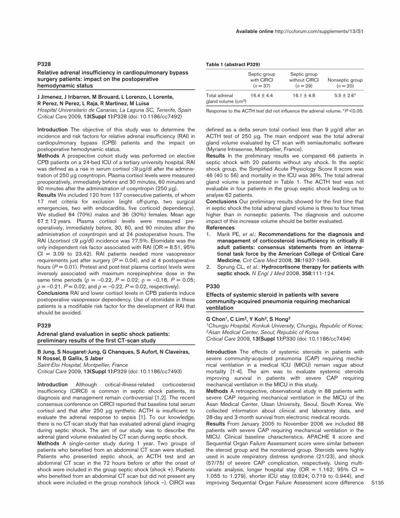
S135
P328Relative adrenal insufficiency in cardiopulmonary bypasssurgery patients: impact on the postoperativehemodynamic status
J Jimenez, J Iribarren, M Brouard, L Lorenzo, L Lorente, R Perez, N Perez, L Raja, R Martinez, M LuisaHospital Universitario de Canarias, La Laguna SC, Tenerife, SpainCritical Care 2009, 13(Suppl 1):P328 (doi: 10.1186/cc7492)
Introduction The objective of this study was to determine theincidence and risk factors for relative adrenal insufficiency (RAI) incardiopulmonary bypass (CPB) patients and the impact onpostoperative hemodynamic status.Methods A prospective cohort study was performed on electiveCPB patients on a 24-bed ICU of a tertiary university hospital. RAIwas defined as a rise in serum cortisol ≤9 μg/dl after the adminis-tration of 250 μg cosyntropin. Plasma cortisol levels were measuredpreoperatively, immediately before and 30 minutes, 60 minutes and90 minutes after the administration of cosyntropin (250 μg).Results We included 120 from 137 consecutive patients, of whom17 met criteria for exclusion (eight off-pump, two surgicalemergencies, two with endocarditis, five corticoid dependency).We studied 84 (70%) males and 36 (30%) females. Mean age67 ± 12 years. Plasma cortisol levels were measured pre-operatively, immediately before, 30, 60, and 90 minutes after theadministration of cosyntropin and at 24 postoperative hours. TheRAI (Δcortisol ≤9 μg/dl) incidence was 77.5%. Etomidate was theonly independent risk factor associated with RAI (OR = 8.51, 95%CI = 3.09 to 23.42). RAI patients needed more vasopressorrequirements just after surgery (P = 0.04), and at 4 postoperativehours (P = 0.01). Pretest and post-test plasma cortisol levels wereinversely associated with maximum norepinephrine dose in thesame time periods (ρ = –0.22, P = 0.02; ρ = –0.18, P = 0.05;ρ = –0.21, P = 0.02; and ρ = –0.22, P = 0.02, respectively).Conclusions RAI and lower cortisol levels in CPB patients inducepostoperative vasopressor dependency. Use of etomidate in thesepatients is a modifiable risk factor for the development of RAI thatshould be avoided.
P329Adrenal gland evaluation in septic shock patients:preliminary results of the first CT-scan study
B Jung, S Nougaret-Jung, G Chanques, S Aufort, N Claveiras, N Rossel, B Gallix, S JaberSaint-Eloi Hospital, Montpellier, FranceCritical Care 2009, 13(Suppl 1):P329 (doi: 10.1186/cc7493)
Introduction Although critical-illness-related corticosteroidinsufficiency (CIRCI) is common in septic shock patients, itsdiagnosis and management remain controversial [1,2]. The recentconsensus conference on CIRCI reported that baseline total serumcortisol and that after 250 μg synthetic ACTH is insufficient toevaluate the adrenal response to sepsis [1]. To our knowledge,there is no CT-scan study that has evaluated adrenal gland imagingduring septic shock. The aim of our study was to describe theadrenal gland volume evaluated by CT scan during septic shock.Methods A single-center study during 1 year. Two groups ofpatients who benefited from an abdominal CT scan were studied.Patients who presented septic shock, an ACTH test and anabdominal CT scan in the 72 hours before or after the onset ofshock were included in the group septic shock (shock +). Patientswho benefited from an abdominal CT scan but did not present anyshock were included in the group nonshock (shock –). CIRCI was
defined as a delta serum total cortisol less than 9 μg/dl after anACTH test of 250 μg. The main endpoint was the total adrenalgland volume evaluated by CT scan with semiautomatic software(Myriane Intrasense, Montpellier, France).Results In the preliminary results we compared 66 patients inseptic shock with 20 patients without any shock. In the septicshock group, the Simplified Acute Physiology Score II score was46 (40 to 56) and mortality in the ICU was 36%. The total adrenalgland volume is presented in Table 1. The ACTH test was notevaluable in four patients in the group septic shock leading us toanalyse 62 patients.Conclusions Our preliminary results showed for the first time thatin septic shock the total adrenal gland volume is three to four timeshigher than in nonseptic patients. The diagnosis and outcomeimpact of this increase volume should be better evaluated.References1. Marik PE, et al.: Recommendations for the diagnosis and
management of corticosteroid insufficiency in critically illadult patients: consensus statements from an interna-tional task force by the American College of Critical CareMedicine. Crit Care Med 2008, 36:1937-1949.
2. Sprung CL, et al.: Hydrocortisone therapy for patients withseptic shock. N Engl J Med 2008, 358:111-124.
P330Effects of systemic steroid in patients with severecommunity-acquired pneumonia requiring mechanicalventilation
G Chon1, C Lim2, Y Koh2, S Hong2
1Chungju Hospital, Konkuk University, Chungju, Republic of Korea;2Asan Medical Center, Seoul, Republic of KoreaCritical Care 2009, 13(Suppl 1):P330 (doi: 10.1186/cc7494)
Introduction The effects of systemic steroids in patients withsevere community-acquired pneumonia (CAP) requiring mecha-nical ventilation in a medical ICU (MICU) remain vague aboutmortality [1-4]. The aim was to evaluate systemic steroidsimproving survival in patients with severe CAP requiringmechanical ventilation in the MICU in this study.Methods A retrospective, observational study in 88 patients withsevere CAP requiring mechanical ventilation in the MICU of theAsan Medical Center, Ulsan University, Seoul, South Korea. Wecollected information about clinical and laboratory data, and28-day and 3-month survival from electronic medical records.Results From January 2005 to November 2006 we included 88patients with severe CAP requiring mechanical ventilation in theMICU. Clinical baseline characteristics, APACHE II score andSequential Organ Failure Assessment score were similar betweenthe steroid group and the nonsteroid group. Steroids were highlyused in acute respiratory distress syndrome (21/23), and shock(57/75) of severe CAP complication, respectively. Using multi-variate analysis, longer hospital stay (OR = 1.162; 95% CI =1.055 to 1.279), shorter ICU stay (0.824; 0.719 to 0.944), andimproving Sequential Organ Failure Assessment score difference
Available online http://ccforum.com/supplements/13/S1
Table 1 (abstract P329)
Septic group Septic group with CIRCI without CIRCI Nonseptic group
(n = 37) (n = 29) (n = 20)
Total adrenal 16.4 ± 4.4 16.1 ± 4.8 5.5 ± 2.6*gland volume (cm3)
Response to the ACTH test did not influence the adrenal volume. *P <0.05.

S136
from day 1 to day 7 (1.447; 1.103 to 1.898) were associated withan increased 3-month survival. But systemic steroids did notimprove 28-day and 3-month survival.Conclusions The effects of systemic steroids in patients withsevere CAP requiring mechanical ventilation in the MICU did notimprove survival.References1. Garcia-Vidal C, et al.: Effects of systemic steroids in
patients with severe community-acquired pneumonia. EurRespir J 2007, 30:951-956.
2. Confalonieri M, et al.: Hydrocortisone infusion for severecommunity-acquired pneumonia. Am J Respir Crit CareMed 2005, 171:242-248.
3. Christ-Crain M, et al.: Free and total cortisol levels as pre-dictors of severity and outcome in community-acquiredpneumonia. Am J Respir Crit Care Med 2007, 176:913-920.
4. Franchimont D, et al.: Glucocorticoids and inflammationrevisited. Neuroimmunomodulation 2003, 10:247-260.
P331Statin therapy in patients admitted to hospital withpresumed infection
P Kruger1, M Harward1, J Helyar1, B Venkatesh2, M Jones2
1Princess Alexandra Hospital, Brisbane, Australia; 2University ofQueensland, Brisbane, AustraliaCritical Care 2009, 13(Suppl 1):P331 (doi: 10.1186/cc7495)
Introduction Several studies, largely retrospective, have suggestedimproved outcomes and a reduction in inflammatory response inpatients who develop infection whilst on statin therapy. This studyreports outcomes for patients on established statin therapyadmitted to hospital with presumed infection compared with thosepatients not on a statin.Methods The study was approved by the Princess AlexandraHospital Research Ethics Committee and conducted from May2006 to October 2008 as part of a randomised controlled studyinvestigating continuing established statin therapy. A dailycomputer-generated report identified all patients admitted tohospital with a potential diagnosis of infection.Results From a total of 2,291 patients screened, 2,239 wereconsidered to have presumed infection. Of these, 2,161 wereaged >30 years and were included in the final analysis (statin users =633, no statin use = 1,528). The statin group was significantlyolder with a mean age (± SD) of 69 ± 12 versus 61 ± 16 years(P = 0.0001) and included more men (62.1% vs. 55.2%,P = 0.004). The source of presumed infection was not statisticallydifferent between the groups (overall chi-square P = 0.16). Apresumed respiratory source was the most common in bothgroups (statin 35.5% vs. no statin 37.1%, respectively) followed byskin (19.0 vs. 20.3%), gastrointestinal (17.8 vs. 14.1%), urosepsis(13.0 vs. 11.7%) and other sources (14.7 vs. 16.8%). Two or moreSIRS criteria was seen in 65.4% of the statin group patients and67.8% of the no statin group (P = 0.30). Hospital mortality wassimilar in both groups (7.4% statin group and 6.5% no statingroup, P = 0.64), as was the rate of ICU admission (17% statingroup and 16% no statin group; P = 0.63). Hospital length of staywas not significantly different between the groups(11.4 ± 15.4 days in the statin group and 12.5 ± 26.2 days in theno statin group, P = 0.10).Conclusions In a heterogeneous cohort of patients admitted tohospital with presumed infection, prior statin therapy was notassociated with an improved outcome or reduced systemic inflam-matory response. The age and gender differences observed are inkeeping with previous literature and support the assertion that
statin users represent a different patient group to those not usingstatin therapy.
P332Evolution of inflammation in non-ICU patients withinfections: pilot prospective cohort study
A Donnelly1, NK Adhikari2, R Pinto2, Z Salih1, C McKenzie1, M Terblanche1
1St Thomas’s Hospital, London, UK; 2Sunnybrook Health SciencesCentre, Toronto, ON, CanadaCritical Care 2009, 13(Suppl 1):P332 (doi: 10.1186/cc7496)
Introduction Statins may prevent organ dysfunction in patientswith infections, but the optimal time for this therapy is unknown.Our objective was to determine the evolution of inflammation inpatients treated for infection on general wards and the impact ofprevious statin use.Methods We performed a single-centre prospective cohort study(April to September 2008) in unselected patients admitted tomedical wards with infection, collecting data on demographics,comorbidities, and statin use before admission; and 10-day follow-up data on intensive care or high-dependency unit (ICU/HDU)admission and death, systemic inflammatory response syndrome(SIRS) criteria and organ dysfunction (using a modified SequentialOrgan Failure Assessment (SOFA) score), and infection markers(C-reactive protein (CRP), white blood cells (WBC)). Evolution oforgan dysfunction, SIRS, WBC and CRP were analysed descrip-tively; continuous data are expressed as the mean (SD) or median(Q1 to Q3). The effect of statins was explored in regressionmodels accounting for within-patient correlation, with P <0.05taken as statistically significant.Results Two hundred and nine patients were admitted withinfections (lung 51.0%, urinary 34.2%, skin/soft tissue 18.5%,other 5.2%; ≥1 infection/patient possible): age 63.8 years (20.7),49.8% male, Charlson score 2 (1 to 3), previous statin users27.8%, WBC 15.6 (12.0) x 109/l, CRP 105 (113) mg/l. Onadmission, 88.9% had ≥1 SIRS criterion (median 2 (1 to 3)) and72.3% had modified SOFA score ≥1 (median 1 (0 to 2)), with nostatin versus non-statin group differences. CRP, WBC, and theproportion of patients with ≥1 SIRS criterion and modified SOFAscore ≥1 decreased over time (P <0.0001), but generalized linearmixed models showed no effect of statins (P = 0.98, 0.51, 0.55,and 0.25) when adjusted for time, age, sex, and Charlson score.By day 10, seven patients were admitted to the ICU/HDU, fourpatients had died, and 64 patients had ≥1 day with a highermodified SOFA score versus admission. Overall, 35.9% of patientsdeveloped this combined outcome (statin (44.8%) vs. non-statin(32.5%); OR = 1.66, 95% CI = 2.85 to 3.25 after adjustment forage, sex, Charlson score).Conclusions Ward patients with infection often develop some organdysfunction, but the risk of death/higher care is low. Trials of statins toprevent such clinically important outcomes would need to be large.
P333Heart rate variability in Egyptian children with acuterheumatic fever
MM Farid, M Abd Elmonim, M Elganzoury, M Abou Elmaaty, O YoussefFaculty of Medicine – Ain Shams University, Cairo, EgyptCritical Care 2009, 13(Suppl 1):P333 (doi: 10.1186/cc7497)
Introduction Autonomic dysfunction in relation to cardiovascularsystem morbidity and mortality has been reported. The objective of
Critical Care March 2009 Vol 13 Suppl 1 29th International Symposium on Intensive Care and Emergency Medicine
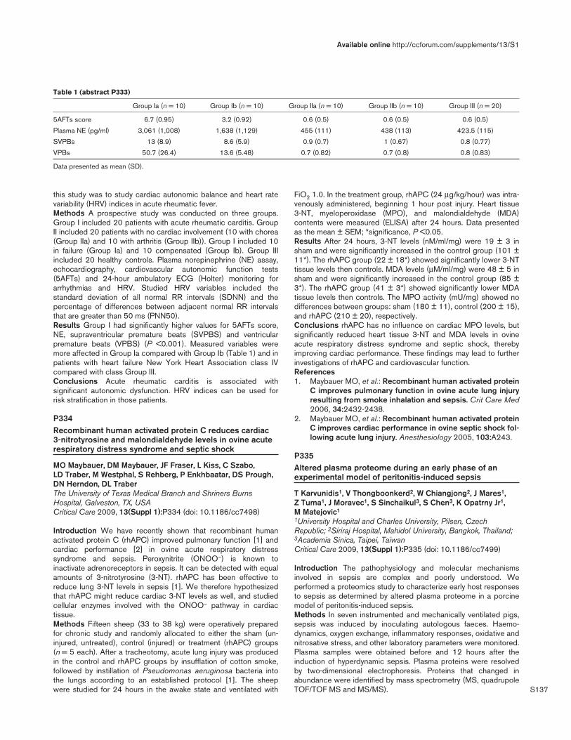
S137
this study was to study cardiac autonomic balance and heart ratevariability (HRV) indices in acute rheumatic fever.Methods A prospective study was conducted on three groups.Group I included 20 patients with acute rheumatic carditis. GroupII included 20 patients with no cardiac involvement (10 with chorea(Group IIa) and 10 with arthritis (Group IIb)). Group I included 10in failure (Group Ia) and 10 compensated (Group Ib). Group IIIincluded 20 healthy controls. Plasma norepinephrine (NE) assay,echocardiography, cardiovascular autonomic function tests(5AFTs) and 24-hour ambulatory ECG (Holter) monitoring forarrhythmias and HRV. Studied HRV variables included thestandard deviation of all normal RR intervals (SDNN) and thepercentage of differences between adjacent normal RR intervalsthat are greater than 50 ms (PNN50).Results Group I had significantly higher values for 5AFTs score,NE, supraventricular premature beats (SVPBS) and ventricularpremature beats (VPBS) (P <0.001). Measured variables weremore affected in Group Ia compared with Group Ib (Table 1) and inpatients with heart failure New York Heart Association class IVcompared with class Group III.Conclusions Acute rheumatic carditis is associated withsignificant autonomic dysfunction. HRV indices can be used forrisk stratification in those patients.
P334Recombinant human activated protein C reduces cardiac3-nitrotyrosine and malondialdehyde levels in ovine acuterespiratory distress syndrome and septic shock
MO Maybauer, DM Maybauer, JF Fraser, L Kiss, C Szabo, LD Traber, M Westphal, S Rehberg, P Enkhbaatar, DS Prough,DN Herndon, DL TraberThe University of Texas Medical Branch and Shriners BurnsHospital, Galveston, TX, USACritical Care 2009, 13(Suppl 1):P334 (doi: 10.1186/cc7498)
Introduction We have recently shown that recombinant humanactivated protein C (rhAPC) improved pulmonary function [1] andcardiac performance [2] in ovine acute respiratory distresssyndrome and sepsis. Peroxynitrite (ONOO–) is known toinactivate adrenoreceptors in sepsis. It can be detected with equalamounts of 3-nitrotyrosine (3-NT). rhAPC has been effective toreduce lung 3-NT levels in sepsis [1]. We therefore hypothesizedthat rhAPC might reduce cardiac 3-NT levels as well, and studiedcellular enzymes involved with the ONOO– pathway in cardiactissue.Methods Fifteen sheep (33 to 38 kg) were operatively preparedfor chronic study and randomly allocated to either the sham (un-injured, untreated), control (injured) or treatment (rhAPC) groups(n = 5 each). After a tracheotomy, acute lung injury was producedin the control and rhAPC groups by insufflation of cotton smoke,followed by instillation of Pseudomonas aeruginosa bacteria intothe lungs according to an established protocol [1]. The sheepwere studied for 24 hours in the awake state and ventilated with
FiO2 1.0. In the treatment group, rhAPC (24 μg/kg/hour) was intra-venously administered, beginning 1 hour post injury. Heart tissue3-NT, myeloperoxidase (MPO), and malondialdehyde (MDA)contents were measured (ELISA) after 24 hours. Data presentedas the mean ± SEM; *significance, P <0.05.Results After 24 hours, 3-NT levels (nM/ml/mg) were 19 ± 3 insham and were significantly increased in the control group (101 ±11*). The rhAPC group (22 ± 18*) showed significantly lower 3-NTtissue levels then controls. MDA levels (μM/ml/mg) were 48 ± 5 insham and were significantly increased in the control group (85 ±3*). The rhAPC group (41 ± 3*) showed significantly lower MDAtissue levels then controls. The MPO activity (mU/mg) showed nodifferences between groups: sham (180 ± 11), control (200 ± 15),and rhAPC (210 ± 20), respectively.Conclusions rhAPC has no influence on cardiac MPO levels, butsignificantly reduced heart tissue 3-NT and MDA levels in ovineacute respiratory distress syndrome and septic shock, therebyimproving cardiac performance. These findings may lead to furtherinvestigations of rhAPC and cardiovascular function.References1. Maybauer MO, et al.: Recombinant human activated protein
C improves pulmonary function in ovine acute lung injuryresulting from smoke inhalation and sepsis. Crit Care Med2006, 34:2432-2438.
2. Maybauer MO, et al.: Recombinant human activated proteinC improves cardiac performance in ovine septic shock fol-lowing acute lung injury. Anesthesiology 2005, 103:A243.
P335Altered plasma proteome during an early phase of anexperimental model of peritonitis-induced sepsis
T Karvunidis1, V Thongboonkerd2, W Chiangjong2, J Mares1, Z Tuma1, J Moravec1, S Sinchaikul3, S Chen3, K Opatrny Jr1, M Matejovic1
1University Hospital and Charles University, Pilsen, CzechRepublic; 2Siriraj Hospital, Mahidol University, Bangkok, Thailand;3Academia Sinica, Taipei, TaiwanCritical Care 2009, 13(Suppl 1):P335 (doi: 10.1186/cc7499)
Introduction The pathophysiology and molecular mechanismsinvolved in sepsis are complex and poorly understood. Weperformed a proteomics study to characterize early host responsesto sepsis as determined by altered plasma proteome in a porcinemodel of peritonitis-induced sepsis.Methods In seven instrumented and mechanically ventilated pigs,sepsis was induced by inoculating autologous faeces. Haemo-dynamics, oxygen exchange, inflammatory responses, oxidative andnitrosative stress, and other laboratory parameters were monitored.Plasma samples were obtained before and 12 hours after theinduction of hyperdynamic sepsis. Plasma proteins were resolvedby two-dimensional electrophoresis. Proteins that changed inabundance were identified by mass spectrometry (MS, quadrupoleTOF/TOF MS and MS/MS).
Available online http://ccforum.com/supplements/13/S1
Table 1 (abstract P333)
Group Ia (n = 10) Group Ib (n = 10) Group IIa (n = 10) Group IIb (n = 10) Group III (n = 20)
5AFTs score 6.7 (0.95) 3.2 (0.92) 0.6 (0.5) 0.6 (0.5) 0.6 (0.5)
Plasma NE (pg/ml) 3,061 (1,008) 1,638 (1,129) 455 (111) 438 (113) 423.5 (115)
SVPBs 13 (8.9) 8.6 (5.9) 0.9 (0.7) 1 (0.67) 0.8 (0.77)
VPBs 50.7 (26.4) 13.6 (5.48) 0.7 (0.82) 0.7 (0.8) 0.8 (0.83)
Data presented as mean (SD).
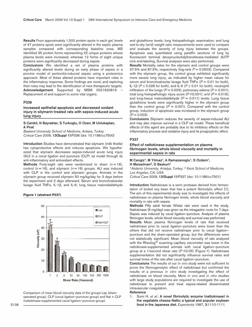
S138
Results From approximately 1,500 protein spots in each gel, levelsof 47 proteins spots were significantly altered in the septic plasmasamples compared with corresponding baseline ones. MSidentified 35 protein forms representing 22 unique proteins whoseplasma levels were increased, whereas 12 forms of eight uniqueproteins were significantly decreased during sepsis.Conclusions We identified a set of plasma proteins withsignificantly altered levels during an early phase of sepsis in aporcine model of peritonitis-induced sepsis using a proteomicsapproach. Most of these altered proteins have important roles inthe inflammatory response. Some findings are novel, and exploringtheir roles may lead to the identification of new therapeutic targets.Acknowledgement Supported by MSM 0021620819 –Replacement of and support to some vital organs.
P336Increased epithelial apoptosis and decreased oxidantinjury in silymarin-treated rats with sepsis-induced acutelung injury
S Canikli, N Bayraktar, S Turkoglu, O Ozen, M Unlukaplan, A PiratBaskent University School of Medicine, Ankara, TurkeyCritical Care 2009, 13(Suppl 1):P336 (doi: 10.1186/cc7500)
Introduction Studies have demonstrated that silymarin (milk thistle)has cytoprotective effects and induces apoptosis. We hypothe-sized that silymarin decreases sepsis-induced acute lung injury(ALI) in a cecal ligation and puncture (CLP) rat model through itsanti-inflammatory and antioxidant effects.Methods Forty-eight rats were randomized to sham (n = 16),control (n = 16), and silymarin (n = 16) groups. ALI was inducedwith CLP in the control and silymarin groups. Animals in thesilymarin group received silymarin 50 mg/kg/day for 3 days beforethe experiment and 2 days afterward. Serum and bronchoalveolarlavage fluid TNFα, IL-1β, and IL-6; lung tissue malondialdehyde
and glutathione levels; lung histopathologic examination; and lungwet-to-dry (w/d) weight ratio measurements were used to compareand evaluate the severity of lung injury between the groups.Apoptosis was quantitated using paraffin sections of lung byfluorescent terminal deoxynucleotidyltransferase-mediated dUTPnick end-labeling. Survival analyses were also performed.Results Mortality rates for the silymarin and control groups were37.5% and 87.5%, respectively (log-rank P = 0.0503). Comparedwith the silymarin group, the control group exhibited significantlymore severe lung injury, as indicated by higher mean values forserum and bronchoalveolar lavage fluid TNFα (P = 0.01 for both),IL-1β (P ≤ 0.028 for both), and IL-6 (P ≤ 0.01 for both); neutrophilinfiltration of the lungs (P = 0.003); pulmonary edema (P = 0.001);total lung histopathologic injury score (P <0.001); w/d (P = 0.019);and lung-tissue malondialdehyde (P = 0.011) levels. Lung tissueglutathione levels were significantly higher in the silymarin groupthan the control group (P = 0.001). Compared with the controlgroup, induction of apoptosis was increased in the silymarin group(P = 0.003).Conclusions Silymarin reduces the severity of sepsis-induced ALIand may also improve survival in a CLP rat model. These beneficialeffects of this agent are probably due to its inhibitory effects on theinflammatory process and oxidative injury and its proapoptotic effect.
P337Effect of nattokinase supplementation on plasmafibrinogen levels, whole blood viscosity and mortality inexperimental sepsis in rats
M Cengiz1, M Yilmaz1, A Ramazanoglu1, S Ozdem1, H Meiselman2, O Baskurt1
1Akdeniz University, Antalya, Turkey; 2 Keck School of Medicine,Los Angeles, CA, USACritical Care 2009, 13(Suppl 1):P337 (doi: 10.1186/cc7501)
Introduction Nattokinase is a serin protease derived from fermen-tation of boiled soy bean that has a potent fibrinolytic effect [1].The aim of this experimental study was to investigate the effects ofnattokinase on plasma fibrinogen levels, whole blood viscosity andmortality in rats with sepsis.Methods Fifty adult female Wistar rats were used in the study.Nattokinase (6 mg/day) was given via the intragastric route for 7 days.Sepsis was induced by cecal ligation–puncture. Analysis of plasmafibrinogen levels, whole blood viscosity and survival was performed.Results Mean plasma fibrinogen levels of rats that receivednattokinase prior to cecal ligation–puncture were lower than theothers that did not receive nattokinase prior to cecal ligation–puncture and the sham-operated group, but the differences werenot statistically significant. Mean blood viscosity of rats analyzedwith the Rheolog™ scanning capillary viscometer was lower in thenattokinase-supplemented animals with cecal ligation–puncturegroup at a 1/second shear rate (P <0.05) (Figure 1). Nattokinasesupplementation did not significantly influence survival rates andsurvival times of the rats after cecal ligation–puncture.Conclusions The results of our in vivo study were not sufficient toprove the fibrinogenolytic effect of nattokinase but confirmed theresults of a previous in vitro study investigating the effect ofnattokinase on blood viscosity. More in vivo and in vitro studieswith large study populations are required to investigate the use ofnattokinase to prevent and treat sepsis-related disseminatedintravascular coagulation.Reference1. Sumi H, et al.: A novel fibrinolytic enzyme (nattokinase) in
the vegetable cheese Natto; a typical and popular soybeanfood in the Japanese diet. Experientia 1987, 3:1110-1111.
Critical Care March 2009 Vol 13 Suppl 1 29th International Symposium on Intensive Care and Emergency Medicine
Figure 1 (abstract P337)
Comparison of mean blood viscosity data of the groups Lap (sham-operated group), CLP (cecal ligation–puncture group) and Nat + CLP(nattokinase-supplemented cecal ligation–puncture group).

S139
P338Physicians’ perceptions of current management strategiesfor Gram-negative pneumonia: a multinational study
A FrankBayer Schering Pharma AG, Berlin, GermanyCritical Care 2009, 13(Suppl 1):P338 (doi: 10.1186/cc7502)
Introduction Hospital-acquired pneumonia and its most seriousmanifestation, ventilator-associated pneumonia, are associatedwith high rates of mortality and account for 25% of all infections inthe ICU [1]. Pneumonia in mechanically ventilated (MV) patients isoften complicated by the involvement of multidrug-resistant (MDR)Gram-negative bacteria [2]. This multinational study exploredphysicians’ prescribing behaviour when treating Gram-negativepneumonia in MV patients.Methods Online interviews were conducted with 510 critical/intensive care, infectious disease and pulmonary/respiratory specia-lists in the USA (n = 130), Germany (n = 100), Mexico (n = 100),Spain (n = 80) and Japan (n = 100). Participants were practicingphysicians (3 to 31 years), were involved in the management of MVpatients and were familiar with treatment strategies for pneumonia.Results The involvement of Gram-negative (vs. Gram-positive)bacteria in pneumonia in MV patients was perceived to carry greatermortality risks. Most physicians (63 to 77% in all countries exceptJapan; 41% in Japan) were extremely concerned about the impacton treatment outcomes of increasing antibiotic resistance in Gram-negative species. Excessive antibiotic use and failure to effectivelyde-escalate antibiotic therapy were perceived as key contributors toresistance development. Achieving rapid cure (78%) and minimizingthe duration of MV (76%) were cited as the most important aims oftreatment. Key considerations for antibiotic selection were activityagainst Gram-negative bacteria, including MDR strains, and relativeextent of lung tissue penetration. Over 80% of respondents sawaerosolized antibiotics as a valuable potential addition to the currenttreatment armamentarium (no antibiotic aerosols are currentlylicensed for pneumonia), particularly for elderly patients and thosewith respiratory distress, who are at risk of the worst outcomes.Conclusions Gram-negative pneumonia in MV patients is a seriouscomplication in ICUs. Antibiotic prescribing for Gram-negativepneumonia among respondents is influenced by the risk of MDRbacterial involvement. Aerosolized antibiotics are seen as apotentially valuable adjunct to systemic therapies for treatingGram-negative pneumonia in MV patients.References1. American Thoracic Society/Infectious Diseases Society of
America: Guidelines for the management of adults withhospital-acquired, ventilator-associated, and healthcare-associated pneumonia. Am J Respir Crit Care Med 2005,171:388-416.
2. Parker CM, et al.: Ventilator-associated pneumonia causedby multidrug-resistant organisms or Pseudomonas aerug-inosa: prevalence, incidence, risk factors, and outcomes. JCrit Care 2008, 23:18-26.
P339Pilot proforma to aid the diagnosis of sepsis in burnspatients
T Evans, J McLennanMorriston Hospital, Swansea, UKCritical Care 2009, 13(Suppl 1):P339 (doi: 10.1186/cc7503)
Introduction The aim was to produce and pilot a data collectionproforma aiding the diagnosis of sepsis in the burns population. It
should be relevant, reliable and lead to reproducible results. Wealso looked at the data collected to assess the relevance of anyfindings. In June 2007, the American Burn Association ConsensusConference [1] produced standardized definitions in an attempt toaid clarification of sepsis in those already fulfilling the diagnosis ofa systemic inflammatory response syndrome (SIRS). This wouldallow more consistent diagnosis and accuracy to further trialsoccurring in this field. The intention is that the proforma will beused by the INTERBURNS research group for a large prospectivestudy.Methods This was a retrospective case series. Patients includedwere those admitted to a regional burns unit with more than 20%burns during December 2007, numbering four. These patientswere matched for age, sex, and mechanism of injury. The initialproforma was produced on the basis of the American BurnAssociation recommendations [1]. Data were collected andreviewed. This revealed potential confounding factors, so theproforma was modified and data recollected. A daily diagnosis ofSIRS and sepsis was made from the information collected.Results Data were collected at 12 p.m. daily initially. It was notedthat haemofiltration and noradrenaline were routinely used.Dressing changes occurred in the morning, leading to increasedanalgesia, sedation and the release of inflammatory mediatorsconfounding physiological parameters measured. Therefore theproforma was modified and the data recollected at 6 a.m. daily,thus achieving reliable and reproducible data. Despite the smallnumbers, the data indicated a potential association between anincrease in noradrenaline dose of 25% and either instigation of atreatment modality or a diagnosis of SIRS not previously identified,or both.Conclusions The proforma is reproducible and gives relevantreliable data for further analysis. The results have highlighted theneed for further research to clarify any association betweennoradrenaline and its place in the diagnosis of sepsis. Thisproforma and its subsequent utilisation by the INTERBURNSresearch group should improve our ability to diagnose sepsis andinstigate early treatment in this patient population.Reference1. Greenhalgh DG, et al.: American Burn Association consen-
sus conference to define sepsis and infection in burns. JBurn Care Res 2007, 28:776-790.
P340Surviving sepsis campaign guidelines for severe sepsisand septic shock: implementation and outcome of a 3-yearfollow up
A Castellanos Ortega1, B Suberviola1, LA Garcia Astudillo1, MS Holanda1, MA Hernandez1, F Ortiz Melon1, R Tejido1, FJ Llorca2, B Fernandez Miret1
1University Hospital Marques de Valdecilla, Santander, Spain;2University of Cantabria, Santander, SpainCritical Care 2009, 13(Suppl 1):P340 (doi: 10.1186/cc7504)
Introduction The purpose of the study was to describe the effective-ness of the Surviving Sepsis Campaign bundles with regard to bothimplementation and outcome in patients with septic shock.Methods A single-center prospective observational study ofpatients admitted to the medical–surgical ICU fulfilling criteria forthe international sepsis definitions. After a widespread 2-montheducational program, implementation of Surviving SepsisCampaign Resuscitation Bundles (RB) and Management Bundles(MB) were accomplished. A reinforcement educational programwas performed in October 2007. Patients were recruited fromSeptember 2005 to August 2008.
Available online http://ccforum.com/supplements/13/S1

S140
Results We analyzed 384 episodes of septic shock. The meanage was 64.5 ± 15 years, APACHE II score 23.2 ± 7.2, SequentialOrgan Failure Assessment score 9.5 ± 3, and global hospitalmortality 37.5%. The rate of compliance with the RB was 35.4%.There were significant differences in mortality between compliant(C) and noncompliant (NC) groups despite the similar charac-teristics and the severity of septic shock. The mortality rate was42.5% in the NC group and 23.6% in the C group. Thecompliance rate with MB was only 10%, there were no differencesin mortality between C and NC groups (41% vs. 37%). When theinfluence of age, severity, emergency department origin, and ICUadmission delay was controlled by multivariate analysis,compliance with RB was independently associated with survival(OR = 0.39, 95% CI = 0.22 to 0.70, P <0.01). Compliance rateswith RB during three consecutive 12-month time periods were34%, 23% and 45.4%, respectively (P <0.01); inhospital mortalityrates in those periods were 37%, 47% and 31%, respectively(P = 0.03). Compliance with MB decreased from 20% (firstperiod) to 3% (third period).Conclusions Implementation of RB was associated withdecreased mortality in patients with septic shock. The compliancerate with MB was poor and had no impact on survival.Acknowledgements Supported by IFIMAV Expte. PRF/07/04 andthe Instituto de Salud Carlos III. Expte. PI070723.
P341Sepsis bundles: just think about it?
A Cardinale, L Giunta, C Di Maria, C Pellegrini, P De Luca, G Di Salvio, T Russo, P Masturzo, E De BlasioHospital ‘G. Rummo’, Benevento, ItalyCritical Care 2009, 13(Suppl 1):P341 (doi: 10.1186/cc7505)
Introduction The aim of the study is to verify the impact of theimplementation of the diagnostic and therapeutic bundlessuggested by Sepsis Surviving Campaign (SSC) [1] on the out-come of patients with severe sepsis and septic shock.Methods A retrospective analysis of the outcome of severe sepsisand septic shock patients before, during and after theimplementation of the bundles according to the SSC in an eight-bed polyvalent ICU. We evaluated ICU and hospital mortality, thelength of stay, the level of compliance to the bundles and its impacton the mortality. Statistical analysis was performed using the chi-square test.
Results A total of 127 patients were enrolled from 2005 onwards(38 patients in 2005, 43 patients in 2006 and 46 patients in2007). We observed a reduction of the overall ICU and hospitalmortality from 73% to 43%, not statistically significant, even if thedifference between the observed and predicted mortality showed astatistically significant improvement (P = 0.027) (Figure 1). On thecontrary, the length of stay [2] was 18 days in 2005, 23 days in2006, 25 days in 2007. Our compliance was respectively 59% in2006 and 55% in 2007 for resuscitation bundles and 58% in2006 and 78% in 2007 for management bundles. The mortalitywas higher (70%) in the group with an overall compliance score ≤6compared with 40% for those with a score ≥7.Conclusions Even though the main difficulty was to timely reachthe targets of the bundles, the existence of a protocolizedapproach to severe sepsis and septic shock seems to contributeto the reduction of mortality in our population of patients.References1. Dellinger RP: Surviving Sepsis Campaign: international
guidelines for management for severe sepsss and septicshock 2008. Crit Care Med 2008, 36:296-327.
2. Ferrer R: Improvement in process of care and outcomeafter a multicenter severe sepsis educational program inSpain. JAMA 2008, 299:2294-2303.
P342Compliance with sepsis resuscitation but not managementbundles improves the survival of surgical patients withseptic shock
B Suberviola1, A Castellanos-Ortega1, LA Garcia Astudillo1, MS Holanda1, C Gonzalez Mansilla1, FJ Llorca2, B Fernandez Miret1, F Ortiz Melon1, M Hernandez1
1University Hospital Marques de Valdecilla, Santander, Spain;2University of Cantabria, Santander, SpainCritical Care 2009, 13(Suppl 1):P342 (doi: 10.1186/cc7506)
Introduction The purpose of this study was to describe theeffectiveness of the Surviving Sepsis Campaign bundles withregard to both implementation and outcome in surgical patientswith septic shock.Methods A single centre prospective observational study ofsurgical patients admitted to the ICU from September 2005 toAugust 2008 fulfilling criteria for the international sepsisdefinitions.Results We analyzed prospectively 149 surgical patients withseptic shock. The mean age was 69 ± 13 years, APACHE II score:23 ± 7, Sequential Organ Failure Assessment: 8 ± 2. The mortalityrate was 34.2% in the ICU and 42.3% in the hospital. There wereno significant differences in the characteristics and the severity ofseptic shock between the compliant (C) and noncompliant (NC)groups. We found differences in the ICU and hospital mortalitybetween the C and NC groups in two Resuscitation Bundles (RB):central venous oxygen saturation (ScvO2) ≥70% (25% vs. 43%,P = 0.01 and 32% vs. 52%, P = 0.02) and source control (14%vs. 56%, P <0.001 and 24% vs. 63%, P <0.001, respectively).The compliance rate of all RB was 27% and there were significantdifferences in mortality between the C and NC groups (17% vs.40%, P = 0.01 and 35% vs. 49%, P = 0.009 in the ICU andhospital, respectively). There were no significant differences in theICU and hospital mortality with the Management Bundlescompliance. In the multivariate analysis, source control, ScvO2≥70%, compliance of all RB, mechanical ventilation, APACHE IIand Sequential Organ Failure Assessment were independentlyassociated with mortality. When the influence of age and severitywas controlled by logistic regression, source control was
Critical Care March 2009 Vol 13 Suppl 1 29th International Symposium on Intensive Care and Emergency Medicine
Figure 1 (abstract P341)
Severe sepsis and septic shock hospital mortality.
S141
independently associated with survival (OR = 0.17, 95% CI = 0.05to 0.55, P = 0.003).Conclusions Implementation of the RB was associated withdecrease mortality of surgical patients with septic shock. Amongall sepsis bundle elements, the source control was the onlyindependent predictor of survival. The compliance of ManagementBundles had no impact on survival.
P343Standard operating procedure in patients with severesepsis and septic shock
T Schwab, A Schmitz, S Richter, C Bode, H BuschUniversity Hospital, Freiburg, GermanyCritical Care 2009, 13(Suppl 1):P343 (doi: 10.1186/cc7507)
Introduction Patients with severe sepsis and septic shock still havea high mortality rate, despite improvements in intensive care therapy.In the present study we assessed the impact of a standard operatingalgorithm adjusted on international treatment recommendations inpatients with severe sepsis and/or septic shock (for example, volumeresuscitation, hemodynamic control, glycemic control, substitution ofselenium and/or hydrocortisone and the use of recombinant humanactivated protein C (rhAPC)) on the outcome.Methods A retrospective analysis of 144 patients admitted to ourmedical ICU in 2004 and 2006. In 2004, before implementation ofthe standard operating procedure (SOP), 74 patients fulfillingcriteria for diagnosis of severe sepsis and/or septic shock wereanalysed and compared with 70 patients after implementation ofevidence-based SOP in 2006.Results Both groups did not show any difference in initialAPACHE II score and clinical baseline characteristics. With imple-mentation of the SOP, use of volume in the first 6 hours (1,506 vs.2,154 ml, P <0.05) and in the first day (4,005 vs. 6,122 ml,P <0.001) significantly increased. Furthermore, treatment with hydro-cortisone, selenium and insulin increased after implementation of theSOP significantly. Catecholamines and rhAPC were unaffected.Mortality in the patient group without SOP was 57%, and in theintervention group with SOP the mortality was 38.5% (P <0.05).Conclusions Owing to the implementation of a standard algorithm,a significant increase in volume therapy and adjunctive sepsistherapy were realised more frequently compared with patientstreated without the SOP and could be associated with a lowermortality rate in patients. The realisation of an evidence-basedSOP in daily practice in patients with severe sepsis and/or septicshock might be effective, shown in the alteration of treatmentpractice and the reduction of mortality.
P344Epidemiology and outcome of patients with severe sepsisin six Spanish ICUs
L Lorente1, M Martín2, C Díaz3, L Labarta4, J Ferreres5, J Solé-Violán6, J Borreguero1, Y Barrios1
1Hospital Universitario de Canarias, La Laguna SC, Tenerife, Spain;2Hospital Universitario Nuestra Señora de Candelaria, S/C deTenerife, Spain; 3Hospital Insular, Las Palmas de Gran Canaria,Spain; 4Hospital San Jorge, Huesca, Spain; 5Hospital ClínicoUniversitario de Valencia, Spain; 6Hospital Universitario de GranCanaria Dr. Negrín, Las Palmas de Gran Canaria, SpainCritical Care 2009, 13(Suppl 1):P344 (doi: 10.1186/cc7508)
Introduction The objective of this study was to describe theepidemiology, consumption of resources and outcome of patientswith severe sepsis.
Methods A prospective, observational and multicenter studyperformed in six Spanish ICUs. Only patients with severe sepsiswere included.Results A total of 122 patients with severe sepsis were included(female 33.1%; mean age 58.01 ± 15.40 years; mean APACHE IIscore at admission to the ICU 21.10 ± 8.89). The source of theinfection was respiratory in 56.2%, abdominal in 19.2% and otherfoci in 24.6%. Sepsis-related Organ Failure Assessment (SOFA)scores at the time of diagnosis of severe sepsis were the following:global 10.21 ± 3.75, respiratory 2.66 ± 1.08, haematological0.75 ± 1.10, hepatic 0.70 ± 1.01, cardiovascular 3.54 ± 1.22,neurological 0.96 ± 1.50, and renal 1.46 ± 1.61. Rates of organfailure were the following: respiratory 93.8%, haematological38.5%, hepatic 37.7%, cardiovascular 90.8%, neurological 35.4%and renal 55.4%. The rate of use of medical resources was asfollows: adrenergic agents 90.8%, mechanical ventilation 86.2%,extrarenal depuration 20% and recombinant activated protein C15.4%. Mean length of stay in the ICU was 16.88 ± 18.98 days.The mortality rate in the ICU was 43.44%. We found that lacticacid serum levels (OR = 1.26, 95% CI = 1.11 to 1.43, P <0.001),SOFA score (OR = 1.24, 95% CI = 1.12 to 1.37, P <0.001) andplasminogen activator inhibitor-1 (PAI-1) plasma levels (OR = 1.02,95% CI = 1.01 to1.03, P = 0.001) at the time of diagnosis werepredictors of mortality.Conclusions Severe sepsis is an important cause of mortality andit incurs a considerable use of resources. Lactic acid serum levels,SOFA score and PAI-1 plasma levels at diagnosis were found tobe predictors of mortality.
P345Epidemiology of severe sepsis in India
S Chatterjee1, S Todi1, S Sahu2, M Bhattacharyya1
1AMRI Hospitals, Kolkata, India; 2Kalinga Hospital, Cuttack, IndiaCritical Care 2009, 13(Suppl 1):P345 (doi: 10.1186/cc7509)
Introduction A multicentre, prospective, observational studyconducted in four intensive therapy units (ITUs) in India from June2006 to September 2008 to determine the incidence and outcomeof severe sepsis among adult patients.Methods All patients admitted to the ITU were screened daily forSIRS, organ dysfunction and severe sepsis as defined by the ACCPand SCCM. Patients with severe sepsis were further studied.Results A total of 4,183 ITU admissions were studied. SIRS withorgan dysfunction was found in 1,286 (30.74%) patients, of which688 (53.50%) were due to severe sepsis. The incidence of severesepsis was 16.45% of all admissions. The mean age of the studypopulation was 56.72 years (SD = 18.20), of which 62.63% weremale. The median APACHE II score was 19 (IQR 18 to 20) withpredominant (90.93%) medical admission. The ITU mortality of alladmissions was 17.70% and that of severe sepsis was 46.30%.The hospital mortality and 28-day mortality of severe sepsis were53.39% and 55.05%, respectively. The standardized mortality ratioof severe sepsis patients was 2.20. The median duration of stay inthe ITU for the severe sepsis cohort who survived was 4 days (IQR4 to 5). The number of episodes where infection was the primaryreason for admission to the ITU was 98.11%. Culture positivitywas found in 44.48%. Lung was the predominant source of sepsis(35.90%). Gram-negative organisms were responsible for 57.86%of cases and Gram-positive for 16.63%. The rest were parasitic,viral and fungal infections.Conclusions Severe sepsis was common in Indian ITUs. The ITUmortality was higher compared with the western literature. Gram-positive infections were less common although the incidence ofparasitic and viral infection was higher than in the West.
Available online http://ccforum.com/supplements/13/S1

S142
P346Improving identification of severe sepsis by junior doctors:an observational study
M Slattery, D Hepburn, R Jagadeeswaran, P TemblettMorriston Hospital, Swansea, UKCritical Care 2009, 13(Suppl 1):P346 (doi: 10.1186/cc7510)
Introduction Sepsis is a significant cause of morbidity and mor-tality worldwide. Early recognition and treatment are of paramountimportance in reducing mortality; this has been highlighted by theSurviving Sepsis Campaign (SSC) [1]. A consensus of opinionexists as to Time Zero being the first instance where severe sepsisis present. We undertook a study of newly qualified doctors in auniversity teaching hospital, to evaluate how effective they were atrecognising severe sepsis; and whether a simple intervention couldimprove their ability to perform this task.Methods The case file of a patient with septic inflammatoryresponse syndrome leading to severe sepsis was identified andanonymised. Time Zero was identified by the investigators basedon the SSC criteria. The case file was distributed to a sample ofjunior doctors over a 2-month period. They were asked to study theretrospective case record and fill in a questionnaire identifyingwhen they felt Time Zero occurred. These data were collected andthen the doctors were given an educational tool about the SSCguidelines and the definition of Time Zero. They were then asked toreappraise the case and reassess the Time Zero point based ontheir new knowledge.Results Thirty junior doctors participated; all had less than4 months postgraduate experience. Time Zero was correctlyidentified on the first attempt by 17% (n = 5) of the juniors. TimeZero estimates ranged from –70 to +920 minutes of the actualvalue. After implementation of the teaching tool, 100% of thedoctors correctly identified Time Zero according to the SSC criteria.Conclusions These results show that there can be significantdiscrepancies in the accuracy of prediction of Time Zero, which inturn could have significant implications for the diagnosis and timelyinception of treatment in severe sepsis. A simple intervention suchas an educational tool based on current SSC guidelines canimprove the accuracy and speed of diagnosis, and this may havean effect on morbidity and mortality.Reference1. Dellinger RP, et al: Surviving Sepsis Campaign: interna-
tional guidelines for management of severe sepsis andseptic shock: 2008. Crit Care Med 2008, 36:296-232.
P347Prevalence of ICU infection in South Africa and accuracy oftreating physician diagnosis and treatment
S Bhagwanjee, J Scribante, F Paruk, G RichardsUniversity of the Witwatersrand, Johannesburg, South AfricaCritical Care 2009, 13(Suppl 1):P347 (doi: 10.1186/cc7511)
Introduction ICU infection is an important cause of morbidity andmortality. Equally, it is important to establish to what extentphysicians are capable of making effective diagnoses andimplement correct treatment for sepsis. A national prevalence ofICU infection study was conducted in South African ICUs toevaluate these two issues.Methods Approval to conduct the study was obtained from allappropriate authorities. A 1-day prevalence of sepsis study wasundertaken on 16 August 2005. A proportional probability samplewas determined from all ICUs in the country. Attending physicianswere asked to indicate their diagnosis and treatment on the day of
the study. Relevant clinical data were collected for each patientand reviewed by two independent intensivists.Results All units chosen agreed to participate. The 28-day hospitalmortality was 58 of 248 patients (23%). The prevalence of eachcategory of the SIRS [1] criteria and associated percentagemortality respectively was nil 59 (8%), SIRS 120 (9%), sepsis 40(28%), severe sepsis 13 (35%) and septic shock 16 (41%). Onehundred and ninety-six patients were deemed to have one of thesepsis diagnoses by the primary physician, an overdiagnosis of51%. Antibiotic prescription and associated mortality percentagerespectively were: appropriate 82 (12%) and inappropriate 100(28%). Antibiotic therapy was changed correctly after microbiologydata in 21 of 88 patients (24%). Duration of therapy was correct in51 of 183 patients (28%).Conclusions The prevalence of sepsis and the associatedmortality is similar to that described in other studies [1]. Physicianstend to overdiagnose sepsis. Antibiotic prescribing practices wereincorrect with respect to selection, modification after microbiologydata and duration of treatment. A major deficiency in diagnosticand therapeutic ability among physicians was identified in thisstudy. Appropriate steps must be taken to remedy this deficiency.Reference1. Dombrovskiy VY, et al.: Crit Care Med 2007, 35:1244-1250.
P348Defining severe community-acquired pneumonia: a significant barrier to improving patient outcomes
MM Balp, C NaujoksNovartis Pharma AG, Basel, SwitzerlandCritical Care 2009, 13(Suppl 1):P348 (doi: 10.1186/cc751)
Introduction Severe community-acquired pneumonia (sCAP) is anexaggerated inflammatory and coagulation response to infection.sCAP has high rates of mortality and necessitates modifiedtreatment to that for mild/moderate community-acquired pneu-monia (CAP). Despite the grave nature of this condition it is poorlycharacterised. A systematic review was performed to gaugecurrent data and identify unmet needs associated with sCAP.Methods MEDLINE was searched for English-language papersconcerning sCAP published after 1998.Results There are a number of published indices for diagnosingsCAP but these are difficult to use in the clinical setting. In theliterature, sCAP is defined as CAP requiring admittance to an ICUor as CAP that results in death. Current International StatisticalClassification of Disease and diagnosis-related group codes donot specify whether pneumonia is community acquired or hospitalacquired or indicate the severity of the infection. Estimates of theprevalence of sCAP in patients with CAP range from 6.6 to 16.0%[1,2]. Mortality rates for patients with sCAP range from 10 to 55%[3,4]. The discrepancy in these rates emphasises the variation inhospital practices due to the lack of an objective definition ofsCAP. The absence of a clear definition for sCAP could result ininappropriate treatment of this life-threatening condition, increasingmortality rates. There are scant data concerning the costs oftreating sCAP; increased expenditure for patients with sCAPversus those with CAP results from ICU treatment, increasedlength of hospital stay, mechanical ventilation, vasopressor use andrehabilitation costs. It is expected that the clinical and cost benefitsof new therapies will be more easily recognised if sCAP isconsistently defined. Defining sCAP appropriately requires afocused international initiative and collaboration between cliniciansand payers.Conclusions There is a major unmet need for a meaningfuldefinition of sCAP. Poor characterisation of sCAP has resulted in
Critical Care March 2009 Vol 13 Suppl 1 29th International Symposium on Intensive Care and Emergency Medicine

S143
variable reports of its prevalence and may result in inappropriatetreatment leading to increased mortality in this patient population.The results of this systematic review form the basis for developinga new treatment pathway to improve outcomes for patients withsCAP.References1. Buising KL, et al.: A prospective comparison of severity
scores for identifying patients with severe community-acquired pneumonia: reconsidering what is meant bysevere pneumonia. Thorax 2006, 61:419-424.
2. Ewig S, et al.: Severe community-acquired pneumonia:sssessment of severity criteria. Am J Respir Crit Care Med1998, 158:1102-1108.
3. El-Solh AA, et al.: Etiology of severe pneumonia in the veryelderly. Am J Respir Crit Care Med 2001, 163:645-651.
4. Roson B, et al.: Etiology, reasons for hospitalization, riskclasses, and outcomes of community-acquired pneumo-nia in patients hospitalized on the basis of conventionaladmission criteria. Clin Infect Dis 2001, 33:158-165.
P349Outcome of severe sepsis in the ICU is independent ofhaemoglobin levels
J Wood, D PanditWilliam Harvey Hospital, Ashford, UKCritical Care 2009, 13(Suppl 1):P349 (doi: 10.1186/cc7513)
Introduction In this observational cohort study we attempted toelucidate a haemoglobin (Hb) target that favours the mortalityoutcome in severe sepsis patients. The optimum level of Hb toinfluence outcome in severe sepsis is yet to be determined.Although the analysis of severe sepsis patients in the Canadiantransfusion requirement in critical care study [1] did not find anymortality benefit with a Hb level of 10 g/dl or above, the early goal-directed therapy trial [2] showed significant mortality benefit byachieving haematocrit ≥30%, equivalent to a Hb value of 10 g/dl,during the early treatment period. This pilot study was thereforeundertaken to look for any relationship between Hb levels andmortality in severe sepsis patients admitted to the ICU.Methods All patients 16 years or older with severe sepsis or septicshock who stayed longer than 24 hours between July 2006 andJune 2007 were retrospectively included in the study. Patientdemographics, Hb levels measured by the blood gas analyserduring their period of severe sepsis and outcome data werecollected. Binary logistic regression analysis was performed withICU survival and 28-day survival as the dependent variables.Results Of the 62 patients enrolled in the study, the average agewas 66.8 years (16 to 87 years), with mean admission APACHE IIscore and Simplified Acute Physiology Score II being 18 (8 to 30)and 58 (17 to 78), respectively. Their ICU and 28-day mortalityrates were 35.5% and 41.9%, respectively. On analysis nosignificant relationship was found between average Hb, Hbvariation, minimum Hb, maximum Hb, and number of unitstransfused with ICU or 28-day survival. In addition, no significantrelationship was found between Hb falling below thresholds of 7,8, 9, or 10 g/dl, or variation from an Hb of 7, 8, 9, 10 g/dl duringsevere sepsis and the ICU survival or 28-day survival.Conclusions While a haematocrit of 30% represents aphysiological optimum between viscosity and oxygen carriage, theequivalent Hb from this pilot study shows no advantage in theoutcome of severe sepsis. This study suggests that any effect Hblevels may have on the outcome from severe sepsis is likely to besmall.
References1. Hebert P, et al.: A multicentre, randomized, controlled clini-
cal trail of transfusion requirements in critical care. N EnglJ Med 1999, 340:409-417.
2. Rivers E, et al.: Early goal directed therapy in the treatmentof severe sepsis and septic shock. N Engl J Med 2001,345:1368-1377.
P350Abstract withdrawn
P351TNFββ+250 polymorphism and hyperdynamic state incardiac surgery with extracorporeal circulation
J Iribarren, JJ Jimenez, M Brouard, L Lorente, R Perez, L Lorenzo, L Raja, S Palmero, N Perez, R Martinez, M MoraHospital Universitario de Canarias, La Laguna SC, Tenerife, SpainCritical Care 2009, 13(Suppl 1):P351 (doi: 10.1186/cc7515)
Introduction We have investigated genetic and clinical factorsassociated with the hyperdynamic state (HS) after heart surgerywith extracorporeal circulation (ECC).Methods We performed a prospective cohort study of consecutivepatients who underwent elective heart surgery with ECC. The HSwas defined as hyperthermia (>38ºC), cardiac index (CI)>3.5 l/min/m2 and systemic vascular resistance index (SVRI)<1,600 dynes x s/cm5 x m2. The study included demographicvariables, gene polymorphisms (A/G) of TNFβ+250, G/A-1082 ofIL-10, polymorphism of IL-1 receptor antagonist, comorbidity, typeof surgery, serum levels of IL-6, and postoperative course. Weused the Pearson chi-square test or Fischer exact test, and theStudent t test for univariate analysis, with forward stepwise logisticregression for multivariate adjustment.Results Eighty patients were studied, of whom 22 (27.5%)developed HS. The presence of allele G of TNFβ+250 poly-morphism was associated with an increased incidence of HS(68% vs. 37%; P = 0.011). In the multivariate analysis, a longerduration of ECC, and the presence of the G allele, wereassociated with the development of HS.Conclusions The G allele of TNFβ+250 polymorphism, and pro-longed extracorporeal circuit times, may favour the development ofa hyperdynamic state after heart surgery with ECC.
P352Role of 4G/5G plasminogen activator inhibitor-1 (PAI-1)gene polymorphism and the PAI-1 plasma levels in theoutcome of patients with severe sepsis
L Lorente1, M Martín2, L Labarta3, C Díaz4, J Solé-Violán5, J Blanquer6, J Borreguero1, Y Barrios1
1Hospital Universitario de Canarias, La Laguna SC, Tenerife, Spain;2Hospital Universitario Nuestra Señora de Candelaria, S/C deTenerife, Spain; 3Hospital San Jorge, Huesca, Spain; 4HospitalInsular, Las Palmas de Gran Canaria, Spain; 5Hospital Universitariode Gran Canaria Dr. Negrín, Las Palmas de Gran Canaria, Spain;6Hospital Clínico Universitario de Valencia, SpainCritical Care 2009, 13(Suppl 1):P352 (doi: 10.1186/cc7516)
Introduction The objective of this study was to evaluate the effectof the 4G/5G plasminogen activator inhibitor-1 (PAI-1) genepolymorphism on the plasma PAI-1 levels and outcome in criticallyill patients with severe sepsis.
Available online http://ccforum.com/supplements/13/S1

S144
Methods A prospective, observational and multicenter study carriedout in six Spanish ICUs. Only patients with severe sepsis wereincluded. Epidemiological data, severity scores, site of infection andoutcome of sepsis were recorded. Measurement of PAI-1 plasmalevels (at diagnosis, 72 hours and 7 days of diagnosis of severesepsis) and the DNA genotyping of the PAI-1 were carried out.Results A total of 122 patients with severe sepsis were included.Nonsurvivor patients (n = 53) exhibited higher PAI-1 plasmaconcentrations than the survivor patients (n = 69) at the time ofdiagnosis of severe sepsis (60.17 ± 30.34 vs. 42.51 ± 28.85;P = 0.001), at 72 hours (34.74 ± 20.79 vs. 17.91 ± 12.85;P ≤0.001), and at 7 days (35.93 ± 21.16 vs. 24.77 ± 15.85;P = 0.07). PAI-1 plasma levels at diagnosis were found a predictorof mortality (OR = 1.02, 95% CI = 1.01 to 1.03, P = 0.001). Wedid not find significant differences between 4G/5G PAI-1 genepolymorphisms in the PAI-1 plasma levels (49.20 ± 26.15 with4G/4G genotype, 49.39 ± 30.63 with 4G/5G genotype, and48.06 ± 32.26 with 5G/5G genotype), and in the mortality (10/23(41%) with 4G/4G genotype, 25/60 (40%) with 4G/5G and18/39 (47%) with 5G/5G).Conclusions PAI-1 plasma levels at the time of diagnosis aremarkers of an unfavourable prognosis in patients with severesepsis. We have not, however, found any correlation between the4G/5G PAI-1 gene polymorphism, plasma levels of PAI-1 ormortality of patients with severe sepsis.
P353Role of monocyte apoptosis for the final outcome of theseptic host with peritonitis
A Kotsaki1, I Tzepi1, P Carrer1, K Louis1, G Zografos2, E Giamarellos-Bourboulis1
1Attikon University Hospital, Athens, Greece; 2HippokrateionGeneral Hospital, Athens, GreeceCritical Care 2009, 13(Suppl 1):P353 (doi: 10.1186/cc7517)
Introduction Former studies have shown that monocyte apoptosisis crucial for the final outcome of sepsis in the field of acutepyelonephritis [1]. The significance of apoptosis in acute intra-abdominal infection was studied.Methods Acute peritonitis was induced after cecal ligation andpuncture in 17 male New Zealand rabbits. Four sham-operatedrabbits were also studied. Peripheral blood mononuclear cellswere isolated after Ficol gradient centrifugation; monocytes werefurther isolated after plastic adherence. Apoptosis of monocyteswas estimated after staining positive for annexin V and negative forpropidium iodine by flow cytometric analysis.Results Monocyte apoptosis is presented in Table 1. A rate ofapoptosis greater than 40% was found in six animals after24 hours; it was lower than 40% for 11 animals. The mediansurvival of the former was 15.2 days and of the latter 4.2 days (log-rank: 5.49, P = 0.019).Conclusions Sepsis arising in the field of peritonitis isaccompanied by inhibition of apoptosis of monocytes. The rate ofapoptosis 24 hours after sepsis induction is detrimental for thesurvival of the septic host.
Reference1. Antonopoulou A, et al.: Clin Exp Immunol 2007, 149:103-
108.
P354Genomic variations within matrix metalloproteinase-9 andsevere sepsis
Q Chen, Y Jin, X FangFirst Affiliated Hospital, Zhejiang University, Hangzhou, ChinaCritical Care 2009, 13(Suppl 1):P354 (doi: 10.1186/cc7518)
Introduction Sepsis is a multiple-gene disease resulting from theinteraction of environmental and genetic components. Matrix metallo-proteinase-9 (MMP-9) has been demonstrated to play an importantrole in organ dysfunction and outcome of sepsis [1-3]. However,genetic predisposition of MMP-9 to sepsis remained unknown.Methods Seven common SNPs within the functional regions ofMMP-9 gene (rs17576, rs2274756, rs2250889, rs9509,rs3918240, rs3918241 and rs3918242) were investigated in 192patients with severe sepsis and 262 healthy controls. Meanwhile,the plasma levels of MMP-9 were measured via ELISA.Results The genotype distributions and allelic frequencies of theabove seven SNPs were not significantly different between patientswith severe sepsis and controls, as well as between surviving andnonsurviving patients with severe sepsis (all P >0.05). HaplotypeGGCTTTC, AGGTCTC, GGCCTTC, GACTTAT and AGCCCTCare the five most common haplotypes. The distribution of thehaplotypes was also comparable among the defined groups. Themedian plasma levels of MMP-9 was 37.66 ng/ml in 32 patientswithin the first 24 hours following the diagnosis of severe sepsis,and 30.15 ng/ml in 19 healthy controls. Compared with those insurviving patients with severe sepsis (median 37.66 ng/ml, n = 16)and healthy controls, the concentrations of MMP-9 appeared anincreasing trend in nonsurviving patients with severe sepsis (median36.06 ng/ml, n = 16).Conclusions The present findings suggest that common poly-morphisms within the function regions of MMP-9 gene may notplay a major role in the predisposition to severe sepsis in theChinese Han cohort. The plasma levels of MMP-9 may associatewith the outcome of severe sepsis. Further studies in largesamples need to be guaranteed.References1. Renckens R, et al.: Matrix metalloproteinase-9 deficiency
impairs host defense against abdominal sepsis. J Immunol2006, 176:3735-3741.
2. Steinberg J, et al.: Metalloproteinase inhibition reduceslung injury and improves survival after cecal ligation andpuncture in rats. J Surg Res 2003, 111:185-195.
3. Lalu MM, et al.: Matrix metalloproteinase inhibitors attenu-ate endotoxemia induced cardiac dysfunction: a potentialrole for MMP-9. Mol Cell Biochem 2003, 251:61-66.
P355Association of IL-10 promoter polymorphism –1082 G/Awith adverse outcome in severe sepsis and septic shock
O Sabelnikovs1, L Nikitina-Zake2, J Zhuravlova3, E Sama3, I Vanags1
1Riga Stradinš University, Riga, Latvia; 2Latvian BiomedicalResearch and Study Centre, Riga, Latvia; 3P. Stradins ClinicalUniversity Hospital, Riga, LatviaCritical Care 2009, 13(Suppl 1):P355 (doi: 10.1186/cc7519)
Introduction IL-10 is an anti-inflammatory cytokine with pleiotropiceffect in immunoregulation and inflammation. Several recent
Critical Care March 2009 Vol 13 Suppl 1 29th International Symposium on Intensive Care and Emergency Medicine
Table 1 (abstract P353)
Apoptosis of monocytes over follow-up
Median (%) 1.5 hours 4 hours 24 hours 48 hours
Sham 21.4 45.1 16.3 27.9
Peritonitis 18.5 10.9* 21.9* 13.3
*P < 0.05 between groups.

S145
papers reported association of the IL-10 –1082G allele withadverse outcome in sepsis [1,2]. The study objective was toinvestigate whether IL-10 promoter polymorphism –1082 G/A isassociated with adverse outcome in severe sepsis and septicshock patients.Methods The study was conducted in the mixed medical–surgicaladult ICU of P. Stradinš Clinical University Hospital in Riga in 2007and 2008. A total of 103 critically ill patients who met theproposed severe sepsis and septic shock criteria were included.The IL-10 –1082 polymorphism was genotyped by sequencing. Allpatients were followed up throughout their stay in the ICU toclinical outcome. The frequency distributions of the genotypes inthe subgroups were compared using the Pearson chi-square test.P <0.05 was considered to indicate statistical significance.Results Of the 103 patients observed, 44 (43%) had an adverseoutcome in the ICU. In the survival group 11 (19%) had G/G atposition –1082 of the IL-10 gene, 25 (42%) were heterozygousG/A, and 23 (39%) were homozygous A/A. In the nonsurvivalgroup five (11%) had G/G, 30 (68%) had A/G, and nine (21%)had the A/A genotype. The polymorphism frequencies betweensurvival and nonsurvival differed significantly (P = 0.034, χ2 = 6.8).The relative risk for adverse outcome in IL-10 –1082 alternative Gallele carriers (G/G and G/A genotypes) was 1.6 (95% CI = 0.91to 2.83).Conclusions We found association of the IL-10 promoterpolymorphism –1082 G/A with clinical outcome in severe sepsisand septic shock patients; alternative –1082 G allele carriageseems to be associated with greater risk for adverse outcome inthe studied patient population.References1. Gallagher PM, et al.: Association of IL-10 polymorphism
with severity of illness in community acquired pneumonia.Thorax 2003, 58:154-156.
2. Stanilov SA, et al.: Interleukin-10-1082 promoter polymor-phism in association with cytokine production and sepsissusceptibility. Intensive Care Med 2006, 32:260-266.
P356Activated protein C restores kidney function in endotoxin-induced acute renal failure in the rat
E Almac1, T Johannes2, E Mik3, M Legrand4, K Unertl2, C Ince1
1Academisch Medisch Centrum, Amsterdam, the Netherlands;2University Hospital Tuebingen, Germany; 3Erasmus MedicalCenter, Rotterdam, the Netherlands; 4Lariboisière Hospital,University of Paris, FranceCritical Care 2009, 13(Suppl 1):P356 (doi: 10.1186/cc7520)
Introduction Activated protein C (APC) has been shown to havebeneficial effects on the inflammatory process and coagulationduring sepsis. Inflammation and coagulopathy impair the micro-vasculature and therefore disturb oxygen transport to the tissue.The hypothesis of our study was that APC treatment improvesrenal microvascular oxygenation and kidney function in endotoxin-induced acute renal failure in the rat.Methods In 18 anesthetized and ventilated (FiO2 0.4) male Wistarrats, the arterial blood pressure and renal blood flow wererecorded. The renal microvascular PO2 was continuously measuredby phosphorescence lifetime technique. All animals received alipopolysaccharide (LPS) bolus (10 mg/kg) to induce endotoxemicshock. All rats received fluid resuscitation (hydroxyethyl starch130 kDa) 1 hour after LPS application. In one group of animals,APC (drotrecogin alpha, Xigris®; Lilly) was continuously infused ina concentration of 10 μg/kg/hour. Another group received acontinuous infusion of 100 μg/kg/hour APC.
Results Data are presented in Figure 1.Conclusions APC at a concentration of 100 μg/kg/hour signifi-cantly restored kidney function compared with standard fluidresuscitation during endotoxemia. This application best improvedthe mean arterial pressure. APC had no beneficial effects,however, on the average renal microvascular PO2.
P357Angiopoietin-2 is a mediator involved in the advent ofhypotension following endotoxin shock
PD Carrer1, V Grosomanidis2, B Fyntanidou2, S Panidis2, K Kotzampassi2, EJ Giamarellos-Bourboulis1
1University of Athens Medical School, Athens, Greece; 2Universityof Thessaloniki Medical School, Thessaloniki, GreeceCritical Care 2009, 13(Suppl 1):P357 (doi: 10.1186/cc7521)
Introduction Angiopoietin-2 (Ang-2) is a mediator produced byendothelial cells [1]. It is in doubt whether its production in theintubated septic host is due to an effect of anesthetic medicationor to the sepsis process. The present study attempted to providean answer.Methods In 10 mechanically ventilated pigs, anesthesia wasmaintained by sevoflurane inhalation. Then shock was induced bythe intravenous administration of 25 mg/kg lipopolysaccharide(LPS) of Escherichia coli 111: B4 in eight pigs; two were controls.Blood was collected by a Swan–Ganz catheter and by a peripheralvein. Ang-2 was estimated by an enzyme immunoassay.Results The median Ang-2 of the peripheral circulation amongcontrols and pigs at shock was at baseline 1,403.2 and2,390.6 pg/ml (P = NS), respectively; at 1 hour 914.2 and2,192.5 pg/ml (P = 0.048), respectively; and at 2 hours 2,013 and1,738.7 pg/ml (P = NS). Respective values of the lung circulationwere 995.8 and 1,738.7 pg/ml at baseline (P = NS); 831.3 and1,663.3 pg/ml at 1 hour (P = NS); and at 2 hours 943.8 and1,757.8 pg/ml (P = NS).Conclusions Ang-2 is a mediator involved in the advent of shocknot influenced by the administration of sevoflurane.Reference1. Giamarellos-Bourboulis EJ, et al.: Kinetics of angiopoietin-2
in serum of multi-trauma patients: correlation with patientseverity. Cytokine 2008, 44:310-313.
Available online http://ccforum.com/supplements/13/S1
Figure 1 (abstract P356)
FR = fluid resuscitation; T0 = baseline; t1 = 1 hour after LPS bolus; t2 = resuscitation (3 hours); MAP = mean arterial pressure; RBF =renal blood flow; RVR = renal vascular resistance; CLcrea = creatinineclearance; cμPO2 = cortical microvascular PO2; mμPO2 = medullarymicrovascular PO2.

S146
P358Inhibition of the lectin-like oxidized low-density lipoproteinreceptor-1 improves intestinal microcirculation inexperimental endotoxaemia
C Lehmann1, D Pavlovic2, S Wilk2, C Thaumüller2, M Otto2, M Wendt2, S Felix2, M Landsberger2
1Dalhousie University, Halifax, NS, Canada; 2Ernst Moritz ArndtUniversity, Greifswald, GermanyCritical Care 2009, 13(Suppl 1):P358 (doi: 10.1186/cc7522)
Introduction Lectin-like oxidized low-density lipoprotein receptor-1(LOX-1) is a major endothelial receptor for oxidized low-densitylipoprotein [1]. Its expression is induced by pro-atherogenic stimulias well as by inflammatory cytokines. LOX-1 acts also as anadhesion molecule involved in leukocyte recruitment. The systemicleukocyte activation in sepsis represents a crucial factor in theimpairment of the microcirculation of different tissues, causingmultiple organ failure and death. The aim of our experimental studywas therefore to evaluate the effects of LOX-1 inhibition on theendotoxin-induced leukocyte adherence within the intestinalmicrocirculation using intravital microscopy.Methods Group 1 (n = 10 Lewis rats) remained untreated andserved as the control group. In Group 2 (n = 10) endotoxemia wasinduced by intravenous administration of 2 mg/kg lipopolysaccha-ride (LPS). In Group 3 (n = 10) endotoxemic animals were treatedwith 10 mg/kg anti-Lox-1-IgG. Endotoxemic animals of Group 4(n = 10) were treated with an unspecific IgG. Following 2 hours ofendotoxin challenge or placebo administration, intestinal micro-circulation was evaluated using intravital microscopy. LOX-1expression was quantified by western blot and reverse-trans-cription PCR.Results LOX-1 inhibition reduced significantly the leukocyteadherence in the submucosal venules of the intestinal wall(P <0.05). Functional capillary density of the intestinal muscularlayers as well as in the mucosa increased following administrationof the LOX-1-antibody in comparison with untreated LPS animals(P <0.05). At the mRNA level, LOX-1 expression was significantlyincreased in the untreated LPS group (P <0.05) whereas animalswith LOX-1-antibody administration showed a significant reductionof the expression (P <0.05).Conclusions LOX-1-antibody administration in experimentalendotoxaemia significantly reduced leukocyte adherence andincreased microvascular perfusion within the intestinal micro-circulation. The inhibition of the lectin-like oxidized low-densitylipoprotein receptor-1 may represent an attractive target for themodulation of endotoxin-induced impairment of the micro-circulation in sepsis.Reference1. Honjo M, et al.: Lectin-like oxidized LDL receptor-1 is a cell-
adhesion molecule involved in endotoxin-induced inflam-mation. Proc Natl Acad Sci U S A 2003, 100:1274-1279.
P359Effect of the a7nAChR agonist GTS-21 on inflammationduring human endotoxemia
J Pompe, M Kox, C Hoedemaekers, P Pickkers, A Van Vugt, J Van der HoevenRadboud University Nijmegen Medical Centre, Nijmegen, theNetherlandsCritical Care 2009, 13(Suppl 1):P359 (doi: 10.1186/cc7523)
Introduction Activation of the cholinergic anti-inflammatory path-way via vagus nerve stimulation or a7nAChR agonists improves
outcome in animal models of endotoxemia, sepsis and experi-mental arthritis. This vagal anti-inflammatory pathway is mediatedby the nicotinergic a7nACh receptor that can be selectivelystimulated by GTS-21. Up to now, the anti-inflammatory effects oforal administration of GTS-21 in humans in vivo have not beeninvestigated. The aim of this study was to investigate the anti-inflammatory effects of oral administration of GTS-21 on theinflammatory response in the human endotoxemia model.Methods We performed a double-blind placebo-controlledrandomized study in 12 healthy, nonsmoking male volunteers (18to 28 years) during experimental endotoxemia. Subjects received150 mg GTS-21 or placebo orally three times daily 3 days beforelipopolysaccharide (LPS) injection and on the day of the experi-ment, the last dose 1 hour before LPS administration (t = –1). Onehour after the last dose of GTS-21 or placebo, LPS derived fromEscherichia coli O:113 was injected (2 ng/kg intravenously).Results The main study endpoint was the concentration ofcirculating cytokines after LPS in the absence and presence ofGTS-21. The effects of GTS-21 on TNFα and IL-10 release areshown in Figures 1 and 2. There was a trend towards a decreasein TNFα levels and an increase in IL-10 levels after GTS-21administration. A similar trend was observed in levels of otherproinflammatory and anti-inflammatory cytokines.
Critical Care March 2009 Vol 13 Suppl 1 29th International Symposium on Intensive Care and Emergency Medicine
Figure 1 (abstract P359)
Figure 2 (abstract P359)
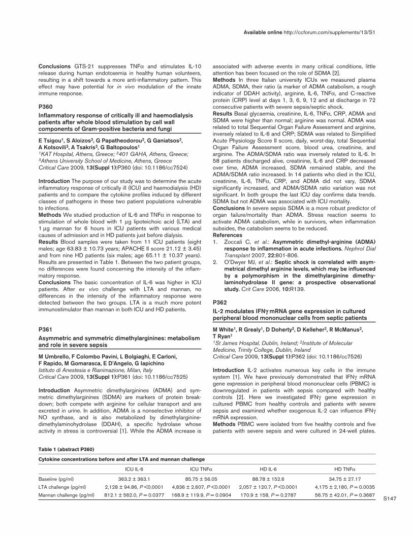
S147
Conclusions GTS-21 suppresses TNFα and stimulates IL-10release during human endotoxemia in healthy human volunteers,resulting in a shift towards a more anti-inflammatory pattern. Thiseffect may have potential for in vivo modulation of the innateimmune response.
P360Inflammatory response of critically ill and haemodialysispatients after whole blood stimulation by cell wallcomponents of Gram-positive bacteria and fungi
E Tsigou1, S Aloizos2, G Papatheodorou2, G Ganiatsos2, A Kotsovili3, A Tsakris3, G Baltopoulos1
1KAT Hospital, Athens, Greece; 2401 GAHA, Athens, Greece;3Athens University School of Medicine, Athens, GreeceCritical Care 2009, 13(Suppl 1):P360 (doi: 10.1186/cc7524)
Introduction The purpose of our study was to determine the acuteinflammatory response of critically ill (ICU) and haemodialysis (HD)patients and to compare the cytokine profiles induced by differentclasses of pathogens in these two patient populations vulnerableto infections.Methods We studied production of IL-6 and TNFα in response tostimulation of whole blood with 1 μg lipoteichoic acid (LTA) and1 μg mannan for 6 hours in ICU patients with various medicalcauses of admission and in HD patients just before dialysis.Results Blood samples were taken from 11 ICU patients (eightmales; age 63.83 ± 10.73 years; APACHE II score 21.12 ± 3.45)and from nine HD patients (six males; age 65.11 ± 10.37 years).Results are presented in Table 1. Between the two patient groups,no differences were found concerning the intensity of the inflam-matory response.Conclusions The basic concentration of IL-6 was higher in ICUpatients. After ex vivo challenge with LTA and mannan, nodifferences in the intensity of the inflammatory response weredetected between the two groups. LTA is a much more potentimmunostimulator than mannan in both ICU and HD patients.
P361Asymmetric and symmetric dimethylarginines: metabolismand role in severe sepsis
M Umbrello, F Colombo Pavini, L Bolgiaghi, E Carloni, F Rapido, M Gomarasca, E D’Angelo, G IapichinoIstituto di Anestesia e Rianimazione, Milan, ItalyCritical Care 2009, 13(Suppl 1):P361 (doi: 10.1186/cc7525)
Introduction Asymmetric dimethylarginines (ADMA) and sym-metric dimethylarginines (SDMA) are markers of protein break-down; both compete with arginine for cellular transport and areexcreted in urine. In addition, ADMA is a nonselective inhibitor ofNO synthase, and is also metabolized by dimethylarginine-dimethylaminohydrolase (DDAH), a specific hydrolase whoseactivity in stress is controversial [1]. While the ADMA increase is
associated with adverse events in many critical conditions, littleattention has been focused on the role of SDMA [2].Methods In three Italian university ICUs we measured plasmaADMA, SDMA, their ratio (a marker of ADMA catabolism, a roughindicator of DDAH activity), arginine, IL-6, TNFα, and C-reactiveprotein (CRP) level at days 1, 3, 6, 9, 12 and at discharge in 72consecutive patients with severe sepsis/septic shock.Results Basal glycaemia, creatinine, IL-6, TNFα, CRP, ADMA andSDMA were higher than normal; arginine was normal. ADMA wasrelated to total Sequential Organ Failure Assessment and arginine,inversely related to IL-6 and CRP; SDMA was related to SimplifiedAcute Physiology Score II score, daily, worst-day, total SequentialOrgan Failure Assessment score, blood urea, creatinine, andarginine. The ADMA/SDMA ratio was inversely related to IL-6. In58 patients discharged alive, creatinine, IL-6 and CRP decreasedover time, ADMA increased, SDMA remained stable, and theADMA/SDMA ratio increased. In 14 patients who died in the ICU,creatinine, IL-6, TNFα, CRP, and ADMA did not vary, SDMAsignificantly increased, and ADMA/SDMA ratio variation was notsignificant. In both groups the last ICU day confirms data trends.SDMA but not ADMA was associated with ICU mortality.Conclusions In severe sepsis SDMA is a more robust predictor oforgan failure/mortality than ADMA. Stress reaction seems toactivate ADMA catabolism, while in survivors, when inflammationsubsides, the catabolism seems to be reduced.References1. Zoccali C, et al.: Asymmetric dimethyl-arginine (ADMA)
response to inflammation in acute infections. Nephrol DialTransplant 2007, 22:801-806.
2. O’Dwyer MJ, et al.: Septic shock is correlated with asym-metrical dimethyl arginine levels, which may be influencedby a polymorphism in the dimethylarginine dimethy-laminohydrolase II gene: a prospective observationalstudy. Crit Care 2006, 10:R139.
P362IL-2 modulates IFNγγ mRNA gene expression in culturedperipheral blood mononuclear cells from septic patients
M White1, R Grealy1, D Doherty2, D Kelleher2, R McManus2, T Ryan1
1St James Hospital, Dublin, Ireland; 2Institute of MolecularMedicine, Trinity College, Dublin, IrelandCritical Care 2009, 13(Suppl 1):P362 (doi: 10.1186/cc7526)
Introduction IL-2 activates numerous key cells in the immunesystem [1]. We have previously demonstrated that IFNγ mRNAgene expression in peripheral blood mononuclear cells (PBMC) isdownregulated in patients with sepsis compared with healthycontrols [2]. Here we investigated IFNγ gene expression incultured PBMC from healthy controls and patients with severesepsis and examined whether exogenous IL-2 can influence IFNγmRNA expression.Methods PBMC were isolated from five healthy controls and fivepatients with severe sepsis and were cultured in 24-well plates.
Available online http://ccforum.com/supplements/13/S1
Table 1 (abstract P360)
Cytokine concentrations before and after LTA and mannan challenge
ICU IL-6 ICU TNFα HD IL-6 HD TNFα
Baseline (pg/ml) 363.2 ± 363.1 85.75 ± 56.05 88.78 ± 152.6 34.75 ± 27.17
LTA challenge (pg/ml) 2,128 ± 94.86, P <0.0001 4,836 ± 2,607, P <0.0001 2,057 ± 120.7, P <0.0001 4,175 ± 2,180, P = 0.0035
Mannan challenge (pg/ml) 812.1 ± 562.0, P = 0.0377 168.9 ± 119.9, P = 0.0904 170.9 ± 158, P = 0.2787 56.75 ± 42.01, P = 0.3687

S148
Cells were incubated in medium alone or stimulated with either1 μg/ml lipopolysaccharide (LPS) for 24 hours (activate monocytesand B cells), 3 μg/ml anti-CD3 (mAb) for 4 hours (activate T cells),or 10 ng/ml phorbol myristate acetate (PMA) + 1 μM ionomycin for4 hours (activate all cells). Each experiment was performed in theabsence and presence of 50 U recombinant IL-2 (rIL-2). Total RNAwas isolated and reverse transcribed. IFNγ mRNA gene expressionwas quantified with quantitative RT-PCR. Results were analysedwith ANOVA and the t test where appropriate.Results IFNγ mRNA production is lower in PBMC of patients withsepsis compared with healthy controls (P = 0.03) but similar whenmeasured in the presence of rIL-2. With T-cell stimulation, IFNγmRNA was greater in PBMC from septic patients compared withcontrols (P = 0.02) but similar in the presence of rIL-2. With LPSstimulation of monocytes and B cells, IFNγ mRNA was lower inPBMC from septic patients compared with controls (P = 0.02), butsimilar in the presence of rIL-2. With PMA stimulation, IFNγ mRNAwas similar in PBMC from septic patients and controls in thepresence and absence of rIL-2.Conclusions IFNγ mRNA production is reduced in PBMC insepsis but can be stimulated ex vivo to produce normal IFNγ levels.In the absence of rIL-2, IFNγ mRNA production is inducible inT cells but not in monocytes and B cells. IL-2 modulation of IFNγmRNA production appears to be stimulus dependent, and maycontribute to the host defence mechanism.References1. Burkett P, et al.: Diverse functions of IL-2, IL-15, and IL-7 in
lymphoid homeostasis. Annu Rev Immunol 2006, 24:657-679.2. O’Dwyer MJ, et al.: The occurrence of severe sepsis and
septic shock are related to distinct patterns of cytokinegene expression. Shock 2006, 26:544-550.
P363Compliment activity patterns in patients with sepsis afterhuman purified C1-esterase inhibitor infusion
A Igonin, N Lazareva, L DolzhenkovaI.M. Sechenov Medical Academy, Moscow, Russian FederationCritical Care 2009, 13(Suppl 1):P363 (doi: 10.1186/cc7527)
Introduction Our purpose was to assess the effects of super-physiologic dosages of human purified C1-esterase inhibitorinfusion (C1INH) on complement-dependent pathways of systemicinflammatory response in patients with sepsis.
Methods In the terms of an open-label prospective control study,human purified C1INH (Bicizar; BioGenius LLC, Russia) wasadministered at total dosage of 12,000 U to 20 sepsis patients.Patients (n = 22) who did not receive C1INH infusion wereenrolled as controls. C3 and C4 complement subunits, IL-6, C-reactive protein and procalcitonin results were analyzed in quartilesaccording to C1INH baseline activity.Results C1INH activity was different in quartiles at entry in bothgroups (P <0.03). C1INH infusion resulted in elevation of C1INHactivity in the treatment arm (Figure 1). The most significantdifference was observed between the upper quartiles of thegroups (P <0.01). The degree of C1INH activity shift had a nega-tive association with the baseline results (r = –0.635, P <0.01). C3(P <0.01) and C4 (P <0.05) levels displayed a more rapid andpronounced rise after C1INH infusion in patients with higherbaseline C1INH activity. The C-reactive protein concentrationdropped significantly in patients who received C1INH (130 mg/l(28 to 293)) already on the third day of the study in comparisonwith the control (185 mg/l (72 to 343); P = 0.022). The sametrend characterized the IL-6 level. Patients with elevatedprocalcitonin values at the onset of sepsis had a more significantC1INH increase (r = 0.635, P <0.05).Conclusions Downregulation of complement activity with C1INHmight help to contain severe systemic inflammation. Presumably,the response to C1INH infusion was related to its baseline activity.
P364Detrimental hemodynamic and inflammatory effects ofmicroparticles
S Mortaza, P AsfarCHU Angers, FranceCritical Care 2009, 13(Suppl 1):P364 (doi: 10.1186/cc7528)
Introduction Microparticles (MPs) are membrane vesicles withprocoagulant and proinflammatory properties released during cellactivation and might be potentially involved in the pathophysiologyof septic shock [1,2]. The present study was designed to assessthe effects of MPs from septic origin on systemic hemodynamicsas well as on the inflammatory, oxidative and nitrosative stresses.Methods We designed a prospective, randomized, controlledexperimental study with repeated measurements. Forty healthy ratswere randomly allocated to three groups: 10 animals inoculatedwith MPs isolated from control rats (cMPs), 15 animals inoculated
Critical Care March 2009 Vol 13 Suppl 1 29th International Symposium on Intensive Care and Emergency Medicine
Figure 1 (abstract P363)
C1INH activity in the Bicizar (q) and control groups (c): analysis in quartiles.

S149
with MPs isolated from sham rats (shMPs) and 15 animalsinoculated with MPs isolated from rats with peritonitis (sMPs). Ratswere anesthetized, mechanically ventilated and were infused withthe same amount of cMPs or shMPs or sMPs. We measured theheart rate, mean arterial pressure, carotid artery blood flow andportal vein blood flow. Hemodynamic parameters were recordedduring 7 hours, and then animals were sacrificed. The aorta andheart were harvested for further in vitro tissue analyses.Results (1) The cellular origin (phenotype) but not the circulatingconcentration of MPs was different in septic rats, characterized bya significant increase in leukocyte-derived MPs. (2) sMPs but notcMPs or shMPs decreased the mean arterial pressure without anyeffect on carotid artery and portal vein blood flows. (3) Ratsinoculated with sMPs exhibited an increase in superoxide ionproduction and NF-κB activity, overexpression of inducible NOsynthase with subsequent NO overproduction and decrease inendothelial NO synthase activation.Conclusions Rats with sepsis induced by peritonitis exhibited aspecific phenotype of MPs. Inoculation of sMPs in healthy ratsreproduced hemodynamic, septic inflammatory patterns, asso-ciated with oxidative and nitrosative stresses.References1. Morel O, et al.: Cellular microparticles, a disseminated
storage pool of bioactive vascular effectors. Curr OpinHematol 2004, 11:156-164.
2. Martinez MC, et al.: Shed membrane microparticles fromcirculating and vascular cells in regulating vascular func-tion. Am J Physiol Heart Circ Physiol 2005, 288:1004-1009.
P365Hydrogen sulfide: anti-inflammatory and cytoprotectiveeffects
C SzaboIkaria Inc., Seattle, WA, USACritical Care 2009, 13(Suppl 1):P365 (doi: 10.1186/cc7529)
Introduction Pharmacological actions of gaseous biologicalmediator hydrogen sulfide (H2S) include vasodilatation, inhibitionof mitochondrial respiration as well as induction of suspendedanimation-like state [1,2]. Its beneficial cellular actions includecytoprotection and anti-inflammatory effects [2].Methods Rodent models of ischemia and reperfusion of the heart,murine and ovine models of acute respiratory distress syndrome, acanine model of cardiopulmonary bypass, porcine models ofmyocardial infarction and thoracoabdominal aortic aneurysmsurgery were used. H2S was administered in an iso-osmolar, pH-neutral intravenous formulation (IK-1001).Results In a mouse model of myocardial infarction, a significantprotection by IK-1001 was seen in terms of reduction ofmyocardial infarct size. These effects were accompanied byreduction in myocardial IL-1 levels and reduction in neutrophilinfiltration [3]. The cardiac protection of H2S was confirmed in aporcine model of myocardial ischemia [4] and in a canine model ofcardiopulmonary bypass surgery. IK-1001 was also protective inmurine and ovine models of acute respiratory distress syndrome,where improvement in survival and pulmonary function wasaccompanied by a reduction in oxidant stress and suppression ofthe production of IL-6 and inhibition of the expression of induciblenitric oxide synthase [5]. In models of thoracoabdominal aneurysmsurgery, reperfusion was accompanied with significant increases inthe production of IL-1 and IL-6, which were reduced by IK-1001.IK-1001 also improved renal function, provided hemodynamicstabilization and attenuated oxidative DNA damage [6].
Conclusions H2S thus exerts organ-protective and anti-inflam-matory effects in various animal models of critical illness. Themechanisms may involve metabolic effects leading to the inductionof hypothermia, antioxidant mechanisms, modulation of geneexpression, activation of KATP channels, and inhibition of inflam-matory cell activation.References1. Blackstone E, et al.: H2S induces a suspended animation-
like state in mice. Science 2005, 308:518.2. Szabo C: Hydrogen sulphide and its therapeutic potential.
Nat Rev Drug Discov 2007, 6:917-935.3. Elrod JW, et al.: Hydrogen sulfide attenuates myocardial
ischemia–reperfusion injury by preservation of mitochon-drial function. Proc Natl Acad Sci U S A 2007, 104:15560-15565.
4. Sodha NR, et al.: The effects of therapeutic sulfide onmyocardial apoptosis in response to ischemia–reperfusioninjury. Eur J Cardiothorac Surg 2008, 33:906-913.
5. Esechie A, et al.: Protective effect of hydrogen sulfide in amurine model of acute lung injury induced by combinedburn and smoke inhalation. Clin Sci (Lond) 2008, 115:91-97.
6. Simon F, et al.: Hemodynamic and metabolic effects ofhydrogen sulfide during porcine ischemia/reperfusioninjury. Shock 2008, 30:359-364.
P366Dimethyl sulphoxide administration decreases renalischemic–reperfusion injury
W Hoyos, I Medina, R López, J Ramos, Z Garcia, S LópezUniversidad ‘Dr. José Matías Delgado’, Santa Tecla, La Libertad, ElSalvadorCritical Care 2009, 13(Suppl 1):P366 (doi: 10.1186/cc7530)
Introduction Kidney ischemia is one of the mechanisms of acuterenal failure and it is known that the free oxygen radicals play animportant role in this type of injury. We tested the hypothesis thatdimethyl sulphoxide (DMSO), a free radical scavenger, has aprotective role in a renal ischemia–reperfusion animal model [1].Methods Renal ischemia was induced to 45 male, New Zealandrabbits. The subjects were divided into three groups based onischemia duration: 30, 60 or 90 minutes. Each group was dividedinto three subgroups: (a) DMSO previous to ischemia, (b) DMSOafter ischemia, (c) control group. All subjects were given 6 hours ofreperfusion. Three blood samples were taken at baseline, ischemicand reperfusion phases. Each sample was tested for serumcreatinine, blood ureic nitrogen and urea. After reperfusion,bilateral nephrectomy was performed on each subject beforeeuthanasia. A pathological analysis evaluated tubular and base-ment membrane changes. The level of injury was scaled in threestages: mild, moderate and severe.Results The histological analysis showed severe damage in 33%of the control group, compared with 0% in both treatment groups(chi-square P = 0.00) (Table 1). Blood chemistry analysis in thecontrol group at 60 and 90 minutes of ischemia showed higher
Available online http://ccforum.com/supplements/13/S1
Table 1 (abstract P366)
Proportion estimate of histopathological findings
n = 45 Control (%) DMSO pre (%) DMSO post (%)
Mild 46 40 60
Moderate 20 60 40
Severe 33 0 0
S150
values than both treatment groups. Creatinine values wereanalyzed over a proportion estimate, showing that 26% of subjectsin the control group had an improvement comparing the ischemicphase versus the reperfusion phase. Treatment groups showedthat 46% and 60% of the subjects improved their creatinine valueswhen DMSO was administered pre ischemia and post ischemia,respectively.Conclusions The animal model showed an increasing trend of allblood chemistry parameters evaluated in the control group. DMSOapplied as prophylaxis or treatment post ischemia demonstratesdiminished renal function deterioration. Histological analysis revealedthe absence of severe lesions when DMSO is administered.Reference1. Kolb KH, et al.: Absorption, distribution and elimination of
labeled dimethyl sulfoxide in man and animals. Ann NYAcad Sci 1967, 141:85-95.
P367Beneficial effects of the heme oxygenase-1/carbonmonoxide system
N Takeyama, S Takaki, Y Kajita, T Yabuki, H Noguchi, Y Miki, Y Inoue, T Nakagawa, H NoguchiAichi Medical University, Aichi, JapanCritical Care 2009, 13(Suppl 1):P367 (doi: 10.1186/cc7531)
Introduction It has been reported that the blood level ofcarboxyhemoglobin (CO-Hb) is increased in critically ill patientssuch as those with sepsis or multiple trauma [1,2]. Becausecarbon monoxide (CO) is one of the metabolites of hemecatabolism, it has been suggested that there may be increasedbreakdown of heme in these patients. Heme oxygenase (HO) is theenzyme involved in the rate-limiting step for catabolism of heme-containing proteins. It was recently reported that HO-1 acts as apotent anti-inflammatory agent and antioxidant through its products[3]. HO-1 may therefore be an inducible defense against cellularstress that occurs during the inflammatory process [1,4]. Againstthis background, we decided to evaluate the relation among theblood level of CO, HO-1 expression by monocytes, oxidativestress, and the outcome of sepsis.Methods Thirty patients who fulfilled the criteria for severe sepsisor septic shock and 17 other patients without sepsis during theirstay in the ICU were studied. HO-1 expression by monocytes,arterial CO, oxidative stress, and cytokines were measured.Results Arterial blood levels of CO, cytokines, as well as monocyteHO-1 expression were higher in septic shock patients than innonseptic patients. Increased HO-1 expression was significantlycorrelated with the arterial CO concentration and oxidative stress.There was a positive correlation between survival and higher HO-1expression or CO level.Conclusions We found that the increase of endogenous COproduction in sepsis mainly reflects increased heme turnoversecondary to upregulation of HO-1, which is partially in responseto systemic oxidative stress. A strong correlation between theblood CO level and survival supports the beneficial effect of HO-1upregulation and increased CO production in patients with sepsis.References1. Hoetzel A, et al.: Carbon monoxide in sepsis. Antioxid
Redox Signal 2007, 9:2013-2026.2. Bauer M, et al.: The heme oxygenase–carbon monoxide
system: regulation and role in stress response and organfailure. Intensive Care Med 2008, 34:640-648.
3. Foresti R, et al.: Use of carbon monoxide as a therapeuticagent: promises and challenges. Intensive Care Med 2008,34:649-658.
4. Scott JR, et al.: Restoring homeostasis: is heme oxyge-nase-1 ready for the clinic? Trends Pharmacol Sci 2007,28:200-205.
P368Assessment of IL-18 values in septic acute lunginjury/acute respiratory distress syndrome patients
T Kikkawa, Y Suzuki, H Makabe, S Shibata, G Takahashi, N Matsumoto, N Sato, S EndoIwate Medical University, Morioka, JapanCritical Care 2009, 13(Suppl 1):P368 (doi: 10.1186/cc7532)
Introduction IL-18 is said to be involved in organ injury. Weinvestigated the IL-18 values of septic acute lung injury (ALI) andacute respiratory distress syndrome (ARDS) patients.Methods The subjects were 38 patients during the 3-year periodfrom 2004 to 2007 from whom it was possible to collect a bloodspecimen within approximately 6 hours of the onset of septic ALI orARDS. Their mean age was 67 years, and their mean APACHE IIscore was 29. Their Sequential Organ Failure Assessment scorewas 13, and their mean PaO2/FiO2 ratio was 170. The PaO2/FiO2ratio was 246 in the ALI group and 135 in the ARDS group. Therewere four cases (10.5%) in the 28-day mortality group, and sixcases (15.8%) in the 90-day mortality group.Results The value of IL-18 in the dead group was significantlyhigher than in the surviving group (1,649 ± 1,056 pg/ml vs. 4,523 ±2,798 pg/ml; P <0.05), and in the ARDS group was alsosignificantly higher than in the ALI group (2,467 ± 1,880 pg/ml vs.1,314 ± 800 pg/ml; P <0.05).Conclusions These results suggested that IL-18 may play a majorrole in progression of ARDS in respiratory disorder as multipleorgan failure.
P369Cytochrome C is not released in the heart during sepsis-induced myocardial depression
L Smeding, TA Van Veelen, WJ Van der Laarse, RR Lamberts, J Groeneveld, FB Plötz, MC KneyberInstitute for Cardiovascular Research VU, VU University MedicalCenter, Amsterdam, the NetherlandsCritical Care 2009, 13(Suppl 1):P369 (doi: 10.1186/cc7533)
Introduction Sepsis is often accompanied by myocardial depres-sion; one of the possible mechanisms includes mitochondrial injury.We hypothesized that opening of the mitochondrial permeabilitytransition pore (mPTP) may occur during sepsis-inducedmyocardial depression. We investigated the opening of the mPTPin septic hearts by measuring cytochrome C release into thecytosol.Methods Sepsis was induced in rats by 7.5 mg/kg lipopoly-saccharide injection intraperitoneally. After 4 hours, hearts wereexcised and mounted in a Langendorff setup to study myocardialcontractility ex vivo. Subsequently, hearts were frozen and 5 μmcryostat sections were made. Sections were stained for cytochromeC using immunohistochemistry. Healthy rats served as controls.Results Septic animals showed a decreased contractility(P <0.005) and lower developed pressure (P <0.001) whencompared with healthy controls. Immunohistochemistry revealed norelease of cytochrome C in healthy or septic hearts.Conclusions Cytochrome C is not released during sepsis-inducedmyocardial depression. These results indicate that the mito-chondrial permeability pore may not be involved in thedevelopment of myocardial dysfunction during sepsis.
Critical Care March 2009 Vol 13 Suppl 1 29th International Symposium on Intensive Care and Emergency Medicine

S151
P370Strain-specific and pathogen-specific physiologic andgenomic differences in murine inflammatory cardiacdysfunction
G Ackland1, R Agrawal2, C Hou3, A Patterson2
1University College London, UK; 2Stanford University, Stanford,CA, USA; 3Washington University School of Medicine, St Louis,MO, USACritical Care 2009, 13(Suppl 1):P370 (doi: 10.1186/cc7534)
Introduction Comparing genomic changes in mice strains demon-strating physiologic differences with pathologic insults is a novelapproach to elucidate potential mechanisms. Our hypothesis wasthat murine strains exhibit different cardiac/genomic responses tospecific pathogens.Methods The end-systolic pressure–volume relationship (ESPVR)and end-diastolic pressure–volume relationship (EDPVR) cardiacperformance was compared in B6, C57 and FVB mice (pressure–volume loops, Millar catheter; isoflurane anesthesia) 4 hours afterzymosan (ZYM) or endotoxin (LPS) intraperitoneally. Gene expres-sion profiles unique to mouse strain/baseline/treatment werecreated using two-way ANOVA/two-fold filtering.Results ESPVR improved in B6/C57 mice after ZYM (Figure 1).Diastolic compromise (Figure 2) occurred in FVB mice followingZYM but in B6 mice after LPS. Genomic analyses within strainsrevealed pathogen-specific differences: for example, ZYM-treated
FVB mice (diastolic impairment) demonstrated downregulation ofkey cell cycle, vascular endothelial growth factor, L-type calciumchannel genes, with upregulation of T-cell receptor and the src-family kinase (exaggerated inflammatory response).Conclusions Comparative genomic analyses provide new insightsinto septic cardiac pathophysiology.Acknowledgement Supported in part by the National Heart, Lungand Blood Institute (Patterson), and the Intensive Care Society(Ackland).
P371Diastolic but not systolic dysfunction is associated withtroponin and N-terminal pro-brain natriuretic peptideelevation in sepsis
G Landesberg, Y Meroz, S Goodman, D LevinHadassah University Hospital, Jerusalem, IsraelCritical Care 2009, 13(Suppl 1):P371 (doi: 10.1186/cc7535)
Introduction Cardiac dysfunction is one of the key features insepsis and septic shock, yet its mechanism is poorly understood.We aimed to investigate the pathophysiology of cardiac dys-function in sepsis by integrating echocardiographic withbiochemical and inflammatory markers.Methods Over 14 months, 127 consecutive, septic/systemicinflammatory response syndrome (SIRS) patients in our ICU werecollected. All patients underwent transthoracic echocardiographywith measurements of systolic and diastolic function, and bloodsamples were collected and serum separated for measurements ofbiomarkers. All clinical parameters were collected from the ICUcharts during the days of echocardiography studies and bloodsampling. Outcome measures were ICU, inhospital survival andsurvival up to 2 years. Patients with segmental wall motionabnormality – indicating myocardial infarction or regional ischemiaand/or significant mitral or aortic disease – were excluded from theanalyses.Results Out of 86 patients without regional myocardial dysfunctionand/or significant valvular disease, 36 (42%) died during follow-up,almost all of them within the first 6 months. Thirty-one (36%)patients had positive blood cultures and they were moretachycardic and hypotensive and had shorter E-wave decelerationtime than SIRS (negative culture) patients (P = 0.024). Theechocardiographic measurements most predictive of mortality byCox survival analysis were E-wave/Em ratio (Exp(β) = 1.12,P = 0.006) and pressure gradient over the tricuspid valve (Exp(β) =1.04, P <0.0001). Among the biomarkers, N-terminal pro-brainnatriuretic peptide (NT-proBNP), and IL-18 were the strongestpredictors of mortality (P = 0.004 and P <0.001). Troponin andNT-proBNP best correlated with higher E-wave/Em ratio, andlower Em and Sm waves and with the cytokines TNFα and IL-8.The left ventricular end-diastolic volume, left ventricular end-systolic volume or left ventricular ejection fraction did not predictsurvival and did not correlate with troponin or NT-proBNPelevation.Conclusions After exclusion of all patients with coronary arterydisease and/or significant valvular dysfunction, there is still asignificant incidence of troponin and NT-proBNP elevation inseptic/SIRS patients. Echocardiographic features most signifi-cantly associated with troponin and NT-proBNP elevation weremeasures of diastolic dysfunction (high E-wave/Em ratio, low Smand Em). Measures of systolic dysfunction did not correlate withtroponin or NT-proBNP elevation.
Available online http://ccforum.com/supplements/13/S1
Figure 1 (abstract P370)
RVUs, relative volume units.
Figure 2 (abstract P370)

S152
P372L-Threonine treatment enhances heat shock protein 25expression and prevents apoptosis in heat-stressedintestinal epithelial-18 cells
CR Hamiel, A Kallweit, R Beck, K Queensland, PE WischmeyerUniversity of Colorado, Aurora, CO, USACritical Care 2009, 13(Suppl 1):P372 (doi: 10.1186/cc7536)
Introduction Osmotically acting amino acids, such as glutamine,can be cytoprotective following injury in vitro and in vivo. Asthreonine (THR) can also induce cell-swelling, the aim of this studywas to investigate the potential for THR to induce cellularprotection in intestinal epithelial-18 cells and to elucidate themechanism by which it may work.Methods Cells were treated for 15 minutes with increasing dosesof THR up to 20 mM, with/without subsequent heat stress (HS)injury. Cell survival was evaluated via MTS assay 24 hoursfollowing lethal HS (44°C x 50 min). All HS groups werenormalized to their non-HS controls. Western blot analysis wasused to determine active caspase-3 activity (an indicator ofapoptosis), and heat shock protein 25 (HSP25) expression/cellularlocalization in cells subjected to non-lethal HS (43°C x 45 min).Enhanced nuclear translocation of HSP25 has been linked todecreased apoptosis. Microscopy was used to visualize cell sizeand morphology since cytoplasmic HSP25 has been shown toenhance actin stabilization during cellular stress.Results THR increased cell survival in a dose-dependent manner(P = 0.008 vs. HS controls (CT)), n = 3. A control amino acidcocktail (20 mM valine, alanine and phenylalanine) failed to provideprotection from lethal heat stress. Active caspase-3 activity washighest in HS cells and decreased with THR (P = 0.0006 vs. HSCT). HSP25 was predominantly cytoplasmic in non-HS cells andincreased in a dose-dependent manner with THR (P = 0.0006 vs.CT). HS caused nuclear translocation of HSP25, and this effectwas increased even further with THR treatment (P <0.05 vs. HSCT). Microscopy showed preserved cell size and structuralintegrity of the actin cytoskeleton in HS cells treated with THR.Cell size decreased during HS by 40 ± 5% (P = 0.003 vs. non-HScontrols). These effects were completely attenuated with THRtreatment (P = 0.00001 vs. HS CT).Conclusions THR protected cells from lethal HS by decreasingapoptosis. THR can induce HSP25 in CT cells, and enhancenuclear translocation in HS cells. THR treatment preserved thestructural integrity of the actin cytoskeleton and prevented cellularcrenation during HS. It is possible that THR’s mechanism ofcellular protection involves cytoskeletal stabilization and decreasedapoptosis-mediated by HSP25.
P373Repeated measurements of N-terminal pro-brainnatriuretic peptide enable dynamic risk stratification incritically ill patients
B Meyer1, M Hülsmann1, P Wexberg1, M Nikfardjam1, G Strunk2, T Szekeres1, G Gouya1, R Pacher1, G Heinz1
1Medical University of Vienna, Austria; 2University of Economicsand Business Administration Vienna, AustriaCritical Care 2009, 13(Suppl 1):P373 (doi: 10.1186/cc7537)
Introduction Risk stratification is a major problem in the care ofcritically ill patients. To date, there is no widespread acceptance ofany prognostic marker for ongoing risk stratification. In the presentstudy we aim to determine whether N-terminal pro-brain natriuretic
peptide (NT-pro-BNP) serves as a marker of dynamic riskstratification.Methods This prospective observational study was performed inthe ICU of the Department of Cardiology/Medical University ofVienna between August 2004 and June 2007. Adult patients witha length of ICU stay ≥48 hours were included. In addition to routineclinical and laboratory assessment, blood samples for deter-mination of NT-pro-BNP were obtained in all patients on admission(NT-pro-BNP-0h) and after 48 hours (NT-pro-BNP-48h). NT-pro-BNP plasma levels were assessed by use of commercially availablekits.Results Out of 286 patients included (196 male (68.5%), age64 ± 14 years), there were 226 ICU survivors (79%). ICU survivorshad significantly lower NT-pro-BNP-0h as well as NT-pro-BNP-48hlevels compared with ICU nonsurvivors (7,063 ± 9,183 vs.15,254 ± 12,850 pg/ml, P <0.0001, NT-pro-BNP-0h; and 8,304 ±9,147 vs. 17,302 ± 12,687 pg/ml, P <0.0001, NT-pro-BNP-48h,respectively). There was no statistically significant change in NT-pro-BNP levels in ICU survivors compared with ICU nonsurvivors(Δ-NT-pro-BNP 1,240 ± 7,814 vs. 2,047 ± 11,081 pg/ml, P =0.624), but significantly more ICU survivors had a decrease in NT-pro-BNP within 48 hours (37% vs. 33%, P <0.0001). In Coxregression models, NT-pro-BNP-0h, NT-pro-BNP-48h andincrease/decrease of NT-pro-BNP were independent predictors ofICU mortality within 28 days, with NT-pro-BNP-48h being the mostpotent parameter (NT-pro-BNP-0h Wald 11.289, P = 0.001; NT-pro-BNP-48h Wald 17.630, P <0.001; increase/decrease Wald4.992, P = 0.025, respectively). The area under the receiver opera-ting characteristic curve with respect to prediction of ICU survivalwas 0.714 (P <0.0001) for NT-pro-BNP-0h, 0.713 (P <0.0001)for NT-pro-BNP-48h and 0.489 (P = 0.800) for Δ-NT-pro-BNP.Conclusions NT-pro-BNP reflects not only the severity of thedisease on ICU admission, but – more importantly – the plasticityof NT-pro-BNP monitors the severity of the disease during the ICUstay.
P374CT-pro-endothelin-1 and prognosis in critically ill patientswith respiratory failure
B Meyer1, P Wexberg1, M Nikfardjam1, G Heinz1, N Morgenthaler2, A Bergmann2, J Struck2, R Pacher1, M Hülsmann1
1Medical University of Vienna, Austria; 2BRAHMS AG,Hennigsdorf, GermanyCritical Care 2009, 13(Suppl 1):P374 (doi: 10.1186/cc7538)
Introduction Endothelin-1 is known to be elevated in patients withvarious pulmonary and nonpulmonary diagnoses. CT-pro-endo-thelin-1 (CT-pro-ET-1) is a stable precursor molecule of endothelin-1. In the present study we tested whether CT-pro-ET-1 is elevatedin critically ill patients admitted to the ICU with respiratory failure.Moreover, we tested whether an elevation of CT-pro-ET-1 is apredictor of an adverse outcome.Methods In this prospective observational study we included 78patients with documented respiratory failure on ICU admission and266 patients with various other diagnoses and without respiratoryfailure. Blood samples for determination of CT-pro-ET-1 wereobtained in all patients on ICU admission. CT-pro-ET-1 wasdetermined by use of a new sandwich immunoassay.Results Respiratory failure was attributed to a primary pulmonarycause in 66 patients: chronic obstructive pulmonary disease(n = 15), pulmonary hypertension (n = 7), pneumonia (n = 17),acute respiratory distress (n = 3), pulmonary embolism (n = 6),postoperative respiratory failure (n = 12) and various/mixed causes
Critical Care March 2009 Vol 13 Suppl 1 29th International Symposium on Intensive Care and Emergency Medicine

S153
(n = 6). A total of 12 patients had respiratory failure because ofprimary cardiogenic edema. Patients presenting with primarypulmonary failure on ICU admission had significantly higherCT-pro-ET-1 levels compared with patients with diagnosis ofcardiogenic pulmonary edema and patients without respiratoryfailure (193 ± 117 vs. 160 ± 67 vs. 148 ± 94 pmol/l, P = 0.007).In patients with primary pulmonary failure and in patients withcardiogenic edema, there was no statistically significant differencein CT-pro-ET-1 levels between ICU survivors and ICU nonsurvivors(195 ± 155 vs. 191 ± 127 pmol/l, P = 0.908 and 160 ± 75 vs.164 ± 5 pmol/l, P = 0.940). In contrast, in the mixed cohort ofcritically ill patients without respiratory failure, CT-pro-ET-1 levelswere statistically significantly lower in ICU survivors compared withICU nonsurvivors (140 ± 93 vs. 179 ± 95 pmol/l, P = 0.007).Conclusions CT-pro-ET-1 plasma levels are increased in patientsadmitted to the ICU because of respiratory failure. Elevated plasmalevels of CT-pro-ET-1 are a potent marker of adverse outcome inour mixed cohort of critically ill patients. In the subgroup of patientsadmitted to the ICU because of respiratory failure due to a primarypulmonary cause or due to cardiogenic edema, however, elevationof CT-pro-ET-1 did not provide statistically significant prognosticinformation.
P375Serum high-mobility group box-1 protein as a specificmarker of severe abdominal injury
Y Sakamoto1, K Mashiko1, H Matsumoto1, H Yokota2
1Chiba Hokusou Hospital, Nippon Medical School, Chiba, Japan;2Nippon Medical School, Tokyo, JapanCritical Care 2009, 13(Suppl 1):P375 (doi: 10.1186/cc7539)
Introduction Trauma cases who have altered consciousness arecandidates for missed injury of the abdomen. A specific marker ofsevere abdominal trauma would therefore be considered useful forrapid evaluation and treatment of abdominal injuries in traumacases. On the other hand, high-mobility group box-1 protein(HMGB1) has been widely studied in relation to its role in sepsisand inflammation [1,2].Methods We measured the serum HMGB1 concentrations in 50consecutive trauma patients as early as possible after they arrivedin the emergency room. All cases with an abbreviated injury scaleover 3 were enrolled in the study. The correlations between theserum HMGB1 levels and the body region of injury were examined.Then, a comparative evaluation of the injury severity score, revisedtrauma score and probability of survival was conducted in thepatients with and without elevated serum HMGB1 concentrations.In addition, we measured the serum HMGB1 levels in 45 septicshock patients and the relationship between the HMGB1 level andunderlying disease.Results There were no significant correlations between the serumHMGB-1 levels and the presence of severe head, chest orextremity injury. On the other hand, the serum HMGB1 levels weresignificantly higher in the subject group with severe abdominalinjury (mean HMGB-1 level, 16.9 ng/ml; 10 cases) as comparedwith that in the subject group without severe abdominal injury(mean HMGB-1 level, 2.2 ng/ml; 40 cases) (P = 0.0069). In septicshock patients, the serum HMGB1 levels were significantly higherin the subject group with peritonitis (mean HMGB-1 level,19.1 ng/ml; 22 cases) as compared with that in the subject groupwithout peritonitis (mean HMGB-1 level, 8.8 ng/ml; 23 cases)(P = 0.0447).Conclusions According to our data, high serum levels of HMGB1were not correlated with an increased likelihood of head injury,chest injury or extremity injury, but were significantly correlated with
the presence of severe abdominal injury. This result suggests thepossibility of the serum HMGB-1 level representing a specificmarker of severe abdominal injury.References1. Sakamoto Y, et al.: Relationship between effect of
polymyxin B-immobilized fiber and high mobility groupbox-1 protein in septic shock patients. ASAIO J 2007, 53:324-328.
2. Sakamoto Y, et al.: Clinical responses and improvement ofsome laboratory parameters following polymyxin B-immo-bilized fiber treatment in septic shock. ASAIO J 2007, 53:646-650.
P376LightCycler SeptiFast in early diagnosis of sepsis: our experience
SM Raineri, D Canzio, C Sarno, ND Cascio, G Mineo, R Chiaramonte, A GiarratanoUniversity of Palermo, ItalyCritical Care 2009, 13(Suppl 1):P376 (doi: 10.1186/cc7540)
Introduction The conventional sepsis diagnosis, using thecultivation technique, needs 24 hours for bacterial identificationand 36 hours for fungal. The use of empiric therapy makes thegrowth of bacteria and fungi slower or may yield negative findingsin many cases of septic shock. The molecular technique cancontribute to a more rapid and specific diagnosis in septic patients.SeptiFast detects 26 bacterial and fungal species DNAs, using thePCR in real time and giving results after 6 hours. This is importantfor de-escalation therapy and beginning of appropriate antibiotictreatment. The aim of this study is to evaluate the sensibility andthe specificity of the SeptiFast test versus traditional diagnosis.Methods We enrolled 16 patients admitted to the ICU in the past6 months with surgical severe sepsis and septic shock. All patientswere treated with empiric antibiotics at the time of testing. Fungaland bacterial DNA was detected by the LightCycler SeptiFast. Wecompared the results of molecular diagnosis with the cultivationtechnique (BACTEC 9050) and microbiological blood cultureResults The preliminary results are presented in Table 1.
Table 1 (abstract P376)
Comparison of results
BACTEC SeptiFast
Coagulase-negative staphylococci 7 6
Acinetobacter baumanii 3 4
Klebsiella 3 4
Pseudomonas aeruginosa 2 3
Enterococcus faecalis 8 8
Stenotrophomonas maltophilia 1 1
Conclusions The results of BACTEC and SeptiFast were thesame in 12/16 (75%) patients. In 3/16 (18%) patients theSeptiFast test showed a positive result, not detected by BACTEC.In only 1/16 (6%) we found a positivity in BACTEC, and negativityin SeptiFast. This result was probably due to contamination ofsamples for BACTEC. SeptiFast is a valuable add-on to thetraditional gold standard with cultivation methods, also duringantimicrobial treatment. This study represents a limited assessmentof the PCR’s performance. To properly assess the clinical value ofthis technique, a prospective study with a larger population couldbe useful.
Available online http://ccforum.com/supplements/13/S1
S154
P377Differential diagnosis of systemic inflammatory responsesyndrome versus sepsis based on a multiplex quantitativePCR assay
A Kortgen1, M Bauer1, E Möller2, S Rußwurm2, K Reinhart1
1Friedrich-Schiller-University, Jena, Germany; 2SIRS-Lab GmbH,Jena, GermanyCritical Care 2009, 13(Suppl 1):P377 (doi: 10.1186/cc7541)
Introduction Differential diagnosis of systemic inflammation versussepsis is based primarily on clinical criteria and laboratory teststhat lack the required sensitivity and specificity. This dilemma holdstrue for medical as well as surgical patients, for example afterbypass surgery or autoimmune disease. Both populations arecharacterized by the presence of signs of systemic inflammation orby increased risk for nosocomial infection and, thus, reflect patientpopulations at risk for sepsis with significant diagnosticuncertainty. Novel and robust biomarkers are urgently needed tocorrectly and timely identify infection as the underlying cause of asystemic host response because each hour of delay of anti-infectious therapy leads to increased mortality.Methods We analyzed the clinical utility of a new class of trans-criptomic biomarkers derived from circulating leukocytes. Prospec-tively collected whole blood samples from 460 patients admitted tothe operative ICU were included in a microarray/quantitative PCRstudy to identify sensitive and specific biomarkers. Theidentification of a signature specific for the discrimination betweensystemic inflammatory response syndrome and sepsis in patientssuffering from shock and organ dysfunction was performed inindependent training and test phases. The training set of 96patients was selected by an independent ICU committee.Results An algorithm was established combining and transformingthe gene-expression signals into a continuous, nondimensionalscore indicating either infectious or noninfectious causes for organdysfunction. The resulting classificator was validated in a test setcomprising 1,784 ICU-days of 364 patients. For each marker, arobust quantitative PCR assay was established. The finalmicroarray signature could be transferred into a multiplex quanti-tative PCR format retaining full sensitivity and specificity with timeto result of approximately 5 hours. Moreover, it could bedemonstrated that the combination of seven biomarkers possessesthe same accuracy compared with the complete biomarker set.The AUC in the test group was determined as 0.79 (procalcitonin0.65, C-reactive protein 0.67).Conclusions With its high predictive value for the differentiationbetween infectious and none infectious causes of shock and organdysfunction, this new class of biomarkers may help to identifypatients with life-threatening infections among patients at risk andto guide therapy, for example with anti-infective agents.
P378Sepsis in the emergency department: pathogenidentification by blood cultures and PCR
S Hettwer, J Wilhelm, D Hammer, M Schürmann, M Amoury, S Scheubel, F Hofmann, A Oehme, D Wilhelms, AS Kekulé, K WerdanMartin-Luther-University, Halle, GermanyCritical Care 2009, 13(Suppl 1):P378 (doi: 10.1186/cc7542)
Introduction Sepsis in the early stage is a common disease inemergency medicine, and rapid diagnosis is essential. The aim ofour monocentric observational study (characterization of patients
with sepsis in the emergency department) was to comparepathogen diagnosis by blood cultures (BC) and PCR.Methods Two (aerobic and anaerobic) BC and blood for PCRtesting were taken from the patients immediately after admission,before initiating antibiotic therapy. For PCR testing we used theLightCycler SeptiFast (Roche Diagnostics, Basel, Switzerland),which enables rapid diagnosis of the 20 most important sepsis-pathogens. SeptiFast test kits were provided free of charge.Results We analyzed 95 patients with suspected severe infection.Thirty-four patients (35.8%) had a PCT value ≥2 ng/ml and wereclassified as septic. In septic and nonseptic patients, age (63.8 ±17.7 vs. 62.9 ± 7.4 years, P = NS) and male sex (60.3% vs.64.1%, P = NS) were comparable. Thirty-five patients (36.8% oftotal and 44.1% of septic) had positive BC, and 32 patients(33.7% of total and 52.9% of septic) had positive PCR tests.Septic patients showed significantly more positive PCR resultsthan nonseptic patients (P <0.01). No difference was found forBC. PCR showed an accordance of 84.4% to BC. In 11.4% ofpatients BC was positive and PCR was negative (agent not in PCRrecognition spectrum, n = 2 (Micrococcus luteus); agent in thePCR spectrum, but not found, n = 6 (Streptococcus spp. andcoagulase-negative Staphylococcus spp.); different pathogensidentified by PCR and BC, n = 3). In seven patients (7.3%) withnegative BC, additional, mostly Gram-negative bacteria were foundby PCR. The PCR-positive in comparison with PCR-negativepatients had significantly higher values for procalcitonin (17.3 ±26.4 vs. 13.8 ± 57.2 ng/ml, P <0.01), IL-6 (4,220 ± 14,385 vs.626 ± 1,666 pg/ml, P <0.01) and APACHE II score (19.1 ± 9.1vs. 14.6 ± 8.8, P <0.05) and Sequential Organ Failure Assess-ment score (4.5 ± 3.4 vs. 2.9 ± 2.6, P <0.05). For BC-positivepatients a difference was found only for Sequential Organ FailureAssessment score (4.0 ± 3.0 vs. 2.9 ± 2.6, P <0.05).Conclusions In septic patients in the emergency department, PCRidentifies pathogens in about 50%. A positive PCR correlates withsepsis severity as well as the inflammatory response of the host.
P379Rapid detection and identification of pathogens in criticallyill and immunocompromised hosts using moleculartechniques
M Prucha1, S Pekova1, P Stastny2, R Zazula2, J Vydra3, M Kouba4, T Kozak3
1Na Homolce Hospital, Prague, Czech Republic; 2ThomyaerHospital, Prague, Czech Republic; 3Faculty Hospital KralovskeVinohrady, Prague, Czech Republic; 4Institute of Haematology andBlood Transfusion, Prague, Czech RepublicCritical Care 2009, 13(Suppl 1):P379 (doi: 10.1186/cc7543)
Introduction Infectious complications in immunocompromisedpatients and critically ill patients represent a serious clinicalproblem. In these individuals, not only common nosocomial agentsbut also very unusual pathogens, pathogens with specific cultiva-tion demands or fastidious pathogens can be identified. Timelyinformation on the causative agents is highly important for rapidand accurate clinical decision-making and targeted pharmaco-therapy.Methods Patients fulfilling the criteria for sepsis, severe sepsis andseptic shock, as well as patients with hematological malignancies,patients undergoing chemotherapy, and transplant and post-transplant patients were included in our cohort. We developed asystem employing pathogen-specific probes and real-time PCRtechnology to detect the 25 most frequent human pathogenscausing severe nosocomial infections and five fastidiouspathogens causing pulmonary infections. Each sample has also
Critical Care March 2009 Vol 13 Suppl 1 29th International Symposium on Intensive Care and Emergency Medicine

S155
been tested using a pan-bacterial and pan-fungal broad-rangePCR system coupled with direct sequencing of PCR products forprecise identification of the causative agents.Results We have investigated 548 clinical samples (peripheralblood 236 samples, bronchoalveolar lavage (BAL) 186 samples,cerebrospinal fluid 32 samples, sputum 29 samples, aspirate fromthorax cavity 19 samples, drainage fluids 12 samples, abscesses11 samples, urine 10 samples, tissue 13 samples). In addition tocommon pathogens we have identified a set of unusual andfastidious pathogens: Chlamydiophilla pneumoniae (BAL), Myco-plasma pneumoniae (BAL), Peptostreptococcus micros (thoraxcavity), Fusobacterium nucleatum, Listeria monocytogenes andPorphyromonas endodontalis (cerebrospinal fluid), CandidatusNeoehrlichia mikurensis (peripheral blood), Mycobacteriumtuberculosis (tissue, cerebrospinal fluid), Aspergillus flavus (tissue,sputum), Malassezia pachydermatis (tissue), and Cryptococcuscarnescens (BAL).Conclusions A pathogen-specific real-time PCR techniquecoupled with direct pan-bacterial and pan-fungal sequencingrepresents a very fast and useful tool to accelerate and refine thediagnostics of infections in critically ill patients.
P380Microalbuminuria: a biomarker of sepsis
S Basu1, M Bhattacharya1, A Majumdar1, T Chatterjee2, S Todi11AMRI Hospitals, Kolkata, India; 2Jadavpur University, Kolkata, IndiaCritical Care 2009, 13(Suppl 1):P380 (doi: 10.1186/cc7544)
Introduction Assessment of microalbuminuria as a diagnostic toolin predicting sepsis in the critically ill patient.Methods A prospective observational study in a 20-bed ICU in atertiary-care hospital. Microalbuminuria estimated as the spot urinealbumin–creatinine ratio (ACR, mg/g) was measured on ICUadmission (ACR1) and after 24 hours (ACR2). A total of 242patients were recruited for the study between January 2007 andMay 2008. Patients with an ICU stay of less than 24 hours,pregnancy, menstruation, anuria, hematuria, urinary tract infection,and proteinuria due to renal and postrenal structural diseases wereexcluded.Results Patients with sepsis (n = 95) had a significantly highermedian ACR1 (145.8 (IQR 46 to 305)) and ACR2 (104.3 (IQR 33
to 179)) in comparison with those without sepsis (n = 147)(ACR1 = 56.6 (IQR 27 to 111) and ACR2 = 37.8 (IQR 18 to 93))(P < 0.0001) (Figure 1). In a receiver operating characteristiccurve analysis, ACR1 emerged as the most reliable indicator ofsepsis (area under curve (AUC) of ACR1 = 0.710 >AUC ofACR2 = 0.694). ACR1 concentration of 145.7 mg/g had sensi-tivity of 50.5% and specificity of 87.1% with positive predictivevalue of 71.6% and negative predictive value of 73.1% indiagnosis of sepsis.Conclusions Absence of significant microalbuminuria at the timeof ICU admission is unlikely to be associated with sepsis.
P381Newly developed endotoxin measurement method(endotoxin activity assay) may reflect the severity of sepsis
H Murayama, Y Kakihana, T Oryoji, N Kiyonaga, S Tashiro, T Imabayashi, T Yasuda, Y KanmuraKagoshima University Hospital, Kagoshima, JapanCritical Care 2009, 13(Suppl 1):P381 (doi: 10.1186/cc7545)
Introduction Endotoxin (ET) is a structural molecule of the Gram-negative bacilli extracellular membrane, which activates target cellsincluding macrophages and neutrophils, and causes septic shock.But it is known that the conventional ET measurement method hasmany problems; for example, a discrepancy between plasma ETconcentration and clinical manifestation in the septic patient. Wetherefore evaluate the usefulness of a new developed method tomeasure the plasma ET activity (endotoxin activity assay (EAA)) [1]in patients under sepsis compared with the ordinary method of thelimulus amebocyte lysate (LAL) assay.Methods With institutional approval and informed consent, wemeasured the EAA in 40 patients (aged 63.5 ± 17.7 years)admitted to the ICU. The EAA was measured using a chemi-luminometer (Autolumat LB953; EG & G. Berthold). Patients weredivided to five categories: (1) control group, (2) systemic inflam-matory response syndrome (SIRS) group, (3) sepsis (SIRS andinfection) group, (4) severe sepsis group, and (5) septic shockgroup. We then compared the EAA level between each group andcontrol group. We made the statistical evaluation by unpaired t testand a significant difference was P <0.05.Results The EAA levels were significantly increased as sepsisseverity rises. The measured EAA levels were (0.18 ± 0.09),(0.33 ± 0.19), (0.39 ± 0.16), (0.65 ± 0.25) and (0.78 ± 0.34) incontrol, SIRS, sepsis, severe sepsis and septic shock groups,respectively. In the LAL method, four of the severe sepsis groupand two of the septic shock group exceeded the cutoff value. Inthe EAA level, severe patients had a tendency to exceed the cutoffvalue. There was no significant correlation between the EAA leveland ET density.Conclusions This newly developed method named EAA measuresET using anti-lipopolysaccharide monoclonal antibody. Our trialsuggests this method can evaluate the severity of sepsis correctlycompared with the usual method of measuring ET.Reference1. Romanschin AD, Harris DM, Riberio MB, et al.: A rapid assay
of endotoxine in whole blood using autologous neutophil-dependent chemiluminescence. J Immunol Methods 1998,212:169-185.
Available online http://ccforum.com/supplements/13/S1
Figure 1 (abstract P380)
Comparison of ACR1 between sepsis and nonsepsis patients.
S156
P382Activated protein C–protein C inhibitor complex as aprognostic marker in sepsis
L Heslet1, R Hald2, C Recke2, K Bangert2, LO Uttenthal21Rigshospitalet, Copenhagen, Denmark; 2BioPorto DiagnosticsA/S, Gentofte, DenmarkCritical Care 2009, 13(Suppl 1):P382 (doi: 10.1186/cc7546)
Introduction The PROWESS study and later trials of activatedprotein C (APC) treatment in sepsis have shown only modestreductions in mortality. A recent Cochrane systematic review(CD004388) records doubtful efficacy and serious adverseeffects. To optimize the benefit/risk ratio of APC treatment of eachpatient, a biomarker of protein C (PC) activation is urgentlyneeded, and the use of such a marker, APC–protein C inhibitor(PCI), has been investigated in the present study.Methods APC–PCI was measured in acid citrate plasma bymeans of a newly developed sandwich ELISA (median normalvalue 0.13 ng/ml, range 0.07 to 0.26, n = 16). Levels of APC–PCIand PC were monitored (daily to alternate days) in 135consecutive critically ill patients, 53 of whom had sepsis during theobservation period. The state of PC activation to APC wascategorized as nonactivated (APC–PCI <0.25 ng/ml), moderatelyactivated (APC–PCI = 0.25 to 0.72 ng/ml) or highly activated(APC–PCI >0.72 ng/ml), based on maximum APC–PCI values inrelation to the normal range.Results The maximum APC–PCI values ranged from 0.03 to29 ng/ml, median 0.44 ng/ml. The overall mortality of the 53 sepsispatients was 32% (17/53). The mortality in the PC activationgroups was significantly different (P = 0.032, chi-square test):nonactivated 44% (7/16), moderately activated 13% (3/23) andhighly activated 50% (7/14). A bell-shaped mortality relationshipwas noted, with high mortalities in both the nonactivated and highlyactivated groups, and a much lower mortality in the moderatelyactivated group. Subdividing the PC activation groups accordingto APACHE II score yielded the highest mortality, 71% (5/7), in thenonactivated subgroup with APACHE II ≥25, whereas theAPACHE II score showed no relationship with mortality in the otherPC activation groups. Minimum PC levels did not correlate withAPC–PCI and showed no significant differences between theactivation groups.Conclusions Nonactivation of PC in sepsis may represent thefailure of an appropriate protective response and is thereforeassociated with increased mortality, especially when the APACHEII score is elevated. Septic patients without PC activation and ahigh APACHE II score may be those who are most likely to benefitfrom APC treatment. PC measurements were not predictive of PCactivation as indicated by APC–PCI levels.
P383Copeptin is a strong and independent predictor ofoutcome in cardiogenic shock
B Meyer1, P Wexberg1, J Struck2, A Bergmann2, N Morgenthaler2, G Heinz1, R Pacher1, M Hülsmann1
1Medical University of Vienna, Austria; 2BRAHMS AG,Hennigsdorf, GermanyCritical Care 2009, 13(Suppl 1):P383 (doi: 10.1186/cc7547)
Introduction As a stress hormone, arginine vasopressin (AVP) issignificantly increased in acute hemodynamic instability. AVP isreleased in response to osmotic and haemodynamic changesaiming to maintain fluid volume and vascular tone. Copeptin is astable fragment of pre-pro-vasopressin that is synthesised and
released in equimolar quantities as AVP. Unlike AVP, copeptin ishighly stable ex vivo. We aimed to test the prognostic potency ofan elevation of copeptin in patients with cardiogenic shock.Methods In this prospective observational study we includedconsecutive patients with cardiogenic shock admitted to the ICUof the Department of Cardiology/Medical University of Viennabetween November 2004 and March 2006. In all patients, bloodsamples for determination of routine laboratory tests and N-terminal pro-brain natriuretic peptide (NT-pro-BNP) and copeptinplasma levels were obtained on admission. Copeptin wasassessed using an immunoassay in the chemiluminescence/coatedtube format. Copeptin, NT-pro-BNP, age, gender, presence ofacute renal failure and mechanical ventilation were analysed forprediction of ICU survival.Results We included 91 consecutive patients (66 male (72%),age 66.5 ± 11.4 years) with diagnosis of cardiogenic shock onICU admission. All patients required intravenous inotropic support,19 patients (21%) were treated with an intraaortic balloon counter-pulsation and nine patients (9%) were on extracorporeal circulatorysupport (eight patients (8%) had an extracorporeal membraneoxygenation, one patient (1%) was on novacor support). Fifty-sixpatients (62%) survived and 35 patients (38%) died. Copeptinplasma levels were significantly higher in ICU nonsurvivors than inICU survivors (164.4 ± 117.8 vs. 248.2 ± 256.6 pg/ml, P = 0.034).In a logistic regression model, copeptin was the best predictor ofICU survival, with only NT-pro-BNP providing independentadditional information (copeptin OR = 1.002; P = 0.001 andNT-pro-BNP OR = 1.001; P = 0.05).Conclusions Elevated plasma levels of copeptin are a strong andindependent predictor of adverse outcome in patients withcardiogenic shock.
P384Daily assay of procalcitonin, C-reactive protein and IL-6roles in diagnosis and management of severe sepsis
M Umbrello, S Marzorati, E Mantovani, F Colombo Pavini, F Rapido, G Mistraletti, M Langer, G IapichinoIstituto di Anestesia e Rianimazione, Milan, ItalyCritical Care 2009, 13(Suppl 1):P384 (doi: 10.1186/cc7548)
Introduction Early diagnosis of sepsis is crucial for managementand outcome of critically ill patients. The use of clinical parameters,white cell count or body temperature proved far from ideal inidentifying patients who need antimicrobial therapy. The lack of asensible and specific marker of infection may be responsible fordelaying or prolonging antibiotic use [1]. The aims of this workwere to test the predictive ability of the serial monitoring of IL-6,procalcitonin (PCT), and C-reactive protein (CRP) to stratify thedifferent levels of sepsis and to assess whether their measurementcould add to the therapeutic decision-making process during long-term ICU stay.Methods In a prospective observational study we studied the timecourse of inflammatory markers in consecutive cases of long-termcritical illness in the general ICU of a university hospital. Daily serawere subsequently analyzed for CRP, PCT and IL-6 (only in the last16 patients) in all patients with length of stay >6 days.Results We enrolled 26 patients, for a total of 592 days. In sevenpatients that never experienced severe sepsis/septic shock CRP,PCT and IL-6 levels decreased over time, the 14 who recoveredhad a reduction of markers, while no variation was found in fivepatients expired in sepsis. One hundred and ninety-eight daysclassified as severe sepsis/septic shock had CRP (72.1 (43.4 to127.8 IQR) vs. 90.8 (46.8 to 213.9)), PCT (0.19 (0.09 to 0.49) vs.1.9 (0.49 to 4.92)) and IL-6 (83.90 (57.25 to 133.85) vs. 199.18
Critical Care March 2009 Vol 13 Suppl 1 29th International Symposium on Intensive Care and Emergency Medicine
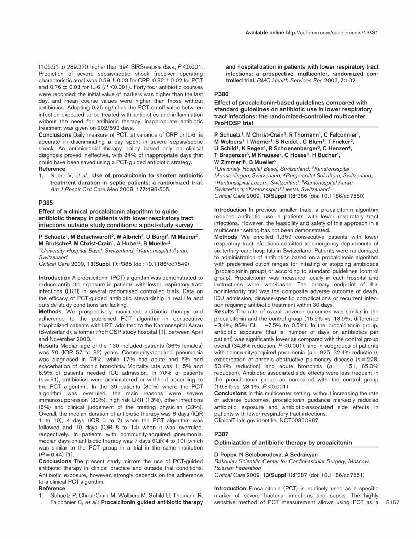
S157
(105.51 to 289.27)) higher than 394 SIRS/sepsis days, P <0.001.Prediction of severe sepsis/septic shock (receiver operatingcharacteristic area) was 0.59 ± 0.03 for CRP, 0.82 ± 0.02 for PCTand 0.76 ± 0.03 for IL-6 (P <0.001). Forty-four antibiotic courseswere recorded; the initial value of markers was higher than the lastday, and mean course values were higher than those withoutantibiotics. Adopting 0.25 ng/ml as the PCT cutoff value betweeninfection expected to be treated with antibiotics and inflammationwithout the need for antibiotic therapy, inappropriate antibiotictreatment was given on 202/592 days.Conclusions Daily measure of PCT, at variance of CRP or IL-6, isaccurate in discriminating a day spent in severe sepsis/septicshock. An antimicrobial therapy policy based only on clinicaldiagnosis proved ineffective, with 34% of inappropriate days thatcould have been saved using a PCT-guided antibiotic strategy.Reference1. Nobre V, et al.: Use of procalcitonin to shorten antibiotic
treatment duration in septic patients: a randomized trial.Am J Respir Crit Care Med 2008, 177:498-505.
P385Effect of a clinical procalcitonin algorithm to guideantibiotic therapy in patients with lower respiratory tractinfections outside study conditions: a post-study survey
P Schuetz1, M Batschwaroff2, W Albrich2, U Bürgi2, M Maurer2,M Brutsche2, M Christ-Crain1, A Huber2, B Mueller2
1University Hospital Basel, Switzerland; 2Kantonsspital Aarau,SwitzerlandCritical Care 2009, 13(Suppl 1):P385 (doi: 10.1186/cc7549)
Introduction A procalcitonin (PCT) algorithm was demonstrated toreduce antibiotic exposure in patients with lower respiratory tractinfections (LRTI) in several randomised controlled trials. Data onthe efficacy of PCT-guided antibiotic stewardship in real life andoutside study conditions are lacking.Methods We prospectively monitored antibiotic therapy andadherence to the published PCT algorithm in consecutivehospitalized patients with LRTI admitted to the Kantonsspital Aarau(Switzerland), a former ProHOSP study hospital [1], between Apriland November 2008.Results Median age of the 130 included patients (36% females)was 70 (IQR 57 to 82) years. Community-acquired pneumoniawas diagnosed in 78%, while 17% had acute and 5% hadexacerbation of chronic bronchitis. Mortality rate was 11.5% and6.9% of patients needed ICU admission. In 70% of patients(n = 91), antibiotics were administered or withheld according tothe PCT algorithm. In the 39 patients (30%) where the PCTalgorithm was overruled, the main reasons were severeimmunosuppression (30%), high-risk LRTI (13%), other infections(8%) and clinical judgement of the treating physician (33%).Overall, the median duration of antibiotic therapy was 6 days (IQR1 to 10), 4 days (IQR 0 to 7) when the PCT algorithm wasfollowed and 10 days (IQR 8 to 14) when it was overruled,respectively. In patients with community-acquired pneumonia,median days on antibiotic therapy was 7 days (IQR 4 to 10), whichwas similar to the PCT group in a trial in the same institution(P = 0.44) [1].Conclusions The present study mirrors the use of PCT-guidedantibiotic therapy in clinical practice and outside trial conditions.Antibiotic exposure, however, strongly depends on the adherenceto a clinical PCT algorithm.Reference1. Schuetz P, Christ-Crain M, Wolbers M, Schild U, Thomann R,
Falconnier C, et al.: Procalcitonin guided antibiotic therapy
and hospitalization in patients with lower respiratory tractinfections: a prospective, multicenter, randomized con-trolled trial. BMC Health Services Res 2007, 7:102.
P386Effect of procalcitonin-based guidelines compared withstandard guidelines on antibiotic use in lower respiratorytract infections: the randomized-controlled multicenterProHOSP trial
P Schuetz1, M Christ-Crain1, R Thomann1, C Falconnier1, M Wolbers1, I Widmer1, S Neidet1, C Blum1, T Fricker2, U Schild1, K Regez1, R Schoenenberger3, C Henzen4, T Bregenzer5, M Krausse2, C Hoess2, H Bucher1, W Zimmerli6, B Mueller5
1University Hospital Basel, Switzerland; 2KanstonsspitalMünsterlingen, Switzerland; 3Bürgerspital Solothurn, Switzerland;4Kantonsspital Luzern, Switzerland; 5Kantonsspital Aarau,Switzerland; 6Kantonsspital Liestal, SwitzerlandCritical Care 2009, 13(Suppl 1):P386 (doi: 10.1186/cc7550)
Introduction In previous smaller trials, a procalcitonin algorithmreduced antibiotic use in patients with lower respiratory tractinfections. However, the feasibility and safety of this approach in amulticenter setting has not been demonstrated.Methods We enrolled 1,359 consecutive patients with lowerrespiratory tract infections admitted to emergency departments ofsix tertiary-care hospitals in Switzerland. Patients were randomizedto administration of antibiotics based on a procalcitonin algorithmwith predefined cutoff ranges for initiating or stopping antibiotics(procalcitonin group) or according to standard guidelines (controlgroup). Procalcitonin was measured locally in each hospital andinstructions were web-based. The primary endpoint of thisnoninferiority trial was the composite adverse outcome of death,ICU admission, disease-specific complications or recurrent infec-tion requiring antibiotic treatment within 30 days.Results The rate of overall adverse outcomes was similar in theprocalcitonin and the control group (15.5% vs. 18.9%; difference–3.4%, 95% CI = –7.5% to 0.5%). In the procalcitonin group,antibiotic exposure (that is, number of days on antibiotics perpatient) was significantly lower as compared with the control groupoverall (34.8% reduction, P <0.001), and in subgroups of patientswith community-acquired pneumonia (n = 925, 32.4% reduction),exacerbation of chronic obstructive pulmonary disease (n = 228,50.4% reduction) and acute bronchitis (n = 151, 65.0%reduction). Antibiotic-associated side effects were less frequent inthe procalcitonin group as compared with the control group(19.8% vs. 28.1%; P <0.001).Conclusions In this multicenter setting, without increasing the rateof adverse outcomes, procalcitonin guidance markedly reducedantibiotic exposure and antibiotic-associated side effects inpatients with lower respiratory tract infections.ClinicalTrials.gov identifier NCT00350987.
P387Optimization of antibiotic therapy by procalcitonin
D Popov, N Beloborodova, A SedrakyanBakoulev Scientific Center for Cardiovascular Surgery, Moscow,Russian FederationCritical Care 2009, 13(Suppl 1):P387 (doi: 10.1186/cc7551)
Introduction Procalcitonin (PCT) is routinely used as a specificmarker of severe bacterial infections and sepsis. The highlysensitive method of PCT measurement allows using PCT as a
Available online http://ccforum.com/supplements/13/S1
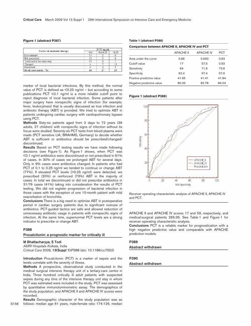
S158
marker of local bacterial infections. By this method, the normalvalue of PCT is defined as <0.25 ng/ml – but according to somepublications PCT <0.1 ng/ml is a more reliable cutoff point toreject diagnosis of local bacterial infection. Some patients aftermajor surgery have nonspecific signs of infection (for example,fever, leukocytosis) that is usually discussed as true infection andantibiotic therapy (ABT) is provided. We tried to optimize ABT inpatients undergoing cardiac surgery with cardiopulmonary bypassusing PCT.Methods Sixty-six patients aged from 2 days to 72 years (39adults, 27 children) with nonspecific signs of infection without itsfocus were studied. Seventy-six PCT tests from blood plasma weremade (PCT sensitive LIA; BRAHMS, Germany) to decide whetherABT is sufficient or antibiotics should be prescribed/changed/discontinued.Results Based on PCT testing results we have made followingdecisions (see Figure 1). As Figure 1 shows, when PCT was<0.1 ng/ml antibiotics were discontinued or not prescribed in 61%of cases. In 30% of cases we prolonged ABT for several days.Only in 9% cases were antibiotics changed. In patients who hadPCT of 0.1 to 0.25 ng/ml we tended to continue or change ABT(77%). If elevated PCT levels (>0.25 ng/ml) were detected, weprescribed (20%) or reinforced (73%) ABT in the majority ofcases. In total we discontinued or did not prescribe antibiotics in31/76 cases (41%) taking into consideration the results of PCTtesting. We did not register progression of bacterial infection inthose cases with the exception of one 10-month patient with mildexacerbation of bronchitis.Conclusions There is a big need to optimize ABT in postoperativeperiod in cardiac surgery patients due to significant overuse ofantibiotics. PCT-guided tactics are safe and allowed reduction ofunnecessary antibiotic usage in patients with nonspecific signs ofinfection. At the same time, supernormal PCT levels are a strongindicator to prescribe or change ABT.
P388Procalcitonin: a prognostic marker for critically ill
M Bhattacharyya, S TodiAMRI Hospitals Kolkata, IndiaCritical Care 2009, 13(Suppl 1):P388 (doi: 10.1186/cc7552)
Introduction Procalcitonin (PCT) is a marker of sepsis and thelevels correlate with the severity of illness.Methods A prospective, observational study conducted in themedical surgical intensive therapy unit of a tertiary-care centre inIndia. Three hundred critically ill adult patients with suspectedsepsis during any time of the intensive therapy unit stay in whomPCT was estimated were included in the study. PCT was assessedby quantitative immunoluminometric assay. The demographics ofthe study population, and APACHE II and APACHE IV scores wererecorded.Results Demographic character of the study population was asfollows: median age 61 years, male:female ratio 174:126, median
APACHE II and APACHE IV scores 17 and 59, respectively, andmedical:surgical patients 265:35. See Table 1 and Figure 1 forreceiver operating characteristic analysis.Conclusions PCT is a reliable marker for prognostication with ahigh negative predictive value and comparable with APACHEprediction models.
P389Abstract withdrawn
P390Abstract withdrawn
Critical Care March 2009 Vol 13 Suppl 1 29th International Symposium on Intensive Care and Emergency Medicine
Figure 1 (abstract P387)
Figure 1 (abstract P388)
Receiver operating characteristic analysis of APACHE II, APACHE IVand PCT.
Table 1 (abstract P388)
Comparison between APACHE II, APACHE IV and PCT
APACHE II APACHE IV PCT
Area under the curve 0.66 0.693 0.63
Cutoff value 17 57.5 0.93
Sensitivity 64 71.6 73.6
Specificity 62.4 57.4 57.6
Positive predictive value 41.95 41.41 41.84
Negative predictive value 80.33 82.78 84.04
S159
P391Raised C-reactive protein on ICU discharge is notassociated with subsequent poor outcome
TE Reynolds, N Al-Subaie, A Myers, M Saidi, R Sunderland, A Rhodes, RM GroundsSt George’s Hospital, London, UKCritical Care 2009, 13(Suppl 1):P391 (doi: 10.1186/cc7555)
Introduction It has been suggested that plasma C-reactive protein(CRP) levels measured on discharge from the ICU may be a usefulpredictor of either unplanned readmission or unexpected death onthe ward. Previous work has found that raised plasma CRPindependently predicted, in separate studies, each of these twopoor outcomes [1,2]. We investigated whether these results couldbe repeated in our mixed medical/surgical ICU for a compositepoor outcome measure combining death and readmission.Methods We prospectively enrolled a cohort comprised of all ICUadmissions over 1 year. We collected admission clinical anddemographic data and APACHE II scores. At discharge werecorded the white cell count and serum levels of CRP andalbumin, and we observed death and readmission outcomes up to2 weeks after ICU discharge. This time period was chosen on thegrounds that an association between discharge CRP andpostdischarge outcome should relate to persisting inflammatoryactivity and would be of most relevance in the short term.Results Of 1,487 admissions to our ICU, 181 (12.2%) resulted inthe patient’s death on the ICU and 110 (7.4%) ended with eitherthe patient’s discharge to another hospital or ICU and thus theirloss to follow up. Eleven patients discharged for palliation wereexcluded from analysis. A total of 1,185 (79.7%) were dischargedto a ward in the hospital and so could potentially have suffered anunexpected deterioration resulting in ICU readmission or death. Ofthese, 117 (9.9%) of the discharge episodes were followed by anunexpected poor outcome of either readmission (n = 83, 7.0%) or
death (n = 34, 2.9%) within 2 weeks. A composite outcomemeasure combining these two poor outcomes was associated withage, higher APACHE II score, and a low plasma albumin on ICUdischarge. Means and median values were compared usingP <0.05 as the test of significance. Plasma CRP on discharge fromthe ICU was not associated with this composite measure.Conclusions In a mixed medical/surgical ICU, CRP on the day ofdischarge is not associated with a poor outcome measure ofunexpected readmission or ward death.References1. Ho KM, et al.: C-reactive protein concentration as a predic-
tor of in-hospital mortality after ICU discharge: a prospec-tive cohort study. Intensive Care Med 2008, 34:481-487.
2. Kaben A, et al.: Readmission to a surgical intensive careunit: incidence, outcome and risk factors. Crit Care 2008,12:R123.
P392Influence of age on the distribution of morphine andmorphine-3-glucuronide across the blood–brain barrier insheep
P Ederoth1, J Bengtsson2, D Ley1, S Hansson1, I Amer-Wåhlin1,L Hellström-Westas1, K Marsal1, M Hammalund-Udenaes2, CH Nordström1
1University Hospital, Lund, Sweden; 2Uppsala University, Uppsala,SwedenCritical Care 2009, 13(Suppl 1):P392 (doi: 10.1186/cc7556)
Introduction Neonates are recommended smaller doses ofmorphine than adults and, accordingly, we hypothesised that thedistribution of morphine and its metabolites over the blood–brainbarrier differs between these age groups. In addition, neonatalasphyxia, with potentially harmful effects on the brain, in theory,might further affect the pharmacokinetics of substances over theblood–brain barrier.Methods During anaesthesia, microdialysis probes were insertedinto the brain cortex and in a central vein of 11 exteriorized near-term lambs (127 gestation days) and six nonpregnant adult sheep.Five of these lambs were subjected to 10 minutes of asphyxiathrough umbilical cord occlusion during delivery. Morphine,1 mg/kg, was thereafter intravenously administered as a 10 minuteconstant infusion. Microdialysis and blood samples were collectedfor up to 360 minutes after morphine administration, and analyzedusing liquid chromatography followed by tandem massspectrometry. Data presented as the mean ± SD.Results The morphine unbound drug brain:blood distribution ratio(Kp,uu) was 1.19 ± 0.20 and 1.89 ± 0.51 for the sheep andpremature lambs without asphyxia, respectively (P = 0.018). Thehalf-lives in the blood and brain cortex, clearance, volume ofdistribution, and distribution in the brain of unbound drug were allnumerically significantly higher in the adult sheep as comparedwith the premature lambs. The morphine-3-glucuronide Kp,uu valueswere 0.27 ± 0.16 and 0.17 ± 0.15 in sheep and premature lambs(P = NS), indicating a net efflux from the brain in both groups.Induced asphyxia did not affect the results.Conclusions The morphine Kp,uu was above unity, indicating a netinflux of morphine into the brain. In addition, influx was significantlyhigher in premature lambs than in adult sheep. We interpret this asan active transport of morphine into the brain, which may becounteracted with increased efflux with age. Further, neonatalasphyxia did not change these pharmacokinetic findings. The Kp,uuin the sheep was different from the values obtained in humans(0.64), rats (0.49), mice (0.5) and pigs (0.47), where a net efflux ofmorphine from the brain was observed.
Available online http://ccforum.com/supplements/13/S1
S160
P393Epidural anesthesia during surgery: friend or foe?
E Koepfli, S Brandt, O Kimberger, L HiltebrandUniversity Hospital Bern, SwitzerlandCritical Care 2009, 13(Suppl 1):P393 (doi: 10.1186/cc7557)
Introduction Decreased intestinal microcirculatory blood flow(MBF) is an important contributor to perioperative morbidity.Epidural anesthesia (EA) may improve intestinal MBF [1] but otherresearchers found that EA decreases cardiac output (CO) andthus compromises intestinal MBF [2]. We tested whether EA plusintravenous colloids result in increased intestinal MBF comparedwith colloid administration alone.Methods Twenty pigs (30 ± 2.5 kg) were anesthetized and venti-lated. A laparotomy was performed for instrumentation and a colonanastomosis. After baseline measurements the animals were randomlyassigned to one of the following treatments: Group HES, hydroxyethylstarch (130/0.4) was given to maintain SvO2 ≥60%; Group EA, inaddition to the same colloid treatment an epidural catheter wasinserted at the lumbar level and after a bolus of 4 ml ropivacain 0.2%,7 ml/hour were continuously administered. The CO and MBF in themucosa of the jejunum, colon and anastomosis were measured.Results In both groups the CO and MBF increased similarly(Table 1). No difference in the circulatory parameters between thetwo groups was found. However, in Group EA significantly morefluids (56%) were administered to achieve SvO2 ≥60%.
Table 1 (abstract P393)
Comparison of the treatment groups
Group HES Group EA
HES (ml) 831 ± 94 1296 ± 81*Mean arterial pressure (%) 130 ± 3 124 ± 6 CO (%) 143 ± 7 143 ± 6MBF jejunum 145 ± 8 107 ± 8MBF colon 133 ± 6 130 ± 3MBF anastomosis 166 ± 27 150 ± 17
Data presented as percentage ± SEM of baseline unless otherwise stated.*P <0.01 compared with group CO.
Conclusions The present study did not show any positive effectsof epidural anesthesia on intestinal microcirculatory blood flow. Onthe contrary, over 50% more fluids were needed to maintain similarperfusion parameters than with fluids alone. Such a fluid load asneeded in the epidural group may potentially be harmful.References1. Sielenkämper AW, et al.: Thoracic epidural anesthesia
increases mucosal perfusion in ileum of rats. Anesthesiol-ogy 2000, 93:844-851.
2. Schwarte LA, et al.: Effects of thoracic epidural anaesthesiaon microvascular gastric mucosal oxygenation in physio-logical and compromised circulatory conditions in dogs. BrJ Anaesth 2004, 93:552-559.
P394Epidural analgesia compared with peripheral nerveblockade after major knee surgery
E Tricarico, S Tomasino, L D’OrlandoASS n.3 ‘Alto Friuli’, Gemona del friuli, ItalyCritical Care 2009, 13(Suppl 1):P394 (doi: 10.1186/cc7558)
Introduction The aim of this study was to undertake a randomizedtrial between lumbar continuous epidural analgesia and continuous
femoral blockade in adults undergoing major knee surgery,including comparison of analgesic efficacy [1], side effects, patientsatisfaction and rehabilitation indices [2].Methods We studied 91 patients in 2 years that were randomizedinto two groups: epidural group (EG) 40 and femoral group (FG)51. All patients received spinal anesthesia with 1 ml of 1% bupiva-caine without a narcotic. The EG received a 5 ml/hour continuousinfusion in elastomeric pump with 0.125% bupivacaine andmorphine 20 μg/ml. The FG received a 5 ml/hour continuous infu-sion in an elastomeric pump with 0.125% bupivacaine andtramadol 1 mg/kg/day + ketorolac 30 mg intravenously x 3.Results No statistically significant difference was noted betweenthe two groups about pain scores (VAS at rest and on movement),nausea, vomiting, arterial hypotension, morphine consumption,headache, pruritus, patient satisfaction and rehabilitation indices(active knee flexion). A statistically significant difference was notedbetween the two groups about contralateral analgesia, urinaryretention that required bladder catheterization and motor blockwith major incidence in the EG.Conclusions Continuous femoral blockade represents the bestbalance between analgesia and side effects as a choice ofpostoperative analgesic technique for major knee surgery,especially as the risk of injury to the neuraxis is negligible. Overall,however, we believe that there is no sufficient evidence that lumbarepidural analgesia should not be used routinely.References1. Fowler SJ, Symons J, Sabato S, Myles PS: Epidural analge-
sia compared with peripheral nerve blockade after majorknee surgery: a systematic review and meta-analysis ofrandomized trials. Br J Anaesth 2008, 100:154-164.
2. Singelyn FJ, Ferrant T, Malisse MF, Joris D: Effects of intra-venous patient-controlled analgesia with morphine, con-tinuous epidural analgesia, and continuous femoral nervesheath block on rehabilitation after unilateral total-hiparthroplasty. Reg Anesth Pain Med 2005, 30:452-457.
P395Postconditioning effects following sevoflurane inhalationalsedation in the ICU: a pilot study in cardiac surgerypatients
K Röhm1, J Mayer1, J Boldt1, S Suttner1, S Piper2
1Klinikum Ludwigshafen, Germany; 2Hospital of Frankenthal,GermanyCritical Care 2009, 13(Suppl 1):P395 (doi: 10.1186/cc7559)
Introduction Volatile anaesthetics using the anesthetic conservingdevice (ACD) have become an alternative ICU sedation regimen[1,2]. The purpose of this study was to evaluate postconditioningeffects following sevoflurane or propofol administration in patientsafter elective cardiac surgery.Methods Fifty patients either received sevoflurane via ACD orpropofol for ICU sedation. The primary endpoint was a change introponine T (TNT), besides measures of myocardial creatine kinase(CK-MB), N-terminal pro brain natriuretic peptide (NT-proBNP) andhaemodynamics. Measure points were set at baseline, end ofsurgery, 24 hours and 48 hours after surgery.Results TNT and CK-MB levels were significantly higher(P <0.0001) at all measure points compared with baseline in eachgroup, without any significant differences between both groups.NT-proBNP values were significantly lower following sevoflurane at24 and 48 hours (P <0.05) compared with propofol (Figure 1).Conclusions Postoperative sevoflurane sedation via ACD led tolower NT-proBNP levels at 24 and 48 hours, while TNT and CK-MB values were comparable in both study groups.
Critical Care March 2009 Vol 13 Suppl 1 29th International Symposium on Intensive Care and Emergency Medicine

S161
References1. Röhm KD, et al.: Short-term sevoflurane sedation using the
Anaesthetic Conserving Device after cardiothoracicsurgery. Intensive Care Med 2008, 34:1673-1679.
2. Sackey PV, et al.: Prolonged isoflurane sedation of inten-sive care unit patients with the Anesthetic ConservingDevice. Crit Care Med 2004, 32:2241-2246.
P396Multicenter randomized trial of sedation using dailywake-up calls, bispectral index or clinical sedation scoresin a mixed medical–surgical ICU population
J Binnekade1, R Wilde2, AJ Slooter3, J Sluijs4, O Beenen3, M Schultz1, M Dijkgraaf1, P Berg2, M Vroom1
1AMC, Amsterdam, the Netherlands; 2LUMC, Leiden, theNetherlands; 3UMCU, Utrecht, the Netherlands; 4MCH, Den Haag,the NetherlandsCritical Care 2009, 13(Suppl 1):P396 (doi: 10.1186/cc7560)
Introduction Daily interruption of continuous infusions of sedatives(wake-up calls (WC)) has been found previously to promote morerapid withdrawal of ventilatory support in ICU patients. Bispectralindex (BIS) monitoring has been reported to adequately describethe depth of sedation in critically ill patients. We compared theeffectiveness and safety of both sedation strategies in a mixedmedical–surgical ICU population.Methods Patients expected to need sedation for at least 1 daywere randomly assigned to a WC strategy, sedation guided byBIS, or a control group. Clinical assessment of the sedation depth,the Ramsay score, was performed in all three groups. Primaryoutcome: ICU length of stay (LOS); secondary outcomes: durationof mechanical ventilation, accidental removal of indwelling devices,ICU readmission rate, hospital LOS, 90-day mortality, and stressfulevents (interview at ICU exit and after 3 months).Results In four ICUs, three academic and one teaching hospital, atotal number of 617 patients was randomized; 205 to the WCstrategy, 202 to sedation guided by BIS, and 210 to the controlgroup. The study groups were well balanced according to baselinecharacteristics. Neither ICU LOS (median (IQR) 11 (6 to 21)(WC), 12 (7 to 23) (BIS), and 11 (6 to 24) days (control),
P = 0.67) nor duration of mechanical ventilation (median 165 (0 to581) (WC), 158 (3 to 590) (BIS), and 163 (5 to 385) hourscontrol, P = 0.97) were different between groups. Accidentalremoval of indwelling devices was encountered 37 times (10 timesin nine patients (WC), six times in five patients (BIS), and 21 timesin 11 patients (control), P = 0.01). The ICU readmission rate,hospital LOS, and 90-day mortality were not different betweengroups. Interviews showed similar stress experiences.Conclusions Although a significant higher number of incidentswith indwelling devices occurred in the control group, no additionaldifferences between the three sedation regimens were revealed.Although current guidelines on ICU sedation have incorporatedWC next to clinical sedation scales, the results from this largerstudy do not promote either WC or BIS in guiding sedation of ICUpatients.
P397Safety and length of different sedations for endoscopicprocedures
O Martinez, A Algaba, D Ballesteros, M Chana, B Estebanez, B Lopez, C Martin, L Vigil, R BlancasHospital del Tajo, Aranjuez, SpainCritical Care 2009, 13(Suppl 1):P397 (doi: 10.1186/cc7561)
Introduction Endoscopic procedures have improved thanks to thefact that they are performed currently under sedation. We describea series of endoscopic studies, sedated by the Intensive CareDepartment. Different sedation protocols are described, empha-sizing some patients sedated with ketamine plus midazolam, drugsrarely reported for adults.Methods Patients older than 18 years, whose performedendoscopic, colonoscopic or both procedures under sedationwere performed by the Intensive Care Department of the Hospitaldel Tajo. Data were collected for 6 months. Demographiccharacteristics, medical history, American Society of Anesthes-iology classification, drugs bolus and total dosages, respiratory andhemodynamic data, the length of procedure and recovery, andcomplications were collected. Tolerance was assessed by anendoscopist, with a 1 (very bad) to 5 (very good) scale. Differentindices of two groups of treatment were compared, propofol-fentanyl (Group A) versus ketamine-midazolam (Group B) usingthe chi-square test and the Student t test.Results In total, 245 procedures were included. The procedureswere 168 (66.6%) colonoscopies, 34 (13.9%) endoscopies and43 (17.6%) both procedures together. Tolerance: 33.5% were 5;58.8% were 4; 4.5% were 3; 0.8% were 2. There were 72(29.39%) complications, the most common hypotension being 22(8.98%) and respiratory depression 12 (4.9%). Length ofprocedure was 31.20 minutes (SD = 15.53) and recovery timewas 64.86 minutes (SD = 37.56). Comparing Group A versusGroup B, hypotension (12.67% vs. 2.9%, P = 0.023) and respira-tory depression (6.67% vs. 0%, P = 0.03) were more frequent withGroup A. Group B presented a higher percentage of vomiting (0%vs. 5.8%, P = 0.009) and hallucinations (0% vs. 11.59%,P < 0.001). The length of recovery was longer for Group B(61.12 min vs. 78.98 min, P = 0.008).Conclusions Sedation for endoscopic procedures performed byintensivists is safe and well tolerated for most patients, with asimilar percentage of complications compared with previousreports. Ketamine plus midazolam are safe and could be useful forshort-length sedation in patients with a high risk of respiratorydepression.
Available online http://ccforum.com/supplements/13/S1
Figure 1 (abstract P395)
NT-proBNP levels.

S162
P398Novel responsiveness index measures the level ofsedation in cardiac ICU patients
P Lapinlampi1, H Viertiö-Oja1, M Särkelä1, K Uutela1, P Meriläinen1, P Ramsay2, T Walsh2
1GE Healthcare, Helsinki, Finland; 2Edinburgh Royal Infirmary,Edinburgh, UKCritical Care 2009, 13(Suppl 1):P398 (doi: 10.1186/cc7562)
Introduction Sedation is an integral part of the management ofpatients requiring mechanical ventilation in the ICU. At present,sedation is usually managed by clinical assessments, often inconjunction with a protocol for adjusting drug doses. Thisapproach has been shown to decrease ventilation times and isconsidered the best practice in recent guidelines [1]. Such scales,however, provide only intermittent and, to some extent, subjectiveinformation about the patient’s state. Also, not every scale used inclinical practice is validated for ICU use and interobserver variationmay occur.Methods We have developed a novel method that analyzes thepatterns in the electromyographic (EMG) component of the frontalbiopotential signal that are associated with activation and arousalprocesses. By quantifying the amount and magnitude of theresponse patterns in the past 60 minutes, a responsiveness index(RI) ranging between 0 and 100 (0 corresponds to anonresponsive patient and 100 to a high amount of responses) isderived. Previously, we have compared the RI with EEG spectralentropy [2] in general ICU patients. Now we have studied theperformance of the RI with 17 cardiac ICU patients and comparedthese with the development dataset analyzed by Viertiö-Oja andcolleagues [2]. A subgroup of 17 patients (of total 30 patients)with a clinically assessed low probability of encephalopathy wasused as the primary development data. The patients in bothdatasets were consenting adult ICU patients with non-neurologicprimary ICU diagnosis. A modified Ramsay score was used as areference of the sedation level.Results The performance of the RI in reference to the Ramsayscores was analyzed by computing the prediction probability (PK).The development and test data contained 213 and 96 eligibleRamsay assessments, respectively. The PK for separating deepsedation levels 4 to 6 from levels 1 to 3 was 0.91 (0.03) in thedevelopment data and 0.96 (0.02) in the test data. For separatingall Ramsay levels, the PK values in the development data and testdata were 0.82 (0.02) and 0.89 (0.02), respectively.Conclusions These results showed good RI performance in thecardiac ICU data. The RI continues to show promise as anindicator of the level of sedation in ICU patients.References1. Nasraway SA, et al.: Crit Care Med 2002, 30:117-118.2. Viertiö-Oja H, et al.: Crit Care 2006, 10(Suppl 1):P442.
P399Sedation protocols for intubated patients and noninvasiveventilation: additional concepts for a noniatrogenicintensive care
G Chanques, JM Constantin, B Jung, N Rossel, M Cissé, S JaberSaint-Eloi Hospital, Montpellier, FranceCritical Care 2009, 13(Suppl 1):P399 (doi: 10.1186/cc7563)
Introduction Noninvasive ventilation (NIV) and sedation protocolsduring invasive mechanical ventilation (IMV) can both separatelyreduce the duration of mechanical ventilation in ICU patients.
However, no study has shown the impact of these two conceptswhen associated.Methods All consecutive patients admitted to a 14-bedmedical–surgical ICU were retrospectively studied from 2000 to2006 after implementation of NIV (2000 to 2001) and sedationprotocols (2003 to 2004). The duration of mechanical ventilationshown by categories was analyzed using a chi-square test.Results During the 7 years of the study, 2,839 admissions wereperformed in 2,511 patients (59 ± 17 years, women 36%, medicaladmission 43%, Simplified Acute Physiology Score II 38 ± 17,mortality in the ICU 17%). The incidence and duration of IMVsignificantly decreased during the study (P <0.001) (Figure 1). TheNIV use progressively increased during the study period, both inmedical and surgical patients.Conclusions These results showed that the implementation ofsedation protocols for intubated patients added to the imple-mentation of NIV decreased moreover the duration of IMV.
P400Does choice of sedative agent affect duration of ICU stay,mortality or neurological outcome in patients undergoingtherapeutic hypothermia?
M Gillies, R Pratt, J Borg, J Brooks, C Mckenzie, S TibbyGuy’s & St Thomas’ NHS Foundation Trust, London, UKCritical Care 2009, 13(Suppl 1):P400 (doi: 10.1186/cc7564)
Introduction The effect of therapeutic hypothermia on sedativedrug clearance and neurological prognostication is unknown.Hypothermia has been shown to affect clearance of drugsmetabolised by the cytochrome p450 system (fentanyl, midazolamand muscle relaxants) [1]. We undertook a retrospective study toascertain whether the use of remifentanil sedation was associatedwith reduced duration of ICU stay, ventilation, mortality or neuro-logical outcome compared with fentanyl in patients undergoingtherapeutic hypothermia.Methods Data were collected on all patients undergoing thera-peutic hypothermia in a 2.5-year period using the Carevue ICUDatabase (Phillips) and case-note review. All patients receivedpropofol and either remifentanil or fentanyl as sedation. Patientdemographic data included age, weight, sex, APACHE II, time toreturn of spontaneous circulation and arrest type. Measuredoutcomes were ICU-free days, ventilator-free days, ICU mortality,
Critical Care March 2009 Vol 13 Suppl 1 29th International Symposium on Intensive Care and Emergency Medicine
Figure 1 (abstract P399)
S163
hospital mortality and neurological outcome at hospital discharge.Good neurological outcome was classified as Pittsburgh CerebralPerformance Criteria grade 1 to 2 at hospital discharge [2].Predictive models used multivariable logistic regression.Results Eighty-three patients received therapeutic hypothermia(crude ICU mortality 43%, median APACHE II score 18); of which54 received fentanyl sedation and 29 remifentanil. In themultivariable models, remifentanyl was not associated with ICU(P = 0.35) or hospital (P = 0.22) mortality or good neurologicaloutcome (P = 0.75). However, remifentanyl was associated with apositive trend towards an increase in both ventilator-free and ICU-free days (both P = 0.08).Conclusions Use of remifentanil was not associated with areduction in ICU or hospital mortality, nor with good neurologicaloutcome. However, there was a trend to increased ventilator-freeand ICU-free days. This effect needs to be confirmed prospectivelywith a larger dataset. Caution must be exercised regarding theeffect of delayed sedative drug metabolism on neurologicalassessment in patients who have undergone therapeutichypothermia, especially drugs metabolised by the cytochromep450 system.References1. Tortorici MA, et al.: Therapeutic hypothermia-induced phar-
macokinetic alterations on CYP2E1 chlorzoxazone-medi-ated metabolism in a cardiac arrest rat model. Crit CareMed 2006, 34:785-791.
2. The HACA Study Group: Mild therapeutic hypothermia toimprove the neurologic outcome after cardiac arrest. N Eng J Med 2002, 346:549-556.
P401Positive inotropic effects of midazolam anaesthesia in ratsduring noninvasive heart studies
R Lauwers, S Droogmans, I HubloueUZ Brussel, BelgiumCritical Care 2009, 13(Suppl 1):P401 (doi: 10.1186/cc7565)
Introduction Pinhole gated single photon emission computedtomography (SPECT) and transthoracic echocardiography (TTE)are easier, more time sparing and much more animal friendly in theassessment of the left ventricular (LV) function in rats comparedwith the techniques used in the past. Where the aim of previousstudies was to determine the cardiac function under differentanaesthetics in an extracorporeal model, the aim of our study is tofind the ideal anaesthetic for the assessment of the cardiacfunction in the rat.Methods Two different studies were performed using pinholegated SPECT and TTE in male Wistar rats. For the TTE study 32rats were randomised to four groups and were anaesthetized withtwo different products with a washout period of 3 days. A linearprobe was used for assessing M-mode images and LV volumes(ml) were calculated using an ellipsoid model 10 minutes afteradministration of anaesthesia. Pentobarbital (50 mg/kg) intra-peritoneally, propofol (200 mg/kg) intraperitoneally, midazolam(70 mg/kg) intraperitoneally, isoflurane (2%), sevoflurane (2%) ordesflurane (7%) were the anaesthetics studied. For the pinholegated SPECT study, 48 rats were scanned two times with a1-week interval: once under pentobarbital (60 mg/kg intraperito-neally) anesthesia for the control study, and a second time usingthe same anesthetics as the TTE study. Pinhole gated SPECT wasperformed 1 hour after intravenous injection of 330 MBq 99mTc-MIBI. Parameters of cardiac function were derived usingquantitative gated SPECT.
Results Augmented ejection fractions were observed for midazolamcompared with all other anaesthetics (85% in the SPECT study and91% in the TTE study). Anterior wall thickening was remarkablyaugmented: 84 ± 9% compared with propofol 51 ± 12% (P <0.001)and pentobarbital 64 ± 23 (P = 0.03) in the TTE study, but nodifference was observed in the inferior thickening. In the SPECTstudy, augmented wall thickening was observed in the lateral, inferior,septal and anterior region (all P <0.001) compared with pentobarbital.Conclusions Anaesthetic agents seem to play a role in regionalcontractile function, affecting global LV function. Previous studiesdemonstrate conflicting results concerning the effect of midazolamon cardiac function. Both of our studies suggest that, at the givendose, midazolam has a positive inotropic effect.
P402Comparison of cerebral hemodynamic variables inhemorrhagic stroke using dexmedetomidine–propofolversus dexmedetomidine–midazolam
FJ Perez-Rada1, JL Franco-Calderon2, M Torres-Cortes2
1Hospital San Jose Tec de Monterrey, NL, Mexico; 2UMAE 25,IMSS, Monterrey, NL, MexicoCritical Care 2009, 13(Suppl 1):P402 (doi: 10.1186/cc7566)
Introduction Dexmedetomidine has been described as having apositive cerebral hemodynamic effect by reducing the cerebralblood flow/cerebral metabolic index ratio (CBF/CMI ratio) andimproving CFB self-regulation; however, no data are available onhow this actually modifies brain metabolism.Methods We performed a multicenter, prospective, randomized,active comparator (dexmedetomidine–propofol or dexmedeto-midine–midazolam) study. We included consecutive adult patients(>18 years old) admitted to ICUs with hemorrhagic stroke andmoderate or severe brain edema (measured directly at surgery orby CT scan) who needed sedation for at least 72 hours afterrandomization. We excluded patients with mild brain edema,contraindications to the use of sedatives and those who could nothave a jugular venous line placed. Randomized groups receivedsedation using dexmedetomidine + midazolam (Group 1) or dex-medetomidine + propofol (Group 2). We obtained basal measure-ment of several variables indirectly related to cerebral metabolism(jugular venous O2 saturation – SvjO2, jugular arteriovenous O2difference – Da-vjO2, cerebral oxygen extraction – CEO2,estimated cerebral metabolic rate – ECMRO2) and repeated thesemeasurements after 72 hours. Statistical analysis was performedusing chi-square and proportions for categorical variables, and themedian, standard deviation and Student t test for continuousvariables. We considered P ≤0.05 significant.Results Twenty-eight consecutive patients were included (15male), average age 61.2 ± 8.6 years, presenting with moderatebrain edema (5/28, 17.9%) or severe brain edema (23/28,82.1%). Both randomized groups had 14 patients. We found nodifference between groups in SvjO2 (P = 0.49 basal, P = 0.19 at72 hours), Da-vjO2 (P = 0.68 basal, P = 0.67 at 72 hours), CEO2(P = 0.49 basal, P = 0.19 at 72 hours), or ECMRO2 (P = 0.34basal, P = 0.50 at 72 hours).Conclusions No significant differences in the measured brainmetabolism variables were found among the two groups. Wesuggest comparing these two groups versus a placebo group inanother study.Reference1. Drummond JC, et al.: Effect of dexmedetomidine on cerebral
blood flow velocity, cerebral metabolic rate, and carbondioxide response in normal humans. Anesthesiology 2008,108:225-232.
Available online http://ccforum.com/supplements/13/S1

S164
P403Effect of low-dose ketamine regimen with or withoutmagnesium sulphate adjunct in tramadol patient controlledanalgesia in a major abdominal surgery ICU population
F Meurant, P GeukensKirchberg Hospital, LuxembourgCritical Care 2009, 13(Suppl 1):P403 (doi: 10.1186/cc7567)
Introduction We evaluated the potential benefits of adding mag-nesium sulphate (MgSO4) to ketamine (K) in a patient controlledanalgesia (PCA) model with tramadol (T) on postoperative painand cognitive function in major abdominal surgery.Methods Twenty-four abdominal open surgery patients wereincluded in a double-blind, randomized study. After extubation aPCA pump with T was placed to promote optimal analgesia. In thefirst group K was added (0.5 mg/ml) to the pump (Gk, n = 6), inthe second group it was MgSO4 (0.03 mg/ml) (Gm, n = 6), in thethird group MgSO4 was adjunct to K (Gm-k, n = 6) and in the lastgroup NaCl 0.9% was added and served as the control group (Gc,n = 6). Consumption of T and the cognition (using the Mini-MentalState of Examination (MMSE) [1]) were evaluated at the end ofhospitalisation in the ICU. For statistical analysis, a Shapiro–Wilktest, Wilcox text and Student t test were used.Results K and MgSO4, separately added to T, did not improvesignificantly its global consumption, demonstrating comparablepostoperative pain scales between groups (P = 0.08). MgSO4coupled with K (Gm-k) improved postoperative pain compared withGc (P <0.05). This combination reduced the mean global Tconsumption by 15% (±5% SD) during the first 24 hours and by25% (±8% SD) during the second day of pump infusion comparedwith the other groups (P <0.02). K and MgSO4 did not modifycognitive functioning compared with the control group (meanMMSE scoring: >28 ± 2, SD) (P = 0.1).Conclusions Adding a low dose of K or MgSO4 to T in post-operative major abdominal surgery did not improve analgesia butthe combination of both had a statistical sparing effect on Tconsumption. Magnesium plays a determinant role, by interactingin the glutaminergic pathways, on the K effect. Cognitive deter-minants in ICU patients are not modified by a low dose of K.Reference1. Folstein MF, Folstein SE, McHugh PR: ‘Mini-Mental State’, a
practical method for grading the cognitive state ofpatients for the clinician. J Psychiatr Res 1975, 12:189-198.
P404Impact of ketamine on dynamic compliance and airwayresistance of sedated and mechanically ventilated ICUpatients
E ElaminUniversity of Florida, Gainesville, FL, USACritical Care 2009, 13(Suppl 1):P404 (doi: 10.1186/cc7568)
Introduction Little is known about the potential hemodynamicbenefits of continuous ketamine sedation and analgesia in adultICU patients.Methods In a pilot multicenter, prospective, double-blind,randomized control trial, we screened 66 adult ICU patients whorequired sedation and analgesia. Patients meeting entry criteriawere randomized to continuous infusion of ketamine (study) orfentanyl (control) for >24 hours to achieve a Ramsay SedationScale of 4. We recorded lung compliance and airway resistance.Sixty percent of patients (3/5) received ketamine with low-dosemidazolam for 24 hours followed by midazolam only and 40% (2/5)
received fentanyl. We measured dynamic compliance and airwayresistance for both groups before sedative infusion and every4 hours thereafter.Results There was a statistically significant increase in thedynamic compliance in the study group compared with the controlgroup (P <0.05). There was a statistically significant decrease inthe airway resistance in the study group compared with the controlgroup (Figure 1).Conclusions This preliminary report illustrates the possible valueof ketamine for continuous ICU sedation and analgesia.
P405Etomidate versus ketamine for rapid sequence intubationin acutely ill patients: a multicenter randomized controlledtrial
P Jabre1, X Combes1, A Ricard-Hibon2, P Mirat3, JF Cibien4, C Bourzeix5, M Dahouadi6, F Adnet7
1Mondor Hospital, Créteil, France; 2Beaujon Hospital, Clichy,France; 3Necker Hospital, Paris, France; 4Montauban Hospital,Montauban, France; 5Nancy Hospital, Nancy, France; 6NimesHospital, Nimes, France; 7Avicenne Hospital, Bobigny, FranceCritical Care 2009, 13(Suppl 1):P405 (doi: 10.1186/cc7569)
Introduction Critically ill patients often require emergencyintubation with administration of etomidate as the sedative agent.The use of etomidate has been challenged as it causes a reversibleadrenal insufficiency probably leading to an increase in hospitalmorbidity.Methods In this prospective, randomized, controlled, single-blindtrial, we assigned 234 patients to receive 0.3 mg/kg etomidate and235 patients to receive 2 mg/kg ketamine for intubation. Theprimary endpoint was the maximum value of the Sequential OrganFailure Assessment score (SOFAmax) during the first inhospital3 days. (ClinicalTrials.gov number, NCT00440102.)Results The mean SOFAmax score between the two groups wasnot significantly different (10.3 ± 3.7 for etomidate vs. 9.6 ± 3.9 forketamine; P = 0.056). There was no significant difference in 28-day mortality (81 (35%) deaths vs. 72 (31%) deaths, P = 0.36),the number of patients needing catecholamine (137 (59%) patientsvs. 120 (51%) patients, P = 0.10), median (IQR) ventilator-freedays (12 (0 to 25) days vs. 15 (0 to 26) days, P = 0.36) andmedian (IQR) hospital-free days (4 (0 to 22) days vs. 6 (0 to 23)days, P = 0.57). Adrenal insufficiency incidence was significantlyhigher in etomidate than ketamine group (86% vs. 48%, P <0.001).There was no significant difference between the two groups inintubation conditions. There was no significant difference inoutcome between the etomidate and ketamine groups onprespecified subgroup analysis (trauma or sepsis patients).
Critical Care March 2009 Vol 13 Suppl 1 29th International Symposium on Intensive Care and Emergency Medicine
Figure 1 (abstract P404)

S165
Conclusions Our results do not provide evidence for contra-indicating etomidate during emergency intubation in critically illpatients. However, ketamine can constitute a good alternative.
P406Effect of clonidine on vomiting and diarrhea associatedwith ultra-rapid opioid detoxification: a randomized clinicaltrial
H Farzam1, F Najafi21Imam-Reza Hospital, Kermanshah University of Medical Sciences,Kermanshah, Iran; 2School of Population Health, Kermanshah, IranCritical Care 2009, 13(Suppl 1):P406 (doi: 10.1186/cc7570)
Introduction Ultra-rapid opioid detoxification (UROD) as a newtechnique for abstinence is in progress. While diarrhea andvomiting are two frequent withdrawal symptoms, clonidine caneffectively suppress such complications. This study aimed toinvestigate the effects of clonidine (oral) on diarrhea and vomitingduring and after UROD.Methods A double-blind randomized clinical trial was conducted inFarabi Hospital in Kermanshah, in the west of Iran. We assigned88 patients to three groups: C1 (0.2 mg) = 29, C2 (0.4 mg) = 29and C3 (1 mg) = 30 patients. Clonidine was gavaged to patientsafter general anesthesia. After that, patients were monitored fordiarrhea and vomiting during (4 to 5 hours) and after (12 hours)UROD. They were matched with respect to the type of drugabused and its route of administration.Results Three groups were comparable in terms of demographiccharacteristics and use of treatments except for use of differentdoses of propofol and midazolam. The most commonly misuseddrug was crack (19.3%). While there was no statistically differ-ences between three groups in terms of development of diarrheaduring UROD, the greater proportion of patients in group C1(31.0%) developed diarrhea after UROD (P = 0.001). Threegroups were comparable in terms of developing vomiting eitherduring or after UROD. Compared with group C2 (1,248 ± 226.4)and C3 (1,100 ± 232.3), the mean dosage of propofol was largeramong group C1 (1,336.2 ± 248.9) (P = 0.001). Such resultswere similarly seen with midazolam (P = 0.04).Conclusions Consistent with some other reports [1], this studyshowed that the use of larger dose of clonidine can effectivelydecrease the incidence of diarrhea after UROD. It can alsodecrease the need for larger doses of hypnotic and sedative drugs.Reference1. Ma H, et al.: The effect of clonidine on gastrointestinal side
effects associated with ultra-rapid opioid detoxification.Anesth Anal 2003, 96:1409-1412.
P407Efficacy and pharmacokinetics of intravenous paracetamolin the critically ill patient
AD Samson, NG Hunfeld, DJ Touw, PH MeliefHAGA ziekenhuis, Den Haag, the NetherlandsCritical Care 2009, 13(Suppl 1):P407 (doi: 10.1186/cc7571)
Introduction Paracetamol (PCM) is a drug with analgesic andantipyretic properties. Despite its frequent use, little is knownabout its efficacy and pharmacokinetics (PK) when intravenouslyadministered in the critically ill patient. A previous study suggeststhat therapeutic concentrations are not always reached [1]. Theprimary aim of this open-label, multiple-dose study was to evaluateintravenous PCM therapy in critically ill, secondary aim was tostudy the PK of intravenous PCM.
Methods Ventilated patients needing PCM treatment according toour ICU protocol (1 g PCM intravenously four times daily) wereeligible for inclusion. Excluded were those with severe liver failureand those treated with PCM on the time of admission to the ICU.Blood samples were collected at 0, 30, 60, 180 and 300 minutesafter the first and, if possible, the fifth and 21st doses. Acomputerized model was used to estimate population PK.Results Nineteen patients were included of which 13 were male,with a mean APACHE IV score of 94.8. No antipyretic effect couldbe measured in any of the patients. PK parameters have beencalculated for all patients after the first PCM dose. The half-life was2.2 hours, the volume of distribution was 1.03 l/kg, and theclearance was 0.33 l/kg/hour. Data from 15 patients could beanalysed after the fifth dose and from five patients after the 21stdose. The PK of intravenous PCM in our population show abiphasic profile (Figure 1). One hour after the dose, the meanserum concentration level was below the therapeutic level. In 18out of 19 patients serum concentration dropped below 5 mg/mlbefore the next dose, resulting in a lack of build-up of a suitabletherapeutic level of PCM after multiple dosages.Conclusions The recommended dose of 1 g intravenous PCM fourtimes daily is not sufficient to achieve a therapeutic effect in criticallyill patients. This can be explained by the low serum levels reached.These results warrant the development of an adequate dosingscheme for intravenous PCM followed by a large clinical trialstudying the effects and safety of this regimen in critically ill patients.Reference1. Kelley MT, et al.: Pharmacokinetics and pharmacodynam-
ics of ibuprofen isomers and acetaminophen in febrilechildren. Clin Pharmacol Ther 1992; 52:181-189.
P408Abstract withdrawn
P409Predictive value of a preoperative respiratory failure indexon the development of postoperative delirium
J Kutsogiannis, S NorrisUniversity of Alberta, Edmonton, AB, CanadaCritical Care 2009, 13(Suppl 1):P409 (doi: 10.1186/cc7573)
Introduction The development of delirium within the ICU andpostoperatively is an important problem in the ICU, and it haspreviously been associated with an increased hospital mortality.
Available online http://ccforum.com/supplements/13/S1
Figure 1 (abstract P407)
Pharmacokinetic profile.

S166
Methods The preoperative risk of cardiac or respiratory eventswas scored using the revised cardiac risk index (RCRI) and thepreoperative respiratory failure index (PRFI) in 403 patients. Theoutcome measure of delirium was included with those of cardiacand respiratory eventsResults Of the 403 noncardiac surgical patients, 50 were caredfor in the ICU and 353 were cared for in a high-intensity bed or asurgical ward bed postoperatively. Delirium occurred in 10 (20%)patients admitted into the ICU versus 15 (4.2%) of those admittedto the surgical wards (P <0.001). Independent predictors ofdeveloping delirium included the patient’s age, the PRFI, andassignment to the ICU a priori. The RCRI was not an independentpredictor of delirium. (See Table 1.)Conclusions In a cohort of patients undergoing high-risknoncardiac surgery, age, PRFI, and assignment to the ICU a prioripredicted the development of delirium.
P410Risk factors for intensive care delirium
B Van Rompaey, M Elseviers, L BossaertUniversity of Antwerp, BelgiumCritical Care 2009, 13(Suppl 1):P410 (doi: 10.1186/cc7574)
Introduction Delirium is a common complication in the ICU. Theattention of researchers has shifted from the treatment to theprevention of the syndrome, necessitating the study of associatedrisk factors.Methods In a multicenter study of one university hospital, twocommunity hospitals and one private hospital, newly admitted adultpatients were included when reaching a Glasgow coma scalegreater than 10. Nurse researchers assessed the patients fordelirium using the NEECHAM confusion scale. Risk factors weresplit into four groups: patient characteristics, chronic pathology,acute illness and environmental factors. Odds ratios werecalculated using univariate binary logistic regression.Results A total population of 508 patients was screened fordelirium. The 36 studied factors showed some variability accordingto the participating hospitals. The overall delirium incidence was29%. For patient characteristics, age was not a significant factor.Intensive smoking (OR = 2.04) and living singly at home (OR =1.94), however, contributed to the development of delirium. In thegroup of chronic pathology, a pre-existing cognitive impairmentseemed important as a risk factor (OR = 2.41). In the group offactors related to acute illness, the use of drains, tubes andcatheters, acute illness scores, the use of psychoactivemedication, a complete sedation or coma and a preceding periodof mechanical ventilation showed significant ORs ranging from1.04 to 13.66. Environmental risk factors were noise, induced by ahigh number of alarms (OR = 5.37), isolation (OR = 3.81), theabsence of visit (OR = 2.83), the absence of visible daylight (OR =1.80), a bed in a shared ICU room (OR = 1.70), a transfer fromanother ward or hospital (OR = 3.01) and admittance through theemergency room (OR = 2.19).
Conclusions The present multicenter study indicated four groupsof risk factors for delirium in the ICU. Particularly among thoserelated to the acute illness and the environment, several factors aresuitable for preventive action.
P411Delirium assessment in ICU patients: a comparison study
AJ Slooter, MM Van Eijk, IA Klijn, N De Wit, J Kesecioglu, RJ Van MarumUMCU, Utrecht, the NetherlandsCritical Care 2009, 13(Suppl 1):P411 (doi: 10.1186/cc7575)
Introduction Delirium is a frequent problem in the ICU and isassociated with a poor prognosis. Delirium in the ICU seems to beunderdiagnosed by nursing and medical staff. Several detectionmethods have been developed for use in ICU patients. The aim ofthis study was to compare the value of three detection methods(the Confusion Assessment Method for the ICU (CAM-ICU), theIntensive Care Delirium Screening Checklist (ICDSC) and theimpression of the ICU physician) with the diagnosis of apsychiatrist, neurologist or geriatrician.Methods All patients were eligible for inclusion. We excludeddeeply sedated patients (Ramsay score greater than 4), comatosepatients (Glasgow coma score less than 8), patients in whom noinformed consent was obtained, patients who did not speak orunderstand Dutch or English, or patients who were deaf. Deliriumevaluations were performed by different investigators indepen-dently and blinded to each other assessments. The CAM-ICU wasused by a trained ICU nurse, the ICDSC was administered byanother ICU nurse, the ICU physician was asked whether a patientwas delirious or not. Further, a psychiatrist, geriatrician, or neuro-logist serving as reference rater diagnosed delirium using DSM-IVcriteria, based on all available information.Results During an 8-month period, 126 patients (mean age 62.4,SD 15.0; mean APACHE II score 20.9, SD 7.5) admitted to a 32-bed mixed medical and surgical ICU were studied. The CAM-ICUshowed higher sensitivity and negative predictive value (64% and83%) than the ICDSC (43% and 75%). The ICDSC showed betterspecificity and positive predictive value (95% and 82% vs. 88%and 72%). The sensitivity of the physicians view was only 29%,with 96% specificity, 75% positive predictive value and 74%negative predictive value.Conclusions The present study shows that the impression ofintensive care physicians is not sensitive enough to identifydelirium. In this investigation in a mixed ICU population, the CAM-ICU had a higher sensitivity than the ICDSC [1].Reference1. Van Eijk MMJ, van Marum RJ, Klijn FAM, de Wit N, Kesecioglu
J, Slooter AJC: Delirium assessment in intensive care unitpatients: a comparison study. Crit Care Med 2009, in press.
P412Effect of magnesium on the incidence of deliriumoccurrence in the ICU
F Esen, E Senturk, P Ergin Özcan, E Kiraner, B Dogruel, R Disci, N Cakar, L TelciUniversity of Istanbul, TurkeyCritical Care 2009, 13(Suppl 1):P412 (doi: 10.1186/cc7576)
Introduction Sedation agents and opioids present an importantsubgroup of medications known to promote delirium. This studywas designed to test the hypothesis that intravenous magnesiumadministration reduces the incidence of delirium occurrence by
Critical Care March 2009 Vol 13 Suppl 1 29th International Symposium on Intensive Care and Emergency Medicine
Table 1 (abstract P409)
Standard Characteristic Odds ratio 95% CI error P value
Intercept 0.00047
PRFI 1.04 1.00 to 1.09 0.02 0.036
Age, per year 1.06 1.01 to 1.12 0.024 0.011
ICU bed allocated 1.80 1.07 to 3.01 0.26 0.027

S167
causing less sedative consumption in a mixed population of ICUpatients.Methods Sixty-three adult medical and surgical ICU patientsrequiring mechanical ventilation were randomized to receivemagnesium (2 g bolus intravenously, continued by 16 g/day infusion,target range 1.0 to 2.0 mmol/l) or saline. Patients were sedatedwith remifentanil and midazolam titrated to achieve the desiredlevel of sedation measured by the Richmond Agitation andSedation Scale (RASS). Those patients achieving a RASS scoreof ±1 or 0 were monitored daily for delirium using the ConfusionAssessment Method for the ICU (CAM-ICU). The incidence ofdelirium, delirium duration, daily remifentanil and midazolam con-sumption were compared between groups. Statistical performancewas compared using Mann–Whitney U and chi-square tests.Results The groups were comparable in terms of age, gender, andadmission APACHE II (17.2 ± 5 vs. 18.5 ± 8) and SequentialOrgan Failure Assessment (4.9 ± 2 vs. 5.6 ± 3) scores. Theincidence of at least one episode of delirium occurrence was 25%in the magnesium group and 27.3% in the control group. Themagnesium group achieved the desired level of sedation of RASSscore ±1.0 much earlier than the control group and spent moretime within that level of RASS score (68% vs. 36%) Addition ofmagnesium resulted in more days without delirium (89% vs. 55%).There was a reduction in the daily remifentanil and midazolamconsumption in the magnesium group.Conclusions This is a preliminary report of an ongoing study. Ourresults demonstrated that addition of magnesium to the sedationprotocol lowers both the remifentanil and midazolam consumptionand resulted in more days without delirium in a mixed population ofICU patients.
P413Memory dysfunction after brain traumatic injury dependson the Marshall score but not on the duration of sedation:preliminary findings
A Di Filippo, ML Migliaccio, C Gonnelli, G Zagli, M Bonizzoli, A PerisTeaching Hospital Careggi, Florence, ItalyCritical Care 2009, 13(Suppl 1):P413 (doi: 10.1186/cc7577)
Introduction The aim of the study was to identify the causes ofmemory dysfunction after brain traumatic injury. Recently, a lightanaesthesia, more than a deep anaesthesia, has been accused ofcausing postoperative cognitive dysfunction [1]. Conversely, thedaily sedative interruption in ICU patients does not result inadverse mental outcome [2]. Patients affected by brain injury canhave severe memory dysfunction after discharge [3].Methods One hundred and twenty-one patients were admitted toan emergency department ICU in 2007 with major trauma (injuryseverity score >15) and severe to moderate brain injury (Glasgowcoma scale <13). Of these patients, 25 died in the days followingtrauma. Thirty-four of them – submitted, during the ICU stay, toseveral lengths of sedation (17.2 ± 9.2 days; range 1 to 40) –were evaluated at the follow-up visit 6 months after discharge fromthe ICU. As a part of neurological evaluation, verbal and visuo-spatial memory tests were administered to the patients to obtain adiagnosis of memory dysfunction. During the ICU stay, from theICU database (FileMaker Pro 5.5v2; FileMaker, Inc., Santa Clara,CA, USA) with authorization of the Careggi Teaching HospitalCommittee of the Emergency Department, the following para-meters were collected for every patient: age, sex, abbreviated injuryscale, injury severity score, Simplified Acute Physiology Score II, inthe field Glasgow coma scale, Marshall score, worst values oflactates and ScvO2 within 24 hours from trauma, length of
sedation and ICU stay. The median of each collected parameter inthe group of patients was used as the cutoff value to divide theminto two categories: risk factor for memory dysfunction present orabsent. A two-by-two analysis was carried out and Fisher’s exacttest was used for comparisons. P <0.05 was considered significant.Results Memory dysfunction was observed in 15 out of 34patients. Only Marshall score >2 (8/15 vs. 3/19; P <0.05)represented a significant risk factor for memory dysfunction.Conclusions In brain traumatic injury patients the extent of braindamage, qualified by the Marshall score, is related to late memorydysfunction regardless of the length of sedation and the severity ofthe initial clinical picture.References1. Cottrell JE: We care, therefore we are: anesthesia-related
morbidity and mortality: the 46th Rovenstine Lecture.Anesthesiology 2008, 109:377-388.
2. Kress JP, et al.: The long-term psychological effects ofdaily sedative interruption on critically ill patients. Am JResp Crit Care Med 2003, 168:1457-1461.
3. MacDonald CL, et al.: Verbal memory deficit following trau-matic brain injury: assessment using advanced MRImethods. Neurology 2008, 71:1199-1201.
P414COX-2 and E-selectin expression evaluation after acutenormovolemic hemodilution
M Kahvegian1, D Fantoni2, D Otsuki1, C Holms1, C Massoco3, J Auler Jr1
1Faculdade de Medicina da Universidade de São Paulo, Brazil;2Faculdade de Medicina Veterinária e Zootecnia, São Paulo, Brazil;3Sales Gomes Consulting, São Paulo, BrazilCritical Care 2009, 13(Suppl 1):P414 (doi: 10.1186/cc7578)
Introduction In recent years there has been increasing evidencethat resuscitation strategy with different fluids can have widelydivergent impacts on the immune response. This study wasundertaken to determine the COX-2 and E-selectin expression inthe lung tissue in the swine model during an acute normovolemichemodilution (ANH) procedure with hydroxyethyl starch (HES130/0,4), normal saline solution (NS) or gelatin (GEL).Methods Twenty-eight pigs were anesthetized, instrumented andrandomized into four groups: control, ANH + HES, ANH + NS andANH + GEL. Animals in the ANH group were submitted to ANH toa target hematocrit of 15% with volume replacement performedwith HES and GEL at a 1:1 ratio and NS at a 3:1 ratio. Thewithdrawn blood was returned to the animals 120 minutes after theend of hemodilution. After euthanasia, the chest cavity was openedand the lobes of the lung were removed and fixed in 10%formaldehyde solution. After the histopathological assessment, thelungs were prepared with antibody anti-COX-2 and E-selectin. Thepathologist, who was blinded to the experimental protocol, scoredthe expression of COX-2 and E-selectin by semiquantitativelyevaluating the intensity (absent – 0, weak – 1, moderate – 2 andstrong – 3) and incidence (absent – 0, 10% – 1, 10 to 50% – 2,50 to 75% – 3 and 75 to 100% – 4) of positively stained endo-thelium of the interstitial pulmonary microvasculature. The totalindex was obtained from the sum of the frequency and the intensityscores. For the immunohistochemistry score, a nonparametricFisher’s test was used.Results The intravascular leukocytes expression of COX-2 in theNS group was weak and differed significantly only from the GELgroup (P <0.05). In contrast, groups HES and GEL showed amoderate and strong COX-2 expression, respectively, which wasdifferent from the control group (P <0.05, P <0.001, respectively).
Available online http://ccforum.com/supplements/13/S1

S168
E-selectin expression in the HES and GEL groups differedsignificantly from the control. In the HES and GEL groups, a weakto moderate expression of E-selectin (P <0.05, P <0.001) wasdetected in endothelial cells from pulmonary arteries and veins.Conclusions Fluid replacement during ANH increased inflamma-tion determined by an increase in COX-2 and E-selectin expression.
P415Influence of hematocrit detection methodology ontransfusion practice
M Béchir, JF Stover, M Bosshart, R StockerUniversity Hospital Zürich, SwitzerlandCritical Care 2009, 13(Suppl 1):P415 (doi: 10.1186/cc7579)
Introduction Transfusions of allogeneic red blood cells (RBC) areassociated with viral and bacterial infections, activation ofinflammatory pathways, transfusion-related acute lung injury andtransfusion-associated circulatory overload. To reduce and preventsuch harmful consequences, a clear indication for transfusion ofRBC is required. However, our clinical experience shows that themethod of assessing hematocrit is of crucial importance whenimplementing a strict trigger-dependent transfusion practice withthe aim of preventing unnecessary transfusions, especially in non-bleeding critically ill patients. The aims of the present study weretherefore to compare two different hematocrit-assessing methodsused in clinical routine and to determine whether the resultsobtained by these two methods would influence our RBCtransfusion management.Methods A total of 50 patients treated on our ICU for at least5 days in which daily blood counts including hematocrit analysiswere performed were investigated (250 paired samples).Hematocrit was analyzed using our blood gas analyzer (ABLflex800) located on our ICU and the routine method used by thecentral laboratory (ADVIA® 2120). Post hoc, patients weregrouped according to the predefined transfusion triggers (24%and 28%) used on our surgical ICUs for different illnesses.Results Bland–Altman analysis for repeated measurementsshowed a good correlation with a bias of +1.39% and twostandard deviations were ± 3.12%. The 24% hematocrit group(n = 30) showed a correlation of r2 = 0.87 and the kappa was0.56; that is, 22.7% of the cases would have been differentlytransfused. In the 28% hematocrit group (n = 20) there was acorrelation of r2 = 0.8 and the kappa was 0.58; that is, 21% of thecases would have been differently transfused.Conclusions Despite a good agreement between the twomethods with which hematocrit is determined in clinical routine, thecalculated difference of 1.4% will substantially influence trans-fusion practice, resulting in erroneous RBC transfusion in approxi-mately 21% of our patients. This, in turn, could endanger thesepatients. Consequently, the transfusion trigger used in clinicalroutine is strongly dependent on the used method of analysis andmust be taken into account.
P416Effect of correction of anemia in severe congestive heartfailure
H Michalopoulou1, J Vaitsis1, S Massias2, P Stamatis2
1‘Metaxa’ Hospital, Athens, Greece; 2‘Elena Venizelou’ Hospital,Athens, GreeceCritical Care 2009, 13(Suppl 1):P416 (doi: 10.1186/cc7580)
Introduction Anemia is common in patients with heart failure, andthe combination of erythopoietin and intravenous iron has been
shown to improve their clinical status. We investigated the effect ofcorrection of anemia on cardiac function in patients withcongestive heart failure (CHF) with just intravenous iron.Methods Sixty-four patients with severe CHF (NYHA III or IV), aleft ventricular ejection fraction (EF) ≤35% despite maximallytolerated doses of CHF medication and whose hemoglobin levels(Hb) were <12 gr% were randomized into two equal groups:treatment group (n = 32) receiving iron sucrox complex (200 mgweekly for 6 weeks), and control group (n = 32) receiving saline.Patients did not receive erythopoietin throughout the study.Results See Figure 1.Conclusions Correction of anemia may be an important addition tothe treatment of CHF. Intravenous iron is a simple therapy thatincreases Hb, reduces symptoms and improves EF in anemicpatients with CHF.
P417Effect of length of storage of red blood cell units onoutcome in critically ill children
O Karam1, M Tucci1, ST Bateman2, T Ducruet1, P Spinella3, AG Randolph4, J Lacroix1
1CHU Sainte-Justine, Montreal, QC, Canada; 2University ofMassachusetts Medical Center, Worcester, MA, USA;3Connecticut Children’s Medical Center, Hartford, CT, USA;4Children’s Hospital, Boston, MA, USACritical Care 2009, 13(Suppl 1):P417 (doi: 10.1186/cc7581)
Introduction Transfusion is a common treatment in the pediatricICU (PICU). No study has evaluated the clinical effect of length ofstorage of red blood cell (RBC) units in children. We undertook ananalysis of a large PICU transfusion database to assess any effectof RBC length of storage on clinical outcome in children admittedto a PICU.Methods A prospective, observational study conducted in 30North American PICUs between September 2004 and March2005 [1]. Data collected on consecutive patients aged <18 yearswith a PICU stay >48 hours included length of storage (days) fortransfused RBC units, daily severity of illness score (multiple organdysfunction syndrome (MODS)), PICU length of stay, and 28-daymortality. Data were analyzed in transfused patients with recordedlength of storage according to the oldest RBC unit length ofstorage per patient. The primary outcome measure was develop-
Critical Care March 2009 Vol 13 Suppl 1 29th International Symposium on Intensive Care and Emergency Medicine
Figure 1 (abstract P416)
S169
ment of new or progressive MODS after transfusion. Thesecondary outcomes analyzed were 28-day mortality and PICUlength of stay. Odds ratios were adjusted for age, number ofMODS at admission and total number of transfusions using amultiple logistic regression model.Results From a total of 977 enrolled children, 49% (475) weretransfused with 2,146 transfusion events: 115 (24%) weretransfused once and 360 (76%) had multiple transfusions. Thelength of storage was recorded for 1,288 transfusions (60%) witha median length of storage of 14 days. The recorded length ofstorage for at least one RBC unit was known in 329 transfusedpatients (69%). For patients receiving blood older than the medianlength of storage (>14 days), the adjusted odds ratio fordevelopment of new or progressive MODS was 1.81 (95% CI =1.03 to 3.18, P <0.01). There were no significant differences in thetotal length of PICU stay (adjusted mean difference +0.04 days,95% CI –0.04 to +0.13) or mortality odds ratios (0.94, 95% CI =0.33 to 2.54).Conclusions In children admitted to the PICU, transfusion of RBCunits stored for more than 14 days seems to be associated with asignificantly increased risk of MODS. Further studies are warrantedto ascertain the effect of RBC unit length of storage on outcome incritically ill children.Reference1. Bateman ST, et al.: Anemia, blood loss, and blood transfu-
sions in North American children in the intensive care unit.Am J Respir Crit Care Med 2008, 178:26-33.
P418Pandemic triage: clipboard medicine or evidenced based?
T Guest, G Tantam, K Tantam, L White, N Donlin, H McMillan, A TillyardDerriford Hospital, Plymouth, UKCritical Care 2009, 13(Suppl 1):P418 (doi: 10.1186/cc7582)
Introduction The World Health Organisation states that it is not acase of if, but when, the next influenza pandemic strikes. In theevent of a pandemic, intensive care resources are predicted to beoverwhelmed [1]. One means of allocating and maintaining ICUsupport is the triage protocol developed by Christian andcolleagues [2] based on the Sequential Organ Failure Assessmentscore, comorbidity, and age. This study assesses the efficacy ofthe triage criteria on all patients admitted to our 28-bed ICU.Methods Data required to apply the triage protocol were collectedfor consecutive critical care admissions over a 60-day period.Triage categories were assigned as per the protocol and the out-come data were collected from the hospital patient data system.Results Of the 255 admissions, 190 were nonelective. Of these,the triage category was blue (that is, would have been refusedadmission to ICU) on admission for 32, and a further 29 who werenot (that is, were deemed appropriate ICU admissions by the triageprotocol) became blue by 48 hours. Of these 61 patients, 29 weredischarged from hospital alive. For our sample of emergencyadmissions, the sensitivity of the triage protocol for correctlyidentifying patients at admission who survive to hospital dischargewas 0.87. The corresponding specificity was 0.27.Conclusions Thirty-two per cent of emergency admissions wouldhave been denied critical care using the triage protocol, of which48% survived to hospital discharge. We conclude that the triagecriteria are not fit for purpose: they are neither sensitive norspecific enough to differentiate survivors from nonsurvivors, andwould still leave clinicians with too many patients for too few beds.References1. Menon D, Taylor B, Ridley S: Modelling the impact of an
influenza pandemic on critical care services in England.Anaesthesia 2005, 60:952-954.
2. Christian MD, Hawryluck L, Wax RS, Cook T, Lazar NM, Her-ridge MS, et al.: Development of a triage protocol for criti-cal care during an influenza pandemic. Can Med Assoc J2006, 175:1377-1381.
P419Do trauma patients on ventilators in the emergencydepartment need multiple blood gas analysis to optimizetreatment?
MS MoengJohannesburg Hospital, Houton, Johannesburg, South AfricaCritical Care 2009, 13(Suppl 1):P419 (doi: 10.1186/cc7583)
Introduction The importance of adequate ventilatory support aftersevere trauma is highly stressed. However, this may lead to atendency to hyperventilate trauma patients [1] put on a ventilator inthe emergency department. The aim of this study was toinvestigate whether trauma patients on ventilator in the emergencydepartment are hyperventilated.Methods A prospective collection of data for trauma patients puton a ventilator in the emergency department. The questionnaireincluded demographic information, indication for ventilation,analysis on initial and repeat arterial blood gases, and managementchanges. Patients were recruited over a period of 6 months from 1November 2006 to 30 April 2007.Results Fifty-nine patients were identified, 54 (91.5%) were maleand five (8.5%) were female. Age ranged from 4 to 60 years. Theindication for ventilator treatment was isolated head injury in 28(47.5%) patients, polytrauma in 13 (22%) patients, polytraumawith severe head injury in nine (15.3%) patients and isolatedairway and ventilation indication in the remaining nine (15.3%)patients. Thirty-seven patients had a Glasgow coma scale (GCS)of 3 to 8, 13 had GCS of 9 to 12, and nine patients had GCS of13 to 15. The first arterial blood gas was done within 10 minutes ofventilator treatment in 48 (81%) of the cases, whereas the secondwas done within 90 minutes in 41 (69.5%) of the cases. In 23(39%) patients, pCO2 was less than 30 mmHg on the first gas,and in 19 (32%) patients on the second gas. Oxygenation wasadequate with levels above 200 mmHg in 40 (68%) of initial and in47 (80%) of second gases. The most common ventilatory manipula-tion was a reduction in minute volume after the first gas in 14(24%). Reduction of inspiratory oxygen was the most commonchange after the second gas in 28 (47%). The number of cases thatshould have had ventilatory adjustment was reduced from 12 (20%)after the first arterial blood gas to one (1.7%) after the second.Conclusions Although there was a tendency to hyperventilatepatients on ventilators in the emergency department in ourinstitution, satisfactory ventilation was achieved almost uniformlyafter the second blood gas analysis.Reference1. O’Neill JF, et al.: Resuscitation 2007, 73:82-85.
P420CT scans at outlying hospitals improve understanding ofadvanced trauma life support noncompliance
D Mohan, AE Barnato, JE Bost, MR Rosengart, DC AngusUniversity of Pittsburgh, PA, USACritical Care 2009, 13(Suppl 1):P420 (doi: 10.1186/cc7584)
Introduction We investigated the incidence, determinants, andconsequences of CT scans performed at outlying hospitals on
Available online http://ccforum.com/supplements/13/S1
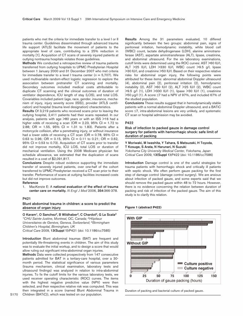
S170
patients who met the criteria for immediate transfer to a level I or IItrauma center. Guidelines disseminated through advanced traumalife support (ATLS) facilitate the movement of patients to theappropriate level of care, contributing to a 25% reduction inmortality [1]. Acquisition of CT scans of severely injured patients atoutlying nontrauma hospitals violates those guidelines.Methods We conducted a retrospective review of trauma patientstransferred from outlying hospitals to UPMC Presbyterian Hospitalbetween 1 January 2000 and 31 December 2007 who met criteriafor immediate transfer to a level I trauma center (n = 5,707). Weused multivariable random-effect logistic regression to explore theassociation between pretransfer CT scanning and mortality.Secondary outcomes included medical costs attributable toduplicate CT scanning and the clinical outcomes of duration ofventilator dependence, ICU length of stay (LOS), and total LOS.Covariables included patient (age, race, gender, insurance, mecha-nism of injury, injury severity score (ISS)), provider (ATLS certifi-cation) and hospital (trauma level designation) characteristics.Results Of 3,219 patients who received scans prior to leaving theoutlying hospital, 2,411 patients had their scans repeated. In ouranalysis, patients with age >80 years or with an ISS >16 had ahigher odds of receiving a scan (OR = 2.23, 95% CI = 1.72 to2.88; OR = 1.55, 95% CI = 1.31 to 1.83). Patients after amotorcycle collision, after a penetrating injury, or without insurancehad a lower odds of receiving a CT scan (OR = 0.78, 95% CI =0.63 to 0.96; OR = 0.15, 95% CI = 0.11 to 0.21; OR = 0.62,95% CI = 0.53 to 0.73). Acquisition of CT scans prior to transferdid not improve mortality, ICU LOS, total LOS or duration ofmechanical ventilation. Using the 2008 Medicare physician feeschedule allowance, we estimated that the duplication of scansresulted in a cost of $2,091,817.Conclusions Despite robust evidence supporting the immediatetransfer of severely injured patients, over one-half of the cohorttransferred to UPMC Presbyterian received a CT scan prior to theirtransfer. Performance of scans at outlying facilities increased costsbut did not improve outcomes.Reference1. MacKenzie E: A national evaluation of the effect of trauma
center care on mortality. N Engl J Med 2006, 354:366-378.
P421Blunt abdominal trauma in children: a score to predict theabsence of organ injury
O Karam1, O Sanchez2, B Wildhaber2, C Chardot3, G La Scala2
1CHU Sainte-Justine, Montreal, QC, Canada; 2HôpitauxUniversitaires de Genève, Geneva, Switzerland; 3BirminghamChildren’s Hospital, Birmingham, UKCritical Care 2009, 13(Suppl 1):P421 (doi: 10.1186/cc7585)
Introduction Blunt abdominal traumas (BAT) are frequent andpotentially life-threatening events in children. The aim of this studywas to evaluate the initial workup, and to design a score that wouldallow ruling out significant intra-abdominal organ injuries.Methods Data were collected prospectively from 147 consecutivepatients admitted for BAT in a tertiary-care hospital, over a 30-month period. The statistical significance of various parameters(trauma mechanism, clinical examination, laboratory tests andultrasound findings) was analyzed in relation to intra-abdominalinjuries. To fix the cutoff limits for the various laboratory tests, weused receiver operating characteristic (ROC) curves. The itemswith the highest negative predictive value (NPV) were thenselected, and their respective relative risk was computed. This wasthen integrated in a score (named Blunt Abdominal Trauma inChildren (BATiC)), which was tested on our population.
Results Among the 31 parameters evaluated, 10 differedsignificantly between the two groups: abdominal pain, signs ofperitoneal irritation, hemodynamic instability, white blood cell(WBC) count, lactate dehydrogenase (LDH), alanine aminotrans-ferase (AST), aspartate aminotransferase (ALT), lipase, creatinine,and abdominal ultrasound. For the six laboratory examinations,cutoff limits were determined using the ROC curves: AST >60 IU/l,ALT >25 IU/l, LDH >330 IU/l, WBC count >9.5 g/l, lipase>30 IU/l, and creatinine >50 IU/l. Based on their respective relativerisks for abdominal organ injury, the following points wereattributed for these items: abnormal abdominal Doppler ultrasound(4), abdominal pain (2), peritoneal irritation (2), hemodynamicinstability (2), AST >60 IU/l (2), ALT >25 IU/l (2), WBC count>9.5 g/l (1), LDH >330 IU/l (1), lipase >30 IU/l (1), creatinine>50 μg/l (1). A score ≤7 had a NPV of 97%, and included 67% ofthe studied population.Conclusions These results suggest that in hemodynamically stablepatients with a normal abdominal Doppler ultrasound, and a BATiCscore ≤7, intra-abdominal lesions are very unlikely, and systematicCT scan or hospital admission may be avoided.
P422Risk of infection to packed gauze in damage controlsurgery for patients with hemorrhagic shock: safe limit ofduration of packing
Y Moriwaki, M Iwashita, Y Tahara, S Matsuzaki, H Toyoda, T Kosuge, S Arata, N Harunari, N SuzukiYokohama City University Medical Center, Yokohama, JapanCritical Care 2009, 13(Suppl 1):P422 (doi: 10.1186/cc7586)
Introduction Damage control is one of the useful strategies fortrauma patients with hemorrhagic shock and critically ill patientswith septic shock. We often perform gauze packing for the firststep of damage control (damage control surgery). We are anxiousabout infection of packed gauze, and some experts said that weshould remove the packed gauze within 48 to 72 hours. However,there is no evidence concerning the relation between duration ofpacking and risk of infection of the packed gauze. The aim of thisstudy is to clarify this relation.
Critical Care March 2009 Vol 13 Suppl 1 29th International Symposium on Intensive Care and Emergency Medicine
Figure 1 (abstract P422)
Duration of packing and bacterial culture of packed gauze.

S171
Methods A prospective observational case-series study. Weperformed bacterial culture of the removed gauze in patients whounderwent gauze packing in damage control surgery and removalof the packed gauze. We examined the relation between theduration of packing and risk of infection of the packed gauze.Results Twenty-one patients were enrolled. The duration ofpacking ranged from 19 to 146 hours. Bacterial culture wasnegative in 52% of cases and positive in 48% of cases. Bacterialculture was positive in all cases in whom the duration of packingwas over 100 hours. The duration of packing was longer in positivebacterial culture cases than in negative (no statistical difference).More cases were positive in bacterial culture in cases withgastrointestinal perforation (GIP) than without GIP (no statisticaldifference). However, many cases with GIP were obligated tocontinued packing for a long duration. There was no relationbetween bacterial culture and preoperative and operative bleeding(Figure 1).Conclusions Gauze packing in damage control surgery is thoughtto be safe within 96 hours from the viewpoint of infection. Wecannot determine the relation between bacterial infection and GIP.
P423Comparison between open and open-vacuum-assistedclosure treatment in the adjuvant treatment of theabdominal compartment syndrome
A Nella1, A Di Filippo1, S Matano1, M Bonizzoli2, M Linden2, G Manca2, A Peris2
1University of Florence, Italy; 2Careggi Teaching Hospital, Florence,ItalyCritical Care 2009, 13(Suppl 1):P423 (doi: 10.1186/cc7587)
Introduction Abdominal compartment syndrome (ACS) representsa potentially life-threatening condition in critically ill patients. Thecontrol of intra-abdominal pressure (IAP) plays a pivotal role inprevention/treatment of ACS. We compared the efficacy of twodifferent therapies – open and open-vacuum-assisted closure(open-VAC) treatment [1] – on resolution of intra-abdominalhypertension/prevention of ACS.Methods We prospectively analyzed data of 26 consecutivepatients admitted to the ICU of our emergency department(Careggi Teaching Hospital, Florence) with sepsis from anabdominal source, hemorrhagic shock, and major trauma. Allpatients underwent laparotomic surgery before ICU admission withopen-VAC treatment. As a control group we retrospective analyzed17 patients admitted with the same diagnosis and treated withopen treatment. IAP was evaluated by measuring the urinarybladder pressure [2]. Patients’ demographic and clinical charac-teristics, and laboratory parameters were collected from the ICUdatabase (FileMaker Pro 5.5v2; FileMaker, Inc., USA) withauthorization of the Careggi Teaching Hospital Committee of theEmergency Department. For each patient, data were collectedbefore the surgical treatment with a closed abdomen (T1), 6 hoursafter ICU admission (T6), after 24 hours (T24), 24 hours afterabdomen closing (T24C), and at monitoring suspension (TMS).Statistical analysis: Student’s t test (*P <0.05). Data reported asthe mean ± SD.Results The two groups were similar in demographic and clinicalcharacteristics. The open-VAC group patients showed a signifi-cantly shorter length of stay in the ICU (open: 20.58 ± 15.97;VAC: 11.46 ± 11.99) and had a shorter time before laparotomyclosure (open: 6.82 ± 3.85; VAC: 4.57 ± 2.88) if compared withthe control group. Finally, both IAP values from T1 (open: 17.9 ±6.41; open-VAC: 15.1 ± 5.68) to T24C (open: 15.7 ± 3.4; open-VAC: 12.6 ± 2.63) and lactate levels from T1 (open: 6.34 ± 4.65;
open-VAC: 4.1 ± 3.1) to T6 (open: 5.72 ± 5.71; open-VAC:2.62 ±2.08) and T24 (open: 5.66 ± 5.81; open-VAC: 2.15 ± 2.12) weresignificantly lower in a time-dependent manner in the open-VACgroup.Conclusions The present study suggests that the open-VACcould have more beneficial effects than the traditional opentechnique in the treatment of patients with intra-abdominalhypertension/ACS.References1. Fred AL, et al.: In Acute Care Surgery Principles and Practice.
Edited by Britt LD. New York: Springer; 2007:176-185.2. Ejike CJ, et al.: What is the normal intra-abdominal pres-
sure in critically ill children and how should we measureit? Crit Care Med 2008, 36:2157-2162.
P424Upper gastrointestinal tract bleeding in the ICU
P Araujo, C Soriano, M Oliveros, A Marban, R Fdez Tajuelo, J Manzanares, M Mateo, E Martin, MJ LendínezH.U. La Paz, Madrid, SpainCritical Care 2009, 13(Suppl 1):P424 (doi: 10.1186/cc7588)
Introduction The aim of this study was to analyze the incidence,features and consequences of upper gastrointestinal tract bleeding(UGITB) in patients admitted to an ICU in a 6-year period.Methods A retrospective observational study in an ICU of a universityhospital. We recorded the UGITB episodes of all admissions (6,245patients) from January 2001 to December 2007 excluding those inwhom the UGITB motivated the admission, or it occurred within thefirst 48 hours. UGITB was defined as: melena and or nasogastrictube bleed with or without anemia, or anemia with a concomitantlesion on G-scope. All patients were on UGITB prophylaxis.Demographic data, APACHE II score, previous medical history andrisk factors for the development of UGITB were recorded. Also theinformation regarding G-scope findings, type of treatment receivedand consequences of the episode were recorded.Results We included a total of 73 patients (1.17%), 46 male(63%). The average APACHE II score was 22 – global mortality of49%. The relative incidence distributed for age groups showsmore incidence in the 41 to 50 years group. Six patients were onoral anticoagulation (8.2%), 14 on platelet anti-aggregation(19.1%), and one patient on low molecular weight heparin (1.4%).There were 11 patients with chronic liver disease (15% and 3.3%of total admissions (TA), three with previous history of UGITB(4.1% and 1.93% TA), four with peptic ulcer disease (5.5% and1.16% TA) and two patients with inflammatory bowel disease(2.8%). We analyzed: organ failure, sepsis, coagulopathy andnutrition, and we found an important association of UGITB withsepsis, shock, organ failure and mechanical ventilation nutrition [1].Presentation of UGITB: hematemesis/bleeding nasogastric tube52 (71.2%), melenas 10 (13.7%) and acute anemia 11 (15%).Endoscopic findings: ulcers 43%, erosions 37%, esophagitis andgastritis 10%. G-scope was performed in 54 patients (74%): 12 ofthem (16.4%) required sclerosis, one patient (1.4%) a Sengs-taken-Blakemore tube, and two patients (2.8%) arteriography withembolization. Most of the episodes resolved spontaneously. Theoverall mortality was 49.3% (36 patients); 1.1% of deaths weredirectly related to the UGITB.Conclusions In our series UGITB is not a common entity, and themost frequent findings were ulcers and erosions. UGITB must beconsidered a new severity criterion because although it is neitherrelated with the typical predisposing factors nor age, in thesepatients we observed higher mortality than the expected for theirAPACHE II score.
Available online http://ccforum.com/supplements/13/S1

S172
Reference1. Vartic M, et al.: Grastrointestinal bleeding in intensive care.
Chirurgia (Bucur) 2006, 101:365-374.
P425A circulating factor modulates apoptosis of lymphocytesand monocytes after multiple trauma
A Kotsaki1, A Savva1, M Mouktaroudi1, T Kanni1, E Giamarellos-Bourboulis1, K Kotzampassi21Attikon University Hospital, Athens, Greece; 2University ofThessaloniki, GreeceCritical Care 2009, 13(Suppl 1):P425 (doi: 10.1186/cc7589)
Introduction The advent of multiple trauma is accompanied bylymphopenia often due to apoptosis. This may be due to theexistence of a circulating factor modulating apoptosis. Thepresence of such a factor in the serum of patients was assessed.Methods Serum was sampled at standard time intervals from 21patients bearing multiple injuries and admitted to the ICU.Peripheral blood mononuclear cells (PBMCs) were isolated fromfive healthy volunteers after gradient centrifugation over Ficoll.PBMCs were stimulated overnight in a 96-well plate with 0.1 mlsampled serum. The percentage of annexin-V+/PI–/CD4+ cells andannexin-V+/PI–/CD14+ cells were estimated by flow cytometry.Results were compared with those obtained after cell incubationwith serum of healthy volunteers.Results Median induction of apoptosis of CD4 lymphocytes and ofCD14 monocytes after incubation with serum of healthy volunteerswas 1.89% and 30.56%, respectively. Median percentages ofapoptotic CD4 lymphocytes and CD14 monocytes of PBMCs afterincubation with patient’s serum sampled on consecutive days afteradmission are shown in Table 1. Similar results for patients compli-cated with sepsis on the first day of its advent are also presented.
Table 1 (abstract P425)
Induction of apoptosis of PBMCs after incubation with patients’serum
Advent of Median Day 1 Day 4 Day 7 sepsis
CD4 cells (%) 3.57* 4.04 3.57* 3.48*
CD14 cells (%) 3.00* 41.43 3.00 33.3
*P <0.05 compared with incubation with patients’ serum.
Conclusions Incubation with serum of patients with multipletrauma-induced apoptosis of CD4 lymphocytes and of CD14monocytes. These data strengthen the hypothesis for the existenceof a circulating factor inducing apoptosis after multiple trauma.
P426Abstract withdrawn
P427Treatment with p-hydroxyphenylpyruvate in the absence ofresuscitation extends survival in rats subjected to lethalblood loss
A Cotoia, JV Gefter, RL DeludeUniversity of Pittsburgh, PA, USACritical Care 2009, 13(Suppl 1):P427 (doi: 10.1186/cc7591)
Introduction We tested the hypothesis that treatment with anenone-containing compound, p-hydroxyphenylpyruvate (pHPP), the
first intermediate in the tyrosine catabolism pathway, couldameliorate the survival time in rats subjected to profound hemor-rhagic shock (HS) in the absence of resuscitation. We based thishypothesis on the knowledge that ethyl pyruvate (EP) and relatedenone-containing compounds are protective in numerous in vitroand in vivo models of oxidant-mediated and cytokine-mediatedcellular and organ injury.Methods We compared the anti-inflammatory effects of EP andpHPP using U373 cells exposed to IL-1β and RAW 264.7 cellsexposed to lipopolysaccharide. Volume-controlled HS was initiatedby withdrawing 21.7 ml blood per kg body weight over 20 minutes,followed by an additional 14 ml/kg over 40 minutes. A continuousinfusion was delivered during the final 20 minutes of hemorrhage.Groups of six rats randomly received: (1) 2.8 ml saline per kg or4% of total estimated blood volume (EBV); (2) 2.8 ml/kg of 0.25nM pHPP; (3) 1.4 ml/kg of 0.50 nM pHPP; (4) 1.4 ml/kg (2% EBV)of 0.25 nM homogentisic acid (HGA); (5) 1 g dichloroacetic acid(DCA) in 2.8 ml/kg; or (6) DCA plus 0.25 nM pHPP in 2.8 ml/kg.Results Cell culture studies revealed that pHPP blockedexpression of CD54 in U373 cells and blocked release of NO• anddecreased induction of inducible nitric oxide synthase mRNA inRAW cells at lower concentrations compared with EP. Wetherefore tested its efficacy in a profound hemorrhagic shockmodel. Rats in both groups receiving pHPP survived significantlylonger (P <0.001) than rats receiving vehicle alone. All rats in thevehicle group were dead by 140 minutes. Five out of 12 animalstreated with pHPP survived longer than 4 hours and one ratsurvived for 455 minutes. Prompted by the recognition that pHPPis an endogenous metabolite converted to HGA in the tyrosinecatabolism pathway, we sought to determine whether treatmentwith HGA could extend survival in the same HS model. Thepercentage of treated rats that survived until 5 hours in groups 2 or4 was 33.3%. No statistical significance was observed among ratsin groups 2, 3, 4. In order to obtain additional information regardingthe mechanism responsible for the protective effects, weadministered an inhibitor of the tyrosine catabolism, dichloroaceticacid alone or in addition to pHPP. Accordingly, the data showedthat all rats in groups 5 or 6 died within 4 hours.Conclusions These findings show that administration of ananomolar dose of pHPP markedly improves survival followinglethal HS. This improvement is very probably related to a directpharmacologic effect of pHPP on inflammation that is shared withEP and together with pHPP acting as an anaerobic carbon source.Infusion of drug in only 2% of the EBV strongly suggests that theseeffects are not related to resuscitation.
P428Guidelines-associated decrease in mortality withrecombinant activated factor VII for refractory hemorrhage
AS Schneider, P Voirol, M Perez, P Schoettker, JB Wasserfallen, A Angelillo-Scherrer, R Chiolero, J Cotting, P EggimannCHUV, Lausanne, SwitzerlandCritical Care 2009, 13(Suppl 1):P428 (doi: 10.1186/cc7592)
Introduction Recombinant activated factor VII (rFVIIa; Novoseven)is indicated for treatment of bleeding in hemophilic patients withantibodies against factor VIII or factor IX. Although not licensed oragreed for other indications, its use for refractory hemorrhage isincreasingly reported. We reviewed our experience with off-labeluse of rFVIIa in this indication.Methods We retrospectively reviewed all patients treated withrFVIIa from March 2004 to November 2008 in our hospital. Thoseincluded in a multicentric study (n = 10) or treated within approved
Critical Care March 2009 Vol 13 Suppl 1 29th International Symposium on Intensive Care and Emergency Medicine

S173
(n = 3) or nonhemorrhage (n = 4) indications were excluded. Initialuse imposed constringent guidelines implementation. According tothese, use of rFVIIa requires cross-checking for active correction ofhypothermia, acidosis, and coagulation factors and considerationfor any feasible haemostatic surgery or embolisation.Results Forty-two patients were included. Median age was 39years and 60% were male. The underlying condition was trauma in14% of cases and surgery-associated hemorrhage in 76%.Bleeding stopped in 79% of cases. Potentially associatedthromboembolic complications were observed in five (11.9%)cases, only one being very probably linked to rFVIIa. The overall 30-day mortality was 40.5%. The initial experience was followed by adecline in the use of rFVIIa, and the release of guidelines followedby a continuous increase. We observed a progressive decline inthe 30-day mortality rate, from 70% in the first 10 patients to 17%in the last 10 (Figure 1). The average number of blood transfusionsbefore rFVIIa remained stable but the median rFVIIa dosesdecreased from 206 μg/kg in the first 10 patients to 80 μg/kg inthe last 10.Conclusions Local guidelines release did not decrease thenumber of cases but seem to impact on when and to whom rFVIIais administered. Their release, associated with increasing experi-ence, was associated with a striking lowering of mortality rate.
P429Recombinant activated factor VII: does it really save lives?
U EdkeShaikh Khalifa Medical City, Abudhabi, United Arab EmiratesCritical Care 2009, 13(Suppl 1):P429 (doi: 10.1186/cc7593)
Introduction At pharmacological doses, recombinant activatedfactor VII (rFVIIa) bypasses conventional steps in coagulationcascade and directly generates a thrombin burst on the activatedplatelets at the injury, leading to the generation of fully stabilizedtight fibrin clot.Methods Using the experience of our institute and an Internetsearch. In one of the largest prospective, randomized, multicentricstudies of rFVIIa, 301 patients with blunt/penetrating trauma wereenrolled – of whom 277 patients at the end of study wereanalyzable. The trial included 399 patients, all diagnosed by CTscan within 3 hours of intracerebral hemorrhage onset. Patientswere randomly assigned to receive placebo (n = 96), 40 μg/kg(n = 108), 80 μg/kg (n = 92), or 160 μg/kg (n = 103) doses ofrFVIIa within 1 hour of the baseline scan. The results of the Survivalof Myocardial Infarction Long-Term Evaluation study (that is, the
largest controlled study showing the relationship between geneticpolymorphisms and disease) demonstrated that propensity to highfactor VII levels is not associated with a risk for myocardialinfarction.Results One study has demonstrated that one single dose ofrFVIIa (80 μg/kg) could reduce transfusion requirements incirrhotic patients undergoing orthotopic liver transplant. Comparedwith 29% growth in the placebo arm, intracerebral hemorrhagevolume growth in the 40, 80, and 160 μg/kg arms was 16%, 14%,and 11%, respectively (corresponding to a relative reduction of45%, 52% and 62%).Conclusions Use of rFVIIa should be reserved for salvageablepatients with coagulopathic bleeding that is unresponsive toexisting medical therapies. Definitive surgical control of accessiblesites of hemorrhage is nevertheless a necessary intervention.References1. Hendriks HG, Meijer K, et al.: Reduced transfusion require-
ments by rFVIIa in orthotopic liver transplantation. Trans-plantation 2001, 71:402-405.
2. Mayer SA, Brun NC, et al.: rFVIIa for acute intracerebralhemorrhage. N Engl J Med 2005, 352:777-785.
P430Recombinant activated factor VII for the treatment ofbleeding in abdominal surgery and cardiac surgery
G Michalska, R StanekClinical Hospital Nr1, Szczecin-Police, PolandCritical Care 2009, 13(Suppl 1):P430 (doi: 10.1186/cc7594)
Introduction Recombinant activated factor VII (rFVIIa) plays a keyrole in hemostasis. The aim of this study was the clinical evaluationof the efficacy of rFVIIa in the treatment of bleeding during andafter abdominal and cardiac surgery.Methods A meta-analyses of case-series studies (n = 67) on thetreatment of bleeding with rFVIIa regarding reduction or cessationof bleeding and mortality. We used the questioners of NovoNordisk to assess the indications and effectiveness of treatment.We compared the amount of blood loss within 12 hours beforeand within 12 hours after giving rFVIIa, and the dynamics ofbleeding. In 10 patients the bleeding started after surgeryintraoperatively, in 57 patients severe hemorrhage developed in thepostoperative period. We also compared the full blood count andlaboratory coagulation profile parameters before treatment, and 2and 12 hours after treatment. In cardiac surgery rFVIIa wasadministered 5 to 49 minutes after neutralization of heparin withprotamine sulfate. The dosage of rFVIIa was 39.23 ± 20.70 μ/kgWe used Student’s t test for statistical analysis.Results After administration of the first median dose (14.45 to81.35 μg/kg) of rFVIIa, bleeding stopped in 47 patients. Bleedingwas markedly decreased in 15 patients. Five patients who did notbenefit from initial rFVIIa administration received additional drug ina dose of 38.25 μg/kg with good results. The average blood losswithin 12 hours before treatment was 2,510.00 ± 1,642.07 ml,and the average blood loss within 12 hours after treatment was1,057.75 ± 810.67 ml. The average dynamics of bleeding beforetreatment were 216.35 ± 138.82 ml/hour, and 87.90 ± 67.82 ml/hourafter treatment. Transfusion requirements were reduced for packedred blood cells, fresh frozen plasma, platelets, and crystalloidand/or colloids. Reduction in transfusion requirements wasstatistically significant (P ≤0.05).Conclusions The meta-analysis of case series showed that in amean of 85% patients rFVIIa achieved at least a reduction ofbleeding, reducing the need for hemotransfusions.
Available online http://ccforum.com/supplements/13/S1
Figure 1 (abstract P428)
Thirty-day mortality after rFVIIa.
S174
P431Safety of activated recombinant factor VII in patients withcirculatory assist devices
AS Schneider, P Tozzi, P Voirol, P Schoettker, MH Perez, A Angelillo-Scherrer, J Wasserfallen, R Chiolero, J Cotting, L Von Segesser, P EggimannCHUV, Lausanne, SwitzerlandCritical Care 2009, 13(Suppl 1):P431 (doi: 10.1186/cc7595)
Introduction Recombinant activated factor VII (rFVIIa; Novoseven)is indicated for treatment of bleeding in hemophilic patients withantibodies against factor VIII or factor IX. Although not licensed oragreed for other use, it is increasingly reported for refractoryhemorrhage. Implantation of a circulatory assist device requirestight anticoagulation therapy and may be complicated by massivebleeding. We report our experience with off-label use of rFVIIa innine patients with circulatory assistance.Methods We retrospectively reviewed all patients treated withrFVIIa for massive bleeding secondary to anticoagulation duringcirculatory assistance between March 2004 and November 2008.rFVIIa use and dosage was decided according to local guidelineswhen available (released in May 2006). According to theseguidelines, systematic crosscheck for active correction of hypo-thermia, acidosis, and coagulation factors with parallel considera-tion for any feasible interventional embolisation or haemostaticsurgery are required for authorization of rFVIIa use.Results Nine patients with circulatory assistance received rFVIIa.Median patient age was 43 years (<1 to 55). Eight patientsunderwent cardiac surgery (including two heart transplants). Onehad a bipulmonary transplant. The device was an extracorporealmembrane oxygenator in seven patients and a ventricular assistdevice in two patients. Median rFVIIa dose was 100 μg/kg (45 to180), split into two or three injections in six cases. Bleeding controlwas obtained in five patients (60%). The mortality rate at 30 dayswas 40%, including two within 48 hours from failure to controlhemorrhage. We observed one thromboembolic complication(stroke) which may potentially be related to rFVIIa administration.Conclusions Our series suggests that the cautious use of rFVIIamay be an option in patients with a circulatory assist devicesuffering from massive hemorrhage refractory to conventionaltreatment.
P432Analysis of the off-licence use of recombinant activatedfactor VII for patients with uncontrolled haemorrhage in aUK tertiary referral hospital
M Spivey, H Eve, M DuffyDerriford Hospital, Plymouth, UKCritical Care 2009, 13(Suppl 1):P432 (doi: 10.1186/cc7596)
Introduction Recombinant activated factor VII (rFVIIa) isincreasingly used for the treatment of acquired coagulopathiesassociated with uncontrolled haemorrhage. Consensus guidelines[1] have been published for the administration of rFVIIa in thissetting. We examined the off-licence use of rFVIIa in our 1,071-bedtertiary referral hospital.Methods Hospital blood-bank data were examined from the first off-licence use of rFVIIa in November 2002 until August 2008. Casenotes were reviewed on these patients to gain demographics,indications and dosage of rFVIIa. Pathology data were cross-referenced to gain results before and after administration of rFVIIa.Results Twenty-two patients were identified over the 6-year periodwho received off-licence rFVIIa. The majority of uses were in
cardiac surgery (nine patients), followed by general/vascularsurgery (five patients), and trauma (four patients). Of thesepatients, 41% (nine patients) did not survive >24 hours. Meandosage was 7.8 mg (89.4 μg/kg) and was given after meantransfusions of 24.8 units of packed red cells, 13 units of freshfrozen plasma, 3.5 units of pooled platelets and 2 units ofcryoprecipitate, in the preceding 24 hours. At this time, blood testsreveal a mean of 91 platelets (SD = 54.9), but nearly one-quarterof patients received the drug with a platelet count <50;prothrombin time 17.9 seconds, activated partial thromboplastintime 58.7 seconds, and pH 7.18. Mean temperature prior to rFVIIaadministration was 34.7°C. Full APACHE II data were available for64% of the patients with a mean of 18.3 (SD = 6.5), and for thetrauma patients a mean revised trauma score of 5.9. Following theadministration of rFVIIa, the mean prothrombin time reduced to13.4 seconds at 1 hour. Sixty-eight per cent of patients (15)received blood product transfusions in the immediate 24 hoursafter having rFVIIa with a mean of 2 units packed red cells, 3.2units fresh frozen plasma, 1.5 units pooled platelets and 1.1 unitscryoprecipitate.Conclusions In our establishment, off-licence use of rFVIIa hasoften been unsuccessful and may have been used as salvagetherapy. There is heterogeneity in prescribing rFVIIa and its usedoes not conform to European guidelines [1]. Controversycontinues about the merits of using off-licence rFVIIa at all [2].References1. Vincent JL, et al.: Recommendations on the use of recom-
binant activated factor VII as an adjunctive treatment formassive bleeding – a European perspective. Crit Care2006, 10:R120.
2. Stanworth S, et al.: Recombinant factor VIIa for the preven-tion and treatment of bleeding in patients without haemo-philia. Cochrane Database Syst Rev 2007, 2:CD005011.
P433Treatment of obstetric hemorrhage with recombinantactivated factor VII
P Czuprynski, G MichalskaClinical Hospital Nr1, Szczecin-Police, PolandCritical Care 2009, 13(Suppl 1):P433 (doi: 10.1186/cc7597)
Introduction Massive postpartum bleeding remains a major causeof maternal morbidity and death. An estimated 90% of such deathsmight be preventable.Methods rFVIIa was used in our ICU in the treatment of 12 womenwith severe or life-threatening postpartum bleeding (PPB) in 2005to 2008 due to placenta previa accrete, rupture of the uterus andpre-eclampsia with haemolysis, elevated liver enzymes and lowplatelets. The average patient age was 30 years. We used thequestioners of Novo Nordisk to assess the indications andeffectiveness of treatment. All patients had normal coagulationparameters before surgery. We compared the amount of bloodloss within 12 hours before and 12 hours after giving rFVIIa, thedynamics of bleeding (assessed in ml/hour) before and aftertreatment and coagulation profile parameters. We used smalldoses of rFVIIa (15.25 to 50 μg/kg). We used Student’s t test forstatistical analysis of the laboratory data prior to and after rFVIIa.Results Indications for administration of rFVIIa were consideredwhen sudden blood loss exceeded 1,800 ml or when continuingblood loss of 500 ml/hour (in the drain). Patients after rFVIIatreatment received a lower number of packed red cell transfusions(4.0 ± 4.46 vs. 9.61 ± 6.7, P = 0.007), compared with beforetreatment. In addition, the median amount of crystalloid/colloidsadministered decreased markedly. After administration, rFVIIa
Critical Care March 2009 Vol 13 Suppl 1 29th International Symposium on Intensive Care and Emergency Medicine

S175
bleeding stopped in eight patients. Bleeding markedly decreasedin two patients during 3 hours. Two patients who did not benefitfrom initial rFVIIa administration received additional drug in a doseof 35.65 μg/kg with good results. No adverse effects were directlyrelated to treatment with rFVIIa.Conclusions Clinical reports and hematologic data suggestimprovement for more than 80% of women after rVIIa administration.
P434Thromboelastography versus viscoelastography forevaluation of coagulopathies in patients after abdominalsurgery for cancer
O Tarabrin, A Vladyka, S Shcherbakov, A Belyakov, A Petelkaki, A BudnyukOdessa Medical University, Odessa, UkraineCritical Care 2009, 13(Suppl 1):P434 (doi: 10.1186/cc7598)
Introduction The study compared the efficacy of viscoelastography(VG) versus thromboelastography (TEG) for monitoring of peri-operative coagulation balance after abdominal surgery for cancer.Methods A complete coagulation screen, the activated clottingtime (ACT), TEG and VG were performed before surgery and atthe end of the operation, and monitoring of LMWH anticoagulationtherapy on postoperative days 1, 2, 3, and 7. We tested thehypothesis that the parallel use of standard TEG and VG can fullyaccess the postoperative state of blood coagulation.Results The elastic shear modulus of standard MA (Gt) andVGMVA (Gh), which reflect the total clot strength and pro-coagulatory protein component, was calculated. The differencewas an estimate of the platelet component (Gp). There was a 14%increase of standard MA, corresponding to a 48% increase of Gt(P <0.05) and an 80 to 86% contribution of the calculated Gp toGt. Using multiple linear regression, all coagulation, TEG and VGvariables were used to model perioperative thrombophylia. Theresults showed that some components of TEG failed to identifyhypercoagulation (r <0.2, P <0.75). However, three componentsof the routine coagulation assay, including the bleeding time,prothrombin time and platelet count, could be modeled to showprolonged postoperative hypercoagulability (P <0.01). Weconclude that all components of the VG test reflect postoperativecoagulopathies. Serial standard TEG and viscoelastic analysis mayreveal the independent contribution of platelets and ofprocoagulatory proteins to clot strength.Conclusions These results suggest that viscoelastic techniquesmight be useful in the determining the coagulation status in cancerpatients perioperatively. Hypercoagulability is not reflectedcompletely by standard coagulation monitoring and TEG, andseems to be predominantly caused by increased platelet reactivity.Reference1. Samama CV: Anesthesiology 2001, 94:74-78.
P435Prediction of long-term survival and coagulation factor VIIIlevels: interactions with gender in a large hospital-basedcohort
F Kovar1, C Marsik-Strasser1, C Joukhadar2, T Schickbauer1, P Kyrle1, O Wagner1, G Endler1
1General Hospital Vienna – AKH, MUW, Vienna, Austria; 2HarvardMedical School, Boston, MA, USACritical Care 2009, 13(Suppl 1):P435 (doi: 10.1186/cc7599)
Introduction Elevated coagulation factor VIII activity (FVIII:C) hasbeen associated with increased risk for both venous and arterial
thrombosis. The current study evaluated the influence of factor VIIIlevels and interactions with gender on all-cause mortality in a largeAustrian cohort.Methods A total of 11,203 individuals were included in this study.The median observation period was 5 years, covering a total of46,000 person-years. The death rate was 17.1%.Results Compared with individuals within the reference category(FVIII:C <94%) hazard ratios gradually increased from 1.4 (95% CI =1.1 to 1.8) in the 152 to 170% category (fifth decile) to finally 4.4(95% CI = 3.5 to 5.5) in the >313% category (highest decile, allP <0.05). The association between FVIII:C levels and mortalityremained essentially unchanged when considering noncancermortality, all-cause vascular mortality or mortality due to ischemicheart disease. Compared with males, females with elevated FVIII:Chad a worse outcome resulting in higher hazard ratios reaching 6.8(95% CI = 4.6 to 9.9) within the highest decile compared withmales (hazard ratio = 3.4, 95% CI = 2.6 to 4.5).Conclusions In our large patient cohort we are firstly able todemonstrate that FVIII:C plasma activity is strongly associated withall-cause mortality. Additionally, FVIII:C appears to interact withgender. Especially in women, FVIII:C might help identifying high-risk cohorts that might benefit from individualized preventionstrategies.
P436Gel point and fractal microstructure of incipient blood clotsare significant new markers of haemostasis
P Evans1, K Hawkins1, R Morris2, N Thirumalai3, R Munro3, L Wakeman3, M Lawrence1, A Beddal3, P Williams1
1Swansea University, Swansea, UK; 2UWIC, Cardiff, UK;3Morriston Hospital, Swansea, UKCritical Care 2009, 13(Suppl 1):P436 (doi: 10.1186/cc7600)
Introduction We have investigated the hypothesis that in wholeblood the incipient clot formed at the gel point (GP) ischaracterised by a fractal microstructure [1,2] and that this could
Available online http://ccforum.com/supplements/13/S1
Figure 1 (abstract P436)

S176
be detected in healthy and anticoagulated blood. We comparedthe gel time (GT) required to form the incipient clot and thecorresponding fractal dimension (df) against laboratory markers ofhaemostasis and thromboelastography (TEG).Methods Blood samples were taken from 52 healthy adults andsimilarly 34 individuals whose blood had been anticoagulated withheparin over the therapeutic range.Results The incipient clot in normal blood is established as asample-spanning network cluster at the GP [2]. The value of df inwhole healthy human blood is 1.74 (±0.07), which indicates a highdegree of branching in the fibrin network at criticality and iscommensurate with that reported to arise in other biologicalsystems. There was a significant reduction in the value of df and acorresponding prolongation in the GT in the heparin group ascompared with the healthy group (Figure 1).Conclusions We describe for the first time that the incipient clotformed at the GP of whole blood is characterised by a fractalmicrostructure. The values of df and GT discriminate between clotstructure in healthy and anticoagulated blood. The relationshipbetween these new markers may provide a basis for exploring therelationship between coagulation pathways and clot quality.References1. Evans PA, et al.: Rheometrical detection of incipient blood
clot formation by Fourier transform mechanical spec-troscopy. J Non Newt Fluid Mech 2008, 148:122-126.
2. Muthukumar M, Winter HH: Fractal dimension of a cross-linking polymer at the gel point. Macromolecules 1986, 19:1284-1285.
P437Acquired bleeding model induced by dilutionalcoagulopathy in the rabbit
I Pragst, B Doerr, F Kaspereit, W Krege, E Raquet, G DickneiteCSL Behring GmbH, Marburg, GermanyCritical Care 2009, 13(Suppl 1):P437 (doi: 10.1186/cc7601)
Introduction Severe traumatic or intraoperative blood lossnecessitates massive transfusion. This blood loss and the dilutionof coagulation factors result in insufficient haemostasis. Treatmentsto enhance the haemostatic capacity need to be evaluated. Anacquired bleeding model due to a dilutional coagulopathy wasdeveloped in the rabbit to estimate the contribution of PCC(Beriplex P/N; CSL Behring) on haemorrhage.Methods Rabbits were anesthetised using isofluran and weremandatorily ventilated. The animals were instrumented to monitorthe cardiovascular and respiratory system. Dilutional coagulopathywas induced by phased blood withdraw, salvaged erythrocyteretransfusion and volume substitution with hydroxyethyl starch.After the dilutional procedure the bleeding was inflicted by cuttingthe lateral kidney pole. To characterise the model, animals wereallocated to groups: I, sham operation (no dilution, placebotreatment; n = 5); II, negative control (dilution, placebo treatment;n = 7); III, PCC 25 U/kg intravenously (dilution, verum treatment;n = 6). Coagulation factor activity, thrombin generation (TGA),prothrombin time (PT) and the bleeding from the kidney woundwere assessed.Results Rabbits without a dilutional coagulopathy demonstratedrapid onset of haemostasis. Post-injury blood loss (BL) and time tohaemostasis (TH) were 4.7 ± 2.4 ml and 4.4 ± 1.1 minutes,respectively. After the dilution procedure coagulation factors werereduced to less than 50% of baseline. Haemostasis deterioratedafter colloid transfusion (BL 60.0 ± 25.9 ml, TH 19.1 ± 2.2 min).
The TGA showed normal lag phase but only 50% of the normalpeak thrombin level. The PT was prolonged after the dilution about1.8-fold. Using PCC treatment it was demonstrated that the peakthrombin level was corrected (P <0.0001) and the BL (21.5 ±11.1 ml; P <0.01) as well as TH (12.4 ± 1.4 min; P <0.01) weredecreased significantly.Conclusions In this rabbit model of a dilutional coagulopathy itwas demonstrated that a dilution of coagulation factors of morethan 50% affects TGA, PT and bleeding. Restoration ofcoagulation factors II, VII, IX and X using PCC corrects the TGAand reduces BL and TH. This model could be used to investigatenew drugs in dilutional coagulopathy.
P438Impact of nebulized unfractionated heparin andN-acetylcysteine in management of smoke inhalation injury
E Elamin1, A Miller2
1University of Florida, Gainesville, FL, USA; 2State University ofNew York Downstate & Kings County Hospital, Brooklyn, NY, USACritical Care 2009, 13(Suppl 1):P438 (doi: 10.1186/cc7602)
Introduction Approximately 70% of all fire victims in the USA whodie within the first 12 hours post burn die from smoke inhalationinjury (SIJ). Airway edema combined with obstructive castsproduced from cellular debris, fibrin clots, polymorphonuclearleukocytes, mucus and mucin B5 are believed to cause the airwayobstruction contributing to pulmonary failure.Methods A single-center retrospective study with historical controlover a 5-year period of 30 mechanically ventilated adult subjectsadmitted within 48 hours of their bronchoscopy with confirmed SIJ.Both the experimental (n = 16) group and control (n = 14) groupswere treated with ventilator support but the former received inaddition 10,000 units of nebulized heparin sulfate mixed in 3 mlnormal saline every 4 hours and 3 ml 20% nebulized N-acetylcysteine plus 0.5 ml albuterol sulfate every 4 hours for 7consecutive days starting on the day of admission. We calculatedAPACHE III scores on admission in addition to the daily lung injuryscore for 7 days.Results There was no significant difference in admission APACHEIII scores (38.7 vs. 34.6; α = 0.05) or lung injury scores (0.7 vs.1.1; α = 0.05) between the experimental and control groups. Theexperimental group showed significant improvement in lung injuryscores (P <0.05) over the duration of the study, with the greatestbenefit seen over the first 7 days (0.91 ± 0.14 vs. 1.79 ± 0.41;P <0.01). Respiratory resistance and compliance measurementsrevealed significant improvement in the experimental groupreaching a significance level of P <0.01 by day 4. There was astatistically significant survival benefit in the experimental groupthat was most pronounced in patients with APACHE III scores>35. Survival for the control group versus experimental group was0.5714 ± 0.1497 versus 0.9375 ± 0.0605, respectively (RR =–0.0055; 95% CI = –0.0314 to 0.0204; HR = 1.003).Conclusions The use of aerosolized unfractionated heparin and N-acetylcysteine attenuates lung injury and the progression of acutelung injury in ventilated adult patients with SIJ.References1. Desai MH, Mlcak R, Richardson J, et al.: J Burn Care Rehabil
1998, 19:210-212.2. Tasaki O, Mozingo DW, Dubick MA, et al.: Crit Care Med
2002, 30:637-643.
Critical Care March 2009 Vol 13 Suppl 1 29th International Symposium on Intensive Care and Emergency Medicine

S177
P439Anti-activated factor X response to enoxaparin in criticallyill patients
S Einav, I Dezigibker, M Naamad, M Hersch, D Bitran, D Shemesh, S ZevinShaare Zedek Medical Center, Jerusalem, IsraelCritical Care 2009, 13(Suppl 1):P439 (doi: 10.1186/cc7603)
Introduction Low molecular weight heparin (LMWH) dosingprotocols for deep vein thrombosis prophylaxis in critically illpatients (CIPs) are not based on pharmacologic studies in thesepatients. Antithrombotic LMWH activity correlates with peak anti-activated factor X (aFXa) levels (4 hours post injection) of 0.1 to0.2 IU/ml. The hypothesis of this study is that most CIPs do notachieve therapeutic levels for prevention of DVT.Methods A prospective study of CIPs randomized to receiveenoxaparin once daily intravenously 0.5 mg/kg, subcutaneously0.5 mg/kg or subcutaneously 40 mg. Blood was sampled for peakand trough aFXa levels (5 days). Inclusion criteria: CIPs aged≥18 years, expected to require mechanical ventilation >3 days.Exclusion criteria: full anticoagulation, platelets <75,000, creatinineclearance <30 ml/min/m2, BMI >30, International Normalized Ratio>1.7, contraindication to anticoagulation. Outcome measures:primary, achievement of target peak/trough levels of aFXa;secondary, monitoring the effect of concomitant administration ofnoradrenalin on aFXa levels.Results Included were 35 patients (5, 15 and 15 accordingly).Mostly male (n = 23, 66%), nonsmokers (n = 23,66%) aged 68 ±15 years (range 25 to 92) with a BMI of 25 ± 3 (range 17 to 30).Most were admitted for diseases of the digestive/respiratorysystems (ICD-9). APACHE scores were 19 ± 4 (range 12 to 29).Forty percent underwent surgery before ICU admission (n = 14).Upon inclusion, 34% (n = 12) were receiving noradrenalin, bloodpressures averaged 83 ± 6 mmHg, blood creatinine 1.08 ± 0.27,platelets 206,942 ± 95,128 and International Normalized Ratio1.3 ± 0.16. No protocol resulted in aFXa levels within thetherapeutic range; peak levels were too high and trough levels toolow. The only significant difference between patients receiving/not-receiving noradrenalin was found in the trough levels of the SQ0.5 mg/kg protocol. Hemorrhagic/thorombotic complicationsincluded: one gastrointestinal bleeding (SQ 40 mg) and two distalinferior limb DVTs (SQ 0.5 mg/kg and intravenous 0.5 mg/kgprotocols). ICU length of stays averaged 9 ± 6 days (median 8,range 4 to 33). Three patients died for causes seemingly unrelatedto the study (overwhelming fungal sepsis, severe pneumonia andmalignant reperfusion syndrome post carotid endarterectomy).Conclusions Current DVT prophylaxis protocols do not result inpeak aFXa levels within the recommended antithrombotic range inCIPs. This suggests subtherapeutic levels of LMWH activity andshould lead to further studies on optimal dosing in these patients.
P440Effect of amniotic fluid on blood coagulation
H Hayami, S Ohama, A Sakurai, J Yamada, O Yamaguchi, Y Koide, M OkudaYokohama City University Medical Center, Yokohama, JapanCritical Care 2009, 13(Suppl 1):P440 (doi: 10.1186/cc7604)
Introduction Amniotic fluid embolism (AFE) is a catastrophiccomplication of pregnancy. The incidence of AFE is very rare, andthe clinical presentation; central hemodynamic data in humans aretypically obtained more than 1 hour after the event and from onlythe subset of women who have survived the initial insult. This
makes the exact pathophysiology remain elusive, despite numerousattempts at understanding AFE syndrome. We tried to evaluate theeffect of amniotic fluid on blood coagulation by making a simple in-vitro model, using an activated clotting time (ACT) monitor.Methods Sixteen healthy women undergoing elective cesareansection under spinal anesthesia. A venous sampling line wasinserted before induction of anesthesia. After artificial rupture ofthe membrane, amniotic fluid was carefully obtained by thesurgeon. At the same time, 10 ml venous blood was obtained fromthe sampling line, and two series of samples were made; (1) 5 mlvenous blood with 0.1 ml amniotic fluid (group 1), and (2) 5 mlvenous blood alone (group 2 = control). ACT was measured using2 ml sample in each group.Results The ACT was 66.5 ± 20.9 seconds in Group 1 versus102.4 ± 15.8 seconds in Group 2 (P <0.0001). Especially, therewas one case that coagulated immediately after mixing theamniotic fluid and the ACT could not be obtained in Group 1(excluded from analysis). The strong coagulation ability by amnioticfluid was induced immediately, even in very small doses.Conclusions A dose of 0.1 ml was enough to form the 5 mlvenous blood thrombosis, which is thought to be a sufficientamount to cause AFE, and we consider that its pathophysiologyresembles pulmonary embolism. We suggest that AFE may beinduced by a smaller dose such as 0.1 ml amniotic fluid than it hadbeen thought. A part of the mechanism of amniotic fluid embolismin humans can be explained by pulmonary embolism theory bymaking a simple in vitro AFE model.
P441Is it safe to thrombolyse patients with pulmonaryembolism and right heart mobile thrombi?
J Belohlavek, V Dytrych, A Kral, O Smid, A LinhartGeneral Teaching Hospital, Prague, Czech RepublicCritical Care 2009, 13(Suppl 1):P441 (doi: 10.1186/cc7605)
Introduction Right heart mobile thrombi (RHMT) are not frequent inpatients with acute pulmonary embolism (APE); however, bear asignificant risk of APE recurrence and hemodynamic compromise.Immediate therapy is recommended, but the optimal approach isconsidered to be controversial in the absence of controlled trials [1].Methods We report an analysis of 21 out of 209 (10%)consecutive patients with echocardiographically diagnosed RHMTmanaged at our institution during the past 5 years. For demo-graphics, vital signs and laboratory values on entry see Table 1.Results Eight (38%) out of 21 patients with RHMT presented withmassive, 12 (57%) submassive and one with small APE. Rightventricle overload was detected in 20 out of 21 patients (95%).Fifteen (71%) patients were thrombolysed, three (14%) patientshave undergone surgical embolectomy. Thrombolysis wassuccessful in 13 out of 15 (87%) patients, RHMT was dislodged intwo patients; in one of them with fatal outcome, the other one was
Available online http://ccforum.com/supplements/13/S1
Table 1 (abstract P441)
With RHMT Without RHMT P value
n 21 188Age 57.6 57.1 NSWomen 11 101 NSSystolic blood pressure 105.6 124.3 <0.001Heart rate 106.1 92.5 0.02Tropinin I 0.396 0.186 0.03Tropinin I maximal 2.094 0.436 0.005Brain natriuretic peptide 873 287 0.025

S178
urgently managed by venoarterial extracorporeal membraneoxygenation and subsequently by surgical embolectomy. The 30-day mortality was 10% (n = 2) in RHMT and 6% (n = 11 out of188) in patients without RHMT.Conclusions Systemic thrombolysis is a safe and effectivetreatment for APE with RHMT. Urgent venoarterial extracorporealmembrane oxygenation as a rescue option for hemodynamiccollapse in thrombus dislodgment opens a new therapeutic bridgeto further decision and treatment.Reference1. Torbicki A, et al.: Guidelines on the diagnosis and manage-
ment of acute pulmonary embolism: the Task Force forthe Diagnosis and Management of Acute PulmonaryEmbolism of the European Society of Cardiology (ESC).Eur Heart J 2008, 29:2276-2315.
P442Argatroban for heparin-induced thrombocytopenia therapyin ICU patients with sepsis or multiple organ dysfunctionsyndrome
B Saugel, W Huber, RM SchmidKlinikum Rechts der Isar der Technischen Universität München,GermanyCritical Care 2009, 13(Suppl 1):P442 (doi: 10.1186/cc7606)
Introduction Heparin-induced thrombocytopenia (HIT) is a serious,prothrombotic, immune-mediated adverse reaction triggered byheparin therapy. When HIT is diagnosed or suspected, heparinsshould be discontinued and an alternative, fast-acting, parenteral,nonheparin anticoagulation such as argatroban should be initiated.The recommended initial dose of argatroban in HIT is 2 μg/kg/minute with following adjustment to the activated partial thrombo-plastin times (aPTTs). There are limited and inconsistent dataabout dosing of argatroban therapy in ICU patients with multipleorgan dysfunction syndrome [1,2].Methods Evaluation of 11 patients with sepsis or multiorgan dys-function syndrome treated with argatroban to demonstrate dosingadjustment difficulties of argatroban in critically ill ICU patients.Results Eleven ICU patients with sepsis or multiple organdysfunction syndrome were treated with argatroban for suspectedHIT (n = 11; mean thrombocyte count 44,727) using a meanargatroban starting dose of 0.46 μg/kg/minute (min 0.04; max0.56) to achieve aPTTs >60 seconds or aPTTs of 1.5 to threetimes the baseline aPTT. In 10 patients (91%), desired levels ofanticoagulation were achieved. Adjustment to aPTT required dosereduction in five patients (45%). Patients were treated for a meanof 4 days. The final mean dose in these critically ill ICU patientswas 0.33, about one-sixth of the usually recommended dose,probably reflecting the degree of illness and that there was atendency to target the lower end of the therapeutic aPTT range.Conclusions Patients with multiple organ dysfunction syndromeand HIT can be effectively treated using argatroban. In critically illpatients, the dosing of argatroban has to be adjusted. In all patientswith multiorgan failure syndrome a decrease in the initial dosage ismandatory. Further studies are needed to fully elucidate argatrobanelimination and dosage adjustments for intensive care patients.References1. Gray A, Wallis DE, Hursting MJ, Katz E, Lewis BE: Argatroban
therapy for heparin-induced thrombocytopenia in acutely illpatients. Clin Appl Thromb Hemost 2007, 13:353-361.
2. Williamson DR, Boulanger I, Tardif M, Albert M, Gregoire G:Argatroban dosing in intensive care patients with acuterenal failure and liver dysfunction. Pharmacotherapy 2004,24:409-414.
P443Compression ultrasonography training program for ICUphysicians does improve deep vein thrombosis riskmanagement
A Cecchi, S Batacchi, F Barbani, L Cerchiara, M Bonizzoli, E Lucente, A Di Filippo, M Boddi, A PerisCareggi Teaching Hospital, Florence, ItalyCritical Care 2009, 13(Suppl 1):P443 (doi: 10.1186/cc7607)
Introduction ICU patients are a very high-risk population for deepvein thrombosis (DVT) and prophylaxis is often underused. Thisstudy was aimed to develop an educational program for ICUphysicians to increase DVT prevention.Methods This study has been developed in two steps. By theretrospective one we assessed DVT incidence in patients admittedfrom December 2004 to December 2005 who were ≥18 years oldand had been in the ICU longer than 72 hours. Exclusion criteriawere DVT within the previous 6 months, thrombophilic profile, andhigh haemorrhagic risk. After this period all ICU physicians weretrained to perform compression ultrasonography (CUS) for DVTscreening; moreover, a DVT risk scoring form was introduced intothe clinical practice. In the prospective phase we enrolled patientsreferred to the ICU from January 2007 to March 2008: inclusionand exclusion criteria were the same as the retrospective step. ICUphysicians assessed the DVT risk, scoring daily for each patient, soas to prescribe the best DVT prophylaxis, and CUS screening wasperformed twice a week. Then we compared the two steps of thestudy.Results During the prospective step we observed an increase inprophylaxis prescription: elastic stockings administration increasedfrom 42.80% to 64.00% (P <0.000) and the use of pneumaticcompression devices increased from 3.17% to 4.90% (P = NS);low molecular weight heparin (U/kg) prescription did not changeover the study periods. In the prospective phase the DVT incidencein the ICU was 4.5%, significantly lower than the 10.31% observedin the retrospective step (P <0.05). This reduction was obtained inspite of the increase in the number of CUS screening for DVT:from 1.94 ± 1.38 CUS/patient (retrospective phase) to 2.42 ±2.02 CUS/patient (P <0.014).Conclusions ICU physician involvement in DVT risk managementcan be achieved through a training program to practice CUSscreening and introduction of a DVT risk scoring form into theclinical practice. In our ICU these interventions were associatedwith a significant reduction in DVT incidence.
P444Prothrombotic state at hospital discharge increases 1-yearmortality in pneumonia and sepsis survivors
FB Mayr1, S Yende1, JA Kellum1, G D’Angelo2, DC Angus1
1University of Pittsburgh, PA, USA; 2Washington University Schoolof Medicine, St Louis, MO, USACritical Care 2009, 13(Suppl 1):P444 (doi: 10.1186/cc7608)
Introduction Prothrombotic conditions, like myocardial infarctionand pulmonary embolism, are more common after respiratoryinfections, but underlying mechanisms are unclear. We hypo-thesized that the imbalance between procoagulant and fibrinolyticresponse during community-acquired pneumonia (CAP) persists athospital discharge and increases the risk of 1-year mortality.Methods We conducted a 1-year follow-up of 893 hospitalsurvivors of the GenIMS study, an observational cohort of subjectswith CAP. We measured coagulation and fibrinolysis markers(antithrombin (AT), thrombin–antithrombin (TAT) complexes, factor
Critical Care March 2009 Vol 13 Suppl 1 29th International Symposium on Intensive Care and Emergency Medicine

S179
IX, D-dimer, and plasminogen activator inhibitor 1 (PAI-1)) athospital discharge. We used a Gray’s model to estimate time-varying hazard ratios of death over 1 year, and the National DeathIndex to determine all-cause and case-specific mortality.Results Geometric means of factor IX and AT activities, TAT,PAI-1, and D-dimer levels were 117.8%, 91.6%, 3.6 ng/ml,3.7 IU/ml and 605.5 ng/ml, respectively. Although the majority ofpatients (88.4%) had clinically recovered at hospital discharge,coagulation markers were frequently abnormal. D-dimer, TAT andPAI-1 levels were abnormal in 80%, 40% and 11% of patients.Subjects with higher TAT, D-dimer, and PAI-1 levels were morelikely to die over 1 year, even after adjusting for patientdemographics, comorbidities, and severity of illness. Risk of deathwas highest initially and decreased over time (for each log-unitincrease the range of adjusted hazard ratios were 1.39 to 1.01(P = 0.01), 1.48 to 1.03 (P = 0.004), and 1.50 to 0.76 (P = 0.005)for TAT, D-dimer, and PAI-1, respectively). The adjusted hazardratios remained unchanged when analyses were limited to subjectsdischarged home, or stratified by severe sepsis occurrence. Mostsubjects died due to cardiovascular disease (27.8%) and cancer(29.9%). Chronic lower respiratory tract infection (14.6%),infection (10.4%), renal failure (7.6%), and other causes (9.7%)accounted for the remaining deaths.Conclusions A prothrombotic state at hospital discharge is asso-ciated with increased 1-year mortality in CAP survivors. Pharma-cologic interventions targeted to reduce coagulation abnormalitiesafter acute infection should be investigated to improve long-termoutcomes.Acknowledgement FBM was supported by T32 HL007820-10.
P445Thromboembolic risk factors and thromboprophylaxis in amedical–surgical ICU in Mexico
FJ Perez-Rada, JM Cerda-Arteaga, E Villanueva-Guzman, VM Sanchez-Nava, E Fernandez-RangelHospital San Jose Tec de Monterrey, NL, MexicoCritical Care 2009, 13(Suppl 1):P445 (doi: 10.1186/cc7609)
Introduction Venous thromboembolic disease (VTD) is animportant cause of morbimortality of critically ill patients, whomfrequently have several VTD risk factors but lack the appropriatepreventive measures. We conducted a study to identify the VTDrisk factors, prevalence and use of prophylaxis in our ICU.Methods We conducted a prospective, observational study of allpatients admitted to the adult ICU of our hospital in a 4-monthperiod. We documented epidemiological data, VTD risk factors,and use of thromboprophylaxis. We used a risk scale that stratifiedpatients in one of four levels of thromboembolic risk (low,intermediate, high, and very high) according to the number of VTDrisk factors present. Daily clinical screenings for pulmonary orperipheral venous thromboembolism (VTE) were performed.Patients were contacted 30 days after discharge from the ICU toassess for any post-ICU VTE events.Results Two hundred and eighteen consecutive patients wereincluded (134 males) with an average age of 61 ± 18.6 years. Theaverage length of stay at the ICU was 5.7 ± 12.9 days. The mostfrequent risk factors were age >60 years (83%), obesity (41%),smoking (33%), recent major surgical procedure (30%), anddiabetes (22%). A total of 51.4% (112/218) of the patients hadfive or more VTD risk factors (very-high-risk category), while 34%(74/218) had three to four VTD risk factors (high-risk category).Less than 50% of the patients received any thromboprophylaxis.Most of those that had VTD prophylaxis used mechanical devices(96/218; 44%), followed by low molecular weight heparins such
as enoxaparin (82/218; 38%). Twelve patients developed anidentifiable thrombotic event: six male patients developed VTEwhile in the ICU; two of them developed massive pulmonaryembolism and died. On follow-up assessment we contacted 183patients and identified six that developed clinical data of a new,nonsevere VTE event after ICU discharge. All 12 patients whodeveloped VTE had five or more VTD risk factors, locating them inthe very-high-risk category.Conclusions Most of the patients admitted to our ICU had severalrisk factors that predisposed them to VTE events. In spite of that, lessthan one-half of our patients received VTD prophylaxis. VTE deve-loped only in patients who had five or more risk factors. We suggestperforming regular VTE assessments in this group of patients.Reference1. Geerts WH, et al.: Chest 2004, 126:338S-400S.
P446Hydroxyethyl starch or gelatin impairs, but Ringer’s acetateenhances, coagulation capacity dose dependently aftercardiac surgery
A Schramko, R Suojaranta-Ylinen, A Kuitunen, S Kukkonen, P Raivio, T NiemiHelsinki University Central Hospital, HUS, FinlandCritical Care 2009, 13(Suppl 1):P446 (doi: 10.1186/cc7610)
Introduction All colloid solutions have negative effects on bloodcoagulation, but these effects are dependent on the dose and typeof fluid administered [1,2]. Since cardiopulmonary bypassincreases the risk of postoperative bleeding, we examined to whatextent various doses of rapidly degradable hydroxyethyl starch(HES) or gelatin, in comparison with Ringer’s acetate, impairedwhole blood coagulation after cardiac surgery.Methods Forty-five patients received three relatively rapid boluses(each 7 ml/kg) of either 6% HES (130/0.4) (n = 15), 4% gelatin(n = 15), or Ringer’s acetate (n = 15) in a randomized, blindedfashion after elective on-pump cardiac surgery to maintain optimalintravascular volume. The study solution was continued as aninfusion (7 ml/kg) for the following 12 hours. The total cumulativedose of the study solution was 28 ml/kg. If signs of hypovolemiawere observed, Ringer’s acetate was given. Blood coagulation wasassessed by thromboelastometry (ROTEM).Results Clot formation time was prolonged, and the alpha-angle aswell as maximum clot firmness (MCF) decreased similarly afterinfusion of 7 ml/kg both colloid solutions (P <0.05). These ROTEMparameters, indicative of delayed clot formation and impaired clotstrength, still deteriorated after the cumulative doses of 14 ml/kgand 21 ml/kg colloids (P <0.05). In contrast, after infusion of14 ml/kg and 21 ml/kg Ringer’s acetate MCF increased slightly butsignificantly. Some signs of disturbed coagulation were seen in thegelatin group on the first postoperative morning: MCF and the αangle were still decreased in comparison with the Ringer group(P <0.05). Signs of excessive fibrinolysis were not observed.Chest tube output was comparable between all groups. No clinicalthromboses were observed.Conclusions HES (130/0.4) 7 ml/kg or gelatin impaired clotformation and firmness shortly after cardiac surgery. This effectbecame more pronounced as the dose increased. Ringer’s acetateincreased blood coagulation capacity slightly.References1. Niemi T, et al.: Gelatin and hydroxyethyl starch, but not
albumin, impair hemostasis after cardiac surgery. AnesthAnalg 2006, 102:998-1006.
2. Van der Linden P, et al.: The effects of colloid solutions onhemostasis. Can J Anaesth 2006, 53:30-39.
Available online http://ccforum.com/supplements/13/S1
S180
P447Effects of Ringer’s lactate or Ringerfundin resuscitation onthe acid–base status and serum electrolytes in septiconcologic patients
F Galas1, L Hajjar1, C Simoes2, S Vieira2, R Kalil Filho1, J Auler1
1Heart Institute, São Paulo, Brazil; 2Cancer Institute, São Paulo,BrazilCritical Care 2009, 13(Suppl 1):P447 (doi: 10.1186/cc7611)
Introduction Administration of resuscitation fluids has been asso-ciated with the development of metabolic and electrolyticdisturbances. The duration and severity of such metabolic acidosisappear to depend on many factors, and the acidosis may continuefor hours after the initial intervention. It is unknown whetherdifferent types of fluids may affect such changes differently.Ringerfundin is a recently developed crystalloid with similar proper-ties to human plasma, perhaps resulting in less electrolytic andacid–basic disorders. Accordingly, we conducted a randomizedcontrolled study of fluid resuscitation in a clinical oncologic ICU.The aim of this study was to determine the effects of resuscitationon patients’ electrolyte concentrations and acid–base status.Methods Forty patients in the first hours after diagnosis of septicshock (early hours) were randomized to receive solutions of eitherRinger’s lactate (n = 20) or Ringerfundin (n = 20) for fluid resus-citation. The amount of fluid needed to reach the goals of therapywas compared between groups. Also, bicarbonate, base excess,pH, chloride, sodium, potassium, calcium, magnesium, albumin,anion gap, SIDe, SIG and lactate were compared at baseline, 2hours after initial therapy, 6 hours after and 24 hours after.Results There were no differences in demographic, baselinecharacteristics, electrolytic and acid–basic status of the groupsbefore therapy. After randomization, bicarbonate and base excessincreased significantly at 2 hours and 6 hours in the Ringerfundingroup compared with Ringer’s lactate group (P <0.0001). Patientswho received Ringer’s lactate presented a significantly greaterincrease in serum chloride (P <0.002) and a lower clearance oflactate after 2 hours, 6 hours and 24 hours (P <0.002). There wereno differences in amount of fluid needed, SIG, SIDe and anion gapbetween groups.Conclusions When comparing Ringer’s lactate and Ringerfundin,the choice of resuscitation fluid is an independent predictor ofacid–base status and serum electrolytes. Ringerfundin adminis-tration was associated with a better preservation of electrolytic andacid–basic pattern.References1. Bellomo R: Crit Care Med 2006, 34:2891-2897.2. Hamill-Ruth RJ: Crit Care Med 1999, 27:2296-2297.
P448Is Stewart’s approach useful in evaluation of acid–basedisorders in septic shock patients?
J Mallat, D Michel, P Salaun, D TheveninHospital of Lens, FranceCritical Care 2009, 13(Suppl 1):P448 (doi: 10.1186/cc7612)
Introduction Disorders of acid–base equilibrium are common incritically ill patients. The Stewart approach has been found usefulin explaining these disorders, where conventional analysis wasdeficient. However, much uncertainty remains about the usefulnessand clinical meaning of this new approach. The aim of our studywas to compare the traditional method based on the analysis ofstandard base excess (SBE) and anion gap with the Stewart
approach based on the analysis of the independent variables inseptic shock.Methods A prospective observational study with all patientsadmitted to the ICU of our hospital for septic shock betweenSeptember 2007 and July 2008. Na+, K+, Ca2+, Mg2+, Cl–, pH,PaCO2, lactate, phosphorus, albumin were measured at admission.Bicarbonate, SBE, anion gap corrected for albumin (AGc), Clcorrected for water excess/deficit (Clc), apparent and effectivestrong ion differences and strong ion gap (SIG) were calculated.Unmeasured anions were identified if SIG >6 mEq/l.Results Thirty-one patients were included. Metabolic acidosis(SBE <–3 mEq/l) was found in 20 (64.5%) patients, while theStewart approach identified metabolic acidosis in 27 (87%)patients. The traditional approach failed to identify seven cases ofmetabolic acidosis (five patients considered normal and two havingan alkalosis).The inclusion of AGc in the traditional analysis basedon SBE resulted in a different clinical interpretation of acid–basestatus than the Stewart method in only two (6.4%) patients. TheStewart approach did not identify one patient with metabolicacidosis observed with the use of SBE and AGc. Hyperlactatemia(lactate >2 mmol/l) was present in 18 (58%) patients. SIG andAGc were elevated in 25 (81%) and 24 (77.4%) patients.Hypoalbuminemia was found in all patients. Patients with normalSBE and high SIG had lower Clc than patients with low SBE(103.1 ± 2.3 vs. 108 ± 3.6; P = 0.03). Albumin was not differentbetween these two groups. The normal SBE found with high SIGwas almost completely explained by the alkalinizing effect ofhypochloremia. AGc and SIG showed a very good self-correlation(r2 = 0.708; P = 0.001). Thirteen patients died before day 28. Allmetabolic acid–base variables failed to predict outcome.Conclusions The Stewart approach and traditional methodassociating the AGc are clinically interchangeable in identifying theacid–base disorders in septic shock patients. Because of itssimplicity and very good correlation with the complex calculation ofSIG, AGc must be used in clinic practice. Critically ill patients maypresent high SIG with a normal value of SBE as a result ofassociated hypochloremic alkalosis.
P449Acidbase.org: online decision support in complexacid–base disorders using the Stewart approach
P Elbers1, R Gatz2
1St Antonius Hospital, Nieuwegein, the Netherlands; 2SkaraborgCentral Hospital, Skovde, SwedenCritical Care 2009, 13(Suppl 1):P449 (doi: 10.1186/cc7613)
Introduction The Stewart approach to acid–base managementhas evolved to be the method of choice [1]. While the quantitativeapproach and the traditional Henderson–Hasselbalch method donot mathematically exclude one another, the Stewart approach mayprovide better mechanistic insight in complex acid–base distur-bances. However, the approach is perceived as complex, requiringmany calculations. This is why we developed an online tool thatmay facilitate its use.Methods We programmed several scripts interacting with aMySQL database. Users may reach the application online [2] andmust enter acid–base and chemistry data. Cases can be savedanonymously for later review. A tool is provided to run hypotheticalscenarios by changing different physiological parameters. Theapplication was tested using historical data from the author’shospital and limited public use.Results The application has been online since January 2008(Figure 1). Initial testing led to various improvements. Withoutadvertising, over 500 doctors worldwide have used the software
Critical Care March 2009 Vol 13 Suppl 1 29th International Symposium on Intensive Care and Emergency Medicine

S181
generating a total of 15,000 page-views as of December 2008.Feedback by users is voluntary and indicates multiple changes indiagnostic and/or therapeutic strategies.Conclusions We have shown it is feasible to build an onlinesoftware application that aids in the interpretation of complexacid–base disorders using the Stewart approach. In addition thistool enables the clinician to judge the impact of possibleintervention using simulation. We will next set out to investigate theimpact of exposure to this decision support tool on clinical patientmanagement.References1. Stewart’s Textbook of Acid–Base; 2009 [www.acidbase.org]2. [www.acidbase.org]
P450Comparative study of factors that contribute to the severityof metabolic alkalosis in a surgical ICU
G Drimousis, M Natoudi, D Theodorou, K Toutouzas, A Larentzakis, ME Theodoraki, ST KatsaragakisHippocration Hospital, Athens, GreeceCritical Care 2009, 13(Suppl 1):P450 (doi: 10.1186/cc7614)
Introduction This is a comparative study of factors involved in thedevelopment of acid–base disorders in surgical patients. The aimof this study was to isolate factors that may affect the severity ofalkalosis, the prolongation of hospitalization and the final outcomein patients admitted to an ICU.Methods Data were collected retrospectively from the electronicdatabase of the surgical ICU of Hippocration Hospital, Athens,Greece. Inclusion criteria were defined as patients that developedmetabolic alkalosis (pH >7.45 and [HCO3
–] >24) at a certain timeduring their hospitalization. Patients were divided into two groups;group I included those with 7.45 <pH ≤7.55 and group II thosewith pH >7.55. Statistical analysis was performed between groupswith regard to gender, age, type of surgery, blood ureaconcentration, white blood cell count, hematocrit, APACHE IIscore, serum potassium concentration, creatinine levels, totalbilirubin levels, the presence of sepsis criteria, the recurrence ofthe disorder, the need for mechanical ventilation, the total days ofthe disorder, the length of hospital stay, the post-admittance daywhen the maximum pH was recorded, the post-surgery day ofmaximum pH, the use and the quantity of furosemide adminis-
tration, the use and quantity of dobutamine administration, the useof human albumin and the survival. Statistical analysis wasperformed with the use of chi-square for comparison of data.Results Data charts from 1999 to 2005 were reviewed. In total,987 patients were admitted to the ICU in this time period. Ofthese, 239 (24.2%) patients developed metabolic alkalosis at acertain time in their course. Group I included 210 patients (87.9%)and group II included 29 patients (12.1%). From the parametersthat were analyzed, statistical significance was observed betweenthe two groups in the value of the blood urea concentration, in theamount of sepsis criteria that were present and in the developmentof recurrence. We did not observe a statistical difference betweenthe groups in creatinine levels, in the furosemide quantity or inserum potassium concentration.Conclusions The severity of metabolic alkalosis is affected byrenal function as expressed by blood urea concentration and theseverity of sepsis–septic shock. Moreover, unlike what weexpected, furosemide administration did not prove of importance inthe development of the alkalosis in this study.
P451Acid–base disorders evaluation in critically ill patients:hyperchloremia is associated with mortality
M Boniatti, RK Castilho, PR Cardoso, G Friedman, L Fialkow,SP Rubeiro, SR VieiraHospital de Clínicas de Porto Alegre, BrazilCritical Care 2009, 13(Suppl 1):P451 (doi: 10.1186/cc7615)
Introduction Acid–base disorders are common in critically illpatients, and they are generally associated with greater morbidityand mortality. The objectives of this study are to find out whetherthe diagnostic evaluation of acid–base disorders in a population ofcritically ill patients can be improved using Stewart’s methodcompared with the traditional model, and whether acid–basevariables are associated with hospital mortality.
Available online http://ccforum.com/supplements/13/S1
Figure 1 (abstract P449)
Summary AcidBase.org interpretation.
Figure 1 (Abstract P451)
ROC curves for the models including SOFA, age, albumin andchloride.
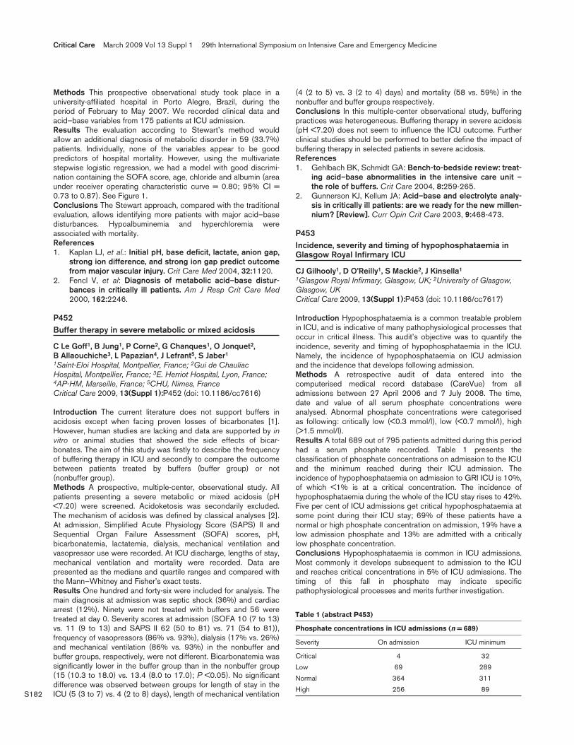
S182
Methods This prospective observational study took place in auniversity-affiliated hospital in Porto Alegre, Brazil, during theperiod of February to May 2007. We recorded clinical data andacid–base variables from 175 patients at ICU admission.Results The evaluation according to Stewart’s method wouldallow an additional diagnosis of metabolic disorder in 59 (33.7%)patients. Individually, none of the variables appear to be goodpredictors of hospital mortality. However, using the multivariatestepwise logistic regression, we had a model with good discrimi-nation containing the SOFA score, age, chloride and albumin (areaunder receiver operating characteristic curve = 0.80; 95% CI =0.73 to 0.87). See Figure 1.Conclusions The Stewart approach, compared with the traditionalevaluation, allows identifying more patients with major acid–basedisturbances. Hypoalbuminemia and hyperchloremia wereassociated with mortality.References1. Kaplan LJ, et al.: Initial pH, base deficit, lactate, anion gap,
strong ion difference, and strong ion gap predict outcomefrom major vascular injury. Crit Care Med 2004, 32:1120.
2. Fencl V, et al: Diagnosis of metabolic acid–base distur-bances in critically ill patients. Am J Resp Crit Care Med2000, 162:2246.
P452Buffer therapy in severe metabolic or mixed acidosis
C Le Goff1, B Jung1, P Corne2, G Chanques1, O Jonquet2, B Allaouchiche3, L Papazian4, J Lefrant5, S Jaber1
1Saint-Eloi Hospital, Montpellier, France; 2Gui de ChauliacHospital, Montpellier, France; 3E. Herriot Hospital, Lyon, France;4AP-HM, Marseille, France; 5CHU, Nimes, FranceCritical Care 2009, 13(Suppl 1):P452 (doi: 10.1186/cc7616)
Introduction The current literature does not support buffers inacidosis except when facing proven losses of bicarbonates [1].However, human studies are lacking and data are supported by invitro or animal studies that showed the side effects of bicar-bonates. The aim of this study was firstly to describe the frequencyof buffering therapy in ICU and secondly to compare the outcomebetween patients treated by buffers (buffer group) or not(nonbuffer group).Methods A prospective, multiple-center, observational study. Allpatients presenting a severe metabolic or mixed acidosis (pH<7.20) were screened. Acidoketosis was secondarily excluded.The mechanism of acidosis was defined by classical analyses [2].At admission, Simplified Acute Physiology Score (SAPS) II andSequential Organ Failure Assessment (SOFA) scores, pH,bicarbonatemia, lactatemia, dialysis, mechanical ventilation andvasopressor use were recorded. At ICU discharge, lengths of stay,mechanical ventilation and mortality were recorded. Data arepresented as the medians and quartile ranges and compared withthe Mann–Whitney and Fisher’s exact tests.Results One hundred and forty-six were included for analysis. Themain diagnosis at admission was septic shock (36%) and cardiacarrest (12%). Ninety were not treated with buffers and 56 weretreated at day 0. Severity scores at admission (SOFA 10 (7 to 13)vs. 11 (9 to 13) and SAPS II 62 (50 to 81) vs. 71 (54 to 81)),frequency of vasopressors (86% vs. 93%), dialysis (17% vs. 26%)and mechanical ventilation (86% vs. 93%) in the nonbuffer andbuffer groups, respectively, were not different. Bicarbonatemia wassignificantly lower in the buffer group than in the nonbuffer group(15 (10.3 to 18.0) vs. 13.4 (8.0 to 17.0); P <0.05). No significantdifference was observed between groups for length of stay in theICU (5 (3 to 7) vs. 4 (2 to 8) days), length of mechanical ventilation
(4 (2 to 5) vs. 3 (2 to 4) days) and mortality (58 vs. 59%) in thenonbuffer and buffer groups respectively.Conclusions In this multiple-center observational study, bufferingpractices was heterogeneous. Buffering therapy in severe acidosis(pH <7.20) does not seem to influence the ICU outcome. Furtherclinical studies should be performed to better define the impact ofbuffering therapy in selected patients in severe acidosis.References1. Gehlbach BK, Schmidt GA: Bench-to-bedside review: treat-
ing acid–base abnormalities in the intensive care unit –the role of buffers. Crit Care 2004, 8:259-265.
2. Gunnerson KJ, Kellum JA: Acid–base and electrolyte analy-sis in critically ill patients: are we ready for the new millen-nium? [Review]. Curr Opin Crit Care 2003, 9:468-473.
P453Incidence, severity and timing of hypophosphataemia inGlasgow Royal Infirmary ICU
CJ Gilhooly1, D O’Reilly1, S Mackie2, J Kinsella1
1Glasgow Royal Infirmary, Glasgow, UK; 2University of Glasgow,Glasgow, UKCritical Care 2009, 13(Suppl 1):P453 (doi: 10.1186/cc7617)
Introduction Hypophosphataemia is a common treatable problemin ICU, and is indicative of many pathophysiological processes thatoccur in critical illness. This audit’s objective was to quantify theincidence, severity and timing of hypophosphataemia in the ICU.Namely, the incidence of hypophosphataemia on ICU admissionand the incidence that develops following admission.Methods A retrospective audit of data entered into thecomputerised medical record database (CareVue) from alladmissions between 27 April 2006 and 7 July 2008. The time,date and value of all serum phosphate concentrations wereanalysed. Abnormal phosphate concentrations were categorisedas following: critically low (<0.3 mmol/l), low (<0.7 mmol/l), high(>1.5 mmol/l).Results A total 689 out of 795 patients admitted during this periodhad a serum phosphate recorded. Table 1 presents theclassification of phosphate concentrations on admission to the ICUand the minimum reached during their ICU admission. Theincidence of hypophosphataemia on admission to GRI ICU is 10%,of which <1% is at a critical concentration. The incidence ofhypophosphataemia during the whole of the ICU stay rises to 42%.Five per cent of ICU admissions get critical hypophosphataemia atsome point during their ICU stay; 69% of these patients have anormal or high phosphate concentration on admission, 19% have alow admission phosphate and 13% are admitted with a criticallylow phosphate concentration.Conclusions Hypophosphataemia is common in ICU admissions.Most commonly it develops subsequent to admission to the ICUand reaches critical concentrations in 5% of ICU admissions. Thetiming of this fall in phosphate may indicate specificpathophysiological processes and merits further investigation.
Critical Care March 2009 Vol 13 Suppl 1 29th International Symposium on Intensive Care and Emergency Medicine
Table 1 (abstract P453)
Phosphate concentrations in ICU admissions (n = 689)
Severity On admission ICU minimum
Critical 4 32
Low 69 289
Normal 364 311
High 256 89

S183
P454Hypernatraemia in the neurointensive care unit: centraldiabetes insipidus and noncentral diabetes insipidus
V Spatenkova1, A Kazda2, P Suchomel11Regional Hospital, Liberec, Czech Republic; 2Charles University,Prague, Czech RepublicCritical Care 2009, 13(Suppl 1):P454 (doi: 10.1186/cc7618)
Introduction Hypernatraemia is a prognostically serious complica-tion in the neurointensive care unit. A typical, well-known syndromeassociated with acute brain disease is central diabetes insipidus(cDI). However, cDI is not a frequent reason of hypernatraemia inneurointensive care. There are other causes of hypernatraemia,most frequently osmotherapy by mannitol or renal failure [1]. Thepurpose of our study was to evaluate retrospectively all hyper-natraemias in patients with acute brain diseases admitted to ourneurologic–neurosurgical care unit (NNICU) over a period of5 years.Methods We analysed all patients with acute brain disease andserum sodium above 150 mmol/l. Firstly we diagnosed cDIaccording to serum and urine osmolality, hourly diuresis, renalfunction parameters and response to desmopressin. The remaininghypernatraemias were categorised as non-cDI. Patients withoutmeasured serum osmolality were excluded.Results There were 75 hypernatraemic patients (mean age56.8 years; 41 male) with the following diagnoses: stroke 43,tumour 20, trauma 7, hydrocephalus 4 and epilepsy 1. The meanGlasgow coma scale (GCS) on onset of hypernatraemia was 12 ±3.4, the mean Glasgow outcome scale (GOS) upon dischargefrom the NNICU was 3.3 ± 1.4. We found cDI only in eightpatients (altogether 15 days). We classified 59 patients as non-cDIhypernatraemia (163 days). The remaining patients were notevaluated because they had no serum osmolality measurements.Between the two groups there were no differences in GCS(P = 0.631), GOS (P = 0.857), serum sodium (P = 0.736), andserum osmolality (P = 0.476), but patients with cDI had low urineosmolality (P = 0.001). On the other hand, the non-cDI groupreceived more antioedematic therapy (P = 0.013) and diuretics(P = 0.019). In this group, mannitol was received in 48 (81.4%)patients and six (10.2%) had creatinine clearance below thereference range (range <1.15 ml/s).Conclusions In neurointensive care cDI is not the most commontype of hypernatraemia. Most hypernatraemias are non-cDI due toanti-oedematic therapy and kidney dysfunction.Reference1. Aiyagari V, Deibert E, Diringer M: Hypernatremia in the neu-
rologic intensive care unit: how high is too high? J CritCare 2006, 21:163-172.
P455Impact of hypernatremia, hyponatremia and theircomparison on ICU mortality
M Mokhtari, M Kouchek, M Miri, R GoharaniSBU Medical Sciences, Tehran, IranCritical Care 2009, 13(Suppl 1):P455 (doi: 10.1186/cc7619)
Introduction Serum sodium homeostasis is important in thecritically ill patient [1-3]. We studied the effect of hypernatremia,hyponatremia and their comparison on ICU mortality in our patientpopulation.Methods We studied 274 patients hospitalized in a general ICU ofa major teaching hospital in Tehran, Iran, cross-sectionally,between 2004 and 2006. Patients’ biographical data, reasons for
admission, hyponatremia (sodium less than 135 mEq/l) andhypernatremia (sodium more than 145 mEq/l) during the ICU staywere recorded and their effects on mortality were assessed.Results The mortalities of patients with hyponatremia (34.33%versus 15.37% with OR = 5.69 and 95% CI from 2.87 to 11.30)and with hypernatremia (55.17% versus 17.78% with OR = 2.80and 95% CI from 1.43 to 5.49) were significantly higher thanpatients without it. The mean age of patients with sodiumimbalance was higher compared with those without it (hypo-natremia and hypernatremia 52.25 ± 21.12 vs. 44.40 ± 20.28years and 55.59 ± 19.69 vs. 48.52 ± 21.83 years, respectively).Mortality was significantly higher in hypernatremic compared withhyponatremic patients (55.2% vs. 34.3%, respectively, 95% CIfrom 7.7% to 33.9%; P = 0.0034).Conclusions Serum sodium imbalances carry unacceptably highmortality [4]. Patients with hypernatremia showed higher ICUmortality as compared with those with hyponatremia.References1. Kraft MD, Btaiche IF, Sacks GS, Kudsk KA: Treatment of
electrolyte disorders in adult patients in the intensive careunit. Am J Health Syst Pharm 2005, 62:1663-1682.
2. Zilberberg MD, Exuzides A, Spalding J, Foreman A, Jones AG,Colby C, et al.: Epidemiology, clinical and economic out-comes of admission hyponatremia among hospitalizedpatients. Curr Med Res Opin 2008, 24:1601-1608.
3. Patel GP, Balk RA: Recognition and treatment of hypona-tremia in acutely ill hospitalized patients. Clin Ther 2007,29:211-229.
4. Lindner G, Funk GC, Schwarz C, Kneidinger N, Kaider A,Schneeweiss B, et al.: Hypernatremia in the critically ill isan independent risk factor for mortality. Am J Kidney Dis2007, 50:952-957.
P456Hyponatremia is associated with increased mortality inpatients with acute decompensated hear failure
J Vaitsis1, H Michalopoulou1, C Thomopoulos2, S Massias2, P Stamatis1
1‘Metaxa’ Hospital, Athens, Greece; 2‘Elena Venizelou’ Hospital,Athens, GreeceCritical Care 2009, 13(Suppl 1):P456 (doi: 10.1186/cc7620)
Introduction Hyponatremia has been identified as a risk factor forincreased morbidity and mortality in patients with chronic heartfailure (HF) but there are few data for its prevalence and clinicalimplication in patients with acute decompensated HF.Methods One hundred and eighty-two patients (142 male, age72.1 ± 11.6 years) presented as worsening HF, NYHA III to IV,48% with ejection fraction ≤30%, 43% of ischemic etiology, wereenrolled during the period from August 2007 to July 2008. In aretrospective analysis we investigated the relationship betweenhyponatremia and inhospital mortality, 60-day mortality and 60-dayrehospitalization. We divided the patients into three groupsaccording to their admission serum sodium. Group A, Na≥136 mEq/l. Group B, Na = 135 to 131 mEq/l. Group C, Na≤130 mEq/l.Results Patients in Group C had a more severe heart failure, lowerblood pressure, lower ejection fraction and increased mortality(6.4% in Group C vs. 3.4% in B vs. 1.8% in A, P <0.05 forinhospital mortality), (13.7% in Group C vs. 7.2% in B vs. 4.7% inA, P <0.05 for 60-day mortality), (39.3% in Group C vs. 29.1% inB vs. 27.2% in A, P <0.05 for rehospitalization). In multivariableanalysis, either moderate hyponatremia (Group B) or severehyponatremia (Group C) still remained predictors of death. Group
Available online http://ccforum.com/supplements/13/S1
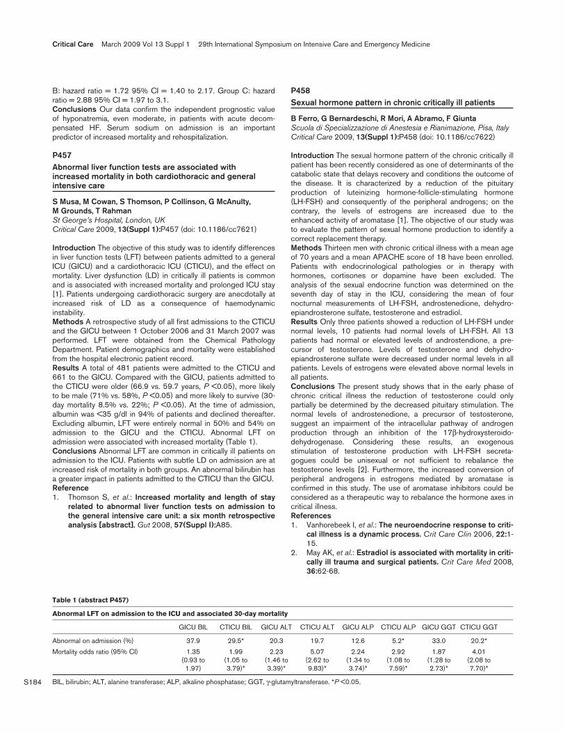
S184
B: hazard ratio = 1.72 95% CI = 1.40 to 2.17. Group C: hazardratio = 2.88 95% CI = 1.97 to 3.1.Conclusions Our data confirm the independent prognostic valueof hyponatremia, even moderate, in patients with acute decom-pensated HF. Serum sodium on admission is an importantpredictor of increased mortality and rehospitalization.
P457Abnormal liver function tests are associated withincreased mortality in both cardiothoracic and generalintensive care
S Musa, M Cowan, S Thomson, P Collinson, G McAnulty, M Grounds, T RahmanSt George’s Hospital, London, UKCritical Care 2009, 13(Suppl 1):P457 (doi: 10.1186/cc7621)
Introduction The objective of this study was to identify differencesin liver function tests (LFT) between patients admitted to a generalICU (GICU) and a cardiothoracic ICU (CTICU), and the effect onmortality. Liver dysfunction (LD) in critically ill patients is commonand is associated with increased mortality and prolonged ICU stay[1]. Patients undergoing cardiothoracic surgery are anecdotally atincreased risk of LD as a consequence of haemodynamicinstability.Methods A retrospective study of all first admissions to the CTICUand the GICU between 1 October 2006 and 31 March 2007 wasperformed. LFT were obtained from the Chemical PathologyDepartment. Patient demographics and mortality were establishedfrom the hospital electronic patient record.Results A total of 481 patients were admitted to the CTICU and661 to the GICU. Compared with the GICU, patients admitted tothe CTICU were older (66.9 vs. 59.7 years, P <0.05), more likelyto be male (71% vs. 58%, P <0.05) and more likely to survive (30-day mortality 8.5% vs. 22%; P <0.05). At the time of admission,albumin was <35 g/dl in 94% of patients and declined thereafter.Excluding albumin, LFT were entirely normal in 50% and 54% onadmission to the GICU and the CTICU. Abnormal LFT onadmission were associated with increased mortality (Table 1).Conclusions Abnormal LFT are common in critically ill patients onadmission to the ICU. Patients with subtle LD on admission are atincreased risk of mortality in both groups. An abnormal bilirubin hasa greater impact in patients admitted to the CTICU than the GICU.Reference1. Thomson S, et al.: Increased mortality and length of stay
related to abnormal liver function tests on admission tothe general intensive care unit: a six month retrospectiveanalysis [abstract]. Gut 2008, 57(Suppl I):A85.
P458Sexual hormone pattern in chronic critically ill patients
B Ferro, G Bernardeschi, R Mori, A Abramo, F GiuntaScuola di Specializzazione di Anestesia e Rianimazione, Pisa, ItalyCritical Care 2009, 13(Suppl 1):P458 (doi: 10.1186/cc7622)
Introduction The sexual hormone pattern of the chronic critically illpatient has been recently considered as one of determinants of thecatabolic state that delays recovery and conditions the outcome ofthe disease. It is characterized by a reduction of the pituitaryproduction of luteinizing hormone-follicle-stimulating hormone(LH-FSH) and consequently of the peripheral androgens; on thecontrary, the levels of estrogens are increased due to theenhanced activity of aromatase [1]. The objective of our study wasto evaluate the pattern of sexual hormone production to identify acorrect replacement therapy.Methods Thirteen men with chronic critical illness with a mean ageof 70 years and a mean APACHE score of 18 have been enrolled.Patients with endocrinological pathologies or in therapy withhormones, cortisones or dopamine have been excluded. Theanalysis of the sexual endocrine function was determined on theseventh day of stay in the ICU, considering the mean of fournocturnal measurements of LH-FSH, androstenedione, dehydro-epiandrosterone sulfate, testosterone and estradiol.Results Only three patients showed a reduction of LH-FSH undernormal levels, 10 patients had normal levels of LH-FSH. All 13patients had normal or elevated levels of androstendione, a pre-cursor of testosterone. Levels of testosterone and dehydro-epiandrosterone sulfate were decreased under normal levels in allpatients. Levels of estrogens were elevated above normal levels inall patients.Conclusions The present study shows that in the early phase ofchronic critical illness the reduction of testosterone could onlypartially be determined by the decreased pituitary stimulation. Thenormal levels of androstenedione, a precursor of testosterone,suggest an impairment of the intracellular pathway of androgenproduction through an inhibition of the 17β-hydroxysteroido-dehydrogenase. Considering these results, an exogenousstimulation of testosterone production with LH-FSH secreta-gogues could be unisexual or not sufficient to rebalance thetestosterone levels [2]. Furthermore, the increased conversion ofperipheral androgens in estrogens mediated by aromatase isconfirmed in this study. The use of aromatase inhibitors could beconsidered as a therapeutic way to rebalance the hormone axes incritical illness.References1. Vanhorebeek I, et al.: The neuroendocrine response to criti-
cal illness is a dynamic process. Crit Care Clin 2006, 22:1-15.
2. May AK, et al.: Estradiol is associated with mortality in criti-cally ill trauma and surgical patients. Crit Care Med 2008,36:62-68.
Critical Care March 2009 Vol 13 Suppl 1 29th International Symposium on Intensive Care and Emergency Medicine
Table 1 (abstract P457)
Abnormal LFT on admission to the ICU and associated 30-day mortality
GICU BIL CTICU BIL GICU ALT CTICU ALT GICU ALP CTICU ALP GICU GGT CTICU GGT
Abnormal on admission (%) 37.9 29.5* 20.3 19.7 12.6 5.2* 33.0 20.2*
Mortality odds ratio (95% CI) 1.35 1.99 2.23 5.07 2.24 2.92 1.87 4.01(0.93 to (1.05 to (1.46 to (2.62 to (1.34 to (1.08 to (1.28 to (2.08 to
1.97) 3.79)* 3.39)* 9.83)* 3.74)* 7.59)* 2.73)* 7.70)*
BIL, bilirubin; ALT, alanine transferase; ALP, alkaline phosphatase; GGT, γ-glutamyltransferase. *P <0.05.

S185
P459Economic comparison of the traditional bathing methodwith the basinless bathing method in coma patients
MJ Sucre, A De NicolaSan Leonardo Hospital, Castellammare di Stabia, ItalyCritical Care 2009, 13(Suppl 1):P459 (doi: 10.1186/cc7623)
Introduction The coma patient is a particular and unique patient:he depends completely on the caregivers, more than a neonate.The bed bath of a coma patient is important to maintain hispersonal hygiene, to clean and refresh, to monitor skin conditionand to promote comfort. Modern ICU management must offer ahigh quality of coma patient care and should be constantly fightingfor cost-effective ways of providing that care.Methods The aim of this study was to compare the traditional bed-bathing method with the basinless bed-bathing method. The studyfocused on nurse satisfaction or acceptance, and the costs of twomethods. The study comprised 100 baths of coma patients: 50completed using the Comfort Bath® (a prepackaged disposablebed bath) and 50 completed using the traditional method. Inaddition, 30 nurses were recruited and interviewed about theirpreferences and opinions. The economic study type was cost-minimization and, in part, cost-effectiveness analysis.Results For skin softness, administration ease, and nursesatisfaction, there were statistical variations between the two setsof scores, in favour of the Comfort Bath® method. Nursesexpressed a clear and significant preference for the disposablebath, corroborating findings by other investigators [1]. The directcosts were: for the soap, water, laundry, equipment and labour, forthe time taken to perform the bed-bathing, for logistics anddepreciation. The overall cost was less for the disposable bath dueto savings in nurses’ time and salary (Table 1).
Table 1 (abstract P459)
Costs of bath technology required
Traditional bath Comfort Bath®
Total bath costs 18.46 16.74
Savings 1.73
Conclusions The present study confirmed that Comfort Bath®, aone-step bed bath, is an economic and preferred alternative to thebasin of water. Considering the cost-minimization, the cost-effective use and the significant preference, this prepackageddisposable bed bath symbolizes a new concept in coma patienthygiene.Reference1. Larson EL, et al.: Comparison of traditional and disposable
bed baths in critically ill patients. Am J Crit Care 2004, 13:235-241.
P460In critically ill patients, how often is their weight estimatedand how accurate is that estimate?
D Mathew, C Kirwan, D Dawson, B PhilipsSt George’s Hospital, London, UKCritical Care 2009, 13(Suppl 1):P460 (doi: 10.1186/cc7624)
Introduction Weight is frequently required to determine the doseof many medications and is important in quantifying treatmentssuch as haemofiltration and calorific requirement. We present thefindings of an audit to determine how frequently a patient’s weight
is estimated in one ICU and the results of a pilot study to deter-mine the accuracy of an estimated weight in the critically ill patient.Methods A prospective audit of 100 admissions to an ICU wasundertaken over a 6-week period. Data were collected fromobservation charts and hospital notes. Following data analysis apilot study was undertaken to assess the accuracy of the estimatedweight of patients in the ICU. Urine bags and drains were emptiedbefore weighing and the same number of pillows and blankets wasensured on each bed. Due to a short loan period of scales, only 18patients could be weighed.Results Audit The median (IQR) patient weight was 75 kg (64 to85). Ninety-four ICU observation charts had a weight recorded,and of these 38 (40.4%) were estimated. Sixteen out of 20 (80%)medical patients and 22/74 (29.7%) surgical patients had anestimated weight. Sixty-three per cent of patients had a recentaccurate weight recorded in the notes and only 43 (68.3%) hadthe same weight recorded on the ICU observation chart. Seventyper cent of patients required a weight for cardiac output monitoringand 54% for drug dose calculation. Pilot study The median (range)measured weight was 92 (66.8 to 137.4) kg. Twelve out of 18 hadan estimated weight on the ICU chart. The mean (precision) ofestimation was 10.1 (±21.4) kg. Seven out of 12 (58%) had agreater than 10% difference between the estimated and trueweights. Of the six patients who had a recent accurate weightdocumented in the notes, the mean difference (precision) betweenthat and the true weight was 5.8 (±4.0) kg. Two out of six (33%) hada greater than 10% difference between estimated and true weights.Conclusions A patient’s weight is an important parameter in guidingtherapy. A large proportion of ICU patients have their weightestimated. In some cases this is unnecessary as a recent weight canbe found in the notes. A more comprehensive study is required toassess the clinical impact of weight estimation on the ICU.
P461Alcohol misuse requiring admission to a tertiary referralICU: resource implications
D Trainor, L Martin, G LaveryBelfast HSC Trust & Faculty of Life and Health Sciences, Universityof Ulster, Belfast, UKCritical Care 2009, 13(Suppl 1):P461 (doi: 10.1186/cc7625)
Introduction Alcohol misuse is associated with many conditionsrequiring ICU management [1]. In 2006/2007, 1.53% of allhospital admissions were related to alcohol misuse (regional audit).We conducted a prospective audit in a tertiary referral adult ICU toascertain the proportion of ICU capacity used for the treatment ofconditions related to alcohol misuse.Methods Patients admitted to a tertiary referral ICU between 1November 2006 and 31 March 2007 were prospectively auditedwith the aim of identifying all cases in which the reason foradmission could be attributed to acute or chronic alcohol misuse.Blood alcohol levels, past history from the patient/relatives orprevious relevant clinical investigations and/or diagnoses werereviewed for evidence of acute or chronic alcohol misuse.Results Of 318 ICU admissions during the audit, 9.43% (30admissions – 20 male/10 female) were judged to be due to eitheracute (19 of 30) or chronic (11 of 30) alcohol consumption. Themean patient age was 48.7 years (SD 14.1, range 25 to 74).Blood alcohol level on admission to hospital was available in 13patients (43%), the median value being 170 mg% (IQR = 66 to274). Seventeen patients (56.6%) were admitted following trauma,of whom 10 (33.3%), had brain injuries. The median length of stay(LOS) in the ICU was 10 days (IQR = 4 to 15) and the medianLOS in hospital was 22 days (IQR = 10 to 38). The ICU mortality
Available online http://ccforum.com/supplements/13/S1

S186
rate was 23.3% (seven out of 30) and hospital mortality was 30%(nine out of 30). During the audit period, a total of 2,567 ICU bed-days were available, of which 357 bed-days (13.9%) were used totreat this group of patients. The mean ICU occupancy during thisperiod was 95.6%.Conclusions Alcohol misuse was implicated in almost 10% of ICUadmissions – over six times the equivalent rate for total hospitaladmissions. These admissions required approximately 14% of totalICU capacity at a time when occupancy was almost 100%.Intensive care is a finite and expensive hospital resource. Publichealth bodies should be informed of the proportion of that resourcerequired to treat alcohol-related illness and injury.Reference1. O’Brien J, et al.: Alcohol dependence is independently
associated with sepsis, septic shock, and hospital mortal-ity among adult intensive care unit patients. Crit Care Med2007, 35:345-350.
P462Systemic rheumatic diseases in the ICU
P Vernikos, T Topalis, A Mega, A Tzifas, N Archontoulis, J FlorosLaiko General Hospital, Athens, GreeceCritical Care 2009, 13(Suppl 1):P462 (doi: 10.1186/cc7626)
Introduction Patients with systemic rheumatic diseases may beadmitted to the ICU because of pulmonary, renal or multiple organdysfunction, caused by worsening of the rheumatic disease,infection or adverse effects of immunosuppressive drugs.Methods In a 3-year period, 14 patients (six males/eight females),18 to 80 years old (average 56.78), with systemic rheumaticdiseases were admitted in the ICU. Four of them had rheumatoidarthritis, one systemic lupus erythematosus, three dermatopoly-myositis, one Sjogren’s syndrome, two vasculitis, two systemicsclerosis and one psoriatic arthritis. Three of them were admittedto the ICU after major surgical operation, nine of them because ofrespiratory failure and the others because of coma and/or shock.The admission APACHE II score, chest X-ray, bronchial secretioncultures, and PaO2/FiO2 were recorded, as well as the broncho-alveolar lavage findings, when it was necessary, and the finaloutcome.Results Nine patients were admitted because of acute respiratoryfailure: APACHE II score ranged from 17 to 28, X-ray showeddiffuse alveolar infiltrates, PaO2/FiO2 ranged from 75 to 184, onlytwo patients had positive cultures of bronchial secretions. Twopatients were admitted after major surgical operation, one afternecrotizing pancreatitis: APACHE II score ranged from 9 to 21,X-ray showed diffuse alveolar infiltrates with a cavity in one,PaO2/FiO2 >300, the bronchial secretions cultures were negative.One patient was admitted because of coma and one otherbecause of shock: APACHE II score was 25 and 32, respectively,X-ray without any acute process, PaO2/FiO2 was >200 and noneof them had positive cultures of bronchial secretions. Broncho-alveolar lavage was performed on six patients: on the second dayfrom ICU admission in four patients, on the eighth day in onepatient and on the 21st day in one patient. The total mortality ratewas 50% (7/14).Conclusions Early bronchoalveolar lavage (during the first 48hours from admission) could help in differential diagnosis between:exacerbation of rheumatic disease, infection, drug-inducedrespiratory failure, and alveolar hemorrhage.
P463Distances covered by ICU nurses during their shifts
E Nahk, S FrolovaEast-Tallinn Central Hospital, Tallinn, EstoniaCritical Care 2009, 13(Suppl 1):P463 (doi: 10.1186/cc7627)
Introduction We monitored the distances covered by nursesduring their work in the ICU. An automatic step counter was usedto estimate the distances.Methods Seventeen nurses were monitored during 102 hours intotal. Eight of them were responsible for one ICU patient during themonitoring period and nine took care of two patients at the sametime.Results The mean distance covered was 0.9 (range 0.4 to 1.3)km/hour, resulting in a mean 10.7 km per 12-hour shift. Thedistance was significantly different according to the number ofpatients the nurse was responsible for. The nurses taking care oftwo patients walked significantly more compared with the nurseswith one patient (1.0 with range 0.9 to 1.3 compared with 0.7 withrange 0.4 to 1.1 km/hour, P = 0.027). During their shift the nurseswalked a mean 12.3 km when taking care of two patients and8.9 km when working with only one ICU patient.Conclusions Nurses need to cover long distances during theireveryday work. In some cases the walking distance may beexpected to reach 15 km during the 12-hour shift. Optimization ofthe working load and logistics to reduce the walking distancesshould be the goal.
P464ICU triage in terror-related mass casualty events
Y Klein1, M Klein2, K Peleg3
1Kaplan Medical Center, Rehovot, Israel; 2Soroka Medical Center,Beer Sheva, Israel; 3Gertner Institue, Tel Hashomer, IsraelCritical Care 2009, 13(Suppl 1):P464 (doi: 10.1186/cc7628)
Introduction Admitting patients to the ICU is one of the triageprocesses needed during mass casualty events (MCE). The highoccupancy of the ICU and the fact that it cannot be readilyevacuated make this process even more challenging [1,2]. Wecompared the triage to the ICU in terror-related MCE versus othertrauma in Israel.Methods A retrospective cohort study of all patients recorded inthe Israeli National Trauma registry (ITR) that were injured in MCEsecondary to terrorist acts by explosions from 1 October 2000 to31 December 2005. Medical records of all patients hospitalized inthe ICU were retrieved and reviewed. Over-triage was defined as apatient that is discharged from the ICU within 24 hours ofadmission without any specific intervention besides monitoring.Under-triage was defined as an admission to the ICU more than24 hours after an admission to a different department.Results The ITR included 122,208 patients during the studyperiod. A total of 1,272 patients (1.0%) were injured in terror-related MCE. Three hundred and sixty-five patients (28.7%) of theMCE group and 9,093 patients (7.5%) of the non-MCE groupwere admitted to the ICU. Eighty-one (6.4%) of the patients in theMCE group and 2,445 (2.0%) of the patients in the non-MCEgroup were admitted to the ICU and discharged within 24 hours(P <0.0001). A total of 3.3% of the patients in the MCE and 9.2%of the patients in the non-MCE group were admitted to the ICUmore than 24 hours after admission to another department.Patients that were over-triaged to the ICU during MCE hadsignificantly more injuries, and to more body regions, than thepatients that were over-triaged to the ICU due to non-MCE trauma.
Critical Care March 2009 Vol 13 Suppl 1 29th International Symposium on Intensive Care and Emergency Medicine

S187
Conclusions Over-triage to the ICU is probably unavoidable inMCE. This fact and the increased injury severity, anticipated interror-related MCE, should lead the hospital to adopt a plan forexpansion of the ICU capabilities. A post-event ICU re-evaluationround and judicious patient discharge may reduce prolongedoveruse of ICU resources.References1. Frykberg ER: Medical management of disasters and mass
casualties from terrorist bombings: how can we cope? JTrauma 2002, 53:201-212.
2. Sprung CL, et al.: Evaluation of triage decisions for inten-sive care admission. Crit Care Med 1999, 27:1073-1079.
P465Outcome of weekend and afterhour admissions comparedwith weekday and daytime admissions in the ICU withonsite intensivist coverage
M Hawari, M Shaaban, N Abouchala, K Maghrabi, M Hijazi, M KherallahKing Faisal Specialist Hospital and Research Center, Riyadh, SaudiArabiaCritical Care 2009, 13(Suppl 1):P465 (doi: 10.1186/cc7629)
Introduction The objective of this study was to determine whetheronsite intensivist coverage would improve the outcome of weekendand afterhour admissions in our ICU.Methods In a cohort study at a tertiary-care medical–surgical ICUstaffed 24 hours/7 days by onsite consultant intensivists withpredominantly North American Critical Care Board certifications, weincluded all ICU admissions (January 2007 to February 2008) froma prospectively collected ICU database. Results were stratifiedbased on the time of the admission (daytime (8:00 to 17:00) orafterhour time (17:01 to 07:59)) and the day of the week (weekdayor weekend). Predicted mortality rates were calculated using anAPACHE IV prediction model. The primary outcome was hospitalmortality, and standardized mortality ratios (SMRs) were calculated.Results A total of 1,315 admissions were included in the study.Forty-eight percent of patients were admitted in daytime and 52%in afterhour time with average APACHE IV scores of 59.8 and 55,respectively (P <0.01), the SMR for the daytime group was 0.86(95% CI = 0.69 to 1.05, P = 0.15) compared with an SMR of theafterhour group of 0.98 (95% CI = 0.84 to 1.15, P = 0.83).Seventy-two percent of patients were admitted on a weekday and28% on weekends with average APACHE IV scores of 57.7 and58.7, respectively (P = 0.52); there was no significant difference inmortality rates between the weekday and weekend groups (17.3%vs. 21.4, P = 0.09, with SMRs of 1.05 vs. 0.89). After adjustmentfor severity of illness it appears that there is a trend towards alower mortality rate for daytime and weekday admissions comparedwith afterhour and weekend admissions; however, there is nostatistical difference.Conclusions Our results indicate no difference in mortality ratesfor weekend and afterhour admissions to an ICU that is staffed byonsite certified intensivists.
P466Scottish audit of interhospital transfers of acutely ill adults
J Bruce1, R Colquhoun2, G Smith1, M Fried1
1St Johns Hospital, Livingston, UK; 2SAS, Paisley, UKCritical Care 2009, 13(Suppl 1):P466 (doi: 10.1186/cc7630)
Introduction The audit was commissioned by the Scottish Govern-ment Health Department to identify the number of acutely ill adults
transported between hospitals by the Scottish Ambulance Service(SAS). Currently interhospital transfer (IHT) of acutely ill adultsrequiring medical escorts is undertaken by the SAS Accident andEmergency (A&E) ambulances or by dedicated transport teams (forexample, Glasgow Shock Team and Emergency Medical RetrievalService).Methods The audit ran for 20 weeks. Patients over 16 years oldwho required intervention/vital signs monitoring during transfer orwho required a nursing or medical escort were defined as acutelyill. Audit forms were placed in every A&E ambulance.Results A total of 3,068 forms were received, with 2,396 audited.Twenty per cent were emergencies, 73% urgent and 7% planned.The median SAS journey times, patient and dedicated teamjourney times are presented in Table 1. The majority of IHTsoccurred for specialist management (65.9%) or specialistinvestigation (23%). A total of 820 IHTs were clinically escorted,including the Glasgow Shock Team (n = 192) and the EmergencyMedical Retrieval Service (n = 13). Two hundred and forty-eightpatients were ventilated (10.3%). Eighty-four (3.5%) IHTs requiredclinical intervention to correct clinical deterioration. An adverseevent occurred in 1.5% (22). One hundred and thirteen (18%)escorted IHTs by nondedicated transport teams had unsecuredmedical equipment versus 2/205 (0.97%) in dedicated transportteams (P = 0.004). Equipment failures occurred in 9/97(9.2%)ventilated transfers by nondedicated teams versus 1/156 (0.6%)by dedicated teams (P = 0.001). The SAS executive informationservice data showed a total number of IHTs during the audit periodconveyed by A&E ambulances as 19,747 (18,343 adults). Thisapproximates to a total of 51,342 per annum (47,692 adults) or917 adults per week.Conclusions The most frequent reason for IHT is specialistmanagement or investigation. Adverse events are reassuringlyuncommon. Unsecured medical equipment and equipment failureduring an IHT is significantly more frequent when undertaken bynondedicated transport teams.
P467Outcome and resource utilization following interhospitaltransfer of critically ill patients
D Vandijck, S Oeyen, P Coucke, P Lafaire, D Benoit, J DecruyenaereGhent University Hospital, Ghent, BelgiumCritical Care 2009, 13(Suppl 1):P467 (doi: 10.1186/cc7631)
Introduction Patients who undergo transfer to the ICU of anacademic referral centre represent a group of sicker patients withincreased risk for grim prognosis than those admitted directly.These patients’ weight in terms of increased resource utilization is,however, less studied. This study aimed to compare the outcomeand resource consumption between directly admitted patients andthose transferred from a peripheral hospital to the ICU of a largetertiary-care referral centre.
Available online http://ccforum.com/supplements/13/S1
Table 1 (abstract P466)
Journey times (hh:mm)
SAS n = 2,396 Median 01:08
Patients n = 2,396 Median 00:44
Ventilated patients n = 248 Median 00:28
Dedicated team n = 205 Median 03:05
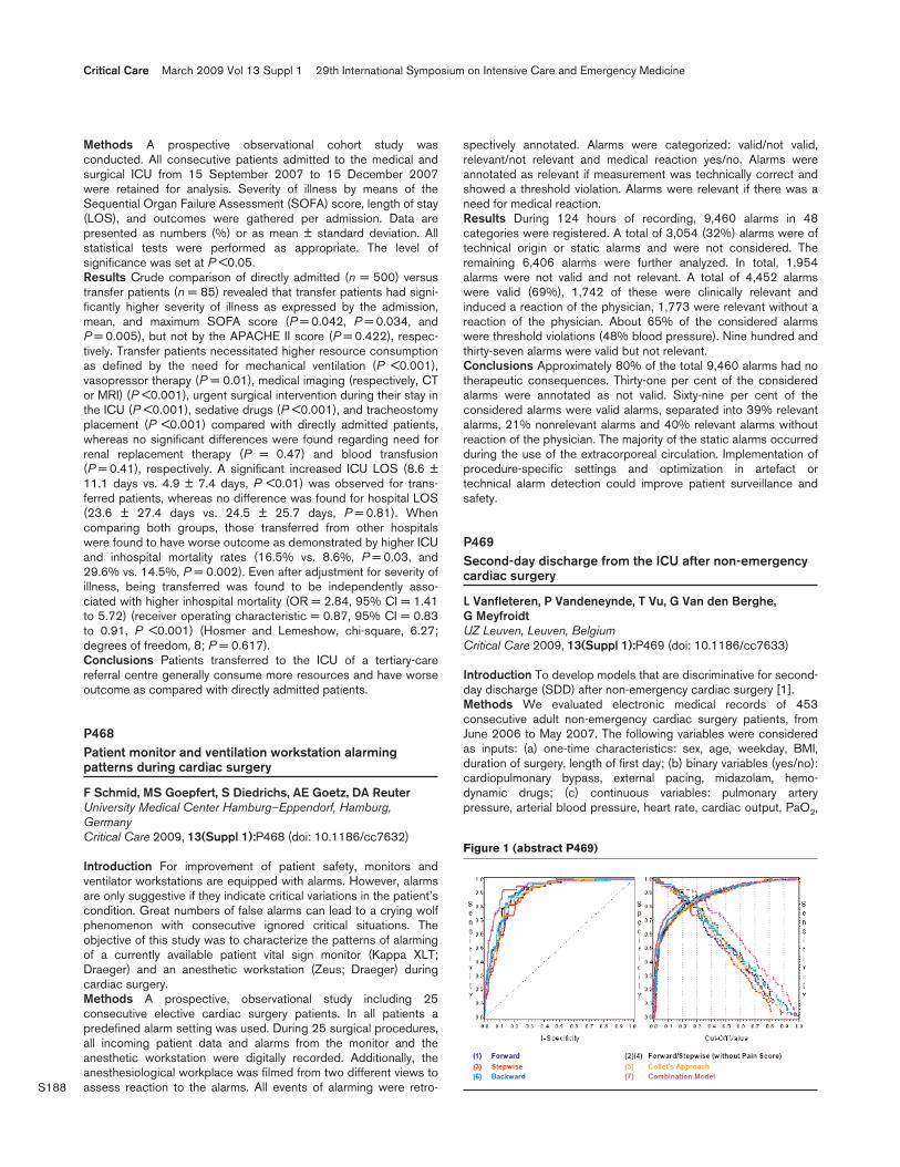
S188
Methods A prospective observational cohort study wasconducted. All consecutive patients admitted to the medical andsurgical ICU from 15 September 2007 to 15 December 2007were retained for analysis. Severity of illness by means of theSequential Organ Failure Assessment (SOFA) score, length of stay(LOS), and outcomes were gathered per admission. Data arepresented as numbers (%) or as mean ± standard deviation. Allstatistical tests were performed as appropriate. The level ofsignificance was set at P <0.05.Results Crude comparison of directly admitted (n = 500) versustransfer patients (n = 85) revealed that transfer patients had signi-ficantly higher severity of illness as expressed by the admission,mean, and maximum SOFA score (P = 0.042, P = 0.034, andP = 0.005), but not by the APACHE II score (P = 0.422), respec-tively. Transfer patients necessitated higher resource consumptionas defined by the need for mechanical ventilation (P <0.001),vasopressor therapy (P = 0.01), medical imaging (respectively, CTor MRI) (P <0.001), urgent surgical intervention during their stay inthe ICU (P <0.001), sedative drugs (P <0.001), and tracheostomyplacement (P <0.001) compared with directly admitted patients,whereas no significant differences were found regarding need forrenal replacement therapy (P = 0.47) and blood transfusion(P = 0.41), respectively. A significant increased ICU LOS (8.6 ±11.1 days vs. 4.9 ± 7.4 days, P <0.01) was observed for trans-ferred patients, whereas no difference was found for hospital LOS(23.6 ± 27.4 days vs. 24.5 ± 25.7 days, P = 0.81). Whencomparing both groups, those transferred from other hospitalswere found to have worse outcome as demonstrated by higher ICUand inhospital mortality rates (16.5% vs. 8.6%, P = 0.03, and29.6% vs. 14.5%, P = 0.002). Even after adjustment for severity ofillness, being transferred was found to be independently asso-ciated with higher inhospital mortality (OR = 2.84, 95% CI = 1.41to 5.72) (receiver operating characteristic = 0.87, 95% CI = 0.83to 0.91, P <0.001) (Hosmer and Lemeshow, chi-square, 6.27;degrees of freedom, 8; P = 0.617).Conclusions Patients transferred to the ICU of a tertiary-carereferral centre generally consume more resources and have worseoutcome as compared with directly admitted patients.
P468Patient monitor and ventilation workstation alarmingpatterns during cardiac surgery
F Schmid, MS Goepfert, S Diedrichs, AE Goetz, DA ReuterUniversity Medical Center Hamburg–Eppendorf, Hamburg,GermanyCritical Care 2009, 13(Suppl 1):P468 (doi: 10.1186/cc7632)
Introduction For improvement of patient safety, monitors andventilator workstations are equipped with alarms. However, alarmsare only suggestive if they indicate critical variations in the patient’scondition. Great numbers of false alarms can lead to a crying wolfphenomenon with consecutive ignored critical situations. Theobjective of this study was to characterize the patterns of alarmingof a currently available patient vital sign monitor (Kappa XLT;Draeger) and an anesthetic workstation (Zeus; Draeger) duringcardiac surgery.Methods A prospective, observational study including 25consecutive elective cardiac surgery patients. In all patients apredefined alarm setting was used. During 25 surgical procedures,all incoming patient data and alarms from the monitor and theanesthetic workstation were digitally recorded. Additionally, theanesthesiological workplace was filmed from two different views toassess reaction to the alarms. All events of alarming were retro-
spectively annotated. Alarms were categorized: valid/not valid,relevant/not relevant and medical reaction yes/no. Alarms wereannotated as relevant if measurement was technically correct andshowed a threshold violation. Alarms were relevant if there was aneed for medical reaction.Results During 124 hours of recording, 9,460 alarms in 48categories were registered. A total of 3,054 (32%) alarms were oftechnical origin or static alarms and were not considered. Theremaining 6,406 alarms were further analyzed. In total, 1,954alarms were not valid and not relevant. A total of 4,452 alarmswere valid (69%), 1,742 of these were clinically relevant andinduced a reaction of the physician, 1,773 were relevant without areaction of the physician. About 65% of the considered alarmswere threshold violations (48% blood pressure). Nine hundred andthirty-seven alarms were valid but not relevant.Conclusions Approximately 80% of the total 9,460 alarms had notherapeutic consequences. Thirty-one per cent of the consideredalarms were annotated as not valid. Sixty-nine per cent of theconsidered alarms were valid alarms, separated into 39% relevantalarms, 21% nonrelevant alarms and 40% relevant alarms withoutreaction of the physician. The majority of the static alarms occurredduring the use of the extracorporeal circulation. Implementation ofprocedure-specific settings and optimization in artefact ortechnical alarm detection could improve patient surveillance andsafety.
P469Second-day discharge from the ICU after non-emergencycardiac surgery
L Vanfleteren, P Vandeneynde, T Vu, G Van den Berghe, G MeyfroidtUZ Leuven, Leuven, BelgiumCritical Care 2009, 13(Suppl 1):P469 (doi: 10.1186/cc7633)
Introduction To develop models that are discriminative for second-day discharge (SDD) after non-emergency cardiac surgery [1].Methods We evaluated electronic medical records of 453consecutive adult non-emergency cardiac surgery patients, fromJune 2006 to May 2007. The following variables were consideredas inputs: (a) one-time characteristics: sex, age, weekday, BMI,duration of surgery, length of first day; (b) binary variables (yes/no):cardiopulmonary bypass, external pacing, midazolam, hemo-dynamic drugs; (c) continuous variables: pulmonary arterypressure, arterial blood pressure, heart rate, cardiac output, PaO2,
Critical Care March 2009 Vol 13 Suppl 1 29th International Symposium on Intensive Care and Emergency Medicine
Figure 1 (abstract P469)

S189
PaCO2, FiO2, creatinine; (d) categorical values: Glasgow comascale eye–motor–verbal, Bloomsbury score, pain score, positiveend-expiratory pressure; (e) averages over time: urine output, bloodloss, piritramide and propofol dosage; (f) percentage of time onmechanical ventilation. Variables associated with SDD in univariateanalysis were entered into six multivariate logistic regressionmodels. We calculated the area under the receiver operatingcharacteristic curve (aROC) for discrimination; calibration wasevaluated by Hosmer–Lemeshow goodness-of-fit.Results Ninety-nine patients (21.9%) were discharged on thesecond day. All models showed good discrimination (aROC =0.881 to 0.905) and calibration (P >0.05) (Figure 1). Ninedeterminants were associated with SDD in all models: sex, age,weekday, hemodynamic drugs, systolic arterial blood pressure,heart rate, verbal Glasgow coma scale, urine output andpercentage of time on mechanical ventilation. Entering these nineterms into a combination model could improve discrimination(aROC = 0.933) but led to overfitting (P = 0.0143).Conclusions An electronic medical record can be used to createrisk prediction models after cardiac surgery. We derived nineparameters associated with SDD.Reference1. Tu JV, et al.: Multicenter validation of a risk index for mor-
tality, intensive care unit stay, and overall hospital lengthof stay after cardiac surgery. Steering Committee of theProvincial Adult Cardiac Care Network of Ontario. Circula-tion 1995, 91:677-681.
P470Audit on compliance with NICE Clinical Guideline 50 (NCG 50): ‘Recognition of and response to acute illness inadults in hospital’
E Denison Davies, S Coone, P Glynne, S Adam, J Down, G Bellingan, D HowellUniversity College London Hospitals NHS Foundation Trust,London, UKCritical Care 2009, 13(Suppl 1):P470 (doi: 10.1186/cc7634)
Introduction The clinical deterioration of patients is often heraldedby a decline in physiological parameters by up to 24 hours [1].NICE clinical guideline 50 (NCG 50) was published in July 2007 –‘Recognition of and response to acute illness in adults in hospital’ –with the aim of improving the care of acutely ill patients. We under-took an audit to determine how our institution currently performed.Methods Case records were randomly collated documenting themanagement of 89 adult patients admitted to and discharged fromcritical care over a 2-month period. An audit tool was developed tocompare these cases against all 25 NCG 50 standards using 50complete and 39 incomplete datasets.Results One hundred per cent compliance with NCG 50 wasachieved in 2/25 (8%) parameters – measurement of admissionheart rate and physiological observations ≤12 hourly. Furthermore,8/25 (36%) achieved 94% compliance including blood pressuremeasurement, oxygenation, response to deterioration and a writtendischarge plan. However, 4/25 (16%) parameters had ≤50%compliance including documentation of routine observations andtheir recording frequency.Conclusions This audit has shown that compliance with NCG 50standards is excellent in several key areas. In order to improvewhere standard adherence was suboptimal we have introduced anovel strategy to ensure that ward-based documentation of theobservation frequency is now mandatory, with specific guidelines
for escalation dependent on the clinical scenario. We believe theresults of this audit are likely to be applicable to otherorganisations and suggest that a similar action plan be consideredto ensure NCG 50 goals are achieved.Reference1. Kause J, Smith G, Prytherch D, Parr M, Flabouris A, Hillman
KA: Comparison of antecedents to cardiac arrests, deathsand emergency intensive care admissions in Australia andNew Zealand, and in the United Kingdom – the ACADEMIAstudy. Resuscitation 2004, 62:275-282.
P471Unique rapid response system with emergency call usingan inhospital whole paging system
Y Moriwaki, M Iwashita, S Arata, N Harunari, Y Tahara, N SuzukiYokohama City University Medical Center, Yokohama, JapanCritical Care 2009, 13(Suppl 1):P471 (doi: 10.1186/cc7635)
Introduction The system of an inhospital rapid response team(RRT) is desired to be set up. Our hospital adopted a unique rapidresponse system (RRS) constructed with two different systems(organized and systematic system to activate RRT members, andnonorganized and nonsystematic system to gather doctors andnurses near the scene) with an inhospital whole paging system(doctor call (DC)). The object of this study is to clarify theusefulness and problems of our RRS.Methods We examined the records of 55 patients enrolled in ourRRS for the past 2.5 years and evaluated the change of the deathrate in whole discharged patients before and after the establish-ment of the RRS. Our unique RRS is as follows. A hospital staffmember who finds a collapsed patient/visitor or a patient/visitorrequiring urgent medical support or another staff near the scenecalls the inhospital whole paging system (DC), which is announcedin all areas of the hospital. Staff of the Critical Care and EmergencyCenter (CCE Center) bring an automated extracorporeal defibril-lator and one bag prepared for the emergency crisis for oneperson. Other staff near the scene bring a monitor, oxygen,emergency cart and stretcher. We established an inhospitaleducational course for basic life support and basic resuscitationskill and a self-educating system using an e-learning system.Results The events mainly occurred in the diagnostic andtreatment room, waiting area, examination room for blood samplingor X-ray examination in the outpatient department (55%) and in thelavatory (5%). They seldom occurred in the critical care area (2%).The reasons why bystanders decided to start up the RRS aresuspected cardiac arrest (13%), loss of consciousness (18%),witnessed falling down (31%), and lying down (16%). The meantime interval between the event and DC is 0.96 and that betweenthe event and responding staff arrival at the scene is 1.81 minutes,respectively. The definitive diagnoses were cardiopulmonary arrestin 15%, cardiac event in 5% and psychiatric in 27%. In ninecardiopulmonary arrest cases, 33% were identified as an indicationof resuscitation because of ‘do not attempt resuscitation’ duringactivity. The RRT managed 59% of the patients within 1.1 minutes.Both the hospital death rate per total discharged patients and thatexcepting patients treated in our CCE Center showed adecreasing tendency after official organization in the hospital as aregular system.Conclusions Our unique RRS is thought to work well. However, itneeds to be helped by other doctors working nearer the scenethan the RRT.
Available online http://ccforum.com/supplements/13/S1

S190
P472Influence of medical emergency team call systemintroduction on critically ill patients
M Pavlik, V Zvonicek, L Dadak, T Bartosik, V SramekSaint Ann’s University Hospital, Brno, Czech RepublicCritical Care 2009, 13(Suppl 1):P472 (doi: 10.1186/cc7636)
Introduction Many patients admitted to hospital develop earlysigns of instability (such as tachycardia, hypotension, hypoxemia,and so on) before a critical event occurs [1]. Often those patientsare admitted to the ICU with significant delay after multiple organfailure has developed. Different systems of early detection ofunstable patients have been designed, one of them called themedical emergency team (MET) call system. The efficacy ofsystems is still debated [2,3]. Introducing this system in ouruniversity hospital was supposed to improve the mortality andlength of ICU stay for patients admitted to the ICU through thissystem.Methods The MET was established in our hospital in June 2006.Two groups of patients admitted to the ICU during a 2-year periodwere compared (before the MET call system and after itsintroduction).Results The standard group of patients admitted to the ICU withina 6-month period before introducing the MET call system included115 patients; male/female 76/39; APACHE II score 25 (18 to 31);age 63 (53 to 74) years; length of stay 4 (2 to 9) days; mortality22.6%, and was compared with the group admitted through theMET system: 100 patients; male/female 58/42; APACHE II score23 (21 to 29); age 69 (59 to 75) years; length of stay 5 (3 to 9)days; mortality 32%. Variables are expressed as the median (IQR).There were no statistically significant differences between groupscalculated by the chi-square and Mann–Whitney U tests.Conclusions The mortality and length of ICU stay of patientsadmitted to the ICU by the standard system compared with thoseadmitted through the MET call system were not different. Earlydetection of the critically ill patients in the university hospital issupposed to improve their management and outcome. Accordingto our results, we cannot so far confirm this statement.References1. Wilson RM, Harrison BT, Gibberd RW, et al.: An analysis of
the causes of adverse events from the quality in Aus-tralian health care study. Med J Aust 1999, 170:411-415.
2. Bellomo R, et al.: A prospective before-and-after trial of amedical emergency team. Med J Aust 2003, 179:283-287.
3. Hillman K: Implementing the Medical Emergency TeamSystem into Your Hospital. Liverpool: South Western SydneyArea Health Service Publication; 1999.
P473Impact of critical care outreach in blood and marrowtransplantation
O Hayani, A Al-Beihany, A Kharaba, A Chou, A Baxter, R Patel,D AllanThe Ottawa Hospital, Ottawa, ON, CanadaCritical Care 2009, 13(Suppl 1):P473 (doi: 10.1186/cc7637)
Introduction Hematopoietic stem cell transplantation (HSCT) isthe standard treatment for various hematological disorders. Whileoffering the potential for cure, it continues to be associated withsignificant morbidity and mortality, with 10 to 40% of recipientsrequiring admission to the ICU. Critical Care Outreach (CCO) is aservice designed to provide timely access to ICU-level care andearlier stabilization of critically ill patients. The objective of our
study was to compare outcomes of adult HSCT recipients beforeand after the initiation of a CCO team.Methods Adults undergoing HSCT at The Ottawa Hospital weredivided into the pre-CCO cohort (January 2000 to December2004, n = 520) and the post-CCO cohort (January 2005 toDecember 2007, n = 309). The primary outcome was nonrelapsemortality at day 100 post-transplant (NRM-100). Secondaryoutcomes were incidence of ICU admission, duration of ICUadmission, incidence of mechanical ventilation (MV), duration ofMV, APACHE II score and number of failing organs at time of ICUadmission, and NRM-100 for patients admitted to the ICU.Results Following the introduction of CCO, there was a trendtowards reduced NRM-100 (10.2% vs. 8.4%, P = 0.46) andreduced incidence of ICU admission (13.6% vs. 12.3%, P = 0.65)for all HSCT patients. For those admitted to the ICU, there were nosignificant differences pre-CCO and post-CCO in the proportionof patients requiring MV (70.3% vs. 78.6%, P = 0.47), duration ofMV (8.9 vs. 8.5 days, P = 0.87), length of ICU admission (12.6 vs.17.4 days, P = 0.25), APACHE II score (23.8 vs. 23.5, P = 0.78),and NRM-100 (48% vs. 50%, P = NS). The number of failedorgans upon admission to the ICU, however, was significantlyreduced (2.31 vs. 1.85, P = 0.039). Patients undergoing allo-geneic transplants post-CCO had a nonsignificant reduction inNRM-100 (22.5% vs. 18.0%, P = 0.25), and for those admitted tothe ICU we detected a trend towards reduced duration of MV(10.4 vs. 6.0 days, P = 0.11) and improved NRM-100 (71.4% vs.53.8%, P = 0.19).Conclusions CCO in a tertiary care HSCT program is associatedwith a trend towards reduced nonrelapse mortality and ICUadmission. Patients transferred to the ICU in the post-CCO erahad fewer failed organ systems at the time of ICU admission.Those patients at highest risk of treatment-related complicationsappeared to experience greater overall benefit from the CCOintervention. Our experience suggests that CCO be considered animportant service for HSCT transplant centres.
P474Audit of care provided prior to ICU admission
R Appleton, D Alcorn, A McCollRoyal Alexandra Hospital, Glasgow, UKCritical Care 2009, 13(Suppl 1):P474 (doi: 10.1186/cc7638)
Introduction Our hypothesis was that critically ill patients admittedto our district general ICU were frequently receiving suboptimalresuscitative care and monitoring prior to ICU admission. A confi-dential enquiry over 10 years ago in two hospitals in England foundsuch a situation [1].Methods All patients admitted to our ICU from 1 January to 8March 2008 had their case notes, monitoring charts and drugprescriptions reviewed by an ICU consultant. The adequacy ofresuscitative care and monitoring was assessed using a 10 cmvisual analogue scale (VAS). Where care was felt to beinadequate, reasoning was documented. If a central venouscatheter (CVC) had been inserted, the specialty of the doctor whoperformed the procedure was documented.Results There were 64 admissions during the study period, eightwere excluded (ICU transfers from other hospitals) and all 56remaining were analysed. Median VAS scores for adequacy ofresuscitative care and monitoring are presented in Table 1. In nine(17%) patients care was felt to be inadequate. The two mostcommon reasons (five patients each) were inadequate circulatorymanagement, particularly failure to commence inotropic/vaso-pressor therapy, and inadequate monitoring, particularly fluidbalance and invasive cardiovascular monitoring. CVCs were
Critical Care March 2009 Vol 13 Suppl 1 29th International Symposium on Intensive Care and Emergency Medicine

S191
inserted by ICU/anaesthetic staff on 56% of occasions, byemergency department staff on 25% of occasions, surgical staff on13% and medical staff on 6%.Conclusions Suboptimal resuscitative care and monitoring prior toICU admission is present in a significant minority of our patientsand appears particularly related to the management of hypotensionand shock. Further training focusing on these areas (includingCVC insertion) for noncritical care trained staff may help improvepatient care and outcome.Reference1. McQuillan P, et al.: Confidential inquiry into quality of care
before admission to intensive care. BMJ 1998, 316:1853-1858.
P475Covering the cardiac ICU at night: a national survey ofexisting levels of cover and the potential for change withimplementation of the European working time directive
RL Eve, PM RobbinsDerriford Hospital, Plymouth, UKCritical Care 2009, 13(Suppl 1):P475 (doi: 10.1186/cc7639)
Introduction During 2009 the European working time directive(EWTD) requires trainee doctors to work an average 48-hour week[1]. Hospitals will be seeking novel methods to cover existingmedical rotas. We aimed to establish whether it would bereasonable to reduce current levels of out-of-hours (OOH) medicalcover in cardiac ICUs (CICUs) based upon current UK practice.Methods All UK units caring for cardiac surgical patients werecontacted in January 2008. OOH medical cover was ascertainedby speaking to either a consultant or a senior nurse. The 30-daysurvival rate and annual caseload for all cardiac surgery over a3-year period up to March 2006 were analysed [2].Results Thirty-nine CICUs were contacted. Three were excludedbecause of incomplete data. OOH anaesthetic specialist registrar(ASpR) cover for CICUs is presented in Table 1. The 30-daysurvival rate for cardiac surgery over a 3-year period ending March2006 was either the same as or higher than expected based onpredictive EuroSCORE data for all units. One centre performing<1,000 cases per year employed a nurse practitioner trained inintubation and central venous cannulation rather than having adedicated ASpR.Conclusions Units undertaking >1,000 cases per year havededicated OOH ASpR cover. EuroSCOREs are increasingamongst UK cardiac surgical patients because of increasing age,and this leads to lower predicted survival [3]. A reduction in
medical cover on CICUs to facilitate implementation of the EWTDwould be counterintuitive and outside current UK practice for unitsperforming >1,000 cases per year. Novel models exist but morework is required to establish their safety in larger units.References1. Waurick R, et al.: The European Working Time Directive:
effect on education and clinical care. Curr Opin Anaesthe-siol 2007, 20:576-579.
2. [http://heartsurgery.healthcarecommission.org.uk] (accessed11/1/2007).
3. Horan PG, et al.: Progressively increasing operative riskamong patients referred for coronary artery bypasssurgery. Ulster Med J 2006, 75:136-140.
P476Timing of first review of new ICU admissions by consultantintensivists in a UK district general hospital
P Mullen1, A Dawood2, J White1, M Anthony-Pillai11Countess of Chester NHS Foundation Trust Hospital, Chester,UK; 2Edinburgh Royal Infirmary, Edinburgh, UKCritical Care 2009, 13(Suppl 1):P476 (doi: 10.1186/cc7640)
Introduction Patient outcome has been linked to the timing of thefirst senior medical review in emergency medical admissions tohospital, and after ICU admission [1,2]. The National ConfidentialEnquiry into Patient Outcome and Death (NCEPOD) 2005 reportrevealed that in participating UK ICUs 75% of new ICU patientshad a consultant intensivist review within 12 hours of admission[3]. This is now a recommended standard, although data were onlyavailable for 40% of patients, and neither supportive patientoutcome data nor pre-ICU review timings were cited.Methods By retrospective review of patients’ ICU medical notes,we studied the length of time in hours to first consultant intensivistreview before and after ICU admission in 122 consecutivepatients. We also examined mortality and length of stay (LOS)outcomes in patients reviewed early (<12 hours) and late(>12 hours) after ICU admission. Continuous data are expressedas the median (IQR).Results Overall, data were available for 96 patients reviewed afterICU admission (median = 6.0, IQR = 1.1 to 14.7 hours), 79 (82%)of these reviews being early (median = 3, IQR = 0.5 to 7.5). ICUmortality for the early group was 31.6% during a median ICU LOSof 1.8 days (IQR = 0.9 to 7.9). Median review time for the lategroup (n = 17) was 21 hours (IQR = 18.5 to 28.4), with an ICUmortality of 23.5%, during a median ICU LOS of 8.1 days (IQR =2.1 to 14). The early group had a significantly shorter ICU LOS(P = 0.007, two-sided Mann–Whitney test) than the late group,and there was no significant proportional difference in ICUmortality between the two groups. Furthermore, before ICUadmission, 56/122 patients (46%) had been reviewed by aconsultant intensivist (median = 2.2, IQR = 1.2 to 4.0 hours), andin many patients this constituted the only such review prior to12 hours into ICU admission.Conclusions Review by a consultant intensivist is common beforeICU admission. Early review after ICU admission may be
Available online http://ccforum.com/supplements/13/S1
Table 1 (abstract P474)
Adequacy of resuscitative care and monitoring VAS scores
Oxygen Airway Breathing Circulation Monitoring
9.2 (0 to 9.7 (8.8 to 9.3)) 9.1 (4.6 to 9.8 (9.0 to 9.3)) 9.2 (1.5 to 9.6 (8.9 to 9.3)) 8.8 (1.5 to 9.6 (5.6 to 9.3)) 8.8 (0.8 to 9.7 (5.5 to 9.2))
Data presented as median (range (IQR)).
Table 1 (abstract P475)
Anaesthetic cover for UK CICUs outside normal working hours
Resident Resident Number of operations/year ASpR – yes ASpR – no
Less than 1,000 6 19
More than 1,000 11 0
S192
associated with a shorter length of ICU stay. Timing of the firstreview is likely to be influenced by many factors, other thanNCEPOD recommendations.References1. McQuillan P, et al.: Confidential inquiry into quality of care
before admission to intensive care. BMJ 1998, 316:1853-1858.
2. Seward E, et al.: A confidential study of deaths after emer-gency medical admission: issues relating to quality ofcare. Clin Med 2003, 3:425-434.
3. [http://www.ncepod.org.uk]
P477Effects of establishing an intermediate care unit: changesin case mix at a cardiologic–pulmonary ICU
AM Kersten, M Engelmann, W Lepper, M KelmUniversity Hospital Aachen, GermanyCritical Care 2009, 13(Suppl 1):P477 (doi: 10.1186/cc7641)
Introduction Intermediate care or step-down units are widelyintroduced to improve utilization of critical care resources, tosmooth transition for patients transferred from the ICU and toreduce the ICU length of stay. However, the effect thatintermediate care units can have on the case-mix of specific ICUsand how this relates to measures of morbidity, mortality andseverity of illness is not clear.Methods We analyzed data from two cohorts of ICU patientsstaying >24 hours, which were prospectively collected from March2000 to April 2001 and from March 2006 to April 2007 in a 12-bed cardiologic–pulmonary ICU in a tertiary-care center universityhospital. Two intermediate care step-down units were introducedin between, with the units fully operational in 2005. We compareddemographics, measures of resource use, severity of illness andmortality.Results Totals of 705 and 389 patients (–45%) were admitted in2000/01 and in 2006/07. There were no significant differences ingender distribution or age (68% vs. 64% male, mean age 64 ± 13.5vs. 66 ± 14.2 years). While acute coronary syndrome remained themost common diagnosis (33% vs. 32%), a larger proportion ofpatients were admitted due to cardiogenic shock (8% vs. 26%)and sepsis (2% vs. 8%). More patients were admitted after out-of-hospital cardiac arrest (8% vs. 25%) and on MV (14% vs. 35%).ICU mortality increased from 9% to 22% with subsequent mortalityoutside the ICU equal at 4% for both cohorts. The meanSequential Organ Failure Assessment score on the admission dayincreased from 2.1 ± 2.9 to 7.6 ± 2.8 for survivors and 7.7 ± 4.8to 10.8 ± 3.0 for the dead. ICU length of stay (LOS) increasedfrom 5 ± 10.2 to 8 ± 12.2 days for survivors and 7 ± 10.8 to 12 ±14.8 days for the dead, while hospital LOS was nearly unchangedregardless of survival status. Physician staffing and nurse-to-patientratios did not change (two physicians and 1:3 during days and onephysician and 1:4 during nights).Conclusions The implementation of an intermediate care unit ledto a significant change in the ICU case-mix with increased ICULOS, mortality and severity of illness of patients admitted. Effectson nurse workloads, team job satisfaction, and critical care triageshould be further analyzed to optimize the treatment of thesecritically ill patients.
P478Improvement in clinical outcomes following institution ofdedicated critical care support in the high-dependency unit
DJ Beckett, CF Gordon, M HawkinsStirling Royal Infirmary, Stirling, UKCritical Care 2009, 13(Suppl 1):P478 (doi: 10.1186/cc7642)
Introduction A recent Scottish Medical and Scientific AdvisoryCommittee report has recommended greater intensivist input intohigh-dependency units (HDUs). At Stirling Royal Infirmary, patientsin the HDU remained under speciality team care, in an open unitmodel, leading to significant inconsistencies in quality of care. InDecember 2007 a closed unit policy was implemented withintensivists providing clinical leadership of the unit. We hypo-thesized that having a single team responsible for all HDU patientswould improve continuity of care, with this being reflected inclinical outcome.Methods The Scottish Intensive Care Society’s database,Wardwatcher™, was used to retrospectively gather data from the12 months preceding, and 12 months following critical careinvolvement in the HDU. We investigated three outcome measuresof patients who required transfer to the ICU from the HDU duringthese periods: requirement for renal replacement therapy (RRT),length of stay (level 3 days), and ICU mortality.Results Following critical care involvement in the HDU, the totalnumber of HDU admissions per annum fell from 1,372 to 1,197,allowing for the closure of two HDU beds. Ninety-three patientswere transferred to the ICU from the HDU in the pre-change12 months, compared with 67 subsequently. There were nosignificant differences in age, APACHE II scores, or requirementfor mechanical ventilation. The requirement for RRT decreasedsignificantly from 32% to 16% (P = 0.02). This alone is associatedwith a cost saving of between £15,000 and £25,000. The meanlength of stay (level 3) reduced from 7.0 days to 6.0 days, which incombination with the reduction in ICU admissions saved 250 level3 bed-days over 12 months. Finally there was a trend towardsreduced ICU mortality (31% vs. 19%, P = 0.09).Conclusions Implementing a critical-care-led closed unit policy inthe HDU was associated with an immediate reduction in both HDUand ICU bed-days, allowing planned bed closures. There was asignificant reduction in need for RRT and a trend towards reducedICU mortality. Finally this organisational change has been carriedout with significant cost savings that are likely to be sustained.
P479Effects of a closed ICU model on a general surgical ICU
K Chittawatanarat, T PamorsinlapathumChiang Mai University, Chiang Mai, ThailandCritical Care 2009, 13(Suppl 1):P479 (doi: 10.1186/cc7643)
Introduction A closed model of ICU care is associated withimproved outcomes and less resource utilization in mixed medicaland surgical ICUs as well as traumatic ICUs. However, most of ourcountry ICUs use an open model, especially in surgical ICUs dueto lack of specialized physicians. The aims of this study are tocompare the effects of closed and open models on ICU mortalityand length of ICU stay.Methods We conducted a retrospective study to compare twoperiods of time that used an open model (July 2002 to June 2004;1,038 patients) and a closed model (July 2004 to June 2006;1,231 patients) on a single general surgical ICU in a universityhospital. The closed model was defined as an ICU service led andmanaged by an intensivist. The open model was an ICU service in
Critical Care March 2009 Vol 13 Suppl 1 29th International Symposium on Intensive Care and Emergency Medicine
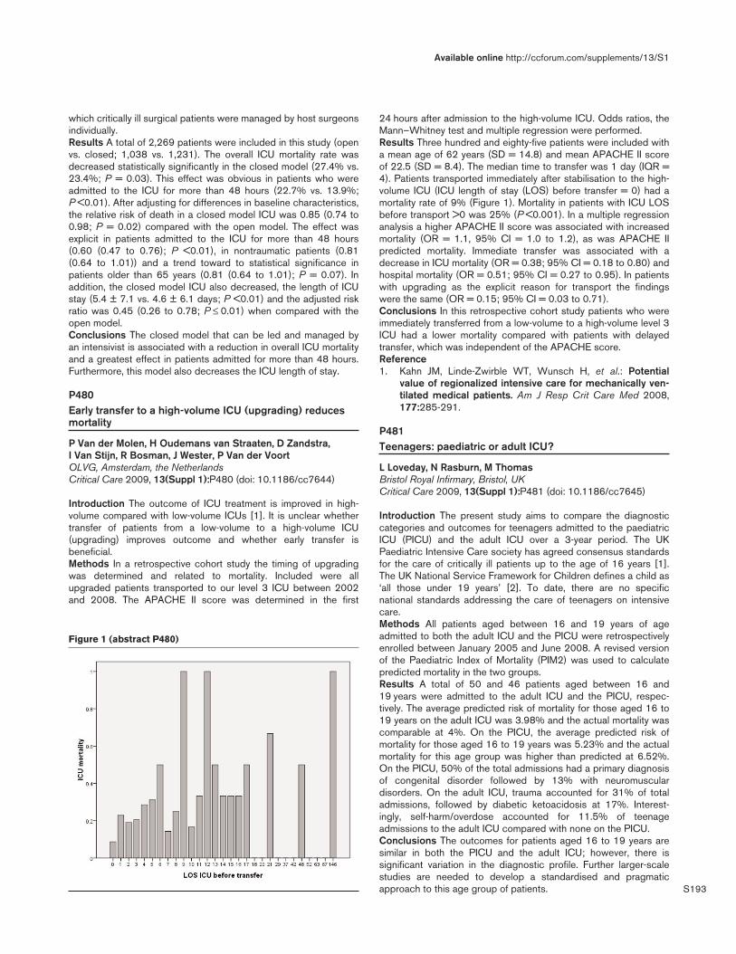
S193
which critically ill surgical patients were managed by host surgeonsindividually.Results A total of 2,269 patients were included in this study (openvs. closed; 1,038 vs. 1,231). The overall ICU mortality rate wasdecreased statistically significantly in the closed model (27.4% vs.23.4%; P = 0.03). This effect was obvious in patients who wereadmitted to the ICU for more than 48 hours (22.7% vs. 13.9%;P <0.01). After adjusting for differences in baseline characteristics,the relative risk of death in a closed model ICU was 0.85 (0.74 to0.98; P = 0.02) compared with the open model. The effect wasexplicit in patients admitted to the ICU for more than 48 hours(0.60 (0.47 to 0.76); P <0.01), in nontraumatic patients (0.81(0.64 to 1.01)) and a trend toward to statistical significance inpatients older than 65 years (0.81 (0.64 to 1.01); P = 0.07). Inaddition, the closed model ICU also decreased, the length of ICUstay (5.4 ± 7.1 vs. 4.6 ± 6.1 days; P <0.01) and the adjusted riskratio was 0.45 (0.26 to 0.78; P ≤ 0.01) when compared with theopen model.Conclusions The closed model that can be led and managed byan intensivist is associated with a reduction in overall ICU mortalityand a greatest effect in patients admitted for more than 48 hours.Furthermore, this model also decreases the ICU length of stay.
P480Early transfer to a high-volume ICU (upgrading) reducesmortality
P Van der Molen, H Oudemans van Straaten, D Zandstra, I Van Stijn, R Bosman, J Wester, P Van der VoortOLVG, Amsterdam, the NetherlandsCritical Care 2009, 13(Suppl 1):P480 (doi: 10.1186/cc7644)
Introduction The outcome of ICU treatment is improved in high-volume compared with low-volume ICUs [1]. It is unclear whethertransfer of patients from a low-volume to a high-volume ICU(upgrading) improves outcome and whether early transfer isbeneficial.Methods In a retrospective cohort study the timing of upgradingwas determined and related to mortality. Included were allupgraded patients transported to our level 3 ICU between 2002and 2008. The APACHE II score was determined in the first
24 hours after admission to the high-volume ICU. Odds ratios, theMann–Whitney test and multiple regression were performed.Results Three hundred and eighty-five patients were included witha mean age of 62 years (SD = 14.8) and mean APACHE II scoreof 22.5 (SD = 8.4). The median time to transfer was 1 day (IQR =4). Patients transported immediately after stabilisation to the high-volume ICU (ICU length of stay (LOS) before transfer = 0) had amortality rate of 9% (Figure 1). Mortality in patients with ICU LOSbefore transport >0 was 25% (P <0.001). In a multiple regressionanalysis a higher APACHE II score was associated with increasedmortality (OR = 1.1, 95% CI = 1.0 to 1.2), as was APACHE IIpredicted mortality. Immediate transfer was associated with adecrease in ICU mortality (OR = 0.38; 95% CI = 0.18 to 0.80) andhospital mortality (OR = 0.51; 95% CI = 0.27 to 0.95). In patientswith upgrading as the explicit reason for transport the findingswere the same (OR = 0.15; 95% CI = 0.03 to 0.71).Conclusions In this retrospective cohort study patients who wereimmediately transferred from a low-volume to a high-volume level 3ICU had a lower mortality compared with patients with delayedtransfer, which was independent of the APACHE score.Reference1. Kahn JM, Linde-Zwirble WT, Wunsch H, et al.: Potential
value of regionalized intensive care for mechanically ven-tilated medical patients. Am J Resp Crit Care Med 2008,177:285-291.
P481Teenagers: paediatric or adult ICU?
L Loveday, N Rasburn, M ThomasBristol Royal Infirmary, Bristol, UKCritical Care 2009, 13(Suppl 1):P481 (doi: 10.1186/cc7645)
Introduction The present study aims to compare the diagnosticcategories and outcomes for teenagers admitted to the paediatricICU (PICU) and the adult ICU over a 3-year period. The UKPaediatric Intensive Care society has agreed consensus standardsfor the care of critically ill patients up to the age of 16 years [1].The UK National Service Framework for Children defines a child as‘all those under 19 years’ [2]. To date, there are no specificnational standards addressing the care of teenagers on intensivecare.Methods All patients aged between 16 and 19 years of ageadmitted to both the adult ICU and the PICU were retrospectivelyenrolled between January 2005 and June 2008. A revised versionof the Paediatric Index of Mortality (PIM2) was used to calculatepredicted mortality in the two groups.Results A total of 50 and 46 patients aged between 16 and19 years were admitted to the adult ICU and the PICU, respec-tively. The average predicted risk of mortality for those aged 16 to19 years on the adult ICU was 3.98% and the actual mortality wascomparable at 4%. On the PICU, the average predicted risk ofmortality for those aged 16 to 19 years was 5.23% and the actualmortality for this age group was higher than predicted at 6.52%.On the PICU, 50% of the total admissions had a primary diagnosisof congenital disorder followed by 13% with neuromusculardisorders. On the adult ICU, trauma accounted for 31% of totaladmissions, followed by diabetic ketoacidosis at 17%. Interest-ingly, self-harm/overdose accounted for 11.5% of teenageadmissions to the adult ICU compared with none on the PICU.Conclusions The outcomes for patients aged 16 to 19 years aresimilar in both the PICU and the adult ICU; however, there issignificant variation in the diagnostic profile. Further larger-scalestudies are needed to develop a standardised and pragmaticapproach to this age group of patients.
Available online http://ccforum.com/supplements/13/S1
Figure 1 (abstract P480)
S194
References1. Paediatric Intensive Care Society: Paediatric Intensive Care
Society Standards Document. London: PICS; 2001.2. Department of Health: National Service Framework for Chil-
dren, Young People and Maternity Services. London: Depart-ment of Health; 2004.
P482Verbal orders drug therapy and prescription in a UK ICUsetting
S Evans1, P Mullen2
1Christie Hospital, Manchester, UK; 2Countess of Chester NHSFoundation Trust Hospital, Chester, UKCritical Care 2009, 13(Suppl 1):P482 (doi: 10.1186/cc7646)
Introduction Verbal orders (VOs) drug therapy refers to drugadministration without prescription, usually in the context oftelephone instructions from a physician at a remote location orother circumstances involving delayed prescription. In somemedical care settings VOs may constitute 2 to 13% of medicationorders [1]. Prescription errors associated with VOs are reputedlycommon [2], although some studies suggest otherwise [3].Although prescription errors are not uncommon in critical care [4],little is known about VOs in this setting.Methods We prospectively audited VOs in a hardcopy-prescribingfive-bed ICU setting over a period of 3 weeks, with voluntary dataentry by any member of staff issuing or receiving a drug therapy VO.We collected information regarding VO timing, medication involved,clinical indication, and subsequent drug event prescription.Results Nine VOs were recorded during multiple druginterventions for 19 ICU patients in this period, with a VO eventrate of 1 for every 10 ITU bed-days. All VOs were for intravenousprescription only medication, five of which were for fluid therapy totreat acute physiological deterioration. Other drugs includedmetoclopramide, furosemide, atracurium, and adrenaline. No VOsfor opiates were recorded. Reasons for initial nonprescriptionincluded remote location, aseptic procedure, and emergencytreatment. Eight prescriptions were completed within 2 hours. NoVOs were recorded during overnight shifts. All grades of medicalstaff issued VOs. One transcription error was noted. No adverseevents were noted.Conclusions Critical care VOs are not uncommon. Minimisationcan be achieved by good communication, hardcopy or electronicbundle prescribing, locally agreed independent nonphysicianprescribing arrangements, or drug administration in specificsettings, particularly pre-prescription of fluid bolus therapy.References1. Kaplan J, et al.: J Paediatr 2006, 149:461-467.2. Paparella S: J Emerg Nurs 2004, 30:157-159.3. West D, et al.: Arch Paediatr Adolesc Med 1994, 148:1322-
1326.4. Ridley S, et al.: Anaesthesia 2004, 59:1193-1200.
P483Effects of organizational change in the ICU on high-risksurgical patients: a comparison of open and closedformats
B Liebman, J Beute, C Slagt, JW Mulder, AF EngelZaans Medical Centre, Zaandam, the NetherlandsCritical Care 2009, 13(Suppl 1):P483 (doi: 10.1186/cc7647)
Introduction In 2000 the ICU in a teaching hospital in theNetherlands changed from open to closed format. In this
retrospective study, outcomes of high-risk surgical patients beforeand after the organizational change were compared.Methods From 1990 to 2004, all surgical patients wereprospectively collected in a database containing information aboutthe type of surgery, use of the ICU, length of hospital stay,complications, and mortality. Multivariate logistic regression wasused on these data to develop a risk prediction model for mortality.The model consisted of the variables acute hospital admission,acute surgery, severity of surgery and age. Severity of surgery wasbased on a classification of surgical operations used in teachinghospitals in the Netherlands, scoring operations as one to sevenpoints. More than three points was defined as severe. Age wasdivided into quartiles. Points were scored per variable. In total, sixpoints could be scored: acute admission, acute surgery and severesurgery received one point. Age scored zero to three points. A totalscore of four points or more was defined as high risk. The modelwas validated through cross-validation. The area under the receiveroperating characteristics curve was 0.9. ICU use, morbidity andmortality before and after the change from open to closed formatwere retrospectively evaluated.Results Two groups were defined: Group A: high-risk surgicalpatients in the ICU in 1996 and 1997 (open format, n = 230) andGroup B: high-risk surgical patients in the ICU in 2003 and 2004(closed format, n = 228). The primary outcome was inhospitalmortality. Secondary outcomes were inhospital morbidity andlength of ICU stay. The groups were comparable in age, genderand type of surgery.Mortality of the ICU patients was 25.4% in Group A and 15.9% inGroup B (P <0.01, 95% CI = 2.5 to 17.3). This was a relativechange of 37.6%. Morbidity was 48.3% in Group A and 46.3% inGroup B. The average length of ICU stay was 2.5 days in Group A,and 4.8 days in Group B (P = 0.001).Conclusions Changing an ICU organization to a closed formatreduces mortality in high-risk surgical patients, without affectingmorbidity.
P484Study of the ability of ICU staff to set a bed to thesemi-recumbent position
MG Keane, R KapoorKent and Canterbury Hospital, Canterbury, UKCritical Care 2009, 13(Suppl 1):P484 (doi: 10.1186/cc7648)
Introduction The aim of this study was to assess the accuracy ofsetting the angle of the head of a bed to the semi-recumbentposition. Ventilator-acquired pneumonias (VAPs) account for 31%of all ICU infections. Aetiology appears to be aspiration ofcontaminated oropharyngeal and gastric secretions around theendotracheal tube. The semi-recumbent position compared withthe supine position is known to significantly reduce the incidenceof VAP (8% vs. 34%) [1]. Hence the National Institute for ClinicalExcellence in the UK publishing a recommendation that ventilatedpatients be nursed at an angle of 30° to 45°.Methods Fifty-five members of ICU staff were asked to set theangle of the head of a standard ICU bed to 30° and 45°. Theestimated bed angle was then measured accurately using aMathey Dearman protractor.Results When estimating the head of the bed, nursing staff werethe most accurate (Table 1) while other staff significantlyunderestimated the angle of the bed. We also found that greaterexperience of working in the ICU is likely to improve ability toestimate the angle of the head of bed accurately.
Critical Care March 2009 Vol 13 Suppl 1 29th International Symposium on Intensive Care and Emergency Medicine

S195
Conclusions Evidence suggests patients should be nursed in thesemi-recumbent position to prevent VAP. However, we have foundestimating the angle of a bed by eye is difficult and ofteninaccurate. Simple aids that highlight the angle of the bed havebeen shown to improve compliance to the semi-recumbentposition by up to 71.5% compared with 21% in one study [2].Such aids to set the angle of the bed should be introduced incommon practice to improve compliance with VAP guidelines.References1. Drakulovic MB, et al.: Supine body position as a risk factor
for nosocomial pneumonia in mechanically ventilatedpatients: a randomised trial. Lancet 1999, 354:1851-1858.
2. Williams Z, et al.: A simple device to increase rates of com-pliance in maintaining 30-degree head-of-bed elevation inventilated patients. Crit Care Med 2008, 36:1155-1157.
P485Diagnosis-related group-based reimbursement isunrealistic for ICUs
A Mclaughlin, J Hardt, J Canavan, MB DonnellyAdelaide Meath Hospital, Dublin, IrelandCritical Care 2009, 13(Suppl 1):P485 (doi: 10.1186/cc7649)
Introduction The objective of this study was to compare theresults from a microcosting analysis of a cohort of ICU patientswith the reimbursement based on existing diagnosis-related group(DRG) systems. Hence, we open the discussion on resourceallocation using ICD-10 coding and its impact on intensive care.Methods A prospective study costing 58 consecutive admissionsover a 2-month period in a mixed medical/surgical ICU. Subse-quently, aligning these patient costs with the attributed costs usingICD-10 coding. Medical records in Ireland are coded using ICD-10for diagnoses and for procedures. Experienced clinical codersassign codes, which are entered into the code mapping program(AR-DRG V5.0).Results Our microcosting study demonstrated that the mediandaily ICU cost (IQR) was €2,205 (€1,932 to €3,073) and themedian total ICU cost was €10,916 (€4,294 to €24,091). Themicrocosting study demonstrated that the total ICU cost for 58admissions was €1,200,524. Reimbursement for the total hospitalstay including the ICU stay based on DRGs was €782,077.During our study, use of antifungals, hemodialysis and bloodproducts were found to be independent predictors of increasedICU cost. These frequently used intensive care treatments are notadequately costed.Conclusions Advances in intensive care diagnostics andtreatment contribute to the high cost of this specialty. CurrentDRG-based funding fails to adequately capture and cost ICUactivity, and thus underfunds intensive care. We recommend thatan ICU-specific DRG coding be developed for intensive care.
P486Waiting rooms and facilities available to patients’ familiesand visitors in Italian pediatric ICUs: a national survey
A Giannini1, G Miccinesi2, C Montani1, S Leoncino1
1Fondazione IRCCS Ospedale Maggiore Policlinico, Mangiagalli eRegina Elena, Milan, Italy; 2Centro per lo Studio e la PrevenzioneOncologica, Florence, ItalyCritical Care 2009, 13(Suppl 1):P486 (doi: 10.1186/cc7650)
Introduction Families of critically ill patients spend a considerableamount of time in hospital, both beside their loved ones in the ICUand outside the unit waiting to visit them or to receive news. Nopublished data are available to date on the provision of waitingrooms in Italy’s pediatric ICUs (PICUs) and on the facilitiesavailable to patients’ families and visitors. We investigated theseaspects in the course of a national survey concerning visitingpolicies in Italian PICUs.Methods An email questionnaire was sent to the heads of all 34Italian PICUs asking about their visiting policies. Questions aboutwaiting rooms and facilities for patients’ families and visitors werealso included.Results The response rate was 100%. The median daily visitingtime was 300 minutes (range 30 minutes to 24 hours). No waitingroom was provided in 32% of PICUs. In other PICUs, families andvisitors were provided with seats (50% of PICUs), armchairs(32%), lockers for personal effects (32%), magazines and books(18%), drinks machines (15%) and snack machines (6%). Abathroom was available to families and visitors in 41% of units, useof the PICU’s kitchen in 3% and access to the hospital canteen in47%.Conclusions Overall these data indicate that in Italian PICUs,alongside a clear tendency to apply restricted visiting policies,there is limited attention to the comfort of families of patients in thePICU. Comfort is one of five domains (in addition to reassurance,proximity, information and support) associated with the needs offamilies who have critically ill loved ones [1,2]. Our survey couldcontribute to promoting more attentive and supportive care for thepatient’s family [3].Acknowledgment Supported by Associazione per il BambinoNefropatico, Milan, Italy.References1. Leske JS: Needs of relatives of critically ill patients: a
follow-up. Heart Lung 1986, 15:189-193.2. Bijttebier P, et al.: Needs of relatives of critical care
patients: perceptions of relatives, physicians and nurses.Intensive Care Med 2001, 27:160-165.
3. Deitrick L, et al.: Evaluation and recommendations from astudy of a critical-care waiting room. J Healthc Qual 2005,27:17-25.
P487Patient satisfaction in the ICU: level of satisfaction andinfluencing factors
AC Jansen, M Van den Beld, M Goudriaan, HA Middelkoop, MS ArbousLeiden University Medical Center, Leiden, the NetherlandsCritical Care 2009, 13(Suppl 1):P487 (doi: 10.1186/cc7651)
Introduction To optimize patient care, knowledge of patientsatisfaction and its determinants is essential. So far few researchershave focused on this subject in intensive care medicine. Thepurpose of this study was to measure the level of patientsatisfaction and determine its influencing factors in ICU patients.
Available online http://ccforum.com/supplements/13/S1
Table 1 (abstract P484)
Mean estimates
Group 30° estimate 45° estimate
Nurses 30.4 43.1
Doctors 27.1 42.1
Other staff 26.1 39.6

S196
Methods In a pilot study, we developed a questionnaire thatconsisted of 60 questions in eight domains (General satisfaction,Reception, Physical care, Mental care, Empathy and attention,Communication and information, Surroundings and Physicaldiscomfort). Answering possibilities ranged from 1 (not satisfied) to5 (completely satisfied). In the final study, 23 March to 29 May2008, all consecutive adult patients from a 29-bed, tertiary-care,medical, surgical, neurosurgical and thoracic–surgical ICU wereinterviewed within 4 days after discharge. Demographics andclinical characteristics were obtained from electronic records.General linear modeling (GLM) with Bonferroni–Holmes correctionwas used to study the relationship between overall patientsatisfaction scores and the eight domains, corrected for patientand ICU-related factors. The model was tested for clinicallyrelevant interaction between determinants.Results Ninety-eight patients were interviewed. The mean overallpatient satisfaction score was 4.60 (SD = 0.93). Of the eightdomains, only Communication and information (P = 0.039)appeared to be significant in predicting general satisfaction. Alsosex (P = 0.046), length of ICU stay (P = 0.042), the interactionbetween Communication and information and age (P = 0.016), andthe interaction between Communication and information andMinimal Mental State Examination score (P = 0.013) were signifi-cant. An indepth analysis of Communication and informationshowed that the interaction of communication with birth country(P <0.0001) and with duration of mechanical ventilation(P = 0.041) was significant. This implies that, for Dutch versusnon-Dutch patients and patients longer on mechanical ventilation,clear communication with doctors and nurses was particularlyimportant.Conclusions Patient satisfaction was high in this mixed populationof ICU patients. From an especially developed questionnaire,Communication and information was the most important predictor.In addition, older age, female sex, Dutch nationality, longer ICU stay,long duration of mechanical ventilation and a high Minimal MentalState Examination score were related to less satisfied patients.
P488Use of deferred proxy consent in two emergency criticalcare trials
E Kompanje, T Jansen, J Le Noble, H De Geus, J BakkerErasmus MC University Medical Center, Rotterdam, theNetherlandsCritical Care 2009, 13(Suppl 1):P488 (doi: 10.1186/cc7652)
Introduction Almost all patients with life-threatening conditions areincapacitated to give informed consent for participation inemergency critical care research. Deferred proxy consent (DPC) isan ethically valid surrogate for informed patient consent. Here wepresent our experience in two emergency trials [1].Methods We analysed the use of DPC in the Early Lactate-directed Therapy in Intensive Care Trial (NCT 00270673) and theNGAL study (TC 1405).Results In the lactate-directed therapy trial, 362 patients wererandomized. In 75.4% DPC could be obtained, 3.9% of therelatives refused DPC and in 20.7% DPC could not be obtainedbefore the end of the study (= 72 hours). In the NGAL study, 550patients were randomized. DPC or patient consent was obtained in89.8%, 0.7% refused DPC and in 9.4% DPC could not beobtained due to early patient death. In both studies combined, onlyin 2% was DPC refused and in 10% consent could not beobtained due to early patient death.Conclusions DPC is an ethically valid and practically feasible wayto include patients in emergency critical care trials.
Reference1. Jansen TC, Kompanje EJO, Druml C, Menon D, Wiedermann
CJ, Bakker J: Deferred consent in emergency research:what if the patient dies early? Use the data or not? Inten-sive Care Med 2007, 33:894-900.
P489Outcome and prognostic factors in critically ill cancerpatients with diagnosis of sepsis in the ICU
R Kalil Filho1, L Hajjar1, M Carmona1, S Vieira2, F Galas1, C Simoes2, J Auler1
1Heart Institute, São Paulo, Brazil; 2Cancer Institute, São Paulo,BrazilCritical Care 2009, 13(Suppl 1):P489 (doi: 10.1186/cc7653)
Introduction As overall survival times increase for patients withcancer due to novel antitumour treatments, there will be aninevitable escalation in the number of such patients who present tointensive care with intercurrent acute illness, and sepsis is themost frequent diagnosis in this context. Epidemiologic estimates ofsepsis among cancer patients are limited. The aim of this studywas to assess survival in cancer patients admitted to an ICU with adiagnosis of sepsis, trying to identify predictive factors of mortality.Methods A prospective cohort study in an ICU at a universitycancer referral center. From a total of 95 patients admitted to theICU during a period of 3 months, 78 patients had the diagnosis ofsepsis into 24 hours of admission. Patient charts were analyzedwith respect to underlying disease, cause of admission, APACHE IIscore, need and duration for mechanical ventilation, neutropeniaand development of septic shock, type of infection, time to anti-biotic therapy, as well as ICU survival and survival after discharge.Results ICU survival of septic patients was 76%, and 1-monthsurvival was 66%. In a multivariate analysis, prognosis wasnegatively influenced by respiratory insufficiency, the need formechanical ventilation, and development of septic shock and renalfailure after 72 hours of ICU stay. Admission after cardiopulmonaryresuscitation yielded high ICU mortality. Age, neutropenia, type ofinfection, and underlying disease did not influence outcomesignificantly. Admission APACHE II scores were significantlyhigher in nonsurvivors but failed to predict individual outcomesatisfactorily.Conclusions A combination of factors must be taken into accountto estimate a critically ill cancer patient’s prognosis in the ICUadmitted because of sepsis. The APACHE II scoring system aloneshould not be used to make decisions about therapy prolongation.As ICU mortality is 24%, comparable with severely ill noncancerpatients, general reluctance to admit cancer patients to an ICUdoes not seem to be justified.References1. Farquhar-Smith WP, Wigmore T: Outcomes for cancer
patients in critical care. Curr Anesthesia Crit Care 2008, 19:91-95.
2. Danai PA, et al.: The epidemiology of sepsis in patientswith malignancy. Chest 2006, 129:1432-1440.
P490Toxic epidermal necrolysis: review of 15 cases
H Oueslati, K Bousselmi, I Rahmani, H Jihene, A MessadiTraumatology and Burn Center, Ben Arous, TunisiaCritical Care 2009, 13(Suppl 1):P490 (doi: 10.1186/cc7654)
Introduction Toxic epidermal necrolysis (TEN) or Lyell’s syndromeis a severe bullous skin disease induced by drugs. It is charac-
Critical Care March 2009 Vol 13 Suppl 1 29th International Symposium on Intensive Care and Emergency Medicine

S197
terized by an extensive skin rash with blisters and exfoliation similarto that of major burns. This study presents our experience oftreating and management of TEN in a burn center.Methods We retrospectively analysed the charts of all patientswith severe skin disease admitted to the burn center of Tunisbetween 2001 and 2008. The patients were evaluated accordingto their history of pre-existing diseases and medication, suspectedcause, extent of skin involvement (TBSA), mucosal involvement,definitive diagnosis, therapy, complications and outcome. Resultswere collected and examined with SPSS 16.0 software.Results During the study period, 21 patients with severe bullousskin disease were admitted. Fifteen of them had TEN (threepatients per year). All patients were referred from outsideinstitutions, especially from dermatology departments, meanhospital duration of 4 ± 2 days. The group of 15 patients consistedof 10 women and five men. The average age was 50 ± 17 years.The average affected skin area was 49 ± 15% of TBSA. Thesuspected causal agent was aspirin in three cases, phenobarbitalin three cases, allopurinol in three cases, antibiotics in two casesand paracetamol in one case. It was unknown in three cases. Itwas medical prescription in 10 cases, automedication in threecases and unknown in two cases. The mean time delay betweendrug intake and the onset of symptoms was 4.5 ± 1.5 days.Mucosal involvement was observed in all patients, such as ocular,gastrointestinal, respiratory and genital localizations. The mostcommon complications ware: sepsis, ARDS and hematologicaldisorders (two pancytopenia and one CIVD). The mean duration ofhospitalization was 9 ± 6 days. The overall mortality rate was 30%.There was a significant statistical difference between survival andnonsurviving patients, in involved skin area (P = 0.04) and septiccomplications (P <0.001).Conclusions TEN is a severe mucocutaneous reaction, associatedwith high morbidity and mortality. Management of theses patientsmust be provided cautiously by an experienced interdisciplinaryteam in an ICU.
P491Pesticide poisoning in the region of Rodopi, Greece
N Vavatsiklis, G Grigoriadis, C Diakalis, V Bagiazidou, I VavatsiklisGeneral Hospital of Komotini–Greece, Komotini, GreeceCritical Care 2009, 13(Suppl 1):P491 (doi: 10.1186/cc7655)
Introduction Pesticide poisoning constitutes a problem that oftenoccurs in the daily medical practice and mainly concerns the ruralareas of Greece. The purpose of this project is to mention theepidemic characteristics and our observations on unintentionalpesticide and herbicide poisonings in the prefecture of Rodopi,Greece.Methods In our retrospective study that refers to a period of2 years (January 2006 to December 2007), 152 patients sufferingfrom pesticide poisoning were offered medical treatment in thepathologic ward of the General Hospital of Komotini. One hundredand nineteen (78.30%) of them were patients who unintentionallysuffered pesticide poisoning. The remaining 20 (13.15%) werepoisoned with pharmaceutical substances of domestic use. Therewere also 13 (8.55%) cases of attempted suicide with intentionalintake of an overdose of antidepressants that were exempt fromour study.Results Fourteen out of the 119 patients (11.76%) were trans-ferred to hospital in a state of a coma (Glasgow coma scale <8)and were treated in the ICU and underwent blood refinery. Therewere two deaths (2/119 or 1.68%). The average age of ourpatients was 37 years (14 to 89). The proportion of men/women
was 2.4/1 (84/35) and the poisonings took place at the beginningof the summer.Conclusions Unintentional pesticide poisonings are frequent inour area. They concern people of low educational level, mainly menof a reproductive age [1,2]. The time spent from the intake of thepesticide to their transfer to hospital plays an important role in thesuccessful treatment of such cases [3]. Moreover, for a positiveoutcome in these severe cases it is essential that these patientsare treated in ICUs [3,4].References1. Rousos X: Drug Poisonings and Drug Coma. Intensive Care.
Athens: Paschalidi; 1997:440-454.2. Koppel C, et al.: Frequency of intoxications in elderly
patients. Int Care Med 1996, 22:155.3. Biernbaumer D: Poisoning and ingestions. In Current Criti-
cal Care Diagnosis and Treatment. Edited by Bongard FS,Sue DY. Appleton and Lange; 1994:686-715.
4. Krenzelok EP, Hall AH: Overview of 10 Years (1983–1992)of Poisoning Data. Yearbook of International Care and Emer-gency Medicine. Edited by Vincent JL. Berlin: Springer;1996;781.
P492Characterization of organophosphate poisoning patients inthe ICU: a 4-year review
A Sá, E Tomas, J Silva, M Fernandes, F Santos, F Moura, P Santos, R Lopes, E LafuenteCentro Hospitalar Tamega Sousa, Penafiel, PortugalCritical Care 2009, 13(Suppl 1):P492 (doi: 10.1186/cc7656)
Introduction We reviewed clinical characterization of patients withorganophosphate poisoning admitted to the ICU as well as themortality rate [1].Methods A retrospective study, without need for local ethicalcommittee approval, of patients with organophosphate poisoningwho were admitted to the ICU between 2004 and 2008.Sociodemographic data, psychiatric history, the organophosphatecompound, time elapsed since ingestion, clinical presentation andcomplications, laboratory data, Simplified Acute Physiology score(SAPS) II and treatment approach were analyzed. Biomarkers usedon admission: serum cholinesterase level, arterial pH and lacticacid. The influence of obidoxime and atropine therapy on thepatient outcome was also analyzed.Results Twenty patients were admitted to the ICU. Dimethoatewas the compound most often involved, in which 55% of thepatients were females. The mean age recorded was 54.3 ± 14.7years. The time between exposure and treatment initiation was notquantified in 65% of patients, but when discriminated it was in themajority of cases less than 1 hour. Clinical muscarinic signs in85% of the cases, nicotinic signs in 55% and central nervoussigns in 95%. An intermediate syndrome was identified in onecase. Most frequent complications identified were pneumonitis85%, miocarditis 35% and renal failure 50% of the patients. Themean SAPS II value was 36.6 ± 16.9. Selective decontaminationand treatment with obidoxime (continuous intravenous infusionafter the loading dose) and atropine was verified in every patient(Figure 1). The total ICU mortality was 40%.Conclusions Clinical severity and serum cholinesterase levels andacidosis were the independent variables that mostly seemed tohave contributed for the final result. High doses of atropineseemed to be more favorable to a good outcome. Even though thenumber of patients included in this review was small, the resultsmay alert us to the importance of an organized approach helped byprotocols between the emergency department/emergency room
Available online http://ccforum.com/supplements/13/S1

S198
and the ICU, in order to successfully manage patients withorganophosphate poisoning.Reference1. Peter JV: Oxime therapy and outcomes in human organo-
phosphate poisoning: an evaluation using meta-analytictechniques. Crit Care Med 2006, 34:502-510.
P493Metformin-associated lactic acidosis: three patientssurviving after continuous venovenous haemofiltration
F Barbani, A Di Filippo, M Linden, A Pasquini, R Cammelli, G Cianchi, A PerisCareggi Teaching Hospital, Florence, ItalyCritical Care 2009, 13(Suppl 1):P493 (doi: 10.1186/cc7657)
Introduction Metformin-associated lactic acidosis (MALA) is a life-threatening condition (up to 50% mortality is described [1]) and itsdeveloping rate increases with the number of contraindications tothe treatment.Methods We retrospectively observed cases of lactic acidosis,searching for MALA cases, admitted to the ICU from January 2008to November 2008.Results We present three patients. Patient 1 is a 63-year-ols manwith type 2 diabetes who came to the emergency department (ED)with leg pain for 2 days; he was on metformin 3 g daily. Laboratoryresults showed acute renal failure (ARF) and lactic acidosis (pH6.88; lactate 23 mmol/l). Angiography imaging showed acutebilateral lower arm ischemia. Continuous venovenous haemo-filtration (CVVH) was started due to ARF and acidosis (Table 1).Patient 2, a 77-year-old man with type 2 diabetes, came to the EDwith vomiting and diarrhea for 3 days. He was on metformin 3 gdaily. Laboratory results showed ARF, lactic acidosis (pH 6.80;lactate 18 mmol/l) and sepsis indexes (white blood cells19.000/μl, serum procalcitonin 10.85 μg/l); due to ARF andacidosis, CVVH was started. Patient 3 is a 72-year-old woman withtype 2 diabetes who came to the ED with diarrhea and vomiting for4 days; she was on metformin 3 g daily. Laboratory results showedleucocytosis, ARF and lactic acidosis (pH 6.83; lactate 16 mmol/l).
No signs of lactic acidosis differential diagnosis were found, soCVVH was started due to ARF and acidosis.Conclusions All three patients survived after support therapy andCVVH were started. In these cases, metformin could be theprincipal causal agent of acidosis or has a worsening clinicalcondition and accelerates acidosis development effect.Reference1. Misbin RI, et al.: N Engl J Med 1998, 338:265-266.
P494Epidemiology and nature of adverse events in the ICU
S Ouerghi1, S Frikha2, K Moncer2, T Mestiri1, M Ben Ammar2, M Mebazaa2
1Hospital A Mami, Ariana, Tunisia; 2Mongi Slim Hospital, La Marsa,TunisiaCritical Care 2009, 13(Suppl 1):P494 (doi: 10.1186/cc7658)
Introduction Critically ill patients require high-intensity care maybe at especially high risk of iatrogenic injury because they areseverely ill [1]. We sought to study the incidence and nature ofadverse events in the critical care setting.Methods We conducted a prospective 6-month observationalstudy in a medicosurgical ICU. We included patients whose lengthof stay was >24 hours. Reporters were asked to provide theidentification of the adverse events, date and time, and details withregard to the risk factors. The morbi-mortality was also recorded.Results A total of 250 patients were admitted to the ICU during anobservation period, 102 were included. The incidence of adverseevents was 59.8%. We found 222 adverse events (2.2 adverseevents per patient). The most common adverse events ascategorized by organ systems were metabolic (52%), cardio-vascular, respiratory and finally infectious. Among all adverseevents, only hyperglycaemia and hypoglycaemia were consideredto be iatrogenic adverse events. No association was observedbetween adverse events and mortality.Conclusions We found that the incidence of adverse events was59.8%. Some prior critical care safety studies have identified feweradverse events than this one (3 to 37%) [2]. This difference mayhave occurred because methods are not standardized; importantdifferences refer to the definitions of the concept of iatrogenic,modes of assessment (prospective, retrospective or transversal),population size, and the observation period. The high incidence ofadverse events in our study is due to the exhaustive assessment ofadverse events.References1. Giraud T: Iatrogenic complications in adult intensive care
units: a prospective two-center study. Crit Care Med 1993,21:40-51.
2. Soufir L: Ann Fr Anesth Reanim 2008, 27:59-63.
P495Drug–drug interactions in the ICU
S Ray1, M Bhattacharyya2, J Pramanik1, S Todi21Jadavpur University, Kolkata, India; 2AMRI Hospitals Kolkata, IndiaCritical Care 2009, 13(Suppl 1):P495 (doi: 10.1186/cc7659)
Introduction The present study intended to identify and analyzethe significance of drug–drug interaction (DDI) in critically illpatients.Methods All consecutive adult patients admitted to amedical/surgical ICU of a tertiary-care hospital, from September2006 to April 2007, were included in this prospective obser-vational study. Patients who stayed less than 48 hours were
Critical Care March 2009 Vol 13 Suppl 1 29th International Symposium on Intensive Care and Emergency Medicine
Figure 1 (abstract P492)
LOS, length of stay; CRP, C-reactive protein; ER, emergency room.
Table 1 (abstract P493)
Start CVVH 4 hours 24 hours (mmol/l) (mmol/l) (mmol/l)
Patient 1 – lactate 23.0 20.0 6.3
Patient 2 – lactate 10.3 3.1 2.4
Patient 3 – lactate 28.0 20.0 4.8

S199
excluded. All the drugs that were given during the ICU stay werechecked for the presence of potential interactions as per thereference manual. DDIs that were detected clinically or throughinvestigations were recorded and also any therapeutic actionstaken for DDIs were noted.Results The study included 400 patients (Table 1). Sixty-fourpercent (n = 256) of patients from the study cohort had potentialDDI. The total number of potential DDI found in all prescriptionswas 602; however, only in 34% (n = 208) was DDI actuallyobserved; of which a clinically relevant interaction (requiring drugmodification or other therapeutic intervention) was observed in49.5% (103 of 208). The average number of drugs used perpatients was nine (SD ±4) and potential DDIs found per patientwere two (SD ±2). The drug interactions were classified into threegroups, minor (55.29%), moderate (23.08%) and major (21.63%)according to the severity of and requirement for drug modification.Conclusions DDIs are common in the ICU population in thepresence of polypharmacy, and a substantial proportion of themare clinically relevant.
P496Outcome of patients with metastatic cancer orhaematological malignancy in intensive care
I Meynaar, S Sleeswijk Visser, L Dawson, P Tangkau, M Van Spreuwel-VerheijenReinier de Graaf Gasthuis, Delft, the NetherlandsCritical Care 2009, 13(Suppl 1):P496 (doi: 10.1186/cc7660)
Introduction We studied our ICU database to estimate the effectof metastatic cancer and haematological malignancy on hospitalmortality after intensive treatment.Methods The Reinier de Graaf Hospital is a 500-bednonacademic teaching hospital with a 30-bed dedicated medicaloncology ward supervised by a team of medical oncologists and a10-bed mixed intensivist-led ICU. Data on all patients admitted tothe ICU including illness severity scores, the presence of meta-static cancer or haematological malignancy and hospital mortalityare prospectively collected in the ICU database. We studied thehospital mortality of all consecutive patients in the databaseadmitted from 2004 until 2007 with respect to the presence orabsence of metastatic cancer or haematological malignancy.Results A total of 2,951 patients were admitted in the studyperiod. Patients with metastatic cancer or haematological malig-nancy were older and had higher illness severity scores onadmission to the ICU (Table 1). Not surprisingly, hospital mortality
was significantly higher in patients with metastatic cancer orhaematological malignancy. We performed logistic regressionanalysis to see whether the higher mortality could be explained bythe usual other confounders. After correction for APACHE IIexpected mortality, age, admission type, sepsis and mechanicalventilation, we found however that the presence of metastaticcancer or haematological malignancy independently increases therisk of dying (OR = 1.7 (95% CI = 1.1 to 2.8, P = 0.024)).Conclusions Hospital mortality is higher in patients with metastaticcancer or haematological malignancy than in other patients, evenafter correction for age, APACHE II expected mortality, admissiontype, sepsis and mechanical ventilation.
P497Mortality rate of vasopressor-dependent septic shock from1997 through 2006
B Afessa, G Schramm, R Kashyap, O GajicMayo Clinic, Rochester, MN, USACritical Care 2009, 13(Suppl 1):P497 (doi: 10.1186/cc7661)
Introduction Recently introduced interventions including earlygoal-directed therapy (EGDT) improve the mortality of severesepsis/septic shock. We undertook this study to describe thetrend in the mortality rate of vasopressor-dependent septic shockover a 10 year-period.Methods We used the APACHE III database of our institution toidentify patients with septic shock. Patients with an ICU admissiondiagnosis of infection and who were on a vasopressor on the firstICU day were considered to have vasopressor-dependent septicshock. We divided the study period into pre-EGDT (1997 to2000), transit (2001, 2002), and post-EGDT (2003 to 2006). Wecalculated the standardized mortality ratio (SMR) with the 95%confidence interval.Results Of 53,096 admissions, 4,981 (9.4%) had a diagnosis ofinfection. The hospital mortality rates of the patients with andwithout infection were 21.2% and 9.3%, respectively (P <0.001).Vasopressor was infused in 1,340 (26.9%) of the patients withinfection. The hospital mortality rate of the vasopressor-receivingpatients with infection was 32.4% compared with 17.1% of thosewho did not receive vasopressor (P <0.001). The SMR of thevasopressor-dependent septic patients was lowest during thepost-EGDT period (Table 1).
Available online http://ccforum.com/supplements/13/S1
Table 1 (abstract P495)
Demographics of study population
Age (mean ± SD) (years) 61 ± 16.9
Male:female 239:161
APACHE II (mean ± SD) 16 ± 8
Table 1 (abstract P496)
MC/HM no MC/HM yes P value
Number 2,805 146
Mean age 66.1 (16.5) 68.5 (11.5) <0.001
Mean APACHE II score 13.6 (7.6) 17.2 (6.8) <0.001
APACHE II expected mortality 19.5% 28.2% <0.001
Mortality 439 (15.7%) 39 (26.7%) 0.001
MC/HM, metastatic cancer or haematological malignancy.
Table 1 (abstract P497)
Mortality of vasopressor-dependent septic shock during the study period
Study period Number Predicted mortality (%) Mortality (n (%)) SMR (95% CI)
Pre-EGDT 417 31.5 133 (31.9) 1.015 (0.850 to 1.203)
Transit 285 37.7 117 (41.1) 1.093 (0.904 to 1.310)
Post-EGDT 638 40.0 184 (28.8) 0.722 (0.621 to 0.834)
S200
Conclusions The hospital mortality rate of patients admitted to theICU for infection is higher than that of patients admitted for otherreasons. The hospital survival rate of patients with vasopressor-dependent septic shock has improved in the post-EGDT era.
P498Patients with severe sepsis and solid tumours: outcomeand prognostic factors
S Pereira, C Dias, A Martins, S Quintela, F FariaInstituto Português de Oncologia de Francisco Gentil, EPE, Porto,PortugalCritical Care 2009, 13(Suppl 1):P498 (doi: 10.1186/cc7662)
Introduction Admission to the ICU and the appropriate selectionof patients improved the treatment and survival rates of criticaloncological patients [1]. Infection is an important complication inthese patients, which frequently leads to acute organ dysfunctionand even death [2,3]. The objective is to evaluate the outcome andprognostic factors in a group of patients with severe sepsis andsolid tumours.Methods A retrospective observational study of collected data in140 consecutive admissions to the ICU over a 24-month period atour oncological hospital; 108 patients were evaluated, corres-ponding to 140 admissions to the ICU (19.5% of total admissions).Results Age, sex, the underlying disease and tumour stage (TNMclassification), type of previous anticancer treatment, performancestatus, severity scores (APACHE II, Simplified Acute PhysiologyScore II, Sequential Organ Failure Assessment), ICU and hospitalmortalities and hospital outcome at 3, 6 and 12 months wereanalysed. The majority were male with median age 64.5 years;26% had head and neck tumours, 21% oesophagus and gastriccancers, 16% colon and rectum cancer, 9% breast cancer, 1%genitourinary tumours and 14% other solid tumours; 91.4% ofpatients required mechanical ventilation, 42% vasopressors, and4% kidney replacement therapy. Decisions to forgo (withhold orwithdraw) life-sustaining therapies were taken in 19% of patients.In the patients with the intention to treat, the ICU and hospitalsurvival rates were 76.5% and 56.3%. Survival rates at 3, 6 and 12months were 97%, 81% and 45%, respectively.Conclusions Severity scores (APACHE II, Simplified AcutePhysiology Score II, Sequential Organ Failure Assessment) in thefirst 24 hours did not discriminate clearly between survivors andnonsurvivors [1]. Mortality for cancer patients with severe sepsisturned out to be unrelated to age [4]. Clinical judgment betweenoncologists and intensive care physicians is necessary to improvethe selection of patients who might benefit from intensive care [5].Long-term survival is predicted by cancer disease-prognostic factors.References1. Soares M, et al.: Intensive Care Med 2007, 33:2009-2013.2. Azoulay E, et al.: Intensive Care Med 2006, 32:3-5.3. Disis M, et al.: J Clin Oncol 2005, 23:8923-8925.4. Thiéry G, et al.: J Clin Oncol 2005, 23:4406-4413.5. Williams M, et al.: Crit Care 2004, 8:291-298.
P499Evolution of acute lung injury/acute respiratory distresssyndrome mortality in an ICU in Southern Brazil
L Fialkow, M Farenzena, S Vieira, M BozzettiHospital de Clinicas de Porto Algre, Porto Alegre, BrazilCritical Care 2009, 13(Suppl 1):P499 (doi: 10.1186/cc7663)
Introduction Acute lung injury (ALI), including acute respiratorydistress syndrome (ARDS), mortality rates are high despite
advances in the management of patients with this condition. Ourobjectives were to compare the ALI/ARDS mortality rates and theALI mortality risk factors between two cohort studies of the sameICU in different periods of time: 1999/2000 (2000 cohort [1]) and2004/2007 (2007 cohort).Methods A prospective cohort that included 1,115 patientsadmitted to the ICU between April 2004 and April 2007 whoneeded mechanical ventilation for at least 24 hours. These resultswere compared with a previous cohort of 1,301 patients admittedto the same ICU between April 1999 and April 2000 [1]. In bothstudies all patients were evaluated regarding the presence/development of ALI/ARDS according to the 1994 American–European Consensus Conference. Data were compared betweenthe two cohorts using Student’s t or chi square tests. Multivariateanalysis by conditional logistic regression was used to identify ALImortality risk factors.Results In ALI patients (n = 50, 2000 cohort; n = 347, 2007cohort) we observed: (i) an increase in both hospital mortality (from50 to 67%, P = 0.02) and ICU mortality (from 44 to 63%,P = 0.01); (ii) a higher mean (±SD) APACHE II score (from 17.7 ±6.5 to 23.7 ± 8.2, P <0.001), and (iii) an increased percentage ofpatients with three or more organ failures (from 30 to 42%,P <0.001). Regarding ARDS patients (n = 30, 2000 cohort;n = 307, 2007 cohort), there were an increase in hospital mortality(from 47% to 68%, P = 0.03), in the mean (±SD) APACHE IIscore (from 17.6 ± 6.9 to 23.6 ± 8.3, P <0.001) and in medicalpatients (from 46 to 73%, P = 0.01). The factors independentlyassociated with increased hospital mortality in ALI patients wererenal (P = 0.002) and hematological (P = 0.02) failures in the 2000cohort. In the 2007 cohort, renal (P = 0.01) and hematological(P = 0.02) failures, use of vasoactive drugs (P <0.001) and ICUlength of stay (P <0.001) were independently associated withmortality.Conclusions We observed an increase in the mortality rates ofpatients with ALI/ARDS, which are more severely ill. Renal andhematological failures remained independently associated withmortality in these patients. The results suggest that ALI/ARDSpatients in our ICU have more severe illness, what could explainthe increase in the mortality rates, highlighting the relevance of abetter understanding of such findings.Reference1. Fialkow L, et al.: Acute lung injury and acute respiratory
distress syndrome at the intensive care unit of a generaluniversity hospital in Brazil: an epidemiological studyusing the American-European Consensus Criteria. Inten-sive Care Med 2002, 28:1644-1648.
P500Demographic changes over a 12-year period in intensivecare
V Hariharan, JJ PaddleRoyal Cornwall Hospital, Truro, UKCritical Care 2009, 13(Suppl 1):P500 (doi: 10.1186/cc7664)
Introduction We describe the demographic characteristics ofpatients admitted to the ICU of a large general hospital over thepast 12 years and the severity of their condition on admission,examining the relationships between these changes and trends inmortality and length of stay (LOS).Methods All patients admitted to the ICU from 1 January 1996 to31 December 2007 were included in this retrospective study. Wecaptured data on age, sex, admission category, APACHE IIpredicted mortality, duration of ICU stay, and hospital mortality.Statistical analysis was conducted with SPSS version 15.0 using
Critical Care March 2009 Vol 13 Suppl 1 29th International Symposium on Intensive Care and Emergency Medicine

S201
logistical regression models. To investigate trends in mortalityindependent of demographic changes in the ICU population,expected deaths based on the age–sex mortality experienceacross the whole 12-year period were calculated for each year.Expected mortality rates based on APACHE II probability of deathscores were also calculated for each year.Results In total 7,158 patients were included in this study. Therewas no significant trend in median age across the 12 yearsstudied; however, there has been a steady increase in theproportion of ICU patients over the age of 80. In 1996, 6% wereover 80 years old and by 2008 it was 12.5%. There was nosignificant trend in the sex distribution of ICU patients (60:40 maleto female). The LOS for medical and emergency surgical patientsis similar, with an average of 6 days. However, the LOS of electivesurgical patients has increased from around 3 to 4 days. Themedian APACHE II probability of death for ICU patients increasedover the period, the significant linear trend (P = 0.001, logisticregression) showing an increase of almost 1% per year. Thehospital mortality over the study period was 20%. During thisperiod mortality rates rose in correlation with predicted mortality,before starting a downward trend in 2003 to eventually fall belowAPACHE II predicted levels in 2007.Conclusions The average age of ICU admissions is not signifi-cantly increasing over time. However, the proportion of patientsaged over 80 has almost doubled to 12.5%. The severity of illnesshas increased and this is seen across all categories of admission,but seems to be due particularly to an increase in the subgroup ofover 80 year olds. There has been no consistent change inmortality rates over the 12-year period.
P501Clinical characteristics and prognostic factors of severeacute pancreatitis
B Kostadinovska, MS SosolcevaCity Surgical Clinic ‘St Naum Ohridski’ – Skopje, MacedoniaCritical Care 2009, 13(Suppl 1):P501 (doi: 10.1186/cc7665)
Introduction The aim of this study was to investigate the clinicalcharacteristics and prognostic factors of a consecutive series ofpatients with severe acute pancreatitis (SAP). SAP is charac-terized by high morbidity and mortality.Methods Clinical data of SAP patients admitted to our hospitalfrom January 2006 to January 2008 were retrospectively reviewed.Collected data included the age, gender, etiology, length ofhospitalization, APACHE II score at admission, local and organ/systemic complications of the patients.Results Of the 125 acute pancreatitis patients, 42 developed SAP[1]. The mean age of SAP patients was 57 years, male/female22/20 patients, the commonest etiology was cholelitiasis (43.5%),the mean length of hospitalization was 60 days, the mean score ofAPACHE II was 8.1. Fifty-seven percent of the patients developednecrosis, 23% abscessus, 11% pseudocyst. Fifty-eight percentdeveloped organ/systemic failure. A total of 25.2% (11/42) of theSAP patients died. Respiratory failure was the most common organdysfunction (90.0%) in deceased SAP patients, follow bycardiovascular failure (86.4%), and renal failure (50.0%). In theSAP patients, 90.9% (9/11) developed multiple organ/systemicfailures. There were significant differences in age, length ofhospitalization, APACHE II score and incidences of respiratoryfailure, cardiovascular failure, renal failure between deceased SAPpatients and surviving SAP patients [2]. By multivariate logisticregression analysis, the independent prognostic factors formortality were respiratory failure, cardiovascular failure and renalfailure.
Conclusions SAP patients are characterized by advanced age,high APACHE II score and organ failure, and their death is mainlydue to multiple organ/systemic failures. In patients with SAP,respiratory, cardiovascular and renal failure can predict the fataloutcome and more attention must be paid to their clinicalevaluation.References1. Isenman R, et al.: Early severe acute pancreatitis. Pancreas
2001, 22:274-278.2. Company L, et al.: Factors predicting mortality in severe
acute pancreatitis. Pancreatology 2003, 3:144-148.
P502ICU admittance prediction of the seventh-day organdysfunction clustering
E Esprooz, F Sacco, G Testa, RA De BlasiSant’Andrea Hospital, Rome, ItalyCritical Care 2009, 13(Suppl 1):P502 (doi: 10.1186/cc7666)
Introduction Organ dysfunction is usually evaluated by gravityscores (Logistic Organ Dysfunction Score, Multi-Organ Dys-function Score and Sequential Organ Failure Assessment), aimedto predict the patient’s hospital outcome [1,2]. Despite theirdiffusion, these scores do not provide indications about thetreatment’s efficacy for each patient. Our aim was to utilize theprevious organ dysfunction scores to perform early strategies topreserve organ function. This study tested the following hypo-thesis: first, that the previous organ dysfunction scores can detectclustering of organ dysfunctions; second, that each clusteringcould have an own sequence; and, finally, that we can develop amodel to predict from the scores at ICU admittance the organdysfunctions after the seventh day.
Available online http://ccforum.com/supplements/13/S1
Table 1 (abstract P502)
Percentage error of model prediction for each organ
Organ %
Liver 18
Heart 14
Kidney 17
Lung 12
Haematic 12
Nervous 5
Figure 1 (abstract P502)

S202
Methods We retrospectively collected scores, at admission anddaily for 6 days, of 85 ICU patients admitted in a 6-month period atSant’Andrea University Hospital of Rome, Italy. The inclusioncriteria were: at least two organ dysfunctions measured by one ofthe three scores and ICU stay ≥24 hours. Data were processed bycluster analysis (VARCLUS di SAS System). We used adiscriminant linear model for the prediction of organ dysfunction.Results The results are reported in Table 1 and Figure 1. The scoreswe used detect three clusters of organ dysfunction characterizedby a different outcome. The model we elaborate allows one topredict with great accuracy the organ dysfunction over time.Conclusions The present study can provide the clinician with auseful tool to enact therapeutic strategies to prevent additionalorgan damage.References1. Levy MM, Macias WL, Vincent JL: Early changes in organ
function predict eventual survival in severe sepsis. CritCare Med 2005, 33:2194-2201.
2. Lopes Ferreira F, Peres Bota D, et al.: Serial evaluation ofthe SOFA score to predict outcome in critically ill patient.JAMA 2001, 286:1754-1758.
P503Serum lactic acid in the evaluation of outcome in the ICU
A Khalaf, N Abouchala, M Jamil, M KherallahKing Faisal Specialist Hospital and Research Center, Riyadh, SaudiArabiaCritical Care 2009, 13(Suppl 1):P503 (doi: 10.1186/cc7667)
Introduction The purpose of this study was to determine the utilityof an initial serum lactate measurement for identifying high risk ofdeath in patients admitted to the ICU.Methods In a cohort study at a tertiary-care nontrauma medical–surgical ICU, we included all ICU admissions (May 2007 toFebruary 2008) from a prospectively collected ICU database.Patients’ gender, age, admission physiologic measurements, lacticacid, and APACHE IV score were noted. Outcome was acutephase death (death within 3 days of admission) and inhospitaldeath. Outcome was stratified by the initial serum lactic acid levelin three groups: low, 0.0 to 2.0; intermediate, 2.1 to 3.9; high,4.0 mmol/l or above.Results Total of 882 patients were included in the study, acutephase death rates were 2.5%, in the low group, 5.1% in inter-mediate group and 8.1% in the high group, whereas inhospitalmortality rates were 13.9%, 28.6%, and 38.7%, respectively.Acute-phase deaths and inhospital deaths increased linearly withlactate. An initial lactate ≥4.0 mmol/l was associated with at least athreefold higher odds ratio for both acute-phase deaths 2.8 (95%CI = 1.1 to 7.3) and inhospital mortality 3.4 (95% CI = 2.0 to 5.8).Conclusions An initial lactate ≥4.0 mmol/l substantially increasesthe probability of acute-phase death and inhospital mortality incritically ill patients.
P504APACHE II score may predict mortality in burns patients
R Martynoga, M FriedSt John’s Hospital, Livingston, UKCritical Care 2009, 13(Suppl 1):P504 (doi: 10.1186/cc7668)
Introduction The APACHE II scoring system has not beenvalidated in burn injuries. The validation studies were performed inthe general and cardiac ICUs of teaching hospitals in the USA;units that did not admit burn injuries [1]. We correlated known
predictors of mortality in burns (age, percentage total burn surfacearea (%TBSA), inhalation injury) [2,3] with mortality data in ourburns cohort. This was repeated for the APACHE II score toinvestigate its predictive value.Methods We identified all patients with burns admitted to our ICU,linked to the South East Scotland Regional Burns Unit, from 1995to 2005. Demographic data and APACHE II score at 24 hourswere found on the ICU database. The case notes for each patientwere reviewed to verify the %TBSA and survival.Results The cohort comprised 100 patients: 75% male. The agerange was 14 to 87 years (mean 45.2; median 45). Injury severitywas assessed by %TBSA: range 0 to 99% (mean 23.8%, median16%, IQR = 8.6 to 30%). Zero %TBSA is inhalation injury only.APACHE II scores ranged from 7 to 42 (mean 15.9, median 15,IQR = 10 to 19.5). The apparent fall in mortality at scores 27 to 31is due to low numbers (n = 3) in this group. Both the %TBSA andAPACHE II score correlate with increasing mortality (Figure 1).Stepwise logistic regression analysis was performed: %TBSA andAPACHE II score both behaved as independent predictors ofmortality (P <0.0001), as did age (P = 0.002). This cohort behavesas other published data [2,3].Conclusions Scoring systems with applicability to all critically illpatients provide a standardised objective assessment tool allowingcomparison between groups, and can inform clinical decision-making. The strong correlation shown between APACHE II scoreand mortality provides evidence for the utility of APACHE II scoringin critically ill patients with burn injuries.References1. Knaus W, et al.: APACHE II: a severity of disease classifica-
tion system. Crit Care Med 1985, 13:818-829.2. Bull J: Revised analysis of mortality due to burns. Lancet
1971, 2:1133-1134.3. Ryan C, et al.: Objective estimates of the probability of
death from burn injuries. N Engl J Med 1998, 338:362-366.
P505Scoring systems (APACHE II and Simplified AcutePhysiology Score II) for predicting outcome in statusasthmaticus
A Hachimi, B Charra, A Benslama, S MotaouakkilHôpital Ibn Rochd, Casablanca, MoroccoCritical Care 2009, 13(Suppl 1):P505 (doi: 10.1186/cc7669)
Introduction The scoring systems (SS) can be used globally toestimate the severity of critically ill patients. In status asthmaticus(SA), these SS were not evaluated to predict mortality. Our aim is
Critical Care March 2009 Vol 13 Suppl 1 29th International Symposium on Intensive Care and Emergency Medicine
Figure 1 (abstract P504)

S203
to evaluate the ability of nonspecific scores (APACHE II andSimplified Acute Physiology Score (SAPS) II) to predict outcomein SA in a medical ICU.Methods A prospective and monocentric study during 8 years(2000 to 2007) included all patients who were admitted to our unitfor SA. Demographic and clinical data were collected at admission.We collected also therapeutic aspects in all patients. Daily, a follow-up of the SS value was calculated with their average. We defined asubgroup that has been mechanically ventilated to evaluate the valueof SS in SA, because mechanical ventilation is a main prognosticfactor among others. Modulus evaluation required a discriminativeapplication using receiver operating characteristic curves. Thestatistic analysis has been based on SPSS 11.0 for windows.Results A total of 246 cases of SA (9.7% of all patients admittedin our unit) have been counted during the duration of study. Themean value of APACHE II and of SAPS II was 8 ± 5 and 20 ± 10,respectively. The mortality rate was 12%. In the subgroup, 90% ofpatients had APACHE II score ≤8 and 77% of them had SAPS II<20. Both scoring systems had a weak discrimination, with an areaunder the curve (receiver operating characteristic) 0.655 and0.557 for SAPS II and APACHE II, respectively.Conclusions APACHE II and SAPS II are not good prognostictools for status asthmaticus. A new and more adapted evaluationtool is required.
P506Is Simplified Acute Physiology Score 3 better thanAPACHE II to predict mortality in transplanted criticalpatients?
VO Oliveira1, E Rodrigues Filho2, J Brauner1, S Regina Rios Vieira1
1Hospital de Clinicas de Porto Alegre, Brazil; 2ComplexoHospitalar da Santa Casa de Porto Alegre, BrazilCritical Care 2009, 13(Suppl 1):P506 (doi: 10.1186/cc7670)
Introduction The performance of general prognostic models inpatients with transplantation in need for ICU admission is poor,showing a tendency towards significant underestimation of the riskof dying. The objective of our study is to evaluate the APACHE IIscore and Simplified Acute Physiology Score (SAPS) 3 and their90-day mortality prediction after liver, renal and pulmonarytransplantation.Methods A prospective cohort study in a transplantation ICU inPorto Alegre, Brazil, during the period May 2006 to July 2007.Clinical data of 277 post-transplantation patients admitted to theICU were collected at admission and the SAPS 3 and APACHE IIscore calculated with respective estimated mortality rates. Thearea under the receiver operating characteristic curve (AUROC)was obtained for both scores.Results Patients enrolled included 172 men and 105 women, withmean age 45 ± 2 years. There were 86 (31%) liver transplanta-tions, 170 (61.3%) renal transplantations and 21 (7.6%) pulmo-nary transplantations. The 90-day mortality for liver, renal andpulmonary transplantation was: 12 (15.4%), four (3%) and four(26.3%). The mean SAPS 3 score was 40 ± 10.2, 21 ± 5.8 and63 ± 19, and the APACHE II score was 25 ± 8.2, 16 ± 5.9 and17 ± 21.4, respectively. Comparing the AUROC from both scoreswe observed: in liver transplantation the AUROC for SAPS 3 was0.677 (95% CI = 0.447 to 0.887) versus 0.749 (95% CI = 0.623to 0.875) for APACHE II; in renal transplantation the AUROC forSAPS 3 was –0.619 (95% CI = 0.355 to 0.882) versus 0.604(95% CI = 0.355 to 0.882) for APACHE II; in pulmonary trans-plantation the AUROC for SAPS 3 was –0.936 (95% CI = 0.805to 1.06) versus 0.750 (95% CI = 0.489 to 1.01) for APACHE II.
Conclusions In these preliminary results, no differences wereobserved comparing SAPS 3 and APACHE II in the mortalityprediction from liver, renal and pulmonary transplanted patients.References1. Higgins TL: J Intensive Care Med 2007, 22:141-156.2. Capuzzo M, et al.: Curr Opin Crit Care 2008, 14:485-490.3. Zimmerman JE, et al.: Curr Opin Crit Care 2008, 14:491-497.
P507Inter-rater reliability of APACHE II scores in the medical ICU
X Hu, L Weng, J Peng, D Yu, B DuPeking Union Medical College Hospital, Beijing, ChinaCritical Care 2009, 13(Suppl 1):P507 (doi: 10.1186/cc7671)
Introduction The aim of this study was to determine inter-raterreliability of APACHE II scores between an ICU specialist and awell-trained ICU fellow.Methods In a prospective observational study, two raters collectAPACHE II scores on 50 consecutive patients in a medical ICUrespectively. Intraclass correlation coefficients were calculated forthe APACHE II total score, the APACHE II component scores, andthe Glasgow coma scale components. Concordance correlationcoefficients for each chosen value of the APACHE II componentwere assessed.Results Mean (standard deviation) APACHE II scores were 21.6(8.2) for the specialist, and 21.7 (7.1) for the fellow. The intraclasscorrelation coefficient was 0.848 for the APACHE II total score.Within the score components, the inter-rater reliabilities of acutephysiology score, age, and chronic health evaluation were 0.860,0.987, and 0.645, respectively. See Table 1. Further analysis ofeach chosen value of the APACHE II component found the lowestreliability for the mean artery pressure, which was 0.482. Furtherinvestigation discovered that the most common reason for inter-rater difference is data dropout.
Table 1 (abstract P507)
Overall agreement
Items Interclass correlation coefficient
APACHE II score 0.848
Acute physiology score 0.860
Age 0.987
Chronic health evaluation 0.645
Conclusions The agreements of the APACHE II score among thetwo collectors was good, but there were more differences incollecting the value of mean artery pressure, for which the originaldata could not be read directly from medical chart.
P508Validation of Simplified Acute Physiology Score II andSimplified Acute Physiology Score III as mortality andmorbidity risk models
F Franchi, L Cubattoli, P Mongelli, C Porciani, M Nocci, E Casadei, S Scolletta, P GiomarelliUniversity of Siena, ItalyCritical Care 2009, 13(Suppl 1):P508 (doi: 10.1186/cc7672)
Introduction Simplified Acute Physiology Score (SAPS) II andSAPS III are valuable scoring systems used to predict the risk ofmortality in the ICU. The main purposes of this study were toassess the strength of both the scores in predicting the mortality
Available online http://ccforum.com/supplements/13/S1

S204
risk, to check which one gained the best performance, and toascertain whether they also achieved a good level of morbidity riskprediction in our patient population.Methods Two hundred and forty-one patients (82 female, 159male, mean age 53 ± 23), out of 393 admitted to our ICU over12 months, were included in the study. Exclusion criteria were age<18, and ICU stay <24 hours. SAPS II and SAPS III wereprospectively collected. The derived probability of death wascalculated according to the original descriptions. To evaluate thecapability of morbidity risk prediction of these scoring models, theGIVITI definitions (Italian Group for the Evaluation of Interventionsin the ICU) of organ dysfunction and failure were used. Univariateand multivariate analyses were applied. The area under the receiveroperating characteristic curve (AUC) was calculated.Results Median values for SAPS II and SAPS III were 35 and 58,respectively. Mortality and morbidity rates resulted 16% and40.5%, respectively. The mean ICU stay was 9 ± 10 days.Although the univariate analysis found similar statistical signifi-cances (P <0.001) for both the scores, at the multivariate analysisonly SAPS II maintained a statistical significance (P <0.001) inpredicting the probability of death. Moreover, SAPS II showed asignificantly higher AUC (0.91 vs. 0.73, SAPS II vs. SAPS III,respectively, P <0.05). With respect to morbidity, the AUC of boththe scores gained a poor level of predictive power (AUCs = 0.618and 0.605, SAPS II and SAPS III, respectively). However, in thesubgroup of patients admitted to our ICU for major trauma (41% ofpatients), SAPS III reached a high degree of discriminativestrength in predicting the morbidity rate (AUC = 0.81, P <0.01).Conclusions SAPS II seemed a suitable tool in predicting the riskof death, but not morbidity. Conversely, SAPS III, even though itdid not gain a sufficient degree of predictive power towardmortality, could be a useful morbidity risk model when applied tomajor trauma patients. In our ICU, SAPS II and SAPS III are notinterchangeable, but they should be carefully used in the rightcircumstances. This might avoid the pitfalls leading to errors inforecasting mortality and morbidity risk.
P509Performance of the APACHE IV system in patients withacute renal failure
T Dahhan, I Al-Sanouri, H Al-Awadhi, M Jamil, M KherallahKing Faisal Specialist Hospital and Research Center, Riyadh, SaudiArabiaCritical Care 2009, 13(Suppl 1):P509 (doi: 10.1186/cc7673)
Introduction Scoring systems represent classification systems orpoint systems that have been designed for making quantitativestatements regarding the severity of a disease, its course and itsprognosis [1]. These systems are based on physiologic abnor-malities and have been successful in measuring severity of illnessamong critically ill patients [2]. The APACHE IV system has alreadybeen validated in our population at King Faisal Specialist Hospitaland Research Center [3], and the purpose of this study is toevaluate the performance of this system in a subset of patients withacute renal failure.Methods Fifty-four consecutive patients with acute renal failuredefined as a creatinine value ≥1.5 mg/dl and urine output<410 ml/24 hours were admitted to a medical/surgical ICUbetween January 2007 and March 2008 and were enrolled in thestudy. The length of stay and mortality percentages were predictedusing APACHE IV, and both variables were compared with theobserved data. The Wilcoxon rank sum test is used to calculatestatistical significance for continuous data and the chi-square testfor categorical data.
Results A total of 54 patients with mean age of 53.8 years wereincluded. Fifty-six percent were males; 62% of patients weremedical, 38% were surgical. The predicted ICU length of stay of6.3 days (SD = 2, range of 2.1 to 10.2) was insignificantly lowerthan the observed ICU length of stay of 8.0 days (SD = 8.5, rangeof 1 to 45) (P <0.12: Wilcoxon test for nonparametric) (Figure 1).The observed mortality of 44.4% was not statistically different frompredicted mortality of 42.1 (χ2 = 0.07, P = 0.8) (Figure 2). Thestandardized mortality ratio is 1.06 (P = 0.79, 95% CI = 0.7 to 1.5).Conclusions The APACHE IV predicted mortality and length ofstay equations perform well in a subset of patients with acute renalfailure in our population.References1. Knaus WA, et al.: Crit Care Med 1985, 9:591-597.2. Zimmerman JE, et al.: Crit Care Med 2006, 34:1297-1310.3. Kherallah M, et al.: Chest 2008, 134:112003S [abstract].
P510APACHE IV: benchmarking in an Indian ICU
M Bhattacharyya, S TodiAMRI Hospitals Kolkata, IndiaCritical Care 2009, 13(Suppl 1):P510 (doi: 10.1186/cc7674)
Introduction APACHE II has been used worldwide for measuringICU performance. The patient cohort from which APACHE II was
Critical Care March 2009 Vol 13 Suppl 1 29th International Symposium on Intensive Care and Emergency Medicine
Figure 1 (abstract P509)
Observed and predicted length of stay (LOS).
Figure 2 (abstract P509)
Observed and predicted mortality among patients.

S205
derived was calibrated and validated two decades ago. Due tosubstantial advancement in critical care, a new cohort was studiedrecently and APACHE IV was derived for benchmarking ICUperformance. The objective of the study is to compare APACHE IIwith APACHE IV outcome prediction in the same cohort ofcritically ill population.Methods The study was carried out in a 40-bed mixed medical–surgical, adult, semiclosed ICU at a tertiary-care centre. Allconsecutive patients from January 2007 to October 2008 werestudied. Patient demography was recorded. APACHE II andAPACHE IV scores were calculated on each patient and statisticalcomparison was performed.Results During the study period the total ICU admission was3,647 patients. Patients with limitation of life support anddischarge against medical advice were excluded. Cases wheredata were incomplete were also excluded, and 2,919 patientswere finally analyzed. The median APACHE II and APACHE IVscores of the study cohort were 12 (8 to 18) and 44 (30 to 62).The median predicted mortality over the study period wassignificantly higher with APACHE II scoring in comparison withAPACHE IV (15 (8 to 25) vs. 4.6 (1.66 to 12.67)) (P = 0.00001).The standardized mortality ratio was likewise significantly lowerwith APACHE II in comparison with APACHE IV (median = –0.87vs. 2.85, mean = –1.13 vs. 8.18) (Figure 1) (P = 0.00001).Conclusions Currently, APACHE II benchmarking overestimatesICU performance and APACHE IV might be more relevant tool toestimate ICU performance.
P511Validation of the APACHE IV scoring system in patients withsevere sepsis and comparison with the APACHE II system
T Dahhan, M Jamil, A Al-Tarifi, N Abouchala, M KherallahKing Faisal Specialist Hospital and Research Center, Riyadh, SaudiArabiaCritical Care 2009, 13(Suppl 1):P511 (doi: 10.1186/cc7675)
Introduction APACHE IV is a successful scoring systemassessing severity of illness and prognosis of ICU patients [1]. Ithas been evaluated and validated in our patients for mortalityoutcome [2]. The objective of this study was to validate theAPACHE IV benchmark for patients admitted with severe sepsis
and septic shock to the ICU and to compare the performance ofthe APACHE IV system with APACHE II.Methods In a cohort study at a tertiary-care medical–surgical ICUwith a total of 35 acute-care beds, we included all ICU patientswith the diagnosis of severe sepsis and septic shock between 1January 2007 and 29 February 2008 from a prospectivelycollected ICU database. Observed mortality rates were comparedwith predicted mortality rates for both the APACHE II andAPACHE IV scoring systems, and standardized mortality ratios(SMR) were determined. The mortality percentages were predictedusing the APACHE IV system and were compared with theobserved data. The chi-square test is used to calculate statisticalsignificance for categorical data.Results One hundred and seventy-two patients were included inthe study with an average age of 48.9 years, 53% of patients weremales and 47% were females with observed mortality of 33.1%.The average APACHE II score was 20.7 with predicted mortality of39.9% and SMR 0.83 (95% CI = 0.63 to 1.06, P = 0.15)compared with an average APACHE IV score of 73.1 withpredicted mortality of 32.2% and SMR of 0.99 (95% CI = 0.78 to1.32, P = 0.82) (Figure 1). The hospital mortality calibration curvefor APACHE IV shows good calibration, with the line of perfect fitagreement lying within the 95% CI for all the risk ranges. P valuesof more than 0.05 in all risk ranges confirm adequate calibration.Although the curve shows that observed mortality differs fromexpected in the ranges of 20 to 40%, there is no statisticalsignificance for its deviations from perfect fit (Figure 2).
Available online http://ccforum.com/supplements/13/S1
Figure 1 (abstract P510)
A2 SMR, APACHE II standardized mortality rate; A4 SMR, APACHE IVstandardized mortality rate.
Figure 1 (abstract P511)
Observed versus predicted mortality rates in the APACHE II andAPACHE IV systems.
Figure 2 (abstract P511)
Calibration curve comparing hospital versus predicted mortality rates.
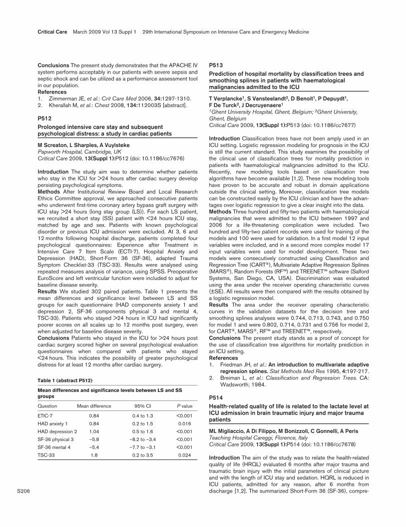
S206
Conclusions The present study demonstrates that the APACHE IVsystem performs acceptably in our patients with severe sepsis andseptic shock and can be utilized as a performance assessment toolin our population.References1. Zimmerman JE, et al.: Crit Care Med 2006, 34:1297-1310.2. Kherallah M, et al.: Chest 2008, 134:112003S [abstract].
P512Prolonged intensive care stay and subsequentpsychological distress: a study in cardiac patients
M Screaton, L Sharples, A VuylstekePapworth Hospital, Cambridge, UKCritical Care 2009, 13(Suppl 1):P512 (doi: 10.1186/cc7676)
Introduction The study aim was to determine whether patientswho stay in the ICU for >24 hours after cardiac surgery developpersisting psychological symptoms.Methods After Institutional Review Board and Local ResearchEthics Committee approval, we approached consecutive patientswho underwent first-time coronary artery bypass graft surgery withICU stay >24 hours (long stay group (LS)). For each LS patient,we recruited a short stay (SS) patient with <24 hours ICU stay,matched by age and sex. Patients with known psychologicaldisorder or previous ICU admission were excluded. At 3, 6 and12 months following hospital discharge, patients completed fourpsychological questionnaires: Experience after Treatment inIntensive Care 7 Item Scale (ECTI-7), Hospital Anxiety andDepression (HAD), Short-Form 36 (SF-36), adapted TraumaSymptom Checklist-33 (TSC-33). Results were analysed usingrepeated measures analysis of variance, using SPSS. PreoperativeEuroScore and left ventricular function were included to adjust forbaseline disease severity.Results We studied 302 paired patients. Table 1 presents themean differences and significance level between LS and SSgroups for each questionnaire (HAD components anxiety 1 anddepression 2, SF-36 components physical 3 and mental 4,TSC-33). Patients who stayed >24 hours in ICU had significantlypoorer scores on all scales up to 12 months post surgery, evenwhen adjusted for baseline disease severity.Conclusions Patients who stayed in the ICU for >24 hours postcardiac surgery scored higher on several psychological evaluationquestionnaires when compared with patients who stayed<24 hours. This indicates the possibility of greater psychologicaldistress for at least 12 months after cardiac surgery.
Table 1 (abstract P512)
Mean differences and significance levels between LS and SSgroups
Question Mean difference 95% CI P value
ETIC-7 0.84 0.4 to 1.3 <0.001
HAD anxiety 1 0.84 0.2 to 1.5 0.016
HAD depression 2 1.04 0.5 to 1.6 <0.001
SF-36 physical 3 –5.8 –8.2 to –3.4 <0.001
SF-36 mental 4 –5.4 –7.7 to –3.1 <0.001
TSC-33 1.8 0.2 to 3.5 0.024
P513Prediction of hospital mortality by classification trees andsmoothing splines in patients with haematologicalmalignancies admitted to the ICU
T Verplancke1, S Vansteelandt2, D Benoit1, P Depuydt1, F De Turck2, J Decruyenaere1
1Ghent University Hospital, Ghent, Belgium; 2Ghent University,Ghent, BelgiumCritical Care 2009, 13(Suppl 1):P513 (doi: 10.1186/cc7677)
Introduction Classification trees have not been amply used in anICU setting. Logistic regression modeling for prognosis in the ICUis still the current standard. This study examines the possibility ofthe clinical use of classification trees for mortality prediction inpatients with haematological malignancies admitted to the ICU.Recently, new modeling tools based on classification treealgorithms have become available [1,2]. These new modeling toolshave proven to be accurate and robust in domain applicationsoutside the clinical setting. Moreover, classification tree modelscan be constructed easily by the ICU clinician and have the advan-tages over logistic regression to give a clear insight into the data.Methods Three hundred and fifty-two patients with haematologicalmalignancies that were admitted to the ICU between 1997 and2006 for a life-threatening complication were included. Twohundred and fifty-two patient records were used for training of themodels and 100 were used for validation. In a first model 12 inputvariables were included, and in a second more complex model 17input variables were used for model development. These twomodels were consecutively constructed using Classification andRegression Tree (CART®), Multivariate Adaptive Regression Splines(MARS®), Random Forests (RF™) and TREENET™ software (SalfordSystems, San Diego, CA, USA). Discrimination was evaluatedusing the area under the receiver operating characteristic curves(±SE). All results were then compared with the results obtained bya logistic regression model.Results The area under the receiver operating characteristiccurves in the validation datasets for the decision tree andsmoothing splines analyses were 0.744, 0.713, 0.743, and 0.750for model 1 and were 0.802, 0.714, 0.731 and 0.756 for model 2,for CART®, MARS®, RF™ and TREENET™, respectively.Conclusions The present study stands as a proof of concept forthe use of classification tree algorithms for mortality prediction inan ICU setting.References1. Friedman JH, et al.: An introduction to multivariate adaptive
regression splines. Stat Methods Med Res 1995, 4:197-217.2. Breiman L, et al.: Classification and Regression Trees. CA:
Wadsworth; 1984.
P514Health-related quality of life is related to the lactate level atICU admission in brain traumatic injury and major traumapatients
ML Migliaccio, A Di Filippo, M Bonizzoli, C Gonnelli, A PerisTeaching Hospital Careggi, Florence, ItalyCritical Care 2009, 13(Suppl 1):P514 (doi: 10.1186/cc7678)
Introduction The aim of the study was to relate the health-relatedquality of life (HRQL) evaluated 6 months after major trauma andtraumatic brain injury with the initial parameters of clinical pictureand with the length of ICU stay and sedation. HQRL is reduced inICU patients, admitted for any reason, after 6 months fromdischarge [1,2]. The summarized Short-Form 36 (SF-36), compre-
Critical Care March 2009 Vol 13 Suppl 1 29th International Symposium on Intensive Care and Emergency Medicine

S207
hending physical functioning, general health, and social function-ing, is useful to evaluate HQRL after major trauma [3].Methods One hundred and twenty-one patients were admitted toan emergency department ICU in 2007 with major trauma (injuryseverity score >15) and severe to moderate brain injury (Glasgowcoma scale <13). Of these, 25 died in the days following trauma.Thirty-four of these were evaluated, during the follow-up visit,6 months after discharge from the ICU. HRQL was assessed bythe SF-36 questionnaire. At admission to the ICU, from the ICUdatabase (FileMaker Pro 5.5v2; FileMaker, Inc., USA) withauthorization of the Careggi Teaching Hospital Committee of theEmergency Department, the following parameters were collectedfor every patient: age, sex, abbreviated injury scale, injury severityscore, Simplified Acute Physiology Score II, in the field Glasgowcoma scale, Marshall score at the first head CT, worst values oflactates and ScvO2 within 24 hours from trauma. Also, the lengthof sedation and ICU stay were collected. The mean summarizedSF-36 was related with the Pearson test to the other parameters.Results Only lactate levels showed a significant correlation withthe mean summarized SF-36 (y = –0.0035x + 4.83; R = 0.59;P = 0.007).Conclusions In the patients affected by major trauma and braintraumatic injury, the HRQL appears to be related to the severity ofthe clinical picture on the first day of trauma assessed by lactatelevels regardless of the length of ICU stay and sedation.References1. Hofhuis JG, et al.: The impact of critical illness on per-
ceived health-related quality of life during ICU treatment,hospital stay, and after hospital discharge: a long-termfollow-up study. Chest 2008, 133:377-385.
2. Hobrook TL, et al.: Outcome after major trauma: dischargeand 6-month follow-up results from the Trauma RecoveryProject. J Trauma 1998, 45:315-323
3. Andelic N, et al.: Functional outcome and health-relatedquality of life 10 years after moderate-to-severe traumaticbrain injury. Acta Neurol Scand 2008. [Epub ahead of print]
P515Long-term survival after surgical intensive care admission:50% die within 10 years
T Timmers1, M Verhofstad1, K Moons2, L Leenen2
1St Elisabeth Hospital, Tilburg, the Netherlands; 2UniversitryMedical Centre, Utrecht, the NetherlandsCritical Care 2009, 13(Suppl 1):P515 (doi: 10.1186/cc7679)
Introduction ICU treatment of surgical patients comes along withmajor disadvantages that have to be justified by an acceptableshort-term and long-term outcome. Short-term effects of ICUtreatment have been well documented, but this is the first study todescribe the long-term effect in a large cohort after surgical ICUadmission on survival.Methods Of all surgical patients admitted to the ICU of a singlehospital between 1995 and 2000, patient characteristics, diseasecategory, APACHE II score and survival were prospectivelyregistered. A continuous follow up until 6 to 11 years afterdischarge was achieved.Results Of the 1,822 patients included, 936 (51%) had died. Fifty-two patients were lost to follow up. Overall ICU and inhospitalmortalities were 11% and 16%. Age, gender, APACHE II score andsurgical diagnosis were independently associated with long-termsurvival. Mortality increased with age of admittance to the ICU,whereas female patients had a lower chance of dying (Figure 1).Long-term mortality rates in various surgical diagnostic groups variedbetween 29% for trauma and 70% for oncological patients. In gas-trointestinal, oncological, general surgical and/or high-aged patientsthis negative effect even exceeds 5 years. In general, 10 years afterICU discharge the survival was only 50%. The mortality ratio wasincreased twofold in comparison with the general population.Conclusions Survival after ICU treatment follows distinct patternsin which age, gender, surgical diagnosis and APACHE II score areindependent determinants. A negative effect of an ICU stayappears to be long lasting.
Available online http://ccforum.com/supplements/13/S1
Figure 1 (abstract P515)

S208
P516Quality of life 1 year after intensive care
A Zidková, I Chytra, B Šestáková, E KasalUniversity Hospital, Plzen, Czech RepublicCritical Care 2009, 13(Suppl 1):P516 (doi: 10.1186/cc7680)
Introduction The aim of our study was the comparison of quality oflife before admission to the ICU and 1 year after the dischargefrom ICU.Methods Quality of life was examined in patients admitted to theinterdisciplinary ICU of a university hospital from January 2000 toDecember 2004 for more than 3 days. The EuroQoL-5D and StateTrait Anxiety Inventory questionnaires were used for the evaluationof quality of life, anxiety and psychological outcome. The Wilcoxonsigned-rank test and the chi-square test were used accordingly;P <0.05 was considered statistically significant. Stepwise multiplelogistic regression analysis was performed to evaluate thedependence of quality of life after the discharge on the followingassessed parameters: age, severity of the state at the time of ICUadmission, length of ICU and hospital stay, length of mechanicalventilation, extent of organ dysfunction during ICU stay, andprevious quality of life. The study was approved by the hospitalethical committee.Results Questionnaires were sent by mail to 483 patients, and194 patients answered (41.16%). One year after discharge fromthe ICU, 39 (20.1%) patients were less mobile, 56 (28.9%) hadworse quality of self-care, 76 (39.2%) of patients complained of alower level of normal daily activities, 87 (44.8%) of patientssuffered from a higher level of pain and 58 (29.9%) of patientsperceived a higher degree of anxiety. Significant elevation ofanxiety for both women and men (22.3% and 19.3% respectively)was found by the State Trait Anxiety Inventory questionnaire. All ofthe above mentioned changes were significant (P <0.0001). Astepwise multiple logistic regression identified age (OR = 1.03,95% CI = 1.004 to 1.059; P = 0.025), length of stay (OR = 1.0695% CI = 1.02 to 1.10; P = 0.008), length of mechanicalventilation (OR = 1.05, 95% CI = 1.01 to 1.09; P = 0.017), andlevel of pain (OR = 8.92, 95% CI = 1.14 to 69.64; P = 0.037) andanxiety (OR = 1.04, 95% CI = 1.00 to 1.08; P = 0.048) beforeadmission to the ICU as independent predictors of worsening ofquality of life 1 year after the discharge.Conclusions One year after the discharge from the ICU, patientssuffered from deterioration in all aspects of quality of life evaluatedby the questionnaires used. Quality of life 1 year after thedischarge from intensive care was influenced by its previous level,by the age, by the length of stay and by the length of mechanicalventilation.Acknowledgement Supported by the research projectMSM0021620819.
P517Health-related quality of life in critically ill octogenarians: a long-term follow-up study
J Hofhuis1, H Van Stel2, A Schrijvers2, J Rommes1, J Bakker3, P Spronk1
1Gelre Hospitals, Apeldoorn, the Netherlands; 2Julius Center forHealth Sciences and Primary Care, Utrecht, the Netherlands;3Erasmus MC, Rotterdam, the NetherlandsCritical Care 2009, 13(Suppl 1):P517 (doi: 10.1186/cc7681)
Introduction Mortality after critical illness is higher and recovery isslower in the elderly compared with younger patients. Elderlypatients recovering from critical illness may show reduced health-
related quality of life (HRQOL) [1]. We studied the impact of ICUstay on HRQOL of octogenarians, and compared their HRQOLwith the same age-matched general population.Methods We performed a long-term prospective study in amedical–surgical ICU. Patients ≥80 years (n = 129) admitted for>48 hours were included. We used the Short-Form 36 (SF-36) toevaluate HRQOL before ICU admission (using proxies), at ICUdischarge and at hospital discharge and at 3, 6 and 12 monthsfollowing ICU discharge. Furthermore we compared HRQOLbefore ICU admission and 12 months after ICU discharge withthose of an age-matched general Dutch population. The Wilcoxonsigned rank test was used for evaluating changes between twotime points. To analyze individual changes over time, a MANOVAtest was used, with Wilks’ lambda as the multivariate test.Results Mortality was considerable in the 129 includedoctogenarians. Patients could be evaluated 6 months (n = 48) and12 months (n = 27) after ICU discharge (11 patients were lost tofollow-up, 70 patients died). All SF-36 dimensions changedsignificantly over time (all P <0.001), with the exception of the role-emotional dimension. However, after an initial drop, HRQOLrecovered to pre-ICU levels in all dimensions. The physicalcomponent scores changed significantly over time (all P <0.001),whereas the mental component scores showed only a modestdecline at ICU discharge but recovered rapidly and had improvedto higher than baseline values with longer follow-up to 12 monthsafter ICU discharge. The pre-admission HRQOL in survivingoctogenarians was already lower when compared with the age-matched general population and remained so at 12 months afterICU discharge.Conclusions Critically ill octogenarians demonstrate a sharpdecline of HRQOL during the ICU stay and a gradual improvementduring the 6 months and 12 months following ICU discharge tobaseline values. Recovery begins following ICU discharge to thegeneral ward. This implies that intensive care treatment in patients≥80 years is worthwhile.Reference1. Vazquez MG, et al.: Factors related to quality of life 12
months after discharge from an intensive care unit. CritCare Med 1992, 20:1257-1262.
P518Assessment and health-related quality of life evaluation ofcritical care patients, 1 year after intensive care discharge
M Regueira, E Echavarria, L Ramos, E Alfonso, M Noya, L Schwartzmann, R Alonso, H BagnuloHospital Maciel, Montevideo, UruguayCritical Care 2009, 13(Suppl 1):P518 (doi: 10.1186/cc7682)
Introduction Outcome from intensive care has focused onreducing morbidity–mortality during hospitalization, but nowadaysit incorporates the assessment of long-term survival and quality oflife in survivors [1]. Our aim was to assess morbidity (physical andpsychological) and health-related quality of life (HRQL) in patientsdischarged from the ICU.Methods A longitudinal prospective and descriptive study ofpatients discharged from the ICU. They were interviewed at 3, 6and 12 months at an outpatient clinic. Assessment of physicalmorbidity was performed each time, as well as Folstein’s MiniMental State Examination (MMSE) for cognitive evaluation, Barthelindex and Short-Form 36 version 2 (SF-36) to evaluate functionalactivity and HRQL.Results We included 132 patients (13 with less than 1 year offollow-up); mean ± SD age 37 ± 17 years. Clinical data: SimplifiedAcute Physiology Score II 30 ± 14; stay in the ICU 21 ± 16 days,
Critical Care March 2009 Vol 13 Suppl 1 29th International Symposium on Intensive Care and Emergency Medicine

S209
in hospital 39 ± 28 days. Causes of admission: neurologic 59,septic 27, trauma 18. Required mechanical ventilation, 107 (81%)patients. In the first interview 130 patients were at home. A yearafter discharge, 90 patients survived, three died and 26 were lostto follow up. At first control 22 patients had dysphonia, six trachealstenosis and eight swallowing-related problems; pressure ulcers,16 patients. Four were aphasics, 50 with muscle weakness,paralysis or numbness. In 46 patients, psychological disorderswere diagnosed; 32 anxiety/depression, 14 sleep disorders. Sixty-five patients had a previous job, only 14 (21%) returned to work at3 months, rising to 22 (34%) at 1 year. Only 13 patients had aMMSE with a cutoff point <27. The SF-36 showed better results inall areas in patients with ≤10 days stay, being significant forphysical function. Higher physical dependence was associated
with worse perception of health and quality of life. ComparingSF-36 dimensions at 3, 6 and 12 months, better results wereobtained in most of them. Longitudinal analysis of the Barthel indexshowed at 1 year that most patients were low dependent orindependent.Conclusions Follow-up of critically ill patients after dischargeallows us to identify common physical and psychological problems.The results showed worse HRQL perception in patients withlonger stays. A year after discharge, most patients showed a greatimprovement in HRQL perception and lower physical dependencylevels.Reference1. Griffith R, et al.: Intensive Care Aftercare. Edinburgh: Butter-
worth Heinemann; 2003.
Available online http://ccforum.com/supplements/13/S1
Related Documents