This article appeared in a journal published by Elsevier. The attached copy is furnished to the author for internal non-commercial research and education use, including for instruction at the authors institution and sharing with colleagues. Other uses, including reproduction and distribution, or selling or licensing copies, or posting to personal, institutional or third party websites are prohibited. In most cases authors are permitted to post their version of the article (e.g. in Word or Tex form) to their personal website or institutional repository. Authors requiring further information regarding Elsevier’s archiving and manuscript policies are encouraged to visit: http://www.elsevier.com/copyright

Welcome message from author
This document is posted to help you gain knowledge. Please leave a comment to let me know what you think about it! Share it to your friends and learn new things together.
Transcript

This article appeared in a journal published by Elsevier. The attachedcopy is furnished to the author for internal non-commercial researchand education use, including for instruction at the authors institution
and sharing with colleagues.
Other uses, including reproduction and distribution, or selling orlicensing copies, or posting to personal, institutional or third party
websites are prohibited.
In most cases authors are permitted to post their version of thearticle (e.g. in Word or Tex form) to their personal website orinstitutional repository. Authors requiring further information
regarding Elsevier’s archiving and manuscript policies areencouraged to visit:
http://www.elsevier.com/copyright

Author's personal copy
Angiogenesis inhibition by the maleimide-based small molecule GNX-686
Patrycja Nowak-Sliwinska a,⁎, Mariangela Storto b, Tiziana Cataudella c, Jean-Pierre Ballini a, Randall Gatz d,Marco Giorgio e, Hubert van den Bergh a, Simon Plyte b, Georges Wagnières a
a Medical Photonics Group, Swiss Federal Institute of Technology (EPFL), 1015 Lausanne, Switzerlandb Congenia Srl, 20139 Milan, Italyc DAC Research, 20139 Milan, Italyd OphthalmoPharma Ltd, 6060 Sarnen, Switzerlande European Institute of Oncology, 20139 Milan, Italy
a b s t r a c ta r t i c l e i n f o
Article history:Accepted 19 October 2011Available online 28 October 2011
We investigated the anti-angiogenic properties of GNX-686, a newly identifiedmaleimide-based smallmolecule.In vitro studies on HUVEC showed that GNX-686 inhibited cell growth with an ED50 of 20–25 μM, while humanHeLa tumor cells and non-transformed embryonic mouse fibroblasts were less sensitive for the drug. Moreimportantly, at 4 μM, a concentration that was non-toxic to any cell in culture, GNX-686 showed a significantinhibitory effect on tube formation by HUVEC, indicating a profound anti-angiogenic activity. Angiogenesis inhi-bition was subsequenly tested in the chorioallantoic membrane (CAM) of the chicken embryo. A significantangiostatic activity was observed in the CAMmodel, and results were comparedwith the effect of bevacizumab,awell known and clinically usedVEGF inhibitor. Under our experimental conditions, GNX-686was found to be aseffective as bevacizumab, significantly changing the morphology of the vascular network, as illustrated andquantified by the relative number of branching points and the relative meanmesh size of the vascular network.In another in vivo model of neovascularization, the mouse retinopathy of prematurity (ROP), the vascular net-work of GNX-686-treated mice was significantly altered, reducing the density of the retinal microvasculature,as compared to the control retinas. Immunohistochemical processing of the GNX-686 treated (4 μM) eyesshowed over 50% reduction of the number of cell nuclei associated with neovasculature, as compared to thecontrol-treated eye. Taken together these results demonstrate that GNX-686 is a promising anti-angiogenic com-pound that could be developed for the treatment of diseases characterized by aberrant angiogenesis such asocular pathologies and cancer.
© 2011 Elsevier Inc. All rights reserved.
Introduction
Angiogenesis, the process of new blood vessel formation, plays animportant role in many physiological and pathological processes(Folkman, 1995). Under normal conditions, angiogenesis is associatedwith processes such as embryogenesis and wound healing. Also,many diseases are dependent on neovascularization, among whichare cancer, atherosclerosis and rheumatiod arthritis (Carmeliet andJain, 2000). Ocular pathologies such as the wet form of age-relatedmacular degeneration (AMD), proliferative diabetic retinopathy(PDR) and retinopathy of prematurity (ROP) are associated with neo-vascularization and are major causes of blindness worldwide (Cheunget al., 2010; Chiang and Regillo, 2011; Gergely and Gerinec, 2010).
These diseases are multifactorial in etiology and progression. However,ocular neovascularization is a mechanism common to all of thesepathologies and contributes significantly to vision loss (Ambati et al.,2003). Increased understanding of the mechanisms that control neo-vascularization has led to the development of novel treatments thatcan improve and restore vision in these diseases (Tolentino, 2009).
Pathologic ocular neovascularization can be treated using varioustreatment modalities e.g. photodynamic therapy (American Academyof Ophthalmology, 2000), laser photocoagulation (Sasai et al., 1997), orsurgical removal (Kaplan, 1996). Over the past few years, several drugshave been introduced to the market for intravitreal injections for ocularconditions involving angiogenesis, including pegaptanib sodium/Macugen® (Apte et al., 2007), ranibizumab/Lucentis® (Donahueet al., 2010; Fayers, 2011; Schmucker et al., 2010), and bevacizumab/Avastin® (Schmucker et al., 2010). All of these agents neutralizeVEGF preventing the induction of endothelial proliferation and angio-genesis. These pharmaceutical approaches may be advantageous forthe treatment of wet AMD over more invasive therapy modalities,due to the ability of the eye towithstandmultiple drug injectionswith-out sustaining mechanical damage. However, monthly intravitreal
Microvascular Research 83 (2012) 105–110
Abbreviations: AMD, age-related macular degeneration; CAM, chorioallantoicmembrane of the chicken embryo; EDD, embryo development day; PDR, proliferativediabetic retinopathy; ROP, retinopathy of prematurity.⁎ Corresponding author at: Medical Photonics Group, Institute of Bioengineering,
Swiss Federal Institute of Technology (EPFL), Station 6, Lausanne, CH-1015, Switzerland.Fax: +41 21 6935110.
E-mail address: [email protected] (P. Nowak-Sliwinska).
0026-2862/$ – see front matter © 2011 Elsevier Inc. All rights reserved.doi:10.1016/j.mvr.2011.10.004
Contents lists available at SciVerse ScienceDirect
Microvascular Research
j ourna l homepage: www.e lsev ie r .com/ locate /ymvre
Author's personal copy
injections of Lucentis® or Avastin® are required to maintain an anti-angiogenic effect, but sustained suppression of neovascularization isnot totally achieved and vision benefits are not striking (Biswas et al.,2011). Thesemacular disorders are chronic and require long-term con-tinuous treatment. Therefore, present studies focus on improving theefficiency and effectiveness of drug delivery to the posterior chamber(Acharya and Young, 2004; Conway, 2008; Yasukawa et al.,2005),(Ali and Lehmussaari, 2006), on the combination of anti-angiogenesis with other treatment modalities like photodynamic ther-apy (Becerra et al., 2011; van den Bergh, 2001), as well as on targetingnon-VEGF pathways in order to inhibit angiogenesis (Miller, 2010).
Current therapeutic approaches targeting angiogenic pathways arealso focused on the development of small molecular weight inhibitors.These agents primarily target the signal transduction processes in-volved in angiogenesis and are currently being tested in oncologicalsettings (Tamaskar and Pili, 2009; Vasile et al., 2008). Recently, pre-clinical studies with carboxyamidotriazole, a small molecule calciumchannel blocker that ultimately interferswith VEGF receptor signaling,has been described as a potential angiogenesis inhibitor for prolifera-tive retinopathies in humans (Afzal et al., 2010). Furthermore,IMS2186, an anti-angiogenic molecule with an undetermined mecha-nism of action, is currently undergoing development for the treatmentof choroidal neovascularization as this molecule appears to combinean anti-angiogenic effect with an anti-proliferative effect. This couldresult in long term efficacy in proliferative retinal disorders such asAMD and PDR (Falkenstein et al., 2008).
We have identified a new small molecule, the phenyl substitutedmaleimide GNX-686, and tested the compound for its anti-angiogenicproperties. Data are presented that GNX-686 is active in preventingVEGF induced endothelial cell proliferation and tube formation invitro. Moreover, it is found to be angiostatic in two in vivo models ofneovascularization: the chicken embryo chorioallantoic membrane(CAM) and the mouse retinopathy of prematurity (ROP) model.
Materials and methods
Materials and chemicals
GNX-686, a phenyl-substituted maleimide (Fig. 1A) is a product ofCongenia (Milan, Italy). A 10 mM stock solution of the GNX-686 was
prepared in 100% DMSO (Sigma-Aldrich, Buchs, Switzerland). Thestock solution (1 μl) was then dropped into 1 ml of 0.9% NaCl andmixed vigorously to produce a 10 μM working solution in 0.1% DMSO/NaCl. Avastin® (bevacizumab) is a product of Genentech (San Francisco,USA). Fluorescein isothiocyanate dextran (FITC–dextran, 20 kDa,25 mg/ml) was purchased at Sigma-Aldrich (Buchs, Switzerland).Embryos were from Animalco AG (Staufen, Switzerland). The eggswere closed after all treatments by Parafilm®. India ink was purchasedat Pebeo S.A. (Romanel-sur-Morges, Switzerland). The injections in theCAM were performed with Microliter® syringes equipped with 33-gauge metal Hub (N) needles, both from Hamilton (Reno, USA). Indiaink was filtered prior to use with a sterile cellulose acetate membrane(0.2 μm pores, Renner GmbH, Darmstadt, Germany).
Cell culture
All cells were cultured in 10 cm culture dishes and maintained at37 °C in a humidified atmosphere containing 95% air and 5% CO2.Human umbilical vein endothelial cells (HUVEC, BD Biosciences,Buccinasco, Italy) were cultured on gelatin coated plates in MCDB-131 medium supplemented with 20% (v/v) heat-inactivated fetalbovine serum (NA FBS), 1% glutamine, 1% penicillin and streptomycin,1 mM Na pyruvate, 50 mg/ml endothelial cell grow supplement(ECGS) and 100 mg/ml heparin. Human HeLa tumor cells (ATCC,LGC Standards S.r.l., Italy) were maintained in MCDB-131 mediumsupplemented with 10% [v/v] heat-inactivated fetal bovine serum(NA FBS), 1% glutamine, 1% penicillin and streptomycin, 1 mg/mlhydrocortisone and 10 ng/ml EGF. Mouse embryonic fibroblasts(MEF; isolated in house as previously described (Xu, 2005)) were cul-tured in DMEM high-glucose medium (Gibco), 10% [v/v] heat-inactivated fetal bovine serum (NA FBS), 1% glutamine, 1% penicillinand streptomycin.
Endothelial cell proliferation assay
Cell proliferationwas assessed using the CellTiter-Glo® LuminescentCell Viability Assay (Promega). Cells (3×10³ cells/well) were seeded on96-well gelatin coated white tissue culture plates (Costar #3610) in theappropriate growth medium and allowed to attach overnight. Mediumwas removed and replaced with 150 μl/well of tissue culture medium
A
*
*
**
B
D
*
E F
C
2 4 8 16 32
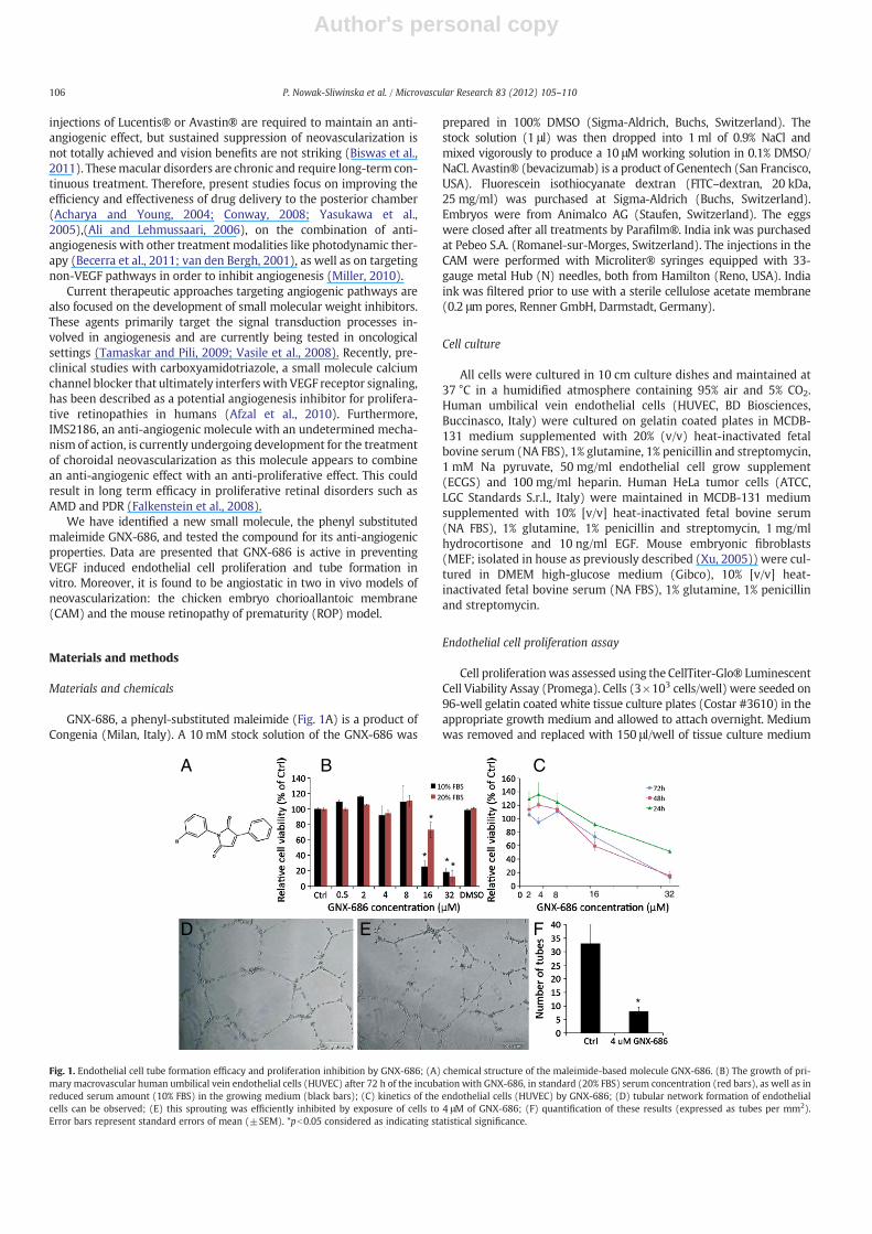
Fig. 1. Endothelial cell tube formation efficacy and proliferation inhibition by GNX-686; (A) chemical structure of the maleimide-based molecule GNX-686. (B) The growth of pri-mary macrovascular human umbilical vein endothelial cells (HUVEC) after 72 h of the incubation with GNX-686, in standard (20% FBS) serum concentration (red bars), as well as inreduced serum amount (10% FBS) in the growing medium (black bars); (C) kinetics of the endothelial cells (HUVEC) by GNX-686; (D) tubular network formation of endothelialcells can be observed; (E) this sprouting was efficiently inhibited by exposure of cells to 4 μM of GNX-686; (F) quantification of these results (expressed as tubes per mm2).Error bars represent standard errors of mean (±SEM). *pb0.05 considered as indicating statistical significance.
106 P. Nowak-Sliwinska et al. / Microvascular Research 83 (2012) 105–110
Author's personal copy
containing GNX-686 (0.5–32 mM) and grown for a further 24 h, 48 hand 72 h. At each time point, cell proliferationwas evaluated by additionof 150 μl of CellTiter-Glo® reagent to each well and luminescence wasread on a Tecan infinity F200 plate reader.
In vitro tube formation assay
HUVEC cells were plated on 48-well plates (2.5×10⁴ cells/well),previously coated with 140 μl of BD Matrigel (Basement MembraneMatrix growth factor-reduced), in the presence of GNX-686 (4 μM)or control (0.05% DMSO) in MCDB 10% FBS and ECGS 50 mg/ml.Tube formation was assessed by counting the number of capillary-like structures per mm2 that had formed after a subsequent culturefor 15 h at 37 °C.
The in ovo CAM model
Fertilized chicken eggs were maintained as described previously(Nowak-Sliwinska et al., 2010b, 2011). Briefly, eggs were labeledand transferred into a hatching incubator. On embryo developmentday (EDD) 3, a hole of approximately 3 mm in diameter was openedin the eggshell and coveredwith a LaboratoryWrapping Film, Parafilm®(Pechiney, Menasha, USA) to prevent dehydratation and possible infec-tions. The eggs were returned to the incubator with a relative airhumidity of 65% and a temperature of 37 °C in a static position untiluse. At EDD 7, the shell above the air pouch of the egg was extendedto a diameter of approximately 3 cm in order to provide access to thechorioallantoic membrane. For the topical application of the testedagents and their controls on EDD7, polyethylene rings from Pasteurpipettes (Copan, Brescia, Italy) were cut in sterile conditions and de-posited on the CAM. These polyethylene rings (diameter 5 mm; wallthickness 0.5 mm, 1 mm height) were used to confine the topicaldrug to only one small and well-defined part of the CAM surface.Subsequently, 20 μl of freshly prepared GNX 686 (1–10 μM in 0.1%DMSO/0.9% NaCl) or Avastin® (1–10 μM in 0.9% NaCl) or their controls(0.1% DMSO/0.9% NaCl or 0.9% NaCl, respectively) were deposited top-ically (PipetmanNeo, Gilson, Middleton, USA) onto the CAM surfacewithin the ring. The embryos were then labeled and returned to theincubator in a static position for 24 h. The second topical administra-tion of reagents (20 μl of GNX-686 and Avastin® solutions, or of theircontrol solutions) took place in the same way on EDD8. Again, the em-bryos were returned to the incubator in a static position for 24 h forlater image-processed analysis on EDD9. Fluorescence images wereobtained with an epi-fluorescence microscope (Nikon Eclipse E600FN, Japan) equipped with a Nikon objective (CFI achromat; magnifica-tion ×10; N.A.: 0.30,Working distance: 30 mm) coupled to an F-view II12-bit monochrome Peltier-cooled digital CCD camera (Soft ImagingSystem GmbH, Münster, Germany) driven with the “analySIS DOCU”software (Soft Imaging System GmbH, Münster, Germany) (Nowak-Sliwinska et al., in press). It should be noted that an objective 10×is minimally necessary for a proper quantitative visualization of thecapillaries. Fluorescence images (1280×1024 pixels with 4096 graylevels, i.e. 12 bits) of the CAM superficial vessels were recorded byexciting the fluorescence with a filtered (λex=450–490 nm) highpressure Hg-arc lamp (Osram, GmbH, Augsburg, Germany) togetherwith a long pass emission filter (λ>520 nm) for observation of theFITC–dextran fluorescence. These images were then classified interms of the quantification of the capillary features (size, and quantity)in the treated area and then used to study the GNX-686 or Avastin®-induced changes in comparison with a similar area treated with thecontrol solution. The latter was in general not performed on thesame embryo. To obtain the fluorescence images, a volume of 20 μlof FITC–dextran (20 kDa)was injected into one of the principle vesselsof the CAM (~200 μm in diameter), directly under the microscope. Inorder to increase the quality (contrast) of the recorded angiograms30 μl of well-known light absorber, India ink, was injected shortly
after into the extra-embryonic cavity, just under the deposited poly-ethylene ring, in order to decrease the embryo's interfering fluores-cence from deeper located vessels. This interfering luminescence canchange rapidly with time due to the embryo's movement. Prior to in-jection, the India ink was filtered using a sterile cellulose acetatemembrane.
In order to quantify the vascular effects, we used our automaticimage-processing method described recently (Nowak-Sliwinska etal., 2010a). Using this method, two descriptors were selected to char-acterize and quantify the growth of vessels for the period rangingbetween EDD7 and EDD9. These descriptors are (i) the number ofbranching points/mm2 (all equivalent and non-equivalent bifurca-tions, ≥3 neighbors), and (ii) the mean area of the vessel networkmeshes defined as the closed area surrounded by capillaries and/orvessels, expressed in the units of 102 μm2. Both descriptors were re-lated to the control values (Nowak-Sliwinska et al., 2010a). It shouldbe noted that, with increasing GNX-686 concentration, the percentageof DMSO was higher, respectively. Each point on the reported plotsrepresents the average of measurements performed on 9–10 embryos.At least 5 images taken at different areas of (1.4×1.12 mm2) wererecorded and analyzed with the objective 10× on each individualCAM. These images were located within the polyethylene ring thatdelineated the agent-treated zone.
Mouse retinopathy of prematurity (ROP) model
Neonatal mice (C57BL/6J) were obtained from breeding coloniesmaintained within the Gemelli Hospital (Rome, Italy). Litters of post-natal day 7 (P7) pups were placed in either hyperoxia (75% O2) ornormoxia (20% O2) for 5 days. Neonatal P7 mice were placed in anoxygen chamber together with their mother (oxygen content wasregulated by an automated O2/N2 mixer to 75%±2% O2) with sufficientfood and water to sustain them for 5 days. The cage temperature wasmaintained at 23 °C±2 °C. At postnatal day 12 mice were returned tonormoxia and maintained until postnatal day 18. At postnatal day 12“hyperoxic” animals were treated with GNX-686 in the right eye and1% DMSO in the left eye (control). GNX-686 (1 μl of 4 μM solution in10% DMSO, ca. 0.4 μM final concentration in the vitreous) or DMSO(1 μl of a 10% solution, ca. 1% final concentration in the vitreous) wasinjected directly into the ocular vitreous using a sterile Hamilton syringe.At postnatal day 18 success of the model was assessed in a satellitegroup of animals (normoxic vs hyperoxic) by fluorescein-dextran angi-ography. Quantitation of neoangiogenesis in the presence or absence ofcompound was assessed by immunohistochemistry.
Mice were anesthetized intraperitoneally with tribromoethanol(0.2 ml/10 g body weight) and then perfused through the left ventriclewith 1 ml of phosphate buffered saline (PBS) containing 50 mg of2×106 molecular weight fluorescein-dextran (Sigma, St.Louis, USA).Before use, the fluorescein-dextran solution was clarified by centrifu-gation for 5 min at 10,000 rpm (model 235C, Fisher Scientific, Spring-field, USA). The eyes were marked for orientation, enucleated, andplaced in 4% paraformaldehyde for 3 to 24 h. Lenses were removedand peripheral retinas were cut to allow flat-mounting with glycerol-gelatin. The flat-mounted retinas were viewed by fluorescencemicros-copy (Zeiss Axiophot, Thornwood, USA) and photographed.
At P18, mice were sacrificed with intraperitoneal Tribromoethanol(0.1 ml/g body weight). The eyes were enucleated, immersed in 4%paraformaldehyde in PBS for at least 24 h, and embedded in paraffin.Serial sections (6 mm) of whole eyes were cut through the corneaand parallel to the optic nerve and stained with eosin and hematoxy-lin. Nuclei from new vessels and vessel profiles could be distinguishedfrom other structures in the retina and counted in cross-section withlight microscopy (magnification ×400). Approximately 150 serialsections were cut from each eye. In the central half of the globe, 10sections on each side of the optic nerve, 30 to 90 nm apart, werecounted for neovascularization. Vascular cell nuclei, identified under
107P. Nowak-Sliwinska et al. / Microvascular Research 83 (2012) 105–110

Author's personal copy
light microscopy with hematoxylin staining, were considered to beassociated with new vessels if they were found on the vitreal side ofthe internal limiting membrane.
Statistical analysis
Data are presented as standard error of means±SEM. Statisticalcomparisons were made using Student's t-test, with pb0.05 consideredas indicating statistical significance (*).
Results
GNX-686 inhibits endothelial cell proliferation and tube formation
Two major hallmarks of angiogenesis are endothelial cell prolifera-tion and migration. In order to investigate the anti-angiogenic activityof GNX-686, we assessed the activity of this compound on both pro-cesses. Endothelial cell growth was assessed using the CellTiterGlo®assay (Promega), a colorimetric assay that quantifiesmetabolic activityby detection of ATP concentration in the cell culture. After a 72 h expo-sure to GNX-686, a reduction in HUVEC growth was observed at con-centrations as of 16 μM. The ED50 value for endothelial cells grown innormal culture medium was approximately 20–25 μM, while cellsgrown under reduced serum conditions were slightly more sensitive(ED50 of 10–15 μM, Fig. 1B). Under these conditions, concentrationsof 32 μM inhibited cell growth by 80–90%. The kinetics of HUVEC sen-sitivity to GNX-686 are shown in Fig. 1C. At the highest concentrationtested (32 μM) inhibitory activity after 24 h is approximately 50%. Asthe compound is dissolved in DMSO, control solutions containingequal amounts of DMSO were tested and proved the specific activityof the compound. In this assay, also human HeLa tumor cells andmouse embryonic fibroblasts were tested. Although the effect onHUVEC was most pronounced (50% inhibition at 32 μM), there wasalso some activity in HeLa cells (37% inhibition at 32 μM) and mouseembryonic fibroblasts (29% inhibition at 32 μM) after 24 h of exposureto GNX-686.
A clearer anti-angiogenic effect was shown in the tube formationassay using HUVECs on a 3-dimensional gel (Matrigel). In this assay,cellular reorganization, migration and tubular network formation ofendothelial cells can be observed (Fig. 1D). Network formation wasefficiently inhibited by exposure of cells to concentrations of GNX-686 as low as 4 μM (Fig. 1E). At this concentration none of the cul-tured cells tested showed any sensitivity. Quantification of the resultshown is given in Fig. 1F. The lack of effects on endothelial cell growthat this concentration, indicate GNX-686 has a specific anti-angiogenicactivity.
GNX-686 inhibits in vivo angiogenesis in the CAM model
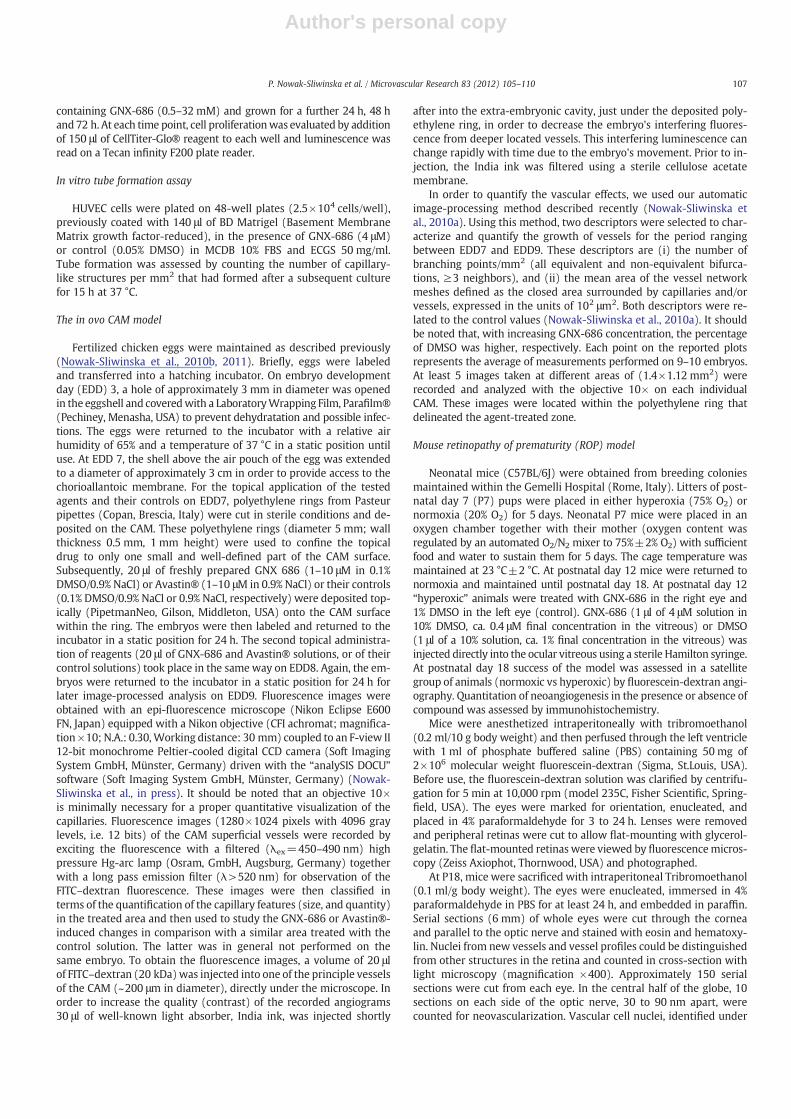
In vivo effects of GNX-686 on angiogenesis were first tested in theCAM assay. The vasculature in the developing CAM is still incompleteand immature at embryonic development day 7 (EDD7). At this day,GNX-686 treatment was started by topical application of a 20 μl solu-tion within the ring and treatment was repeated at EDD8. Concentra-tions used ranged from 1 to 10 μM (Table 1). Since GNX-686 is pre-dissolved in DMSO, we also tested the effect of equal amounts ofthis solvent alone. We have previously shown in the developmentalCAM model (Nowak-Sliwinska et al., 2009) that DMSO concentrationsof ≤0.1% DMSO do not affect outgrowth of the vasculature in theCAM. The anti-angiogenic efficacy of GNX-686 was compared withthat of Avastin® at various concentrations ranging from 1 to 10 μM.Observations were digitally analyzed and are shown as the relativenumber of branching points/mm2 (Fig. 2A), and relative mean meshsize of the vessel network expressed in the unit of 102 μm2 (Fig. 2B).Both descriptors are presented relative to the control treated CAMs.Concentrations of GNX-686 ≥7 μM effectively influenced the capillary
outgrowth in the CAM. At concentrations ranging between 3.7 and10 μM the inhibitory efficacy of GNX-686 was comparable to that ofAvastin®. Representative fluorescein-dextran fluorescence angiogra-phies show EDD9 CAM 48 h after topical treatment with 0.9% NaCl(control, C), GNX-686 (10 μM, D), and Avastin® (10 μM, E). Thesedata further support the hypothesis that GNX-686 is endowed withanti-angiogenic properties.
Retinopathy of prematurity (ROP) mouse model
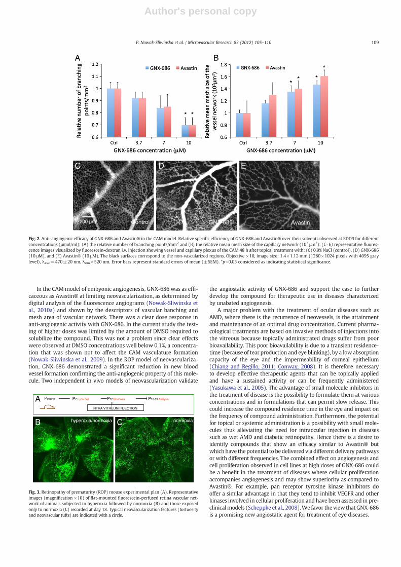
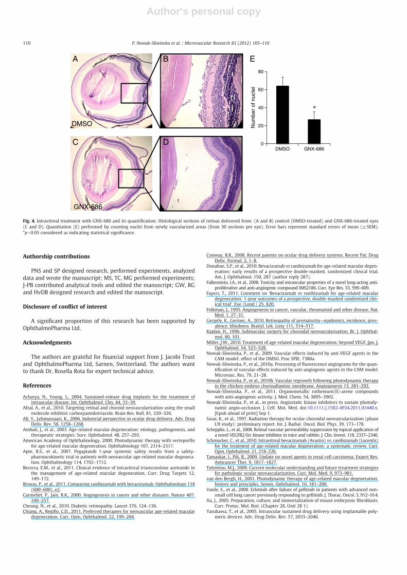
The ROPmousemodel was used as an independent, eye-related, invivo model to show angiogenesis inhibition of GNX-686. The scheduleof the experiment is shown in Fig. 3A. The structure of the retinalblood vessel tree, 5 days after a return to normoxia, was evaluatedin fluorescein-injected flat-mounted retinas. This demonstrates the de-gree of neovascularization in this model compared to control neonataleyes not having undergone a hyperoxia–normoxia cycle (Figs. 3B andC). The results indicate that in all animals conditions were achievedfor enhanced neovascularization in vivo. Representative retinal imagesare shown (Figs. 3B: Hyperoxia–normoxia exposed animals and C: ani-mals exposed to normoxia only (control)). The vascular network in theROP model was significantly altered, as compared with that from con-trol eyes. The ROP retina contained highly tortuous blood vessels andneovascular tufts, as indicated with arrows in Fig. 3B. These alterationsappeared as a severe loss of retinal microvasculature. Qualitatively, ret-inas from ROP eyes treated with GNX-686 looked remarkably similar tothe retinas from control (normoxia) eyes (data not shown). Quantifica-tion of this phenomenon was performed by counting vascular nuclei(identified under light microscopy with hematoxylin staining) fromretinal sections that were found on the vitreal side of the internal limit-ing membrane (Figs. 4A–D (indicated by arrows)). Each animal re-ceived GNX-686 in the right eye and a DMSO control in the left eye.A significant reduction in retinal neovascularization of >50% wasobserved in animals treated with 0.4 μM GNX-686 compared to thosereceiving DMSO (Fig. 4E).
Discussion
We have identified a small molecular weight maleimide compound,GNX-686, with anti-angiogenic activity. At doses with a significanteffect on endothelial tube formation (4 μM), GNX-686 had no effecton cell growth characteristics, suggesting a profound anti-angiogenicactivity of this molecule. At the highest concentration tested (32 μM)inhibitory activity after 24 h is approximately 50%. As the compoundis dissolved in DMSO, control solutions containing equal amounts ofDMSO were tested and no toxicity was observed below 0.1% DMSO.
The molecular mechanism by which GNX-686 exerts its anti-angiogenic activity is currently under investigation. However, thecompound does not interferewithVEGF receptor signaling (MAPkinasecascade). Neither does it affect HIFα stabilization in HUVEC cells andtissue from neonatal mouse eyes (Congenia, unpublished data).
Table 1Concentrations of the active solutions, both GNX-686 and Avastin® administered topicallyon the CAM in the form of 20 μl droplets. Corresponding percentage of DMSO in GNX-686solutions is also indicated.
Agent Agent concentration
μg/ml μM % of DMSO
GNX-686 0.28 1 0.01Avastin® 160 0GNX-686 0.8 3.7 0.025Avastin® 560 0GNX-686 1.4 7 0.05Avastin® 1100 0GNX-686 2.8 10 0.1Avastin® 1700 0
108 P. Nowak-Sliwinska et al. / Microvascular Research 83 (2012) 105–110
Author's personal copy
In the CAMmodel of embyonic angiogenesis, GNX-686 was as effi-caceous as Avastin® at limiting neovascularization, as determined bydigital analysis of the fluorescence angiograms (Nowak-Sliwinska etal., 2010a) and shown by the descriptors of vascular banching andmesh area of vascular network. There was a clear dose response inanti-angiogenic activity with GNX-686. In the current study the test-ing of higher doses was limited by the amount of DMSO required tosolubilize the compound. This was not a problem since clear effectswere observed at DMSO concentrations well below 0.1%, a concentra-tion that was shown not to affect the CAM vasculature formation(Nowak-Sliwinska et al., 2009). In the ROP model of neovasculariza-tion, GNX-686 demonstrated a significant reduction in new bloodvessel formation confirming the anti-angiogenic property of this mole-cule. Two independent in vivo models of neovascularization validate
the angiostatic activity of GNX-686 and support the case to furtherdevelop the compound for therapeutic use in diseases characterizedby unabated angiogenesis.
A major problem with the treatment of ocular diseases such asAMD, where there is the recurrence of neovessels, is the attainmentand maintenance of an optimal drug concentration. Current pharma-cological treatments are based on invasive methods of injections intothe vitreous because topically administrated drugs suffer from poorbioavailability. This poor bioavailability is due to a transient residence-time (because of tear production and eye blinking), by a low absorptioncapacity of the eye and the impermeability of corneal epithelium(Chiang and Regillo, 2011; Conway, 2008). It is therefore necessaryto develop effective therapeutic agents that can be topically appliedand have a sustained activity or can be frequently administered(Yasukawa et al., 2005). The advantage of small molecule inhibitors inthe treatment of disease is the possibility to formulate them at variousconcentrations and in formulations that can permit slow release. Thiscould increase the compound residence time in the eye and impact onthe frequency of compound administration. Furthermore, the potentialfor topical or systemic administration is a possibility with small mole-cules thus alleviating the need for intraocular injection in diseasessuch as wet AMD and diabetic retinopathy. Hence there is a desire toidentify compounds that show an efficacy similar to Avastin® butwhich have the potential to be delivered via different delivery pathwaysor with different frequencies. The combined effect on angiogenesis andcell proliferation observed in cell lines at high doses of GNX-686 couldbe a benefit in the treatment of diseases where cellular proliferationaccompanies angiogenesis and may show superiority as compared toAvastin®. For example, pan receptor tyrosine kinase inhibitors dooffer a similar advantage in that they tend to inhibit VEGFR and otherkinases involved in cellular proliferation and have been assessed in pre-clinicalmodels (Scheppke et al., 2008).We favor the view thatGNX-686is a promising new angiostatic agent for treatment of eye diseases.
P0 Birth P7 Hyperoxia P12 Normoxia P18-19 Analysis
INTRA VITREUM INJECTION
A
hyperoxia/normoxiaB C normoxia
Fig. 3. Retinopathy of prematurity (ROP) mouse experimental plan (A). Representativeimages (magnification ×10) of flat-mounted fluorescein-perfused retina vascular net-work of animals subjected to hyperoxia followed by normoxia (B) and those exposedonly to normoxia (C) recorded at day 18. Typical neovascularization features (tortuosityand neovascular tufts) are indicated with a circle.
AvastinControl200 µm
C D E
A
* *
B
**
**
GNX-686
Fig. 2. Anti-angiogenic efficacy of GNX-686 and Avastin® in the CAMmodel. Relative specific efficiency of GNX-686 and Avastin® over their solvents observed at EDD9 for differentconcentrations (μmol/ml); (A) the relative number of branching points/mm2 and (B) the relative mean mesh size of the capillary network (102 μm2); (C–E) representative fluores-cence images visualized by fluorescein-dextran i.v. injection showing vessel and capillary plexus of the CAM 48 h after topical treatment with: (C) 0.9% NaCl (control), (D) GNX-686(10 μM), and (E) Avastin® (10 μM). The black surfaces correspond to the non-vascularized regions. Objective ×10, image size: 1.4×1.12 mm (1280×1024 pixels with 4095 graylevel), λexc=470±20 nm, λem>520 nm. Error bars represent standard errors of mean (±SEM). *pb0.05 considered as indicating statistical significance.
109P. Nowak-Sliwinska et al. / Microvascular Research 83 (2012) 105–110
Author's personal copy
Authorship contributions
PNS and SP designed research, performed experiments, analyzeddata and wrote the manuscript; MS, TC, MG performed experiments;J-PB contributed analytical tools and edited the manuscript; GW, RGand HvDB designed research and edited the manuscript.
Disclosure of conflict of interest
A significant proportion of this research has been supported byOphthalmoPharma Ltd.
Acknowledgments
The authors are grateful for financial support from J. Jacobi Trustand OphthalmoPharma Ltd, Sarnen, Switzerland. The authors wantto thank Dr. Rosella Rota for expert technical advice.
References
Acharya, N., Young, L., 2004. Sustained-release drug implants for the treatment ofintraocular disease. Int. Ophthalmol. Clin. 44, 33–39.
Afzal, A., et al., 2010. Targeting retinal and choroid neovascularization using the smallmolecule inhibitor carboxyamidotriazole. Brain Res. Bull. 81, 320–326.
Ali, Y., Lehmussaari, K., 2006. Industrial perspective in ocular drug delivery. Adv. DrugDeliv. Rev. 58, 1258–1268.
Ambati, J., et al., 2003. Age-related macular degeneration: etiology, pathogenesis, andtherapeutic strategies. Surv. Ophthalmol. 48, 257–293.
American Academy of Ophthalmology, 2000. Photodynamic therapy with verteporfinfor age-related macular degeneration. Ophthalmology 107, 2314–2317.
Apte, R.S., et al., 2007. Pegaptanib 1-year systemic safety results from a safety-pharmacokinetic trial in patients with neovascular age-related macular degenera-tion. Ophthalmology 114, 1702–1712.
Becerra, E.M., et al., 2011. Clinical evidence of intravitreal triamcinolone acetonide inthe management of age-related macular degeneration. Curr. Drug Targets 12,149–172.
Biswas, P., et al., 2011. Comparing ranibizumab with bevacizumab. Ophthalmology 118(600–600), e2.
Carmeliet, P., Jain, R.K., 2000. Angiogenesis in cancer and other diseases. Nature 407,249–257.
Cheung, N., et al., 2010. Diabetic retinopathy. Lancet 376, 124–136.Chiang, A., Regillo, C.D., 2011. Preferred therapies for neovascular age-related macular
degeneration. Curr. Opin. Ophthalmol. 22, 199–204.
Conway, B.R., 2008. Recent patents on ocular drug delivery systems. Recent Pat. DrugDeliv. Formul. 2, 1–8.
Donahue, S.P., et al., 2010. Bevacizumab vs ranibizumab for age-related macular degen-eration: early results of a prospective double-masked, randomized clinical trial.Am. J. Ophthalmol. 150, 287 (author reply 287).
Falkenstein, I.A., et al., 2008. Toxicity and intraocular properties of a novel long-acting anti-proliferative and anti-angiogenic compound IMS2186. Curr. Eye Res. 33, 599–609.
Fayers, T., 2011. Comment on ‘Bevacizumab vs ranibizumab for age-related maculardegeneration: 1-year outcomes of a prospective, double-masked randomised clin-ical trial’. Eye (Lond.) 25, 820.
Folkman, J., 1995. Angiogenesis in cancer, vascular, rheumatoid and other disease. Nat.Med. 1, 27–31.
Gergely, K., Gerinec, A., 2010. Retinopathy of prematurity—epidemics, incidence, prev-alence, blindness. Bratisl. Lek. Listy 111, 514–517.
Kaplan, H., 1996. Submacular surgery for choroidal neovascularisation. Br. J. Ophthal-mol. 80, 101.
Miller, J.W., 2010. Treatment of age-related macular degeneration: beyond VEGF. Jpn. J.Ophthalmol. 54, 523–528.
Nowak-Sliwinska, P., et al., 2009. Vascular effects induced by anti-VEGF agents in theCAM model: effect of the DMSO. Proc SPIE. 7380a.
Nowak-Sliwinska, P., et al., 2010a. Processing of fluorescence angiograms for the quan-tification of vascular effects induced by anti-angiogenic agents in the CAM model.Microvasc. Res. 79, 21–28.
Nowak-Sliwinska, P., et al., 2010b. Vascular regrowth following photodynamic therapyin the chicken embryo chorioallantoic membrane. Angiogenesis 13, 281–292.
Nowak-Sliwinska, P., et al., 2011. Organometallic ruthenium(II)-arene compoundswith anti-angiogenic activity. J. Med. Chem. 54, 3895–3902.
Nowak-Sliwinska, P., et al., in press. Angiostatic kinase inhibitors to sustain photody-namic angio-occlusion. J. Cell. Mol. Med. doi:10.1111/j.1582-4934.2011.01440.x.[Epub ahead of print] Sep 1.
Sasai, K., et al., 1997. Radiation therapy for ocular choroidal neovascularization (phaseI/II study): preliminary report. Int. J. Radiat. Oncol. Biol. Phys. 39, 173–178.
Scheppke, L., et al., 2008. Retinal vascular permeability suppression by topical application ofa novel VEGFR2/Src kinase inhibitor in mice and rabbits. J. Clin. Invest. 118, 2337–2346.
Schmucker, C., et al., 2010. Intravitreal bevacizumab (Avastin) vs. ranibizumab (Lucentis)for the treatment of age-related macular degeneration: a systematic review. Curr.Opin. Ophthalmol. 21, 218–226.
Tamaskar, I., Pili, R., 2009. Update on novel agents in renal cell carcinoma. Expert Rev.Anticancer Ther. 9, 1817–1827.
Tolentino, M.J., 2009. Current molecular understanding and future treatment strategiesfor pathologic ocular neovascularization. Curr. Mol. Med. 9, 973–981.
van den Bergh, H., 2001. Photodynamic therapy of age-related macular degeneration:history and principles. Semin. Ophthalmol. 16, 181–200.
Vasile, E., et al., 2008. Erlotinib after failure of gefitinib in patients with advanced non-small cell lung cancer previously responding to gefitinib. J. Thorac. Oncol. 3, 912–914.
Xu, J., 2005. Preparation, culture, and immortalization of mouse embryonic fibroblasts.Curr. Protoc. Mol. Biol. (Chapter 28, Unit 28 1).
Yasukawa, T., et al., 2005. Intraocular sustained drug delivery using implantable poly-meric devices. Adv. Drug Deliv. Rev. 57, 2033–2046.
A B
C D
GNX-686
DMSO
0
20
40
60
80
DMSO GNX-686
Num
ber
of n
ucle
i
E
*
Fig. 4. Intravitreal treatment with GNX-686 and its quantification: Histological sections of retinas delivered from: (A and B) control (DMSO-treated) and GNX-686-treated eyes(C and D). Quantitation (E) performed by counting nuclei from newly vascularized areas (from 30 sections per eye). Error bars represent standard errors of mean (±SEM).*pb0.05 considered as indicating statistical significance.
110 P. Nowak-Sliwinska et al. / Microvascular Research 83 (2012) 105–110
Related Documents