Angiogenesis-independent tumor growth mediated by stem-like cancer cells Per Ø. Sakariassen a , Lars Prestegarden a , Jian Wang a , Kai-Ove Skaftnesmo a , Rupavathana Mahesparan a,b , Carla Molthoff c , Peter Sminia d , Eirik Sundlisæter a , Anjan Misra e , Berit Bølge Tysnes a , Martha Chekenya a , Hans Peters f , Gabriel Lende b , Karl Henning Kalland g,h , Anne M. Øyan g,h , Kjell Petersen i , Inge Jonassen i,j , Albert van der Kogel f , Burt G. Feuerstein k , A. Jorge A. Terzis a,l , Rolf Bjerkvig a,l , and Per Øyvind Enger a,b,m a NorLux NeuroOncology, Department of Biomedicine, University of Bergen, N-5020 Bergen, Norway; Departments of b Neurosurgery and h Microbiology and Immunology, Haukeland University Hospital, N-5021 Bergen, Norway; Departments of c Nuclear Medicine and Positron Emission Tomography Center and d Radiation Oncology, Section Radiobiology, Vrije Universiteit University Medical Center, 1081 HV Amsterdam, The Netherlands; Departments of e Neurosurgery and k Laboratory Medicine, University of California, San Francisco, CA 94143; f Department of Radiation Oncology, University Medical Center, 6500 HB Nijmegen, The Netherlands; g The Gade Institute, University of Bergen, N-5021 Bergen, Norway; i Bergen Center for Computational Science, Unifob AS and j Department of Informatics, University of Bergen, N-5021 Norway; and l NorLux Neuro-Oncology, Centre Recherche de Public Sante ´ , L-1150 Luxembourg Communicated by Erkki Ruoslahti, University of California, Santa Barbara, CA, September 1, 2006 (received for review May 5, 2006) In this work, highly infiltrative brain tumors with a stem-like phenotype were established by xenotransplantation of human brain tumors in immunodeficient nude rats. These tumors coopted the host vasculature and presented as an aggressive disease without signs of angiogenesis. The malignant cells expressed neural stem cell markers, showed a migratory behavior similar to normal human neural stem cells, and gave rise to tumors in vivo after regrafting. Serial passages in animals gradually transformed the tumors into an angiogenesis-dependent phenotype. This pro- cess was characterized by a reduction in stem cells markers. Gene expression profiling combined with high throughput immunoblot- ting analyses of the angiogenic and nonangiogenic tumors iden- tified distinct signaling networks in the two phenotypes. Further- more, proinvasive genes were up-regulated and angiogenesis signaling genes were down-regulated in the stem-like tumors. In contrast, proinvasive genes were down-regulated in the angio- genesis-dependent tumors derived from the stem-like tumors. The described angiogenesis-independent tumor growth and the un- coupling of invasion and angiogenesis, represented by the stem- like cancer cells and the cells derived from them, respectively, point at two completely independent mechanisms that drive tumor progression. This article underlines the need for developing ther- apies that specifically target the stem-like cell pools in tumors. glioma invasiveness vessel cooption A basic principle in tumor progression is the requirement for angiogenesis, yet several clinical studies have reported limited efficacy of angiogenesis inhibitors to control tumor growth (1–7). This finding has been explained by pharmacokinetic parameters such as the mode of delivery, inadequate biodistribution, and misfolding of the therapeutic proteins (8). Still, some studies suggest that the nature of this problem may not be inherent in the therapeutic compound, but rather underlies the concept of angio- genesis-dependency itself (9–11). An alternative mechanism for obtaining essential nutrients may be that the malignant cells are sustained by the preexisting vasculature of the host tissue, as they invade the surrounding parenchyma. Stem cells and tumor cells share the ability of cell division. Moreover, EGF and FGF, which maintain neural stem cells in a proliferative state in vitro, also increase proliferation of glioma cells (12–14). Similar to migrating neural stem cells grafted in adult rat brain, invading glioma cells may be supported by the vascular network in the normal brain (15–19). However, studies suggest that although tumor cells initially coopt surrounding vessels, subsequent growth requires angiogenesis (20, 21). Thus, the prevailing view is that solid tumor growth is angiogenesis-dependent (22–24). Glioblastomas (GBMs) are highly vascular brain tumors that are considered to be attractive candidates for antiangiogenic therapy (25). GBMs are classified as high-grade gliomas because of the presence of necrosis and microvascular proliferations, and most often arise de novo in patients not previously diagnosed with a low-grade glioma. They are then referred to as primary GBMs and display a characteristic set of genetic changes (26, 27). However, these tumors may also arise from the malignant progression of invasive, low-grade gliomas without microvascular proliferations (26, 28). Apart from the onset of angiogenesis, this transition is characterized by progressive genetic changes different from those observed in primary GBMs (29). In this work, we xenografted 10 biopsies from primary glioblastomas into nude rat brains. Surpris- ingly, the resulting tumors recapitulated the infiltrative growth pattern of low-grade gliomas, coopting the host vasculature without any signs of angiogenesis or necrosis. Upon passaging in vivo, they progressed toward a highly malignant phenotype displaying tumor angiogenesis and large necrotic regions. This progression was not Author contributions: P.Ø.S. and L.P. contributed equally to this work; P.Ø.S., L.P., R.M., G.L., B.G.F., R.B., and P.Ø.E. designed research; P.Ø.S., L.P., J.W., K.-O.S., R.M., C.M., P.S., E.S., A.M., H.P., K.H.K., A.M.Ø., A.v.d.K., and P.Ø.E. performed research; C.M., P.S., and H.P. contributed new reagentsanalytic tools; P.Ø.S., L.P., J.W., K.-O.S., A.M., B.B.T., M.C., H.P., K.H.K., A.M.Ø., K.P., I.J., B.G.F., A.J.A.T., R.B., and P.Ø.E. analyzed data; and P.Ø.S., L.P., R.B., and P.Ø.E. wrote the paper. The authors declare no conflict of interest. Freely available online through the PNAS open access option. Abbreviations: CGH, comparative genomic hybridization; GBM, glioblastoma. m To whom correspondence should be addressed at: Department of BiomedicineSAC, University of Bergen, Jonas Lie’s Vei 91, 5009 Bergen, Norway. E-mail: per.enger@biomed. uib.no. © 2006 by The National Academy of Sciences of the USA Table 1. Tumor take, engraftment rate, and passaging data on 10 primary GBM biopsies Case Tumor take (%) Survival, days, mean SEM* Passaged in vivo 1 12 of 13 (92) 117.5 8.6 No 2 7 of 7 (100) 97 1.7 Yes 3 4 of 5 (80) 169.5 22.1 No 4 3 of 5 (60) 252 1.6 No 5 7 of 8 (88) 64 1.5 No 6 2 of 10 (20) 93.5 10.6 No 7 6 of 6 (100) 104.5 1.4 Yes 8 8 of 8 (100) 119.5 3.5 Yes 9 12 of 14 (86) 137.5 5 No 10 7 of 7 (100) 126.5 2.9 Yes *Survival data were recorded only from animals where tumor take was confirmed after histological examination. 16466 –16471 PNAS October 31, 2006 vol. 103 no. 44 www.pnas.orgcgidoi10.1073pnas.0607668103

Welcome message from author
This document is posted to help you gain knowledge. Please leave a comment to let me know what you think about it! Share it to your friends and learn new things together.
Transcript
Angiogenesis-independent tumor growth mediatedby stem-like cancer cellsPer Ø. Sakariassena, Lars Prestegardena, Jian Wanga, Kai-Ove Skaftnesmoa, Rupavathana Mahesparana,b,Carla Molthoffc, Peter Sminiad, Eirik Sundlisætera, Anjan Misrae, Berit Bølge Tysnesa, Martha Chekenyaa, Hans Petersf,Gabriel Lendeb, Karl Henning Kallandg,h, Anne M. Øyang,h, Kjell Peterseni, Inge Jonasseni,j, Albert van der Kogelf,Burt G. Feuersteink, A. Jorge A. Terzisa,l, Rolf Bjerkviga,l, and Per Øyvind Engera,b,m
aNorLux NeuroOncology, Department of Biomedicine, University of Bergen, N-5020 Bergen, Norway; Departments of bNeurosurgery and hMicrobiologyand Immunology, Haukeland University Hospital, N-5021 Bergen, Norway; Departments of cNuclear Medicine and Positron Emission Tomography Center anddRadiation Oncology, Section Radiobiology, Vrije Universiteit University Medical Center, 1081 HV Amsterdam, The Netherlands; Departments of eNeurosurgeryand kLaboratory Medicine, University of California, San Francisco, CA 94143; fDepartment of Radiation Oncology, University Medical Center, 6500 HB Nijmegen,The Netherlands; gThe Gade Institute, University of Bergen, N-5021 Bergen, Norway; iBergen Center for Computational Science, Unifob A�S and jDepartmentof Informatics, University of Bergen, N-5021 Norway; and lNorLux Neuro-Oncology, Centre Recherche de Public Sante, L-1150 Luxembourg
Communicated by Erkki Ruoslahti, University of California, Santa Barbara, CA, September 1, 2006 (received for review May 5, 2006)
In this work, highly infiltrative brain tumors with a stem-likephenotype were established by xenotransplantation of humanbrain tumors in immunodeficient nude rats. These tumors cooptedthe host vasculature and presented as an aggressive diseasewithout signs of angiogenesis. The malignant cells expressedneural stem cell markers, showed a migratory behavior similar tonormal human neural stem cells, and gave rise to tumors in vivoafter regrafting. Serial passages in animals gradually transformedthe tumors into an angiogenesis-dependent phenotype. This pro-cess was characterized by a reduction in stem cells markers. Geneexpression profiling combined with high throughput immunoblot-ting analyses of the angiogenic and nonangiogenic tumors iden-tified distinct signaling networks in the two phenotypes. Further-more, proinvasive genes were up-regulated and angiogenesissignaling genes were down-regulated in the stem-like tumors. Incontrast, proinvasive genes were down-regulated in the angio-genesis-dependent tumors derived from the stem-like tumors. Thedescribed angiogenesis-independent tumor growth and the un-coupling of invasion and angiogenesis, represented by the stem-like cancer cells and the cells derived from them, respectively, pointat two completely independent mechanisms that drive tumorprogression. This article underlines the need for developing ther-apies that specifically target the stem-like cell pools in tumors.
glioma � invasiveness � vessel cooption
A basic principle in tumor progression is the requirement forangiogenesis, yet several clinical studies have reported limited
efficacy of angiogenesis inhibitors to control tumor growth (1–7).This finding has been explained by pharmacokinetic parameterssuch as the mode of delivery, inadequate biodistribution, andmisfolding of the therapeutic proteins (8). Still, some studies suggestthat the nature of this problem may not be inherent in thetherapeutic compound, but rather underlies the concept of angio-genesis-dependency itself (9–11). An alternative mechanism forobtaining essential nutrients may be that the malignant cells aresustained by the preexisting vasculature of the host tissue, as theyinvade the surrounding parenchyma.
Stem cells and tumor cells share the ability of cell division.Moreover, EGF and FGF, which maintain neural stem cells in aproliferative state in vitro, also increase proliferation of glioma cells(12–14). Similar to migrating neural stem cells grafted in adult ratbrain, invading glioma cells may be supported by the vascularnetwork in the normal brain (15–19). However, studies suggest thatalthough tumor cells initially coopt surrounding vessels, subsequentgrowth requires angiogenesis (20, 21). Thus, the prevailing view isthat solid tumor growth is angiogenesis-dependent (22–24).
Glioblastomas (GBMs) are highly vascular brain tumors that areconsidered to be attractive candidates for antiangiogenic therapy(25). GBMs are classified as high-grade gliomas because of the
presence of necrosis and microvascular proliferations, and mostoften arise de novo in patients not previously diagnosed with alow-grade glioma. They are then referred to as primary GBMs anddisplay a characteristic set of genetic changes (26, 27). However,these tumors may also arise from the malignant progression ofinvasive, low-grade gliomas without microvascular proliferations(26, 28). Apart from the onset of angiogenesis, this transition ischaracterized by progressive genetic changes different from thoseobserved in primary GBMs (29). In this work, we xenografted 10biopsies from primary glioblastomas into nude rat brains. Surpris-ingly, the resulting tumors recapitulated the infiltrative growthpattern of low-grade gliomas, coopting the host vasculature withoutany signs of angiogenesis or necrosis. Upon passaging in vivo, theyprogressed toward a highly malignant phenotype displaying tumorangiogenesis and large necrotic regions. This progression was not
Author contributions: P.Ø.S. and L.P. contributed equally to this work; P.Ø.S., L.P., R.M.,G.L., B.G.F., R.B., and P.Ø.E. designed research; P.Ø.S., L.P., J.W., K.-O.S., R.M., C.M., P.S., E.S.,A.M., H.P., K.H.K., A.M.Ø., A.v.d.K., and P.Ø.E. performed research; C.M., P.S., and H.P.contributed new reagents�analytic tools; P.Ø.S., L.P., J.W., K.-O.S., A.M., B.B.T., M.C., H.P.,K.H.K., A.M.Ø., K.P., I.J., B.G.F., A.J.A.T., R.B., and P.Ø.E. analyzed data; and P.Ø.S., L.P., R.B.,and P.Ø.E. wrote the paper.
The authors declare no conflict of interest.
Freely available online through the PNAS open access option.
Abbreviations: CGH, comparative genomic hybridization; GBM, glioblastoma.
mTo whom correspondence should be addressed at: Department of Biomedicine�SAC,University of Bergen, Jonas Lie’s Vei 91, 5009 Bergen, Norway. E-mail: [email protected].
© 2006 by The National Academy of Sciences of the USA
Table 1. Tumor take, engraftment rate, and passaging data on10 primary GBM biopsies
Case Tumor take (%)Survival, days,mean � SEM*
Passagedin vivo
1 12 of 13 (92) 117.5 � 8.6 No2 7 of 7 (100) 97 � 1.7 Yes3 4 of 5 (80) 169.5 � 22.1 No4 3 of 5 (60) 252 � 1.6 No5 7 of 8 (88) 64 � 1.5 No6 2 of 10 (20) 93.5 � 10.6 No7 6 of 6 (100) 104.5 � 1.4 Yes8 8 of 8 (100) 119.5 � 3.5 Yes9 12 of 14 (86) 137.5 � 5 No
10 7 of 7 (100) 126.5 � 2.9 Yes
*Survival data were recorded only from animals where tumor take wasconfirmed after histological examination.
16466–16471 � PNAS � October 31, 2006 � vol. 103 � no. 44 www.pnas.org�cgi�doi�10.1073�pnas.0607668103

paralleled by progressive genetic derangements because the angio-genic and nonangiogenic phenotypes had almost identical arraycomparative genomic hybridization (CGH) profiles. However, theydisplayed distinct gene-expression profiles, suggesting that tran-scriptional modulation mediated the phenotypic shift. Our findingsdemonstrate that even highly vascular and aggressive tumors, withno definable precursor lesions, contain tumor cells that can revertand adapt the growth characteristics of low-grade tumors. Subse-quently, these tumors can again progress to become vascular andnecrotic. Our results show that the cellular heterogeneity andadaptive behavior demonstrated by these tumor cells bears aresemblance to the plasticity of stem cells and implies that anti-angiogenic cancer therapy should be combined with a therapy thattargets the invasive stem-like cell populations.
ResultsPatient Characteristics, Immunohistochemistry, and Engraftment Rateof Tumor Biopsies and Glioma Spheroids. Spheroids derived frombiopsy tissue of 10 patients with GBM all developed tumors(hereafter termed first-generation tumors) when transplanted intothe CNS of nude rats (30, 31), although at varying rates (Table 1).All tumors were previously untreated, primary glioblastomas, withhistological features defined by nuclear pleomorphism, mitosis,necrosis, and endothelial cell proliferation (Fig. 6a, which is pub-lished as supporting information on the PNAS web site). The tissue
specimens were minced and cultured in vitro in serum containingmedium to form glioma spheroids before implantation (Fig. 6Right). Immunohistochemical staining displayed a strong expres-sion of glial fibrillary acidic protein (GFAP) both in the tumorbiopsies and the biopsy spheroids (Fig. 6b), whereas nestin wasup-regulated in the spheroids (Fig. 6c). The tumor biopsies showedsome staining for the cancer stem cell marker CD133, in contrastto the spheroids, which were CD133 negative (Fig. 6d).
Highly Vascular Brain Tumors Contain Cancer Cells with the Capacityto Generate New Tumors Without Angiogenesis. To study tumorprogression, we used longitudinal MRI over three time points (Fig.1a). The T2 scans displayed diffuse lesions that occupied most of thehemispheres in the terminal stage, causing a shift of midlinestructures. Although engraftment took place from all of the biop-sies, the xenografts from seven patients developed without signs ofcontrast enhancement (Fig. 1b). For two biopsies, only minorenhancement was visible, and only one biopsy developed into atumor with contrast enhancement (data not shown). Animalsdisplaying no contrast enhancements were subsequently infusedwith 18F-3�-deoxy-3�-fluorothymidine ([18F]FLT) and examined bypositron emission tomography (32). The scans showed a diffuseintracranial uptake of [18F]FLT, indicating a disseminated spread ofdividing tumor cells throughout the brain (Fig. 1c). Similarly, brainsections from rats pulsed with BrdU before killing, showed BrdU-positive cells spreading over the corpus callosum to the contralat-eral hemisphere (Fig. 1d). Moreover, we performed triple stainingfor the basement membrane marker collagen IV and BrdU in ratssystemically injected with Hoechst 33342 (Fig. 1e). BrdU-positivecells were observed between blood vessels with no Hoechst leakageinto the surrounding parenchyma, suggesting a normal vascularmorphology and a functionally intact blood–brain barrier. Immu-nostaining and morphometric quantification for the vascularmarker CD31 revealed that the area fraction representing vascularelements and vascular counts per field was slightly lower in thetumors compared with the normal brain (Fig. 1f, g, and j). This
Fig. 1. Tumor growth without angiogenesis. (a) MRI scans (T2 sequence) atthree different time points. The midline structure at 18 weeks, as indicated byarrowheads. (b) T1 sequence after gadodiamid administration. (c) A [18F]FLTpositron emission tomography scan of a rat brain with a tumor. (d) Coronaryrat brain section costained with BrdU (green) and collagen IV (red). (e) Triplestaining of the tumor bed for BrdU (green), collagen IV (red), and Hoechst(blue). ( f) CD31 staining of vessels in the normal brain. (g) CD31 staining ofvessels in the tumor. (h) Costaining for von Willebrand factor (red) and Ki67(brown). Ki67-positive tumor cell nucleus (arrow), and Ki67-negative endo-thelial nucleus (arrowhead) are shown. (i) Double staining for collagen IV (red)and pimonidazol (green). (j) Morphometric quantification of vascular param-eters in the first-generation tumors and in the normal brain. Error bars showSEM. [Scale bars: 1 cm (c and d); 100 �m (e–g); 40 �m (h); and 5 mm (i).]
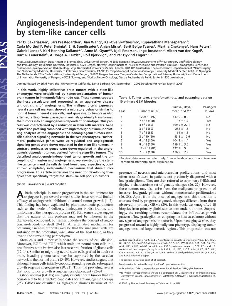
Fig. 2. Nonangiogenic tumors contain cells with stem-like features. (a) Brainsections at different time points corresponding to the MRI scans. The maintumor mass has a purple color because of immunostaining with a human-specific antibody against vimentin. Costaining with anti-human vimentin (red)and Ki67 (brown) show dividing and nondividing tumor cells in differentregions of the brain: corpus callosum (b), tumor bulk (c), and contralateralhemisphere (d). (e) Nestin-positive cancer cells (brown) invading the paren-chyma in the contralateral hemisphere. ( f and g) Migration along corpuscallosum of vimentin-positive cancer cells (brown) from a tumor spheroid ( f)and of human neural stem cells (g). (h) Musashi-1-positive cells (green) mi-grating from a tumor spheroid (red). (Scale bars: 50 �m.)
Sakariassen et al. PNAS � October 31, 2006 � vol. 103 � no. 44 � 16467
MED
ICA
LSC
IEN
CES

result may be attributed to cells infiltrating the vascular bed, therebyincreasing the distance between neighboring vessels. Double stain-ing for Ki67 and von Willebrand factor showed dividing tumor cellsamong quiescent normally sized blood vessels (Fig. 1h). No dividingendothelial cells were observed in the tumors. Double staining forcollagen IV and the hypoxia marker pimonidazol revealed no signof hypoxia in the first-generation tumor (Fig. 1i). Sections of ratbrains perfused with India ink (Fig. 7a, which is published assupporting information on the PNAS web site) and transmissionelectron microscopy (Fig. 7b) also revealed a normal endothelialmorphology and tight junctions between the endothelial cells.Immunohistochemical detection of reactive endothelial cells bystaining for angiopoietin-2 was negative (Fig. 8a, which is publishedas supporting information on the PNAS web site).
Angiogenesis-Independent Growth Is Mediated by Tumor Cells ThatExhibit Stem-Like Characteristics. Rat brains harvested at the time ofMRI (Fig. 1a) allowed comparison with histological sections fromcorresponding regions (Fig. 2a). In all regions of the brain, weidentified Ki67-positive tumor cells, which also stained positive forhuman-specific vimentin, a marker present in neuroepithelial pro-genitors and stem cells (33) (Fig. 2 b–d). Tumor cells migratingalong the corpus callosum, entering the cortex, also expressed theneural stem cell marker nestin (Fig. 2e). For comparison, trans-planted glioma spheroids and human neural stem cells (HNSC 100)were stained for vimentin and showed a striking similarity in theirmigratory pattern (Fig. 2 f and g). The tumor cells also expressedthe neural stem cell marker musashi-1 (Fig. 2h), an RNA-bindingprotein involved in asymmetric cell division during Drosophilaneural development (34).
Serial Passaging in Vivo Changes the Nonangiogenic Tumor to a HighlyVascular Phenotype. To further investigate tumor progression, first-generation tumors from four patients (Table 1) were removed andserially passaged in rats for 4–5 generations (hereafter termedhigh-generation tumors). In the subsequent generations, the tumorsbecame more vascular and circumscribed (Fig. 3a) with emergingnecrotic regions (Fig. 3 b and c). Moreover, MRI scans showed lessinvasive (Fig. 3d), strongly contrast-enhancing tumors (Fig. 3e) inthe high generation. The less-invasive nature of the high-generationtumors was also confirmed by positron emission tomography scans,where they appeared sharply demarcated (Fig. 3f). Immunohisto-chemistry revealed tumors with a disordered vasculature, dilatedvessels, and endothelial cell proliferations (Fig. 3 g and h). Triplestaining for collagen IV and the hypoxia marker pimonidazole inrats infused with Hoechst 33342 revealed numerous hypoxic areassurrounded by dilated vessels with Hoechst leakage into the sur-rounding parenchyma (Fig. 3i). This leakage was also confirmed inrats that had received systemic injections of India ink (data notshown). A morphometric quantification of the vascular parameters(Fig. 3j) revealed lower vascular counts per visual field in thehigh-generation tumors compared with normal rat brain, whereasthe area fraction representing endothelial cells per visual field wasincreased. Finally, the proliferative capillary index was 6% in thetumors compared with 0% in the normal brain. The onset ofangiogenesis coincided with a significant decrease in survival from113 � 2.6 SEM to 43 � 2.1 SEM days (Fig. 3k).
Angiogenesis-Independent and -Dependent Phenotypes Are Geneti-cally Similar, but Display Different Gene and Protein ExpressionProfiles and Distinct Patterns of Intracellular Signaling. Array CGHshowed that the human biopsy and the first- and high-generation
Fig. 3. Angiogenesis-independent stem-like tumorsprogress to become vascular and necrotic tumors. (a) H&Estaining of a high-generation tumor. Dashed lines indicate thetumor periphery. (b) Picture of the same tumor exhibitingmacroscopic necrosis (arrowhead). (c) H&E staining of a high-generation tumor at high magnification with enlarged vesselsand arrowheads indicating necrotic areas. (d and e) T2-weighted (d) and gadodiamid-enhanced T1-weighted (e) MRIscans of a high-generation tumor. White area in e representscontrast enhancement. ( f) Positron emission tomography scanof the rat brain tumor. (Scale bar: 1 cm.) (g) CD31 staining(brown) of the tumor bed. (h) Costaining for von Willebrand(red) and Ki67 (brown). (Inset) Proliferating endothelial cells(arrowheads). (i) Triple staining of a tumor section againstpimonidazol (green), collagen IV (red), and Hoechst (blue). (j)Quantification of vascular parameters and comparison withnormal brain. (k) Kaplan–Meyer curves presenting survivaldata for animals grafted with four patient biopsies that werepassaged from first- to high-generation. (Scale bars: 100 �m,unless otherwise indicated.)
16468 � www.pnas.org�cgi�doi�10.1073�pnas.0607668103 Sakariassen et al.

tumors displayed nearly identical chromosomal profiles (Fig. 4a),where they showed loss on chromosome 5p, gain on 7 with EGFRamplification, INK4A�ARF homozygous deletion, loss of chromo-some 10, and interstitial loss of 15q. The striking similarities in thearray CGH profiles between the tumors suggested that transcrip-tional regulation is an important component in the phenotypic shiftobserved. Therefore, a comprehensive gene-expression analysiscomparing first- and high-generation tumors was performed. Intotal, we found 77 genes whose differential expression was 2-fold ormore between the two tumor phenotypes, using three differentmicroarray platforms [16,000-oligonucleotide cDNA; Agilent Tech-nologies (Palo Alto, CA), 44,000-oligonucleotide; Agilent Technol-ogies, and 37,000-oligonucleotide microarrays; Applied Biosystems(Foster City, CA)] (Fig. 4b, and Table 2, which is published assupporting information on the PNAS web site). Furthermore, twoof the array platforms contained vimentin and nestin, which whereup-regulated 200% and 70% in the first-generation, respectively. Toensure that this up-regulation was human-specific and not causedby reactive host-derived cells, we designed primers specific for ratvimentin. Quantitative real-time PCR (RT-qPCR) from low- andhigh-generation confirmed that the expression was from the tumorcells (Fig. 9, which is published as supporting information on thePNAS web site). Moreover, a comprehensive Kinetworks multiim-munoblotting screen was performed, which represents a systems-biology approach providing simultaneous expression and phos-phorylation states of hundreds of target proteins. The Kinetworksscreen revealed numerous proteins to be differentially expressed,including main components of intracellular signaling pathways (Fig.4c). Based on the gene-expression profiles and the Kinetworksscreen, we found that components of the Wnt, PI3K, and NF-k�signaling pathways were overexpressed in the invasive first-generation tumors compared with the high-generation tumors. Inaddition, although components of the Ras signaling pathway wereexpressed in both first- and high-generation tumors, they weresignificantly up-regulated in the high generation (Fig. 4d). More-
over, the first-generation tumors displayed up-regulation of genesinvolved in fetal development and cell motility (Table 3, which ispublished as supporting information on the PNAS web site).
Invasion and Angiogenesis, Two Independent Strategies for TumorProgression. The tumor cell invasion marker SPARC (35–37) wasup-regulated in first-generation tumors, whereas the high-generation tumors displayed weak or no staining (Fig. 5a). Fur-thermore, spheroids from first-generation tumors were highly in-vasive when tested in a collagen-invasion-gel assay, whereas thehigh-generation tumor spheroids only displayed a modest invasionin the gel (Fig. 5b).
Conversely, immunostaining for HIF-1� and VEGF were neg-ative in sections from first-generation tumors, whereas staining forboth markers were positive in the high-generation tumors (Fig. 5 cand d). The same staining pattern was seen for angiopoitin-2 (Fig.8). Furthermore, Western blotting for HIF-1� and quantitativereal-time PCR (RT-qPCR) for its target gene carbon anhydrase IX(CAIX) showed up-regulation in the high-generation tumors,whereas RT-qPCR for VEGF in the first-generation tumorsshowed levels comparable with normal brain (Fig. 10 a–c, which ispublished as supporting information on the PNAS web site). Inaddition, HIF-1� was not detected in tumor spheroids in normoxicconditions but was up-regulated in hypoxia, followed by an increaseof CAIX expression (Fig. 11 a and b, which is published assupporting information on the PNAS web site). Moreover, wefunctionally assessed the angiogenic potential of first- and high-generation tumors in a rat aortic ring assay (Fig. 5e). Endothelialcell sprouting was evident only from aortic rings that receivedconditioned medium from high-generation tumor spheroids. Con-ditioned media from first-generation tumor spheroids induced nooutgrowth of endothelial cells during the observation period of 11days, suggesting that first-generation tumors do not secrete thenecessary amounts of angiogenic factors to trigger angiogenesis.
Fig. 4. Comparison of chromosomal DNA, gene expression, and protein profiles between first- and high-generation tumors. (a) Array CGH showing the relativechromosome copy numbers of the parent biopsy, first- and high-generation tumors. (b) Bar graph presenting the genes with the biggest difference in expressionlevels between the first- and high-generation tumors. (c) Immunoblot analysis of protein extracts from first- and high-generation tumor tissue. VEGF wasanalyzed from cerebrospinal fluid. (d) Signaling pathways differentially activated in the two tumor phenotypes.
Sakariassen et al. PNAS � October 31, 2006 � vol. 103 � no. 44 � 16469
MED
ICA
LSC
IEN
CES

DiscussionMalignant gliomas are the most common cancers in the brain andremain difficult to cure despite advances in surgery and adjuvanttherapy. Recent studies have identified tumor cell subpopula-tions that might be responsible for tumor initiation and progres-sion. Cancer stem cells have been identified in leukemias andbreast, prostate, and brain cancer (38–44). In some cases, thesetumor-initiating cells can be distinguished from the non-tumor-initiating cancer cells based on cell surface marker expression.For instance, it has been found that only CD44��CD24��lineage� breast cancer cells form new tumors in animals (45).Similarly, CD133 has been proposed as a cancer stem cell markerin brain cancers (46). However, we established tumors in vivofrom GBM-derived spheroids that contained nestin��GFAP��CD133� cells. This discrepancy may be due to different cultureconditions because we cultured our biopsy material in serum-containing medium. The implanted tumor spheroids developedtumors with a stem-like, nonangiogenic and highly invasivephenotype. The first-generation tumors mediated a fulminantfatal disease course, and 7 of 10 specimens produced thisphenotype. Two other specimens developed into highly invasivetumors with a predominantly normal vasculature, and only onebiopsy produced contrast enhancement. Although the cellularprogram mediating the nonangiogenic phenotype is possibly aremnant of fetal development that lies dormant during normaltumorigenesis, the program may be reactivated to drive tumorprogression in a clinical setting when patients are treated withangiogenic inhibitors. In contrast to the dormant tumors thatbecome malignant only after the onset of angiogenesis (21), ourresults challenge the current view of malignant tumor growth asan angiogenesis-dependent process.
Despite the fact that the nonangiogenic phenotype recapitulatesdevelopmental signaling pathways and expresses stem cell markers,it is not clear whether these cells are derived from transformedneural stem cells, from stem cell fusion events (47), or fromotherwise restricted subpopulations within the tumor. The geneticsimilarities between the different tumor phenotypes, as demon-strated by almost identical array CGH profiles, do not support amajor involvement of clonal selection, but suggest that transcrip-tional regulation mediates the phenotypes observed. Furthermore,it has been shown that an astrocytoma cell line became moreinvasive after knocking out the HIF-1� gene (48).
In later generations, transition to a vascular tumor phenotypeis mediated by cells where the Ras-signaling pathway is activated.Thus, the capacity for tumor growth is neither limited to agenetic subclone nor to a certain cell phenotype, but is sharedbetween groups of phenotypically diverse cells, where some arecharacterized by a diffuse growth pattern and others by angio-genesis. Accordingly, the uncoupling of invasion and angiogen-esis, represented by the stem-like cancer cells and the cells
derived from them respectively, points at two different mecha-nisms that drive tumor progression. Although the mechanismbehind the phenotypic shift is not fully understood, HIF-1�expression seems to be triggered by hypoxia, because it was notconstitutively expressed by high-generation tumor spheroidscultured under normoxic conditions. The results showing thatboth phenotypes can mediate a fulminant disease course suggestthat even a 100%-effective therapy directed toward one of thebiological entities (either invasion or angiogenesis) will not curethe cancer. Cancer treatment strategies need to pursue both theinvasive stem-like cancer cells and angiogenic targets. A majorchallenge will be to design therapies that target the stem-likecancer cells without destroying the normal stem cell pools thatare needed to maintain normal tissue function.
Materials and MethodsCell Culture and in Vitro Assays. Biopsy spheroids were prepared asdescribed (49). After 1–2 weeks in culture, spheroids withdiameters between 200 and 300 �m were selected for intrace-rebral implantation.
In Vivo Experiments. Nude immunodeficient rats (Han: rnu�rnuRowett) were fed a standard pellet diet and were provided withwater ad libitum. All procedures were approved by The NationalAnimal Research Authority. Biopsy spheroids were stereotacticallyimplanted into the right brain hemisphere, and the rats were killedwhen symptoms developed.
Immunohistochemistry. After deparaffinization, all sections wereboiled in citrate buffer, pH 6.2, for 20 min, except for the vonWillebrand staining, where the sections were treated with protein-ase K (DAKO, Glostrup, Denmark) for 10 min. Sections were thentreated with protein-blocking solution (DAKO) for 10 min, and theprimary antibody was incubated for 45 min at room temperature,washed four times, incubated for 35 min with En Vision� Systemspolymer-conjugated secondary antibody (DAKO), washed fourtimes, and finally incubated with DAB for 5 min.
Transmission Electron Microscopy. The rats were perfusion fixed, andthe brains were removed and embedded in Epon 812, followed byultrathin sectioning in preparation for electron microscopy.
Hypoxia Experiment. Spheroids were cultured at 37°C with 5% CO2,94% N2, and 1%O2 for 16 h in a Mini Galaxy incubator (RSBiotech, Ayrshire, Scotland, U.K.).
Western Blotting. Cerebrospinal fluid was run on SDS�PAGE byusing NuPage precast gels (Invitrogen, Carlsbad, CA). After blot-ting, the nitrocellulose membrane was blocked for 30 min at roomtemperature and incubated overnight at 4°C in buffer (TBS with0.1% Tween 20, 5% milk powder) containing anti-VEGF-A diluted
Fig. 5. Inverse relationship between angiogenesis and invasion. (a) SPARC immunostaining (brown) at the tumor periphery in first- and high-generationtumors. (b) Invasion of tumor cells in a collagen gel from first- and high-generation glioma spheroids. (c and d) Hif-1� and VEGF expression (brown), respectively,in first- and high-generation tumors. (e) Aortic ring explants incubated with conditioned medium from first- and high-generation tumor spheroids. Pictures fromaortic ring and collagen-invasion assays were all taken on day 5. (Scale bars: 100 �m.)
16470 � www.pnas.org�cgi�doi�10.1073�pnas.0607668103 Sakariassen et al.

1:100 (Abcam, Cambridge, U.K.), anti-HIF1� diluted 1:100 (BDBiosciences, San Diego, CA), or anti-GAPDH diluted 1:2,000(Abcam). The primary antibody was detected by using an HRP-conjugated goat anti-rabbit�mouse secondary antibody (Immuno-tech, Fullerton, CA) diluted 1:2,500. Extraction of protein fromcultured spheroids was done by washing in PBS two times andhomogenizing in lysis buffer by sonication twice for 15 sec by usingSonics Vibra Cell (Cole–Parmer Instruments, Vernon Hills, IL).Whole lysate was used for subsequent analysis. Twenty microgramsof protein was applied in each well.
Protein Kinase and Phosphosite Screening. The procedure is de-scribed in refs. 50 and 51). The following screens were performed:KPKS-1.2A, KPKS-1.2B, KPSS-2.1, KPSS-4.1, and KPSS-1.3. Fordetails, see the Kinexus (Advent Software, San Francisco, CA)home page www.kinexus.com.
Quantitative RT-PCR. cDNA was generated by using the iScriptcDNA synthesis kit according to the manufacturers instructions(Bio-Rad, Hercules, CA). Each reaction is in triplicate on the plate,and a similar plate was repeated three times. The reactions wereperformed by using iQ SYBR green Supermix reagents kit (Bio-Rad), and the PCR was run on a BioRad iCycle detection system(Bio-Rad).
Gene-Expression Analysis. Single-stranded cDNA was reverse tran-scribed from 2 �g of total RNA and T7 RNA polymerase promoter-containing double-stranded cDNAs, and T7 RNA polymerase-amplified RNAs (cRNAs) were generated according to the T7Megakit protocol (Ambion, Austin, TX) as described (52).
Agilent DNA Microarrays. The Agilent 16,000-oligonucleotidecDNA microarrays were processed as described (53).
ABI1700 DNA Oligonucleotide Microarrays. The Human GenomeSurvey Microarray, Chemiluminescence Detection kit, AppliedBiosystems Chemiluminescent RT-IVT Labeling kit, and AppliedBiosystems 1700 Chemiluminescent Microarray Analyzer was usedas recommended.
Bioinformatic Analysis of DNA Microarray Data. In total, six hybrid-izations were performed, two for each platform. The result filesfrom the three different image-processing software programswere all imported into the analysis software J-Express (54).Controls and flagged spots were removed. J-Express is availableat www.molmine.com.
Array CGH. To determine the copy number across all chromosomes,we did comparative genomic hybridizations on whole-genomearrays of 2,400 chromosomally mapped BAC clones (Hum.Array1.14) following methods described in ref. 55.
Supporting Information. For more information, see Supporting Ma-terials and Methods, which is published as supporting information onthe PNAS web site.
We thank Aina Johannessen, Linda Vabø, and Tore-Jacob Raa fortechnical assistance. This work was supported by the Norwegian CancerSociety, the Norwegian Research Council, Innovest AS, Helse-Vest,Haukeland University Hospital, the Bergen Translational ResearchProgram, the Centre Recherche de Public Sante Luxembourg, and theEuropean Commission 6th Framework Program Contract 504743.
1. Eisterer W, Jiang X, Bachelot T, Pawliuk R, Abramovich C, Leboulch P, Hogge D,Eaves C (2002) Mol Ther 5:352–359.
2. Garber K (2002) Nat Biotechnol 20:1067–1068.3. Brem S, Grossman SA, Carson KA, New P, Phuphanich S, Alavi JB, Mikkelsen T,
Fisher JD (2005) Neuro-oncol 7:246–253.4. Akella NS, Twieg DB, Mikkelsen T, Hochberg FH, Grossman S, Cloud GA, Nabors
LB (2004) J Magn Reson Imaging 20:913–922.5. Gagner JP, Law M, Fischer I, Newcomb EW, Zagzag D (2005) Brain Pathol
15:342–363.6. Hansma AH, Broxterman HJ, van der Horst I, Yuana Y, Boven E, Giaccone G,
Pinedo HM, Hoekman K (2005) Ann Oncol 16:1695–1701.7. Wedam SB, Low JA, Yang SX, Chow CK, Choyke P, Danforth D, Hewitt SM,
Berman A, Steinberg SM, Liewehr DJ, et al. (2006) J Clin Oncol 24:769–777.8. Marshall E (2002) Science 295:2198–2199.9. Kunkel P, Ulbricht U, Bohlen P, Brockmann MA, Fillbrandt R, Stavrou D, Westphal
M, Lamszus K (2001) Cancer Res 61:6624–6628.10. O’Donnell A, Padhani A, Hayes C, Kakkar AJ, Leach M, Trigo JM, Scurr M,
Raynaud F, Phillips S, Aherne W, et al. (2005) Br J Cancer 93:876–883.11. Bouscary D, Legros L, Tulliez M, Dubois S, Mahe B, Beyne-Rauzy O, Quarre MC,
Vassilief D, Varet B, Aouba A, et al. (2005) Br J Haematol 131:609–618.12. Engebraaten O, Bjerkvig R, Pedersen PH, Laerum OD (1993) Int J Cancer
53:209–214.13. Ignatova TN, Kukekov VG, Laywell ED, Suslov ON, Vrionis FD, Steindler DA
(2002) Glia 39:193–206.14. Dvorak P, Dvorakova D, Hampl A (2006) FEBS Lett 580:2869–2874.15. Englund U, Fricker-Gates RA, Lundberg C, Bjorklund A, Wictorin K (2002) Exp
Neurol 173:1–21.16. Hurelbrink CB, Armstrong RJ, Dunnett SB, Rosser AE, Barker RA (2002) Eur
J Neurosci 15:1255–1266.17. Aboody KS, Brown A, Rainov NG, Bower KA, Liu S, Yang W, Small JE, Herrlinger
U, Ourednik V, Black PM, et al. (2000) Proc Natl Acad Sci USA 97:12846–12851.18. Visted T, Enger PO, Lund-Johansen M, Bjerkvig R (2003) Front Biosci 8:e289–304.19. Brabletz T, Jung A, Spaderna S, Hlubek F, Kirchner T (2005) Nat Rev Cancer
5:744–749.20. Holash J, Maisonpierre PC, Compton D, Boland P, Alexander CR, Zagzag D,
Yancopoulos GD, Wiegand SJ (1999) Science 284:1994–1998.21. Naumov GN, Bender E, Zurakowski D, Kang SY, Sampson D, Flynn E, Watnick RS,
Straume O, Akslen LA, Folkman J, Almog N (2006) J Natl Cancer Inst 98:316–325.22. Carmeliet P (2005) Oncology 69 (Suppl) 3:4–10.23. Folkman J (1971) N Engl J Med 285:1182–1186.24. Ribatti D (2005) Br J Haematol 128:303–309.25. Ribatti D, Vacca A (2005) Curr Cancer Drug Targets 5:573–578.26. Collins VP (2004) J Neurol Neurosurg Psychiatry 75 (Suppl 2):ii2–ii11.27. Liu L, Backlund LM, Nilsson BR, Grander D, Ichimura K, Goike HM, Collins VP
(2005) J Mol Med 83:917–926.28. Karcher S, Steiner HH, Ahmadi R, Zoubaa S, Vasvari G, Bauer H, Unterberg A,
Herold-Mende C (2006) Int J Cancer 118:2182–2189.
29. Kleihues P, Ohgaki H (1999) Neuro-oncol 1:44–51.30. Engebraaten O, Hjortland GO, Hirschberg H, Fodstad O (1999) J Neurosurg
90:125–132.31. Mahesparan R, Read TA, Lund-Johansen M, Skaftnesmo KO, Bjerkvig R, Enge-
braaten O (2003) Acta Neuropathol (Berlin) 105:49–57.32. Shields AF, Grierson JR, Dohmen BM, Machulla HJ, Stayanoff JC, Lawhorn-Crews
JM, Obradovich JE, Muzik O, Mangner TJ (1998) Nat Med 4:1334–1336.33. Villa A, Snyder EY, Vescovi A, Martinez-Serrano A (2000) Exp Neurol 161:67–84.34. Okabe M, Imai T, Kurusu M, Hiromi Y, Okano H (2001) Nature 411:94–98.35. Schultz C, Lemke N, Ge S, Golembieski WA, Rempel SA (2002) Cancer Res
62:6270–6277.36. Rich JN, Hans C, Jones B, Iversen ES, McLendon RE, Rasheed BK, Dobra A,
Dressman HK, Bigner DD, Nevins JR, West M (2005) Cancer Res 65:4051–4058.37. Shi Q, Bao S, Maxwell JA, Reese ED, Friedman HS, Bigner DD, Wang XF, Rich JN
(2004) J Biol Chem 279:52200–52209.38. Yuan X, Curtin J, Xiong Y, Liu G, Waschsmann-Hogiu S, Farkas DL, Black KL, Yu
JS (2004) Oncogene 23:9392–9400.39. Reya T, Morrison SJ, Clarke MF, Weissman IL (2001) Nature 414:105–111.40. Marx J (2003) Science 301:1308–1310.41. Singh SK, Clarke ID, Terasaki M, Bonn VE, Hawkins C, Squire J, Dirks PB (2003)
Cancer Res 63:5821–5828.42. Collins AT, Berry PA, Hyde C, Stower MJ, Maitland NJ (2005) Cancer Res
65:10946–10951.43. Nakano I, Kornblum HI (2006) Pediatr Res 59:54R–58R44. Bonnet D (2005) Cell Prolif 38:357–361.45. Al-Hajj M, Wicha MS, Benito-Hernandez A, Morrison SJ, Clarke MF (2003) Proc
Natl Acad Sci USA 100:3983–3988.46. Singh SK, Hawkins C, Clarke ID, Squire JA, Bayani J, Hide T, Henkelman RM,
Cusimano MD, Dirks PB (2004) Nature 432:396–401.47. Bjerkvig R, Tysnes BB, Aboody KS, Najbauer J, Terzis AJ (2005) Nat Rev Cancer
5:899–904.48. Blouw B, Song H, Tihan T, Bosze J, Ferrara N, Gerber HP, Johnson RS, Bergers G
(2003) Cancer Cell 4:133–146.49. Bjerkvig R, Tonnesen A, Laerum OD, Backlund EO (1990) J Neurosurg 72:463–475.50. Wang J, Laschinger C, Zhao XH, Mak B, Seth A, McCulloch CA (2005) Biochem
Biophys Res Commun 330:123–130.51. Lin HJ, Hsieh FC, Song H, Lin J (2005) Br J Cancer 93:1372–1381.52. Halvorsen OJ, Oyan AM, Bo TH, Olsen S, Rostad K, Haukaas SA, Bakke AM,
Marzolf B, Dimitrov K, Stordrange L, et al. (2005) Int J Oncol 26:329–336.53. Gjertsen BT, Oyan AM, Marzolf B, Hovland R, Gausdal G, Doskeland SO, Dimitrov
K, Golden A, Kalland KH, Hood L, Bruserud O (2002) J Hematother Stem Cell Res11:469–481.
54. Dysvik B, Jonassen I (2001) Bioinformatics 17:369–370.55. Snijders AM, Nowak N, Segraves R, Blackwood S, Brown N, Conroy J, Hamilton G,
Hindle AK, Huey B, Kimura K, et al. (2001) Nat Genet 29:263–264.
Sakariassen et al. PNAS � October 31, 2006 � vol. 103 � no. 44 � 16471
MED
ICA
LSC
IEN
CES
Related Documents