Anesthesia and Acid-Base Status in Congenital Heart Disease Chung- Yuan Lin, M.D. University of Chicago Department of Anesthesiology 950 East 59th Street Chicago, Illinois 60637 In his review of anesthesia for cardiac surgery, Moffitt 1 noted that essentially every agent and every technique known in anesthesia have been used for cardiac surgery. All of the commonly used general anesthetic agents, with the possible exception of nitrous oxide, have a depressant action on the myocardium and peripheral vascular bed, 2 the extent of which, in general, is closely correlated with the potency of a given agent as an anesthetic. Our group believes that there are some advantages of certain techniques and an- esthetic agents over others in the presence of complicated pathophysiological conditions of congenital heart disease which are undergoing surgical correction. I shall discuss the current practices which are used at the University of Chicago for pediatric cardiac anesthesia. PREANESTHETIC VISIT The preanesthetic visit is essential. It not only establishes rapport with the patient or the patient's family, but it also gives the anesthesiologist an opportunity to assess the nature and severity of the cardiac lesion and the patient's general status. In addition, it familiarizes the physician with the patient's drug history. We recommend stopping digitalis 48 hours prior to surgery. PREMEDICATION }he cyanotic patient is said to have a.n increased sensitivity to sedatives, 3 and most narcotics decrease arterial oxygen tension. However, preoperative sedation provides smoother induction and, by relieving anxiety, lessens the emotional trauma in a patient facing a cardiac operation. During the induction period, especially in the case of cyanotic patients, straining, crying, and fighting should be avoided because they raise the intraalveolar pressure, leading to reduced pulmonary perfusion, increased pulmonary vascular resistance, and hence increased right-to-left shunting with worsening of systemic hypoxia. They also increase intra-thoracic pressure and impede the venous return, leading to a decrease in cardiac output. Therefore, we highly recommend that heavier sedation or premedication than that regularly used in pediatric cases be given one hour prior to induction. VOLUME II, NUMBER I, 1979 3

Welcome message from author
This document is posted to help you gain knowledge. Please leave a comment to let me know what you think about it! Share it to your friends and learn new things together.
Transcript
Anesthesia and Acid-Base Status in Congenital Heart Disease
Chung-Yuan Lin, M.D.
University of Chicago Department of Anesthesiology 950 East 59th Street Chicago, Illinois 60637
In his review of anesthesia for cardiac surgery, Moffitt 1 noted that essentially every agent and every technique known in anesthesia have been used for cardiac surgery.
All of the commonly used general anesthetic agents, with the possible exception of nitrous oxide, have a depressant action on the myocardium and peripheral vascular bed,2 the extent of which, in general, is closely correlated with the potency of a given agent as an anesthetic.
Our group believes that there are some advantages of certain techniques and anesthetic agents over others in the presence of complicated pathophysiological conditions of congenital heart disease which are undergoing surgical correction.
I shall discuss the current practices which are used at the University of Chicago for pediatric cardiac anesthesia.
PREANESTHETIC VISIT
The preanesthetic visit is essential. It not only establishes rapport with the patient or the patient's family, but it also gives the anesthesiologist an opportunity to assess the nature and severity of the cardiac lesion and the patient's general status. In addition, it familiarizes the physician with the patient's drug history. We recommend stopping digitalis 48 hours prior to surgery.
PREMEDICATION
}he cyanotic patient is said to have a.n increased sensitivity to sedatives,3 and most narcotics decrease arterial oxygen tension. However, preoperative sedation provides smoother induction and, by relieving anxiety, lessens the emotional trauma in a patient facing a cardiac operation.
During the induction period, especially in the case of cyanotic patients, straining, crying, and fighting should be avoided because they raise the intraalveolar pressure, leading to reduced pulmonary perfusion, increased pulmonary vascular resistance, and hence increased right-to-left shunting with worsening of systemic hypoxia. They also increase intra-thoracic pressure and impede the venous return, leading to a decrease in cardiac output. Therefore, we highly recommend that heavier sedation or premedication than that regularly used in pediatric cases be given one hour prior to induction.
VOLUME II, NUMBER I, 1979 3
The usual doses for premedication are morphine, 0.1 to 0.2 mgm/kg, scopolamine, 0.01 mgm/kg, and pentobarbital, 1 to 2 mgm/kg. With this dose schedule, children are usually very calm and will submit to the face mask without struggle or fear, thus permitting a smooth induction of anesthesia.
After premedication on the ward, the patients usually receive supplemental oxygen. This is especiaJiy important for the cyanotic patient and is necessary during transportation to the operating room. The oxygen ensures that there is no deterioration in oxygenation which might result from an increased anatomical dead space following the use of scopolamine, or from a respiratory depression, in both rate and depth, due to morphine and pentobarbital. Placement of intravenous needles or catheters is done after induction in children and infants to avoid any unnecessary struggle or anxiety.
For infants about to undergo deep hypothermia, premedication consists solely of scopolamine, 0.1 mg intramuscularly.
MANAGEMENT OF ANESTHESIA
Ketamine in small doses has been used as the main anesthetic agent for many years. Recently, however, Ketamine has been found to cause a dose-dependent increase in pulmonary vascular resistance; therefore we are not using it in patients who have a reduced pulmonary flow.4•5
As a muscle relaxant for reasonably short cases, we recommend succinylcholine. For most open-heart surgery, however, we use Pavulon, 0.1-0.15 mgm/kg of body weight. Just before the patient is placed on the pump, or during extracorporeal circulation, dilution due to priming volumes for the pump must be accounted for, and 1-2 mgm Pavulon are required for adequate muscle relaxation.
Light halothane anesthesia coupled with relaxants remains a good choice for a variety of situations, mainly because it provides easy control over blood pressure and, especially in children, gives rapid, smooth induction.6 Thus, for most pediatric cardiac surgical procedures, halothane is the anesthetic agent of choice.
The common problems in pumping of the infant are balance of volume, and cannulation of the superior and inferior vena cava and aorta.
Both of these problems are related to the size of the pediatric patient. If the S. V.C. cannula is advanced too far, it can easily be wedged into the smaller venous branch, thus obstructing the remaining S.V.C. drainage and causing cerebral edema. It can also happen that the I.V.C. cannula is pushed too far, causing obstruction of renal venous drainage and thus a marked reduction of urinary output during cardiopulmonary bypass, which can lead to ascites. If the aortic cannula is too long, it can become wedged in branch vessels of the arch and cause serious problems.
It is difficult to balance the volume between patient and oxygenator; this requires meticulous attention from the perfusion team.
Since we calibrate the pump head against the water column, adjusting roller pump occlusion to "just occlude the pump head," we seldom observe gross hemolysis in the urine, even during a 3-hour pump run with a bubble oxygenator. If such hemolysis does occur, however, we add 12.5 gm Mannitol and 20 mgm Lasix into the extracorporeal circulation to prevent a "heme" deposit on the renal tubule.
4 THE JOURNAL OF EXTRA-CORPOREAL TECHNOLOGY
We use high-flow perfusion with 2.3-2.5 l/minjM2 of BSA. In cases of cyanosis, we compensate for the excessive bronchial flow by increasing the calculated flow by 10-20%. During extracorporeal circulation, the use of any vasoconstrictor to elevate the blood pressure should be avoided. If the pump head is adjusted to "just occlusive" prior to surgery, a flow of 2.3-2.5 l/min/M2 should provide enough oxygen to the patient.
The pressure reading from the radial artery sometimes differs considerably from the central arterial pressure. Low pressure is usually caused by a decrease in hematocrit and a decrease in viscosity ,7 and hence by a decrease in peripheral vascular resistance. If some urine output is definitely present, it can be assumed that the perfusion is adequate for the tissues.
PRIMING
Because the blood volume in infants is small compared to the priming volume of the oxygenator, we recommend adding sufficient blood to maintain a hemoglobin content of not less than 8 mg%. Dissolved oxygen in the plasma accounts for a very small fraction of the total oxygen-carrying capacity of the blood. One gram of hemoglobin can carry 1.34 cc of oxygen, but 100 cc of plasma per mmHg of oxygen carries only 0.003 cc of oxygen; therefore, even if the p02 is raised to 400 mmHg, only 1.2 cc of oxygen is carried by the plasma. Unless hypothermia is introduced to reduce the total oxygen demand of the body, the oxygen-carrying capacity is dependent mainly on the amount of hemoglobin in the blood.
Even the smallest priming volume for an infant circuit requires about 1000 cc, compared with the infant's blood volume of around 300-400 cc. Thus it is not easy to maintain extracorporeal circulation with a fine balance.
PROFOUND HYPOTHERMIA
Advocates of the use of the cardiopulmonary bypass state that it avoids the profound and incompletely understood metabolic abnormalities and the potential for cerebral injury which are associated with the use of profound hypothermia with complete circulatory arrest.8 We believe, however, that the use of profound hypothermia has a role in cardiac surgery in place of the cardiopulmonary bypass, if the body weight of a patient requiring total repair of a complicated heart defect is below 8 kg.
The outstanding advantage in the use of profound hypothermia is that excellent surgical conditions are obtained in the still, relaxed, and bloodless heart which is free of cannulas obstructing the surgical field. This advantage is most attractive when complete surgical repair of complicated heart defects is performed, especially in neonates or small infants for whom such operations carry a high morbidity and mortality rate, compared to that in adults, when extracorporeal circulation is used. All surgeons prefer to have as few cannulas as possible hanging from a very small chest when a fine repair of complicated heart defects in infants is required.
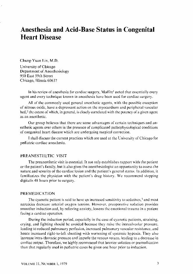
Our anesthetic management during deep hypothermia consists of the use of a totally closed circuit, which avoids the danger of explosion and maintains sufficient humidity. A Harvard pump injects diethyl ether into the anesthetic circuit for maintenance of a constant ether concentration in the blood (Fig. 1).
VOLUME 11, NUMBER 1, 1979 5
..
I~ ' ' , • , I'
====~==-~~----~ - L---------------------~ HARVARD SYRINGE PUMP
NARKOTEST
PRESSURE GAUGE
Alit:_ SHIELD
VENTILATOR
Figure 1. Block diagram of anesthetic system used in deep hypothermia (Columbia pediatric circle system). Oxygen is supplied through a pressure regulator and a needle valve adjuster. Ether is injected into the circle system by a Harvard pump which is regulated by a programmed curve (Data Trak). The ether concentration is monitored continuously at the expiratory arm of the circle system by a Narkotest analyzer.
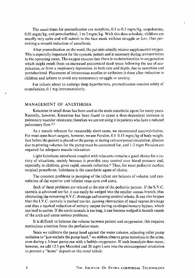
During surface cooling, in order to reduce the increased blood viscosity caused by hypothermia, we give 2 cc of lactated Ringer's solution per kilogram of body weight per degree of temperature decrease from 35° to 25°C. Lasix, 10 mgm, and 5 gm of Mannitol are given intravenously to protect the kidney during hypothermia and circulatory arrest. The procedure is illustrated in Fig. 2.
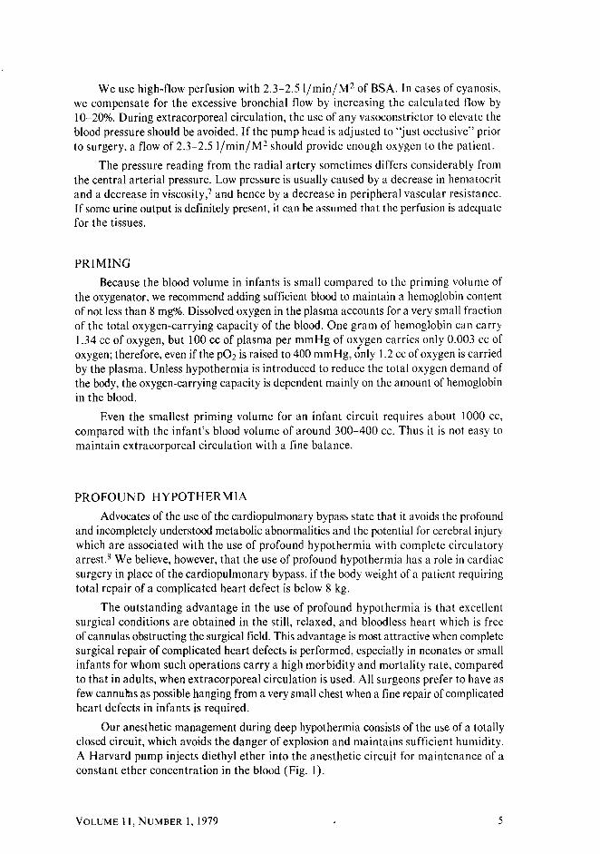
We have used this technique, with diethyl ether as the anesthetic agent, during correction of complicated cardiac anomalies in more than I 00 neonates and infants weighing less than 8 kg, and have never encountered ventricular fibrillation or cardiac arrest. The patients are generally very stable hemodynamically, and the heart has stayed in sinus rhythm throughout the cooling phase (Fig. 3). In contrast to other anesthesia techniques with deep hypothermia, with this method the absence of irritability during handling of the heart prior to intracardiac surgery is remarkable when the desired temperature has been reached. Total cardiac arrest is obtained after occlusion of the vena cava and ascending aorta by injection of several milliliters of Young's solution into the root of the aorta proximal to the aortic occlusion clamp. Total cardiopulmonary arrest can be maintained safely for approximately I hour at a core temperature of approximately 20°C.
Following completion of intracardiac corrective surgery, both core and surface rewarming are used. The heat exchanger in the pump serves for core rewarming; the
6 THE JOURNAL OF EXTRA-CORPOREAL TECHNOLOGY

I I I I
----: 1 tv Protamine 6 mgm/kg
""' v 0
Q) .... ;:) -0 Q) 28 0.
E Q)
I- 24
20
IEKG
CVP Radial A.
lap IPrecordial Stetho. I INDUCTION 1 Ketamine 5mgm/kg IM I IV 1 sec 2mgm/kg IV I lntubat1on I ETHER, Programmed I Closed circuit
Scopolamme 0 1 mg1m 1M
I I I I I I I I I I I I I
I I I I I I
I I I I
f-Surface Rewarming:
I I I I I I I I
f-CaCI2 300mgm to pump1 I I
t-Mannitol 12.5gm l to pumlp LaSI< IOmgm
1 1' t:--- Partial Bypass 1 IVC, SVC OccluSion I 1 Aorta cross ciQmp : I Young 1s Sol 1n 1 I
16~~-------L------~--------~------~------~------~ -1 0 2 3 4 5
Time in Hours
Figure 2. Typical time sequence during surgical correction of congenital heart disease under deep hypothermia with closed-circuit ether anesthesia. Warming is carried out both extracorporeally and with surface rewarming.
TEMP •c 34
..... .,~4
H.R. '"''~
liP
ECG •I ,I •I ... " I
TEMP ·c 27
II.P. 7J/.a
H.R. ! ... ~.
liP
ECG ·I,
33
'%,
f•
J'J \}
•I, •I .,, ·II
I I
26
'.Y.o
·.t '1• I
32 31
·I ·I II I
I I i ,
215 24
30
23
,x.
29
22
28
'Y.,
21
Figure 3. Example of electrocardiograms and typical blood pressure changes occurring during hypothermia. Hemodynamically, the blood pressure remained stable and the ECG stayed in sinus rhythm throughout.
VOLUME II, NUMBER 1, 1979 7
temperature difference between water and blood is maintained at no more than 10°C so that possible liberation of gas bubbles in the blood is avoided.
MONITORING DURING BYPASS
During extracorporeal circulation, we monitor the following parameters:
1. Arterial pressure
2. Central venous pressure
3. Line pressure
4. Body temperature and blood temperature
5. Flow rate
6. Urine output
7. Blood gases
8. EEG
1. Arterial Pressure During extracorporeal circulation, we try to maintain a mean perfusion pressure
of 60-80 mmHg. If the pressure rises above 80 mmHg, various peripheral vasodilators, such as nitroprusside, nitroglycerine, chlorpromazine, regitine, and halothane, can be used. I personally prefer halothane because it is easily controlled, and because it can serve as a supplementary anesthetic. Halothane has a depressant effect on the myocardium, but during a cardiopulmonary bypass a concentration of more than 1 o/o is seldom required, and the heart is excluded from the circulation during the major part of the bypass procedure. Halothane also decreases the myocardial metabolism, thus providing some protection from anoxic cardiac arrest. When external work is required of the heart at the end of extracorporeal circulation, the halothane can be eliminated very quickly from the circulation.
When the Teflo-Travenol membrane oxygenator is used, it is difficult to provide halothane because of the membrane characteristics. Nitroprusside can be used instead.
2. Central Venous Pressure We are very reluctant to use subclavian venous puncture or a percutaneous approach
to the internal jugular vein for CVP monitoring prior to surgery, because there is a high risk of undetected pneumothorax and hemomediastinum. We often introduce the catheter percutaneously through an antecubital vein or through an external jugular vein into the superior vena cava prior to surgery, after the patient has been intubated.
In small infants, if monitoring with a venous pressure catheter cannot be established, we often wait until mediastinotomy is performed; then we introduce a medium-size "intracath" through an innominate vein.
Central venous pressure monitoring provides only limited information on the status of the total blood volume during or following the operation, but it is often used as a therapeutic guide for infusion of blood or plasma. Occasionally, CVP monitoring also serves as a guide for proper placement of the S. V .C. cannula.
8 THE JOURNAL OF EXTRA-CORPOREAL TECHNOLOGY
3. Line Pressure Selection of the arterial cannula is a critical factor. We utilize cannulas that allow
us to maintain a flow of 2.3-2.5 ljM2 of BSA, but at a line pressure from the pump of less than 200 mmHg. The line pressure should be maintained at the lowest possible value, so that the likelihood of a cavitation effect and of shear stress on the red cells in the cannula during perfusion, and thus the likelihood of damage to red cells, is minimized.
4. Body Temperature and Blood Temperature We monitor blood temperature and rectal temperature during perfusion. Some
centers use only esophageal temperature, but this differs from the true patient temperature because of aortic cannulation. At present, we almost always use the ascending aorta for the site of aortic cannulation; this lets the esophageal temperature equilibrate very quickly with the perfusion temperature.
5. Flow Rate We commonly maintain a flow of 2.3-2.5 l/min/M2 throughout the course of a total
cardiopulmonary bypass, in order to provide enough tissue perfusion and to monitor the status of the peripheral vascular bed.
6. Urine Output During the pump run, the minimum urine output should be more than 0.5 ccjkgjhr.
If the amount is lower, it is necessary to check whether there is kinking of the urinary catheter line or whether the perfusion pressure is too low; if neither of these problems exists, 10 mgm Lasix should be added to the pump circuit.
7. Blood Gases Because tissue oxygenation is impaired due to displacement of the oxygen disso
ciation curve caused by alterations in temperature, pH, and carbon dioxide and 2,3,DPG levels, we try to maintain the blood gas and acid-base values as close to the normal range as possible.
In patients with sickle cell disease, we avoid any degree of hypothermic perfusion to prevent intra-operative sickling. In such patients, we also keep the blood slightly alkaline by administering sodium bicarbonate, so that a crisis is prevented.
We always monitor arterial and mixed venous blood gases during cardiopulmonary bypass. We regard venous p02 or oxygen saturation as good indices of tissue perfusion during extracorporeal circulation, except in rare cases in which high venous oxygen saturation is caused by precapillary shunting. A drop in the venous oxygen saturation below 600;6 always indicates either that the perfusion flow is insufficient to provide enough oxygen, or that the anesthesia or muscle relaxation is inadequate and thus increases the oxygen demand.
8. EEG It is not easy to use an electroencephalogram for monitoring during cardiopulmonary
bypass in a noisy operating room. With the conventional electroencephalograph, it is quite difficult to obtain a stable, clear wave form, and even if the tracing is clear, it is difficult to follow possible changes.
VOLUME II, NUMBER I, 1979 9

I I I I
~J:, , , rrr, I
t I t f
5VC I'W:
I "
I L L" , Is
.1 1 -- 1
f bypau
on
cannvkltlon
ill.
T f
j
- I I
1., Ill l. LL .. d 1...0
'I ''i ·~I"
I
J I j
Figure 4. During venous cannulation, the cannula obstructed the venous return; this led to marked transient depression of the CFM.
For the past I 1h years, we have used a Cerebral Function Monitor (CFM), which filters and compresses the electroencephalographic signal into a single tracing from a pair of parietal electrodes. This tracing has proved to be a very useful parameter for monitoring, according to Branthwaite.9 Figures 4-6 show several typical CFM changes which occurred during surgery. It is very easy to observe these changes during the critical stages of the operation.
TERMINATION OF CARDIOPULMONARY BYPASS
Before completion of the cardiopulmonary bypass," the flow is gradually reduced, while blood pressure and cardiac action are observed carefully. During or shortly before this time, 300 mg of calcium chloride are put into the pump and given approximately 5 minutes to equilibrate. The vital signs, including CVP, blood pressure, pupil size, and temperature, are checked and their condition noted. Blood is prepared for transfusion. The lungs are hyperinflated several times.
1 bypass
on
Figure 5. After starting of the pump, the aortic cannula was found to be clamped, so that it withdrew too much venous blood into the pump oxygenator, causing a marked drop in the voltage as shown in the CFM.
t bypass
on
t t rewarming ~~ass
Figure 6. Effect of lowered temperature on the CFM recording. The voltage may become significantly low at temperatures below 28°C.
10 THE JOURNAL OF EXTRA-CORPOREAL TECHNOLOGY

The pump is brought to a standstill, and adequate transfusion from the pump is given. Pharmacological support is rarely used after the termination of cardiopulmonary bypass; we feel free to use such support, however, if there is any doubt of circulatory stability. Relief of intense vasoconstriction during the pumping procedure by appropriate anesthesia or by mild alpha blockers is an important factor in reducing the need for pharmacological support in the postperfusion period. A weak myocardium that may have been insulted with hypoxia during the operative procedure may not have the power to overcome the high peripheral resistance that can be present at this time.
If use of a vasoactive drug is essential, the present drug of choice is dopamine, given by continuous intravenous infusion at a rate of 10 1-lg/kgjmin. If the cardiac rate is low, isoproterenol is usually given at a rate of 0.01-0.05 1-lg/kgjmin, depending on theresponse.
MANAGEMENT AFTER CARDIOPULMONARY BYPASS
In the immediate post-perfusion period, patients rarely require anesthesia of greater potency than provided by 50% nitrous oxide. Blood gases are measured while the patient is ventilated on 10096 oxygen; in this way, the presence of any shunting that might require an elevated oxygen concentration during the immediate postoperative period can be determined. Also, if there is any deviation from the normal acid-base status, it is corrected at this time, generally with sodium bicarbonate. The dose of sodium bicarbonate is calculated as the base deficit in milliequivalents multiplied by the body weight in kilograms and divided by six.
Blood replacement is guided by the circulatory response of the patient. The venous pressure probably provides the most reliable information on the response to transfusion; it also contributes to an understanding of the functioning and efficiency of the heart. A judgment as to whether a blood transfusion should be given is made on the basis of the arterial and venous pressures.
In order to overcome any negative inotropic influence of protamine on the heart, one should give a dose of 10-20 mgmjkg of body weight of calcium chloride intravenously just prior to injection of the protamine. The calcium ion effect on the myocardium, which consists of an increase in myocardial contractility and hence an increase in cardiac output, cancels the negative inotropic effect due to subsequent injection of protamine.
Routinely, patients who have undergone cardiopulmonary bypass during cardiac surgery are ventilated, at least over night, with a volume-limited ventilator, so that the workload of breathing is reduced and so that the postoperative circulatory status can be assessed under optimal respiratory conditions. There are some exceptions, such as simple ASD PS and noncomplicated VSD. We extubate soon after the surgery is completed.
ACID-BASE BALANCE
The increasing therapeutic use of hypothermia with or without extracorporeal circulation has emphasized the need for temperature correction of blood gas measurements. Acid-base balance will not be discussed in detail, except for temperature corrections and a simple method of calculating base excess.
VOLUME II, NUMBER I, 1979 II

• --· 21"( • ow·
Figure 7. CFM during deep hypothermia. The mean voltage drops to zero very rapidly following induction of cardiac arrest, then gradually recovers to the preoperative level following bypass rewarming.
I. Temperature Corrections There is always some temperature discrepancy between the patient's blood and the
blood gas measuring apparatus. Because of this difference, a temperature correction must be applied to the measurement, so tliat an actual patient value can be obtained.
The error due to temperature differences is minimized if the temperature of the measuring apparatus is kept close to that of the patient; this is not always possible, however, especially in a clinical setting.
pH: Rosenthal, in 1944, 10 re-examined the empirical equation recommended by Stadie and Martin for calculation of the pH of blood at a given temperature from the value at 38°C. He obtained a value for ~pH/ ~T of 0.0147 ;oc for whole blood. This value appeared to be independent of pH and remained constant when blood samples were diluted with plasma or saline. Rosenthal's temperature correction factor received general acceptance and is still used widely. When the temperature rises, not only does the activity of free hydrogen ions increase, but hydrogen ions are also liberated from the bonded or buffered condition, causing the blood pH to decrease, i.e., causing the blood to become more acid.
A very simple formula describes the relationship for gas going into solution:
C=AXp
The content (C) of gas is assumed to be constant; however, there is a change in solubility (A) with temperature. A rise in temperature decreases the solubility; for example, if a carbonated beverage is kept at room temperature, soon there will be no gas left in it. At constant C, the solubility A will decrease when the temperature is raised. This decrease is associated with a rise in partial pressure, p.
pC02: Carbon dioxide dissolves in blood in many different forms, but the correction factor for pC02 is quite simple; the relationship is pC02 T+ 1 I pC02 T = 1.043. For every degree of temperature rise, the partial pressure of C02 rises by 4.3%. 11
p02: This simple formula also applies to the oxygen dissolved in plasma, where p02 T+ 1 I p02 T = 1.0 1212; i.e., for every degree of temperature rise of plasma, there is 1.2% change in p02. The correction factor for blood is more complex because of the hemoglobin content. Usually, the acid-base status of the blood per se does not affect the magnitude of the p02 change with temperature. Hemoglobin saturation, however, has a marked influence on the p02 correction for temperature.
12 THE JOURNAL OF EXTRA-CORPOREAL TECHNOLOGY
(/')
I 1-
~ z 0 (/') z LlJ 1-
C/) <( C)
0 LlJ a: :::> (/) <( LlJ ~
·30 _l I I I /
20 I I I I ///. 1: KflMAN & NUNN I ll'ff.a
TEMPERATURE CORRECTION CHART ~ !W 10E ; ~ OOE_ ~ :.---
E_
~ 1:: ---....... ·90
E --~ ~ / ~ ~ ~ ~ / A W/ ·80 v / w v 1- ~ h 1- ~ ____ v
t/~ VL t::/ / 70E:::
P':;: ~-/ V/ ~ / / v .p ~/ V/ v~ /'1
V/ ~ VJ~~' ·5S / /r..'<.-
~v.Q,o~ v~ If ·SO ~· /
~~~ v~ ~ v 29" 31" 33" 3s" 37" 39"
97. / / TEMPERATURE 4.5
~//~ r 1·15
h ~J) --.00
Q:~W" I-I-
1·10
v~ 1- v·· -I-
v/ ·35
H~ r-
_L ~ 1-~~~ I{) 1- IJ ~ l 1- 100--=::
30 1- -
~ 1-
•
~r::;r -
below --
·2S 1-/·~ -
t- 95V,~V --
. ~-A~&-- --
·20 21" 23" 2S" 27" 34 3S 36 37 31 39 <C
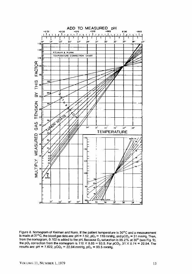
Figure 8. Nomogram of Kelman and Nunn. If the patient temperature is 30°C and a measurement is made at 37°C, the blood gas data are: pH = 7 .52, p02 = 110 mmHg, and pC02 = 31 mmHg. Then, from the nomogram, 0.102 is added to the pH. Because 0 2 saturation is 99.2% at 30° (see Fig. 9), the p02 correction from the nomogram is 110 X 0.85 = 93.5. For pC02 , 31 X 0.74 = 22.94. The results are: pH= 7.622, pC02 = 22.94 mmHg, p02 = 93.5 mmHg.
VOLUME II, NUMBER I, 1979 13
7 8
-15
77
7 6 -10
120
75
-5
100
95
85
•5
15
• 10
10
65
•15
Bo>t• t .. ·60
pH IPH COR!HCTIQN fA( TORS
P apparent-
02 -
100
0-90
"'"
110
70 97 >20
1JO
60 96 •c 140
150
50 95 160
99 170
180
40 94 190 98
200
210
97 30 93 220
230 TEMP flf..,•
96 20 92
10 91
Po2 So2 Po1 So,
OISSOC lA llON CURVE
Po2 x I pH x IBEx I temp
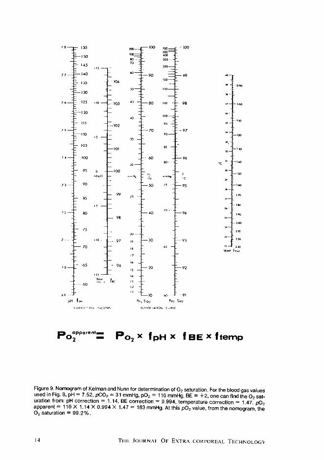
Figure 9. Nomogram of Kelman and Nunn for determination of 0 2 saturation. For the blood gas values used in Fig. 8, pH= 7.52, pC02 = 31 mmHg, p02 = 110 mmHg, BE= +2, one can find the 0 2 saturation from: pH correction= 1.14, BE correction= 0.994, temperature correction= 1.47. p0
2 apparent= 110 X 1.14 X 0.994 X 1.47 = 183 mmHg. At this p02 value, from the nomogram, the 0 2 saturation= 99.2%.
14 THE JOURNAL OF EXTRA-CORPOREAL TECHNOLOGY

Temperature affects the oxygen dissociation curve. Even at constant pH and pC02, a rise in temperature shifts the dissociation curve toward the right. If the temperature of an enclosed blood sample is increased, the pH decreases and the pC02 increases, and the oxygen dissociation curve shifts farther toward the right. If the hemoglobin saturation is constant, every degree of temperature rise shifts the dissociation curve to the right and causes a p02 change of 7.4%13: p02T+I jp02 T = 1.074. For total blood, however, because of this difference in the temperature correction factors for plasma and blood, the magnitude of the p02 change with temperature is influenced markedly by the hemoglobin saturation. The Blood Gas Calculator of Severinghaus makes use of the correction factor 1.074 for the 0 2 dissociation curve. 14 As explained by Severinghaus, this factor is valid only if the hemoglobin saturation is below 80%. Usually, however, we are dealing with an arterial blood saturation exceeding 80%; therefore the Blood Gas Calculator does not give accurate temperature corrections. I personally recommend using the nomogram of Kelman and Nunn (Fig. 8). For pH, one adds the value obtained from the nomogram, and for pC02, one multiplies the value obtained from the nomogram. For p02, the correction factor changes considerably as a function of the saturation, as shown in Fig. 8.
2. Oxygen Saturation In clinical practice, oxygen saturation is seldom measured directly. The value cus
tomarily derived from blood gas measurements is shown in the nomogram for the oxygen dissociation curve (Fig. 9). For example, if the blood gas has a pH of 7.30, base excess of -10, and p02 of 100, the correction factor for the pH is about 0.90 and that for base excess, 1.03, so that the actual p02 is 100 X 0.90 X 1.03 = 92.7 and the oxygen saturation is 97.1%.
3. Base Excess Various measurements are used for the nonrespiratory component of the acid-base
balance, including total carbon dioxide concentration, standard bicarbonate concentration, bicarbonate ion concentration, buffer base, and base excess. There are two schools of thought regarding the best interpretation of the data; one uses base excess 15 and the other, a direct measurement of bicarbonate ion concentration, pC02, or pH. 16 In practice, these methods of determining the non-respiratory component are equally satisfactory, 17
but none can provide information as to what process is the primary disturbance and what process is providing compensation. In our practice, we use base excess for reasons of convenience.
To obtain the base excess from a blood gas measurement when a nomogram or slide rule is not available, one can estimate the base excess from the blood gas pH and pC02 as follows: The two figures beyond the decimal point of the pH value are added to the pC02 value; 80 is subtracted from the sum, and the resulting difference is divided by 2, as shown in the two examples below:
pH 7.40 7.20 pC02 40 30
80 50
Base excess 80-80
=0 50-80
= -15 2 2
VOLUME 11, NUMBER 1, 1979 15

This simple estimate may not be valid when the pC02 is extremely high, but in clinical practice it provides a very useful method for obtaining the base excess.
REFERENCES
1. Moffitt, E. A., Tarhan, S., and Lundborg, R. 0.: Anesthesia for cardiac surgery. Anesthesiology, 29: 1181, 1968. 2. Moffitt, E. A., and Sessler, A. 0.: The circulation and anesthesia, Can. Anaesth. Soc. J., 11: 173, 1964. 3. Lowenstein, E., Walker, H. L., and Zarloff, L. 1.: The effect of a standard cyanotic circulatory lesion on the
induction dose of methohexital in the dog. Anesthesiology, 26: 254, 1965. 4. Strong, M. J., Keats, A. S., and Cooley, D. A.: Anesthesia for cardiovascular surgery in infancy. Anesthesi
ology, 27: 257, 1966. 5. Diaz, F. A., Bianco, J. A., Bello, A., Beer, N., Velarde, H., Izquierdo, J.P., Jean, R.: Effect of Ketamine on
canine cardiovascular function. Brit. J. Anaesth., 48: 941, 1976. 6. Goldberg, A. H., Kean, P. W., Phear, W. P. C.: Effects of Ketamine on contractile performance and excitability
of isolated heart muscle. J. Pharmacol. Exp. Ther., 175: 388, 1970. 7. Gordon, R. J., Ravin, M., Rawitscher, R. E., Daicoff, G. R.: Change in arterial pressure, viscosity, and resistance
during cardiopulmonary bypass. J. Thorac. Cardiovasc. Surg., 69: 552, 1975. 8. Sagimura, S., Starr, A.: Cardiopulmonary bypass in infants under four months of age. J. Thorac. Cardiovasc.
Surg., 73: 894, 1977. 9. Branthwaite, M. A.: Detection of neurological damage during open heart surgery. Thorax., 28: 464,
1973. 10. Rosenthal, T. B.: The effect of temperature on the pH of blood and plasma in vitro. J. Bioi. Chern., 173: 25, 1948. 11. Kelman, G. R., Nunn, J. F.: Nomograms for correction of blood p02, pC02, pH and base excess for time and temperature. J. Appl. Physiol., 21: 1484, 1966. 12. Hedley-Whyte, J., Laver, M. B.: 0 2 solubility in blood and temperature correction factors for p02. J. Appl. Physiol. 19: 901, 1964. 13. Nunn, J. F., Bergman, N. A., Bunatyan, A., Coleman, A. J.: Temperature coefficients for pC02 and p02 of blood in vitro. J. Appl. Physiol., 20: 23, 1965. 14. Severinghaus, J. W.: Blood gas calculator. J. Appl. Physiol., 21: 1108, 1966. 15. Siggard-Anderson, 0.: Blood acid-base alignment nomogram. Scales for pH, pC02 base excess of whole blood of different hemoglobin concentrations, plasma bicarbonate and plasma total C02, Scan. J. Clin. Lab. Invest., 15: 211, 1963. 16. Schwartz, W. B., Reiman, A. S.: A critique of the parameters used in the evaluation of acid-base disorders. N. Eng. J. Med., 268: 1382, 1963. 17. Lewis, D. G., and Stoddart, J. C.: Alternative measurements of the acid-base status in actual clinical situations. Clin. Sci., 44: 297, 1973.
16 THE JOURNAL OF EXTRA-CORPOREAL TECHNOLOGY
Related Documents