Non-affinity factors modulating vascular targeting of nano- and microcarriers ☆ Jacob W. Myerson a , Aaron C. Anselmo b , Yaling Liu c , Samir Mitragotri b , David M. Eckmann a , Vladimir R. Muzykantov a, ⁎ a University of Pennsylvani, United States b University of California, Santa Barbara, United States c Lehigh University, United States abstract article info Article history: Received 1 June 2015 Received in revised form 29 September 2015 Accepted 9 October 2015 Available online 24 October 2015 Keywords: Targeted delivery Vascular biology Nanomedicine Pharmacokinetics Hemodynamics Leukocytes Platelets Red blood cells Particles capable of homing and adhering to specific vascular biomarkers have potential as fundamental tools in drug delivery for mediation of a wide variety of pathologies, including inflammation, thrombosis, and pulmonary disorders. The presentation of affinity ligands on the surface of a particle provides a means of targeting the par- ticle to sites of therapeutic interest, but a host of other factors come into play in determining the targeting capac- ity of the particle. This review presents a summary of several key considerations in nano- and microparticle design that modulate targeted delivery without directly altering epitope-specific affinity. Namely, we describe the effect of factors in definition of the base carrier (including shape, size, and flexibility) on the capacity of car- riers to access, adhere to, and integrate in target biological milieus. Furthermore, we present a summary of fun- damental dynamics of carrier behavior in circulation, taking into account interactions with cells in circulation and the role of hemodynamics in mediating the direction of carriers to target sites. Finally, we note non-affinity as- pects to uptake and intracellular trafficking of carriers in target cells. In total, recent findings presented here may offer an opportunity to capitalize on mitigating factors in the behavior of ligand-targeted carriers in order to optimize targeting. © 2015 Elsevier B.V. All rights reserved. Contents 1. Introduction . . . . . . . . . . . . . . . . . . . . . . . . . . . . . . . . . . . . . . . . . . . . . . . . . . . . . . . . . . . . . . . 98 2. Modulation of pharmacokinetics and targeting by carrier geometry . . . . . . . . . . . . . . . . . . . . . . . . . . . . . . . . . . . . . . 98 2.1. Carrier geometry and blood clearance . . . . . . . . . . . . . . . . . . . . . . . . . . . . . . . . . . . . . . . . . . . . . . . . 98 2.2. Carrier geometry and target site accessibility . . . . . . . . . . . . . . . . . . . . . . . . . . . . . . . . . . . . . . . . . . . . . 99 2.3. Carrier geometry and modulation of affinity interactions . . . . . . . . . . . . . . . . . . . . . . . . . . . . . . . . . . . . . . 100 3. Modulation of vascular targeting by carrier mechanical properties . . . . . . . . . . . . . . . . . . . . . . . . . . . . . . . . . . . . . 100 3.1. Circulation of mechanically flexible carriers . . . . . . . . . . . . . . . . . . . . . . . . . . . . . . . . . . . . . . . . . . . . 100 3.2. Carrier flexibility and penetration in porous/microstructured environments . . . . . . . . . . . . . . . . . . . . . . . . . . . . . . 101 3.3. Carrier flexibility and targeting . . . . . . . . . . . . . . . . . . . . . . . . . . . . . . . . . . . . . . . . . . . . . . . . . . 101 4. Role of blood elements in vascular targeting . . . . . . . . . . . . . . . . . . . . . . . . . . . . . . . . . . . . . . . . . . . . . . . 102 4.1. Indirect effects of cellular blood components on vascular targeting . . . . . . . . . . . . . . . . . . . . . . . . . . . . . . . . . . 102 4.2. Interactions with red blood cells to augment vascular targeting . . . . . . . . . . . . . . . . . . . . . . . . . . . . . . . . . . . 103 4.3. Interactions with leukocytes to augment vascular targeting . . . . . . . . . . . . . . . . . . . . . . . . . . . . . . . . . . . . . 103 4.4. Interactions with platelets for targeting and enhancement of thrombosis . . . . . . . . . . . . . . . . . . . . . . . . . . . . . . . . 103 5. Regulation of delivery and targeting by hemodynamic factors . . . . . . . . . . . . . . . . . . . . . . . . . . . . . . . . . . . . . . . 104 5.1. Interaction between hemodynamic factors and nanocarrier size . . . . . . . . . . . . . . . . . . . . . . . . . . . . . . . . . . . 104 Advanced Drug Delivery Reviews 99 (2016) 97–112 Abbreviations: RES, reticulo-endothelial system; PEG, poly(ethylene) glycol; RBC, red blood cell; ICAM, intercellular adhesion molecule; NIPAm, n-isopropylacrylamide; AFM, atomic force microscopy; GPIIb/IIIa, glycoprotein IIb/IIIa, integrin α IIb β 3 ; PLGA, poly(lactic-co-glycolic acid); vWF, von Willebrand Factor; GPIbα, glycoprotein Ib, alpha subunit; RGD, arginylglycylaspartic acid; IgG, Immunoglobulin G; PECAM, platelet endothelial cell adhesion molecule; VCAM-1, vascular cell adhesion molecule 1; PAAm, polyallylamine; PAH, poly(allylamine hydrochloride); BSA, bovine serum albumin. ☆ This review is part of the Advanced Drug Delivery Reviews theme issue on “Non-Antigenic Regulators-Maiseyeu”. ⁎ Corresponding author at: Department of Pharmacology, University of Pennsylvania, 10-125 Translational Research Center, 3400 Civic Center Blvd, Philadelphia, PA 19104-5158. E-mail address: [email protected] (V.R. Muzykantov). http://dx.doi.org/10.1016/j.addr.2015.10.011 0169-409X/© 2015 Elsevier B.V. All rights reserved. Contents lists available at ScienceDirect Advanced Drug Delivery Reviews journal homepage: www.elsevier.com/locate/addr

Welcome message from author
This document is posted to help you gain knowledge. Please leave a comment to let me know what you think about it! Share it to your friends and learn new things together.
Transcript
Advanced Drug Delivery Reviews 99 (2016) 97–112
Contents lists available at ScienceDirect
Advanced Drug Delivery Reviews
j ourna l homepage: www.e lsev ie r .com/ locate /addr
Non-affinity factors modulating vascular targetingof nano- and microcarriers☆
Jacob W. Myerson a, Aaron C. Anselmo b, Yaling Liu c, Samir Mitragotri b,David M. Eckmann a, Vladimir R. Muzykantov a,⁎a University of Pennsylvani, United Statesb University of California, Santa Barbara, United Statesc Lehigh University, United States
Abbreviations: RES, reticulo-endothelial system; PEG,force microscopy; GPIIb/IIIa, glycoprotein IIb/IIIa, integarginylglycylaspartic acid; IgG, Immunoglobulin G; PECpoly(allylamine hydrochloride); BSA, bovine serum album☆ This review is part of the Advanced Drug Delivery Revi⁎ Corresponding author at: Department of Pharmacolo
E-mail address: [email protected] (V.R.
http://dx.doi.org/10.1016/j.addr.2015.10.0110169-409X/© 2015 Elsevier B.V. All rights reserved.
a b s t r a c t
a r t i c l e i n f oArticle history:Received 1 June 2015Received in revised form 29 September 2015Accepted 9 October 2015Available online 24 October 2015
Keywords:Targeted deliveryVascular biologyNanomedicinePharmacokineticsHemodynamicsLeukocytesPlateletsRed blood cells
Particles capable of homing and adhering to specific vascular biomarkers have potential as fundamental tools indrug delivery formediation of awide variety of pathologies, including inflammation, thrombosis, and pulmonarydisorders. The presentation of affinity ligands on the surface of a particle provides a means of targeting the par-ticle to sites of therapeutic interest, but a host of other factors come into play in determining the targeting capac-ity of the particle. This review presents a summary of several key considerations in nano- and microparticledesign that modulate targeted delivery without directly altering epitope-specific affinity. Namely, we describethe effect of factors in definition of the base carrier (including shape, size, and flexibility) on the capacity of car-riers to access, adhere to, and integrate in target biological milieus. Furthermore, we present a summary of fun-damental dynamics of carrier behavior in circulation, taking into account interactionswith cells in circulation andthe role of hemodynamics in mediating the direction of carriers to target sites. Finally, we note non-affinity as-pects to uptake and intracellular trafficking of carriers in target cells. In total, recent findings presented heremay offer an opportunity to capitalize on mitigating factors in the behavior of ligand-targeted carriers in orderto optimize targeting.
© 2015 Elsevier B.V. All rights reserved.
Contents
1. Introduction . . . . . . . . . . . . . . . . . . . . . . . . . . . . . . . . . . . . . . . . . . . . . . . . . . . . . . . . . . . . . . . 982. Modulation of pharmacokinetics and targeting by carrier geometry . . . . . . . . . . . . . . . . . . . . . . . . . . . . . . . . . . . . . . 98
2.1. Carrier geometry and blood clearance . . . . . . . . . . . . . . . . . . . . . . . . . . . . . . . . . . . . . . . . . . . . . . . . 982.2. Carrier geometry and target site accessibility . . . . . . . . . . . . . . . . . . . . . . . . . . . . . . . . . . . . . . . . . . . . . 992.3. Carrier geometry and modulation of affinity interactions . . . . . . . . . . . . . . . . . . . . . . . . . . . . . . . . . . . . . . 100
3. Modulation of vascular targeting by carrier mechanical properties . . . . . . . . . . . . . . . . . . . . . . . . . . . . . . . . . . . . . 1003.1. Circulation of mechanically flexible carriers . . . . . . . . . . . . . . . . . . . . . . . . . . . . . . . . . . . . . . . . . . . . 1003.2. Carrier flexibility and penetration in porous/microstructured environments . . . . . . . . . . . . . . . . . . . . . . . . . . . . . . 1013.3. Carrier flexibility and targeting . . . . . . . . . . . . . . . . . . . . . . . . . . . . . . . . . . . . . . . . . . . . . . . . . . 101
4. Role of blood elements in vascular targeting . . . . . . . . . . . . . . . . . . . . . . . . . . . . . . . . . . . . . . . . . . . . . . . 1024.1. Indirect effects of cellular blood components on vascular targeting . . . . . . . . . . . . . . . . . . . . . . . . . . . . . . . . . . 1024.2. Interactions with red blood cells to augment vascular targeting . . . . . . . . . . . . . . . . . . . . . . . . . . . . . . . . . . . 1034.3. Interactions with leukocytes to augment vascular targeting . . . . . . . . . . . . . . . . . . . . . . . . . . . . . . . . . . . . . 1034.4. Interactions with platelets for targeting and enhancement of thrombosis . . . . . . . . . . . . . . . . . . . . . . . . . . . . . . . . 103
5. Regulation of delivery and targeting by hemodynamic factors . . . . . . . . . . . . . . . . . . . . . . . . . . . . . . . . . . . . . . . 1045.1. Interaction between hemodynamic factors and nanocarrier size . . . . . . . . . . . . . . . . . . . . . . . . . . . . . . . . . . . 104
poly(ethylene) glycol; RBC, red blood cell; ICAM, intercellular adhesion molecule; NIPAm, n-isopropylacrylamide; AFM, atomicrin αIIbβ3; PLGA, poly(lactic-co-glycolic acid); vWF, von Willebrand Factor; GPIbα, glycoprotein Ib, alpha subunit; RGD,AM, platelet endothelial cell adhesion molecule; VCAM-1, vascular cell adhesion molecule 1; PAAm, polyallylamine; PAH,in.ews theme issue on “Non-Antigenic Regulators-Maiseyeu”.gy, University of Pennsylvania, 10-125 Translational Research Center, 3400 Civic Center Blvd, Philadelphia, PA 19104-5158.Muzykantov).
98 J.W. Myerson et al. / Advanced Drug Delivery Reviews 99 (2016) 97–112
5.2. Effect of nanocarrier shape on hemodynamic behavior . . . . . . . . . . . . . . . . . . . . . . . . . . . . . . . . . . . . . . . 1045.3. Carrier interactions with the glycocalyx under flow . . . . . . . . . . . . . . . . . . . . . . . . . . . . . . . . . . . . . . . . . 1065.4. Effect of flow dependent forces on carrier targeting . . . . . . . . . . . . . . . . . . . . . . . . . . . . . . . . . . . . . . . . . 106
6. Non-affinity modulation of intracellular uptake of nanocarriers . . . . . . . . . . . . . . . . . . . . . . . . . . . . . . . . . . . . . . . 1066.1. Carrier geometry and intracellular uptake . . . . . . . . . . . . . . . . . . . . . . . . . . . . . . . . . . . . . . . . . . . . . 1066.2. Cell phenotype and microenvironment as factors in carrier internalization . . . . . . . . . . . . . . . . . . . . . . . . . . . . . . . 108
7. Conclusion . . . . . . . . . . . . . . . . . . . . . . . . . . . . . . . . . . . . . . . . . . . . . . . . . . . . . . . . . . . . . . . 108Acknowledgments . . . . . . . . . . . . . . . . . . . . . . . . . . . . . . . . . . . . . . . . . . . . . . . . . . . . . . . . . . . . . . 109References . . . . . . . . . . . . . . . . . . . . . . . . . . . . . . . . . . . . . . . . . . . . . . . . . . . . . . . . . . . . . . . . . 109
1. Introduction
The vascular system is both the route to and the intended site fortherapeutic intervention via drug delivery inmany diseases. Blood com-ponents and endothelial cells lining the luminal surface of blood vesselsrepresent preferred targets for pharmacotherapy of ischemia, inflam-mation, bleeding and thrombotic disorders, stroke, pulmonary diseases,and neurological diseases, amongothers. Devising carriers that optimizedelivery of drugs in the vascular system may improve management ofthese prevalent conditions with high morbidity and mortality [1–6].
Carriers designed for this goal (including liposomes, polymeric andnon-polymeric particles, protein conjugates and dendrimers, etc.) mayaccumulate at the desired site either relatively non-specifically(e.g., bymechanical or charge-mediated retention) or via specific inter-action provided by ligands with affinity to molecules typical to orenriched in target tissues. Ligand presentation allows these carriers tospecifically bind to endothelial surface determinants or, for instance,components of blood clots (e.g., platelets and fibrin) and blood cells.The latter – red blood cells, white blood cells, platelets –may serve as ei-ther target or a secondary carrier for drug delivery. “Active targeting”using ligands (e.g., antibodies and their derivatives, peptides, aptamers,etc.,) in theory offers more controlled delivery. It also enables guidedsub-cellular addressing of drugs via anchoring to specific cellular deter-minants providing internalization via appropriate pathways [7–11].
However, many characteristics of a drug carrier and its microenviron-ment in the vascular systemmodulate its circulation and distribution andits interactions with target and non-target counterparts. As a result, thesefactorsmust be taken into account in the course of design and applicationof a targeted drug delivery system [12–15]. The goal of this review is tobriefly analyze how factors pertaining to vascular physiology and param-eters of carrier design other than affinitymodulate vascular targeting anddrug delivery with nanocarriers and microcarriers.
2.Modulation of pharmacokinetics and targeting by carrier geometry
Two parameters defining carrier geometry, size and shape, pro-foundlymodulate every aspect of behavior in the body, including accessto delivery routes, clearance route and rate, specific and non-specific ac-cumulation in target and non-target sites, binding, uptake and intracel-lular trafficking, and ultimately effects of the drug cargo.
2.1. Carrier geometry and blood clearance
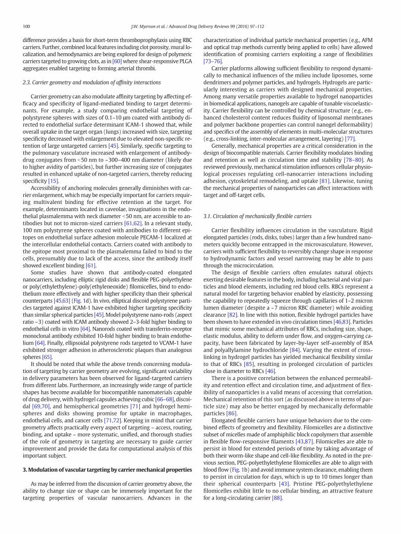
Oneof themost important and extensively studiedparametersmod-ulating carrier behavior in the bloodstream is size. The sizes of typicalcarrier particles can range from below 10 nm to a few microns.Dendrimers, micelles, gold nanoparticles, and iron oxide nanoparticlesoften manifest diameters below 50 nm [16,17], while polymericspheres, liposomes, and nano-shells are hundreds of nanometers in di-ameter [18,19]. Polymeric, lipid, and silica-based microspheres andmicroemulsions have diameters up to a fewmicrons [20,21]. Moreover,carrier shape can also vary from spherical (e.g., lipid-based beads) tospheroidal, cylindrical, or discoidal particles [22–24], virus-templatedparticles [25], and nanopolypods [26] (Fig. 1a).
Generally, intravascular injection is needed for carriers in the sizerange of 50–300 nm. Smaller particles may access other administrationroutes (e.g., pulmonary), though the effectiveness of the vascular routeis generally unrivaled. After administration, size is a key parametermodulating the pharmacokinetics of carriers in the vasculature. Parti-cles smaller than 10 nm undergo renal filtration [27,28] and extravasa-tion in tissues [29]. Drug carriers bigger than approximately 20 nm areeliminated from circulation predominantly by the reticulo-endothelialsystem (RES, including liver, spleen and lymph nodes) [30,31].
Ultra-short carbon nanotubes, quantum dots, gold nanoparticles,dendrimers and other smaller particles spread into various organs bypassing through tight endothelial junctions (10–20 nm diameter) andcan be easily excreted through the glomeruli of the kidneys [29,32].Larger particles have the advantage of carrying higher payloads whenused as targeted vascular carriers.
Overall, for longer circulation after intravascular injection, carriersshould escape recognition and sequestration by the RES (including ac-cumulation in bone marrow, red pulp, lymph nodes, and phagocyticcells in the sinusoids of the liver). In order to increase circulating half-life, sub-micron carriers can be coated with excipient polymers (e.g.,poly(ethylene) glycol, PEG) [33–35], but size and shape also have an im-pact on rate and mechanism of clearance.
Deposition of spherical silica beads in non-RES organs was shown toreduce monotonically with diameter between 700 nm and 3 μm [36],while 3–4 μm plastic microspheres tend to accumulate permanently inthe spleen [37]. Moreover, while engulfment by RES phagocytic cellshappens for particles as big as 4–5 μm [38,39], generally, particles largerthan ~500 nm are prone to mechanical entrapment in capillaries [40].For instance, polystyrene particles larger than 5–7 μm mainly trap inthe alveolar capillaries of the lungs [40], while particles bigger than10 μm are retained in the liver, RES and lungs [41].
Generally, rigid spherical particles between 100 nm and 200 nm indiameter manifest longer circulation times because they are largeenough to avoid sequestration in the liver and small enough to escapesplenic filtration. For non-spherical particles, at least one dimensionshould be kept N100 nm to avoid accumulation in the liver. To avoid en-trapment in the sinusoids of the spleen, at least two dimensionsmust bemaintained b200 nm [42]. For long-circulating non-spherical particles,the effects of particle shape and size are thus closely related, wherethe geometry of non-spherical or flexible particles can significantly pro-long the circulation time. In rodents, long worm-shaped PEG-polyethylethylene filomicelles manifest prolonged circulation time,avoid macrophage internalization, and favor accumulation in tumors[43,44] (Fig. 1b,c), and disk-shaped polystyrene particles have length-ened circulation half-lives relative to analogous spherical particles[45], with work comparing silica spheres and disks finding lesser accu-mulation in the liver for discoidal particles [36]. Similarly for hydrogeldisks, Merkel et al. demonstrated prolonged circulation for particleswith diameter comparable to RBCs, as compared to smaller particles ofidentical composition [46]. As discussed later in this review, carrier me-chanical flexibility can generally confound trends relating clearancetime to particle size, where softer particles generally exhibit longer cir-culation times related to altered hemodynamic behavior [43,47] and in-teractions with phagocytic cells [48,49].
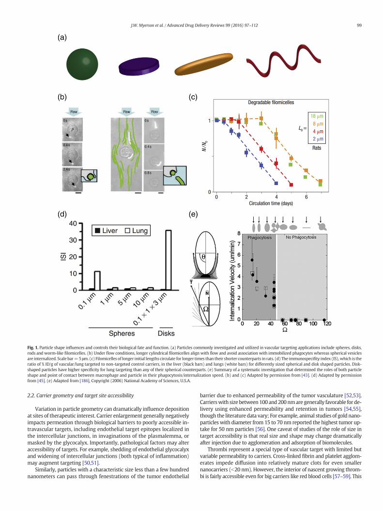
Fig. 1. Particle shape influences and controls their biological fate and function. (a) Particles commonly investigated and utilized in vascular targeting applications include spheres, disks,rods and worm-like filomicelles. (b) Under flow conditions, longer cylindrical filomicelles align with flow and avoid association with immobilized phagocytes whereas spherical vesiclesare internalized. Scale bar=5 μm. (c) Filomicelles of longer initial lengths circulate for longer times than their shorter counterparts in rats. (d) The immunospecifity index (IS),which is theratio of % ID/g of vascular/lung targeted to non-targeted control carriers, in the liver (black bars) and lungs (white bars) for differently sized spherical and disk shaped particles. Disk-shaped particles have higher specificity for lung targeting than any of their spherical counterparts. (e) Summary of a systematic investigation that determined the roles of both particleshape and point of contact between macrophage and particle in their phagocytosis/internalization speed. (b) and (c) Adapted by permission from [43]. (d) Adapted by permissionfrom [45]. (e) Adapted from [186], Copyright (2006) National Academy of Sciences, U.S.A.
99J.W. Myerson et al. / Advanced Drug Delivery Reviews 99 (2016) 97–112
2.2. Carrier geometry and target site accessibility
Variation in particle geometry can dramatically influence depositionat sites of therapeutic interest. Carrier enlargement generally negativelyimpacts permeation through biological barriers to poorly accessible in-travascular targets, including endothelial target epitopes localized inthe intercellular junctions, in invaginations of the plasmalemma, ormasked by the glycocalyx. Importantly, pathological factors may alteraccessibility of targets. For example, shedding of endothelial glycocalyxand widening of intercellular junctions (both typical of inflammation)may augment targeting [50,51].
Similarly, particles with a characteristic size less than a few hundrednanometers can pass through fenestrations of the tumor endothelial
barrier due to enhanced permeability of the tumor vasculature [52,53].Carrierswith size between100 and200nmare generally favorable for de-livery using enhanced permeability and retention in tumors [54,55],though the literature data vary; For example, animal studies of gold nano-particles with diameter from 15 to 70 nm reported the highest tumor up-take for 50 nm particles [56]. One caveat of studies of the role of size intarget accessibility is that real size and shape may change dramaticallyafter injection due to agglomeration and adsorption of biomolecules.
Thrombi represent a special type of vascular target with limited butvariable permeability to carriers. Cross-linked fibrin and platelet agglom-erates impede diffusion into relatively mature clots for even smallernanocarriers (b20 nm). However, the interior of nascent growing throm-bi is fairly accessible even for big carriers like red blood cells [57–59]. This
100 J.W. Myerson et al. / Advanced Drug Delivery Reviews 99 (2016) 97–112
difference provides a basis for short-term thromboprophylaxis using RBCcarriers. Further, combined local features including clot porosity,mural lo-calization, and hemodynamics are being explored for design of polymericcarriers targeted to growing clots, as in [60]where shear-responsive PLGAaggregates enabled targeting to forming arterial thrombi.
2.3. Carrier geometry and modulation of affinity interactions
Carrier geometry can alsomodulate affinity targeting by affecting ef-ficacy and specificity of ligand-mediated binding to target determi-nants. For example, a study comparing endothelial targeting ofpolystyrene spheres with sizes of 0.1–10 μm coated with antibody di-rected to endothelial surface determinant ICAM-1 showed that, whileoverall uptake in the target organ (lungs) increased with size, targetingspecificity decreased with enlargement due to elevated non-specific re-tention of large untargeted carriers [45]. Similarly, specific targeting tothe pulmonary vasculature increased with enlargement of antibody-drug conjugates from b50 nm to ~300–400 nm diameter (likely dueto higher avidity of particles), but further increasing size of conjugatesresulted in enhanced uptake of non-targeted carriers, thereby reducingspecificity [15].
Accessibility of anchoring molecules generally diminishes with car-rier enlargement, whichmay be especially important for carriers requir-ing multivalent binding for effective retention at the target. Forexample, determinants located in caveolae, invaginations in the endo-thelial plasmalemma with neck diameter b50 nm, are accessible to an-tibodies but not to micron-sized carriers [61,62]. In a relevant study,100 nm polystyrene spheres coated with antibodies to different epi-topes on endothelial surface adhesion molecule PECAM-1 localized atthe intercellular endothelial contacts. Carriers coated with antibody tothe epitope most proximal to the plasmalemma failed to bind to thecells, presumably due to lack of the access, since the antibody itselfshowed excellent binding [61].
Some studies have shown that antibody-coated elongatednanocarriers, including elliptic rigid disks and flexible PEG-polyethyleneor poly(ethylethylene)-poly(ethyleneoxide) filomicelles, bind to endo-thelium more effectively and with higher specificity than their sphericalcounterparts [45,63] (Fig. 1d). In vivo, elliptical discoid polystyrene parti-cles targeted against ICAM-1 have exhibited higher targeting specificitythan similar spherical particles [45].Model polystyrenenano-rods (aspectratio ~3) coated with ICAM antibody showed 2–3-fold higher binding toendothelial cells in vitro [64]. Nanorods coated with transferrin-receptormonoclonal antibody exhibited 10-fold higher binding to brain endothe-lium [64]. Finally, ellipsoidal polystyrene rods targeted to VCAM-1 haveexhibited stronger adhesion in atherosclerotic plaques than analogousspheres [65].
It should be noted that while the above trends concerning modula-tion of targeting by carrier geometry are evolving, significant variabilityin delivery parameters has been observed for ligand-targeted carriersfrom different labs. Furthermore, an increasingly wide range of particleshapes has become available for biocompatible nanomaterials capableof drug delivery,with hydrogel capsules achieving cubic [66–68], discoi-dal [69,70], and hemispherical geometries [71] and hydrogel hemi-spheres and disks showing promise for uptake in macrophages,endothelial cells, and cancer cells [71,72]. Keeping in mind that carriergeometry affects practically every aspect of targeting – access, routing,binding, and uptake – more systematic, unified, and thorough studiesof the role of geometry in targeting are necessary to guide carrierimprovement and provide the data for computational analysis of thisimportant subject.
3.Modulation of vascular targeting by carriermechanical properties
Asmay be inferred from the discussion of carrier geometry above, theability to change size or shape can be immensely important for thetargeting properties of vascular nanocarriers. Advances in the
characterization of individual particle mechanical properties (e.g., AFMand optical trap methods currently being applied to cells) have allowedidentification of promising carriers exploiting a range of flexibilities[73–76].
Carrier platforms allowing sufficient flexibility to respond dynami-cally to mechanical influences of the milieu include liposomes, somedendrimers and polymer particles, and hydrogels. Hydrogels are partic-ularly interesting as carriers with designed mechanical properties.Among many versatile properties available to hydrogel nanoparticlesin biomedical applications, nanogels are capable of tunable viscoelastic-ity. Carrier flexibility can be controlled by chemical structure (e.g., en-hanced cholesterol content reduces fluidity of liposomal membranesand polymer backbone properties can control nanogel deformability)and specifics of the assembly of elements in multi-molecular structures(e.g., cross-linking, inter-molecular arrangement, layering) [77].
Generally, mechanical properties are a critical consideration in thedesign of biocompatible materials. Carrier flexibility modulates bindingand retention as well as circulation time and stability [78–80]. Asreviewed previously, mechanical stimulation influences cellular physio-logical processes regulating cell-nanocarrier interactions includingadhesion, cytoskeletal remodeling, and uptake [81]. Likewise, tuningthe mechanical properties of nanoparticles can affect interactions withtarget and off-target cells.
3.1. Circulation of mechanically flexible carriers
Carrier flexibility influences circulation in the vasculature. Rigidelongated particles (rods, disks, tubes) larger than a few hundred nano-meters quickly become entrapped in the microvasculature. However,carrierswith sufficient flexibility to reversibly change shape in responseto hydrodynamic factors and vessel narrowing may be able to passthrough the microcirculation.
The design of flexible carriers often emulates natural objectsexerting desirable features in the body, including bacterial and viral par-ticles and blood elements, including red blood cells. RBCs represent anatural model for targeting behavior enabled by elasticity, possessingthe capability to repeatedly squeeze through capillaries of 1–2 micronlumen diameter (despite a ~7 micron RBC diameter) while avoidingclearance [82]. In line with this notion, flexible hydrogel particles havebeen shown to have extended in vivo circulation times [46,83]. Particlesthat mimic some mechanical attributes of RBCs, including size, shape,elastic modulus, ability to deform under flow, and oxygen-carrying ca-pacity, have been fabricated by layer-by-layer self-assembly of BSAand polyallylamine hydrochloride [84]. Varying the extent of cross-linking in hydrogel particles has yielded mechanical flexibility similarto that of RBCs [85], resulting in prolonged circulation of particlesclose in diameter to RBCs [46].
There is a positive correlation between the enhanced permeabil-ity and retention effect and circulation time, and adjustment of flex-ibility of nanoparticles is a valid means of accessing that correlation.Mechanical retention of this sort (as discussed above in terms of par-ticle size) may also be better engaged by mechanically deformableparticles [86].
Elongated flexible carriers have unique behaviors due to the com-bined effects of geometry and flexibility. Filomicelles are a distinctivesubset of micelles made of amphiphilic block copolymers that assemblein flexible flow-responsive filaments [43,87]. Filomicelles are able topersist in blood for extended periods of time by taking advantage ofboth their worm-like shape and cell-like flexibility. As noted in the pre-vious section, PEG-polyethylethylene filomicelles are able to align withbloodflow (Fig. 1b) and avoid immune systemclearance, enabling themto persist in circulation for days, which is up to 10 times longer thantheir spherical counterparts [43]. Pristine PEG-polyethylethylenefilomicelles exhibit little to no cellular binding, an attractive featurefor a long-circulating carrier [88].
101J.W. Myerson et al. / Advanced Drug Delivery Reviews 99 (2016) 97–112
3.2. Carrier flexibility and penetration in porous/microstructuredenvironments
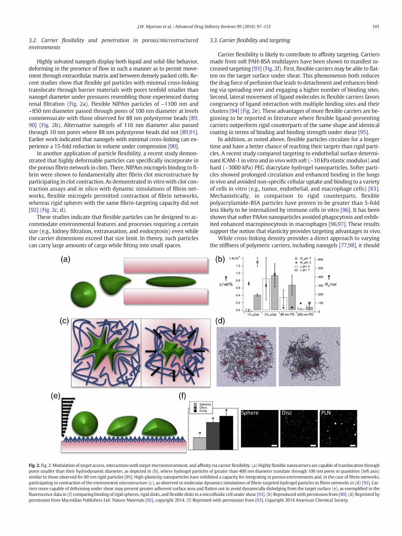
Highly solvated nanogels display both liquid and solid-like behavior,deforming in the presence of flow in such a manner as to permit move-ment through extracellular matrix and between densely packed cells. Re-cent studies show that flexible gel particles with minimal cross-linkingtranslocate through barrier materials with pores tenfold smaller thannanogel diameter under pressures resembling those experienced duringrenal filtration (Fig. 2a). Flexible NIPAm particles of ~1100 nm and~850 nm diameter passed through pores of 100 nm diameter at levelscommensurate with those observed for 88 nm polystyrene beads [89,90] (Fig. 2b). Alternative nanogels of 116 nm diameter also passedthrough 10 nm pores where 88 nm polystyrene beads did not [89,91].Earlier work indicated that nanogels with minimal cross-linking can ex-perience a 15-fold reduction in volume under compression [90].
In another application of particle flexibility, a recent study demon-strated that highly deformable particles can specifically incorporate inthe porousfibrin network in clots. There, NIPAmmicrogels binding tofi-brin were shown to fundamentally alter fibrin clot microstructure byparticipating in clot contraction. As demonstrated in vitrowith clot con-traction assays and in silico with dynamic simulations of fibrin net-works, flexible microgels permitted contraction of fibrin networks,whereas rigid spheres with the same fibrin-targeting capacity did not[92] (Fig. 2c, d).
These studies indicate that flexible particles can be designed to ac-commodate environmental features and processes requiring a certainsize (e.g., kidney filtration, extravasation, and endocytosis) even whilethe carrier dimensions exceed that size limit. In theory, such particlescan carry large amounts of cargo while fitting into small spaces.
Fig. 2. Fig. 2.Modulation of target access, interactionswith targetmicroenvironment, and affinitpores smaller than their hydrodynamic diameter, as depicted in (b), where hydrogel particlessimilar to those observed for 80 nm rigid particles [89]. High-plasticity nanoparticles have exhiparticipating in contraction of the environment microstructure (c), as observed inmolecular dyriers more capable of deforming under shear may present greater adherent surface area and flafluorescence data in (f) comparingbinding of rigid spheres, rigid disks, andflexible disks in amicpermission fromMacmillan Publishers Ltd: Nature Materials [92], copyright 2014. (f) Reprinte
3.3. Carrier flexibility and targeting
Carrier flexibility is likely to contribute to affinity targeting. Carriersmade from soft PAH-BSA multilayers have been shown to manifest in-creased targeting [93] (Fig. 2f). First, flexible carriersmay be able to flat-ten on the target surface under shear. This phenomenon both reducesthe drag force of perfusion that leads to detachment and enhances bind-ing via spreading over and engaging a higher number of binding sites.Second, lateral movement of ligand molecules in flexible carriers favorscongruency of ligand interaction with multiple binding sites and theirclusters [94] (Fig. 2e). These advantages of more flexible carriers are be-ginning to be reported in literature where flexible ligand-presentingcarriers outperform rigid counterparts of the same shape and identicalcoating in terms of binding and binding strength under shear [95].
In addition, as noted above, flexible particles circulate for a longertime and have a better chance of reaching their targets than rigid parti-cles. A recent study compared targeting to endothelial surface determi-nant ICAM-1 in vitro and in vivowith soft (~10kPa elasticmodulus) andhard (~3000 kPa) PEG diacrylate hydrogel nanoparticles. Softer parti-cles showed prolonged circulation and enhanced binding in the lungsin vivo and avoided non-specific cellular uptake and binding to a varietyof cells in vitro (e.g., tumor, endothelial, and macrophage cells) [83].Mechanistically, in comparison to rigid counterparts, flexiblepolyacrylamide-BSA particles have proven to be greater than 5-foldless likely to be internalized by immune cells in vitro [96]. It has beenshown that softer PAAmnanoparticles avoided phagocytosis and exhib-ited enhanced macropinocytosis in macrophages [96,97]. These resultssupport the notion that elasticity provides targeting advantages in vivo.
While cross-linking density provides a direct approach to varyingthe stiffness of polymeric carriers, including nanogels [77,98], it should
y via carrier flexibility. (a) Highlyflexible nanocarriers are capable of translocation throughof greater than 400 nm diameter translate through 100 nm pores in quantities (left axis)bited a capacity for integrating in porous environments and, in the case of fibrin networks,namics simulations of fibrin-targeted hydrogel particles in fibrin networks in (d) [92]. Car-tten out to avoid dynamically dislodging from the target surface (e), as exemplified in therofluidic cell under shear [93]. (b) Reproducedwith permission from [89]. (d) Reprintedbyd with permission from [93]. Copyright 2014 American Chemical Society.
102 J.W. Myerson et al. / Advanced Drug Delivery Reviews 99 (2016) 97–112
be noted that addition of cross-linkers can also alter size, shape, and sur-face chemistry of nanoparticles. Therefore, careful consideration of thetotality of nanoparticle properties is necessary prior to drawing conclu-sions about the relationship between nanoparticle targeting and nano-particle mechanics.
4. Role of blood elements in vascular targeting
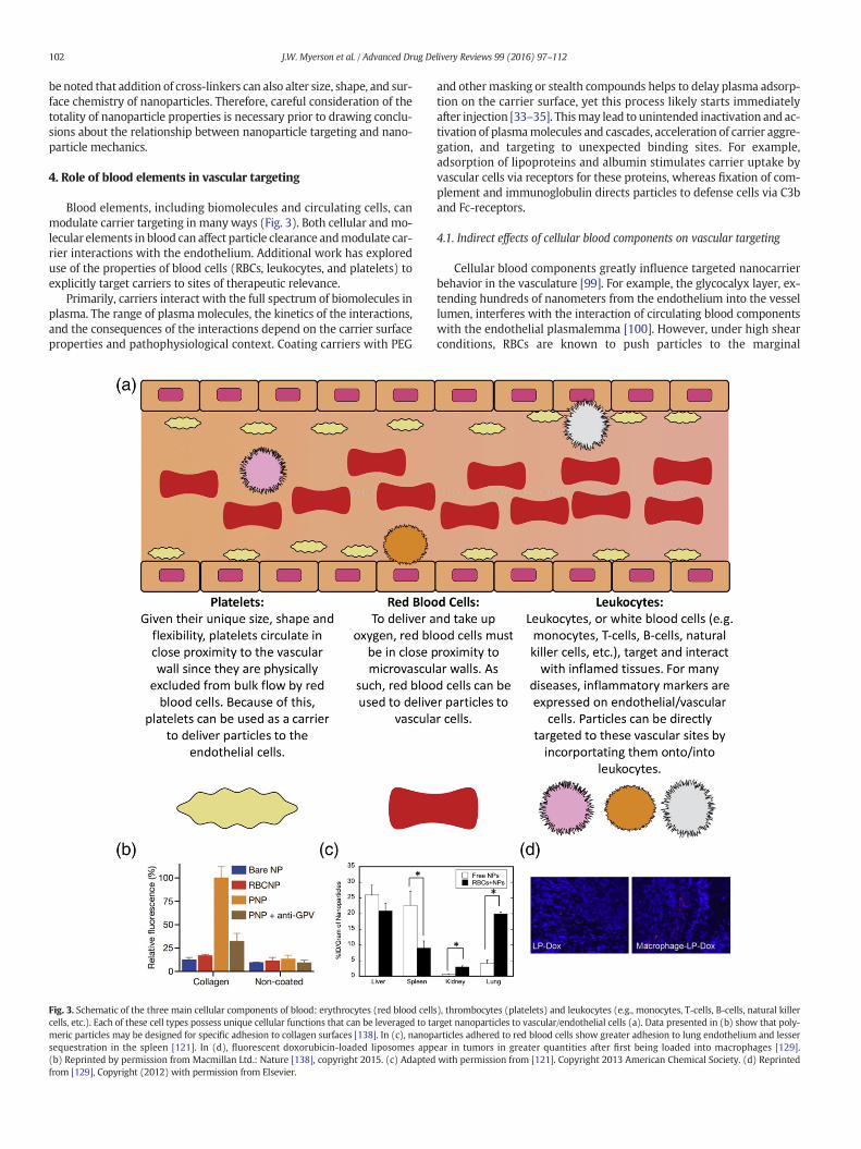
Blood elements, including biomolecules and circulating cells, canmodulate carrier targeting in manyways (Fig. 3). Both cellular and mo-lecular elements in blood can affect particle clearance andmodulate car-rier interactions with the endothelium. Additional work has exploreduse of the properties of blood cells (RBCs, leukocytes, and platelets) toexplicitly target carriers to sites of therapeutic relevance.
Primarily, carriers interact with the full spectrum of biomolecules inplasma. The range of plasmamolecules, the kinetics of the interactions,and the consequences of the interactions depend on the carrier surfaceproperties and pathophysiological context. Coating carriers with PEG
Fig. 3. Schematic of the three main cellular components of blood: erythrocytes (red blood cellcells, etc.). Each of these cell types possess unique cellular functions that can be leveraged to tameric particles may be designed for specific adhesion to collagen surfaces [138]. In (c), nanopsequestration in the spleen [121]. In (d), fluorescent doxorubicin-loaded liposomes app(b) Reprinted by permission from Macmillan Ltd.: Nature [138], copyright 2015. (c) Adaptedfrom [129], Copyright (2012) with permission from Elsevier.
and othermasking or stealth compounds helps to delay plasma adsorp-tion on the carrier surface, yet this process likely starts immediatelyafter injection [33–35]. Thismay lead to unintended inactivation and ac-tivation of plasmamolecules and cascades, acceleration of carrier aggre-gation, and targeting to unexpected binding sites. For example,adsorption of lipoproteins and albumin stimulates carrier uptake byvascular cells via receptors for these proteins, whereas fixation of com-plement and immunoglobulin directs particles to defense cells via C3band Fc-receptors.
4.1. Indirect effects of cellular blood components on vascular targeting
Cellular blood components greatly influence targeted nanocarrierbehavior in the vasculature [99]. For example, the glycocalyx layer, ex-tending hundreds of nanometers from the endothelium into the vessellumen, interferes with the interaction of circulating blood componentswith the endothelial plasmalemma [100]. However, under high shearconditions, RBCs are known to push particles to the marginal
s), thrombocytes (platelets) and leukocytes (e.g., monocytes, T-cells, B-cells, natural killerrget nanoparticles to vascular/endothelial cells (a). Data presented in (b) show that poly-articles adhered to red blood cells show greater adhesion to lung endothelium and lesserear in tumors in greater quantities after first being loaded into macrophages [129].with permission from [121]. Copyright 2013 American Chemical Society. (d) Reprinted
103J.W. Myerson et al. / Advanced Drug Delivery Reviews 99 (2016) 97–112
glycocalyx-protected flow layer, enhancing interactionwith endothelialcells [101,102]. Indeed, it has long been known that the hematocrit, theconcentration of RBCs, modulates interactions of targeted particles withthe vessel wall, as exemplified by an early series of studies examiningtargeting of antibody-carrying RBCs to collagen-coated surfaces imitat-ing blood vessels with denuded endothelium [103]. When antibody-carrying RBCs were perfused in these collagen-coated channels, addi-tion of naive RBCs to a perfusion buffer enhanced binding of targetedRBCs to the collagen target [104]. Other work has recapitulated this par-adigm with ligand-targeted nanoparticles, showing that naive RBCsstimulate binding of targeted nanoparticles to target molecules inmodel vessels simulating pathologically altered endothelium [101,105–107]. Computational and intravital microscopy work indicates arole for size-dependent sequestration of particles near the vessel wallin the RBC-mediated enhancement of interactions with the endothelialsurface [101].
Furthermore, computational studies simulated interactions betweennanocarriers and endothelium in large and small vessels in the presenceand absence of RBCs [108,109]. Two factors, margination of particles byRBCs and an increase in particle dispersion coefficient via rotation andtumbling of RBCs under shear, act together to increase the carrier con-centration in the cell-free-layer at the vessel wall. In smaller vessels,similar simulations show that carrier accumulation is enhanced further,likely due to the larger role volume exclusion plays when RBCs may beforced to physically contact the vessel wall [109].
For the remainder of this discussion, we note that the term “margin-ation” refers to particle distribution in the vessel lumen toward the ves-sel wall, not to adhesion or binding to the wall. There is a correlationbetween margination and binding, even for non-targeted particles[110], but it is not necessarily the case that more effective marginationof a particlewill enhance binding; for example, if there is a repulsion be-tween the particle and vessel wall due to electric charge [111]. Bindingbeyond margination is mostly controlled by factors pertaining to parti-cle avidity, so the details of binding as a result of margination are be-yond the scope of this review.
Themargination of particles in blood cell suspensions is enhanced byincreased carrier size. Smaller particles show a reduction inmarginationto the cell-free-layer as they tend to flow along with blood, accessingthe spaces between blood cells. In vitro work by Liu et al. found thatblood enhances both 210 nm and 2 μm polystyrene particle binding inmicrocirculation. The binding density increased threefold more for the2 μm particles compared to the 210 nm particles [112]. Eniola-Adefesoet al. examined the effect of particle size (500 nm to 10 μm) onmargin-ation propensity in blood and observed an increase in this phenomenonwith increasing particle size [101,105]. In vivo, better margination inmousemicrovasculaturewas observed for 1 μmPEG-coatedpolystyreneparticles compared to 200 nm particles [102].
Spherical particles exhibit slightly better margination than ellipsoi-dal carriers, but ellipsoidal particles are more favorable for adhesiondue to slower rotational dynamics, as determined computationallyand experimentally in microfluidic model vessels [113,114]. Margin-ation probability also depends on the hematocrit in simulations [113].Extensive particle margination was observed at high hematocrit,which is favorable for drug delivery to tumors because the tumormicro-vasculature tends to concentrate blood cells [115].
4.2. Interactions with red blood cells to augment vascular targeting
Blood cells themselves, especially RBCs, represent attractive “super-carriers” for drug delivery. RBCs spend the majority of their lifetime inblood circulation, and are natural carriers that (i) take up oxygen inthe lungs, (ii) circulate throughout the body's vasculature while carry-ing oxygen, and (iii) deliver oxygen to all tissues in the body while re-maining in blood circulation. These RBC biological functions are adirect result of unique physical properties, especially flexibility andhigh surface area to volume ratio, which permits passage through
blood vessels that are smaller in diameter than the cell itself. These fea-tures have been explored for decades as the basis for loading RBCs withdrugs to improve delivery and effects in the bloodstream [116]. Drugscan be encapsulated in isolated RBC prior to re-infusion, or coupled toRBC surface [82,117].
To facilitate oxygen transfer, RBCs must be in close proximity to themicrovascularwalls [82]. As such, since RBCs spenda significant amountof their lifetime interacting with the endothelium, RBC-based deliveryof nanoparticles to endothelium is an interesting and promising ap-proach. Methods of attachment of particles to cells and specific applica-tions of these cell-mediated delivery systems have been reviewedextensively elsewhere [118–120]. In model studies in vitro andin vivo, 200 nm diameter polystyrene nanoparticles were loaded ontomouse RBCs ex vivo and injected intravenously into mice. For timepoints ranging from 30 min to 24 h, lung accumulation of red bloodcell-adsorbed nanoparticles was increased 7-fold compared to identicalnon-adsorbed nanoparticles (Fig. 3c). The most dramatic effects of lungtargeting were seen at shorter time points, as lung accumulation wastransient and shown to sharply decrease for red blood cell-adsorbednanoparticles at 24 h. Other benefits of this delivery system included in-creased blood persistence of nanoparticles and also further enhancedlung targeting, both at short and long time points, when nanoparticleswere modified with lung targeting antibodies [121]. Likewise, it hasbeen reported that polystyrene particles of various sizes attached torat RBCs circulate longer (over 10 h) than those not attached [122]. Itis tempting to hope that these initial observations will be translated tomedically useful drug delivery approaches, warranting studies of themechanisms of carrier loading on RBCs and transfer to endotheliumand, perhaps, other targets.
4.3. Interactions with leukocytes to augment vascular targeting
Leukocytes (e.g., monocytes, T-cells, B-cells, etc.) have also beenused as vehicles to enhance the delivery of particles to the vasculature.Typically, carrier-leukocyte systems are designed to take advantage ofthe innate targeting and barrier penetration abilities of leukocytes[119]. Leukocytes are circulatory immune cells with the natural abilityto target pathological tissues (e.g., sites of infection and inflammation)[123,124] and in theory can be used to facilitate drug delivery to in-flamed tissues in many disorders [125]. Of note, leukocytes transmi-grate through the endothelial barrier into tissues, including throughthe blood brain barrier and into the brain, which are otherwise not ac-cessible to drug carriers.
For example, loading of drug carriers inmonocytes/macrophages hasbeen explored in animals for delivery of: (i) micron-sized antiviral drug-loaded particles to reduce HIV replication in the brain [126]; (ii) sub-50 nm polystyrene particles to breast cancer metastases in the brain[127]; and (iii) catalase loaded polyethyleneimine-PEG nanoparticlesto reduce inflammation [128]. Macrophage-mediated delivery systemsfor doxorubicin-bearing liposomes have also been devised and displayedanti-tumor effects [129] (Fig. 3d). Most recently, it was shown thatmonocytes can be used to target delivery of micron sized polyelectrolyteparticles to locally inflamed lung and skin tissue following antibody-mediated attachment of the particles to monocytes. In this case, mono-cytes showed ~2-fold preference for inflamed over normal lung tissue.This approach also augmented delivery of monocyte-bound drugs topathological sites in a local skin inflammation model [130].
4.4. Interactions with platelets for targeting and enhancement of thrombosis
Nano- andmicroparticles capable of mimicking and complementingthe behavior of platelets have received growing attention in recentyears. Enhancing interactions betweennanocarriers and platelets repre-sents a promising means of targeting sites of vascular injury contribut-ing to ongoing thrombosis, either to modulate that process directly orfor purposes of drug delivery. The range of synthetic particles

104 J.W. Myerson et al. / Advanced Drug Delivery Reviews 99 (2016) 97–112
recapitulating or taking advantage of platelet behavior has been ad-dressed elsewhere [131]. This subsection provides a condensed summa-ry of recent work with an emphasis on directed interactions withplatelets for vascular targeting.
A basic approach to enhancement of platelet-carrier interactionsemploys fibrinogen-derived RGD sequences. Particles coated withthese peptides bind to the platelet fibrinogen receptor, GPIIb/IIIa[132–135]. Natural homing of platelets to sites of clotting facilitatestargeting of such nanoparticles, which in turn support platelet aggrega-tion by cross-linking fibrinogen receptors. Particles binding GPIIb/IIIahave a stabilizing effect on hemostatic clots and provide an anti-hemorrhagic effect at sites of vascular injury. In an early example ofemploying fibrinogen mimetic peptides to participate in platelet aggre-gation, Coller et al. functionalized RBCs to preferentially bind to activat-ed platelets [132]. Targeting to activated platelets has also beenreported with synthetic carriers, including liposomes [136] and PLGAnanoparticles [135,137], presenting fibrinogen-based sequences. Re-cently, another approach involving coating of nanoparticles with plate-let membranes was shown to enhance the binding of these particles tocollagen coated surfaces, as compared to bare nanoparticles and controlRBC-membrane coated nanoparticles (Fig. 3b) [138]. PAH-BSA particleshave also been designed to adhere directly to platelets by presentingvWF fragments targeted to platelet GPIbα [95]. Most recently, particlesengaging in both platelet aggregation (via RGD peptides) and adhesion(via collagen and vWF binding) have been demonstrated [93,139].
Non-affinity factors profoundly modulate interaction of carrierstargeted to platelets and vascular injuries. Enhancement of interactionwith platelets and platelet-like behavior has been achieved throughmodulation of particle size and shape, with larger and more oblate par-ticles exhibitingmore localization toward the vessel wall and improvedparticipation in aggregation and adhesion [93,95,140]. Individual parti-clemechanical properties have additional effects on platelet-like behav-ior of vascular carriers. Lipid membrane fluidity and thus flexibility ofGPIbα liposomes was shown to enhance adhesion to vWF [141].Albumin-based particles developed by Mitragotri's group have com-bined the effects of platelet-mimetic geometry and flexibility (as wellas surface chemistry) to enhance association with platelet adhesion rel-ative to analogous polystyrene spheres [93,95].
As addressed above, particle flexibility has also been used to mimicthe role of platelets in clot contraction. Brown et al. employedmicrogelswith no cross-linking to recapitulate the compatibility of platelets withclot microstructure [92]. Essentially, it was demonstrated that onlyfibrin-binding particles with minimized cross-linking could incorporatein contracting clots. Rigid polystyrene particles and even hydrogel par-ticles with small amounts of additional cross-linking interfered withclot microstructure, while “ultrasoft” microgels successfully participat-ed in clot contraction alongside platelets [92].
In general, loading carriers onto cells that naturally home in on path-ological tissues may evolve into a new strategy and, perhaps, help to re-duce immunological side effects of using exogenous proteins orantibodies for particle targeting. Of note, the specific characteristics ofthis approach, including percent of dose taken up by the “transporting”cells and that delivered to the target tissue, remain to be determined. Onthe other hand, these cells are defense agents and their effects (not me-diated by or perhaps countering that of the drug cargo) at pathologicalsites may not necessarily be desirable.
5. Regulation of delivery and targeting by hemodynamic factors
Expanding on aspects of the above discussions of geometry andme-chanical properties, hemodynamic factors play an important role in de-livering nanocarriers to the vessel wall so that binding interactions,uptake, and drug delivery can occur. Among themost important factorsregulating carrier motion in blood flow are carrier geometry, the role ofthe glycocalyx, and the exposure to fluid mechanical forces. A broad-spectrum computational protocol devised for calculating binding
between functionalized nanocarriers and the endothelial cell surfacehas considered rotational as well as translational nanocarrier move-ment under flow [99] (Fig. 4). With carrier shape restricted to sphericalobjects, changing particle size alone only moderately enhances bindingdue to the entropy loss associated with bound receptors [142]. Exten-sion of this work into effects of cell membrane features (e.g., degree ofmembrane excess and membrane elasticity) and nanocarrier shape(e.g., ellipsoidal carriers) is revealing additional non-affinity related de-terminants of targeting efficiencymediated through both the carrier be-havior in flow and mechanical effects of flow at the endothelial cell-carrier binding interface.
5.1. Interaction between hemodynamic factors and nanocarrier size
The influence of hydrodynamic factors on carrier behavior andtargeting depends on carrier size. In vitro flow chamber binding exper-iments using targeted spherical polystyrene particles of diameter100 nm to 10 μm coated with antibodies to the endothelial surface de-terminant E-Selectin showed that specific binding to endothelium in-creased as diameter increased from 500 nm to 10 μm at a shear rate of200 s−1. However at higher shear (e.g., 1500 s−1), spheres of 5 μmand 10 μmdiameterwere less likely to adhere to E-Selectin than smallerspheres [101].
As noted in Section 4, RBCs segregate away from the vessel wall invessels between 10 and 300 μm in diameter, leading to changes in vis-cosity and local hematocrit as characterized by both the Fåhraeus andFåhraeus–Lindqvist effects [143]. As a result, particles in vascular flowcan demonstrate “margination,” dispersing toward the wall into thecell-free plasma layer [101,102,105,108,109,112–114,144].
To exploit this effect for targeted carrier based drug delivery, theo-retical studies have suggested that a diameter of ~200 nm or smallerprovides optimal margination, while diameters greater than 200 nm re-sult in less efficient margination (as determined by the time a particletakes to migrate toward the vessel wall) [111]. However, shear flow ex-periments have shown that particles of diameter larger than 200 nmundergo effective margination driven by gravitational force [110].These studies performed with a parallel plate flow chamber indicatedthat polystyrene nanocarriers larger than 200 nm undergo the greatestmargination due to sedimentation in horizontal capillaries and thegreatest lateral drift in vertical capillaries with downward flow. Thismotion toward the vessel wall increased the probability of binding tothe endothelium [110].
It is worth mentioning that the model presented in reference [110]works in terms of volume concentration, rather than number of parti-cles, assuming nanospheres are fed into blood at a higher numericalconcentration than microspheres in concluding that the quantity ofmarginating particles scales with the volume concentration for particleslarger than 200 nm. Namdee et al. [105] found that increasing the nu-merical concentration of nanospheres in bloodflowdoes not necessarilyresult in a linear increase in adhesion to endothelial cells. While a five-fold increase in concentration resulted in a five-fold increase in adhe-sion for 2 μm and 5 μm particles, a five-fold increase in concentrationproduced an approximately two-fold increase in adhesion for 200 nmand 500 nmparticles. Decuzzi et al. have also predicted a positive corre-lation between carrier density and propensity to migrate toward thevessel wall [111]. Thompson et al. have experimentally addressed theproblemof density inmargination, comparing polystyrene, silica, and ti-tania particles in an endothelialized flow cell and finding greater mar-gination for more dense particles [107].
5.2. Effect of nanocarrier shape on hemodynamic behavior
Nanocarrier shape has significant influence on nanocarrier motion inblood flow [145]. Particles that are non-spherical can migrate laterallyin flow [146]. Moreover, it has been shown that the lateral drifting veloc-ity is directly related to aspect ratio [147,148] for non-spherical particles.
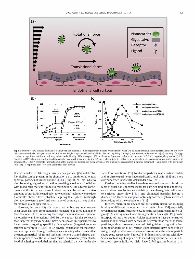
Fig. 4. Depiction of flow-induced nanocarrier translational and rotational (tumbling) motion induced by fluid forces, which will be dependent on nanocarrier size and shape. The wavy,deformable endothelial cell layer surface and presence of the glycocalyx are included as additional factors regulating binding (a). For instance, as determined in [51], shedding of the gly-cocalyx via heparinase infusion significantly enhances the binding of ICAM-targeted 100 nm diameter fluorescent polystyrene spheres (1a29 FLMs) in postcapillary venules (b). Asdepicted in [167], there is a non-linear relationship between wall shear and binding of 3 μm L-selectin targeted polystyrene microspheres to a complementary surface (L-selectinspheres/PSGL-1) (c). A threshold shear rate, implicated in inducing tumbling of the spheres near the binding surface, resulted in optimal binding. (b) Reproduced with permissionfrom [51]. (c) Reprinted from [167] with permission from Elsevier.
105J.W. Myerson et al. / Advanced Drug Delivery Reviews 99 (2016) 97–112
Discoid particles circulate longer than spherical particles [45], and flexiblefilomicelles can be present in the circulation up to ten times as long asspherical particles of similar volume [43,149] (Fig. 1b, c). This is due totheir becoming aligned with the flow, enabling avoidance of collisionswith blood cells that contributes to margination. One adverse conse-quence of this is that carrier-wall interactions can be reduced: in vivotargeting of anti-ICAM coated poly(ethylethylene)-poly(ethyleneoxide)filomicelles showed lower absolute targeting than spheres (althoughthe ratio between targeted and non-targeted counterparts was similarfor filomicelles and spheres) [63].
However, the probability of a nanorod carrier binding under modestshear stress has been computationally modeled to be three-fold higherthan that of a sphere, indicating that shape manipulation can enhancenanocarrier-wall interactions [150]. Further support for this concept isthat targeted polystyrene disks have been shown in experiments tohave greater targeting specificity than spheres (targeted to non-targeted carrier ratio= 35.7) [45]. A physical explanation for these phe-nomena is provided throughmathematicalmodeling,which reveals thatthe nonsymmetrical rolling and tumbling motions that rod-shaped par-ticles experience near the vessel wall causes them to have greater likeli-hood of adhering to endothelium than do spherical particles under the
same flow conditions [151]. For discoid particles, mathematical modelsand in-vitro experiments have predicted lateral drift [152] and moreavid adherence to vascular walls under flow [99,153].
Further modeling studies have demonstrated the possible advan-tages of other non-spherical shapes for particles binding to endothelialcells in shear flow. For instance, oblate particles have greater adherenceto surfaces under flow [153], and elongated particles having adiameter b 200 nmcanmarginate optimally and thereby have increasedinteractions with the endothelium [111].
In vitro, microfluidic devices are particularly useful for studyingbinding of different nanocarrier shapes under flow [154], especiallygiven that geometric features relevant to the vasculature in different or-gans [155] and significant vascular segments or tissues [60,156] can beincorporated into their design. Fluidics experiments have demonstratedmargination of discoid silica particles to be greater than that of sphericalparticles, without, however, a method distinguishing margination frombinding or adhesion [140]. Micron-sized particles have been studiedusing straight and bifurcated channels to examine the role of particleshape (e.g., aspect ratio, flatness) in influencing targeting with flow.Studies of binding of targeted disk-shaped polystyrene particles in a bi-furcated system indicated disks have 5-fold greater binding than
106 J.W. Myerson et al. / Advanced Drug Delivery Reviews 99 (2016) 97–112
targeted spheres [157]. Additional work has found that selectin-targeted ellipsoidal polystyrene rods have greater adherence to endo-thelial cells compared to identically targeted spheres under flow, withhigher aspect ratio increasing the quantity of bound particles [114]. Par-ticle shape affects binding more for large particles and for particlestransiting bifurcations. Of note, higher numbers of rod-shaped particlesand disk-like structures adhere in model fluidics systems compared tospherical objects [114,157], in agreement with in vivo studies [45,65].
5.3. Carrier interactions with the glycocalyx under flow
The glycocalyx is a gel-like layer which extends ~500 nm from thein vivo luminal surface of endothelial and blood cells (among othercell types). A brush-like structure, formed by strongly negativelycharged carbohydrate chains of glycoproteins, the glycocalyx affects nu-merous cardiovascular physiological and pathological processes, includ-ing sensing of flow by endothelium and separating blood cells from theendothelial plasmalemma proper.
The glycocalyx has a significant role inmodulating nanocarrier bind-ing to endothelial cells by acting as an energy barrier (see Fig. 4). Me-chanical and biochemical properties of the glycocalyx as well as itsinteractions with erythrocytes and leukocytes [158] have been probedexperimentally [159]. Generally, the strongnegative charge of the glyco-calyx represents a mechanism for non-specific (i.e., bypassing ligand-mediated targeting) binding of cationic carriers in the vasculature. Fur-thermore, the glycocalyx regulates interaction of biotherapeutic pro-teins targeted to endothelial and blood cells with their physiologicalcounterparts in plasma [160,161].
The in vivo binding of 100 nm diameter polystyrene microspherescoated with ICAM-1 antibody has been shown to increase as much as500-fold following chemical degradation of the glycocalyx (Fig. 4b)[51]. The glycocalyx'smechanical behavior is demonstrably viscoelastic,and its responses to fluid shear and cellular motion have been used topredict the bending rigidity of its core proteins to be in the range of700 pN·nm2 [162]. This stiffness translates into an energy barrier thatacts to reduce the binding between nanocarriers and the endothelialcell surface [163].
Of note, biologically relevant glycocalyx layer that can be seenin vivo is very difficult to reproduce in cell culture, especially in staticcells lacking hemodynamic stimulation. Themagnitude bywhich glyco-calyx thickness and stiffness are altered in vitro compared to in vivo isunknown, so the effect it bears on nanocarrier binding in vivo cannotbe extrapolated from results of cell culture-based studies.
5.4. Effect of flow dependent forces on carrier targeting
Nanocarrier circulation in the bloodstream is dependent to a largeextent on local flow conditions. As depicted for rod-shaped carriers inFig. 5, margination, a process dependent on flow conditions, can repre-sent a first step in vascular delivery. Flow dependent forces that gener-ate nanocarrier rotational and translational motion regulate adhesion.Shear flow introduces both torque and drag forces, which can influencenanocarrier orientation, deformation, and transport within blood flow.Through the mechanical effects of torque and drag, blood flow modu-lates the interaction between nanocarriers and cell surfaces. For exam-ple, targeted 1 μm spherical polystyrene carriers displayed a fewtypical patterns of behavior in binding to endothelium in vitro, depend-ing on the targeting conditions: 1) they move with the flow withoutshowing any sign of binding interactions with cells; 2) they roll contin-uously over cells; 3) they begin by rolling followed by binding firmly tocells; 4) or they initially roll and subsequently detach and continue trav-eling along the cell surface [164]. These motions indicate interplay be-tween binding interactions and the motion induced by fluid forces.
Though available studies modeling carrier behavior in buffer do notfully reflect conditions in the bloodstream, previous work with polysty-rene carriers in buffer has shown that binding is inversely proportional
to shear stress [165,166]. Computational analysis has shown that the in-fluence of shear stress on nanocarrier binding diminishes once a thresh-old level of antibody surface density is surpassed [99]. In experimentalobservations, flow actually enhances adhesion between targeted smallparticles (or leukocytes) and the endothelial cell surface, aswell as cellu-lar uptake of particles after binding events occur. This phenomenon iscounter-intuitive; one should anticipate that increases in flow wouldfavor the displacement of particles in the downstreamdirection. Howev-er, the initial tethering and continuous rolling of small particles (or cells)requires some threshold flow shear stress (Fig. 4c). This seems to be inviolation of the idea that the dissociation rate increases exponentiallywith increasing applied force. Flow enhancement of adhesion has twostages: initial tethering followed by rolling after attachment. Flow en-hanced tethering may result from convective transport which increasesthe collision frequency between bindingmolecules (e.g., receptor and li-gand) [167]. Flow-enhanced rolling is typically explained by “catch-slip”kinetics in which the lifetime of receptor-ligand bonds is prolonged byapplication of tensile force acting on the cells [168] or as a result ofchanges in molecular structures as a result of the applied shear stress[169]. In either case, these are fluid mediated forces.
The underlying physics and mechanisms by which flow enhances,rather than destabilizing, binding is still a topic of discussion. The effectof drag force on binding following margination depends on many fac-tors, including particle size, shape, avidity, and endothelial topography.As result, a particle can exhibit high margination but have low binding,for example, due to inadequate ligand density. Various theoreticalmodels have been developed to elucidate deposition mechanismsunder flow. Numericalmethods developed in recent years provide a rig-orousway to study the full transportation and adhesion dynamics of ar-bitrarily shaped nanoparticles. At the micron scale, Liu et al. estimatedPLGA nanoparticle binding affinity with endothelial cells [99]; Kinget al. studied multiparticle adhesion dynamics for polystyrene beadsand applied their findings to leukocyte rolling [170,171]. Fogelsonet al. coupled ligand-receptor binding with platelet aggregation [172].Shipley et al. [173] andModok et al. [174]modeled delivery of sphericalNPs to tumors. Liu et al. [175] and Zhang et al. [176] studied the deposi-tion of NPs in the lungs. It is also important to predict the loss of NPs inthe upstream and the NP distribution in specific vascular environments[177]. However, there are not many attempts in the literature to linkmolecular and cellular scale particle adhesion dynamics to tissue andorgan scale transport and distribution.
6. Non-affinity modulation of intracellular uptake of nanocarriers
In many instances, the intended site of drug action is inside the tar-get cell. Achieving optimal sub-cellular addressing is one of the mostimportant and challenging problems in the field of drug delivery. Thisextremely complicated process, mediated by intricate interactions be-tween the carrier and the cell, is greatly influenced by features of thecarrier, the cell, and their milieu.
6.1. Carrier geometry and intracellular uptake
More than a century ago, Metchnikoff noted that phagocytosis ismodulated by particle size. In macrophages, IgG-opsonized micronsized particles are taken up and transported to lysosomes more quicklythan 100 nm counterparts [178]. For spherical polystyrene particles inmacrophages and B16 tumor cells, size has been shown to affect bothrate (with protracted internalization for larger particles) and mecha-nism of internalization [179,180]. Optimal size for uptake depends onfactors including the receptor, endocytic pathway, cell type, and func-tional status of the target cell. For example, the optimal size range foruptake of particles by endothelial cells is about an order of magnitudesmaller than for uptake by macrophages [181,182]. Likewise, mathe-matical models have predicted a critical particle radius (as a functionof ligand and receptor density) below which internalization is not
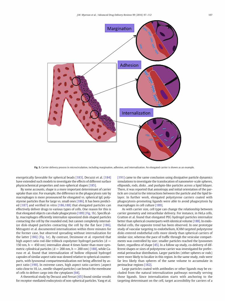
Fig. 5. Carrier delivery process in microcirculation, including margination, adhesion, and internalization. An elongated carrier is shown as an example.
107J.W. Myerson et al. / Advanced Drug Delivery Reviews 99 (2016) 97–112
energetically favorable for spherical beads [183]. Decuzzi et al. [184]have extended suchmodels to investigate the effects of different surfacephysicochemical properties and non-spherical shapes [185].
By some accounts, shape is a more important determinant of carrieruptake than size. For example, the difference in the phagocytosis rate bymacrophages is more pronounced for elongated vs. spherical IgG poly-styrene particles than for large vs. small ones [186]. It has been predict-ed [187] and verified in vitro [186,188] that elongated particles caneffectively deliver drugs to various types of cells. One reason for this isthat elongated objects can elude phagocytosis [189] (Fig. 1b). Specifical-ly, macrophages efficiently internalize opsonized disk-shaped particlescontacting the cell by the rounded end, but cannot completely internal-ize disk-shaped particles contacting the cell by the flat face [186].Mitragotri et al. documented internalization within three minutes forthe former case, but observed spreading without internalization forthe latter [186] (Fig. 1e). By contrast, Desimone et al. reported thathigh aspect ratio rod-like triblock copolymer hydrogel particles (d =150 nm, h = 450 nm) internalize about 4 times faster than more sym-metric cylindrical particles (d= 200 nm, h= 200 nm) [188], while Ca-ruso et al. found that internalization of hollow ellipsoid hydrogelcapsules of similar aspect ratiowas slowed relative to spherical counter-parts, with lysosomal compartmentalization not being affected by as-pect ratio [190]. In extreme cases, high aspect ratio carriers (aspectratio close to 10, i.e., needle-shaped particles) can breach themembraneof cells to deliver cargo into the cytoplasm [64].
A theoretical study by Decuzzi and Ferrari [65] found similar resultsfor receptor-mediated endocytosis of non-spherical particles. Yang et al.
[191] came to the same conclusion using dissipative particle dynamicssimulations to investigate the translocation of nanometer-scale spheres,ellipsoids, rods, disks , and pushpin-like particles across a lipid bilayer.There, it was reported that anisotropy and initial orientation of the par-ticle are crucial to the interactions between the particle and the lipid bi-layer. In further work, elongated polystyrene carriers coated withphagocytosis-promoting ligands were able to avoid phagocytosis bymacrophages in cell culture [189].
As with carrier size, cell type can change the relationship betweencarrier geometry and intracellular delivery. For instance, in HeLa cells,Gratton et al. found that elongated PEG hydrogel particles internalizebetter than spherical counterpartswith identical volume [188]. In endo-thelial cells, the opposite trend has been observed. In one prototypestudy of vascular targeting to endothelium, ICAM targeted polystyrenedisks entered endothelial cells more slowly than spherical carriers ofsimilar size, whereas the pace of traffic through the vesicular compart-ments was controlled by size; smaller particles reached the lysosomesfaster, regardless of shape [45]. In a follow up study, co-delivery of dif-ferent shapes or sizes of polystyrene carrier was investigated for prefer-ential perinuclear distribution. Larger particles (either spheres or rods)were more likely to localize in this region. In the same study, rods werefar less likely than spheres of the same volume to accumulate inperinuclear regions [182].
Large particles coated with antibodies or other ligands may be ex-cluded from the natural internalization pathways normally servingthose ligands. Since internalization starts with anchoring to thetargeting determinant on the cell, target accessibility for carriers of a
108 J.W. Myerson et al. / Advanced Drug Delivery Reviews 99 (2016) 97–112
given size and shape is a key parameter. For example, some anchoringmolecules that could provide favorable pathways for internalization,such as caveolar molecules, are poorly accessible for particles biggerthan 50–100 nm [192]. On the other hand, in some cases multivalentligand-coated particles induce endocytosis via binding to determinantsthat do not normally favor uptake of the ligand. For example, PECAM-1antibodies bind to endothelial cells but do not accumulate significantlyin intracellular compartments [16,42], while the multivalent binding ofnanocarriers coated with PECAM-1 antibody leads to intracellular up-take mediated by the pathway known as CAM-endocytosis, distinctfrom clathrin-mediated or caveolar endocytosis, phagocytosis, andpinocytosis [17,42].
Large particles, especially non-spherical ones, require excessivewrapping by plasmalemma, which may exceed the capacity of a givencell type, as demonstrated in computational work addressing geometryand sizewithmodelmembranes [193]. In this context, a smooth particlerequires less wrapping than a polymorphous “particle” with the sameeffective diameter, making endocytosis easier with the former. For ex-ample, endothelial cells internalize PECAM-targeted polymorphousprotein conjugates with size b500 nm [178], but internalize sphericalparticles with diameter of up to a fewmicrons coatedwith the same an-tibody [194]. Similar results have been observed when comparingpolymethacrylate hydrogel cubes to spheres for non-targeted uptakein HeLa cells [195].
6.2. Cell phenotype and microenvironment as factors in carrierinternalization
The functional status of cells modulates endocytosis. Conditions thatup-regulate expression and turnover of the targeting determinant in theplasmalemma usually facilitate endocytosis. For example, activated en-dothelium internalizes ICAM-targeted polystyrene nanocarriers fasterthan quiescent endothelium in vitro and in vivo [196]. Most likely, thisis due to the fact that the cell surface density of ICAM is elevated incytokine-challenged cells. But additional and/or alternative mecha-nisms may involve stimulation of vesicular turnover involved in cyto-kine signaling.
The hydrodynamic factors regulating endocytosis of targeted car-riers have just recently started to emerge as an important considerationin vascular drug delivery. Despite the fact that endothelial cells in vivoare exposed to flow, the majority of studies on cellular uptake and traf-ficking of nanocarriers have employed static cell lines. A few studieshave attempted to define the role of flow in endothelial uptake of nano-particles using flow chambers and microfluidics, mostly dealing withnon-targeted particles, though with a variety of particle compositionsand sizes, including microparticles derived from activated cells, semi-conductor quantum dots, and silica particles [197,198].
The rheological regulation of intracellular delivery represents anintriguing area of bioengineering and biomedicine. Blood flow altersthe adhesive interactions between carriers and endothelium [99].Flow-driven rolling on the endothelial surface may assist carriers inengaging PECAM-1, thereby increasing the strength of endocytic sig-naling [199]. Alternatively, rotational motion due to the flow-derived torque applied to PECAM-1-anchored carriers may furthermechanically stimulate endothelial cells and enhance signaling in-ternalization. On the other hand, flow is known to modulate manyparameters of endothelial functional status, some of which may beinvolved in endocytic processes directly or indirectly. In fact, shearstress governs endothelial processes including cytoskeletal remodel-ing, gene expression, ion transport, and endocytosis [199–202]. Ithas long been recognized that internalization of extracellular fluidand macromolecules (such as LDL) into endothelial cells is stimulat-ed by flow [199,203]. Stimulatory effects of flow have been observedin endothelial pinocytosis [196,199,204], clathrin-dependent endo-cytosis [194,205], and CAM-dependent endocytosis [44,199]. It isconceivable that flow stimulates endocytosis via a generalized
mechanism, such as enhancement of the rate of plasmalemma vesi-cle maturation or dynamic changes of the cytoskeleton.
Recent studies in flow chambers revealed opposite effects of chronicvs. acute flow on endothelial uptake of targeted particles. Prolonged ex-posure to flow led to partial, yet significant, inhibition of endocytosis ofpolystyrene nanocarriers targeted to ICAMand PECAM [196,199]. Theseresults correlated with in vivo data showing less effective internaliza-tion of anti-ICAM nanocarriers in arterioles relative to capillaries,i.e., vascular areas in which endothelial cells do and do not adapt toflow, respectively [196]. This effect is attributed to organization of theactin cytoskeleton into stress fibers in the course of endothelial adapta-tion toflow,which impedes actin involvement in endocytosis [196,199].In contrast, acute shear stress at physiological levels typical of veins ac-celerated endothelial endocytosis of PECAM-targeted carriers, likely viasignaling mechanisms including caveolae [199]. Such a stimulatory ef-fect may happen in reperfusion and in physiological hyper-perfusionin exertion.
7. Conclusion
Targeted delivery of drug carriers in the vasculature is an importantbiomedical goal. It is safe to postulate that affinity features of the drugdelivery system, controlling specific recognition of molecular targetsand anchoring on the target surface, is the main factor of the carrier de-sign that governs targeting. Nonetheless, targeting itself is a complicat-ed outcome of the interplay of characteristics of the carrier (includingfeatures of the targeting ligands, their surface density, spatial freedom,ability to engage in multivalent interactions with clusters of anchoringmolecules, etc.) and the target (surface density, clustering and accessi-bility of anchoring molecules). Furthermore, other factors in carrier de-sign, target, and target microenvironment exert important, and in someinstances decisive, influences over targeting. For example, if the bindingsite becomes inaccessible under given circumstances, no matter howavidly a carrier would bind given access, no targeting can occur.
In this article,we have attempted a systematic analysis of several keynon-affinity influences on targeting of drug carriers. We acknowledge adepth of additional considerations in the design of nanomaterials fortargeted drug delivery applications, including a wealth of possibilitiesfor materials design, but leave a deeper consideration of the materialsscience of biomedical nanocarriers and microcarriers to other sources[42,81]. Although inevitably oversimplified, our analysis may conveyan impression of the complexity of drug delivery via vehicles at thenanometer andmicron length scales. Indeed, as Fig. 6 illustrates, numer-ous factors of systemic and local biological features interplaywith carri-er design characteristics, individually and collectively, creating theintertwined matrix of cues that either favor or impede targeting. Thegoal is to employ the factors that we can rationally modulate to achievethe former outcome. Our ability to control the biological factors is, mild-ly put, limited (such factors include choosing the route for infusion andmodulation of perfusion, e.g., using hyperthermia and vasoactiveagents). However, understanding of the influence of non-controllablebiological factors will guide iterative re-engineering of carriers to opti-mize targeting. It is tempting to believe that continuing accumulationof experimental data and empirical paradigms in this domainwill even-tually support design of theoretical models and approaches that will ac-celerate this optimization process.
Therefore, non-affinity characteristics of both the delivery systemand biological factors govern targeted drug delivery and its effects.This realization puts yet more emphasis on the necessity to pursuedrug delivery in a multidisciplinary fashion, combining approachesand ideas coming from bothmaterials and biomedical science and engi-neering. It also dictates use of more adequate model systems for inves-tigation of vascular targeting, including endothelial cell culture systemsthat account for effects of flow, specifics of cellular phenotype, and cellmicroenvironment.
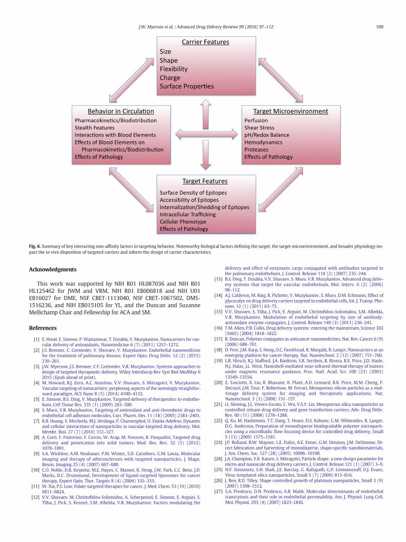
Fig. 6. Summary of key interacting non-affinity factors in targeting behavior. Noteworthy biological factors defining the target, the target microenvironment, and broader physiology im-pact the in vivo disposition of targeted carriers and inform the design of carrier characteristics.
109J.W. Myerson et al. / Advanced Drug Delivery Reviews 99 (2016) 97–112
Acknowledgments
This work was supported by NIH R01 HL087036 and NIH R01HL125462 for JWM and VRM, NIH R01 EB006818 and NIH U01EB16027 for DME, NSF CBET-1113040, NSF CBET-1067502, DMS-1516236, and NIH EB015105 for YL, and the Duncan and SuzanneMellichamp Chair and Fellowship for ACA and SM.
References
[1] E. Hood, E. Simone, P. Wattamwar, T. Dziubla, V. Muzykantov, Nanocarriers for vas-cular delivery of antioxidants, Nanomedicine 6 (7) (2011) 1257–1272.
[2] J.S. Brenner, C. Greineder, V. Shuvaev, V. Muzykantov, Endothelial nanomedicinefor the treatment of pulmonary disease, Expert Opin. Drug Deliv. 12 (2) (2015)239–261.
[3] J.W. Myerson, J.S. Brenner, C.F. Greineder, V.R. Muzykantov, Systems approaches todesign of targeted therapeutic delivery, Wiley Interdiscip Rev Syst Biol MedMay 62015 (Epub ahead of print).
[4] M. Howard, B.J. Zern, A.C. Anselmo, V.V. Shuvaev, S. Mitragotri, V. Muzykantov,Vascular targeting of nanocarriers: perplexing aspects of the seemingly straightfor-ward paradigm, ACS Nano 8 (5) (2014) 4100–4132.
[5] E. Simone, B.S. Ding, V. Muzykantov, Targeted delivery of therapeutics to endothe-lium, Cell Tissue Res. 335 (1) (2009) 283–300.
[6] S. Muro, V.R. Muzykantov, Targeting of antioxidant and anti-thrombotic drugs toendothelial cell adhesion molecules, Curr. Pharm. Des. 11 (18) (2005) 2383–2401.
[7] R.B. Huang, S. Mocherla, M.J. Heslinga, P. Charoenphol, O. Eniola-Adefeso, Dynamicand cellular interactions of nanoparticles in vascular-targeted drug delivery, Mol.Membr. Biol. 27 (7) (2010) 312–327.
[8] A. Corti, F. Pastorino, F. Curnis, W. Arap, M. Ponzoni, R. Pasqualini, Targeted drugdelivery and penetration into solid tumors, Med. Res. Rev. 32 (5) (2012)1078–1091.
[9] S.A. Wickline, A.M. Neubauer, P.M. Winter, S.D. Caruthers, G.M. Lanza, Molecularimaging and therapy of atherosclerosis with targeted nanoparticles, J. Magn.Reson. Imaging 25 (4) (2007) 667–680.
[10] C.O. Noble, D.B. Kirpotin, M.E. Hayes, C. Mamot, K. Hong, J.W. Park, C.C. Benz, J.D.Marks, D.C. Drummond, Development of ligand-targeted liposomes for cancertherapy, Expert Opin. Ther. Targets 8 (4) (2004) 335–353.
[11] W. Xia, P.S. Low, Folate-targeted therapies for cancer, J. Med. Chem. 53 (19) (2010)6811–6824.
[12] V.V. Shuvaev, M. Christofidou-Solomidou, A. Scherpereel, E. Simone, E. Arguiri, S.Tliba, J. Pick, S. Kennel, S.M. Albelda, V.R. Muzykantov, Factors modulating the
delivery and effect of enzymatic cargo conjugated with antibodies targeted tothe pulmonary endothelium, J. Control. Release 118 (2) (2007) 235–244.
[13] B.S. Ding, T. Dziubla, V.V. Shuvaev, S. Muro, V.R. Muzykantov, Advanced drug deliv-ery systems that target the vascular endothelium, Mol. Interv. 6 (2) (2006)98–112.
[14] A.J. Calderon, M. Baig, B. Pichette, V. Muzykantov, S. Muro, D.M. Eckmann, Effect ofglycocalyx on drug delivery carriers targeted to endothelial cells, Int. J. Transp. Phe-nom. 12 (1) (2011) 63–75.
[15] V.V. Shuvaev, S. Tliba, J. Pick, E. Arguiri, M. Christofidou-Solomidou, S.M. Albelda,V.R. Muzykantov, Modulation of endothelial targeting by size of antibody-antioxidant enzyme conjugates, J. Control. Release 149 (3) (2011) 236–241.
[16] T.M. Allen, P.R. Cullis, Drug delivery systems: entering themainstream, Science 303(5665) (2004) 1818–1822.
[17] R. Duncan, Polymer conjugates as anticancer nanomedicines, Nat. Rev. Cancer 6 (9)(2006) 688–701.
[18] D. Peer, J.M. Karp, S. Hong, O.C. Farokhzad, R. Margalit, R. Langer, Nanocarriers as anemerging platform for cancer therapy, Nat. Nanotechnol. 2 (12) (2007) 751–760.
[19] L.R. Hirsch, R.J. Stafford, J.A. Bankson, S.R. Sershen, B. Rivera, R.E. Price, J.D. Hazle,N.J. Halas, J.L. West, Nanoshell-mediated near-infrared thermal therapy of tumorsunder magnetic resonance guidance, Proc. Natl. Acad. Sci. 100 (23) (2003)13549–13554.
[20] E. Tasciotti, X. Liu, R. Bhavane, K. Plant, A.D. Leonard, B.K. Price, M.M. Cheng, P.Decuzzi, J.M. Tour, F. Robertson, M. Ferrari, Mesoporous silicon particles as a mul-tistage delivery system for imaging and therapeutic applications, Nat.Nanotechnol. 3 (3) (2008) 151–157.
[21] I.I. Slowing, J.L. Vivero-Escoto, C. Wu, V.S.Y. Lin, Mesoporous silica nanoparticles ascontrolled release drug delivery and gene transfection carriers, Adv. Drug Deliv.Rev. 60 (11) (2008) 1278–1288.
[22] Q. Xu, M. Hashimoto, T.T. Dang, T. Hoare, D.S. Kohane, G.M. Whitesides, R. Langer,D.G. Anderson, Preparation of monodisperse biodegradable polymer microparti-cles using a microfluidic flow‐focusing device for controlled drug delivery, Small5 (13) (2009) 1575–1581.
[23] J.P. Rolland, B.W. Maynor, L.E. Euliss, A.E. Exner, G.M. Denison, J.M. DeSimone, Di-rect fabrication and harvesting of monodisperse, shape-specific nanobiomaterials,J. Am. Chem. Soc. 127 (28) (2005) 10096–10100.
[24] J.A. Champion, Y.K. Katare, S. Mitragotri, Particle shape: a new design parameter formicro-and nanoscale drug delivery carriers, J. Control. Release 121 (1) (2007) 3–9.
[25] N.F. Steinmetz, S.N. Shah, J.E. Barclay, G. Rallapalli, G.P. Lomonossoff, D.J. Evans,Virus templated silica nanoparticles, Small 5 (7) (2009) 813–816.
[26] J. Ren, R.D. Tilley, Shape controlled growth of platinum nanoparticles, Small 3 (9)(2007) 1508–1512.
[27] S.A. Predescu, D.N. Predescu, A.B. Malik, Molecular determinants of endothelialtranscytosis and their role in endothelial permeability, Am. J. Physiol. Lung Cell.Mol. Physiol. 293 (4) (2007) L823–L842.
110 J.W. Myerson et al. / Advanced Drug Delivery Reviews 99 (2016) 97–112
[28] P. Oh, P. Borgström, H. Witkiewicz, Y. Li, B.J. Borgström, A. Chrastina, K. Iwata, K.R.Zinn, R. Baldwin, J.E. Testa, J.E. Schnitzer, Live dynamic imaging of caveolaepumping targeted antibody rapidly and specifically across endothelium in thelung, Nat. Biotechnol. 25 (3) (2007) 327–337.
[29] H.S. Choi, W. Liu, P. Misra, E. Tanaka, J.P. Zimmer, B.I. Ipe, M.G. Bawendi, J.V.Frangioni, Renal clearance of quantum dots, Nat. Biotechnol. 25 (10) (2007)1165–1170.
[30] S.E. Leucuta, Systemic and biophase bioavailability and pharmacokinetics ofnanoparticulate drug delivery systems, Curr. Drug Deliv. 10 (2) (2013) 208–240.
[31] R. Fraley, D. Papahadjopoulos, Liposomes: the development of a new carrier sys-tem for introducing nucleic acid into plant and animal cells, Curr. Top. Microbiol.Immunol. 96 (1982) 171–191.
[32] W.H. De Jong, W.I. Hagensb, P. Krystekc, M.C. Burgera, A.J.A.M. Sipsb, R.E.Geertsmad, Particle size-dependent organ distribution of gold nanoparticles afterintravenous administration, Biomaterials 29 (12) (2008) 1912–1919.
[33] S. Moghimi, J. Szebeni, Stealth liposomes and long circulating nanoparticles: criticalissues in pharmacokinetics, opsonization and protein-binding properties, Prog.Lipid Res. 42 (6) (2003) 463–478.
[34] D.E. Owens, N.A. Peppas, Opsonization, biodistribution, and pharmacokinetics ofpolymeric nanoparticles, Int. J. Pharm. 307 (1) (2006) 93–102.
[35] S.M. Moghimi, A.C. Hunter, J.C. Murray, Nanomedicine: current status and futureprospects, FASEB J. 19 (3) (2005) 311–330.
[36] P. Decuzzi, B. Godin, T. Tanaka, S.Y. Lee, C. Chiappini, X. Liu, M. Ferrari, Size andshape effects in the biodistribution of intravascularly injected particles, J. Control.Release 141 (3) (2010) 320–327.
[37] L.T. Chen, Microcirculation of the spleen: and open or closed circulation? Science201 (4351) (1978) 157–159.
[38] D.C. Litzinger, A.M.J. Buiting, N.V. Rooijen, L. Huang, Effect of liposome size on thecirculation time and intraorgan distribution of amphipathic poly(ethylene glycol)-containing liposomes, Biochim. Biophys. Acta Biomembr. 1190 (1) (1994) 99–107.
[39] M. Herant, V. Heinrich, M. Dembo, Mechanics of neutrophil phagocytosis: experi-ments and quantitative models, J. Cell Sci. 119 (9) (2006) 1903–1913.
[40] J.D. Slack, M. Kanke, G.H. Simmons, P.P. DeLuca, Acute hemodynamic effects andblood pool kinetics of polystyrene microspheres following intravenous administra-tion, J. Pharm. Sci. 70 (6) (1981) 660–664.
[41] L. Illum, S. Davis, The targeting of drugs parenterally by use of microspheres, PDA J.Pharm. Sci. Technol. 36 (6) (1982) 242–248.
[42] R.A. Petros, J.M. DeSimone, Strategies in the design of nanoparticles for therapeuticapplications, Nat. Rev. Drug Discov. 9 (8) (2010) 615–627.
[43] Y. Geng, P. Dalhaimer, S. Cai, R. Tsal, M. Tewari, T. Minko, D.E. Discher, Shape effectsof filaments versus spherical particles in flow and drug delivery, Nat. Nanotechnol.2 (2007) 249–255.
[44] Y. Liu, J. Tan, A. Thomas, D. Ou-Yang, V.R. Muzykantov, The shape of things tocome: importance of design in nanotechnology for drug delivery, Ther. Deliv. 3(2) (2012) 181–194.
[45] S. Muro, C. Garnacho, J.A. Champion, J. Leferovich, C. Gajewski, E.H. Schuchman, S.Mitragotri, V.R. Muzykantov, Control of endothelial targeting and intracellular de-livery of therapeutic enzymes by modulating the size and shape of ICAM-1targeted carriers, Mol. Ther. 16 (2008) 1450–1458.
[46] T.J. Merkel, K. Chen, S.W. Jones, A.A. Pandya, S. Tian, M.E. Napier, W.E. Zamboni, J.M.DeSimone, The effect of particle size on the biodistribution of low-modulus hydro-gel PRINT particles, J. Control. Release 162 (1) (2012) 37–44.
[47] S. Cai, K. Vijayan, D. Cheng, E. Lima, D. Discher,Micelles of differentmorphologies—advantages of worm-like filomicelles of peo-pcl in paclitaxel delivery, Pharm. Res.24 (2007) 2099–2109.
[48] W. Liu, X. Zhou, Z. Mao, D. Yu, B. Wang, C. Gao, Uptake of hydrogel particles withdifferent stiffness and its influence on HepG2 cell functions, Soft Matter 8 (2012)9235–9245.
[49] A.N. Zelikin, K. Breheney, R. Robert, E. Tjipto, K. Wark, Cytotoxicity and internaliza-tion of polymer hydrogel capsules by mammalian cells, Biomacromolecules 11 (8)(2010) 2123–2129.
[50] A.W. Mulivor, H.H. Lipowsky, Inflammation- and ischemia-induced sheddingof venular glycocalyx, Am. J. Physiol. Heart Circ. Physiol. 286 (5) (2004)H1672–H1680.
[51] A.W. Mulivor, H.H. Lipowsky, Role of glycocalyx in leukocyte-endothelial cell adhe-sion, Am. J. Physiol. Heart Circ. Physiol. 283 (4) (2002) H1282–H1291.
[52] Y. Matsumura, H. Maeda, A new concept for macromolecular therapeutics in can-cer chemotherapy: mechanism of tumoritropic accumulation of proteins and theantitumor agent smancs, Cancer Res. 46 (12 Part 1) (1986) 6387–6392.
[53] D.C. Drummond, O. Meyer, K. Hong, D.B. Kirpotin, D. Papahadjopoulos, Optimizingliposomes for delivery of chemotherapeutic agents to solid tumors, Pharmacol.Rev. 51 (4) (1999) 691–744.
[54] H. Maeda, Macromolecular therapeutics in cancer treatment: the EPR effect andbeyond, J. Control. Release 164 (2) (2012) 138–144.
[55] S. Mitragotri, J. Lahann, Physical approaches to biomaterial design, Nat. Mater. 8 (1)(2009) 15–23.
[56] B.D. Chithrani, W.C. Chan, Elucidating the mechanism of cellular uptake and re-moval of protein-coated gold nanoparticles of different sizes and shapes, NanoLett. 7 (6) (2007) 1542–1550.
[57] K.C. Gersh, S. Zaitsev, V. Muzykantov, D.B. Cines, J.W. Weisel, The spatial dynamicsof fibrin clot dissolution catalyzed by erythrocyte-bound vs. free fibrinolytics, J.Thromb. Haemost. 8 (5) (2010) 1066–1074.
[58] S. Zaitsev, D. Spitzer, J.C. Murciano, B.S. Ding, S. Tliba, M.A. Kowalska, K. Bdeir, A.Kuo, V. Stepanova, J.P. Atkinson, M. Poncz, D.B. Cines, V.R. Muzykantov, Targetingof a mutant plasminogen activator to circulating red blood cells for prophylactic fi-brinolysis, J. Pharmacol. Exp. Ther. 332 (3) (2010) 1022–1031.
[59] S. Zaitsev, K. Danielyan, J.C. Murciano, K. Ganguly, T. Krasik, R.P. Taylor, S. Pincus, S.Jones, D.B. Cines, V.R. Muzykantov, Human complement receptor type 1-directedloading of tissue plasminogen activator on circulating erythrocytes for prophylacticfibrinolysis, Blood 108 (6) (2006) 1895–1902.
[60] N. Korin, M. Kanapathipillai, B.D. Matthews, M. Crescente, A. Brill, T. Mammoto, K.Ghosh, S. Jurek, S.A. Bencherif, D. Bhatta, A.U. Coskun, C.L. Feldman, D.D. Wagner,D.E. Ingber, Shear-activated nanotherapeutics for drug targeting to obstructedblood vessels, Science 337 (2012) 738–742.
[61] C. Garnacho, S.M. Albelda, V.R. Muzykantov, S. Muro, Differential intra-endothelialdelivery of polymer nanocarriers targeted to distinct PECAM-1 epitopes, J. Control.Release 130 (3) (2008) 226–233.
[62] A. Chrastina, P. Valadon, K.A. Massey, J.E. Schnitzer, Lung vascular targeting usingantibody to aminopeptidase P: CT-SPECT imaging, biodistribution and pharmaco-kinetic analysis, J. Vasc. Res. 47 (6) (2010) 531–543.
[63] V.V. Shuvaev, M.A. Ilies, E. Simone, S. Zaitsev, Y. Kim, S. Cai, A. Mahmud, T. Dziubla,S. Muro, D.E. Discher, V.R. Muzykantov, Endothelial targeting of antibody-decorated polymeric filomicelles, ACS Nano 5 (2011) 6991–6999.
[64] P. Kolhar, A.C. Anselmo, V. Gupta, K. Pant, B. Prabhakarpandian, E. Ruoslahti, S.Mitragotri, Using shape effects to target antibody-coated nanoparticles to lungand brain endothelium, Proc. Natl. Acad. Sci. U. S. A. 110 (2013) 10753–10758.
[65] K. Namdee, A.J. Thompson, A. Golinski, S. Mocherla, D. Bouis, O. Eniola-Adefeso, Invivo evaluation of vascular-targeted spheroidal microparticles for imaging anddrug delivery application in atherosclerosis, Atherosclerosis 237 (1) (2014)279–286.
[66] O. Shchepelina, V. Kozlovskaya, E. Kharlampieva, W. Mao, A. Alexeev, V.V. Tsukruk,Anisotropic micro- and nano-capsules, Macromol. Rapid Commun. 31 (2010)2041.
[67] A. Yashchenok, B. Parakhonskiy, S. Donatan, D. Kohler, A. Skirtach, H. Möhwald,Polyelectrolyte multilayer microcapsules templated on spherical, elliptical andsquare calcium carbonate particles, J. Mater. Chem. B 1 (2013) 1223–1228.
[68] V. Kozlovskaya, Y. Wang, W. Higgins, J. Chen, Y. Chen, E. Kharlampieva, pH-triggered shape response of cubical ultrathin hydrogel capsules, Soft Matter 8(2012) 9828–9839.
[69] V. Kozlovskaya, J. Baggett, B. Godin, X. Liu, E. Kharlampieva, Hydrogen-bondedmultilayers of silk fibroin: from coatings to cell-mimicking shapedmicrocontainers, ACS Macro Lett. 1 (2012) 384–387.
[70] S. She, Q. Li, B. Shan, W. Tong, C. Gao, Fabrication of red-blood-cell-like polyelectro-lyte microcapsules and their deformation and recovery behavior through amicrocapillary, Adv. Mater. 25 (2013) 5814.
[71] V. Kozlovskaya, J.F. Alexander, Y. Wang, T. Kuncewicz, X. Liu, B. Godin, E.Kharlampieva, Internalization of red blood cell-mimicking hydrogel capsuleswith pH-triggered shape responses, ACS Nano 8 (6) (2014) 5725–5737.
[72] J. Chen, V. Kozlovskaya, A. Goins, J. Campos-Gomez,M. Saeed, E. Kharlampieva, Bio-compatible shaped particles from dried multilayer polymer capsules,Biomacromolecules 14 (11) (2013) 3830–3841.
[73] R.E. Mahaffy, C.K. Shih, F.K. MacKintosh, J. Käs, Scanning probe-based frequency-dependent microrheology of polymer gels and biological cells, Phys. Rev. Lett. 85(2000) 880–883.
[74] W.A. Lam, O. Chaudhuri, A. Crow, K.D. Webster, T.D. Li, A. Kita, J. Huang, D.A.Fletcher, Mechanics and contraction dynamics of single platelets and implicationsfor clot stiffening, Nat. Mater. 10 (1) (2011) 61–66.
[75] H. Zhang, K.K. Liu, Optical tweezers for single cells, J. R. Soc. Interface 5 (24) (2008)671–690.
[76] J. Luo, G. Xia, D. Guo, Mechanical properties of nanoparticles: basics and applica-tions, J. Phys. D. Appl. Phys. 47 (2014) 013001.
[77] D.M. Eckmann, R.J. Composto, A. Tsourkas, V.R. Muzykantov, Nanogel carrier de-sign for targeted drug delivery, J. Mater. Chem. B Mater. Biol. Med. 2 (46) (2014)8085–8097.
[78] Y.Y. Yuan, J.Z. Du, W.J. Song, F.Wang, X.Z. Yang, M.H. Xiong, J.Wang, Biocompatibleand functionalizable polyphosphate nanogel with a branched structure, J. Mater.Chem. 22 (2012) 9322–9329.
[79] H. Urakami, J. Hentschel, K. Seetho, H. Zeng, K. Chawla, Z. Guan, Surfactant-freesynthesis of biodegradable, biocompatible, and stimuli-responsive cationicnanogel particles, Biomacromolecules 14 (10) (2013) 3682–3688.
[80] T.J. Merkel, S.W. Jones, K.P. Herlihy, F.R. Kersey, A.R. Shields, M. Napier, J.C. Luft, H.Wu, W.C. Zamboni, A.Z. Wang, J.E. Bear, J.M. DeSimone, Using mechanobiologicalmimicry of red blood cells to extend circulation times of hydrogel microparticles,Proc. Natl. Acad. Sci. U. S. A. 108 (2) (2011) 586–591.
[81] S. Mitragotri, J. Lahann, Physical approaches to biomaterial design, Nat. Mater. 8(2009) 15–23.
[82] V.R. Muzykantov, Drug delivery by red blood cells: vascular carriers designed bymother nature, Expert Opin. Drug Deliv. 7 (4) (2010) 403–427.
[83] A.C. Anselmo, M. Zhang, S. Kumar, D.R. Vogus, S. Menegatti, M.E. Helgeson, S.Mitragotri, Elasticity of nanoparticles influences their blood circulation, phagocy-tosis, endocytosis, and targeting, ACS Nano 9 (3) (2015) 3169–3177.
[84] N. Doshi, A.S. Zahra, S. Bhaskar, J. Lahann, S. Mitragotri, Red blood cell-mimickingsynthetic biomaterial particles, Proc. Natl. Acad. Sci. 106 (51) (2009)21495–21499.
[85] Y.C. Fung, Biomechanics: Mechanical Properties of Living Tissues, 1993.[86] L.E. Euliss, J.A. DuPont, S. Gratton, J. DeSimone, Imparting size, shape, and compo-
sition control of materials for nanomedicine, Chem. Soc. Rev. 35 (2006)1095–1104.
[87] E.A. Simone, T.D. Dziubla, F. Colon-Gonzalez, D.E. Discher, V.R. Muzykantov, Effectof polymer amphiphilicity on loading of a therapeutic enzyme into protective fila-mentous and spherical polymer nanocarriers, Biomacromolecules 8 (12) (2007)3914–3921.
111J.W. Myerson et al. / Advanced Drug Delivery Reviews 99 (2016) 97–112
[88] P. Dalhaimer, A.J. Engler, R. Parthasarathy, D.E. Discher, Targeted worm micelles,Biomacromolecules 5 (5) (2004) 1714–1719.
[89] G.R. Hendrickson, L.A. Lyon, Microgel translocation through pores under confine-ment, Angew. Chem. Int. Ed. 49 (12) (2010) 2193–2197.
[90] A.S. Iyer, L.A. Lyon, Self-healing colloidal crystals, Angew. Chem. Int. Ed. 48 (25)(2009) 4562–4566.
[91] W.H. Blackburn, L.A. Lyon, Size-controlled synthesis of monodisperse core/shellnanogels, Colloid Polym. Sci. 286 (5) (2007) 563–569.
[92] A.C. Brown, S.E. Stabenfeldt, B. Ahn, R.T. Hannan, K.S. Dhada, E.S. Herman, V.Stefanelli, N. Guzzetta, A. Alexeev, W.A. Lam, L.A. Lyon, T.H. Barker, Ultrasoftmicrogels displaying emergent platelet-like behaviours, Nat. Mater. 13 (2014)1108–1114.
[93] A.C. Anselmo, C.L. Modery-Pawlowski, S. Menegatti, S. Kumar, D.R. Vogus, L.L. Tian,M. Chen, T.M. Squires, A. Sen Gupta, S. Mitragotri, Platelet-like nanoparticles: mim-icking shape, flexibility, and surface biology of platelets to target vascular injuries,ACS Nano 8 (2014) 11243–11253.
[94] R.C. Gunawan, D.T. Auguste, The role of antibody synergy andmembrane fluidity inthe vascular targeting of immunoliposomes, Biomaterials 31 (5) (2010) 900–907.
[95] N. Doshi, J.N. Orje, B. Molins, J.W. Smith, S. Mitragotri, Z. Ruggeri, Platelet mimeticparticles for targeting thrombi in flowing blood, Adv. Mater. 24 (28) (2012)3864–3869.
[96] K.A. Beningo, Y.l. Wang, Fc-receptor-mediated phagocytosis is regulated by me-chanical properties of the target, J. Cell Sci. 115 (4) (2002) 849–856.
[97] X. Banquy, F. Suarez, A. Argaw, J.M. Rabanel, P. Grutter, J.F. Bouchard, P. Hildgen, S.Giasson, Effect of mechanical properties of hydrogel nanoparticles on macrophagecell uptake, Soft Matter 20 (5) (2009) 3984–3991.
[98] F. Rehfeldt, A.J. Engler, A. Eckhardt, F. Ahmed, D.E. Discher, Cell responses to themechanochemical microenvironment— implications for regenerative medicineand drug delivery, Adv. Drug Deliv. Rev. 59 (2007) 1329–1339.
[99] J. Liu, G.E.R. Weller, B. Zern, P.S. Ayyaswamy, D.M. Eckmann, V.R. Muzykantov, R.Radhakrishnan, Computational model for nanocarrier binding to endothelium val-idated using in vivo, in vitro, atomic force microscopy experiments, Proc. Natl.Acad. Sci. U. S. A. 107 (2010) 16530–16535.
[100] C. Desjardins, B.R. Duling, Heparinase treatment suggests a role for the endothelialcell glycocalyx in regulation of capillary hematocrit, Am. J. Physiol. 258 (3 Pt 2)(1990) H647–H654.
[101] P. Charoenphol, R.B. Huang, O. Eniola-Adefeso, Potential role of size and hemody-namics in the efficacy of vascular-targeted spherical drug carriers, Biomaterials31 (2010) 1392–1402.
[102] T.R. Lee, M. Choi, A.M. Kopacz, S.Y. Yun, W.K. Liu, P. Decuzzi, On the near-wall ac-cumulation of injectable particles in the microcirculation: smaller is not better,Sci. Rep. 3 (2013) 2079.
[103] G.P. Samokhin, M.D. Smirnov, V.R. Muzykantov, S.P. Domogatsky, V.N. Smirnov,Red blood cell targeting to collagen-coated surfaces, FEBS Lett. 154 (2) (1983)257–261.
[104] G.P. Samokhin, M.D. Smirnov, V.R. Muzykantov, S.P. Domogatsky, V.N. Smirnov, Ef-fect of flow rate and blood cellular elements on the efficiency of red blood cell tocollagen-coated surfaces, J. Appl. Biochem. 6 (1–2) (1984) 70–75.
[105] K. Namdee, A.J. Thompson, P. Charoenphol, O.E. Adefeso, Margination propensity ofvascular-targeted spheres from blood flow in a microfluidic model of humanmicrovessels, Langmuir 29 (8) (2013) 2530–2535.
[106] C. Migliorini, Y. Qian, H. Chen, E.B. Brown, R.K. Jain, L.L. Munn, Red blood cells aug-ment leukocyte rolling in a virtual blood vessel, Biophys. J. 83 (4) (2002)1834–1841.
[107] A.J. Thompson, O. Eniola-Adefeso, Dense nanoparticles exhibit enhanced vascularwall targeting over neutrally buoyant nanoparticles in human blood flow, ActaBiomater. 21 (2015) 99–108.
[108] J. Zhang, P.C. Johnson, A.S. Popel, Effects of erythrocyte deformability and aggrega-tion on the cell free layer and apparent viscosity of microscopic blood flows,Microvasc. Res. 77 (3) (2009) 265–272.
[109] J. Tan, A. Thomas, Y. Liu, Influence of red blood cells on nanoparticle targeted deliv-ery in microcirculation, Soft Matter 8 (6) (2011) 1934–1946.
[110] F. Gentile, A. Curcio, C. Indolfi, M. Ferrari, P. Decuzzi, Themargination propensity ofspherical particles for vascular targeting in the microcirculation, J. Nanobiotechnol.6 (2008) 9.
[111] P. Decuzzi, S. Lee, B. Bhushan,M. Ferrari, A theoretical model for the margination ofparticles within blood vessels, Ann. Biomed. Eng. 33 (2005) 179–190.
[112] A. Thomas, J. Tan, Y. Liu, Characterization of nanoparticle delivery in microcircula-tion using a microfluidic device, Microvasc. Res. 94 (2014) 17–27.
[113] K. Muller, D.A. Fedosov, G. Gompper, Margination of micro- and nano-particles inblood flow and its effect on drug delivery, Sci. Rep. 4 (2014) 4871.
[114] A.J. Thompson, E.M. Mastria, O. Eniola-Adefeso, The margination propensity of el-lipsoidal micro/nanoparticles to the endothelium in human blood flow, Biomate-rials 34 (23) (2013) 5863–5871.
[115] E.M. Sevick, R.K. Jain, Viscous resistance to blood-flow in solid tumors — effect ofhematocrit on intratumor blood viscosity, Cancer Res. 49 (13) (1989) 3513–3519.
[116] V.N. Smirnov, S.P. Domogatsky, V.V. Dolgov, V.B. Hvatov, A.L. Klibanov, V.E.Koteliansky, V.R. Muzykantov, V.S. Repin, G.P. Samokhin, B.V. Shekhonin, Carrier-directed targeting of liposomes and erythrocytes to denuded areas of vessel wall,Proc. Natl. Acad. Sci. U. S. A. 83 (17) (1986) 6603–6607.
[117] L. Rossi, S. Serafini, F. Pierigé, A. Antonelli, A. Cerasi, A. Fraternale, L. Chiarantini, M.Magnani, Erythrocyte-based drug delivery, Expert Opin. Drug Deliv. 2 (2) (2005)311–322.
[118] A.C. Anselmo, S. Mitragotri, Cell-mediated delivery of nanoparticles: taking advan-tage of circulatory cells to target nanoparticles, J. Control. Release 190 (2014)531–541.
[119] E.V. Batrakova, H.E. Gendelman, A.V. Kabanov, Cell-mediated drug delivery, ExpertOpin. Drug Deliv. 8 (4) (2011) 415–433.
[120] M.T. Stephan, D.J. Irvine, Enhancing cell therapies from the outside in: cell surfaceengineering using synthetic nanomaterials, Nano Today 6 (3) (2011) 309–325.
[121] A.C. Anselmo, V. Gupta, B.J. Zern, D. Pan, M. Zakrewsky, V. Muzykantov, S.Mitragotri, Delivering nanoparticles to lungs while avoiding liver and spleenthrough adsorption on red blood cells, ACS Nano 7 (12) (2013) 11129–11137.
[122] E. Chambers, S. Mitragotri, Prolonged circulation of large polymeric nanoparticles bynon-covalent adsorption on erythrocytes, J. Control. Release 100 (1) (2004) 111–119.
[123] B.A. Imhof, M. Aurrand-Lions, Adhesion mechanisms regulating the migration ofmonocytes, Nat. Rev. Immunol. 4 (6) (2004) 432–444.
[124] C. Shi, E.G. Pamer, Monocyte recruitment during infection and inflammation, Nat.Rev. Immunol. 11 (11) (2011) 762–774.
[125] C. Murdoch, A. Giannoudis, C.E. Lewis, Mechanisms regulating the recruitment ofmacrophages into hypoxic areas of tumors and other ischemic tissues, Blood 104(8) (2004) 2224–2234.
[126] H. Dou, C.B. Grotepas, J.M. McMillan, C.J. Destache, M. Chaubal, J. Werling, J. Kipp, B.Rabinow, H.E. Gendelman, Macrophage delivery of nanoformulated antiretroviraldrug to the brain in a murine model of neuroAIDS, J. Immunol. 183 (1) (2009)661–669.
[127] M.R. Choi, R. Bardhan, K.J. Stanton-Maxey, S. Badve, H. Nakshatri, K.M. Stantz, N.Cao, N.J. Halas, S.E. Clare, Delivery of nanoparticles to brain metastases of breastcancer using a cellular Trojan horse, Cancer Nanotechnol. 3 (1–6) (2012) 47–54.
[128] A.M. Brynskikh, Y. Zhao, R.L. Mosley, S. Li, M.D. Boska, N.L. Klyachko, A.V. Kabanov,H.E. Gendelman, E.V. Batrakova, Macrophage delivery of therapeutic nanozymes ina murine model of Parkinson's disease, Nanomedicine (London) 5 (3) (2010)379–396.
[129] J. Choi, H.Y. Kim, E.J. Ju, J. Jung, J. Park, H.K. Chung, J.S. Lee, J.S. Lee, H.J. Park, S.Y.Song, S.Y. Jeong, E.K. Choi, Use of macrophages to deliver therapeutic and imagingcontrast agents to tumors, Biomaterials 33 (16) (2012) 4195–4203.
[130] A.C. Anselmo, J.B. Gilbert, S. Kumar, V. Gupta, R.E. Cohen, M.F. Rubner, S. Mitragotri,Monocyte-mediated delivery of polymeric backpacks to inflamed tissues: a gener-alized strategy to deliver drugs to treat inflammation, J. Control. Release 199(2015) 29–36.
[131] C.L. Modery-Pawlowski, L.L. Tian, V. Pan, K.R. McCrae, S. Mitragotri, A. Sen Gupta,Approaches to synthetic platelet analogs, Biomaterials 34 (2) (2013) 526–541.
[132] B.S. Coller, K.T. Springer, J.H. Beer, N. Mohandas, L.E. Scudder, K.J. Norton, S.M.West, Thromboerythrocytes. In vitro studies of a potential autologous, semi-artificial alternative to platelet transfusions, J. Clin. Invest. 89 (2) (1992) 546–555.
[133] Y. Okamura, T. Fujie, H. Maruyama, M. Handa, Y. Ikeda, S. Takeoka, Prolonged hemo-static ability of polyethylene glycol-modified polymerized albumin particles carryingfibrinogen gamma-chain dodecapeptide, Transfusion 47 (7) (2007) 1254–1262.
[134] J.P. Bertram, C.A.Williams, R. Robinson, S.S. Segal, N.T. Flynn, E.B. Lavik, Intravenoushemostat: nanotechnology to halt bleeding, Sci. Transl. Med. 1 (11) (2009) 11–22.
[135] Y. Okamura, S. Takeoka, K. Eto, I. Maekawa, T. Fujie, H. Maruyama, Y. Ikeda, M.Handa, Development of fibrinogen gamma-chain peptide-coated, adenosinediphosphate-encapsulated liposomes as a synthetic platelet substitute, J. Thromb.Haemost. 7 (3) (2009) 470–477.
[136] T. Nishiya, M. Kainoh, M. Murata, M. Handa, Y. Ikeda, Platelet interactions with li-posomes carrying recombinant platelet membrane glycoproteins or fibrinogen:approach to platelet substitutes, Artif. Cells Blood Substit. Immobil. Biotechnol.29 (6) (2001) 453–464.
[137] M.M. Lashof-Sullivan, E. Shoffstall, K.T. Atkins, N. Keane, C. Bir, P. VandeVord, E.B.Lavik, Intravenously administered nanoparticles increase survival following blasttrauma, Proc. Natl. Acad. Sci. U. S. A. 111 (28) (2014) 10293–10298.
[138] C.M.J. Hu, R.H. Fang, K.C. Wang, B.T. Luk, S. Thamphiwatana, D. Dehaini, P. Nguyen,P. Angsantikul, C.H. Wen, A.V. Kroll, Nanoparticle biointerfacing by platelet mem-brane cloaking, Nature 526 (7571) (2015) 118–121.
[139] M. Ravikumar, C.L. Modery, T.L. Wong, A. Sen Gupta, Peptide-decorated liposomespromote arrest and aggregation of activated platelets under flow on vascular injuryrelevant protein surfaces in vitro, Biomacromolecules 13 (5) (2012) 1495–1502.
[140] F. Gentile, C. Chiappini, D. Fine, R.C. Bhavane, M.S. Peluccio, M.M. Cheng, X. Liu, M.Ferrari, P. Decuzzi, The effect of shape on the margination dynamics of non-neutrally buoyant particles in two-dimensional shear flows, J. Biomech. 41(2008) 2312–2318.
[141] S. Takeoka, Y. Teramura, Y. Okamura, E. Tsuchida, M. Handa, Y. Ikeda, Rolling prop-erties of rGPIbalpha-conjugated phospholipid vesicles with different membraneflexibilities on vWf surface under flow conditions, Biochem. Biophys. Res.Commun. 296 (3) (2002) 765–770.
[142] J. Liu, N.J. Agrawal, A.J. Calderon, P.S. Ayyaswamy, D.M. Eckmann, R.Radhakrishnan, Multivalent binding of nanocarrier to endothelial cells undershear flow, Biophys. J. 101 (2) (2011) 319–326.
[143] P.S. Ayyaswamy, In fluid mechanics, in: P.K. Kundu, I.M. Cohen (Eds.), AcademicPress, Maryland Heights, Missouri 2008, pp. 765–840.
[144] W.S. Uijttewaal, E.J. Nijhof, P.J. Bronkhorst, E. Den Hartog, R.M. Heethaar, Near-wallexcess of platelets induced by lateral migration of erythrocytes in flowing blood,Am. J. Physiol. Heart Circ. Physiol. 264 (1993) H1239–H1244.
[145] N. Daum, C. Tscheka, A. Neumeyer, M. Schneider, Novel approaches for drug deliv-ery systems in nanomedicine: effects of particle design and shape, WileyInterdiscip. Rev. Nanomed. Nanobiotechnol. 4 (1) (2012) 52–65.
[146] T.N. Swaminathan, K. Mukundakrishnan, H.H. Hu, Sedimentation of an ellipsoid in-side an infinitely long tube at low and intermediate Reynolds numbers, J. FluidMech. 551 (2006) 357–385.
[147] E. Gavze, M. Shapiro, Motion of inertial spheroidal particles in a shear flow near asolid wall with special application to aerosol transport in microgravity, J. FluidMech. 371 (1998) 59–79.
112 J.W. Myerson et al. / Advanced Drug Delivery Reviews 99 (2016) 97–112
[148] M. Kojić, N. Filipović, B. Stojanović, N. Kojić, ComputerModeling in Bioengineering:Theoretical Background, Examples and Software, John Wiley & Sons, 2008.
[149] S. Cai, K. Vijayan, D. Cheng, E. Lima, D. Discher,Micelles of differentmorphologies—advantages of worm-like filomicelles of peo-pcl in paclitaxel delivery, Pharm. Res.24 (2007) 2099–2109.
[150] J. Tan, S. Shah, A. Thomas, H.D. Ou-Yang, Y. Liu, The influence of size, shape and ves-sel geometry on nanoparticle distribution, Microfluid. Nanofluid. 14 (1–2) (2013)77–87.
[151] S. Shah, Y. Liu, W. Hu, J. Gao, Modeling particle shape-dependent dynamics innanomedicine, J. Nanosci. Nanotechnol. 11 (2011) 919–928.
[152] S.Y. Lee, M. Ferrari, P. Decuzzi, Design of bio-mimetic particles with enhanced vas-cular interaction, J. Biomech. 42 (12) (2009) 1885–1890.
[153] P. Decuzzi, M. Ferrari, The adhesive strength of non-spherical particles mediated byspecific interactions, Biomaterials 27 (2006) 5307–5314.
[154] M. Björnmalm, Y. Yan, F. Caruso, Engineering and evaluating drug delivery particlesin microfluidic devices, J. Control. Release 190 (2014) 139–149.
[155] N.S. Bhise, J. Ribas, V. Manoharan, Y.S. Zhang, A. Polini, S. Massa, M.R. Dokmeci, A.Khademhosseini, Organ-on-a-chip platforms for studying drug delivery systems,J. Control. Release 190 (2014) 82–93.
[156] B. Prabhakarpandian, M.C. Shen, J.B. Nichols, I.R. Mills, M. Sidoryk-Wegrzynowicz,M. Aschner, K. Pant, SyM-BBB: a microfluidic blood brain barrier model, Lab Chip13 (2013) 1093–1101.
[157] N. Doshi, B. Prabhakarpandian, A. Rea-Ramsey, K. Pant, S. Sundaram, S. Mitragotri,Flow and adhesion of drug carriers in blood vessels depend on their shape: a studyusing model synthetic microvascular networks, J. Control. Release 146 (2010)196–200.
[158] S. Weinbaum, J.M. Tarbell, E.R. Damiano, The structure and function of the endo-thelial glycocalyx layer, Annu. Rev. Biomed. Eng. 9 (2007) 121–167.
[159] D.R. Potter, E.R. Damiano, The hydrodynamically relavant endothelial cell glycoca-lyx observed in vivo is absent in vitro, Circ. Res. 102 (2008) 770–776.
[160] K. Ganguly, J.C. Murciano, R. Westrick, J. Leferovich, D.B. Cines, V.R. Muzykantov,The glycocalyx protects erythrocyte-bound tissue-type plasminogen activatorfrom enzymatic inhibition, J. Pharmacol. Exp. Ther. 321 (1) (2007) 158–164.
[161] C.F. Greineder, A.M. Chacko, S. Zaytsev, B.J. Zern, R. Carnemolla, E.D. Hood, J. Han,B.S. Ding, C.T. Esmon, V.R. Muzykantov, Vascular immunotargeting to endothelialdeterminant ICAM-1 enables optimal partnering of recombinant scFv-thrombomodulin fusion with endogenous cofactor, PLoS One 8 (11) (2013),e80110.
[162] S. Weinbaum, X. Zhang, Y. Han, H. Vink, C. Cowin, Mechanotransduction and flowacross the endothelial glycocalyx, Proc. Natl. Acad. Sci. U. S. A. 100 (2003)7988–7995.
[163] N.J. Agrawal, R. Radhakrishnan, The role of glycocalyx in nanocarrier-cell adhesioninvestigated using a thermodynamics model and monte carlo simulations, J. Phys.Chem. C 111 (2007) 15848–15856.
[164] A.J. Calderon, V. Muzykantov, S. Muro, D.M. Eckmann, Flow dynamics, binding anddetachment of spherical carriers targeted to ICAM-1 on endothelial cells,Biorheology 46 (2009) 323–341.
[165] J.E. Blackwell, N.M. Dagia, J.B. Dickerson, E.L. Berg, D.J. Goetz, Ligand coated nano-sphere adhesion to E- and P-selectin under static and flow conditions, Ann.Biomed. Eng. 29 (2001) 523–533.
[166] A. Lin, A. Sabnis, S. Kona, S. Nattama, H. Patel, J.F. Dong, K.T. Nguyen, Shear-regulated uptake of nanoparticles by endothelial cells and development ofendothelial-targeting nanoparticles, J. Biomed. Mater. Res. A 93A (2010) 833–842.
[167] T. Yago, V.I. Zarnitsyna, A.G. Klopocki, R.P. McEver, C. Zhu, Transport governs flow-enhanced cell tethering through l-selectin at threshold shear, Biophys. J. 92 (2007)330–342.
[168] B.T. Marshall, M. Long, J.W. Piper, T. Yago, R.P. McEver, C. Zhu, Direct observation ofcatch bonds involving cell-adhesion molecules, Nature 423 (2003) 190–193.
[169] J. Lou, T. Yago, A.G. Klopocki, P. Mehta, W. Chen, V.I. Zarnitsyna, N.V. Bovin, C. Zhu,R.P. McEver, Flow-enhanced adhesion regulated by a selectin interdomain hinge, J.Cell Biol. 174 (2006) 1107–1117.
[170] M.R. King, D.A. Hammer, Multiparticle adhesive dynamics: hydrodynamic recruit-ment of rolling leukocytes, Proc. Natl. Acad. Sci. U. S. A. 98 (26) (2001)14919–14924.
[171] M.R. King, S.D. Rodgers, D.A. Hammer, Hydrodynamic collisions suppress fluctua-tions in the rolling velocity of adhesive blood cells, Langmuir 17 (14) (2001)4139–4143.
[172] A.L. Fogelson, R.D. Guy, Immersed-boundary-type models of intravascular plateletaggregation, Comput. Methods Appl. Mech. Eng. 197 (25–28) (2008) 2087–2104.
[173] R.J. Shipley, S.J. Chapman, Multiscale modelling of fluid and drug transport in vas-cular tumours, Bull. Math. Biol. 72 (6) (2010) 1464–1491.
[174] S. Modok, R. Scott, R.A. Alderden, M.D. Hall, H.R. Mellor, S. Bohic, T. Roose, T.W.Hambley, R. Callaghan, Transport kinetics of four-coordinate and six-coordinateplatinum compounds in the multicell layer tumour model, Br. J. Cancer 97 (2)(2007) 194–200.
[175] Y. Liu, R.M.C. So, C.H. Zhang, Modeling the bifurcating flow in a human lung airway,J. Biomech. 35 (4) (2002) 465–473.
[176] Z. Zhang, C. Kleinstreuer, Airflow structures and nano-particle deposition in ahuman upper airway model, J. Comput. Phys. 198 (1) (2004) 178–210.
[177] S. Sohrabi, J. Zheng, E.A. Finol, Y. Liu, Numerical simulation of particle transport anddeposition in the pulmonary vasculature, J. Biomech. Eng. 136 (12) (2014) 121010.
[178] R. Wiewrodt, A.P. Thomas, L. Cipelletti, M. Christofidou-Solomidou, D.A. Weitz, S.I.Feinstein, D. Schaffer, S.M. Albelda, M. Koval, V.R. Muzykantov, Size-dependent in-tracellular immunotargeting of therapeutic cargoes into endothelial cells, Blood 99(3) (2002) 912–922.
[179] J. Rejman, V. Oberle, I.S. Zuhorn, D. Hoekstra, Size-dependent internalization ofparticles via the pathways of clathrin-and caveolae-mediated endocytosis,Biochem. J. 377 (2004) 159–169.
[180] M. Koval, K. Preiter, C. Adles, P.D. Stahl, T.H. Steinberg, Size of IgG-opsonized parti-cles determinesmacrophage response during internalization, Exp. Cell Res. 242 (1)(1998) 265–273.
[181] S. Muro, M. Koval, V. Muzykantov, Endothelial endocytic pathways: gates for vas-cular drug delivery, Curr. Vasc. Pharmacol. 2 (3) (2004) 281–299.
[182] P. Kolhar, S. Mitragotri, Polymer microparticles exhibit size and shape dependentaccumulation around the nucleus after endocytosis, Adv. Funct. Mater. 22 (18)(2012) 3759–3764.
[183] H. Gao, W. Shi, L.B. Freund, Mechanics of receptor-mediated endocytosis, Proc.Natl. Acad. Sci. U. S. A. 102 (27) (2005) 9469–9474.
[184] P. Decuzzi, M. Ferrari, The role of specific and non-specific interactions in receptor-mediated endocytosis of nanoparticles, Biomaterials 28 (18) (2007) 2915–2922.
[185] P. Decuzzi, M. Ferrari, Design maps for nanoparticles targeting the diseased micro-vasculature, Biomaterials 29 (3) (2008) 377–384.
[186] J.A. Champion, S. Mitragotri, Role of target geometry in phagocytosis, Proc. Natl.Acad. Sci. U. S. A. 103 (13) (2006) 4930–4934.
[187] P. Decuzzi, M. Ferrari, The receptor-mediated endocytosis of nonspherical particles,Biophys. J. 94 (10) (2008) 3790–3797.
[188] S.E.A. Gratton, P.A. Ropp, P.D. Pohlhaus, J.C. Luft, V.J. Madden, M.E. Napier, J.M.DeSimone, The effect of particle design on cellular internalization pathways,Proc. Natl. Acad. Sci. 105 (33) (2008) 11613–11618.
[189] J.A. Champion, S. Mitragotri, Shape induced inhibition of phagocytosis of polymerparticles, Pharm. Res. 26 (1) (2009) 244–249.
[190] O. Shimoni, Y. Yan, Y. Wang, F. Caruso, Shape-dependent cellular processing ofpolyelectrolyte capsules, ACS Nano 7 (1) (2013) 522–530.
[191] K. Yang, Y.-Q. Ma, Computer simulation of the translocation of nanoparticles withdifferent shapes across a lipid bilayer, Nat. Nanotechnol. 5 (8) (2010) 579–583.
[192] L. Pelkmans, A. Helenius, Endocytosis via caveolae, Traffic 3 (5) (2002) 311–320.[193] S. Dasgupta, T. Auth, G. Gompper, Shape and orientation matter for the cellular up-
take of nonspherical particles, Nano Lett. 14 (2) (2014) 687–693.[194] S. Muro, R. Wiewrodt, A. Thomas, L. Koniaris, S.M. Albelda, V.R. Muzykantov, M.
Koval, A novel endocytic pathway induced by clustering endothelial ICAM-1 orPECAM-1, J. Cell Sci. 116 (8) (2003) 1599–1609.
[195] B. Xue, V. Kozlovskaya, F. Liu, J. Chen, J.F. Williams, J. Campos-Gomez, M. Saeed, E.Kharlampieva, Intracellular degradable hydrogel cubes and spheres for anti-cancerdrug delivery, ACS Appl. Mater. Interfaces 7 (24) (2015) 13633–13644.
[196] T. Bhowmick, E. Berk, X. Cui, V.R. Muzykantov, S. Muro, Effect of flow on endothe-lial endocytosis of nanocarriers targeted to ICAM-1, J. Control. Release 157 (3)(2012) 485–492.
[197] S.P. Samuel, N. Jain, F. O'Dowd, T. Paul, D. Kashanin, V.A. Gerard, Y.K. Gun'ko, A.Prina-Mello, Y. Volkov, Multifactorial determinants that govern nanoparticle up-take by human endothelial cells under flow, Int. J. Nanomedicine 7 (2012)2943–2956.
[198] A.D. Terrisse, N. Puech, S. Allart, P. Gourdy, J.M. Xuereb, B. Payrastre, P. Sié, Internal-ization of microparticles by endothelial cells promotes platelet/endothelial cell in-teraction under flow, J. Thromb. Haemost. 8 (12) (2010) 2810–2819.
[199] J. Han, B.J. Zern, V.V. Shuvaev, P.F. Davies, S. Muro, V. Muzykantov, Acute andchronic shear stress differently regulate endothelial internalization of nanocarrierstargeted to platelet-endothelial cell adhesion molecule-1, ACS Nano 6 (10) (2012)8824–8836.
[200] B.P. Helmke, Molecular control of cytoskeletal mechanics by hemodynamic forces,Physiology 20 (2005) 43–53.
[201] M.I. Cybulsky, P.A. Marsden, Effect of disturbed blood flow on endothelial cell geneexpression: a role for changes in RNA processing, Arterioscler. Thromb. Vasc. Biol.34 (9) (2014) 1806–1808.
[202] J.M. Tarbell, Shear stress and the endothelial transport barrier, Cardiovasc. Res. 87(2) (2010) 320–330.
[203] M. Traoré, R.J. Sun, S. Fawzi-Grancher, D. Dumas, X. Qing, R. Santus, J.F. Stoltz, S.Muller, Kinetics of the endocytotic pathway of low density lipoprotein (LDL) inhuman endothelial cells line under shear stress: an in vitro confocal microscopystudy, Clin. Hemorheol. Microcirc. 33 (2005) 243–251.
[204] P.F. Davies, C.F. Dewey Jr., S.R. Bussolari, E.J. Gordon, M.A. Gimbrone Jr., Influence ofhemodynamic forces on vascular endothelial function. In vitro studies of shearstress and pinocytosis in bovine aortic cells, J. Clin. Invest. 73 (4) (1984)1121–1129.
[205] V.R. Muzykantov, M. Christofidou-Solomidou, I. Balyasnikova, D.W. Harshaw, L.Schultz, A.B. Fisher, S.M. Albelda, Streptavidin facilitates internalization and pul-monary targeting of an anti-endothelial cell antibody (platelet-endothelial cell ad-hesion molecule 1): A strategy for vascular immunotargeting of drugs, Proc. Natl.Acad. Sci. U. S. A. 96 (1999) 2379–2384.
Related Documents