UC Irvine UC Irvine Previously Published Works Title 6-Hydroxydopamine treatments enhance behavioral responses to intracerebral microinjection of D1- and D2-Dopamine agonists into nucleus accumbens and striatum without changing dopamine antagonist binding Permalink https://escholarship.org/uc/item/80j9542b Journal J Pharmacol Env Therap, 240 Authors Bondy, SC Breese, GR Duncan, G et al. Publication Date 1987 Copyright Information This work is made available under the terms of a Creative Commons Attribution License, availalbe at https://creativecommons.org/licenses/by/4.0/ Peer reviewed eScholarship.org Powered by the California Digital Library University of California

Welcome message from author
This document is posted to help you gain knowledge. Please leave a comment to let me know what you think about it! Share it to your friends and learn new things together.
Transcript
UC IrvineUC Irvine Previously Published Works
Title6-Hydroxydopamine treatments enhance behavioral responses to intracerebral microinjection of D1- and D2-Dopamine agonists into nucleus accumbens and striatum without changing dopamine antagonist binding
Permalinkhttps://escholarship.org/uc/item/80j9542b
JournalJ Pharmacol Env Therap, 240
AuthorsBondy, SCBreese, GRDuncan, Get al.
Publication Date1987
Copyright InformationThis work is made available under the terms of a Creative Commons Attribution License, availalbe at https://creativecommons.org/licenses/by/4.0/ Peer reviewed
eScholarship.org Powered by the California Digital LibraryUniversity of California
at ASPE
T Journals on June 9, 2015
jpet.aspetjournals.orgD
ownloaded from
0022-3565/87 /2401-0167$02.00/0 THE JOURNAL or PHARMACOLOGY AND ExPERIMENTAL THERAPEU'nCS Copyright Cl 1987 by The American Society for PbarmacoloCY and Experimental Tbenpeutica
Vol. 240, No. I Prit&Ud in U.S.A.
6-Hydroxydopamine Treatments Enhance Behavioral Responses to lntracerebral Microinjection of D1- and D2-Dopamine Agonists into Nucleus Accumbens and Striatum Without Changing Dopamine Antagonist Binding 1
GEORGE R. BREESE, GARY E. DUNCAN, T. CELESTE NAPIER,2 STEPHEN C. BONDY,3 LOUIS C. IORIO and ROBERT A. MUELLER Departments of Psychiatry, Pharmacology and Anesthesiology, Biological ScienceS Research Center, University of North Csrollna at Chapel Hill, Chapel Hill, North Carolina (G.R.B., G.D., T.C.N., R.A.M.); Laboratory of Behavioral and Neurological Toxicology, National Institute of Environmental Health Sciences, Research Triangle Park, North Csrollna (S.C.B.); and Pharmaceutical Research Division, Schering-Plough Corporation, Bloomfield, New Jersey (L.C.I.)
Accepted for publication October 10, 1986
ABSTRACT Behavioral responses to o, and Drdopamine agonists are enhanced when these agonists are administered systemically to 6-hydroxydopamine (6-0HDA)-lesioned rats. In the present investigation, microinjectiOn of SKF-38393, a 0,-dopamine agonist, into the nucleus accumbens of adult rats lesioned as neonates with 6-0HDA produced a dose-related increase in locomotor activity that was enhanced markedly compared to control. LY-171555, a Dragonist, elicited less locomotor activity than did SKF-38393 after microinjectiOn into this site. Administration of SKF-38393 or LY-171555 into the nucleus accumbens did not increase locomotiOn in unlesioned rats at the doses administered to lesioned animals. In adult-6-0HDA-lesioned rats, microinjectiOn of SKF-38393 into the nucleus accumbens also increased locomotiOn more than did LY-171555. As described previously, systemic administration of SKF-38393 produced little locomotion in adult-6-0HDA-lesioned rats, whereas L Y-171555 produced a markedly enhanced response. Administration of SKF-38393 or L Y-171555 into the caudate nucleus of neonatally and adult-6-0HDA-lesioned rats produced negligible locomotor activity, but did induce stereotypic behaviors similar to those observed after systemic treatment with these drugs. Stereotypic behaviors oc-
The loss of catecholamine-containing neurons has a dramatic effect on the responsiveness of animals to dopamine agonists (Ungerstedt, 1971; Uretsky and Schoenfeld. 1981; Schoenfeld and Uretsky, 1972; Hollister et al., 1974, 1979; Breese et al.,
Received for publication March 27, 1986. 1 This work was supported by U.S. Public Health Service Grants MH-36294,
HL-31424, NS-21345 and HD-03110. • Present address: Department of Pharmacology & Experimental Therapeutics,
Loyola University Stritch School of Medicine, 2160 S. First Ave., Maywood, IL 60153.
•Present address: Department of Community and Environmental Medicine, College of Medicine, University of California, Irvine, CA 92717.
curred to a greater degree in the 6-0HDA-lesioned rats than in unlesioned controls. A regional specificity for certain behaviors induced by dopamine agonist administration was observed. In spite of the enhanced behavioral responses of o, and Dz-dopamine agonists after microinjection into the brain of 6-0HDAlesioned rats, binding of [3H]spiperone (Drreceptor antagonist ligand) and [3H]SCH 23390 (O,-receptor antagonist ligand) to tissue from striatum and nucleus accumbens was not altered significantly. In contrast to this lack of change in binding characteristics in 6-0HDA-lesioned rats, blockade of dopaminergic transmission with haloperidol treatment caused an elevation of [3H)spiperone binding sites in striatum without affecting affinity for the site. However, chronic haloperidol treatment did not alter significantly [3H]SCH 23390 binding to striatal membranes. These latter findings suggest that chronic dopamine receptor blockade need not produce the same adaptive mechanisms as destruction of dopamine-containing neurons. Thus, a change in receptor characteristics as measured by dopamine antagonist binding does not account for the behavioral supersensitivity observed after o,- and Drdopamine agonist administratiOn to neonatally or adult-6-0HDA-treated rats.
1979; Kilts et al., 1979; Setler et al., 1978). This is true of agonists acting on either Di- or D2-dopamine receptor sites proposed by Kebabian and Caine (1979), although the age at which the lesion occurs has a dramatic effect on the degree of change (Breese et al., 1985b). For example, Breese et al. (1985b) found that locomotor activity is increased markedly after systemic administration of SKF-38393 (a Di-receptor agonist) to adult rats lesioned as neonates with 6-0HDA; less locomotor activity occurred after similar treatment with the Drdopamine agonist, LY-171555 (Tsuruta et al., 1981). Conversely, rats lesioned as adults with 6-0HDA exhibit more locomotor activ·
ABBREVIATIONS: 6-0HOA, 6-hydroxydopamine; SMB, self-mutilation behavior; a..... maximum number of binding sites.
187

at ASPE
T Journals on June 9, 2015
jpet.aspetjournals.orgD
ownloaded from
168 Breese et al.
ity to LY-171555 than to systemic administration of SKF-38393 (Breese et al., 1985b). The behavioral responses after SKF -38393 administration were antagonized by SCH 23390, a D1-dopamine receptor antagonist, but not by haloperidol, a D2-dopamine receptor antagonist (Breese et al., 1985a,b). These data support the conclusion that D1 -dopamine receptors are distinct from D2-receptors (Amt and Hyttel, 1984; Christensen et al., 1984; Amt, 1985; Breese and Mueller, 1985; Breese et al., 1985b). Furthermore, these findings indicate that D1· and D2-dopamine receptor agonist administration to 6-0HDA-lesioned rats induce greater behavioral responses than seen in unlesioned controls (Breese et al., 1984a, 1985a,b; Molloy and Waddington, 1984).
Microinjection of dopamine agonists into dopamine terminal regions can produce behavioral effects. For example, administration of dopamine and Drdopamine agonists into the nucleus accumbens is associated primarily with locomotor activity, whereas microinjection into the striatum generally causes stereotyped behaviors (Costall et al., 1975; Jackson et al., 1975; Pijnenburg et al., 1976). Such microinjection experiments have not been undertaken with SKF -38393, a D1 -dopamine agonist.
A unilateral lesion of dopamine-containing neurons in adult rats increases [3H)spiperone binding in the striatum, ipsilateral to the side of the lesion (Creese et al., 1977; Goldstein, et al., 1980; Heikkela et al., 1981; Neve et al., 1984; Staunton et al., 1981). This increase in [3H)spiperone binding is presumed to reflect an increase in D2-dopamine receptors, which is suggested to be responsible for the enhanced turning observed after dopamine agonist administration to animals with unilateral lesions of dopaminergic neurons (Creese et al., 1977). Preliminary investigations of [3H]spiperone binding to striatal tissue from rats treated intracistemally with 6-0HDA failed to demonstrate a similar increase in the number of receptors in striatum (Mailman et al., 1981, 1983). To date, no literature is available concerning the effect of dopaminergic lesions on binding of [3H]SCH-23390, a marker for Di-dopamine receptor sites (Billard et al., 1984).
The goals of the present investigation were 2-fold. The first series of experiments conducted was to examine the behavioral effects of central administration of D1· and D2·dopamine agonists into nucleus accumbens and striatum to document that behavioral responses induced were enhanced after 6-0HDA treatment. A second set of experiments was developed to measure binding of [3H]spiperone and [3H]SCH 23390 to nucleus accumbens and striatal tissue to determine whether altered receptor number or affinity could be associated with the enhanced behavioral responses observed after Di- and D2·dopamine agonist administration to 6-0HDA-treated rats. Binding results for these D1 - and Drdopamine receptor antagonist ligands in neonatally and adult-6-0HDA lesioned rats were compared to binding alterations after chronic haloperidol treatment.
Methods
General. Pregnant Sprague-Dawley female and adult rats (>225 g) were purchased from Charles River Laboratories (Wilmington, MA). Pregnant females were housed in clear plastic cages with wood chip bedding. Other rats were housed four per cage. The rats were maintained under a 7:00 A.M. to 7:00 P.M. light-dark cycle, temperature was maintained between 23-25°C and the rats had continuous access to water and food (Wayne Lab Blox laboratory chow). Adult rats received saline or 6-0HDA hydrobromide (200 µ.g free base in 25 µ.l) intracister-
Vol. 240
nally 30 min after pargyline (50 mg/kg) and an additional dose of 6-0HDA (200 p.g) 1 week later (Breese and Traylor, 1970). At 5 days of age, neonatal rats received saline or 6-0HDA (10 µI; 100 p.g free base) intracisternally (Breese and Traylor, 1972; Smith et al., 1973). Litters were limited to 10 rats each. Adult rats sometimes required additional care to treat aphagia and adipsia (i.e., tube feeding and fruit BUpplement; Breese et al., 1973). Neonates treated with 6-0HDA required no special support until after weaning, at which time lesioned rats were given fruit and sunflower seeds to BUpplement their diet. A separate set of adult rats (equal males and females) were given haloperidol (1 mg/kg s.c. in a 0.5% tartaric acid solution) for 16 days to allow comparison of the 6-0HDA treatments with a protocol known to increase [8H]spiperone binding (Seeman, 1980).
When adult-treated rats recovered from the acute aphagia and adipsia (Breese et al., 1973), they were challenged with apomorphine hydrochloride (1 mg/kg i.p., salt) and the locomotor response measured. Once neonatal rats reached 30 days of age, they were challenged with 15 mg/kg of L-dopa (i.p.) 1 hr after the decarboxylase inhibitor, R0-4-4602 (50 mg/kg i.p.) and locomotor activity was determined. The locomotor activity had to be greater than 10,000 counts per 2 hr (i.e., a criterion for functional superaensitivity) for the rat to be retained for further testing. Those neonatally lesioned rats found to have an e:uggerated locomotor response to L-dopa were subsequently given three 3-mg/kg (i.p.) doses of SKF-38393 at weekly intervals. The third locomotor response to SKF-38393 had to be in excess of 10,000 activity counts per 2 hr for the rat to be used in other investigations. Some of the neonatally lesioned rats that reached criterion were given 100 mg/ kg of L-dopa after R0-4-4602 and the incidence of SMB noted (Breese et al., 1984a). Only those neonatally lesioned rats demonstrating SMB were used in experiments in which behavior was assessed after microinjection of drugs into brain. These criteria assured that the lesioned animals were behaviorally supersensitive to dopamine agonists before surgery. It has been established that the different drug regimens to test for behavioral superaensitivity in adult- and neonatal-6-0HDA-treated rats are not responsible for the different behaviors reported when these two groups are given the same treatments (Breese et al., 1984a; 1985a,b). Control unlesioned rats received treatments like those described for neonatally or adult-6-0HDA-treated rats. Behaviors observed in the present study were qualitatively the same as those described earlier by Breese et al. (1984a).
Administration of drap into brain. In order to permit microinjection of drugs and saline into striatum and nucleus accumbens, cannulas were placed bilaterally into the brains of anesthetized rats (45 mg/kg, pentobarbital sodium) and secured to the skull with stainless-steel screws surrounded by acrylic cement. Stereotaxic coordinates with incisor bar set at 3.3 mm (flat skull) for rats weighing 270 to 350 g were: AP, +o.92; ML, +o.3; DV, -0.5 for striatum and AP, 1.12; ML, +0.13; DV, -0.53 for the nucleus accumbens with bregma serving as zero. Animals were allowed to recover for at least 10 days before microinjection of drug solutions or saline. A 10-p.l syringe (Precision Instrument Co., Baton Rouge, LA) driven by a Sage infusion pump (White Plains, NY) delivered the solution to the bilateral cannulas through polyethylene tubing. Drugs were infused into brain in a volume of 0.5 µI over a 5-min period through 33-gauge injection cannulas which extended 1 mm below each of the guide tubes (Breese et al., 1984b). The animals were restrained gently by hand during the injection procedure. The bilateral injection cannulas remained in place for 1 min after the infusion. Animals were infused up to 5 times at weekly intervals. Identification of the cannula tip placements was obtained by cutting frozen sections through the site.
Evaluation of locomotor activity. Locomotor activity was measured after various doses of dopamine agonists were microinjected into the nucleus accumbens or caudate as well as after systemic administration. This activity was quantified as described previously (Hollister et al., 1974, 1979) using circular activity monitors with sensors about the perimeter. Each interruption of light beam resulted in an activity count. Rats were habituated to the chamber for 45 or 60 min before receiving the dopamine agonists. Counts were accumulated every 10 min for 2 to

at ASPE
T Journals on June 9, 2015
jpet.aspetjournals.orgD
ownloaded from
1987
3 hr depending upon the dopamine agonist being investigated. Unlesioned-control rats for the neonatal- and adult-6-0HDA groups rats did not demonstrate a difference in response to saline or to dopamine agonists and these data were combined. Data were presented for each 10-min collection period or as the total accumulated counts for the period of time that data were collected.
Evaluation of behavior. Behaviors induced by the dopamine agonists were assessed by a trained observer using the procedure described previously (Breese et al., 1984a). A second observer verified periodically behaviors scored by the primary observer. Behavior of individual rats was observed for a 1-min period every 10 min for 2 hr. Each 1-min period of observation was divided into four 15-aec intervals and occurrence of a behavior during each interval was recorded. The proportional incidence of a behavior was determined for each 1-min observation period by dividing the occurrence of a behavior during each of the observation intervals by 4 (i.e., total occurrence per 4) and scores for each behavior were summed for the 12 1-min observation periods (Breese et al., 1985a). The behaviors monitored included sniffing, rearing, grooming, head nodding, locomotion, "taffy pulling," paw treading, self-biting, licking, jumping, digging in and eating wood chips and skin laceration. Taffy pulling is the repeated movement of the forepaws toward and away from the nose (Breese et al., 19848, 1985b). This evaluation allowed us to compare the incidence of a behavior induced by dopamine agonists in 6-0HDA-lesioned rats with incidence in control rats (see "Statistical evaluations").
Radioligand binding to brain ti&Bue. The 6-0HDA-treated and unlesioned-control rats used for binding investigations were from the same treatment groups that received drugs into nucleus accumbens and caudate, but these rats chosen for the binding studies were not implanted with cannulas. Only those rats exhibiting supersensitive behavioral responses after systemic injection of dopamine agonists were included for investigation. For binding determinations, rats lesioned with 6-0HDA were drug-free for at least 3 weeks before decapitation. Haloperidol-treated rats were sacrificed 48 hr after treatment was discontinued. The whole brain was removed and placed on an ice-cold glass plate. The striatum and nucleus accumbens were disaected from brain, weighed, fro7A!n on dry ice and stored at -10·c until radioligand binding to membranes was performed. The olfactory tubercles and posterior striatum were disaected to permit monoamine determinations on each rat. Drug binding to brain membranes was performed with ( 3H]SCH 23390 utilizing the method described by Billard et al. (1984) and for [3H]spiperone using the method of Hamblin et al. (1984). Tissue was homogeni7A!d in 50 mM Tris at pH 7 .5 with a Beckman polytron. After centrifugation (32,000 x g for 30 min), the resulting pellet was homogeni7A!d in the same volume of 50 mM Tris-HCl and again centrifuged at 32,000 x g for 30 min. The final pellet was suspended in Tris buffer (0.12 N NaCl or as indicated in the text) to yield a tissue concentration of 0.4 mg of original tissue per ml of buffer for [3H] spiperone or 3 mg/ml for (3H]SCH 23390. Tissue in each tube corresponded to 2 mg (5-ml suspension) of the net weight of fresh brain for (3H]spiperone and 3 mg (1-ml suspension) of brain tissue for [3H]SCH 23390. Because of the low tissue weights for the nucleus accumbens, (3H]spiperone binding was performed on tissue from adult-6-0HDA-· lesioned rats and corresponding controls and (3H]SCH-23390 was performed on nucleus accumbens tissue from neonatal-6-0HDA-lesioned rats and their controls. Labeled agents and unlabeled competitors (e.g., haloperidol or SCH 23390) were added to the suspension. Appropriate preliminary studies were performed to establish that binding was at equilibrium within 10 min at 37"C. For single concentration assays, the final concentration of labeled compounds was 1.0 nM (nucleus accumbens) or 0.2 nM (striatum) for (3H]spiperone (24.5 Ci/ mmol) and 0.3 nM (all tissues) for (3H]SCH 23390 (72 Ci/mmol). Concentrations of labeled compounds were varied for saturation analysis. This required pooling three striata from each group to generate a single saturation curve. All determinations were in triplicate as were the tubes for nonspecific binding (l µM SCH-23390 for (3H]SCH 23390 and 1 µM haloperidol for (3H)spiperone). Ketanserin (20 nM) was added to tubes to eliminate an interaction of [3H)spiperone with
189
serotonergic receptors in striatum (Hamblin et al., 1984). This was not necessary for the [3H]SCH 23390 samples because ketanserin does not alter binding of this ligand (Billard et al., 1984; Hess et al., 1986; see "Results"). At the end of the incubation, 5 ml of ice-cold buffer was added to the mature for each sample; the contents were filtered on glass-fiber discs (Gelman, Ann Arbor, Ml) and washed with 10 ml of Tris-HCl, pH 7.5, at o•c. Filters were dried and counted in 10 ml of scintillation fluid (Scintiverse Il, Fisher Scientific Co., Springfield, NJ). A Scat.chard transform of the saturation analysis was used to calculate Kl and B- from the individual observations of three to five separate experiments.
Monoamlne determinations. Concentrations of dopamine and its major metabolites wen determined in brain areas associated with dopamine terminal fields (striatum and olfactory tubercles) to assess the effectiveness of the 6-0HDA treatments. Compounds wen separated with high-performance liquid chromatography and detected with an electrochemical detector as described by Kilts et al. (1981).
StatiRical evaluations. Locomotor responses, amine data and binding of ligands wen tested for significance among groups with an analysis of variance and then compared with a Newman-Keuls or Dunnett test for post hoc comparison of means. Behavioral scores were compared using an analysis of variance. For each analysis that yielded a significant F ratio, the Newman-Keuls test was applied to allow mean comparisons of behavioral responses by various groups (Breese et al., 1985a). In cases in which only two groups were compared, a Student's t test was used. Significance was considered to be P < .05 for all comparisons.
Results Behavioral effects of SKF-38393 and L Y-171555 ad
ministration into nucleus accumbeu of neonatally 8-0BDA-lesioned rats. Using adult rats lesioned as neonates with 6-0HDA with proven behavioral supersensitivity to dopamine agonists after systemic (i.p.) adminsitration (see "Methods"), experiments were initiated to assesa the action of SKF-38393 and LY-171555 microinjected into nucleus accumbens. Administration of SKF-38393 into the nucleus accumbens of neonatal-6-0HDA-treated rats produced a dose-related increase in locomotor activity (fig. 1). The magnitude of the activity after microinjection of SKF-38393 into the nucleus accumbens of the lesioned rats was considerably greater than that observed in unlesioned rats (see fig. 1; table 1). LY-171555 also increased locomotion, but less so than did SKF-38393 at the same dose. Duration of the responses after intracerebral microinjection of SKF-38393 (0.3 p.g) and of LY-171555 (0.3 p.g) in neonatally 6-0HDA-treated rats is illustrated in figure 2. These data demonstrate that SKF-38393 induced a sustained level of activity for over 4 hr, whereas the duration of the response to LY-171555 was approximately 2 hr. The nucleus accumbens seemed critical for the locomotor responses induced by SKF-38393, because fewer activity counts were observed when 0.3 µ.g of SKF-38393 was microinjected into the caudate (table 1).
The frequency of behaviors induced by SKF-38393 microinjection (0.3 µ.g/side) into the nucleus accumbens was also measured in an open-field condition (table 2). SKF-38393 administered into the nucleus accumbens increased the incidence of locomotion, sniffing, licking and grooming without affecting the incidence of rearing, when compared to SKF-38393-induced responses in unlesioned controls (table 2). At this dose, behaviors other than these were not observed. In contrast to the lack of an effect by SKF -38393 in control, unlesioned rats after i.p. administration (Breese et al., 1985a), this Di-dopamine agonist produced a small, but significant, increase in the incidence of

at ASPE
T Journals on June 9, 2015
jpet.aspetjournals.orgD
ownloaded from
170
z ~
~ ~ Q x
35
30
25
20
Bnlese et ••.
o--o CONTROL-SKF-38393 - 6-0HDA-SKF-38393 _....,. CONTROL-LY-171555
- 6-0HDA-LY-171555
0.03 0.1 0.3 1.0 3.0
DOPAMINE AGONIST DOSE (pg/side)
Fig. 1. SKF-38393 and L Y-171555-induced locomotor activity after microinjection into the nucleus aocumbens of neonatally 6-0HDA-treated rats. The dose-response curve for SKF-38393 is significantly different from that observed in control rats (P < .001 ). The responses obtained with the 0.3- and 1.0-µg doses of LY-171555 in the neonatally lesioned rats are significantly different from control responses (P < .01). There are four to eight determinations at each dose of the drugs for the various groups.
TABLE 1 Locomotor llCllvlty Induced by SKF-38393 after microinjeclion Into nucleus eccumbena or c:eudete of neonatally 8-0HDA·treated ms SKF-38393 (0.3 11g/side) was administered bilaterally into either the nucleus acambens a the caudate after 45 min of habituation to the actMty chambers. Activity counts Mre accumulated aver a 180-nin period. The dose of SKF-38393 was administered in a volume of 0.5 ,.I aver a 5-min period. In la1tre8ted rats the response to saline administered into the nucleus accumbens was 1255 ± 317 COLl'lts per 180 min (N = 6) and 1538 ± 246 counts when administered Into the caudate (N - 5). The response to saline administered into the nucleus accumbens of neonatally 6-0HDA treated rats was 1611 ± 392 counts per 180 min (N - 5). There are at least five determinations for each of the groups.
Group Nucleus Aaunbans Caudalt (Colllls/180 r'*1 ± S.E.M.)
lkllesioned control 3121 ± 974• 1583 ± 342 Neonatal-6-0HDA 37,687 ± 4785°0 • 3236 ± 1603
• p < .05 when compared to saline treated control; ••• p < .001 when compared to control response.
sniffing, rearing, grooming and locomotion when administered into the nucleus accumbens of these animals.
Representative placements of the injector tips aimed at the nucleus accumbens from rats used in these experiments are presented in figure 3. Depletion of dopamine in olfactory tubercle and striatum of neonatally lesioned rats is presented in table 3. The elevation of serotonin and 5-hydroxyindole-acetic acid observed previously after this treatment was also noted in these animals (table 3; see Breese et al., 1984a).
SKF-38393- and LY-171555-induced locomotion after microinjection into nucleus accumbens of adult-6-0HDA-lesioned rats. Locomotor activity was also measured in adult-6-0HDA-lesioned rats after bilateral microinjection of SKF-38393 and LY-171555 into the nucleus accumbens (table 4). A moderate increase in locomotion was observed after LY-171555 (0.3 and 1.0 µg/side) injection into nucleus accumbens of adult-6-0HDA-lesioned rats, but a greater locomotor increase was observed when 1.0 µg of SKF-38393 was microinjected bilaterally into this brain site.
Because adult-6-0HDA-treated rats reportedly show a
Vol. 240
greater locomotor response to a Drdopamine agonist than to a Di-dopamine receptor agonist after systemic i.p. administration (Breese et al., 1985b), the adult-lesioned animals used for the site injections were given the D1· and D2-dopamine agonists peripherally and locomotor activity was again measured (table 4). The adult-6-0HDA-treated rats demonstrated the same characteristics as those used in earlier studies (i.e., a greater response to the D2·dopamine agonist compared to the response to the D1 agonist; see Breese et al., 1985b).
Representative placements of the injector tips within the nucleus accumbens from rats used in these investigations are illustrated in figure 3. The concentrations of dopamine and its metabolites in olfactory tubercle and striatum for control rats as well as for the adult-6-0HDA-lesioned group are presented in table 3. As expected, there were marked reductions in dopamine and its major metabolites in the adult-6-0HDA-treated rats without a change in serotonin content (Breese et al., 1984a).
Behavioral effects of SKF-38393 and LY-171666 microinjected into the caudate of 6-0BDA-lesioned rats. Administration of 3 µg/side of SKF-38393 into the caudate nucleus of rats lesioned as neonates with 6-0HDA increased the incidence of rearing, snifrmg and to a lesser degree locomotion, licking and head weaving when compared to unlesioned controls that received this dose of SKF-38393 (table 5). Administration of this dose of SKF-38393 to adult-6-0HDA-treated rats produced greater sniffing than in unlesioned controls (table 5). There was no evidence of self-biting in either of the 6-0HDA treatment groups, even after the bilateral microinjection of 3 µg of SKF-38393 into the caudate (P > .1; data not presented). In unlesioned controls, microinjection of SKF-38393 produced a small increase in the incidence of rearing and grooming when compared to saline.
Microinjection of 3 µg/side of L Y-171555 into the caudate of neonatally 6-0HDA-treated rats resulted in an increase in sniffmg, rearing, locomotion and taffy pulling (table 5). In adult-6-0HDA-treated rats, the incidence of rearing and locomotion was increased compared to the incidence of these behaviors in control-unlesioned rats (table 5). Head weaving was observed in some rats. Placements for injector tips within the caudate from selected animals used in these studies are presented in figure 3.
Effect of 6-0BDA lesions OD binding of [8H]spiperone and [8H]SCH 23390 to nucleus accumbens membrane11. Because of the behavioral superaensitivity to dopamine agonist microinjection into the nucleus accumbens of 6-0HDA-lesioned rats (fig. 1), the binding of [3H]spiperone and (3H]SCH 23390 to tissue from this brain region was determined (table 6). Binding for [3H]spiperone was performed on tissue from adult-6-0HDA-lesioned rats that demonstrated a supersensitive locomotor response to LY-171555 or apomorphine treatment. Binding for [3H]SCH-23390, the 0 1-dopamine receptor antagonist (Iorio et al., 1983), was performed on tissue from neonatal-6-0HDA-lesioned rats that demonstrated an increased locomotor response to SKF-38393 administration and SMB after treatment with L-dopa. (3H)Spiperone (the D2-
dopamine receptor antagonist, Seeman, 1980) binding to the nucleus accumbens membranes was not altered significantly by the adult-6-0HDA-induced lesion when compared to unlesioned controls (table 6). Similarly, binding of (3H)SCH 23390 to nucleus accumbens membranes was not altered by the neonatal-6-0HDA-induced lesion.
Effect of 6-0BDA lesions OD binding of [8H)spiperone

at ASPE
T Journals on June 9, 2015
jpet.aspetjournals.orgD
ownloaded from
1987 D1 Receptor Superleneltlvlt 171
3000
z 2500
::::E Q
2000 ..... (/) I-z :J 1500 0 u >-!::: IOOO > ~ u
..... SKF-38393 (0.3"9)
- LY-17155510.3"9)
Fig. 2. Time COU'Se of the locomotor response to the 0,- and Dragonlsts after mlcrolnjection of 0.3 ,.g Into the nucleus acwnbens of neonatally 6-0HDA-11'8ated rats. These determinations were from five rats In each group.
~
0 3 7 9 11 13 15
10 MN. PERIODS
TABLE2 Effect of mlcroinjection of SKF-38393 Into nucleus accumbena on behavior In an open field Each value is the mean ± S.E.M. of the percentage of scoring Intervals unmed for the 12 observation periods. There are fOl6 rats in each or the groups. Saline administration into nucleus aca.mbens ol unlesioned controls cld not clfJer from values for saline administered to unlesioned rats (see representative values when saline injected into caudate; table 5).
Sniffing Rearing Locomotion Eating and digging in
wood chips Grooming Taffy pulHng Self-biting Paw treading Licking
Neonalal-&-Oll> SKF (0.3 ,.g/ride)
8.3 ± 1.8*·t 2.6±0.9t 8.1 ± 2.6*·t 0.0 ±0.0
2.8 ± 0.9*·t 0.0±0.0 0.0±0.0 0.0±0.0 2.0 ± 1.2*·t
4.3 ± 1.ot 1.7 ± 0.7t 1.0 ± 0.4t 0.0±0.0
1.0 ± 0.3t 0.0±0.0 0.0±0.0 0.0±0.0 0.0±0.0
1.7 ± 0.6 0.2±0.2 0.1 ± 0.1 0.0±0.0
0.0±0.0 0.0±0.0 0.0±0.0 0.0±0.0 0.0±0.0
• P < .05 when compared to unlesioned control tnlated rats with SKF-38393; t p < .05 when compared to lesioned control rats that l8C8ived salne.
TABLE3 Effect of 8-oHDA treatments on brain IROl!Oemlnes In olfKtory tubercle and caudate Values are the mean nanograms per mimgram of protein ± S.E.M. AdUt 6-0HDA, rats lr8ated as adults n neonatal 6-0HDA, rats lr8ated when 5 days of age. There are at least 10 rats in each !JOUP. ND, not det8ctable; DOPAC, 3,4-dlhydroxyphenylacetlc acid; HVA, homovanilc acid; 5-HT, ~; 5-HIAA, 5-hydroxylndole-acetlc acid.
Olfactory tubercle (ng/mg protein ± S.E.M.)
Dopamine DOPAC HVA 5-HT 5-HIAA
Caudate (ng/mg protein± S.E.M.)
Dopamine DOPAC HVA 5-HT 5-HIAA
66.9 ± 5.0 4.5 ± o.a··· 11.0±1.1 1.3 ± o.a··· 2.6± 0.3 0.2 ± 0.1···
11.7 ± 0.8 9.9 ± 1.9 4.0± 0.6 4.1±0.4
100.8 ± 2.1 3.2 ± 1.1··· 6.9±0.7 0.7 ± 0.4*** 2.5±0.8 0.1±0.5··· 5.7± 0.6 4.9±0.7 4.3±0.3 4.6±0.7
••• P < .001 when compared to content in control rats.
8.2 ±0.1··· 1.1 ±0.2··· 0.3 ± o.oa···
13.3 ±0.5 4.9 ±0.7
1.7 ± 0.1··· 1.6 ±0.5···
ND*** 12.5 ± 0.5··· 7.2 ± o.8···
17 19 21 23
and (8H)SCH-23390 to striatal membranes. Because Hamblin et al. (1984) and MacKenzie and Zigmond (1984) provided evidence that [3H]spiperone can interact with serotonin-2 receptors, studies were performed to see what degree this interaction was occurring in striatal samples prepared in our laboratory, as serotonin-2 receptors in striatum are localiz.ed regionally (Altar et al., 1985). As shown in table 7, ketanserin, a serotonin-2 antagonist, displaced approximately 12% of (3H]spiperone bound specifically to striatal membranes from control or neonatally 6-0HDA-lesioned rats. However, no difference in [3H]spiperone binding was noted between control and neonatally lesioned rats in the presence or absence of ketanserin (table 7). Studies performed with [3H]SCH 23390 in the presence of ketanserin did not reveal a significant change (P > .1) in the binding ofthia ligand to striatal tissue (data not shown); these data are in agreement with that presented by Hess et al. (1986). Therefore, because of the results obtained with ketanserin, the scatchard analysis performed for [3H] spiperone binding in tissue from neonatal- and adult-lesioned rats was performed in the presence of ketanserin, whereas that for [3H]SCH 23390 was not.
As shown in figure 4, the binding characteristics of [3H) spiperone were not altered by neonatal-6-0HDA treatment. Neither the K,, nor B- for spiperone binding in striatum was different from that obtained from unlesioned-control rats (fig. 4). Furthermore, bindinl of [3H]spiperone to striatal membranes from adult-6-0HDA-lesioned rats was not altered (table 8). In contrast to results with 6-0HDA lesioned rats, binding of [3H]spiperone to striatal membranes was elevated significantly in rats treated chronically with haloperidol ( 1 mg/kg/ day for 16 days; see table 8). Thus, the 6-0HDA-lesioned rats do not demonstrate the change in [3H)spiperone binding seen after chronic haloperidol treatment.
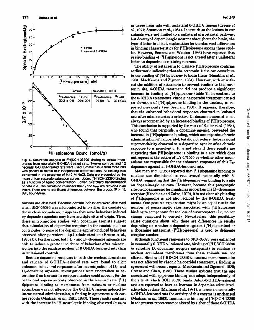
Binding characteristics for [3H]SCH 23390 to striatal tissue from neonatally lesioned rats did not differ from control (fig. 5). Binding of [3H]SCH 23390 to striatal membranes from adult-6-0HDA-treated rats was also found not to differ from control (control, 32.8 ± 2.8 pmol/g of tissue; adult-6-0HDA, 35.2 ± 2.0 pmol/g of tissue; P > .1; N = 10 and 6, respectively, for the groups). In contrast to the elevated [3H]spiperone binding observed in rats treated chronically with haloperidol (table 8), (3H)SCH-23390 was not altered after this treatment (30.6 ± 1.3 pmol/g in controls and 33 ± 2. 7 pmol/g in rats treated chronically with haloperidol; P > .1).

at ASPE
T Journals on June 9, 2015
jpet.aspetjournals.orgD
ownloaded from
172 BraeMetel. Vol. 240
TABLE4 Effect of SKF-38393 end LY-171555 on locomotor llCtivlty In edult-8-c>HDA·treeted rats after perlpherel end nucleua eccumbens lnjectlona Anmals received drugs periphel a1y or by central microinjeclion. Control refers to unlesioned rats. Rats were habituated to the chlmbers 45 mil before drug treatments. Numbers in para !theses, number of determinations.
Traallnant, Dose
AOMy ± $.E.M. {Cculls/180 mil)
LY-171555
Peripheral injection• Control, 1.0 mgjkg Adult 6-0HDA, 1.0 mgjkg
1716 ± 549 (4) 3299 ± 958 (6)
5112 ± 1365 (4) 30633 ± 7833*** (6)
Nucleus accumbens microinjection• Control, 0.3 µg/side Control, 1.0 µg/side Adult-6-0HDA, 0.3 µg/side Adult-6-0HDA, 1.0 µg/side
2121 ± 974 (4) 1451 ± 491 (5)
1799 ± 451 (4) 1724 ± 864 (4) 2980 ± 463*** (4)
23581 ± 7840*** (5) 11884 ± 4827*** (5)
•Response of control rats to i.p. saline administration was 1346 ± 426 CXU11S/180 mil end after miaoiljeclion of saline into the nucleus accumbens wes 1511 :1:: 344 counts/180 mil (N =- 4 for each group).
••• P < .001 when 6-0HDA response is compared to control.
10060
9410
8820
B.
•NEONATES •CONTROLS
Fig. 3. R8P,'esef1tative placements of injector tips in 6-0HDA-treated animals and controls. Placement was determined by histological procedures performed in randomly selected rats. Each symbol represents tip placement of bilateral injections. A, nucleus accumbens placements; B, caudate nucleus placements. Numerals to the right indicate anterior/ posterior position, In micrometers, according to Konig and Klippel (1963).
Discussion
Systemic administration of Di- and D2-dopamine agonists induces behavioral responses in 6-0HDA-lesioned rats that are of a greater magnitude than that induced in unlesioned controls
TABLES
(Breese et al., 1985a,b; Arnt, 1985). The enhanced response to dopamine agonists in 6-0HDA-lesioned rats could be reflecting an increased number of dopamine receptors, a change in the sensitivity of secondary and tertiary messenger systems linked to the dopamine recognition sites or an alteration in nondopaminergic neural mechanisms which influence the response to dopamine receptor stimulation. A major focus for the present work was to determine if a change in Di- and D2-dopamine receptor binding explained the enhanced behavioral responses induced by Di- and Drdopamine agonist administration to 6-0HDA-lesioned rats. In order to establish that the brain areas used for the binding studies were behaviorally sensitive to Diand D2-dopamine agonists, SKF-38393 (Di) and LY-171555 (D2) were administered into the nucleus accumbens and the caudate, the dopamine-rich terminal regions used for the binding investigations, and behavioral responses were assessed in unlesioned controls and the 6-0HDA-lesioned rats.
Intracerebral administration of dopamine into nucleus accumbens increases locomotor activity (Costall et al., 1975; Pijnenburg et al., 1976). Receptor binding studies have demonstrated the presence of both Di- and D2-dopamine receptors in nucleus accumbens tissue (table 5; Seeman, 1980; Schulz et al., 1985). In the present study, the locomotor response induced by Di- and D2-dopamine agonist administration into nucleus accumbens increased locomotor activity more in the 6-0HDA-
Behevlonll effecta lnducecl by SKF·38393 end LY-171555 edmlnlatralion Into the ceudete In neonetlllly end lldult-1-0HDA·treeted rats Behavior scores for each behavior are the IMWI ± S.E.M. of the percentage of scoring intervals summed for the 12 obeervatlon periods CNflf 2 IY. See table 1 for group designations. Values obtained after saline administration into the ceudate of unlesioned rats were not dlffarant from Vlllues obtained after saline admilistlatlon to unlesloned rats (P > .1 ). SKF-38393 was administered to adult 6-0HOA-treated rats end both were 8dmi listanld to unlesioned controls. There are six to eight rats In each group.
lllug Tl8llnall
Sahl SKF-38393 LY-171555 Behavior (3 ,.g/9idle) (3 ,.g/9idle)
Uilasianed Uilasianed Neonatal&- MIA Uilasianed Neonatal&- MIA conllOI conllOI OHDA 6-0K>A canllol Oll>A 6-0K>A
Sniffing 0.9 ±0.3 1.6 ± 0.5 7.0 ± 1.1* 4.9 ± 1.6* 2.8±1.2t 8.1 ± 1.3* 5.8± 1.8 Rearing 0.1 ± 0.1 0.8 ± 0.4t 3.5 ± 1.5* 0.7 ± 0.1 0.2 ± 0.1 3.2 ± 1.6* 3.8± 1.5* Locomotion 0.1 ± 0.1 0.3 ± 0.1 2.1 ± 0.9* 1.5 ± 1.3 0.5± 0.4 8.3 ± 1.7* 2.9 ± 1.3* Eating and digging in 0.0 ± 0.0 0.0±0.0 0.7±0.4 0.0±0.0 0.0±0.0 0.0±0.0 0.0±0.0
wood chips Grooming 0.0 ± 0.0 1.6 ± 0.5t 1.0 ± 0.4 2.0±0.3 0.3±0.2 0.0±0.0 0.9±0.4 Licking 0.0± 0.0 0.0±0.0 1.1 ±0.7* 0.4±0.3 0.0±0.0 0.0±0.0 0.0±0.0 Head weaving 0.0±0.0 0.0 ± 0.0 1.2 ± 0.5* 0.5±0.5 0.0±0.0 0.8 ±0.4 1.9 ± 1.6 Taffy pulling 0.0 ± 0.0 0.0±0.0 0.0± 0.0 0.0±0.0 0.0±0.0 3.3 ± 1.6* 0.0±0.0
• p < .05 when compared to corresponding drug responss in unlesioned control rats; t p < .05 when compared to saline responss In unlesioned control.

at ASPE
T Journals on June 9, 2015
jpet.aspetjournals.orgD
ownloaded from
1987
TABLE& Effect of neonatal end adult 1-0HDA-leslons on binding of ['H]SCH-23390 •nd (3H]spiperone to nucleus eccumbens Values represent the n.... picomoles per gram of tissue :t S.E.M. at a single concentration of the ligand which was determi11ed with 1.0 nM [8H)apipelo11e and 0.3 nM [3HJSCH-23390. Numbers In paanlhaaaa, number of deternii11atio11s made In triplicate. Adult 6-0HDA-treeled rats (Adult 6-0HDA) demo11111 Bl8cl a supersensitive locomotor response to L Y-171555 or to apomorphi111 (1 mg/kg) and neonatal 6-0HDA-treeled rats (neonatal 6-0HDA) showed an elevated looomotor rwponse after three treatments with SKF-38393 (3 mg/kg). No 8lgniflcant diff81• IC8S were obserVed among the groups for either lgand (P > .1).
T1811111111nt
Control Neonatal 6-0HDA Adult 6-0HDA
TABLE7
7.0± 0.9(9) 7.8 ± 0.7 (11)
10.8 ± 0.9 (7)
9.4 ±0.9(8)
Effect of ketanserln on the binding of~ to atrlellll membranes "With" refers to addition of ketarmerin (20 nM) to incWetion from IDesloned controls and neonataly 6-0HDA-lesloned rats. See "Methods" for delalls. N, number of detem*1ationS. P > .1forcomparisonofcontrolwith6-0HDA-treeted groups.
No ketanserin With ketanserin
N
8 8
pmd/gtlsu
21.2 ± 1.0 21.9 ± 1.3 18.8 ± 0.7 19.3 ± 1.1
lesioned rats than in unlesioned controls. The locomotor response to L Y-171555 was considerably less than that observed after SKF-38393 microinjection into the nucleus accumbens of both neonatally and adult-6-0HDA-lesioned rats. The observation in neonatally lesioned rats is consistent with other data obtained after i.p. administration of these drugs. In contrast, the locomotor responses observed with adult-6-0HDA-lesioned rats after injection of L Y-171555 or SKF-38393 into the nucleus accumbens did not parallel locomotor responses observed after parenteral administration (Breese et al., 1985a,b). The neural basis of these apparently paradoxical findings could have relevance to why locomotor responses to the Dr and D2-dopamine agonists differ between adult- and neonatal-6-0HDA-lesioned rats after systemic (i.p.) administration. Perhaps these findings in the 6-0HDA-lesioned rats relate to evidence that striatal efferents are 8880Ciated with different dopamine receptors (Herrera-Marschitz and Ungerstedt, 1984) or that Di-dopamine receptors interact with D2-dopamine receptor function (Breese and Mueller, 1985; Breese et al., 1985a,b). Regardless of the interpretation, the present data document the presence of Dr and D2·dopamine receptors in the nucleus accumbens of both 6-0HDA-lesioned groups which, when activated with selective dopamine agonists, result in enhanced behavioral responses.
Administration of dopamine agonists into the striatum has been 8880Ciated with behaviors such as sniffing, biting, licking and repetitive movements (Creese and Iversen, 1975; Kelly et al., 1975). This brain area is believed to be the primary site at which these drugs act to produce stereotyped behaviors (Costall et al., 1975; Ernst and Smelik, 1966). In the present investigation, administration of SKF-38393 into the caudate produced a variety of behaviors in 6-0HDA-lesioned rats, with sniff'mg being the most prominent. After microinjection of LY-171555 into the caudate, sniffmg, rearing and locomotion were observed in both 6-0HDA-treatment groups, whereas taffy pulling was observed only in neonatally lesioned rats. Inasmuch as some behaviors were not observed after microinjection of dopamine
D, Receptor 8uperseMillvlty 173
• control a. o neonatal 8-0HDA -1:1
-; 100 0 e .s 1:1 75 .e I GI 0 Cll 50 (I) (I) N I :c u
Cl) 25 I :c .. .2 :g • ~ Cl) 2 4 8 8
C•H-SCH-23390] nM
b. 400
300 .. I 0 .. x 200 ~ ... ID
100
20 40 80 80 100 Amount Bound (p molelg)
Fig. 4. &mntion analysis of (3H]spiperone binding to striatal membnll 188 from control and neonataly 6-0HDA-traated rats. Al detem*lations were In the preseuce of 0.12 M NaCl and 20 nM ketanserln. See table 8 for other values for rats lesioned with 6-0HDA when adults or rats treated cnoulcally with haloperidol. Strlatal tissue from three rats was pooled for each of the three lndepa ldent detem*lations (I.e., total • 9). Data are presented as the mean of these separate detamlnations. ['HJSplperone binding is presented as a Ulction of ligand concentration: a, Scatchard transformation; b, the calQMted values for the Ko and 8-are provided In the insert. There were no sigliflcant dffaalC8S between groups (P > .1). B/F, bound/fnle.
TABLES ......., of binding ch8nlclerlstlca for ['tl]8plpera .. binding ..... edult 1-0HDA ....... or llelaperldol Adult 8-0HDA ...... to rats that l'8Clllwld 6-0HDA when Bid. Clvonlc haloperldol ,.,.. to rats that l'8Clllwld haloperldol dally 11 mg/kg) for 1e days. va.s from theee exper'.ments ara the ll19t :t S.E.M. of tine lnclvidull ScatdllRI determinations (See Ilg. 5 for value for neonallly lesioned rats and matched controlll).
r.-- 8- K..
Experiment I Control AcMt6-0HDA
Experiment II Control Chronic haloperldol
pmd/g ""
38.3±1.36 39.5±1.73
36.2±3.0 46.0± 2.9••
0.1 ±0.005 0.09 ± 0.015
0.1±0.008 0.1±0.003
- p < .01 when COll1*9d to control.
agonists into the caudate, it is possible that there is regional specificity within the caudate for certain behaviors such as selfbiting or, alternatively, that this brain region is not involved in all behaviors elicited after systemic administration of dopamine agonists to lesioned rats. Another possibility is that higher doses must be microinjected into the caudate before such be-
at ASPE
T Journals on June 9, 2015
jpet.aspetjournals.orgD
ownloaded from
174
i: f 20
w I!>
.Si! !:: 10
! 5
Bntneetal.
• control o neonatal 6-0HDA
0o 0.1 0.2 0.3 04 05 0.6 0.7 0.8 0.9 1.0
125
.,, 100 I
0
x 75 LL ...... m so
25
[3H-splperone] nM
Control Neonotol 6- OHDA
Bmox(prlll'ie/cJ) Ko(n'lol) Bmox(pmole/g) Ko!nM)
30.2 ± 0.5 .09± .006 29. 5 ±176 .09±.003
o~~~-1~0~~~~2~0~~~~30--~~~~~~
JH-spiperone Boood (pmol/g)
Fig. 5. Saturation analysis of [3H]SCH-23390 binding to striatal membranes from neonatally 6-0HDA-treated rats. Twelve controls and 12 neonatal 6-0HDA-treated rats were used. Striatal tissue from three rats was pooled to obtain four independent detenninations. All binding was performed in the presence of 0.12 M Nacl. Data are presented as the mean of four separate saturation curves. Upper, [3H]SCH-23390 binding as a function of ligand concentration. Lower, Scatchard transformation of data in A. The calculated values for the Kd and S.... are provided in an insert. There are no significant differences between the groups (P > .1 ). B/F, bound/free.
haviors are observed. Beca11se certain behaviors were observed when SKF -38393 was microinjected into either the caudate or the nucleus accumbens, it appears that some behaviors induced by dopamine agonists may have multiple sites of origin. Thus, these microinjection studies with dopamine agonists suggest that stimulation of dopamine receptors in the caudate nucleus contributes to some of the dopamine-agonist-induced behaviors observed after parenteral (i.p.) administration (Breese et al., 1985a,b). Furthermore, both Di- and Drdopamine agonists are able to induce a greater incidence of behaviors after microinjection into the caudate nucleus of 6-0HDA-lesioned rats than in unlesioned controls.
Because dopamine receptors in both the nucleus accumbens and caudate of 6-0HDA-lesioned rats were found to elicit enhanced behavioral responses after microinjection of Di- and D2-dopamine agonists, investigations were undertaken to determine if an increase in receptor number could account for the behavioral supersensitivity observed in the lesioned rats. [3H] Spiperone binding to membranes from striatum or nucleus accumbens was not altered by the 6-0HDA lesions induced by intracistemal administration, a finding in agreement with earlier reports (Mailman et al., 1981, 1983). These results contrast with the increase in 3H-neuroleptic binding observed in vitro
Vol.240
in tissue from rats with unilateral 6-0HDA lesions (Creese et al., 1977; Staunton et al., 1981). Inasmuch as the lesions in our animals were not limited to a unilateral nigrostriatal pathway, but destroyed dopaminergic neurons throughout the brain, the type of lesion is a likely explanation for the observed differences in binding characteristics for [3H]spiperone among these studies. However, Bennett and Woot.en (1986) have reported that in vivo binding of [3H]spiperone is not altered after a unilateral lesion to dopamine-containing neurons.
The ability of ketanserin to displace (3H]spiperone confirms earlier work indicating that the serotonin-2 site can contribut.e to the binding of [3H]spiperone to brain tissue (Hamblin et al., 1984; MacKenzie and Zigmond, 1984). However, with or without the addition of ketanserin to prevent binding to this serotonin site, 6-0HDA treatment did not produce a significant increase in binding of [3H]spiperone (table 7). In contrast to the 6-0HDA treatments, chronic haloperidol treatment caused an elevation of [3H]spiperone binding in the caudat.e, as reported previously (see Seeman, 1980). It appears, therefore, that the enhanced behavioral responses observed in lesioned rats after administering a selective 1;>2-dopamine agonist is not always accompanied by an increased binding of [3H]spiperontf. This conclusion is supported by the work of Koller et al. (1984), who found that pergolide, a dopamine agonist, prevented the increase in [3H]spiperone binding, which accompanies chronic administration of haloperidol, but did not reduce the behavioral supersensitivity observed to a dopamine agonist after chronic exposure to a neuroleptic. It is not clear if these results are suggesting that [3H]spiperone is binding to a site which does not represent the action of LY-171555 or whether other mechanisms are responsible for the enhanced responses of this 0 2-
dopamine agonist in 6-0HDA-lesioned rats. Mailman et al. (1983) reported that [3H]dopamine binding to
caudate was diminished in rats treated neonatally with 6-0HDA, suggesting that the (3H]dopamine was binding to a site on dopaminergic neurons. However, because this presynaptic site on dopaminergic terminals has properties of a D2-dopamine receptor (Kebabian and Caine, 1979), it is not clear why binding of [3H]spiperone is not also reduced by the 6-0HDA treatments. One possible explanation might be an equal rise in the number of postsynaptic sites associated with (3H)spiperone binding to compensate for the loss of autoreceptors (i.e., no net change compared to control). Nevertheless, this possibility raises questions about why there are differences in results depending on whether a dopamine agonist ([3H]dopamine) or a dopamine antagonist ([3H]spiperone) is used to delineate receptor number.
Although functional responses to SKF-38393 were enhanced in neonatally 6-0HDA-lesioned rats, binding of (3H]SCH 23390 (a selective Di-dopamine receptor antagonist) to caudate or nucleus accumbens membranes from these animals was not altered. Binding of [3H]SCH-23390 to caudate membranes also was not affected by chronic haloperidol treatment, a finding in agreement with recent reports (MacKenzie and Zigmond, 1985; Creese and Chen, 1985). These studies indicate that the site associated with spiperone binding can adapt independently of the site at which SCH 23390 binds. Adult-6-0HDA-lesioned rats are reported to have an increase in dopamine-stimulatedadenylate cyclase (Mailman et al., 1981), whereas in neonatally 6-0HDA-lesioned rats the activity of this enzyme is unchanged (Mailman et al., 1983). Inasmuch as binding of [3H]SCH 23390 in the present report was not altered by either of these 6-0HDA

at ASPE
T Journals on June 9, 2015
jpet.aspetjournals.orgD
ownloaded from
1987
treatments, it appears that a simple association between [3H) SCH 23390 binding and adenylate cyclase activity does not exist. It is not known whether these observations indicate two distinct D1 -dopamine receptor sites, an alteration in cyclase reactivity without a change in receptor number or both.
Because Parkinson's patients as well as children with LeschNyhan disease have reduced brain dopamine (Homykiewicz, 1973; Lloyd et al., 1981), binding results obtained in the 6-0HDA-lesioned rats, depending upon the age at which treatment occurred (Breese et al., 1984a), may be relevant to the neuropathological changes in these clinical disorders. Although no data are available presently on binding of dopamine receptor antagonist ligands in Lesch-Nyhan disease, there are data reported from Parkinson's patients at autopsy. For example, r:1H]haloperidol binding has been found to be elevated in putamen (Guttman and Seeman, 1985) of patients with Parkinsonism, particulary those that had not received L-dopa (Lee et al., 1978; Bokobza et al., 1984). Binding of [3H)haloperidol in caudate or putamen tissue from Parkinson's patients has not been found to differ from normal, if the patients were being treated with L-dopa (Guttman and Seeman, 1985), although no change (Bobobza et al., 1984) and increases (Guttman and Seeman, 1985) are reported in caudate of patients not receiving this drug. In view of these clinical results, the absence of a change in [3H)spiperone receptor binding in the 6-0HDAtreated rats may be associated with the fact that these animals had been exposed to dopamine agonists. However, even if this should be the case, enhanced behavioral responses to the D2-dopamine agonist persist in the lesion rats, even though the number of dopamine antagonist binding sites are not altered.
In addition to results with 3H-neuroleptic ligands, several studies have been performed on tissue from patients with Parkinsonism to evaluate adenylate cyclase activity and [3H) SCH 23390 binding. Dopamine-stimulated adenylate cyclase activity is reported to be decreased (Shibuya, 1979) or increased (Nagatsu et al., 1978) in patients with Parkinsonism. On the other hand, [3H]SCH 23390 binding is reported to be not changed (Pimoule et al., 1985; Raisman et al., 1985) or elevated (Raisman et al., 1985) in patients with Parkinsonism. The length of time that patients are free of L-dopa was suggested as a possible reason for the conflicting results, because no change in [3H)SCH 23390 was observed in those patients that had not received L-dopa from 5 days to 4 years before death (Raisman et al., 1985). Thus, the results with [3H)SCH 23390 in patients with Parkinsonism seem consistent with the absence of a change in [3H)SCH 23390 binding to striatal tissue observed in adult-6-0HDA-lesioned rats. From the absence of a change in [3H)SCH 23390 binding in neonatally lesioned rats, it would not seem likely that a change in [3H)SCH-23390 binding will be observed in patients with Lesch-Nyhan disease.
In summary, the present investigation has demonstrated the following: 1) D1- and Drdopamine agonists produced supersensitive behavioral responses when microinjected into the nucleus accumbens and caudate nucleus of 6-0HDA-lesioned rats; 2) locomotor responses from adult-6-0HDA-lesioned rats induced by intra-accumbens microinjection of D1- and Drdopamine agonists were not the same as those observed after systemic administration; and 3) (3H)SCH 23390 and [3H)spiperone binding to nucleus accumbens and caudate tissue were not altered in neonatally and adult-6-0HDA-treated rats that exhibited enhanced behavioral responses to Di- and D2·dopamine agonist administration. Thus, a change in binding characteristics for
D, Receptor SupenenaltivitJ 175
the dopamine-antagonist ligands was not associated with the behavioral supersensitivity observed with 6-0HDA-lesioned rats after D,- and D2·dopamine agonist administration. The absence of an association between the enhancement of dopamine-agonist induced behavior and dopamine-antagonist binding suggest that there are deficiencies in our present understanding of dopamine receptor mechanisms. Studies of dopamine agonist binding in dopaminergic terminal areas, evaluation of neural systems that can modulate dopamine receptor function and further definition of cellular mechanisms beyond the dopamine receptor level of lesioned rats are areas for future experimentation that might suggest a molecular mechanism for the enhanced behavioral responses observed after dopamine agonist administration to rats with lesions of dopamine-containing neurons.
Acluaowledcmeata
The authors acknowledge the ezcellent technical uaiatance of Susan Emerick, Edna F.dwarda and Marcine Garrison and the typing of the manUICJ'ipt by Carolyn Reama.
Refere-
ALTAR, C.A., Kua, H. AND MARsHALL, J. F.: Computer imqing and analyai.B of dopamine (I>.) and aerotonin (So) binding lit.ea in rat basal pnglia or neocort.ex labeled by ["H)apiroperidol J. Pharmacol. Exp. Ther. 233: 627-538, 1986.
ARNT, J.: Hyperactivity induced by stimulation oheparate D-1 and D-2 receptors in rate with bilateral 6-0HDA leliona. Life Sci. 37: 717-723, 1986.
ARNT, J. AND HY'ITBL, J.: DiffeNntial inhibition by dopamine D-1 and D-2 antagoniata of circling behavior induced by dopamine qoniata in rate with unilateral 6-hydroxydopamine leaioDL Eur. J. Pharmacol. 102: 349-364, 1984.
BENNE'IT, J. P., JR. AND WOOTEN, G. F.: Dopamine denervation does not alter in uiuo "H-epiperone binding in rat lltriatwn: Implicationa for external imqing of dopamine receptors in Parkinaon'• cliaeue. Ann. Neurol. 19: 378-383, 1986.
BILLARD, W., RUPERTO, V., CROSBY, G., IORIO, L. C. AND BARNE'IT, A.: Characterimtion of the binding of "H-Sch-23390, a aelective D-1 receptor antagoniat ligand, in rat lltriatwn. Life Sci. 36: 1886-1893, 1984.
BOKOBZA, B., RUBERG, M., ScATl'ON, B., JAVOY-AGID, F. AND AGID, Y.; ("HJ apiperone binding, dopamine and HVA concentrationa in parkinaon'1 diaeaae and aupranuclear palsy. Eur. J. Pharmacol 99: 167-175, 1984.
BREESE, G. R., BAUMEISTER, A. A., McCOWN, T. J., EMERICK, S. G., FRYE, G. D., CROTTY, IC. AND MUELLER, R. A.: Behavioral differencee betwwn neonatal and adult 6-hydroxydopamine-treated rate to dopamine qoniata: Relevance to neurological l)'IDptoma in clinical 1yndro- with Nduced brain dopamine. J. Pharmacol. Exp. Ther. 231: 343-364, 1984a.
BRUSE, G. R., BAUMEISTER, A., NAPIER, T. C., FRYE, G. D. AND MUELLER, R. A.: Evidence that D-1 dopamine receptors contribute to the supenenlitive behavioral reepoll8ell induced by L-dihydroxyphenylalanine in rate tNated neonatally with 6-hydroxydopamine. J. Pharmacol Exp. Ther. 234: 287-296, 1985a.
BRUSE, G. R., FRYE, G. D., McCoWN, T. J. AND MIJELLER, R. A.: Comparilon of tha CNS eff'ecta induced by TRH and bic:uculline after microinjection into various brain lit.ea: Abaence of IUJIPOrt for a GABA antagoniat action for TRH. Pharmacol Biochem. Behav. 21: 146-149, 1984b.
BRUSE, G. R. AND MUELLER, R. A.: SCH-23390 antagonism of a D-2 dopamine agoniat depends upon catecholamineqic neurons. Eur. J. Pharmacol 113: 109-114, 1986.
BREESE, G. R., MUELLER, R. A. AND MAILMAN, R. B.: Eff'ecta of dopamineqic agonilta and antagoniata on in uiuo cyclic nucleotide content: Relation of guanOlline 3'6' -monophoephate (cGMP) changea in cerebellum to behavior. J. Pharmacol. Exp. Ther. 209: 262-270, 1979.
BRBESE, G. R., NAPIER, T. C. AND MUELLER, R. A.: Dopamine qoniat.-induced locomotor activity in rate treated with 6-hydroxydopamine at differing apa: Functional aupenenlitivity of D-1 dopamine receptors in neonatally lesioned rate. J. Pharmacol Exp. Ther. 234: 447-466, 1985b.
BREESE, G. R., SMITH, R. D., CooPER, B. R. AND GRANT, L. D.: Alterations in conaummatory behavior following intracistemal injection of 6-bydroxydopamine. Pharmacol Biochem. Behav. 1: 319-328, 1973.
BRUSE, G. R. AND TRAYLOR, T. D.: Eff'ecta of 6-bydroxydopamine on brain norepinephrine and dopamine: Evidence for aelective degeneration of catecholamine neuroDL J. Pharmacol Exp. Ther. 174: 413-420, 1970.
BREESE, G. R. AND TRAYLOR, T. D.: Developmental charac:terilticl of brain catecholamines and tyroaine hydroxylaae in the rata: Eff'ecta of 6-hydroxydopamine. Br. J. Pharmacol 44: 210-222, 1972.
COBTALL, B., NAYLOR, R. J. AND NEUMEYER, J. L.: Differenc:ee in the nature of the ate..otyped behavior by apomorpbine derivatives in the rat and in their actions in extrapyramidal and mesolimbic brain areas. Eur. J. Pharmacol 31: 1-16, 1976.
CHRISTENSEN, A. V., ARNT, J., HY'ITBL, J., LARsON, J. J. AND SVENDSEN, 0.:

at ASPE
T Journals on June 9, 2015
jpet.aspetjournals.orgD
ownloaded from
176 Breese et al.
Pharmacological effects of a specific dopamine D-1 antagonist SCH-23390 in comparison with neuroleptica. Life Sci. 34: 1529-1540, 1984.
CREESE, )., BURT, D. R. AND SNYDER, S. H.: Dopamine receptor binding enhancement accompanies lesion-induced behavioral supenenaitivity. Science (Waah. DC) 197: 596-598, 1977.
CREESE, I. AND CHEN, A.: Selective D1 dopamine receptor increase following chronic treatment with SCH 23390. Eur. J. Pharmacol. 109: 127-128, 1985.
CREESE, I. AND IVERSEN, s. D.: The pharmacological and anatomical suhatrates of the amphetamine response in the rat. Brain Res. 83: 419-436, 1975.
ERNST, A. M. AND SMELIK, D. G.: Site of action of dopamine and apomorphine on compulsive gnawing behaviour in rats. Experientia (Basel) 22: 837-838, 1966.
GOLDSTEIN, M., LEW, T. Y., AsANO, T. AND WETA, K.: Alterations in dopamine recepton. Effects of lesion• and haloperidol treatment. Commun. Psychopharmacol. 4: 21-25, 1980.
GUTTMAN, M. AND SEEMAN, P.: L-DOPA revenes the elevated density of Dr dopamine receptore in Parkinson's diseased striatum. J. Neural Tranam. 84: 93-103, 1985.
HAMBLIN, M. W., I.Err, S. E. AND CREESE, I.: Interactions of qoniats with D-2 dopamine receptore: Evidence for a single receptor population emting in multiple qonilt affinity-states in rat striatal membranes. Biochem. Pharmacol. 33: 877-887, 1984.
HEIKKILA, R. E., SHAPIRO, B. S. AND DUVOISIN, R. C.: The relationship between 1088 of dopamine nerve terminals, atriatal ["HJ-spiroperidol binding and rotational behavior in unilaterally 6-hydroxydopamine-lesioned rats. Brain Rea. 211: 285-292, 1981.
HESS, E. J., BATTAGLIA, G., NORMAN, A. B., )ORIO, L. C. AND CREESE, I.; Guanine nucleotide regulation of agoniat interactions of "H-SCH 23390-labelled D,-dopamine receptore in rat 1triatum. Eur. J. Pharmacol. 121: 31-38, 1986.
HERRERA-MARSCHITZ, M. AND UNGERSTEDT, U.: Evidence that striatal efferents relate to different dopamine receptore. Brain Res. 323: 269-278, 1984.
HOLLISTER. A. s .. BREESE, G. R. AND CoOPER, B. R.: Comparison of tyrosine hydroxylaae and dopamine-P-hydroxylaae inhibition with the effects ofvarioua 6-hydroxydopamine treatments on d-amphetamine induced motor activity. Psychopharmacologia 38: 1-16, 1974.
HOLLISTER, A. S., BREESE, G. R., AND MUELLER, R. A.: Role of monoamine neural ayatema in L-dihydroxyphenylalanine atimuleted activity. J. Pharmacol. Exp. Ther. 208: 37-43, 1979.
HORNYKIEWICZ, 0.: Parkinson'• disease: From brain homogenate to treatment. Fed. Proc. 32: 183-190, 1973.
)ORIO, L. C., BARNETT, A., LEITZ, F. H., HOUSER, V. P. AND KORDUBA, A.: SCH-23390, a potential benzuepine antjplychotic with unique interactions on dopaminergic systems. J. Pharmacol. Exp. Ther. 228: 462-468, 1983.
JACKSON, D. M., ANDEN, N.-E. AND DAHLSTROM, A.: A functional effect of dopamine in the nucleus accumbens and in some dopamine-rich areaa of the rat brain. Psychopharmacologia 46: 139-149, 1975.
KEBAlllAN, J. W. AND CALNE, D. B.: Multiple receptore for dopamine. Nature (Lond). 227: 93-96, 1979.
KELLY, P.H., SEVIOUR, P. W. AND IVERSEN, S. D.: Amphetamine and apomorphine responees in the rat following 6-0HDA lesions of the nucleus acc:umbens septi and corpus 1triatum. Brain Res. 94: 507-522, 1975.
KILTS, C. D., BREESE, G. R. AND MAILMAN, R. B.: Simultaneous quantification of dopamine, 5-hydroxytryptamine, and four metabolically related compounds by means of reveree phase HPLC with electrochemical detection. J. Chromatogr. Biol. Med. Appl. 226: 347-357, 1981.
KILTS, C. D., SMITH, D; A., ONDRUSEK, M. G., MAILMAN, R. B., MUELLER, R. A. AND BREESE, G. R.: Differential effects of "dopaminergic agoniats" on measures of dopaminergic function. Soc. Neuroeci. Abatr. 6: 562, 1979.
KOLLER, W. C., CORTIN, J. C. AND FIELDS, J. Z.: Pergolide down-regulates D-2 dopamine receptore but fails to block haloperidol induced behavioral supenensitivity. Soc. Neurosci. Abatr. 10: 1136, 1984.
KONIG, J. F. R. AND KLIPPEL, R. A.: The Rat Brain: A Stereotuic Atlas of the Forebrain and Lower Parts of the Brain Stem, pp. 1-162, R. E. Kriepr Publishing Co., 1963.
LEE, T., SEEMAN, P., RA.IPUT, A., FARLEY,). J. AND HORNYKIEWICZ, 0.: Receptor baaia for dopaminergic supenensitivity in Parkinson'• disease. Nature (Lond.) 273: 59-61, 1978.
Val. 240
LLoYD, K. G., HORNYKIBWICZ, 0., DAVIDSON, L., SHANNAK, K., FAJU.BY, I., GoLDSTBIN, M., SHIBUYA, M., KELLEY, w. N. AND Fox, I. H.: Biochemical evidence of dyafunction of brain neurotnnamitt.en in the Lesch-Nyhan IYJI· drome. N. En,l. J. Med. 306: 1106-1111, 1981.
MACKENZIE, R. G. AND ZIGMOND, M. J.: High and low-affinity state. of atriatal D-2 receptors are not affected by 6-hydroxydopamine or chronic haloperidol treatment. J. Neurochem. 43: 1310-1318, 1984.
MACKENZIE, R. G. AND ZIGMOND, M. J.: Chronic neuroleptic tzeatment mc-0. but not D1 receptors in rat atriatum. Eur. J. Pharmacol. 118: 169-1116, 1985.
MAILMAN, R. B., KILTS, C. D., BEAUMONT, IC. AND BIUlllSB, G. R.: •8upenenlitivity• of dopamine 1y&te1111: Compariaons i.tw.n baloperidol withdrawal, intraciaternal and unilat.eral 6-hydroxydopamine (6-0HDA) tzeatments. Fed. Proc. 40: 291, 1981.
MAILMAN, R. B., TOWLE, A., ScHULZ, D. W., LEw18, M. H., BRBBIB, G. R., DEHAVEN, D. H. AND KRIGMAN, M. R.: Neonatal 6-0HDA treatment of rats: Cbanps in dopamine (DA) recepton, ltriatal nwrochemiatry and anatomy. Soc. Neuroeci. Abatr. 9: 932, 1983.
MOLLOY, A.G. AND WADDINGTON, T. L.: Dopaminerp: behavior lt.enolpecifically promoted by the Di qoniat R-SKF-38393 and lllectively blocked by the D, antqonilt SCH 23390. Paychopharmacolol)' 82: 409-410, 1984.
NAGATSU, T., KANAMORI, T., KATO, R., bzUKA, R. AND NARABAYASHI, H.: Dopamine-atimuleted adenylate cyclase activity in the human brain. Cbanps in Parkimoniam. Biocbem. Med. 19: 360-366, 1978.
NBVE, K. A., ALTAR, C. A., WONG, C. A. AND MAasHALL, T. F.: Quantitatiw analylis of ('H)lpiroperidol binding to rat forebrain lectionl: Pluticity of ll80lltriata1 dopamine receptors after nigroetriatal injury. Brain Rea. 302: 9-18, 1984.
PuNBNBURG, A. J. J., HONIG, W. M. M., VAN DER HEYDBN, J. A. M. AND VAN RoSSUM, J. M.: Effects of chemical atimuletion of the mesolimbic dopamine lyBtem upon locomotor activity. Eur. J. Pharmacol. 36: 46-58, 1976.
PIMOULE, C., ScHOBMAKBR, H., RBYNOLDS, G. P. AND LANGER, S. Z.: ('HJ SCH 23390 labeled Di-dopamine receptore are unchanpd in schizophrenia and Parkimon'1 m-. Eur. J. Pharmacol. 114: 236-237, 1985.
RAIBMAN, R., CASH, R., RUBBRG, M., JAVOY-AGID, F. AND AGm, Y.: Binding of ("H)SCH 23390 to D-1 receptors in the putamen of control and Parkimonian subjecta. Eur. J. Pharmacol. 118: 467-468, 1985.
ScHOBNPBLD, R. AND URETSKY, N.: Altered reaponae to apomorphine in 6-hydros:ydopamine-treated rata. Eur. J. Pharmacol.19: 115-118, 1972.
ScHULZ, D. W., STANPORD, E. J., WYRICK, S. W. AND MAILMAN, R. B.: Binding of "H-SCH 23390 in rat brain: Reciona1 distribution and effects of~ conditions and GTP suaeat interactions at a D,-like dopamine receptor. J. Neurochem. 46: 1601-1611, 1985.
SEEMAN, P.: Brain dopamine recepton. Pharmacol. Rev. 32: 229-313, 1980. SETLER, P. E., SARAU, H. M., ZIRxLE, c. L. AND SAUNDBRS, H. L.: The central
effects of a novel dopamine agonilt. Eur. J. Pharmacol. 60: 419-430, 1978. SHIBUYA, M.: Dopamine-lenlitve aclenylate cycJa. activity in the atriatum in
Parkinaon'1 m-. J. Neural Tramm. 44: 287-296, 1979. SMmt, R. D., CooPD, B. R. AND BRBBSB, G. R.: Growth and behavioral cbanps
in developins rats treated intnci8t.enta11y with 6-~ Evidence for involvement of brain dopamine. J. Pharmacol. Exp. Ther. lH: 609-619, 1973.
STAUNTON, D. A., WOLPE, B. B., GROVES, P. M. AND MOUNOrr, P. B.: Dopamine receptor c:hanpe following deatruction of the nipoetriatal pathway: Lack of a relationship to rotation behavior. Brain Rea. 211: 315-327, 1981.
TSURUTA, K., FREY, E. A., GREWE, C. W., CoTE, T. E., EllKAY, R. L. AND KBBAlllAN, T. W.: Evidence that LY-141865 specifically atimuletes the D-2 dopamine receptor. Nature (Lond.) 292: 483-465, 1981.
UNGERSTBDT, U.: POltlynaptic IUJ>l!llenaitivity after 6-hydroxydopamine induced depneration of the nipo-atriatal dopamine syatem. Acta Phyliol. Scand. 387: euppl. 69-93, 1971.
URBTSKY, N. J. AND ScHOBNPBLD, R. I.: Effect of L-DOPA on the locomotor activity of rats pretnated with 6-hydroiiydopemine. Nat. New Biol. 234: 157-159, 1981.
Send reprillt reqa-. to: Dr. Georp R. s-. BiolOlical Sciencee Research Center, University of North Carolina School of Medicine, Chepel Hill, NC 27514.
Related Documents