J. clin. Path., 30, Suppl. (Ass. Clin. Path.), 7, 1-7 Anatomy of the hypothalamus and pituitary gland P. M. DANIEL From the Department of Applied Physiology and Surgical Science, Royal College of Surgeons of England, Lincoln's Inn Fields, London WC2A 3PN Some knowledge of the anatomy of the hypo- thalamus and pituitary and of the neurovascular pathways connecting them is essential for under- standing the endocrine and other dysfunctions that result from any lesion involving either the hypotha- lamus or the pituitary or which damages their con- necting pathways. When the pituitary gland is removed or is deprived of stimuli from the hypo- thalamus'hypopituitarism' results. In hypopituitar- ism the endocrine organs show the most striking changes, but all the tissues of the body are affected to a greater or lesser extent (Sheehan, 1937; Sheehan and Summers, 1949; Daniel and Prichard, 1975). The anterior lobe (pars distalis) of the pituitary gland develops as an upgrowth of epithelial tissue from the primitive pharynx (Rathke's pouch) which [7771 Neural Epithelial tissue tissue Fig. 1 Diagram of human pituitary gland in sagittal section. 'Neural tissue' indicates the neurohypophysis; 'epithelial tissue' the adenohypophysis. IP = infundibular process (posterior lobe). LIS = lower infundibular stem. PD = pars distalis (anterior lobe). PT = pars tuberalis. UIS = upper infundibular stem. meets a downgrowth from the base of the brain, the latter being destined to form not only the infundibu- lar process (posterior, or neural, lobe) of the pituitary but also the neural part of the pituitary stalk. The early arrangements in the human have been illus- trated by Daniel (1966a). The parts of the adult human pituitary gland and of the pituitary stalk are shown in Fig. 1. The nomenclature of these structures is confusing and some of the names which have been applied to certain parts of the pituitary complex and the hypothalamus in animals should not be transferred uncritically to man. Daniel and Prichard (1975) have explained the names of the various parts of these structures. Hypothalamus The hypothalamus lies at the base of the brain, around the third ventricle, extending from a plane immediately anterior to the optic chiasma to one immediately posterior to the mamillary bodies. Laterally its borders, somewhat ill-defined, are roughly the optic tract, the internal capsule, pes pedunculi, globus pallidus, and ansa penduncularis at various anteroposterior planes, while superiorly it does not extend above the level of the anterior commissure. Its weight in the adult human is less than 2.5 g. L6veque (1974) thinks that the hypo- thalamus should be given the status of an endocrine organ, while Stumpf (1975) suggests that the whole brain should be regarded as an endocrine gland. NERVE CELLS OF THE HYPOTHALAMUS The small mass of cerebral tissue which comprises the hypothalamus contains some extremely well- defined nuclei but also others whose outline cannot be so easily determined. Some of the latter stand out more clearly in the fetus than in the adult (Clark, 1938). The general arrangements of the more readily identifiable nuclei are shown in Figs. 2 and 3. The most striking of all are the supraoptic, composed almost wholly of large nerve cells, and the para- ventricular nuclei, composed mainly of large nerve 1 copyright. on January 15, 2020 by guest. Protected by http://jcp.bmj.com/ J Clin Pathol: first published as 10.1136/jcp.s1-7.1.1 on 1 January 1976. Downloaded from

Welcome message from author
This document is posted to help you gain knowledge. Please leave a comment to let me know what you think about it! Share it to your friends and learn new things together.
Transcript
J. clin. Path., 30, Suppl. (Ass. Clin. Path.), 7, 1-7
Anatomy of the hypothalamus and pituitaryglandP. M. DANIEL
From the Department of Applied Physiology and Surgical Science, Royal College of Surgeons of England,Lincoln's Inn Fields, London WC2A 3PN
Some knowledge of the anatomy of the hypo-thalamus and pituitary and of the neurovascularpathways connecting them is essential for under-standing the endocrine and other dysfunctions thatresult from any lesion involving either the hypotha-lamus or the pituitary or which damages their con-necting pathways. When the pituitary gland isremoved or is deprived of stimuli from the hypo-thalamus'hypopituitarism' results. In hypopituitar-ism the endocrine organs show the most strikingchanges, but all the tissues of the body are affectedto a greater or lesser extent (Sheehan, 1937; Sheehanand Summers, 1949; Daniel and Prichard, 1975).The anterior lobe (pars distalis) of the pituitary
gland develops as an upgrowth of epithelial tissuefrom the primitive pharynx (Rathke's pouch) which
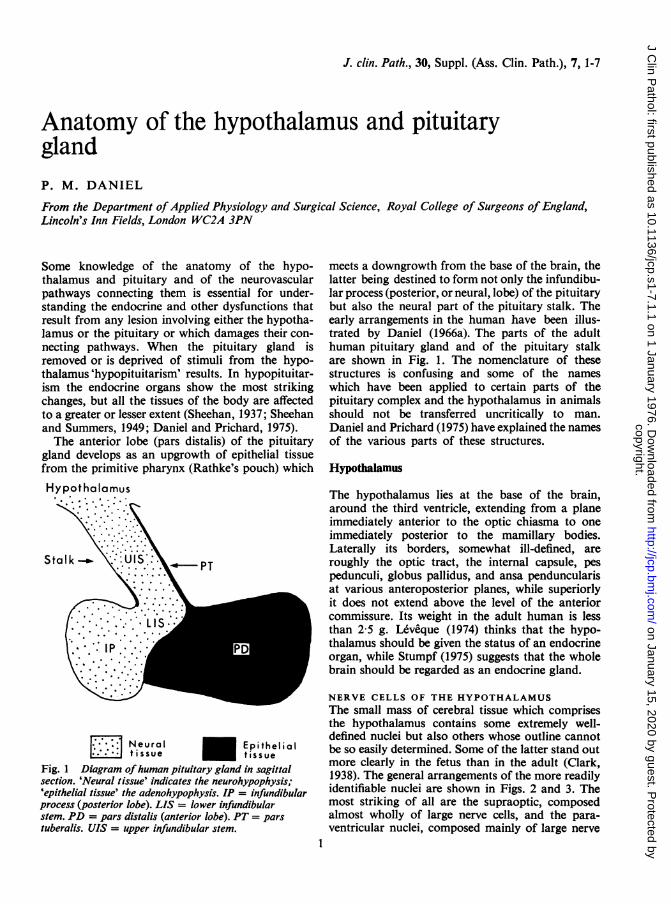
[7771 Neural Epithelialtissue tissue
Fig. 1 Diagram ofhuman pituitary gland in sagittalsection. 'Neural tissue' indicates the neurohypophysis;'epithelial tissue' the adenohypophysis. IP = infundibularprocess (posterior lobe). LIS = lower infundibularstem. PD = pars distalis (anterior lobe). PT = parstuberalis. UIS = upper infundibular stem.
meets a downgrowth from the base of the brain, thelatter being destined to form not only the infundibu-lar process (posterior, or neural, lobe) of the pituitarybut also the neural part of the pituitary stalk. Theearly arrangements in the human have been illus-trated by Daniel (1966a). The parts of the adulthuman pituitary gland and of the pituitary stalkare shown in Fig. 1. The nomenclature of thesestructures is confusing and some of the nameswhich have been applied to certain parts of thepituitary complex and the hypothalamus in animalsshould not be transferred uncritically to man.Daniel and Prichard (1975) have explained the namesof the various parts of these structures.
Hypothalamus
The hypothalamus lies at the base of the brain,around the third ventricle, extending from a planeimmediately anterior to the optic chiasma to oneimmediately posterior to the mamillary bodies.Laterally its borders, somewhat ill-defined, areroughly the optic tract, the internal capsule, pespedunculi, globus pallidus, and ansa penduncularisat various anteroposterior planes, while superiorlyit does not extend above the level of the anteriorcommissure. Its weight in the adult human is lessthan 2.5 g. L6veque (1974) thinks that the hypo-thalamus should be given the status of an endocrineorgan, while Stumpf (1975) suggests that the wholebrain should be regarded as an endocrine gland.
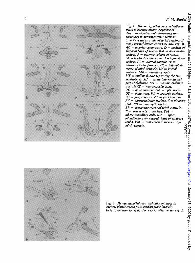
NERVE CELLS OF THE HYPOTHALAMUSThe small mass of cerebral tissue which comprisesthe hypothalamus contains some extremely well-defined nuclei but also others whose outline cannotbe so easily determined. Some of the latter stand outmore clearly in the fetus than in the adult (Clark,1938). The general arrangements of the more readilyidentifiable nuclei are shown in Figs. 2 and 3. Themost striking of all are the supraoptic, composedalmost wholly of large nerve cells, and the para-ventricular nuclei, composed mainly of large nerve
1
copyright. on January 15, 2020 by guest. P
rotected byhttp://jcp.bm
j.com/
J Clin P
athol: first published as 10.1136/jcp.s1-7.1.1 on 1 January 1976. Dow
nloaded from
P. M. Daniel
Fig. 2 Human hypothalamus and adjacentparts in coronal planes. Sequence ofdiagrams showing main landmarks andstructures in anteroposterior sections(a to f) based on study of serial sections ofmany normal human cases (see also Fig. 3)AC = anterior commissure. D = nucleus ofdiagonal band ofBroca. DM = dorsomedialnucleus. F = anterior column offornix.GC= Gudden's commissure. I= infundibularnucleus. IC = internal capsule. IF =intraventricular foramen. IR = infundibularrecess of third ventricle. LV = lateralventricle. MB = mamillary body.MF = midline fissure separating the twohemispheres. M] = massa intermedia andpart Of thalamus. MT = mamillo-thalamictract. NVZ = neurovascular zone.OC = optic chiasma. ON = optic nerve.OT = optic tract. PO = preoptic nucleus.PP = pes pedunculi. PT = pars tuberalis.
A PV = paraventricular nucleus. S = pituitarystalk. SO = supraoptic nucleus.SR = supraoptic recess of third ventricle.T = lateral tuberal nucleus. TM =tubero-mamillary cells. UIS = upperinfundibular stem (neural tissue ofpituitarystalk). VM = ventromedial nucleus. V3=third ventricle.
Fig. 3 Human hypothalamus and adjacent parts insagittal planes tracedfrom median plane laterally(a to d; anterior to right). For key to lettering see Fig. 2.
2
copyright. on January 15, 2020 by guest. P
rotected byhttp://jcp.bm
j.com/
J Clin P
athol: first published as 10.1136/jcp.s1-7.1.1 on 1 January 1976. Dow
nloaded from
Anatomy of the hypothalamus and pituitary gland
cells but also with a number of smaller cells. Thelarge cells of these nuclei synthesise vasopressin andoxytocin and also a binding protein, neurophysin,which is rich in cystine (Hope, 1975; Watkins, 1975;Zimmerman et al., 1975).
This complex of substances, but probably mainlythe neurophysin, can be stained by Gomori's chrome-alum-haematoxylin method. Since this complexpasses down the axons of the cells into the hypo-thalamo-neurohypophysial tract on its way to endby entering the capillaries of the infundibular processthe course of the axons, both in the hypothalamusand the tract, may be traced by this means. Theclassical studies on neurosecretion by the Scharrersand by Bargmann are reviewed by Stutinsky(1974), as is more recent work.There are still many puzzling features about
neurosecretion. For example, in the normal adulthuman hypothalamus and hypothalamo-neurohypo-physial tract few cells or nerve fibres take the stainfor neurosecretory material, although in childrenand in cases of hypophysectomy or section of thepituitary stalk the picture is different (Daniel andPrichard, 1975). Knowles and Vollrath (1974)review neurosecretion.The nuclei in the basal part of the hypothalamus,
especially those in the tuber cinereum (around the
Hypotholamus
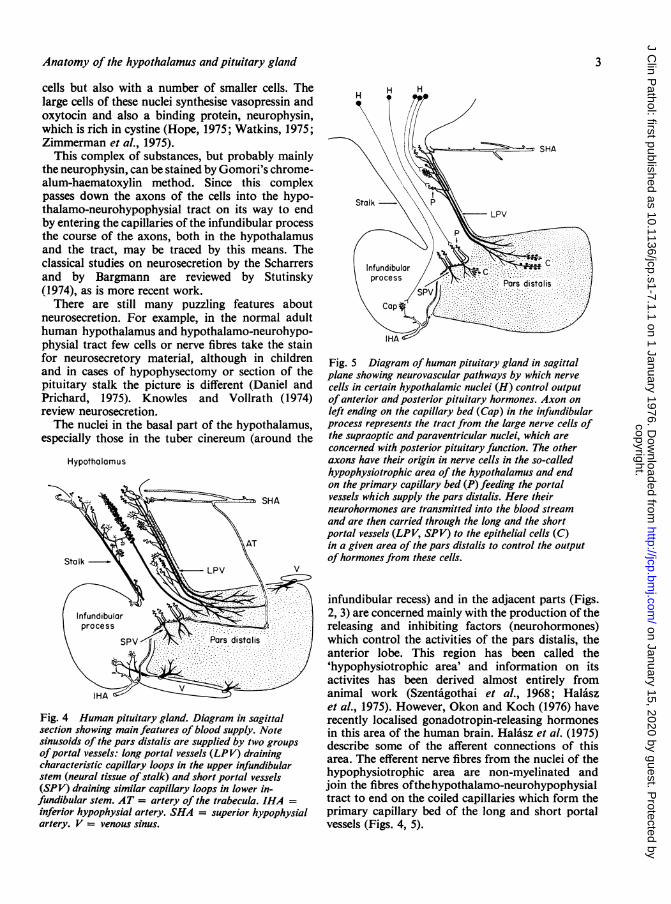
Fig. 4 Human pituitary gland. Diagram in sagittalsection showing main features of blood supply. Notesinusoids of the pars distalis are supplied by two groups
ofportal vessels: long portal vessels (LPV) drainingcharacteristic capillary loops in the upper infundibularstem (neural tissue ofstalk) and short portal vessels(SPV) draining similar capillary loops in lower in-fundibular stem. AT = artery of the trabecula. IHA =
inferior hypophysial artery. SHA = superior hypophysialartery. V = venous sinus.
Fig. 5 Diagram ofhuman pituitary gland in sagittalplane showing neurovascular pathways by which nervecells in certain hypothalamic nuclei (H) control outputof anterior and posterior pituitary hormones. Axon onleft ending on the capillary bed (Cap) in the infundibularprocess represents the tract from the large nerve cells ofthe supraoptic and paraventricular nuclei, which areconcerned with posterior pituitary function. The otheraxons have their origin in nerve cells in the so-calledhypophysiotrophic area of the hypothalamus and endon the primary capillary bed (P) feeding the portalvessels which supply the pars distalis. Here theirneurohormones are transmitted into the blood streamand are then carried through the long and the shortportal vessels (LPV, SPV) to the epithelial cells (C)in a given area of the pars distalis to control the outputof hormones from these cells.
infundibular recess) and in the adjacent parts (Figs.2, 3) are concerned mainly with the production of thereleasing and inhibiting factors (neurohormones)which control the activities of the pars distalis, theanterior lobe. This region has been called the'hypophysiotrophic area' and information on itsactivites has been derived almost entirely fromanimal work (Szentaigothai et al., 1968; Halaszet al., 1975). However, Okon and Koch (1976) haverecently localised gonadotropin-releasing hormonesin this area of the human brain. Halasz et al. (1975)describe some of the afferent connections of thisarea. The efferent nerve fibres from the nuclei of thehypophysiotrophic area are non-myelinated andjoin the fibres ofthehypothalamo-neurohypophysialtract to end on the coiled capillaries which form theprimary capillary bed of the long and short portalvessels (Figs. 4, 5).
3
copyright. on January 15, 2020 by guest. P
rotected byhttp://jcp.bm
j.com/
J Clin P
athol: first published as 10.1136/jcp.s1-7.1.1 on 1 January 1976. Dow
nloaded from

P. M. Daniel
The identifiable nuclei of the hypothalamus lieamong numerous small nerve cells, which are notgrouped into recognisable nuclei, and among finenerve fibres which, in the main, are non-myelinated.A few tracts composed of myelinated fibres, such asthe fornix and the mamillo-thalamic tract (Figs.2, 3), pass through the hypothalamus, in whichthey originate or end.
There has recently been renewed interest in thefunction of some of the specialised cells of theependyma of the third ventricle. Certain of thesecells pass their secretions into the cerebrospinalfluid and others appear to send processes which endon coiled capillaries, and perhaps on other capil-laries, into which they probably pass their secretions.Rodriguez (1976) gives a valuable review of thesubject, while Kumar and Kumar (1975) reportinteresting findings about the so-called 'tanycyteependyma'.
AFFERENT AND EFFERENT NEURAL PATHWAYSOF THE HYPOTHALAMUSThe major afferent tracts tend to lie in the lateralparts of the hypothalamus while the efferent tractslie nearer to the midline, although large numbersof both afferent and efferent non-myelinated nervefibres connect the hypothalamic nerve cells with thevarious parts of the cerebral hemispheres, brainstem, and elsewhere and form a sort of capsule ofnerve fibres around the hypothalamus. Relativelyfew of the neural pathways in the human hypo-thalamus are known in detail, and much of ourknowledge of hypothalamic connections has had tobe gained from animal experimental work (Raisman,1966). Much remains to be learned from carefulmorphological studies of the human hypothalamus,especially from brains in which lesions have occurredwhich might be termed 'experiments of nature'(Daniel and Treip, 1977). There are various singlestudies on human material and Clark (1948) hasreviewed some of the connections of the hypo-thalamus with the frontal lobes of the brain.A massive tract of myelinated fibres, the fornix
brings impulses from each temporal lobe to theipsilateral mamillary body. One major efferenttract, more medially situated, is the mamillothala-mic tract. It is composed of myelinated fibres andconnects each mamillary body with the ipsilateralanterior nucleus of the thalamus, from whichimpulses are relayed to the frontal lobes. That thereare efferent hypothalamo-autonomic tracts passingdown the spinal cord directly to the preganglionicnuclei of the sympathetic and parasympatheticsystems has recently been confirmed by Saper et al.(1976).Another major efferent tract, composed almost
entirely of non-myelinated nerve fibres, is the hypo-thalamo-neurohypophysial tract formed by theaxons of neurosecretory nerve cells in the supraopticand paraventricular nuclei which carry neuro-hormones to the neural lobe (infundibular process)of the pituitary. This important neuroendocrine tractcarries vasopressin and oxytocin, the hormoneswhich are destined to be released into the bloodstream in the neural lobe or infundibular process(Figs. 4, 5). The hypothalamo-neurohypophysialtract is joined by fibres of the tubero-infundibulartract arising from nerve cells situated in the hypo-physiotrophic areas, which are destined to end oncoiled capillaries (Fig. 5).
BLOOD SUPPLY OF THE HYPOTHALAMUSThe arterial supply is derived from the perforatingvessels which spring from the various parts of thecircle of Willis and pass through the anterior andposterior perforated substances. In addition to thesesmall arteries two vessels, the superior hypophysialarteries, which arise from the internal carotidarteries, form an arterial ring around the tubercinereum. Branches from this ring supply the opticchiasma and the adjacent parts of the hypothalamus.Many small arterial twigs from the ring pass into thepituitary stalk (Figs. 4, 6). The venous drainageenters into fairly large veins running in the basalcisterns (Duvernoy, 1975). Daniel (1963, 1966a, b)gives further details of the blood supply.
Pituitary Gland
The general arrangements of the pituitary gland areshown diagrammatically in Fig. 1. The epithelial cellsforming the anterior lobe of the gland, or parsdistalis, secrete their hormones directly into the bloodflowing through the sinusoids that run between thecells (Figs. 5, 7). The granules within these cellsidentify the various types of cell. These granuleshowever, are difficult to study in human glandssince it is not easy to obtain fresh specimens.Conklin (1966, 1968) gives useful descriptions ofthese cells in the human (see also Doniach, 1977).The literature on the cells of the pars distalis inanimals is vast, though relatively little work hasbeen done on human material. The cells of theinfundibular process have been rather neglected inrecent years, but Daniel and Prichard (1975) givesome data on their reactions.
Neurovascular link between hypothalamus andpituitary gland
The pituitary stalk comprises mainly neural andvascular components, though an incomplete layer of
4
copyright. on January 15, 2020 by guest. P
rotected byhttp://jcp.bm
j.com/
J Clin P
athol: first published as 10.1136/jcp.s1-7.1.1 on 1 January 1976. Dow
nloaded from
Anatomy of the hypothalamus andpituitary gland
epithelial cells, the pars tuberalis, whose function isuncertain, covers its ventral aspect (Figs. 1, 5). Thebulk of the stalk is made up of neural tissue inwhich lie the various coiled capillary vessels on whichend the nerve fibres that are derived from cells in thehypophysiotrophic area. The neurohormones comingdown these nerve fibres are transferred from theendings of the fibres into the blood passing throughthe coiled capillaries, and thus into the portal vessels.These portal vessels, as was first pointed out byXuereb et al. (1954b), can be classified as long andshort (Fig. 4). The origin of the vessels which supplythem makes a distinction most important. Theafferent arterioles to the coiled capillaries from whichthe long portal vessels are derived spring from thearterial ring supplied by the superior hypophysialarteries (arising from the internal carotid arteries
above the level of the diaphragma sellae), whilethose which supply the coiled capillaries that formthe short portal vessels are derived from the inferiorhypophysial arteries, which leave the internalcarotid arteries within the cavernous sinus. The longportal vessels run down the pituitary stalk to supplythe larger part of the pars distalis, while the shortportal vessels supply a restricted part of the lobeadjacent to that part of the lower infundibularstem which is buried in the pars distalis (Fig. 4).Xuereb et al. (1954a, b) describe this system ofvessels, and Daniel and Prichard (1975) also describethe system in other species. A portal system ofvessels is found in all vertebrates, and a valuablerecent study is of that in the horse (Vitums, 1975).When the pituitary stalk is cut surgically, to try
to produce regression of various forms of carcinoma
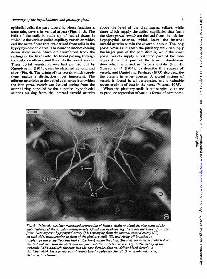
Fig. 6 Injected, partially macerated preparation ofhuman pituitary gland showing some of themain features of the vascular arrangements. Gland and neighbouring structures are viewedfrom thefront. Note superior hypophysial artery (SH) springing from the internal carotid artery (IC)on each side, anastomosing in front of the pituitary stalk (S), and giving off branches tosupply a primary capillary bed (not visible here) within the stalk. The long portal vessels which drainthis bed and run down the stalk into the pars distalis are better seen in Fig. 7. The artery of thetrabecula (AT), although plunging into the pars distalis, does not deliver blood directly tothis lobe, which has a purely portal venous blood supply (see Fig. 4.) 0 = ophthalmic artery.OC = optic chiasma.
5
copyright. on January 15, 2020 by guest. P
rotected byhttp://jcp.bm
j.com/
J Clin P
athol: first published as 10.1136/jcp.s1-7.1.1 on 1 January 1976. Dow
nloaded from

P. M. Daniel
or halt the progress of diabetic retinopathy, all theblood coming down the stalk in the long portalvessels is cut off and a large infarct is formed in thepars distalis involving up to 90% of the lobe. Theafferent blood supply to the short portal vessels,however, is spared so that they continue to transmitblood and the small part of the pars distalis whichthey supply remains viable (Daniel and Prichard,1975).
In one interesting case of head injury, reported byDaniel et al. (1959), the pituitary stalk had been tornacross above the level of the arterial ring derivedfrom the superior hypophysial arteries (Fig. 4). Thusthe afferent arterial supply to the coiled capillariesin the stalk, which gives origin to the long portalvessels, was preserved and an infarct had not de-veloped in the pars distalis.Although a small part of the pars distalis survives
after transsection of the pituitary stalk the surviving
cells do not secrete any appreciable quantity ofhormones, since the nerve fibres bringing neuro-hormones down the stalk have been severed and thecells are thereby effectively 'denervated'-that is,the blood supplying them does not contain releasingor inhibiting factors (Daniel and Prichard, 1975).It should be noted that there is no appreciable directarterial supply to the pars distalis.Advances in knowledge of the pathological
changes in the human hypothalamus and pituitarycan be made only by experts in the field. Danieland Prichard (1975) indicate the difficult and timeconsuming nature of the necessary investigations.
Much of the work on which this paper is based wasdone with the assistance of grants from the NuffieldFoundation and from the Research Fund of theBethlem Royal and Maudsley Hospitals.
Fig. 7 Injected, partially maceratedpreparation ofhuman pituitary glandseen from the front showing someof the main features of the vasculararrangements. The long portalvessels (LPV) run down the stalk(S) and break up into the sinusoids
a :X (Si) ofpars distalis. ATartery of the trabecilla.
6
copyright. on January 15, 2020 by guest. P
rotected byhttp://jcp.bm
j.com/
J Clin P
athol: first published as 10.1136/jcp.s1-7.1.1 on 1 January 1976. Dow
nloaded from
Anatomy of the hypothalamus and pituitary gland 7
References
Clark, W. E. le Gros (1938). Morphological aspects of thehypothalamus. In The Hypothalamus. Morphological,Functional, Clinical and Surgical Aspects. By W. E. leGros Clark, J. Beattie, G. Riddoch, and N. M. Dott, p.1. Oliver and Boyd, Edinburgh.
Clark, W. E. le Gros (1948). The connexions of the frontallobes of the brain. Lancet, 1, 353.
Conklin, J. L. (1966). The identification of acidophilic cellsin the human pars distalis. Anatomical Record, 156, 347.
Conklin, J. L. (1968). A histochemical study of mucoid cellsin the pars distalis of the human hypophysis. AnatomicalRecord, 160, 59.
Daniel, P. M. (1963). The pituitary gland and its blood supply.In Scientific Basis of Medicine Annual Reviews 1963,83.
Daniel, P. M. (1966a). The blood supply of the hypothalamusand pituitary gland. British Medical Bulletin, 22, 202.
Daniel, P. M. (1966b). The anatomy of the hypothalamus andpituitary gland. In Neuroendocrinology. Ed. L. Martiniand W. F. Ganong, vol. 1, p. 15. Academic Press, NewYork.
Daniel, P. M., and Prichard, M. M. L. (1975). Studies of theHvpothalamus and the Pituitary Gland. Alden Press, Oxford.(also Acta Endocrinologica, 80, Supplement 201).
Daniel, P. M., Prichard, M. M. L., and Treip, C. S. (1959).Traumatic infarction of the anterior lobe of the pituitarygland. Lancet, 2, 927.
Daniel, P. M., and Treip, C. S. (1977). The pathology of thehypothalamus. Clinics in Endocrinology and Metabolism,6, 3.
Doniach, I. (1977). Histopathology of the anterior pituitary.Clinics in Endocrinology and Metabolism, 6, 21.
Duvernoy, H. M. (1975). The Superficial Veins ofthe HumanBrain. Springer, Berlin and New York.
Halasz, B., Koves, K., R6thely, M., Bodoky, M., andKoritsanszky, S. (1975). Recent data on neuronalconnections between nervous structures involved in thecontrol of the adenohypophysis. In Anatomical Neuro-endocrinology. Ed. W. E. Stumpf and L. D. Grant.Karger, Basle, London, New York.
Hope, D. B. (1975). The neurophysin proteins: historicalaspects. Annals of the New York Academy of Sciences,248, 6.
Kumar, K., and Kumar, T. C. A. (1975). The habenularependyma: a neuroendocrine component of the epi-thalamus in the rhesus monkey. In Anatomical Neuro-endocrinology. Ed. W. E. Stumpfand L. D. Grant. Karger,Basle, London, New York.
Knowles, F., and Vollrath, L. (1974). Neurosecretion-theFinal Neuroendocrine Pathway. Springer, Berlin, Heidelberg,New York.
Leveque, T. F. (1974). The endocrine hypothalamus: anhistorical review. Canadian Journal of NeurologicalSciences, 1, 24.
Okon, E., and Koch, Y. (1976). Localisation of gonado-tropin-releasing and thyrotropin-releasing hormones inhuman brain by radioimmunoassay. Nature, 263, 345.
Raisman, G. (1966). Neural connexions of the hypothalamus.British Medical Bulletin, 22, 197.
Rodriguez, E. M. (1976). The cerebrospinal fluid as a path-way in neuroendocrine integration. Journal of Endo-crinology, 71, 407.
Saper, C. B., Loewy, A. D., Swanson, L. W., and Cowan,W. M. (1976). Direct hypothalamo-autonomic connections.Brain Research, 117, 305.
Sheehan, H. L. (1937). Post-partum necrosis of the anteriorpituitary. Journal of Pathology and Bacteriology, 45,189.
Sheehan, H. L., and Summers, V. K. (1949). The syndrome ofhypopituitarism. Quarterly Journal of Medicine, 18,319.
Stumpf, W. E. (1975). The brain: an endocrine gland andhormone target. In Anatomical Neuroendocrinology, Ed.W. E. Stumpf and L. D. Grant. Karger, Basle, London,New York.
Stutinsky, F. (1974). Morphological and physiologicalreactions of the supraoptic and paraventricular nuclei.In Neurosecretion-the Final Neuroendocrine Pathway.Ed. F. Knowles and L. Vollrath. Springer, Berlin andNew York.
Szenthgothai, J., Flerk6, B., Mess, B., and Halasz, B. (1968).Hypothalamic Control of the Anterior Pituitary. AcademiaiKiad6, Budapest.
Vitums, A. (1975). Observations on the equine hypophysialportal system. Anatomy, Histology, and Embryology, 4,149.
Watkins, W. B. (1975). Neurosecretory neurons in the hypo-thalamus and median eminence of the dog and sheep asrevealed by immunohistochemical methods. Annals ofthe New York Academy of Sciences, 248, 134.
Xuereb, G. P., Prichard, M. M. L., and Daniel, P. M.(1954a). The arterial supply and venous drainage of thehuman hypophysis cerebri. Quarterly Journal of Experi-mental Physiology, 39, 199.
Xuereb, G. P., Prichard, M. M. L., and Daniel, P. M.(1954b). The hypophysial portal system of vessels in man.Quarterly Journal of Experimental Physiology, 39, 219.
Zimmerman, E. A., Defendini, R., Sokol, H. W., andRobinson, A. G. (1975). The distribution of neurophysin-secreting pathways in the mammalian brain: light micro-scopic studies using the immunoperoxidase technique.Annals of the New York Academy of Sciences, 248, 92.
copyright. on January 15, 2020 by guest. P
rotected byhttp://jcp.bm
j.com/
J Clin P
athol: first published as 10.1136/jcp.s1-7.1.1 on 1 January 1976. Dow
nloaded from
Related Documents