Anatomy of the Ear Anatomy of the Ear Stefan Sivkov Stefan Sivkov Dept. of Anatomy, Histology and Dept. of Anatomy, Histology and Embryology Embryology

Anatomy of the Ear Stefan Sivkov Dept. of Anatomy, Histology and Embryology.
Dec 28, 2015
Welcome message from author
This document is posted to help you gain knowledge. Please leave a comment to let me know what you think about it! Share it to your friends and learn new things together.
Transcript
Anatomy of the EarAnatomy of the Ear
Stefan SivkovStefan Sivkov
Dept. of Anatomy, Histology and Dept. of Anatomy, Histology and EmbryologyEmbryology
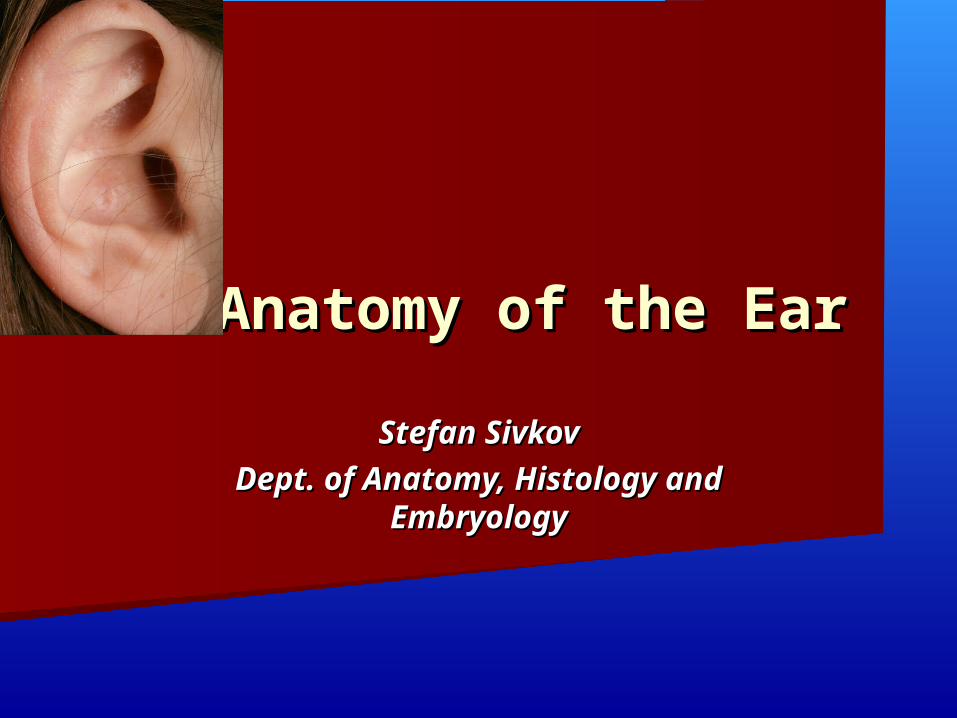
Major Divisions of the EarMajor Divisions of the Ear
Peripheral Mechanism Central Mechanism
Outer Outer EarEar
Middle Middle EarEar
Inner Inner EarEar
VIII VIII CraniaCrania
l l NerveNerve
BrainBrain

External ear, Auris externaAuriculaMeatus acusticus externus
Middle ear, Auris mediaCavitas tympaniMembrana tympanicaOssicula auditusTuba auditiva
Inner ear, Auris internaLabyrinthus membranaceus - Labyrinthus vestibularis
- Labyrinthus cochlearis
Labyrinthus osseus
- Vestibulum
- Canales semicirculares ossei
- Cochlea
- Meatus acusticus internus

Outer EarOuter Ear
Pinna
External Auditory Meatus
Pinna
Preauricular Tags
Preauricular Pits
EAM
Cerumen
Function
EAM resonance
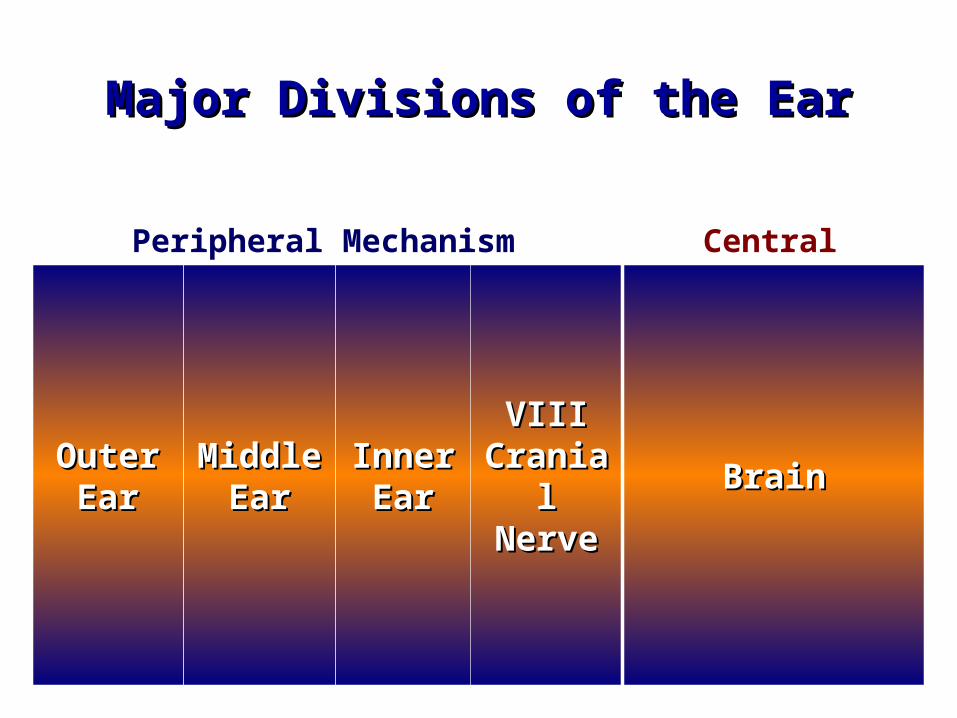
Function of Outer EarFunction of Outer Ear
Collects soundCollects sound LocalizationLocalization ResonatorResonator ProtectionProtection Sensitive Sensitive
(earlobe)(earlobe)
PinnaPinna
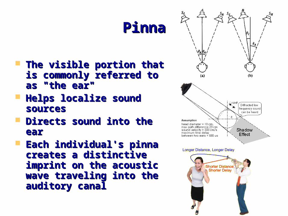
The visible portion that is The visible portion that is commonly referred to as commonly referred to as "the ear" "the ear"
Helps localize sound Helps localize sound sourcessources
Directs sound into the Directs sound into the earear
Each individual's pinna Each individual's pinna creates a distinctive creates a distinctive imprint on the acoustic imprint on the acoustic wave traveling into the wave traveling into the auditory canal auditory canal
External Auditory External Auditory MeatusMeatus
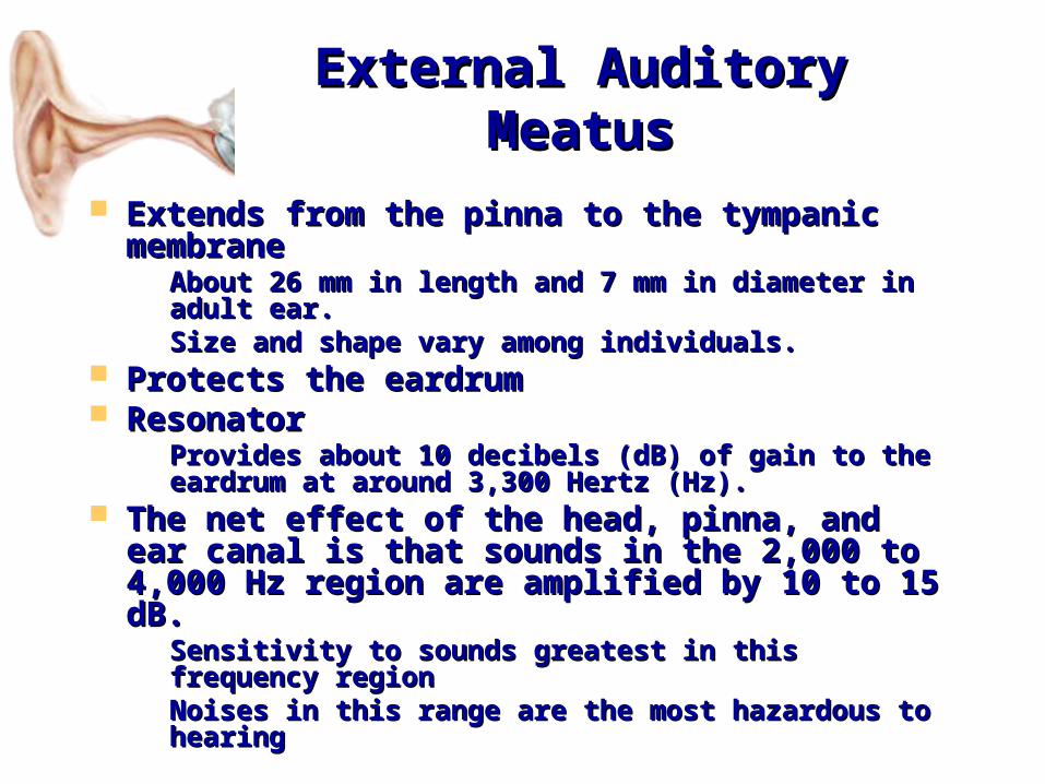
Extends from the pinna to the tympanic Extends from the pinna to the tympanic membranemembrane– About 26 mm in length and 7 mm in diameter About 26 mm in length and 7 mm in diameter
in adult ear. in adult ear. – Size and shape vary among individuals. Size and shape vary among individuals.
Protects the eardrumProtects the eardrum ResonatorResonator
– Provides about 10 decibels (dB) of gain to the Provides about 10 decibels (dB) of gain to the eardrum at around 3,300 Hertz (Hz). eardrum at around 3,300 Hertz (Hz).
The net effect of the head, pinna, and ear The net effect of the head, pinna, and ear canal is that sounds in the 2,000 to 4,000 canal is that sounds in the 2,000 to 4,000 Hz region are amplified by 10 to 15 dB. Hz region are amplified by 10 to 15 dB. – Sensitivity to sounds greatest in this frequency Sensitivity to sounds greatest in this frequency
regionregion– Noises in this range are the most hazardous to Noises in this range are the most hazardous to
hearinghearing

Outer earOuter ear
Tissues: Tissues: elastic cartilage covered with skinelastic cartilage covered with skin
A.A. Meatus acusticus externusMeatus acusticus externus besides the hair besides the hair follicles and fat glands contains:follicles and fat glands contains:
GlandulaeGlandulae ceruminosaeceruminosae – – modified sweat modified sweat glands on the lateral wall of the canalglands on the lateral wall of the canal..
ССerumerumееnn (ear wax) combination of wax and (ear wax) combination of wax and fat glands secret and desquamated epithelial fat glands secret and desquamated epithelial cellscells..

Middle EarMiddle Ear
Tympanic CavityTympanic Membrane
OssiclesMiddle Ear Muscles
Eustachian TubeMastoid
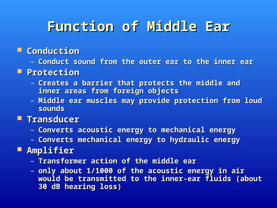
Function of Middle EarFunction of Middle Ear
ConductionConduction– Conduct sound from the outer ear to the inner earConduct sound from the outer ear to the inner ear
ProtectionProtection– Creates a barrier that protects the middle and inner areas Creates a barrier that protects the middle and inner areas
from foreign objectsfrom foreign objects– Middle ear muscles may provide protection from loud Middle ear muscles may provide protection from loud
soundssounds TransducerTransducer
– Converts acoustic energy to mechanical energyConverts acoustic energy to mechanical energy– Converts mechanical energy to hydraulic energyConverts mechanical energy to hydraulic energy
AmplifierAmplifier– Transformer action of the middle earTransformer action of the middle ear– only about 1/1000 of the acoustic energy in air would be only about 1/1000 of the acoustic energy in air would be
transmitted to the inner-ear fluids (about 30 dB hearing loss) transmitted to the inner-ear fluids (about 30 dB hearing loss)
Tympanic cavity
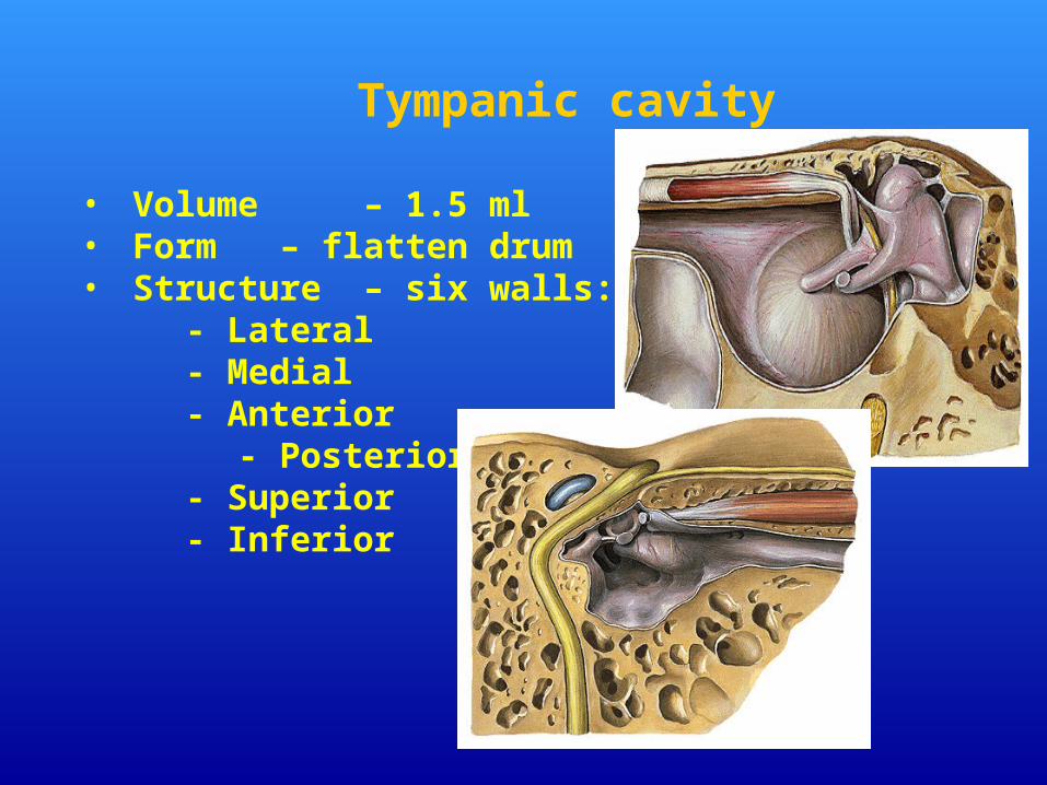
• Volume – 1.5 ml • Form – flatten drum• Structure – six walls:
- Lateral- Medial- Anterior
- Posterior- Superior - Inferior
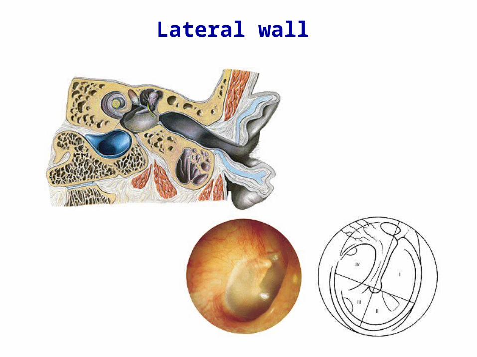
Lateral wall
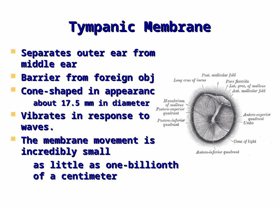
Tympanic MembraneTympanic Membrane
Separates outer ear from middle earSeparates outer ear from middle ear Barrier from foreign objectsBarrier from foreign objects Cone-shaped in appearanceCone-shaped in appearance
– about 17.5 mm in diameterabout 17.5 mm in diameter
Vibrates in response to sound Vibrates in response to sound waves. waves.
The membrane movement is The membrane movement is incredibly smallincredibly small– as little as one-billionth of a as little as one-billionth of a
centimetercentimeter
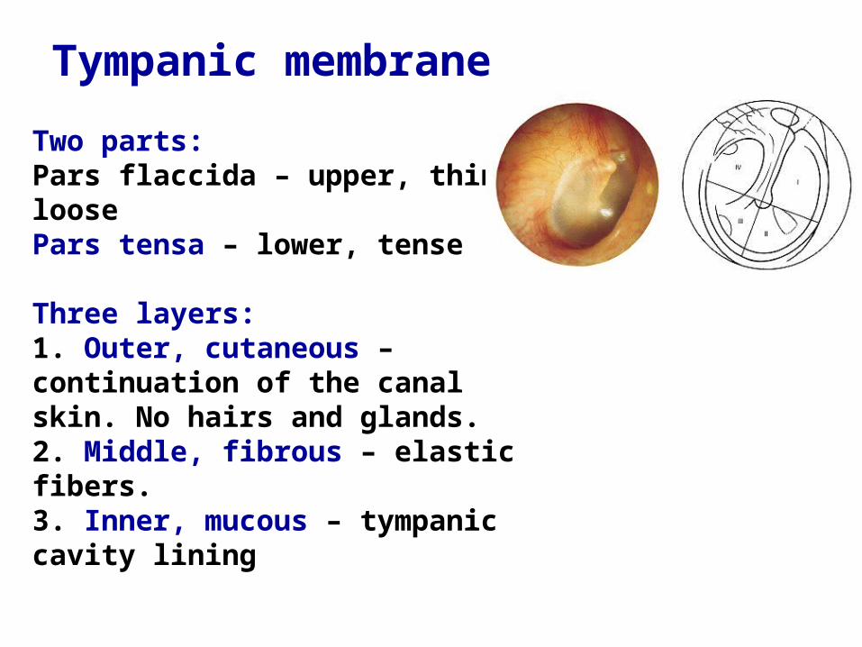
Two parts:Pars flaccida – upper, thin, loosePars tensa – lower, tense
Three layers:1. Outer, cutaneous – continuation of the canal skin. No hairs and glands.2. Middle, fibrous – elastic fibers.3. Inner, mucous – tympanic cavity lining
Tympanic membrane

Most complex.On this wall are distinguished:
-fenestra vestibuli-fenestra cochleae-promontorium -prominentia canalis semicircularis lateralis- prominentia canalis facialis
Superior wall, paries tegmentalisSeparates tympanic from cranial cavity.
Children less than 2 years – infections of the middle ear can pass to the cranial cavity.
Medial wall, paries labyrinthicus

Inferior wall, paries jugularisSeparates tympanic cavity from fossa jugularis
Anterior wall, paries caroticusSeparates tympanic cavity from
canalis caroticus-canalis musculotubularis
Posterior wall, paries mastoideusComposed of:
•Styloid complex of Procter•Antrum mastoideum•Fossa incudis
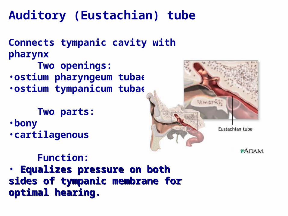
Auditory (Eustachian) tube
Connects tympanic cavity with pharynx
Two openings:•ostium pharyngeum tubae•ostium tympanicum tubae.
Two parts:•bony•cartilagenous
Function: • Equalizes pressure on both sides of Equalizes pressure on both sides of tympanic membrane for optimal hearing.tympanic membrane for optimal hearing.
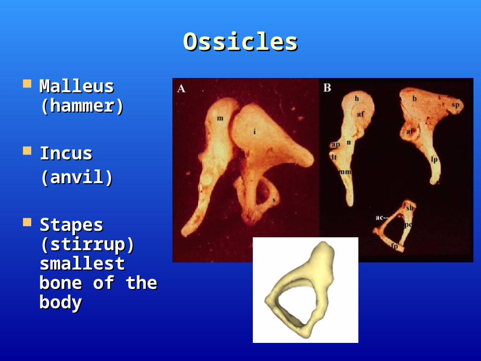
OssiclesOssicles
Malleus Malleus (hammer)(hammer)
Incus Incus (anvil)(anvil)
Stapes Stapes (stirrup) (stirrup) smallest bone smallest bone of the bodyof the body
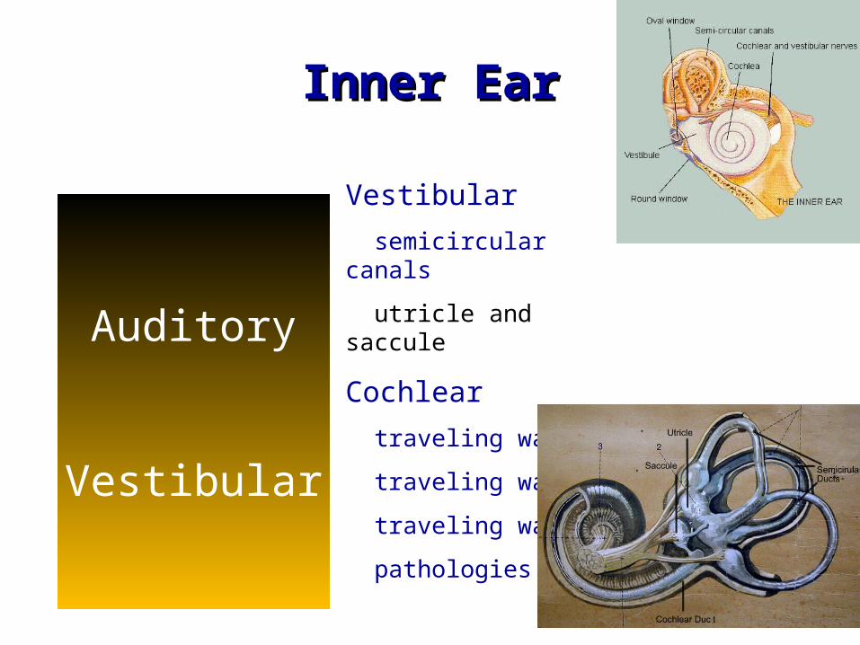
Inner EarInner Ear
Auditory
Vestibular
Vestibular
semicircular canals
utricle and saccule
Cochlear
traveling wave
traveling wave
traveling wave
pathologies
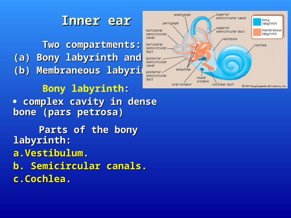
Inner earInner ear
Two compartments: Two compartments: ((аа) Bony labyrinth and) Bony labyrinth and (b) Membraneous labyrinth.(b) Membraneous labyrinth.
Bony labyrinth:: complex cavity in dense bonecomplex cavity in dense bone ((pars petrosapars petrosa))
Parts of the bony labyrinth:Parts of the bony labyrinth:a.Vestibuluma.Vestibulum..b. Semicircular canals.b. Semicircular canals. c.Cochleac.Cochlea..
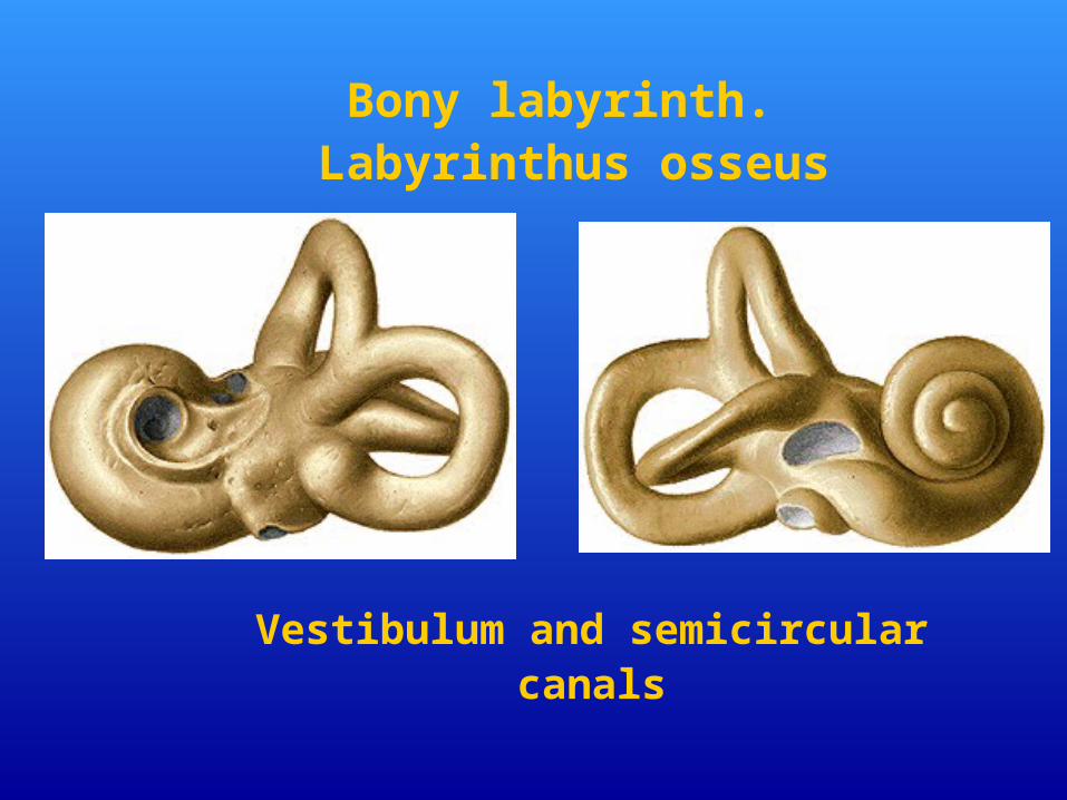
Bony labyrinth. Labyrinthus osseus
Vestibulum and semicircular canals

Vestibulum
Two walls: External and internal.
External wall has • Fenestra vestibuli.
Internal wall has:•Recessus ellipticus•Recessus sphericus•Recessus cochlearis•Maculae cribrosae superior, medius, inferior

Openings into vestibulumOpenings into vestibulum
aa. . Fenestra vestibuli.Fenestra vestibuli.
b.b. Fenestra cochleae. Fenestra cochleae.
c.c. Openings (5) of the semicircular canals Openings (5) of the semicircular canals
d.d. Aqueductus vestibuli Aqueductus vestibuli

3: anterior, posterior and lateral.
Have ampulla and crus.
Canalis semicircularis lateralis –horizontal.
- eminentia canalis semicircularis lateralis on the medial wall of
tympanic cavity.
Canalis semicircularis anterior –frontal.
- eminentia arcuata on pars petrosa of os temporale.
Canalis semicircularis posterior –sagittal
Semicircular canals
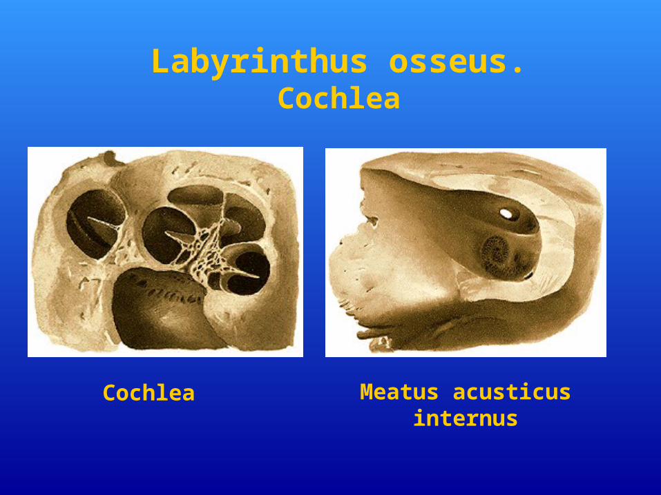
Cochlea Meatus acusticus internus
Labyrinthus osseus. Cochlea
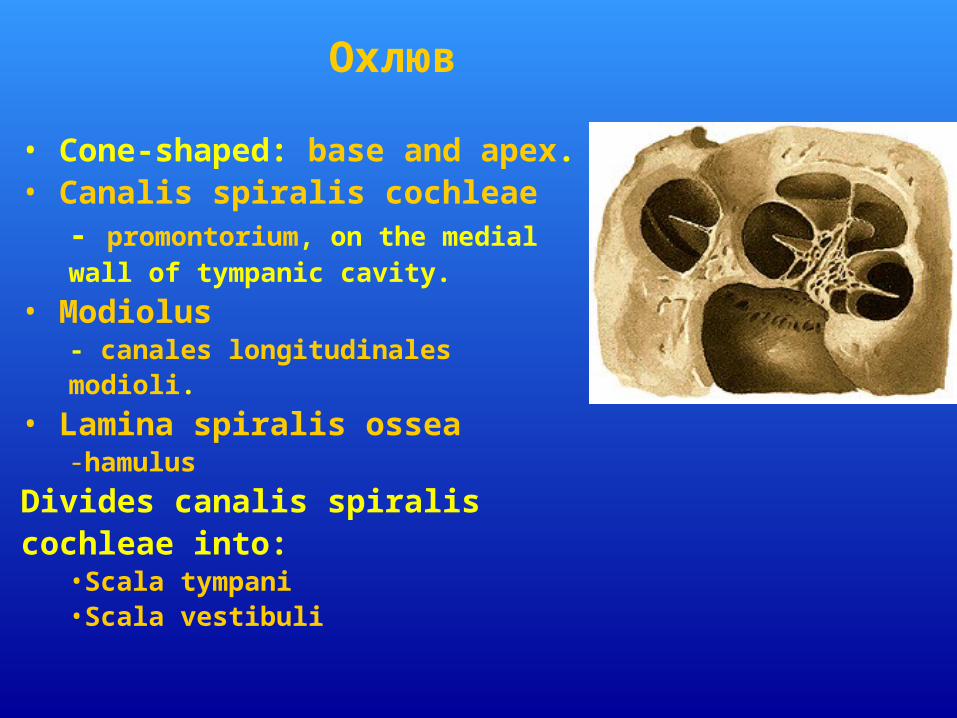
• Cone-shaped: base and apex.• Canalis spiralis cochleae
- promontorium, on the medial wall of tympanic cavity.
• Modiolus - canales longitudinales modioli.
• Lamina spiralis ossea -hamulus
Divides canalis spiralis cochleae into:
•Scala tympani•Scala vestibuli
Охлюв
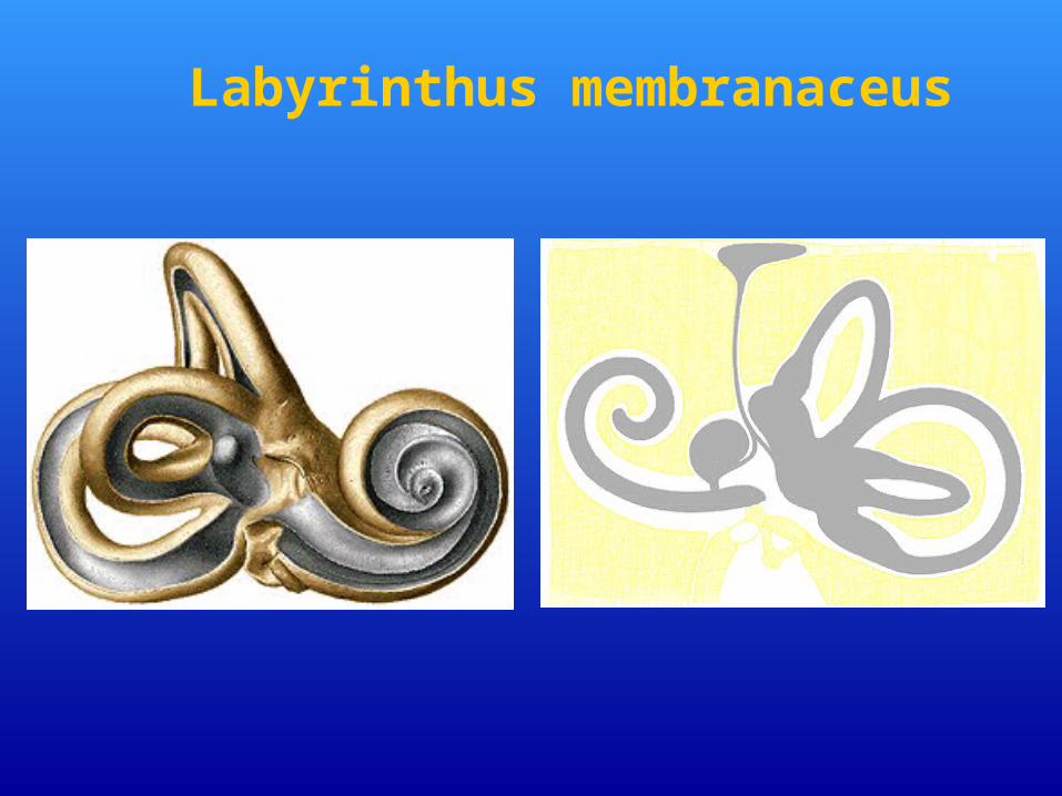
Labyrinthus membranaceus
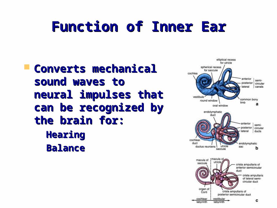
Function of Inner EarFunction of Inner Ear
Converts mechanical Converts mechanical sound waves to neural sound waves to neural impulses that can be impulses that can be recognized by the brain recognized by the brain for: for: – HearingHearing– BalanceBalance
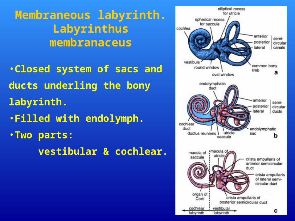
•Closed system of sacs and ducts
underling the bony labyrinth.
•Filled with endolymph.
•Two parts:
vestibular & cochlear.
Membraneous labyrinth.Labyrinthus
membranaceus
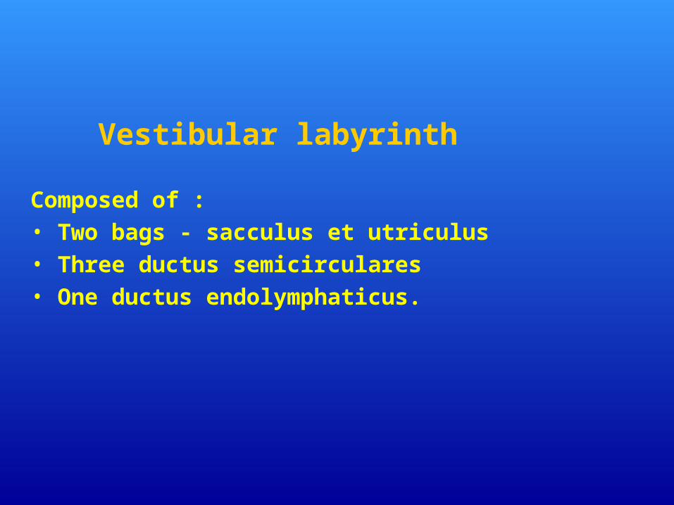
Vestibular labyrinth Composed of : • Two bags - sacculus et utriculus• Three ductus semicirculares• One ductus endolymphaticus.
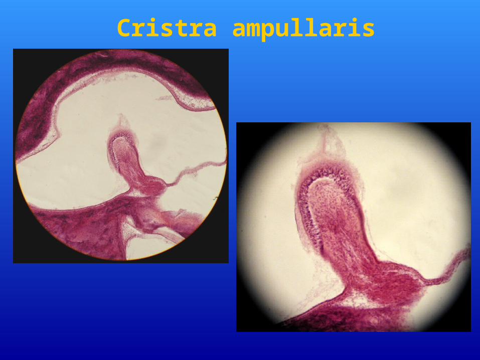
Cristra ampullaris

BalanceBalance
Linear motionLinear motion Rotary motionRotary motion
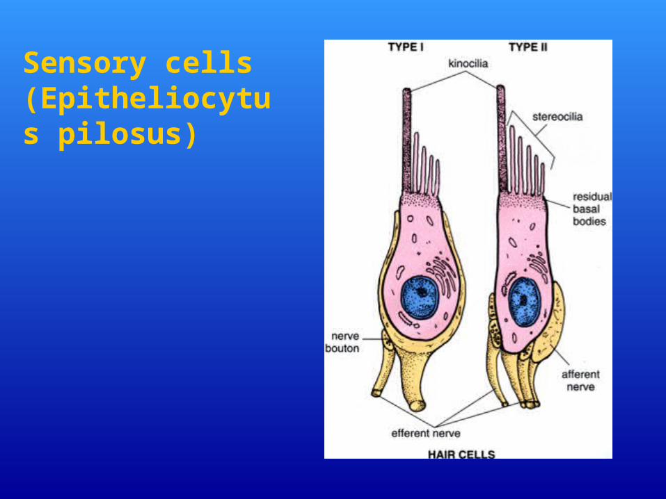
Sensory cells (Epitheliocytus pilosus)
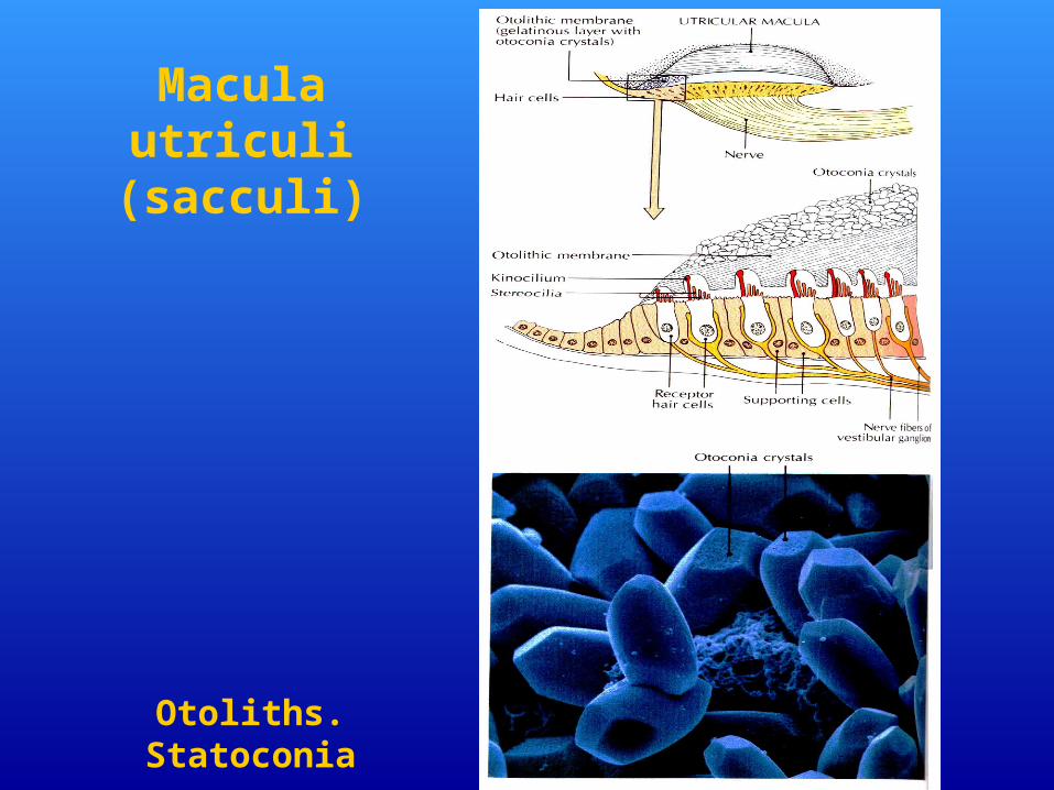
Macula utriculi (sacculi)
Otoliths. Statoconia
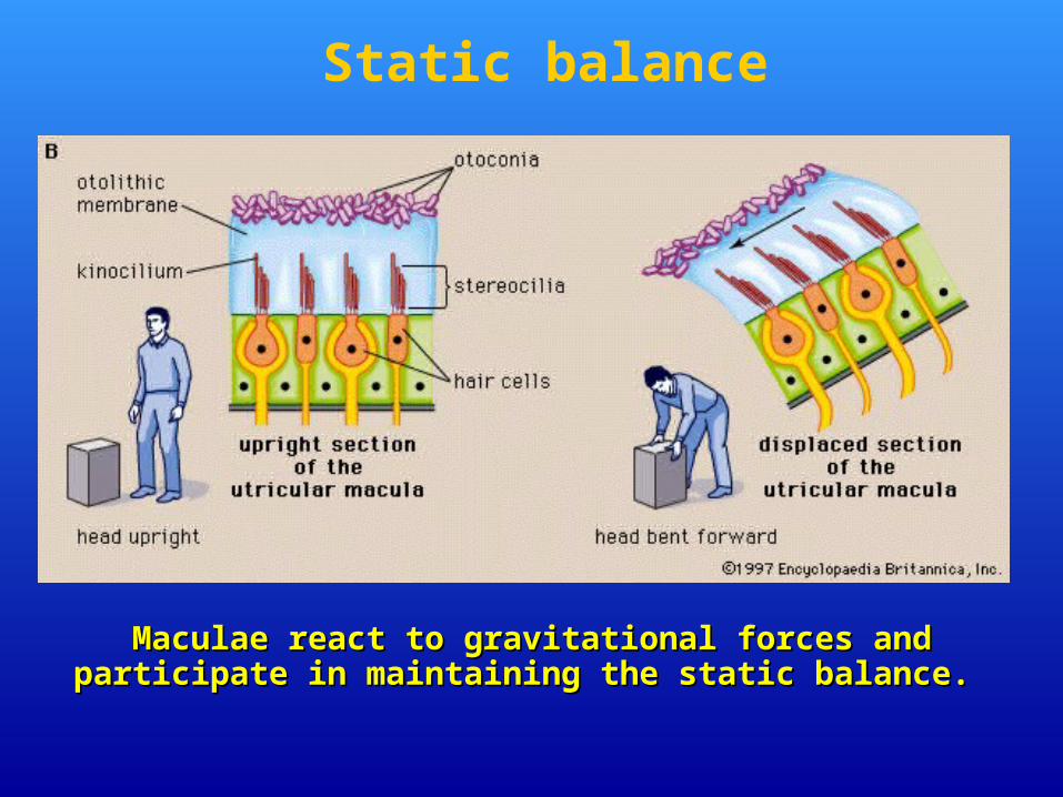
MMaculae aculae react to gravitational forcesreact to gravitational forces and and participate in maintaining the static balanceparticipate in maintaining the static balance. .
Static balance
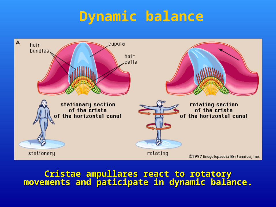
CCristae ristae ampullares react to rotatory movementsampullares react to rotatory movements and paticipate in dynamic balanceand paticipate in dynamic balance..
Dynamic balance
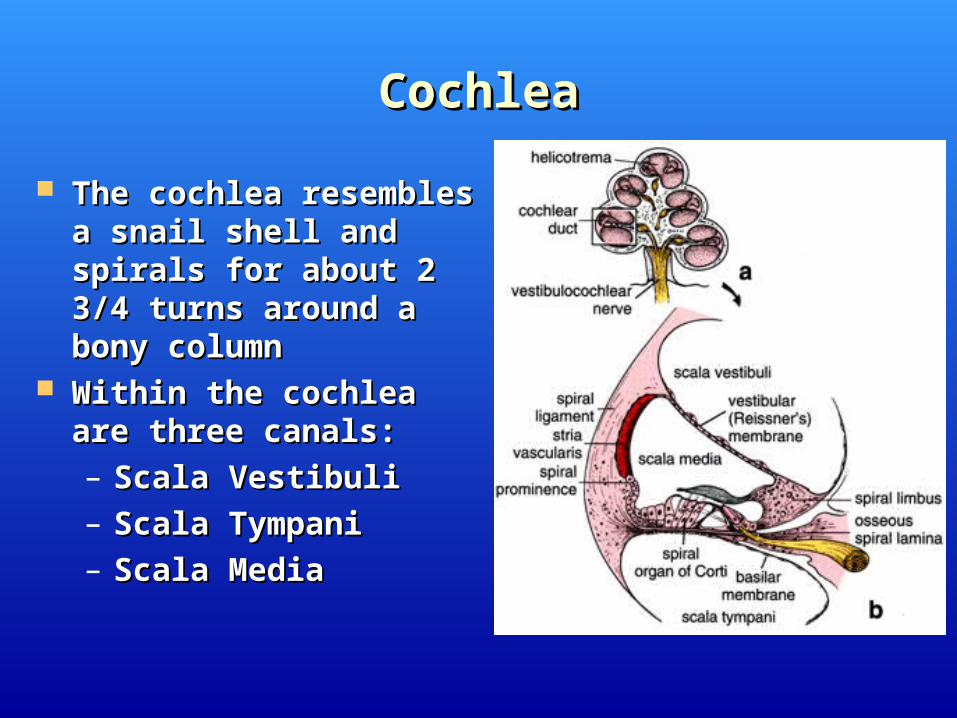
CochleaCochlea
The cochlea resembles a The cochlea resembles a snail shell and spirals for snail shell and spirals for about 2 3/4 turns around a about 2 3/4 turns around a bony columnbony column
Within the cochlea are Within the cochlea are three canals:three canals:– Scala Vestibuli Scala Vestibuli – Scala TympaniScala Tympani– Scala MediaScala Media
Spiral canal - ductus cochlearis.
Occupies scala media of the spiral canal.
Has two blind ends - cecum vestibulare and cecum
cupulare.
Has three walls:
• paries vestibularis
• paries externus
• paries tympanicus- organ of Corti, basal membrane
Cochlear labyrinth
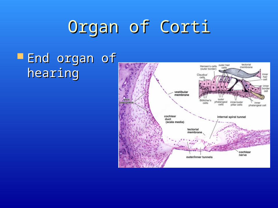
Organ of CortiOrgan of Corti
End organ of End organ of hearinghearing
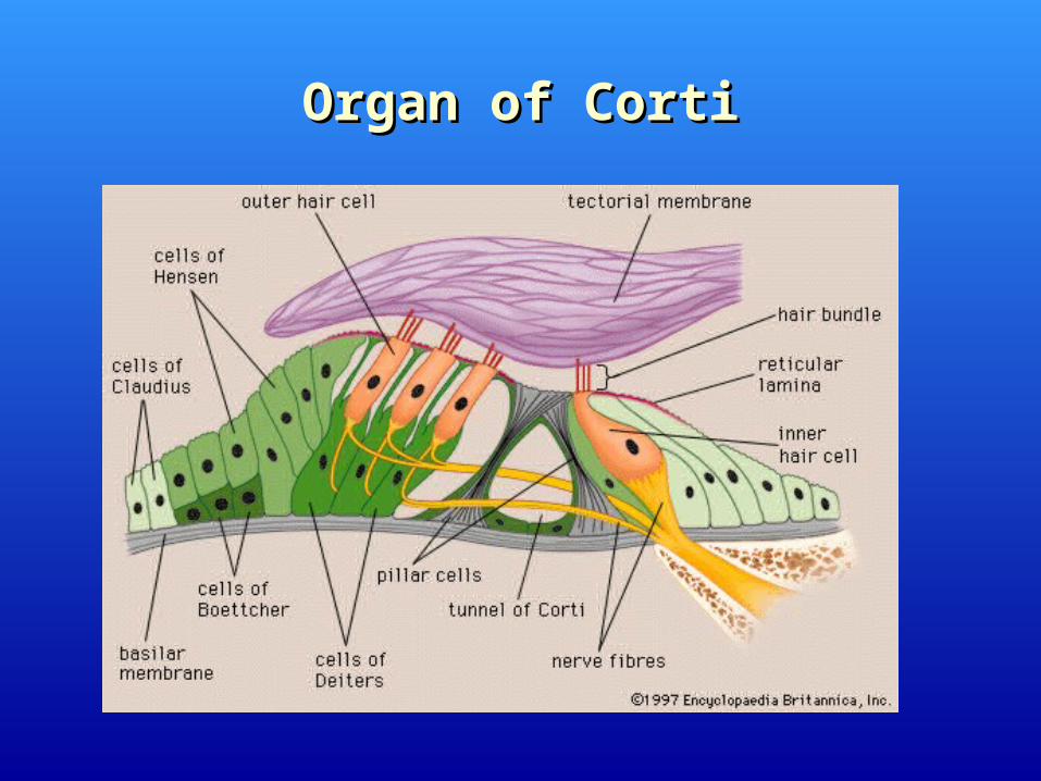
Organ of CortiOrgan of Corti
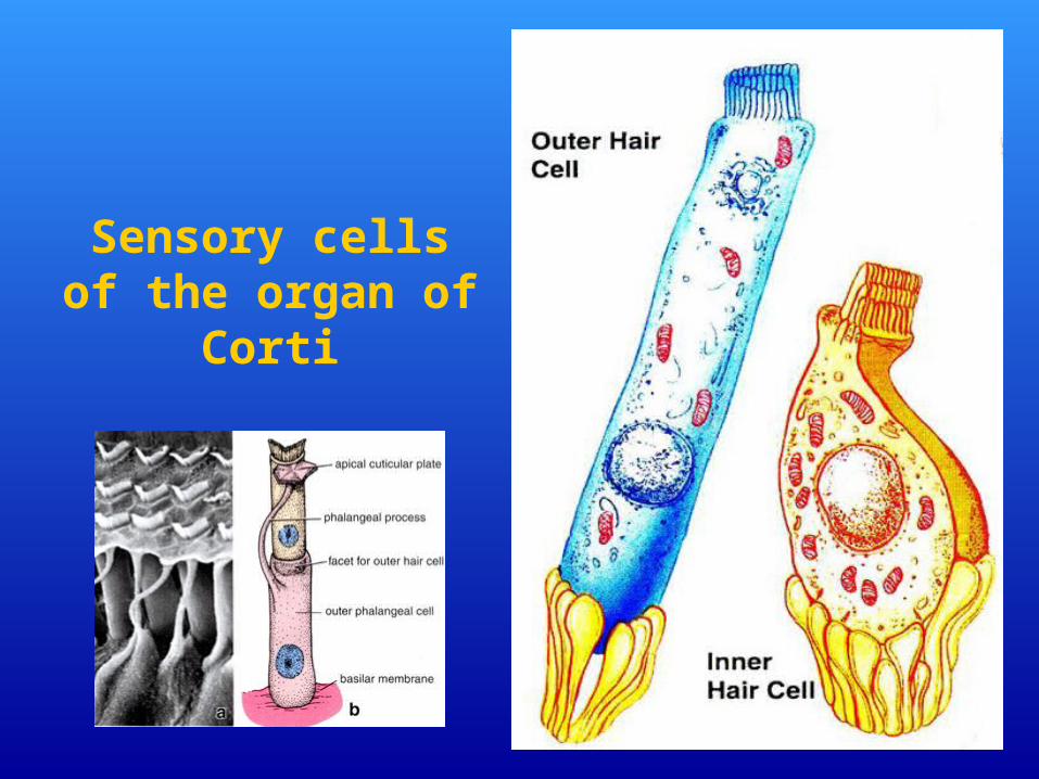
Sensory cells of the organ of Corti
Related Documents