Anatomy Of The Cardiovascular System Lecture 3 Abbas A. A. Shawka Medical student 1 st stage

Welcome message from author
This document is posted to help you gain knowledge. Please leave a comment to let me know what you think about it! Share it to your friends and learn new things together.
Transcript

Anatomy Of The Cardiovascular System
Lecture 3
Abbas A. A. ShawkaMedical student
1st stage
Subjects
• Heart vasculature
• Heart conducting system
Coronary vasculature• Two coronary arteries arise from the aortic sinuses in
the initial portion of the ascending aorta and supplythe muscle and other tissues of the heart.
• They circle the heart in the coronary sulcus, withmarginal and interventricular branches, in theinterventricular sulci, converging toward the apex ofthe heart.
• The returning venous blood passes through cardiacveins, most of which empty into the coronary sinus.This large venous structure is located in the coronarysulcus on the posterior surface of the heart betweenthe left atrium and left ventricle. The coronary sinusempties into the right atrium between the opening ofthe inferior vena cava and the right atrioventricularorifice.


Coronary arteries1- Right coronary artery
• Early atrial branch
• Sinoatrial nodal branch
• Right marginal branch
• Posterior interventricular branch
2- Left coronary artery• Anterior interventricular branch ( left anterior descending
artery – LAD )
• Circumflex branch
• Left marginal branch

Right coronary artery.• The right coronary artery
originates from the right aortic sinus of the ascending aorta.
• It passes anteriorly and then descends vertically in the coronary sulcus, between the right atrium and right ventricle
• On reaching the inferior margin of the heart, it turns posteriorly and continues in the sulcus onto the diaphragmatic surface and base of the heart.
• During this course, several branches arise from the main stem of the vessel:
1. An early atrial branch passes in the groove between the right auricle and ascending aorta, and gives off
2. the sinu-atrial nodal branch, which passes posteriorly around the superior vena cava to supply the sinu-atrial node.
3. A right marginal branch is given off as the right coronary artery approaches the inferior (acute) margin of the heart and continues along this border toward the apex of the heart.
4. As the right coronary artery continues on the base/ diaphragmatic surface of the heart, it supplies a small branch to the atrioventricular node
5. final major branch, the posterior interventricular branch , which lies in the posterior interventricular sulcus.
The right coronary artery supplies the right atrium and right ventricle, the sinu-atrial and atrioventricular nodes, the interatrial septum, a portion of the left atrium, the posteroinferior one third of the interventricular septum, and a portion of the posterior part of the left ventricle.

left coronary artery.• The left coronary artery
originates from the left aortic sinus of the ascending aorta.
• It passes between the pulmonary trunk and the left auricle before entering the coronary sulcus.
• Emerging from behind the pulmonary trunk, the artery divides into its two terminal branches, the anterior interventricular and the circumflex.
1. The anterior interventricular branch (left anterior descending artery-LAD) continues around the left side of the pulmonary trunk and descends obliquely toward the apex of the heart in the anterior interventricular sulcus . During its course, one or two large diagonal branches may arise and descend diagonally across the anterior surface of the left ventricle.
2. The circumflex branch courses toward the left, in the coronary sulcus and onto the base diaphragmatic surface of the heart, and usually ends before reaching the posterior interventricular sulcus. A large branch, the left marginal artery , usually arises from it and continues across the rounded obtuse margin of the heart.

The distribution pattern of the left coronary artery enables it to supply most of the left atrium and left ventricle, and most of the interventricular septum, including the atrioventricular bundle and its branches .

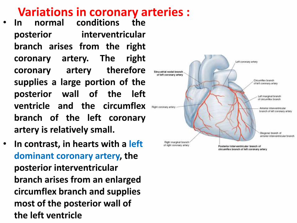
Variations in coronary arteries :• In normal conditions the
posterior interventricular branch arises from the right coronary artery. The right coronary artery therefore supplies a large portion of the posterior wall of the left ventricle and the circumflex branch of the left coronary artery is relatively small.
• In contrast, in hearts with a left dominant coronary artery, the posterior interventricular branch arises from an enlarged circumflex branch and supplies most of the posterior wall of the left ventricle

Variations in coronary arteries :• Another point o f variation
relates t o the arterial supply to the sinu-atrial and atrioventricular nodes. In most cases, these two structures are supplied by the right coronary artery. However, vessels from the circumflex branch of the left coronary artery occasionally supply these structures.

Cardiac veins :

Cardiac veins :
Cardiac veins :
• The coronary sinus receives four maj or tributaries: the great, middle, small, and posterior cardiac veins.
1. Great cardiac vein. The great cardiac vein begins at the apex of the heart. It ascends in the anterior interventricular sulcus, where it is related to the anterior interventricular artery and is often termed the anterior interventricular vein. Reaching the coronary sulcus , the great cardiac vein turns to the left and continues onto the base/diaphragmatic surface of the heart. At this point, it is associated with the circumflex branch of the left coronary artery. Continuing along its path in the coronary sulcus, the great cardiac vein gradually enlarges to form the coronary sinus, which enters the right atrium.

Cardiac veins :2. Middle cardiac vein. The middle cardiac vein (posterior
interventricular vein) begins near the apex of the heart and ascends in the posterior interventricular sulcus toward the coronary sinus. It is associated with the posterior interventricular branch of the right or left coronary artery throughout its course.
3. Small cardiac vein. The small cardiac vein begins in the lower anterior section of the coronary sulcus between the right atrium and right ventricle. It continues in this groove onto the base/diaphragmatic surface of the heart where it enters the coronary sinus at its atrial end. It is a companion of the right coronary artery throughout its course and may receive the right marginal vein. This small vein accompanies the marginal branch of the right coronary artery along the acute margin of the heart. If the right marginal vein does not join the small cardiac vein, it enters the right atrium directly.

Cardiac veins :4. Posterior cardiac vein. The posterior cardiac vein lies on the
posterior surface of the left ventricle just to the left of the middle cardiac vein. It either enters the coronary sinus directly or joins the great cardiac vein.
5. The anterior veins of the right ventricle (anterior cardiac veins) are small veins that arise on the anterior surface of the right ventricle (Fig. 3 . 7 S A) . They cross the coronary sulcus and enter the anterior wall of the right atrium. They drain the anterior portion of the right ventricle. The right marginal vein may be part of this group if it does not enter the small cardiac vein.
6. A group of smallest cardiac veins (venae cordis minimae or veins of Thebesius) have also been described. Draining directly into the cardiac chambers, they are numerous in the right atrium and right ventricle, are occasionally associated with the left atrium, and are rarely associated with the left ventricle.

Coronary lymphatics• The lymphatic vessels of the heart follow the coronary
arteries and drain mainly into :
• brachiocephalic nodes, anterior to the brachiocephalic veins.
• tracheobronchial nodes, at the inferior end of the trachea.
Conducting system• The musculature of the atria and ventricles is capable of
contracting spontaneously.
• The cardiac conduction system initiates and coordinates contraction.
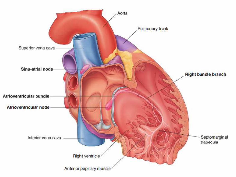
• The conduction system consists of nodes and networks of specialized cardiac muscle cells organized into four basic components:
1. the sinu-atrial node,
2. the atrioventricular node,
3. the atrioventricular bundle with its right and left bundle branches, and
4. the subendocardial plexus of conduction cells (the Purkinje fibers) .

• The unique distribution pattern of the cardiac conduction system establishes an important unidirectional pathway of excitation/contraction. Throughout its course, large branches of the conduction system are insulated from the surrounding myocardium by connective tissue. This tends to decrease inappropriate stimulation and contraction of cardiac muscle fibers.
• The number of functional contacts between the conduction pathway and cardiac musculature greatly increases in the subendocardial network. Thus, a unidirectional wave of excitation and contraction is established, which moves from the papillary muscles and apex of the ventricles to the arterial outflow tracts.

Sino-Atrial node• Impulses begin at the sinu-atrial
node , the cardiac pacemaker. • This collection of cells is located
at the superior end of the crista terminalis at the junction of the superior vena cava and the right atrium.
• This is also the junction between the parts o f the right atrium derived from the embryonic sinus venosus and the atrium proper.
• The excitation signals generated by the sinu-atrial node spread across the atria, causing the muscle to contract.

Atrioventricular node• Concurrently, the wave of
excitation in the atria stimulates the atrioventricular node , which is located near the opening of the coronary sinus, close to the attachment of the septal cusp of the tricuspid valve, and within the atrioventricular septum.
• The atrioventricular node is a collection o f specialized
• cells that forms the beginning of an elaborate system of
• conducting tissue, the atrioventricular bundle, which extends the excitatory impulse to all ventricular musculature
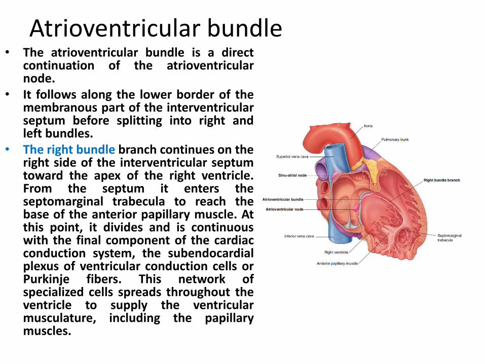
Atrioventricular bundle• The atrioventricular bundle is a direct
continuation of the atrioventricular node.
• It follows along the lower border of the membranous part of the interventricular septum before splitting into right and left bundles.
• The right bundle branch continues on the right side of the interventricular septum toward the apex of the right ventricle. From the septum it enters the septomarginal trabecula to reach the base of the anterior papillary muscle. At this point, it divides and is continuous with the final component of the cardiac conduction system, the subendocardialplexus of ventricular conduction cells or Purkinje fibers. This network of specialized cells spreads throughout the ventricle to supply the ventricular musculature, including the papillary muscles.
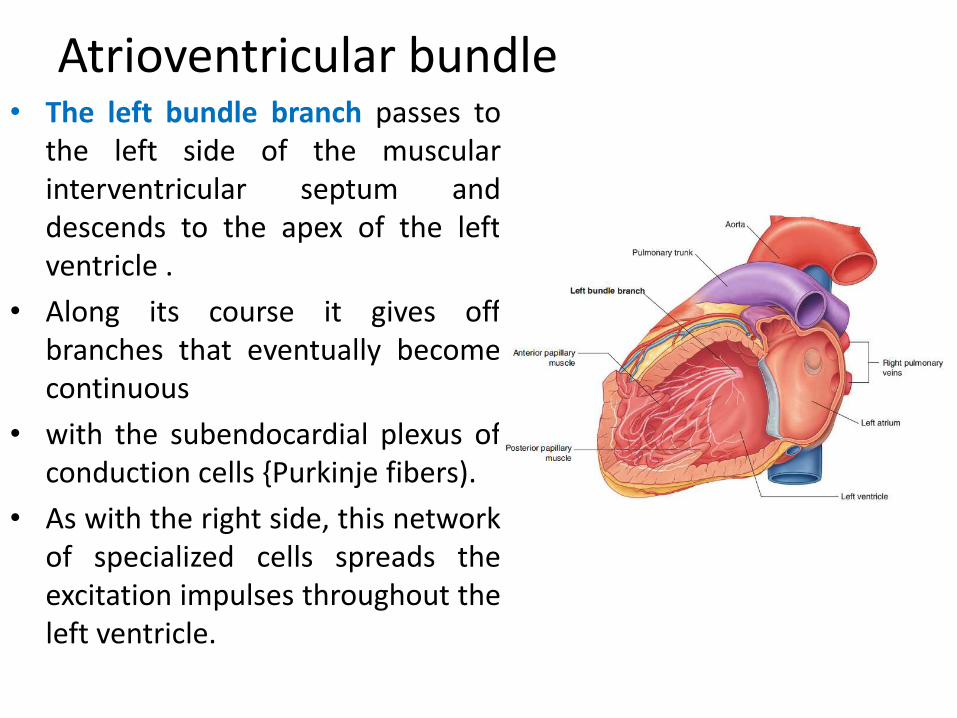
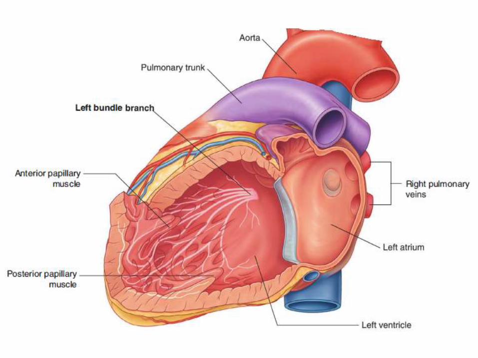
Atrioventricular bundle• The left bundle branch passes to
the left side of the muscular interventricular septum and descends to the apex of the left ventricle .
• Along its course it gives off branches that eventually become continuous
• with the subendocardial plexus of conduction cells {Purkinje fibers).
• As with the right side, this network of specialized cells spreads the excitation impulses throughout the left ventricle.
Cardiac innervation• The autonomic division of the peripheral nervous system is directly
responsible for regulating:
1. heart rate,
2. force of each contraction, and
3. cardiac output.
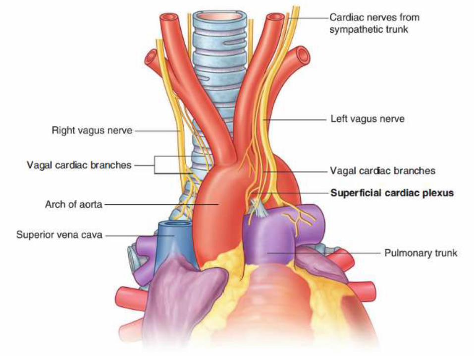
• Branches from both the parasympathetic and sympathetic systems contribute to the formation of the cardiac plexus. This plexus consists of a superficial part, inferior to the aortic arch and between it and the pulmonary trunk, and a deep part, between the aortic arch and the tracheal bifurcation.


Parasympathetic innervation• Stimulation of the parasympathetic system:
1. decreases heart rate,
2. reduces force of contraction, and
3. constricts the coronary arteries.
• The preganglionic parasympathetic fibers reach the heart as cardiac branches from the right and left vagus nerves.
• They enter the cardiac plexus and synapse in ganglia located either within the plexus or in the walls of the atria.
Sympathetic innervation• Stimulation of the sympathetic system:1. increases heart rate, and2. increases the force of contraction.• Sympathetic fibers reach the cardiac plexus
through the• cardiac nerves from the sympathetic trunk. • Preganglionic sympathetic fibers from the upper
four or five segments of the thoracic spinal cord enter and move through the sympathetic trunk.
• They synapse in cervical and upper thoracic sympathetic ganglia, and postganglionic fibers proceed as bilateral branches from the sympathetic trunk to the cardiac plexus.

Visceral afferents• Visceral afferents from the heart are also a component of the
cardiac plexus.
• These fibers pass through the cardiac plexus and return to the central nervous system in the cardiac nerves from the sympathetic trunk and in the vagal cardiac branches.
• The afferents associated with the vagal cardiac nerves return to the vagus nerve [10] .
• They sense alterations in
• blood pressure and blood chemistry and are there for primarily concerned with cardiac reflexes.
• The afferents associated with the cardiac nerves from the sympathetic trunks return to either the cervical or the thoracic portions of the sympathetic trunk.
• If they are in the cervical portion of the trunk, they normally descend to the thoracic region, where they reenter the upper four or five thoracic spinal cord segments, along with the afferents from the thoracic region of the sympathetic trunk. Visceral afferents associated with the sympathetic system conduct
• pain sensation from the heart, which is detected at the cellular level as tissue-damaging events (i.e. , cardiac ischemia). This pain is often "referred" to cutaneous regions supplied by the same spinal cord levels

Related Documents


