DR. RAJESH KUMAR ATRCTRI, BIKANER

Anatomy of oral cavity, pharynx& larynx-Dr Rajesh Kumar
Jul 16, 2015
Welcome message from author
This document is posted to help you gain knowledge. Please leave a comment to let me know what you think about it! Share it to your friends and learn new things together.
Transcript
DR. RAJESH KUMARATRCTRI, BIKANER
ORAL CAVITY
PHARYNX
LARYNX
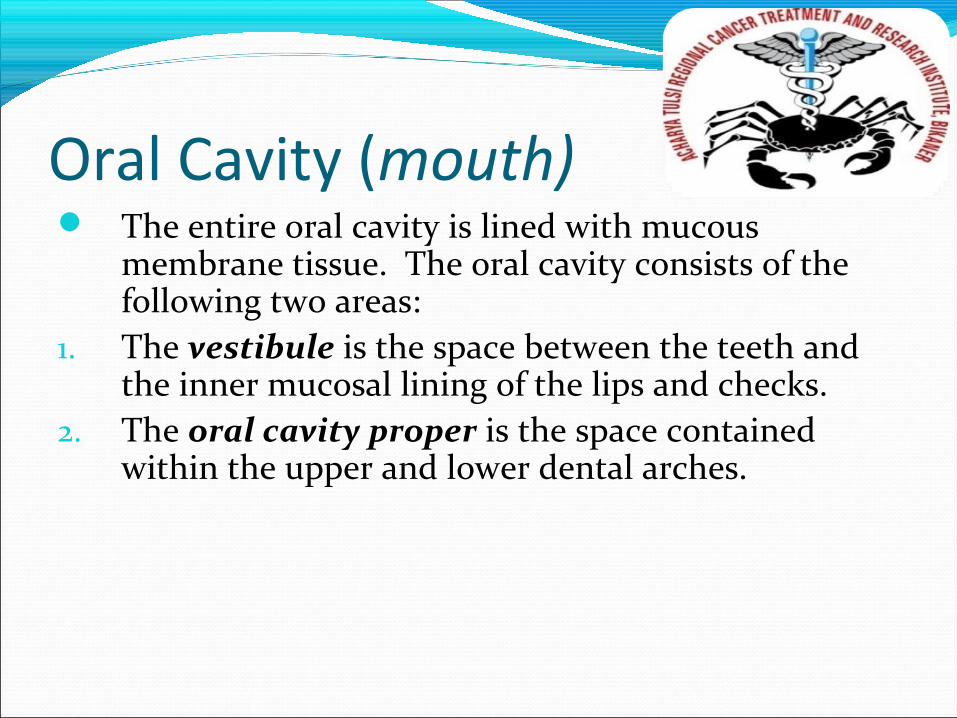
Oral Cavity (mouth) The entire oral cavity is lined with mucous
membrane tissue. The oral cavity consists of the following two areas:
1. The vestibule is the space between the teeth and the inner mucosal lining of the lips and checks.
2. The oral cavity proper is the space contained within the upper and lower dental arches.
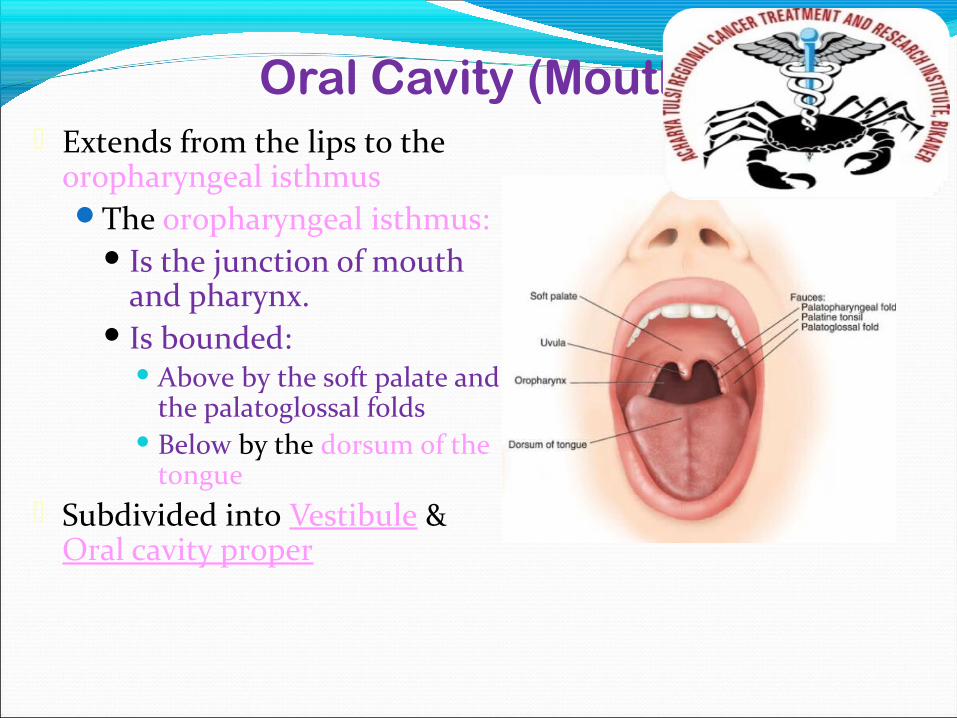
Oral Cavity (Mouth) Extends from the lips to the
oropharyngeal isthmusThe oropharyngeal isthmus:
Is the junction of mouth and pharynx.
Is bounded: Above by the soft palate and
the palatoglossal folds Below by the dorsum of the
tongue Subdivided into Vestibule &
Oral cavity proper
The DentitionsThe term dentition is used to describe the
natural teeth in the jawbones.Primary dentition is the first set of 20 primary
teeth. Also referred to as “baby teeth” or “deciduous teeth”
Permanent dentition refers to the 32 secondary or “adult” teeth.
Mixed dentition occurs when both primary and permanent teeth are present, usually between the ages of 6 to 12.
Dental archesThe maxillary arch (upper arch), actually part of the
skull, is fixed and not capable of movement. The teeth in the upper arch are set in the maxilla, the maxillary bone.
The mandibular arch (lower arch) is capable of movement through the action of the temporomandibular joint. The mandible, the mandibular bone supports the teeth in the lower arch.
Eruption & ExfoliationEruption is the movement of the tooth through the
surrounding tissues so that more of the tooth becomes visible in the mouth.
Exfoliation is the process by which the roots of the baby tooth are resorbed and dissolved until so little root remains that the baby tooth falls out.
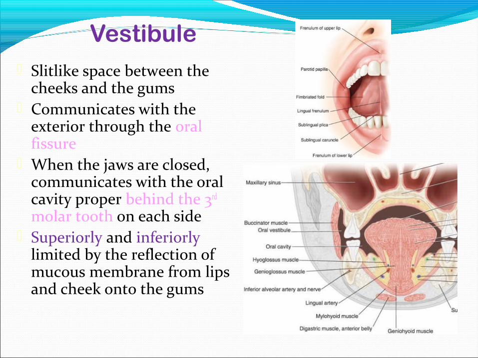
Vestibule Slitlike space between the
cheeks and the gums Communicates with the
exterior through the oral fissure
When the jaws are closed, communicates with the oral cavity proper behind the 3rd molar tooth on each side
Superiorly and inferiorly limited by the reflection of mucous membrane from lips and cheek onto the gums
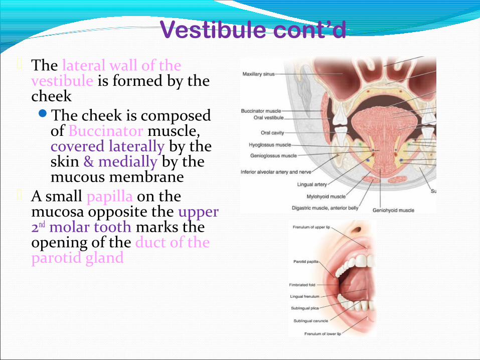
Vestibule cont’d The lateral wall of the
vestibule is formed by the cheekThe cheek is composed
of Buccinator muscle, covered laterally by the skin & medially by the mucous membrane
A small papilla on the mucosa opposite the upper 2nd molar tooth marks the opening of the duct of the parotid gland
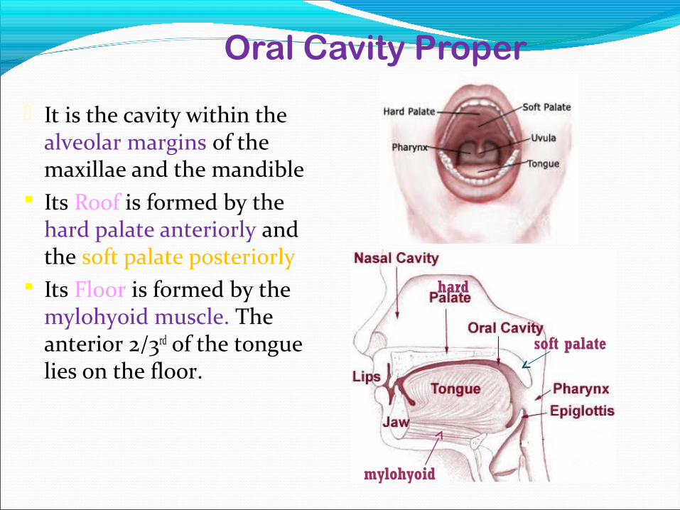
Oral Cavity Proper
It is the cavity within the alveolar margins of the maxillae and the mandible
Its Roof is formed by the hard palate anteriorly and the soft palate posteriorly
Its Floor is formed by the mylohyoid muscle. The anterior 2/3rd of the tongue lies on the floor.
hard
soft palate
mylohyoid
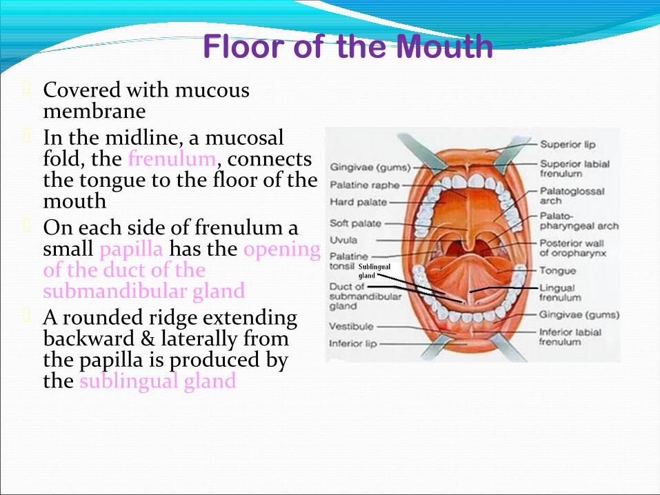
Floor of the Mouth Covered with mucous
membrane In the midline, a mucosal
fold, the frenulum, connects the tongue to the floor of the mouth
On each side of frenulum a small papilla has the opening of the duct of the submandibular gland
A rounded ridge extending backward & laterally from the papilla is produced by the sublingual gland
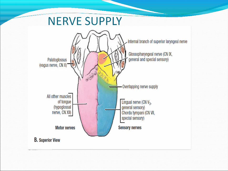
Nerve Supply
o Sensory Roof: by greater palatine and nasopalatine nerves
(branches of maxillary nerve) Floor: by lingual nerve (branch of mandibular nerve) Cheek: by buccal nerve (branch of mandibular nerve)
o Motor Muscle in the cheek (buccinator) and the lip (orbicularis
oris) are supplied by the branches of the facial nerve
13
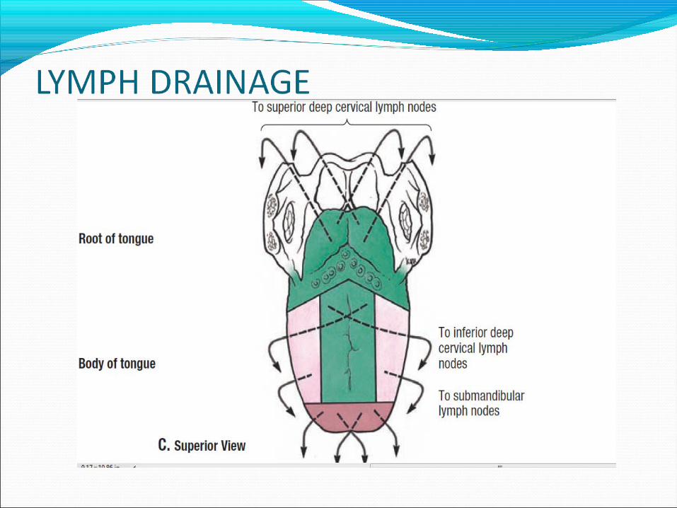
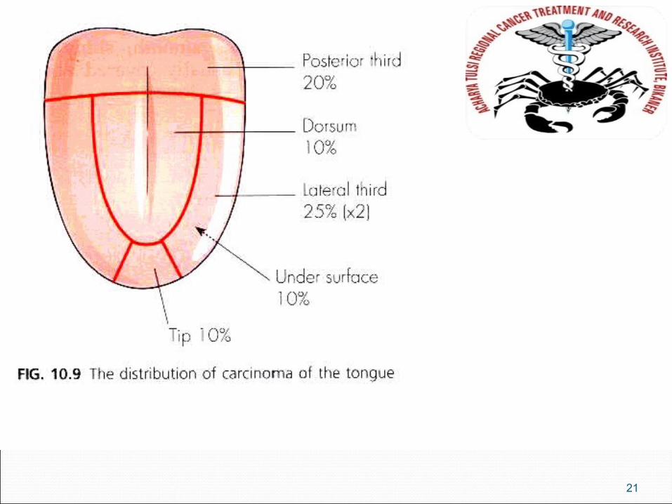
The tongue is a mobile muscular organ can assume a variety of shapes and posit ions. The tongue is partly in the oral cavity and partly in the pharynx.
At rest i t occupies essential ly al l the oral cavity proper.
TONGUE
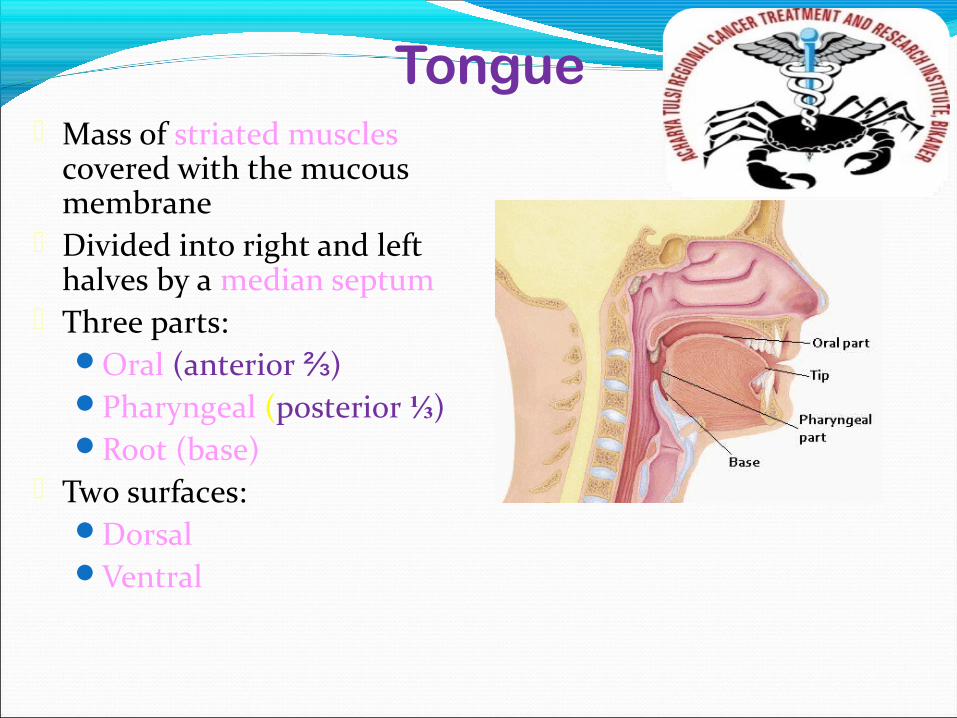
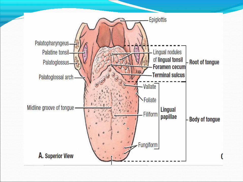
Tongue Mass of striated muscles
covered with the mucous membrane
Divided into right and left halves by a median septum
Three parts:Oral (anterior ⅔)Pharyngeal (posterior ⅓)Root (base)
Two surfaces:Dorsal Ventral
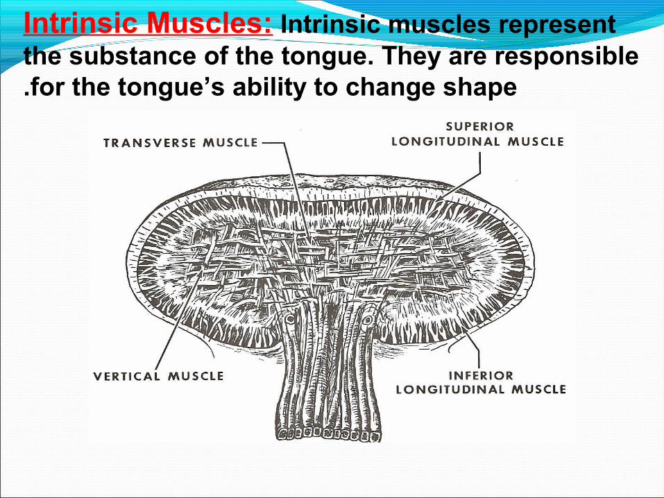
Intrinsic Muscles: Intrinsic muscles represent the substance of the tongue. They are responsible for the tongue’s ability to change shape.
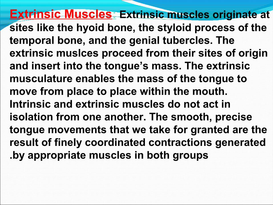
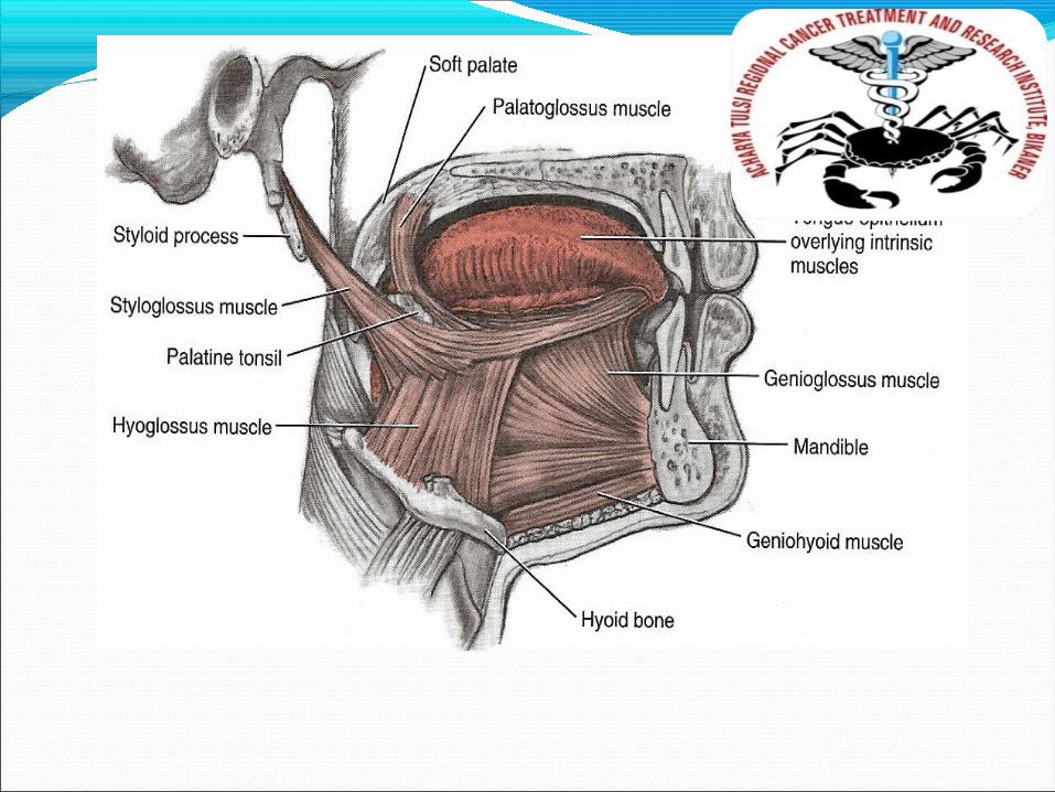
Extrinsic Muscles: Extrinsic muscles originate at sites like the hyoid bone, the styloid process of the temporal bone, and the genial tubercles. The extrinsic muslces proceed from their sites of origin and insert into the tongue’s mass. The extrinsic musculature enables the mass of the tongue to move from place to place within the mouth. Intrinsic and extrinsic muscles do not act in isolation from one another. The smooth, precise tongue movements that we take for granted are the result of finely coordinated contractions generated by appropriate muscles in both groups.
21
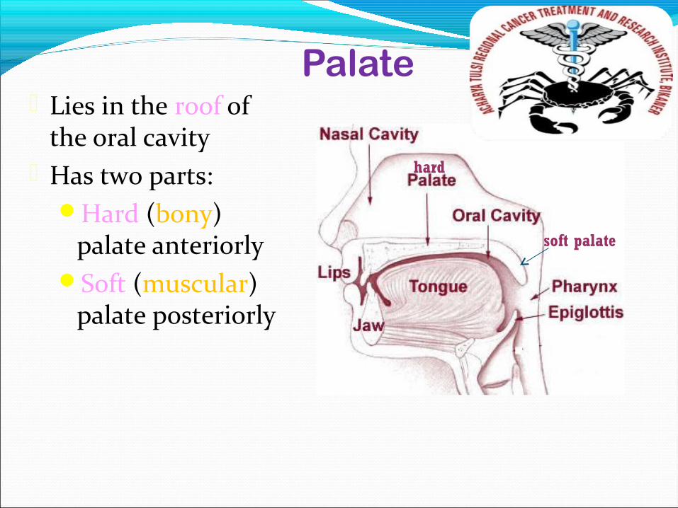
Palate Lies in the roof of
the oral cavity Has two parts:
Hard (bony) palate anteriorly
Soft (muscular) palate posteriorly
hard
soft palate
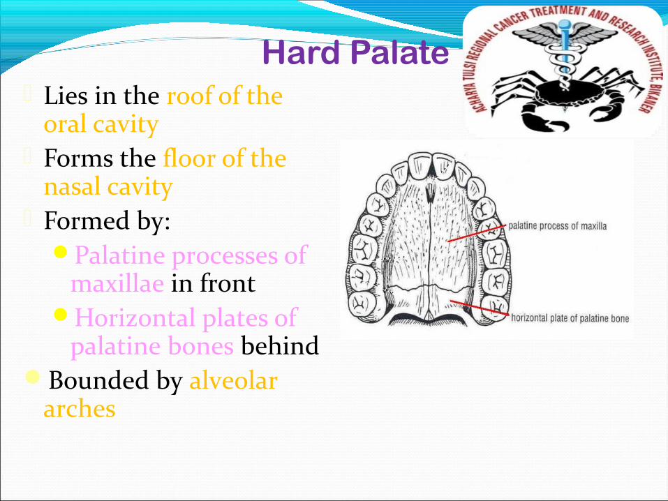
Hard Palate Lies in the roof of the
oral cavity Forms the floor of the
nasal cavity Formed by:
Palatine processes of maxillae in front
Horizontal plates of palatine bones behind
Bounded by alveolar arches
Hard Palate
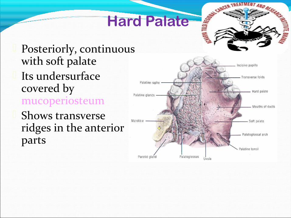
Posteriorly, continuous with soft palate
Its undersurface covered by mucoperiosteum
Shows transverse ridges in the anterior parts
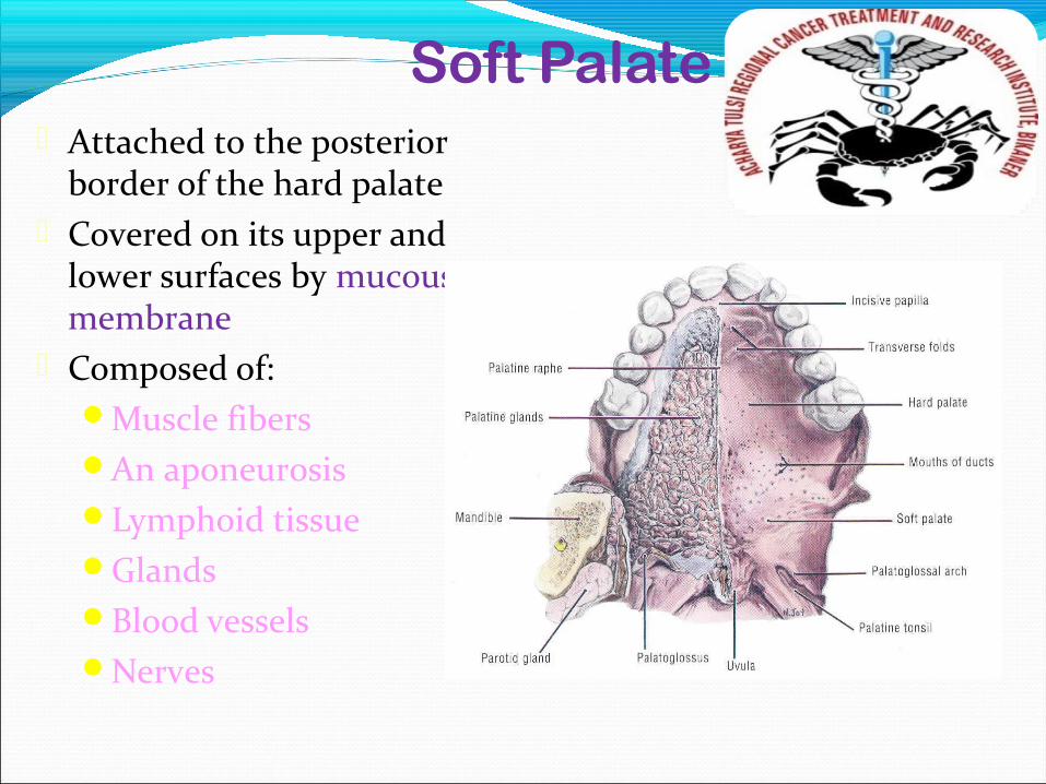
Soft Palate Attached to the posterior
border of the hard palate Covered on its upper and
lower surfaces by mucous membrane
Composed of:Muscle fibersAn aponeurosisLymphoid tissueGlandsBlood vesselsNerves
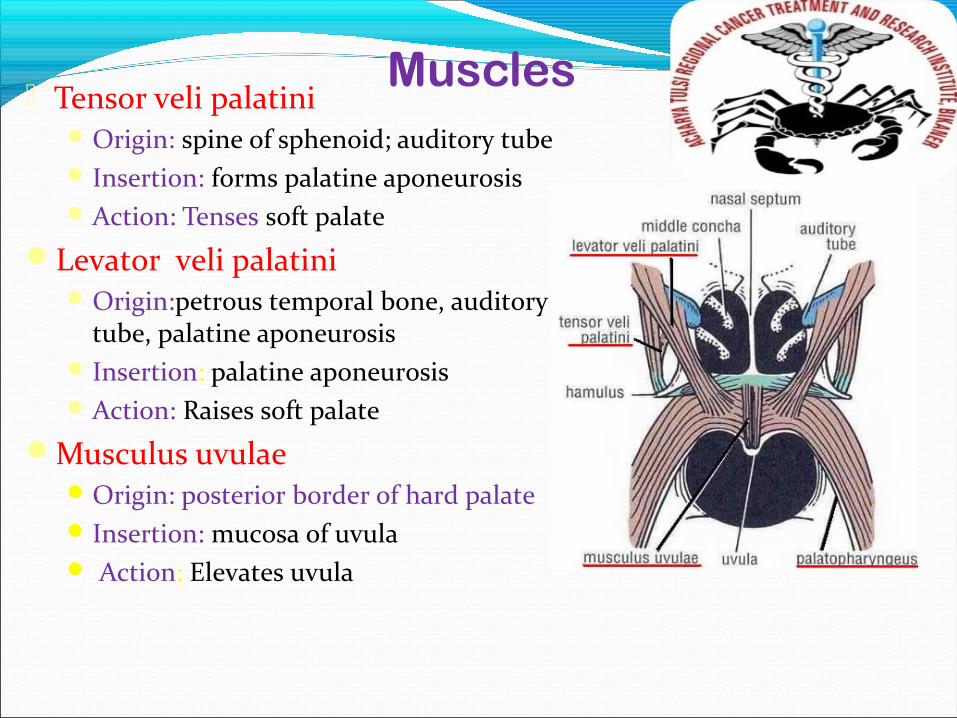
Muscles Tensor veli palatini
Origin: spine of sphenoid; auditory tube Insertion: forms palatine aponeurosisAction: Tenses soft palate
Levator veli palatiniOrigin:petrous temporal bone, auditory
tube, palatine aponeurosis Insertion: palatine aponeurosisAction: Raises soft palate
Musculus uvulaeOrigin: posterior border of hard palate Insertion: mucosa of uvula Action: Elevates uvula
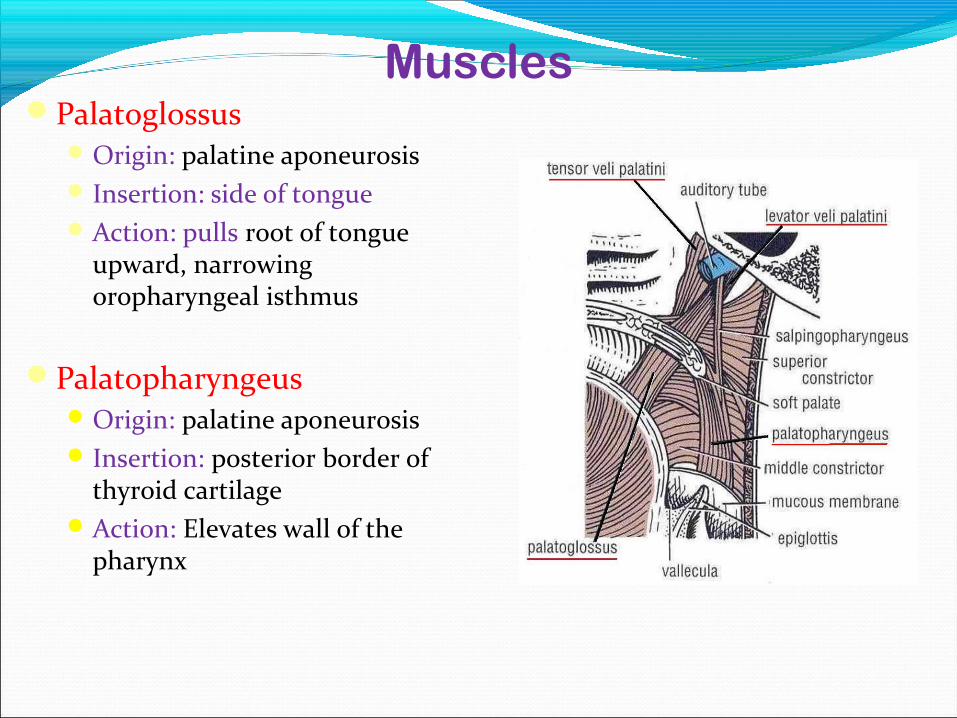
MusclesPalatoglossus
Origin: palatine aponeurosis Insertion: side of tongueAction: pulls root of tongue
upward, narrowing oropharyngeal isthmus
PalatopharyngeusOrigin: palatine aponeurosis Insertion: posterior border of
thyroid cartilageAction: Elevates wall of the
pharynx
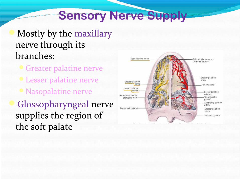
Sensory Nerve SupplyMostly by the maxillary
nerve through its branches:Greater palatine nerveLesser palatine nerveNasopalatine nerve
Glossopharyngeal nerve supplies the region of the soft palate
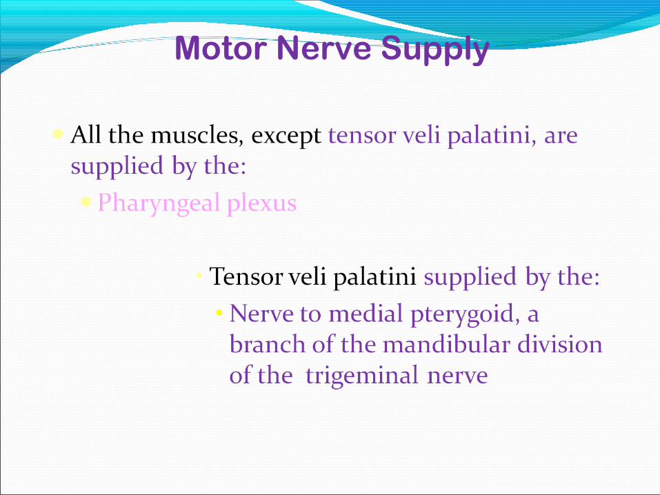
Motor Nerve Supply
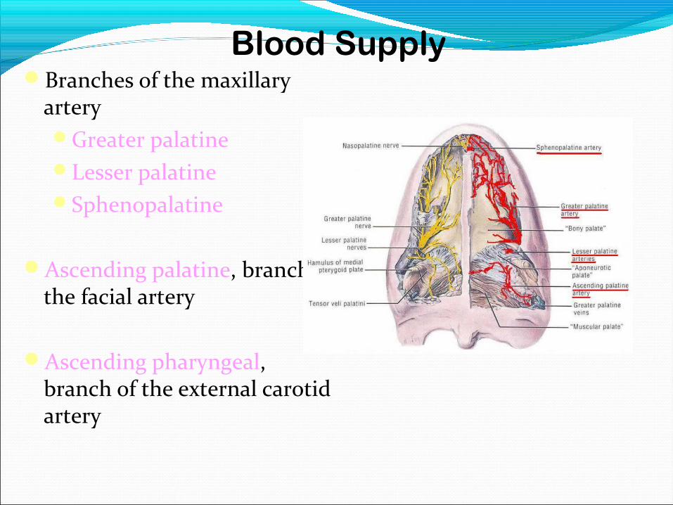
Blood SupplyBranches of the maxillary
arteryGreater palatineLesser palatineSphenopalatine
Ascending palatine, branch of the facial artery
Ascending pharyngeal, branch of the external carotid artery
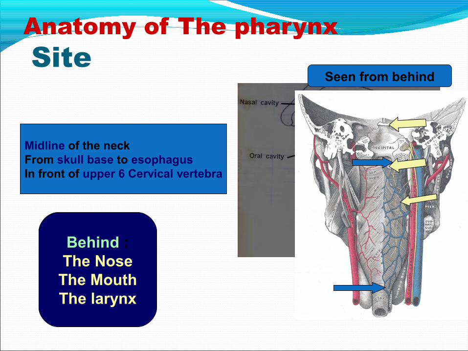
Anatomy of The pharynx Site
Midline of the neckFrom skull base to esophagusIn front of upper 6 Cervical vertebra
Behind :The Nose
The MouthThe larynx
Seen from behind
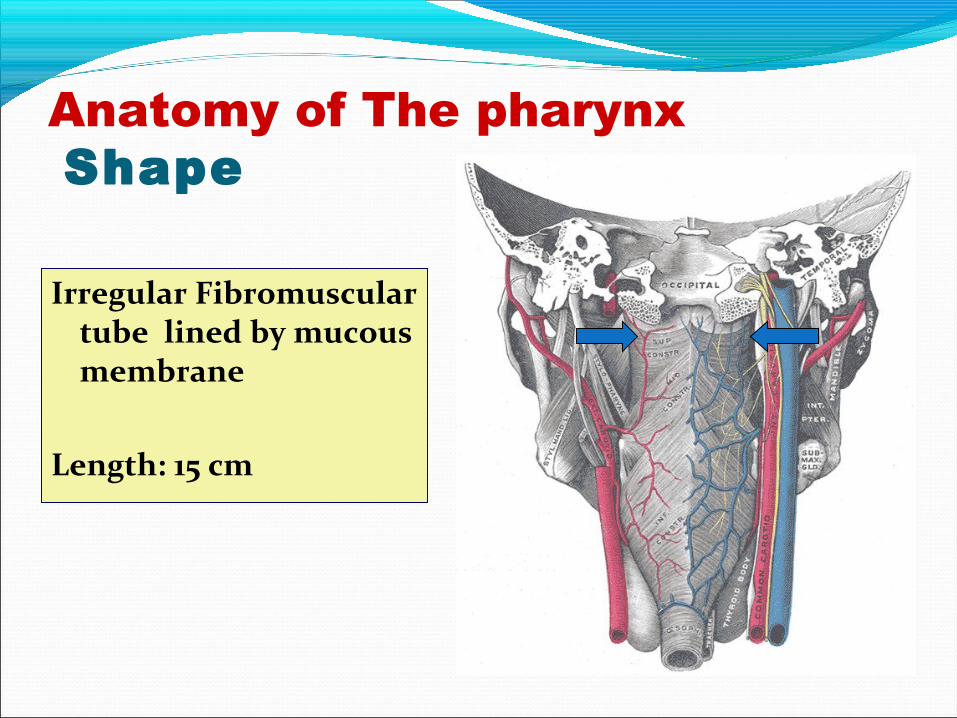
Anatomy of The pharynx Shape
Irregular Fibromuscular tube lined by mucous membrane
Length: 15 cm
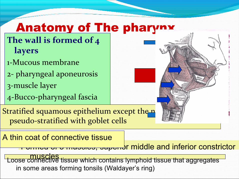
Anatomy of The pharynx Structure The wall is formed of 4 layers
1-Mucous membrane2- pharyngeal aponeurosis3-muscle layer4-Bucco-pharyngeal fascia
Stratified squamous epithelium except the nasopharynx, it is pseudo-stratified with goblet cells
Loose connective tissue which contains lymphoid tissue that aggregates in some areas forming tonsils (Waldayer’s ring)
Formed of 3 muscles, superior middle and inferior constrictor muscles
A thin coat of connective tissue
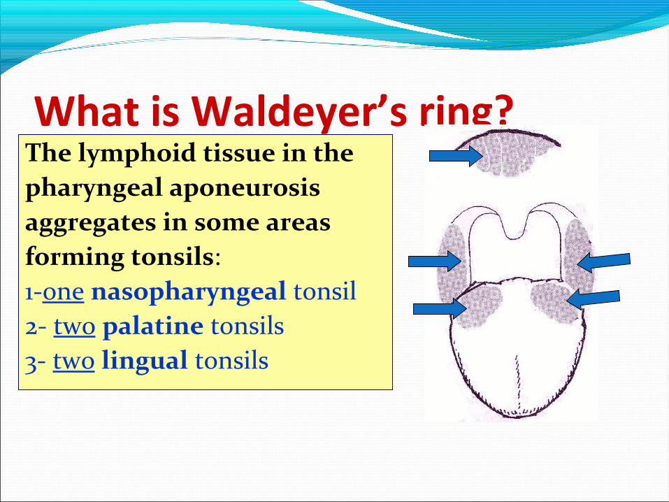
What is Waldeyer’s ring?The lymphoid tissue in thepharyngeal aponeurosisaggregates in some areasforming tonsils:1-one nasopharyngeal tonsil2- two palatine tonsils3- two lingual tonsils
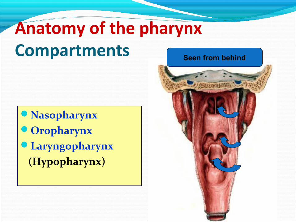
Anatomy of the pharynxCompartments
NasopharynxOropharynxLaryngopharynx (Hypopharynx)
Seen from behind
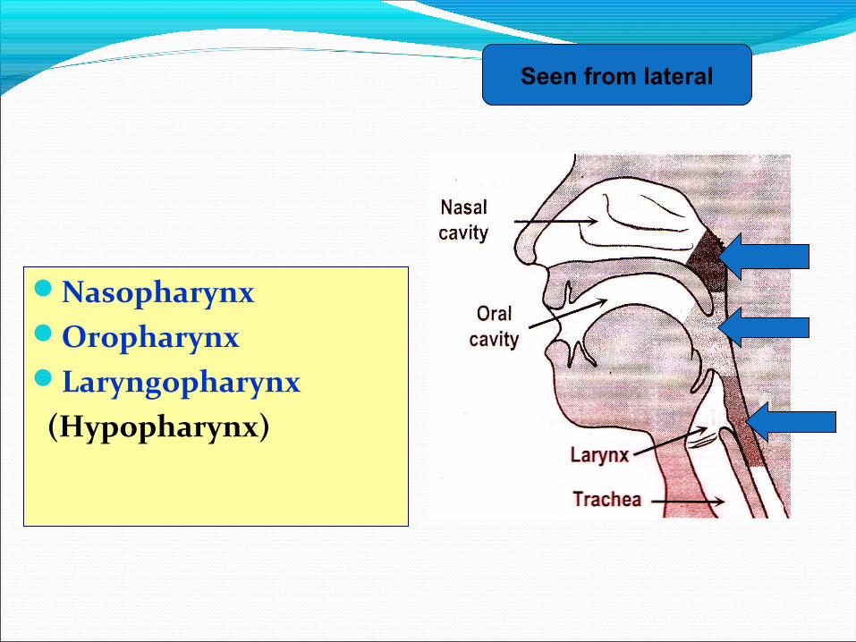
NasopharynxOropharynxLaryngopharynx (Hypopharynx)
Seen from lateral
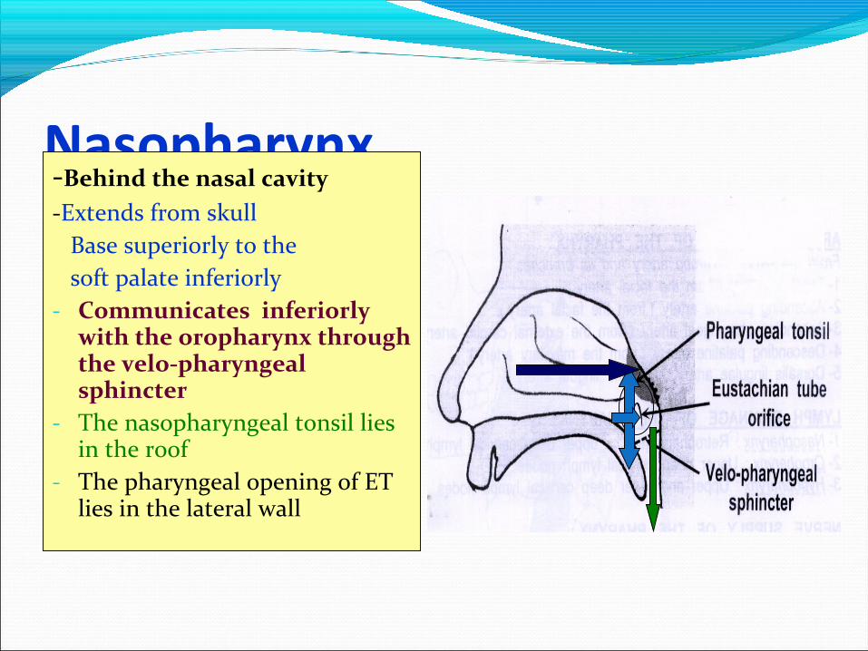
Nasopharynx -Behind the nasal cavity-Extends from skull Base superiorly to the soft palate inferiorly- Communicates inferiorly
with the oropharynx through the velo-pharyngeal sphincter
- The nasopharyngeal tonsil lies in the roof
- The pharyngeal opening of ET lies in the lateral wall
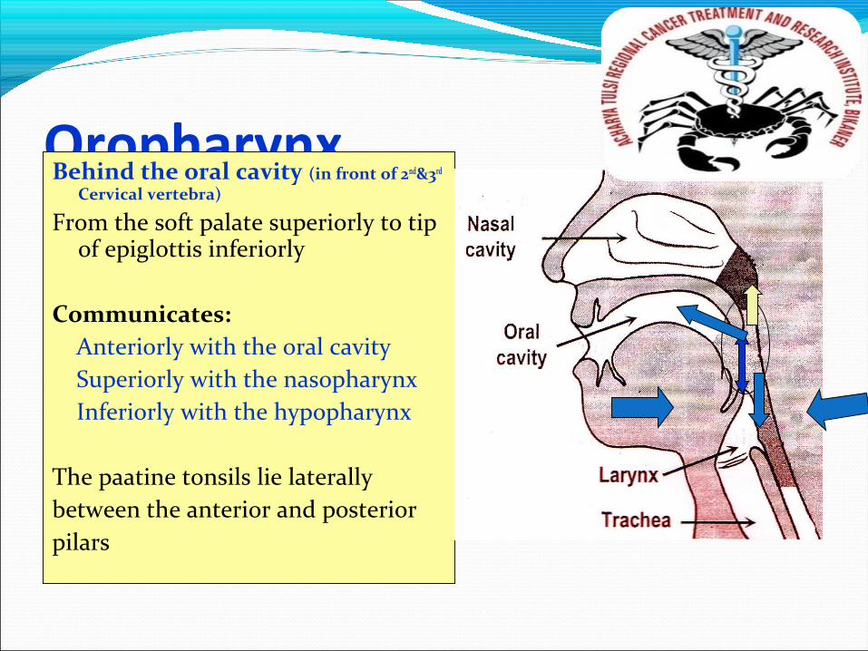
Oropharynx Behind the oral cavity (in front of 2nd&3rd
Cervical vertebra)
From the soft palate superiorly to tip of epiglottis inferiorly
Communicates: Anteriorly with the oral cavity Superiorly with the nasopharynx Inferiorly with the hypopharynx
The paatine tonsils lie laterallybetween the anterior and posteriorpilars
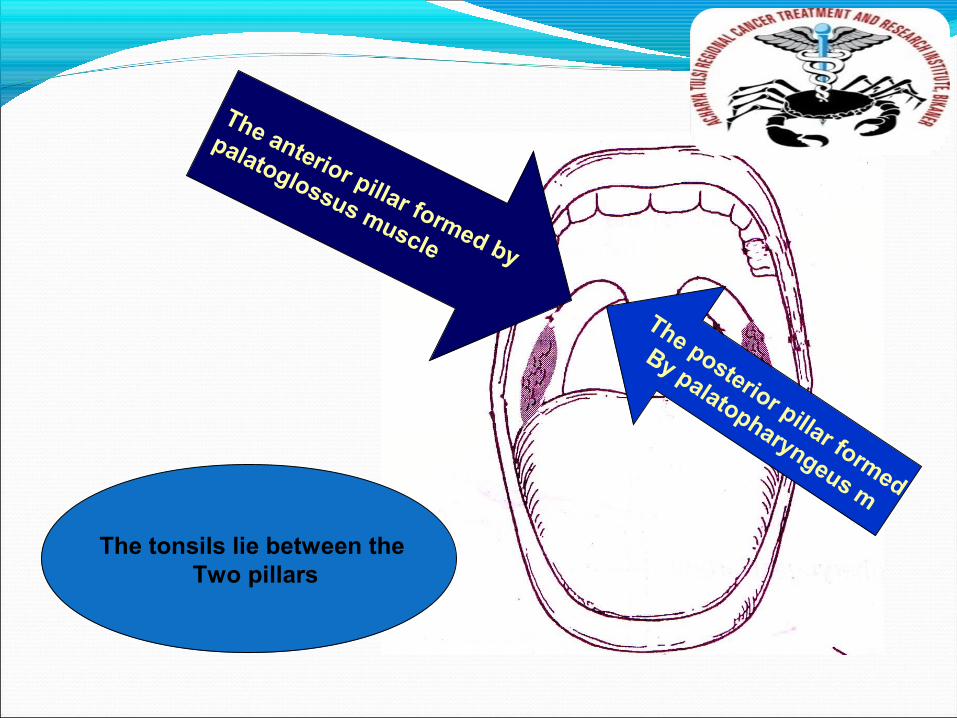
The anterior pillar formed by
palatoglossus muscle
The posterior pillar formed
By palatopharyngeus m
The tonsils lie between the Two pillars

Hypopharynx Behind the Larynx (in front of 3rd
to 6th Cervical vertebra)
From the tip of epiglottis superiorly to
the lower border of cricoid cartilageInferiorly
Communicates:- Anteriorly with the Larynx- Superiorly with the oropharynx- Inferiorly with the esophagus
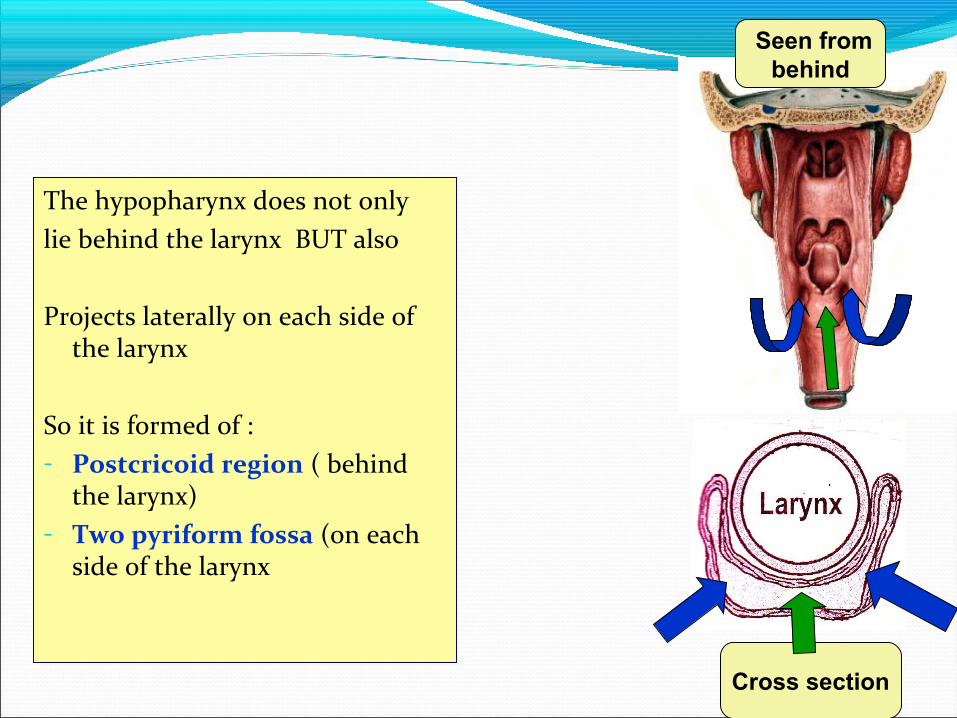
The hypopharynx does not onlylie behind the larynx BUT also
Projects laterally on each side of the larynx
So it is formed of :- Postcricoid region ( behind
the larynx)- Two pyriform fossa (on each
side of the larynx
Seen from behind
Cross section
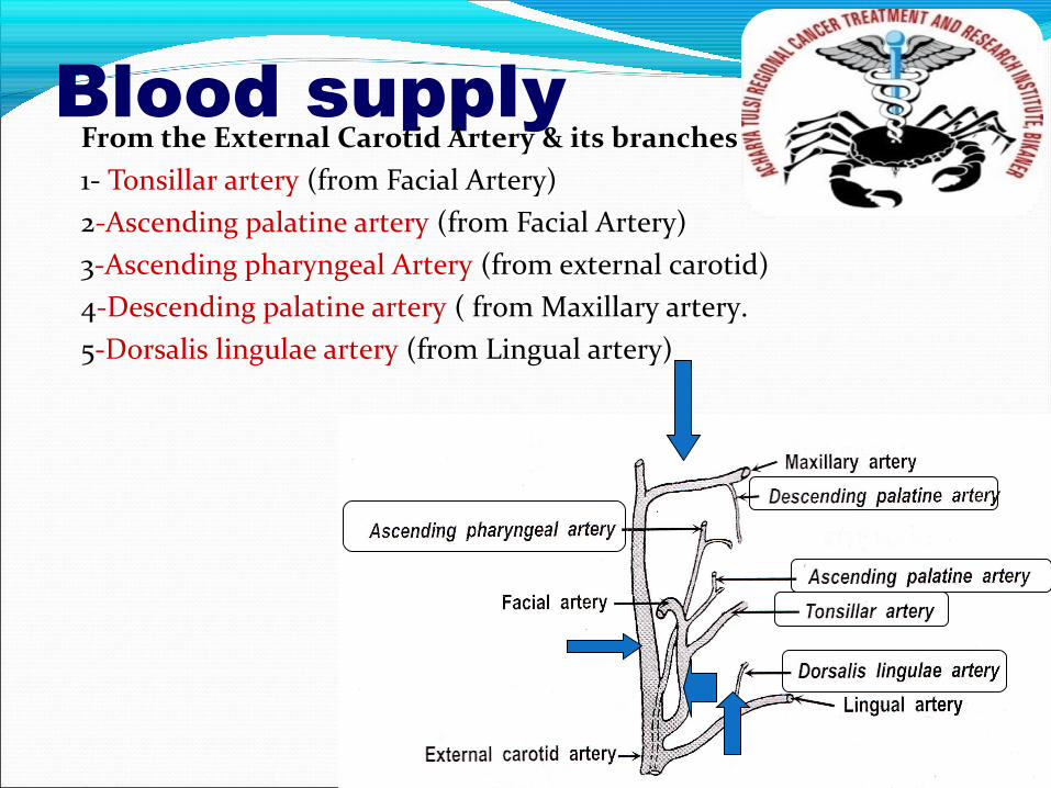
Blood supplyFrom the External Carotid Artery & its branches1- Tonsillar artery (from Facial Artery)2-Ascending palatine artery (from Facial Artery)3-Ascending pharyngeal Artery (from external carotid)4-Descending palatine artery ( from Maxillary artery.5-Dorsalis lingulae artery (from Lingual artery)
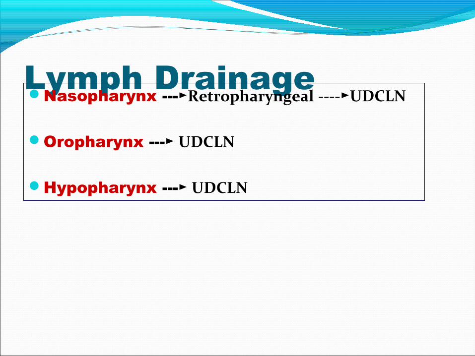
Lymph DrainageNasopharynx ---►Retropharyngeal ----►UDCLN
Oropharynx --- ► UDCLN
Hypopharynx --- ► UDCLN
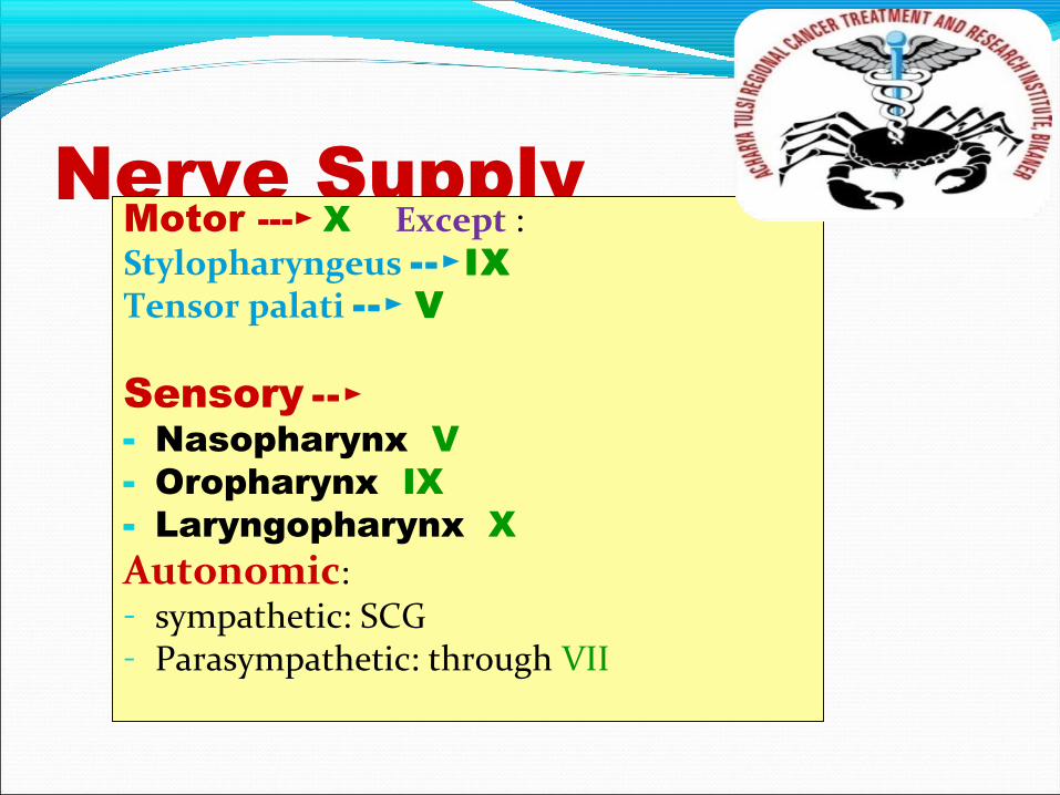
Nerve SupplyMotor ---► X Except : Stylopharyngeus --►IXTensor palati -- ► V
Sensory --►- Nasopharynx: V- Oropharynx: IX- Laryngopharynx: X Autonomic:- sympathetic: SCG- Parasympathetic: through VII

Laryngeal CartilagesPaired
Arytenoid cartilageCorniculate cartilageCuneiform cartilage
Unpaired:Thyroid cartilageCricoid cartilageEpiglottis
45
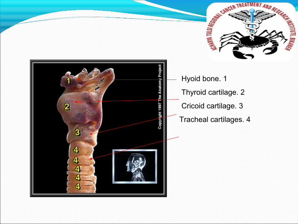
1. Hyoid bone
2. Thyroid cartilage
3. Cricoid cartilage
4. Tracheal cartilages
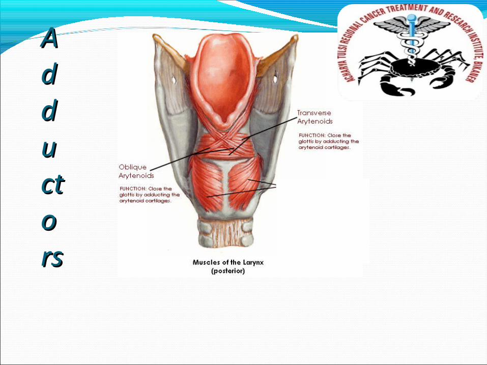
AAdddduuctctoorsrs
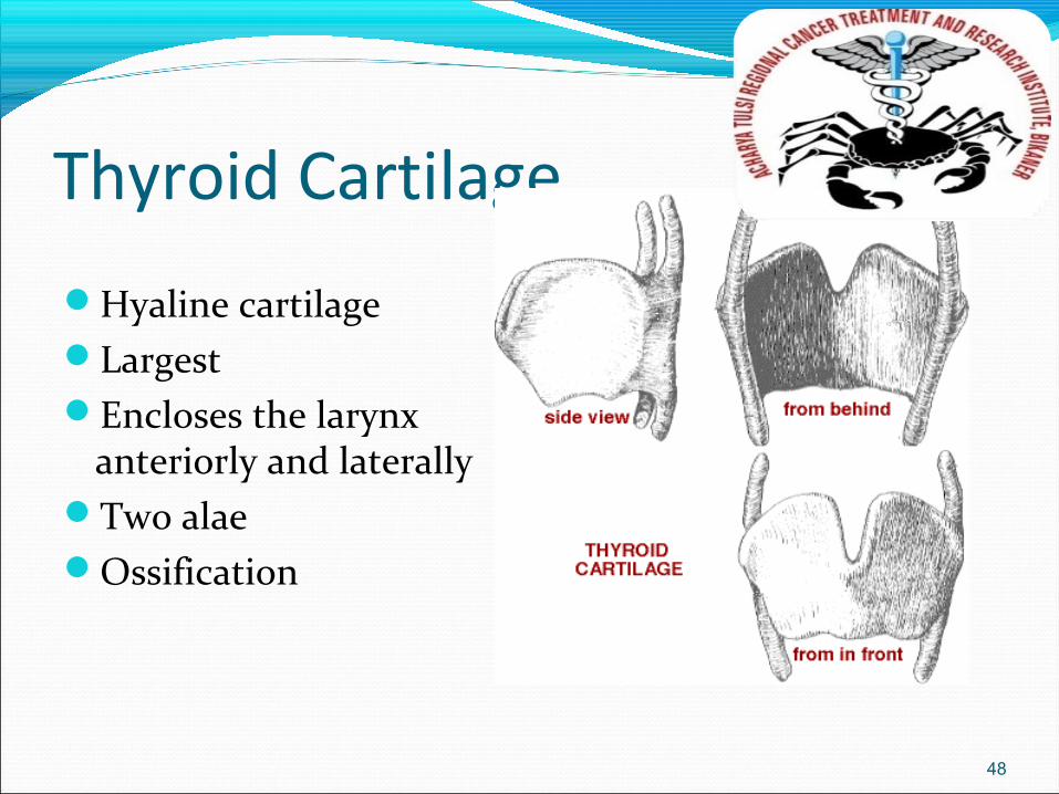
Thyroid Cartilage
Hyaline cartilageLargestEncloses the larynx
anteriorly and laterallyTwo alaeOssification
48
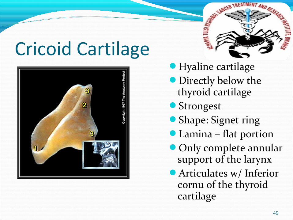
Cricoid CartilageHyaline cartilageDirectly below the
thyroid cartilageStrongestShape: Signet ringLamina – flat portionOnly complete annular
support of the larynxArticulates w/ Inferior
cornu of the thyroid cartilage
49
EpiglottisFibroelastic cartilageLeaf-shaped structurePetiole – small narrow
portion of the glottis
50

Arytenoid CartilageMostly hyaline cartilageSmaller in sizeResponsible for opening and closing of the larynxShape: pyramidal
51
Arytenoid CartilageAnterior
Vocal process - receives the attachement of the mobile end of each VC
LateralMuscular process
ArticulationCricoarytenoid joint
52
Corniculate CartilagesFibroelasticCartilages of SantoriniSmall cartilages above the arytenoid and in the
aryepiglottic folds
53
Cuneiform CartilagesFirboelastic cartilagesCartilages of WrisbergElongated pieces of small yellow elastic cartilage
in the aryepiglottic folds
54
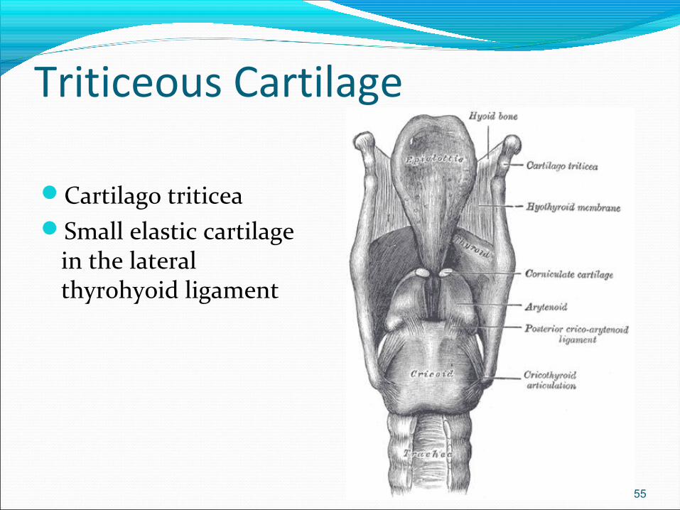
Triticeous Cartilage
Cartilago triticeaSmall elastic cartilage
in the lateral thyrohyoid ligament
55
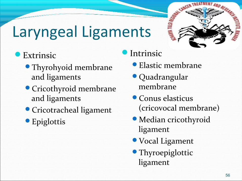
Laryngeal LigamentsExtrinsic
Thyrohyoid membrane and ligaments
Cricothyroid membrane and ligaments
Cricotracheal ligamentEpiglottis
IntrinsicElastic membraneQuadrangular
membraneConus elasticus
(cricovocal membrane)Median cricothyroid
ligamentVocal LigamentThyroepiglottic
ligament56
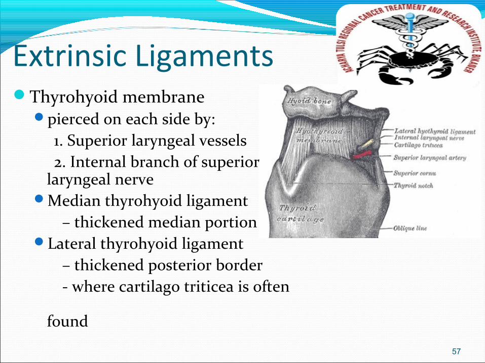
Extrinsic LigamentsThyrohyoid membrane
pierced on each side by: 1. Superior laryngeal vessels 2. Internal branch of superior
laryngeal nerveMedian thyrohyoid ligament – thickened median portionLateral thyrohyoid ligament – thickened posterior border - where cartilago triticea is often
found
57
Extrinsic LigamentsCricothyroid membrane and ligaments
May be pierced for emergency tracheotomy (cricothyrotomy)
58
Extrinsic LigamentsCricotracheal Ligament
Attaches the cricoid cartilage to the first attached ring
Epiglottis suspended in position by membranous connections to
the hyoid bone, thyroid cartilage and base of the tongue
59
Intrinsic LigamentsElastic membrane
Divided into upper and lower parts by the ventricle of the larynx
Quadrangular membraneUpper part of the elastic membraneBoundaries
Epiglottis , arytenoid, corniculate cartilage, false cordForms part of wall between upper pyriform sinus and
laryngeal vestibule
60
Intrinsic LigamentsConus elasticus (cricovocal membrane)
Lower part of elastic membraneComposed mainly of yellow elastic tissueBoundaries
Inferior: superior border of cricoid cartilage Superoanterior: deep surface of angle thyroid cartilage Superoposterior: vocal process of arytenoid cartilage
Median cricothyroid ligament – thickened anteior partVocal Ligament – free upper edge
Thyroepiglottic ligament
61
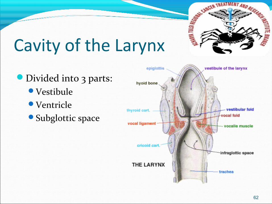
Cavity of the Larynx
Divided into 3 parts:Vestibule VentricleSubglottic space
62
Cavity of the Larynx
Vestibule – boundaries:Anterior: posterior surface of epiglottisPosterior: interval between arytenoid
cartilagesLateral: inner surface of aryepiglottic
folds and upper surfaces of the false cord
63
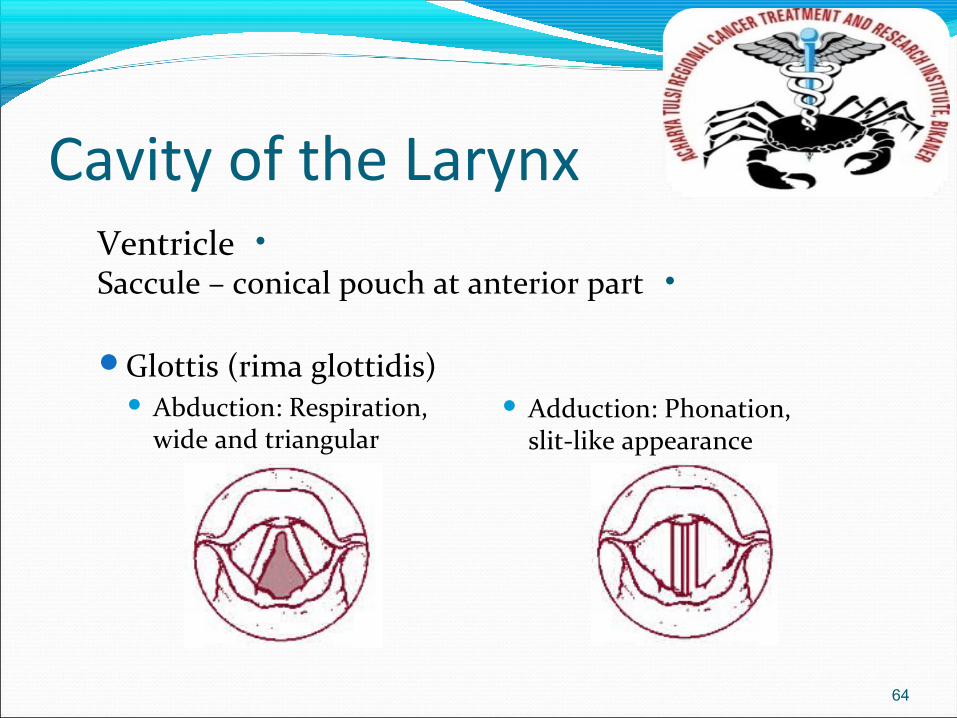
Cavity of the Larynx
Glottis (rima glottidis) Abduction: Respiration,
wide and triangular Adduction: Phonation,
slit-like appearance
64
• Ventricle• Saccule – conical pouch at anterior part
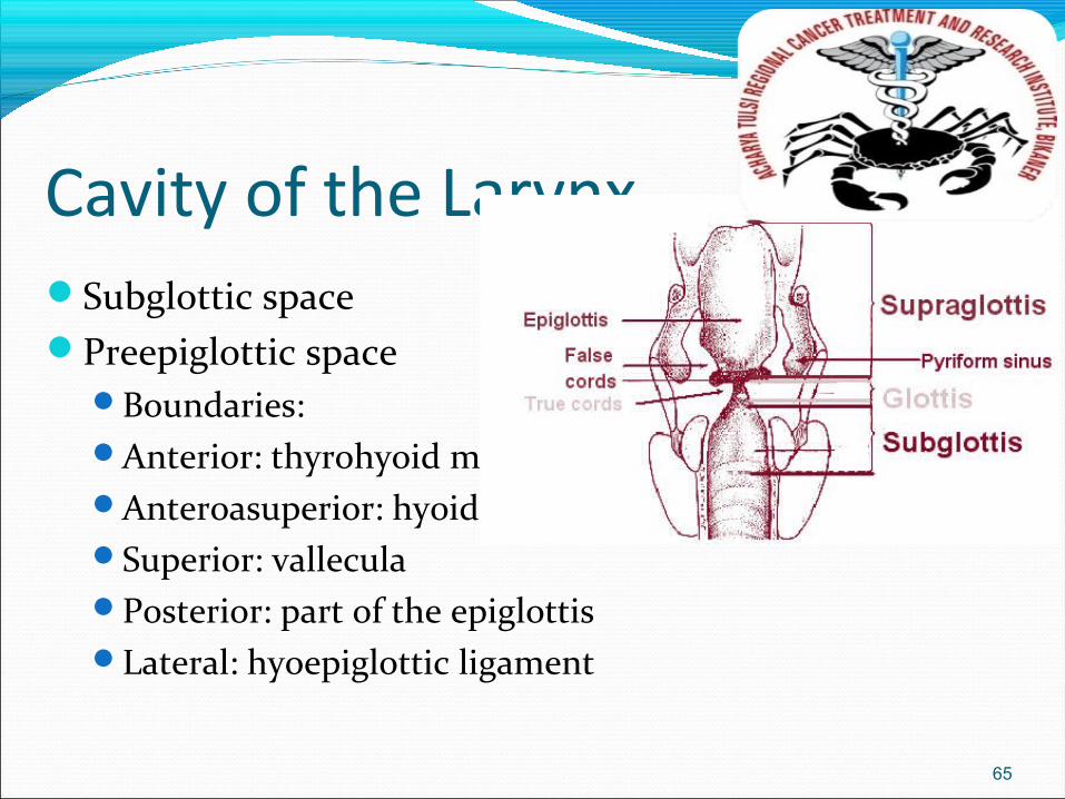
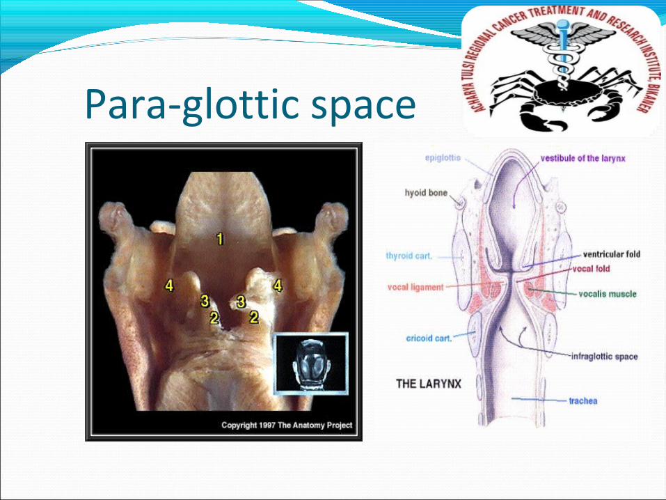
Cavity of the LarynxSubglottic spacePreepiglottic space
Boundaries:Anterior: thyrohyoid membraneAnteroasuperior: hyoidSuperior: valleculaPosterior: part of the epiglottisLateral: hyoepiglottic ligament
65
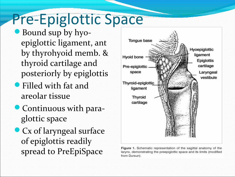
Pre-Epiglottic SpaceBound sup by hyo-
epiglottic ligament, ant by thyrohyoid memb. & thyroid cartilage and posteriorly by epiglottis
Filled with fat and areolar tissue
Continuous with para-glottic space
Cx of laryngeal surface of epiglottis readily spread to PreEpiSpace
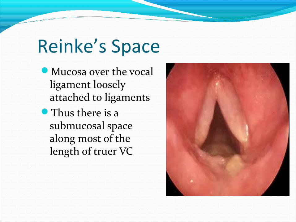
Reinke’s SpaceMucosa over the vocal
ligament loosely attached to ligaments
Thus there is a submucosal space along most of the length of truer VC
Para-glottic space
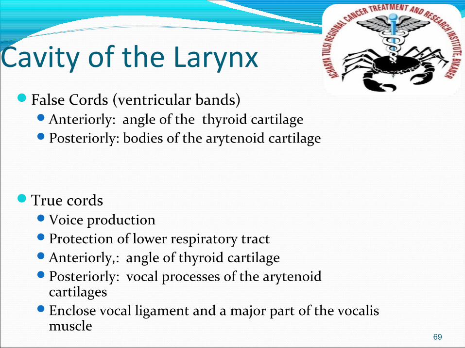
Cavity of the LarynxFalse Cords (ventricular bands)
Anteriorly: angle of the thyroid cartilagePosteriorly: bodies of the arytenoid cartilage
True cordsVoice productionProtection of lower respiratory tractAnteriorly,: angle of thyroid cartilagePosteriorly: vocal processes of the arytenoid
cartilagesEnclose vocal ligament and a major part of the vocalis
muscle 69
Laryngeal JointsCricothyroid Joint
Between inferior cornu of the thyroid cartilage and facet on the cricoid cartilage at the junction of the arch and lamina
Two movements:RotationGliding
Cricoarytenoid Jointbet. base of the
arytenoid cartilage and the facet on the upper border of the lamina of the cricoid cartilage
Two movements:RotationGliding
70
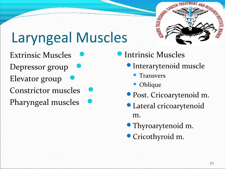
Laryngeal MusclesIntrinsic Muscles
Interarytenoid muscle Transvers Oblique
Post. Cricoarytenoid m.Lateral cricoarytenoid
m.Thyroarytenoid m.Cricothyroid m.
71
Extrinsic MusclesDepressor group
Elevator groupConstrictor musclesPharyngeal muscles
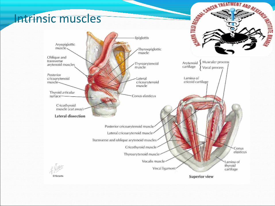
Intrinsic muscles
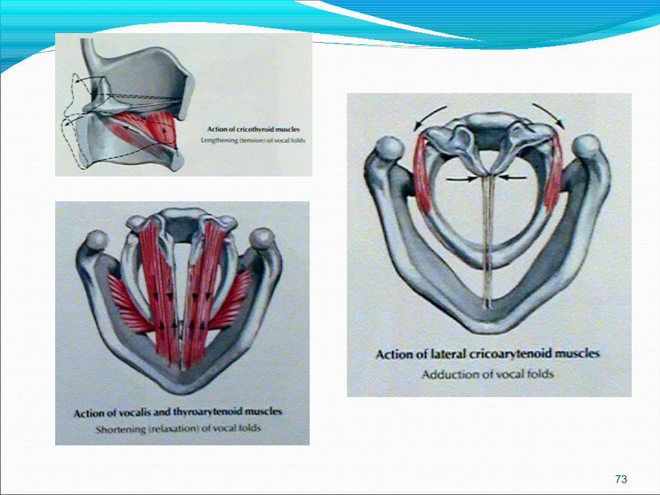
73
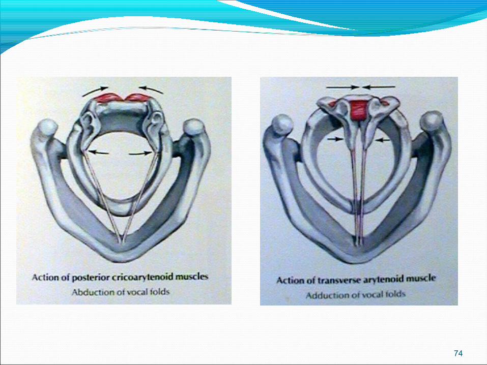
74
Nerve SupplySupplied by Vagus nerve:
Superior laryngeal n. Internal branch (sensory) – areas above the glottis External branch (motor and sensory) Motor – Cricothyroid muscle
Sensory – Anterior infraglottic larynx at level of cricothyroid membrane
Inferior (recurrent) laryngeal n. Motor – all intrinsic laryngeal muscles of SAME side (except
cricothyroid) and interarytenoid muscle of BOTH sides Sensory – areas below the glottis
75
Blood SupplyUpper Larynx
External carotid arterySuperior thyroid arterySuperior laryngeal
arteryLower Larynx
Subclavian arteryThyrocervical arteryInferior thyroid arteryInferior laryngeal artery
76
Venouos DrainageUpper Larynx
Superior laryngeal veinSuperior thyroid veinInternal jugular vein
Lower LarynxInferior laryngeal veinInferior thyroid veinInnominate vein
77
Lymphatic DrainageMain: Deep Cervical group L.N.
Supraglottic area98%: Pedicle Ant. End of aryepiglottic fold ->
pass laterally and leave the larynx through the thyrohyoid membrane ->Upper deep cervical nodes (bet. Digastric tendon and omohyoid muscle)
2%: Lower cervical chain or spinal accessory chain
78

Lymphatic DrainageInfraglottic area – 3 pedicles
1. Anterior pedicle -> cricothyroid membrane -> prelaryngeal (Delphian) nodes ->deep inferior cervical nodes
2. 2 Posterolateral pedicles -> cricotracheal membrane -> paratracheal chain/others to inferior jugular chain
79
THANKS
Related Documents

![Pharynx [للقراءة فقط] - KSU · 2017. 8. 23. · • Elevate pharynx and larynx during speech/swallow 1. Palatopharyngeus 2. Salpingopharyngeus 3. Stylopharyngeus 4. Levator](https://static.cupdf.com/doc/110x72/6115f23eb95e174a953ed119/pharynx-ksu-2017-8-23-a-elevate-pharynx-and-larynx.jpg)