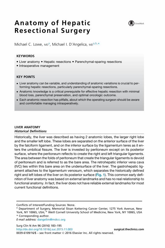
Anatomy of Hepatic Resectional Surgery Michael C. Lowe, MD a , Michael I. D’Angelica, MD a,b, * LIVER ANATOMY Historical Definitions Historically, the liver was described as having 2 anatomic lobes, the larger right lobe and the smaller left lobe. These lobes are separated on the anterior surface of the liver by the falciform ligament, and on the inferior surface by the ligamentum teres as it en- ters the umbilical fissure. The liver is invested by peritoneum except on its posterior surface, where the peritoneum reflects to create the right and left triangular ligaments. The area between the folds of peritoneum that create the triangular ligaments is devoid of peritoneum and is referred to as the bare area. The retrohepatic inferior vena cava (IVC) lies within this bare area on the undersurface of the liver. The gastrohepatic lig- ament attaches to the ligamentum venosum, which separates the historically defined right and left lobes of the liver on its posterior surface (Fig. 1). This common early defi- nition of liver anatomy was based on external landmarks and has no real relationship to functional anatomy. In fact, the liver does not have reliable external landmarks for most current functional definitions. Conflicts of Interest/Funding Sources: None. a Department of Surgery, Memorial Sloan Kettering Cancer Center, 1275 York Avenue, New York, NY 10065, USA; b Weill Cornell University School of Medicine, New York, NY 10065, USA * Corresponding author. E-mail address: [email protected] KEYWORDS Liver anatomy Hepatic resections Parenchymal-sparing resections Intraoperative management KEY POINTS Liver anatomy can be variable, and understanding of anatomic variations is crucial to per- forming hepatic resections, particularly parenchymal-sparing resections. Anatomic knowledge is a critical prerequisite for effective hepatic resection with minimal blood loss, parenchymal preservation, and optimal oncologic outcome. Each anatomic resection has pitfalls, about which the operating surgeon should be aware and comfortable managing intraoperatively. Surg Clin N Am 96 (2016) 183–195 http://dx.doi.org/10.1016/j.suc.2015.11.003 surgical.theclinics.com 0039-6109/16/$ – see front matter Ó 2016 Elsevier Inc. All rights reserved.

Welcome message from author
This document is posted to help you gain knowledge. Please leave a comment to let me know what you think about it! Share it to your friends and learn new things together.
Transcript
Anatomy of HepaticResectional Surgery
Michael C. Lowe, MDa, Michael I. D’Angelica, MDa,b,*
KEYWORDS
� Liver anatomy � Hepatic resections � Parenchymal-sparing resections� Intraoperative management
KEY POINTS
� Liver anatomy can be variable, and understanding of anatomic variations is crucial to per-forming hepatic resections, particularly parenchymal-sparing resections.
� Anatomic knowledge is a critical prerequisite for effective hepatic resection with minimalblood loss, parenchymal preservation, and optimal oncologic outcome.
� Each anatomic resection has pitfalls, about which the operating surgeon should be awareand comfortable managing intraoperatively.
LIVER ANATOMYHistorical Definitions
Historically, the liver was described as having 2 anatomic lobes, the larger right lobeand the smaller left lobe. These lobes are separated on the anterior surface of the liverby the falciform ligament, and on the inferior surface by the ligamentum teres as it en-ters the umbilical fissure. The liver is invested by peritoneum except on its posteriorsurface, where the peritoneum reflects to create the right and left triangular ligaments.The area between the folds of peritoneum that create the triangular ligaments is devoidof peritoneum and is referred to as the bare area. The retrohepatic inferior vena cava(IVC) lies within this bare area on the undersurface of the liver. The gastrohepatic lig-ament attaches to the ligamentum venosum, which separates the historically definedright and left lobes of the liver on its posterior surface (Fig. 1). This common early defi-nition of liver anatomywas based on external landmarks and has no real relationship tofunctional anatomy. In fact, the liver does not have reliable external landmarks for mostcurrent functional definitions.
Conflicts of Interest/Funding Sources: None.a Department of Surgery, Memorial Sloan Kettering Cancer Center, 1275 York Avenue, NewYork, NY 10065, USA; b Weill Cornell University School of Medicine, New York, NY 10065, USA* Corresponding author.E-mail address: [email protected]
Surg Clin N Am 96 (2016) 183–195http://dx.doi.org/10.1016/j.suc.2015.11.003 surgical.theclinics.com0039-6109/16/$ – see front matter � 2016 Elsevier Inc. All rights reserved.
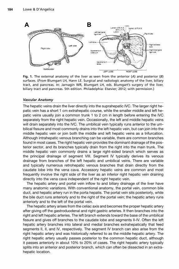
Fig. 1. The external anatomy of the liver as seen from the anterior (A) and posterior (B)surfaces. (From Blumgart LH, Hann LE. Surgical and radiologic anatomy of the liver, biliarytract, and pancreas. In: Jarnagin WR, Blumgart LH, eds. Blumgart’s surgery of the liver,biliary tract and pancreas. 5th edition. Philadelphia: Elsevier; 2012; with permission.)
Lowe & D’Angelica184
Vascular Anatomy
The hepatic veins drain the liver directly into the suprahepatic IVC. The larger right he-patic vein has a short 1 cm extrahepatic course, while the smaller middle and left he-patic veins usually join a common trunk 1 to 2 cm in length before entering the IVCseparately from the right hepatic vein. Occasionally, the left and middle hepatic veinswill drain separately into the IVC. The umbilical vein typically runs anterior to the um-bilical fissure and most commonly drains into the left hepatic vein, but can join into themiddle hepatic vein or join both the middle and left hepatic veins as a trifurcation.Although intrahepatic venous branching can be variable, there are common branchesfound in most cases. The right hepatic vein provides the dominant drainage of the pos-terior sector, and its branches typically drain from the right into the main trunk. Themiddle hepatic vein commonly drains a large right-sided branch which serves asthe principal drainage of segment VIII. Segment IV typically derives its venousdrainage from branches of the left hepatic and umbilical veins. There are variableand typically numerous retrohepatic venous branches that drain directly from thecaudate lobe into the vena cava. Accessory hepatic veins are common and mostfrequently involve the right side of the liver as an inferior right hepatic vein drainingdirectly into the vena cava independent of the right hepatic vein.The hepatic artery and portal vein inflow to and biliary drainage of the liver have
many anatomic variations. With conventional anatomy, the portal vein, common bileduct, and hepatic artery run in the porta hepatis. The portal vein sits posteriorly, whilethe bile duct runs anteriorly and to the right of the portal vein; the hepatic artery runsanteriorly and to the left of the portal vein.The hepatic artery arises from the celiac axis and becomes the proper hepatic artery
after giving off the gastroduodenal and right gastric arteries. It then branches into theright and left hepatic arteries. The left branch extends toward the base of the umbilicalfissure and gives off branches to the caudate lobe and segments II-IV. Often the lefthepatic artery branches into lateral and medial branches extrahepatically that feedsegments II, II, and IV, respectively. The segment IV branch can also arise from theright hepatic artery and was historically referred to as the middle hepatic artery. Theright hepatic artery usually passes posterior to the common hepatic duct, althoughit passes anteriorly in about 10% to 20% of cases. The right hepatic artery typicallysplits into an anterior and posterior branch, which can often be dissected in an extra-hepatic location.
Anatomy of Hepatic Resectional Surgery 185
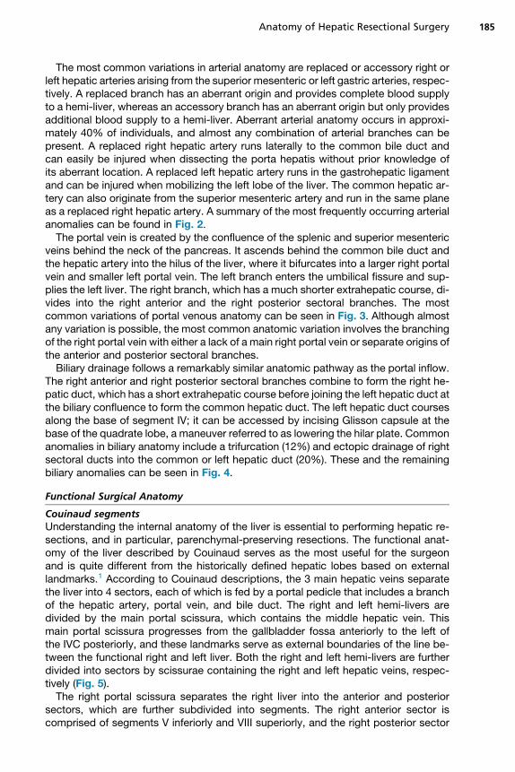
The most common variations in arterial anatomy are replaced or accessory right orleft hepatic arteries arising from the superior mesenteric or left gastric arteries, respec-tively. A replaced branch has an aberrant origin and provides complete blood supplyto a hemi-liver, whereas an accessory branch has an aberrant origin but only providesadditional blood supply to a hemi-liver. Aberrant arterial anatomy occurs in approxi-mately 40% of individuals, and almost any combination of arterial branches can bepresent. A replaced right hepatic artery runs laterally to the common bile duct andcan easily be injured when dissecting the porta hepatis without prior knowledge ofits aberrant location. A replaced left hepatic artery runs in the gastrohepatic ligamentand can be injured when mobilizing the left lobe of the liver. The common hepatic ar-tery can also originate from the superior mesenteric artery and run in the same planeas a replaced right hepatic artery. A summary of the most frequently occurring arterialanomalies can be found in Fig. 2.The portal vein is created by the confluence of the splenic and superior mesenteric
veins behind the neck of the pancreas. It ascends behind the common bile duct andthe hepatic artery into the hilus of the liver, where it bifurcates into a larger right portalvein and smaller left portal vein. The left branch enters the umbilical fissure and sup-plies the left liver. The right branch, which has a much shorter extrahepatic course, di-vides into the right anterior and the right posterior sectoral branches. The mostcommon variations of portal venous anatomy can be seen in Fig. 3. Although almostany variation is possible, the most common anatomic variation involves the branchingof the right portal vein with either a lack of a main right portal vein or separate origins ofthe anterior and posterior sectoral branches.Biliary drainage follows a remarkably similar anatomic pathway as the portal inflow.
The right anterior and right posterior sectoral branches combine to form the right he-patic duct, which has a short extrahepatic course before joining the left hepatic duct atthe biliary confluence to form the common hepatic duct. The left hepatic duct coursesalong the base of segment IV; it can be accessed by incising Glisson capsule at thebase of the quadrate lobe, a maneuver referred to as lowering the hilar plate. Commonanomalies in biliary anatomy include a trifurcation (12%) and ectopic drainage of rightsectoral ducts into the common or left hepatic duct (20%). These and the remainingbiliary anomalies can be seen in Fig. 4.
Functional Surgical Anatomy
Couinaud segmentsUnderstanding the internal anatomy of the liver is essential to performing hepatic re-sections, and in particular, parenchymal-preserving resections. The functional anat-omy of the liver described by Couinaud serves as the most useful for the surgeonand is quite different from the historically defined hepatic lobes based on externallandmarks.1 According to Couinaud descriptions, the 3 main hepatic veins separatethe liver into 4 sectors, each of which is fed by a portal pedicle that includes a branchof the hepatic artery, portal vein, and bile duct. The right and left hemi-livers aredivided by the main portal scissura, which contains the middle hepatic vein. Thismain portal scissura progresses from the gallbladder fossa anteriorly to the left ofthe IVC posteriorly, and these landmarks serve as external boundaries of the line be-tween the functional right and left liver. Both the right and left hemi-livers are furtherdivided into sectors by scissurae containing the right and left hepatic veins, respec-tively (Fig. 5).The right portal scissura separates the right liver into the anterior and posterior
sectors, which are further subdivided into segments. The right anterior sector iscomprised of segments V inferiorly and VIII superiorly, and the right posterior sector
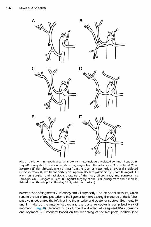
Fig. 2. Variations in hepatic arterial anatomy. These include a replaced common hepatic ar-tery (A), a very short common hepatic artery origin from the celiac axis (B), a replaced (C) oraccessory (E) right hepatic artery arising from the superior mesenteric artery, and a replaced(D) or accessory (F) left hepatic artery arising from the left gastric artery. (From Blumgart LH,Hann LE. Surgical and radiologic anatomy of the liver, biliary tract, and pancreas. In:Jarnagin WR, Blumgart LH, eds. Blumgart’s surgery of the liver, biliary tract and pancreas.5th edition. Philadelphia: Elsevier; 2012; with permission.)
Lowe & D’Angelica186
is comprised of segments VI inferiorly and VII superiorly. The left portal scissura, whichruns to the left of and posterior to the ligamentum teres along the course of the left he-patic vein, separates the left liver into the anterior and posterior sectors. Segments IVand III make up the anterior sector, and the posterior sector is comprised only ofsegment II (Fig. 6). Segment IV can further be divided into segment IVA superiorlyand segment IVB inferiorly based on the branching of the left portal pedicle (see
Fig. 3. Variations in portal venous anatomy. In normal anatomy the splenic vein (SV) and su-perior mesenteric vein combine to form the portal vein, which branches into the right portalvein (RPV) and left portal vein (LPV). The RPV further branches in right anterior sectoral(RAS) and right posterior sectoral (RPS) branches (A). The RAS, RPS, and LPV can all ariseindependently from the main portal vein (B). The RPS may arise early from the portalvein, which then bifurcates in the RAS and LPV (C). CV, coronary vein; IMV, inferior mesen-teric vein; SMV, superior mesenteric vein. (From Blumgart LH, Hann LE. Surgical and radio-logic anatomy of the liver, biliary tract, and pancreas. In: Jarnagin WR, Blumgart LH, eds.Blumgart’s surgery of the liver, biliary tract and pancreas. 5th edition. Philadelphia: Elsevier;2012; with permission.)
Anatomy of Hepatic Resectional Surgery 187
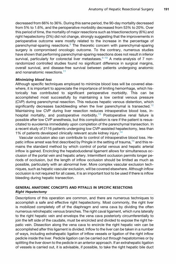
Fig. 5), although this branching is variable and does not often result in an easily definedsegment IVA and B. The caudate lobe (segment I) is the portion of liver that lies be-tween the IVC and the main portal pedicle and straddles the retrohepatic IVC(Fig. 7). It is supplied by vessels from both the right and left portal pedicles, and biliarydrainage follows a similar pattern. The caudate lobe is the only portion of the liver thatdrains directly into the IVC.
Brisbane terminologyAn alternative nomenclature was devised by the Scientific Committee of the Interna-tional Hepato-Pancreato-Biliary Association (IHPBA) in Brisbane, Australia, in 2000.2
This terminology was created in an attempt to clarify the confusion surrounding the ter-minology of liver anatomy and resections. The main difference between the Couinauddescription and the Brisbane 2000 Terminology is the renaming of Couinaud sectors assections. Furthermore, the left liver is not divided into 2 sectors based on the left he-patic vein. The left liver is defined as having a lateral section (segments II and III) anda medial section (segment IV). This new classification of the left liver is not based onthe left portal scissura, but rather on the division of the left liver by the line betweenthe falciform ligament and the umbilical fissure. The anatomic terms, Couinaud seg-ments, and surgical resection terms for all anatomic resections are included in Table 1.
Variations in Anatomy
Ultimately, the true definition of segmental anatomy is dictated by the vascular/biliarypedicle supplying each segment. While the previously described anatomic
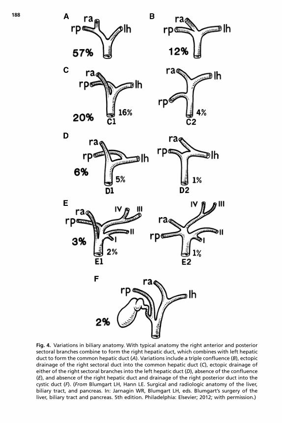
Fig. 4. Variations in biliary anatomy. With typical anatomy the right anterior and posteriorsectoral branches combine to form the right hepatic duct, which combines with left hepaticduct to form the common hepatic duct (A). Variations include a triple confluence (B), ectopicdrainage of the right sectoral duct into the common hepatic duct (C), ectopic drainage ofeither of the right sectoral branches into the left hepatic duct (D), absence of the confluence(E), and absence of the right hepatic duct and drainage of the right posterior duct into thecystic duct (F). (From Blumgart LH, Hann LE. Surgical and radiologic anatomy of the liver,biliary tract, and pancreas. In: Jarnagin WR, Blumgart LH, eds. Blumgart’s surgery of theliver, biliary tract and pancreas. 5th edition. Philadelphia: Elsevier; 2012; with permission.)
188
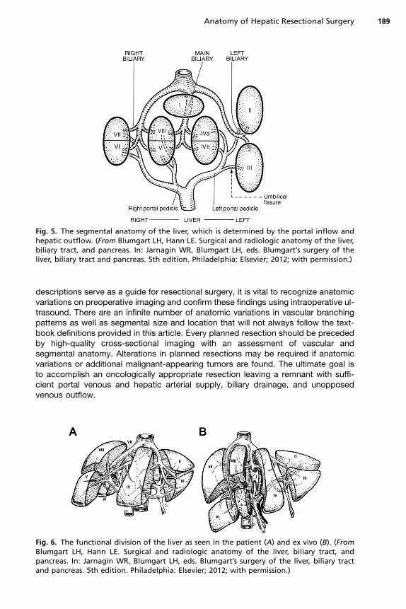
Fig. 5. The segmental anatomy of the liver, which is determined by the portal inflow andhepatic outflow. (From Blumgart LH, Hann LE. Surgical and radiologic anatomy of the liver,biliary tract, and pancreas. In: Jarnagin WR, Blumgart LH, eds. Blumgart’s surgery of theliver, biliary tract and pancreas. 5th edition. Philadelphia: Elsevier; 2012; with permission.)
Anatomy of Hepatic Resectional Surgery 189
descriptions serve as a guide for resectional surgery, it is vital to recognize anatomicvariations on preoperative imaging and confirm these findings using intraoperative ul-trasound. There are an infinite number of anatomic variations in vascular branchingpatterns as well as segmental size and location that will not always follow the text-book definitions provided in this article. Every planned resection should be precededby high-quality cross-sectional imaging with an assessment of vascular andsegmental anatomy. Alterations in planned resections may be required if anatomicvariations or additional malignant-appearing tumors are found. The ultimate goal isto accomplish an oncologically appropriate resection leaving a remnant with suffi-cient portal venous and hepatic arterial supply, biliary drainage, and unopposedvenous outflow.
Fig. 6. The functional division of the liver as seen in the patient (A) and ex vivo (B). (FromBlumgart LH, Hann LE. Surgical and radiologic anatomy of the liver, biliary tract, andpancreas. In: Jarnagin WR, Blumgart LH, eds. Blumgart’s surgery of the liver, biliary tractand pancreas. 5th edition. Philadelphia: Elsevier; 2012; with permission.)
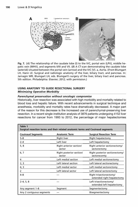
Fig. 7. (A) The relationship of the caudate lobe (I) to the IVC, portal vein (LPV), middle he-patic vein (MHV), and segments II/III and VII. (B) A CT scan demonstrating the caudate lobe(asterisk) situated between the portal vein (arrow) and the IVC (V). a, Aorta. (From BlumgartLH, Hann LE. Surgical and radiologic anatomy of the liver, biliary tract, and pancreas. In:Jarnagin WR, Blumgart LH, eds. Blumgart’s surgery of the liver, biliary tract and pancreas.5th edition. Philadelphia: Elsevier; 2012; with permission.)
Lowe & D’Angelica190
USING ANATOMY TO GUIDE RESECTIONAL SURGERYMinimizing Operative Morbidity
Parenchymal preservation without oncologic compromiseHistorically, liver resection was associated with high morbidity and mortality related toblood loss and hepatic failure. With recent advancements in surgical technique andanesthesia, morbidity and mortality rates have dramatically decreased. A major partof the reason for this decrease is the increased use of parenchymal-preserving liverresection. In a recent single-institution analysis of 3876 patients undergoing 4152 liverresections for cancer from 1993 to 2012, the percentage of major hepatectomies
Table 1Surgical resection terms and their related anatomic terms and Couinaud segments
Couinaud Segments Anatomic Term Surgical Resection Term
5–8 Right liver Right hepatectomy
2–4 Left liver Left hepatectomy
5, 8 Right anterior section/sector
Right anterior sectionectomy/sectorectomy
6, 7 Right posterior section/sector
Right posterior sectionectomy/sectorecomty
4 Left medial section Left medial sectionectomy
2, 3 Left lateral section Left lateral sectionectomy
4, 3 Left medial sector Left medial sectorectomy
2 Left lateral sector Left lateral sectorectomy
4–8 — Right trisectionectomy/extended right hepatectomy
2–4, 5, 8 — Left trisectionectomy/extended left hepatectomy
Any segment 1–8 Segment Segmentectomy
Any 2 contguous segments — Bisegmentectomy
Anatomy of Hepatic Resectional Surgery 191
decreased from 66% to 36%. During this same period, the 90-daymortality decreasedfrom 5% to 1.6%, and the perioperative morbidity decreased from 53% to 20%. Overthis period of time, the mortality of major resections such as trisectionectomy (6%) andright hepatectomy (3%) did not change, strongly suggesting that the improvements inperioperative outcome were mostly related to the increase in the percentage ofparenchymal-sparing resections.3 The theoretic concern with parenchymal-sparingsurgery is compromised oncologic outcome. To the contrary, numerous studieshave shown that performing parenchymal-sparing resections does not result in inferiorsurvival, particularly for colorectal liver metastases.4–10 A meta-analysis of 7 non-randomized controlled studies found no significant difference in surgical margins,overall survival, and disease-free survival between patients undergoing anatomicand nonanatomic resections.11
Minimizing blood lossAlthough specific techniques employed to minimize blood loss will be covered else-where, it is important to appreciate the importance of limiting hemorrhage, which his-torically has contributed to significant perioperative morbidity. This can beaccomplished most successfully by maintaining a low central venous pressure(CVP) during parenchymal resection. This reduces hepatic venous distention, whichsignificantly decreases backbleeding when the liver parenchymal is transected.12
Maintaining low CVP during liver resection reduces intraoperative blood loss, in-hospital mortality, and postoperative morbidity.13 Postoperative renal failure ispossible after low CVP anesthesia, but this complication is rare if the patient is resus-citated to euvolemia immediately upon completion of the parenchymal transection. Ina recent study of 2116 patients undergoing low CVP-assisted hepatectomy, less than1% of patients developed clinically relevant acute kidney injury.14
Vascular occlusion also can contribute to control of intraoperative blood loss. He-patic inflow arrest was first described by Pringle in the setting of trauma,15 and this re-mains the standard method by which control of portal venous and hepatic arterialinflow is gained. Encircling the hepatoduodenal ligament allows for simultaneous oc-clusion of the portal vein and hepatic artery. Intermittent occlusion permits longer pe-riods of occlusion, but the length of inflow occlusion should be limited as much aspossible, particularly with an abnormal liver. More complex vascular exclusion tech-niques, such as hepatic vascular exclusion, will be covered elsewhere. Although inflowocclusion is not required for all cases, it is an important tool to be used if there is inflowbleeding during hepatic transection.
GENERAL ANATOMIC CONCEPTS AND PITFALLS IN SPECIFIC RESECTIONSRight Hepatectomy
Descriptions of this operation are common, and there are numerous techniques toaccomplish a safe and effective right hepatectomy. Most commonly, the right liveris mobilized completely off of the diaphragm and vena cava by dividing the oftennumerous retrohepatic venous branches. The right caval ligament, which runs laterallyto the right hepatic vein and envelops the vena cava posteriorly circumferentially tojoin the left side of the caudate, must be encircled and divided to expose the right he-patic vein. Dissection along the vena cava to encircle the right hepatic vein can beaccomplished after this ligament is divided. Inflow to the liver can be taken in a numberof ways, including extrahepatic ligation of inflow vessels or ligation of the right inflowpedicle inside the liver. Pedicle ligation can be carried out through hepatotomies or bysplitting the liver down to the pedicle in an anterior approach. If an extrahepatic ligationof vessels is carried out, it is advisable, if possible, to take the right hepatic bile duct

Lowe & D’Angelica192
intrahepatically in the main pedicle to minimize the chance of contralateral biliaryinjury. Regardless of the approach to the inflow vessels, knowledge of the inflow ves-sels and their anatomy and variation is critical. Once inflow is taken, the right hepaticvein is divided and most commonly divided extrahepatically. The line of transection isalong the middle hepatic vein, which can be taken or preserved depending on thelocation of the tumor to be resected. Maintaining transection in this plane is importantin order to prevent leaving ischemic liver tissue behind.There are several anatomic variations that are relevant to the performance of this
operation. A large inferior right hepatic vein can be present and serve as the primaryvenous drainage of segment VI. This needs to be divided as the liver is mobilized off ofthe vena cava. The right portal vein has a wide range of anatomic presentationsranging from a long segment prior to sectoral/segmental branching, to the completelack of a true right portal vein with immediate branching into segmental/sectoralbranches. Lastly, the right anterior portal vein and pedicle can originate off of theleft inflow and necessitate intrahepatic control. The right portal vein almost alwaysgives off a small posterior branch to the caudate process that can be a commonsource of bleeding during extrahepatic dissection. The right hepatic artery typicallyruns posterior to the common hepatic duct, but in about 10% to 20% of cases, itruns anterior to the duct. A replaced right hepatic artery is common and runs posteri-orly in the porta between the portal vein and common bile duct. The most commonbiliary anomalies include a right sectoral branch draining into the left hepatic duct.Lastly, the left hepatic duct as it arises from the common hepatic duct can originateclose to the right inflow, and injury to this duct during transection of right-sided inflowstructures can be disastrous. Lowering of the hilar plate is helpful to both identify andprotect the left hepatic duct. These abnormalities can all be detected on high-qualitycross-sectional imaging and should be evaluated prior to operation.
Right Posterior Sectorectomy
This resection removes segments VI and VII of the liver, and the principle transectionplane is the right portal scissura along the right hepatic vein. The right liver should bemobilized as is done for a right hepatectomy. Although not critical for every case, it isadvisable to dissect and control the right hepatic vein. Knowledge of the posterior sec-toral inflow anatomy is important. There often is not a common posterior sectoral trunkdividing into segment VI and VII branches. Often the segment VI and VII inflow pediclesbranch almost immediately off of the right inflow pedicle. This requires taking thesepedicles individually inside the liver. Although the posterior sectoral inflow vesselscan be dissected extrahepatically, this is, in general, difficult and not necessary.The plane of the right hepatic vein can be visualized by intraoperative ultrasoundand marked. The transection plane is somewhat horizontal toward the right edge ofthe vena cava. The transection plane is divided down to the inflow pedicles, whichare divided intrahepatically to ensure that the dissection has not gone too far to theleft into the anterior sectoral pedicles. Although preferable to preserve the right hepat-ic vein, this vessel can be taken, since the middle hepatic vein is the principal venousdrainage for the anterior sector.
Right Anterior Sectorectomy
This resection is challenging in that it requires 2 long lines of transection to encompasssegments V and VIII. The main anterior pedicle at its origin will almost always take all ofthe inflow to these segments, although occasionally segment V will have extra inflowpedicles from the posterior sectoral pedicle or the central inflow pedicles at the mainbranching points in the porta. The key to the operation is identification and ligation of
Anatomy of Hepatic Resectional Surgery 193
the main anterior pedicle, which, is best accomplished intrahepatically if the tumor al-lows. The main anterior pedicle can be found by transecting along the middle hepaticvein, since it terminates in branches coursing to the left of and anterior to the anteriorpedicle. Thus transection of the liver in the main portal scissura exposes the main rightanterior pedicle after ligating and dividing these terminal middle hepatic veinbranches. The middle hepatic vein can be taken in this operation, since segment IVcan drain into the umbilical vein, which most commonly originates from the left hepaticvein. It is critical to preserve the right hepatic vein, since this is the venous drainage ofthe posterior sector. The posterior extent of dissection does not require division downto the vena cava, since the well-vascularized caudate lobe lies posteriorly and doesnot need to be taken as part of this operation.
Central Hepatectomy
This resection is similar to an anterior sectorectomy, except that the left extent of theresection takes some or all of segment IV and mandates resection of the middle he-patic vein. The inflow to segment IV can easily be taken intrahepatically while transect-ing the liver to the right of the umbilical fissure. The segment IV pedicles are almostalways multiple and do not neatly divide into segment IVA and IVB branches. At thebase of segment IV, the dissection must turn sharply to the right to avoid the mainleft inflow structures. Similar to an anterior sectorectomy, dissection then commencesto the right, taking terminal middle hepatic vein branches to isolate the main right ante-rior inflow pedicle. Other technical issues are the same as an anterior sectorectomy.
Left Hepatectomy
The left liver is situated anterior to the caudate lobe and therefore does not requiremobilization off of the vena cava. The inflow to the left liver is more accessible thanthe right-sided structures, and extrahepatic isolation and division are fairly straightfor-ward. The left hepatic artery courses cephalad along the left side of the porta hepatisanteriorly and is relatively easy to encircle. Once encircled, it should be ensured thatone has not come around the proper hepatic artery prior to branching; this can happeneasily if the branching is more distal. Simple dissection or clamping with palpation of aright-sided pulse is sufficient. Often the left hepatic artery branches into medial(segment IV) and lateral (segments II and III) branches, but a common variation isthat the segment IV branch (previously called the middle hepatic artery) comes offthe right hepatic artery. Posteriorly, the left portal vein can be dissected. Confirmationof portal anatomy requires visualization of the portal bifurcation. The left portal veingives off a small branch to the caudate lobe, which should be identified and preservedif possible. Just beyond the caudate branch, the ligamentum venosum inserts into theleft portal vein and can be seen with careful dissection. This area of insertion usually issurrounded by fibrous tissue. The safest place to divide the left portal vein is betweenthe caudate branch and the insertion of the ligamentum venosum. The left bile ductruns at the base of segment IV before branching into the left liver. Whenever possible,the left bile duct should be divided within the main left pedicle intrahepatically. Biliaryanomalies are most relevant for left-sided resections, since right sectoral branchescan be found draining into the left bile duct. If a tumor mandates division of the leftbile duct extrahepatically, these biliary anomalies should be sought and definedwith preoperative imaging and during dissection. The left hepatic vein typicallycourses anterior to the segment II pedicle and then runs posterior to the segment IIIpedicle.A left hepatectomy can be performed either by taking or preserving the middle he-
patic vein. Although the middle hepatic vein is typically the dominant venous drainage
Lowe & D’Angelica194
of the right anterior sector, there is almost always sufficient venous collateral to theright hepatic vein to provide adequate drainage. It is often difficult to isolate the leftand middle hepatic vein individually in an extrahepatic location, but sometimes withcareful dissection this can be done. In order to divide the left and middle hepatic veinsat their common trunk extrahepatically, dissection between the right and middle he-patic veins superiorly at the suprahepatic vena cava demonstrates a wide tunnel. Pos-terior to the left liver, the ligamentum venosum is divided and retracted cephalad toexpose a space anterior to the caudate lobe and between the hepatic parenchymaand the left/middle hepatic vein common trunk. This is often a tight space and requiresmeticulous dissection to avoid vascular injury or bleeding. Alternatively, either vein canbe approached by splitting the liver back to its origin and dividing the veins after theparenchymal dissection has exposed their origin. As for a right hepatectomy, the planeof transection is in the main portal scissura in the plain of the middle hepatic vein.
Left Lateral Sectionectomy
A left lateral sectionectomy is a relatively straightforward resection that is most easilyaccomplished by taking inflow and outflow structures intrahepatically. The segmentII and III pedicles are relatively constant in branching and location but can occasion-ally vary. Sometimes their origins are close, and they may even share a commontrunk. Small branches other than the main segmental pedicles are commonlyencountered. Although these inflow pedicles can be dissected in the umbilicalfissure or encircled by making flanking hepatotomies, the simplest method toexpose them is to split the liver in the plane just to the left of the umbilical fissuredown to their origins. In this manner, each pedicle can be encircled and divided.The left hepatic vein typically joins the middle hepatic vein in a relatively intrahepaticposition, and therefore isolating the left hepatic vein extrahepatically can be difficultand is usually not necessary. The left hepatic vein origin can be defined by splittingthe liver to the point where the left hepatic vein joins the middle hepatic vein. The um-bilical vein runs in the plane anterior to the umbilical fissure and most commonly joinsthe left hepatic vein, although sometimes it will come off the common channel ormiddle hepatic vein. The umbilical vein is in the plane of resection for a left lateralsectionectomy and can be preserved or taken; either way, its location should benoted as it can be a source of bleeding during transection.
Wedge/Atypical/Single Segment Resections
Detailed descriptions of every potential atypical or single segment resection arebeyond the scope of this article. Nonetheless, careful review of anatomic variationand careful intrahepatic dissection with the help of intraoperative ultrasound shouldbe able to identify any inflow or outflow structure in order to accomplish any ofthese resections in proper anatomic planes without devascularizing large amountsof hepatic parenchyma. Single-segment resections require careful isolation of theinflow pedicle or pedicles, and knowledge of common variations is necessary.For example, segment VIII typically has a single dominant pedicle that dividesinto a ventral and dorsal branch. This is a remarkably consistent anatomic finding.On the other hand, segment V typically does not have a dominant inflow pedicleand has numerous inflow branches, most commonly coming off the main right ante-rior pedicle. Segmental resections also mandate that parenchymal transection takesplace in a scissura along the main hepatic veins. As operations for multiple bilobartumors have become more common the use of wedge and atypical resections hasbecome more common. From an anatomic perspective, the surgeon, with carefulreview of imaging and use of intraoperative ultrasound, must ensure that these
Anatomy of Hepatic Resectional Surgery 195
resections do not divide major inflow and outflow vessels to other parts of the liver.As long as major vessels are preserved, these atypical resections outside of typicalanatomic planes are safe and effective.
REFERENCES
1. Couinaud C. Anatomic principles of left and right regulated hepatectomy: tech-nics. J Chir (Paris) 1954;70(12):933–66 [in French].
2. Strasberg SM. Nomenclature of hepatic anatomy and resections: a review of theBrisbane 2000 system. J Hepatobiliary Pancreat Surg 2005;12(5):351–5.
3. Kingham TP, Correa-Gallego C, D’Angelica MI, et al. Hepatic parenchymal pres-ervation surgery: decreasing morbidity and mortality rates in 4,152 resections formalignancy. J Am Coll Surg 2015;220(4):471–9.
4. Gold JS, Are C, Kornprat P, et al. Increased use of parenchymal-sparing surgeryfor bilateral liver metastases from colorectal cancer is associated with improvedmortality without change in oncologic outcome: trends in treatment over time in440 patients. Ann Surg 2008;247(1):109–17.
5. Karanjia ND, Lordan JT, Quiney N, et al. A comparison of right and extended righthepatectomy with all other hepatic resections for colorectal liver metastases: aten-year study. Eur J Surg Oncol 2009;35(1):65–70.
6. Kokudo N, Tada K, Seki M, et al. Anatomical major resection versus nonanatom-ical limited resection for liver metastases from colorectal carcinoma. Am J Surg2001;181(2):153–9.
7. Lalmahomed ZS, Ayez N, van der Pool AE, et al. Anatomical versus nonanatom-ical resection of colorectal liver metastases: is there a difference in surgical andoncological outcome? World J Surg 2011;35(3):656–61.
8. Stewart GD, O’Suilleabhain CB, Madhavan KK, et al. The extent of resection in-fluences outcome following hepatectomy for colorectal liver metastases. Eur JSurg Oncol 2004;30(4):370–6.
9. Tanaka K, Shimada H, Matsumoto C, et al. Impact of the degree of liver resectionon survival for patients with multiple liver metastases from colorectal cancer.World J Surg 2008;32(9):2057–69.
10. von Heesen M, Schuld J, Sperling J, et al. Parenchyma-preserving hepaticresection for colorectal liver metastases. Langenbecks Arch Surg 2012;397(3):383–95.
11. Sui CJ, Cao L, Li B, et al. Anatomical versus nonanatomical resection of colorectalliver metastases: a meta-analysis. Int J Colorectal Dis 2012;27(7):939–46.
12. Jones RM, Moulton CE, Hardy KJ. Central venous pressure and its effect onblood loss during liver resection. Br J Surg 1998;85(8):1058–60.
13. Chen H, Merchant NB, Didolkar MS. Hepatic resection using intermittent vascularinflow occlusion and low central venous pressure anesthesia improves morbidityand mortality. J Gastrointest Surg 2000;4(2):162–7.
14. Correa-Gallego C, Berman A, Denis SC, et al. Renal function after low centralvenous pressure-assisted liver resection: assessment of 2116 cases. HPB(Oxford) 2015;17(3):258–64.
15. Pringle JH. V. Notes on the arrest of hepatic hemorrhage due to trauma. Ann Surg1908;48(4):541–9.
Related Documents