european journal of histochemistry a journal of functional cytology ISSN 1121-760X volume 51/supplement 1 2007 under the auspices of the University of Pavia, Italy Trimestrale – Sped. Abb. Post. – 45% art. 2, comma 20B, Legge 662/96 - Filiale di Pavia. Il mittente chiede la restituzione dei fascicoli non consegnati impegnandosi a pagare le tasse dovute ejh The Fathers of Italian Histology Guest Editors F.A. Manzoli, P. Carinci

Welcome message from author
This document is posted to help you gain knowledge. Please leave a comment to let me know what you think about it! Share it to your friends and learn new things together.
Transcript
european journal of histochemistry
a journal of functional cytology
ISSN 1121-760X
volume 51/supplement 1
2007
under the auspices of
the University of Pavia, Italy
Trimestrale–Sped.Abb.Post.–45%art.2,comma20B,Legge662/96-FilialediPavia.Ilmittentechiedelarestituzionedeifascicolinonconsegnatiimpegnandosiapagareletassedovute
ejhThe Fathers of Italian Histology
Guest Editors
F.A. Manzoli, P. Carinci

TABLE OF CONTENTS
Osteogenic and chondrogenic differentiation: comparison of human andrat bone marrow mesenchymal stem cells cultured into polymeric scaffoldsB. Zavan, C. Giorgi, G.P. Bagnara, V. Vindigni, G. Abatangelo, R. Cortivo ...................1-8
Tendon crimps and peritendinous tissues responding to tensional forcesM. Franchi, M. Quaranta, V. De Pasquale, M. Macciocca, E. Orsini, A.Triré, V. Ottani,A. Ruggeri ............................................................................................................9-14
The mechanism of transduction of mechanical strains into biological signalsat the bone cellular levelG. Marotti, C. Palumbo .......................................................................................15-20
Cytoskeletal reorganization in skeletal muscle differentiation:from cell morphology to gene expressionL. Formigli, E. Meacci, S. Zecchi-Orlandini, G.E. Orlandini....................................21-28
Sarcoglycan subcomplex in normal and pathological human muscle fibersG. Anastasi, G. Cutroneo, G. Rizzo, A. Favaloro ....................................................29-34
Stem cell-mediated muscle regeneration and repair in aging and neuromusculardiseasesA. Musarò, C. Giacinti, L. Pelosi, G. Dobrowolny, L. Barberi, C. Nardis, D. Coletti,B.M. Scicchitano, S. Adamo, M. Molinaro ............................................................35-44
Anatomy of emotion: a 3D study of facial mimicryV. F. Ferrario, C. Sforza .......................................................................................45-52
New findings on 3-D microanatomy of cellular structures in human tissuesand organs. An HRSEM studyA. Riva, F. Loy, R. Isola, M. Isola, G. Conti, A. Perra, P. Solinas, F.Testa Riva ........53-58
Non-traditional large neurons in the granular layer of the cerebellar cortexG. Ambrosi, P. Flace, L. Lorusso, F. Girolamo, A. Rizzi, L. Bosco, M. Errede,D. Virgintino, L. Roncali, V. Benagiano ................................................................59-64
The solitary chemosensory cells and the diffuse chemosensory systemof the airwayF. Osculati, M. Bentivoglio, M. Castellucci, S. Cinti, C. Zancanaro, A. Sbarbati .......65-72
The modality of transendothelial passage of lymphocytes and tumor cellsin the absorbing lymphatic vesselG. Azzali.............................................................................................................73-78
Scatter factor-dependent branching morphogenesis:structural and histological featuresP. Comoglio, L.Trusolino, C. Boccaccio .................................................................79-92
Models of epithelial histogenesisA. Casasco, M. Casasco, A.Icaro Cornaglia, F. Riva, A. Calligaro .........................93-100
Adult stem cells: the real root into the embryo?G. Zummo, F. Bucchieri, F. Cappello, M. Bellafiore, G. La Rocca, S. David,V. Di Felice, R. Anzalone, G. Peri, A. Palma, F. Farina.......................................101-104
Extracellular matrix and growth factors in the pathogenesis of some craniofacialmalformationsP. Carinci, E. Becchetti,T. Baroni, F. Carinci, F. Pezzetti, G. Stabellini,P. Locci, L. Scapoli, M.Tognon, S. Volinia, M. Bodo..........................................105-116
The nuclear envelope, human genetic diseases and ageingN.M. Maraldi, G. Mazzotti, R. Rana, A. Antonucci, R. Di Primio, L. Guidotti .....117-124
Nuclear phosphatidylinositol 3,4,5-trisphosphate, phosphatidylinositol 3-kinase,Akt, and PTEN: emerging key regulators of anti-apoptotic signalingand carcinogenesisA. M. Martelli, L. Cocco, S. Capitani, S. Miscia, S. Papa, F. A. Manzoli .............125-132
Neuroendocrine regulation and tumor immunityR.Toni, P. Mirandola, G. Gobbi, M.Vitale.........................................................133-138
european journal
of histochemistry
ISSN 1121-760X
volume 51/supplement 1
2007
table of contents
ejh

European Journal of Histochemistry — Vol. 51 supplement 1 2007 — pp. 1-140
Published by the Società Italiana di Istochimica
©Società Italiana di Istochimica
Editorial Office: Dipartimento di Biologia AnimalePiazza Botta 10 – 27100 Pavia (Italy)Phone: +39.0382.986420 - Fax: +39.0382.986325E-mail: [email protected]
Printed quarterly by:
Tipografia PIME Editrice srlvia Vigentina 13627100 PAVIA, ItalyPhone: +39.0382.572169 – Fax +39.0382.572102E-mail: [email protected] no. 00280810185
Editing by:
medit SNCvia G. Belli, 427100 Pavia, ItalyE-mail: [email protected]
Annual Subscriptions
Europe: Euro 160All other Countries: $ 200
Subscriptions, cancellations, business correspondence andany enquiries must be sent to the Tipografia PIME Editricesrl, Pavia, Italy.Cancellations must be received before the end of Septemberto take effect at the end of the same year.
No part of this publication may be reproduced, stored in aretrieval system or transmitted in any form or by any means(electronic, electrostatic, magnetic type, mechanical, photo-copying or otherwise) without written permission by thePublishers.
Reg. Tribunale di Pavia n. 289/23.2.1984.
Supported by the Ministero per i Beni e le Attività Culturali,Italy as a publication of high cultural value.
Associato all’USPIUnione Stampa Periodica Italiana
Disclaimer. Whilst every effort is made by the publishers and theeditorial board to see that no inaccurate or misleading data,opinion or statement appears in this journal, they wish to makeit clear that the data and opinions appearing in the articles oradvertisements herein are the responsibility of the contributoror advisor concerned. Accordingly, the publisher, the editorialboard and their respective employees, officers and agentsaccept no liability whatsoever for the consequences of any inac-curate or misleading data, opinion or statement.
Editor-in-ChiefM.G. Manfredi Romanini
Dipartimento di Biologia Animale, Università di Pavia
Co-EditorC. Pellicciari
Dipartimento di Biologia Animale, Università di Pavia

The European Journal of Histochemistry was
founded in 1954 by Maffo Vialli and published till
1979 under the title of Rivista di Istochimica
Normale e Patologica , from 1980 to 1990 as
Basic and Applied Histochemistry and in 1991 as
European Journal of Basic and Applied
Histochemistry. It is published under the auspices
of the Università of Pavia and of the Ferrata Storti
Foundation, Pavia, Italy.
The European Journal of Histochemistry is the offi-
cial organ of the Italian Society of Histochemistry
and a member of the journal subcommittee of the
International Federation of Societies for
Histochemistry and Cytochemistry (IFSHC).
The Journal publishes original papers, technical
reports, letters to the editor, review articles con-
cerning investigations performed with the aid of
biophysical, biochemical, molecular-biological,
enzymatic, immunohistochemical, cytometric, and
image analysis techniques.
Areas of particular interest to the European
Journal of Histochemistry include:
- functional cell and tissue biology in animals and
plants;
- cell differentiation and death;
- cell-cell interaction and molecular trafficking;
- biology of cell development and senescence;
- nerve and muscle cell biology;
- cellular basis of diseases
Managing EditorsC.A. Redi (Dipartimento di Biologia Animale, Universitàdi Pavia)E. Solcia (Dipartimento di Patologia Umana ed Eredi-taria, Università di Pavia)
for Europe: J.E. Scott (University of Manchester)for Japan: M. Fukuda (Fukui Medical School, Fukui)for Latin America: R.F. Donoso (Universidad de Chile)for USA: H.A. Crissman (Los Alamos National Laboratory)
Assistant EditorsM. Biggiogera (Università di Pavia), for MinireviewsD. Formenti (Università di Pavia), Advisor for statisticsP. Rovere Querini (H. San Raffaele, Milan), for Special issues
Editorial SecretaryC. Soldani (Università di Pavia)
Managing Board of the Italian Society of Histo-chemistry for the years 2006-2009N.M. Maraldi (President) Università di BolognaG. Meola (Vice-President) Università di MilanoA. Lauria (Secretary) Università di MilanoG. Bottiroli (Member) National Research Council, PaviaA. Paparelli (Member) Università di PisaE. Bonucci (Past-President) Università di Roma
Editorial BoardB. Agostini, Heidelberg, P. Bonfante, Torino, E. Bonucci,Roma, V.YA. Brodsky,Moscow, G. Bussolati, Torino,F. Clementi, Milano, L. Cocco, Bologna, R.R. Cowden,Mobile, A. Diaspro, Genova, G. Donelli, Roma, S. Fakan,Lausanne, G. Gerzeli, Pavia, R.S. Gilmour, Cambridge,G.Giordano Lanza, Napoli, C.E. Grossi, Genova, M.Gutierrez, Cadiz,W. Hilscher, Neuss, H. Luppa, Leipzig,F.A. Manzoli, Bologna, G. Meola, Milano, G.S. Montes,São Paulo, W. Nagl, Kaiserslautern, K. Nakane,Mountain View, CH. Pilgrim, Ulm, C.A. Pinkstaff,Morgantown, J.M. Polak, London, G.N. Ranzani, Pavia,E. Reale,Hannover, T. Renda,Roma,G. Rindi,Parma,A.Riva, Cagliari, C. Sotelo, Paris, A.T. Sumner, EastLothian, J.P. Tremblay, Quebec, P. Van Duijn, Leiden, S.Van Noorden, London.
Members appointed by Scientific SocietiesE. Bàcsy (Histochemical Section of the Society of theHungarian Anatomists), B. Bloch (Societé Française deMicroscopie Electronique), A. Lòpez Bravo (FederacionIberoamericana de Biologia Celular y Molecular), B.Bilinska (Polish Histochemical and CytochemicalSociety), M.A. Nahir (Israel Society for Histochemistryand Cytochemistry), D. Onicescu (Romanian Society ofHistochemistry and Cytochemistry), W. Ovtscharoff(Histochemical Section of the Society of Anatomy,Histology and Embryology of Bulgaria), P. Panula(Finnish Society of Histochemistry and Cytochemistry),L. J. Pelliniemi (Finnish Society of Histochemistry andCytochemistry), J. Renau Piqueras (Spanish Society forCell Biology), B. Rodé (Histochemical and CytochemicalSection of Croatian Society of Anatomists), M. Rosety(Sociedad Iberoamericana de Histoquimica y Cito-quimica)
European Journal of Histochemistrya journal of functional cytology
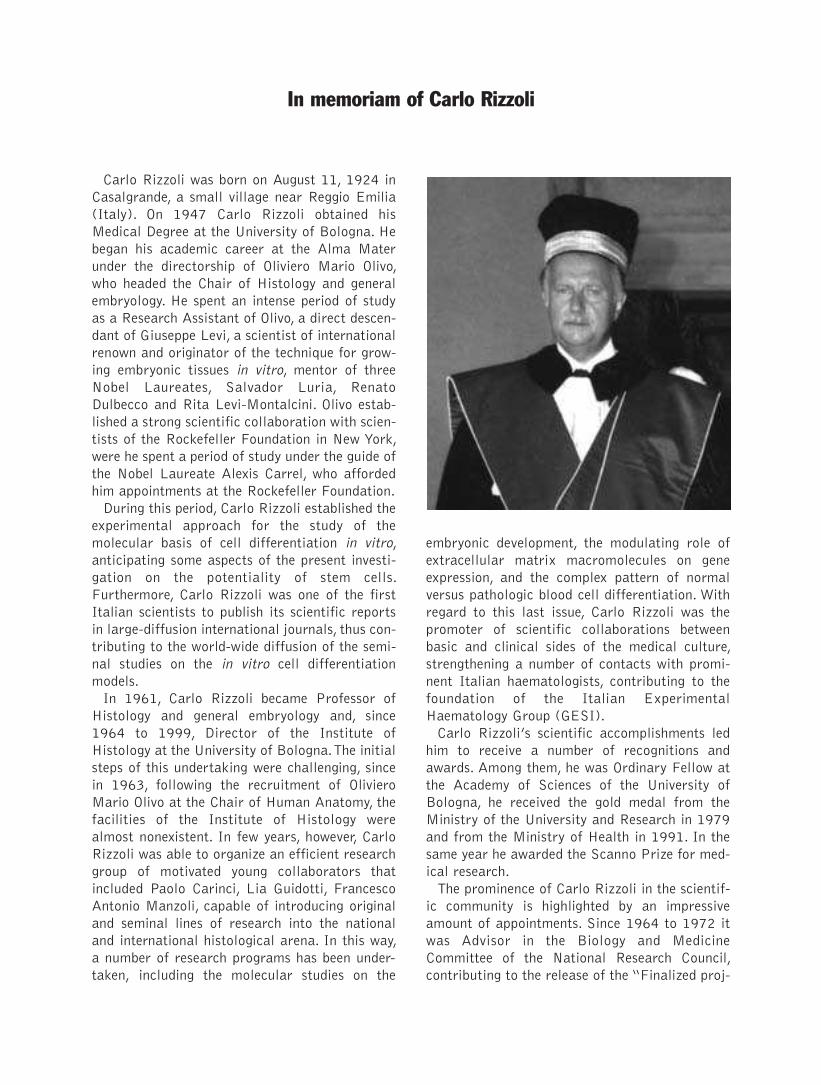
Carlo Rizzoli was born on August 11, 1924 in
Casalgrande, a small village near Reggio Emilia
(Italy). On 1947 Carlo Rizzoli obtained his
Medical Degree at the University of Bologna. He
began his academic career at the Alma Mater
under the directorship of Oliviero Mario Olivo,
who headed the Chair of Histology and general
embryology. He spent an intense period of study
as a Research Assistant of Olivo, a direct descen-
dant of Giuseppe Levi, a scientist of international
renown and originator of the technique for grow-
ing embryonic tissues in vitro, mentor of three
Nobel Laureates, Salvador Luria, Renato
Dulbecco and Rita Levi-Montalcini. Olivo estab-
lished a strong scientific collaboration with scien-
tists of the Rockefeller Foundation in New York,
were he spent a period of study under the guide of
the Nobel Laureate Alexis Carrel, who afforded
him appointments at the Rockefeller Foundation.
During this period, Carlo Rizzoli established the
experimental approach for the study of the
molecular basis of cell differentiation in vitro,
anticipating some aspects of the present investi-
gation on the potentiality of stem cells.
Furthermore, Carlo Rizzoli was one of the first
Italian scientists to publish its scientific reports
in large-diffusion international journals, thus con-
tributing to the world-wide diffusion of the semi-
nal studies on the in vitro cell differentiation
models.
In 1961, Carlo Rizzoli became Professor of
Histology and general embryology and, since
1964 to 1999, Director of the Institute of
Histology at the University of Bologna.The initial
steps of this undertaking were challenging, since
in 1963, following the recruitment of Oliviero
Mario Olivo at the Chair of Human Anatomy, the
facilities of the Institute of Histology were
almost nonexistent. In few years, however, Carlo
Rizzoli was able to organize an efficient research
group of motivated young collaborators that
included Paolo Carinci, Lia Guidotti, Francesco
Antonio Manzoli, capable of introducing original
and seminal lines of research into the national
and international histological arena. In this way,
a number of research programs has been under-
taken, including the molecular studies on the
embryonic development, the modulating role of
extracellular matrix macromolecules on gene
expression, and the complex pattern of normal
versus pathologic blood cell differentiation. With
regard to this last issue, Carlo Rizzoli was the
promoter of scientific collaborations between
basic and clinical sides of the medical culture,
strengthening a number of contacts with promi-
nent Italian haematologists, contributing to the
foundation of the Italian Experimental
Haematology Group (GESI).
Carlo Rizzoli’s scientific accomplishments led
him to receive a number of recognitions and
awards. Among them, he was Ordinary Fellow at
the Academy of Sciences of the University of
Bologna, he received the gold medal from the
Ministry of the University and Research in 1979
and from the Ministry of Health in 1991. In the
same year he awarded the Scanno Prize for med-
ical research.
The prominence of Carlo Rizzoli in the scientif-
ic community is highlighted by an impressive
amount of appointments. Since 1964 to 1972 it
was Advisor in the Biology and Medicine
Committee of the National Research Council,
contributing to the release of the “Finalized proj-
In memoriam of Carlo Rizzoli

ects” to ensure an European dimension to the
Italian research. Since 1968 to 1976 it was Dean
of the Faculty of Medicine at the University of
Bologna and, since 1976 to 1985, Chancellor of
the University of Bologna. As Chancellor of the
Alma Mater, Carlo Rizzoli had to face the most
risky period of the student protest during the sev-
enties; his mettle and cleverness succeeded in
maintaining the balance between the authority of
the institution and the requests of innovation.
During this period he supported the development
of research programs, the widening of the posi-
tions both of the teaching and technical staff,
establishing a sound management at the
University of Bologna.
Carlo Rizzoli was also appointed, since 1976 to
1989, as President of the CINECA, the most
important institution for the electronic computa-
tion in Italy, endowing the Centre with the most
powerful and up-to-date electronic computers
available at that time. As President of the
National Institute for Physical Training (ISEF),
since 1965 to 1999, he founded the Seats of
Verona and Catanzaro and obtained the recogni-
tion of the Physical Training Faculty into the
Medical School. Carlo Rizzoli was among the
founders and Member of the Board of Directors
of the University “G. D’Annunzio” in Chieti, since
1976 to 1989, and it contributed to the develop-
ment of the Medical School. As President of the
Italian Society of Histochemistry, Carlo Rizzoli
gave a strong contribution to the development of
this branch of the morphological sciences.
The Italian histological school founded by Carlo
Rizzoli includes a large group of his pupils and
collaborators which head the Department of
Histology or Human Anatomy in the Universities
of Bologna, Ancona, Chieti, Ferrara, Genova,
Perugia,Trieste, Parma, Urbino, Cassino.
Despite this impressive involvement in academ-
ic and administrative appointments, Carlo Rizzoli
never neglected its role in teaching and mentor-
ing. Thanks to the effort and the commitment of
Carlo Rizzoli and Valerio Monesi, histology, which
was an ancillary share of anatomy, rose to the
dignity of a basic teaching. His Atlas of Histology,
in cooperation with Carla Castaldini and Maria
Antonietta Brunelli, and his contribution to the
treatise of Histology formerly edited by Valerio
Monesi are landmark textbooks which have been
used by a generation of Italian students. Carlo
Rizzoli was a fascinating speaker and left a
strong and enduring mark on all of the pupils that
have been the chance of listen his lectures. During
the last period of his career, before its retirement,
Carlo Rizzoli continued to teach with the same
passion and involvement, joining at its scientific
knowledge its wide experience and its foresight of
the future development of the Medical Sciences.
In remembering Carlo Rizzoli, we celebrate his
legacy his scientific flair, his impressive academic
commitment, his wide classical culture. We will
miss his many-sided personality, his skill in over-
coming family tragic events by finding in the daily
engagement the reasons of the existence.
Francesco Antonio Manzoli
Paolo Carinci
This supplement of the European Journal of
Histochemistry is dedicated to the memory of
Carlo Rizzoli.
The evaluation of the scientific contribution of
Carlo Rizzoli to the evolution of the morphologi-
cal sciences in Italy can be appreciated by con-
sidering the peculiar period of time, the fifties and
the sixties of the past century, a crucial moment
for the identification of the main fields of
research which will characterize the impressive
strengthening of cell biology. These trends were,
from the beginning, based on either an analytical
or a synthetic approach.The morphological trend,
mainly based on the ultrastructural analysis of
the fine cell organization into distinct compo-
nents also analyzed by cell fractionation
approaches, tended to dissect the cell organiza-
tion and to analyze single events in an analytical
way. A second trend, based on the tri-dimensional
study of macromolecule organization, lead to the
deciphering of the DNA structure, of the gene
code and of the protein synthesis, integrating
these topics into the analytic dissection of the
cell. A third trend, which mainly utilized in vitro
cell cultures and morpho-functional techniques,
was aimed to consider the cell into its structural
integrity in order to better describe its functions,
mainly during the crucial events of embryonic
development and tissue differentiation.
The evolution of the histological disciplines was
mainly based on the first and third trend and in
this area the scientific contribution of Carlo
Rizzoli appears to be of fundamental impact. In
fact, since its doctoral dissertation, dealing with
the mechanisms of uptake of the yolk in the chick
embryo, Carlo Rizzoli emphasized its interest
towards the analysis of fundamental biological
processes by means of biochemical and histo-
chemical techniques. The brand of the scientific
output of Carlo Rizzoli in this period was repre-
sented by the identification of the chemico-physi-
cal bases of tissue staining techniques, which
were mainly based on empirical observations. In
particular, the critical approach to histochemical
techniques such as the Alcian and PAS staining,
contributed to clarify the structural organization
of the amorphous matrix of connective tissues,
mainly of the cartilage. The wide use of in vitro
cell culture methods also represented a key strat-
egy, according to the lines of the Levi and Olivo
school, that allowed Carlo Rizzoli to face the
complexity of the cell functions in a olistic view,
paving the way to the impressive evolution of the
studies on the effects of regulatory factors on the
differentiation of stem cells. On these bases, Carlo
Rizzoli significantly contributed to the achieve-
ment of an innovatory discipline such as the his-
tochemistry, not only by its scientific work, but
also pursuing in introducing the discipline into the
rules of the Medical School.
At the beginning of the seventies, the autonomy
of the Histology with respect to other morpho-
logical disciplines, emerged owing to the wide
knowledge about tissue differentiation mecha-
nisms.
This situation required to be officially recog-
nized, by including Histology into the fundamen-
tal curriculum of the Faculty of Medicine.Thanks
to their academic ascendancy, Carlo Rizzoli,
Valerio Monesi and Lorenzo Gotte, attained this
recognition in 1975.
The increasing prominence of Carlo Rizzoli in
promoting the policy of research as well as the
wide involvement in academic appointments, as
Dean of the Faculty of Medicine and Chancellor
of the University of Bologna, and in national
agencies of the research and public health, includ-
ing the National Research Council and the Health
Superior Council, partly demanded its attention
and involvement, so that the continuity of the
School was pursued by Paolo Carinci and
Francesco Antonio Manzoli. The group of Carinci
has been mainly involved in studies concerning the
mechanism of control of the synthesis of the
extracellular matrix and on its role in modulating
the embryonic development, and the Manzoli’s
group in the identification of the functional role
in cell proliferation and differentiation of a sig-
nalling system based on inositol lipids located at
specific nuclear domains.
The many-sided scientific personality of Carlo
Rizzoli was based on an unusual ability in main-
taining a wide cultural open-mindedness (from
the statistics to the organic chemistry) and the
Introductory remarks

stringency in applying this knowledge to specific
research aims. Its unique personality contributed
not only to the admiration but also to the fasci-
nation and affection of his pupils and followers.
On April 21, 2007, a Symposium, dedicated to
memory of Carlo Rizzoli, has been held at the
Institute of Human Anatomy of the University of
Bologna. The contributions of the participants to
the Symposium represent a sort of florilegium of
the main results obtained in the last years by the
large group of pupils, friends and colleagues of
Carlo Rizzoli, which, in this way, want to witness
their belonging to a common cultural adventure.
Paolo Carinci
Francesco A. Manzoli

The Fathers of Italian Histology
Scientific meeting in memory of Carlo Rizzoli, Magister
Bologna, April 21st, 2007
Aula Olivo - Dipartimento di Scienze Anatomiche Umane
University of Bologna
Session I: SKELETAL TISSUESChairmen: G.C. Balboni
Osteogenic and chondrogenic differentiation: comparison of human and rat bone marrow mesenchymal
stem cells cultured into polymeric scaffolds
B. Zavan, C. Giorgi, G.P. Bagnara,V.Vindigni, G. Abatangelo, R. Cortivo
Tendon crimps and peritendinous tissues responding to tensional forces
M. Franchi, M. Quaranta,V. De Pasquale, M. Macciocca, E. Orsini, A.Triré,V. Ottani, A. Ruggeri
The mechanism of transduction of mechanical strains into biological signals at the bone cellular level
G. Marotti, C. Palumbo
Session II: MUSCLE DIFFERENTIATION AND REGENERATIONChairmain: D. Zaccheo
Cytoskeletal reorganization in skeletal muscle differentiation: from cell morphology to gene expression
L. Formigli, E. Meacci, S. Zecchi-Orlandini, G.E. Orlandini
Sarcoglycan subcomplex in normal and pathological human muscle fibers
G. Anastasi, G. Cutroneo, G. Rizzo, A. Favaloro
Stem cell-mediated muscle regeneration and repair in aging and neuromuscular diseases
A. Musarò, C. Giacinti, L. Pelosi, G. Dobrowolny, L. Barberi, C. Nardis, D. Coletti, B.M. Scicchitano,
S. Adamo, M. Molinaro
Session III: ANATOMY AND MICROANATOMYChairman: G. Azzali
Anatomy of emotion: a 3D study of facial mimicry
V. F. Ferrario, C. Sforza
New findings on 3-D microanatomy of cellular structures in human tissues and organs. An HRSEM study
A. Riva, F. Loy, R. Isola, M. Isola, G. Conti, A. Perra, P. Solinas, F.Testa Riva

Non-traditional large neurons in the granular layer of the cerebellar cortex
G. Ambrosi, P. Flace, L. Lorusso, F. Girolamo, A. Rizzi, L. Bosco, M. Errede, D. Virgintino, L. Roncali,
V. Benagiano
The solitary chemosensory cells and the diffuse chemosensory system of the airway
F. Osculati, M. Bentivoglio, M. Castellucci, S. Cinti, C. Zancanaro, A. Sbarbati
The modality of transendothelial passage of lymphocytes and tumor cells in the absorbing lymphatic vessel
G. Azzali
Session IV: HISTOGENESIS AND MORPHOGENESIS
Chairman: G. Filogamo
Scatter factor-dependent branching morphogenesis: structural and histological features
P. Comoglio, L.Trusolino, C. Boccaccio
Models of epithelial histogenesis
A. Casasco, M. Casasco, A. Icaro Cornaglia, F. Riva, A. Calligaro
Adult stem cells: the real root into the embryo?
G. Zummo, F. Bucchieri, F. Cappello, M. Bellafiore, G. La Rocca, S. David,V. Di Felice, R. Anzalone, G. Peri,
A. Palma, F. Farina
Session V: PATHOGENETIC MODELS OF GENETIC DISEASES
Chairman: M.G. Manfredi-Romanini
Extracellular matrix and growth factors in the pathogenesis of some craniofacial malformations
P. Carinci, E. Becchetti, T. Baroni, F. Carinci, F. Pezzetti, G. Stabellini, P. Locci, L. Scapoli, M. Tognon,
S.Volinia, M. Bodo
The nuclear envelope, human genetic diseases and ageing
N.M. Maraldi, G. Mazzotti, R. Rana, A. Antonucci, R. Di Primio, L. Guidotti
Session VI: TUMOR CELL BIOLOGY
Chairman: R. Bortolami
Nuclear phosphatidylinositol 3,4,5-trisphosphate, phosphatidylinositol 3-kinase,Akt, and PTEN: emerging
key regulators of anti-apoptotic signaling and carcinogenesis
A.M. Martelli, L. Cocco, S. Capitani, S. Miscia, S. Papa, F.A. Manzoli
Neuroendocrine regulation and tumor immunity
R.Toni, P. Mirandola, G. Gobbi, M.Vitale
ORIGINAL PAPER
Stem cells, essential building blocks of multi-
cellular organisms, are capable of both self-
renewal and differentiation into at least one
mature cell type. Stem cells are extremely versatile,
differentiating as a function of when and where they
are produced during development.The best charac-
terized are embryonic stem cells (ESCs) derived
from very early embryos. These cells proliferate
indefinitely in culture,while retaining the capacity to
differentiate into virtually any cell type when the
appropriate site of the developing organism is
reached. Thus, ESCs can generate large quantities
of any desired cell useful for clinical purposes
(Jorgensen C, et al. 2004). Stem cells collected
from adult tissues or older embryos appear more
restricted in their developmental potential, their
ability to proliferate, and their capacity for self-
renewal. Human bone marrow has a multipotent
population of cells capable of differentiating into a
number of mesodermal lineages.Mesenchymal stem
cells (MSCs) are, in fact, the progenitors of all con-
nective tissue cells. MSCs have been successfully
isolated from the bone marrow of a variety of
species including human, rat; dog;mouse and rabbit
(Radice et al. 2000). After expansion in culture,
they differentiate into several tissues such as bone,
cartilage, fat,muscle, tendon, liver, kidney, heart, and
even brain cells (Alhadlaq A et al. 2004). Due to
their multilineage differentiating potential, and to
their capacity to undergo extensive replication with-
out losing this capacity, MSCs have enormous
potential in the fields of cell therapy and tissue engi-
neering. These cells can be induced to differentiate
when submitted to specific environmental factors;
however, to regenerate a true functional human tis-
sue for in vivo application, it is necessary the use of
fully characterized MSC and scaffolds. The behav-
iour of MSC embedded in biomaterials, in the long
term and in the context of pathological joints,
1
Osteogenic and chondrogenic differentiation: comparison of human and
rat bone marrow mesenchymal stem cells cultured into polymeric
scaffolds
B. Zavan,1 C. Giorgi,1 G.P. Bagnara,2 V. Vindigni,1 G. Abatangelo,1 R. Cortivo1
1Dep. of Histology, Microbiology and Biomedical Technology; University of Padova; 2Institute of Histology
and Embriology, University of Bologna, Italy
©2007, European Journal of Histochemistry
Hyaluronan-based scaffold were used for in vitro commit-ment of human and rat bone marrow mesenchymal stemcells (MSC). Cells were cultured either in monolayer and in3D conditions up to 35 days. In order to monitor the differ-entiating processes molecular biology and morphologicalstudies were performed at different time points. All thereported data supported the evidence that both human andrat MSC grown onto hyaluronan-derived three-dimensionalscaffold were able to acquire a unique phenotype of chon-drocytes and osteocytes depending on the presence of spe-cific differentiation inducing factors added into the culturemedium without significative differences in term of timeexpression of extracellular matrix proteins.
Key words: mesenchymal stem cell, bone, cartilage,hyaluronan
Correspondence: Barbara Zavan,Dep. of Histology, Microbiology andBiomedical Technology, University of PadovaViale G. Colombo, 3 35125 Padova, ItalyTel: +39.049.8276096.E-mail: [email protected]
European Journal of Histochemistry2007; vol. 51 supplement 1:1-8
B. Zavan et al.
2
remains to be studied before clinical application
can take place. On the light of these considerations
in the present study, we compared the differentia-
tion of MSCs collected from two of the most uti-
lized bone marrow species: human and rat.
Using tissue engineering techniques and hyaluronan
(HA) derived biopolymers as supporting scaffolds
for three dimensional in vitro cell culture, MSCs
were stimulated to give rise to bone and cartilage
tissue. Biopolymers (HYAFFtm biomaterial, Fidia
Advanced Biopolimers, AbanoTerme,Padova, Italy)
have been extensively studied for in vitro recon-
struction of tissues such as epidermis, dermis and
cartilage (Tonello C, et al. 2005;Brun et al. 1999).
These engineered tissues are used in clinical prac-
tice for the treatment of skin and cartilage lesions
(Galassi et al. 2000; Hollander AP, et al. 2006).
In the current study, progenitor cells were seeded
into an HA biomaterial of non-woven mesh and cul-
tures were supplemented with chondrogenic and
osteogenic medium to develop bone and cartilage
tissue in vitro. Time course of expression for the
principal extracellular protein of bone and cartilage
were analized and compaired.
Materials and Methods
BiomaterialsThe biomaterial used in the present study was
derived from the total esterification of hyaluronan
(synthesized from 80-200 kDa sodium hyalu-
ronate) with benzyl alcohol, and is referred to as
HYAFF-11®.The final product is an uncross-linked
linear polymer with an undetermined molecular
weight; it is insoluble in aqueous solution yet spon-
taneously hydrolyzes over time, releasing benzyl
alcohol and hyaluronan. HYAFF-11® was used to
create non-woven meshes of 50 m-thick fibers,
with a specific weight of 100 g/m2. These devices
were obtained from Fidia Advanced Biopolymers
(FAB, Abano Terme, Italy).
Flow cytometric analysisFor flow cytometric analysis, the following phyco-
erythrin (PE)– and fluorescein isothiocyanate
(FITC)–labeled mouse monoclonal antibodies and
isotype negative controls were used: CD29-PE,
CD166-PE, CD14-PE, CD34-PE, CD45-PE, SH2-
PE, SH3-FITC, CD73 –PE and SH4–PE (DAKO,
Glostrup, Denmark; Beckman Coulter, Miami, FL,
USA). Cells were incubated with antibody for 15
minutes at room temperature for labelling, washed
twice with 0.5% bovine serum albumin (BSA) in
phosphate-buffered saline (PBS) and fixed in 1%
paraformaldehyde in PBS. Flow cytometric analy-
sis was performed with a FACScan (Becton
Dickinson), for which settings and compensation
were adjusted weekly by means of CaliBRITE
beads (Becton Dickinson). The data were analyzed
by CELLQuest and PAINT-A-GATE software
(Becton Dickinson).
Cell culturesHuman/rat bone marrow mesenchyal stem cell
(MSC) cultures
Bone marrow aspirates from human/inbred Fisher
rat (Charles River Laboratories, Wilmington, MA,
USA) femur were seeded on Petri dishes. After one
day of culture, the medium was discarded and the
adherent cell layer was washed twice and then cul-
tured in DMEM supplemented with 10% FCS and
1% penicillin/streptomycin. The media were
changed twice a week and MSCs were allowed to
grow until confluence. Cells were then trypsinized,
tested for viability by eosin exclusion dye and final-
ly seeded on HYAFF-11® three-dimensional scaf-
folds as described below.
Three-dimensional and monolayer cultures
Pieces (1×1 cm) of the HYAFF-11® non-woven
material were fixed to culture plates with a fibrin
clot and MSCs were seeded at a density of 5×10⁵
cells/cm2. MSC were seed onto Petri dishes (1
cm2)at the same density. Culture media were sup-
plemented with the following osteoblastic or chon-
drogenic factors:
Osteoblastic induction
DMEM supplemented with 10% fetal calf serum,
1% L-glutamine, 50 g/mL L-ascorbic acid
(Sigma), 10 ng/mL fibroblast growth factor (FGF)
(Calbiochem, CA, USA), dexamethasone 10 nM; βglycerophosphate 10 mM.
Chondrogenic induction
DMEM supplemented with 10% fetal calf serum,
1% L-glutamine, 50 g/mL L-ascorbic acid
(Sigma), 1 ng/mL transforming growth factor-β1(TGF-β1) (Calbiochem), 1 ng/mL of insulin
(Sigma), 1 ng/mL epidermal growth factor (EGF),
(Sigma) and 10 ng/mL basic fibroblast growth fac-
tor (EGF) (Sigma).

After 3, 7, 14 and 21 days of culture, scaffolds and
supernatants were separately collected and
analysed for cell growth and differentiation.
In vitro proliferation of MSC culturesTo determine the kinetics of cell growth in mono-
layer and three-dimensional cultures, the MTT-
based (Thiazolyl blue) cytotoxicity test was per-
formed on days 3, 7, 14 and 21 according to the
method of Denizot and Lang (Denizot et al. 1986)
with minor modifications.
Electron microscopyFor ultrastructural evaluation, at day 21 three-
dimensional osteogenic cultures were fixed in 2.5%
glutaraldehyde in 0.1 M phosphate buffer pH 7.4
for 3 h, post-fixed with 1% osmium tetroxide, dehy-
drated in a graded series of ethanol, and embedded
in araldite. Semithin sections were stained with
toluidine blue and used for light microscopy analy-
sis. Ultrathin sections were stained with uranyl
acetate and lead citrate, and analyzed with a Philips
EM400 electron microscope.
Immunohistochemical and histological analysis ofthree-dimensional culturesCryostatic sections (7 µm) of three-dimensional
HYAFF-11® cultures were layered over gelatine-
coated glass slides, fixed with absolute acetone for
10' at room temperature, and cryopreserved at
20°C until use.Type II collagen fibers present in the
MSC-secreted extracellular matrix were visualized
with the APAAP procedure (acid phosphatase anti-
acid phosphatase). Briefly, after saturating non-spe-
cific antigen sites with 1:20 rabbit serum in 0,05M
maleate TRIZMA (Sigma) pH 7,6 for 20', both
1:100 mouse anti-human/rat type II collagen
(Sigma) were added to the samples. After incuba-
tion, samples were rinsed with buffer solution, and
then second antibody was added for 30' (Link Ab-
DAKO-, rabbit anti-mouse). After rinsing, sections
were incubated for 30' with 1:50 mouse APAAP
Ab-DAKO, rinsed again, and lastly, reacted for 20'
with the Fast Red Substrate (Sigma). Counter
staining was performed with haematoxylin
(Sigma).
Real time RT-PCRFor each target gene, primers and probes were
selected using Primer3 software. All primers are
listed in Table 1. Gene expression was measured
using real-time quantitative PCR on a Rotor-
GeneTM 3500 (Corbett Research). PCR reactions
were carried out using the primers at 300 nM and
the SYBR Green I (Invitrogen) (using 2 mM
MgCl2) with 40 cycles of 15 s at 95°C and 1 min
at 60°C. All cDNA samples were analysed in dupli-
cate. Fluorescence thresholds (Ct) were determined
automatically by the software with efficiencies of
amplification for the studied genes ranging between
92% and 110%. For each cDNA sample, the Ct
value of the reference gene L30 was subtracted
from the Ct value of the target sequence to obtain
the ∆Ct.The level of expression was then calculatedas 2-∆Ct and expressed as the mean±SD of quad-
ruplicate samples of two separate. Relative quanti-
Original Paper
3
Table 1.
Primer Sequence Size
Human GAPDH S TGGTATCGTGGAAGGACTCATGAC 190AS TGCCAGTGAGCTTCCCGTTCAGC
Human Osteocalcin S ATGAGAGCCCTCACACTCCTC 303AS CTAGACCGGGCCGTAGAAGCG
Human Osteonectin S ACATGGGTGGACACGG 405AS CCAACAGCCTAATGTGAA
Human Osteopontin S CTTTCCAAAGTCAGCCGTGAATTC 532AS ACAGGGAGTTTCCATGAAGCCACA
Human Coll I S GGTGGTTATGACTTTGGTTAC 702AS CAGGCGTGATGGCTTATTTGT
Human Coll II S AACTGGCAAGCAAGGAGACA 621AS AGTTTCAGGTCTCTGCAGGT
Rat GAPDH S GCCATCAACGACCCCTTCATT 212AS CGCCTGCTTCACCACCTTCTT
Rat Osteocalcin S CAGCCCCCTACCCAGAT 232AS TGTGCCGTCCATACTTTC
Rat Osteonectin S ACTGGCTCAAGAACGTCCTG 438AS GAGAGAATCCGGTACTGTGG
Rat Osteopontin S CCAAGTAAGTCCAACGAAAG 348AS GGTGATGTCCTCGTCGTCTA
Table 2.
Amplification product Annealing T° Time Cycle
Human GAPDH 62° C 60 sec 25Human Coll I
Human 70 °C 60 sec 40OsteocalcinOsteopontinOsteonectin
Human Coll II 65 °C 60 sec 40
Rat GAPDH 58 °C 60 sec 35
Rat 58 °C 60 sec 40OsteocalcinOsteopontinOsteonectin
tation of marker gene expression (Table 1) is given
as a percentage of the beta actin product and the t-
test was applied.
Statistical analysisThe one-way analysis of variance (Anova test) of
the software package Excel (Microsoft office
2000) was used for data analyses. Repeat meas-
urement analysis of variance (Re-ANOVA) and
paired t tests were used to determine if there were
significant (p<0.05) changes. Repeatability was
calculated as the standard deviation of the differ-
ence between measurements of the test performed.
Results
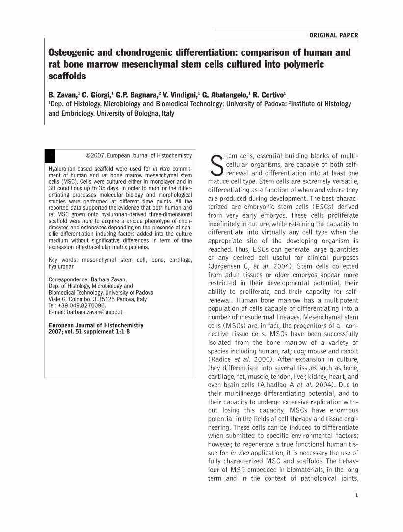
Phenotypic characterization of human MSCsFigure 1 illustrates the phenotypic characterization
of culture-expanded human MSCs (hMSC) by flow
cytometric analysis. Cells were consistently positive
for β1 integrin (CD 29: 98.98%), CD 166
(95.86%), SH2 (93.22%), SH3 (96.63%) and
SH4 (89.35%). Specific hematopoietic markers
such as CD 14, CD 34 and CD 45 were consistent-
ly negative. Rat MSCs had a similar flow cytomet-
ric profile as humans: positivity for CD29; CD166;
CD73 (data not shown)
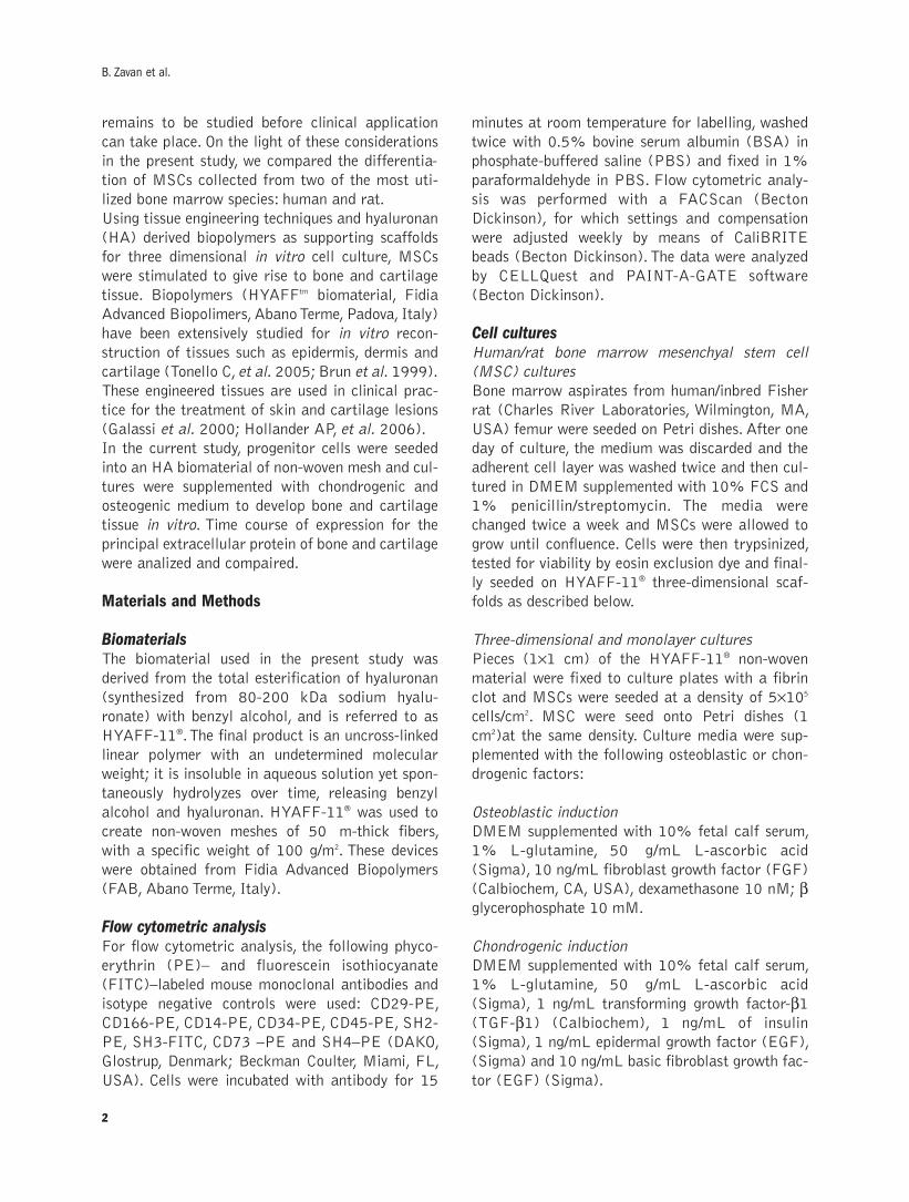
MSC proliferation analysisFigure 2 illustrates MSC growth in the presence of
osteogenic differentiating medium in monolayer
and three-dimensional conditions. Figure 2a shows
that human cells proliferated and peaked as early as
day 7. From day 14 to day 21, proliferation
decreased and then stabilized at a lower plateau.
Rat MSC proliferation peaked at day 15 of culture
(Figure 2b). Comparing monolayer with 3D condi-
tions is well evident, for both cell type, the positive
effect of non woven on cell proliferation.The main-
tenance and proliferation of human and rat MSC
onto the scaffold is confirmed by the higher MTT
values. Indeed, in monolayer cells reach in 15 days
confluence conditions showing a plateau in MTT
value lower than 3D one where cells are able to
growth in a bigger substrate eluding contact inhibi-
tion effect.
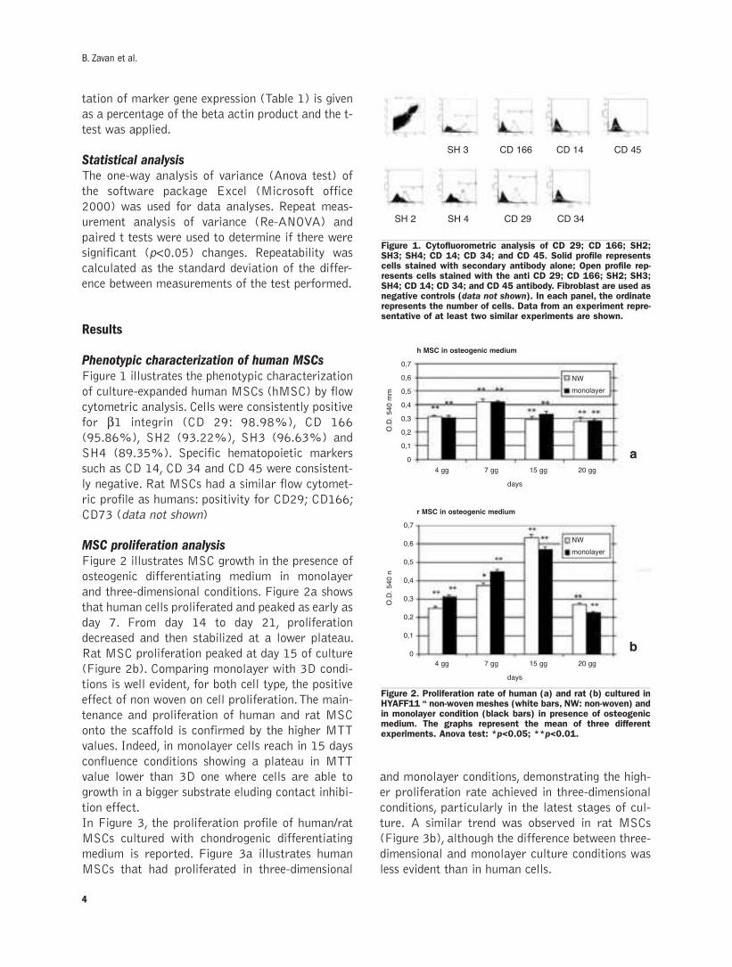
In Figure 3, the proliferation profile of human/rat
MSCs cultured with chondrogenic differentiating
medium is reported. Figure 3a illustrates human
MSCs that had proliferated in three-dimensional
and monolayer conditions, demonstrating the high-
er proliferation rate achieved in three-dimensional
conditions, particularly in the latest stages of cul-
ture. A similar trend was observed in rat MSCs
(Figure 3b), although the difference between three-
dimensional and monolayer culture conditions was
less evident than in human cells.
B. Zavan et al.
4
Figure 1. Cytofluorometric analysis of CD 29; CD 166; SH2;SH3; SH4; CD 14; CD 34; and CD 45. Solid profile representscells stained with secondary antibody alone; Open profile rep-resents cells stained with the anti CD 29; CD 166; SH2; SH3;SH4; CD 14; CD 34; and CD 45 antibody. Fibroblast are used asnegative controls (data not shown). In each panel, the ordinaterepresents the number of cells. Data from an experiment repre-sentative of at least two similar experiments are shown.
Figure 2. Proliferation rate of human (a) and rat (b) cultured inHYAFF11 m non-woven meshes (white bars, NW: non-woven) andin monolayer condition (black bars) in presence of osteogenicmedium. The graphs represent the mean of three differentexperiments. Anova test: *p<0.05; **p<0.01.
SH 3
SH 2
h MSC in osteogenic medium
r MSC in osteogenic medium
days
a
b
4 gg
0,7
0,6
0,5
0,4
0,3
0,2
0,1
0
0,7
0,6
0,5
0,4
0,3
0,2
0,1
0
7 gg 15 gg 20 gg
days
4 gg 7 gg 15 gg 20 gg
SH 4 CD 29 CD 34
CD 166 CD 14 CD 45
O.D.540mm
O.D.540n
NW
monolayer
NW
monolayer
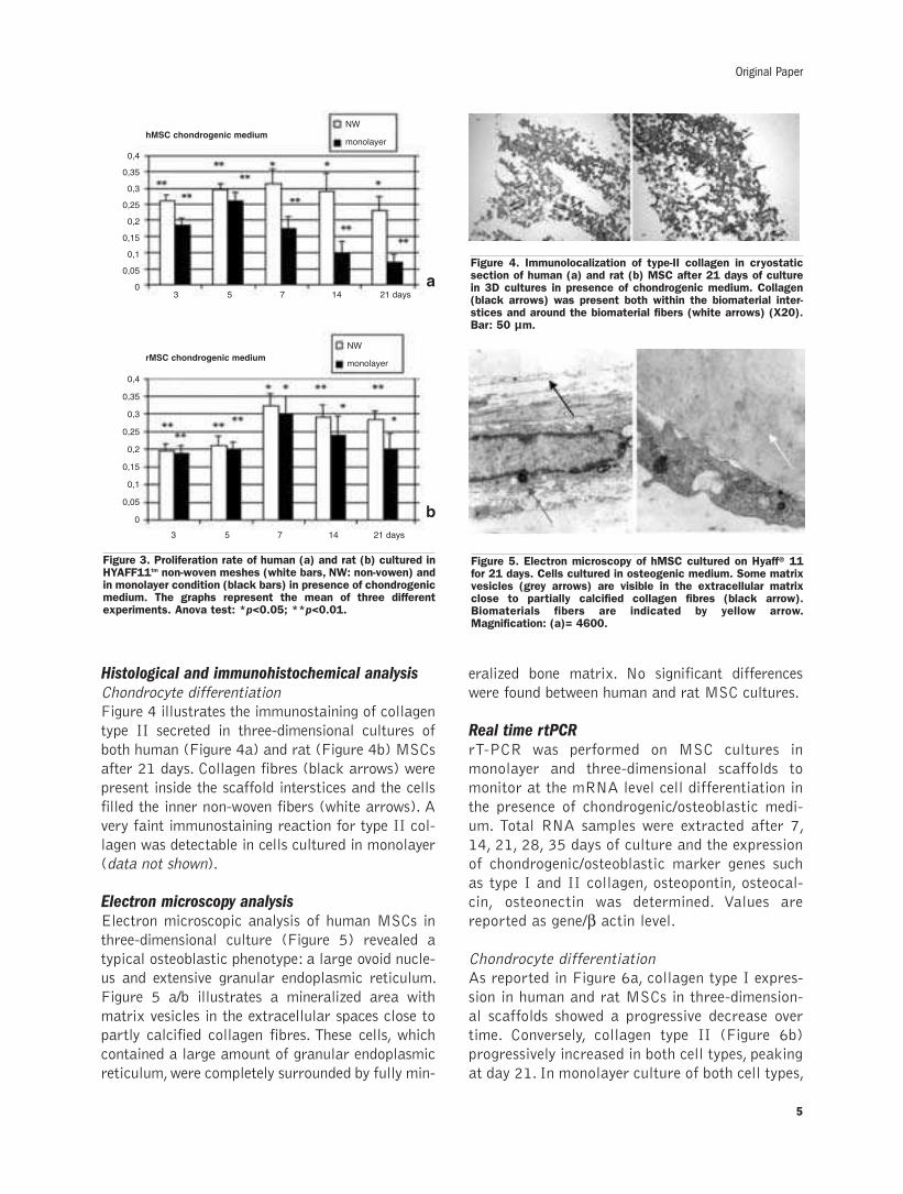
Histological and immunohistochemical analysisChondrocyte differentiation
Figure 4 illustrates the immunostaining of collagen
type II secreted in three-dimensional cultures of
both human (Figure 4a) and rat (Figure 4b) MSCs
after 21 days. Collagen fibres (black arrows) were
present inside the scaffold interstices and the cells
filled the inner non-woven fibers (white arrows). A
very faint immunostaining reaction for type II col-
lagen was detectable in cells cultured in monolayer
(data not shown).
Electron microscopy analysisElectron microscopic analysis of human MSCs in
three-dimensional culture (Figure 5) revealed a
typical osteoblastic phenotype: a large ovoid nucle-
us and extensive granular endoplasmic reticulum.
Figure 5 a/b illustrates a mineralized area with
matrix vesicles in the extracellular spaces close to
partly calcified collagen fibres. These cells, which
contained a large amount of granular endoplasmic
reticulum,were completely surrounded by fully min-
eralized bone matrix. No significant differences
were found between human and rat MSC cultures.
Real time rtPCRrT-PCR was performed on MSC cultures in
monolayer and three-dimensional scaffolds to
monitor at the mRNA level cell differentiation in
the presence of chondrogenic/osteoblastic medi-
um. Total RNA samples were extracted after 7,
14, 21, 28, 35 days of culture and the expression
of chondrogenic/osteoblastic marker genes such
as type I and II collagen, osteopontin, osteocal-
cin, osteonectin was determined. Values are
reported as gene/β actin level.
Chondrocyte differentiation
As reported in Figure 6a, collagen type I expres-
sion in human and rat MSCs in three-dimension-
al scaffolds showed a progressive decrease over
time. Conversely, collagen type II (Figure 6b)
progressively increased in both cell types, peaking
at day 21. In monolayer culture of both cell types,
Original Paper
5
Figure 3. Proliferation rate of human (a) and rat (b) cultured inHYAFF11tm non-woven meshes (white bars, NW: non-vowen) andin monolayer condition (black bars) in presence of chondrogenicmedium. The graphs represent the mean of three differentexperiments. Anova test: *p<0.05; **p<0.01.
Figure 4. Immunolocalization of type-II collagen in cryostaticsection of human (a) and rat (b) MSC after 21 days of culturein 3D cultures in presence of chondrogenic medium. Collagen(black arrows) was present both within the biomaterial inter-stices and around the biomaterial fibers (white arrows) (X20).Bar: 50 µm.
Figure 5. Electron microscopy of hMSC cultured on Hyaff® 11for 21 days. Cells cultured in osteogenic medium. Some matrixvesicles (grey arrows) are visible in the extracellular matrixclose to partially calcified collagen fibres (black arrow).Biomaterials fibers are indicated by yellow arrow.Magnification: (a)= 4600.
a
b
3 5 7 14 21 days
hMSC chondrogenic medium
rMSC chondrogenic medium
0,4
0,35
0,3
0,25
0,2
0,15
0,1
0,05
0
3 5 7 14 21 days
0,4
0,35
0,3
0,25
0,2
0,15
0,1
0,05
0
NW
monolayer
NW
monolayer

collagen type I was consistently expressed over
time (Figure 6c), while type II collagen was
weakly expressed (Figure 6d) and tended to
decrease over time.
Osteocyte differentiation
Figure 7a illustrates the expression of collagen type
I in human and rat MSCs cultured in three-dimen-
sional scaffolds. Collagen I mRNA production
peaked at day 14 and after a temporary drop off at
day 21, progressively increased. Figure 7b illus-
trates the comparatively lower expression of colla-
gen type I in human and rat MSCs cultured in
monolayer conditions.
Figure 8a illustrates the expression of osteocalcin,
Figure 9a of osteopontin and Figure 10a of osteo-
nectin in human and rat MSCs both in three-dimen-
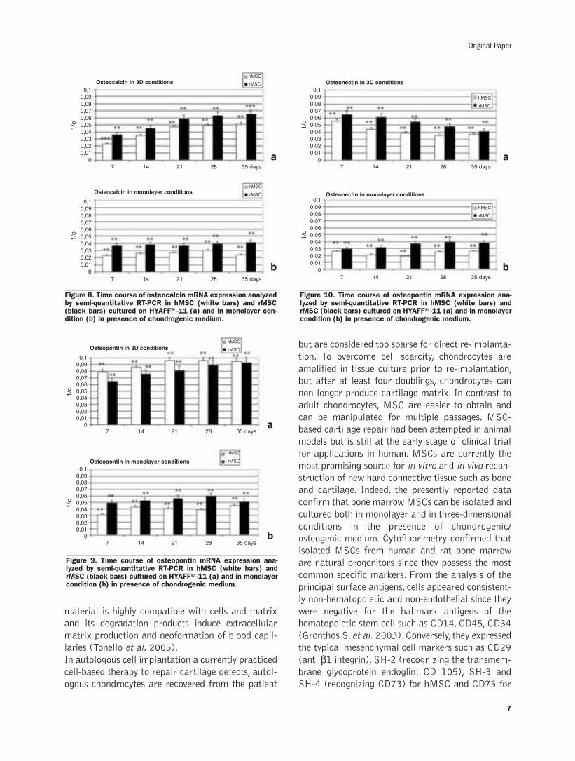
sional and in monolayer conditions. Osteocalcin
expression was similar in both cell types and
increase over time. Osteopontin expression was
greater than osteocalcin during and appeared con-
stant over time. Osteonectin expression showed a
progressive decrease over time for both cell types.
In monolayer culture, osteocalcin, osteopontin and
osteonectin expression was comparatively lower, but
demonstrated the same trend as in three-dimen-
sional cultures (Figures 8/9/10b).
DiscussionIn vitro tissue replacement of bone and cartilage has
long been a conundrum to be solved by clinicians and
tissue engineers. Developments in therapeutic strate-
gies on cartilage repair have increasingly focused on
the promising technology of cell-assisted repair pro-
posing to used autologous chondrocytes or other cell
types to regenerate articular cartilage in situ. The
necessary requisites include the correct cell type and
ideal degradable and biocompatible 3D scaffold with
favourable structural features for cell attachment,
proliferation, chondrogenesis and osteogenesis in
vitro and functional integration in vivo. As regard to
biomaterial, hyaluronan based scaffolds, such as
HYAFF11, are biodegradable materials currently
used for tissue engineering of skin and cartilage.This
B. Zavan et al.
6
Figure 6. Time course of: collagen I mRNA expression analyzedby semi-quantitative RT-PCR in hMSC (white bars) and rMSC(black bars) cultured on HYAFF®-11 (a) and in monolayer con-dition (c) in presence of chondrogenic medium. Collagen IImRNA expression analyzed by semi-quantitative RT-PCR inhMSC (white bars) and rMSC (black bars) cultured on HYAFF®-11 (b) and in monolayer condition (d) chondrogenic medium.
Figure 7. Time course of: collagen I mRNA expression analyzedby semi-quantitative RT-PCR in hMSC (white bars) and rMSC(black bars) cultured on HYAFF®-11 (a) and in monolayer con-dition (b) in presence of osteogenic medium.
Coll I with chondrogenic medium in 3D conditions
Coll II with chondrogenic medium in 3D conditions
Coll II with chondrogenic medium monolayer conditions
Coll II with chondrogenic medium monolayer conditions
1/ct
1/ct
1/ct
1/ct
a
b
C
d
7
0,1
0,08
0,06
0,04
0,02
014 21 28
hMSC
rMSC
hMSC
rMSC
hMSC
rMSC
hMSC
rMSC
35 days
7
0,1
0,08
0,06
0,04
0,02
014 21 28 35 days
7
0,1
0,08
0,06
0,04
0,02
014 21 28 35 days
7
0,05
0,04
0,03
0,02
0,01
0
14 21 28 35 days
Coll I with osteogenic medium in 3D conditions
Coll I with osteogenic medium monolayer conditions
a
b
1/ct
0,1
0,08
0,06
0,04
0,02
0
1/ct
0,1
0,08
0,06
0,04
0,02
0
hMSC
rMSC
hMSC
rMSC
7 14 21 28 35 days
7 14 21 28 35 days
material is highly compatible with cells and matrix
and its degradation products induce extracellular
matrix production and neoformation of blood capil-
laries (Tonello et al. 2005).
In autologous cell implantation a currently practiced
cell-based therapy to repair cartilage defects, autol-
ogous chondrocytes are recovered from the patient
but are considered too sparse for direct re-implanta-
tion. To overcome cell scarcity, chondrocytes are
amplified in tissue culture prior to re-implantation,
but after at least four doublings, chondrocytes can
non longer produce cartilage matrix. In contrast to
adult chondrocytes, MSC are easier to obtain and
can be manipulated for multiple passages. MSC-
based cartilage repair had been attempted in animal
models but is still at the early stage of clinical trial
for applications in human. MSCs are currently the
most promising source for in vitro and in vivo recon-
struction of new hard connective tissue such as bone
and cartilage. Indeed, the presently reported data
confirm that bone marrowMSCs can be isolated and
cultured both in monolayer and in three-dimensional
conditions in the presence of chondrogenic/
osteogenic medium. Cytofluorimetry confirmed that
isolated MSCs from human and rat bone marrow
are natural progenitors since they possess the most
common specific markers. From the analysis of the
principal surface antigens, cells appeared consistent-
ly non-hematopoietic and non-endothelial since they
were negative for the hallmark antigens of the
hematopoietic stem cell such as CD14, CD45, CD34
(Gronthos S, et al. 2003).Conversely, they expressed
the typical mesenchymal cell markers such as CD29
(anti β1 integrin), SH-2 (recognizing the transmem-brane glycoprotein endoglin: CD 105), SH-3 and
SH-4 (recognizing CD73) for hMSC and CD73 for
Original Paper
7
Figure 8. Time course of osteocalcin mRNA expression analyzedby semi-quantitative RT-PCR in hMSC (white bars) and rMSC(black bars) cultured on HYAFF®-11 (a) and in monolayer con-dition (b) in presence of chondrogenic medium.
Figure 10. Time course of osteopontin mRNA expression ana-lyzed by semi-quantitative RT-PCR in hMSC (white bars) andrMSC (black bars) cultured on HYAFF®-11 (a) and in monolayercondition (b) in presence of chondrogenic medium.
Figure 9. Time course of osteopontin mRNA expression ana-lyzed by semi-quantitative RT-PCR in hMSC (white bars) andrMSC (black bars) cultured on HYAFF®-11 (a) and in monolayercondition (b) in presence of chondrogenic medium.
Osteocalcin in 3D conditions Osteonectin in 3D conditions
Osteonectin in monolayer conditions
a
b
a
b
1/c
0,1
0,09
0,08
0,07
0,06
0,05
0,04
0,03
0,02
0,01
0
1/c
0,1
0,09
0,08
0,07
0,06
0,05
0,04
0,03
0,02
0,01
0
1/c
0,1
0,09
0,08
0,07
0,06
0,05
0,04
0,03
0,02
0,01
0
1/c
0,1
0,09
0,08
0,07
0,06
0,05
0,04
0,03
0,02
0,01
0
Osteocalcin in monolayer conditions
hMSC
rMSC
hMSC
rMSC
hMSC
rMSC
hMSC
rMSC
7 14 21 28 35 days 7 14 21 28 35 days
7 14 21 28 35 days7 14 21 28 35 days
1/c
0,1
0,09
0,08
0,07
0,06
0,05
0,04
0,03
0,02
0,01
0
Osteopontin in 3D conditions
hMSC
rMSC
7 14 21 28 35 days
1/c
0,1
0,09
0,08
0,07
0,06
0,05
0,04
0,03
0,02
0,01
0
Osteopontin in monolayer conditions
hMSC
rMSC
7 14 21 28 35 days
a
b
rMSC (Haynesworth SE, et al. 1992). After expan-
sion in monolayer culture and in the presence of
chondrogenic and osteogenic inducing factors,
human and rat MSCs differentiated into chondro-
cytes and osteoblasts, respectively.When cultured in
osteogenic conditions, the proliferation rate of MSCs
increased during the initial period of culture, pro-
gressively decreasing after differentiation both in 3D
and in monolayer conditons. Detailed rtPCR analy-
ses of extracellular matrix components (collagen
type I, osteopontin, osteocalcin and osteonectin)
confirmed the presence of osteogenic molecules
already after one week of monolayer or three-dimen-
sional culture. In particular, in this early phase of
osteogenic differentiation high levels of osteonectin,
a molecule fundamentally important for cellular-
bone matrix interaction and for matrix mineraliza-
tion, were observed in 3D conditions. Collagen type
I molecules, essential for formation and maturation
of hydroxyapatite crystals,were also detected during
the first 10 days of culture. Light and electron
microscopy of three-dimensional cultures of MSCs in
osteogenic medium demonstrated a well organized
extracellular matrix in which type I collagen fibres
and calcium phosphate crystals were co-localized.
Interestingly, both cell proliferation and expression
of human and rat MSCs were consistently higher in
osteogenic cells in three-dimensional versus mono-
layer culture. The three-dimensional hyaluronan
scaffolds permitted differentiation of MSCs to a
chondrogenic phenotype as well. Time dependent
increases in cell proliferation were greater in three-
dimensional compared to monolayer culture condi-
tions. These are similar findings to those observed
with adult chondrocytes (Brun et al. 1999). The
expression and production of collagen type II, a well-
documented marker of hyaline articular cartilage
always found in freshly isolated chondrocytes, was
determined by molecular expression and (rtPCR)
morphological analyses. Findings again confirmed
that the chondrogenic differentiation process was
better promoted in three-dimensional culture than in
monolayer. Conversely, collagen type I was expressed
in three-dimensional culture predominately during
the initial phases of the differentiating process,while
in monolayer conditions it increased progressively
over time. Although human and rat MSCs have the
same diferentiating potential, they do behave differ-
ently during the proliferation process. While human
cell proliferation peaks after one week of culture, rat
cell proliferation peaks after two weeks. These
results demonstrate that both human and rat MSCs
can be cultured in three-dimensional scaffolds made
from hyaluronan based polymers in the presence of
the necessary stimuli that support differentiation
towards osteogenic or chondrogenic phenotypes.The
delivery vehicles investigated in this study are easily
applicable to clinical practice since hyaluronan scaf-
folds have been already extensively studied both for
the in vitro reconstruction of skin and cartilage sub-
stitutes and for their clinical application. In the end,
these data clearly confirm that bone marrow cells
are progenitor cells that are clearly superior to tis-
sue biopsy-isolated cells for use in tissue engineering.
Tissue samples from patients have to be isolated by
enzymes such as collagenase and hyaluronidase to
remove extracellular matrix components and, as is
well known, adult stem cells usually are very scarce-
ly supplied within tissues. MSCs isolated from the
bone marrow would be a valuable source for cell
transplantation since their characteristic features
include a high potential for proliferation and multi-
lineage differentiation.
References
Alhadlaq A, Mao JJ. Mesenchymal stem cells: isolation and therapeutics.Stem Cells Dev 2004;13:436-48.
Benedetti L, Cortivo R, Berti T, Berti A, Pea F, Mazzo M, et al.Biocompatibility and biodegradation of different hyaluronan derivatives(Hyaff) implanted in rats. Biomaterials 1993;14:1154-60.
Brun P, Abatangelo G, Radice M, Zacchi V, Guidolin D, Daga Gordini D, etal. Chondrocyte aggregation and reorganization into three-dimensionalscaffolds. J Biomed Mater Res 1999;46:337-46.
Denizot, R. Lang. Rapid colorimetric assay for cell growth and survival. JImmunol Methods 1986;89:271–7.
Galassi G,Brun P,RadiceM,Cortivo R,Zanon GF,Genovese P,et al.In vitroreconstructed dermis implanted in human wounds:degradation studies ofthe HA-based supporting scaffold.Biomaterials 2000;21:2183-91.
Gronthos S, Zannettino AC, Hay SJ, et al. Molecular and cellular charac-terisation of highly purified stromal stem cells derived from human bonemarrow. J Cell Sci 2003;116: 1827-35.
Haynesworth SE, Baber MA, Caplan AI. Cell surface antigens on humanmarrow derived mesenchymal calls are detected by monoclonal antibod-ies. Bone 1992;13:69.
Hollander AP, Dickinson SC, Sims TJ, Brun P, Cortivo R, Kon E, et al.Maturation of tissue engineered cartilage implanted in injured andosteoarthritic human knees.Tissue Eng 2006;12:1787-98.
Jorgensen C, GordeladzeJ and Noel D.Tissue engineering through autolo-gous mesenchymal stem cells. Curr Opin Biotechnol 2004; 15: 406-10.
Radice M, Brun P, Cortivo R, Scapinelli R, Battaliard C, Abatangelo G.Hyaluronan-based biopolymers as delivery vehicles for bone-marrow-derived mesenchymal progenitors. J BiomedMater Res 2000;50:101-9.
Tonello C,Vindigni V, Zavan B, Abatangelo S, Abatangelo G, Brun P, et al.In vitro reconstruction of an endothelialized skin substitute provided withamicrocapillary network using biopolymer scaffolds.FASEB J 2005;21.
Zanasi S, Borrione A, De Luca C, Pavesio A, Soranzo C, Abatangelo G.Maturation of tissue engineered cartilage implanted in injured andosteoarthritic human knees.Tissue Eng 2006;12:1787-98.
B. Zavan et al.
8

9
©2007, European Journal of Histochemistry
Tendons transmit forces generated from muscle to bone mak-ing joint movements possible. Tendon collagen has a complexsupramolecular structure forming many hierarchical levels ofassociation; its main functional unit is the collagen fibril form-ing fibers and fascicles. Since tendons are enclosed by looseconnective sheaths in continuity with muscle sheaths, it is like-ly that tendon sheaths could play a role in absorbing/trans-mitting the forces created by muscle contraction.In this study rat Achilles tendons were passively stretched invivo to be observed at polarized light microscope (PLM),scanning electron microscope (SEM) and transmission elec-tron microscope (TEM). At PLM tendon collagen fibers inrelaxed rat Achilles tendons ran straight and parallel, showinga periodic crimp pattern. Similarly tendon sheaths showedapparent crimps. At higher magnification SEM and TEMrevealed that in each tendon crimp large and heterogeneouscollagen fibrils running straight and parallel suddenlychanged their direction undergoing localized and variablemodifications. These fibril modifications were named fibrillarcrimps. Tendon sheaths displayed small and uniform fibrilsrunning parallel with a wavy course without any ultrastructur-al aspects of crimp. Since in passively stretched Achilles ten-dons fibrillar crimps were still observed, it is likely that duringthe tendon stretching, and presumably during the tendonelongation in muscle contraction, the fibrillar crimp may bethe real structural component of the tendon crimp acting asshock absorber. The peritendinous sheath can be stretchedas tendon, but is not actively involved in the mechanism ofshock absorber as the fibrillar crimp. The different functionalbehaviour of tendons and sheaths may be due to the differ-ent structural and molecular arrangement of their fibrils.
Key words: Achilles tendon, sheaths, collagen fibrils, TEM,SEM.
Correspondence: Marco Franchi,Dipartimento di Scienze Anatomiche Umane eFisiopatologia dell’Apparato Locomotore, via Irnerio 48,40126, Bologna, ItalyTel: +39.0512091553.Fax: +39.0512091659.E-mail: [email protected]
European Journal of Histochemistry2007; vol. 51 supplement 1:9-14
Tendon crimps and peritendinous tissues responding to tensional forces
M. Franchi, M. Quaranta, V. De Pasquale, M. Macciocca, E. Orsini, A. Trirè, V. Ottani, A. Ruggeri
Dipartimento di Scienze Anatomiche Umane e Fisiopatologia dell’Apparato Locomotore, University of
Bologna, Italy
Joint movements of the body in mammals are
generated by skeletal muscle cell activity, but
the structures of the muscle-tendon complex
able to transmit the forces of muscle contraction to
bone are tendons and aponeuroses (Magnusson et
al., 2003). Tendons are considered highly flexible
but inextensible structures offering a considerable
resistance to tension. They also act as mechanical
buffers or shock absorbers in protecting tendons to
bone attachment during the initial elongation relat-
ed to rapid muscle contraction (Stolinski, 1995a).
Tendons are dense fibrous collagen structures
organized in a hierarchical manner whose main
functional unit, strong and stiff in tension, is the col-
lagen fibril (Kannus, 2000; Provenzano and
Vanderby, 2006). The particular arrangement and
dimensions of the collagen fibrils, together with
their interactions with hydrophilic proteoglycans of
the extracellular matrix, are responsible for the
transmission of forces and resistance to tension.
Collagen fibrils run straight and parallel in relaxed
tendons, and are always arranged in fibers, fibril
bundles and fascicles showing a zig-zag or wave-
form aspect called crimping. During initial stretch-
ing the crimps disappear or become more flattened
acting as shock absorbers to tension (Diamant et
al., 1972; Kastelic et al., 1980; Screen et al.,
2004; Franchi et al., 2007). Increasing the tensile
strength, the intra- and intermolecular cross-links of
collagen fibrils are primarily involved in the trans-
mission of mechanical forces (Kjaer, 2004;
Provenzano and Vanderby, 2006). During this phase
proteoglycans with their bridges also play a role in
absorbing and/or transmitting the tension stress to
bone (Cribb and Scott, 1995; Fratzl et al., 1998;
Scott, 2003).
Tendons are often surrounded by loose connective
sheaths forming the paratenon, epitenon, peritenon
and endotenon (Strocchi et al., 1985; Kannus,
2000; Kjaer, 2004). According to Trotter and
Purslow (1992) and Kjaer (2004) tendon sheaths
ORIGINAL PAPER
are linked to skeletal muscle sheaths and it is rea-
sonable to think that even these apparently indif-
ferent membranes play a role in absorbing and/or
transmitting tensional forces in tendon.
Microscopic and ultrastructural analyses of rat
tendons in this study may shed light on the mor-
phologic and functional changes to collagen in ten-
don and peritendinous tissues when tendon is
mechanically stretched in vivo.
Materials and Methods
AnimalsTwelve female Sprague-Dawley rats (3 months
old) were anaesthetized with an intraperitoneal
injection of 87 mg/kg ketamine (Ketavet, Farma-
ceutici Gellini Spa, Italy) and 13 mg/kg xylazine
(Rompun, Bayer Italia Spa, Italy). A resin brace,
modified to induce foot dorsal flexion, was applied
to one posterior leg in order to reach a final 55°
angle flexion.
The stretching position was kept for 10 minutes.
At the end of the stretching session and still under
anaesthesia, the tendon of the gastrocnemius mus-
cle with its sheaths was exposed and fixed in situ
(i.e. still connected to the muscle belly and to the
bone) in Karnovsky’s solution. The tendon of the
controlateral leg of each animal was kept relaxed
and fixed as with the stretched tendon to be
analysed as a control sample. Finally, the rats were
euthanized via an intracardiac injection of Tanax
(Hoechst, Frankfurt am Main, Germany).
All stretched and control tendons with their own
sheaths were excised. Ten tendons (five stretched
and five controls) were processed for polarized light
microscopy (PLM). The other eight tendons (four
stretched and four controls) were processed for
transmission electron microscopy (TEM) and the
last six tendons (three stretched and three controls)
were longitudinally cut to be investigated by scan-
ning electron microscopy (SEM).
The experimental protocols were conducted in
accordance with Italian and European Laws on
laboratory animals use and care.
Polarized light microscopySpecimens were fixed in 10% buffered formalin,
dehydrated in graded concentrations of ethanol,
embedded in paraffin and longitudinally sectioned
at 6 µm. The sections were stained with 5%
Picrosirius Red to enhance the natural bir-
ifrangence of collagen fibers when observed under
the polarized light microscope (Leitz Ortholux 2,
Wetzlar, Germany).
Transmission electron microscopySpecimens for TEM were fixed in Karnovsky’ s
solution, rinsed with a 0.1M sodium cacodylate
buffer (pH 7.2) and post-fixed in 1% osmium
tetroxide.Thereafter, they were dehydrated in grad-
ed alcohols and embedded in Araldite resin. The
ultrathin sections were stained with lead citrate and
uranyl acetate and viewed under a Philips CM-10
electron microscope.
Scanning electron microscopyFor SEM study, the samples were fixed in
Karnovsky’s solution, dehydrated in a graded
ethanol series and then in hexamethyldisilazane.
Finally they were mounted on metal stubs and coat-
ed with gold using a sputter coater (Emitech
K550). Observations were made under SEM
(Philips 515 and Philips XL30-FEG) operating in
secondary-electron mode.
Results
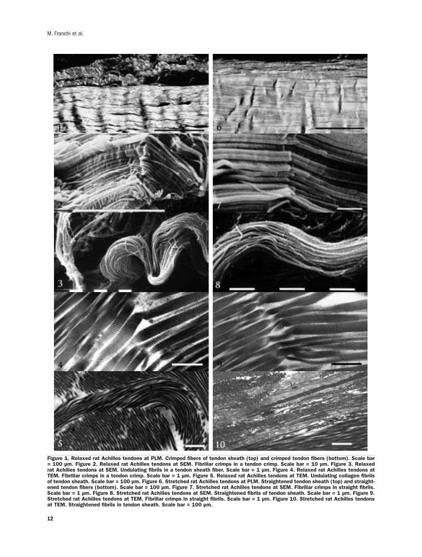
Relaxed Achilles tendonLongitudinal sections of relaxed rat Achilles ten-
don analyzed by light microscopy showed parallel
collagen fibers with a wavy course that under polar-
ized light microscope is displayed as alternating
dark and light bands corresponding to tendon crimps
(Figure 1). Flat fibroblast-like cells were interposed
between adjacent fiber bundles.The outer surface of
the Achilles tendon was covered by a sheath of col-
lagen fiber bundles running in a waveform pattern.
At the polarized light microscope the collagen fibers
of this sheath showed dark and light bands similar to
the tendon crimps (Figure 1).
Other specimens observed at SEM showed the
tendon fibers to be composed of large plurimodal
collagen fibrils running straight and parallel. At the
crimp apex these fibrils suddenly changed their
direction showing an evident elbow with knots cor-
responding to deformations of the fibril shape. In
particular, collagen fibrils appeared bent on the
same plane like bayonets, or twisted and bent
(Raspanti et al., 2005; Franchi et al., 2007)
(Figure 2). The tendon sheath appeared composed
of thin wavy collagen fibers made up of small uni-
modal collagen fibrils.No crimps were recognizable
10
M. Franchi et al.

11
Original Paper
along these fibril bundles (Figure 3).
Other specimens analysed at TEM better showed
that tendon collagen fibrils, when changing their
direction at the crimp apex, modified their shape
(bent on the same plane like bayonets, or twisted
and bent) and lost their D-period disclosing their
microfibrillar arrangement (Figure 4). As in previ-
ous SEM observations, thin sections showed the
small collagen fibrils of the sheaths running in a
smooth undulating arrangement without any ultra-
structural aspects of crimp (Figure 5).
Stretched Achilles tendonLongitudinal sections of stretched rat Achilles
tendons observed at direct and polarized light
microscope showed most of the tendon collagen
fibers running straight and parallel with a few flat-
tened crimps (Figure 6). The collagen fibers in
stretched tendon sheaths ran straight with a slight-
ly wavy course.
In similar specimens observed at SEM tendon
fibers showed rare or otherwise completely flat-
tened crimps. In all crimps, including those whose
collagen fibrils appeared completely straightened,
the fibrils still retained the knots at the apex of the
crimps as in relaxed specimens (Figure 7). On the
contrary tendon sheath collagen fibrils showed a
less undulating path than the relaxed specimens and
no ultrastructural knot or fibril size deformation
was detectable at ultrastructural level (Figure 8).
At TEM, the same fibril knot described in relaxed
specimens were detected even in straightened fibrils
of completely flattened crimps (Figure 9). Collagen
fibrils of fiber bundles in tendon sheath appeared
partially stretched along the main axis of tendon
(Figure 10).
DiscussionA waveform configuration of collagen fibers in
tendon was first described in polarized light
microscopy investigations. The authors correlated
the periodic crimping pattern to tendon functions
observing that crimping disappeared when tendons
were slightly stretched in vitro (Rigby et al., 1959;
Elliot, 1965; Viidik and Ekholm, 1968; Stromberg
and Wiederhielm, 1969; Viidik, 1972; Hess et al.,
1989). Some authors (Diamant et al., 1972;
Atkinson et al., 1999; Hansen et al., 2002) sug-
gested that the alignment of collagen fibers during
stretching of the tendon might correspond to the
toe region of the stress-strain curve of tendon.
Ultrastructural studies were also carried out to
improve the morphological or functional meaning
of tendon crimps, but no new functional data were
reported (Rowe, 1985a,b; Gathercole and Keller,
1991; Stolinski, 1995a; Magnusson et al., 2002;
Hurschler et al., 2003). Recently Franchi et al.
(2007) described a morphological deformation of
collagen fibrils in tendon crimps and named it fib-
rillar crimp.They also observed that fibrillar crimps
did not disappear when the Achilles tendon was
physiologically stretched in vivo, suggesting a mod-
ification of the fibril structure at the level of fibril-
lar crimps.
The study of tendon stretching may help to shed
light on the mechanism of force transmission during
muscle contraction.
According to Kjaer (2004) tendon sheaths are in
continuity with the peri- and intra-muscular colla-
gen sheaths thereby ensuring a functional link
between the skeletal muscle and bone. In particular
the perimysium seems to play a role in transmitting
tensile force (Trotter and Purslow, 1992). It has
been suggested that the connective tissue of skele-
tal muscle and tendon is like a lively structure with
a dynamic protein turnover, highly able to adapt to
changes in the external environment such as
mechanical loading or inactivity and disuse (Kjaer,
2004). As tendon is tightly connected to the skele-
tal muscle via connective tissue of tendon and mus-
cle sheaths it is probable that the peritendinous col-
lagen fibers might be involved in transmission of
forces from muscle to tendon.
Morphological flattened waves of collagen fibers
comparable to those described in tendons were also
observed in nerve sheath as in the epineurium
(Stolinski, 1995b).The pattern was observed in cut
or relaxed fascicles in situ as well as in isolated and
split layers of the nerve sheath. It is interesting that
the pattern was not observed on nerve fascicles
under tension.The nature of the wavy structure sug-
gested that the sheath length might change on
stretching or contraction to accommodate the dis-
placement and movement of nerve fibres (Stolinski,
1995b).
At polarized light microscope the present study
disclosed a waveform pattern of collagen fibers
both in tendon and tendon sheaths. However, while
the waveform pattern of tendon crimps is due to a
peculiar structural characteristic of the collagen
fibrils (a structure specifically acting as a shock
absorber and named fibrillar crimp), the waveform
12
M. Franchi et al.
Figure 1. Relaxed rat Achilles tendons at PLM. Crimped fibers of tendon sheath (top) and crimped tendon fibers (bottom). Scale bar= 100 µm. Figure 2. Relaxed rat Achilles tendons at SEM. Fibrillar crimps in a tendon crimp. Scale bar = 10 µm. Figure 3. Relaxedrat Achilles tendons at SEM. Undulating fibrils in a tendon sheath fiber. Scale bar = 1 µm. Figure 4. Relaxed rat Achilles tendons atTEM. Fibrillar crimps in a tendon crimp. Scale bar = 1 µm. Figure 5. Relaxed rat Achilles tendons at TEM. Undulating collagen fibrilsof tendon sheath. Scale bar = 100 µm. Figure 6. Stretched rat Achilles tendons at PLM. Straightened tendon sheath (top) and straight-ened tendon fibers (bottom). Scale bar = 100 µm. Figure 7. Stretched rat Achilles tendons at SEM. Fibrillar crimps in straight fibrils.Scale bar = 1 µm. Figure 8. Stretched rat Achilles tendons at SEM. Straightened fibrils of tendon sheath. Scale bar = 1 µm. Figure 9.Stretched rat Achilles tendons at TEM. Fibrillar crimps in straight fibrils. Scale bar = 1 µm. Figure 10. Stretched rat Achilles tendonsat TEM. Straightened fibrils in tendon sheath. Scale bar = 100 µm.
configuration of tendon sheath appears as a simple
undulating arrangement of collagen fibrils with no
fibrillar crimps. Therefore, the straightening of the
sheath collagen fibrils should be interpreted as a
passive morphological adaptation to changes in
tendon length.
Transmission of forces from skeletal muscle to
bone involves different phases in tendon elongation.
During initial tendon stretching crimps disappear or
become more flattened acting as shock absorbers to
tension with no local tissue strain increase
(Diamant et al., 1972; Kastelic et al., 1980;
Screen et al., 2004; Franchi et al., 2007).
Increasing the tensile strength, the intra- and inter-
molecular cross-links of collagen fibrils are then
involved in the transmission of mechanical forces
(Kjaer, 2004; Provenzano and Vanderby, 2006).
Some authors suggest that short proteoglycan
bridges linked to collagen fibrils, like decorin, may
also absorb and then transmit the tension stress to
bone (Cribb and Scott, 1995; Fratzl et al., 1998;
Scott, 2003). Our results may suggest that during
the passive static stretching of tendon, and presum-
ably during tendon elongation in muscle contrac-
tion, the peritendinous sheath can be stretched like
tendon, but is not actively involved in the shock
absorber mechanism like the fibrillar crimp.The dif-
ferent functional behaviour of these two structures
(tendons and sheaths) is also due to the different
structural and molecular arrangement of the fibrils:
tendon fibrils are large in diameter, parallely tight-
ly packed and with a straight microfibrillar
arrangement; fibrils in tendon sheaths are small
and uniform in diameter, run in thin wavy bundles
and have an helicoidal microfibrillar arrangement.
Attending to the distribution in the connective tis-
sue of the body, tendons are prevalently submitted
to unidirectional tensional forces while sheaths
undergo multidirectional loading (Ottani et al.,
2001).
AcknowledgementsWe are indebted to Gianfranco Filippini,
D.I.S.T.A., University of Bologna, for his technical
assistance with SEM.This research was supported
by MIUR grant (prot. 2004055533).
References
Atkinson TS, Ewers BJ, Haut RC. The tensile and stress relaxationresponses of human patellar tendon varies with specimen cross-sectional area. J Biomech 1999; 32: 907-14.
Cribb AM, Scott JE.Tendon response to tensile stress: an ultrastruc-tural investigation of collagen: proteoglycans interactions instressed tendon. J Anat 1995; 187: 423-8.
Diamant J, Keller A, Baer E, Litt M, Arride RG. Collagen: ultra-structure and its relation to mechanical properties as a function ofageing. Proc R Soc B 1972; 180: 293-315.
Elliott DH. Structure and function of mammalian tendon. Biol RevCamb Philos Soc 1965; 40: 392-421.
Franchi M, Fini M, Quaranta M, De Pasquale V, Raspanti M,Giavaresi G, Ottani V, Ruggeri A. Crimp morphology in relaxed andstretched rat Achilles tendon. J Anat 2007; 210: 1-7.
Fratzl P, Misof K, Zizak I, Rapp G, Amenitsch H, Bernstorff S.Fibrillar structure and mechanical properties of collagen. J StructBiol 1998; 122: 119-22.
Gathercole LJ, Keller A. Crimp morphology in the fibre-forming col-lagens. Matrix 1991; 11: 214-34.
Hansen KA,Weiss JA, Barton JK. Recruitment of tendon crimp withapplied tensile strain. J Biomech Eng 2002; 124: 72-7.
Hess GP, Cappiello WL, Poole RM, Hunter SC. Prevention and treat-ment of overuse tendon injuries. Sports Med 1989; 8: 371-84.
Hurschler C, Provenzano PP,Vanderby RJr. Scanning electron micro-scopic characterization of healing and normal rat ligamentmicrostructure under slack and loaded conditions. Connect TissueRes 2003; 44: 59-68.
Kannus P. Structure of the tendon connective tissue. Scand J MedSci Sports 2000; 10: 312-20.
Kastelic J, Palley I, Baer E. A structural mechanical model for ten-don crimping. J Biomech 1980; 13: 887-93.
Kjaer M. Role of extracellular matrix in adaptation of tendon andskeletal muscle to mechanical loading. Physiol Rev 2004; 84: 649-98.
Magnusson SP, Qvortrup K, Larsen JO, Rosager S, Hanson P,Aagaard P, Krogsgaard M, Kjaer M. Collagen fibril size and crimpmorphology in ruptured and intact Achilles tendons. Matrix Biol2002; 21: 369-77.
Magnusson SP, Hansen P, Kjaer M. Tendon properties in relation tomuscular activity and physical training. Scand J Med Sci Sports2003; 13: 211-23.
Ottani V, Raspanti M, Ruggeri A. Collagen structure and functionalimplications. Micron 2001; 32: 251-60.
Provenzano PP, Vanderby R Jr. Collagen fibril morphology andorganization: implications for force transmission in ligament andtendon. Matrix Biol 2006; 25: 71-84.
Raspanti M, Manelli A, Franchi M, Ruggeri A. The 3D structure ofcrimps in the rat Achilles tendon. Matrix Biol 2005; 24: 503-07.
Rigby BJ, Hirai N, Spikes JD, Eyring H. The mechanical propertiesof rat tail tendon. J Gen Physiol 1959; 43: 265-83.
Rowe RW. The structure of rat tail tendon. Connect Tissue Res1985a; 14: 9-20.
Rowe RW.The structure of rat tail tendon fascicles. Connect TissueRes 1985b; 14: 21-30.
Scott JE. Elasticity in extracellular matrix shape modules of tendon,cartilage, etc. A sliding proteoglycan-filament model. J Physiol2003; 553: 335-43.
Screen HR, Lee DA, Bader DL, Shelton JC. An investigation into theeffects of the hierarchical structure of tendon fascicles on micro-mechanical properties. Proc Inst Mech Eng [H] 2004; 218: 109-19.
13
Original Paper

14
Stolinski C. Disposition of collagen fibrils in human tendons. J Anat1995a; 186: 577-83.
Stolinski C. Structure and composition of the outer connective tissuesheaths of peripheral nerve. J Anat 1995b; 186: 123-30.
Strocchi R, Leonardi L, Guizzardi S, Marchini M, Ruggeri A.Ultrastructural aspects of rat tail tendon sheaths. J Anat 1985;140: 57-67.
Stromberg DD, Wiederhielm CA. Viscoelastic description of a col-lagenous tissue in simple elongation. J Appl Physiol 1969; 26:
857–62.
Trotter JA, Purslow PP. Functional morphology of the endomysium
in series fibered muscles. J Morphol 1992; 212: 109-22.
Viidik A, Ekholm R. Light and electron microscopic studies of colla-
gen fibers under strain. Z Anat Entwicklungsgesch 1968; 127:
154–64.
Viidik A. Simultaneous mechanical and light microscopic studies of
collagen fibers. Z Anat Entwicklungsgesch 1972; 136: 204–12.
M. Franchi et al.
15
REVIEW
©2007, European Journal of Histochemistry
As appears from the literature, the majority of boneresearchers consider osteoblasts and osteoclasts the onlyvery important bony cells. In the present report we provideevidence, based on personal morphofunctional investiga-tions, that such a view is incorrect and misleading. Indeedosteoblasts and osteoclasts undoubtedly are the only boneforming and bone reabsorbing cells, but they are transientcells, thus they cannot be the first to be involved in sensingboth mechanical and non-mechanical agents which controlbone modeling and remodeling processes. Briefly, accordingto our view, osteoblasts and osteoclasts represent the armsof a worker; the actual operation center is constituted by thecells of the osteogenic lineage in the resting state. Such aresting phase is characterized by osteocytes, bone liningcells and stromal cells, all connected in a functional syn-cytium by gap junctions, which extends from the bone to thevessels. We named this syncytium the Bone Basic CellularSystem (BBCS), because it represents the only permanentcellular background capable first of sensing mechanicalstrains and biochemical factors and then of triggering anddriving both processes of bone formation and bone resorp-tion. As shown by our studies, signalling throughout BBCScan occur by volume transmission (VT) and/or wiring trans-mission (WT). VT corresponds to the routes followed by solu-ble substances (hormones, cytokines etc.), whereas WT rep-resents the diffusion of ionic currents along cytoplasmicprocesses in a neuron-like manner. It is likely that non-mechanical agents first affect stromal cells and diffuse by VTto reach the other cells of BBCS, whereas mechanical agentsare first sensed by osteocytes and then issued throughoutBBCS by WT.
Key words: osteogenic cells, osteoclasts, cytokines,mechanical strains.
Correspondence: Gastone Marotti,Dipartimento di Anatomia e Istologia Policlinico,Largo del Pozzo, 71 41100 Modena, ItalyTel: +39.059.4224800.Fax: +39.059.4224861.E-mail: [email protected]
European Journal of Histochemistry2007; vol. 51 supplement 1:15-20
The mechanism of transduction of mechanical strains into biological
signals at the bone cellular level
G. Marotti, C. Palumbo
Department of Anatomy and Histology, University of Modena and Reggio Emilia, Italy
It is a well established fact that, under the con-
trol of mechanical agents (body weight, force of
gravity, muscular tone and strength) and non-
mechanical agents (hormones, vitamins, cytokines,
growth factors), bone cells regulate bone homeosta-
sis and take part in the maintenance of mineral
homeostasis, by means of three processes: bone
growth, bone modeling and bone remodeling. Bone
growth and bone modeling are only devoted to the
regulation of bone homeostasis, whereas bone
remodeling takes part in the regulation of bone
homeostasis as well as of mineral homeostasis, by
respectively improving bone structure in response to
mechanical demands and setting free calcium and
phosphate ions during the reabsorbing phase.
Frost’s mechanostat theory (Frost, 1987) and
Utah paradigm (1985) have greatly rationalized
bone modeling and remodeling processes and what
they involve at the bone macroscopic level. However
what happens at the cellular level still remains to be
defined. We do not know, for instance: a) how
mechanical agents and non-mechanical agents
interact at the cellular level; b) which is the mecha-
nism of transduction of mechanical strains into bio-
logical signals; updated literature ascribes to osteo-
cytes the function of sensing the strains induced into
the bone matrix by mechanical stresses but, as we
will discuss below, all cells of the osteogenic system
are likely to be affected by mechanical strains; c)
how osteocytes transmit mechanical stimuli to, and
interact with, the other bone cells.
In the attempt to answer these questions we will
first summarize the results of the morphofunctional
investigations we carried out on the cells of the
osteogenic lineage during the last three decades.
Then we will discuss some functional implications.
The cells of the osteogenic lineage: morphologicalaspect and functionIn the early 1970s, we showed that the exponen-
tial decrement of the appositional growth rate,

which has been shown to occur during osteon for-
mation by means of triple fluorochrome technique
(Manson and Waters, 1963; Marotti and Camosso,
1968), depends on the diminution in size of the
osteoblasts and their progressive flattening. At the
beginning of osteon formation, when the apposi-
tional rate is high, the osteoblasts are big and pris-
matic, whereas towards the end of osteon forma-
tion, when the rate is low, they are smaller and flat
(Marotti, 1976).
Since these facts were also observed in trabecu-
lar bone, our conclusion was that the rate at which
the bone tissue is laid down depends on the ratio
between the volume of the osteoblasts and their
secretory territory: the greater the osteoblast vol-
ume and the smaller its secretory territory, the
higher the rate of bone apposition (Marotti, 1976).
Additionally we showed that, during the edifica-
tion of osteons, also the osteocytes decrease in size,
in parallel to the decrement of osteoblast dimension
and the appositional growth rate. This finding
implies that the size of the osteocytes strictly
depends on the size of the osteoblasts from which
they differentiate: the bigger the osteoblasts the
larger the size of the osteocytes (Marotti, 1976).
The functional meaning of this fact as yet to be
established. However we found, in human osteons,
that the decrement in size of osteocytes from the
cement line towards the Haversian canal is paral-
leled by a thinning of osteocytic-loose (collagen
poor) lamellae and, consequently, by a diminution
of the distance between non-osteocytic-dense (col-
lagen rich) lamellae, whose thickness does not sig-
nificantly change throughout the osteonic wall.
Mechanically speaking, this fact involves an
increase in collagen fibers, namely in bone strength,
along the bone surfaces where stresses and strains
reach the highest values (Ardizzoni et al., 1999).
In more recent years, we showed by transmission
and scanning electron microscopes that the
arborization of osteocytes is asymmetrical as
regards both number and length of cytoplasmic
processes. Vascular dendrites (those radiating
toward the bone vascular surface) are more
numerous (Marotti et al., 1985) and incompara-
bly longer than mineral dendrites (those radiating
towards the opposite surface) (Palumbo, 1986;
Palumbo et al., 1990a, 1990b). Therefore osteo-
cyte appear to be polarized cells, towards the bone
surface where they come into contact whether
osteoblasts or bone lining cells, according to which
the bone surface is growing or resting.
Additionally we found that the number of osteo-
cyte vascular dendrites coming into contact with
each osteoblast is inversely proportional to the
osteoblast size, namely to its bone forming activity.
This fact suggests a possible inhibitory effect of
osteocytes on osteoblasts (Marotti et al., 1992).
In subsequent series of transmission electron
microscope investigations we found that also bone-
associated stromal cells are dendritic elements.
They form a continuous cytoplasmic network which
extends from endothelial cells to bone lining cells or
osteoblasts (Palazzini et al., 1998).Since gap junc-
tions (actually considered as electrical synapses,
when active) were observed throughout all cells of
the osteogenic system, including stromal cells, it
seems likely that not only osteocytes but all cells of
the osteogenic lineage are functionally connected in
a syncytium.
On the basis of these findings, we postulated that
the transmission of signals throughout the cells of
the osteogenic system may occur by means of two
mechanisms: volume transmission (VT) and wiring
transmission (WT). VT corresponds to the well-
known routes followed by hormones, cytokines and
growth factors to reach the bone cells. The novelty
of our hypothesis lies in the suggestion that the cells
of the osteogenic lineage may communicate recip-
rocally and modulate their activity by WT, namely
in a neuron-like manner (Marotti et al. 1993,
1996; Marotti, 1996). Indeed some similarities do
exist between osteocytes and neurons.Mineral cyto-
plasmic processes of osteocytes resemble neuronal
dendrites in that they are shorter, thicker and may
contain cell organelles, whereas osteocyte vascular
cytoplasmic processes are longer, slender and do
not contain organelles, thus resembling neuronal
axons. Transmission of signals through osteocytes
seems to occur by gap junctions instead of synaps-
es, though it has been shown that osteocytes pro-
duce typical neurotransmitters like nitric oxide
(Zaman et al., 1999) and amino acid glutamate
(Skerry, 1999).
In recent years we provide evidence thatWT real-
ly occurs along osteocytes in amphibian (Rubinacci
et al., 1998) as well as in murine (Rubinacci et al.,
2002) cortical bone. Metatarsal bones, placed in
an experimental chamber in ex vivo conditions, were
subjected by a mechanical stimulator to pulsing
axial loading by varying the loading parameters:
amplitude and frequency. A 200 micra hole was
16
G. Marotti, C. Palumbo

previously drilled through the metatarsal cortex
and the ionic currents entering the hole were moni-
tored by a two-dimensional vibrating probe system.
The following results were obtained. Before load-
ing: signal of 15.5±4.6 micronA/cm2 was recorded
for living bone; no signal was detected for dead
bone (i.e. dead osteocytes).After loading under 5 g
at 1Herz: a) dead bone, too, exhibited an ionic cur-
rent, but living bone drove a current about 4 times
higher; b) the time pattern decay in dead bone tend-
ed linearly to 0 within 70’; in living bone it
decreased exponentially, approaching the basal val-
ues within 15’ and afterwards it remained steady
over time. By increasing the load from 0.7 to 12 g
at a fixed frequency of 1Hz, the current increased
with increasing loads up to 8 g only, but under high-
er loads it persisted at a higher level over time. By
increasing the frequencies from static to 2Hz at a
fixed load of 5 g, we recorded the same results
obtained by increasing the loads at a constant fre-
quency. Static load did not induce any current.
Briefly, these findings indicate that: 1) bone strains
induce an ionic streaming potential within the
osteocyte lacuno-canalicular system that activates
osteocytes which, in turn, increase and maintain
steady the basal current; 2) osteocytes are capable
of summarizing the whole amount of energy they
receive.The fact that osteocyte effect persists over
time suggests the hypothesis that, under physiolog-
ical loads, they have an inhibitory activity on the
other cells of the osteogenic lineage and, conse-
quently, on bone remodeling.
Discussion and functional implicationsIt resulted from our morphological investigations
that the osteogenic cellular system (stromal cells,
osteoblasts or bone lining cells, osteocytes) consti-
tutes a functional syncytium whose variously
shaped cells play different roles and have different
relationships with the surrounding environment.The
cytoplasmic network of stellate stromal cells is
immersed in the interstitial fluid, and extends from
vascular endothelium to the cells carpeting the bone
surface, i.e. osteoblasts or bone lining cells.
Osteocytes display an asymmetrical dendrite
arborization polarized towards osteoblasts or bone
lining cells, and are enclosed inside bone microcav-
ities filled with the bone fluid compartment, having
a different composition from the perivascular inter-
stitial fluid where stromal cells are located.
Osteoblasts and bone lining cells form cellular lam-
inae in between two networks of dendrites: on their
vascular side they are in contact with stromal cell
processes, whereas on their bony side they are in
contact with osteocyte vascular dendrites.
Moreover osteoblasts and bone lining cells separate
the bone fluid compartment from the perivascular
interstitial fluid.
In our opinion, one of the biggest mistake made
by the majority of researchers, particularly molec-
ular biologists, was to consider the bones only in the
active phases of formation and/or resorption, and
thus only osteoblasts and osteoclasts were deeply
studied. We should, however, bear in mind that
osteoblasts and osteoclasts are transient cells; they
constitute the arms of a worker. If we wish to
detect where is the operation center, in order to
understand how the processes of bone formation
and bone resorption are first triggered and then
modulated, we must focus our investigations on the
events occurring in the bone cellular system start-
ing from the resting, steady state.
According to our morphological studies, the rest-
ing phase is characterized by osteocytes, bone lining
cells, and stromal cells, all connected in a function-
al syncytium, which extends from the bone to the
17
Review
Figure 1. Schematic drawing of the cells of the osteogenic lin-eage in the resting phase, the so called Bone Basic CellularSystem. From left to right: osteocytes (OC), bone lining cells(BLC), stromal cells (SC) and a vascular capillary. This networkof cells forms a functional syncytium since they are all joined bygap junctions. It is suggested that this syncytium is capable ofsensing both mechanical strains and biochemical factors and,at any moment, after having combined the two types of stimuli,it issues by wiring and/or volume transmission the appropriatesignals that activate bone formation or bone resorption.

18
endothelial lining (Figure 1). We named this syn-
cytium the Bone Basic Cellular System (BBCS)
because it represents the cellular background capa-
ble of triggering and driving both processes of bone
formation and bone resorption, under the control of
mechanical and non-mechanical agents. It is likely
that mechanical agents are first sensed by osteo-
cytes and, in second instance, probably also by the
other cells of the osteogenic lineage, whereas non-
mechanical agents first affect stromal cells and
then diffuse into the bone fluid volume to reach the
bone lining cells and finally the osteocytes via their
canalicular system. In our view BBCS represents
the bone operation center.This view is supported by
the following facts: a) bone overloading and
unloading respectively induce modeling-dependent
bone gain and remodeling-dependent bone loss also
in adult skeleton, in which no or few osteoblasts and
osteoclasts are present whereas BBCS is surely
present, thus suggesting it intervenes in activating
both bone formation and bone resorption; b) bone
resorption was found to occur in regions less sub-
jected to mechanical loading in biochemical osteo-
poroses (Lozupone and Favia, 1988; Bagi and
Miller, 1994), whereas in disuse osteoporosis it
takes place uniformly throughout the skeletal seg-
ments (Lozupone and Favia, 1982; Bagi and Miller,
1994), thus indicating that osteoclast activity is
activated and driven by local signals which can but
be issued by BBCS.
As regards osteoclasts, they are free cells that
never become part of the osteogenic cell network;
on the contrary, it seems likely that they should
destroy stromal cells and bone lining cells, before
reabsorbing the bone matrix and osteocytes.
Therefore, strictly speaking, osteoclasts do not per-
tain to bone cells.They instead appear to be work-
ers specialized in bone destruction and, when their
activity is needed, BBCS calls them, probably by
secreting osteoclast activating cytokines (RANKL),
and tell them where, when and how long they have
to work (Palumbo et al., 2001). Osteoclasts are
also under the control of blood derived systemic
factors,whereas they should not be capable of sens-
ing mechanical strains being free cells.
In conclusion, according to our view all processes
of bone formation and bone resorption, occurring in
response to mechanical agents and non-mechanical
agents, are triggered, modulated, and stopped by
the BBCS.This appears to be the real bone opera-
tions center capable of sensing both mechanical
strains and biochemical factors and, at any
moment, after having combined the two types of
stimuli it issues by wiring transmission and/or vol-
ume transmission the signals that activate the
processes of either bone formation or bone resorp-
tion. Such view, which ascribes a determinant func-
tion to the cells of the osteogenic lineage in the con-
trol of bone formation and bone resorption, has
recently been supported by molecular biology. It
has been discovered that the osteogenic cells pro-
duce the Receptor Activator of NF-kB ligand
(RANKL) which interacts with its receptor, RANK,
on hemopoietic precursors to promote osteoclast
formation and activity. On the other hand the
osteogenic cells also produce another protein,
Osteoprotegerin (OPG),which bind RANKL to limit
its activity and thus bone resorption (Martin, 2004;
Hofbauer et al., 2004).
References
Ardizzoni A, Muglia MA, Marotti G. Osteocyte size-lamellar thicknessrelationships. 8th Congress of ISBM, J Bone Miner Res 1999.
Bagi CM, Miller SC. Comparison of osteopenic changes in cancellousbone induced by ovariectomy and/or immobilization in adult rats.Anat Rec 1994; 239:243-54.
Frost H.M. Bone “mass” and the “mechanostat”: A proposal. AnatRec 1987; 219:1-9.
Frost HM. Introduction to a new skeletal physiology. Vols I, II. PajaroGroup, Pueblo, CO, 1995.
Hofbauer LC, Kuhne CA,Viereck V.The OPG/RANKL/RANK system inmetabolic bone diseases. J Musculoskel Neuron Interact 2004;4:268-75.
Lozupone E, Favia A. Density of trabecular framework and osteogenicactivity in the spongiosa of long bones subjected to drastic changesin mechanical loading. Anat Anz 1982; 152:245-61.
Lozupone E, Favia A. Distribution of resorption processes in the com-pacta and spongiosa of bones from lactating rats fed a low-calciumdiet. Bone 1988; 9:215-24.
Manson JD,Waters NE. Maturation rate of osteon of the cat. Nature,Lond 1963; 200:89-490.
Marotti G. Decrement in volume of osteoblasts during osteon forma-tion and its effect on the size of the corresponding osteocytes. In:“Bone histomorphometry” Meunier P.J ed.; Armour Montagu,Levallois, 1976, pp. 385-97.
Marotti G. The structure of bone tissues and the cellular control oftheir deposition. Italian J Anat Embryol 1996; 101:25-79.
Marotti G, Camosso ME. Quantitative analysis of osteonic bonedynamics in the various periods of life. In “LesTissus Calcifiés”, eds.Milhaud G,Owen M,Blackwood HJJ,SEDES,Paris, 1968, pp. 423-7.
Marotti G, Ferretti M, Muglia MA, Palumbo C, Palazzini S. A quanti-tative evaluation of osteoblast-osteocyte relationships on growingendosteal surface of rabbit tibiae. Bone 1992; 13:363-8.
Marotti G, Palazzini S, Palumbo C.Evidence of a twofold regulation ofosteoblast activity: “Volume transmission” and “Wiring transmis-sion”. Calcif Tissue Int 1993; 53:440.
Marotti G, Palazzini S, Palumbo C, Ferretti M. Ultrastructural evi-dence of the existence of a dendritic network throughout the cells ofthe osteogenic lineage: the novel concept of wiring- and volume-transmission in bone. Bone 1996; 19(Suppl. 3):151S.
Marotti G, Remaggi F, Zaffe D. Quantitative investigation on osteocyte
G. Marotti, C. Palumbo

canaliculi in human compact and spongy bone. Bone 1985; 6:335-7.
Martin TJ. Parine regulation of osteoclast formation and activity:Milestones in discovery. J Musculoskel Neuron Interact 2004;4:243-53.
Palazzini S, Palumbo C, Ferretti M. Marotti G. Stromal cell structureand relationships in perimedullary spaces of chick embryo shaftbones. Anat Embryol 1998; 197:349-57.
Palumbo C.A three-dimensional ultrastructural study of osteoid-osteo-cytes in the tibia of chick embryos. Cell Tissue Res 1986; 246:125-31.
Palumbo C, Ferretti M, Ardizzoni A, Zaffe D, Marotti G. Osteocyte-osteoclast morphological relationships and the putative role ofosteocytes in bone remodeling. J Musculoskel Neuron Interact2001; 1:327-32.
Palumbo C, Palazzini S, Marotti G. Morphological study of intercellu-lar junctions during osteocyte differentiation. Bone 11; 1990a:401-6.
Palumbo C, Palazzini S, Zaffe D, Marotti G. Osteocyte differentiation
in the tibia of newborn rabbit: an ultrastructural study of the for-mation of cytoplasmic processes. Acta Anat 1990b; 137:350-8.
Rubinacci A, Covini M, Bisogni C, Villa I, Galli M, Palumbo C, et al.Bone as an ion exchange system: evidence for a limk betweenmechanotransduction and metabolic needs. Am J Physiol EndocrinlMetab 2002; 282:E851-64.
Rubinacci A,Villa I, Dondi Benelli F, Borgo E, Ferretti M, Palumbo C,et al. Osteocyte-bone lining cell system at the origin of steady ioniccurrent in amphibian bone. Calc Tissue Int 1998; 63:331-9.
SkerryTM.Signalling pathways activated during functional adaptationof the skeleton to mechanical loading suggest a role for excitatoryamino acid glutamate. 1st International Workshop onMusculoskeletal Interactions, Santorini Greece, I.S.M.N.I., 1999, p.20.
Zaman G, Pitsillides AA, Rawlinson SCF, Suswillo RFL, Mosley JR,Cheng MZ, et al. Mechanical strain stimulates nitric oxide produc-tion by rapid activation of endothelial nitric oxide synthase in osteo-cytes J Bone Miner Res 1999; 14:1123-31.
19
Review

20
G. Marotti, C. Palumbo
21
©2007, European Journal of Histochemistry
Actin cytoskeleton profoundly influence a variety of signalingevents, including those related to cell growth, survival anddifferentiation. Recent evidence have provided insights intothe mechanisms underlying the ability of cytoskeleton to reg-ulate signal transduction cascades involved in muscle devel-opment. This review will deal with the most recent aspects ofthis field paying particular attention to the role played byactin dynamics in the induction of skeletal muscle-specificgenes.
Key words: myogenesis, skeletal muscle, cytoskeleton,stretch-activated channels.
Correspondence: Giovanni E. Orlandini,Department of Anatomy, Histology, Forensic MedicineUniversity of Florence,viale Morgagni, 85, 50134 Florence, ItalyE-mail: [email protected]
European Journal of Histochemistry2007; vol. 51 supplement 1:21-28
Cytoskeletal reorganization in skeletal muscle differentiation:
from cell morphology to gene expression
L. Formigli,1 E. Meacci,2 S. Zecchi-Orlandini,1 G.E. Orlandini1
Depts. of 1Anatomy, Histology, Forensic Medicine and of 2Biochemical Sciences, University of Florence,
Italy
Actin cytoskeleton and cell functionsThe definition of cell cytoskeleton has evolved over
the past half century. It includes, in fact, not only sta-
ble filamentous structures composed largely of inter-
mediate filament proteins but also dynamic struc-
tures, such as tubulin-derived microtubular struc-
tures and actin filaments that can assemble, disas-
semble, and redistribute rapidly within the cells in
response to signals that regulate many cellular func-
tions, including cell shaping, intracellular organelle
transport, cell motility, cell proliferation and differ-
entiation. In particular, the understanding of the
dynamics of actin-based structures may represent a
major key for the comprehension of how cells
respond to stimuli in the environment. Classically, fil-
amentous actin has been considered essential for
cells to form and maintain their shape.The structur-
al basis for this event is provided by the formation of
bundles of filamentous (F)-actin which are linked
through focal adhesion complexes (FA) to members
of the integrin family of the extracellular matrix
(ECM) receptors (Geiger and Bershadsky, 2002). A
large repertoire of actin-binding proteins consistent-
ly regulates the assembly and spatial organization of
actin filaments (Disanza et al., 2005); among these
are proteins that: i) promote globular (G)-actin
polymerization, such as Arp2/3 complex; ii) affect
depolymerization of filaments, such as the actin-
depolymerizing protein ADF/cofilin and profilin at
the pointed and barbed ends, respectively; iii) bind to
the ends of filaments and prevent further elongation,
such as tropomodulin and gelsolin); iv) crosslink
actin filaments in tight bundles, namely fascin, fil-
amin and α-actinin); vi) provide filament contractionand protein transport, such as myosin II; vii) anchor
filament to membrane and to ECM receptors,
including vinculin, paxillin, talin.
However, the establishment of actin cytoskeletal
interaction with the extracellular matrix (cell-matrix
adhesion) and with the neighboring cells (cell-cell
adhesion) is important not only for the acquisition of
REVIEW
22
a peculiar cell architecture but also for the genera-
tion of forces for the remodeling of cell morphology
and the promotion of the motile behavior (Disanza
et al., 2005; Revenu et al., 2004). In fact, cell
migration is a complex process which requires the
dynamic turn-over of cell-substrate adhesion accom-
panied by de novo and site-directed polymerization
of actin filaments at the periphery of the cells, lead-
ing to the formation of filopodia or lamellipodia.
Complexity is emerging with the observations that
actin filament formation may also be critically
involved in multiple cell functions, including cell cycle
exit, gene expression, embryonic and tissue develop-
ment, immunological response and cancer (Ingber,
2003). Actin and actin-binding proteins are, in fact,
crucially involved in these processes,and also respon-
sible for the coupling of actin-based cytoskeleton to
changes in gene expression in a cell type-specific
manner. Indeed, detachment of epithelial cells from
the substratum leads to cell death (Frish and Francis,
1994), while fibroblasts or myoblasts respond to
non-adherent conditions by reversible arrest in G0
and uncoupling of the cell cycle control from activa-
tion of muscle-specific genes (Milasincic et al.,
1996). This article will review some aspects of the
role played by actin cytoskeletal in skeletal muscle
differentiation, with the aim of summarizing the
progress made in this field with particular emphasis
on the molecular mechanisms linking actin remodel-
ing to skeletal myogenic process.
Actin cytoskeleton and muscle differentiationActivation of muscle differentiation-specific genes
is controlled by the myogenic regulatory factors
(MRFs), which belong to the bHLH family of tran-
scription factors (Berkes and Tapscott, 2005;
Hawke and Garry, 2001).The MRF family consists
of four members: Myf5, MyoD, myogenin and
MRF4, all of which bind to sequence-specific DNA
elements (E-box:…CANNTG…) present in the pro-
moters of muscle genes. Selective and productive
recognition of E-boxes on muscle promoters
requires heterodimerization of MRFs with ubiqui-
tously expressed bHLH E-proteins, rendering the
formation of this functional heterodimer the key
event in skeletal myogenesis. Different MRFs are
expressed at different times during myogenesis.
MyoD and Myf5 are required for commitment to the
myogenic lineage, whereas myogenin is responsible
for the induction of terminal differentiation and reg-
ulates, as a transcriptional factor, the expression of
skeletal-muscle specific genes, such as actin and
myosin sarcomeric proteins, muscle creatine kinase
and acethylcholine receptor. MRF4 has aspects of
both functions, partly subserving the specification
and differentiation roles. Fusion of myoblasts into
multinucleated myotubes is the terminal step of
muscle differentiation. In many of these steps,
cytoskeletal remodeling is required. Indeed, either
disruption of actin cytoskeleton with cytochalasins
or latrunculin B (Figure 1), or inhibition of SF for-
mation with 1-butanol to block phospholipase D
(PLD)-dependent SF formation, or even inhibition
the acto-myosin contractility with myosin II
inhibitors, have been shown to block myoblast dif-
ferentiation (Formigli et al., in press; Komati et al.,
2005;Dhawan and Helfman,2004).Moreover, actin
reorganization is required for the activation of
serum response factor (SRF)-dependent muscle
gene transcription (Wei et al. 1998; Gauthier-
Rouviere et al., 1996; Hill et al., 1995).
A consistent body of evidence has shown that
actin-mediated effects on muscle differentiation and
development are dependent on the activation of
members of the Rho family of small GTPase (Bryan
et al., 2005;Charrasse et al, 2005). In fact, the inhi-
bition of Rho functions by pretreatment with C3
exoenzyme (a toxin isolated from Clostridium botu-
linum), or with Y-27632 (a specific Rho kinase
inhibitor), or with transfection with RhoGDI (a
physiological inhibitor of GTP dissociation from
Rho), suppreses actin remodeling and the expression
levels of myogenin, MRF4 and contractile protein
genes (Komati et al., 2005; Takano et al., 1998;
Carnac et al., 1998).A number of downstream Rho-
targets have indeed been identified as critical regu-
lators of actin polymerization including, Rho kinase
and mDia. Rho kinase induces SF bundling and con-
traction through the inhibition of myosin-light chain
(MLC) kinase (Katoh et al., 2001) and promotes
actin polymerization through the activation of LIM
kinases (LIMKs) (Sah et al., 2000), while mDia1
protein modulates actin filament formation through
its interaction with the actin-depolymerizing protein
profilin (Watanabe et al., 1997).
On the basis of the growing evidence suggesting
that cell structure research may overlap with themes
of gene expression and tissue development, this
review will address selected aspects in this field and
concentrate on the mechanisms that the authors
consider novel and important for the understanding
of actin-based regulation of muscle genes expres-
L. Formigli et al.
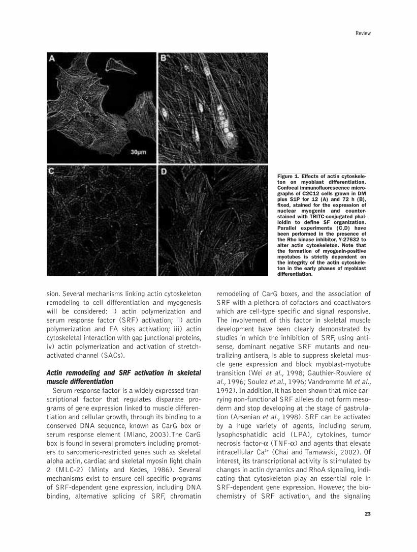
23
sion. Several mechanisms linking actin cytoskeleton
remodeling to cell differentiation and myogenesis
will be considered: i) actin polymerization and
serum response factor (SRF) activation; ii) actin
polymerization and FA sites activation; iii) actin
cytoskeletal interaction with gap junctional proteins,
iv) actin polymerization and activation of stretch-
activated channel (SACs).
Actin remodeling and SRF activation in skeletalmuscle differentiationSerum response factor is a widely expressed tran-
scriptional factor that regulates disparate pro-
grams of gene expression linked to muscle differen-
tiation and cellular growth, through its binding to a
conserved DNA sequence, known as CarG box or
serum response element (Miano, 2003).The CarG
box is found in several promoters including promot-
ers to sarcomeric-restricted genes such as skeletal
alpha actin, cardiac and skeletal myosin light chain
2 (MLC-2) (Minty and Kedes, 1986). Several
mechanisms exist to ensure cell-specific programs
of SRF-dependent gene expression, including DNA
binding, alternative splicing of SRF, chromatin
remodeling of CarG boxes, and the association of
SRF with a plethora of cofactors and coactivators
which are cell-type specific and signal responsive.
The involvement of this factor in skeletal muscle
development have been clearly demonstrated by
studies in which the inhibition of SRF, using anti-
sense, dominant negative SRF mutants and neu-
tralizing antisera, is able to suppress skeletal mus-
cle gene expression and block myoblast-myotube
transition (Wei et al., 1998; Gauthier-Rouviere et
al., 1996; Soulez et al., 1996;Vandromme M et al.,
1992). In addition, it has been shown that mice car-
rying non-functional SRF alleles do not form meso-
derm and stop developing at the stage of gastrula-
tion (Arsenian et al., 1998). SRF can be activated
by a huge variety of agents, including serum,
lysophosphatidic acid (LPA), cytokines, tumor
necrosis factor-α (TNF-α) and agents that elevateintracellular Ca2+ (Chai and Tarnawski, 2002). Of
interest, its transcriptional activity is stimulated by
changes in actin dynamics and RhoA signaling, indi-
cating that cytoskeleton play an essential role in
SRF-dependent gene expression. However, the bio-
chemistry of SRF activation, and the signaling
Review
Figure 1. Effects of actin cytoskele-ton on myoblast differentiation.Confocal immunofluorescence micro-graphs of C2C12 cells grown in DMplus S1P for 12 (A) and 72 h (B),fixed, stained for the expression ofnuclear myogenin and counter-stained with TRITC-conjugated phal-loidin to define SF organization.Parallel experiments (C,D) havebeen performed in the presence ofthe Rho kinase inhibitor, Y-27632 toalter actin cytoskeleton. Note thatthe formation of myogenin-positivemyotubes is strictly dependent onthe integrity of the actin cytoskele-ton in the early phases of myoblastdifferentiation.
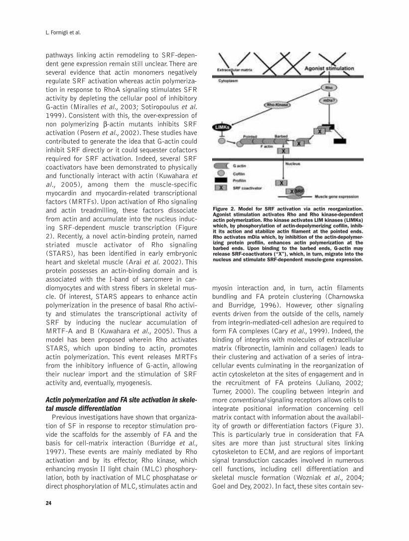
24
pathways linking actin remodeling to SRF-depen-
dent gene expression remain still unclear.There are
several evidence that actin monomers negatively
regulate SRF activation whereas actin polymeriza-
tion in response to RhoA signaling stimulates SFR
activity by depleting the cellular pool of inhibitory
G-actin (Miralles et al., 2003; Sotiropoulus et al.
1999). Consistent with this, the over-expression of
non polymerizing β-actin mutants inhibits SRF
activation (Posern et al., 2002).These studies have
contributed to generate the idea that G-actin could
inhibit SRF directly or it could sequester cofactors
required for SRF activation. Indeed, several SRF
coactivators have been demonstrated to physically
and functionally interact with actin (Kuwahara et
al., 2005), among them the muscle-specific
myocardin and myocardin-related transcriptional
factors (MRTFs). Upon activation of Rho signaling
and actin treadmilling, these factors dissociate
from actin and accumulate into the nucleus induc-
ing SRF-dependent muscle transcription (Figure
2). Recently, a novel actin-binding protein, named
striated muscle activator of Rho signaling
(STARS), has been identified in early embryonic
heart and skeletal muscle (Arai et al. 2002). This
protein possesses an actin-binding domain and is
associated with the I-band of sarcomere in car-
diomyocytes and with stress fibers in skeletal mus-
cle. Of interest, STARS appears to enhance actin
polymerization in the presence of basal Rho activi-
ty and stimulates the transcriptional activity of
SRF by inducing the nuclear accumulation of
MRTF-A and B (Kuwahara et al., 2005). Thus a
model has been proposed wherein Rho activates
STARS, which upon binding to actin, promotes
actin polymerization. This event releases MRTFs
from the inhibitory influence of G-actin, allowing
their nuclear import and the stimulation of SRF
activity and, eventually, myogenesis.
Actin polymerization and FA site activation in skele-tal muscle differentiationPrevious investigations have shown that organiza-
tion of SF in response to receptor stimulation pro-
vide the scaffolds for the assembly of FA and the
basis for cell-matrix interaction (Burridge et al.,
1997). These events are mainly mediated by Rho
activation and by its effector, Rho kinase, which
enhancing myosin II light chain (MLC) phosphory-
lation, both by inactivation of MLC phosphatase or
direct phosphorylation of MLC, stimulates actin and
myosin interaction and, in turn, actin filaments
bundling and FA protein clustering (Charnowska
and Burridge, 1996). However, other signaling
events driven from the outside of the cells, namely
from integrin-mediated-cell adhesion are required to
form FA complexes (Cary et al., 1999). Indeed, the
binding of integrins with molecules of extracellular
matrix (fibronectin, laminin and collagen) leads to
their clustering and activation of a series of intra-
cellular events culminating in the reorganization of
actin cytoskeleton at the sites of engagement and in
the recruitment of FA proteins (Juliano, 2002;
Turner, 2000). The coupling between integrin and
more conventional signaling receptors allows cells to
integrate positional information concerning cell
matrix contact with information about the availabil-
ity of growth or differentiation factors (Figure 3).
This is particularly true in consideration that FA
sites are more than just structural sites linking
cytoskeleton to ECM, and are regions of important
signal transduction cascades involved in numerous
cell functions, including cell differentiation and
skeletal muscle formation (Wozniak et al., 2004;
Goel and Dey, 2002). In fact, these sites contain sev-
L. Formigli et al.
Figure 2. Model for SRF activation via actin reorganization.Agonist stimulation activates Rho and Rho kinase-dependentactin polymerization. Rho kinase activates LIM kinases (LIMKs)which, by phosphorylation of actin-depolymerizing cofilin, inhib-it its action and stabilize actin filament at the pointed ends.Rho activates mDia which, by inhibition of the actin-depolymer-izing protein profilin, enhances actin polymerization at thebarbed ends. Upon binding to the barbed ends, G-actin mayrelease SRF-coactivators (“X”), which, in turn, migrate into thenucleus and stimulate SRF-dependent muscle-gene expression.

eral tyrosine kinases and adaptor proteins, such as
paxillin and p130Cas, which, acting as signaling
scaffolds for the components of FA, allow them to
properly interact with their substrate. FAK, a non
receptor tyrosine kinase, has emerged as a key sig-
naling component of FA. It is activated by autophos-
phorylation that is initiated by its clustering into FA
sites. When phosphorylated, FAK creates docking
sites for the binding of SH2-containing proteins and
regulates activation of additional kinases and phos-
phatases, acting as a switch for multiple signaling
outputs (Parsons, 2003; Oktay et al., 1999). Of
interest, FAK phosphorylation has been associated
with the induction of skeletal myogenesis (Huang et
al., 2006; Clemente et al. 2005; Wozniak et al.,
2004; Goel and Dey, 2002; Lee et al., 1999), name-
ly through the activation of members of Src protein
family, of mitogen-activated protein kinase (MAPK)
family (namely p38MAPK) and of phosphatidyli-
nositol (PI)3-kinases, whose involvement in Rho-
dependent muscle differentiation has been well
established (Khurana and Dey, 2003; Cabane et al.,
2003;Goel and Dey,2002;Aikawa et al., 2002;Wei
et al., 2001). It is worthy to point out that FAK
phosphorylation and activation critically depends on
the integrity of actin cytoskeleton during muscle cell
differentiation (Goel and Dey, 2002; Lee et
al.,1999), thus supporting a model in which
cytoskeletal remodeling may trigger internal signal-
ing and be converted into changes of gene expres-
sion (Wozniak et al., 2004).
Actin remodeling and gap junctional proteins inskeletal muscle differentiationA consistent body of evidence has demonstrated
that specific types of cell contacts, the gap junction
(GJ) are present between skeletal muscle cells. GJ
are composed of intercellular channels formed by
the conjunction of two hemichannels made of six
proteins belonging to the connexin (Cx) family,
whose Cx43 is the most widely expressed member
(Saez et al., 2003). Thus far, these structures have
not been found between mature innervated muscle
fibers and exist as transitory state during myoblast
differentiation. It has long been suggested that the
transfer of small metabolites and signaling mole-
cules between adjacent skeletal muscle cells through
the gap junctions, plays a fundamental role in the
regulation and coordination of myoblast differentia-
tion (Constantin et al., 2000). Indeed, the applica-
tion of intercellular communication inhibitors
(Proulx et al., 1997) and the inducible deletion of
Cx43 proteins (Araya et al. 2005, 2003) dramati-
cally affect myogenesis. Notably, our recent findings
provide novel evidence for a role of actin cytoskele-
ton in the Cx43-mediated effects on myogenesis
(Squecco et al., 2006). In particular, the reduced
interaction between a mutated form of Cx43 and
actin and cortactin, as well as the inhibition of p38
MAPK-dependent signaling pathway essential for
this interaction, are able to completely inhibit the
expression of myogenic marker proteins (myogenin,
myosin heavy chain, caveolin-3) and the achieve-
ment of the fully differentiated phenotype elicited by
sphingosine 1-phosphate, a bioactive lipid that par-
ticipates in the regulation of myoblast biology
(Squecco et al., 2006; Formigli et al., 2005; Donati
et al., 2005; Formigli et al., 2004; Meacci et al.,
2003; Meacci et al., 2002; Formigli et al., 2002).
Notably, the drastic inhibition of myogenesis
occurred even if the intercellular conductance was
only partially affected in these conditions. These
data have led to the suggestion that Cx43 expression
may also stimulate skeletal myogenesis through
25
Review
Figure 3. Model for FA activation. Integrin activation afterengagement with ECM or induction of Rho signaling in responseto receptor activation lead to actin cytoskeletal reorganizationand to accumulation of FA proteins at the sites of engagement.Subsequently, FAK becomes phosphorylated thus creating thebinding sites for adaptor proteins (paxillin and Cas) and for Src.Phosphorylation of Src by FAK triggers MAPK cascade, therebyresulting in gene expression.

gap-junction independent mechanisms. The finding
concerning the role of Cx43 as membrane-cytoskele-
ton anchor protein in myoblasts may indeed repre-
sent a crucial aspect in the molecular mechanisms
involved in the promotion of muscle gene expression
by Cx43 expression. These data are in agreement
with recent studies that pointed out the important
role of GJ-independent functions of Cx43 in the reg-
ulation of many cellular processes such as growth,
survival and migration (Jiang and Gu, 2005;
Giepmans, 2004; Stout 2004; Dang et al. 2003;
Morby et al., 2001; Omori and Yamasaki, 1998;
Huang et al., 1998). Moreover, accumulating evi-
dence has demonstrated a direct interaction of Cx43
C-terminus with cytoskeletal proteins, such as the
tight junction protein Zona Occludens-1 (ZO-1)
(Sing et al., 2005; Tokyofoku, 2001), tubulin
(Giepmans et al., 2001) and the actin binding pro-
tein drebrin (Butkevich et al., 2004) as well as with
signal molecules such as c-src and v-src tyrosine
kinase (Giepmans et al., 2001).
Actin polymerization and SAC-activation inskeletal muscle differentiationStretch-activated cation channels have been
described in a huge variety of cells in different
organisms ranging from bacteria to mammals.
These channels allow the passage of cations, like
Na+, K+,Mg2+, and Ca2+ (Munevar et al., 2004) and
participate in several physiological processes, rang-
ing from cell volume regulation and muscle con-
traction to cell differentiation (Jakkaraju et al.
2003;Minke and Cook, 2002). In particular, recent
reports have demonstrated that SACs activate sec-
ond messengers, namely Ca2+ and Ca2+-dependent
signal pathways, necessary for modulating gene
expression in different mammalian cells (Kumar et
al., 2003; Inoh et al., 2002). Abnormal regulation
of SACs and the excessive increase in the intracel-
lular Ca2+ concentration also contribute to the
pathogenesis of several diseases, including muscu-
lar dystrophy and cardiac arrhythmias (Kumar et
al., 2004).The mechanical distension of the plasma
membrane modulates the ion-transporting activity
of these channels by producing conformational
changes that alter their opening or closing rates
through the distortion of the associated lipid layer
or through the displacement of intramolecular gat-
ing domains. In such a view, activation of SACs rep-
resents an important transduction mechanism that
convert mechanical forces into electrical and bio-
chemical signals in physiological process (Ingber,
2006). Single molecule force spectroscopy studies
have shown that individual peptide domains within
proteins found in the actin cytoskeleton and FA
complex unfold when SACs are mechanically
extended, suggesting a close morphological and
structural interaction between these channels and
cytoskeletal elements (Oberhauser et al., 1999;
Janmey, 1998). However, the functional impact of
actin cytoskeleton reorganization on SACs activity
remains controversial and seems to be strictly
dependent on the different status of microfilaments
in specialized cells. Previous studies have docu-
mented that actin cytoskeletal disruption with
cytochalasins or latrunculin increases the channels’
sensitivity to stretch and promotes SAC activation
in cultured fibroblasts (Wu et al., 1999). On the
other hand, actin cytoskeletal disassembly causes a
decrease in single current and conductance of SACs
in myeloid leukemia cells (Staruschenko et al.,
2005), suggesting that the organization of the cor-
tical microfilaments may be determinant in nega-
tively modulate channel function in these cells. In
addition, recent reports from our group have
demonstrated that not only actin depolymerization
but also actin polymerization and SF formation
may modulate SAC function in myoblastic cells
(Formigli et al., 2005). Using an atomic force
microscopy, we have also shown that the formation
of a well structured actin cytoskeleton is indeed
capable to impose a mechanical strain on the
myoblast plasma membrane and lead to SAC-medi-
ated Ca2+ current inwards (Paternostro et al.,
2006). Notably, we have also observed that SF for-
mation and SAC activation during the early phases
of myoblast differentiation play a pivotal role in the
regulation of skeletal myogenesis (Formigli et al., in
press). Indeed, consistent with a previous investiga-
tion (Wedhas et al., 2005), the treatment of C2C12
myoblasts with Gadolinium chloride, a specific SAC
channel blocker, inhibits myotube formation and the
expression of myogenic markers of differentiation.
These effects are modulated by cytoskeletal com-
ponents and are abolished after treatment with
actin disrupting agents, in perfect agreement with a
model whereby actin polymerization modulates
SAC opening and Ca2+ channel inward current and,
in turn, the activation of Ca2+-mediated pathways
leading to muscle-specific gene expression.
26
L. Formigli et al.
27
Concluding remarksThe dominant view in cell biology is that cell func-
tion is controlled by soluble factors and adhesive lig-
ands, which exert their effects by binding to cell sur-
face receptors, thereby activating signal transduc-
tion cascades inside the cell, leading to modifications
in gene expression.Complexity is now emerging from
the growing evidence suggesting that changes in
actin organization may represent a critical step in
the cell response to stimuli, linking receptor activa-
tion with the generation of regulatory signals. In
particular, several specific signaling involved in
skeletal muscle differentiation, such as SRF, paxillin
and FAK, Cx43-formed channel and SACs activa-
tion can be considered as downstream effectors of
actin cytoskeleton and its dynamic state.The analy-
ses of the relationship existing between actin dynam-
ics and muscle development will certainly shed light
on the understanding of the mechanisms underlying
satellite cell activation and differentiation during
skeletal muscle regeneration and also on the identi-
fication of new therapeutic strategies in muscle dis-
eases, such as dystrophy, characterized by alter-
ations in cytoskeletal organization and cell adhesion.
References
Aikawa R,NagaiT, Kudoh S, ZouY,Tanaka M,Tamura M, et al. Integrinsplay a critical role in mechanical stress-induced p38 MAPK activation.Hypertension 2002; 39:233-8.
Arai A,Spencer JA,Olson EN.STARS, a striated muscle activator of Rhosignaling and serum response factor-dependent transcription. J BiolChem 2002; 277:24453-9.
Araya R, Eckardt D, Maxeiner S, Kruger O, Theis M, Willecke K, et al.Expression of connexins during differentiation and regeneration of skele-tal muscle: functional relevance of connexin43. J Cell Sci 2005; 8: 27-37.
Araya R, Eckardt D, Riquelme M A,Willecke K, Saez JC. Presence andimportance of connexin43 during myogenesis. Cell Commun Adhes2003; 10: 451-6.
Arsenian S,Weinhold B, Oelgeschlager M, Ruther U, Nordheim A. Serumresponse factor is essential for mesoderm formation during mouseembryogenesis. EMBO J 1998 ;17:6289-99.
Berkes CA,Tapscott SJ.MyoD and the transcriptional control of myogen-esis. Semin Cell Dev Biol 2005; 16:585-95.
Bryan BA, Mitchell DC, Zhao L, Ma W, Stafford LJ, Teng BB, et al.Modulation of muscle regeneration,myogenesis,and adipogenesis by theRho family guanine nucleotide exchange factor GEFT. Mol Cell Biol2005; 25:11089-101.
Burridge K, Charzanowska-Wodnicka M,Zhong C. Focal adhesion assem-bly. Trends Cell Biol 1997; 7:342-7.
Butkevich E,Hulsmann S,Wenzel D,ShiraroT,Duden R,Majoul I.Drebrinis a novel connexin-43 binding partner that links gap junctions to thesubmembrane cytoskeleton. Curr Biol 2004; 14: 650-8.
Cabane C,EnglaroW,Yeow K,Ragno M,Derijard B.Regulation of C2C12myogenic terminal differentiation by MKK3/p38alpha pathway. Am JPhysiol Cell Physiol 2003; 284:C658-66.
Carnac G,Primig M,Kitzmann M,Chafey P,Tuil D,Lamb N,Fernandez A.RhoA GTPase and serum response factor control selectively the expres-sion of MyoD without affecting Myf5 in mouse myoblasts.Mol Biol Cell1998; 9:1891-902.
Cary LA, Han DC, Guan JL. Integrin-mediated signal transduction path-ways. Histol Histopathol 1999 14:1001-9.
Chai J,Tarnawski AS.Serum response factor: discovery, biochemistry, bio-logical roles and implications for tissue injury healing. J PhysiolPharmacol 2002; 53:147-57.
Charrasse S, Causeret M, Comunale F, Bonet-Kerrache A, Gauthier-Rouviere C. Rho GTPases and cadherin-based cell adhesion in skeletalmuscle development. J Muscle Res Cell Motil 2003; 24:309-13.
Chrzanowska-WodnickaM,Burridge K.Rho-stimulated contractility drivesthe formation of stress fibers and focal adhesions. J Cell Biol 1996;133:1403-15.
Clemente CF,Corat MA,Saad ST,Franchini KG.Differentiation of C2C12myoblasts is critically regulated by FAK signaling. Am J Physiol RegulIntegr Comp Physiol 2005; 289: 862-70.
Constantin B, Cronier L. Involvement of gap junctional communication inmyogenesis. Int Rev Cytol 2000; 196, 1-65.
Dang X,Doble BW,Kardami E.The carboxy-tail of connexin-43 localizesto the nucleus and inhibits cell growth.Mol Cell Biochem 2003;242:35-8.
Dhawan J,Helfman DM Modulation of acto-myosin contractility in skele-tal muscle myoblasts uncouples growth arrest from differentiation. JCell Sci 2004; 117:3735-48.
Disanza A,Steffen A,HertzogM,Frittoli E,Rottner K,Scita G.Actin poly-merization machinary: the finish line of signaling networks, the startingpoint of cellular movement.CMLS,Cell Mol Life Sci 2005;62:955-70.
Donati C,Meacci E,Nuti F,Becciolini L,FarnararoM,Bruni P.Sphingosine1-phosphate regulates myogenic differentiation: a major role for S1P2receptor. FASEB J 2005; 19:449-51.
Formigli L, Meacci E, Sassoli C, Squecco R, Nosi D, Chellini F et al.Cytoskeleton/stretch-activated ion channel interaction regulates myo-genic differentiation of skeletal myoblasts. J Cell Physiol, in press.
Formigli L, Meacci E, Sassoli C, Chellini F, Giannini R, Quercioli F, et al.Sphingosine 1-phosphate induces cytoskeletal reorganization in C2C12myoblasts: physiological relevance for stress fibres in the modulation ofion current through stretch-activated channels. J Cell Sci 2005;118:1161-71.
Formigli L, Meacci E, Vassalli M, Nosi D, Quercioli F, Tiribilli B, et al.Sphingosine 1-phosphate induces cell contraction via calcium-indepen-dent/Rho-dependent pathways in undifferentiated skeletal muscle cells.J Cell Physiol 2004 ;198:1-11.
Formigli L, Francini F, Meacci E, Vassalli M, Nosi D, Quercioli F, et al.Sphingosine 1-phosphate induces Ca2+ transients and cytoskeletalrearrangement in C2C12 myoblastic cells. Am J Physiol Cell Physiol2002; 282:C1361-73.
Frisch SM, Francis H. Disruption of epithelial cell-matrix interactionsinduces apoptosis. J Cell Biol 1994 ;124:619-26.
Gauthier-Rouviere C, Vandromme M, Tuil D, Lautredou N, Morris M,Soulez M et al. Expression and activity of serum response factor isrequired for expression of the muscle-determining factor MyoD in bothdividing and differentiating mouse C2C12 myoblasts. Mol Biol Cell1996 7:719-29.
Geiger B, Bershadsky A. Exploring the neighborhood: adhesion-coupledcell mechanosensors. Cell 2002; 110:139-42.
Giepmans BN.Gap junctions and connexin-interacting proteins.CardiovascRes 2004; 62: 233-45.
Giepmans BN,MoolenaarW H. Interaction of c-src with the gap junctionprotein connexin-43: role in the regulation of cell-cell communication. JBiol Chem 2001; 276: 8544-9.
Goel HL, Dey CS. PKC-regulated myogenesis is associated with increasedtyrosine phosphorylation of FAK, Cas, and paxillin, formation of Cas-CRK complex, and JNK activation. Differentiation 2002; 70:257-71.
HawkeTJ,Garry DJ.Myogenic satellite cells:physiology to molecular biol-ogy. J Appl Physiol 2001; 9:534-51.
Hill CS,Wynne J,Treisman R.The Rho family GTPases RhoA, Rac1, andCDC42Hs regulate transcriptional activation by SRF. Cell 199581:1159-70.
Huang D,Khoe M, Ilic D,Bryer-Ash M.Reduced expression of focal adhe-sion kinase disrupts insulin action in skeletal muscle cells.Endocrinology2006 ;147:3333-43.
Huang GY,Cooper ES,Waldo K,Kirby ML,Gilula N B,Lo CW.Gap junc-tion-mediated cell-cell communication modulates mouse neural crestmigration. J Cell Biol 1998; 143: 1725-34.
Review

28
Ingber DE. Cellular mechanotransduction: putting all the pieces togetheragain. FASEB J 2006; 20:811-27.
Ingber DE.Tensegrity II.How structural networks influence cellular infor-mation processing networks. J Cell Sci 2003; 116:1397-408.
Inoh H, Ishiguro N,Sawazaki S,Amma H,Miyazu M, Iwata H, et al.Uni-axial cyclic stretch induces the activation of transcription factor nuclearfactor kappaB in human fibroblast cells. FASEB J 2002; 16:405-7.
Jakkaraju S, Zhe X, Schuger L. Role of stretch in activation of smoothmuscle cell lineage.Trends Cardiovasc Med 2003 ;13:330-5.
Janmey PA. The cytoskeleton and cell signaling: component localizationand mechanical coupling. Physiol Rev 1998 78:763-81.
Jiang JX, Gu S. Gap junction- and hemichannel-independent actions ofconnexins. Biochim Biophys Acta 2005; 1711:208-14.
Juliano RL. Signal transduction by cell adhesion receptors and thecytoskeleton: functions of integrins, cadherins, selectins, andimmunoglobulin-superfamily members. Annu Rev Pharmacol Toxicol2002; 42:283-323.
Katoh K,KanoY,AmanoM,Onishi H,Kaibuchi K,Fujiwara K.Rho-kinase--mediated contraction of isolated stress fibers. J Cell Biol2001;153:569-84.
Khurana A, Dey CS. p38 MAPK interacts with actin and modulates fila-ment assembly during skeletal muscle differentiation. Differentiation2003; 7:42-50.
Komati H, Naro F, Mebarek S, De Anekis V, Adamo S, Lagarde M, et al.Phospholypase D is involved in myogenic differentiation through remod-elling of actin cytoskeleton.Mol Biol Cell 2005; 16:1232-44.
Kumar A, Khandelwal N, Malya R, Reid MB, Boriek AM. Loss of dys-trophin causes aberrant mechanotransduction in skeletal muscle fibers.FASEB J 2004;18:102-13.
Kumar A,Knox AJ,Boriek AM.CCAAT/enhancer-binding protein and acti-vator protein-1 transcription factors regulate the expression of inter-leukin-8 through the mitogen-activated protein kinase pathways inresponse to mechanical stretch of human airway smooth muscle cells. JBiol Chem 2003; 278:18868-76.
Kuwahara K, Barrientos T, Pipes GC, Li S, Olson EN.Muscle-specific sig-naling mechanism that links actin dynamics to serum response factor.Mol Cell Biol 2005; 25:3173-81.
Lee KH, Lee SH, Kim D, Rhee S, Kim C, Chung CH, et al. Promotion ofskeletal muscle differentiation by K252a with tyrosine phosphorylationof focal adhesion:a possible involvement of small GTPase Rho.Exp CellRes 1999; 252:401-15.
Meacci E,Cencetti F,Donati C,Nuti F,Farnararo M,KohnoT, et al.Down-regulation of EDG5/S1P2 during myogenic differentiation results in thespecific uncoupling of sphingosine 1-phosphate signalling to phospholi-pase D. Biochim Biophys Acta 2003; 1633, 133-42.
Meacci E, Cencetti F, Formigli L, Squecco R, Donati C, Tiribilli B, et al.Sphingosine 1-phosphate evokes calcium signals in C2C12myoblasts viaEdg3 and Edg5 receptors. Biochem. J 2002; 362: 349-57.
Miano JM. Serum response factor: toggling between disparate programsof gene expression. J Mol Cell Cardiol 2003; 35:577-93.
Milasincic DJ, Calera MR, Farmer SR, Pilch PF. Stimulation of C2C12myoblast growth by basic fibroblast growth factor and insulin-likegrowth factor 1 can occur via mitogen-activated protein kinase-depen-dent and -independent pathways.Mol Cell Biol 1996; 16:5964-73.
Minke B, Cook B.TRP channel proteins and signal transduction. PhysiolRev 2002; 82:429-72.
Minty A,Kedes L.Upstream regions of the human cardiac actin gene thatmodulate its transcription in muscle cells: presence of an evolutionarilyconserved repeated motif.Mol Cell Biol. 1986; 6:2125-36.
Miralles F,Posern G,Zaromytidou AI,Treisman R.Actin dynamics controlSRF activity by regulation of its coactivator MAL.Cell 2003;113:329-42.
Moorby C, Patel M. Dual functions for connexins: Cx43 regulates growthindependently of gap junction formation. Exp Cell Res 2201; 271:238-48.
Munevar S,Wang YL, Dembo M. Regulation of mechanical interactionsbetween fibroblasts and the substratum by stretch-activated Ca2+ entry.J Cell Sci 2004; 117:85-92.
Oberhauser AF,Marszalek PE,Carrion-Vazquez M,Fernandez JM.Singleprotein misfolding events captured by atomic force microscopy. NatStruct Biol 1999; 6:1025-8.
Oktay M,Wary KK, Dans M, Birge RB, Giancotti FG. Integrin-mediatedactivation of focal adhesion kinase is required for signaling to Jun NH2-terminal kinase and progression through the G1 phase of the cell cycle.
J Cell Biol 1999; 145:1461-69.
Omori Y,Yamasaki H.Mutated connexin43 proteins inhibit rat glioma cellgrowth suppression mediated by wild-type connexin43 in a dominant-negative manner. Int J Cancer 1998; 78: 446-53.
Paternostro F, Sbrana F, Nosi D, Tiribilli B, Formigli L. Role of actincytoskeleton in the modulation of SACs sensitivity of myoblastic C2C12cells. It J Anat Embriol 2006; 111:200.
Parsons JT. Focal adhesion kinase: the first ten years. J Cell Sci 2003;116:1409-16.
Posern G, Sotiropoulos A,Treisman R. Mutant actins demonstrate a rolefor unpolymerized actin in control of transcription by serum responsefactor.Mol Cell Biol 2002 ;13:4167-78.
Proulx AM,Merrifield PA,Naus CC. Blocking gap junctional intercellularcommunication in myoblasts inhibits myogenin and MRF4 expression.Dev Genet 1997; 20: 133-44.
Revenu C, Athman R, Robine S, Louvard D.The co-workers of actin fila-ments: from cell structures to signals. Nat Rev Mol Cell Biol 2004,5:635-46.
Sàez JC,BerthoudVM ,BranesMC,Martinez AD,Beyer E.Plasmamem-brane channels form by connexin: their regulation and function. PhysiolRev 2003; 83:1359-400.
Sah VP, Seasholtz TM, Sagi SA, Brown JH.The role of Rho in G protein-coupled receptor signal transduction. Annu Rev Pharmacol Toxicol2000; 40:459-89.
Singh D,Solan JL,Taffet SM.,Javier R,Lampe PD.Connexin 43 interactswith zona occludens-1 and -2 proteins in a cell cycle stage-specific man-ner. J Biol Chem 2005; 280, 30416-21.
Sotiropoulos A,Gineitis D, Copeland J,Treisman R. Signal-regulated acti-vation of serum response factor is mediated by changes in actin dynam-ics.Cell 1999; 98:159-69.
Soulez M, Rouviere CG, Chafey P, Hentzen D,Vandromme M, LautredouN, et al. Growth and differentiation of C2 myogenic cells are dependenton serum response factor.Mol Cell Biol 1996; 16:6065-74.
Squecco R, Sassoli C, Nuti F, Martinesi M, Chellini F, Nosi D et al.Sphingosine 1-phosphate induces myoblast differentiation through Cx43protein expression:a role for a gap junction-dependent and -independentfunction.Mol Biol Cell 2006; 17:4896-910.
Staruschenko A, Negulyaev YA, Morachevskaya EA. Actin cytoskeletondisassembly affects conductive properties of stretch-activated cationchannels in leukaemia cells. Biochim Biophys Acta 2005; 166:53-60.
Stout C,Goodenough DA,Paul D L.Connexins:functions without junctions.Curr Opin Cell Biol 2004; 16, 507-12.
Takano H, Komuro I, Oka T, Shiojima I, Hiroi Y, Mizuno T,Yazaki Y.TheRho family G proteins play a critical role inmuscle differentiation.1998;18:1580-9.
ToyofukuT,AkamatsuY, Zhang H,KuzuyaT,Tada M,Hori M. c-Src regu-lates the interaction between connexin-43 and ZO-1 in cardiac myocytes.J Biol Chem 2001; 276:1780-8.
Turner CE. Paxillin and focal adhesion signalling. Nat Cell Biol 2000;2:E231-36.
Vandromme M, Gauthier-Rouviere C, Carnac G, Lamb N, Fernandez A.Serum response factor p67SRF is expressed and required during myo-genic differentiation of both mouse C2 and rat L6 muscle cell lines. JCell Biol 1992; 118:1489-500.
Watanabe N,Madaule P, ReidT, IshizakiT,Watanabe G, et al. p140mDia,a mammalian homolog of Drosophila diaphanous, is a target protein forRho small GTPase and is a ligand for profiling. EMBO J 1997;16:3044–56.
Wedhas N, Klamut HJ, Dogra C, Srivastava AK, Mohan S, Kumar A.Inhibition of mechanosensitive cation channels inhibits myogenic differ-entiation by suppressing the expression of myogenic regulatory factorsand caspase-3 activity. FASEB J 2005; 19:1986-97.
Wei L,Wang L, Carson JA, Agan JE, Imanaka-Yoshida K, Schwartz RJ.beta1 integrin and organized actin filaments facilitate cardiomyocyte-specific RhoA-dependent activation of the skeletal alpha-actin promot-er. FASEB J 2001; 15:785-96.
Wei L,ZhouW,Croissant JD, Johansen FE,Prywes R,BalasubramanyamA, et al. RhoA signaling via serum response factor plays an obligatoryrole in myogenic differentiation. J Biol Chem 1998; 273:30287-94.
Wozniak MA,Modzelewska K,Kwong L,Keely PJ.Focal adhesion regula-tion of cell behavior. Biochem Biophys Acta 2004; 1692:103-19.
Wu Z, Wong K, Glogauer M, Ellen RP, McCulloch CA. Regulation ofstretch-activated intracellular calcium transients by actin filaments.Biochem Biophys Res Commun 1999;261:419-25.
L. Formigli et al.
©2007, European Journal of Histochemistry
Sarcoglycans are a sub-complex of transmembrane proteinswhich are part of the dystrophin-glycoprotein complex (DGC).They are expressed above all in the skeletal, cardiac andsmooth muscle. Although numerous studies have been con-ducted on the sarcoglycan sub-complex in skeletal and car-diac muscle, the manner of distribution and localization ofthese proteins along the non-junctional sarcolemma is stillnot clear. Furthermore, there are unclear data about theactual role of sarcoglycans in human skeletal muscle affect-ed by sarcoglycanopathies. In our studies on human skeletalmuscle, normal and pathological, we determined the local-ization, distribution and interaction of these glycoproteins.Our results, on normal human skeletal muscle, showed thatthe sarcoglycans can be localized both in the region of thesarcolemma over the I band and over the A band, hypothe-sizing a correlation between regions of the sarcolemmaoccupied by costameres and the metabolic type of the fibers(slow and fast). Our data on skeletal muscle affected bysarcoglycanopathy confirmed the hypothesis of a bidirec-tional signaling between sarcoglycans and integrins and theinteraction of filamin2 with both sarcoglycans and integrins.In addition, we have recently demonstrated, in smooth mus-cle, the presence of α-SG, in contrast with data of otherAuthors. Finally, we analyzed the association between con-tractile activity and quantitative correlation between α- andε-SG, in order to better define the arrangement of sarcogly-can subcomplex.
Key words: sarcoglycans, skeletal muscle, cardiac muscle,human, sarcoglycanopathy, muscular diseases.
Correspondence: Angelo Favaloro,Department of Biomorphology and Biotechnologies,Policlinico Universitario G. Martino, University of Messina,Via Consolare Valeria, 1 – 98125 Messina, ItalyTel: +39.0902213361.Fax: +39.090692449.E-mail: [email protected]
European Journal of Histochemistry2007; vol. 51 supplement 1:29-34
Sarcoglycan subcomplex in normal and pathological human muscle
fibers
G. Anastasi, G. Cutroneo, G. Rizzo, A. Favaloro
Department of Biomorphology and Biotechnologies, University of Messina, Italy
29
The sarcoglycan subcomplex (SGC) is a well-
known system of interaction between extra-
cellular matrix and sarcolemma-associated
cytoskeleton in skeletal and cardiac muscle. This
subcomplex is made up of a series of transmem-
brane proteins (Ettinger et al., 1997) which, togeth-
er with other components of the dystrophin-glyco-
protein complex (DGC), regulate interaction
between the cytoskeleton and extracellular matrix
in skeletal muscle and cardiac muscle. In this way,
these glycoproteins stabilize the sarcolemma of the
myofibrils and the cardiomyocytes and protect the
muscle fibers from any possible damage provoked
by continuing cycles of contraction and relaxation
(Ervasti et al., 1990).
The SGC is made up of four glycoproteins linked,
by a lateral binding, to β-dystroglycan, (Crosbie etal., 1997), α-sarcoglycan, 50 kD, a type I protein,that is with the NH-terminal on the intracellular
side, the β-, γ- and the δ-sarcoglycans, 43 kD, 35
kD and 35 kD respectively, all type II proteins, that
is with the NH-terminal on the extracellular side
(Yoshida et al., 1994). The α-sarcoglycan and theγ-sarcoglycan are expressed only in muscular tis-sue, while the other sarcoglycans have a wider dis-
tribution.
A widely expressed fifth sarcoglycan with signifi-
cant homology to α-sarcoglycan, ε-sarcoglycan, hasbeen identified; this sarcoglycan is expressed in both
muscle and non-muscle cells, and in embryos as well
as adults (Ettinger et al., 1997). It is hypothesized
that ε-sarcoglycan might replace α-sarcoglycan insmooth muscle, forming a novel sarcoglycan sub-
complex consisting of ε-, β-, γ-, and δ-sarcoglycan(Barresi et al., 2000).Thus, it is possible that sarco-
glycans, like other components of the DGC, may
play a key role for embryonic development and for
viability of non-muscle tissues (Ettinger et al.,
1997).
Recently, a novel mammalian sarcoglycan, ζ-sar-coglycan, highly related to γ-sarcoglycan and δ-sar-
REVIEW
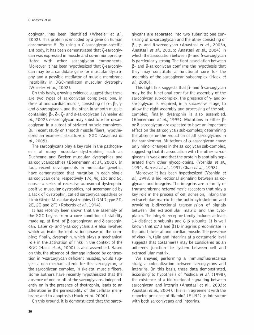
coglycan, has been identified (Wheeler et al.,
2002).This protein is encoded by a gene on human
chromosome 8. By using a ζ-sarcoglycan-specificantibody, it has been demonstrated that ζ-sarcogly-can was expressed in muscle and co-immunoprecip-
itated with other sarcoglycan components.
Moreover it has been hypothesized that ζ-sarcogly-can may be a candidate gene for muscular dystro-
phy and a possible mediator of muscle membrane
instability in DGC-mediated muscular dystrophy
(Wheeler et al., 2002).
On this basis, growing evidence suggest that there
are two types of sarcoglycan complexes; one, in
skeletal and cardiac muscle, consisting of α-, β-, γ-and δ-sarcoglycan, and the other, in smooth muscle,containing β-, δ-, ζ- and ε-sarcoglycan (Wheeler et
al., 2002). ε-sarcoglycan may substitute for α-sar-coglycan in a subset of striated muscle complexes.
Our recent study on smooth muscle fibers, hypothe-
sized an exameric structure of SGC (Anastasi et
al., 2005).
The sarcoglycans play a key role in the pathogen-
esis of many muscular dystrophies, such as
Duchenne and Becker muscular dystrophies and
sarcoglycanopathies (Bönnemann et al., 2002). In
fact, recent developments in molecular genetics
have demonstrated that mutation in each single
sarcoglycan gene, respectively 17q, 4q, 13q and 5q,
causes a series of recessive autosomal dystrophin-
positive muscular dystrophies, not accompanied by
a lack of dystrophin, called sarcoglycanopathies or
Limb Girdle Muscular dystrophies (LGMD type 2D,
2E, 2C and 2F) (Roberds et al., 1994).
It has recently been shown that the assembly of
the SGC begins from a core condition of stability
made up, at first, of β-sarcoglycan and δ-sarcogly-can. Later α- and γ-sarcoglycans are also involvedwhich activate the maturation phase of the com-
plex; finally, dystrophin, which plays a mechanical
role in the activation of links in the context of the
SGC (Hack et al., 2000) is also assembled. Based
on this, the absence of damage induced by contrac-
tion in γ-sarcoglycan deficient muscles, would sug-gest a non-mechanical role for this sarcoglycan, or
the sarcoglycan complex, in skeletal muscle fibers.
Some authors have recently hypothesized that the
absence of one or all of the sarcoglycans, independ-
ently or in the presence of dystrophin, leads to an
alteration in the permeability of the cellular mem-
brane and to apoptosis (Hack et al. 2000).
On this ground, it is demonstrated that the sarco-
glycans are separated into two subunits: one con-
sisting of α-sarcoglycan and the other consisting ofβ-, γ- and δ-sarcoglycan (Anastasi et al., 2003a,
Anastasi et al., 2003b; Anastasi et al., 2004) in
which the association between β- and δ-sarcoglycanis particularly strong.The tight association between
β- and δ-sarcoglycan confirms the hypothesis thatthey may constitute a functional core for the
assembly of the sarcoglycan subcomplex (Hack et
al., 2000).
This tight link suggests that β- and δ-sarcoglycanmay be the functional core for the assembly of the
sarcoglycan sub-complex.The presence of γ- and α-sarcoglycan is required, in a successive stage, to
allow the right assembly and processing of the sub-
complex; finally, dystrophin is also assembled.
(Bönnemann et al., 1995). Mutations in either β-or δ-sarcoglycan are expected to have an importanteffect on the sarcoglycan sub-complex, determining
the absence or the reduction of all sarcoglycans in
the sarcolemma. Mutations of α-sarcoglycan causeonly minor changes in the sarcoglycan sub-complex,
suggesting that its association with the other sarco-
glycans is weak and that the protein is spatially sep-
arated from other glycoproteins. (Yoshida et al.,
1994; Barresi et al., 1997; Chan et al., 1998).
Moreover, it has been hypothesized (Yoshida et
al., 1998) a bidirectional signaling between sarco-
glycans and integrins.The integrins are a family of
transmembrane heterodimeric receptors that play a
key role in the process of cell adhesion, linking the
extracellular matrix to the actin cytoskeleton and
providing bidirectional transmission of signals
between the extracellular matrix and the cyto-
plasm.The integrin receptor family includes at least
14 distinct α subunits and 8 β subunits. It is well
known that α7B and β1D integrins predominate in
the adult skeletal and cardiac muscle.The presence
of vinculin, talin and integrins at a costameric level
suggests that costameres may be considered as an
adherens junction-like system between cell and
extracellular matrix.
We showed, performing a immunofluorescence
study, a colocalization between sarcoglycans and
integrins. On this basis, these data demonstrated,
according to hypothesis of Yoshida et al. (1998),
the existence of a bidirectional signalling between
sarcoglycan and integrin (Anastasi et al., 2003b;
Anastasi et al., 2004).This is in agreement with the
reported presence of filamin2 (FLN2) as interactor
with both sarcoglycans and integrins.
30
G. Anastasi et al.
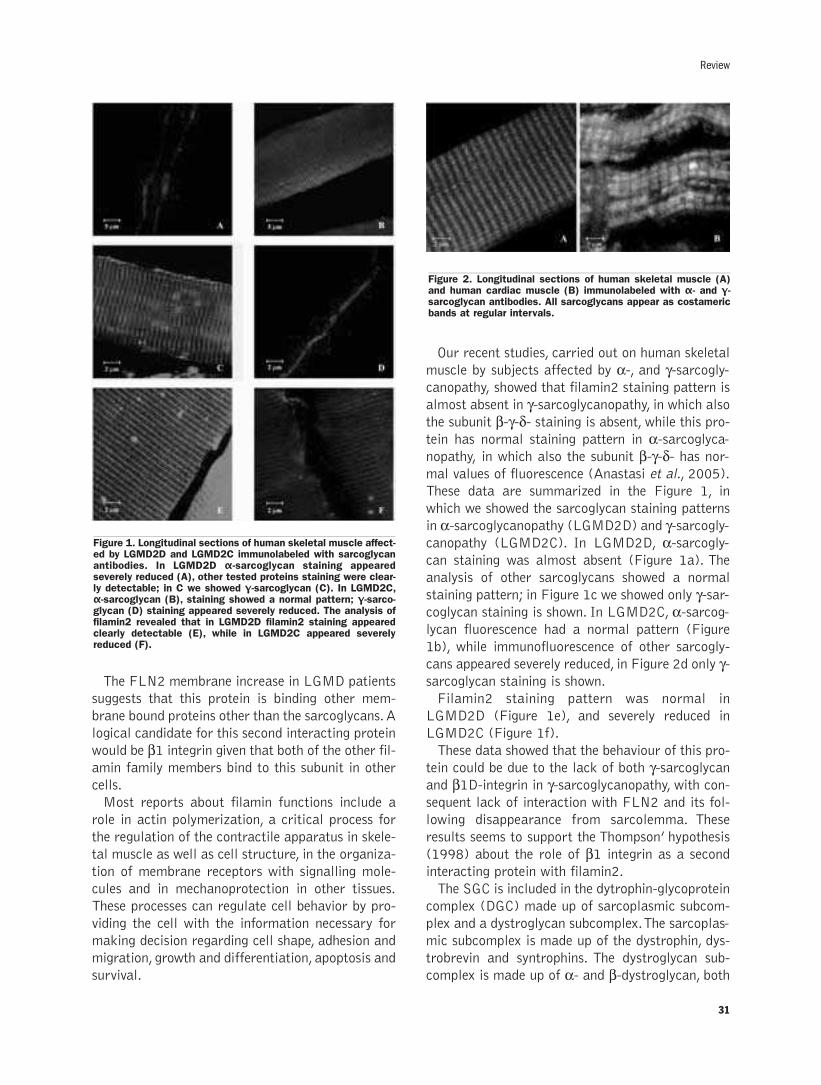
The FLN2 membrane increase in LGMD patients
suggests that this protein is binding other mem-
brane bound proteins other than the sarcoglycans.A
logical candidate for this second interacting protein
would be β1 integrin given that both of the other fil-amin family members bind to this subunit in other
cells.
Most reports about filamin functions include a
role in actin polymerization, a critical process for
the regulation of the contractile apparatus in skele-
tal muscle as well as cell structure, in the organiza-
tion of membrane receptors with signalling mole-
cules and in mechanoprotection in other tissues.
These processes can regulate cell behavior by pro-
viding the cell with the information necessary for
making decision regarding cell shape, adhesion and
migration, growth and differentiation, apoptosis and
survival.
Our recent studies, carried out on human skeletal
muscle by subjects affected by α-, and γ-sarcogly-canopathy, showed that filamin2 staining pattern is
almost absent in γ-sarcoglycanopathy, in which alsothe subunit β-γ-δ- staining is absent, while this pro-tein has normal staining pattern in α-sarcoglyca-nopathy, in which also the subunit β-γ-δ- has nor-mal values of fluorescence (Anastasi et al., 2005).
These data are summarized in the Figure 1, in
which we showed the sarcoglycan staining patterns
in α-sarcoglycanopathy (LGMD2D) and γ-sarcogly-canopathy (LGMD2C). In LGMD2D, α-sarcogly-can staining was almost absent (Figure 1a). The
analysis of other sarcoglycans showed a normal
staining pattern; in Figure 1c we showed only γ-sar-coglycan staining is shown. In LGMD2C, α-sarcog-lycan fluorescence had a normal pattern (Figure
1b), while immunofluorescence of other sarcogly-
cans appeared severely reduced, in Figure 2d only γ-sarcoglycan staining is shown.
Filamin2 staining pattern was normal in
LGMD2D (Figure 1e), and severely reduced in
LGMD2C (Figure 1f).
These data showed that the behaviour of this pro-
tein could be due to the lack of both γ-sarcoglycanand β1D-integrin in γ-sarcoglycanopathy, with con-sequent lack of interaction with FLN2 and its fol-
lowing disappearance from sarcolemma. These
results seems to support the Thompson’ hypothesis
(1998) about the role of β1 integrin as a second
interacting protein with filamin2.
The SGC is included in the dytrophin-glycoprotein
complex (DGC) made up of sarcoplasmic subcom-
plex and a dystroglycan subcomplex.The sarcoplas-
mic subcomplex is made up of the dystrophin, dys-
trobrevin and syntrophins. The dystroglycan sub-
complex is made up of α- and β-dystroglycan, both
31
Review
Figure 2. Longitudinal sections of human skeletal muscle (A)and human cardiac muscle (B) immunolabeled with α- and γ-sarcoglycan antibodies. All sarcoglycans appear as costamericbands at regular intervals.
Figure 1. Longitudinal sections of human skeletal muscle affect-ed by LGMD2D and LGMD2C immunolabeled with sarcoglycanantibodies. In LGMD2D α-sarcoglycan staining appearedseverely reduced (A), other tested proteins staining were clear-ly detectable; in C we showed γ-sarcoglycan (C). In LGMD2C,α-sarcoglycan (B), staining showed a normal pattern; γ-sarco-glycan (D) staining appeared severely reduced. The analysis offilamin2 revealed that in LGMD2D filamin2 staining appearedclearly detectable (E), while in LGMD2C appeared severelyreduced (F).
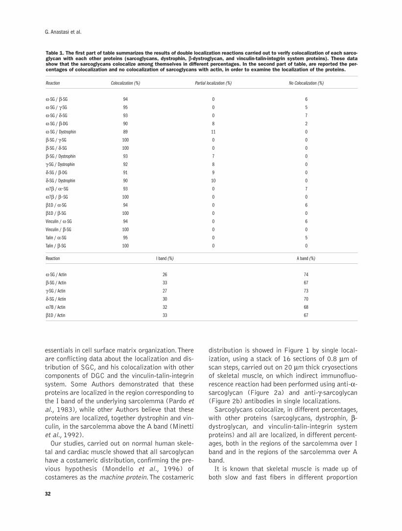
essentials in cell surface matrix organization.There
are conflicting data about the localization and dis-
tribution of SGC, and his colocalization with other
components of DGC and the vinculin-talin-integrin
system. Some Authors demonstrated that these
proteins are localized in the region corresponding to
the I band of the underlying sarcolemma (Pardo et
al., 1983), while other Authors believe that these
proteins are localized, together dystrophin and vin-
culin, in the sarcolemma above the A band (Minetti
et al., 1992).
Our studies, carried out on normal human skele-
tal and cardiac muscle showed that all sarcoglycan
have a costameric distribution, confirming the pre-
vious hypothesis (Mondello et al., 1996) of
costameres as the machine protein.The costameric
distribution is showed in Figure 1 by single local-
ization, using a stack of 16 sections of 0.8 µm of
scan steps, carried out on 20 µm thick cryosections
of skeletal muscle, on which indirect immunofluo-
rescence reaction had been performed using anti-α-sarcoglycan (Figure 2a) and anti-γ-sarcoglycan(Figure 2b) antibodies in single localizations.
Sarcoglycans colocalize, in different percentages,
with other proteins (sarcoglycans, dystrophin, β-dystroglycan, and vinculin-talin-integrin system
proteins) and all are localized, in different percent-
ages, both in the regions of the sarcolemma over I
band and in the regions of the sarcolemma over A
band.
It is known that skeletal muscle is made up of
both slow and fast fibers in different proportion
32
G. Anastasi et al.
Table 1. The first part of table summarizes the results of double localization reactions carried out to verify colocalization of each sarco-glycan with each other proteins (sarcoglycans, dystrophin, ββ-dystroglycan, and vinculin-talin-integrin system proteins). These datashow that the sarcoglycans colocalize among themselves in different percentages. In the second part of table, are reported the per-centages of colocalization and no colocalization of sarcoglycans with actin, in order to examine the localization of the proteins.
Reaction Colocalization (%) Partial localization (%) No Colocalization (%)
α-SG / β-SG 94 0 6
α-SG / γ-SG 95 0 5
α-SG / δ-SG 93 0 7
α-SG / β-DG 90 8 2
α-SG / Dystrophin 89 11 0
β-SG / γ-SG 100 0 0
β-SG / δ-SG 100 0 0
β-SG / Dystrophin 93 7 0
γ-SG / Dystrophin 92 8 0
δ-SG / β-DG 91 9 0
δ-SG / Dystrophin 90 10 0
α7β / α−SG 93 0 7
α7β / β−SG 100 0 0
β1D / α-SG 94 0 6
β1D / β-SG 100 0 0
Vinculin / α-SG 94 0 6
Vinculin / β-SG 100 0 0
Talin / α-SG 95 0 5
Talin / β-SG 100 0 0
Reaction I band (%) A band (%)
α-SG / Actin 26 74
β-SG / Actin 33 67
γ-SG / Actin 27 73
δ-SG / Actin 30 70
α7B / Actin 32 68
β1D / Actin 33 67
(Johnson et al., 1973), while cardiac muscle is
made up exclusively of slow fibers with a highly
oxidative metabolism. Thus, we hypothesized that
slow fibers are characterized by localization of
costameric proteins on the region of the sarcolem-
ma over band I, while fast fibers by localization of
the same proteins in the region over band A
(Anastasi et al., 2003a, Anastasi et al., 2003b).
Moreover, these data confirm the hypothesis of two
subunit, one consisting of a-sarcoglcan and other
formed by β−γ−δ−sarcoglycan (Anastasi et al.,2003a; Anasatsi et al., 2004). All these data are
summarized in Table 1.
It will be intriguing, besides, to integrate these
studies with molecular biology techniques; in fact
the definition of patterns in immunohistochemical
profile would be important to guide the genetic
analysis directly to the responsible gene and abbre-
viate molecular genetic investigations (Bönnemann
et al., 2002).
References
Anastasi G, Cutroneo G, Trimarchi F, Rizzo G, Bramanti P, BruschettaD, et al. Sarcoglycans in human skeletal muscle and human cardiacmuscle: a confocal laser scanning microscope study. Cells TissuesOrgans 2003; 173: 54-63.
Anastasi G, Amato A, Tarone G, Vita G, Monici MC, Magaudda L, et al.Distribution and localization of vinculin-talin-integrin system anddystrophin-glycoprotein complex in human skeletal muscle. CellsTissues Organs 2003; 175: 151-64.
Anastasi G, Cutroneo G, Rizzo G, Arco A, Santoro G, Bramanti P, et al.Sarcoglycan and integrin localization in normal human skeletalmuscle: a confocal laser scanning microscope study. Eur JHistochem 2004; 48: 245-52.
Anastasi G, Cutroneo G, Sidoti A, Santoro G, D'Angelo R, Rizzo G, etal. Sarcoglycan subcomplex in normal human smooth muscle: animmunohistochemical and molecular study. Int J Mol Med 2005;16:367-74.
Barresi R, Moore SA, Stolle CA, Mendell JR, Campbell KP. Expressionof γ-sarcoglycan in smooth muscle and its interaction with thesmooth muscle sarcoglycan-sarcospan complex. J Biol Chem 2000;275: 38554-60.
Bönnemann CG, Modi R, Noguchi S, Mizuno Y, Yoshida M, Gussoni E,
et al. β-sarcoglycan (A3b) mutations cause autosomal recessivemuscular dystrophy with loss of the sarcoglycan complex. Nat Genet1995; 11: 266-72.
Bönnemann CG, Wong J, Jones KJ, Lidov HGW, Feener CA, Shapiro F,et al. Primary γ-sarcoglycanopathy (LGMD 2C): broadening of themutational spectrum guided by the immunohistochemical profile.Neuromuscul Disord 2002; 12: 273-80.
Chan Y, Bönnemann CG, Lidov HGW, Kunkel LM. Molecular organiza-tion of sarcoglycan complex in mouse myotubes in culture. J CellBiol 1998; 143: 2033-44.
Crosbie RH, Lebakken CS, Holt KH, Venzke DP, Straub V, Lee JC, etal. Membrane targeting and stabilization of sarcospan is mediatedby the sarcoglycan subcomplex. J Cell Biol 1999; 145: 153-65.
Ervasti JM, Ohlendieck K, Kahl SD, Gaver MG, Campbell KP.Deficiency of a glycoprotein component of the dystrophin complex indystrophic muscle. Nature 1990; 345: 315-9.
Ettinger AJ, Feng G, Sanes JR. ε-sarcoglycan, a broadly expressedhomologue of the gene mutated in limb-girdle muscular dystrophy2D. J Biol Chem 1997; 272: 32534-8.
Hack AA, Groh ME, McNally EM. Sarcoglycans in muscular dystro-phy. Microsc Res Tech 2000; 48: 167-80.
Johnson MA, Polgar J, Weightman D, Appleton D. Data on the distri-bution of fiber type in thirty-six human muscle. An autopsy study. JNeurol Sci 1973; 18: 111-29.
Minetti C, Beltrame F, Mercenaro G, Bonilla E. Dystrophin at the plas-ma membrane of human muscle fibers shows a costameric localiza-tion. Neuromus Disord 1992; 2: 99-109.
Mondello MR, Bramanti P, Cutroneo G, Santoro G, Di Mauro D,Anastasi G. Immunolocalization of the costameres in human skele-tal muscle fibers: Confocal scanning laser microscope investigations.Anat Rec 1996; 245: 481-7.
Pardo JV, D'Angelo Siliciano J, Craig SW. A vinculin-containing corti-cal lattice in skeletal muscle: transverse lattice elements("costameres") mark sites of attachment between myofibrils and sar-colemma. Proc Natl Acad Sci USA 1983; 80: 1008-12.
Roberds SL, Leturcq F, Allamand V, Piccolo F, Jeanpierr M, AndersonRD, et al. Missense mutations in the adhalin gene linked to autoso-mal recessive muscular dystrophy. Cell 1994; 8: 625- 633.
Thompson TG, Chan YM, Hack AA, Brosius M, Rajala M, Lidov HGW,et al. Filamin 2 (FLN2): a muscle-specific sarcoglycan interactingprotein. J Cell Biol 2000; 1: 115-26.
Wheeler MT, Zarnegar S, McNally EM. Z-sarcoglycan, a novel compo-nent of the sarcoglycan complex, is reduced in muscular dystrophy.Hum Mol Genet 2002; 11: 2147-54.
Yoshida M, Suzuki A, Yamamoto H, Noguchi S, Mizuno Y, Ozawa E.Dissociation of the complex of dystrophin and its associated proteinsinto several unique groups by n-octyl β-D-glucoside. Eur J Biochem1994; 222: 1055-61.
Yoshida T, Pan Y, Hanada H, Iwata Y, Shigekawa M. Bidirectional sig-naling between sarcoglycans and the integrin adhesion system in cul-tured L6 myocytes. J Biol Chem 1998; 273: 1583-90.
33
Review

34
G. Anastasi et al.

35
©2007, European Journal of Histochemistry
One of the most exciting aspirations of current medical sci-ence is the regeneration of damaged body parts. The capac-ity of adult tissues to regenerate in response to injury stim-uli represents an important homeostatic process that untilrecently was thought to be limited in mammals to tissueswith high turnover such as blood and skin. However, it is now generally accepted that each tissue type,even those considered post-mitotic, such as nerve or mus-cle, contains a reserve of undifferentiated progenitor cells,loosely termed stem cells, participating in tissue regenera-tion and repair.Skeletal muscle regeneration is a coordinate process inwhich several factors are sequentially activated to maintainand preserve muscle structure and function upon injurystimuli. In this review, we will discuss the role of stem cells inmuscle regeneration and repair and the critical role of spe-cific factors, such as IGF-1, vasopressin and TNF-α, in themodulation of the myogenic program and in the regulation ofmuscle regeneration and homeostasis.
Key words: IGF-1, vasopressin, oxytocin, TNF-α, satellite cell.
Correspondence: Antonio Musarò,Dipartimento di Istologia ed Embriologia MedicaUniversità degli Studi di Roma "La Sapienza"Via A. Scarpa, 14 00161 RomaTel: +39.06.49766956.Fax: +39.06.4462854.E-mail: [email protected]
European Journal of Histochemistry2007; vol. 51 supplement 1:35-44
Stem cell-mediated muscle regeneration and repair in aging and
neuromuscular diseases
A. Musarò,1,2 C. Giacinti,1 L. Pelosi,1 G. Dobrowolny,1 L. Barberi,1 C. Nardis,1 D. Coletti,1
B.M. Scicchitano,1 S. Adamo,1 M. Molinaro1
1Department of Histology and Medical Embryology, CE-BEMM and Interuniversity Institute of Myology,
Sapienza University of Rome, Italy; 2Edith Cowan University, Perth, Western Australia
Stem cells and muscle regeneration
The contribution of satellite cells to muscle regenerationRegeneration of adult skeletal muscle is a highly
coordinated program that partially recapitulates
the embryonic developmental program. The major
role in growth, remodeling and regeneration is
played by satellite cells, a quiescent population of
myogenic cells residing between the basal lamina
and the plasmalemma (Mauro, 1961) and rapidly
activated in response to appropriate stimuli. RT-
PCR analysis and gene targeting strategies
(Cornelison et al., 1997; Charge et al., 2004)
revealed that satellite cells present a heterogeneous
profile of gene expression depending on the func-
tional stage of the myogenic program. Once activat-
ed, satellite cells express factors involved in the
specification of the myogenic program such as Pax-
7, desmin, MNFα, Myf-5 and MyoD. Activatedsatellite cells proliferate as indicated by the expres-
sion of factors involved in cell cycle progression
such as PCNA and by the incorporation of BrDU.
Ultimately the committed satellite cells fuse togeth-
er or to the existing fibers to form new muscle fibers
during regeneration and muscle repair (Charge et
al., 2004). This aspect of muscle regeneration is
hampered in several muscle diseases, including
aging and muscular dystrophies.
In this context, myoblast cell therapy has there-
fore been extensively explored as a promising alter-
native to correct genetic diseases by contributing to
tissue regeneration. Replacement of diseased mus-
cles with healthy and functional muscle fibers has
long been a major therapeutic strategy for muscular
dystrophies (Grounds, 2000). However, the failure
of injected committed cells to survive in the recipi-
ent animals and successfully engraft within their
target organs has proven disappointing. Indeed,
even under optimized environment for myoblast
transplantation, such as in an immunodeficient, irra-
REVIEW
36
diated mdx host, the majority of transplanted cells
underwent rapid death (Beauchamp et al., 1999;
Smythe et al., 2001). Therefore, the poor survival of
injected cells (less than 1%), minimal migration
from injection site (1 mm) and rapid senescence of
the surviving population, has failed to produce sat-
isfactory protocols of muscle regeneration that
might be considered for therapeutic purposes.
Several lines of research have been employed to
increase the survival of injected myoblasts.
Modulation of the inflammatory reaction to foreign
cells is emerging as a necessary prerequisite for
effective clinical applications of myoblast trans-
plantation (Guerette et al., 1997; Hodgetts et al.,
2000; Hodgetts et al., 2003). Thus, integrating
gene and cell therapy approaches may circumvent
the major problems associated with the survival of
transplanted cells, enhancing cell engraftment and
improving muscle regeneration. The alternative
approach is represented by skeletal muscle tissue
engineering in vitro (recently reviewed by Bach et
al. (Bach et al., 2004). The latter aims to use in
vitro-designed and pre-fabricated artificial muscle
tissue equivalent to be implanted after differentia-
tion has taken place. This approach, though, while
very intriguing (Bach et al., 2006) is still far from
being suitable for clinical practice, differently from
other tissue reconstructions. In summary, these
studies emphasize how the restorative potential of
pathological muscle is dependent not only on the
presence of satellite cells, but also on the support of
optimal environmental cues.
This hypothesis is supported by recent experimen-
tal evidences. It has been suggested that the decline
in the regenerative potential of senescent muscle is
mainly due to a decline in satellite cell number
(Schultz et al., 1982). However, other evidences
suggested alternative explanations. Conboy report-
ed that the dramatic age-related decline in
myoblast generation in response to injury is due to
an impairment of activation rather than a decline in
number of satellite cells, (Conboy et al., 2003)
demonstrating that Notch signaling plays a pivotal
role in satellite cell activation and cell fate deter-
mination. Indeed, to examine the influence of sys-
temic factors on aged progenitor muscle cells, this
group recently established parabiotic pairings (that
is, a shared circulatory system) between young and
old mice (heterochronic parabiosis), exposing old
mice to factors present in young serum (Conboy et
al., 2005). Notably, heterochronic parabiosis
restored the activation of Notch signaling as well as
the proliferation and regenerative capacity of aged
satellite cells.
The limitation of senescent skeletal muscle to sus-
tain an efficient regenerative mechanism raises a
question as to whether this is due to the intrinsic
ageing of stem cells or rather to the impairment of
stem-cell function in the aged tissue environment.
The contribution of stem cells to muscle regenerationThe discovery of stem cell lineages in many adult
tissues has challenged the classic concept that stem
cells in the adult are present in only a few locations,
such as the skin or bone marrow, and are commit-
ted to differentiate into the tissue in which they
reside. In addition, several evidences suggested that
the migration of circulating stem cells into the
injured area represents the mechanisms by which
different tissues are repaired (Blau et al., 2001).
Searches for adult stem cells have relied on infor-
mation derived primarily from studies of stem cells
in the bone marrow, which must renew themselves
daily to maintain the body’s blood supply. An under-
standing of the plasticity of adult stem cells initial-
ly grew from observations that donor cells were
found in non-hematopoietic tissues in the recipients
of bone marrow transplants. Indeed, accounts of the
repopulation of adult organs by bone marrow-
derived stem cells suggest that under the right con-
ditions, they can contribute to virtually any part of
the body. However, this phenomenon seems a rare
event and presents limitations for an efficient tissue
repair. It has been proposed that adult bone mar-
row-derived cells contribute to muscle tissue in a
step-wise biological progression (LaBarge et al.,
2002). Following irradiation-induced damage,
transplanted bone marrow-derived cells become
satellite cell; alternatively, they may fuse directly
into regenerating muscle fibers (Camargo et al.,
2003). However, in all animal studies to date, it has
been necessary to replace host bone marrow with
marked progenitor cells to prove their provenance.
This experimental manipulation inevitably involves
lethal irradiation of the host animal, a process that
is emerging as a necessary prerequisite for bone
marrow engraftment into injured muscle (Morgan
et al., 2002). In any case, the total number of bone
marrow stem cells recruited to a muscle fate in
these studies appears still insufficient to be of ther-
apeutic benefit. In fact it has been reported that the
A. Musarò et al.
poor recruitment of haematopoietic stem cells into
the dystrophic muscle of the mdx mouse is the
major obstacle for muscle regeneration and there-
fore for the rescue of the genetic disease (Ferrari et
al., 2001).
A new class of vessel associated fetal stem cells,
termed mesoangioblasts, has been isolated (Cossu
et al., 2003). These cells show profiles of gene
expression similar to that reported for hematopoi-
etic, neural, and embryonic stem cells. Meso-
angioblasts can differentiate into most mesoderm
(but not other germ layer) cell types when exposed
to certain cytokines or to differentiating cells
(Cossu and Bianco, 2003). Intra-arterial mesoan-
gioblast delivery was effective in restoring expres-
sion of α-sarcoglycan protein and of the othermembers of the dystrophin glycoprotein complex in
treated α-sarcoglycan null mice (Sampaolesi et al.,2003). Restoration of sarcoglycan expression was
also associated with a marked reduction of the
fibrosis and complete functional recovery of treat-
ed muscle. More recently, the same group demon-
strated that mesoangioblast stem cells ameliorate
muscle function in dystrophic dogs, qualifying
mesoangioblasts as candidates for future stem cell
therapy for Duchenne patients (Sampaolesi et al.,
2006).
Although stem cells offer a new tool for regener-
ation in muscle disease, the signalling and molecu-
lar pathways involved in recruitment and myogenic
commitment of progenitors cells is an important
question that remains to be satisfactorily
addressed. In addition, the environment in which
these stem cells operate represents another impor-
tant determinant for cell survival and differentia-
tion, which may be compromised in the dystrophic
milieu.
The regenerative capacity of skeletal muscle is
influenced by several factors (Charge et al., 2004),
including growth factors and hormones, secreted in
an autocrine/paracrine manner. Alterations in these
parameters compromise the ability of skeletal mus-
cle to sustain a regenerative process, leading to
repeated episodes of incomplete muscle repair and
therefore to muscle wasting.
The importance of the tissue niche: the criticalrole of IGF-1One of the crucial parameters of tissue regenera-
tion is the microenvironment in which the stem cell
population should operate. Stem cell microenviron-
ment, or niche, provides essential cues that regu-
lates stem cell proliferation and that directs cell
fate decisions and survival. Moreover, loss of con-
trol over these cell fate decisions might lead to cel-
lular transformation and cancer.
Studies on stem cell niche leaded to the identifi-
cation of critical players and physiological condi-
tions that improve tissue regeneration and repair.
Among growth factors, IGF-1 exerts anabolic
effects in different tissues, including skeletal muscle
where it plays a key role in growth, hypertrophy and
muscle regeneration (Musarò et al., 2006).
In the last decade we studied the molecular and
cellular mechanisms underlying muscle hypertrophy
and regeneration in skeletal muscle. We generated
transgenic mice in which the local isoform of IGF-
1 (mIGF-1) is driven by MLC promoter
(MLC/mIGF-1) (Musarò et al., 2001). Under the
control of skeletal muscle-restricted, postmitotic
regulatory elements, the MLC/mIGF-1 transgene
exerts its effects in an autocrine or paracrine man-
ner, circumventing the adverse side effects of sys-
temic IGF-1 administration. Expression of the
mIGF-1 transgene safely enhanced and preserved
muscle fiber integrity even at advanced ages
(Musarò et al., 2001), suggesting that the
MLC/mIGF-1 transgene acts as a survival factor by
prolonging the regenerative potential of younger
muscle.
The capacity of the mIGF-1 transgene to attenu-
ate the structural and functional consequences of
muscle aging was independent of its action during
embryogenesis or early postnatal life, since local
delivery of mIGF-1 in individual mouse muscles by
AAV virus mediated gene transfer also permanent-
ly blocked age-related loss of muscle size and
strength, presumably by improving regenerative
capacity (Barton-Davis et al., 1998) through
increases in satellite cell activity. Because it is clear
that IGF-1 can prevent aging- related loss of mus-
cle function (Barton-Davis et al., 1998; Musarò et
al., 2001), it is possible that IGF-1 can prevent or
diminish muscle loss associated with disease.
To prove this hypothesis, we introduce mIGF-1
into the mdx dystrophic animals (mdx/mIGF-1).
This approach allowed for the assessment of the
maximum potential benefit that could be derived
from IGF-1 expression for dystrophic muscle, as
well as examination of both the diaphragm and the
extensor digitorum longus (EDL), which display a
spectrum of dystrophic pathologies. By analyzing
37
Review

both muscle morphology and function in transgenic
mdx/mIGF-1 we observed a significant improve-
ment in muscle mass and strength, a decrease in
myonecrosis, and a reduction in fibrosis in aged
diaphragms (Barton et al., 2002). In particular,
even though IGF-1 has been shown to stimulate
fibroblasts, there is a net decrease in fibrosis in the
diaphragm of the mdx/mIGF-1 mice. In fact, age-
related fibrosis in the mdx diaphragm was effec-
tively eliminated by mIGF-1 expression. It may be
that the efficient and rapid repair of the
mdx/mIGF-1 muscles prevents the establishment of
an environment into which the fibroblasts migrate.
This is of particular relevance to the human dys-
trophic condition where virtually all skeletal mus-
cles succumb to fibrosis (Louboutin et al., 1993;
Morrison et al., 2000). Thus, the results found in
the mouse diaphragm suggest that IGF-1 may be
effective not only in increasing muscle mass and
strength, but also in reducing fibrosis associated
with the disease.
Finally, signaling pathways associated with mus-
cle regeneration and protection against apoptosis
were significantly elevated. These results suggest
that a combination of promoting muscle regenera-
tive capacity and preventing muscle necrosis could
be an effective treatment for the secondary symp-
toms caused by the primary loss of dystrophin.
More recently, we reported a protective effects of
muscle-restricted mIGF-1 against the dominant
action of mutant SOD1G93A gene involved in the
progression of a neurodegenerative disease, known
as Amyotrophic Lateral Sclerosis (Dobrowolny et
al., 2005). Muscle-restricted expression of a local-
ized IGF-1 isoform maintained muscle integrity and
enhanced satellite cell activity in SOD1G93A trans-
genic mice, inducing calcineurin-mediated regener-
ative pathways. Muscle-specific mIGF-1 expression
also stabilized neuromuscular junctions, reduced
inflammation in the spinal cord, and enhanced
motor neuronal survival, delaying the onset and
progression of the neuromuscular disease.
These data suggest that IGF-1 is critical in medi-
ating muscle growth and its loss appears central to
muscle atrophy in muscle pathologies.
The anabolic effects of IGF-1 may be due in part
to stimulation of activation of satellite cells that
have a precocious ability to form myotubes com-
pared to those isolated from wild–type littermates,
and in part to the modulation of the tissue niche,
creating a qualitative environment to efficiently
sustain muscle regeneration and repair (Pelosi et
al., 2007). It is not known whether in transgenic
animals, the satellite cells have an increased ability
for self-renewal or whether there is an increased
recruitment of non-satellite cells. Our recent exper-
imental evidences indicate that IGF-1 promotes the
two suggested pathways which can be considered
two temporally separated events of the same bio-
logical process. We demonstrated that upon muscle
injury, stem cells expressing c-Kit, Sca-1, and CD45
antigens increased locally and the percentage of the
recruited cells were conspicuously enhanced by
IGF-1 expression (Musarò et al., 2004).
These results establish mIGF-1 as a potent
enhancer of stem cell-mediated regeneration and
provide a baseline to develop strategies to improve
muscle regeneration in muscle diseases.
The novel role of neurohypophyseal hormones inmuscle development and homeostasisSince this topic has emerged in recent years due
to the work of our and other laboratories, and has
never been the subject of a review, relevant findings
will be summarized here in some detail.
Until the early 1990s neurohypophyseal hor-
mones (vasopressin, AVP, acting on blood vessels,
kidney and CNS; oxytocin, OT, acting on uterus and
mammary gland) were not particularly known for
effects on skeletal muscle. Wakelam et al. had
shown an indeed modest effect of AVP on carbohy-
drate metabolism in muscle fibers (Wakelam et al.,
1982), and the presence of functional AVP recep-
tors in the rat myogenic L6 cell line had been
reported (Wakelam et al., 1987).
Biological effects of vasopressin and oxytocin onskeletal muscleAddition of AVP (and, with a lower sensitivity, of
OT) to the culture medium of L6 and L5 myoblasts
and of satellite cells resulted in a significant
increase of the percentage of fusion and in the for-
mation of hypertrophic myotubes compared to con-
trols, in the absence of significant effects on cell
proliferation. Both early (Myf-5 and myogenin) and
late (myosin, acetylcholine receptor subunits) myo-
genic differentiation markers were stimulated by
AVP in a structure- and concentration- dependent
fashion (Nervi et al., 1995). By setting up an effi-
cient serum-free culture medium for L6 and L5
myoblasts and for mouse satellite cells we could
demonstrate that AVP effectively induced myogenic
38
A. Musarò et al.
differentiation in the absence of other factors,
allowing us to conclude that terminal myogenic dif-
ferentiation requires the presence of differentiation
factors rather than the absence of growth factors.
In addition AVP and any of the IGFs induced max-
imal stimulation of differentiation when co-admin-
istered to the cultures, indicating that the two fac-
tors activated (at least partially) distinct signaling
pathways (Minotti et al., 1998). These findings led
us to propose that AVP (or a still unidentified ana-
log) may represent a novel physiological modulator
of skeletal muscle differentiation. This hypothesis
was also supported by data indicating that both in
human and in mouse embryonic and fetal muscles
high levels of immuno-reactive AVP can be detect-
ed (Smith et al., 1992;Naro et al., 1994), and by
the report of the presence of a vasopressin-like pep-
tide in the mammalian sympathetic nervous system
(Hanley et al., 1984).
Signaling of neurohypophyseal hormones in mus-cle cellsWe investigated the intracellular signals elicited
by AVP in several clones of L6 and L5 cells, in rat
satellite cells and in chick embryo myoblasts, show-
ing that AVP induces concentration-dependent (0.1
nM - 1 µM) stimulation of phospholipase C (PLC)activity and regulates the intracellular pH with
mechanisms involving Na+ and anion transport
across the plasma membrane. Inositol 1,4,5-
trisphosphate production was maximally stimulated
within 2 - 5 sec of treatment with AVP, immediate-
ly followed by release of Ca2+ from intracellular
stores. Activation of protein kinase C as well as
administration of antagonists competing with AVP
for binding at V1 receptors inhibited the responses.
Interestingly, the responsiveness of different L6
clones to AVP positively correlated with their myo-
genic potential (Teti et al., 1993).
AVP stimulation of myogenic cells also results in
the activation of phospholipase D (PLD) - depend-
ent phosphatidylcholine (PtdCho) breakdown. AVP
induces the monophasic generation of phosphatidic
acid (PA) and the biphasic increase of sn-1,2-
diacylglycerol (DAG), consisting in a rapid peak
(within 5 sec of AVP treatment, resulting from PLC
activity), followed by a sustained phase (peaking at
2 min, dependent upon PtdCho-PLD activity and
PA dephosphorylation) (Naro et al., 1997). PLD
activation is elicited at AVP concentrations (EC50 =
0.4 nM) two orders of magnitude lower than those
required for PLC activation (EC50 = 50 nM).
Interestingly, the dose-dependency of myoblast
fusion (EC50 = 0.3 nM) is superimposable to that of
PLD activity, indicating an important role of PLD
in the mechanism of AVP-induced muscle differen-
tiation. Actually, the AVP-dependent stimulation of
PtdCho breakdown in myoblasts is so intense that
it significantly alters the plasma membrane envi-
ronment and the membrane exchange dynamics.
PC-PLD activation in AVP-stimulated L6 myo-
genic cells is accompanied by decreased membrane
fluidity and increased exocytosis which, coupled to
PC de novo synthesis, restores plasma membrane-
PtdCho, conspicuously consumed during PLD-
mediated signal transduction (Coletti et al., 2000b;
Coletti et al., 2000a).
In addition to an obvious cross-talk between PLD
and PLC signaling pathways, AVP signals interfere
with the cAMP system in myoblasts. It is well
known that cAMP-dependent protein kinase (PKA)
negatively regulates myogenic differentiation by
inhibiting the activity of myogenic Helix-Loop-
Helix transcription factors (Li et al., 1992;Winter
et al., 1993). In addition PA, conspicuously pro-
duced upon PLD activation, selectively stimulates
the activity of specific cAMP-phosphodiesterase
isoforms (Némoz et al., 1997). In L6 myoblasts we
observed that AVP stimulation caused a rapid
increase of PDE4 activity which remained elevated
for 48 h. In the continuous presence of vasopressin,
cAMP levels and PKA activity were lowered, thus
allowing the nuclear translocation and the tran-
scriptional activity of myogenesis regulatory fac-
tors (Naro et al., 1999; Naro et al., 2003). It is
worth noting that the IGFs do not possess by them-
selves (in the absence of serum or other factors) the
ability to significantly modulate PDE activity and
this represent a major difference in the signaling
pathway elicited by the two classes of factors (De
Arcangelis et al., 2003)
At the nuclear level, the AVP signaling in myo-
genic cells, relying on the activation of both Ca2+
/calmodulin dependent kinase and calcineurin,
induces the nuclear export of histone deacetylase 4
(known to negatively interact with the activity of
myocyte enhancer factor-2 (MEF2) (Miska et al.,
2001), increased expression and transcriptional
activity of MEF2 and, downstream of this,
increased expression of myogenin and Myf-5
(Scicchitano et al., 2002). In addition, the forma-
tion of multifactor complexes, required for the full
39
Review
expression of the differentiated phenotype, occurs
in AVP-stimulated myoblasts: MEF2–NFATc1
complexes appear to regulate the expression of
early muscle-specific gene products such as myo-
genin, while the activation of muscle-specific gene
expression characteristic of late differentiation
involves the formation of complexes including also
GATA2 (Scicchitano et al., 2005).
Receptors for neurohypophyseal hormones in muscleNeurohypophyseal hormones target cells express
at least one of a family of receptors which include
three AVP receptor subtypes and one OT receptor,
all members of the seven transmembrane domain,
G-protein coupled receptor superfamily and sharing
a high degree of homology both at the gene and at
the protein level (Barberis et al., 1998). V1a and
V1b AVP receptors, and the OTR, are functionally
coupled to PLC and PLD via Gq/11, whereas the
V2 AVP receptor is functionally coupled to adeny-
late cyclase.
Both undifferentiated and differentiated L6 myo-
genic cells express V1aR as the only member of this
receptor family (Naro et al., 2003; Alvisi et al.,
submitted). V1aR is also expressed in human skele-
tal muscle, whereas OTR expression seems to pre-
vail in the rat (Thibonnier et al., 1996; Alvisi et al.,
submitted). Human satellite cells have been recent-
ly reported to express the OTR (Breton et al.,
2002), and mouse satellite cells appear to express
both the OTR and the V1aR (Alvisi M., personal
communication).
Physiological role of neurohypophyseal hormoneson muscle development and homeostasisIndeed the above data indicate that AVP and OT
are potent inducers of myogenic differentiation and
hypertrophy in myogenic cell lines and satellite
cells. The expression of receptors for these hor-
mones in developing and adult muscle and in satel-
lite adds to the physiological relevance of these
data. It is particularly interesting that several
reports indicate that muscular exercise results in a
significant increase of circulating AVP, both in
human and in other mammals, thus posing the the-
oretical basis for the physiological regulation of
muscle hypertrophy by neurohypophyseal hormones
(Melin et al., 1980; Convertino et al., 1981;
Alexander et al., 1991; Melin et al., 1997).
Furthermore the calcineurin pathway, which is
strongly stimulated by AVP, was shown to be essen-
tial for muscle regeneration in normal and dys-
trophic animals (Stupka et al., 2004); and deter-
mination of muscle specificity, an important factor
in muscle development, is finely regulated by SM22,
which in turn is regulated by AVP (Chang et al.,
2001; Kaplan-Albuquerque et al., 2003).
In conclusion, the hypothesis that neurohypophy-
seal hormones play important physiological roles in
skeletal muscle development and homeostasis is
gaining support by a wide body of evidence and
requires further investigation.
Inhibitory signals affecting the myogenic potential of muscle precursor cellsThe relevance of the niche in conditioning the
myogenic potential of muscle precursor cells during
muscle regeneration is apparent from the above.
Muscle regeneration is affected by a wide range of
environmental signals highly variable not only time-
wise but also depending on the physiological or
pathological conditions of the musculature. Several
cytokines and other factors, such as IL-1, IL-6,
TNF-α, and IFN-γ have been proven to negativelyaffect muscle differentiation both in vitro and in
vivo (Miller et al., 1988; Coletti et al., 2002;
Guttridge et al., 2000). Elevated levels of
cytokines, associated to chronic inflammation, are
observed in several chronic diseases, ranging from
cancer to AIDS, and from chronic heart failure to
kidney disease. In these condition a severe form of
muscle wasting often occurs, named cachexia
(Tisdale, 2002; Argiles et al., 1999). Guttridge and
coworkers have recently shown that cancer cachex-
ia is associated to skeletal muscle damage resulting
from deregulation of the dystrophin glycoprotein
complex (Acharyya et al., 2005). We have report-
ed that cachexia is associated to diminished muscle
regeneration following experimentally induced
injury (Coletti et al., 2005). Collectively, this evi-
dence suggests a model whereby the damaged
skeletal muscle activates reparative pathways
involving satellite and myogenic stem cells. Based
on all the above muscle atrophy may actually result
from a combined process of muscle protein reduc-
tion, muscle fiber death and attenuated muscle
regeneration.
In this context it has become urgent to under-
stand the molecular mechanisms underlying the
response of muscle precursor cells to cytokines
inducing cachexia and to other inhibitory signals
which could hamper muscle regeneration.
40
A. Musarò et al.
41
Among the inducers of cachexia TNF-α is proba-
bly the most studied in relation to its regulatory
effects on muscle differentiation. It is established
that TNF-α downregulates the myogenic factors
MyoD and myogenin (Szalay et al., 1997) through
a not fully characterized mechanism involving NF-
kB (Guttridge et al., 2000). Our contribution to
this topic revealed a novel role for caspases in
mediating the block of muscle differentiation
observed in the presence of TNF-α. We have shownthat a Bax- and PW1/Peg3-dependent activation of
caspase pathways occurs upon TNF-α stimulation
in myogenic cells, and that caspase activity is nec-
essary for the block of differentiation to occur
(Coletti et al., 2002). PW1 had been implicated
previously in p53-mediated apoptosis and Bax acti-
vation in non-muscle cells (Relaix et al., 2000). We
showed that PW1 is necessary to recruit p53-
dependent caspase pathways to a negative regula-
tion of muscle differentiation in the presence of
TNF-α (Coletti et al., 2002). PW1 expression in
developing, adult and regenerating muscle, as well
as in stem cells and myogenic cell lines, makes this
protein a very intriguing candidate for the regula-
tion of muscle precursor cell fate.
Using a novel in vivo model of cachexia we
extended our previous observation, confirming that
TNF-α inhibits myogenesis during the adult life
(Coletti et al., 2005). In this work we induced mus-
cle wasting specifically due to TNF-α by overex-
pressing a secreted, circulating form of murine
TNF-α by electroporation-mediated gene delivery
to skeletal muscle. In this context we reported that
the hallmarks of muscle regeneration following
freeze injury were significantly reduced, indicating a
bona fide compromised muscle homeostasis
(Coletti et al., 2005).
The relevance of the findings above stems from
the fact that PW1 regulates muscle response to
cytokines both in vitro and in vivo in concert with
p53 (Schwarzkopf et al., 2007). We reported that
p53-/- mice are less sensitive to cancer cachexia and
that overexpressing a truncated dominant negative
form of PW1 (∆-PW1) in skeletal muscle fibers
protects them from atrophy induced by tumor load.
Interestingly, both PW1 and p53 are necessary for
the TNF-α inhibitory effects on muscle differentia-
tion in vitro to occur. In fact, ablation of p53
expression either genetically or chemically makes
the myogenic cells resistant to TNF-α-mediated
inhibition of differentiation. p53 is expressed in
muscle stem cells and colocalizes with PW1 in
regenerating muscle fibers. Accordingly, PW1 and
p53 seem to participate in a positive regulatory
feedback whereby they regulate each other expres-
sion (Schwarzkopf et al., 2007).
All together these observations support the
hypothesis that muscle stem cells are critical for
muscle homeostasis both in physiological and
pathological conditions (such as cachexia),
although the mechanisms of how perturbation of
stem cells triggers muscle atrophy remains unre-
solved.
AcknowledgementsThe work in the authors' laboratories has been
supported by Telethon, MDA, AFM, ASI, MIUR
Rientro dei Cervelli Programme and by Sapienza
University Progetti di Ateneo.
References
Acharyya S, Butchbach ME, Sahenk Z, Wang H, Saji M, Carathers M,et al. Dystrophin glycoprotein complex dysfunction: a regulatory linkbetween muscular dystrophy and cancer cachexia. Cancer Cell 2005;8:421-32.
Alexander SL, Irvine CH, Ellis MJ, Donald RA. The effect of acuteexercise on the secretion of corticotropin-releasing factor, argininevasopressin, and adrenocorticotropin as measured in pituitaryvenous blood from the horse. Endocrinology 1991; 128:65-72.
Alvisi M, Ciccone L, Palombi V, Alessandrini M, Molinaro M, AdamoS, Naro F. V1a vasopressin receptor expression is modulated duringmyogenic differentiation. Submitted for publication.
Argiles JM, Lopez-Soriano FJ. The role of cytokines in cancer cachex-ia. Med Res Rev 1999; 19:223-48.
Bach AD, Arkudas A, Tjiawi J, Polykandriotis E, Kneser U, Horch RE,et al. A new approach to tissue engineering of vascularized skeletalmuscle. J Cell Mol Med 2006; 10:716-726.
Bach AD, Beier JP, Stern-Staeter J, Horch RE. Skeletal muscle tissueengineering. J Cell Mol Med 2004; 8:413-22.
Barberis C, Mouillac B, Durroux T. Structural bases of vasopressin/oxy-tocin receptor function. J Endocrinol 1998; 156:223-9.
Barton ER, Morris L, Musarò A, Rosenthal N, Sweeney HL. Muscle-specific expression of insulin-like growth factor I counters muscledecline in mdx mice. J Cell Biol 2002; 157:137-48.
Barton-Davis ER, Shoturma DI, Musarò A, Rosenthal N, Sweeney HL.Viral mediated expression of insulin-like growth factor I blocks theaging-related loss of skeletal muscle function. Proc Natl Acad Sci US A 1998; 95:15603-7.
Beauchamp JR, Morgan JE, Pagel CN, Partridge TA. Dynamics ofmyoblast transplantation reveal a discrete minority of precursorswith stem cell-like properties as the myogenic source. J Cell Biol1999; 144:1113-22.
Blau HM, Brazelton TR, Weimann JM. The evolving concept of a stemcell: entity or function? Cell 2001; 105:829-41.
Breton C, Haenggeli C, Barberis C, Heitz F, Bader CR, Bernheim L,Tribollet E. Presence of functional oxytocin receptors in culturedhuman myoblasts. J Clin Endocrinol Metab 2002; 87:1415-8.
Camargo FD, Green R, Capetanaki Y, Jackson KA, Goodell MA. Singlehematopoietic stem cells generate skeletal muscle through myeloidintermediates. Nat Med 2003; 9:1520-7.
Chang PS, Li L, McAnally J, Olson EN. Muscle specificity encoded byspecific serum response factor-binding sites. J Biol Chem 2001;
Review
42
276:17206-12.
Charge SB, Rudnicki MA. Cellular and molecular regulation of muscleregeneration. Physiol Rev 2004; 84:209-38.
Coletti D, Moresi V, Adamo S, Molinaro M, Sassoon D. Tumor NecrosisFactor-� gene transfer induces cachexia and inhibits muscle regener-ation. Genesis 2005; 43:120-8.
Coletti D, Palleschi S, Silvestroni L, Tomei F, Molinaro M, Adamo S.Surface remodeling associated with vasopressin-induced membranetraffic in L6 myogenic cells. Arch Histol Cytol 2000a; 63:441-9.
Coletti D, Silvestroni L, Naro F, Molinaro M, Adamo S, Palleschi S.Vesicle-mediated phosphatidylcholine reapposition to the plasmamembrane following hormone-induced phospholipase D activation.Exp Cell Res 2000b; 256:94-104.
Coletti D, Yang E, Marazzi G, Sassoon D. TNFalpha inhibits skeletalmyogenesis through a PW1-dependent pathway by recruitment ofcaspase pathways. EMBO J 2002; 21:631-42.
Conboy IM, Conboy MJ, Smythe GM, Rando TA. Notch-mediatedrestoration of regenerative potential to aged muscle. Science 2003;302:1575-7.
Conboy IM, Conboy MJ, Wagers AJ, Girma ER, Weissman IL, RandoTA. Rejuvenation of aged progenitor cells by exposure to a youngsystemic environment. Nature 2005; 433:760-4.
Convertino VA, Keil LC, Bernauer EM, Greenleaf JE. Plasma volume,osmolality, vasopressin, and renin activity during graded exercise inman. J Appl Physiol 1981; 50:123-8.
Cornelison DD, Wold BJ. Single-cell analysis of regulatory gene expres-sion in quiescent and activated mouse skeletal muscle satellite cells.Dev Biol 1997; 191:270-83.
Cossu G, Bianco P. Mesoangioblasts--vascular progenitors for extravas-cular mesodermal tissues. Curr Opin Genet Dev 2003; 13:537-42.
De Arcangelis V, Coletti D, Conti M, Lagarde M, Molinaro M, AdamoS, et al. IGF-I-induced differentiation of L6 myogenic cells requiresthe activity of cAMP-phosphodiesterase. Mol Biol Cell 2003;14:1392-404.
Dobrowolny G, Giacinti C, Pelosi L, Nicoletti C, Winn N, Barberi L, etal. Muscle expression of a local Igf-1 isoform protects motor neu-rons in an ALS mouse model. J Cell Biol 2005; 168:193-9.
Ferrari G, Stornaiuolo A, Mavilio F. Failure to correct murine muscu-lar dystrophy. Nature 2001; 411:1014-5.
Grounds MD. Myoblast transfer therapy in the new millennium. CellTransplant 2000; 9:485-7.
Guerette B, Skuk D, Celestin F, Huard C, Tardif F, Asselin I, et al.Prevention by anti-LFA-1 of acute myoblast death following trans-plantation. J Immunol 1997; 159:2522-31.
Guttridge DC, Mayo MW, Madrid LV, Wang CY, Baldwin AS, Jr. NF-kappaB-induced loss of MyoD messenger RNA: possible role in mus-cle decay and cachexia. Science 2000; 289:2363-6.
Hanley MR, Benton HP, Lightman SL, Todd K, Bone EA, Fretten P,Palmers S, Kirk CJ, Michell RH. A vasopressin-like peptide in themammalian sympathetic nervous system. Nature 1984; 309:258-61.
Hodgetts SI, Beilharz MW, Scalzo AA, Grounds MD. Why do culturedtransplanted myoblasts die in vivo? DNA quantification showsenhanced survival of donor male myoblasts in host mice depleted ofCD4+ and CD8+ cells or Nk1.1+ cells. Cell Transplant 2000; 9:489-502.
Hodgetts SI, Grounds MD. Irradiation of dystrophic host tissue priorto myoblast transfer therapy enhances initial (but not long-term)survival of donor myoblasts. J Cell Sci 2003; 116:4131-46.
Kaplan-Albuquerque N, Garat C, Van P, V, Nemenoff RA. Regulation ofSM22 alpha expression by arginine vasopressin and PDGF-BB invascular smooth muscle cells. Am J Physiol Heart Circ Physiol2003; 285:H1444-H1452.
LaBarge MA, Blau HM. Biological progression from adult bone mar-row to mononucleate muscle stem cell to multinucleate muscle fiberin response to injury. Cell 2002; 111:589-601.
Li L, Heller-Harrison R, Czech P, Olson E. Cyclic AMP-dependent pro-tein kinase activity inhibits the activity of myogenic helix-loop-helixproteins. Mol Cell Biol 1992; 12:4478-85.
Louboutin JP, Fichter-Gagnepain V, Thaon E, Fardeau M.Morphometric analysis of mdx diaphragm muscle fibres.Comparison with hindlimb muscles. Neuromuscul Disord 1993;3:463-9.
Mauro A. Satellite cell of skeletal muscle fibers. J Biophys BiochemCytol 1961; 9:493-5.
Melin B, Eclache JP, Geelen G, Annat G, Allevard AM, Jarsaillon E, etal. Plasma AVP, neurophysin, renin activity, and aldosterone duringsubmaximal exercise performed until exhaustion in trained anduntrained men. Eur J Appl Physiol Occup Physiol 1980; 44:141-51.
Melin B, Jimenez C, Savourey G, Bittel J, Cottet-Emard JM, PequignotJM, Allevard AM, Gharib C. Effects of hydration state on hormonaland renal responses during moderate exercise in the heat. Eur J ApplPhysiol Occup Physiol 1997; 76:320-7.
Miller SC, Ito H, Blau HM, Torti FM. Tumor necrosis factor inhibitshuman myogenesis in vitro. Mol Cell Biol 1988; 8:2295-301.
Minotti S, Scicchitano BM, Nervi C, Scarpa S, Lucarelli M, MolinaroM, Adamo S. Vasopressin and insulin-like growth factors synergisti-cally induce myogenesis in serum-free medium. Cell Growth Diff1998; 9:155-64.
Miska EA, Langley E, Wolf D, Karlsson C, Pines J, Kouzarides T.Differential localization of HDAC4 orchestrates muscle differentia-tion. Nucleic Acids Res 2001; 29:3439-47.
Morgan JE, Gross JG, Pagel CN, Beauchamp JR, Fassati A, ThrasherAJ, et al. Myogenic cell proliferation and generation of a reversibletumorigenic phenotype are triggered by preirradiation of the recipi-ent site. J Cell Biol 2002; 157:693-702.
Morrison J, Lu QL, Pastoret C, Partridge T, Bou-Gharios G. T-cell-dependent fibrosis in the mdx dystrophic mouse. Lab Invest 2000;80:881-91.
Musarò A, Dobrowolny G, Rosenthal N. The neuroprotective effects ofa locally acting IGF-1 isoform. Exp Gerontol 2006.
Musarò A, Giacinti C, Borsellino G, Dobrowolny G, Pelosi L, Cairns L,et al. Stem cell-mediated muscle regeneration is enhanced by localisoform of insulin-like growth factor 1. Proc Natl Acad Sci USA2004; 101:1206-10.
Musarò A, McCullagh K, Paul A, Houghton L, Dobrowolny G, MolinaroM, et al. Localized Igf-1 transgene expression sustains hypertrophyand regeneration in senescent skeletal muscle. Nat Genet 2001;27:195-200.
Naro F, Aprile L, Adamo S, Papa F, Bianchetti M, Zolla L. Effects ofarg-vasopressin on developing skeletal muscle. It J Biochem 1994;43:96A-98A.
Naro F, De A, V, Sette C, Ambrosio C, Komati H, Molinaro M, et al. Abimodal modulation of the cAMP pathway is involved in the controlof myogenic differentiation in l6 cells. J Biol Chem 2003;278:49308-15.
Naro F, Donchenko V, Minotti S, Zolla L, Molinaro M, Adamo S. Roleof phospholipase C and D signalling pathways in vasopressin-depen-dent myogenic differentiation. J Cell Physiol 1997; 171:34-42.
Naro F, Sette C, Vicini E, De Arcangelis V, Grange M, Conti M, et al.Involvement of type 4 cAMP phosphodiesterase in the myogenic dif-ferentiation of L6 cells. Mol Biol Cell 1999; 10:4355-4367.
Némoz G, Sette C, Conti M. Selective activation of rolipram-sensitive,cAMP-specific phosphodiesterase isoforms by phosphatidic acid.Mol Pharmacol 1997; 51:242-9.
Nervi C, Benedetti L, Minasi A, Molinaro M, Adamo S. Arginine-vaso-pressin induces differentiation of skeletal myogenic cells and upreg-ulates myogenin and myf-5. Cell Growth Diff 1995; 6:81-9.
Pelosi L, Giacinti C, Nardis C, Borsellino G, Rizzuto E, Nicoletti C, etal. Local expression of IGF-1 accelerates muscle regeneration byrapidly modulating inflammatory cytokines and chemokines. FASEBJ. In press.
Relaix F, Wei X, Li W, Pan J, Lin Y, Bowtell DD, Sassoon DA, Wu X.Pw1/Peg3 is a potential cell death mediator and cooperates withSiah1a in p53-mediated apoptosis. Proc Natl Acad Sci USA 2000;97:2105-2110.
Sampaolesi M, Blot S, D'Antona G, Granger N, Tonlorenzi R, InnocenziA, et al. Mesoangioblast stem cells ameliorate muscle function indystrophic dogs. Nature 2006; 444:574-579.
Sampaolesi M, Torrente Y, Innocenzi A, Tonlorenzi R, D'Antona G,Pellegrino MA, et al. Cell therapy of alpha-sarcoglycan null dys-trophic mice through intra-arterial delivery of mesoangioblasts.Science 2003; 301:487-92.
Schultz E, Lipton BH. Skeletal muscle satellite cells: changes in pro-liferation potential as a function of age. Mech Ageing Dev 1982;20:377-83.
A. Musarò et al.

43
Schwarzkopf M, Coletti D, Sassoon D, Marazzi G. Muscle cachexia isregulated by a P53-PW1/Peg3 dependent pathway. Genes Dev. Inpress.
Scicchitano BM, Spath L, Musarò A, Molinaro M, Adamo S, Nervi C.AVP induces myogenesis through the transcriptional activation ofthe myocyte enhancer factor 2. Mol Endocrinol 2002; 16:1407-16.
Scicchitano BM, Spath L, Musarò A, Molinaro M, Rosenthal N, NerviC, Adamo S. Vasopressin-dependent myogenic cell differentiation ismediated by both Ca2+/calmodulin-dependent kinase and calcineurinpathways. Mol Biol Cell 2005; 16:3632-41.
Smith A, Stephen RJ, Arkley MM, McIntosh N. Immunoreactive argi-nine vasopressin in human fetal and neonatal skeletal muscle. EarlyHum Dev 1992; 28:215-22.
Smythe GM, Hodgetts SI, Grounds MD. Problems and solutions inmyoblast transfer therapy. J Cell Mol Med 2001; 5:33-47.
Stupka N, Gregorevic P, Plant DR, Lynch GS. The calcineurin signaltransduction pathway is essential for successful muscle regenerationin mdx dystrophic mice. Acta Neuropathol (Berl) 2004; 107:299-310.
Szalay K, Razga Z, Duda E. TNF inhibits myogenesis and downregu-lates the expression of myogenic regulatory factors myoD and myo-
genin. Eur J Cell Biol 1997; 74:391-8.
Teti A, Naro F, Molinaro M, Adamo S. Transduction of the arginine-vasopressin signal in skeletal myogenic cells. Am J Physiol 1993;265 (C34):C113-C121.
Thibonnier M, Graves MK, Wagner MS, Auzan C, Clauser E, WillardHF. Structure, sequence, expression, and chromosomal localizationof the human V1a vasopressin receptor gene. Genomics 1996;31:327-34.
Tisdale MJ. Cachexia in cancer patients. Nat Rev Cancer 2002; 2:862-71.
Wakelam MJ, Pette D. The control of glucose 1,6-bisphosphate bydevelopmental state and hormonal stimulation in cultured muscletissue. Biochem J 1982; 204:765-9.
Wakelam MJO, Patterson S, Hanley MR. L6 skeletal muscle cells havefunctional V1 vasopressin receptors coupled to stimulated inositolphospholipid metabolism. FEBS Lett 1987; 210:181-4.
Winter B, Braun T, Arnold HH. cAMP-dependent protein kinaserepresses myogenic differentiation and the activity of the muscle-specific helix-loop-helix transcription factors Myf-5 and MyoD. JBiol Chem 1993; 268:9869-78.
Review

44
A. Musarò et al.
45
ORIGINAL PAPER
©2007, European Journal of Histochemistry
Alterations in facial motion severely impair the quality of lifeand social interaction of patients, and an objective gradingof facial function is necessary. A method for the non-invasivedetection of 3D facial movements was developed.Sequences of six standardized facial movements (maximumsmile; free smile; surprise with closed mouth; surprise withopen mouth; right side eye closure; left side eye closure)were recorded in 20 healthy young adults (10 men, 10women) using an optoelectronic motion analyzer. For eachsubject, 21 cutaneous landmarks were identified by 2-mmreflective markers, and their 3D movements during eachfacial animation were computed. Three repetitions of eachexpression were recorded (within-session error), and fourseparate sessions were used (between-session error). Toassess the within-session error, the technical error of themeasurement (random error, TEM) was computed separate-ly for each sex, movement and landmark. To assess thebetween-session repeatability, the standard deviation amongthe mean displacements of each landmark (four independ-ent sessions) was computed for each movement. TEM for thesingle landmarks ranged between 0.3 and 9.42 mm (intra-session error). The sex- and movement-related differenceswere statistically significant (two-way analysis of variance,p=0.003 for sex comparison, p=0.009 for the six move-ments, p<0.001 for the sex x movement interaction). Amongfour different (independent) sessions, the left eye closurehad the worst repeatability, the right eye closure had the bestone; the differences among various movements were statis-tically significant (one-way analysis of variance, p=0.041). Inconclusion, the current protocol demonstrated a sufficientrepeatability for a future clinical application. Great careshould be taken to assure a consistent marker positioning inall the subjects.
Key words: 3D, motion analysis, mimics.
Correspondence: Virgilio F. Ferrario,Dipartimento di Morfologia Umana, via Mangiagalli, 31I-20133 Milano, ItalyTel: +39.02.50315407.Fax: +39.02.50315387.E-mail: [email protected]
European Journal of Histochemistry2007; vol. 51 supplement 1:45-52
Anatomy of emotion: a 3D study of facial mimicry
V.F. Ferrario, C. Sforza
Functional Anatomy Research Center (FARC), Laboratorio di Anatomia Funzionale dell'Apparato
Stomatognatico (LAFAS), Laboratorio di Anatomia Funzionale dell'Apparato Locomotore (LAFAL),
Dipartimento di Morfologia Umana, Facoltà di Medicina e Chirurgia, Università degli Studi, Milano, Italy
Bones, muscles, cutaneous and subcutaneous
layers all contribute to a unique facial mor-
phology in the single individual (Vidarsdottir
et al., 2002). This morphology is never static, but it
continuously acts and reacts to environmental and
internal stimuli. The face plays a major role in social
communication and interaction (Hennessy et al.,
2005; Johnson and Sandy, 2003; Matoula and
Pancherz, 2006; Nooreyazdan et al., 2004;
Tarantili et al., 2005), and it carries information
that allows the identification of a single person
(DeCarlo et al., 1998; Fraser et al., 2003; Shi et
al., 2006).
Functional impairments in facial expression may
be caused by central nervous system diseases
(Parkinson disease), facial nerve paralysis, dentofa-
cial deformities and scars, congenital anomalies like
cleft lip (Linstrom, 2002; Linstrom et al., 2002;
Mishima et al., 2004; Nooreyazdan et al., 2004;
Tarantili et al., 2005; Trotman et al., 1998a;
Wachtmann et al., 2001), and may provoke serious
alterations in the quality of life of the patients.
Additionally, modifications in facial motion had
been reported in patients affected by several psy-
chiatric disorders (Mergl et al., 2005).
Several qualitative, subjective methods for grad-
ing facial function have been developed for clinical
applications, as recently reviewed (Linstrom, 2002).
Their principal limitation is the reduced inter-
observer agreement (Linstrom, 2002). In contrast,
a quantitative method for the assessment of facial
movements could help in diagnosis, treatment plan-
ning, and post-treatment follow-up (Linstrom,
2002; Linstrom et al., 2002; Trotman et al., 2000;
Tzou et al., 2005). In the past, static (photograph-
ic) and dynamic (cinematographic) two-dimension-
al systems had been devised and applied in several
clinical contests (Linstrom, 2002; Linstrom et al.,
2002; Tarantili et al., 2005; Wachtmann et al.,
2001). Unfortunately, facial motion is a complex
activity that develops in all three spatial planes;
two-dimensional recordings can significantly under-
estimate facial movements (Frey et al., 1999;
Gross et al., 1996; Nooreyazdan et al., 2004).
Currently, various three-dimensional motion ana-
lyzers allow a non-invasive quantitative assessment
of soft tissue facial movements without interfering
with the subject, and details about instruments,
measurement protocols and data analysis have
been reported (Frey et al., 1999; Giovanoli et al.,
2003; Johnston et al., 2003; Mergl et al., 2005;
Mishima et al., 2004; Nooreyazdan et al., 2004;
Weeden et al., 2001).
Previous investigations developed standardized
sequences of facial animations, but only symmetric
movements had been studied so far. In contrast,
asymmetric motions could allow a better assess-
ment of unilateral facial palsy, for instance after
facial nerve lesion. Additionally, repeatability of
movements had seldom been reported (Johnston et
al., 2003; Trotman and Faraway, 1998; Trotman et
al., 1998a; Weeden et al., 2001). Indeed, in all
measuring systems the minimal noise level should
be estimated, and used as a base to detect actual
variations between and within individuals. For ethi-
cal and practical reasons, repeatability should be
assessed with healthy, non-patient subjects; this
could also obtain a normal, reference data base for
the subsequent comparison of patients.
The aim of the present study was to develop a
method for the non-invasive detection of three-
dimensional facial movements. An optoelectronic
motion analyzer was used to record sequences of
standardized facial symmetric and asymmetric
movements. A measurement protocol was devised,
intra-session and inter-session repeatability were
assessed in healthy volunteers, and reported in the
current investigation.
Materials and Methods
Subjects Twenty healthy young adults (10 men and 10
women) aged 20 to 30 years were measured. The
subjects were recruited from the students and staff
attending the Department of Human Morphology
of Milan University. All subjects had a clinically
normal facial function, no previous facial trauma,
paralysis or surgery, no known neurological dis-
eases. They had no current orthodontic treatment
and no facial hair that would interfere with marker
placement.
After the nature and possible risks of the study
had been completely described, written informed
consent was obtained from each participant. The
protocol used in the current study was approved by
the local Ethics Committee, and it did not involve
dangerous or painful activities.
Data collection Facial movements were recorded using an opto-
electronic three-dimensional motion analyzer oper-
ating at 60 Hz (SMART System, E-motion,
Padova, Italy). The method has been described in
detail elsewhere (Ferrario et al., 2002, 2005;
Sforza et al., 2002, 2003). In brief, six high-reso-
lution infrared sensitive charge-coupled device
video cameras coupled with a video processor
defined a working volume of 44 (width) cm × 44
(height) cm × 44 (depth) cm; metric calibration
and correction of optical and electronic distortions
were performed before each acquisition session,
with a resulting mean dynamic accuracy of 0.121
mm (SD 0.086), corresponding to 0.0158%
(Capozzo et al., 2005).
The subject was positioned inside the working vol-
ume sitting on a stool without backrest, and was
asked to perform a series of standardized facial
expressions. During the execution of the movement,
for any TV camera special software recognized the
coordinates of the center of gravity of 21 passive
markers positioned on facial landmarks (Figure 1).
Subsequently, all the coordinates were converted to
real metric data, and a set of x-, y-, z-coordinates
for each landmark in each frame that constituted
each expression was obtained.
For each subject, 2-mm reflective markers were
located on the following 21 anatomical landmarks
(Ferrario et al., 2003; Nooreyazdan et al., 2004):
tr, trichion; n, nasion; prn, pronasale; ls, labiale
superius; sl, sublabiale; pg, pogonion; sci, right and
left superciliare; ex, right and left exocanthion; or,
right and left orbitale; ac, right and left nasal alar
crest; ch, right and left cheilion; li, right and left
lower lip points halfway between cheilion and sub-
labiale; t, right and left tragion; v, vertex (Figure 1).
Markers’ positions were carefully controlled to
avoid any interference with facial movements; bi-
adhesive plaster was used to position the markers
on the skin.
Markers tr, tl and v defined a cranial plane of ref-
erence that was used to eliminate head movements
during facial animations; the plane of reference was
46
V.F. Ferrario, C. Sforza
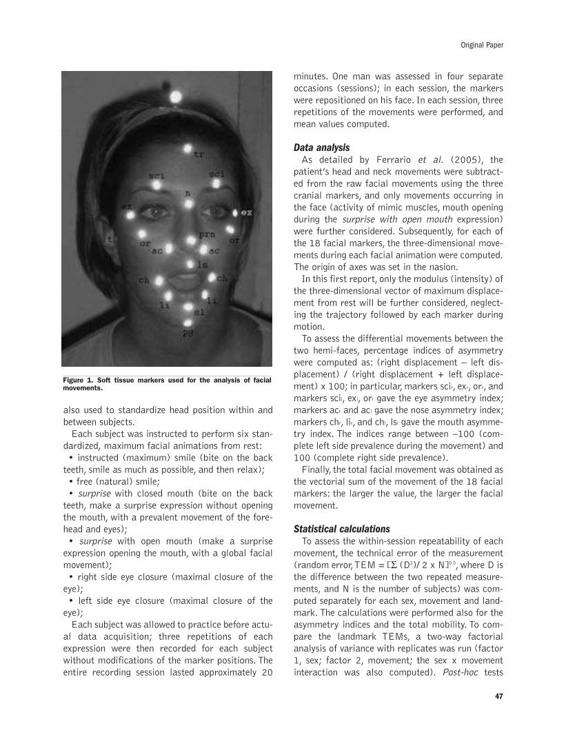
also used to standardize head position within and
between subjects.
Each subject was instructed to perform six stan-
dardized, maximum facial animations from rest:
• instructed (maximum) smile (bite on the back
teeth, smile as much as possible, and then relax);
• free (natural) smile;
• surprise with closed mouth (bite on the back
teeth, make a surprise expression without opening
the mouth, with a prevalent movement of the fore-
head and eyes);
• surprise with open mouth (make a surprise
expression opening the mouth, with a global facial
movement);
• right side eye closure (maximal closure of the
eye);
• left side eye closure (maximal closure of the
eye);
Each subject was allowed to practice before actu-
al data acquisition; three repetitions of each
expression were then recorded for each subject
without modifications of the marker positions. The
entire recording session lasted approximately 20
minutes. One man was assessed in four separate
occasions (sessions); in each session, the markers
were repositioned on his face. In each session, three
repetitions of the movements were performed, and
mean values computed.
Data analysisAs detailed by Ferrario et al. (2005), the
patient’s head and neck movements were subtract-
ed from the raw facial movements using the three
cranial markers, and only movements occurring in
the face (activity of mimic muscles, mouth opening
during the surprise with open mouth expression)
were further considered. Subsequently, for each of
the 18 facial markers, the three-dimensional move-
ments during each facial animation were computed.
The origin of axes was set in the nasion.
In this first report, only the modulus (intensity) of
the three-dimensional vector of maximum displace-
ment from rest will be further considered, neglect-
ing the trajectory followed by each marker during
motion.
To assess the differential movements between the
two hemi-faces, percentage indices of asymmetry
were computed as: (right displacement – left dis-
placement) / (right displacement + left displace-
ment) x 100; in particular, markers scir, exr, orr, and
markers scil, exl, orl gave the eye asymmetry index;
markers acr and acl gave the nose asymmetry index;
markers chr, lir, and chl, lsl gave the mouth asymme-
try index. The indices range between –100 (com-
plete left side prevalence during the movement) and
100 (complete right side prevalence).
Finally, the total facial movement was obtained as
the vectorial sum of the movement of the 18 facial
markers: the larger the value, the larger the facial
movement.
Statistical calculationsTo assess the within-session repeatability of each
movement, the technical error of the measurement
(random error, TEM = [Σ (D2)/ 2 x N]0.5, where D is
the difference between the two repeated measure-
ments, and N is the number of subjects) was com-
puted separately for each sex, movement and land-
mark. The calculations were performed also for the
asymmetry indices and the total mobility. To com-
pare the landmark TEMs, a two-way factorial
analysis of variance with replicates was run (factor
1, sex; factor 2, movement; the sex x movement
interaction was also computed). Post-hoc tests
47
Original Paper
Figure 1. Soft tissue markers used for the analysis of facialmovements.
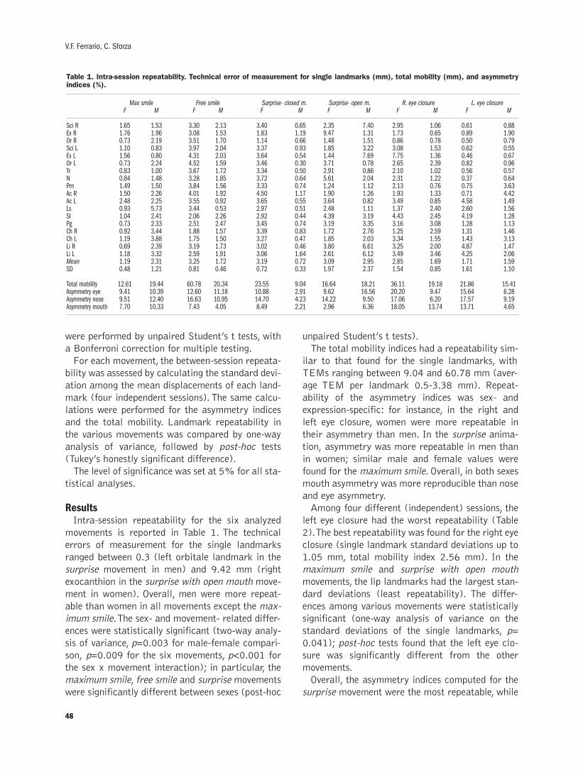
were performed by unpaired Student’s t tests, with
a Bonferroni correction for multiple testing.
For each movement, the between-session repeata-
bility was assessed by calculating the standard devi-
ation among the mean displacements of each land-
mark (four independent sessions). The same calcu-
lations were performed for the asymmetry indices
and the total mobility. Landmark repeatability in
the various movements was compared by one-way
analysis of variance, followed by post-hoc tests
(Tukey’s honestly significant difference).
The level of significance was set at 5% for all sta-
tistical analyses.
Results Intra-session repeatability for the six analyzed
movements is reported in Table 1. The technical
errors of measurement for the single landmarks
ranged between 0.3 (left orbitale landmark in the
surprise movement in men) and 9.42 mm (right
exocanthion in the surprise with open mouth move-
ment in women). Overall, men were more repeat-
able than women in all movements except the max-
imum smile. The sex- and movement- related differ-
ences were statistically significant (two-way analy-
sis of variance, p=0.003 for male-female compari-
son, p=0.009 for the six movements, p<0.001 for
the sex x movement interaction); in particular, the
maximum smile, free smile and surprise movements
were significantly different between sexes (post-hoc
unpaired Student’s t tests).
The total mobility indices had a repeatability sim-
ilar to that found for the single landmarks, with
TEMs ranging between 9.04 and 60.78 mm (aver-
age TEM per landmark 0.5-3.38 mm). Repeat -
ability of the asymmetry indices was sex- and
expression-specific: for instance, in the right and
left eye closure, women were more repeatable in
their asymmetry than men. In the surprise anima-
tion, asymmetry was more repeatable in men than
in women; similar male and female values were
found for the maximum smile. Overall, in both sexes
mouth asymmetry was more reproducible than nose
and eye asymmetry.
Among four different (independent) sessions, the
left eye closure had the worst repeatability (Table
2). The best repeatability was found for the right eye
closure (single landmark standard deviations up to
1.05 mm, total mobility index 2.56 mm). In the
maximum smile and surprise with open mouth
movements, the lip landmarks had the largest stan-
dard deviations (least repeatability). The differ-
ences among various movements were statistically
significant (one-way analysis of variance on the
standard deviations of the single landmarks, p=
0.041); post-hoc tests found that the left eye clo-
sure was significantly different from the other
movements.
Overall, the asymmetry indices computed for the
surprise movement were the most repeatable, while
48
V.F. Ferrario, C. Sforza
Table 1. Intra-session repeatability. Technical error of measurement for single landmarks (mm), total mobility (mm), and asymmetryindices (%).
Max smile Free smile Surprise- closed m. Surprise- open m. R. eye closure L. eye closureF M F M F M F M F M F M
Sci R 1.65 1.53 3.30 2.13 3.40 0.65 2.35 7.40 2.95 1.06 0.61 0.88Ex R 1.76 1.96 3.08 1.53 1.83 1.19 9.47 1.31 1.73 0.65 0.89 1.90Or R 0.73 2.19 3.51 1.70 1.14 0.66 1.48 1.51 0.86 0.78 0.50 0.79Sci L 1.10 0.83 3.97 2.04 3.37 0.93 1.85 3.22 3.08 1.53 0.62 0.55Ex L 1.56 0.80 4.31 2.03 3.64 0.54 1.44 7.69 7.75 1.36 0.46 0.67Or L 0.73 2.24 4.52 1.59 3.46 0.30 3.71 0.78 2.65 2.39 0.82 0.96Tr 0.83 1.00 3.67 1.72 3.34 0.50 2.91 0.86 2.10 1.02 0.56 0.57N 0.84 1.48 3.28 1.85 3.72 0.64 5.61 2.04 2.31 1.22 0.37 0.64Prn 1.49 1.50 3.84 1.56 3.33 0.74 1.24 1.12 2.13 0.76 0.75 3.63Ac R 1.50 2.26 4.01 1.92 4.50 1.17 1.90 1.26 1.93 1.33 0.71 4.42Ac L 2.48 2.25 3.55 0.92 3.65 0.55 3.64 0.82 3.49 0.85 4.58 1.49Ls 0.93 5.73 3.44 0.53 2.97 0.51 2.48 1.11 1.37 2.40 2.60 1.56Sl 1.04 2.41 2.06 2.26 2.92 0.44 4.39 3.19 4.43 2.45 4.19 1.28Pg 0.73 2.33 2.51 2.47 3.45 0.74 3.19 3.35 3.16 3.08 1.28 1.13Ch R 0.92 3.44 1.88 1.57 3.39 0.83 1.72 2.76 1.25 2.59 1.31 1.46Ch L 1.19 3.88 1.75 1.50 3.27 0.47 1.85 2.03 3.34 1.55 1.43 3.13Li R 0.69 2.39 3.19 1.73 3.02 0.46 3.80 6.61 3.25 2.00 4.87 1.47Li L 1.18 3.32 2.59 1.91 3.06 1.64 2.61 6.12 3.49 3.46 4.25 2.06Mean 1.19 2.31 3.25 1.72 3.19 0.72 3.09 2.95 2.85 1.69 1.71 1.59SD 0.48 1.21 0.81 0.46 0.72 0.33 1.97 2.37 1.54 0.85 1.61 1.10
Total mobility 12.61 19.44 60.78 20.34 23.55 9.04 16.64 18.21 36.11 19.18 21.86 15.41Asymmetry eye 9.41 10.39 12.60 11.18 10.88 2.91 9.62 16.56 20.20 9.47 15.64 6.28Asymmetry nose 9.51 12.40 16.63 10.95 14.70 4.23 14.22 9.50 17.06 6.20 17.57 9.19Asymmetry mouth 7.70 10.33 7.43 4.05 8.49 2.21 2.96 6.36 18.05 13.74 13.71 4.65
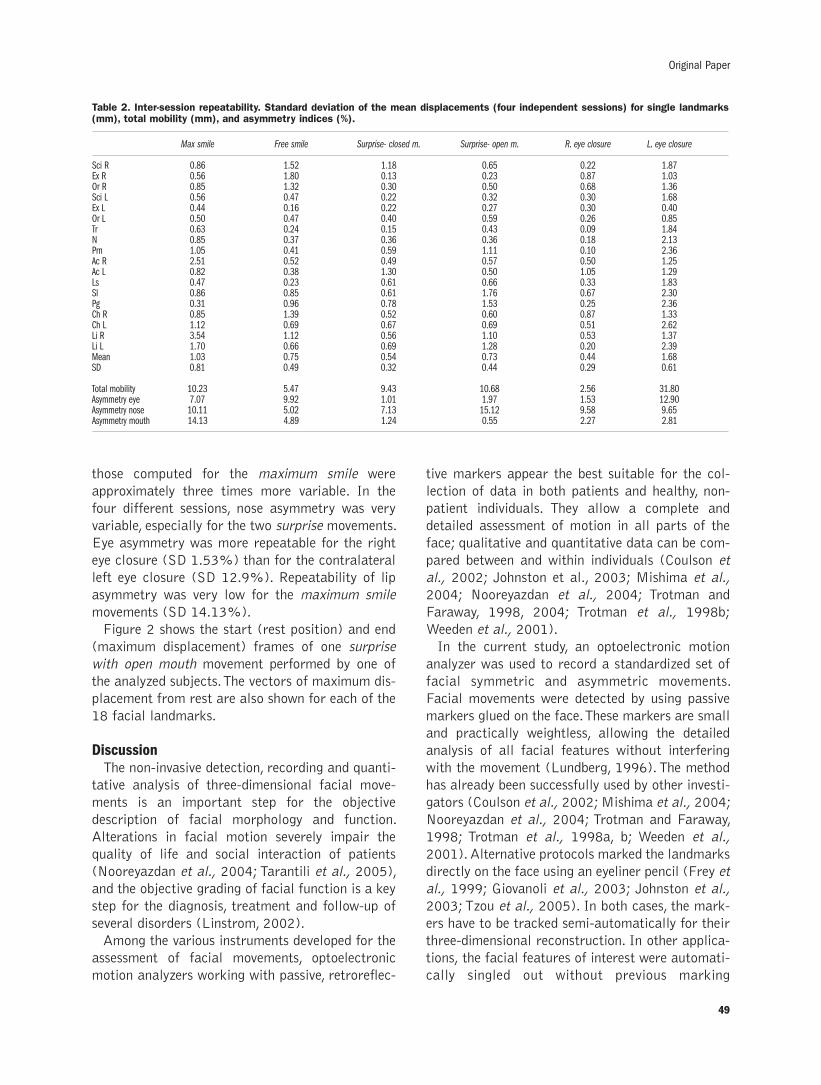
49
Original Paper
those computed for the maximum smile were
approximately three times more variable. In the
four different sessions, nose asymmetry was very
variable, especially for the two surprise movements.
Eye asymmetry was more repeatable for the right
eye closure (SD 1.53%) than for the contralateral
left eye closure (SD 12.9%). Repeatability of lip
asymmetry was very low for the maximum smile
movements (SD 14.13%).
Figure 2 shows the start (rest position) and end
(maximum displacement) frames of one surprise
with open mouth movement performed by one of
the analyzed subjects. The vectors of maximum dis-
placement from rest are also shown for each of the
18 facial landmarks.
DiscussionThe non-invasive detection, recording and quanti-
tative analysis of three-dimensional facial move-
ments is an important step for the objective
description of facial morphology and function.
Alterations in facial motion severely impair the
quality of life and social interaction of patients
(Nooreyazdan et al., 2004; Tarantili et al., 2005),
and the objective grading of facial function is a key
step for the diagnosis, treatment and follow-up of
several disorders (Linstrom, 2002).
Among the various instruments developed for the
assessment of facial movements, optoelectronic
motion analyzers working with passive, retroreflec-
tive markers appear the best suitable for the col-
lection of data in both patients and healthy, non-
patient individuals. They allow a complete and
detailed assessment of motion in all parts of the
face; qualitative and quantitative data can be com-
pared between and within individuals (Coulson et
al., 2002; Johnston et al., 2003; Mishima et al.,
2004; Nooreyazdan et al., 2004; Trotman and
Faraway, 1998, 2004; Trotman et al., 1998b;
Weeden et al., 2001).
In the current study, an optoelectronic motion
analyzer was used to record a standardized set of
facial symmetric and asymmetric movements.
Facial movements were detected by using passive
markers glued on the face. These markers are small
and practically weightless, allowing the detailed
analysis of all facial features without interfering
with the movement (Lundberg, 1996). The method
has already been successfully used by other investi-
gators (Coulson et al., 2002; Mishima et al., 2004;
Nooreyazdan et al., 2004; Trotman and Faraway,
1998; Trotman et al., 1998a, b; Weeden et al.,
2001). Alternative protocols marked the landmarks
directly on the face using an eyeliner pencil (Frey et
al., 1999; Giovanoli et al., 2003; Johnston et al.,
2003; Tzou et al., 2005). In both cases, the mark-
ers have to be tracked semi-automatically for their
three-dimensional reconstruction. In other applica-
tions, the facial features of interest were automati-
cally singled out without previous marking
Table 2. Inter-session repeatability. Standard deviation of the mean displacements (four independent sessions) for single landmarks(mm), total mobility (mm), and asymmetry indices (%).
Max smile Free smile Surprise- closed m. Surprise- open m. R. eye closure L. eye closure
Sci R 0.86 1.52 1.18 0.65 0.22 1.87Ex R 0.56 1.80 0.13 0.23 0.87 1.03Or R 0.85 1.32 0.30 0.50 0.68 1.36Sci L 0.56 0.47 0.22 0.32 0.30 1.68Ex L 0.44 0.16 0.22 0.27 0.30 0.40Or L 0.50 0.47 0.40 0.59 0.26 0.85Tr 0.63 0.24 0.15 0.43 0.09 1.84N 0.85 0.37 0.36 0.36 0.18 2.13Prn 1.05 0.41 0.59 1.11 0.10 2.36Ac R 2.51 0.52 0.49 0.57 0.50 1.25Ac L 0.82 0.38 1.30 0.50 1.05 1.29Ls 0.47 0.23 0.61 0.66 0.33 1.83Sl 0.86 0.85 0.61 1.76 0.67 2.30Pg 0.31 0.96 0.78 1.53 0.25 2.36Ch R 0.85 1.39 0.52 0.60 0.87 1.33Ch L 1.12 0.69 0.67 0.69 0.51 2.62Li R 3.54 1.12 0.56 1.10 0.53 1.37Li L 1.70 0.66 0.69 1.28 0.20 2.39Mean 1.03 0.75 0.54 0.73 0.44 1.68SD 0.81 0.49 0.32 0.44 0.29 0.61
Total mobility 10.23 5.47 9.43 10.68 2.56 31.80Asymmetry eye 7.07 9.92 1.01 1.97 1.53 12.90Asymmetry nose 10.11 5.02 7.13 15.12 9.58 9.65Asymmetry mouth 14.13 4.89 1.24 0.55 2.27 2.81
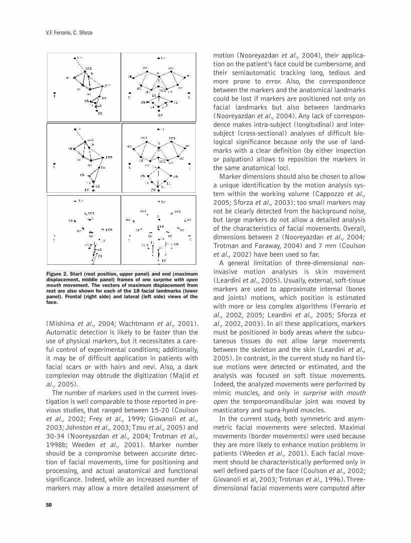
(Mishima et al., 2004; Wachtmann et al., 2001).
Automatic detection is likely to be faster than the
use of physical markers, but it necessitates a care-
ful control of experimental conditions; additionally,
it may be of difficult application in patients with
facial scars or with hairs and nevi. Also, a dark
complexion may obtrude the digitization (Majid et
al., 2005).
The number of markers used in the current inves-
tigation is well comparable to those reported in pre-
vious studies, that ranged between 15-20 (Coulson
et al., 2002; Frey et al., 1999; Giovanoli et al.,
2003; Johnston et al., 2003; Tzou et al., 2005) and
30-34 (Nooreyazdan et al., 2004; Trotman et al.,
1998b; Weeden et al., 2001). Marker number
should be a compromise between accurate detec-
tion of facial movements, time for positioning and
processing, and actual anatomical and functional
significance. Indeed, while an increased number of
markers may allow a more detailed assessment of
motion (Nooreyazdan et al., 2004), their applica-
tion on the patient’s face could be cumbersome, and
their semiautomatic tracking long, tedious and
more prone to error. Also, the correspondence
between the markers and the anatomical landmarks
could be lost if markers are positioned not only on
facial landmarks but also between landmarks
(Nooreyazdan et al., 2004). Any lack of correspon-
dence makes intra-subject (longitudinal) and inter-
subject (cross-sectional) analyses of difficult bio-
logical significance because only the use of land-
marks with a clear definition (by either inspection
or palpation) allows to reposition the markers in
the same anatomical loci.
Marker dimensions should also be chosen to allow
a unique identification by the motion analysis sys-
tem within the working volume (Cappozzo et al.,
2005; Sforza et al., 2003): too small markers may
not be clearly detected from the background noise,
but large markers do not allow a detailed analysis
of the characteristics of facial movements. Overall,
dimensions between 2 (Nooreyazdan et al., 2004;
Trotman and Faraway, 2004) and 7 mm (Coulson
et al., 2002) have been used so far.
A general limitation of three-dimensional non-
invasive motion analyses is skin movement
(Leardini et al., 2005). Usually, external, soft-tissue
markers are used to approximate internal (bones
and joints) motions, which position is estimated
with more or less complex algorithms (Ferrario et
al., 2002, 2005; Leardini et al., 2005; Sforza et
al., 2002, 2003). In all these applications, markers
must be positioned in body areas where the subcu-
taneous tissues do not allow large movements
between the skeleton and the skin (Leardini et al.,
2005). In contrast, in the current study no hard tis-
sue motions were detected or estimated, and the
analysis was focused on soft tissue movements.
Indeed, the analyzed movements were performed by
mimic muscles, and only in surprise with mouth
open the temporomandibular joint was moved by
masticatory and supra-hyoid muscles.
In the current study, both symmetric and asym-
metric facial movements were selected. Maximal
movements (border movements) were used because
they are more likely to enhance motion problems in
patients (Weeden et al., 2001). Each facial move-
ment should be characteristically performed only in
well defined parts of the face (Coulson et al., 2002;
Giovanoli et al, 2003; Trotman et al., 1996). Three-
dimensional facial movements were computed after
50
V.F. Ferrario, C. Sforza
Figure 2. Start (rest position, upper panel) and end (maximumdisplacement, middle panel) frames of one surprise with openmouth movement. The vectors of maximum displacement fromrest are also shown for each of the 18 facial landmarks (lowerpanel). Frontal (right side) and lateral (left side) views of theface.
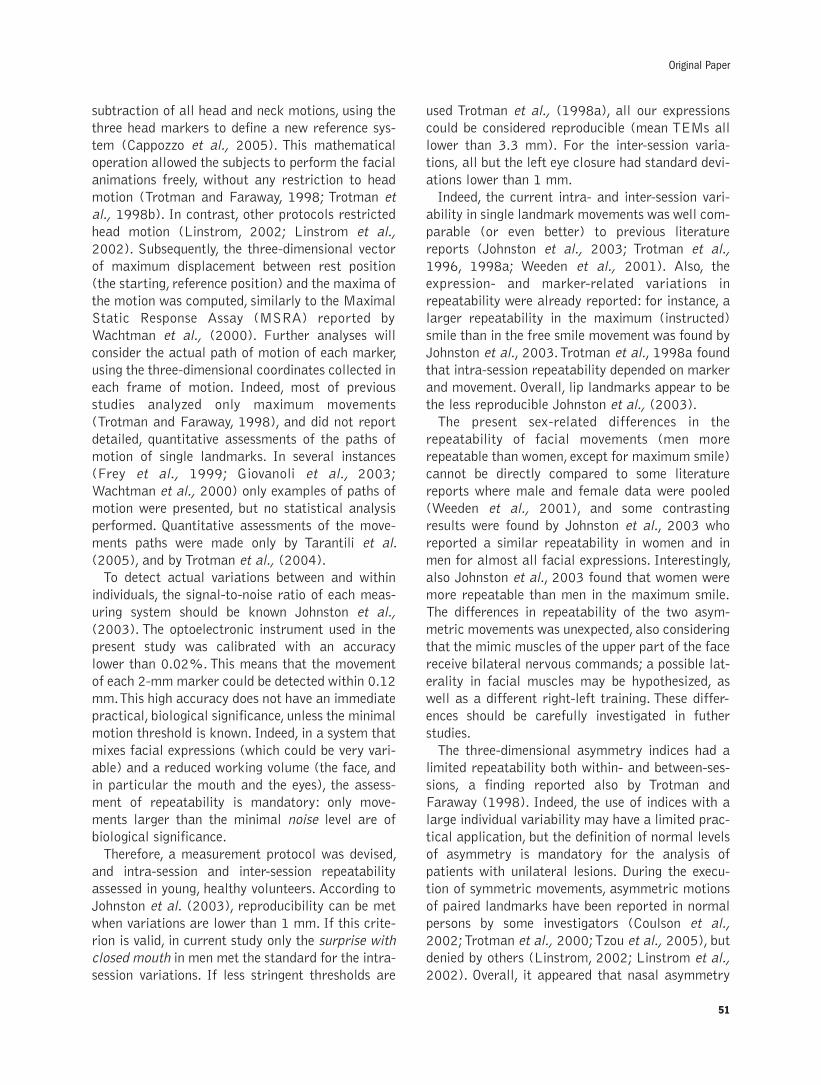
subtraction of all head and neck motions, using the
three head markers to define a new reference sys-
tem (Cappozzo et al., 2005). This mathematical
operation allowed the subjects to perform the facial
animations freely, without any restriction to head
motion (Trotman and Faraway, 1998; Trotman et
al., 1998b). In contrast, other protocols restricted
head motion (Linstrom, 2002; Linstrom et al.,
2002). Subsequently, the three-dimensional vector
of maximum displacement between rest position
(the starting, reference position) and the maxima of
the motion was computed, similarly to the Maximal
Static Response Assay (MSRA) reported by
Wachtman et al., (2000). Further analyses will
consider the actual path of motion of each marker,
using the three-dimensional coordinates collected in
each frame of motion. Indeed, most of previous
studies analyzed only maximum movements
(Trotman and Faraway, 1998), and did not report
detailed, quantitative assessments of the paths of
motion of single landmarks. In several instances
(Frey et al., 1999; Giovanoli et al., 2003;
Wachtman et al., 2000) only examples of paths of
motion were presented, but no statistical analysis
performed. Quantitative assessments of the move-
ments paths were made only by Tarantili et al.
(2005), and by Trotman et al., (2004).
To detect actual variations between and within
individuals, the signal-to-noise ratio of each meas-
uring system should be known Johnston et al.,
(2003). The optoelectronic instrument used in the
present study was calibrated with an accuracy
lower than 0.02%. This means that the movement
of each 2-mm marker could be detected within 0.12
mm. This high accuracy does not have an immediate
practical, biological significance, unless the minimal
motion threshold is known. Indeed, in a system that
mixes facial expressions (which could be very vari-
able) and a reduced working volume (the face, and
in particular the mouth and the eyes), the assess-
ment of repeatability is mandatory: only move-
ments larger than the minimal noise level are of
biological significance.
Therefore, a measurement protocol was devised,
and intra-session and inter-session repeatability
assessed in young, healthy volunteers. According to
Johnston et al. (2003), reproducibility can be met
when variations are lower than 1 mm. If this crite-
rion is valid, in current study only the surprise with
closed mouth in men met the standard for the intra-
session variations. If less stringent thresholds are
used Trotman et al., (1998a), all our expressions
could be considered reproducible (mean TEMs all
lower than 3.3 mm). For the inter-session varia-
tions, all but the left eye closure had standard devi-
ations lower than 1 mm.
Indeed, the current intra- and inter-session vari-
ability in single landmark movements was well com-
parable (or even better) to previous literature
reports (Johnston et al., 2003; Trotman et al.,
1996, 1998a; Weeden et al., 2001). Also, the
expression- and marker-related variations in
repeatability were already reported: for instance, a
larger repeatability in the maximum (instructed)
smile than in the free smile movement was found by
Johnston et al., 2003. Trotman et al., 1998a found
that intra-session repeatability depended on marker
and movement. Overall, lip landmarks appear to be
the less reproducible Johnston et al., (2003).
The present sex-related differences in the
repeatability of facial movements (men more
repeatable than women, except for maximum smile)
cannot be directly compared to some literature
reports where male and female data were pooled
(Weeden et al., 2001), and some contrasting
results were found by Johnston et al., 2003 who
reported a similar repeatability in women and in
men for almost all facial expressions. Interestingly,
also Johnston et al., 2003 found that women were
more repeatable than men in the maximum smile.
The differences in repeatability of the two asym-
metric movements was unexpected, also considering
that the mimic muscles of the upper part of the face
receive bilateral nervous commands; a possible lat-
erality in facial muscles may be hypothesized, as
well as a different right-left training. These differ-
ences should be carefully investigated in futher
studies.
The three-dimensional asymmetry indices had a
limited repeatability both within- and between-ses-
sions, a finding reported also by Trotman and
Faraway (1998). Indeed, the use of indices with a
large individual variability may have a limited prac-
tical application, but the definition of normal levels
of asymmetry is mandatory for the analysis of
patients with unilateral lesions. During the execu-
tion of symmetric movements, asymmetric motions
of paired landmarks have been reported in normal
persons by some investigators (Coulson et al.,
2002; Trotman et al., 2000; Tzou et al., 2005), but
denied by others (Linstrom, 2002; Linstrom et al.,
2002). Overall, it appeared that nasal asymmetry
51
Original Paper
52
was very variable, and the location of these markers
should be carefully controlled, and eventually their
calculations skipped. Indeed, nasal markers are
among those with the least reproducibility
(Trotman et al., 1996).
In conclusion, the protocol devised in the current
study demonstrated a sufficient repeatability for a
future clinical application. The use of both symmet-
ric and asymmetric facial expression may allow a
better definition of the impairments of patients with
unilateral facial lesions. Great care should be taken
to assure a consistent marker positioning in all the
subjects. The next step would be the definition of
reference values for three-dimensional facial move-
ments in subjects of different ages and of both
sexes.
Acknowledgements
The precious work of Drs Domenico Galante,
Nicola Lovecchio and Fabrizio Mian for data col-
lection and analysis is gratefully acknowledged. We
are also deeply indebted to all the staff and stu-
dents of our laboratories, who collaborated to this
project.
References
Cappozzo A, Della Croce U, Leardini A, Chiari L. Human movementanalysis using stereophotogrammetry. Part 1: theoretical back-ground. Gait Posture 2005; 21:186-96.
Coulson SE, Croxson GR, Gilleard WL. Three-dimensional quantifica-tion of the symmetry of normal facial movement. Otol Neurotol2002; 23:999-1002.
DeCarlo D, Metaxas D, Stone M. An anthropometric face model usingvariational techniques. Proc SIGGRAPH ’98, 1998; 32: 67-74.
Ferrario VF, Sforza C, Serrao G, Grassi GP, Mossi E. Active range ofmotion of the head and cervical spine: a three-dimensional investi-gation in healthy young adults. J Orthop Res 2002; 20:122-9.
Ferrario VF, Sforza C, Serrao G, Ciusa V, Dellavia C. Growth and agingof facial soft-tissues: a computerised three–dimensional mesh dia-gram analysis. Clin Anat 2003; 16:420-33.
Ferrario VF, Sforza C, Lovecchio N, Mian F. Quantification of transla-tional and gliding components in human temporomandibular jointduring mouth opening. Archs Oral Biol 2005; 50:507-15.
Fraser NL, Yoshino M, Imaizumi K, Blackwell SA, Thomas CD,Clement JG. A Japanese computer-assisted facial identification sys-tem successfully identifies non-Japanese faces. Forensic Sci Int2003; 135:122-8.
Frey M, Giovanoli P, Gerber H, Slameczka M, Stussi E. Three-dimen-sional video analysis of facial movements: a new method to assessthe quantity and quality of the smile. Plast Reconstr Surg 1999;104:2032-9.
Giovanoli P, Tzou CH, Ploner M, Frey M. Three-dimensional video-analysis of facial movements in healthy volunteers. Br J Plast Surg2003; 56:644-52.
Gross MM, Trotman CA, Moffatt KS. A comparison of three-dimen-sional and two-dimensional analyses of facial motion. Angle Orthod1996; 66:189-94.
Hennessy RJ, McLearie S, Kinsella A, Waddington JL. Facial surface
analysis by 3D laser scanning and geometric morphometrics in rela-tion to sexual dimorphism in cerebral-craniofacial morphogenesisand cognitive function. J Anat 2005; 207:283-95.
Johnson N, Sandy J. An aesthetic index for evaluation of cleft repair.Eur J Orthod 2003;25:243-9.
Johnston DJ, Millett DT, Ayoub AF, Bock M. Are facial expressionsreproducible? Cleft Palate Craniofac J 2003; 40:291-6.
Leardini A, Chiari L, Della Croce U, Cappozzo A. Human movementanalysis using stereophotogrammetry. Part 3. Soft tissue artifactassessment and compensation. Gait Posture 2005; 21:212-25.
Linstrom CJ. Objective facial motion analysis in patients with facialnerve dysfunction. Laryngoscope 2002; 112:1129-47.
Linstrom CJ, Silverman CA, Colson D. Facial motion analysis with avideo and computer system after treatment of acoustic neuroma.Otol Neurotol 2002; 23:572-9.
Majid Z, Chong AK, Ahmad A, Setan H, Samsudin AR.Photogrammetry and 3D laser scanning as spatial data capturetechniques for a national craniofacial database. Photogramm Rec2005; 20:48-68.
Matoula S, Pancherz H. Skeletofacial morphology of attractive andnonattractive faces. Angle Orthod 2006; 76:204-10.
Mergl R, Mavrogiorgou P, Hegerl U, Juckel G. Kinematical analysis ofemotionally induced facial expressions: a novel tool to investigatehypomimia in patients suffering from depression. J NeurolNeurosurg Psychiatry 2005; 76:138-40.
Mishima K, Yamada T, Fujiwara K, Sugahara T. Development and clin-ical usage of a motion analysis system for the face: preliminaryreport. Cleft Palate Craniofac J 2004; 41:559-64.
Nooreyazdan M, Trotman CA, Faraway JJ. Modeling facial movement:II. A dynamic analysis of differences caused by orthognathic sur-gery. J Oral Maxillofac Surg 2004; 62:1380-6.
Sforza C, Grassi GP, Fragnito N, Turci M, Ferrario VF. Three-dimen-sional analysis of active head and cervical spine range of motion:effect of age in healthy male subjects. Clin Biomech 2002; 17:611-4.
Sforza C, Macrì C, Turci M, Grassi GP, Ferrario VF. Neuromuscolarpatterns of finger movements during piano playing. Definition of anexperimental protocol. Ital J Anat Embryol 2003; 108:211-22.
Shi J, Samal A, Marx D. How effective are landmarks and their geom-etry for face recognition? Comput Vis Image Understand 2006;102:117-33.
Tarantili VV, Halazonetis DJ, Spyropoulos MN. The spontaneous smilein dynamic motion. Am J Orthod Dentofacial Orthop 2005; 128:8-15.
Trotman CA, Faraway JJ. Sensitivity of a method for the analysis offacial mobility. II. Interlandmark separation. Cleft Palate CraniofacJ 1998; 35:142-53.
Trotman CA, Gross MM, Moffatt K. Reliability of a three-dimensionalmethod for measuring facial animation: a case report. Angle Orthod1996; 66:195-8.
Trotman CA, Stohler CS, Johnston LE Jr. Measurement of facial softtissue mobility in man. Cleft Palate Craniofac J 1998a; 35:16-25.
Trotman CA, Faraway JJ, Silvester KT, Greenlee GM, Johnston LE Jr.Sensitivity of a method for the analysis of facial mobility. I. Vectorof displacement. Cleft Palate Craniofac J 1998b; 35:132-41
Trotman CA, Faraway JJ, Essick GK. Three-dimensional nasolabial dis-placement during movement in repaired cleft lip and palate patients.Plast Reconstr Surg 2000; 105:1273-83.
Tzou CH, Giovanoli P, Ploner M, Frey M. Are there ethnic differencesof facial movements between Europeans and Asians? Br J PlastSurg 2005; 58:183-95.
Vidarsdottir US, O'Higgins P, Stringer C. A geometric morphometricstudy of regional differences in the ontogeny of the modern humanfacial skeleton. J Anat 2002; 201:211-29.
Wachtman GS, Cohn JF, VanSwearingen JM, Manders EK. Automatedtracking of facial features in patients with facial neuromuscular dys-function. Plast Reconstr Surg 2001; 107:1124-33.
Weeden JC, Trotman CA, Faraway JJ. Three dimensional analysis offacial movement in normal adults: influence of sex and facial shape.Angle Orthod 2001; 71:132-40.
V.F. Ferrario, C. Sforza

53
©2007, European Journal of Histochemistry
We present here findings obtained on a large number ofhuman tissues over a period of more than ten years, by ourmodification of the Osmium maceration method for high res-olution scanning electron microscopy (HRSEM). Data aredocumented by original pictures which illustrate both some3-D intracellular features not previously shown in human tis-sues, and results obtained in our current studies on mito-chondrial morphology and on the secretory process of sali-vary glands.We have demonstrated that mitochondria of cells of practi-cally all human tissues and organs have usually tubularcristae, and that even the cristae that look lamellar arejoined to the inner mitochondrial membrane by tubular con-nexions similar to the crista junctions later seen by electrontomography. Concerning salivary glands an important resultis the development of a morphometric method that allowsthe quantitative evaluation of the secretory events.
Key words: HRSEM, maceration method, mitochondria, cristajunctions, salivary glands, secretory process, cell organelles.
Correspondence: Alessandro Riva,Tel: +39-70-6754025.Fax: +39-70-6754003.E-mail: [email protected]
European Journal of Histochemistry2007; vol. 51 supplement 1:53-58
New findings on 3-D microanatomy of cellular structures in human
tissues and organs. An HRSEM study
A. Riva, F. Loy, R. Isola, M. Isola, G. Conti, A. Perra, P. Solinas, F. Testa Riva
Department of Cytomorphology, University of Cagliari, Cittadella Universitaria di Monserrato, Monserrato,
Cagliari, Italy
The OsO4 maceration method for high resolu-
tion scanning electron microscopy (HRSEM),
introduced in the eighties by Tanaka and his
co-workers (Tanaka, 1980; Tanaka and Naguro,
1981; and particularly that by Tanaka and
Mitsushima, 1984), aroused great interest for its
unique ability of providing three dimensional (3-D)
images of intracellular membranaceous structures.
In a matter of a few years, however, the use of the
technique declined. According to our experience, this
is mainly due to the fact that the protocol is too
long and rigid, and that the use of the freeze-crack-
ing procedure for cutting the samples does not allow
the analytical study of a whole specimen, as it is
compulsory in the case of human needle biopsies.
Thus, in order to make the technique more suitable
for the study of human tissues, we embedded strips
of fixed tissue samples, about 2 mm thick and 7 mm
long in agarose, and cut them entirely into sections
of 100-150 µm by using a tissue sectioner at roomtemperature (Riva et al., 1993). Later on (Riva et
al., 1999), we introduced, as secondary fixative, the
mixture 1% OsO4 - 1.25% K4Fe(CN)6 that
enhances osmium binding to tissue, thereby render-
ing unnecessary the long treatments with the bind-
ing agent tannic acid suggested by Tanaka and
Mitsushima (1984). This modification not only
reduced the preparation time from eight days to
three days or fewer, but also eliminated a source of
contamination and made the whole procedure more
reproducible and easy to perform. Another advan-
tage of our method was that, following the second-
ary fixation by the above mentioned mixture, the
procedure can be suspended for several days by
storing the specimens in phosphate buffered saline
(PBS) at 4C°. Finally, by shaking the tissues during
maceration by a rotating agitator, we succeeded in
removing all cytoplasmic organelles, thus visualiz-
ing, for the first time, the cytoplasmic side of the
plasmalemma and its specializations.
ORIGINAL PAPER
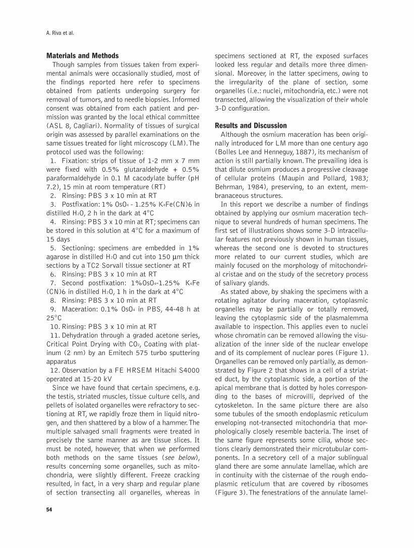
Materials and MethodsThough samples from tissues taken from experi-
mental animals were occasionally studied, most of
the findings reported here refer to specimens
obtained from patients undergoing surgery for
removal of tumors, and to needle biopsies. Informed
consent was obtained from each patient and per-
mission was granted by the local ethical committee
(ASL 8, Cagliari). Normality of tissues of surgical
origin was assessed by parallel examinations on the
same tissues treated for light microscopy (LM). The
protocol used was the following:
1. Fixation: strips of tissue of 1-2 mm x 7 mm
were fixed with 0.5% glutaraldehyde + 0.5%
paraformaldehyde in 0.1 M cacodylate buffer (pH
7.2), 15 min at room temperature (RT)
2. Rinsing: PBS 3 x 10 min at RT
3. Postfixation: 1% OsO4 - 1.25% K4Fe(CN)6 in
distilled H2O, 2 h in the dark at 4°C
4. Rinsing: PBS 3 x 10 min at RT; specimens can
be stored in this solution at 4°C for a maximum of
15 days
5. Sectioning: specimens are embedded in 1%
agarose in distilled H2O and cut into 150 µm thicksections by a TC2 Sorvall tissue sectioner at RT
6. Rinsing: PBS 3 x 10 min at RT
7. Second postfixation: 1%OsO4-1.25% K4Fe
(CN)6 in distilled H2O, 1 h in the dark at 4°C
8. Rinsing: PBS 3 x 10 min at RT
9. Maceration: 0.1% OsO4 in PBS, 44-48 h at
25°C
10. Rinsing: PBS 3 x 10 min at RT
11. Dehydration through a graded acetone series,
Critical Point Drying with CO2, Coating with plat-
inum (2 nm) by an Emitech 575 turbo sputtering
apparatus
12. Observation by a FE HRSEM Hitachi S4000
operated at 15-20 kV
Since we have found that certain specimens, e.g.
the testis, striated muscles, tissue culture cells, and
pellets of isolated organelles were refractory to sec-
tioning at RT, we rapidly froze them in liquid nitro-
gen, and then shattered by a blow of a hammer. The
multiple salvaged small fragments were treated in
precisely the same manner as are tissue slices. It
must be noted, however, that when we performed
both methods on the same tissues (see below),
results concerning some organelles, such as mito-
chondria, were slightly different. Freeze cracking
resulted, in fact, in a very sharp and regular plane
of section transecting all organelles, whereas in
specimens sectioned at RT, the exposed surfaces
looked less regular and details more three dimen-
sional. Moreover, in the latter specimens, owing to
the irregularity of the plane of section, some
organelles (i.e.: nuclei, mitochondria, etc.) were not
transected, allowing the visualization of their whole
3-D configuration.
Results and DiscussionAlthough the osmium maceration has been origi-
nally introduced for LM more than one century ago
(Bolles Lee and Henneguy, 1887), its mechanism of
action is still partially known. The prevailing idea is
that dilute osmium produces a progressive cleavage
of cellular proteins (Maupin and Pollard, 1983;
Behrman, 1984), preserving, to an extent, mem-
branaceous structures.
In this report we describe a number of findings
obtained by applying our osmium maceration tech-
nique to several hundreds of human specimens. The
first set of illustrations shows some 3-D intracellu-
lar features not previously shown in human tissues,
whereas the second one is devoted to structures
more related to our current studies, which are
mainly focused on the morphology of mitochondri-
al cristae and on the study of the secretory process
of salivary glands.
As stated above, by shaking the specimens with a
rotating agitator during maceration, cytoplasmic
organelles may be partially or totally removed,
leaving the cytoplasmic side of the plasmalemma
available to inspection. This applies even to nuclei
whose chromatin can be removed allowing the visu-
alization of the inner side of the nuclear envelope
and of its complement of nuclear pores (Figure 1).
Organelles can be removed only partially, as demon-
strated by Figure 2 that shows in a cell of a striat-
ed duct, by the cytoplasmic side, a portion of the
apical membrane that is dotted by holes correspon-
ding to the bases of microvilli, deprived of the
cytoskeleton. In the same picture there are also
some tubules of the smooth endoplasmic reticulum
enveloping not-transected mitochondria that mor-
phologically closely resemble bacteria. The inset of
the same figure represents some cilia, whose sec-
tions clearly demonstrated their microtubular com-
ponents. In a secretory cell of a major sublingual
gland there are some annulate lamellae, which are
in continuity with the cisternae of the rough endo-
plasmic reticulum that are covered by ribosomes
(Figure 3). The fenestrations of the annulate lamel-
54
A. Riva et al.
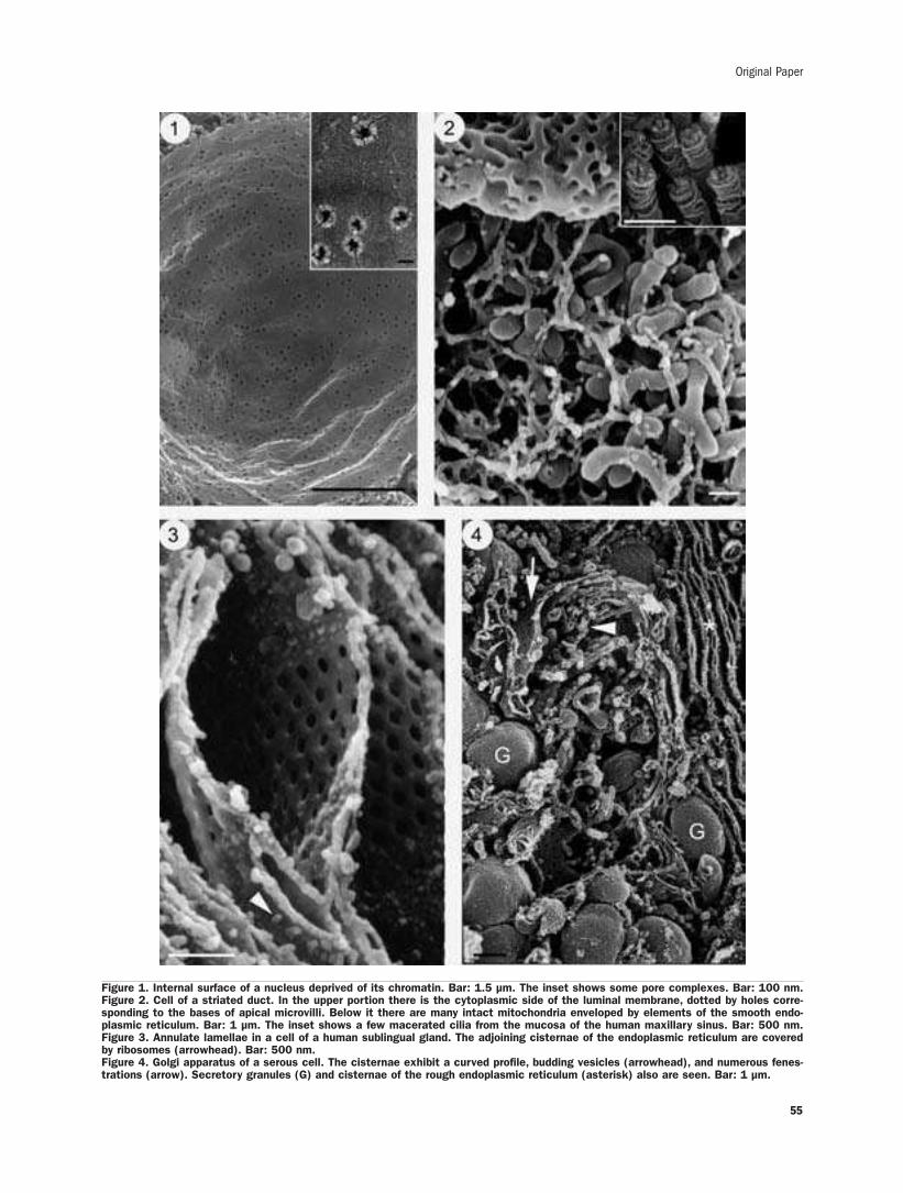
55
Original Paper
Figure 1. Internal surface of a nucleus deprived of its chromatin. Bar: 1.5 µm. The inset shows some pore complexes. Bar: 100 nm.Figure 2. Cell of a striated duct. In the upper portion there is the cytoplasmic side of the luminal membrane, dotted by holes corre-sponding to the bases of apical microvilli. Below it there are many intact mitochondria enveloped by elements of the smooth endo-plasmic reticulum. Bar: 1 µm. The inset shows a few macerated cilia from the mucosa of the human maxillary sinus. Bar: 500 nm.Figure 3. Annulate lamellae in a cell of a human sublingual gland. The adjoining cisternae of the endoplasmic reticulum are coveredby ribosomes (arrowhead). Bar: 500 nm.Figure 4. Golgi apparatus of a serous cell. The cisternae exhibit a curved profile, budding vesicles (arrowhead), and numerous fenes-trations (arrow). Secretory granules (G) and cisternae of the rough endoplasmic reticulum (asterisk) also are seen. Bar: 1 µm.
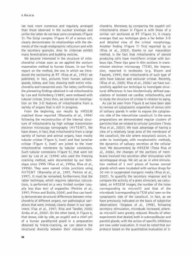
lae look more numerous and regularly arranged
than those observed in the nuclear envelope and
unlike the latter do not bear pore complexes (Figure
3). The Golgi complex (Figure 4) of a serous cell,
clearly demonstrates its relationships with the ele-
ments of the rough endoplasmic reticulum and with
the secretory granules. Also its cisternae exhibit
many fenestrations and budding vesicles.
We become interested in the structure of mito-
chondrial cristae soon as we applied the osmium
maceration method to human tissues. In our first
report on the method, the one in which we intro-
duced the sectioning at RT (Riva et al., 1993) we
published, in fact, pictures from human salivary
glands, kidney and liver, showing both entire mito-
chondria and transected ones. The latter, confirming
the pioneering findings obtained in rat mitochondria
by Lea and Hollenberg (1989), were endowed with
tubular cristae. Since then, we started an investiga-
tion on the 3-D features of mitochondria from a
variety of organs that is still in progress.
From the beginning, our results by HRSEM
matched those reported (Mannella et al., 1994)
following the reconstruction of the internal struc-
ture of mitochondria by high voltage transmission
electron microscopy tomography (HVTEMT). We
have shown, in fact, that mitochondria from a large
variety of human and animal organs, have mostly
tubular cristae (Figure 5, inset) and that lamellar
cristae (Figure 5, inset) are joined to the inner
mitochondrial membrane by tubular connexions.
Such tubular connexions (Figure 5), that were not
seen by Lea et al. (1994) who used the freezing
cracking method, were documented by our tech-
nique since 1995 (Riva et al., 1995a; Riva et al.,
1995b). They were named crista junctions using
HVTEMT (Mannella et al., 1997, Perkins et al.,
1997). It must be remarked, furthermore, that the
latter technique, which requires laborious calcula-
tions, is performed on a very limited number (usu-
ally less than ten) of organelles (Perkins et al.,
1997; Prince and Buttle, 2004), and thus can hard-
ly demonstrate structural differences between mito-
chondria of different organs, nor pathological vari-
ations that were, instead, clearly shown in our spec-
imens (Faa et al., 1997; Riva and Tandler, 2000;
Ambu et al., 2000). On the other hand, in Figure 6,
that shows, side by side, an oxyphil and a chief cell
of a human parathyroid gland in a preparation
obtained by freeze-cracking, we can observe the
structural diversity between their relevant mito-
chondria. Moreover, by comparing the oxyphil cell
mitochondria shown in Figure 6 with those of a
similar cell sectioned at RT (Figure 5), it clearly
emerges that our technique gives a far better 3-D
and detailed view of the cristal morphology.
Another finding (Figure 7) first reported by us
(Riva et al., 2003), thanks to our maceration
method, is the fact that mitochondria of steroid
producing cells have moniliform cristae with bul-
bous tips. These tips gave in thin sections in trans-
mission electron microscopy (TEM), the impres-
sion, reported in textbooks (e.g. Bloom and
Fawcett, 1994), that mitochondria of such type of
cells have tubular and vesicular cristae. Recently,
(Riva et al., 2005; Riva et al., 2006) we have suc-
cessfully applied our technique to investigate struc-
tural differences in two biochemically defined pop-
ulations of isolated rat cardiac mitochondria, and
to study the structure of cristae in relation to aging.
As can be seen from Figure 8 we have been able
to remove all cytoplasmic organelles of serous cells
of salivary glands in order to expose the cytoplas-
mic side of the intercellular canaliculi. In the same
preparations we demonstrated regular clusters of
particles that we related to cellular junctions (Testa
Riva et al., 2003). We took advantage of having a
view of a relatively large area of the membrane of
the canaliculi, the site where exocytosis occurs, in
order to investigate, with morphometric methods,
the dynamics of salivary secretion at the cellular
level. We documented, by HRSEM (Testa Riva et
al., 2006), the changes of the portions of mem-
brane involved into secretion after stimulation with
secretagogue drugs. We set up an in vitro stimula-
tion method of 1 mm3 pieces of human normal
glands which were incubated with various drugs for
30 min in oxygenated inorganic media (Riva et al.,
2002). To quantify the secretory response and to
compare the activity of a given stimulant, we calcu-
lated, on HRSEM images, the number of the holes
corresponding to microvilli and that of the
microbuds (corresponding to TEM pits) seen on the
cytoplasmic side of the canaliculi. In fact, as we
have previously indicated on the basis of subjective
observations (Segawa et al., 1998), following
secretory stimulation, microbuds increased, where-
as microvilli were greatly reduced. Results of other
experiments that dealed, both in submandibular and
parotid glands, with the action of specific inhibitors
are now under evaluation. It must be noted that our
protocol based on the quantitative evaluation of 3-
56
A. Riva et al.
Original Paper
57
Figure 5. RT sectioned mitochondria of an oxyphilic cell of the human parathyroid gland exhibiting tubular cristae. Bar: 1 µm. The insetdemonstrates a lamellar crista viewed en face and linked to the inner mitochondrial membrane by tubular connexions (crista junc-tions). Bar: 500 nm. Figure 6. Picture of an oxyphilic cell of the human parathyroid (left) and of an adjoining chief cell (right) obtainedby freeze cracking the specimen. Note that mitochondria cristae look less 3-D than in the previous image (Figure 5) from an homolo-gous cell sectioned at RT. Bar: 1.5 µm. Figure 7. Cristae with bulbous tips and moniliform constrictions are seen in mitochondria ofa steroid producing organ (human adrenocortical gland, reticulate zone). Bar: 500 nm. Figure 8. Cytoplasmic side of the plasmalem-ma of a serous cell following removal of cytoplasmic organelles. The intercellular canaliculus exhibits microbuds (arrowheads), andholes (arrows) corresponding to the bases of microvilli. The continuous band and clusters of particles (asterisks) placed alongside itare related to junctional complexes. Bar: 500 nm.
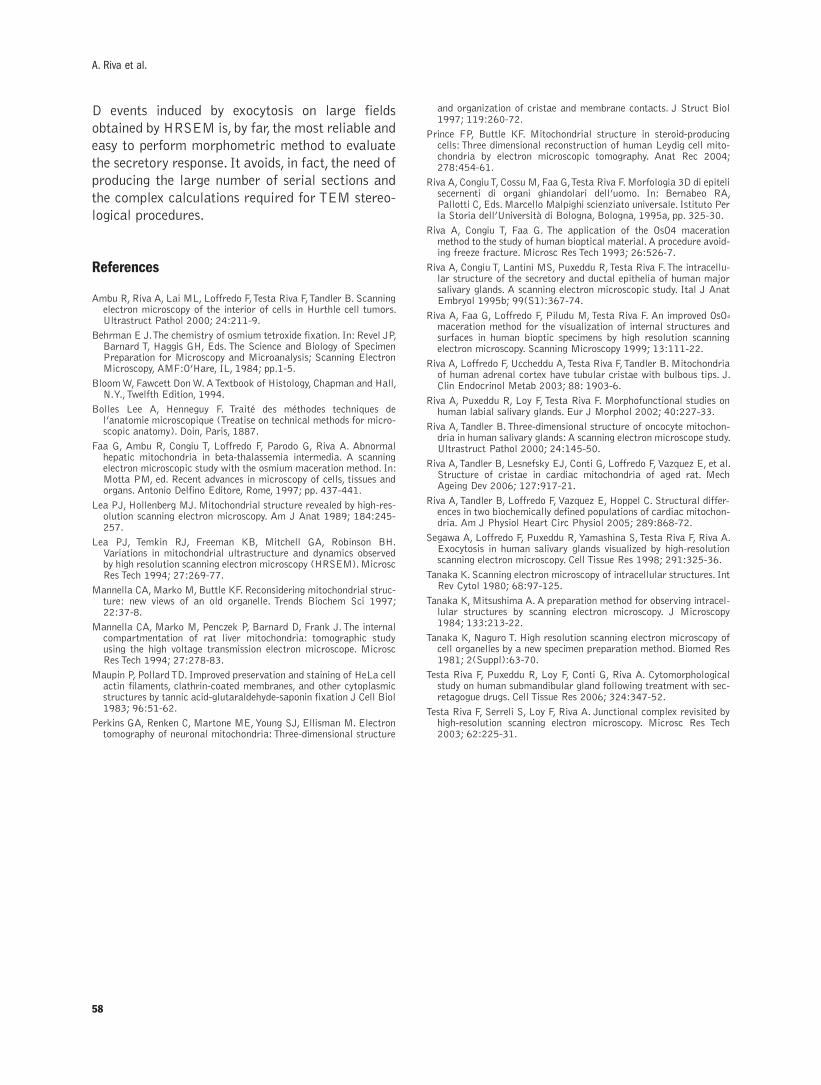
58
D events induced by exocytosis on large fields
obtained by HRSEM is, by far, the most reliable and
easy to perform morphometric method to evaluate
the secretory response. It avoids, in fact, the need of
producing the large number of serial sections and
the complex calculations required for TEM stereo-
logical procedures.
References
Ambu R, Riva A, Lai ML, Loffredo F, Testa Riva F, Tandler B. Scanningelectron microscopy of the interior of cells in Hurthle cell tumors.Ultrastruct Pathol 2000; 24:211-9.
Behrman E J. The chemistry of osmium tetroxide fixation. In: Revel JP,Barnard T, Haggis GH, Eds. The Science and Biology of SpecimenPreparation for Microscopy and Microanalysis; Scanning ElectronMicroscopy, AMF:O’Hare, IL, 1984; pp.1-5.
Bloom W, Fawcett Don W. A Textbook of Histology, Chapman and Hall,N.Y., Twelfth Edition, 1994.
Bolles Lee A, Henneguy F. Traité des méthodes techniques del’anatomie microscopique (Treatise on technical methods for micro-scopic anatomy). Doin, Paris, 1887.
Faa G, Ambu R, Congiu T, Loffredo F, Parodo G, Riva A. Abnormalhepatic mitochondria in beta-thalassemia intermedia. A scanningelectron microscopic study with the osmium maceration method. In:Motta PM, ed. Recent advances in microscopy of cells, tissues andorgans. Antonio Delfino Editore, Rome, 1997; pp. 437-441.
Lea PJ, Hollenberg MJ. Mitochondrial structure revealed by high-res-olution scanning electron microscopy. Am J Anat 1989; 184:245-257.
Lea PJ, Temkin RJ, Freeman KB, Mitchell GA, Robinson BH.Variations in mitochondrial ultrastructure and dynamics observedby high resolution scanning electron microscopy (HRSEM). MicroscRes Tech 1994; 27:269-77.
Mannella CA, Marko M, Buttle KF. Reconsidering mitochondrial struc-ture: new views of an old organelle. Trends Biochem Sci 1997;22:37-8.
Mannella CA, Marko M, Penczek P, Barnard D, Frank J. The internalcompartmentation of rat liver mitochondria: tomographic studyusing the high voltage transmission electron microscope. MicroscRes Tech 1994; 27:278-83.
Maupin P, Pollard TD. Improved preservation and staining of HeLa cellactin filaments, clathrin-coated membranes, and other cytoplasmicstructures by tannic acid-glutaraldehyde-saponin fixation J Cell Biol1983; 96:51-62.
Perkins GA, Renken C, Martone ME, Young SJ, Ellisman M. Electrontomography of neuronal mitochondria: Three-dimensional structure
and organization of cristae and membrane contacts. J Struct Biol1997; 119:260-72.
Prince FP, Buttle KF. Mitochondrial structure in steroid-producingcells: Three dimensional reconstruction of human Leydig cell mito-chondria by electron microscopic tomography. Anat Rec 2004;278:454-61.
Riva A, Congiu T, Cossu M, Faa G, Testa Riva F. Morfologia 3D di epitelisecernenti di organi ghiandolari dell’uomo. In: Bernabeo RA,Pallotti C, Eds. Marcello Malpighi scienziato universale. Istituto Perla Storia dell’Università di Bologna, Bologna, 1995a, pp. 325-30.
Riva A, Congiu T, Faa G. The application of the OsO4 macerationmethod to the study of human bioptical material. A procedure avoid-ing freeze fracture. Microsc Res Tech 1993; 26:526-7.
Riva A, Congiu T, Lantini MS, Puxeddu R, Testa Riva F. The intracellu-lar structure of the secretory and ductal epithelia of human majorsalivary glands. A scanning electron microscopic study. Ital J AnatEmbryol 1995b; 99(S1):367-74.
Riva A, Faa G, Loffredo F, Piludu M, Testa Riva F. An improved OsO4
maceration method for the visualization of internal structures andsurfaces in human bioptic specimens by high resolution scanningelectron microscopy. Scanning Microscopy 1999; 13:111-22.
Riva A, Loffredo F, Uccheddu A, Testa Riva F, Tandler B. Mitochondriaof human adrenal cortex have tubular cristae with bulbous tips. J.Clin Endocrinol Metab 2003; 88: 1903-6.
Riva A, Puxeddu R, Loy F, Testa Riva F. Morphofunctional studies onhuman labial salivary glands. Eur J Morphol 2002; 40:227-33.
Riva A, Tandler B. Three-dimensional structure of oncocyte mitochon-dria in human salivary glands: A scanning electron microscope study.Ultrastruct Pathol 2000; 24:145-50.
Riva A, Tandler B, Lesnefsky EJ, Conti G, Loffredo F, Vazquez E, et al.Structure of cristae in cardiac mitochondria of aged rat. MechAgeing Dev 2006; 127:917-21.
Riva A, Tandler B, Loffredo F, Vazquez E, Hoppel C. Structural differ-ences in two biochemically defined populations of cardiac mitochon-dria. Am J Physiol Heart Circ Physiol 2005; 289:868-72.
Segawa A, Loffredo F, Puxeddu R, Yamashina S, Testa Riva F, Riva A.Exocytosis in human salivary glands visualized by high-resolutionscanning electron microscopy. Cell Tissue Res 1998; 291:325-36.
Tanaka K. Scanning electron microscopy of intracellular structures. IntRev Cytol 1980; 68:97-125.
Tanaka K, Mitsushima A. A preparation method for observing intracel-lular structures by scanning electron microscopy. J Microscopy1984; 133:213-22.
Tanaka K, Naguro T. High resolution scanning electron microscopy ofcell organelles by a new specimen preparation method. Biomed Res1981; 2(Suppl):63-70.
Testa Riva F, Puxeddu R, Loy F, Conti G, Riva A. Cytomorphologicalstudy on human submandibular gland following treatment with sec-retagogue drugs. Cell Tissue Res 2006; 324:347-52.
Testa Riva F, Serreli S, Loy F, Riva A. Junctional complex revisited byhigh-resolution scanning electron microscopy. Microsc Res Tech2003; 62:225-31.
A. Riva et al.

©2007, European Journal of Histochemistry
The granular layer of the cerebellar cortex is composed of twogroups of neurons, the granule neurons and the so-calledlarge neurons. These latter include the neuron of Golgi and anumber of other, lesser known neuron types, generically indi-cated as non-traditional large neurons. In the last few years,owing to the development of improved histological and his-tochemical techniques for studying morphological andchemical features of these neurons, some non-traditionallarge neurons have been morphologically well characterized,namely the neuron of Lugaro, the synarmotic neuron, theunipolar brush neuron, the candelabrum neuron and theperivascular neuron. Some types of non-traditional largeneurons may be involved in the modulation of cortical intrin-sic circuits, establishing connections among neurons distrib-uted throughout the cortex, and acting as inhibitory interneu-rons (i.e., Lugaro and candelabrum neurons) or as excitato-ry ones (i.e., unipolar brush neuron). On the other hand, thesynarmotic neuron could be involved in extrinsic circuits,projecting to deep cerebellar nuclei or to another cortexregions in the same or in a different folium. Finally, theperivascular neuron may intervene in the intrinsic regulationof the cortex microcirculation.
Key words: granular layer; non-traditional neurons; neuron ofLugaro; synarmotic neuron; unipolar brush neuron; cande-labrum neuron; perivascular neuron.
Correspondence: Glauco Ambrosi,Dipartimento di Anatomia Umana e IstologiaUniversità di Bari Policlinico, Piazza Giulio Cesare, 11 70124 Bari, Italy.Tel./Fax: +39.0805478353.E-mail: [email protected]
European Journal of Histochemistry2007; vol. 51 supplement 1:59-64
Non-traditional large neurons in the granular layer of the cerebellar
cortex
G. Ambrosi, P. Flace, L. Lorusso, F. Girolamo, A. Rizzi, L. Bosco,1 M. Errede, D. Virgintino,
L. Roncali, V. Benagiano
Dipartimento di Anatomia Umana e Istologia, Policlinico, Bari; 1Dipartimento di Bioetica,
Palazzo Ateneo, Università di Bari, Italy
59
The granular layer (GL) of the cerebellar cor-
tex shows a homogeneous structure in the dif-
ferent lobes, lobules, laminae and folia of the
mammalian cerebellum, except for its depth, which
varies in the different folia ranging from 400-500
µm, in the apical region of the folium, to about 100µm, in the basal one. The GL is composed of twomain groups of neurons: the granule neurons (gran-
ules) and the so-called large neurons (non-gran-
ules). The granules have a small, spheroid body,
measuring 5-8 µm in diameter, and their bodies arepacked within the GL at a density of 2 to 7×10⁶ per
mm3. In comparison with the granules, the large
neurons show a more voluminous body, with a diam-
eter ranging from 15 to 25 µm, and a much lesserdensity. They include the neuron of Golgi, one of the
five traditional corticocerebellar neurons (together
with stellate, basket, Purkinje and granule neurons),
and a number of other neuron types, generically
indicated as non-traditional (n-trad) large neurons
(Jansen and Brodal, 1958; Eccles et al., 1967;
Voogd and Glickstein, 1998; Mugnaini, 2000;
Geurts et al., 2003; Houck and Mugnaini, 2003;
Flace et al., 2004 Ito, 2006). Although the n-trad
large neurons were first observed in the mammalian
GL a long time ago and increasingly detailed
descriptions have been provided over the years with
a certain regularity (see, e.g., Golgi, 1874; Lugaro,
1894; Ramon y Cajal, 1911; Pensa, 1931; Landau,
1933; Fox, 1959), it has long been debated whether
they should be considered as distinct neuron types
or not. This has depended on objective difficulties
existing in the observation and recognition of these
neurons: (a) the Golgi silver impregnation technique
constantly reveals only a small percentage of neu-
rons (1-2%) and thus only rarely visualizes n-trad
large neurons; (b) the large neurons are often liter-
ally hidden by granules, so the n-trad ones have in
many cases been misinterpreted as traditional Golgi
neurons; (c) finally, the n-trad large neurons may
display differences in their distribution in the GL
REVIEW
and also in their shape and size. In recent years,
owing to the development of improved histological
and histochemical techniques for the study of nerv-
ous tissues, morphological and neurochemical
parameters have been better defined allowing the
discrimination between different n-trad large neu-
ron types. A first classification of the n-trad large
neurons of the human GL was made by Braak and
Braak (1983). Using a technique that stains cyto-
plasmic deposits of lipofuscin, these Authors recog-
nized 3 types of large neurons. In particular, Braak
and Braak’s types 2 and 3 comprise n-trad large
neuron types, but in reality both include various
subtypes, each having a different localization with-
in the layer, different features of the bodies and
processes and most probably different functions.
More recently, an in-depth study of a great number
of large neurons of the human cerebellar cortex,
revealed by immunocytochemistry for glutamic acid
decarboxylase (GAD), the GABA synthesizing
enzyme, has supplied a demonstration of various
types of putative GABAergic n-trad large neurons
(Flace et al., 2004). Ambrosi and coll. have pro-
posed a classification of these GAD-positive n-trad
large neurons by reference to their localization and
position in the GL (conventionally subdivided into
three zones: external, intermediate and internal)
and to the morphological features of their bodies
and processes (for details, see Flace et al., 2004).
Up to now, five types of n-trad large neurons have
been sufficiently characterized from the morpho-
logical and neurochemical standpoints, even if their
functional roles are not yet completely understood.
They include the neuron of Lugaro, the synarmotic
neuron, the unipolar brush neuron, the candelabrum
neuron and the perivascular neuron (Table 1).
Neuron of LugaroThe neuron of Lugaro (NL; also known as inter-
mediate neuron or horizontal neuron) Figure 1 has
been described in the GL of various species of mam-
mals, including humans (Lugaro, 1894; Fox, 1959;
Braak and Braak, 1983; Lainé and Axelrad, 1996;
Geurts et al., 2001; Flace et al., 2004; Melik-
Musian and Fanardzhyan, 2004). The body of the
NL is distributed in all cerebellar lobes and lobules,
localized in the external zone of the GL just beneath
the Purkinje neuron layer. The body is fusiform, hor-
izontal (i.e., parallel to the folium surface), with a
major axis measuring 15-25 µm and lying on thesagittal plane (i.e., orthogonal to the folium major
axis). From the opposite body poles, two dendrite
trunks originate, being sagittally oriented and run-
ning horizontally along the boundary between the
GL and Purkinje neuron layer; they are rectilinear,
remarkably long (up to 1 mm) and branch within
strips of cortex ranging from the internal zone of
the molecular layer to the external zone of the GL.
The dendrites and body of NL offer a very extensive
receptive surface, receiving most inputs from recur-
rent branches of Purkinje neuron axons and in
addition from granule and basket neuron axons. The
axon of the NL originates from one body pole, or
from one dendrite trunk, and spreads with its col-
laterals in latero-lateral direction (i.e., parallel to
the folium major axis) within the molecular layer.
Axon terminals mainly form synapses upon basket
and stellate neurons and apical dendrites of Golgi
neurons.
A second type of NL has also been described, hav-
ing a roundish or triangular body localized in the
GL intermediate zone and with a different spatial
process arrangement, but establishing synaptic con-
tacts similar to those of the fusiform type (Lainé
and Axelrad, 2002; Melik-Musian and Fanard-
zhyan, 2004).
Immunocytochemical investigations have demon-
strated GAD or GABA immunoreactivity in the NL
of the rat (Aoki et al., 1986; Lainé and Axelrad,
1998) and human (Flace et al., 2004), indicating
its putative GABAergic, inhibitory nature, and also
the presence of the inhibitory amino acid glycine
and of a co-localization of glycine and GABA in the
rat (Dumoulin et al., 2001) and macaca monkey
(Crook et al., 2006). Other markers of the NL are
60
G. Ambrosi et al.
Figure 1. Non-traditional large neurons of the granular layerimmunostained for glutamic acid decarboxylase. A. neuron ofLugaro; B. synarmotic neuron; C. candelabrum neuron; D.perivascular neurons. Scale bar: 20 µm.

the cytoplasmic antigen rat-303 (Sahin and
Hockfield, 1990) and the calcium binding protein
calretinin (Geurts et al., 2001).
The NL, expanding in extensive, horizontally
developed regions of cerebellar cortex, creates the
anatomical conditions for the interconnection of
many neurons, located in all cortex layers. It main-
ly receives inputs from Purkinje neurons and proj-
ects to: (a) stellate and basket neurons, which in
turn project back to Purkinje neurons, which are
the main source of outputs from the cortex; (b)
Golgi neurons, which modulate the activity of affer-
ent mossy fibres. Since Purkinje, Lugaro, stellate,
basket and Golgi neurons are all GABAergic,
inhibitory neurons (Benagiano et al., 2001; Flace
et al., 2004), multiple intrinsic (i.e., non-projective)
circuits, each formed by series of GABAergic
synapses, exist in the cerebellar cortex, able to pro-
duce disinhibition (i.e., inhibition of an inhibition)
phenomena which spread within extensive regions
of the folium.
Synarmotic neuron The synarmotic neuron (SyN; also known as neu-
ron of Landau) Figure 1 was described by Landau
(1933) in various mammals, including humans. This
n-trad large neuron type has long been neglected,
being occasionally cited in literature (see Jansen
and Brodal, 1958), but only recently taken into
consideration once more (Katsetos et al., 1993;
Flace et al., 2004). The SyN is distributed in all
lobes and lobules with a preferential localization in
the basal and intermediate regions of the folium.
The body, localized in the internal zone of the GL or,
sometimes, in the subcortical white matter, is ovoid,
horizontal, with a major diameter measuring 20-25
µm. The dendritic tree is confined in the GL, whereit probably receives afferences from mossy fibres.
The axon arises from a body pole and runs in the
white matter, intermingled among efferent axons
from, or afferent axons to, the cerebellar cortex. It
finally re-enters the cortex, associating two cortical
regions in the same folium or in different folia, or
projects to cerebellar nuclei. A similar neuron was
described by Braak and Braak (1983) and includ-
ed in type 2 of their classification, but these
Authors did not mention the SyN.
Little is known about the neurochemical features
of the SyN. Immunoreactivity to GAD, suggesting a
GABAergic nature (Flace et al., 2004), and to the
calcium binding protein calbindin (Katsetos et al.,
1993) has been detected.
The SyN is thought to be involved in extrinsic
nervous circuits. It is a candidate for a second
source (besides the Purkinje neuron) (see also
Braak and Braak, 1983; Müller, 1994) of outputs
from the cerebellar cortex, making associative cor-
tico-cortical connections or projective connections
onto deep cerebellar nuclei. This latter hypothesized
role of the SyN, similar to that of the Purkinje neu-
61
Review
Table 1. Morphological features and classifications of non-traditional large neurons of the granular layer.
NEURON TYPE BODY LOCALIZATION BODY FORM PROCESS FORM CLASSIFICATION PROPOSED CLASSIFICATION PROPOSED BYwith reference to: AND ORIENTATION AND ORIENTATION BY BRAAK & BRAAK (1983) AMBROSI AND COLL.
(a) Cerebellar Lobes; (Flace et al., 2004)(b) Foliar Regions;(c) GL Zones
Lugaro a) All Lobes Fusiform, Horizontal Dendrites originate from opposite Type 2 External Zone/ Type 3b) All Regions body poles and run horizontallyc) External Zone at the boundary with PNL;
axon spreads within the ML
Synarmotic a) All Lobes Ovoid, Horizontal Dendrites are confined in the GL; Type 2 Internal Zone/Type 2b) Basal Region axon runs horizontally at thec) Internal Zone boundary with or inside the white matter
Unipolar Brush a) Flocculo-nodular Lobe Roundish/ Dendrite trunk originates Type 3 Not visualizedb) All Regions Ovoid, Vertical from external bodyc) All Zones pole and spreads
in the GL and ML;axon ramifies in the GL
Candelabrum a) All Lobes Pear-shaped, Vertical Dendrite Type 3 External Zone/b) All Regions and axon ascend from Type 1c) External Zone the external body pole to the ML
Perivascular a) All Lobes Roundish/Ovoid, variously Processes: variously oriented, Not mentioned Perivascular Typeb) All Regions oriented, perivascular perivascularc) All Zones
ML: Molecular Layer; PNL: Purkinje Neuron Layer; GL: Granular Layer.

ron, is in agreement with their common GABAergic
nature.
Unipolar brush neuronThe unipolar brush neuron (UBN; also indicated
as pale or monodendritic neuron) has been
described principally in the GL of the flocculo-
nodular lobe using histological and immunocyto-
chemical techniques (Altman and Bayer, 1977;
Braak and Braak, 1983; Hockfield, 1987; Munoz,
1990; Braak and Braak, 1993; Mugnaini and
Floris, 1994; Dino et al., 2000; Dogue et al., 2005;
Kalinichenko and Okhotin, 2005). The UBN has a
roundish, or ovoid and vertical, body measuring 9-
15 µm (thus intermediate in size between granulesand other large neuron types), localized throughout
the GL. From the external body pole a single den-
drite trunk originates and gives rise at its apex to
packed small branches, spreading in the GL and up
to the neighbouring molecular layer and receiving
synaptic contacts mainly from terminals of mossy
fibres and axons of Golgi neurons. The axon rami-
fies in the GL and its branches end upon dendrites
of granules, participating, like the terminals of
mossy fibres, in the formation of glomerular synap-
tic complexes (intrinsic mossy fibres).
Research carried out on the mouse cerebellar cor-
tex has indicated that the UBN is an excitatory cell,
using glutamate as neurotransmitter (Nunzi et al.,
2001). It also expresses receptors for glutamate
(Jaarsma et al., 1998; Geurts et al., 2001),
immunoreactivity to rat-302 antigen (Hockfield,
1987), chromogranin A (Munoz, 1990) and calre-
tinin (Braak and Braak, 1993; Floris et al., 1994;
Geurts et al., 2001).
The UBN mainly receives excitatory, glutamater-
gic synapses from mossy fibres and in turn projects
its excitatory, glutamatergic axon on granules,
which are also excitatory, glutamatergic. In this
way, a powerful feed-forward amplification system
of excitatory signals is created, coming from out-
side the cerebellum and reaching, via parallel fibres,
the dendritic trees of projective Purkinje neurons.
Candelabrum neuronThe candelabrum neuron (CN; also known as inter-
calated neuron) Figure 1 has been described ubiqui-
tously in the cerebellar cortex of the rat (Lainé and
Axelrad, 1994). It has a pear-shaped body, with a
vertical (i.e., orthogonal to the surface) major axis,
measuring 20-25 µm. Its body is squeezed against
the internal body pole of a Purkinje neuron or,
together with others, forms a row that joins up with
that of Purkinje neuron bodies. From the external
body pole, dendrite trunks originate, that ascend
through the molecular layer and arborize there in a
candelabrum-like fashion, as well as a thin axon that
also spreads in the molecular layer. Moreover, basal
dendrites originate from the internal body pole and
ramify in the GL. The CN receives inputs from axon
recurrent collaterals of Purkinje neurons and from
granule and basket neuron axons. Its axon forms
synapses upon basket and stellate neurons, but not
upon Purkinje neuron dendrites.
Although the CN has only recently been
described, a neuron type with similar morphological
features had already been observed in the cat cere-
bellar cortex by Pensa (1931). Moreover, this neu-
ron type had also been visualized by Braak and
Braak (1983) and included in type 3 of their clas-
sification.
Recently, Flace et al. (2004) demonstrated that
the CN shows immunoreactivity to GAD, also
reporting that this is the most frequent GAD-
immunoreactive large neuron type in the GL of the
human cerebellar cortex. Preliminary data have
indicated the presence of calbindin immunoreactiv-
ity within the CN (Flace et al., unpublished datum).
Owing to its inhibiting, GABAergic nature and in
view of the vertical displacement of its processes,
the CN may play the role of inhibitory interneuron
provided with intrinsic connections and mainly
involved in modulation of the activity of inhibiting,
GABAergic stellate and basket neurons (see also
Lainé and Axelrad, 1998).
Perivascular neuron The perivascular neuron (PN) Figure 1 is an n-
trad large neuron type found in all cerebellar lobes
and lobules and in all GL zones (Flace et al. 2004).
It displays an isodiametric body, lying extensively
along the wall of intracortical capillaries. Its
processes also run for tracts of various length in a
close anatomical relationship with capillaries.
GAD immunoreactivity has been observed in the
body and processes of the PN (Flace et al. 2004).
In accordance with a supposed role of GABA in
the local nervous regulation of intrinsic microves-
sels of the cerebellar cortex (Benagiano et al.,
2001), a vasoregulatory function has been hypoth-
esized for the PN, possibly in part exerted by vol-
ume transmission mechanisms (Flace et al., 2004).
62
G. Ambrosi et al.

Differential diagnosisAs previously noted, the n-trad large neurons of
the GL are often difficult to recognize. Immuno-
cytochemical techniques have made major contri-
butions to their more precise identification.
The NL and CN, as well as the traditional large
neuron of Golgi, are all localized in the external
zone of the GL and display immunoreactivity for
GAD or other GABA-related markers (Benagiano
et al., 2001; Flace et al., 2004). The NL, like the
neuron of Golgi, is immunoreactive to the cytoplas-
mic antigen rat-303 (Sahin and Hockfield, 1990),
but, unlike the Golgi, it is positive for calretinin and
negative for the glutamate receptor mGlu-R2
(Geurts et al., 2001). On the other hand, the CN is
immunonegative for rat-303, calretinin and mGlu-
R2 (Sahin and Hockfield, 1990; Geurts et al.,
2001), but positive for calbindin (Flace et al.,
unpublished data).
The UBN differs from the Golgi neuron and the
LN and CN, apart from its topography, also
because it never displays positivity for GABA-relat-
ed markers (or for other inhibitory neurotransmit-
ters) (Floris at al., 1994), in accordance with its
glutamatergic, excitatory nature (Nunzi et al.,
2001). Like the NL, but unlike the Golgi, the UBN
shows positivity for calretinin (Geurts et al., 2001);
like the Golgi, but unlike the NL, it expresses mGlu-
R2 (Geurts et al., 2001); unlike both the NL and
Golgi, it is negative for rat-303 (Sahin and
Hockfield, 1990), but positive for rat-302 antigen
(Hockfield, 1987).
The SyN, like the Golgi, NL and CN, displays pos-
itivity for GABA-related antigens (Flace et al.,
2004), but, unlike the other three large neuron
types, it is internally localized in the GL and thus
easy to discriminate. It also expresses positivity for
calbindin (Katsetos et al., 1993).
Some conclusive remarksAlthough new data are rapidly accumulating, the
knowledge of the n-trad large neurons of the GL is
still incomplete and concerns only some neuron
types. However, a number of functional roles could
be attributed to the n-trad neurons.
(1) They may be involved in complex intrinsic cir-
cuits of the cerebellar cortex, some establishing
horizontal (i.e., the NL) and some vertical (i.e., the
CN) connections among neurons distributed
throughout the cortex, acting as inhibitory (i.e., the
NL, the CN) or excitatory (i.e., the UBN) interneu-
rons. N-trad large neurons may thus modulate: sig-
nal transmission from afferent fibres to the cortex;
the activity of granules, responsible for transduc-
tion of mossy fibre signals onto Purkinje neurons
(granule-Purkinje neuron pathway); the activity of
Purkinje neurons, main source of outputs from the
cortex.
(2) A type of n-trad large neuron, namely the
SyN, could be involved in extrinsic circuits of the
cortex associating different regions of the cortex or
projecting to cerebellar nuclei.
(3) The PN could represent a type of n-trad large
neurons intervening in the local regulation of blood
microcirculation.
A better knowledge of all these n-trad large neu-
ron types will probably provide a decisive contribu-
tion to the task of unravelling the complex mecha-
nisms on which the working of the cerebellar neu-
ronal machine is based.
AcknowledgementsThis work is a small tribute to the late Professor
Carlo Rizzoli, a true pioneer of modern morpholog-
ical research in Italy, remembered by the Authors
with deep admiration and emotion. The Authors
also acknowledge the profound debt we all owe to
Camillo Golgi (1844-1926), Santiago Ramon y
Cajal (1852-1934), Ernesto Lugaro (1870-1940)
and Antonio Pensa (1874-1970), for their pioneer-
ing contributions to research on the microscopic
structure of the cerebellum.
References
Altman J and Bayer SA. Time of origin and distribution of a new celltype in the rat cerebellar cortex. Exp Brain Res 1977; 29: 265-74.
Aoki E, Semba R and Kashiwamata S. New candidates for GABAergicneurons in the rat cerebellum: an immunocytochemical study withanti-GABA antibody. Neurosci Lett 1986; 68: 267-71.
Benagiano V, Roncali L, Virgintino D, Flace P, Errede M, Rizzi A,Girolamo F, Robertson D, Bormann J and Ambrosi G. GABAimmunoreactivity in the human cerebellar cortex: a light and electronmicroscopical study. Histochem J 2001; 33: 537-43.
Braak E and Braak H. On three types of large nerve cells in the granu-lar layer of the human cerebellar cortex. Anat Embryol 1983; 166:67-86.
Braak E and Braak H. The new monodendritic neuronal type within theadult human cerebellar granule cell layer shows calretinin-immunore-activity. Neurosci Lett 1993; 154: 199-202.
Crook J, Hendrickson A and Robinson FR. Co-localization of glycineand GABA immunoreactivity in interneurons in macaca monkey cere-bellar cortex. Neuroscience 2006; 141: 1951-9.
Dino MR, Nunzi MG, Anelli R and Mugnaini E. Unipolar brush cells ofthe vestibulocerebellum: afferents and targets. Prog Brain Res 2000;124: 123-37.
Dugue GP, Dumoulin A, Triller A and Dieudonne S. Target-dependent useof coreleased inhibitory transmitters at central synapses. J Neurosci2005; 25: 6490-8.
63
Review

Dumoulin A, Triller A and Dieudonne S. IPSC kinetics at identifiedGABAergic and mixed GABAergic and glycinergic synapses ontocerebellar Golgi cells. J Neurosci 2001; 21: 6045-7.
Eccles JC, Ito M and Szentàgothai J. The cerebellum as a neuronalmachine. Springer, Berlin-Heidelberg-New York, 1967.
Flace P, Benagiano V, Lorusso L, Girolamo F, Rizzi A, Virgintino D,Roncali L and Ambrosi G. Glutamic acid decarboxylase immunore-active large neuron types in the granular layer of the human cerebel-lar cortex. Anat Embryol 2004; 208: 55-64.
Floris A, Dino M, Jacobowitz DM and Mugnaini E. The unipolar brushcells of the rat cerebellar cortex and cochlear nucleus are calretinin-positive: a study by light and electron microscopic immunocytochem-istry. Anat Embryol 1994; 189: 495-520.
Fox CA. The intermediate cells of Lugaro in the cerebellar cortex of themonkey. J Comp Neurol 1959; 112: 39-53.
Geurts FJ, De Schutter E and Dieudonne S. Unraveling the cerebellarcortex: cytology and cellular physiology of large-sized interneurons inthe granular layer. Cerebellum 2003; 2: 290-9.
Geurts FJ, Timmermans J-P, Shigemoto R and De Shutter E.Morphological and neurochemical differentiation of large granularlayer interneurons in the adult rat cerebellum. Neuroscience 2001;104: 499-512.
Golgi C. Sulla fina anatomia del cervelletto umano. In: Golgi C., OperaOmnia. Chapter V, vol. 1. Hoepli, Milan, 1874, pp. 99-111.
Hockfield S. A mab to a unique cerebellar neuron generated by immuno-suppression and rapid immunization. Science 1987; 237: 67-70.
Houk JC and Mugnaini E. Cerebellum. In: Squire LR, Bloom FE, McConnell SK, Roberts JL, Spitzer NC and Zigmond MJ (Eds.)Fundamental Neuroscience. Elsevier, Amsterdam, 2003, pp 841-72.
Ito M. Cerebellar circuitry as a neuronal machine. Prog Neurobiol2006; 78: 272-303.
Jaarsma D, Dino MR, Ohishi H, Shigemoto H and Mugnaini E.Metabotropic glutamate receptors are associated with non-synapticappendages of unipolar brush cells in rat cerebellar cortex andcochlear nuclear complex. J Neuocytol 1998; 27: 303-27.
Jansen J and Brodal A. Das Kleinhirn. In: Bargmann W (Ed.) Handbuchder mikroskopischen anatomie des menschen. Vol. 4. Springer, Berlin-Heidelberg-New York, 1958, pp. 91-149.
Kalinichenko SG and Okhotin VE. Unipolar brush cells - a new type ofexcitatory interneuron in the cerebellar cortex and cochlear nuclei ofthe brainstem. Neurosci Behav Physiol 2005; 35: 21-36.
Katsetos CD, Frankfurter A, Christakos S, Mancall EL, Vlachos IN andUrich H. Differential localization of class III, beta-tubulin isotypeand calbindin-D28k defines distinct neuronal types in the developing
human cerebellar cortex. J Neuropathol Exp Neurol 1993; 52: 655-66.
Lainé J and Axelrad H. The candelabrum cell: a new interneuron in thecerebellar cortex. J Comp Neurol 1994; 339: 159-73.
Lainé J and Axelrad H. Morphology of the Golgi impregnated Lugarocell in the rat cerebellar cortex: a reappraisal with a description of itsaxon. J Comp Neurol 1996; 375: 618-40.
Lainé J and Axelrad H. Lugaro cells target basket and stellate cells inthe cerebellar cortex. Neuroreport 1998; 9: 2399-2403.
Lainé J and Axelrad H. Extending the cerebellar Lugaro cell class.Neuroscience 2002; 111: 363-74.
Landau E. La cellule synarmotique dans le cervelet humain. Arch Anat1933; 17: 273-85.
Lugaro E. Sulle connessioni tra gli elementi nervosi della corteccia cere-bellare con considerazioni generali sul significato fisiologico dei rap-porti tra gli elementi nervosi. Riv Sper Freniat 1894; 20: 297-331.
Melik-Musian AB and Fanardzhyan VV. Morphological characteristic ofLugaro cells in the cerebellar cortex. Neurosci Behav Physiol 2004;34: 633-8.
Mugnaini E. GABAergic inhibition in the cerebellar system. In: MartinDL, Olsen RW (Eds.) GABA in the nervous system: the view at fiftyyears. Lippincott, Philadelphia, 2000, pp 383-407.
Mugnaini E and Floris A. The unipolar brush cell: a neglected neuron ofthe mammalian cerebellar cortex. J Comp Neurol 1994; 339: 174-80.
Müller T. Large nerve cells with long axons in the granular layer andwhite matter of the murine cerebellum. J Anat 1994; 184: 419-423.
Munoz DG. Monodendritic neurons: a cell type in the human cerebellarcortex identified by chromogranin A-like immunoreactivity. Brain Res1990; 528: 335-8.
Nunzi MG, Birnstiel S, Bhattacharyya BJ, Slater NT and Mugnaini E.Unipolar brush cells form a glutamatergic projection system withinthe mouse cerebellar cortex. J Comp Neurol 2001; 434: 329-341.
Pensa A. Osservazioni e considerazioni sulla struttura della cortecciacerebellare dei mammiferi. Mem R Accad Naz Lincei, Sci Fis MatNat 1931; ser VI 5: 25-50.
Ramón y Cajal S. Histologie du système nerveux de l’homme et desvertébrés. Vol II. Maloine, Paris, 1911.
Sahin M and Hockfield S. Molecular identification of the Lugaro cell inthe cat cerebellar cortex. J Comp Neurol 1990; 301:575-84.
Voogd J and Glickstein M. Anatomy of the cerebellum. Trends Neurosci1998; 21: 370-5.
64
G. Ambrosi et al.
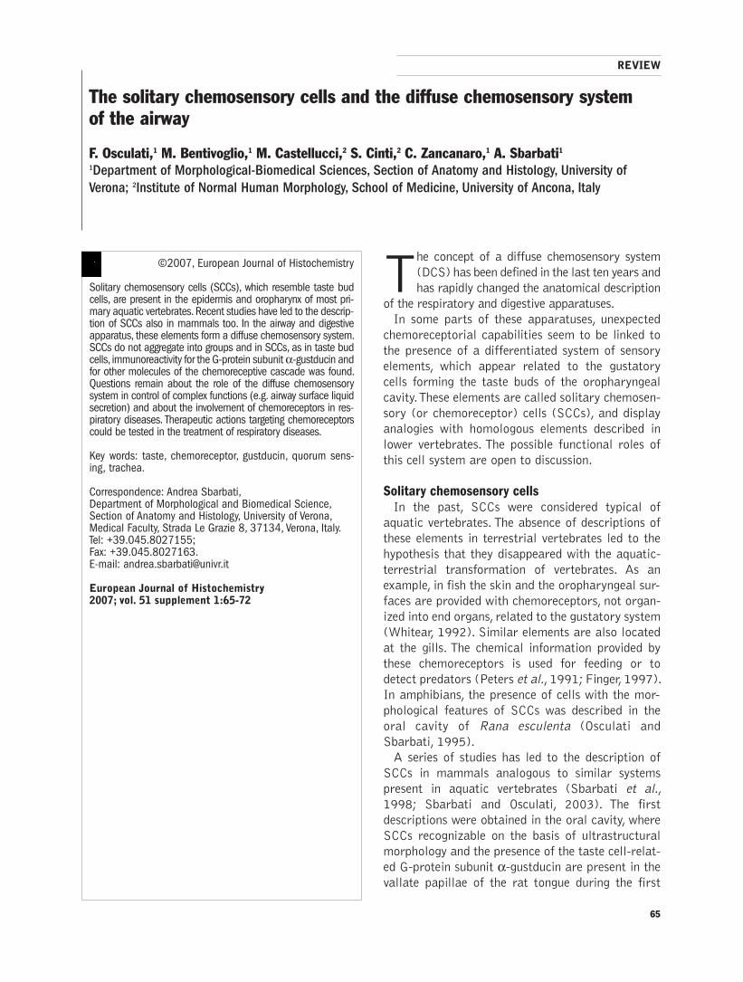
65
©2007, European Journal of Histochemistry
Solitary chemosensory cells (SCCs), which resemble taste budcells, are present in the epidermis and oropharynx of most pri-mary aquatic vertebrates. Recent studies have led to the descrip-tion of SCCs also in mammals too. In the airway and digestiveapparatus, these elements form a diffuse chemosensory system.SCCs do not aggregate into groups and in SCCs, as in taste budcells, immunoreactivity for the G-protein subunit α-gustducin andfor other molecules of the chemoreceptive cascade was found.Questions remain about the role of the diffuse chemosensorysystem in control of complex functions (e.g. airway surface liquidsecretion) and about the involvement of chemoreceptors in res-piratory diseases. Therapeutic actions targeting chemoreceptorscould be tested in the treatment of respiratory diseases.
Key words: taste, chemoreceptor, gustducin, quorum sens-ing, trachea.
Correspondence: Andrea Sbarbati, Department of Morphological and Biomedical Science,Section of Anatomy and Histology, University of Verona,Medical Faculty, Strada Le Grazie 8, 37134, Verona, Italy.Tel: +39.045.8027155;Fax: +39.045.8027163.E-mail: [email protected]
European Journal of Histochemistry2007; vol. 51 supplement 1:65-72
The solitary chemosensory cells and the diffuse chemosensory system
of the airway
F. Osculati,1 M. Bentivoglio,1 M. Castellucci,2 S. Cinti,2 C. Zancanaro,1 A. Sbarbati1
1Department of Morphological-Biomedical Sciences, Section of Anatomy and Histology, University of
Verona; 2Institute of Normal Human Morphology, School of Medicine, University of Ancona, Italy
The concept of a diffuse chemosensory system
(DCS) has been defined in the last ten years and
has rapidly changed the anatomical description
of the respiratory and digestive apparatuses.
In some parts of these apparatuses, unexpected
chemoreceptorial capabilities seem to be linked to
the presence of a differentiated system of sensory
elements, which appear related to the gustatory
cells forming the taste buds of the oropharyngeal
cavity. These elements are called solitary chemosen-
sory (or chemoreceptor) cells (SCCs), and display
analogies with homologous elements described in
lower vertebrates. The possible functional roles of
this cell system are open to discussion.
Solitary chemosensory cellsIn the past, SCCs were considered typical of
aquatic vertebrates. The absence of descriptions of
these elements in terrestrial vertebrates led to the
hypothesis that they disappeared with the aquatic-
terrestrial transformation of vertebrates. As an
example, in fish the skin and the oropharyngeal sur-
faces are provided with chemoreceptors, not organ-
ized into end organs, related to the gustatory system
(Whitear, 1992). Similar elements are also located
at the gills. The chemical information provided by
these chemoreceptors is used for feeding or to
detect predators (Peters et al., 1991; Finger, 1997).
In amphibians, the presence of cells with the mor-
phological features of SCCs was described in the
oral cavity of Rana esculenta (Osculati and
Sbarbati, 1995).
A series of studies has led to the description of
SCCs in mammals analogous to similar systems
present in aquatic vertebrates (Sbarbati et al.,
1998; Sbarbati and Osculati, 2003). The first
descriptions were obtained in the oral cavity, where
SCCs recognizable on the basis of ultrastructural
morphology and the presence of the taste cell-relat-
ed G-protein subunit α-gustducin are present in thevallate papillae of the rat tongue during the first
REVIEW

days of extrauterine life (Sbarbati et al., 1998 and
1999). Similar elements are also present in the
palate of rodents (El Sharaby et al., 2001). Further
investigation led to the description of similar ele-
ments in large parts of the digestive and respirato-
ry apparatuses.
SCC phenotypeSCCs form a rather polymorphic population,
although some characteristics seem to be common
to a majority of them. In general, they are slender
epithelial elements, which display cytological char-
acteristics suggesting a chemosensory role and
which possess signalling mechanisms typical of
taste cells (Sbarbati et al., 1998, 2004a; Finger et
al., 2003). Often they are single, bipolar epithelial
cells contacted by nerves and lacking a specialized
connective bed (Figure 1). SCCs may be surround-
ed by glial-like epithelial cells.
In fish, common ultrastructural features of SCCs
include spindle shape, basal synapses, abundant
endoplasmic reticulum within the proximal part of
the cell, and an apical microvillus. The distal
processes of SCCs contain a distinct Golgi appara-
tus and characteristic vesicles (Whitear and
Kotrschal, 1988). Where the epidermis is thick, the
nucleus of the sensory cell often lies at the level of
the second tier of epithelial cells from the surface,
but in other situations the cell may be elongated,
with its deep pole immediately above the basal layer
of the epidermis. Usually, the apical process is of
sufficient length to raise the presumed receptive
membrane above the mucus covering the surface of
the epithelium. Within the non-olfactory nasal
epithelium of mammals, SCCs are morphologically
similar to the individual cells in taste buds, but
unlike taste cells, they form distinct synapses on
cutaneous nerve fibers of the trigeminal nerve
(Finger et al., 2003).
In aquatic vertebrates, electrophysiological
recordings supported the hypothesis that SCCs are
chemosensory (Peters et al., 1991) and that they
respond to predator-avoidance or food-related
stimuli, although they do not respond to some typi-
cal taste stimuli (Silver and Finger, 1984).
The molecular mechanisms of taste transduction The detection of chemoreceptorial elements in
apparatuses of endodermic origin has mainly been
due to enormous developments in our knowledge of
gustatory science. These developments led to a
detailed description of the molecular machinery
responsible for taste transduction. Five taste quali-
ties exist (i.e., sodium salt, acids, amino acids, sweet
and bitter) (Lindemann, 2001; Margolskee, 2002;
Perez et al., 2003). All taste pathways converge on
common elements that mediate a rise in intracellu-
lar Ca2+ followed by transmitter release. Taste
responses to bitter/sweet compounds and amino
acids are initiated by G-protein-coupled receptors
(GPCRs) and transduced via G-protein signalling
cascades (Chaudhari and Roper, 1998; Gilbertson
et al., 2000).
In taste cells GPCRs are implicated in taste sig-
nal transduction (Adler et al., 2000; Chandra-
shekar et al., 2000; Chaudhari et al., 2000; Max et
al., 2001; Nelson et al., 2001, 2002; Li et al.,
2002; Amrein and Bray, 2003). Differences
between taste qualities are linked to different fam-
ilies of these receptors expressed in sets of taste
receptor cells (Adler et al., 2000; Nelson et al.,
2001; Zhang et al., 2003). Bitter compounds acti-
vate bitter taste T2R/Trb receptors, which are
encoded by a separate gene family consisting of
about 30 members in mice. T2R receptors then
activate gustducin heterotrimers. Activated alpha-
gustducin stimulates phosphodiesterases to
hydrolyze cAMP; the decrease in cAMP levels may
modulate cyclic nucleotide regulated ion channels
and/or kinases. Beta and gamma subunits of gust-
ducin activate phospholipase C of the β2 subtype togenerate IP3, which leads to release of Ca2+ from
internal stores via activation of inositol 1,4,5-
triphosphate receptor type III (IP3R3). Detection
of amino acid and sweet compounds is mainly
effected by the Tas1R (or T1R) gene family, which
encodes three conserved receptors that function as
heterodimers and form either a sugar receptor
(Tas1R2/Tas1R3) or a general amino acid receptor
(Tas1R1/Tas1R3). More in detail, the candidate
receptors for amino acid taste transduction are
ionotropic glutamate receptors, metabotropic glu-
tamate receptors and in particular tastemGluR4,
which is a truncated form of the brain mGluR4,
lacking most of the N-terminal extracellular
domain as well as the Tas1R1–Tas1R3 heteromer
(Chaudhari et al., 2000; Li et al., 2002; Nelson et
al., 2002; Ruiz et al., 2003; He et al., 2004).
Tas1R1 and Tas1R3 are coexpressed in taste buds
in the anterior part of the tongue (Nelson et al.,
2001), while taste mGluR4 is expressed in taste
buds of the circumvallate and foliate papillae (Yang
66
F. Osculati et al.
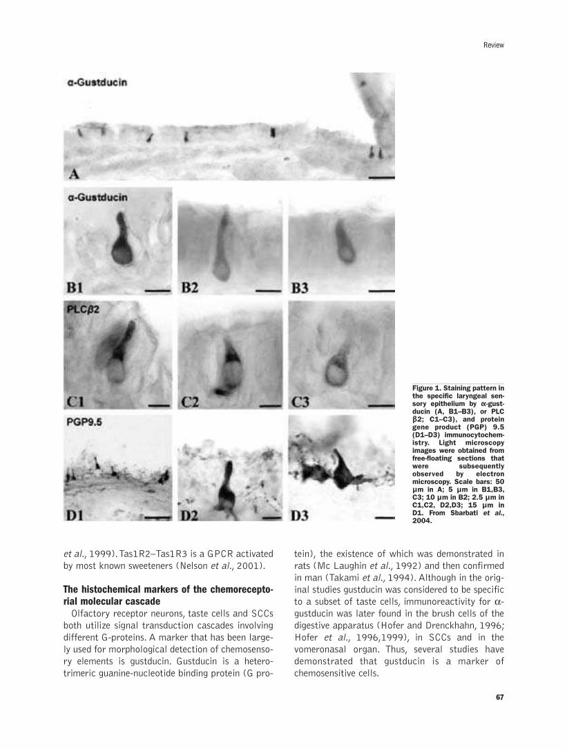
67
Review
et al., 1999). Tas1R2–Tas1R3 is a GPCR activated
by most known sweeteners (Nelson et al., 2001).
The histochemical markers of the chemorecepto-rial molecular cascadeOlfactory receptor neurons, taste cells and SCCs
both utilize signal transduction cascades involving
different G-proteins. A marker that has been large-
ly used for morphological detection of chemosenso-
ry elements is gustducin. Gustducin is a hetero-
trimeric guanine-nucleotide binding protein (G pro-
tein), the existence of which was demonstrated in
rats (Mc Laughin et al., 1992) and then confirmed
in man (Takami et al., 1994). Although in the orig-
inal studies gustducin was considered to be specific
to a subset of taste cells, immunoreactivity for α-gustducin was later found in the brush cells of the
digestive apparatus (Hofer and Drenckhahn, 1996;
Hofer et al., 1996,1999), in SCCs and in the
vomeronasal organ. Thus, several studies have
demonstrated that gustducin is a marker of
chemosensitive cells.
Figure 1. Staining pattern inthe specific laryngeal sen-sory epithelium by α-gust-ducin (A, B1–B3), or PLCβ2; C1–C3), and proteingene product (PGP) 9.5(D1–D3) immunocytochem-istry. Light microscopyimages were obtained fromfree-floating sections thatwere subsequentlyobserved by electronmicroscopy. Scale bars: 50µm in A; 5 µm in B1,B3,C3; 10 µm in B2; 2.5 µm inC1,C2, D2,D3; 15 µm inD1. From Sbarbati et al.,2004.
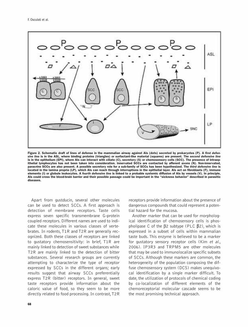
68
Apart from gustducin, several other molecules
can be used to detect SCCs. A first approach is
detection of membrane receptors. Taste cells
express seven specific transmembrane G-protein
coupled receptors. Different names are used to indi-
cate these molecules in various classes of verte-
brates. In rodents, T1R and T2R are generally rec-
ognized. Both these classes of receptors are linked
to gustatory chemosensitivity: in brief, T1R are
mainly linked to detection of sweet substances while
T2R are mainly linked to the detection of bitter
substances. Several research groups are currently
attempting to characterize the type of receptor
expressed by SCCs in the different organs; early
results suggest that airway SCCs preferentially
express T2R (bitter) receptors. In general, sweet
taste receptors provide information about the
caloric value of food, so they seem to be more
directly related to food processing. In contrast, T2R
receptors provide information about the presence of
dangerous compounds that could represent a poten-
tial hazard for the mucosa.
Another marker that can be used for morpholog-
ical identification of chemosensory cells is phos-
pholipase C of the β2 subtype (PLC β2), which isexpressed in a subset of cells within mammalian
taste buds. This enzyme is believed to be a marker
for gustatory sensory receptor cells (Kim et al.,
2006). IP3R3 and TRPM5 are other molecules
that may be used to immunolocalize specific subsets
of SCCs. Although these markers are common, the
heterogeneity of the population composing the dif-
fuse chemosensory system (DCS) makes unequivo-
cal identification by a single marker difficult. To
date, the utilization of protocols of chemical coding
by co-localization of different elements of the
chemoreceptorial molecular cascade seems to be
the most promising technical approach.
F. Osculati et al.
Figure 2. Schematic draft of lines of defense in the mammalian airway against AIs (dots) secreted by prokaryotes (P). A first defen-sive line is in the ASL, where binding proteins (triangles) or surfactant-like material (squares) are present. The second defensive lineis in the epithelium (EPI), where AIs can interact with ciliate (C), secretory (S) or chemosensory cells (SCC). The presence of intraep-ithelial lymphocytes has not been taken into consideration. Innervated SCCs are contacted by afferent axons (N). Non-innervated,paracrine SCCs are also present. A possible secretory role for a sub-family of SCCs has been hypothesized. The third defensive line islocated in the lamina propria (LP), which AIs can reach through interruptions in the epithelial layer. AIs act on fibroblasts (F), immuneelements (I) or globule leukocytes. A fourth defensive line is linked to a probable systemic diffusion of AIs by vessels (V). In principle,AIs could cross the blood-brain barrier and their possible passage could be important in the “sickness behavior” described in parasiticdiseases.

Homology between the SCCs in different speciesTo establish homology among SCCs in fish,
amphibians and mammals is difficult, partly
because these cells form heterogeneous systems. So
far, findings in mammals have generally confirmed
previous findings in fish about the general morphol-
ogy of SCCs, despite the fact that in mammals
SCCs seem to be used as internal rather than as
external chemoreceptors. In the oral cavity, homol-
ogy between SCCs described in the different species
seems evident, even if the relationship with the taste
system requires further clarification. It is more dif-
ficult to determine homology in other parts of the
body, in which complex end organs are lacking.
SCCs in the airwayIn mammals, SCCs were first described in the
oral cavity and seem to be widespread in large por-
tions of the digestive apparatus. SCCs are also well
represented in the respiratory apparatus, which
shares a common endodermic origin with the diges-
tive system. It has been shown that SCCs are dif-
fusely present in the airways and in particular in
the nasal cavity (Zancanaro et al., 1999), where
they detect irritants (Finger et al., 2003). It was
also demonstrated that these cells proliferate and
undergo rapid turnover (Gulbransen and Finger,
2005). SCCs are also present both in the larynx
(Sbarbati et al., 2004 a,b) and in the trachea
(Merigo et al., 2005). These findings were obtained
in rodents, which present very small airways in
which the serous component largely prevails over
the mucous component. Therefore, rodents are not
ideal models for studying aspects that could be rel-
evant for human pathology. Studies in species of
large size and with respiratory mucosa resembling
those of the human airway are in progress. One
example to date is a study in Bos taurus, which
demonstrated the presence of SCCs on the ary-
tenoid epithelium, in the trachea and the bronchi
(Tizzano et al., 2006).
SCCs in the human nasal cavityData about SCCs in humans are scarce. Recent
studies revealed a possible receptor cell in human
and rodent olfactory epithelium. Also, electron
microscopic studies of respiratory epithelium indi-
cated several potential chemosensory cell types.
Immunocytochemical experiments showed cell
types positive for gustducin, calbindin and/or the
vesicular acetylcholine transporter (VAchT) that
closely resembled rodent SCCs (Hansen et al.,
2005). These cells have the morphology of SCCs
and express Trp M5. Subsets of these cells express
gustducin, calbindin and/or VAChT. These findings
suggest the existence of possible unconventional
receptor cell types in the respiratory epithelium of
rodents and humans (Hansen et al., 2006).
The specific laryngeal sensory epitheliumA specialized portion of the DCS seems to be
located in the larynx. A specific laryngeal sensory
epithelium (SLSE), which includes arrays of soli-
tary chemoreceptor cells, has recently been
described in the supraglottic region of the rat
(Sbarbati et al., 2004a). These SCCs lie in this spe-
cific epithelium together with taste buds. Recently,
Finger et al., (2005) demonstrated that taste buds
are clearly innervated by nerve fibers immunoreac-
tive for purinergic receptors, and that stimulation of
taste buds in vitro evokes release of ATP. Thus, ATP
fulfils the criteria for a neurotransmitter linking
taste buds to the nervous system. On the other hand,
laryngeal solitary chemoreceptor cells are not
innervated by purinergic nerve fibers, although such
fibers do innervate nearby epithelium. This indicates
that nerve fibers that innervate laryngeal SCCs uti-
lize a different neurotransmitter and/or receptor
system (Finger et al., 2005). The laryngeal
immunoreactivity for α-gustducin was mainly local-ized in SCCs.
Laryngeal chemosensory clustersIn the larynx of the rat, a new form of chemosen-
sory structure (i.e. the chemosensory cluster) has
also been reported (Sbarbati et al., 2004 b). These
clusters are multicellular organizations which differ
from taste buds and are generally composed of 2-3
chemoreceptor cells (Sbarbati et al., 2004).
Compared with lingual taste buds, chemosensory
clusters show lower height and smaller diameter. In
laryngeal chemosensory clusters, immunocyto-
chemistry using antibodies against either α-gustdu-cin or PLC β2 identified a similar cytotype. PLC β2is expressed in a subset of cells within mammalian
taste buds. The demonstration of the existence of
chemosensory clusters strengthens the hypothesis
of a phylogenetic link between gustatory and soli-
tary chemosensory cells. Due to their structure and
location, chemosensory clusters seem to represent
the missing link between buds and SCCs. Laryngeal
chemosensory clusters appear to be a transitional
69
Review

structure between the rostrally located buds and
SCCs, which are more distally located in specific
areas of the larynx (Sbarbati et al., 2004a).
In vivo approach by pharmacological magneticresonance imaging Considering the large amount of chemoreceptorial
genes the capability of the chemosensory systems to
recognize patterns of exogenous molecules is enor-
mous. In the airway, the secretory responses to air-
borne molecules or to substances produced by micro-
bial biofilms, which act on chemosensors may be
evaluated by in vivo experimental paradigms using
pharmacological magnetic resonance imaging. In
such protocols, the integrity of the tissue is main-
tained such as the connectivity among several differ-
ent cell types, the paracrine interaction, the blood
flow and the innervation. Using this approach, we are
testing on the airway, a large number of infochemi-
cals extracted by bacteria, plants or animals. The pre-
liminary results confirm the possibility that the air-
way secretion may be controlled by chemical cues.
The DCS and bacterial chemosensory systems The presence of a DCS in the airways raises ques-
tions about the role of chemoreceptors in control of
complex functions (e.g. airway surface liquid secre-
tion) and about the involvement of chemoreceptors
in respiratory diseases.
The chemoreceptive capacity of the DCS seems to
protect against exogenous substances. In addiction,
recently published data suggest that the DCS could
have an important role in defense against bacteria.
The elements of this system are located in an opti-
mal position to intercept the exchange of informa-
tion between bacteria operated by the quorum sens-
ing strategy (Kolter, 2005). Briefly, these bacteria
co-ordinate their activities using extracellular sig-
nals, i.e. auto-inducers or pheromones (Hardman et
al., 1998). When such compounds reach a sufficient
concentration (i.e. when the total population is
large enough), the bacteria activate genetic path-
ways often involved in the initiation of aggressive
behavior. Quorum sensing appears therefore to be a
strategy used by bacteria to co-ordinate their activ-
ities, and it is based on the release of small mole-
cules, generally proteins or acyl-lactones. These
findings suggest that a war of communication takes
place on the mucosal surfaces of the digestive and
respiratory systems, with two chemosensory sys-
tems in opposite camps (Sbarbati 2006; Sbarbati
and Osculati, 2006).
Due to its structural and biochemical character-
istics, the DCS appears to be able to intercept com-
munication among bacteria and predict their move-
ments. If messages are indeed detected in this way,
it may be that the organism mounts a highly local-
ized and efficient response to bacterial activation.
This would be based on defenses like the quenching
of auto-inducers, the dilution or removal of bacte-
ria, or secretion of antibiotic agents, and it might
precede or avoid the need for intervention by
immune cells.
The inflammatory reflexIt is well known that in the airway, the control of
inflammation is based on information provided by
vagal sensory afferents and that central integration
devices operate between sensory and effector struc-
tures, which could act on both immune and mucos-
al cells (Figure 2) (Andersson, 2005). This inflam-
matory reflex is a physiological pathway in which
the nervous system detects inflammatory stimuli
and modulates cytokine production. Afferent sig-
nals to the brain are transmitted by the vagus nerve,
which activates a reflex response that culminates in
efferent vagus nerve signalling. Termed the cholin-
ergic anti-inflammatory pathway, efferent activity
in the vagus nerve releases acetylcholine that inter-
acts with macrophage nicotinic receptors (Czura
and Tracey, 2005).
In the past, the afferent input was considered to
be generated by vagal free nerve endings but the
new data demonstrated that the SCCs forming the
DCS may be innervated. Therefore, further studies
must evaluate whether these specialized epithelial
elements significantly contribute to the vagal input
in the context of the inflammatory reflex.
The intramucosal reflexIn addition to central reflexes, further defensive
lines against micro-organisms and xenobiotics are
based on intramucosal reflexes. Figure 1 schemati-
cally illustrates the cell types putatively involved.
Our preliminary results suggest that activation of
the DCS leads to a secretory response by the
mucosa and activation of mucociliary clearance
(Merigo et al., 2007). In particular, bitter sub-
stances can stimulate a secretory reflex that is in
part supported by a chemoreceptorial capacity of
secretory cells (short reflex). The increased activity
of mucosal cells may result in dilution of bacterial
70
F. Osculati et al.
quorum sense signals and their removal by mucocil-
iary clearance.
The relationship between SCCs and brush cells Brush cells (BCs) are elements characterized by
a brush of rigid apical microvilli with long rootlets,
which are found in the digestive and respiratory
apparatuses. In the past, these cells have been given
names such as tuft, fibrillovesicular, multivesicular
or caveolated cells.
The first description of BCs is generally attrib-
uted to Rhodin and Dalham (1956) in the rat tra-
chea. Since the first description, the presence of
BCs has been confirmed in the airway of several
species, including humans (Rhodin, 1959). BCs
were then detected in the lung (Meyrick and Reid,
1968) and in the digestive apparatus (Luciano et
al., 1968 a,b), mainly in the gallbladder (Luciano
and Reale, 1969). The recent description of gust-
ducin (Hofer and Drenckhahn, 1998) and other bit-
ter-taste related molecules in BCs in the digestive
and respiratory apparatuses demonstrated a link
between these cells and elements of taste buds. The
recent results support the idea that BCs may oper-
ate as solitary chemoreceptors (Sbarbati and
Osculati, 2005), probably representing a subfamily
of SCCs localized in specific microenvironments.
ConclusionsSeveral questions remain about SCCs and about
the physiology and morphology of the DCS. In par-
ticular, the links between the molecular mecha-
nisms of taste and secretory apparatuses have not
yet been studied, and the existence of BCs not con-
taining α-gustducin raises the possibility of alterna-tive G-proteins. Such questions could be answered
by a detailed chemical code for the different ele-
ments of the DCS.
This DCS seems to be a potential new drug target
because several elements indicate that information
obtained by this system induces secretory reflexes.
Therefore, modulation of the respiratory and diges-
tive apparatuses by substances acting on their
chemoreceptors could be important in the treat-
ment of diseases such as cystic fibrosis and asthma,
and might open new frontiers in drug discovery.
AcknowledgementsThis work is dedicated to the memory of
Professor Rizzoli, prestigious mentor of the Italian
morphological school.
References
Adler E, Hoon MA, Mueller KL, Chandrashekar J, Ryba NJ, Zuker CS.A novel family of mammalian taste receptors. Cell 2000;100:693–702.
Amrein H, Bray S. Bitter-sweet solution in taste transduction. Cell2003;112:283–7.
Andersson J. The inflammatory reflex. J Intern Med 2005; 257:122-5.
Chaudhari N, Roper SD. Molecular and physiological evidence for glu-tamate (umami) taste transduction via a G protein-coupled receptor.Ann NY Acad Sci 1998; 855:398–406.
Chaudhari N, Landin AM, Roper SD. A metabotropic glutamate recep-tor variant functions as a taste receptor. Nat Neurosci 2000;3:113–9.
Czura CJ, Tracey KJ. Autonomic neural regulation of immunity. JIntern Med 2005; 257:156-66.
El-Sharaby A, Ueda K, Kurisu K, Wakisaka S. Development and matu-ration of taste buds of the palatal epithelium of the rat: histologicaland immunohistochemical study. Anat Res 2001; 263 :260–8.
Finger TE. Evolution of taste and solitary chemoreceptor cell system.Brain Behav Evol 1997; 50 :234–43.
Finger TE, Bottger B, Hansen A, Anderson KT, Alimohammadi H,Silver WL. Solitary chemoreceptor cells in the nasal cavity serve assentinels of respiration. Proc Natl Acad Sci USA 2003; 100: 8981–6.
Finger TE, Danilova V, Barrows J, Bartel DL, Vigers AJ, Stone L, et al.SC. ATP signaling is crucial for communication from taste buds togustatory nerves Science 2005; 310:1495–9.
Gilbertson TA, Damak S, Margolskee RF. The molecular physiology oftaste transduction. Curr Opin Neurobiol 2000; 10: 519–27.
Gulbransen BD, Finger TE. Solitary chemoreceptor cell proliferation inadult nasal epithelium. Journal of Neurocytology 2000; 34:117-22.
Hansen A, Witt M, Hummel T. Unconventional neurons in the nasalcavity of humans. Chem Senses 2005; 30:A55.
Hansen A. Unconventional sensory cells in the nasal epithelia ofrodents and humans. Chem Senses 2006b; 31:E4.
Hardman AM, Stewart GS, Williams P. Quorum sensing and the cell-cell communication dependent regulation of gene expression in path-ogenic and non-pathogenic bacteria. Antonie Van Leeuwenhoek1998; 74:199–210.
He W, Yasumatsu K, Varadarajan V, Yamada A, Lem J, Ninomiya, et al.Umami taste responses are mediated by alpha-transducin andalpha-gustducin. J Neurosci 2004; 24:7674–80.
Höfer D, Drenckhahn D. Cytoskeletal markers allowing discriminationbetween brush and other epithelial cells of the gut includingetnteroendocrine cells. Histochem Cell Biol 1996; 105:405–12.
Höfer D, Drenckhahn D. Identification of the taste cell G-protein alpha-gustducin in brush cells of the rat pancreatic duct system. HistochemCell Bio1998; 110: 303–9.
Höfer D, Puschel B, Drenckhahn D. Taste receptor-like cells in the ratgut identified by expression of �alpha-gustducin. Proc Natl Acad SciUSA 1996; 93: 6631–4.
Höfer D, Asan E, Drenckhahn D. Chemosensory perception in gut. NewPhysiol Sci 1999; 14:18–23.
Kim JW, Roberts C, Marujama Y, Berg S, Roper S, Chaudari N.Faithful expression of GFP from the PLC�2 promoter in a function-al class of taste receptor cells. Chem Senses 2006; 31: 213-19.
Kolter R. Surfacing views of biofilm biology. Trends Microbiol 2005;13: 1–2.
Li X, Staszewski L, Xu H, Durick K, Zoller, M., Adler E. Human recep-tors for sweet and umami taste. Proc NatlAcad Sci 2002; USA 99:4692–6.
Lindemann B. Receptors and transduction in taste. Nature 2001; 413:219–25.
Luciano L, Reale E, Ruska H. On a “chemoreceptive” sensory cell inthe trachea of the rat. Z Zellforsch Mikrosk Anat 1968a; 85: 350 –75.
Luciano L, Reale E, Ruska H. On a glycogen containing brush cell inthe rectum of the rat. Z Zellforsch Mikrosk Anat 1968b; 191: 153–8.
Luciano L, Reale E, Ruska H. Brush cells in the alveolar epithelium of
71
Review
72
the rat lung. Z Zellforsch Mikrosk Anat 1969; 95:198–201.
Margolskee RF. Molecular mechanisms of bitter and sweet taste trans-duction. J Biol Chem 2002; 277:1–4.
Max M, Shanker YG, Huang L, Rong M, Liu Z, Campagne F, WeinsteinH, Damak S, Margolskee RF. Tas1r3, encoding a new candidate tastereceptor, is allelic to the sweet responsiveness locus. Sac NatGenet2001; 28:58–63.
McLaughin SK, McKinnon PJ, Margolskee RF. Gustducin is a taste cellspecific G protein close related to the transducin. Nature 1992;357: 563– 69.
Merigo F, Benati D, Tizzano M, Osculati F, Sbarbati A. �-gustducinimmunoreactivity in the airways. Cell Tissue Res 2005; 319: 211–9.
Merigo F, Benati D, DiChio M, Osculati F, Sbarbati A. Secretory cellsof the airway express molecules of the chemoreceptive cascade. CellTissue Res 2007; 327: 231-47.
Meyrick B, Reid L. The alveolar brush cell in rat lung- a third pneu-mocyte. J Ultrastruct Res 1968; 23:71–80.
Nelson G, Hoon MA, Chandrashekar J, Zhang Y, Ryba NJ, Zuker CS.Mammalian sweet taste receptors. Cell 2001; 106:381–90.
Nelson G, Chandrashekar J, Hoon MA, Feng L, Zhao G, Ryba NJ, et al.An amino-acid taste receptor. Nature 2002; 416:199–202.
Perez CA, Margolskee RF, Kinnamon SC, Ogura T. Making sense withTRP channels: store-operated calcium entry and the ion channelTrpm5 in taste receptor cells. Cell Calcium 2003; 33:541–9.
Peters RC, Kotrschal K, Krautgartner WD. Solitary chemoreceptorcells of Ciliata mustela (Gadidae, teleostei) are tuned to mucoidstimuli. Chem Senses 1991; 16: 31–42.
Osculati F, Sbarbati A. The frog taste disk: aprototype of the vertebrategustatory organ. Prog Neurobiol 1995; 46: 351–399.
Rhodin J. Ultrastructure of the tracheal ciliated mucosa in rat andman. Ann Otol Rhinol Laryngol 1959; 68: 964–74.
Rhodin J, Dahalm T. Electron microscopy of the tracheal ciliatedmucosa in rat. Z Zellforsch Mikrosk Anat 1956; 44:345-412.
Ruiz CJ, Wray K, Delay E, Margolskee RF, Kinnamon SC. Behavioralevidence for a role of alpha-gustducin in glutamate taste. ChemSenses 2003; 28:573–9.
Sbarbati A. Antimicrobial intelligence: how eukaryotes combat thequorum sensing strategy. 16th European Congress of ClinicalMicrobiology and Infectious Diseases. Keynote lecture. Nice, April1-4. Clin Microbiol Infect 2006; 12: K243.
Sbarbati A, Osculati F. Solitary chemosensory cells in mammals?CellTissue Organs 2003; 175:51-5.
Sbarbati A, Osculati F. A new fate for old cells: brush cells and relatedelements. J Anat 2005a; 206: 349-58.
Sbarbati A, Osculati F. The taste cell-related diffuse chemosensory sys-tem. Prog Neurobiol 2005b; 75: 295–307.
Sbarbati A, Osculati F. Allelochemical communication in vertebrates:kairomones, allomones and synomones. Cell Tissue Org 2006; 183:206-19.
Sbarbati A, Crescimanno C, Benati D, Osculati F. Solitary chemosen-sory cells in the developing chemoreceptorial epithelium of the val-late papilla. Journal of Neurocytology 1998; 27:631–5.
Sbarbati A, Crescimanno C, Bernardi P, Osculati F. Alpha-gustducinimmunoreactive solitary chemosensory cells in the developingchemoreceptorial epithelium of the rat vallate papilla. Chem senses1999; 24:469–72.
Sbarbati A, Merigo F, Benati D, Tizzano M, Bernardi P, CrescimannoC, et al. Identification and characterization of a specific sensoryepithelium in the rat larynx. J Comp Neurol 2004a; 475:188-201.
Sbarbati A, Merigo F, Benati D, Tizzano M, Bernardi P, Osculati F.Laryngeal chemosensory clusters. Chem Senses 2004b; 29:683-92.
Silver WL, Finger TE. Electrophysiological examination of a non-olfac-tory, non-gustatory chemosense in the sea robin, Prionotus caroli-nus. J Comp Physiol 1984; A154: 167–74.
Takami S, Getchell TV, McLaughlin SK, Margolskee RF, Getchell ML.Human taste cells express the G protein �alpha-gustducin and neu-ron-specific enolase. Mol Brain Res 1994; 22:193-203.
Tizzano M, Merigo F, Sbarbati A. Evidence of solitary chemosensorycells in a large mammal: the diffuse chemosensory system in BosTaurus airways. J Anat 2006; 209: 333-7.
Whitear M. Solitary chemoreceptor cells. In: Chemoreception in fish-es. Hara T.J. (Ed) Chapman and Hall New York 1992; pp.103–25.
Whitear M, Kotrschal K. The chemosensory anterior dorsal fin in rock-lings (Gaidopsarus and Ciliata, Teleostei, Gadidae): activity, finestructure and innervation. J Zool 1988; 216:339-66.
Yang H, Wanner IB, Roper SD, Chaudhari N. An optimized method forin situ hybridization with signal amplification that allows the detec-tion of rare mRNAs. J. Histochem Cytochem 1998; 47:431–46.
Zancanaro Caretta CM, Merigo F, Cavaggioni A, Osculati F. �-gustducinexpression in the vomeronasal organ of the mouse. Eur J Neurosci1999; 11: 4473 – 5.
Zhang Y, Hoon MA, Chandrashekar J, Mueller KL, Cook B, Wu D, etal. Coding of sweet, bitter, and umami tastes: different receptor cellssharing similar signaling pathways. Cell 2003; 112: 293–301.
F. Osculati et al.

©2007, European Journal of Histochemistry
The modality of transendothelial passage of the macromole-cules and cells (lymphocyte and cancer cells) in the absorb-ing lymphatic vessel (ALV) and the tumor-associated absorb-ing lymphatic (TAAL) vessel is studied. On the basis of thepeculiar plasticity of the lymphatic endothelial cell of thesevessels (lacking a continuous basement membrane, poresand open junctions) the endothelial wall organizes formationof the intraendothelial channel, by means of molecular inter-actions as yet unidentified. The remarkable finding of theintravasation of lymphocyte and experimental tumor cancercells (T84 colon Adenocarcinoma, B16 melanoma in nudemice and spontaneous prostate adenocarcinoma in trans-genic mice) should be stressed. This intravasation takesplace, under both physiologic and pathological conditions,following the same transendothelial morphological modality,i.e. the intraendothelial channel – a dynamic and transiententity - is probably also induced by similar molecular inter-actions, a crucial point that merits future research.
Key words: lymphatic, intravasation, lymphocyte, cancer cell,metastasis, transendothelial migration.
Correspondence: Giacomo Azzali,Director of the Lymphatology Laboratory.Department of Human Anatomy, Pharmacology and forensicMedicine, School of Medicine, University of Parma. ViaGramsci, 14 (Ospedale Maggiore), 43100 Parma, ItalyTel: +39.0521.033031 or 033032.Fax: +39.0521.033033.E-mail: [email protected]
European Journal of Histochemistry2007; vol. 51 supplement 1:73-78
The modality of transendothelial passage of lymphocytes and tumor
cells in the absorbing lymphatic vessel
G. Azzali
Lymphatology Laboratory – Section of Human Anatomy. Department of Human Anatomy, Pharmacology
and forensic Medicine, School of Medicine, University of Parma (Ospedale Maggiore), Parma, Italy
73
The morphological findings obtained in the sec-
ond half of the 1900s regarding what com-
poses the canalization of the lymphatic vas-
cular system (LVS) helps clarify not only the LVS’s
role, complementary to the blood vascular system,
but also its importance in lymphocyte homing, in
regulating tissue homeostasis and in some intersti-
tial matrix pathologies. Furthermore, the fine struc-
ture of the vessels of the LVS allows us to distin-
guish two distinct sectors composed of (a) lymphat-
ic vessels whose main function is that of lymph con-
duction and flow (pre- and post-lymph nodal collec-
tor, lymphatic trunk vessels, thoracic duct); (b) lym-
phatic vessels with high absorption capacity (the
chyliferous vessel; vessels of the mucosal, submu-
cosal and muscular network) (Ottaviani and Azzali,
1965). The latter, unlike collector vessels character-
ized by a monolayer of endothelial cells that rests on
a continuous basement membrane covered external-
ly by one or more strata of smooth muscle fibers,
are lacking a continuous basement membrane, pores
and open junctions. Furthermore, the abluminal sur-
face of endothelial cells establishes an extensive
connection with the components of the extravasal
interstitial matrix. The recent use of specific mark-
ers to detect the lymphatic endothelium [LYVE-1,
Prox-1, tetraspanin, podoplanin, D2-40 (Jackson et
al., 2001; Longo et al., 2001; Prevo et al., 2001;
Kahn et al., 2002)] and the lymphangiogenesis
induced by growth factors [VEGFR-3, VEGF-C and
VEGF-D, etc. (Achen et al., 1998; Swartz and
Skobe, 2001; Sleeman, 2001; Stacker et al.,
2002)] revived interest in the biologic potentiality
of the lymphatic vessel. Despite the prestigious
results obtained, information regarding the mecha-
nisms that regulate the transendothelial passage of
macromolecules and cells into the absorbing lym-
phatic vessel (ALV), is still lacking. In recent
decades, the prevailing view on this subject sustains
the hypothesis of the open junction resulting from
stretching of the anchoring fibers (Casley-Smith,
REVIEW

1964; Leak and Burke, 1968; Castenoltz, 1984),
and of the vesicular pathway for particles suspend-
ed in the interstitial fluid (O’Morchoe et al., 1985).
Azzali demonstrated (1982-1999), under physio-
logic and seasonal conditions of various animals,
that the macromolecule intravasation occurs
through the so-called intraendothelial channel that
the absorbing lymphatic endothelium itself organiz-
es due to stimuli and interactions not yet defined.
The morphological aspect of the intraendothelial
channel resembles that of a mountain tunnel 7.2 �m
long and 1,8-2 �m in diameter, with an abluminal
and a luminal orifice. Following the variations in its
numerical density under normal and experimental
conditions (fasting, seasonal cycle in hibernating
animals, lymph stasis after binding of the prelymph
nodal collector vessels, etc.) this channel should be
considered a dynamic morphological entity that
plays a pivotal role in lymph formation as well
(Azzali, 2003).
Concerning cell intravasation (lymphocyte, leuko-
cyte) into the absorbing lymphatic vessel, the
modality of cell entering, the interactions and trans-
port into the vessel must still be clarified, while the
hypotheses formulated on high endothelial venules
(HEV) of the blood vascular system are numerous
and detailed. For the transendothelial passage of
lymphocyte and leukocyte in the lymphatic vessel
Carr et al., (1975) propose the interendothelial
junctions pathway, but unfortunately the mecha-
nisms that regulate their opening and closing are
still unknown (Dejana et al., 2006); Ohtani et al.,
(1986) and Kato (1988) sustain the hypothesis of
a transendothelial migration without however mak-
ing any reference to the migratory mechanism.
Nieminen et al., 2006 suggest that the para- or
transcellular migratory pathway could be cell-spe-
cific, where the transcellular way would be exclu-
sively for the lymphocyte, while the neutrophil
would use the intercellular way. According to
Mamdouh et al., 2003, the transcellular migration
of the leukocyte could occur following its being
enveloped by an endothelial cell, or by englobing
microvilli rich in vimentin (transmigratory cup, pro-
posed by Carman et al., 2004). Through observa-
tion of ultrathin serial sections of lymphatic vessels
having englobed cells in their endothelium, and
their three-dimensional reconstruction, we demon-
strated that the transendothelial migration of the
lymphocyte and leukocyte, even in a modest inflam-
matory state, occurs only at the level of the lym-
phatic vessel with high absorbing capacity, and not
in lymphatic vessels whose prevailing function is
that of lymph conduction and flow. The lymphocyte
would migrate from the extravasal matrix toward
the lymphatic vessel under the influence of the
microenvironment (Entschladen et al., 2004),
growth factors, and degrading enzymes (proteases,
metalloproteases). The direction of the migratory
process is coordinated by cytoplasmatic protru-
sions, especially ondulopodium-like, whose forma-
tion is guided by the polymerization of ectoplas-
matic actin filaments. This pseudopodium would be
encircled by a ring of ICAM-1, F-actin and caveolin
(Millan et al., 2006) sets that would also act to
recognize the area of the endothelial wall prepared
for adhesion and intravasation (Figure 1). The lym-
phocyte, after having established close adhesion
with the endothelial wall due to the bonding
between L-selectin and the Mannose receptor
(Irjala et al., 2001), enters the vessel lumen
through the intraendothelial channel in the chylifer-
ous vessel and in the lymphatic vessels of the small
intestine submucosal network (Figures 2 and 3).
This channel would be modulated by the endothelial
wall on biomolecular bases not yet defined, without
involving interendothelial contact. A similar modal-
ity of transendothelial migration was confirmed
also in our recent studies on the ALV in interfollic-
ular areas of Peyer’s patches lymphoid tissue, in the
vermiform appendix of different micromammals
and in the lymphatic vessels of the choriallantoic
membrane of 18-day-old chick embryos.
Concerning the tumor cell intravasation in the
tumor-associated lymphatic (TAAL) vessel in the
tumor mass derived from melanoma B16 and colon
adenocarcinoma T84 cell xenografts in nude mice,
we demonstrated that the lymphatic endothelium
has the same ultrastructural characteristics as the
ALV described in normal tissues and organs
(Azzali, 2006). The tumor cell population is formed
of stromal tumor cells (CT) and invasive phenotype
tumor (IPT) cells distributed in a disorganized
manner in the extravasal matrix, and only IPT cells,
by an active collective or individual movement
toward the lymphatic vessel, can reach the endothe-
lial wall (Figure 4). In this migratory movement
there is a multistep cascade of interactions between
surface molecules of the IPT cell and their coun-
terreceptor in the lymphatic endothelium. This
migratory movement through the extravasal matrix
(ECM) provoked by invadopodia, composed of
74
G. Azzali

75
Review
Figure 1. Absorbing lymphatic vessel (L) with cytoplasmatic expansion (*) of a lymphocyte (Ly) wedged in the abluminal orifice of anintraendothelial channel (c). Bv: blood vessel with erythrocytes. ×× 38000, 1/3 original magnification.Figure 2. Three-dimensional model derived from ultrathin serial sections of the absorbing lymphatic vessel of Figure 1, to demonstratein cross-section the route of the cytoplasmatic expansion (*) of the lymphocyte (Ly) inside the intraendothelial channel.Figure 3 and 3a. Lymphatic vessel (L) of the interfollicular area of a Peyer’s patch with a lymphocyte migrated into the vessel lumen(Lu) through the luminal orifice of the intraendothelial channel. In Ly2 a lymphocyte englobed between the cytoplasmatic expansionof endothelial cell 1 and the secondary extension (2) of the endothelial cell 2. ×× 11000, 1/3 original magnification.Figure 4. Tumor cells evolved in the invasive phenotype (IPT) distributed in proximity to the endothelial wall of a tumor-associatedabsorbing lymphatic vessel (TAAL). ×× 8000, 1/3 original magnification.Figure 5. TAAL vessel (L) with IPT cell wedged in an intraendothelial channel formed by endothelial cell 1 cytoplasm and by the sec-ondary extension (2) of the adjacent endothelial cell (arrow), Lu = TAAL vessel lumen. ×× 10000, ½ original magnification.Figure 6. TAAL vessel with IPT cell inside an intraendothelial channel under the sagittal section plane, whose apical cytoplasm isalready in the lymphatic vessel lumen (Lu). Arrows = TAAL vessel endothelial wall. x 11000, 1/2 original magnification.
membrane proteins such as actin, N-WASP, cor-
tactin and ECM degradation enzymes would be
favored, according to Yamaguchi and Condeelis
(2006), by chemoattractants secreted by vasoac-
tive cells (Condeelis and Segall, 2003) or by SLC
CCL21, which guides the directional migration
(Muller, 2002; Nathanson, 2003) released by the
lymphatic endothelium (Gunn et al., 1999). When
the IPT cell reaches the endothelial wall of the
TAAL vessel it adheres to it firmly (Figure 5), fol-
lowing interactions which modulate the CT-
endothelial lymphatic cell adhesion of L-selectin,
18� integrin and 18� integrin, and of the Ig super-
family such as MCAM, JAM2 (Wolf et al., 2003).
This adhesion takes place after recognition of the
lymphatic endothelium area prepared by bidirec-
tional interactions between the IPT cell and adja-
cent stromal cells (fibroblasts, endothelial cells,
immune cells) that are still unknown. Concerning
the way of the metastatic dissemination of IPT cell
from the primary site, it is generally thought that it
occurs a) by the peritumoral lymphatic vessels inva-
sion due to high pressure into the tumor mass
(Carmeliet and Jain, 2000; Williams et al., 2003);
b) by the formation of new lymphatic vessels (lym-
phangiogenesis) induced by the VEGF-C and
VEGF-D overexpression (Stacker et al., 2002).
Serial sequence of the ultrastructural pictures
showing different moments of the migration process
of the IPT cell through the endothelial wall and
their reconstruction in three-dimensional wax mod-
els made it possible to demonstrate formation of
the intraendothelial channel, through which the IPT
cancer cell’s intravasation into lymphatic circula-
tion occurs (Figure 6). This channel presents the
same morphological features documented in the
absorbing lymphatic vessel of man, several micro-
mammals and birds (Azzali, 2003). As a result of
these findings, a reliable answer to the questions
postulated by Stacker et al., 2002; Skobe et al.,
2001; Padera et al., 2004 etc., on the modality of
transendothelial migration of the cancer cell is pro-
vided for the first time. Furthermore, the IPT cell
route inside the TAAL vessel and from there into
the prelymph nodal collector vessel up to parenchi-
ma level of the satellite lymph node, underlines the
active role played by the lymphatic pathway in
metastatic diffusion. Recently, these morphological
findings obtained for melanoma B16 and T84 colon
Adenocarcinoma xenografts, were also confirmed in
prostatic Adenocarcinoma and the seminal vesicle
metastasis tumor mass in transgenic mice (data not
yet published).
These original findings regarding the modality of
the transendothelial passage (intravasation) of the
cell lead us to make some interesting observations:
a. We have demonstrated how cells establish
adhesion to the lymphatic endothelium, which in its
turn organizes, independently of end to end, over-
lapping and interdigitating interendothelial con-
tacts, the intraendothelial channel. This is a mor-
phological, dynamic and transient entity which
changes its numerical density under certain experi-
mental and physiological conditions and plays a
crucial role in immune response (lymphocyte hom-
ing) and in cancer cell metastatic dissemination.
b. The intraendothelial channel is a concrete
answer to hypotheses formulated regarding intrava-
sation modality in the lymphatic circulation of the
lymphocyte and cancer cell. Moreover, this intrava-
sation differs from the multiple factors pathway
(Cao et al., 2004), from the intraendothelial way
via open junctions with anchoring filaments of fib-
rillin (Gerli et al., 2000) and from the non-destruc-
tive way of the endothelial cell, proposed by Timar
et al., 2001.
c. The transendothelial migration of macromole-
cules and cells occurs only in the endothelium of the
peritumoral lymphatic vessel with absorbing capac-
ity, since intratumoral vessel would not be function-
al. Furthermore, the morphological mechanism of
intravasation is the same for both lymphocytes and
cancer cells; this is an interesting functional peculi-
arity of the lymphatic endothelium as compared to
postcapillary venules of blood circulation.
d. The lack of knowledge concerning the molecu-
lar bases that induce the organization of the
intraendothelial channel by the absorbing lymphat-
ic endothelium is critically important and a stimu-
lus for future research. Once acquired, this knowl-
edge would open new therapeutic strategies for fos-
tering or blocking the formation of the intraen-
dothelial channel, for the benefit of certain patholo-
gies of the extracellular matrix (lymphedema) and
for preventing metastatic dissemination of the can-
cer cell.
This study was supported by the University
Scientific Research — Local Funds (FIL), and by
“Fondazione Cariparma” grants.
76
G. Azzali
References
Achen MG, Jeltsch M, Kukk E, Makinen T, Vitali A, Wilks AF, et al.Vascular endothelial growth factor D (VEGF-D) is a ligand for thetyrosine kinases VEGF receptor 2 (Flk1) and VEGF receptor 3(Flt4). Proc Natl Acad Sci USA 1998; 95:548-53.
Azzali G. Transendothelial transport of lipids in the absorbing lym-phatic vessel. Experientia 1982; 38:275-77.
Azzali G. The "intraendothelial channels" of the peripheral absorbinglymphatic vessel. Lymphology 1988; 11(suppl):187-92.
Azzali G. The passage of the cells across endothelial lymphatic wall.Lymphology 1990; 12 (suppl):547-50.
Azzali G. The lymphatic vessels and the so-called "lymphatic stomata"of the diaphragm: a morphologic ultrastructural and three-dimen-sional study. Microvasc Res 1999; 57:30-43.
Azzali G. Transendothelial transport and migration in vessels of theapparatus lymphaticus periphericus absorbens (ALPA). Int RevCytol 2003; 230:41-87.
Azzali G. On the transendothelial passage of tumor cell from extravasalmatrix into the lumen of absorbing lymphatic vessel. Microvasc Res2006, 72:74-85.
Cao R, Bjorndahl MA, Religa P. PDGF-BB induces intratumoral lym-phangiogenesis and promotes lymphatic metastasis. Cancer Cell2004; 6:333-45.
Carman CV, Springer TA. A transmigratory cup in leukocyte diapedesisboth through individual vascular endothelial cells and between them.J Cell Biol 2004; 167:377-88.
Carmeliet P, Jain RK. Angiogenesis in cancer and other diseases.Nature 2000; 407:249-57.
Carr I, Norris P, McGinty F. Reverse diapedesis; the mechanism of inva-sion of lymphatic vessels by neoplastic cells. Experientia 1975;31:590-91.
Casley-Smith JR. An electron microscopic study of injured and abnor-mally permeable lymphatics. Ann NY Acad Sci 1964; 116:803-30.
Castenholz A. Strukturbild und Wirkungsweise der initialenLymphbahn. Lymphology 1984; 8:55-64.
Condeelis J, Segall JE. Intravital imaging of cell movement in tumours.Nat Rev Cancer 2003; 3: 921-30.
Dejana E. The transcellular railway: insights into leukocyte diapedesis.Nat Cell Biol 2006; 8:105-7.
Entschladen F, Drell TL 4th, Lang K, Joseph J, Zaenker KS. Tumour-cell migration, invasion, and metastasis: navigation by neurotrans-mitters. Lancet Oncol 2004; 5:254-8.
Gerli R, Solito R, Weber E, Agliano M. Specific adhesion moleculesbind anchoring filaments and endothelial cells in human skin initiallymphatics. Lymphology. 2000; 33:148-57.
Gunn MD, Kyuwa S, Tam C. Mice lacking expression of secondary lym-phoid organ chemokine have defects in lymphocyte homing and den-dritic cell localization, J Exp Med 1999; 189:451-60.
Irjala H, Johansson EL, Grenman R, Alanen K, Salmi M, Jalkanen S.Mannose receptor is a novel ligand for L-selectin and mediates lym-phocyte binding to lymphatic endothelium. J Exp Med 2001;194:1033-42.
Jackson DG, Prevo R, Clasper S. LYVE-1, the lymphatic system andtumor lymphangiogenesis. Trends Immunol 2001; 22:317-21.
Kahn HJ, Bailey D, Marks A. Monoclonal antibody D2-40, a newmarker of lymphatic endothelium, reacts with Kaposi's sarcoma anda subset of angiosarcomas. Mod Pathol 2002; 15:434-40.
Kato S. Intralobular lymphatic vessels and their relationship to blood
vessels in the mouse thymus. Light- and electron- microscopic study.Cell Tissue Res 1988; 253:181-87.
Leak LV, Burke JF. Ultrastructural studies on the lymphatic anchoringfilaments. J Cell Biol 1968; 36:129-49.
Longo N, Yáñez-Mó M, Mittelbrunn M, de la Rosa G, Muñoz ML,Sanchez-Madrid F et al. Regulatory role of tetraspanin CD9 intumor-endothelial cell interaction during transendothelial invasion ofmelanoma cells. Blood 2001; 98:3717-26.
Mamdouh Z, Chen X, Pierini LM, Maxfield FR, Muller WA. Targetedrecycling of PECAM from endothelial surface-connected compart-ments during diapedesis. Nature 2003; 421:748-53.
Millan J, Hewlett L, Glyn M, Toomre D, Clark P, Ridley AJ. Lymphocytetranscellular migration occurs through recruitment of endothelialICAM-1 to caveola- and F-actin-rich domains. Nat Cell Biol 2006;8:113-23.
Muller WA. Leukocyte-endothelial cell interactions in the inflammato-ry response. Lab Invest 2002; 82:521-33.
Nathanson SD. Insights into the mechanisms of lymph node metasta-sis. Cancer 2003; 98:413-23.
Nieminen M, Henttinen T, Merinen M, Marttila-Ichihara F, ErikssonJE, Jalkanen S. Vimentin function in lymphocyte adhesion and tran-scellular migration. Nat Cell Biol 2006; 8:156-62.
O’Morchoe CCC, Jarosz HM, Jones WR, O'Morchoe PJ. The study ofendothelial vesicles in the isolated perfused lymphatic vessel.Endothelial cell vesicle. Prog Appl Microcirc 1985; 9:88-109.
Ohtani O, Ohtsuka A, Owen RL. Three-dimensional organization of thelymphatics in the rabbit appendix. A scanning electron and lightmicroscopic study. Gastroenterology 1986; 91:947-55.
Ottaviani G, Azzali G. Ultrastructure des capillaries lymphatiques.Simp. Int. Morfologie et Histochemie de la Paroi Vasculaire. Karger,Basel, Fribourg, pp. 325-60.
Padera TP, Stoll BR, Tooredman JB, Capen D, di Tomaso E, Jain RK.Pathology: cancer cells compress intratumour vessels. Nature 2004;427:695.
Prevo R, Banerji S, Ferguson D, Clasper S, Jackson D. Mouse LYVE-1 is an endocytic receptor for hyaluronan in lymphatic endothelium.J Biol Chem 2001; 276:19420-30.
Skobe M, Hawighorst T, Jackson DG. Induction of tumor lymphangio-genesis by VEGF-C promotes breast cancer metastasis. Nat Med2001; 7:192-98.
Sleeman JP, Krishnan J, Kirkin V, Baumann P. Markers for the lym-phatic endothelium: in search of the Holy Grail? Microsc ResTechniq 2001; 55:61-9.
Stacker SA, Baldwin ME, Achen MG. The role of tumor lymphangio-genesis in metastatic spread. FASEB J 2002; 16:922-34.SwartzMA, Skobe M. Lymphatic function, lymphangiogenesis, and cancermetastasis. Microsc Res Tech 2001; 55:92-9.
Timar J, Csuka O, Orosz Z. Molecular pathology of tumor metastasis:I. Predictive pathology. Pathol Oncol Res 2001; 7:217-30.
Williams CS, Leek RD, Robson AM, Banerji S, Prevo R, Harris AL, etal. Absence of lymphangiogenesis and intratumoural lymph vesselsin human metastatic breast cancer. J Pathol 2003; 200:195-206.
Wolf K, Mazo I, Leung H, Engelke K, von Andrian UH, Deryugina EI,et al. Compensation mechanism in tumor cell migration: mesenchy-mal-amoeboid transition after blocking of pericellular proteolysis. JCell Biol 2003; 160:267-77.
Yamaguchi H, Condeelis J. Regulation of the actin cytoskeleton in can-cer cell migration and invasion. Biochim Biophys Acta 2006;[Article in press]
77
Original Paper

78
G. Azzali
©2007, European Journal of Histochemistry
Branching morphogenesis is a multi-step process that controlsthe formation of polarised tubules starting from hollow cysts. Itsexecution entails a series of rate-limiting events which includereversible disruption of cell polarity, dismantling of intercellularcontacts, acquisition of a motile phenotype, stimulation of cellproliferation, and final re-establishment of cell polarity for cre-ation of the definitive structures. Branching morphogenesis takesplace physiologically during development, accounting for theestablishment of organs endowed with a ramified architecturesuch as glands, the respiratory tract and the vascular tree. In can-cer, aberrant implementation of branching morphogenesis leadsto deregulated proliferation, protection from apoptosis andenhanced migratory/invasive properties, which together exacer-bate the aggressive features of neoplastic cells. Under both phys-iological and pathological conditions, branching morphogenesisis mainly accomplished by a family of growth factors known asscatter factors. In this review, we will summarise the currentknowledge on the biological and functional roles of scatter fac-tors during branching morphogenesis, with a special emphasison the phenotypic (structural and histological) consequences ofscatter factor activity in different tissues.
Key words: branching morphogenesis; scatter factors; celladhesion and motility; tyrosine kinases.
Correspondence: Paolo M. ComoglioDivision of Molecular OncologyInstitute for Cancer Research and Treatment (IRCC)University of Turin Medical SchoolStrada Provinciale 142, Km 3.9510060 Candiolo, Torino, ItalyTel: +39.011.993.36.01.Fax: +39.01.993.32.25.E-mail: [email protected]
European Journal of Histochemistry2007; vol. 51 supplement 1:79-92
Scatter factor-dependent branching morphogenesis:
structural and histological features
P. Comoglio, L. Trusolino, C. Boccaccio
Division of Molecular Oncology, Institute for Cancer Research and Treatment (IRCC), University of Turin
Medical School, Candiolo, Torino, Italy
79
Scatter factors, scatter factor receptors, andbranching morphogenesisBranching morphogenesis is the morphological
counterpart for a functional process known as inva-
sive growth and identifies a physiological genetic
programme which is controlled by a family of solu-
ble signals known as Scatter Factors. Under normal
conditions, this programme leads to morphogenetic
movements and a change in the three-dimensional
organisation of tissues at the time of development
and organ regeneration. When the invasive growth
programme is executed in an abnormal manner,
rampageous cell proliferation, uncontrolled migra-
tion, and resistance to programmed cell death occur
in the tissues and organs. Together, such aberrant
processes recapitulate most of the characteristics of
cancer malignancy.
Scatter Factor (SF) has the ability to induce
intercellular dissociation of epithelial cultures with-
in a few hours after administration (hence its
name), and was found in the 1980’s to be secreted
by fibroblasts in culture (Stoker et al., 1987)
(Figure 1). The same protein, isolated from platelets
or from the blood of patients with acute liver failure
(Nakamura et al., 1986, Zarnegar and Micha-
lopoulos, 1989), has been shown to be a potent
growth factor for hepatocytes in culture. Due to this
activity, SF is also named hepatocyte growth factor
(HGF) (Nakamura et al., 1989). Thus, in the fol-
lowing treatise, we will indicate SF with the
acronym SF/HGF. The SF family also includes the
macrophage stimulating protein (MSP or SF-2).
SF/HGF is the ligand for the tyrosine kinase recep-
tor encoded by the proto-oncogene MET (Bottaro
et al., 1991, Naldini et al., 1991), while MSP binds
a receptor highly homologous to MET, encoded by
the RON oncogene (Gaudino et al., 1994).
Interestingly, a further member of the MET recep-
tor family (SEA) was demonstrated to be the avian
counterpart of RON (Huff et al., 1993, Wahl et al.,
1999). RON also mediates epithelial cell scatter
REVIEW
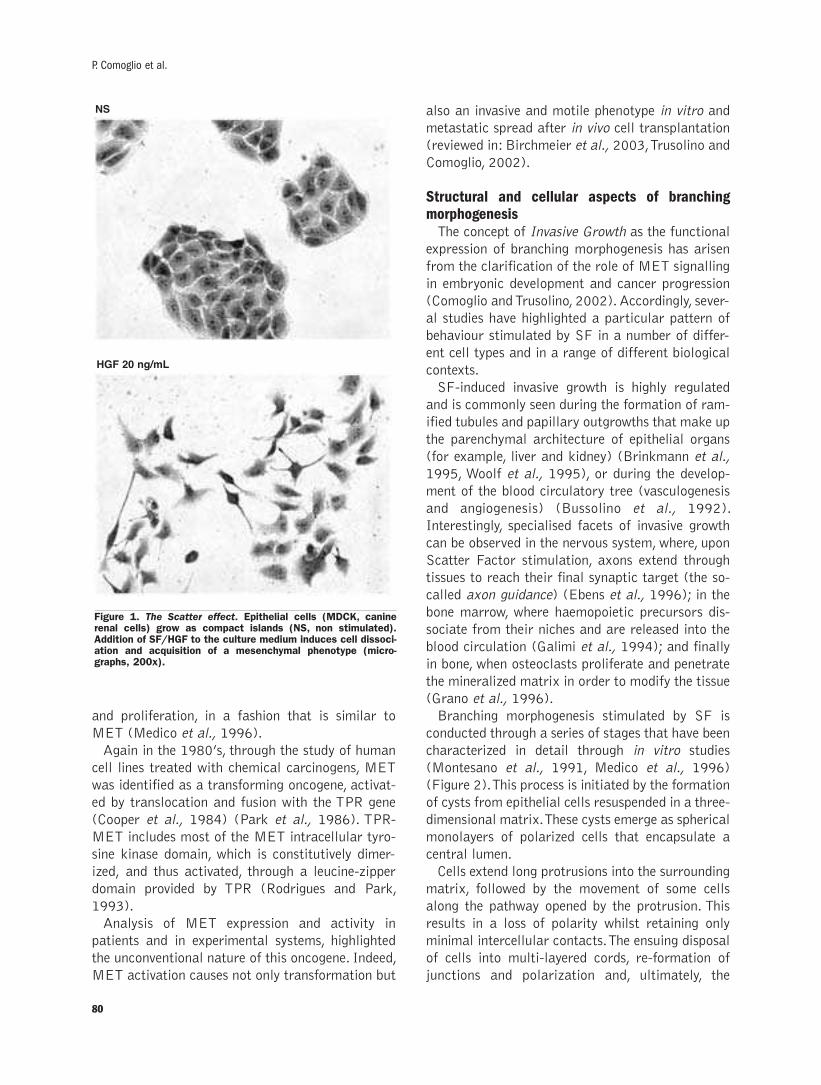
and proliferation, in a fashion that is similar to
MET (Medico et al., 1996).
Again in the 1980’s, through the study of human
cell lines treated with chemical carcinogens, MET
was identified as a transforming oncogene, activat-
ed by translocation and fusion with the TPR gene
(Cooper et al., 1984) (Park et al., 1986). TPR-
MET includes most of the MET intracellular tyro-
sine kinase domain, which is constitutively dimer-
ized, and thus activated, through a leucine-zipper
domain provided by TPR (Rodrigues and Park,
1993).
Analysis of MET expression and activity in
patients and in experimental systems, highlighted
the unconventional nature of this oncogene. Indeed,
MET activation causes not only transformation but
also an invasive and motile phenotype in vitro and
metastatic spread after in vivo cell transplantation
(reviewed in: Birchmeier et al., 2003, Trusolino and
Comoglio, 2002).
Structural and cellular aspects of branching morphogenesis The concept of Invasive Growth as the functional
expression of branching morphogenesis has arisen
from the clarification of the role of MET signalling
in embryonic development and cancer progression
(Comoglio and Trusolino, 2002). Accordingly, sever-
al studies have highlighted a particular pattern of
behaviour stimulated by SF in a number of differ-
ent cell types and in a range of different biological
contexts.
SF-induced invasive growth is highly regulated
and is commonly seen during the formation of ram-
ified tubules and papillary outgrowths that make up
the parenchymal architecture of epithelial organs
(for example, liver and kidney) (Brinkmann et al.,
1995, Woolf et al., 1995), or during the develop-
ment of the blood circulatory tree (vasculogenesis
and angiogenesis) (Bussolino et al., 1992).
Interestingly, specialised facets of invasive growth
can be observed in the nervous system, where, upon
Scatter Factor stimulation, axons extend through
tissues to reach their final synaptic target (the so-
called axon guidance) (Ebens et al., 1996); in the
bone marrow, where haemopoietic precursors dis-
sociate from their niches and are released into the
blood circulation (Galimi et al., 1994); and finally
in bone, when osteoclasts proliferate and penetrate
the mineralized matrix in order to modify the tissue
(Grano et al., 1996).
Branching morphogenesis stimulated by SF is
conducted through a series of stages that have been
characterized in detail through in vitro studies
(Montesano et al., 1991, Medico et al., 1996)
(Figure 2). This process is initiated by the formation
of cysts from epithelial cells resuspended in a three-
dimensional matrix. These cysts emerge as spherical
monolayers of polarized cells that encapsulate a
central lumen.
Cells extend long protrusions into the surrounding
matrix, followed by the movement of some cells
along the pathway opened by the protrusion. This
results in a loss of polarity whilst retaining only
minimal intercellular contacts. The ensuing disposal
of cells into multi-layered cords, re-formation of
junctions and polarization and, ultimately, the
80
P. Comoglio et al.
Figure 1. The Scatter effect. Epithelial cells (MDCK, caninerenal cells) grow as compact islands (NS, non stimulated).Addition of SF/HGF to the culture medium induces cell dissoci-ation and acquisition of a mesenchymal phenotype (micro-graphs, 200x).
NS
HGF 20 ng/mL
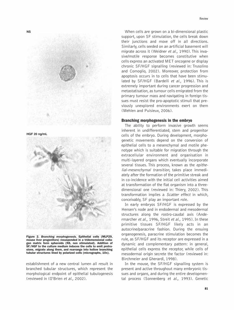
establishment of a new central lumen all result in
branched tubular structures, which represent the
morphological endpoint of epithelial tubulogenesis
(reviewed in (O'Brien et al., 2002).
When cells are grown on a bi-dimensional plastic
support, upon SF stimulation, the cells break down
their junctions and move off in all directions.
Similarly, cells seeded on an artificial basement will
migrate across it (Weidner et al., 1990). This inva-
sive/motile response becomes constitutive when
cells express an activated MET oncogene or display
chronic SF/HGF signalling (reviewed in: Trusolino
and Comoglio, 2002). Moreover, protection from
apoptosis occurs in to cells that have been stimu-
lated by SF/HGF (Bardelli et al., 1996). This is
extremely important during cancer progression and
metastatisation, as tumour cells emigrated from the
primary tumour mass and navigating in foreign tis-
sues must resist the pro-apoptotic stimuli that pre-
viously unexplored environments exert on them
(Mehlen and Puisieux, 2006).
Branching morphogenesis in the embryoThe ability to perform invasive growth seems
inherent in undifferentiated, stem and progenitor
cells of the embryo. During development, morpho-
genetic movements depend on the conversion of
epithelial cells to a mesenchymal and motile phe-
notype which is suitable for migration through the
extracellular environment and organisation in
multi-layered organs which eventually incorporate
several tissues. This process, known as the epithe-
lial-mesenchymal transition, takes place immedi-
ately after the formation of the primitive streak and
in co-incidence with the initial cell activities aimed
at transformation of the flat organism into a three-
dimensional one (reviewed in: Thiery, 2002). This
transformation implies a Scatter effect in which,
conceivably, SF play an important role.
In early embryos SF/HGF is expressed by the
Hensen’s node and in endodermal and mesodermal
structures along the rostro-caudal axis (Ande-
rmarcher et al., 1996, Streit et al., 1995). In these
primitive tissues SF/HGF likely acts in an
autocrine/paracrine fashion. During the ensuing
organogenesis, paracrine stimulation becomes the
rule, as SF/HGF and its receptor are expressed in a
dynamic and complementary pattern: in general,
epithelial cells express the receptor, while cells of
mesodermal origin secrete the factor (reviewed in:
Birchmeier and Gherardi, 1998).
In the mouse, the SF/HGF signalling system is
present and active throughout many embryonic tis-
sues and organs, and during the entire developmen-
tal process (Sonnenberg et al., 1993). Genetic
81
Review
Figure 2. Branching morphogenesis. Epithelial cells (MLP29,mouse liver progenitors) resuspended in a tridemensional colla-gen matrix form spheroids (NS, non stimulated). Addition ofSF/HGF to the culture medium induces the cells to emit protru-sions, migrate along them, and rearrange into hollow branchingtubular structures lined by polarized cells (micrographs, 10x).
HGF 20 ng/mL
NS
analysis of mice has shown that SF/HGF and its
receptor are an absolute requirement for the devel-
opment of specific organs. In knock-out mice abla-
tion of SF/HGF or MET is lethal to the embryo
resulting in impaired formation of the labyrinthine
layer of placenta, the liver, and the diaphragm and
limb muscles (Bladt et al., 1995, Maina et al.,
1996, Schmidt et al., 1995, Uehara et al., 1995).
Interestingly, SF/HGF is expressed throughout the
myoblast pathway and controls cell delamination
from somite-derived axial structures (dermomy-
tomes) and cell directional migration towards
peripheral limb buds (reviewed in: Birchmeier and
Gherardi, 1998, Birchmeier et al., 2003)
The structure of scatter factors and their receptorsMature, biologically active SF have an atypically
large size (94kDa). They consist of two disulphide-
linked chains (α and β). The α chain is character-
ized by the presence of an N-terminal hairpin loop,
followed by four Kringle Domains (80-amino acid
double-looped structures stabilized by internal
disulphide bridges). These Kringle Domains are a
common feature of plasminogen-related proteins
(Nakamura et al., 1989). The β chain is homolo-
gous to serine-proteases (like plasminogen and clot-
ting factors) but lacks proteolytic activity, owing to
substitution of three aminoacidic residues critical
for catalytic functions (Nakamura et al., 1989).
Thence, SF have an interesting relationship with
constituents of the blood clotting cascade as they
are philogenetically related to plasminogen, a cir-
culating proenzyme whose active form, known as
plasmin, is responsible for fibrinolysis (degradation
of blood clots).
SF are similar to coagulation proteins both in
their structure and in their mechanism of activa-
tion. Both SF/HGF and MSP are secreted as sin-
gle-chain inactive precursors (pro-HGF and pro-
MSP) and are activated by a proteolytic cleavage
which is performed by proteins also involved in clot-
ting regulation. The first enzyme to be shown as a
potent activator of pro-HGF was urokinase-type
plasminogen activator (uPA) (Naldini et al., 1992,
Mars et al., 1993). This was followed by evidence
that coagulation factor XII, thrombin and one ser-
ine-protease (XII- like factor) also function as
HGF convertases (Shimomura et al., 1993,
Shimomura et al., 1995).
SF/HGF binds heparin-sulphate proteoglycans,
which provide an extracellular reserve of the factor
in vivo and limit its diffusion through extracellular
fluids. This, in turn, promotes SF/HGF sequestration
on proximity to the site of synthesis and a
paracrine-like mode of activity (Hartmann et al.,
1998). Proteoglycans are not necessary for
SF/HGF binding to its receptor, however they cou-
ple SF/HGF in symmetrical dimers that simultane-
ously engage two receptor molecules, thus inducing
receptor dimerisation and trans-activation (Chir-
gadze et al., 1999, Schwall et al., 1996).
The SF/HGF receptor, encoded by MET, and the
MSP receptor, encoded by RON, sharing approxi-
mately 60% homology, are disulphide-linked α/βheterodimers that form by intracellular proteolytic
processing of a single-chain precursor. In both
receptors the α subunit is completely extracellular,
while the β subunit is a single-pass transmembrane
chain encompassing the tyrosine kinase activity. A
peculiar structural motif, the Sema Domain, char-
acterizes the extracellular region of SF receptors.
The Sema Domain contains over 500 amino acids,
inclusive of the full α chain (approximately 300
amino acids) and the amino-terminal moiety of the
β chain. In recent mutagenesis studies of MET, it
has been demostrated that the Sema Domain is
equipped with low-affinity ligand binding (Gherardi
et al., 2003).
In the extracellular portion there is also a cystein
rich region and a string of four immunoglobin-like
structures that are typical protein-protein interac-
tion domains.
The intracellular domain of the SF receptors is
composed of a tyrosine-kinase catalytic site sur-
rounded by juxtamembrane and carboxy-terminal
regulatory regions. Residues involved in receptor
downregulation are found in the juxtamembrane
domain. These include: (a) a serine residue (S985),
whose phosporylation inhibits tyrosine kinase activ-
ity; (b) a tyrosine residue (Y1003), which, upon
phosporylation, associates with the protein adaptor
CBL, which is an essential intermediary of MET
quantitative downregulation. Two separate recep-
tor-degradation pathways are switched on by CBL.
One of these is controlled by ubiquitination (CBL is
a E3 ubiquitin-ligase) whilst the other is controlled
by endocytosis (CBL recruits regulatory compo-
nents of endocytic vesicles) (reviewed in: Trusolino
and Comoglio, 2002).
In the MET carboxy-terminal domain there is a
conserved sequence (Y1349VHV---Y1356VNV) that
82
P. Comoglio et al.
includes two critical tyrosines for MET signalling.
Following phosphorylation, such tyrosines generate
a comprehensive docking site for signal transduc-
ers. In general, tyrosine kinase receptors use differ-
ent phosphotyrosines to engage distinct SH2-con-
taining signal effectors; in the case of MET, the
above mentioned sequence is capable of engaging
the complete spectrum of transducers that are nec-
essary for invasive growth, hence it is called multi-
functional docking site (Ponzetto et al., 1994).
Indeed, when the two tyrosines of the multi-func-
tional docking sites are replaced by phenylalanine
cells cease to respond to SF/HGF (Ponzetto et al.,
1994).
Genetic in vivo experiments have demonstrated
the importance of these residues for receptor bio-
logical functions. A transgenic mouse expressing a
receptor in which the two tyrosines of the multi-
functional docking site have been phenylalanine-
permutated exhibits a lethal phenotype akin to that
of the MET null-mouse (Maina et al., 1996).
Similarly, mutation of the same tyrosines annuls the
activated MET signalling and oncogenic properties
without affecting the tyrosine kinase function
(Bardelli et al., 1998). This indicates that the tyro-
sine kinase activity of MET is meaningful for sig-
nalling only when the receptor can properly associ-
ate to signal transducers. Therefore, the multi-func-
tional docking site is the force that drives the bio-
logical properties of the SF/HGF receptor, and is
likely to be responsible for its specific ability to
induce branching morphogenesis. Accordingly, when
inserted into the intracellular domain of other
receptor tyrosine kinases, the multi-functional
docking site bestows the ability to transduce the
branching morphogenic signals even to receptors
that usually induce only cell proliferation (Sachs et
al., 1996).
Signal transduction: from private adaptors to mul-tiple co-receptorsSeveral SH2-containing signal transducers can be
recruited by the multi-functional docking site at
high affinity. This ability, which is remarkable and
unique among tyrosine kinase receptors, is based on
the wide range specificity of the consensus sequence
formed by the two phosphorylated tyrosines (Y1349
and Y1356
) and the three aminoacids that follow each
tyrosine (Ponzetto et al., 1994). Thus, MET can
concurrently activate multiple signal transduction
pathways, including GRB2-RAS, phosphatidylinosi-
tol-3 kinase (PI3-K), SRC and signal transducer
and activator of transcription (STAT) (Boccaccio
et al., 1998, Graziani et al., 1991, Ponzetto et al.,
1994).
Activation of RAS plays a key role in the biolog-
ical activities induced by MET, affecting both cell
scattering and proliferation. When the RAS trans-
duction pathway is impaired by expression of a
dominant-negative RAS (Hartmann et al., 1994)
or by micro-injection of neutralizing antibodies, an
inhibition in the motile response to SF/HGF occurs.
Furthermore, direct activation of the RAS pathway
by MET, through association of the GRB2 adaptor
to the multi-functional docking site, is an important
determinant of cell proliferation and therefore also
of the MET oncogenic potential. In fact, mutagen-
esis of the MET multi-functional docking site that
selectively abrogates GRB2 recruitment, interrupts
the proliferative signals and abolishes the trans-
forming potential of the activated MET oncogene
(Ponzetto et al., 1996). However, the same MET
mutant retains an unaffected ability to induce cell
motility, showing that the threshold signal to
achieve this part of the invasive growth response is
nevertheless achieved (Ponzetto et al., 1996). As a
whole, activation of the RAS pathway by MET is an
absolute requirement for branching morphogenesis
(Fournier et al., 1996). Sustained activation of
ERK 1/2 MAP kinases, which are downstream
RAS effectors, has recently been highlighted as a
specific feature of morphogenesis (Boccaccio et al.,
2002, O'Brien et al., 2004). The ability to sustain
prolonged MAP kinase activation sets SF/HGF
apart from pure mitogens that evoke only a tran-
sient peak of MAP kinase activity (Marshall,
1995).
Another crucial signal transducer of MET is PI3-
K, which is activated by direct recruitment through
the multi-functional docking site or as a RAS effec-
tor (Graziani et al., 1991, Ponzetto et al., 1993).
PI3-K is essential for cell scatter in vitro, being
necessary for disassembly of intercellular contacts
{Potempa and Ridley, 1998, Royal, and Park, 1995}
and remodelling of adhesion to the extracellular
substrate (Trusolino et al., 2000). Signalling path-
ways downstream from this transducer are engaged
in either cytoskeletal reorganization and motility
(such as the small GTPase Rac and the protein
kinase PAK) (Royal et al., 1997), or in protection
from apoptosis (such as AKT) (Xiao et al., 2001).
A form of apoptosis, known as anoikis, can occur
83
Review

when a cell loses its normal adhesive contacts.
Therefore, protection from this form of apoptosis is
imperative when a cell detaches from its primary
site and travels through the extracellular matrix
(ECM). It is not surprising, then, that activation of
PI3-kinase by MET is required to accomplish the
process of branching morphogensis and for the
malignant counterpart of invasive growth, namely
cell invasion and metastasis (Bardelli et al., 1999,
Khwaja et al., 1998).
Branching morphogenesis is a complex differen-
tiative process that requires modulation of gene
expression. It has been found that epithelial tubulo-
genesis induced by SF/HGF requires the interven-
tion of STAT, an unconventional transcriptional fac-
tor that can be directly activated by MET through
an SH2 domain (Boccaccio et al., 1998). Also the
transcription factor NFkB, activated downstream
in a transduction cascade initiated by MET, is
required for branching morphogenesis (Muller et
al., 2002).
During the study of the complex signalling cas-
cades orchestrated by MET, an intracellular trans-
ducer was discovered, named GAB1. This is likely
to be a key co-ordinator of the cellular responses
to MET (Maroun et al., 1999, Weidner et al.,
1996). The significance of GAB1 for SF/HGF sig-
nalling is upheld by genetic evidence that GAB1
inactivation in mice phenocopies MET knock-out
animals (Sachs et al., 2000). GAB1 can be con-
sidered a scaffolding adaptor (such as the insulin
receptor substrate, IRS), which does not contain a
canonical phosphotyrosine interaction domain but
is however recruited directly and indirectly by acti-
vated tyrosine kinases (Holgado-Madruga et al.,
1996). However, GAB1 features an extended phos-
phorylation only in response to SF/HGF stimula-
tion, whereas a rapid and transitory phosphoryla-
tion is evoked by other growth factors. This may be
a result of the low-affinity but high avidity associ-
ation between MET and GABI through a specific
Met Binding Domain (Gual et al., 2000, Maroun et
al., 1999). Prolonged phosphorylation by MET
could explain the ability of GAB1 to support the
process of branching morphogenesis. In fact,
GAB1 may function as a signal sustainer that
enhances the recruitment and activation of trans-
ducers such as PLC-γ, the protein tyrosine phos-phatase SHP2, PI3-K and the adaptor CRK-like
(CRKL) (Gual et al., 2000, Holgado-Madruga et
al., 1996). In particular, Shp2 and CRKL may lead
to the sustained stimulation of the MAP kinase
pathway, a feature which is required for the initia-
tion and maintenance of branching morphogene-
sis(O'Brien et al., 2004).
The complexity of the signalling leading to
branching morphogenesis is further increased by
recent studies which involve the presence of cell-
surface molecules acting as MET co-receptors. The
scatter factor receptors (MET and RON) can be
activated by Plexins, when the latter are engaged by
their ligands Semaphorins (Conrotto et al., 2004,
Giordano et al., 2002).
Research has shown that within the physiological
range of ligand concentration the wild-type MET
receptor cannot efficiently engage signalling effec-
tors and mediate invasive growth without being
physically associated with the α6β4 integrin(Bertotti et al., 2005, Trusolino et al., 2001, and et
al., 2006). At the time of receptor activation by the
ligand, the associated integrin is phosphorylated on
tyrosines which provide supplementary docking
sites for signal transducers (such as PI3-K, the
Grb2 adaptor SHC, and the tyrosine phosphatase
Shp2). The fact that the integrin functions as an
adaptor contributes to the signalling strength and
duration that is a distinctive feature of the branch-
ing morphogenesis programme.
When associated with MET, α6β4 integrin elicitsintracellular signals independent of cell adhesion
and acts solely as a biological amplifier of SF/HGF
signalling (Trusolino et al., 1998). The integrin
requirement can be bypassed enforcing the sig-
nalling system by non-physiological high concentra-
tion of the ligand or by receptor over-expression
(Chung et al., 2004).
Besides α6β4 integrin, a variant of the CD44transmembrane molecule has been found to associ-
ate to MET and to be involved in its activation and
signalling (Orian-Rousseau et al., 2002). CD44 has
the ability to activate cell invasion but the mecha-
nism underlying this activity was unclear. CD44 is a
receptor for ECM components (such as hyaluronic
acid, integrins and osteopontin) and with its intra-
cellular moiety binds to the ezrin-radixin-moesin
(ERM) family of proteins. The latter family of pro-
teins control cell motility by mediating a mechani-
cally powerful association of the actin cytoskeleton
to the plasma membrane. Interestingly, ezrin has
been identified as an effector of SF/HGF-depen-
dent branching morphogenesis. In addition, deregu-
lated expression of ERM proteins has been impli-
84
P. Comoglio et al.
cated in cancer metastasis. Therefore, CD44 struc-
turally and functionally couples SF/HGF receptor
(and other tyrosine kinases) to the cytoskeleton and
to the extracellular environment, thence localizing
the motile activity of migrating cells to specific
membrane sites (reviewed in Ponta et al., 2003).
The genetic programme of branching morphogenesisFor branching morphogenesis to be accomplished,
transcriptional and post-transcriptional regulation
of gene expression is required, involving complex,
differential gene expression over time. The branch-
ing morphogenesis signature is currently being
investigated with the assistance of genomic and
proteomic technologies. One of the genes more
prominently upregulated by SF/HGF is osteopontin,
an extracellular matrix-associated protein that
binds integrins and CD44 cell-surface receptors
(Medico et al., 2001). It has been shown that
osteopontin induction is critical for the mor-
phogenic response to SF/HGF, and, interestingly,
that expression of this protein is also associated
with cancer invasive growth and metastasis
(Medico et al., 2001, Kang et al., 2003).
It is likely that genes crucial for branching mor-
phogenesis and invasive growth encode regulators
of cell adhesion and cell migration through foreign
environments.
In order to initiate ECM invasion epithelial cells
must first disassemble adhesive structures that link
them together and to the basement membrane.
Intercellular adhesion is chiefly mediated by
adherens junctions, where transmembrane E-cad-
herins are engaged in reciprocal homophilic recog-
nition with their extracellular domain. With their
intracellular moiety, E-cadherins bind to a sub-
membraneous scaffold of cortical catenins that
form a structural and functional bridge to the actin
cytoskeleton (reviewed in Nagafuchi, 2001). The
exact mechanism of adherens junction destabiliza-
tion is still to be unravelled but at present it is
known that it requires cadherin downregulation. In
this process, a critical mechanistic role is played by
Snail, a transcriptional repressor capable of switch-
ing off E-cadherin expression (Barrallo-Gimeno
and Nieto, 2005). Although early reports suggested
that the level of E-cadherin did not change during
SF/HGF-induced scatter, a reduced expression of
the cadherin protein in the same conditions has
been reported by other groups and attributed to
post-translational regulation (Khoury et al., 2005,
Miura et al., 2001, Tannapfel et al., 1994).
Recently it has been shown that SF/HGF induces
Snail activity, providing a mechanistic link with
transcriptional regulation of E-cadherins (Grotegut
et al., 2006). Other reported effects of SF/HGF on
E-cadherin include the redistribution of cadherin-
catenin complexes from the actin cytoskeleton to
the soluble membrane fraction (Balkovetz et al.,
1997, Balkovetz and Sambandam, 1999); the pro-
teolytic cleavage of cadherins from the cell surface;
and the destabilization of adherens junctions
through tyrosine phosphorylation of cadherins and
catenins (Herynk et al., 2003, Shibamoto et al.,
1994).
The branching morphogenesis programme
requires that, after mutual dissociation, epithelial
cells cross the natural borders made up by the base-
ment membrane to invade the surrounding stroma.
This suggests the ability to react with previously
unrecognised extracellular substrates through the
engagement of appropriate integrins but also high-
lights the need to degrade the ECM by means of
proteases. This is required so that a passage
through macromolecules and cells can be opened
and cryptic adhesion sites can be exposed.
Remodelling of integrin-mediated extracellular
adhesion supplies both mechanic support for cell
motility and an essential signal that protects cells
from anoikis (apoptosis), a process elicited by fail-
ure in recognizing the extracellular environment
(Frisch and Ruoslahti, 1997). SF/HGF controls the
expression and activity of the entire integrin group
thus allowing for recognition of the modified extra-
cellular environment during cell invasion through
multiple matrixes and tissue types, including base-
ment membrane, connectives and endothelia.
Indeed SF/HGF upregulates integrin transcription
by way of continual activation of MAP kinases, at
least in some cases (Liang and Chen, 2001, Nebe et
al., 1998), and stimulates integrin aggregation at
specific adhesive sites thereby increasing their avid-
ity for the substrate (Trusolino et al., 1998, 2000).
SF/ HGF is also capable of regulating the expres-
sion and activity of key actors in ECM modelling,
the matrix metalloproteases (MMPs, reviewed in
Egeblad and Werb, 2002). MMPs manage to local-
ize matrix digestion at the leading edge of migrating
cells by interacting with integrins and CD44 (see
above). SF/HGF has the ability to enhance the tran-
scription of several MMPs and can induce conver-
85
Review
sion of their inactive precursors into active enzymes
(Balkovetz et al., 2004, Rosenthal et al., 1998).
Somewhat unexpectedly though, recent findings
have shown that broad-spectrum MMP inhibition
does not hamper the initial phases of SF/HGF-trig-
gered branching morphogenesis, featuring epithelial
transition and matrix invasion. Instead, MMPs are
necessary for terminal completion of tubulogenesis
and reacquisition of the epithelial, polarized pheno-
type (O'Brien et al., 2004).
It could be argued that MMPs are not limiting
factors for cell invasion and that other ECM-
degrading proteases, such as urokinase-type plas-
minogen activator (uPA) (Jeffers et al., 1996,
Pepper et al., 1992), might have a predominant
role in the branching morphogenesis programme.
As previously mentioned, uPA is the main pro-HGF
convertase and binds with high affinity inactive pro-
HGF. Subsequently, by cleavage at a specific site,
uPA transforms pro-HGF into a biologically active
molecule (Naldini et al., 1992). Moreover, SF/HGF
can induce transcriptional upregulation of uPA
expression, possibly sustaining a positive feedback
on SF/HGF signalling (Boccaccio et al., 1994,
Pepper et al., 1992).
As an SF/HGF effector, uPA is responsible for
regulation of ECM degradation through conversion
of plasminogen into plasmin, an enzyme active on a
number of extracellular substrates. Plasmin prote-
olytic activity can be concentrated in proximity to
the cell membrane, as its activator uPA binds to a
cell surface receptor. Moreover the uPA receptor
has further roles in the branching morphogenesis
process as it also regulates cell adhesion (through
binding of ECM substrates and modulation of inte-
grin function), and evokes a signal transduction
cascade inside the cell (reviewed in Sidenius and
Blasi, 2003).
Lastly, SF/HGF utilises its pro-invasive capabili-
ties not only to induce migration and survival of
epithelial cells but also to modulate properties of
the stromal microenvironment. Named as landscap-
ing effects, these effects are indispensable for com-
plex organogenesis during development, and can
favour tumour growth and metastastization. The
best known of these effects is the induction of
angiogenesis through direct stimulation of endothe-
lial cells (Bussolino et al., 1992).
The control of MET expressionThe MET promoter positively responds to a num-
ber of mitogenic stimuli, including growth factors,
such as SF/HGF itself, tumour promoters (Boc-
caccio et al., 1994, Gambarotta et al., 1994) and
activated oncogenes (Ivan et al., 1997, Webb et al.,
1998). A prominent transcription factor for MET
upregulation is ETS/AP1. Remarkably, ETS is acti-
vated by MET itself through the MAP kinase path-
way and so offers an explanation as to why
SF/HGF can induce its own receptor (Gambarotta
et al., 1996, Paumelle et al., 2002). ETS concomi-
tantly controls transcription of several genes essen-
tial for ECM regulation and thus for branching
morphogenesis and invasive growth (Trojanowska,
2000).
A novel finding is that MET transcription is mod-
ulated by oxygen tension in tissues (Pennacchietti
et al., 2003), through a straightforward mechanism
playing a leading role in regulating embryonic
development and organ morphogenesis (Minet et
al., 2000). The cellular oxygen sensor, a protein
called Prolyl-hydroxylase, regulates the availability
of a transcriptional factor named hypoxia inducible
factor-1α or HIF-1α (Semenza, 2003). When oxy-
gen concentration lowers, for example as in tissues
lacking adequate vascularisation, the oxygen sensor
blocks the degradation of HIF-1α. As result, HIF-1α accumulates in the cell nucleus, and up-regu-
lates the transcription of various genes including
MET (Pennacchietti et al., 2003). In vitro experi-
ments have shown that hypoxia amplifies SF/HGF
signalling and synergizes with SF/HGF in branching
morphogenesis and invasive growth. MET over-
expression is an absolute requirement for branching
morphogenesis induced by hypoxia as shown by
experiments of MET specific inhibition through
RNA interference. Interestingly, analysis of human
tumours has indicated that MET expression occurs
at its highest level in hypoxic areas (in concomi-
tance with elevated HIF-1α expression), while it
decreases in proximity to blood vessels (where HIF-
1α is barely detectable) (Pennacchietti et al.,
2003).
Interestingly, a further support to the key role of
HIF-1α in MET transcriptional regulation derives
from the fact that MET is over-expressed in tumours
affected by inactivation of the Von Hippel-Lindau
(VHL) tumour suppressor gene (Maranchie et al.,
2002). The VHL protein interacts with the oxygen
sensor and targets HIF proteins for degradation in
case of normal oxygen concentration. Inactivation of
VHL prevents HIF-1α degradation even under nor-
86
P. Comoglio et al.

moxic conditions, resulting in elevated MET tran-
scription. In conclusion, hypoxia is a major driving
force of MET expression in vitro and in vivo.
Notably, this condition triggers not only expression
of MET, but also of u-PA receptor (see above)
(Rofstad et al., 2002), and of the chemokine recep-
tor CXCR4, which mobilizes normal stem cells and
cancer cells (thus favouring metastasis) towards tis-
sues that secrete the CXCR4-specific ligand SDF-1α(Muller et al., 2001).
MET and stem cellsMET is thus an inducible gene that is highly sen-
sitive to hypoxia and to extracellular stimuli con-
trolling cell proliferation. Hypoxia and growth fac-
tors are elements present in the stem cell niche. This
is the specific micro-environment responsible for
modulating the properties of stem cells, including
self-renewal, balance between symmetrical and
asymmetrical duplication and mobilization. Much
evidence suggests that in adult tissues MET expres-
sion may be restricted to the stem cell compart-
ment and to its immediate progeny of progenitor
and precursor cells. In the haemopoietic system,
MET has been found in a small fraction of bone
marrow cells, included in the subset expressing the
CD34 marker, corresponding to haemopoietic pro-
genitors and stem cells (Galimi et al., 1994). The
MET promoter invariably contains a binding ele-
ment for the GATA family of transcription factors
which are active in haemopoietic progenitors
(Gambarotta et al., 1994). MET, although
expressed at low levels also by mature hepatocytes,
is a hepatocyte stem cell marker, which can be used
to positively select progenitors from differentiated
cells with antibody-based cell sorting techniques
(Suzuki et al., 2002, Zheng and Taniguchi, 2003).
In skeletal muscle, MET is expressed by myoblasts
but it is downregulated at the time of differentia-
tion in striated fibres (Anastasi et al., 1997, Bladt
et al., 1995). Intriguingly, although not present in
differentiated myofibers, MET is highly expressed
in the skeletal muscle-derived tumours rhab-
domyosarcomas (Ferracini et al., 1996), indicating
that neoplastic cells have regained MET expression
or, more likely, that the tumour is derived from
transformation of myogenic precursors.
Interestingly, MET is a transcriptional target for
the β catenin/TCF transcription factor (Boon et al.,2002), which is activated by the Wnt signalling
pathway. This signalling cascade is physiologically
switched on in gut stem cells and switched off dur-
ing enterocyte differentiation. In the majority of
colon cancers, the same pathway is aberrantly and
constitutively operative; accordingly, MET is com-
monly found over-expressed in human colon carci-
nomas.
Based on all these assumptions, we can speculate
that invasive growth evoked by SF/HGF is a natu-
ral genetic programme for stem cells. Interestingly,
as stem cells multiply and circulate unrestrictedly
to target and reach various locations in the organ-
ism (Wright et al., 2001), they can be considered a
physiological counterpart of metastatic cells.
Therefore, the study of inappropriate activation of
the invasive growth programme in normal stem
cells can provide the key to understanding tumour
progression towards metastasis (Boccaccio and
Comoglio, 2006).
The role of the MET oncogene in tumourprogressionMET was originally identified as a transforming
oncogene generated by chromosomal translocation
in an osteosarcoma cell line treated with a chemi-
cal carcinogen (TPR-MET) (Cooper et al., 1984
Park et al., 1986). The same translocation product
has the ability to induce tumours in transgenic mice
(Boccaccio et al., 2005, Liang et al., 1996), and is
found in a small number of human gastric cancers
(Soman et al., 1990). However, activation of the
MET oncogene is mostly achieved by different
mechanisms in a large number of human tumours.
The following mechanisms are usually involved in
the constitutive activation of the MET tyrosine
kinase: (a) point mutations causing activatory con-
formational changes in the catalytic site; (b) ligand-
receptor autocrine circuits (which liberate cells
from the requirement of paracrine SF/HGF supply),
or increased paracrine stimulation; (c) MET over-
expression, which favours heightened sensitivity to
the factor or ligand-independent trans-activation.
Patient analysis has resulted in the strongest evi-
dence that MET has a causal role in human can-
cers. A group of patients all affected by papillary
renal carcinoma (HPRC), a hereditary form of can-
cer, showed germline missense mutations of MET
(Olivero et al., 1999, Schmidt et al., 1998, Wahl et
al., 1999). The same mutations (and others) have
also been found in non-hereditary tumours such as
sporadic papillary renal cancer (Schmidt et al.,
1999, Wahl et al., 1999) childhood hepatocellular
87
Review
carcinoma (Park et al., 1999), and gastric cancer
(Lee et al., 2000). Hereditary and sporadic papil-
lary renal cancer is usually an indolent neoplasm,
characterised by slow growth and local invasion
(Danilkovitch-Miagkova and Zbar, 2002). However,
somatic mutations of MET have been connected
with increased aggressiveness of hepatocellular
carcinoma and to the metastatic spread of head
and neck squamous carcinoma (Di Renzo et al.,
2000). Notably, in this latter type of cancer the
population of metastatic cells progressively enrich-
es in MET expression, as the tumour invades the
lymph-node stations stage by stage.
Intriguingly, in addition to genetic lesions, MET-
based tumorigenesis might require abnormal
SF/HGF stimulation (either through paracrine or
autocrine mechanisms), as implicated by the fact
that, in classical in vitro assays, cell transformation
by MET mutants is possible only in the presence of
its ligand, and is impaired when SF/HGF specific
inhibitors are present (Michieli et al., 1999).
A function for SF/HGF in maintaining MET-
induced transformation has been identified in
human tumours and assessed in mouse models. The
SF/HGF autocrine loops and/or enhanced
paracrine stimulation are observed in a wide range
of cancers in patients, including osteosarcoma
(Ferracini et al., 1995, Scotlandi et al., 1996), rab-
domyosarcoma (Ferracini et al., 1996, Scotlandi et
al., 1996), glioblastoma (Koochekpour et al.,
1997) and breast carcinoma (Tuck et al., 1996,
Yao et al., 1996). This excessive autocrine/
paracrine production of SF/HGF is often in associ-
ation with aggressive tumour behaviour.
Experimental induction of SF/MET autocrine loops
in cell lines has proven to cause formation of inva-
sive tumours after implantation in mice (Meiners et
al., 1998, Rong et al., 1994). Transgenic mice
expressing SF/HGF under a ubiquitous promoter
develop a wide spectrum of neoplasms of both
epithelial and mesenchymal origin (Takayama et
al., 1997). Among these tumours, melanomas
exhibit a significant correlation between high
metastatic potential and the presence of SF/MET
(Otsuka et al., 1998). In another mouse model, tar-
geted expression of SF/HGF to the mouse mamma-
ry epithelium establishes autocrine and paracrine
loops, sustaining formation of metastatic adenocar-
cinomas (Gallego et al., 2003).
The most frequent mechanism of MET oncogene
activation in human tumours is over-expression in
the absence of any mutation of the coding sequence.
Conceivably, over-expression is often due to hypoxia
(see above), which is a frequent condition of
tumours growing too rapidly to be adequately per-
fused by neo-angiogenic vessels. MET expression is,
again, in association with the metastatic phenotype
and with poor prognosis. For example, in colorectal
carcinoma MET over-expression provides a selec-
tive advantage that fosters the tumours ability to
produce lymph node and liver metastasis (Di Renzo
et al., 1995, Takeuchi et al., 2003). In animal mod-
els, it has been shown that forced expression of
wild-type MET in hepatocytes is sufficient to cause
hepato-carcinomas, which regress after transgene
inactivation (Wang et al., 2001). Conceivably,
increased expression of MET favours receptor
dimerization and thus ligand-independent activa-
tion. However, the environmental availability of
SF/HGF could always be mandatory. Therefore, in
vivo, the tumour stroma, which physiologically pro-
duces, stores and regulates the activation of
SF/HGF could have a critical landscaping role in
promoting MET-dependent tumour growth, either
in the presence of rare MET mutations, or in the
commonly occurring situation of MET over-expres-
sion.
AcknowledgementsWe thank Andrea Bertotti for micrographs,
Antonella Cignetto for secretarial assistance and
Catherine Tighe for editing the manuscript. Work in
the authors’ laboratory is supported by AIRC
(Associazione Italiana per la Ricerca sul Cancro),
MIUR (Ministero dell’Istruzione, Università e
Ricerca), Compagnia di San Paolo, and Fondazione
Cassa di Risparmio di Torino.
References
Anastasi S, Giordano S, Sthandier O, Gambarotta G, Maione R,Comoglio P, et al. A natural hepatocyte growth factor/scatter factorautocrine loop in myoblast cells and the effect of the constitutiveMet kinase activation on myogenic differentiation. J Cell Biol 1997;137:1057-68.
Andermarcher E, Surani MA, Gherardi E. Co-expression of theHGF/SF and c-met genes during early mouse embryogenesis pre-cedes reciprocal expression in adjacent tissues during organogenesis.Dev Genet 1996; 18:254-66.
Balkovetz DF, Gerrard ER, Jr., Li S, Johnson D, Lee J, Tobias JW,Rogers KK, Snyder RW, Lipschutz JH. Gene expression alterationsduring HGF-induced dedifferentiation of a renal tubular epithelialcell line (MDCK) using a novel canine DNA microarray. Am JPhysiol Renal Physiol 2004; 286:F702-F10.
Balkovetz DF, Pollack AL, Mostov KE. Hepatocyte growth factor altersthe polarity of Madin-Darby canine kidney cell monolayers. J BiolChem1997; 272:3471-7.
88
P. Comoglio et al.

Balkovetz DF, Sambandam V. Dynamics of E-cadherin and gamma-catenin complexes during dedifferentiation of polarized MDCK cells.Kidney Int 1999; 56:910-21.
Bardelli A, Basile ML, Audero E, Giordano S, Wennstrom S, MenardS, et al. Concomitant activation of pathways downstream of Grb2and PI 3-kinase is required for MET-mediated metastasis. Oncogene1999; 18:1139-46.
Bardelli A, Longati P, Albero D, Goruppi S, Schneider C, Ponzetto C, etal. HGF receptor associates with the anti-apoptotic protein BAG-1and prevents cell death. EMBO J 1996; 15:6205-12.
Bardelli A, Longati P, Gramaglia D, Basilico C, Tamagnone L, GiordanoS, et al. Uncoupling signal transducers from oncogenic METmutants abrogates cell transformation and inhibits invasive growth.Proc Natl Acad Sci SA 1998; 95:14379-383.
Barrallo-Gimeno A, Nieto MA. The Snail genes as inducers of cellmovement and survival: implications in development and cancer.Development 2005; 132:3151-61.
Bertotti A, Comoglio PM, Trusolino L. Beta4 integrin is a transformingmolecule that unleashes Met tyrosine kinase tumorigenesis. CancerRes 2005; 65:10674-9.
Bertotti A, Comoglio PM, Trusolino L. Beta4 integrin activates a Shp2-Src signaling pathway that sustains HGF-induced anchorage-inde-pendent growth. J Cell Biol 2006;175:993-1003.
Birchmeier C, Birchmeier W, Gherardi E, Vande Woude GF. Met, metas-tasis, motility and more. Nat Rev Mol Cell Biol 2003; 4:915-25.
Birchmeier C, Gherardi E. Developmental roles of HGF/SF and itsreceptor, the c-Met tyrosine kinase. Trends Cell Biol 1998; 8:404-10.
Bladt F, Riethmacher D, Isenmann S, Aguzzi A, Birchmeier C.Essential role for the c-met receptor in the migration of myogenicprecursor cells into the limb bud. Nature 1995; 376:768-71.
Boccaccio C, Ando' M, Comoglio PM. A differentiation switch forgenetically modified hepatocytes. FASEB J 2002; 16:120-2.
Boccaccio C, Ando M, Tamagnone L, Bardelli A, Michieli P, BattistiniC, Comoglio PM. Induction of epithelial tubules by growth factorHGF depends on the STAT pathway. Nature 1998; 391:285-8.
Boccaccio C, Gaudino G, Gambarotta G, Galimi F, Comoglio PM.Hepatocyte growth factor (HGF) receptor expression is inducibleand is part of the delayed-early response to HGF. J Biol Chem 1994;269:12846-51.
Boccaccio C, Sabatino G, Medico E, Girolami F, Follenzi A, Reato G,et al. The Met oncogene drives a genetic programme linking cancerto haemostasis. Nature 2005; 434:396-400.
Boccaccio , Comoglio PM. Invasive growth: a MET-driven genetic pro-gramme for cancer and stem cells. Nature Rev Cancer 2006; 6:637-45.
Boon EM, van der NR, van de WM, Clevers H, Pals ST. Wnt signalingregulates expression of the receptor tyrosine kinase met in colorec-tal cancer. Cancer Res 2002; 62:5126-8.
Bottaro DP, Rubin JS, Faletto DL, Chan AM, Kmiecik TE, Vande WG,Aaronson SA. Identification of the hepatocyte growth factor recep-tor as the c-met proto-oncogene product. Science 1991; 251:802-4.
Brinkmann V, Foroutan H, Sachs M, Weidner KM, Birchmeier W.Hepatocyte growth factor/scatter factor induces a variety of tissue-specific morphogenic programs in epithelial cells. J Cell Biol 1995;131:1573-86.
Bussolino F, Di Renzo MF, Ziche M, Bocchietto E, Olivero M, NaldiniL, et al. Hepatocyte growth factor is a potent angiogenic factorwhich stimulates endothelial cell motility and growth. J Cell Biol1992; 119:629-41.
Chirgadze DY, Hepple JP, Zhou H, Byrd RA, Blundell TL, Gherardi E.Crystal structure of the NK1 fragment of HGF/SF suggests a novelmode for growth factor dimerization and receptor binding. NatStruct Biol 1999; 6:72-9.
Chung J, Yoon SO, Lipscomb EA, Mercurio AM. The Met receptor andalpha 6 beta 4 integrin can function independently to promote car-cinoma invasion. J Biol Chem 2004; 279:32287-32293.
Comoglio PM, Trusolino L. Invasive growth: from development tometastasis. J Clin Invest 2002; 109:857-62.
Conrotto P, Corso S, Gamberini S, Comoglio PM, Giordano S. Interplaybetween scatter factor receptors and B plexins controls invasive
growth. Oncogene 2004; 23:5131-5137.
Cooper CS, Park M, Blair DG, Tainsky MA, Huebner K, Croce CM, etal. Molecular cloning of a new transforming gene from a chemical-ly transformed human cell line. Nature 1984; 311:29-33.
Danilkovitch-Miagkova A, Zbar B. Dysregulation of Met receptor tyro-sine kinase activity in invasive tumors. J Clin Invest 2002; 109:863-7.
Di Renzo MF, Olivero M, Giacomini A, Porte H, Chastre E, Mirossay L,et al. Overexpression and amplification of the met/HGF receptorgene during the progression of colorectal cancer. Clin Cancer Res1995; 1:147-54.
Di Renzo MF, Olivero M, Martone T, Maffe A, Maggiora P, Stefani AD,et al. Somatic mutations of the MET oncogene are selected duringmetastatic spread of human HNSC carcinomas. Oncogene 2000;19:1547-55.
Ebens A, Brose K, Leonardo ED, Hanson MG Jr., Bladt F, BirchmeierC, et al. Hepatocyte growth factor/scatter factor is an axonalchemoattractant and a neurotrophic factor for spinal motor neu-rons. Neuron 1996; 17:1157-72.
Egeblad M, Werb Z. New functions for the matrix metalloproteinasesin cancer progression. Nat Rev Cancer 2002; 2:161-74.
Ferracini R, Di Renzo MF, Scotlandi K, Baldini N, Olivero M, Lollini P,et al. The Met/HGF receptor is over-expressed in human osteosarco-mas and is activated by either a paracrine or an autocrine circuit.Oncogene 1995; 10:739-49.
Ferracini R, Olivero M, Di Renzo MF, Martano M, De Giovanni C, NanniP, et al. Retrogenic expression of the MET proto-oncogene corre-lates with the invasive phenotype of human rhabdomyosarcomas.Oncogene 1996; 12:1697-1705.
Fournier TM, Kamikura D, Teng K, Park M. Branching tubulogenesisbut not scatter of madin-darby canine kidney cells requires a func-tional Grb2 binding site in the Met receptor tyrosine kinase. J BiolChem 1996; 271:22211-7.
Frisch SM, Ruoslahti E. Integrins and anoikis. Curr Opin Cell Biol1997; 9:701-6.
Galimi F, Bagnara GP, Bonsi L, Cottone E, Follenzi A, Simeone A, etal. Hepatocyte growth factor induces proliferation and differentia-tion of multipotent and erythroid hemopoietic progenitors. J CellBiol 1994; 127:1743-54.
Gallego MI, Bierie B, Hennighausen L. Targeted expression of HGF/SFin mouse mammary epithelium leads to metastatic adenosquamouscarcinomas through the activation of multiple signal transductionpathways. Oncogene 2003; 22:8498-508.
Gambarotta G, Boccaccio C, Giordano S, Ando M, Stella MC,Comoglio PM. Ets up-regulates MET transcription. Oncogene 1996;13:1911-7.
Gambarotta G, Pistoi S, Giordano S, Comoglio PM, Santoro C.Structure and inducible regulation of the human MET promoter. JBiol Chem 1994; 269:12852-12857.
Gaudino G, Follenzi A, Naldini L, Collesi C, Santoro M, Gallo KA, et al.RON is a heterodimeric tyrosine kinase receptor activated by theHGF homologue MSP. EMBO J 1994; 13:3524-32.
Gherardi E, Youles ME, Miguel RN, Blundell TL, Iamele L, Gough J, etal. Functional map and domain structure of MET, the product of thec-met protooncogene and receptor for hepatocyte growthfactor/scatter factor. Proc Natl Acad Sci USA 2003; 100:12039-044.
Giordano S, Corso S, Conrotto P, Artigiani S, Gilestro G, Barberis D, etal. The semaphorin 4D receptor controls invasive growth by couplingwith Met. Nat Cell Biol 2002; 4:720-4.
Grano M, Galimi F, Zambonin G, Colucci S, Cottone E, Zallone AZ, etal. Hepatocyte growth factor is a coupling factor for osteoclasts andosteoblasts in vitro. Proc Natl Acad Sci USA 1996; 93:7644-8.
Graziani A, Gramaglia D, Cantley LC, Comoglio PM. The tyrosine-phosphorylated hepatocyte growth factor/scatter factor receptorassociates with phosphatidylinositol 3-kinase. J Biol Chem 1991;266:22087-90.
Grotegut S, von SD, Christofori G, Lehembre F. Hepatocyte growth fac-tor induces cell scattering through MAPK/Egr-1-mediated upregu-lation of Snail. EMBO J 2006; 25:3534-45.
Gual P, Giordano S, Williams TA, Rocchi S, Van Obberghen E, ComoglioPM. Sustained recruitment of phospholipase C-gamma to Gab1 isrequired for HGF-induced branching tubulogenesis. Oncogene 2000;
89
Review
19:1509-18.
Hartmann G, Prospero T, Brinkmann V, Ozcelik C, Winter G, Hepple J,et al. Engineered mutants of HGF/SF with reduced binding toheparan sulphate proteoglycans, decreased clearance and enhancedactivity in vivo. Curr Biol 1998; 8:125-34.
Hartmann G, Weidner KM, Schwarz H, Birchmeier W. The motility sig-nal of scatter factor/hepatocyte growth factor mediated through thereceptor tyrosine kinase met requires intracellular action of Ras. JBiol Chem 1994; 269:21936-9.
Herynk MH, Tsan R, Radinsky R, Gallick GE. Activation of c-Met incolorectal carcinoma cells leads to constitutive association of tyro-sine-phosphorylated beta-catenin. Clin Exp Metastasis 2003;20:291-300.
Holgado-Madruga M, Emlet DR, Moscatello DK, Godwin AK, WongAJ. A Grb2-associated docking protein in EGF- and insulin-receptorsignalling. Nature 1996; 379:560-4.
Huff JL, Jelinek MA, Borgman CA, Lansing TJ, Parsons JT. The pro-tooncogene c-sea encodes a transmembrane protein-tyrosine kinaserelated to the Met/hepatocyte growth factor/scatter factor receptor.Proc Natl Acad Sci USA 1993; 90:6140-4.
Ivan M, Bond JA, Prat M, Comoglio PM, Wynford-Thomas D.Activated ras and ret oncogenes induce over-expression of c-met(hepatocyte growth factor receptor) in human thyroid epithelialcells. Oncogene 1997; 14:2417-23.
Jeffers M, Rong S, Vande WG. Enhanced tumorigenicity and invasion-metastasis by hepatocyte growth factor/scatter factor-met signallingin human cells concomitant with induction of the urokinase proteol-ysis network. Mol Cell Biol 1996; 16:1115-25.
Kang Y, Siegel PM, Shu W, Drobnjak M, Kakonen SM, Cordon-CardoC, et al. A multigenic program mediating breast cancer metastasisto bone. Cancer Cell 2003; 3:537-49.
Khoury H, Naujokas MA, Zuo D, Sangwan V, Frigault MM, PetkiewiczS, et al. HGF converts ErbB2/Neu epithelial morphogenesis to cellinvasion. Mol Biol Cell 2005; 16:550-61.
Khwaja A, Lehmann K, Marte BM, Downward J. Phosphoinositide 3-kinase induces scattering and tubulogenesis in epithelial cellsthrough a novel pathway. J Biol Chem 1998; 273:18793-18801.
Koochekpour S, Jeffers M, Rulong S, Taylor G, Klineberg E, HudsonEA, et al. Met and hepatocyte growth factor/scatter factor expres-sion in human gliomas. Cancer Res 1997; 57:5391-8.
Lee JH, Han SU, Cho H, Jennings B, Gerrard B, Dean M, Set al. Anovel germ line juxtamembrane Met mutation in human gastric can-cer. Oncogene 2000; 19:4947-53.
Liang CC, Chen HC. Sustained activation of extracellular signal-regu-lated kinase stimulated by hepatocyte growth factor leads to integrinalpha 2 expression that is involved in cell scattering. J Biol Chem2001; 276:21146-52.
Liang TJ, Reid AE, Xavier R, Cardiff RD, Wang TC. Transgenic expres-sion of tpr-met oncogene leads to development of mammary hyper-plasia and tumors. J Clin Invest 1996; 97:2872-7.
Maina F, Casagranda F, Audero E, Simeone A, Comoglio PM, Klein R,Ponzetto C. Uncoupling of Grb2 from the Met receptor in vivoreveals complex roles in muscle development. Cell 1996; 87:531-42.
Maranchie JK, Vasselli JR, Riss J, Bonifacino JS, Linehan WM,Klausner RD. The contribution of VHL substrate binding and HIF1-alpha to the phenotype of VHL loss in renal cell carcinoma. CancerCell 2002; 1:247-55.
Maroun CR, Holgado-Madruga M, Royal I, Naujokas MA, FournierTM, Wong AJ, et al. The Gab1 PH domain is required for localiza-tion of Gab1 at sites of cell-cell contact and epithelial morphogene-sis downstream from the met receptor tyrosine kinase. Mol Cell Biol1999; 19:1784-99.
Mars WM, Zarnegar R, Michalopoulos GK. Activation of hepatocytegrowth factor by the plasminogen activators uPA and tPA. Am JPathol 1993; 143:949-58.
Marshall CJ. Specificity of receptor tyrosine kinase signaling: transientversus sustained extracellular signal-regulated kinase activation.Cell 1995; 80:179-85.
Medico E, Gentile A, Lo CC, Williams TA, Gambarotta G, Trusolino L,et al. Osteopontin is an autocrine mediator of hepatocyte growthfactor-induced invasive growth. Cancer Res 2001; 61:5861-8.
Medico E, Mongiovi AM, Huff J, Jelinek MA, Follenzi A, Gaudino G, et
al. The tyrosine kinase receptors Ron and Sea control "scattering"and morphogenesis of liver progenitor cells in vitro. Mol Biol Cell1996; 7:495-504.
Mehlen P, Puisieux A. Metastasis: a question of life or death. Nat RevCancer 2006; 6:449-58.
Meiners S, Brinkmann V, Naundorf H, Birchmeier W. Role of morpho-genetic factors in metastasis of mammary carcinoma cells.Oncogene 1998; 16:9-20.
Michieli P, Basilico C, Pennacchietti S, Maffe A, Tamagnone L,Giordano S, et al. Mutant Met-mediated transformation is ligand-dependent and can be inhibited by HGF antagonists. Oncogene1999; 18:5221-31.
Michieli P, Mazzone M, Basilico C, Cavassa S, Sottile A, Naldini L, etal. Targeting the tumor and its microenvironment by a dual-functiondecoy Met receptor. Cancer Cell 2004; 6:61-73.
Minet E, Michel G, Remacle J, Michiels C. Role of HIF-1 as a tran-scription factor involved in embryonic development, cancer progres-sion and apoptosis (review). Int J Mol Med 2000; 5:253-9.
Miura H, Nishimura K, Tsujimura A, Matsumiya K, Matsumoto K,Nakamura T, et al. Effects of hepatocyte growth factor on E-cad-herin-mediated cell-cell adhesion in DU145 prostate cancer cells.Urology 2001; 58:1064-9.
Montesano R, Matsumoto K, Nakamura T, Orci L. Identification of afibroblast-derived epithelial morphogen as hepatocyte growth factor.Cell 1991; 67:901-8.
Muller A, Homey B, Soto H, Ge N, Catron D, Buchanan ME, et al.Involvement of chemokine receptors in breast cancer metastasis.Nature 2001; 410:50-6.
Muller M, Morotti A, Ponzetto C. Activation of NF-kappaB is essen-tial for hepatocyte growth factor-mediated proliferation and tubulo-genesis. Mol Cell Biol 2002; 22:1060-72.
Nagafuchi A. Molecular architecture of adherens junctions. Curr OpinCell Biol 2001; 13:600-3.
Nakamura T, Nishizawa T, Hagiya M, Seki T, Shimonishi M, SugimuraA, Tashiro K, Shimizu S. Molecular cloning and expression of humanhepatocyte growth factor. Nature 1989; 342:440-3.
Nakamura T, Teramoto H, Ichihara A. Purification and characteriza-tion of a growth factor from rat platelets for mature parenchymalhepatocytes in primary cultures. Proc Natl Acad Sci USA 1986;83:6489-93.
Naldini L, Tamagnone L, Vigna E, Sachs M, Hartmann G, BirchmeierW, et al. Extracellular proteolytic cleavage by urokinase is requiredfor activation of hepatocyte growth factor/scatter factor. EMBO J1992; 11:4825-33.
Naldini L, Weidner KM, Vigna E, Gaudino G, Bardelli A, Ponzetto C, etal. Scatter factor and hepatocyte growth factor are indistinguishableligands for the MET receptor. EMBO J 1991; 10:2867-78.
Nebe B, Sanftleben H, Pommerenke H, Peters A, Rychly J. Hepatocytegrowth factor enables enhanced integrin-cytoskeleton linkage byaffecting integrin expression in subconfluent epithelial cells. Exp CellRes 1998; 243:263-73.
O'Brien LE, Tang K, Kats ES, Schutz-Geschwender A, Lipschutz JH,Mostov KE. ERK and MMPs sequentially regulate distinct stages ofepithelial tubule development. Dev Cell 2004; 7:21-32.
O'Brien LE, Zegers MM, Mostov KE. Opinion: Building epithelial archi-tecture: insights from three-dimensional culture models. Nat RevMol Cell Biol 2002; 3:531-7.
Olivero M, Valente G, Bardelli A, Longati P, Ferrero N, Cracco C, et al.Novel mutation in the ATP-binding site of the MET oncogene tyro-sine kinase in a HPRCC family. Int J Cancer 1999; 82:640-3.
Orian-Rousseau V, Chen L, Sleeman JP, Herrlich P, Ponta H. CD44 isrequired for two consecutive steps in HGF/c-Met signaling. GenesDev 2002; 16:3074-86.
Otsuka T, Takayama H, Sharp R, Celli G, LaRochelle WJ, Bottaro DP,et al. c-Met autocrine activation induces development of malignantmelanoma and acquisition of the metastatic phenotype. Cancer Res1998; 58:5157-67.
Park M, Dean M, Cooper CS, Schmidt M, O'Brien SJ, Blair DG,Vande WG. Mechanism of met oncogene activation. Cell 1986;45:895-904.
Park WS, Dong SM, Kim SY, Na EY, Shin MS, Pi JH, et al. Somaticmutations in the kinase domain of the Met/hepatocyte growth factor
90
P. Comoglio et al.

receptor gene in childhood hepatocellular carcinomas. Cancer Res1999; 59:307-10.
Paumelle R, Tulasne D, Kherrouche Z, Plaza S, Leroy C, Reveneau S,et al. Hepatocyte growth factor/scatter factor activates the ETS1transcription factor by a RAS-RAF-MEK-ERK signaling pathway.Oncogene 2002; 21:2309-19.
Pennacchietti S, Michieli P, Galluzzo M, Mazzone M, Giordano S,Comoglio PM. Hypoxia promotes invasive growth by transcriptionalactivation of the met protooncogene. Cancer Cell 2003; 3:347-61.
Pepper MS, Matsumoto K, Nakamura T, Orci L, Montesano R.Hepatocyte growth factor increases urokinase-type plasminogenactivator (u-PA) and u-PA receptor expression in Madin-Darbycanine kidney epithelial cells. Journal of Biological Chemistry 1992;267:20493-6.
Ponta H, Sherman L, Herrlich, PA. CD44: From adhesion molecules tosignalling regulators. Nat. Rev. Cell Mol. Biol. 2003; 1:33-45.
Ponzetto C, Bardelli A, Maina F, Longati P, Panayotou G, Dhand R,Waterfield MD, Comoglio PM. A novel recognition motif for phos-phatidylinositol 3-kinase binding mediates its association with thehepatocyte growth factor/scatter factor receptor. Mol Cell Biol1993; 13:4600-8.
Ponzetto C, Bardelli A, Zhen Z, Maina F, dalla ZP, Giordano S, et al. Amultifunctional docking site mediates signaling and transformationby the hepatocyte growth factor/scatter factor receptor family. Cell1994; 77:261-71.
Ponzetto C, Zhen Z, Audero E, Maina F, Bardelli A, Basile ML, et al.Specific uncoupling of GRB2 from the Met receptor. Differentialeffects on transformation and motility. J Biol Chem 1996;271:14119-23.
Potempa S, Ridley AJ. Activation of both MAP kinase and phos-phatidylinositide 3-kinase by ras is required for hepatocyte growthFactor/Scatter factor-induced adherens junction disassembly. MolBiol Cell 1998; 9:2185-200.
Rodrigues GA, Park M. Dimerization mediated through a leucine zip-per activates the oncogenic potential of the met receptor tyrosinekinase. Mol Cell Biol 1993; 13:6711-22.
Rofstad EK, Rasmussen H, Galappathi K, Mathiesen B, Nilsen K, GraffBA. Hypoxia promotes lymph node metastasis in human melanomaxenografts by up-regulating the urokinase-type plasminogen activa-tor receptor. Cancer Res 2002; 62:1847-53.
Rong S, Segal S, Anver M, Resau JH, Vande Woude GF. Invasivenessand metastasis of NIH 3T3 cells induced by Met-hepatocyte growthfactor/scatter factor autocrine stimulation. Proc Natl Acad Sci USA1994; 91:4731-5.
Rosenthal EL, Johnson TM, Allen ED, Apel IJ, Punturieri A, Weiss SJ.Role of the plasminogen activator and matrix metalloproteinase sys-tems in epidermal growth factor- and scatter factor-stimulated inva-sion of carcinoma cells. Cancer Res 1998; 58:5221-30.
Royal I, Fournier TM, Park M. Differential requirement of Grb2 andPI3-kinase in HGF/SF-induced cell motility and tubulogenesis. JCell Physiol 1997; 173:196-201.
Royal I, Park M. Hepatocyte growth factor-induced scatter of Madin-Darby canine kidney cells requires phosphatidylinositol 3-kinase. JBiol Chem 1995; 270:27780-7.
Sachs M, Brohmann H, Zechner D, Muller T, Hulsken J, Walther I, etal. Essential role of Gab1 for signaling by the c-Met receptor in vivo.J Cell Biol 2000; 150:1375-84.
Sachs M, Weidner KM, Brinkmann V, Walther I, Obermeier A, UllrichA, Birchmeier W. Motogenic and morphogenic activity of epithelialreceptor tyrosine kinases. J Cell Biol 1996; 133:1095-107.
Schmidt C, Bladt F, Goedecke S, Brinkmann V, Zschiesche W, SharpeM, et al. Scatter factor/hepatocyte growth factor is essential forliver development. Nature 1995; 373:699-702.
Schmidt L, Junker K, Nakaigawa N, Kinjerski T, Weirich G, Miller M,et al. Novel mutations of the MET proto-oncogene in papillary renalcarcinomas. Oncogene 1999; 18:2343-50.
Schmidt L, Junker K, Weirich G, Glenn G, Choyke P, Lubensky I, et al.Two North American families with hereditary papillary renal carci-noma and identical novel mutations in the MET proto-oncogene.Cancer Res 1998; 58:1719-22.
Schwall RH, Chang LY, Godowski PJ, Kahn DW, Hillan KJ, Bauer KD,et al. Heparin induces dimerization and confers proliferative activityonto the hepatocyte growth factor antagonists NK1 and NK2. J Cell
Biol 1996; 133:709-18.
Scotlandi K, Baldini N, Oliviero M, Di Renzo MF, Martano M, Serra M,et al. Expression of Met/hepatocyte growth factor receptor gene andmalignant behavior of musculoskeletal tumors. Am J Pathol 1996;149:1209-19.
Semenza GL. Targeting HIF-1 for cancer therapy. Nat Rev Cancer2003; 3:721-32.
Shibamoto S, Hayakawa M, Takeuchi K, Hori T, Oku N, Miyazawa K,et al. Tyrosine phosphorylation of beta-catenin and plakoglobinenhanced by hepatocyte growth factor and epidermal growth factorin human carcinoma cells. Cell Adhes Commun 1994; 1:295-305.
Shimomura T, Kondo J, Ochiai M, Naka D, Miyazawa K, Morimoto Y,et al. Activation of the zymogen of hepatocyte growth factor activa-tor by thrombin. J Biol Chem 1993; 268:22927-32.
Shimomura T, Miyazawa K, Komiyama Y, Hiraoka H, Naka D,Morimoto Y, et al. Activation of hepatocyte growth factor by twohomologous proteases, blood-coagulation factor XIIa and hepato-cyte growth factor activator. Europ J Biochem 1995; 229:257-61.
Sidenius N, Blasi F. The urokinase plasminogen activator system incancer: recent advances and implication for prognosis and therapy.Cancer Metastasis Rev 2003; 22:205-22.
Soman NR, Wogan GN, Rhim JS. TPR-MET oncogenic rearrangement:detection by polymerase chain reaction amplification of the tran-script and expression in human tumor cell lines. Proc Natl Acad SciUSA 1990; 87:738-42.
Sonnenberg E, Meyer D, Weidner KM, Birchmeier C. Scatterfactor/hepatocyte growth factor and its receptor, the c-met tyrosinekinase, can mediate a signal exchange between mesenchyme andepithelia during mouse development. J Cell Biol 1993; 123:223-35.
Stoker M, Gherardi E, Perryman M, Gray J. Scatter factor is a fibrob-last-derived modulator of epithelial cell mobility. Nature 1987;327:239-42.
Streit A, Stern CD, Thery C, Ireland GW, Aparicio S, Sharpe MJ,Gherardi E. A role for HGF/SF in neural induction and its expres-sion in Hensen's node during gastrulation. Development 1995;121:813-24.
Suzuki A, Zheng Yw YW, Kaneko S, Onodera M, Fukao K, Nakauchi H,et al. Clonal identification and characterization of self-renewingpluripotent stem cells in the developing liver. J Cell Biol 2002;156:173-84.
Takayama H, LaRochelle WJ, Sharp R, Otsuka T, Kriebel P, Anver M,et al. Diverse tumorigenesis associated with aberrant development inmice overexpressing hepatocyte growth factor/scatter factor. ProcNatl Acad Sci U A 1997; 94:701-706.
Takeuchi H, Bilchik A, Saha S, Turner R, Wiese D, Tanaka M, et al. c-MET expression level in primary colon cancer: a predictor of tumorinvasion and lymph node metastases. Clin Cancer Res 2003;9:1480-8.
Tannapfel A, Yasui W, Yokozaki H, Wittekind C, Tahara E. Effect ofhepatocyte growth factor on the expression of E- and P-cadherin ingastric carcinoma cell lines. Virchows Arch 1994; 425:139-44.
Thiery JP. Epithelial-mesenchymal transitions in tumour progression.Nat Rev Cancer 2002; 2:442-54.
Trojanowska M. Ets factors and regulation of the extracellular matrix.Oncogene 2000; 19:6464-71.
Trusolino L, Bertotti A, Comoglio PM. A signaling adapter function foralpha6beta4 integrin in the control of HGF-dependent invasivegrowth. Cell 2001; 107:643-54.
Trusolino L, Cavassa S, Angelini P, Bertotti A, Comoglio PM,Boccaccio C. HGF/scatter factor selectively promotes cell invasionby increasing integrin avidity. FASEB J 2000; 14:1629-40.
Trusolino L, Comoglio PM. Scatter-factor and semaphorin receptors:cell signalling for invasive growth. Nat Rev Cancer 2002; 4:289-300.
Trusolino L, Serini G, Cecchini G, Besati C, Ambesi-Impiombato FS,Marchisio PC, De Filippi R. Growth factor-dependent activation ofalphavbeta3 integrin in normal epithelial cells: implications fortumor invasion. J Cell Biol 1998; 142:1145-56.
Tuck AB, Park M, Sterns EE, Boag A, Elliott BE. Coexpression ofhepatocyte growth factor and receptor (Met) in human breast car-cinoma. Am J Pathol 1996; 148:225-32.
Uehara Y, Minowa O, Mori C, Shiota K, Kuno J, Noda T, Kitamura N.
91
Review
Placental defect and embryonic lethality in mice lacking hepatocytegrowth factor/scatter factor. Nature 1995; 373:702-5.
Wahl RC, Hsu RY, Huff JL, Jelinek MA, Chen K, Courchesne P, et al.Chicken macrophage stimulating protein is a ligand of the receptorprotein-tyrosine kinase Sea. J Biol Chem 1999; 274:26361-8.
Wang R, Ferrell LD, Faouzi S, Maher JJ, Bishop JM. Activation of theMet receptor by cell attachment induces and sustains hepatocellularcarcinomas in transgenic mice. J Cell Biol 2001; 153:1023-34.
Webb CP, Taylor GA, Jeffers M, Fiscella M, Oskarsson M, Resau JH, etal. Evidence for a role of Met-HGF/SF during Ras-mediated tumori-genesis/metastasis. Oncogene 1998; 17:2019-25.
Weidner KM, Behrens J, Vandekerckhove J, Birchmeier W. Scatter fac-tor: molecular characteristics and effect on the invasiveness ofepithelial cells. J Cell Biol 1990; 111:2097-108.
Weidner KM, Di Cesare S, Sachs M, Brinkmann V, Behrens J,Birchmeier W. Interaction between Gab1 and the c-Met receptortyrosine kinase is responsible for epithelial morphogenesis. Nature1996; 384:173-6.
Woolf AS, Kolatsi-Joannou M, Hardman P, Andermarcher E, Moorby
C, Fine LG, et al. Roles of hepatocyte growth factor/scatter factorand the met receptor in the early development of the metanephros.J Cell Biol1995; 128:171-84.
Wright DE, Wagers AJ, Gulati AP, Johnson FL, Weissman IL.Physiological migration of hematopoietic stem and progenitor cells.Science 2001; 294:1933-6.
Xiao GH, Jeffers M, Bellacosa A, Mitsuuchi Y, Vande WG, Testa JR.Anti-apoptotic signaling by hepatocyte growth factor/Met via thephosphatidylinositol 3-kinase/Akt and mitogen-activated proteinkinase pathways. Proc Natl Acad Sci USA 2001; 98:247-52.
Yao Y, Jin L, Fuchs A, Joseph A, Hastings HM, Goldberg ID, et al.Scatter factor protein levels in human breast cancers: clinicopatho-logical and biological correlations. Am J Pathol 1996; 149:1707-17.
Zarnegar R, Michalopoulos G. Purification and biological characteri-zation of human hepatopoietin A, a polypeptide growth factor forhepatocytes. Cancer Res 1989; 49:3314-20.
Zheng YW, Taniguchi H. Diversity of hepatic stem cells in the fetal andadult liver. Semin Liver Dis 2003; 23:337-48.
92
P. Comoglio et al.
93
©2007, European Journal of Histochemistry
Epithelial tissues emerge from coordinated sequences of cellrenewal, specialization and assembly. Like corresponding imma-ture tissues, adult epithelial tissues are provided by stem cellswhich are responsible for tissue homeostasis. Advances inepithelial histogenesis has permitted to clarify several aspectsrelated to stem cell identification and dynamics and to under-stand how stem cells interact with their environment, the so-called stem cell niche. The development and maintenance ofepithelial tissues involves epithelial-mesenchymal signallingpathways and cell-matrix interactions which control target nuclearfactors and genes. The tooth germ is a prototype for such induc-tive tissue interactions and provides a powerful experimental sys-tem for the study of genetic pathways during development.Clonogenic epithelial cells isolated from developing as wellmature epithelial tissues has been used to engineer epithelial tis-sue-equivalents, e.g. epidermal constructs, that are used in clin-ical practise and biomedical research. Information on molecularmechanisms which regulate epithelial histogenesis, including therole of specific growth/differentiation factors and cognate recep-tors, is essential to improve epithelial tissue engineering.
Key words: epithelial histogenesis – developmental biology –stem cell – differentiation – epithelial-mesenchymal interac-tions – tissue engineering.
Correspondence: Casasco Andrea,Department of Experimental MedicineHistology and Embryology UnitVia Forlanini, 10 27100 PAVIA, ItalyTel: +39.0382.987272.Fax: +39.0382.528330.E-mail: [email protected]
European Journal of Histochemistry2007; vol. 51 supplement 1:93-100
Models of epithelial histogenesis
A. Casasco, M. Casasco, A. Icaro Cornaglia, F. Riva, A. Calligaro
Department of Experimental Medicine, Histology and Embryology Unit, University of Pavia, Pavia Italy
Epithelial histogenesis: a chance to unveil theface of human stem cellsAlthough epithelial cells are characterized by
common structural features, especially their
arrangement into cohesive sheets or thee-dimen-
sional aggregates, they provide an enormous variety
of biological functions, including protection, absorp-
tion, secretion, gametogenesis and special senses.
A major difference between developing and
mature epithelial tissues is that differentiated
epithelial cells that are not prone to locomotion. On
the other hand, the migration of immature epithelial
cells is essential for the development of many
organs, including most of exocrine and endocrine
glands, skin appendages and teeth. However, most of
adult epithelial tissues are dynamic as to cell renew-
al and cell cycle activity. In fact, many processes
that are observed during development are operating
also in mature state, thus ensuring tissue homeosta-
sis. Accordingly, most – if not all - of adult epithe-
lial tissues are provided by specific stem cells and
may be regarded to as useful models to study tissue
formation and repair. The occurrence of stem cells in
adult epithelial tissues has permitted the generation
of bioengineered epithelial constructs that can be
applied in cell and tissue therapy.
Epithelial stem cells, like other adult stem cells,
are thought to be slow- or rarely-cycling cells,
which retain clonogenicity and proliferative capac-
ity for a long time. According to current models of
tissue homeostasis, the division of a stem cell gives
rise to another stem cell and one transient amplify-
ing cell. Such a cell, after exhausting its prolifera-
tive potential, undergoes terminal differentiation,
thus generating functional cells which are not fur-
ther capable of proliferation (Leblond, 1981;
Potten, 1983; Fuchs, 1990; Potten and Loeffler,
1990; Jones et al., 1995). Since stem cells are slow
cycling, they minimise DNA replication-related
errors. Stemness properties are greatly conditioned
by the microenvironment and positional creden-
REVIEW
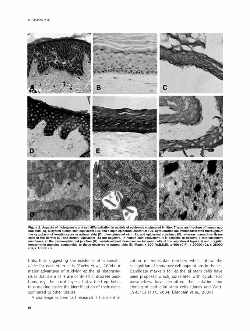
94
tials, thus suggesting the existence of a specific
niche for each stem cells (Fuchs et al., 2004). A
major advantage of studying epithelial histogene-
sis is that stem cells are confined in discrete posi-
tions, e.g. the basal layer of stratified epithelia,
thus making easier the identification of their niche
compared to other tissues.
A challenge in stem cell research is the identifi-
cation of molecular markers which allow the
recognition of immature cell populations in tissues.
Candidate markers for epithelial stem cells have
been proposed which, correlated with cytokinetic
parameters, have permitted the isolation and
cloning of epithelial stem cells (Jones and Watt,
1993; Li et al., 2004; Blanpain et al., 2004).
A. Casasco et al.
Figure 1. Aspects of histogenesis and cell differentiation in models of epidermis engineered in vitro. Tissue architecture of human nat-ural skin (A), bilayered human skin equivalent (B), and simple epidermal construct (C). Cytokeratins are immunodetected thoroughoutthe cytoplasm of keratinocytes in natural skin (D), bioengineered skin (E), and epidermal construct (F), whereas connective tissuecells in the dermis (D) and dermal equivalent (E) are negative. In human skin equivalent, it is possible to observe a thin basementmembrane at the dermo-epidermal junction (G), well-developed desmosomes between cells of the suprabasal layer (H) and irregularkeratohyalin granules comparable to those observed in natural skin (I). Magn. x 300 (A,B,D,E), x 400 (C,F), x 20000 (G), x 26000(H), x 18000 (I).

95
Review
Enamel epithelium: the histogenesis of the hardest tissueThe tooth germ represents a powerful model to
understand molecular mechanisms of organogen-
esis which are mediated by epithelial-mesenchy-
mal interactions. In fact, during odontogenesis, it
is possible to monitor continuously cell differenti-
ation in relative spatial positions, the production
and secretion of specific molecules, and corre-
sponding modifications of the extracellular
matrix.
Mammalian teeth develop from two types of
cells: stomodeal ectoderm cells, which form
ameloblasts, and cranial neural-crest derived
ectomesenchyme cells, which form pulp cells,
odontoblasts and cementoblasts (reviewed by
Sharpe, 2001; Cobourne and Sharpe, 2003).
These two cells types interact to control the entire
process of tooth initiation, morphogenesis and
cytodifferentiation.
Epithelial-mesenchymal interactions that regu-
late the initiation of tooth formation, the differen-
tiation of odontoblasts and ameloblasts and the
acquisition of shape have been characterized by
studies on tissue recombination (Kollar and Baird,
1968; Lumsden, 1988).
Furthermore, cell-to-cell and cell-matrix sig-
nalling pathways and related target nuclear fac-
tors have been identified as mediators of recipro-
cal communication between dental epithelial and
mesenchymal cells (reviewed by Ruch, 1985;
Slavkin, 1990; Jernval and Thesleff, 2000;
Thesleff and Mikkola, 2002; Thesleff, 2003).
Tooth morphogenesis proceeds through charac-
teristic stages, i.e. initiation, bud, cap and bell
stages. As in many organs, the earliest evidence of
tooth development is an epithelial thickening of
the stomodeal lining epithelium. Under the
instructive influence of the odontogenic mes-
enchyme, the inner enamel epithelium undergoes a
precise developmental program, ultimately differ-
entiates to the ameloblast phenotype and initiate
the expression of tissue-specific enamel gene prod-
ucts which direct enamel biomineralization (Ruch,
1985; Jernval and Thesleff, 2000; Thesleff, 2003).
The differentiation programme of the cells of the
inner enamel epithelium can be summarized in
three main phases, including pre-secretory, secre-
tory and maturation phases (Warshawsky and
Smith, 1974; Smith and Warshawsky, 1975;
Nanci et al., 1985; 1987; 1998). During pre-
secretory stage, the cells of the inner enamel
epithelium proliferate and acquire terminal cytod-
ifferentiation; during the secretory stage, differen-
tiated cells, which can be properly called
ameloblasts, become functional and secrete spe-
cific enamel matrix components; in the matura-
tion stage, ameloblasts are involved in the pro-
cessing of enamel matrix which will result in the
formation of the hardest tissue of human body. As
in a romantic drama, ameloblasts, which are
located at the surface of the tooth crown and have
fulfilled their task, will die as soon as tooth erupts,
after which time the enamel cannot be replaced by
new synthesis.
The interaction of a cell with the surrounding
extracellular matrix influences cell proliferation
and differentiation gene expression via specific
membrane receptors which activate downstream
target genes. Much interest has been given to the
phases of odontoblast and ameloblast differentia-
tion which immediately precede the secretion of
specific dentine and enamel matrices (Slavkin et
al., 1976; Bronckers et al., 1993; Couwenhoven
and Snead, 1994). It has been shown that expres-
sion of enamel specific genes is restricted to deter-
mined enamel epithelium cells that have with-
drawn from the cell cycle and have undergone ter-
minal differentiation to the ameloblast phenotype
(Inai et al., 1991; Casasco et al., 1992, 1996).
Dentine and enamel specific proteins have been
proposed as candidate regulatory molecules in
dental epithelial-mesenchymal interactions.
Indeed, it is possible to show that the secretion of
enamel specific proteins immediately precedes
dentine mineralization and that enamel proteins
cross the basement membrane in the epithelial-
mesenchymal interface (i.e. the future dentine-
enamel junction) and reach the odontoblasts layer
(Figure 2). A simplified scheme describing spatial
and temporal aspects of odontoblast and
ameloblast differentiation is shown in Figure 3.
The extension downward of cells of the enamel
epithelium forms the so-called Hertwig root sheath
which defines the final size of the tooth root, being
later replaced by the cementum. Although it is gen-
erally believed that cementoblasts differentiate from
dental follicle, which derive from cranial ectomes-
enchyme, it has also been suggested that cells of the
Hertwig sheath may undergo epithelial-mesenchy-
mal transformation and give rise to cementoblasts
(reviewed by Bosshardt and Schroeder, 1996).
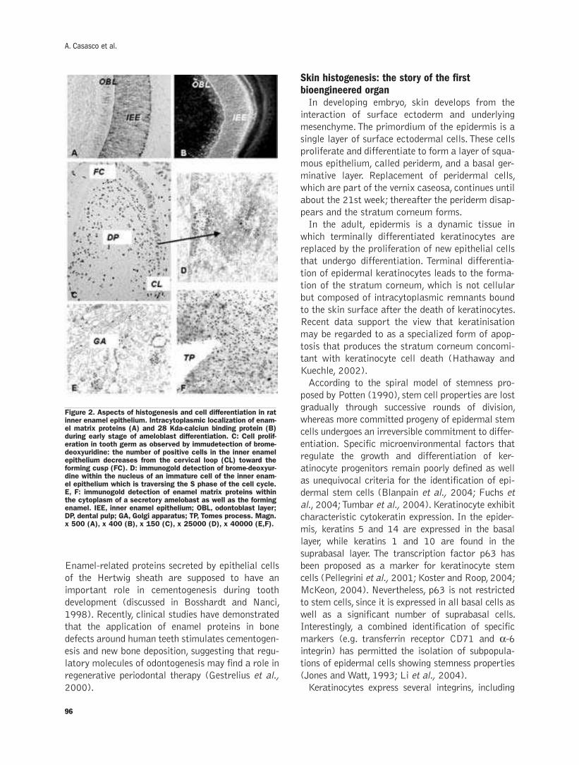
96
Enamel-related proteins secreted by epithelial cells
of the Hertwig sheath are supposed to have an
important role in cementogenesis during tooth
development (discussed in Bosshardt and Nanci,
1998). Recently, clinical studies have demonstrated
that the application of enamel proteins in bone
defects around human teeth stimulates cementogen-
esis and new bone deposition, suggesting that regu-
latory molecules of odontogenesis may find a role in
regenerative periodontal therapy (Gestrelius et al.,
2000).
Skin histogenesis: the story of the firstbioengineered organIn developing embryo, skin develops from the
interaction of surface ectoderm and underlying
mesenchyme. The primordium of the epidermis is a
single layer of surface ectodermal cells. These cells
proliferate and differentiate to form a layer of squa-
mous epithelium, called periderm, and a basal ger-
minative layer. Replacement of peridermal cells,
which are part of the vernix caseosa, continues until
about the 21st week; thereafter the periderm disap-
pears and the stratum corneum forms.
In the adult, epidermis is a dynamic tissue in
which terminally differentiated keratinocytes are
replaced by the proliferation of new epithelial cells
that undergo differentiation. Terminal differentia-
tion of epidermal keratinocytes leads to the forma-
tion of the stratum corneum, which is not cellular
but composed of intracytoplasmic remnants bound
to the skin surface after the death of keratinocytes.
Recent data support the view that keratinisation
may be regarded to as a specialized form of apop-
tosis that produces the stratum corneum concomi-
tant with keratinocyte cell death (Hathaway and
Kuechle, 2002).
According to the spiral model of stemness pro-
posed by Potten (1990), stem cell properties are lost
gradually through successive rounds of division,
whereas more committed progeny of epidermal stem
cells undergoes an irreversible commitment to differ-
entiation. Specific microenvironmental factors that
regulate the growth and differentiation of ker-
atinocyte progenitors remain poorly defined as well
as unequivocal criteria for the identification of epi-
dermal stem cells (Blanpain et al., 2004; Fuchs et
al., 2004; Tumbar et al., 2004). Keratinocyte exhibit
characteristic cytokeratin expression. In the epider-
mis, keratins 5 and 14 are expressed in the basal
layer, while keratins 1 and 10 are found in the
suprabasal layer. The transcription factor p63 has
been proposed as a marker for keratinocyte stem
cells (Pellegrini et al., 2001; Koster and Roop, 2004;
McKeon, 2004). Nevertheless, p63 is not restricted
to stem cells, since it is expressed in all basal cells as
well as a significant number of suprabasal cells.
Interestingly, a combined identification of specific
markers (e.g. transferrin receptor CD71 and α-6integrin) has permitted the isolation of subpopula-
tions of epidermal cells showing stemness properties
(Jones and Watt, 1993; Li et al., 2004).
Keratinocytes express several integrins, including
A. Casasco et al.
Figure 2. Aspects of histogenesis and cell differentiation in ratinner enamel epithelium. Intracytoplasmic localization of enam-el matrix proteins (A) and 28 Kda-calciun binding protein (B)during early stage of ameloblast differentiation. C: Cell prolif-eration in tooth germ as observed by immudetection of brome-deoxyuridine: the number of positive cells in the inner enamelepithelium decreases from the cervical loop (CL) toward theforming cusp (FC). D: immunogold detection of brome-deoxyur-dine within the nucleus of an immature cell of the inner enam-el epithelium which is traversing the S phase of the cell cycle.E, F: immunogold detection of enamel matrix proteins withinthe cytoplasm of a secretory amelobast as well as the formingenamel. IEE, inner enamel epithelium; OBL, odontoblast layer;DP, dental pulp; GA, Golgi apparatus; TP, Tomes process. Magn.x 500 (A), x 400 (B), x 150 (C), x 25000 (D), x 40000 (E,F).

97
collagen-, laminin-, fibronectin- and vitronectin-
receptors. It has been shown that integrins not only
mediate adhesion to the underlying extracellular
matrix, but also regulate keratinocyte differentiation
(Watt et al., 1993; Marchisio et al., 1997); indeed
detachment from the basement membrane seems to
be a prerequisite to undergo terminal differentiation
(Adams and Watt, 1990; Li et al., 2004).
A major aim for tissue engineers is to develop new
culture systems to change the way to conduct bio-
logical experiments and eliminate the flat biology of
Petri dishes in favour of organotypic three-dimen-
sional models. Recent advances in tissue engineer-
ing have permitted the generation of skin and epi-
dermal substitutes in vitro. Different strategies have
been conceived to engineer such substitutes and to
date skin can be regarded to as the first bioengi-
neered organ. Epidermal and dermal stem cells can
be isolated from different sources, including devel-
oping and adult tissues. Long-term subcultivation of
keratinocytes in vitro permitted the formation of
epithelial layers similar to natural epidermis
(Rheinwald and Green, 1975). Subsequently, epi-
dermal constructs have been combined with dermal
Review
Figure 3. The microscopicalpicture shows the stages ofthe cells of the odontoblastlayer (OBL) and of the innerenamel epithelium (IEE) whichprecede and go along with ini-tial deposition of dentine andenamel. The scheme summa-rizes corresponding stages ofcell differentiation and extra-cellular matrix maturation.Interestingly, the secretion ofenamel specific proteins imme-diately precedes dentine min-eralization and enamel pro-teins cross the basementmembrane in the epithelial-mesenchymal interface (i.e.the future dentine-enamel junc-tion) and reach the odonto-blast layer. Ameloblasts with-draw from the cell cycle laterthat odontoblasts, as well asenamel formation is delayedcompared to dentine forma-tion. Magn. x 1000.
98
equivalents to reconstruct the entire skin architec-
ture (Bell et al., 1981; Parenteau et al., 1991;
Stark et al., 1999; Zacchi et al., 1998). Indeed,
organotypic co-culture made of keratinocytes and
dermal cells have been shown to have many in vivo-
like features, such as complete morphologic differ-
entiation, assembly of a basement membrane, pres-
ence of cells with stem-like features, and epithelial-
mesenchymal interactions (Casasco et al., 2001a,b;
2004). Further experiments permitted the introduc-
tion of melanocytes, Langerhans cells, blood vessels
and hairs in advanced models of artificial skin.
Tissue engineering experiments suggest that skin
histogenesis is controlled by epithelial-mesenchy-
mal interactions (Smola et al., 1993). Although the
precise mechanisms are largely hypothetical, sever-
al extracellular matrices of the dermo-epidermal
junction and diffusible factors acting locally have
been implicated in the regulation of keratinocyte
growth and differentiation and skin homeostasis
(Smola et al., 1993; discussed in Turksen, 2005).
Future perspectives
Epithelial histogenesis involves dynamic patterns
of multiple signalling cascades, and molecular and
physical factors play their role with specific posi-
tional and time profiles, thus ensuring the regula-
tion of cell proliferation, differentiation and func-
tional assembly. If we understand how tissues devel-
op, we might understand how engineer their artifi-
cial equivalents. The application of our knowledge
in epithelial histogenesis has permitted the genera-
tion in vitro of tissue equivalents that are currently
used in clinical practise (Gallico et al., 1984;
Falanga et al., 1998) as well as high-fidelity mod-
els for quantitative research in biology and medi-
cine, including tissue responses to drugs, genetic
alterations, hypoxia and physical stimuli.
Moreover, information on the mechanisms that
regulate epithelial histogenesis has been used to
induce tissue regeneration in surgical procedures,
according to biomimetics which derives principles
from the nature for the design of innovative thera-
peutical strategies and tissue engineering systems.
It is reasonable to believe that other factors which
regulate the mechanisms of epithelial histogenesis
will find biotechnological and clinical application
where the need is to induce or enhance cell growth
and differentiation.
AcknowledgementsWe are grateful to Mrs. Aurora Farina
(Department of Experimental Medicine, University
of Pavia) for valuable technical assistance. This
research was supported by grants from the
University of Pavia (F.A.R.), Banca del Monte di
Lombardia Foundation (AC, AC 2004-2006) and
COFIN (AC, AC 2003) from the Italian Ministry of
Education, University and Research. The Authors
apologizes to all contributors in skin and tooth
research for inability to acknowledge all pertinent
works. This paper is dedicated to our master Prof.
Emilio Casasco and our friend Prof. Carlo Rizzoli.
References
Adams JC, Watt FM. Changes in keratinocyte adhesion during terminaldifferentiation: reduction in fibronectin binding precedes α5β1 inte-grin loss from the cell surface. Cell 1990; 63: 425-35.
Bell E, Ehrlich HP, Buttle DJ, Nakatsuji T. Living tissue formed in vitroand accepted as skin-equivalent tissue of full thickness. Science1981; 211: 1052-4.
Blancpain C, Lowry WE, Geoghegan A, Polak L, Fuchs E. Self-renew-al, multipotency, and the existence of two cell populations within anepithelial stem niche. Cell 2004; 118: 635-48.
Bosshardt DD, Schroeder HE. Cementogenesis reviewed: a comparisonbetween human premolars and rat molars. Anat Rec , topical issue“The biology of dental tissues” 1996; 245: 267-92.
Bosshardt DD, Nanci A. Immunolocalization of epithelial and mes-enchymal matrix constituents in association with inner enamelepithelial cells. J Histochem Cytochem 1998; 46: 135-42.
Bronckers ALJJ, D'Sousa R, Butler WT, Lyaruu DM, van Dijk S, GayS, et al. Dentin sialoprotein: biosynthesis and devolopmetal appear-ance in rat tooth germs in comparison with amelogenin, osteocalcinand collagen type-I. Cell Tissue Res 1993; 272: 237-47.
Casasco A, Calligaro A, Casasco M. Proliferative and functional stagesof rat ameloblast differentiation as revealed by combined immuno-cytochemistry against enamel matrix proteins and bromodeoxyuri-dine. Cell Tissue Res 1992; 270: 415-23.
Casasco A. The application of immunocytochemistry for the detectionof proliferating cell populations during tooth development. Anat Rec,topical issue “The biology of dental tissues”, 1996; 245: 162-73.
Casasco A, Casasco M, Icaro Cornaglia A, Zerbinati N, Mazzini G,Calligaro A. Cell kinetics in a model of artificial skin. An immuno-histochemical and flow cytometric analysis. Eur J Histochem 2001;45: 125-30.
Casasco A, Casasco M, Zerbinati N, Icaro Cornaglia A, Calligaro A.Cell proliferation and differentiation in a model of human skin equiv-alent. Anat Rec 2001; 264: 261-72.
Casasco M, Casasco A, Icaro Cornaglia A, Farina A, Calligaro A.Differential distribution of elastic tissue in human natural skin andtissue-engineered skin. J Molecular Histol 2004; 35: 421-8.
Cobourne MT, Sharpe PT. Tooth and jaw: molecular mechanisms ofpatterning in the first branchial arch. Arch oral Biol 2003; 48: 1-14.
Couwenhoven RI, Snead ML. Early determination and permissiveexpression of amelogenin transcription during mouse mandibularfirst molar development. Dev Biol 1994; 164: 290-9.
Falanga V, Margolis D, Alvarez O, Auletta M, Maggiacomo F, AltrmanM, et al. The human skin equivalent investigators group. Rapid heal-ing of venous ulcers and lack of clinical rejection with an allogeniccultured human skin equivalent. Arch Dermatol 1998; 134: 293-300.
Fuchs E. Epidermal differentiation: the bare essentials. J Cell Biol1990; 111: 2807-14.
Fuchs E, Tumbar T, Guasch G. Socializing with the neighbors: stemcells and their niche. Cell 2004; 116: 769-78.
A. Casasco et al.
99
Gallico GG, O'Connor NE, Compton CC, Kehinde O, Green H. A per-manent coverage of large burn wounds with autologous culturedhuman epithelium. N Engl J Med 1984; 311: 448-51.
Gestrelius S, Lyngstadaas SP, Hammarstrom L. Emdogain- periodon-tal regeneration based on biomimicry. Clin Oral Invest 2000; 4: 120-5.
Hathaway TR, Kuechle MK. Apoptosis and cutaneous biology. AdvDermatol 2002; 18: 287-311.
Inai T, Kukita T, Ohsaki Y, Nagata K, Kukita A, Kurisu K.Immunohistochemical demonstration of amelogenin penetrationtoward the dental pulp in the early stages of ameloblast developmentin rat molar tooth germs. Anat Rec 1991; 229: 259-70.
Jones PH, Watt FM. Separation of human epidermal stem cells fromtransit amplifying cells on the basis of differences in integrin func-tion and expression. Cell 1993; 73: 713-24.
Jones PH, Harper S, Watt FM. Stem cell patterning and fate in humanepidermis. Cell 1995; 80: 83-93.
Jernvall J, Thesleff I. Reiterative signalling and patterning during mam-malian tooth morphogenesis. Mech Dev 2000; 15: 19-29.
Kollar EJ, Baird GR. The influence of the dental papilla on the devel-opment of tooth shape in embryonic mouse tooth germ. J EmbryolExp Morphol 1968; 21: 131-48.
Koster M, Roop DR. The role of p63 in development and differentiationof the epidermis. J Dermatol Science 2004; 34: 3.
Leblond CP. The life history of cells in renewing systems. Am J Anat1981; 712: 187-204.
Li A, Pouliot N, Redvers R, Kaur P. Estensive tissue-regenerativecapacity of neonatal human keratinocyte stem cells and their prog-eny. J Clin Invest 2004; 113: 390-400.
Lumsden AG. Spatial organization of the epithelium and the role ofneural crest cells in the initiation of the mammalian tooth germ.Development 1988;103:155-69.
Marchisio PC, Trusolino L, De Luca M. Topography and biological roleof integrins in human skin. Microsc Res Tech 1997; 38: 353-60.
McKeon F. p63 and the epithelial stem cell: more than status quo?Genes Dev 2004; 18: 465-9.
Nanci A, Bendayan M, Slavkin HC. Enamel protein biosynthesis andsecretion in mouse incisor secretory ameloblasts as revealed byhigh-resolution immunocytochemistry. J Histochem Cytochem1985; 33: 1153-60.
Nanci A, Slavkin HC, Smith CE. Application of high-resolutionimmunocytochemistry to the study of the secretory, resorptive, anddegradative functions of ameloblasts. Adv Dent Res 1987;1: 148-61.
Nanci A, Zalzal S, Lavoie P, Kunikata M, Chen W-Y, Kresbach PH, etal. Comparative immunochemical analyses of the developmentalexpression and distribution of ameloblastin and amelogenin in ratincisors. J Histochem Cytochem 1998; 46: 911-34.
Parenteau NL, Nolte CM, Bilbo P, Rosenberg M , Wilkins LM, JohnsonEW, Watson S, Mason VS, Bell E. Epidermis generated in vitro:practical considerations and applications. J Cell Biochem 1991; 45:245-51.
Pellegrini G, Dellambra E, Golisano O, Martinelli E, Fantozzi I,
Bondanza S, et al. p63 identifies keratinocyte stem cells. Proc NatlAcad Sci, USA, 2001; 98: 3156-61.
Potten CS. Stem cells in epidermis from the back of the mouse. In:Potten CS., Stem cells. Their identification and characterisation.Churcill Livingstone, Edinburgh, 1983; 200-32.
Potten CS, Loeffler M. Stem cells: attributes, cycles, spirals, pitfallsand uncertainties lessons for and from the crypt. Development 1990;110: 1001-20.
Rheinwald JG, Green H. Serial cultivation of strains of human epider-mal keratinocytes: the formation of keratinizing colonies from singlecells. Cell 1975; 6: 331-44.
Ruch JV. Epithelial-mesenchymal interactions in formation of mineral-ized tissues. In: Butler NT: The Chemistry and Biology ofMineralized Tissues; Ebsco Media, Birmingham, Alabama, 1985;54-61.
Sharpe P. Neural crest and tooth morphogenesis. Adv Dent Res 2001;15: 4-7.
Slavkin HC, Mino W, Bringas P. The biosynthesis and secretion of pre-cursor enamel protein by ameloblasts as visualized by autoradiogra-phy after tryptophan administration. Anat Rec 1976; 185: 289-312.
Slavkin HC. Molecular determinants of tooth develpment. Crit RevOral Biol Med 1990; 1: 1-16.
Smith CE, Warshawsky H. Cellular renewal in the enamel organ andthe odontoblast layer of the rat incisor as followed by radioautogra-phy using 3H-thymidine. Anat Rec 1975; 183: 523-62.
Smola H, Thiekotter G, Fusenig NE. Mutual induction of growth factorgene expression by epidermal-dermal cell interactions. J Cell Biol1993; 122: 417-29.
Stark HJ, Baur M, Breitkreutz D, Mirancea N, Fusenig NE.Organotypic keratinocyte cocultures in defined medium with regularepidermal morphogenesis and differentiation. J Invest Dermatol1999; 112: 681-91.
Thesleff I, Mikkola M. The role of growth factors in tooth development.Int Rev Cytol 2002; 217: 93-135.
Thesleff I. Epithelial-mesenchymal signalling regulating tooth morpho-genesis. J Cell Sci 2003; 116: 1647-8.
Tumbar T, Guasch G, Greco V, Blanpain C, Lowry WE, Rendl M, FuchsE. Defining the epithelial stem cell niche in the skin. Science 2004;303: 359-63.
Turksen K. Methods in Molecular Biology. Epidermal Cells. Methodsand Protocols. Humana Press, Totowa, New Jersey, 2005; 289: .
Warshawsky H, Smith CE. Morphological classification of rat incisorameloblasts. Anat Rec 1974; 179: 423-46.
Watt FM, Kubler MD, Hotchin NA, Nicholson LJ, Adams JC.Regulation of keratinocyte terminal differentiation by integrin-extra-cellular matrix interactions. J Cell Sci 1993; 106: 175-82.
Zacchi V, Soranzo C, Cortivo R, Radice M, Brun P, Abatangelo G. Invitro engineering of human skin-like tissue. J Biomed Mater Res1998; 40: 187-94
Review

100
A. Casasco et al.

©2007, European Journal of Histochemistry
During embryonic development, a pool of cells may becomea reserve of undifferentiated cells, the embryo-stolen adultstem cells (ESASC). ESASC may be responsible for adult tis-sue homeostasis, as well as disease development.Transdifferentiation is a sort of reprogramming of ESASCfrom one germ layer-derived tissue towards another.Transdifferentiation has been described to take place frommesoderm to ectodermal- or endodermal-derived tissuesand viceversa but not from ectodermal- to endodermal-derived tissues. We hypothesise that two different popula-tions of ESASC could exist, the first ecto/mesoblast-commit-ted and the second endo/mesoblast-committed. If con-firmed, this hypothesis could lead to new studies on themolecular mechanisms of cell differentiation and to a betterunderstanding of the pathogenesis of a number of diseases.
Key words: transdifferentiation, germ layers, self-renewing,gastrulation.
Correspondence: Giovanni Zummo, via del Vespro, 129 – 90127 PalermoE-mail: [email protected]
European Journal of Histochemistry2007; vol. 51 supplement 1:101-104
Adult stem cells: the real root into the embryo?
G. Zummo, F. Bucchieri, F. Cappello, M. Bellafiore, G. La Rocca, S. David, V. Di Felice,
R. Anzalone, G. Peri, A. Palma, F. Farina
Department of Experimental Medicine, Section of Human Anatomy, University of Palermo, Italy
101
Mechanisms responsible for tissue regenera-
tion in organ homeostasis and recovery in
adult organisms consist of 1) mitosis of
differentiated functioning cells with a preserved
proliferative activity (i.e. hepatocytes) and 2) gen-
eration of newly differentiated cell populations
derived from adult stem cells (SC) (i.e. blood)
(Abbott, 2006).
SC are defined as undifferentiated elements capa-
ble of self-renewal and differentiation into special-
ized cells. SC are considered resistant to toxic noxae
and other damaging agents and have a great capac-
ity to restore and renew (almost) all tissues of the
human body. SC have been described in several tis-
sues and it is not excluded that they could migrate
to repair distant tissues. Indeed, they may be main-
tained in the systemic circulation and activated by
pathophysiological stimuli from damaged tissues.
Nevertheless, the regenerative activity of SC is not
unlimited, because of senescence processes
(Asahara and Kawamoto, 2004). Moreover, cancers
may origin from SC, and also cancers’ SC have the
ability to self-renew, determining tumour resistance
to therapy and recurrence (Abbott, 2006).
Nowadays, we are not really able to make a clear
distinction between terms such as undifferentiated,
stem-, germinal-, precursor- and progenitor- cells.
Referring to embryonic tissues might be useful to
disentangle ourselves in this terminological maze.
Embryo-stolen adult stem cellsThe inner cell mass of the blastocyst gives rise to
hypoblast and epiblast. The latter brings forth three
germ layers, ectoderm, mesoderm and endoderm,
that undergo proliferation through lineage commit-
ment to form multi-, tri-, bi- and uni-potent undif-
ferentiated cells, from which, finally, all differentiat-
ed cells and tissues of the body originate (Young and
Black, 2004).
During embryonic development, a pool of cells
may interrupt this continuum to become a reserve of
REVIEW
undifferentiated cells, that we may call embryo-
stolen adult stem cells (ESASC). Whether ESASC
are circulating or resident is a controversial topic,
since the former does not exclude the latter.
Many evidences demonstrate the existence of lin-
eage-committed and lineage-uncommitted SC in
many adult organs in vivo, like bone marrow (Chiu,
2003), heart (Bellafiore et al., 2006), kidney
(Lange et al., 2005), brain (Gritti et al., 2002) and
other organs. In each of these organs, these cells
may originate one or more cytotypes. Examples
include satellite myoblasts in skeletal muscle
(Charge and Rudnicki, 2004), and hemangioblasts
for hematopoietic and endothelial cells (Asahara
and Kawamoto, 2004).
Moreover, isolation and characterization of SC in
vitro becomes a practical approach to study these
differentiation mechanisms. It is unfeasible and
unpractical to report here all the plethora of stud-
ies that have been describing in vitro isolation of
post-natal undifferentiated cells and experiments
concerning their immunophenotyping, proliferation
and differentiation potential and functional charac-
teristics. Nevertheless, as anyone might observe,
most of these studies describe SC derived from
mesoderm differentiating towards ectodermal-
derived tissues [i.e. from blood to neurons
(Brazelton et al., 2000)] or endodermal-derived
tissues (i.e. from marrow to liver (Cantz et al.,
2004)), and SC isolated from ectoderm or endo-
derm originating mesodermal tissues [i.e. from neu-
ron to blood (Bjornson et al., 1999)].
The latter event, consisting of a sort of repro-
gramming of SC from one germ layer-derived tissue
towards another, is called transdifferentiation. This
theory is clashing with the common belief that once
a SC is committed to a specific tissue lineage, it can
not change its genetic program, reverting to a more
primitive stage. Therefore, the existence of transdif-
ferentiation in vivo is not universally recognised by
scientific community. Young and Black (2004) for
example suppose that transdifferentiation, when
occurring in vitro, is due to contamination of isolat-
ed tissue by unrecognised progenitor cells.
At this time we would like to underline that none
of the experiments of transdifferentiation conduct-
ed thus far have reported ectodermal-derived SC
(EcDSC) or endodermal-derived SC (EnDSC) dif-
ferentiating towards each other. To clarify this odd-
ity, we should probably refer again to the embryo.
Gastrulation is probably an important step inESASC formationDuring the third week of embryonic development,
gastrulation takes place from epiblast, from which
all three germ layers derive. Gastrulation is a cru-
cial time in the development of multicellular ani-
mals, since several essential steps are accom-
plished. Phylogenetically, gastrulation originates
from two (diploblastic organisms, i.e. coelenter-
ates) to three (all higher animals) layers. Whether
the emergence of the mesoderm is linked to the evo-
lution of axis formation in metazoan is not yet an
assured fact (Technau and Scholz, 2003).
The molecular mechanisms responsible for gas-
trulation are still not well known. Also in triploblas-
tic organisms, endoderm formation during gastrula-
tion is not always linked to the formation of meso-
derm, and different mechanisms have been pro-
posed to explain this. Is commonly believed that
some of the cells from the surface of the embryo
move to the interior, replicating and thereby form-
ing the new layers. These movements are coupled
with the differentiation of the migrating cells
(caused by the differential activity of certain genes)
into histologically unique layers. The initial migra-
tion and differentiation of cells, which will then
invaginate within the blastocoel, gives rise to the
endoderm and mesoderm germ layers. In particular,
in birds and mammals, epiblast cells converge at
the midline and ingress at the primitive streak.
Ingression of these cells results in formation of the
mesoderm and replacement of some of the
hypoblast cells to produce the definitive endoderm
(Langman, 1995).
Moreover, since the ending –derm is usually
referred to differentiated tissues, Technau and
Scholz (2003) proposed to use the ending –blast to
indicate proliferating but not yet differentiated tis-
sues, such as germ layers (endoblast, ectoblast and
mesoblast).
Hypothesis and conclusionSince endoblast formation during gastrulation
could be independent from the formation of
mesoblast (Technau and Scholz, 2003), and since it
is not well established whether endoblast develop-
ment precedes mesoblast formation, one can not
exclude the latter, as it should be phylogenetically
intuitive. Consequently, ESASC could be considered
as a pool of undifferentiated cells derived from a bi-
layered (ecto-endoblast) embryo, that give rise to
102
G. Zummo et al.

the mesoblast and maintain multiple differentiative
potentiality also in adult organism, being able to
perform transdifferentiation from both EcDSC and
EnDSC to mesoblast derived cells and vice versa; as
a consequence, two populations of ESASC could
exist, the first ecto/mesoblast-committed and the
second endo/mesoblast-committed and this could
explain why transdifferentiation from EcDC to
EnDC has not yet been described. These two
ESASC could be responsible for adult tissue home-
ostasis, as well as disease development, i.e. tumori-
genesis.
In conclusion, in our opinion in vitro studies of SC
could be imperative to discover the molecular
mechanisms of cell commitment; the induction of
cell differentiation in vivo, during the pathogenesis
of a number of diseases, like Alzheimer, myocardial
failure, celiac disease, etc, could become a new ther-
apeutic target for the next generation of physicians.
Acknowledgement
This work was supported by MIUR ex-60% funds
to Prof. G. Zummo.
References
Abbott A. The root of the problem. Nature 2006;442:742-3.
Asahara T and Kawamoto A: Endothelial progenitor cells for postna-tal vasculogenesis. Am J Physiol Cell Physiol, 287:C572-C579,2004.
Bellafiore M, Sivverini G, Cappello F, David S, Palma A, Farina F, et al.Research of cardiomyocyte precursors in adult rat heart. Tissue Cell2006;38:345-51.
Bjornson CR, Rietze RL, Reynolds BA, Magli MC, Vescovi AL. Turningbrain into blood: a hematopoietic fate adopted by adult neural stemcells in vivo. Science. 1999;283:534-7.
Brazelton TR, Rossi FM, Keshet GI, Blau HM. From marrow to brain:expression of neuronal phenotypes in adult mice. Science 2000;290:1775-9.
Cantz T, Sharma AD, Jochheim-Richter A, Arseniev L, Klein C, MannsMP, et al. Reevaluation of bone marrow-derived cells as a source forhepatocyte regeneration. Cell Transplant 2004;13:659-66.
Charge SB, Rudnicki MA. Cellular and molecular regulation of muscleregeneration. Physiol Rev 2004;84:209-38.
Chiu RC. Bone-marrow stem cells as a source for cell therapy. HeartFail Rev 2003;8:247-51.
Gritti A, Vescovi AL, Galli R. Adult neural stem cells: plasticity anddevelopmental potential. J Physiol Paris. 2002;96:81-90.
Lange C, Togel F, Ittrich H, Clayton F, Nolte-Ernsting C, Zander AR,Westenfelder C. Administered mesenchymal stem cells enhancerecovery from ischemia/reperfusion-induced acute renal failure inrats. Kidney Int 2005;68:1613-7.
Langman’s Medical Embryology, Seventh Edition, Williams & Wilkins,1995.
Technau U, Scholz CB. Origin and evolution of endoderm and meso-derm. Int J Dev Biol 2003;47:531-9.
Young HE and Black AC: Adult stem cells. Anat Rec A Discov Mol CellEvol Biol 2004;276:75-102.
103
Review

104
G. Zummo et al.
105
REVIEW
©2007, European Journal of Histochemistry
The normal development of cranial primordia and orofacial struc-tures involves fundamental processes in which growth, morpho-genesis, and cell differentiation take place and interactionsbetween extracellular matrix (ECM) components, growth factorsand embryonic tissues are involved. Biochemical and molecularaspects of craniofacial development, such as the biological reg-ulation of normal or premature cranial suture fusion, has justbegun to be understood, thanks mainly to studies performed inthe last decade. Several mutations has been identified in bothsyndromic and non-syndromic craniosynostosis patients throwingnew light onto the etiology, classification and developmentalpathology of these diseases. In the more common craniosynos-tosis syndromes and other skeletal growth disorders, the muta-tions were identified in the genes encoding fibroblast growth fac-tor receptor types 1–3 (FGFR1, 2 and 3) where they are domi-nantly acting and affect specific and important protein bindingdomain. The unregulated FGF signaling during intramembranousossification is associated to the Apert and Crouzon syndrome. Thenon syndromic cleft of the lip and/or palate (CLP) has a morecomplex genetic background if compared to craniosynostosissyndrome because of the number of involved genes and type ofinheritance. Moreover, the influence of environmental factormakes difficult to clarify the primary causes of this malformation.ECM represents cell environment and results mainly composedby collagens, fibronectin, proteoglycans (PG) and hyaluronate(HA). Cooperative effects of ECM and growth factors regulateregional matrix production during the morphogenetic events, con-nective tissue remodelling and pathological states. In the presentreview we summarize the studies we performed in the last yearsto better clarify the role of ECM and growth factors in the etiolo-gy and pathogenesis of craniosynostosis and CLP diseases.
Key words: extracellular matrix; growth factors; craniosynos-tosis; cleft lip; cleft palate.
Correspondence: Paolo CarinciDepartment of Histology, Embryology and Applied Biology,University of Bologna, ItalyTel: +39.075.5857508.Fax: +39.075.5857434.E-mail: [email protected]
European Journal of Histochemistry2007; vol. 51 supplement 1:105-116
Extracellular matrix and growth factors in the pathogenesis of some
craniofacial malformations
P. Carinci,1 E. Becchetti,2 T. Baroni,2 F. Carinci,3 F. Pezzetti,1 G. Stabellini,4 P. Locci,2 L. Scapoli,1
M. Tognon,5 S. Volinia,6 M. Bodo7
1Department of Histology, Embryology and Applied Biology, University of Bologna; 2Department of
Experimental Medicine and Biochemical Sciences, Section of Histology and Embryology, University of
Perugia; 3Department of Maxillofacial Surgery, University of Ferrara; 4Department of Human Morphology,
University of Milano; 5Chair of Applied Biology and Center of Biotechnology, University of Ferrara;6Department of Morphology and Embryology, University of Ferrara; 7Department of Specialistic Medicine
and Public Health, University of Perugia, Italy
During skull development, osteogenetic events
lead to form the mesodermal neurocranium,
which surrounds and protects the brain, and
the neural crest-derived viscerocranium, which
forms the face in mammals and supports the func-
tions of feeding and breathing. The base of the neu-
rocranium underlie the brain and is formed by endo-
chondral ossification whereas the vault (calvaria) is
formed by membranous ossification. The adjacent
margins of membrane bones form the sutures which
contain osteogenic stem cells and periosteal fibrob-
lasts that differentiate into osteoblasts capable of
producing new bone tissue, and are thus considered
active sites of bone growth. Growth and expansion
of the skull vault takes place to allow free growth of
the brain. Craniosynostosis arises when this mecha-
nism fails, because of the premature loss and ossifi-
cation of sutural growth centres (Morriss-Kay and
Wilkie, 2005). The normal development of the upper
jaw and of the palate starts at about the 6th week of
intra-uterine life and requires growth and fusion of
the medial nasal processes and maxillary processes
to form the lip, while the fusion of the palatal
shelves to form the secondary palate occurs later
(10th week). Craniofacial malformations and in par-
ticular orofacial clefting are the most common
birth defects that occur in humans. Clefts of the lip,
with or without cleft palate, and those that involve
the palate only, are due to a failure in fusion of the
facial processes and/or palatal shelves, and consti-
tute two forms of oral-facial clefts considered sepa-
rate birth defects involving many (but not all) of the
same genetic and environmental causes (Carinci et
al., 2003).
Craniosynostosis (Apert and Crouzon syndromes) Sutures contain osteogenic stem cells as a reser-
voir of potential new osteoblasts and are thus active
sites of bone growth. The premature fusion of one or
more skull sutures due to altered osteogenic
processes at the time of calvarian development is
the most severe anomaly of the calvarium and is
known as craniosynostosis, which prevents further
bone growth along the edges. This leads the cranial
vault to expand in other directions, thereby giving
rise to a wide variety of pathological phenotypes.
Crouzon syndrome accounts for 5% while Apert
syndrome accounts for 4-5% of all cases of cran-
iosynostosis (about 343 per million newborns;
Cohen and MacLean, 2000).
Comparative studies indicate that although Apert
and Crouzon syndromes present very similar cranial
anomalies, they differ in cranial development
(Kreiborg et al., 1993).
Pathogenesis of craniosynostosis
Until just over a decade ago, little was known
about the causes of craniosynostosis. Since then,
several mutations were identified in both syn-
dromic and non-syndromic patients throwing new
light onto the etiology, classification and develop-
mental pathology of these diseases (Morriss-Kay
et al., 2005).
The first mutation to be identified was a het-
erozygous missense mutation in homeotic MSX2
gene in patients with craniosynostosis type 2, also
known as Boston-type, a rare syndrome confined to
a single large family. TWIST1 and EFNB1 are also
two significant genes (Wilkie, 2006). In other more
common craniosynostosis syndromes and other
skeletal growth disorders, the mutations were iden-
tified in the genes encoding fibroblast growth factor
receptors (FGFRs). FGF2, a member of the FGF
family, binds to high- and low-affinity receptors that
are four different transmembrane tyrosine kinase
receptors (FGFR1, -R2, -R3, and -R4). Upon
FGF2/FGFR binding, the FGFR2 receptors
dimerise and thus activate the intracellular tyrosine
kinase domains. This is followed by phosphorylation
of cellular proteins and transmission of signals into
the cell that initiate a cascade of signals influencing
cell division and differentiation. Membranous ossi-
fication of the skull vault is characterized by
expression of FGFR genes in preosteoblasts and
osteoblasts (Delezoide et al., 1998). Genetic analy-
sis of many human skeletal disorders have demon-
strated the critical role of the FGF-FGFR system in
endochondral and endomembranous ossification. In
particular, defective or excessive fibroblast growth
factor (FGF) signaling interferes with normal cra-
nial suture morphogenesis (Naski et al., 1998).
Activating missense mutations occurring in FGFR2
(Kan et al., 2002) cause an unregulated FGF sig-
naling during intramembranous ossification and are
associated to an important category of craniosyn-
ostosis as Apert, Crouzon and Pfeiffer (MIM
101600) syndromes (Pfeiffer syndrome is caused
by mutations in either the FGFR1 or FGFR2 gene).
In particular, the most common mutation of
FGFR2 in Crouzon syndrome is C342Y generated
by the Cys342-to-Tyr substitution. In Apert syn-
drome, the two adjacent amino acid substitutions
Ser252-to-Trp (S252W), and Pro253-to-Arg
(P253R) in the linker stretch between the second
and third Ig-like domains, account for the vast
majority of cases of syndrome (Robertson et al.,
1998) with frequencies of 71 and 26% respective-
ly for the S252W and the P253R mutations. The
C342Y mutation results in a constitutive (ligand-
independent) receptor activation (phosphorylation)
and causes excessive signalling (Ibrahimi et al.,
2004). Apert S252W and P253R mutations and
some Pfeiffer mutations activate receptor only in
the presence of ligand (ligand-dependent) (Yu et
al., 2000).
It is noteworthy that equivalent mutations in
FGFR1, FGFR2, and FGFR3 genes result in differ-
ent phenotypes: the P252R FGFR1 mutation was
identified in a mild form of Pfeiffer syndrome, the
P253R FGFR2 mutation causes Apert syndrome,
and the P250R mutation of FGFR3 causes Muenke
syndrome. An interesting hypothesis to explain such
heterogeneity of phenotypes with similar mutations
is that they function interactively, and loss- or gain-
of-function mutations in one gene that affect the
function of the protein may have secondary effects
on one or both of the other FGFRs (Morriss-Kay et
al., 2005).
Cleft of the lip and/or palate Cleft of the lip and/or palate (CLP) is the most
common congenital orofacial malformation in
humans. The disease is multifactorial and is proba-
bly caused by genetic and/or environmental factors
(Carinci et al., 1995; Scapoli et al., 1997, 1998;
Pezzetti et al., 1998). Although a putative role of
ECM has been assumed for long time in the genesis
of CLP, few reports have analyzed the composition
and relative amount of different types of gly-
cosaminoglycans (GAG) and collagen in human cell
106
P. Carinci et al.
culture either under normal conditions or during
stimulation with growth factors or clefting drugs.
Interaction between ECM and cytokines is thought
to be crucial for palatal development. Indeed, con-
trol of ECM metabolism in the embryonic orofacial
region seems to be essential for normal palatal
development. ECM molecules, in turn, promote the
activities of growth factors and cytokines present in
epithelial cells and palatal mesenchyme (Qiu et al.,
1995). The localization of transforming growth fac-
tors (TGFα and TGFβ isoforms) and fibroblast
growth factors (FGF) in craniofacial tissues sug-
gests that they carry out multiple functions during
development.
CLP can be subdivided into syndromic (such as
chromosomal, Mendelian, teratogen-based, and
uncategorized syndromes) and non syndromic
forms (about 70% of cases). Occurrence estimates
range between 1/300 and 1/2500 births for cleft lip
with or without cleft palate (CLP) and around
1/1500 births for cleft palate alone (CP). The non
syndromic CLP arises when nasal processes and/or
palatal shelves fail to fuse because genetic abnor-
malities and/or a perturbed environment alter
extracellular matrix (ECM) composition and affect
cell patterning, migration, proliferation and differ-
entiation (Young et al., 2000).
Pathogenesis of CLP
The non-syndromic CLP have a more complex
genetic background if compared to craniosynostosis
syndrome because of the number of involved genes
and type of inheritance (for review: Carinci et al.,
2003). Some genes are involved in the formation of
CLP defects. Among them, transforming growth
factor-β3 (TGF-β3), whose mutations and/or defi-ciences give rise to cleft palate in humans (Lidral et
al., 1998) and mice (Kaartinen et al., 1995);
retinoic acid receptor-α (RARA), even though opin-
ions are divergent as to whether RARA should be
considered as a candidate gene in CLP (Scapoli et
al., 2002; Stein, 1995); γ-aminobutyric acid type Areceptor β3 (GABRB3), a protein receptor for γ-aminobutyric acid (GABA), the major inhibitory
neurotransmitter in the mammalian central nervous
system, is also probably involved in human CLP
malformation (Scapoli et al., 2002; Baroni et al.,
2006). Moreover, the influence of environmental
factor (cigarette smoking, alcohol consumption,
corticosteroid, retinoic acid and anticonvulsant
taken during the gestation period) on CLP onset,
makes difficult to clarify the primary causes of this
malformation.
Morphogenetic signals of ECM components during craniofacial development During osteogenesis, mesenchymal cells differen-
tiate in osteoblast lineage and produce a mineral-
ized ECM that takes control of morphogenetic
events. The ECM complex is formed by proteogly-
cans (PG), glycosaminoglycans (GAG), fibronectin,
collagens, and other glycoproteins, which are differ-
ently distributed and organised in tissues and stages
of development. The ECM-cell receptor-link trans-
mits signals across the cell membrane in the cyto-
plasm, thereby initiating a cascade of events that
culminates in the expression of specific genes.
Fibronectin interacts with fibrils of type I collagen
(Moursi et al., 1996), another ECM molecule that
realizes a matrix-mediated tissue interaction.
Laminin, fibronectin, collagen type I and IV are all
distributed in characteristic maps in epithelial-mes-
enchymal interfaces involved in the formation of
avian embryo cartilaginous neurocranium
(Thorogood et al., 1988). Other ECM components
such as PG, and particularly syndecan-III and
biglycan (Xu et al., 1998), control spatially and
temporally ossification during fetal development
and play a role in the terminal differentiation of
embryonic cells.
In addition, an important role in the craniofacial
morphogenesis is ascribed to matrix metallopro-
teinases, enzymes involved in ECM degradation. All
these different transient patterns of ECM compo-
nents are interpreted as reflecting different levels of
morphogenetic specification of skull form in the
developing head.
ECM in pathological conditionsAlterations of the normal balance between ECM
synthesis and catabolism have to be considered a
relevant factor in establishing connective diseases
(Carinci et al., 2005; Malemud et al., 2006).
Potential defects in connective disease may thus
be depending on one or more of the following event
sequence: ECM synthesis and catabolism, ECM
control on growth factor activity, stimulus by
growth factor on specific receptor, intracellular
message production, effect on ECM macromole-
cules synthesis.
Review
107

Role of growth factors in skull morphogenesis andosteogenesisA significant role in skull morphogenetic events is
played by signal molecules present in the early
embryo in vivo. Interleukins (ILs), transforming
growth factor β (TGF-β) and fibroblast growth fac-tor2 (FGF-2) are putative signal peptides present
in skull tissues at the time of active differentiation
and morphogenesis. Interleukin 1 (IL-1) and inter-
leukin 6 (IL-6) are two of other cytokines that are
involved in the regulation of bone cell functions. IL-
1 stimulates cell replication and, at a low dose, bone
collagen synthesis (Canalis et al., 1989); IL-6
enhances bone turnover, stimulating bone resorption
processes. TGF-β and bone morphogenetic proteins
(BMPs), a subfamily of the TGF-β protein family,
regulate chondrocyte proliferation and differentia-
tion, and the ossification of endochondral bones.
Disturbed TGF-β signaling lead to a variety of
human skeletal disorders.
FGF2, which is the most abundant growth factor
in the vault, is another regulator of bone develop-
ment. FGF2/FGFR binding induces a broad spec-
trum of activities into the cells, including growth
and cell migration increase and induction of pro-
teases such as plasminogen activator, catepsin B,
kallicrein (Szebenyi and Fallon et al., 1999).
Materials and MethodsWe performed studies in cells obtained from
Apert and Crouzon patients during corrective sur-
gery for the malformations. Fibroblasts were
obtained from the galea-pericranium and osteo-
blasts from the parietal bone near the coronal
suture. CLP fibroblasts were obtained from the oral
flap edge of hard secondary palate subjects with
familial non-syndromic cleft lip and palate and
from age-matched controls. All cells were obtained
during corrective surgery for the malformation.
Human tissues were obtained with a protocol
approved by our institutions. Informed consent was
obtained from all parents after the nature of the
study had been fully explained.
To study GAG neosynthesis, confluent cultures
were labeled with 5 pCi/mL of (3H)-glucosamine
hydrochloride (NEN Du Pont de Nemours, RFG;
s.a. 29 Ci/mmol) and labelled GAG were precipi-
tated from recovered supernatants and cells.
Secreted collagens were analysed in confluent
cultures, added with L-ascorbic acid (50 pg/mL),
(3-aminopropionitrile fumarate (50 µ/mL) and 10
µCi/mL of (3H)proline (Du Pont NEN, s.a. 29
Ci/mmol).
Fibronectin was isolated from media of confluent
cultures labeled with 20 pCi/mL (35S)-methionine
(s.a.>1,000 Ci/mmol, Amersham International,
England, U.K.) during the last 3 hours of incuba-
tion. Samples were subjected to SDS-PAGE analy-
sis, followed by densitometric quantification of flu-
orographs.
Gene expression was analysed by RT-PCR and
measured semiquantitative radioactive RT-PCR
and/or Northern blotting and autoradiography; for
FGF2 and TGF β receptor counting, a radioactive
binding assay was used.
Expression gene profiling was performed after
total RNA extraction from cultured normal and
pathologic cells. cDNA was synthesized by using
Superscript II (Invitrogen, Paisley, England, UK)
and amino-allyl dUTP (Sigma, St. Louis, MO,
USA). Human 19.2K DNA microarrays, containing
19,200 expressed sequence tag (ESTs), correspon-
ding to at least 15,448 different Unigene clusters,
were used (Ontario Cancer Institute, Ontario,
Canada). Hybridized slides were scanned using
GenePix 4000A (Axon Instruments, Foster City,
CA, USA). Images were analyzed by GENEPIX
PRO 3.0 (Axon Instruments) and data extracted as
described (Carinci et al., 2002). Genetic analyses
were conducted using a sample composed by 38
multiplex pedigrees and 200 sporadic patients.
Polymorphic DNA markers, mainly microsatellite
repeats, were typed by PCR using fluorescent-
labelled primers and subsequent electrophoresis on
a DNA sequencing equipment. Parametric linkage
analyses with Lod score method were performed by
LINKAGE software package. Non parametric
analyses were performed with the standard trans-
mission disequilibrium test (TDT).
Results
Apert syndromePeriosteal pericranial fibroblasts from Apert
patients synthesized and secreted greater amounts
of sulphated (heparan sulphate, HS; chondoitin sul-
phate, CS; dermatan sulphate, DS) and non-sul-
phated (hyaluronic acid, HA) GAG compared to
normal cells (Bodo et al., 1997). Treatment with
IL-1 and IL-6 reduced HA secretion by Apert cells.
IL-1 significantly increased CS secretion by Apert
fibroblasts and IL-6 enhanced HS and DS secre-
108
P. Carinci et al.
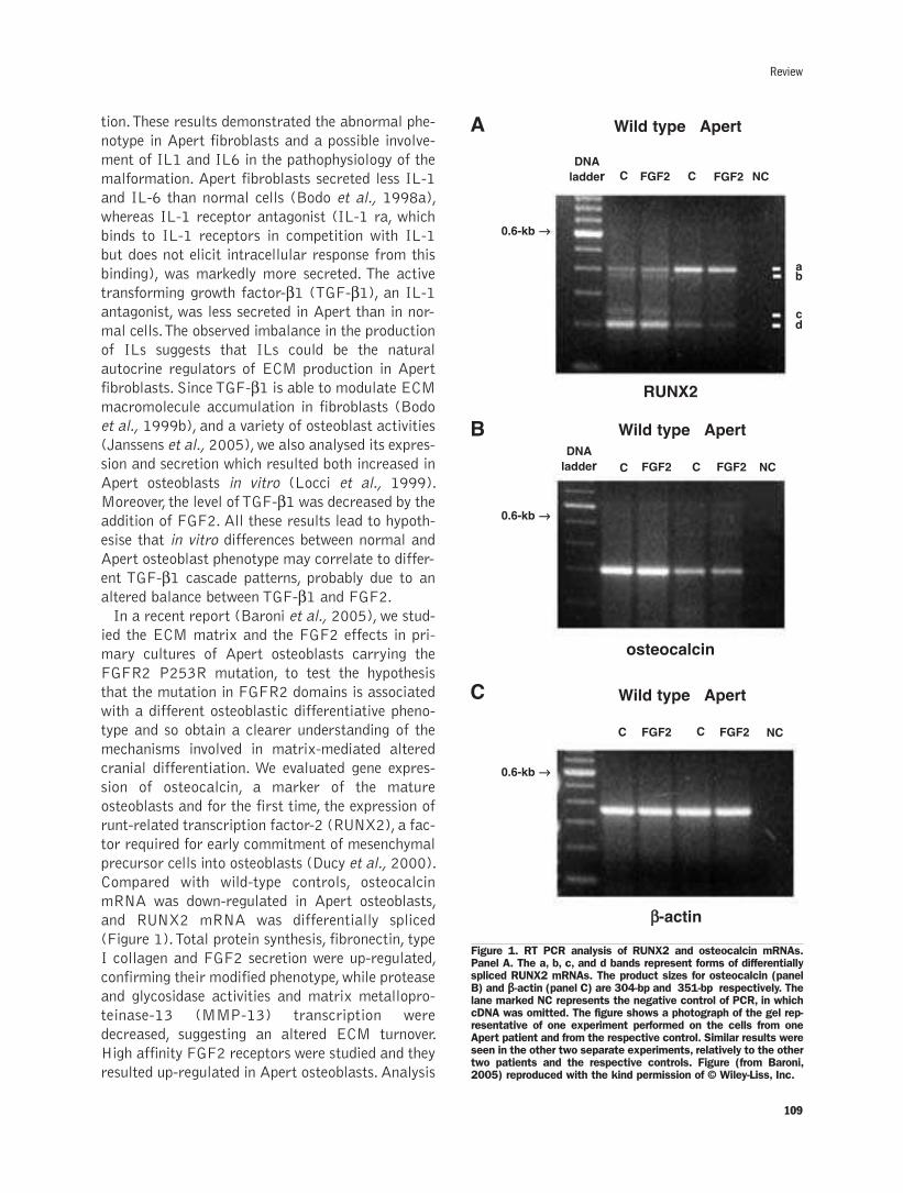
tion. These results demonstrated the abnormal phe-
notype in Apert fibroblasts and a possible involve-
ment of IL1 and IL6 in the pathophysiology of the
malformation. Apert fibroblasts secreted less IL-1
and IL-6 than normal cells (Bodo et al., 1998a),
whereas IL-1 receptor antagonist (IL-1 ra, which
binds to IL-1 receptors in competition with IL-1
but does not elicit intracellular response from this
binding), was markedly more secreted. The active
transforming growth factor-β1 (TGF-β1), an IL-1antagonist, was less secreted in Apert than in nor-
mal cells. The observed imbalance in the production
of ILs suggests that ILs could be the natural
autocrine regulators of ECM production in Apert
fibroblasts. Since TGF-β1 is able to modulate ECMmacromolecule accumulation in fibroblasts (Bodo
et al., 1999b), and a variety of osteoblast activities
(Janssens et al., 2005), we also analysed its expres-
sion and secretion which resulted both increased in
Apert osteoblasts in vitro (Locci et al., 1999).
Moreover, the level of TGF-β1 was decreased by theaddition of FGF2. All these results lead to hypoth-
esise that in vitro differences between normal and
Apert osteoblast phenotype may correlate to differ-
ent TGF-β1 cascade patterns, probably due to analtered balance between TGF-β1 and FGF2. In a recent report (Baroni et al., 2005), we stud-
ied the ECM matrix and the FGF2 effects in pri-
mary cultures of Apert osteoblasts carrying the
FGFR2 P253R mutation, to test the hypothesis
that the mutation in FGFR2 domains is associated
with a different osteoblastic differentiative pheno-
type and so obtain a clearer understanding of the
mechanisms involved in matrix-mediated altered
cranial differentiation. We evaluated gene expres-
sion of osteocalcin, a marker of the mature
osteoblasts and for the first time, the expression of
runt-related transcription factor-2 (RUNX2), a fac-
tor required for early commitment of mesenchymal
precursor cells into osteoblasts (Ducy et al., 2000).
Compared with wild-type controls, osteocalcin
mRNA was down-regulated in Apert osteoblasts,
and RUNX2 mRNA was differentially spliced
(Figure 1). Total protein synthesis, fibronectin, type
I collagen and FGF2 secretion were up-regulated,
confirming their modified phenotype, while protease
and glycosidase activities and matrix metallopro-
teinase-13 (MMP-13) transcription were
decreased, suggesting an altered ECM turnover.
High affinity FGF2 receptors were studied and they
resulted up-regulated in Apert osteoblasts. Analysis
Review
109
Figure 1. RT PCR analysis of RUNX2 and osteocalcin mRNAs.Panel A. The a, b, c, and d bands represent forms of differentiallyspliced RUNX2 mRNAs. The product sizes for osteocalcin (panelB) and β-actin (panel C) are 304-bp and 351-bp respectively. Thelane marked NC represents the negative control of PCR, in whichcDNA was omitted. The figure shows a photograph of the gel rep-resentative of one experiment performed on the cells from oneApert patient and from the respective control. Similar results wereseen in the other two separate experiments, relatively to the othertwo patients and the respective controls. Figure (from Baroni,2005) reproduced with the kind permission of © Wiley-Liss, Inc.
A Wild type
RUNX2
osteocalcin
ββ-actin
Apert
Wild type Apert
Wild type Apert
DNA
ladder
DNA
ladder
C
C
C
0.6-kb →→
0.6-kb →→
0.6-kb →→
C
C
C
NC
NC
NC
ab
cd
FGF2
FGF2
FGF2
FGF2
FGF2
FGF2
B
C

of signal transduction showed elevated levels of
Grb2 tyrosine phosphorylation and the Grb2-p85
beta association, which FGF2 stimulation strongly
reduced. All together these findings suggest
increased constitutive receptor activity in Apert
mutant osteoblasts and an autocrine loop involving
the FGF2 pathway in modulation of Apert
osteoblast behavior.
In conclusion, our results show that the FGFR2
Apert mutation lead the FGF2/FGFR receptor sys-
tem to a gain of function and it seems to influence
osteogenic phenotype in our primary culture cell
system. Our results agree with other reports
(Wilkie, 2005).
Crouzon syndromeIn 2002, we performed a genetic profile by DNA
microarrays in patients with Apert or Crouzon syn-
dromes (Carinci, 2002). The experiment yielded dif-
ferent clusters of expressed sequence tags (ESTs).
Expression profiles from craniosynostosis-derived
fibroblasts differed from those of wild-type fibrob-
lasts (Table 1). Furthermore, two EST clusters dis-
criminated the Crouzon from Apert fibroblasts
(Table 2). The differentially expressed genes cov-
ered a broad range of functional activities, includ-
ing bone differentiation, cell-cycle regulation, apop-
totic stimulation, and signaling transduction,
cytoskeleton, and vescicular transport. So we con-
cluded that the transcriptional program of cran-
iosynostosis fibroblasts differs from that of wild-
type fibroblasts, and moreover, the expression pro-
files of Crouzon and Apert fibroblasts can be dis-
tinguished by two EST expression clusters. The dif-
ferent expression profiles in craniosynostosis cells
and wild-type cells supports the hypothesis that
craniosynostosis cells present either a different
degree of constitutive activation or lower levels of
mutated FGFR with a negative feed-back loop
propagating downstream effectors. In other words,
the abnormal receptor conformation could mimic
and thus accentuate the effects of FGF2-FGFR
binding or, on the contrary, reduce the levels of
binding. In both scenarios, bone differentiation
would be abnormal.
The intriguing difference between the two
Crouzon and the Apert patients reported in that
paper for the first time suggests that the syndromes
110
P. Carinci et al.
Table 1. Differentially expressed genes in craniosynostosis vs. wild type cells: ESTs up-regulated in craniosynostosis cells (down inwild type cells).
Name Symbol GOabr
3-Hydroxy-3-methylglutaryl-coenzyme A synthase 1 (soluble) HMGCS1 Cytoplasm|soluble fraction|lipid metabolism|hydroxymethylglutaryl-CoA synthase
AD024 protein AD024
Aldehyde dehydrogenase 3 family, member B1 ALDH3B1 Lipid metabolism|alcohol metabolism|aldehyde dehydrogenase
Amiloride binding protein 1 (amine oxidase [copper-containing]) ABP1 Metabolism|peroxisome|amine oxidase|drug binding
ATP synthase, H+ transporting, mitochondrial F0 complex, subunit f, isoform 2 ATP5J2
Breast carcinoma amplified sequence 2 BCAS2 Spliceosome|RNA processing|pre-mRNA splicing factor
CDC37 cell division cycle 37 homolog (S. cerevisiae)CDC37 Chaperone|protein binding|protein targeting|
cell cycle regulator|regulation of CDK activity
Cell death-inducing DFFA-like effector b CIDEB Induction of apoptosis by DNA damage
CGI-127 protein LOC51646
Chromosome 21 open reading frame 80 C21orf80
Crystallin, alpha B CRYAB Vision|nucleus|cytoplasm|chaperone|protein folding|muscle contraction
Cyclin D-type binding-protein 1 CCNDBP1
Cytochrome c oxidase subunit VIc COX6C Energy pathways
Dipeptidylpeptidase VI DPP6 Dipeptidyl-peptidase
A selection of genes with significant t-test values is reported in this table. Table (from Carinci, 2002) reproduced with the kind permission of The Feinstein Institute for MedicalResearch.
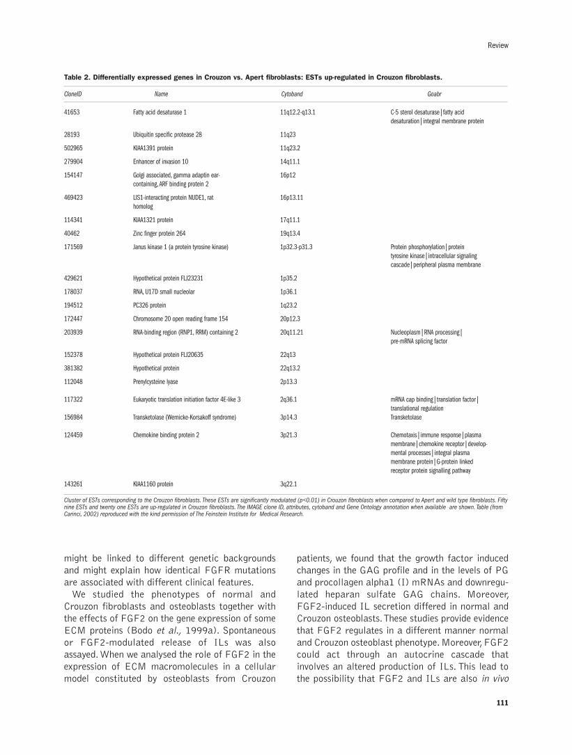
Review
might be linked to different genetic backgrounds
and might explain how identical FGFR mutations
are associated with different clinical features.
We studied the phenotypes of normal and
Crouzon fibroblasts and osteoblasts together with
the effects of FGF2 on the gene expression of some
ECM proteins (Bodo et al., 1999a). Spontaneous
or FGF2-modulated release of ILs was also
assayed. When we analysed the role of FGF2 in the
expression of ECM macromolecules in a cellular
model constituted by osteoblasts from Crouzon
patients, we found that the growth factor induced
changes in the GAG profile and in the levels of PG
and procollagen alpha1 (I) mRNAs and downregu-
lated heparan sulfate GAG chains. Moreover,
FGF2-induced IL secretion differed in normal and
Crouzon osteoblasts. These studies provide evidence
that FGF2 regulates in a different manner normal
and Crouzon osteoblast phenotype. Moreover, FGF2
could act through an autocrine cascade that
involves an altered production of ILs. This lead to
the possibility that FGF2 and ILs are also in vivo
111
Table 2. Differentially expressed genes in Crouzon vs. Apert fibroblasts: ESTs up-regulated in Crouzon fibroblasts.
CloneID Name Cytoband Goabr
41653 Fatty acid desaturase 1 11q12.2-q13.1 C-5 sterol desaturase|fatty acid desaturation|integral membrane protein
28193 Ubiquitin specific protease 28 11q23
502965 KIAA1391 protein 11q23.2
279904 Enhancer of invasion 10 14q11.1
154147 Golgi associated, gamma adaptin ear- 16p12containing, ARF binding protein 2
469423 LIS1-interacting protein NUDE1, rat 16p13.11homolog
114341 KIAA1321 protein 17q11.1
40462 Zinc finger protein 264 19q13.4
171569 Janus kinase 1 (a protein tyrosine kinase) 1p32.3-p31.3 Protein phosphorylation|protein tyrosine kinase|intracellular signalingcascade|peripheral plasma membrane
429621 Hypothetical protein FLJ23231 1p35.2
178037 RNA, U17D small nucleolar 1p36.1
194512 PC326 protein 1q23.2
172447 Chromosome 20 open reading frame 154 20p12.3
203939 RNA-binding region (RNP1, RRM) containing 2 20q11.21 Nucleoplasm|RNA processing|pre-mRNA splicing factor
152378 Hypothetical protein FLJ20635 22q13
381382 Hypothetical protein 22q13.2
112048 Prenylcysteine lyase 2p13.3
117322 Eukaryotic translation initiation factor 4E-like 3 2q36.1 mRNA cap binding|translation factor|translational regulation
156984 Transketolase (Wernicke-Korsakoff syndrome) 3p14.3 Transketolase
124459 Chemokine binding protein 2 3p21.3 Chemotaxis|immune response|plasma membrane|chemokine receptor|develop-mental processes|integral plasma membrane protein|G-protein linkedreceptor protein signalling pathway
143261 KIAA1160 protein 3q22.1
Cluster of ESTs corresponding to the Crouzon fibroblasts. These ESTs are significantly modulated (p<0.01) in Crouzon fibroblasts when compared to Apert and wild type fibroblasts. Fiftynine ESTs and twenty one ESTs are up-regulated in Crouzon fibroblasts. The IMAGE clone ID, attributes, cytoband and Gene Ontology annotation when available are shown. Table (fromCarinci, 2002) reproduced with the kind permission of The Feinstein Institute for Medical Research.

112
jointly involved in the bone-remodelling microenvi-
ronment as local coupling factors. In another work
(Bodo, 2000), we provided the first evidence that
fibroblasts obtained from patients affected by
Crouzon syndrome retain their capacity to respond
to FGF2, despite mutations in the high-affinity
FGF2 receptor. The growth factor reduces IL-1
secretion, down-regulates biglycan and procollagen
alpha, and increases betaglycan gene expressions.
Since betaglycan is a co-receptor for FGF2 signal-
ing, we suggested an alternative signal transduction
pathway in Crouzon fibroblasts to explain the doc-
umented changes in ECM macromolecule produc-
tion. Finally, we analyzed the role of some FGF sig-
nalling molecules involved in FGFR2 regulation
and their effects on the ECM (Bodo et al., 2002).
Compared with normal fibroblasts, excess
fibronectin catabolism is present in Crouzon fibrob-
lasts and differences were more marked when
FGF2 was added. Very few phosphoproteins were
visible in anti-Grb2 immunoprecipitations from
Crouzon fibroblasts, which showed a significant
increase in the number of high affinity and low-
affinity FGF2 receptors. These results suggest that
the abnormal genotype and the Crouzon cellular
phenotype are related. To compensate the low levels
of tyrosine phosphorylation, Crouzon cells might
increase the numbers of FGFR2, thus increasing
the cell surface binding sites for FGF2.
Non-syndromic CLPWe studied (Bodo et al., 1999b) TGF-α, TGF-β,
and TGF-β3 expressions and their effects on ECMmacromolecule production of normal and cleft
palatal fibroblasts in vitro, to investigate the mech-
anisms by which the phenotypic modulation of
fibroblasts occurs during the cleft palate process.
TGF-β isoforms and ECM components were dif-
P. Carinci et al.
Figure 2. Representative samples of thesemiquantitative radioactive RT-PCRused to quantitate the mRNA levels ofdifferent specific genes. Beta-actin wasused as internal control in all PCRs. Cand RA represent untreated and 10µMRA-treated fibroblasts respectively. Theamplification products were elec-trophoresed on 6% polyacrylamide gels.Gels were dried and exposed for elec-tronic autoradiography. Values of semi-quantitative analysis are reported inTable 3. Similar results were seen in fourindependent experiments for each of the4 patients; each experiment was per-formed in quadruplicate. Figure (fromBaroni, 2006) reproduced with the kindpermission of The Feinstein Institute forMedical Research.

ferently expressed and were correlated to the CLP
phenotype. In particular, CLP fibroblasts produced
more GAG and collagen than normal fibroblasts
and when all three TGF-β isoforms were added,
ECM production increased even more. Thus,
strength was given to the hypothesis that TGF-βisoforms are the potential inducers of phenotypic
expression in palatal fibroblasts during develop-
ment and that an autocrine growth factor produc-
tion mechanism may be responsible for the pheno-
typic modifications. TGF-β �is also involved in reg-ulating the interleukin network and IL-1 and IL-6
in particular (Bodo et al., 1998b; Schluns et al.,
1997). IL-6 is a multifunctional cytokine, which,
unlike TGF-β, reduces connective macromoleculeproduction (Roodman et al. 1992). Interactions
between IL-6 and TGF-β3 trigger a cascade ofevents that control developmental processes. We
speculated that a concerted action of TGF-β3 andIL-6 promotes the ECM composition of the CLP
fibroblast phenotype. To test this hypothesis, we
examined collagen, GAG and biglycan proteogly-
can (PG) synthesis in response to IL-6 and deter-
mined how IL-6 production and biglycan expres-
sion were modified in CLP fibroblasts after TFG-
β3 exposure. Our data (Baroni, et al. 2002, 2003)suggested the increase in matrix components that
characterize the CLP fibroblast phenotype might
be due to a concerted TGF-β3-IL-6 action. Wehypothesized changes in cross-talk between TGF-
β3 and IL-6 signal transduction pathways areinvolved in the induction of cleft palate.
During embryogenesis, retinoic acid (RA) and
gamma-aminobutyric acid (GABA)ergic signaling
systems are also potentially involved. We aimed to
verify the presence of phenotypic differences
between primary cultures of secondary palate (SP)
fibroblasts from 2-year old subjects with familial
non-syndromic cleft lip and/or palate (CLP-SP
fibroblasts) and age-matched normal SP (N-SP)
fibroblasts (Baroni et al., 2006). The effects of RA
which, at pharmacologic doses, induces cleft palate
in newborns of many species were also studied. We
demonstrated for the first time that GABA receptor
(GABRB3) mRNA expression was upregulated in
human CLP-SP fibroblasts (Figure 2) (Table3).
RA treatment increased TGF3 and RARA gene
expression in both cell populations but upregulated
GABRB3 mRNA expression only in N-SP cells
(Figure 2) (Table3). These results show that CLP-
SP fibroblasts exhibit an abnormal phenotype in
vitro, respond differently to RA treatment and sug-
gest that altered cross-talk between RA,
GABAergic and TGF-β signaling systems could be
involved in human cleft palate fibroblast phenotype.
Hence, normal orofacial configuration is the end-
product of highly regulated interplay between ECM
molecules and cells from the epithelium and mes-
enchyme which produce growth factors such as the
TGF-β family members (TGF-β1, TGF-β2, andTGF-β3). All three mammalian TGF-β isoforms areexpressed during palatal development and exact
timing and spatial expression are required. TGF-β3appears to play a pivotal role, since TGF-β3 genemutations and/or deficiences give rise to cleft
palate in humans (Lidral et al., 1998) and mice
(Kaartinen et al., 1995). Our data extend previous
findings (Bodo et al., 1999b; Baroni et al., 2003)
that CLP-SP fibroblasts retain an abnormal phe-
notype in vitro which we have studied in terms of
ECM production, TGF-β system, RARA and
GABRB3 expression and different response to RA.
The results contribute to a better understanding of
the interactions between RA and TGF-β signaling
pathways and support the hypothesis that altered
cross-talk between TGF-β and RA signaling sys-
tems plays a role in eliciting the CLP phenotype in
humans.
Our group has investigated the possible involve-
ment of genes coding for growth factors in the eti-
ology of CLP in recent years. The earlier investiga-
tions regarded TGFA gene, which was studied by
both allelic association and linkage analyses
(Scapoli et al, 1998; Pezzetti et al., 1998). We
113
Review
Table 3. Semiquantitative analysis of mRNA for TGF-beta3,TGFBR1, TGFBR2, TGFBR3, RARA and GABRB3 in normal andCLP fibroblasts treated or not with RA.
Normal fibroblasts CLP fibroblasts
Control RA Control RA
TGF-β3 100±13 242±27* 129±15† 205±23*
TGFBR1 100±13 65±7* 93±10‡ 51±6*
TGFBR2 100±11 267±28* 100±10‡ 257±27*
TGFBR3 100±12 95±11 NS 96±12‡ 111±13 NS
RARA 100±14 170±19* 284±32§ 459±49*
GABRB3 100±11 307±35* 486± 53§ 531±61 NS
Actin 100±11 95±10 NS 90±11‡ 82±10 NS
The values indicate mRNA levels, corrected for beta-actin mRNA levels and expressedas the percentage of untreated normal fibroblasts. All values are mean ± SD of fourseparate experiments performed in quadruplicate. The results are analysed by ANOVA.Differences of CLP fibroblasts vs. normal fibroblasts: §F-test significant at 99%; †F-testsignificant at 95%; ‡not significant. Differences vs. control: *F-test significant at 99%;NS, not significant. Table (from Baroni), 2006 reproduced with the kind permission ofThe Feinstein Institute for Medical Research.
observed no allelic association with the Taq I poly-
morphism, however genetic linkage between
microsatellite markers and putative disease locus
was detected in a subset of families. Taking togeth-
er these data suggest a possible role of TGFA gene
or a nearby gene in CLP onset.
On investigating the TGFB3 locus (14q24), our
group obtained only borderline results, thus we were
unable to distinguish whether this gene contributed
or not to the etiology of CLP in our sample
(Scapoli 2002). On the other hand, our family
based investigation, even if with slight statistical
evidence, supports a role for the RARA gene in CLP
disease (Scapoli 2002). Interestingly, our group
observed a significant relationship between the β 3
subunit of the gamma-aminobutyric acid receptor
(GABRB3) and CLP, suggesting that the GABRB3
gene is involved in this congenital disease. Although
GABR is the target of benzodiazepine, none of our
patients presented neurologic diseases. In the same
study, it was also demonstrated that the GAD1
gene, which encodes the GABA-producing enzyme,
is not involved in CLP pathogenesis.
Conclusions
Taken together, these data suggest that the
changes in the distribution of ECM components
participate in the regulation of the complex mor-
phogenetic events that occur during cranial and
orofacial development. Several growth factors are
involved in this cascade of events, each playing a
role in the commitment of calvaria and orofacial
cells to different phenotypes. The balance among
ECM components, cytokines and growth factors as
FGF2 and TGF-β probably determines the degree
and extent of induced cellular response. Research
into the mechanisms regulating this balance has
entered an exciting phase also thanks to cultures
from Apert, Crouzon and CLP patients that provide
a promising model for these studies in view of ther-
apeutic strategies as a complement to surgery.
References
Baroni T, Lilli C, Marinucci L, Bellocchio S, Pezzetti F, Carinci F, et al.Crouzon's syndrome: differential in vitro secretion of bFGF, TGFbetaI isoforms and extracellular matrix macromolecules in patients withFGFR2 gene mutation. Cytokine 2002;19:94-101.
Baroni T, Carinci P, Bellucci C, Lilli C, Becchetti E, Carinci F, et al.Cross-talk between interleukin-6 and transforming growth factor-beta3 regulates extracellular matrix production by human fibrob-lasts from subjects with non-syndromic cleft lip and palate. JPeriodontol 2003; 74:1447-53.
Baroni T, Carinci P, Lilli C, Bellucci C, Aisa MC, Scapoli L, et al.
P253R fibroblast growth factor receptor-2 mutation inducesRUNX2 transcript variants and calvarial osteoblast differentiation.J Cell Physiol 2005; 202:524-35.
Baroni T, Bellucci C, Lilli C, Pezzetti F, Carinci F, Becchetti E, et al.Retinoic acid, GABA-ergic and TGF-beta signaling systems areinvolved in human cleft palate fibroblast phenotype. Mol Med2006;12:237-45.
Bodo M, Carinci F, Baroni T, Becchetti E, Giammarioli M, Bellucci C,et al. Effects of interleukins on Crouzon fibroblast phenotype invitro. Release of cytokines and IL-6 mRNA expression. Cytokine1996; 8:772-83
Bodo M, Carinci F, Baroni T, Giammarioli M, Bellucci C, Bosi G, et al.Apert's syndrome: differential in vitro production of matrix macro-molecules and its regulation by interleukins. Eur J Clin Invest 1997;27:36-42.
Bodo M, Carinci F, Baroni T, Becchetti E, Bellucci C, Giammarioli M,et al. Interleukin pattern of Apert fibroblasts in vitro. Eur J Cell Biol1998a;75:383-8.
Bodo M, Carinci P, Baroni T, Bellucci C, Giammarioli M, Pezzetti F, etal. Role of growth factors on extracellular matrix production bychick embryo fibroblasts in vitro. Antagonist effect of TGF-βthrough the control of IL-1 and IL-1Ra secretion. Cytokine1998b;10: 353-60.
Bodo M, Baroni T, Carinci F, Becchetti E, Bellucci C, Conte C, et al. Aregulatory role of fibroblast growth factor in the expression ofdecorin, biglycan, betaglycan and syndecan in osteoblasts frompatients with Crouzon's syndrome. Eur J Cell Biol. 1999a;78:323-30.
Bodo M, Baroni T, Carinci F, Becchetti E, Bellucci C, Pezzetti F, et al.TGFbeta isoforms and decorin gene expression are modified infibroblasts obtained from non-syndromic cleft lip and palate sub-jects. J Dent Res 1999b;78:1783-90.
Bodo M, Lilli C, Aisa MC, Scapoli L, Bellucci C, Rinaldi E, et al. Basicfibroblast growth factor: Effects on matrix remodeling, receptorexpression, and transduction pathway in human periosteal fibrob-lasts with fgfr2 gene mutation. J Interferon Cytokine Res 2002a ;22: 621-30.
Canalis E, McCarthy TL, Centrella M. The role of growth factors inskeletal remodeling. Endocrinol Metab Clin North Am 1989;18:903-18.
Carinci F, Pezzetti F, Scapoli L, Padula E, Baciliero U, Curioni C, et al.Nonsyndromic cleft lip and palate: evidence of linkage to amicrosatellite marker on 6p23. Am J Hum Genet 1995;56:337-9.
Carinci F, Bodo M, Tosi L, Francioso F, Evangelisti R, Pezzetti F, et al.Expression profiles of craniosynostosis-derived fibroblasts. MolMed. 2002; 8:638-44.
Carinci F, Pezzetti F, Scapoli L, Martinelli M, Avantaggiato A, CarinciP, et al. Recent developments in orofacial cleft genetics. J CraniofacSurg. 2003;14:130-43.
Carinci F, Pezzetti F, Locci P, Becchetti E, Carls F, Avantaggiato A, etal. Apert and Crouzon syndromes: clinical findings, genes and extra-cellular matrix. J Craniofac Surg 2005;16:361-8.
Cohen MM Jr, MacLean RE. Craniosynostosis: Diagnosis, evaluation,and management. 2nd edn. New York: Oxford University Press,2000.
Delezoide AL, Benoist-Lasselin C, Legeai-Mallet L, Le Merrer M,Munnich A, Vekemans M, et al. Spatio-temporal expression of FGFR1, 2, and 3 genes during human embryo-fetal ossification. Mech Dev1998; 77:19-30.
Ducy P. Cbfa1: A molecular switch in osteoblast biology. Dev Dyn2000; 219: 461-71.
Frenz DA, Liu W, Williams JD, Hatcher V, Galinovic-Schwartz V,Flanders KC, et al. Induction of chondrogenesis: requirement forsynergistic interaction of basic fibroblast growth factor and trans-forming growth factor-beta. Development 1994;120:415-24.
Ibrahimi OA, Zhang F, Eliseenkova AV, Itoh N, Linhardt RJ,Mohammadi M. Biochemical analysis of pathogenic ligand-depen-dent FGFR2 mutations suggests distinct pathophysiological mecha-nisms for craniofacial and limb abnormalities. Hum Mol Genet2004, 13: 2313-24.
Janssens K, ten Dijke P, Janssens S, Van Hul W. Transforming growthfactor-beta1 to the bone. Endocr Rev 2005;26:743-74.
Kaartinen V, Voncken JW, Shuler C, Warburton D, Bu D, HeisterkampN. Abnormal lung development and cleft palate in mice lacking
114
P. Carinci et al.
TGF-beta 3 indicates defects of epithelial-mesenchymal interaction.Nat Genet 1995; 11: 415-21.
Kan SH, Elanko N, Johnson D, Cornejo-Roldan L, Cook J, Reich EW,et al. Genomic screening of fibroblast growth-factor receptor 2reveals a wide spectrum of mutations in patients with syndromiccraniosynostosis. Am J Hum Genet 2002; 70:472-486
Kreiborg S, Marsh JL, Cohen MM Jr, Liversage M, Pedersen H,Skovby F, et al. Comparative three-dimensional analysis of CT-scansof the calvaria and cranial base in Apert and Crouzon syndromes. JCraniomaxillofac Surg 1993; 21:181-88.
Lidral AC, Romitti PA, Basart AM, Doetschman T, Leysens NJ, Daack-Hirsch S, Semina EV, Johnson LR, Machida J, Burds A, Parnell TJ,Rubenstein JL, Murray JC. Association of MSX1 and TGFB3 withnonsyndromic clefting in humans. Am J Hum Genet 1998; 63: 557-68.
Locci P, Baroni T, Pezzetti F, Lilli C, Marinucci L, Martinese D, et al.Differential in vitro phenotype pattern, transforming growth factor-beta(1) activity and mRNA expression of transforming growth fac-tor-beta(1) in Apert osteoblasts. Cell Tissue Res 1999; 297:475-83.
Malemud CJ. Matrix metalloproteinases (MMPs) in health and dis-ease: an overview. Front Biosci 2006;11:1696-701.
Massaguè J. Identification of receptors for type-beta transforminggrowth factor. Methods Enzymol. 1987; 146:174-95.
Morriss-Kay GM, Wilkie AO. Growth of the normal skull vault and itsalteration in craniosynostosis: insights from human genetics andexperimental studies. J Anat 2005; 207:637-53.
Moursi AM, Damsky CH, Lull J, Zimmerman D, Doty SB, Aota S, etal. Fibronectin regulates calvarial osteoblast differentiation. J CellSci 1996;109:1369-80.
Naski MC, Ornitz DM. FGF signaling in skeletal development. FrontBiosci 1998; 3: 781–94.
Pezzetti F, Scapoli L, Martinelli M, Carinci F, Bodo M, Carinci P, et al.A locus in 2p13-p14 (OFC2), in addition to that mapped in 6p23, isinvolved in nonsyndromic familial orofacial cleft malformation.Genomics 1998;50:299-305.
Qiu C, Ferguson M. The distribution of PDGFs and PDGFreceptorsduring murine secondary palate development. J Anat 1995;186:17–29.
Robertson SC, Meyer AN, Hart KC, Galvin BD, Webster MK, DonoghueDJ. Activating mutations in the extracellular domain of the fibrob-last growth factor receptor 2 function by disruption of the disulfidebond in the third immunoglobulin-like domain. Proc Natl Acad SciUSA 1998;95: 4567-72.
Roodman GD, Kurihara N, Ohsaki Y. Interleukin-6: Apotentialautocrine/paracrine factor in Paget’s disease of bone. J Clin Invest1992;89:46-52.
Scapoli L, Pezzetti F, Carinci F, Martinelli M, Carinci P, Tognon M.
Evidence of linkage to 6p23 and genetic heterogeneity in nonsyn-
dromic cleft lip with or without cleft palate. Genomics 1997;
15;43:216-20.
Scapoli L, Pezzetti F, Carinci F, Martinelli M, Carinci P, Tognon M.
Lack of linkage disequilibrium between transforming growth factor
alpha Taq I polymorphism and cleft lip with or without cleft palate
in families from Northeastern Italy. Am J Med Genet 1998;75:203-
6.
Scapoli L, Martinelli M, Pezzetti F, Carinci F, Bodo M, Tognon M, et al.
Linkage disequilibrium between GABRB3 gene and nonsyndromic
familial cleft lip with or without cleft palate. Hum Genet
2002;110:15-20.
Schluns KS, Cook JE, Le PT. TGF-beta differentially modulates epi-
dermal growth factor-mediated increases in leukemia-inhibitory fac-
tor, IL-6, IL-1 alpha, and IL-1 beta in human thymic epithelial cells.
J Immunol 1997;2704-11.
Stein JD, Hecht JT, Blanton SH. Exclusion of retinoic acid receptor
and a cartilage matrix protein in non-syndromic CL(P) families. J
Med Genet 1995;32: 78.
Szebenyi G, Fallon JF. Fibroblast growth factors as multifunctional
signaling factors. Int Rev Cytol 1999;185:45-106.
Thorogood P. The developmental specification of the vertebrate skull.
Development 1988;103:141-153.
Yayon A, Klagsbrun M. Autocrine transformation by a chimeric signal-
peptide-basic fibroblast growth factor: Reversal by suramin. Proc
Natl Acad Sci USA 1990; 87: 5346-50.
Young DL, Schneider RA, Hu D, Helms JA. Genetic and teratogenic
approaches to craniofacial development. Crit Rev Oral Biol Med
2000; 11:304-17.
Wilkie AO. Bad bones, absent smell, selfish testes: the pleiotropic con-
sequences of human FGF receptor mutations. Cytokine Growth
Factor Rev 2005; 16:187–203.
Wilkie AO, Bochukova EG, Hansen RM, Taylor IB, Rannan-Eliya SV,
Byren JC, et al. Clinical dividends from the molecular genetic diag-
nosis of craniosynostosis. Am J Med Genet A 2006 (Epub ahead of
print)
Xu T, Bianco P, Fisher LW, Longenecker G, Smith E, Goldstein S, et al.
Targeted disruption of the biglycan gene leads to an osteoporosis-
like phenotype in mice. Nat Genet 1998;20:78-82.
Yu K, Herr AB, Waksman G, Ornitz DM. Loss of fibroblast growth fac-
tor receptor 2 ligand-binding specificity in Apert syndrome. Proc
Natl Acad Sci USA 2000; 97:14536-41.
115
Original Paper

116
P. Carinci et al.

117
REVIEW
©2007, European Journal of Histochemistry
Here we present an overview of the experimental evidenceand of the conceptual basis for the involvement of laminsand nuclear envelope proteins in a group of genetic diseasescollectively referred to as laminopathies. Some of these dis-eases affect a specific tissue (skeletal and/or cardiac mus-cles, subcutaneous fat, peripheral nerves), while othersaffect a variety of tissues; this suggests that the pathogenicmechanism of laminopathies could reside in the alteration ofbasic mechanisms affecting gene expression. On the otherhand, a common feature of cells from laminopathic patientsis represented by nuclear shape alterations and heterochro-matin rearrangements. The definition of the role of lamins inthe fine regulation of heterochromatin organization may helpunderstanding not only the pathogenic mechanism oflaminopathies but also the molecular basis of cell differenti-ation and ageng.
Key words: nuclear envelope, heterochromatin, lamino-pathies, prelamin A, ageing.
Correspondence: Nadir M. Maraldi,Laboratory of Cell Biology and Electron Microscopy,Istituto Ortopedico Rizzoli, via di Barbiano, 1/10 40136 Bologna, ItalyTel: +39.051.6366856.Fax: +39.051.583593.E-mail: [email protected]
European Journal of Histochemistry2007; vol. 51 supplement 1:117-124
The nuclear envelope, human genetic diseases and ageing
N.M. Maraldi,1,2,3 G. Mazzotti,1 R. Rana,4 A. Antonucci,4 R. Di Primio,5 L. Guidotti6
1Department of Scienze Anatomiche Umane e Fisiopatologia Apparato Locomotore, University of
Bologna; 2IGM, C.N.R., Unit of Bologna, c/o I.O.R., Bologna; 3Laboratory of Cell Biology and Electron
Microscopy, Istituto Ortopedico Rizzoli, Bologna; 4Department of Biomorfologia, Università G. d’Annunzio,
Chieti; 5Institute of Morfologia Umana Normale, Università Politecnica delle Marche, Ancona;6Department of Istologia, University of Bologna, Italy
In the last twenty years our research group has
been interested in the study of the molecular
organization of the cell nucleus. These investiga-
tions have been performed by using a combined
approach of biochemical, cytochemical, and ultra-
structural procedures in order to obtain a compre-
hensive design of the morphology and function of the
different intranuclear compartments. The cell nucle-
us presents an organization at least complex as the
cytoplasm; furthermore, whilst the cytoplasm can be
subdivided into cell membrane delimited compart-
ments, intranuclear structures are not membrane-
bounded and are frequently intermixed. These struc-
tures can be defined nuclear domains when can be
identified by light and/or electron microscopy, visual-
ized in vivo by GFP-tagged constructs, isolated in an
enriched form to be biochemically analyzed, and
characterized by a specific class of stably associated
proteins. Once identified, the nuclear domains can be
studied in a dynamic way, in order to determine their
functions.
Our research group contributed to the knowledge
of some aspects of the functional organization of the
main recognized nuclear domains, including the
nuclear matrix (Maraldi et al., 1986; Tait et al.,
1998), the nucleolus (Zini et al., 1994), the splicing
domain (Maraldi et al., 1999a; Bavelloni et al.,
2006), the chromosome territories (Cinti et al.,
1993; Squarzoni et al., 1994; Maraldi et al.,
1999b), and the nuclear signal transduction system
(Maraldi et al., 1992; Mazzotti et al., 1995; Maraldi
et al., 1999a; Maraldi et al., 2000).
In this report, we present the main advance we
obtained in the study of the nuclear envelope and, in
particular, in its involvement in the pathogenesis of a
variety of human genetic diseases.
The nuclear envelopeThe cell nucleus is delimited by the nuclear enve-
lope (NE), constituted by the outer nuclear mem-
brane (ONM), which is part of the endoplasmic

reticulum, by the inner nuclear membrane (INM),
devoid of ribosomes and presenting a set of specific
proteins, and by the nuclear lamina (NL). Our stud-
ies have been particularly devoted to the functions
of the INM-associated proteins and of the lamins,
that are expressed into the nuclear lamina.
The NL appears as a continuous structure, with a
thickness variable from 10 to 300 nm, located
between the INM and the peripheral heterochro-
matin. The ultrastructural analysis identified a 3D
organization of the nuclear lamina in situ only in the
Xenopous oocytes, where it appears as a net with
square meshes formed by 10 nm thick filaments
(Burke and Stewart, 2002). We demonstrated a
similar organization in rat liver isolated nuclei, ana-
lyzed by freeze-fracturing (Maraldi et al., 1986).
The NL is constituted by type V intermediate fila-
ments, the lamins; type B lamins are expressed in
almost all the cells, whilst A type lamins are
expressed in a tissue specific way during cell differ-
entiation. Nuclear lamins undergo transition from
the polymerized to the un-polymerized state, thus
contributing to the NE breaking and formation at
each cell cycle. The first step of the latter process
requires lamin B1 interaction with condensed chro-
mosomes in telophase, the following recruitment of
membrane vesicles capable of interacting with the
nuclear pore complexes (NPCs) and to fuse to form
the perinuclear cisterna, and then the contribute of
lamins A/C to assemble the nuclear lamina. The
assembly of type B lamins with lamins A/C is essen-
tial for the correct NPCs organization, through the
interaction of lamin B1 and the NPC-associated
protein NUP153 (Holaska et al., 2002).
The NE formation also depends on the presence of
a wide set of INM-associated proteins and of some
chromatin-associated proteins. The INM-associated
proteins, once synthesized in the RER, have to inter-
act with lamins or with chromatin, or both, to be
integrated into the INM (Zastrow et al., 2004).
Among the INM-associated proteins, those contain-
ing the LEM domain, that is LAP2, emerin, and
MAN1, interact with the lamins as well as with some
chromatin-associated proteins, including BAF and
HP1. The INM-associated protein LBR, which lacks
the LEM domain, also interacts with both lamin B
and HP1, as well as with DNA and H3/H4 histones.
LAP2α is mainly located in intranuclear regions and
interacts with lamin A/C and chromatin, whilst the
other isoforms of the protein are exclusively present
at the nuclear lamina level. The INM-associated
proteins participate to the NE assembly and to the
chromosome decondensation, being initially LAP2βand LBR involved into an interaction with lamins in
non-centromeric regions of the chromosomes, and
then LAP2α and emerin in the centromeric regions
(Shumaker et al., 2003; Gruenbaum et al., 2005).
Since lamin immunodepletion or the expression of
dominant negative lamins induce the block of repli-
cation, lamins are conceivably interacting with repli-
cation complexes (Zastrow et al., 2004).
Furthermore, the strict association of the nuclear
lamina with the heterochromatin suggests that
lamins could contribute to the repression of gene
transcription. Gene-rich chromosome domains,
indeed, are generally located in inner zones of the
nucleus, whilst gene-poor regions are located close
to the nuclear lamina. On the other hand, both tran-
scription and RNA processing require a correct
expression and an intranuclear localization of
lamins, suggesting that these nuclear activities
require the presence of a nucleoskeletal structure
containing lamins (Gruenbaum et al., 2005).
Interestingly, some transcription factors have been
localized at the nuclear lamina, where they interact
with lamins or INM-associated proteins. Most of
these transcription factors are inhibitory, such as
Oct-1, pRb, GCL, and SREBP1 (Maraldi and
Lattanzi, 2005). Finally, the documented interac-
tions of lamins with proteins that are involved in the
chromatin remodelling, such as HP1, H3/H4 histone
tetramers, and the nuclear actin bound to the
SWI/SNF remodelling complex, suggest that tran-
scription could be repressed by affecting the whole
conformational arrangement of the chromosome
domains (Maraldi et al., 2004).
The induction phases of apoptosis require lamin
proteolysis, which is preceded by lamin phosphory-
lation through PKCα and PKCδ. Lamin hydrolysisby caspases precedes DNA fragmentation and the
lysis of INM-associated proteins, and the cells in
which the lamin expression has been reduced under-
go apoptotic alterations (Holaska et al., 2002).
It is evident, therefore, that lamins and INM-asso-
ciated proteins, not only contribute to the NE
assembly, but play a variety of functions, which are
essential for the control of cell viability, replication
and differentiation. As a consequence, the altered
expression of these nuclear envelope proteins could
result in diseases. In recent years, a wide range of
inherited diseases, collectively termed nuclear
envelopathies if mutations arise in INM-associated
118
N.M. Maraldi et al.
proteins, or laminopathies if mutations arise in
lamins, have been identified. Therefore, attention has
been focused on the molecular characteristics of
these NE components, in order to clarify the patho-
genic mechanisms that could account for the com-
plexity of the observed phenotypic alterations found
in these diseases (Maraldi et al., 2002).
Nuclear envelopathies and laminopathies presentan impressive variety of disease phenotypes butcommon nuclear alterationsAt the moment, disease-causing mutations have
been reported for seven genes coding for nuclear
envelope proteins, i.e. EMD, LMNA, ZMPSTE24,
LBR, MAN1, LAP2, and AAAS (Broers et al.,
2004). Our interest has been mainly devoted to one
nuclear envelopathy, i.e. the X-linked form of Emery-
Dreifuss muscular dystrophy XL-EDMD, due to
mutation of the EMD gene, coding for emerin
(Maraldi et al., 2002; Maraldi and Merlini, 2004),
and to the large group of primary laminopathies, due
to mutation of the LMNA gene, coding for lamin
A/C (Maraldi et al., 2004). Primary laminopathies
include at least ten different diseases in which spe-
cific tissues are affected in isolated fashion, or sev-
eral tissues are systemically involved; according to
these criteria laminopathies can be grouped into five
classes: 1) striated muscle; 2) peripheral nerves; 3)
adipose tissue; 4) premature ageing; 5) overlapping
syndromes (Broers et al., 2006).
The striated muscle laminopathies include AD-
EDMD and AR-EDMD, the autosomal and recessive
forms of the Emery-Dreifuss muscular dystrophy,
which present skeletal and cardiac muscle involve-
ment as well as joint contractures, almost identical
to those found in XL-EDMD. Other striated muscle
laminopathies are the dilated cardiomyopathy with
conduction system defects (DCM-CD), character-
ized by progressive conduction system disease and
dilated cardiomyopathy without skeletal muscle
involvement, and the limb-girdle muscular dystrophy
type 1B (LGMD1B), with cardiological abnormali-
ties and proximal muscle weakness and wasting.
The laminopathies with peripheral nerve involve-
ment include the Charcot-Marie-Tooth type 2
(CMT2B1), characterized by the demyelinization of
motor nerves with a secondary wasting of the distal
lower limb muscles and the autosomal dominant
axonal Charcot-Marie-Tooth disease (AD-CMT2),
characterized by an axonal involvement associated
with muscular dystrophy, cardiac disease and partial
lipodystrophy.
Partial lipodystrophy due to mutation of the
LMNA gene is an autosomal dominant disorder
referred to as familial partial lipodystrophy of
Dunningan type (FPLD). Patients, with the onset of
puberty, present an almost complete loss of subcu-
taneous fat from the upper and lower extremities,
and gluteal and truncal areas, whilst fat accumu-
lates on face and neck, as well as in the intra-
abdominal regions. Patients are insulin resistant and
may develop diabetes, hypertriglycideridemia and
atherosclerotic vascular diseases. A further disease,
identified as type A insulin resistance syndrome,
characterized by polycystic ovary with severe hyper-
androgenism, and severe insulin resistance, is also
due to mutation of the LMNA gene, restricted at a
single aminoacid (G602S). Interestingly, mutations
occurring in FPLD affect residues at the surface of
the Ig fold domain downstream of the nuclear local-
ization signal (NLS) of the lamin A, whilst those
found in striated muscle and peripheral nerve
laminopathies are mainly located in the central rod
domain upstream of the NLS (Hegele, 2005).
The very intriguing class of systemic lamino-
pathies has been recently identified, which include
the mandibuloacral dysplasia (MAD-A), the
Hutchinson-Gilford progeria syndrome (HGPS), the
atypical Werner syndrome (WRN-like), the general-
ized lipoatrophy, insulin-resistant diabetes, leukome-
lanodermic papules, liver steatosis, and hypertrophic
cardiomyopathy (LIRLLC), the restrictive dermopa-
thy (RD), and the lethal fetal akinesia (SFAk). In
MAD-A, lipodystrophy is associated to skeletal
abnormalities, such as mandibular and clavicular
acroosteolysis, joint contractures, delayed closure of
cranial suture, and postnatal growth retardation.
HGPS and WRN-like are premature ageing syn-
dromes, in which, besides the symptoms present in
MAD-A, other tissues and mainly the vascular sys-
tem undergo a premature ageing. HGPS patients, as
a consequence of widespread atherosclerosis, die at
a median age of 13 years. On the other hand, RD and
SFAk are characterized by an impressive variety of
systemic disorders so that they are lethal before or
within few days after birth.
Finally, there are an increasing number of cases in
which LMNA mutations have been reported to
occur in individuals or families harbouring several
tissue involvements, suggesting the presence of an
overlapping continuum within the different types of
laminopathies.
119
Review
The impressive amount of disease phenotypes of
laminopathies rises the question of how mutations in
a gene, which is expressed in nearly every differenti-
ated cell and codes for a structural protein of the
nucleus, could give rise to such a variety of tissue-
restricted pathologies.
Laminopathy disease modelsLamins provide a structural scaffold that main-
tains the nuclear integrity, but they are also inter-
acting with nuclear proteins that modulate chro-
matin arrangement, gene expression and cell cycle
progression. On the basis of these considerations,
different models, that could account for a pathogen-
ic mechanism of laminopathies, were proposed.
The first model suggests that mutant lamin A
results in alteration of the nuclear lamina mechani-
cal properties, and mainly of its resistance to
mechanical stress (Hutchison et al., 2001). This
could lead, in particular in contractile tissues, to
nuclear damages and cell death, provided that
abnormal lamin assembly may destabilize the inter-
actions between the nuclear and the cytoskeletal
networks (Broers et al., 2004).
Another model proposes that mutant lamin A
causes misregulation of different tissue-specific gene
expression, either directly or at the epigenetic level.
In fact several transcriptional regulators have been
found to interact with lamins and their influence on
cell cycle progression and differentiation may be
affected by the expression of mutant lamins
(Zastrow et al., 2004). The details of the molecular
mechanisms underlying this regulation remain
unclear, but lamin-dependent regulation of gene
expression could occur at the epigenetic level, by
modulating heterochromatin organization (Maraldi
et al., 2005).
A further model proposes that mutations in
LMNA impair the balance between cell proliferation
and differentiation that represents the regulatory
mechanism controlling adult stem cells involved in
tissue regeneration (Gotzmann and Foisner, 2005).
The proposed models are not mutually exclusive,
and it is possible that a combination of these mech-
anisms may contribute to various degrees to the
diverse disease phenotypes observed in laminopath-
ic patients. It was initially thought that mutated or
incorrectly spliced lamin variants result in a loss-of-
function phenotype; as a consequence, the structur-
al model, in which a loss of lamin stability could
account for nuclear fragility, was consistent with the
observed muscle tissue alterations. However, since
almost all the inherited changes in LMNA are het-
erozygous, it is assumed that the mutated protein
dominantly affects the structure and/or the function
of the wild-type lamin A expressed by the unaffect-
ed allele, resulting in a gain-of-function phenotype.
In support of this type of pathogenic model,
recently we have obtained experimental evidence in
FPLD (Capanni et al., 2005). It has been demon-
strated that in HGPS (De Sandre-Giovannoli et al.,
2003) and RD (Navarro et al., 2004), mutations in
the LMNA gene activate a cryptic splice site leading
to the expression of a shortened, incompletely
processed prelamin A that accumulates in the nucle-
us (Goldman et al., 2004). We demonstrated that
also in FPLD there is a nuclear accumulation of
incompletely processed prelamin A and that this
mutant lamin A form is able to sequester the tran-
scription factor sterol response element binding pro-
tein 1 (SREBP1), required for adipogenesis and
thus negatively affecting adipocyte differentiation as
occurs in FPLD patients and in other laminopathies
in which lipodystropic changes also occur (Capanni
et al., 2005). Furthermore, the introduction of wild-
type lamin A in cells derived from HGPS patients
was unable to rescue the abnormal phenotype, indi-
cating that the accumulation of mutant not correct-
ly processed lamin A variants, instead of a partial
loss of the wild-type protein, result into toxic effects
that impair key nuclear functions.
However, since prelamin A accumulation does not
occur in all laminopathies, is there possible to find a
detectable marker that allows one to determine
whether laminopathies present a common pathogen-
ic mechanism?
Heterochromatin altered pattern in laminopathiesOur group contributed to the characterization of
the phenotypic alterations of the firstly discovered
nuclear envelopathy, that is XL-EDMD, due to
mutation of the INM-associated protein emerin
(Bione et al., 1994). In fact, we provided evidence
that emerin is connected with the nuclear matrix
(Squarzoni et al., 1998) and could account for some
nuclear and chromatin alterations consisting in
changes in the nuclear profile with deep indentations
and in focal loss and detachments of the peripheral
heterochromatin from the nuclear lamina (Ognibene
et al., 1999). Therefore, we focused attention on the
nuclear envelope related mechanisms that modulate
the chromatin arrangement through chromatin
120
N.M. Maraldi et al.
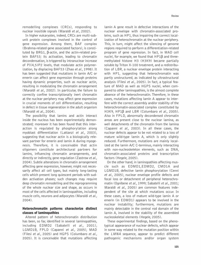
remodelling complexes (CRCs), responding to
nuclear inositide signals (Maraldi et al., 2002).
In higher eukaryotes, indeed, CRCs are multi-sub-
unit protein complexes involved in the control of
gene expression. Among them, BAF complex
(Brahma-related gene associated factors), is consti-
tuted by BRG1, β-actin, and the actin-related pro-tein BAF53; its activation, leading to chromatin
decondensation, is triggered by intranuclear increase
of PI(4,5)P2 levels, that modulate actin polymer-
ization, by displacing BAF53 (Zhao et al., 1998). It
has been suggested that mutations in lamin A/C or
emerin can affect gene expression through proteins
having dynamic properties, such as nuclear actin,
resulting in modulating the chromatin arrangement
(Maraldi et al., 2002). In particular, the failure to
correctly confine transcriptionally inert chromatin
at the nuclear periphery may affect gene expression
in crucial moments of cell differentiation, resulting
in defect in tissue regeneration in the adult organism
(Maraldi et al., 2004).
The possibility that lamins and actin interact
inside the nucleus has been experimentally demon-
strated; moreover it has been found that this inter-
action is regulated by phosphorylation along
myoblast differentiation (Lattanzi et al., 2003),
suggesting that nuclear actin is a biologically rele-
vant partner for emerin and lamin A during myoge-
nesis. Therefore, it is conceivable that actin
oligomers constitute architectural partners for
lamins, influencing chromatin arrangements, and
directly or indirectly, gene regulation (Zastrow et al.,
2004). Subtle alterations in chromatin arrangement
affecting gene expression, however, might not neces-
sarily affect all cell types, but mainly long-lasting
cells which present long quiescent periods with sud-
den activation phases; such changes may require
deep chromatin remodelling and the reprogramming
of the whole nuclear size and shape, as occurs in
most of the cells affected in laminopathies, including
muscle cells, neurons and adipocytes (Maraldi et al.,
2004).
Heterochromatin patterns characterize distinctclasses of laminopathiesAltered pattern of heterochromatin distribution
has been, so far, identified in several laminopathies,
including EDMD2 (Sabatelli et al., 2001),
LGMD1B, FPLD (Capanni et al., 2005), MAD
(Filesi et al., 2005) and HGPS (Columbaro et al.,
2005). It is conceivable that mutations affecting
lamin A gene result in defective interactions of the
nuclear envelope with chromatin-associated pro-
teins, such as HP1, thus impairing the correct local-
ization of heterochromatin at the nuclear periphery.
This, in turn, might affect the silencing of genome
regions required to perform a differentiation-related
program of gene repression. In fact, in MAD cell
nuclei, for example, we found that HP1β and three-
methylated histone H3 (H3K9) became partially
soluble by Triton X-100 treatment, and a redistribu-
tion of LBR, a nuclear envelope protein interacting
with HP1, suggesting that heterochromatin was
partly unstructured, as indicated by ultrastructural
analysis (Filesi et al., 2005). In fact, a typical fea-
ture of MAD as well as HGPS nuclei, when com-
pared to other laminopathies, is the almost complete
absence of the heterochromatin. Therefore, in these
cases, mutations affecting lamin A appear to inter-
fere with the correct assembly and/or stability of the
heterochromatin-associated complex constituted by
H3K9, HP1β and LBR (Columbaro et al., 2005).
Also in FPLD, abnormally decondensed chromatin
areas are present close to the nuclear lamina, as
well detachments of the chromatin from the lamina
(Capanni et al., 2003). In all these cases, the
nuclear defects appear to be not related to a loss of
mature wild-type lamin A, which is only slightly
reduced. Furthermore, mutations are mainly local-
ized at the lamin A/C C-terminus, mainly interacting
with non-nucleoskeleton elements, such as DNA,
chromatin-associated proteins and transcription
factors (Hegele, 2005).
On the other hand, in laminopathies affecting mus-
cle such as EDMD1,EDMD2, CMD1A and
LGMD1B, defective lamin phosphorylation (Cenni
et al., 2005), nuclear envelope profile defects and
focal loss or detachment of peripheral heterochro-
matin (Ognibene et al., 1999; Sabatelli et al., 2001;
Maraldi et al., 2005) are common features inde-
pendent of the site at which mutations occur. In
these cases, a loss of mature wild-type lamin A or
emerin (in EDMD1) appears to be involved in the
nuclear instability; furthermore, mutations are
mainly detectable in the central rod domain of the
lamin A, involved in the stability of the assembled
nucleoskeletal elements (Hegele, 2005).
These experimental findings, based on the pheno-
typical appearance of nuclear defects, which appear
in some way related to the mutation position within
the LMNA sequence, appear to predict different
pathogenic mechanisms and/or organ system
Review
121
involvement in at least two distinct classes of
laminopathies.
Heterochromatin and ageing: a lesson fromlaminopathiesThe most striking feature of non-muscular
laminopathies is the fact that nuclear and chromatin
defects appear not to be due to a loss of mature
lamin A; in fact the over-expression o wild-type
lamin A did not rescue nuclear alterations (Scaffidi
and Misteli, 2005). This suggests that nuclear
defects, and the heterochromatin loss are not due to
a loss a functional lamin A, but conceivably to a
dominant negative effect of accumulating un-prop-
erly processed lamin A. This possibility has been
largely confirmed by experimental data accumulat-
ed in the last two years.
The first hint to this pathogenic model was pro-
vided by the finding that also heterozygous single
point mutations in LMNA linked to FPLD induce a
progressive accumulation of incompletely processed
prelamin A (Capanni et al., 2005), suggesting that
at least a subset of laminopathies might be caused
through aberrant accumulation of prelamin A.
Therefore, a dominant negative effect of mutated
prelamin A seems to account for the observed dis-
ease phenotype. In support of a gain-of-function,
instead of a loss-of-function phenotype, the prelamin
A accumulation was found to result, in FPLD cells,
in a binding of the transcription factor sterol
response element binding protein 1 (SREBP1). The
recruitment by prelamin A of SREBP1 that is
required for adipogenesis could negatively affect
adipocyte differentiation (Capanni et al., 2005;
Maraldi et al., 2006).
A further hint for the understanding of prelamin A
role in the modulation of heterochromatin arrange-
ment, was obtained by analyzing the effect of farne-
syl transferase inhibitors which impair subsequent
processing of lamin A precursor protein by endopro-
teases such as the metalloprotease ZMPSTE24,
which , when mutated, gives rise to MAD (Agarwal
et al., 2003). In fact, we obtained evidence that
prelamin A accumulation by farnesyl transferase
inhibitors in myoblasts caused nuclear lamina
invagination and chromatin arrangement reorgani-
zation (Maraldi et al., 2004). Moreover, accumula-
tion of incompletely processed prelamin A has been
demonstrated to occur in FPLD (Capanni et al.,
2005) and MAD (Filesi et al., 2005). In this case,
accumulation of prelamin A resulted into an altered
distribution of LBR and in the destabilization of
HP1β and of H3K9. These changes can account fora complete heterochromatin remodelling that could
represent a key event in the epigenetic changes
involved in the pathogenesis of systemic lamino-
pathies (Filesi et al., 2005).
This pathogenic model was further confirmed also
in other laminopaties, including those characterized
by premature senescence phenotype. The two pre-
mature ageing diseases are HGPS (progeria of
childhood) an Werner’s syndrome (progeria of
adults). Most cases of HGPS result from a
Gly608Gly mutation that forms an ectopic mRNA
splicing site leading to the expression of a truncated
prelamin A lacking 50 amino acids within its tail
domain. This mutant protein termed LA∆50 or prog-erin (Goldman et al., 2004) lacks the second prote-
olytic cleavage site for the processing of lamina and
the mature protein contains eight residues of
prelamin A and is farnesylated. Fibroblasts from
HGPS patients, when propagated in culture, under-
go typical changing in nuclear shape, including lob-
ulation of the nuclear envelope, thickening of the
nuclear lamina, clustering of the nuclear pore com-
plexes and almost total loss of peripheral hete-
rochromatin (Columbaro et al., 2005). Thus, we pro-
posed that a key element of chromatin-remodeling
complexes may be prelamin A, whose post-transla-
tional modifications may serve as regulatory mech-
anisms affecting higher order chromatin organiza-
tion (Maraldi and Lattanzi, 2005).
A direct demonstration that nuclear alterations in
HGPS are caused by a concentration-dependent
dominant-negative effect of unprocessed prelamin A
has been obtained by the expression of the mutant
lamina in normal cells, which results in similar
nuclear alterations (Goldman et al., 2004). On the
contrary, silencing of the mutant mRNA has been
demonstrated to down-regulate prelamin A expres-
sion and to reverte the nuclear phenotype (Scaffidi
and Misteli, 2005). The dominant-negative effect of
prelamin A expression may be attributed to the far-
nesyl moiety retained at the C terminus as a result
of the second proteolytic cleavage site involved in
the processing being missing (Glynn and Glover,
2005). We obtained a strong experimental evidence
supporting this pathogenic model by treating HGPS
fibroblasts with the farnesyl transferase inhibitor
mevinolin in combination with the histone deacety-
lase inhibitor Trichostatin A. In fact, by this farma-
cological treatment, the progeric altered nuclear
122
N.M. Maraldi et al.

Review
123
phenotype was completely reverted to a normal one,
and the reduced transcriptional rate of HGPS nuclei
was reported to normal levels (Columbaro et al.,
2005).
These results not only confirm that, in a large
group of laminopathies, including systemic progeric
syndromes, chromatin alterations are due to a dom-
inant-negative effect of accumulating mutant
unprocessed prelamin A, but that the main effects
on nuclear arrangement and on transcriptional
activity can be rescued by a pharmacological treat-
ment that is able to reduce the stability of prelamin
A by interfering with its farnesylation (Figure 1).
Since some farnesyl transferase inhibitors are
already in phase II and III clinical trials and appear
to be well tolerated (Young et al., 2005), a possible
drug treatment may be useful in the treatment of
HGPS patients, and, possibly in other laminopathies.
Progeric laminopathies appear to represent an
accelerated model of normal ageing, being almost
all tissues involved into progressive degenerative
processes. This could be due to a reduction of a def-
inite life span of cells committed to differentiation
programs before they enter senescence. Age-depen-
dent alterations can be observed to occur at a phe-
notypic level in the nuclear organization also in very
primitive organisms. In fact, in C. elegans, the
nuclear architecture undergoes in most non-neu-
ronal cell types progressive age-dependent alter-
ations, including lobulation of the nuclear envelope
and heterochromatin rearrangement (Haithcock et
al., 2005). Since these changes resemble those
occurring in HGPS fibroblasts, and appear to
involve changes in lamin A processing, this could
represent a physiological mechanism to regulate cell
senescence (Mattout et al., 2006).
In conclusion, mutations of a gene coding for a
simply structural nuclear envelope protein such as
lamin A/C result in an astonishing variety of func-
tional, systemic diseases disclosing the possibility
that impairment of post-translational processes of
this protein may be at the basis not only of this
group of devastating diseases but also of physiolog-
ical ageing mechanisms. As a consequence, an
increasing interest is expected to develop in the field
of nuclear lamins structure and function especially
in regulation of transcription, chromatin remodel-
ling, and ageing.
AcknowledgementsThis work was supported by Grants from Italian
Ministry for University and Research Cofin 2004, by
a Grant from Fondazione Carisbo, Bologna, Italy,
and by EC Project Euro-Laminopathies FP6-
018690.
References
Agarwal AK, Fryns JP, Auchus RJ, Garg A. Zinc metalloproteinase,ZMPSTE24, is mutated in mandibuloacral displasia. Hum Mol Genet2003;12:1995-2000.
Bavelloni A, Faenza I, Cioffi G, Piazzi M, Parisi D, Matic I, et al.Proteomic-based analysis of nuclear signaling: PLC�1 affects theexpression of the splicing factor SRp20 in Friend erythroleukemiacells. Proteomics 2006;6:5725-34.
Bione S, Maestrini E, Rivella S, Mancini M, Regis S, Romeo G, et al.Identification of a novel X-linked gene responsible for Emery-Dreifuss muscular dystrophy. Nat Genet 1994;8:323-7.
Broers JLV, Hutchison CJ, Ramaekers FCS. Laminopathies. J Pathol2004;204:478-88.
Broers JLV, Ramaekers FCS, Bonne G, Ben Yaou R, Hutchison CJ.Nuclear lamins: laminopathies and their role in premature ageing.Physiol Rev 2006;86:967-1008.
Figure 1. The dominant-negativeeffect of farnesylated prelamin A incells from HGPS patients is recov-ered by a pharmacological treat-ment. The drawing depicts: A)Mature lamin A is expressed in wild-type cells (top), whose nuclei exhib-it a continuous profile and patches ofperipheral heterochromatin; B)Progerin, a farnesylated prelamin Aform (top), accumulates in HGPS cellnuclei which result enlarged, withblebs and indentations, and almostdevoid of heterochromatin; C) Thetreatment with FTI-277 and TSAresults in a dramatic reduction ofprogerin (which, as indicated, is pos-sibly de-farnesylated and fragment-ed); both the nuclear profile and theheterochromatin pattern appearrecovered by the pharmacologicaltreatment.
Nuclear morphological abnormalities
Disorganization of heterochromatin

124
Burke B, Stewart CL. Life at the edge: the nuclear envelope and humandisease. Nat Rev 2002;3:575-85.
Capanni C, Cenni V, Mattioli E, Sabatelli P, Ognibene A, Columbaro M,et al. Failure of lamin A/C to functionally assemble in R482L mutat-ed familial partial lipodystrophy fibroblasts: altered intermolecularinteraction with emerin and implications for gene transcription. ExpCell Res 2003;291:122-34.
Capanni C, Mattioli E, Columbaro M, Lucarelli E, Parnaik VK, NovelliG, et al. Altered pre-lamin A processing is a common mechanismleading to lipodystrophy. Hum Mol Genet 2005;14:1489-502.
Cenni V, Sabatelli P, Mattioli E, Marmiroli S, Capanni C, Ognibene A, etal. Lamin A N-terminal phosphorylation is associated with myoblastactivation: impairment in Emery-Dreifuss muscular dystrophy. J MedGenet 2005;42:214-20.
Cinti C, Santi S, Maraldi NM. Localization of single copy gene byPRINS technique. Nucleic Acids Res 1993;21:5799-800.
Columbaro M, Capanni C, Mattioli E, Novelli G, Parnaik VK, SquarzoniS, et al. Rescue of heterochromatin organization in Hutchinson-Gilford progeria by drug treatment. Cell Mol Life Sci 2005;62:2669-78.
De Sandre-Giovannoli A, Bernard R, Cau P, Navarro C, Amiel J,Boccaccio I, et al. Lamin a truncation in Hutchinson-Gilford proge-ria. Science 2003;300:2055.
Filesi I, Gullotta F, Lattanzi G, D'Apice MR, Capanni C, Nardone AM,et al. Alterations of nuclear envelope and chromatin organization inmandibuloacral dysplasia, a rare form of laminopathy. PhysiolGenomics 2005;23:150-8.
Glynn MW, Glover TW. Incomplete processing of mutant lamin A inHutchinson-Gilford progeria leads to nuclear abnormalities, whichare reversed by farnesyltransferase inhibition. Hum Mol Genet2005;14:2959-69.
Goldman RD, Shumaker DK, Erdos MR, Eriksson M, Goldman AE,Gordon LB, et al. Accumulation of mutant lamin A causes progres-sive changes in nuclear architecture in Hutchinson-Gilford progeriasyndrome. Proc Natl Acad Sci USA 2004;101:8963-8.
Gotzmann J, Foisner R. A-type lamin complexes and regenerative poten-tial: a step towards understanding laminopathic diseases? HistochemCell Biol 2005;125:33-41.
Gruenbaum Y, Margalit A, Goldman RD, Shumaker DK, Wilson KL. Thenuclear lamina comes of age. Nat Rev 2005;6:21-31.
Haitcock E, Dayani Y, Neufeld E, Zahand AJ, Feinstein N, Mattout A,et al. Age-related changes of nuclear architecture in Caenorhabditiselegans. Proc Natl Acad Sci USA 2005;102:16690-5.
Heghele R. LMNA mutation position predicts organ system involvementin laminopathies. Clin Genet 2005;68:31-4.
Holaska JM, Wilson KL, Mansharamani M. The nuclear envelope,lamins and nuclear assembly. Curr Opin Cell Biol 2002;14:357-64.
Hutchison CJ, Alvarez-Reyes M, Vaughan OA. Lamins in disease: Whydo ubiquitously expressed nuclear envelope proteins give rise to tis-sue-specific disease phenotypes? J Cell Sci 2001;114:9-19.
Lattanzi G, Cenni V, Marmiroli S, Capanni C, Mattioli E, Merlini L, etal. Association of emerin with nuclear and cytoplasmic actin is regu-lated in differentiating myoblasts. Biochem Biophys Res Commun2003;3033:764-770.
Maraldi NM, Capitani S, Cinti C, Neri LM, Santi S, Squarzoni S, et al.Chromosome spread for confocal microscopy. Methods Enzymol1999b;307:190-207.
Maraldi NM, Lattanzi G, Marmiroli S, Squarzoni S, Manzoli FA. Newroles for lamins, nuclear envelope proteins and actin in the nucleus.Adv Enzyme Regul 2004;44:155-72.
Maraldi NM, Lattanzi G, Sabatelli P, Ognibene A, Squarzoni S.Functional domains of the nucleus: implications for Emery-Dreifussmuscular dystrophy. Neuromuscul Disord 2002;12:815-23.
Maraldi NM, Lattanzi G. Linkage of lamins to fidelity of gene tran-scription. Crit Rev Euk Gene Exp 2005;15:277:293.
Maraldi NM, Lattanzi G, Capanni C, Columbaro M, Merlini L, MattioliE, et al. Nuclear envelope proteins and chromatin arrangement: a
pathogenic mechanism for laminopathies. Eur J Histochem2006;50:1-8.
Maraldi NM, Marinelli F, Cocco L, Papa S, Santi P, Manzoli FA.Morphometric analysis and topological organization of nuclearmatrix in freeze-fractured electron microscopy. Exp Cell Res1986;163:349-62.
Maraldi NM, Merlini L. Emery-Dreifuss muscular dystrophy. InMiology, AG Engel, C Franzini-Armstrong, eds. (New York: McGraw-Hill), 2004;1027-38.
Maraldi NM, Squarzoni S, Sabatelli P, Capanni C, Mattioli E, OgnibeneA, et al. Laminopathies: involvement of structural nuclear proteins inthe pathogenesis of an increasing number of human diseases. J CellPhysiol 2005;203:319-27.
Maraldi NM, Zini N, Santi S, Manzoli FA. Topology of inositol lipid sig-nal transduction in the nucleus. J Cell Physiol 1999a;307:203-17.
Maraldi NM, Zini N, Santi S, Riccio M, Falconi M, Capitani S, ManzoliFA. Nuclear domains involved in inositol lipid signal transduction.Advan Enzyme Regul 2000;40:219-53.
Maraldi NM, Zini N, Squarzoni S, Del Coco R, Sabatelli P, Manzoli FA.Intranuclear localization of phospholipids by ultrastructural cyto-chemistry. J. Histochem Cytochem 1992;40:1383-92.
Mattout A, Dechat T, Adam SA, Goldman RD, Gruenbaum Y. Nuclearlamins, diseases and aging. Curr Opin Cell Biol 2006;18:1-7.
Mazzotti G, Zini N, Rizzi E, Rizzoli R, Galanzi A, Ognibene A, et al.Immunocytochemical detection of phosphatidylinositol 4,5-bisphos-phate localization sites within the nucleus. J Histochem Cytochem1995;43:181-91.
Navarro CL, De Sandre-Giovannoli A, Bernard R, Boccaccia I, Boyer A,Genevieve D, et al. Lamin A and ZMPSTE24 (FACE-1) defectscause nuclear disorganization and identify restrictive dermopathy asa lethal neonatal laminopathy. Hum Mol Genet 13:2493-2004.
Ognibene A, Sabatelli P, Petrini S, Squarzoni S, Riccio M, Santi S, et al.Nuclear changes in a case of X-linked Emery-Dreifuss muscular dys-trophy. Muscle Nerve 1999;22:864-69.
Sabatelli P, Lattanzi G, Ognibene A, Columbaro M, Capanni C, MerliniL, et al. Nuclear alterations in autosomal-dominant Emery-Dreifussmuscular dystrophy. Muscle Nerve 2001;24:826-9.
Scaffidi P, Misteli T. Reversal of the cellular phenotype in the prematureaging disease Hutchinson-Gilford progeria syndrome. Nat Med2005;11:440-5.
Shumaker DK, Kuszmarski ER, Goldman RD. The nucleoskeleton:lamins and actin are major players in essential nuclear functions.Curr Opin Cell Biol 2003;15:358-66.
Squarzoni S, Cinti C, Santi S, Valmori A, Maraldi NM. Preparation ofchromosome spreads for electron (TEM, SEM, STEM), light andconfocal microscopy. Chromosoma 1994;103:381-92.
Squarzoni S, Sabatelli P, Ognibene A, Toniolo D, Cartegni L, CobianchiF, et al. Immunocytochemical detection of emerin within the nuclearmatrix. Neuromuscul Disord 1998;8:338-344.
Tait D, Riccio M, Sittler A, Scherzinger E, Santi S, Ognibene A, et al.Ataxin-3 is transponed into the nucleus and associates with thenuclear matrix. Hum Mol Genet 1998;7/6:991-7.
Young SG, Fong LG, Michaelis S. Prelamin A, Zmpste24, misshapencell nuclei, and progeria – new evidence suggesting that protein far-nesylation could be important for disease pathogenesis. J Lipid Res2005;46:2531-58.
Zastrow MS, Vlcek S, Wilson KL. Proteins that bind A-type lamins: inte-grating isolated clues. J Cell Sci 2004;117:979-87.
Zhao K, Wang W, Rando OJ, Xue Y, Swiderek K, Kuo A, Crabtree GR.Rapid and phosphoinositol-dependent binding of the SWI/SNF-likeBAF complex to chromatin after T lymphocyte receptor signalling.Cell 1998;95:625-36.
Zini N, Santi S, Ognibene A, Bavelloni A, Neri LM, Valmori A, et al.Discrete localization of different DNA Topoisomerases in HeLa andK562 cell nuclei and subnuclear fractions. Exp Cell Res1994;210:336-48.
N.M. Maraldi et al.

125
©2007, European Journal of Histochemistry
Inositol lipid-derived second messengers have long beenknown to have an important regulatory role in cell physiolo-gy. Phosphatidylinositol 3-kinase (PI3K) synthesizes the sec-ond messenger 3,4,5’-phosphatidylinositol trisphosphate(PtdIns 3,4,5P3) which controls a multitude of cell functions.Down-stream of PI3K/PtdIns 3,4,5P3 is the serine/threonineprotein kinase Akt (protein kinase B, PKB). Since the PI3K/PtdIns 3,4,5P3 /Akt pathway stimulates cell proliferation andsuppresses apoptosis, it has been implicated in carcinogen-esis. The lipid phosphatase PTEN is a negative regulator ofthis signaling network. Until recently, it was thought that thissignal transduction cascade would promote its anti-apoptot-ic effects when activated in the cytoplasm. Several lines ofevidence gathered over the past 20 years, have highlightedthe existence of an autonomous nuclear inositol lipid cycle,strongly suggesting that lipids are important components ofsignaling pathways operating at the nuclear level. PI3K,PtdIns(3,4,5)P3, Akt, and PTEN have been identified withinthe nucleus and recent findings suggest that they areinvolved in cell survival also by operating in this organelle,through a block of caspase-activated DNase and inhibitionof chromatin condensation. Here, we shall summarize themost updated and intriguing findings about nuclear PI3K/PtdIns(3,4,5)P3/Akt/PTEN in relationship with carcinogene-sis and suppression of apoptosis.
Key words: PtdIns(3,4,5)P3; PI3K; Akt; nucleus; apoptosis;cancer; PTEN.
Correspondence: Francesco Antonio Manzoli,Dipartimento di Scienze Anatomiche Umane eFisiopatologia dell’Apparato Locomotore, via Irnerio 48, 40126 Bologna, Italy.Tel: +39.051.2091580.Fax: +39.051.2091695.E-mail:[email protected]
European Journal of Histochemistry2007; vol. 51 supplement 1:125-132
Nuclear phosphatidylinositol 3,4,5-trisphosphate, phosphatidylinositol
3-kinase, Akt, and PTEN: emerging key regulators of anti-apoptotic
signaling and carcinogenesis
A.M. Martelli,1,2 L. Cocco,1 S. Capitani,3 S. Miscia,4 S. Papa,5 F.A. Manzoli1
1Dipartimento di Scienze Anatomiche Umane e Fisiopatologia dell’Apparato Locomotore,
Sezione di Anatomia Umana, Cell Signalling Laboratory, Università di Bologna; 2IGM-CNR, c/o I.O.R., Bologna; 3Dipartimento di Morfologia ed Embriologia, Sezione di Anatomia
Umana, Università di Ferrara, Ferrara; 4Dipartimento di Biomorfologia, Università “G. D’Annunzio” Chieti;5Istituto di Scienze Morfologiche, Centro di Citometria e Citomorfologia, Università degli Studi di Urbino
“Carlo Bo” Urbino, Italy
Transferring of signals from the plasma mem-
brane to the cell nucleus is an extremely com-
plex multistep process which strongly
depends, among other molecules, on PtdIns lipid
signaling molecules (Di Paolo and De Camilli,
2006). The repertoire of cellular processes known to
be directly or indirectly regulated by this class of
lipids has now dramatically expanded. Inositol phos-
pholipids are concentrated at the cytosolic surface
of membranes where they are substrates for phos-
pholipases, kinases, and phosphatases. Among lipid
kinases, PI3K has emerged as a key regulator of
multiple signaling cascades, being involved in the
control of many critical cell responses (Engelman et
al., 2006). PI3K synthesizes four species of non-
canonical, 3'-phosphorylated inositides: PtdIns(3)P,
PtdIns(3,4)P2, PtdIns(3,5)P2, and PtdIns
(3,4,5)P3. Several lines of evidence indicate that
members of PI3K family can also be considered as
oncogenes, because they control cell cycle progres-
sion, differentiation, survival, invasion and metasta-
sis, and angiogenesis (Cully et al., 2006). Many bio-
logical effects of PI3K are mediated through the
activation of the downstream target Akt, a 57-kDa
serine/threonine protein kinase, which belongs to the
family of the AGC protein kinases (Hanada et al.,
2004). Most of the studies performed on PtdIns-
dependent signal transduction pathways have
focused on events that occur at the plasma mem-
brane and in the cytoplasm. However, phosphoinosi-
tides and their biosynthetic machinery are also
localized in the nucleus (Irvine 2004; Martelli et
al., 2005a; Manzoli et al., 2005). Remarkably,
nuclear inositol lipid cycle is largely independent
from that of the plasma membrane, suggesting that
the nucleus constitutes a functionally distinct com-
partment for PtdIns metabolism. PtdIns(3,4,5)P3,
REVIEW
PI3K, and Akt have been reported to be present in
the nucleus (Martelli et al., 2006a). In this review
article, we shall update our knowledge of the roles
played by these molecules in the nucleus in rela-
tionship with carcinogenesis and anti-apoptotic sig-
naling. However, we shall firstly review some gener-
al data about 3’-phosphorylated inositides, PI3K,
and Akt.
3'-phosphorylated inositol lipids and PI3KResting mammalian cells contain significant lev-
els of PtdIns(3)P, but hardly any of the other 3'-
phosphoinositides. While the overall levels of
PtdIns(3)P do almost not increase upon cell stim-
ulation with agonists, the levels of the other 3'-
phosphoinositides can rise dramatically (Van-
haesebroeck et al., 2001). Since these lipids are not
the target of any known phospholipases, they are
metabolized by phosphatases that act on the inosi-
tol ring. PTEN (phosphatase and tensin homologue
deleted on chromosome 10) is a 3’-phosphorylated
inositol lipid-phosphatase which has received much
attention recently, because of its role as a tumor
suppressor gene (Sansal and Sellers, 2004). PTEN
converts PtdIns(3,4)P2 to PtdIns(4)P, and
PtdIns(3,4,5)P3 to PtdIns(4,5)P2. In a significant
number of human cancers, PTEN is mutated and/or
inactivated so that the PI3K signaling pathway is
constitutively activated as a result of the high
PtdIns(3,4,5)P3 levels (Chow and Baker, 2006).
Two other phosphatases, SHIP-1 and SHIP-2 (for
Src Homology domain-containing Inositol
Phosphatases), are capable of removing the 5-phos-
phate from PtdIns(3,4,5)P3 to yield PtdIns
(3,4,)P2 (Backers et al., 2003), but their role in
down-regulating PI3K-dependent signals is not well
understood, taking also into account that PtdIns
(3,4,)P2 shares several functions with
PtdIns(3,4,5)P3 (in addition to unique signalling
properties) and may prolong the duration of
PtdIns(3,4,5)P3 signaling. There are multiple iso-
forms of PI3K in mammalian cells, and these are
subdivided into three classes, referred to as I, II,
and III (Vanhaesebroeck et al., 2001). Our review
will focus on class IA PI3Ks which are the most
investigated because they are generally coupled to
extracellular stimuli. They display a preference in
vivo for PtdIns(4,5)P2 as a substrate. Class IA
PI3Ks are heterodimeric enzymes composed of a
p110 catalytic subunit (α, β, and δ) and an adap-tor/regulatory subunit. There are at least five adap-
tor proteins that are generated by expression and
alternative splicing of three different genes
(referred to as Pik3r1, Pik3r2, and Pik3r3). The
regulatory subunits function as adaptors and act to
localize PI3K to the plasma membrane by the inter-
action of their SH2 (Src homology) domains with
phosphotyrosine residues in activated receptors.
They also serve to stabilize p110 and to limit its
activity.
AktAt present, three members of the Akt family have
been identified and are referred to as Akt1, Akt2,
and Akt3. Although they are products of different
genes, they are highly related exhibiting more than
80% sequence homology (Hanada et al., 2004;
Brazil et al., 2004). In response to a variety of
stimuli (hormones, growth factors, cytokines), inac-
tive (cytosolic) Akt is recruited to the plasma mem-
brane by the products of PI3K, PtdIns(3,4)P2 and
PtdIns(3,4,5)P3. Then, Akt is phosphorylated at
threonine 308 by a phophoinositide-dependent
kinase 1 (PDK1), whose activity strictly depends on
3'-phosphorylated inositol lipids, (Mora et al.,
2004) and at serine 473 by a still undefined kinase.
This double phosphorylation activates Akt (Brazil
et al., 2004). A plethora of Akt substrates have
been identified and these include, among the others,
BAD, Raf1, members of the FoxO family of tran-
scriptions factors, Iκ-B kinase, procaspase-9, GSK-3-α/β, mTOR, cyclin D1, p27KIP1, p21CIP1 (Brazil et
al., 2004). The large variety of proteins that are
phosphorylated by Akt explains why this kinase has
rapidly emerged as a key mediator of cell prolifera-
tion, differentiation, and survival. Moreover,
increasing evidence indicates that Akt plays an
important role in tumorigenesis and resistance to
chemotherapeutic drugs (Fresno Vara et al., 2004;
Martelli et al., 2005b; Martelli et al., 2006b).
Nuclear 3'-phosphorylated inositol lipids andclass IA PI3KS
The presence of these inositol lipids in the nuclear
compartment has been demonstrated by means of
different techniques (radioisotope labeling, immuno-
cytochemistry, quantitative immunogold electron
microscopy) (Deleris et al., 2006; Martelli et al.,
2006a; Lindsay et al., 2006) in a variety of cell
types, including PC12 rat pheochromocytoma,
Saos-2 human osteosarcoma, rat hepatocytes, Hep-
G2 human hepatocarcinoma, and HL60 human
126
A.M. Martelli et al.

promyelocytic leukemia (reviewed in Neri et al.,
2002). While control of cytoplasmic class IA PI3K
is quite well defined, regulation of its nuclear coun-
terpart has been unclear. A major breakthrough has
been achieved in PC12 cells stimulated with NGF.
By means of a yeast two-hybrid approach, Ye et al.
(2000) identified the protein PIKE (Phospho-
Inositide 3-Kinase Enhancer) as a novel physiolog-
ical regulator of nuclear class IA PI3K. PIKE is a
nuclear GTPase characterized by a PX domain and
three proline-rich domains, which typically bind to
SH3 domains of target proteins. Retroviral infec-
tion of PC12 cells showed that NGF-induced
nuclear PI3K activity was blocked by a dominant-
negative form of PIKE, and that PI3K activation by
PIKE was GTP-dependent and required the pres-
ence of both p85 and p110 subunit. Subsequently,
the same group identified nuclear phosphoinositide-
specific phospholipase C (PI-PLC) γ1 as the gua-nine nucleotide exchange factor (GEF) for PIKE
(Ye et al., 2002). Indeed, the SH3 domain of PI-
PLCγ1 directly bound the third proline-rich domain(amino acids 353-362) of PIKE and this interac-
tion stimulated GDP dissociation, markedly
enhanced GTP binding to PIKE, and was required
for nuclear PI3K activation. This finding might
partly explain the previous puzzling observation
that the mitogenic activity of PI-PLCγ1 does not
actually require it to be catalytically active, but
does indeed require the SH3 domain to be present
(Bae et al., 1998). In addition, the same authors
have suggested that down-regulation of nuclear
PI3K activity could result from the interaction
between PIKE and the protein 4.1N (Ye et al.,
2000). Indeed, in NGF-treated PC12 cells, they
observed protein 4.1N translocation to the nucleus
with a slower time course than for PI3K transloca-
tion and PIKE activation. The binding of the protein
4.1N to PIKE inhibited PIKE GTPase activity and
prevented association between PIKE and PI3K,
resulting in nuclear PI3K activity decrease. The ini-
tially identified PIKE isoform is now referred to as
PIKE-S (for Shorter), because two more PIKE iso-
forms have been subsequently identified, PIKE-L
(Longer) and PIKE-A, which specifically binds to
active Akt.While PIKE-S is exclusively localized in
the nucleus, PIKE-L occurs both in the nucleus and
cytoplasm. However, its function in the nucleus has
not been clarified yet (Ye, 2005).
Nuclear Akt It is now clear that phosphorylated (active) Akt
is present within the nucleus. Indeed, some of its
substrates are resident within this organelle, such as
the FoxO family of transcription factors (Arden and
Biggs, 2002) or the transcriptional coactivator
p300 (Pekarsky et al., 2000). Either Akt1 or Akt2
have been reported to migrate into the nucleus in
response to a variety of stimuli including serum,
activation of B-lymphocytes, hypoglicemic coma,
mitogenic stimulation with polypeptide growth fac-
tors such as insulin-like growth factor-1 (IGF-1),
differentiating treatment of PC12 cells with NGF,
or exposure of HL60 and NB4 cells to retinoids
(Neri et al., 2002; Matkovic et al., 2006). The
nuclear localization signal (NLS) motif of Akt has
not been identified so far, nevertheless the oncogene
Tcl1 may be involved in Akt nuclear localization
(Pekarsky et al., 2000). Whether Akt may be phos-
phorylated and activated within the nucleus, is con-
troversial. There are reports showing that Akt did
not require phosphorylation for entering the nucle-
us (e.g. Saji et al., 2005). Even though PDK1 has
been identified in the nucleus (Kikani et al., 2005),
several lines of evidence suggest that Akt migrates
to the nucleus after having been phosphorylated at
the plasma membrane and that nuclear PDK1 does
not target Akt. Rather, it seems that PDK1 nuclear
translocation may be a mechanism to sequestrate it
from activation of cytosolic signaling pathways
(Lim et al., 2003). Indeed, a recent report has
demonstrated that in NGF-stimulated PC12 cells
Akt phosphorylation is essential for nuclear
translocation and retention (Xuan Nguyen et al.,
2006). There exists quite an ample body of litera-
ture on the localization of active Akt in the nucleus
of neoplastic cells. The presence of nuclear phos-
phorylated Akt has been reported in lung, breast,
prostate, and thyroid cancers, as well as in acute
myeloid leukemia blasts (Lee et al., 2002;
Nicholson et al., 2003; Van de Sande et al., 2005;
Vasko et al., 2004; Brandts et al., 2005;Montironi
et al., 2005). It is intriguing that in the prostate, the
extent of Akt nuclear localization increases during
the progression from normal tissue to low grade
prostatic intraepithelial neoplasia (PIN), high
grade PIN, and tumor (Van de Sande et al., 2005).
Furthermore, in prostatic carcinomas the extent of
Akt nuclear localization correlates with the
Gleason score, which is the most powerful predictor
of tumor progression after prostatectomy
127
Review
(Montironi et al., 2005). All three Akt isoforms
display a classic leucine rich, leptomycin-sensitive
nuclear export sequence (NES). Stable overexpres-
sion of Akt1 with a non-functional NES, resulted in
persistent nuclear localization of Akt1 and
enhanced cell migration in vitro of Akt1-/- fibrob-
lasts (Saji et al., 2005). This finding may further
support the hypothesis that Akt nuclear localization
is somehow involved in some aspects of carcinogen-
esis and/or tumor progression.
Nuclear PTENThere are several reports which have addressed
the issue of nuclear PTEN. Four, non-traditional,
putative NLS motifs have been identified in PTEN.
Mutations in each of the four NLS-like region of
PTEN did not alter entry into the nucleus. However,
when mutations were combined, it was found that
nuclear localization of PTEN was affected, thereby
indicating that nuclear import requires two NLS-
like motifs acting in concert. Double NLS mutants
did not interact with the major vault protein
(MVP), a previously hypothesized nuclear-cytoplas-
mic transport protein (Chung et al., 2005).
Consistently with this hypothesis, down-regulation
of MVP decreased the nuclear localization of
PTEN (Minaguchi et al., 2006). Recently, however,
it has been suggested that PTEN enters the nucle-
us by a Ran GTPase-dependent mechanism (Gil et
al., 2006). In contrast, others, have claimed that
PTEN enters the nucleus mainly by diffusion (Liu
et al., 2005). Whichever the case, there is general
consensus over the fact that a decrease in nuclear
PTEN characterizes several types of human neo-
plasia, including thyroid carcinoma and melanoma
(Gimm et al., 2000; Whiteman et al., 2002).
Interestingly, in MCF-7 breast cancer cells, intranu-
clear PTEN levels correlate with the cell cycle, with
the highest levels being observed at/or before G0-
G1. Therefore, it has been suggested that nuclear
PTEN could help coordinate cell cycle arrest
(Ginn-Pease and Eng, 2002). This could be
achieved through a down-regulation of cyclin D1
and involved a specific down-modulation of MAP
kinase by nuclear localized PTEN (Chung et al.,
2006). Interestingly, NGF-mediated differentiation
of PC12 cells (which associates with reduced cell
proliferation) is characterized by increased levels of
nuclear PTEN (Lachyankar et al., 2000).
Furthermore, nuclear PTEN alone is capable of
suppressing anchorage-independent growth of
U251 MG cells without inhibiting Akt activity.
Growth suppression induced by nuclear PTEN is
dependent on possessing a functional lipid phos-
phatase domain (Liu et al., 2005). Therefore, it
seems plausible that this effect of PTEN is related
to a decrease in intranuclear 3’-phosphorylated
inositol lipid mass, and not to its protein phos-
phatase activity. Nevertheless, others have shown
that intranuclear PtdIns(3,4,5)P3 levels are insen-
sitive to PTEN expression in the nucleus (Lindsay
et al., 2006). Catalytically active nuclear PTEN
enhanced cell apoptotic responses (Gil et al., 2006)
and this effect could be in relationship with the
observation that nuclear PTEN forms a complex
with p300 and plays a role in maintenance of high
p53 acetylation in response to DNA damage thus
regulating the p53 levels (Li et al., 2006). As for
Akt, an interesting correlation between PTEN
nuclear localization and cell proliferation/differen-
tiation and transformation has begun to take shape.
Indeed, PTEN usually localizes to the nucleus of
primary normal cells. For example, thyroid follicu-
lar cells, normal melanocytes, and pancreatic islet
cells express PTEN prodominantly in the nucleus,
whereas thyroid carcinomas, melanomas, and
endocrine pancreatic tumors show a dramatic
reduction in PTEN nuclear staining (Gimm et al.,
2000; Whitman et al., 2002; Perren et al., 2000).
Interestingly, in follicular thyroid tumors, the
intranuclear PTEN levels are inversely correlated
to the localization of Akt: while nuclear PTEN
diminishes during the progression from normal tis-
sue to adenoma to carcinoma, the amount of phos-
phorylated Akt within the nucleus increases (Vasko
et al., 2004). Nevertheless, it remains to be estab-
lished whether this findings could be related to a
PtdIns(3,4,5)P3-dependent phosphorylation of Akt
which takes place inside the nucleus.
Involvement of 3’-phosphorylated inositol lipidmetabolism and Akt in NGF-dependent anti-apoptotic signaling of PC12 cellsPI3K/Akt pathway is by far the most important
signaling network for cell survival. Traditionally,
anti-apoptotic signaling by PI3K/Akt has been
thought to take place at the plasma membrane level
and in the cytoplasm (Franke et al., 2003). However,
recent findings point to the likelihood that nuclear
PI3K plays an essential role in promoting cell sur-
vival also through nuclear PtdIns (3,4,5)P3 synthe-
sis (Ye, 2006). PI3K migrates to the PC12 cell
128
A.M. Martelli et al.
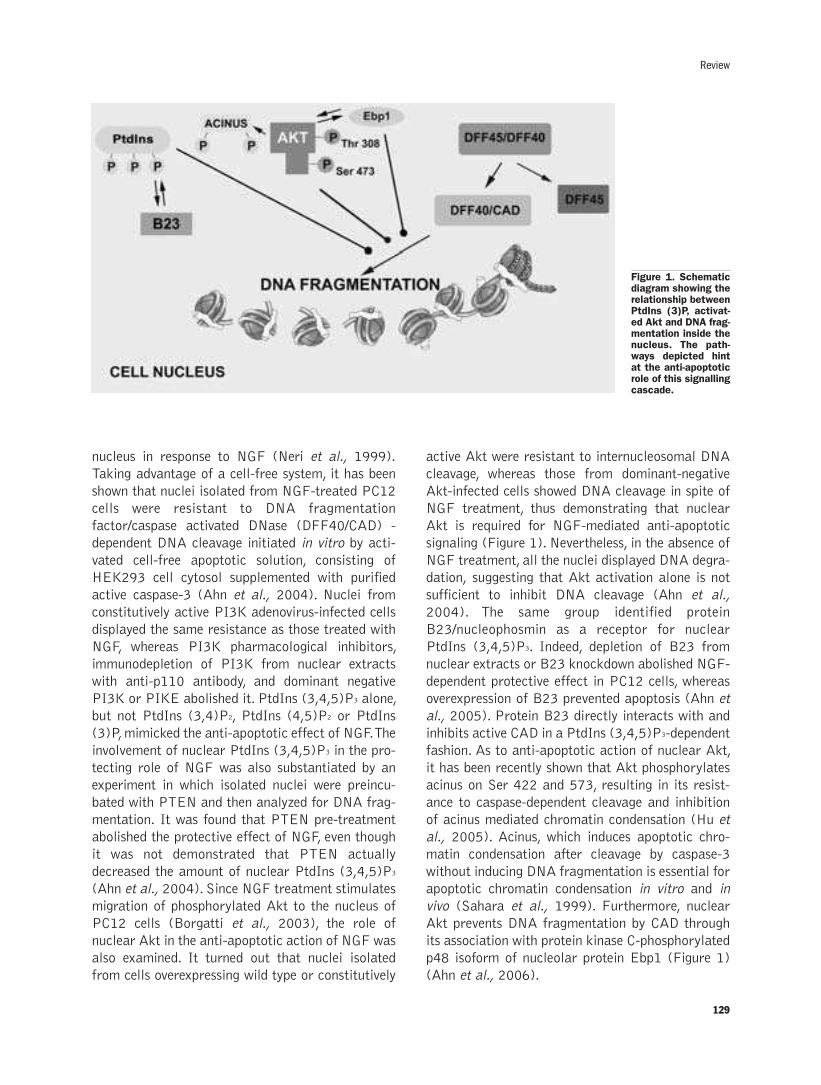
129
Review
nucleus in response to NGF (Neri et al., 1999).
Taking advantage of a cell-free system, it has been
shown that nuclei isolated from NGF-treated PC12
cells were resistant to DNA fragmentation
factor/caspase activated DNase (DFF40/CAD) -
dependent DNA cleavage initiated in vitro by acti-
vated cell-free apoptotic solution, consisting of
HEK293 cell cytosol supplemented with purified
active caspase-3 (Ahn et al., 2004). Nuclei from
constitutively active PI3K adenovirus-infected cells
displayed the same resistance as those treated with
NGF, whereas PI3K pharmacological inhibitors,
immunodepletion of PI3K from nuclear extracts
with anti-p110 antibody, and dominant negative
PI3K or PIKE abolished it. PtdIns (3,4,5)P3 alone,
but not PtdIns (3,4)P2, PtdIns (4,5)P2 or PtdIns
(3)P, mimicked the anti-apoptotic effect of NGF. The
involvement of nuclear PtdIns (3,4,5)P3 in the pro-
tecting role of NGF was also substantiated by an
experiment in which isolated nuclei were preincu-
bated with PTEN and then analyzed for DNA frag-
mentation. It was found that PTEN pre-treatment
abolished the protective effect of NGF, even though
it was not demonstrated that PTEN actually
decreased the amount of nuclear PtdIns (3,4,5)P3
(Ahn et al., 2004). Since NGF treatment stimulates
migration of phosphorylated Akt to the nucleus of
PC12 cells (Borgatti et al., 2003), the role of
nuclear Akt in the anti-apoptotic action of NGF was
also examined. It turned out that nuclei isolated
from cells overexpressing wild type or constitutively
active Akt were resistant to internucleosomal DNA
cleavage, whereas those from dominant-negative
Akt-infected cells showed DNA cleavage in spite of
NGF treatment, thus demonstrating that nuclear
Akt is required for NGF-mediated anti-apoptotic
signaling (Figure 1). Nevertheless, in the absence of
NGF treatment, all the nuclei displayed DNA degra-
dation, suggesting that Akt activation alone is not
sufficient to inhibit DNA cleavage (Ahn et al.,
2004). The same group identified protein
B23/nucleophosmin as a receptor for nuclear
PtdIns (3,4,5)P3. Indeed, depletion of B23 from
nuclear extracts or B23 knockdown abolished NGF-
dependent protective effect in PC12 cells, whereas
overexpression of B23 prevented apoptosis (Ahn et
al., 2005). Protein B23 directly interacts with and
inhibits active CAD in a PtdIns (3,4,5)P3-dependent
fashion. As to anti-apoptotic action of nuclear Akt,
it has been recently shown that Akt phosphorylates
acinus on Ser 422 and 573, resulting in its resist-
ance to caspase-dependent cleavage and inhibition
of acinus mediated chromatin condensation (Hu et
al., 2005). Acinus, which induces apoptotic chro-
matin condensation after cleavage by caspase-3
without inducing DNA fragmentation is essential for
apoptotic chromatin condensation in vitro and in
vivo (Sahara et al., 1999). Furthermore, nuclear
Akt prevents DNA fragmentation by CAD through
its association with protein kinase C-phosphorylated
p48 isoform of nucleolar protein Ebp1 (Figure 1)
(Ahn et al., 2006).
Figure 1. Schematicdiagram showing therelationship betweenPtdIns (3)P, activat-ed Akt and DNA frag-mentation inside thenucleus. The path-ways depicted hintat the anti-apoptoticrole of this signallingcascade.
130
Conclusions
As is clear from this overview, nuclear PI3K,
PtdIns(3,4,5)P3, Akt, and PTEN may be involved
in key cellular processes, including carcinogenesis
and apoptosis protection. While our knowledge of
how this signaling cascade could result in neoplas-
tic transformation is virtually non-existent, we
understand more about its involvement in blocking
apoptosis. A challenge for the future will be to bet-
ter elucidate the anti-apoptotic functions of nuclear
PI3K/ PtdIns (3,4,5)P3/Akt/PTEN signaling. For
example, we do not know whether or not this system
is operative only in neural cells (Ye, 2005) or also
in other cell types, including hepatocytes and car-
diomyocytes, as preliminary evidence would suggest
(Martelli et al., 2006a). A central question is
whether this pathway is also activated by other neu-
rotrophins which protects neural cells from apopto-
sis, such as IGF-1. Identification of additional tar-
gets and/or interacting partners within the nucleus
will be of outmost importance for a better compre-
hension of the roles played by this signal transduc-
tion system. Furthermore, it should not be forgotten
that nuclear PI3K seems to be critically involved in
processes other than tumorigenesis and apoptosis,
such as myeloid cell differentiation (Bertagnolo et
al., 2004). However, further elucidation of this
complex and peculiar nuclear signaling pathway is
expected to provide new potential targets for phar-
macological interventions in major human diseases,
including cancer and degenerative disorders in
which inappropriate apoptosis is thought to play a
fundamental role, such as heart failure, Parkinson’s
disease, and amyotrophic lateral sclerosis.
AcknowledgementsThis work was supported by: Associazione
Italiana Ricerca sul Cancro (AIRC Regional
Grants); Italian MIUR FIRB 2005 and PRIN
2005; Carisbo Foundation.
References
Ahn JY, Rong R, Liu X, Ye K. PIKE/nuclear PI 3-kinase signalingmediates the antiapoptotic actions of NGF in the nucleus. EMBO J2004;23:3995-4006.
Ahn JY, Liu X, Cheng D, Peng J, Chan PK, Wade PA, et al.Nucleophosmin/B23, a nuclear PI(3,4,5)P3 receptor, mediates theantiapoptotic actions of NGF by inhibiting CAD. Mol. Cell2005;18:435-45.
Ahn JY, Liu X, Liu Z, Pereira L, Cheng D, Peng J, et al. Nuclear Aktassociates with PKC-phosphorylated Ebp1, preventing DNA frag-mentation by inhibition of caspase-activated DNase. EMBO J2006;25:2083-95.
Arden KC, Biggs WH. Regulation of the FoxO family of transcriptionfactors by phosphatidylinositol-3 kinase-activated signaling. ArchBiochem Biophys 2002: 403: 292-8.
Backers K, Blero D, Paternotte N, Zhang J, Erneux C.The terminationof PI3K signalling by SHIP1 and SHIP2 inositol 5-phos-phatases.Adv Enzyme Regul 2003;43:15-28.
Bae SS, Lee YH, Chang JS, Galadari SH, Kim YS, Ryu SH, et al. Srchomology domains of phospholipase C �1 inhibit nerve growth fac-tor-induced differentiation of PC12 cells. J Neurochem1998;71:178-85.
Bertagnolo V, Brugnoli F, Marchisio M, Capitani S. Inositide-modifyingenzymes: a cooperative role in regulating nuclear morphology dur-ing differentiation of myeloid cells. J Biol Regul Homeost Agents2004;18:381-6.
Borgatti P, Martelli AM, Tabellini G, Bellacosa A, Capitani S, Neri LM.Threonine 308 phosphorylated form of Akt translocates to thenucleus of PC12 cells under nerve growth factor stimulation andassociates with the nuclear matrix protein nucleolin. J Cell Physiol2003;196:79-88.
Brandts CH, Sargin B, Rode M, Biermann C, Lindtner B, Schwable J,et al. Constitutive activation of Akt by Flt3 internal tandem dupli-cations is necessary for increased survival, proliferation, and myeloidtransformation. Cancer Res 2005;65:9643-50.
Brazil DP, Yang ZZ, Hemmings BA. Advances in protein kinase B sig-nalling: AKTion on multiple fronts. Trends Biochem. Sci 22004;29:233-42.
Chow LM, Baker SJ. PTEN function in normal and neoplastic growth.Cancer Lett 2006;241:184-96.
Chung JH, Ginn-Pease ME, Eng C. Phosphatase and tensin homologuedeleted on chromosome 10 (PTEN) has nuclear localization signal-like sequences for nuclear import mediated by major vault protein.Cancer Res 2005;65: 4108-16.
Chung JH, Ostrowski MC, Romigh T, Minaguchi T, Waite KA, Eng C.The ERK1/2 pathway modulates nuclear PTEN-mediated cell cyclearrest by cyclin D1 transcriptional regulation. Hum Mol Genet2006;15:2553-9.
Cully M, You H, Levine AJ, Mak TW. Beyond PTEN mutations: thePI3K pathway as an integrator of multiple inputs during tumorige-nesis. Nat Rev Cancer 2006;6:184-92.
Deleris P, Gayral S, Breton-Douillon M. Nuclear Ptdlns(3,4,5)P3 sig-naling: an ongoing story. J Cell Biochem 2006;98:469-85.
Di Paolo G, De Camilli P. Phosphoinositides in cell regulation andmembrane dynamics. Nature 2006;443:651-7.
Engelman JA, Luo J, Cantley LC. The evolution of phosphatidylinositol3-kinases as regulators of growth and metabolism. Nat Rev Genet2006;7:606-19.
Franke TF, Hornik CP, Segev L, Shostak GA, Sugimoto C. PI3K/Aktand apoptosis: size matters. Oncogene 2003;22:8983-98.
Fresno Vara JA, Casado E, de Castro J, Cejas P, Belda-Iniesta C,Gonzalez-Baron M. PI3K/Akt signalling pathway and cancer.Cancer Treat Rev 2004; 30: 193-2004.
Gil A, Andres-Pons A, Fernandez E, Valiente M, Torres J, Cervera J, etal. Nuclear localization of PTEN by a Ran-dependent mechanismenhances apoptosis: Involvement of an N-terminal nuclear localiza-tion domain and multiple nuclear exclusion motifs. Mol Biol Cell2006;17:4002-13.
Gimm O, Perren A, Weng LP, Marsh DJ, Yeh JJ, Ziebold U, et al.Differential nuclear and cytoplasmic expression of PTEN in normalthyroid tissue, and benign and malignant epithelial thyroid tumors.Am J Pathol 2000;156:1693-700.
Ginn-Pease ME, Eng C. Increased nuclear phosphatase and tensinhomologue deleted on chromosome 10 is associated with G0-G1 inMCF-7 cells. Cancer Res 2003;63:282-6.
Hanada M, Feng J, Hemmings BA. Structure, regulation and functionof PKB/AKT--a major therapeutic target. Biochim Biophys Acta2004; 1697: 3-16.
Hu Y, Yao J, Liu Z, Liu X, Fu H, Ye K. Akt phosphorylates acinus andinhibits its proteolytic cleavage, preventing chromatin condensation.EMBO J. 2005:24:3543-54.
Huang WC, Chen CC. Akt phosphorylation of p300 at Ser-1834 isessential for its histone acetyltransferase and transcriptional activi-ty.Mol Cell Biol 2005; 25: 6592-602.
Irvine RF. Nuclear lipid signalling. Nat Rev Mol Cell Biol 2004;4: 349-60.
A.M. Martelli et al.
131
Kikani CK, Dong LQ, Liu F. "New"-clear functions of PDK1: beyond amaster kinase in the cytosol? J Cell Biochem 2005;96:1157-62.
Lachyankar MB, Sultana N, Schonhoff CM, Mitra P, Poluha W,Lambert S, et al. A role for nuclear PTEN in neuronal differentia-tion. J Neurosci 200;20:1404-13.
Lee SH, Kim HS, Park WS, Kim YY, Lee KY, Kim SH, et al. Non-smallcell lung cancers frequently express phosphorylated Akt; animmunohistochemical study. APMIS 2002;110: 587-92.
Li AG, Piluso LG, Cai X, Wei G, Sellers WR, Liu X. Mechanisticinsights into maintenance of high p53 acetylation by PTEN. MolCell 2006;23:575-87.
Lim MA, Kikani CK, Wick MJ, Dong LQ. Nuclear translocation of 3'-phosphoinositide-dependent protein kinase 1 (PDK-1): a potentialregulatory mechanism for PDK-1 function. Proc Natl Acad Sci USA2003;100:14006-11.
Lindsay Y, McCoull D, Davidson L, Leslie NR, Fairservice A, Gray A, etal. Localization of agonist-sensitive PtdIns(3,4,5)P3 reveals anuclear pool that is insensitive to PTEN expression. J Cell Sci2006;119:5160-8.
Liu F, Wagner S, Campbell RB, Nickerson JA, Schiffer CA, Ross AH.PTEN enters the nucleus by diffusion. J Cell Biochem2005;96:221-34.
Liu JL, Sheng X, Hortobagyi ZK, Mao Z, Gallick GE, Yung WK.Nuclear PTEN-mediated growth suppression is independent of Aktdown-regulation. Mol Cell Biol 2005;25:6211-24.
Manzoli L, Martelli AM, Billi AM, Faenza I, Fiume R, Cocco L.Nuclear phospholipase C: involvement in signal transduction. ProgLipid Res 2005 Jul;44:185-206.
Martelli AM, Follo MY, Evangelisti C, Falà F, Fiume R, Billi AM, et alNuclear inositol lipid metabolism: more than just second messengergeneration? J Cell Biochem 2005a; 96: 285-92.
Martelli AM, Faenza I, Billi AM, Manzoli L, Evangelisti C, Fala F, etal. Intranuclear 3'-phosphoinositide metabolism and Akt signaling:new mechanisms for tumorigenesis and protection against apopto-sis? Cell Signal 2006a;18:1101-7.
Martelli AM, Tabellini G, Bortul R, Tazzari PL, Cappellini A, Billi AM,Cocco L. Involvement of the phosphoinositide 3-kinase/Akt signalingpathway in the resistance to therapeutic treatments of humanleukemias. Histol Histopathol 2005b;20: 239-52.
Martelli AM, Nyakern M, Tabellini G, Bortul R, Tazzari PL, EvangelistiC, Cocco L. Phosphoinositide 3-kinase/Akt signaling pathway and itstherapeutical implications for human acute myeloid leukemia.Leukemia 2006b;20:911-28.
Matkovic K, Brugnoli F, Bertagnolo V, Banfic H, Visnjic D The role ofthe nuclear Akt activation and Akt inhibitors in all-trans-retinoicacid-differentiated HL-60 cells. Leukemia 2006;20:941-51.
Minaguchi T, Waite KA, Eng C. Nuclear localization of PTEN is regu-lated by Ca2+ through a tyrosil phosphorylation-independent confor-mational modification in major vault protein. Cancer Res2006;66:11677-82.
Montironi R, Mazzuccheli R, Scarpelli M, Lopez-Beltran A, FellegaraG, Algaba F. Gleason grading of prostate cancer in needle biopsiesor radical prostatectomy specimens: contemporary approach, cur-rent clinical significance and sources of pathology discrepancies.BJU Int 2005;95: 1146-52.
Mora A, Komander D, van Aalten DM, Alessi DR. PDK1, the masterregulator of AGC kinase signal transduction. Semin Cell Dev Biol2004;15:161-70.
Neri LM, Borgatti P, Capitani S, Martelli AM. The nuclear phospho-inositide 3-kinase/AKT pathway: a new second messenger system.
Biochim Biophys Acta 2002;1584:73-80.
Neri LM, Martelli AM, Borgatti P, Colamussi ML, Marchisio M,Capitani S. Increase in nuclear phosphatidylinositol 3-kinase activi-ty and phosphatidylinositol (3,4,5) trisphosphate synthesis precedePKC-� translocation to the nucleus of NGF-treated PC12 cells.FASEB J 1999;13: 2299-310.
Nicholson KM, Streuli CH, Anderson NG. Autocrine signalling througherbB receptors promotes constitutive activation of protein kinaseB/Akt in breast cancer cell lines. Breast Cancer Res Treat2003;81:117-28.
Pekarsky Y, Koval A, Hallas C, Bichi R, Tresini M, Malstrom S, et al.Tcl1 enhances Akt kinase activity and mediates its nuclear translo-cation. Proc Natl. Acad Sci USA 2000;97: 3028-33.
Perren A, Komminoth P, Saremaslani P, Matter C, Feurer S, Lees JAet al. Mutation and expression analyses reveal differential subcellu-lar compartmentalization of PTEN in endocrine pancreatic tumorscompared to normal islet cells. Am J Pathol 2000;156: 1693-103.
Ramaswamy S, Nakamura N, Vazquez F, Batt DB, Perera S, RobertsTM, et al. Regulation of G1 progression by the PTEN tumor sup-pressor protein is linked to inhibition of the phosphatidylinositol 3-kinase/Akt pathway.Proc Natl Acad Sci USA 1999;96: 2110-5.
Sahara S, Aoto M, Eguchi Y, Imamoto N, Yoneda Y, Tsujimoto Y.Acinus is a caspase-3-activated protein required for apoptotic chro-matin condensation. Nature 1999;401:168-73.
Saji M, Vasko V, Kada F, Allbritton EH, Burman KD, Ringel MD. Akt1contains a functional leucine-rich nuclear export sequence.BiochemBiophys Res Commun 2005;332:167-73.
Sansal I, Sellers WR.The biology and clinical relevance of the PTENtumor suppressor pathway. J Clin Oncol 2004; 22: 2954-63.
Van de Sande T, Roskams T, Lerut E, Joniau S, Van Poppel H,Verhoeven G, et al. High-level expression of fatty acid synthase inhuman prostate cancer tissues is linked to activation and nuclearlocalization of Akt/PKB. J Pathol 2005;206:214-9.
Vanhaesebroeck B, Leevers SJ, Ahmadi K, Timms J, Katso R, DriscollPC, et al. Synthesis and function of 3-phosphorylated inositol lipids.Annu Rev Biochem 2001;70: 535-602.
Vasko V, Saji M, Hardy E, Kruhlak M, Larin A, Savchenko V, et al. Aktactivation and localisation correlate with tumour invasion and onco-gene expression in thyroid cancer.J Med Genet 2004;41:161-70.
Whiteman DC, Zhou XP, Cummings MC, Pavey S, Hayward NK, EngC. Nuclear PTEN expression and clinicopathologic features in apopulation-based series of primary cutaneous melanoma. Int JCancer 2002;99; 63-7.
Xuan Nguyen TL,Choi JW, Lee SB, Ye K, Woo SD, Lee KH, et al. Aktphosphorylation is essential for nuclear translocation and retentionin NGF-stimulated PC12 cells. Biochem Biophys Res Commun2006;349:789-98.
Ye K. PIKE/nuclear PI 3-kinase signaling in preventing programmedcell death. J Cell Biochem 2005;96: 463-72.
Ye K. PIKE GTPase-mediated nuclear signalings promote cell survival.Biochim Biophys Acta 2006;1761:570-6.
Ye K, Hurt KJ, Wu FY, Fang M, Luo HR, Hong JJ, et al. Pike. A nucleargtpase that enhances PI3kinase activity and is regulated by protein4.1N. Cell 2000;103:919-30.
Ye K, Aghdasi B, Luo HR, Moriarity JL, Wu FY, Hong JJ, et al.Phospholipase C � 1 is a physiological guanine nucleotide exchangefactor for the nuclear GTPase PIKE. Nature 2002;415:541-4.
Review

132
A.M. Martelli et al.

133
©2007, European Journal of Histochemistry
The morphogenetic events leading to the transendothelial pas-sage of lymphoid and tumoral cells are analyzed in light of a veryrecent and global theory of intercellular communication desig-nated as the Triune Information Network (TIN). The TIN system isbased on the assumption that cell-cell interactions primarilyoccur through cell surface informations or topobiological proce-sess, whose mechanisms rely upon expression of adhesion mol-ecules, and are regulated by an array of locally-borne(autocrine/paracrine signals and autonomic inputs) and distant-ly-borne (endocrine secretions) messages. The final aim of the TINis to control homeostatic functions crucial for the organism sur-vival, like morphogenesis. Knowledge of the TIN signals involvedin lymphoid and tumoral cell intravasation might offer a new per-spetive to study the mechanisms of tumor immunity. Recognitionof tumor target cells by immune cytotoxic effectors, in fact, can beconsidered a notable case of TIN-mediated cell to cell interaction.In particular, Natural Killer (NK) cells play a role in the cell-medi-ated control of tumor growth and metastatic spreading. Cell tar-geting and killing are dependent on the different NK cell recep-tors and on the efficacy of NK cells after cytokine and monoclonalantibody administration in cancer therapy. Since efficacy of NKcell-based immunotheraphy has been proven in KIR-mismatchregimens or in TRAIL-dependent apoptosis, the ability to manip-ulate the balance of activating and inhibitory receptors on NKcells and of their cognate ligands as well as the sensitivity oftumor cells to apoptosis, opens new perspectives for NK cellbased immunotherapy.
Key words: Topobiology, paracrine secretion, natural killercells, cytokines, cytotoxicity, tumor immunity, cell activation.
Correspondence: Roberto Toni or Marco VitaleDepartment of Human Anatomy, Pharmacology & Forensic Medicine, Human AnatomySection, University of ParmaOspedale Maggiore, via Gramsci, 14 43100 Parma, talyE-mail: [email protected] or [email protected]
European Journal of Histochemistry2007; vol. 51 supplement 1:133-138
Neuroendocrine regulation and tumor immunity
R. Toni,1,2 P. Mirandola,1 G. Gobbi,1 M. Vitale1
1Department of Human Anatomy, Pharmacology and Forensic Medicine, University of Parma School of
Medicine, Parma, Italy; 2Department of Medicine, Division of Endocrinology, Diabetes and Metabolism,
New England Medical Center - Tufts University School of Medicine, Boston, MA, USA
From an evolutionary and developmental per-
spective the cell-cell interactions occurring
during the transendothelial passage of lym-
phocytes (see Azzali G, this issue) may be seen as a
local morphogenetic event, based on cell surface
interactions or topobiological processes (Toni
2004a). Both lymphocytes and endothelial cells, in
fact, undergo substantial reorganization of cell
shape during intravasation, primarily following cell
surface contact, suggesting that cell adhesion mole-
cules, substrate adhesion molecules, and cell junc-
tional molecules must be called into play during this
phenomenon (Toni 2003). In addition, some com-
mon mesodermal origin of mononuclear and
endothelial cells suggests that they may share a
modality of reciprocal recognition (Toni 2004a).
Indeed, endothelial/vascular precursors have been
isolated in humans from the mononuclear fraction
of peripheral blood CD34, Flk-1, AC133 and Tie2
antigen-positive cells, both in basal state and after
granulocyte-colony stimulating factor treatment of
donors, to favour mobilization of CD34+ elements
from bone marrow to peripheral blood (Asahara
1997). Since specific growth factors may selective-
ly address these mesodermal progenitors towards
either a mononuclear or an endothelial differentia-
tion lineage (Ishikawa 2004), it is likely that an
array of paracrine signals common to both cell
types may constitute an ideal niche for their inter-
action (Fuchs 2004). In addition, the possibility
that the endothelial canalization is triggered also by
different phenotypes of neoplastic cells (see Azzali
G., this issue) suggests that the repertoire of repric-
ocal extracellular signals must be an heritage com-
mon to many different cell lineages. This raises the
possibility that transendothelial passage is a special
case of a more general mechanism regulating inter-
cellular communication. Very recently a global the-
ory of intercellular communication has been pro-
posed, including the paracrine signals as a part of a
hierachically-ordered, informational supersystem of
REVIEW
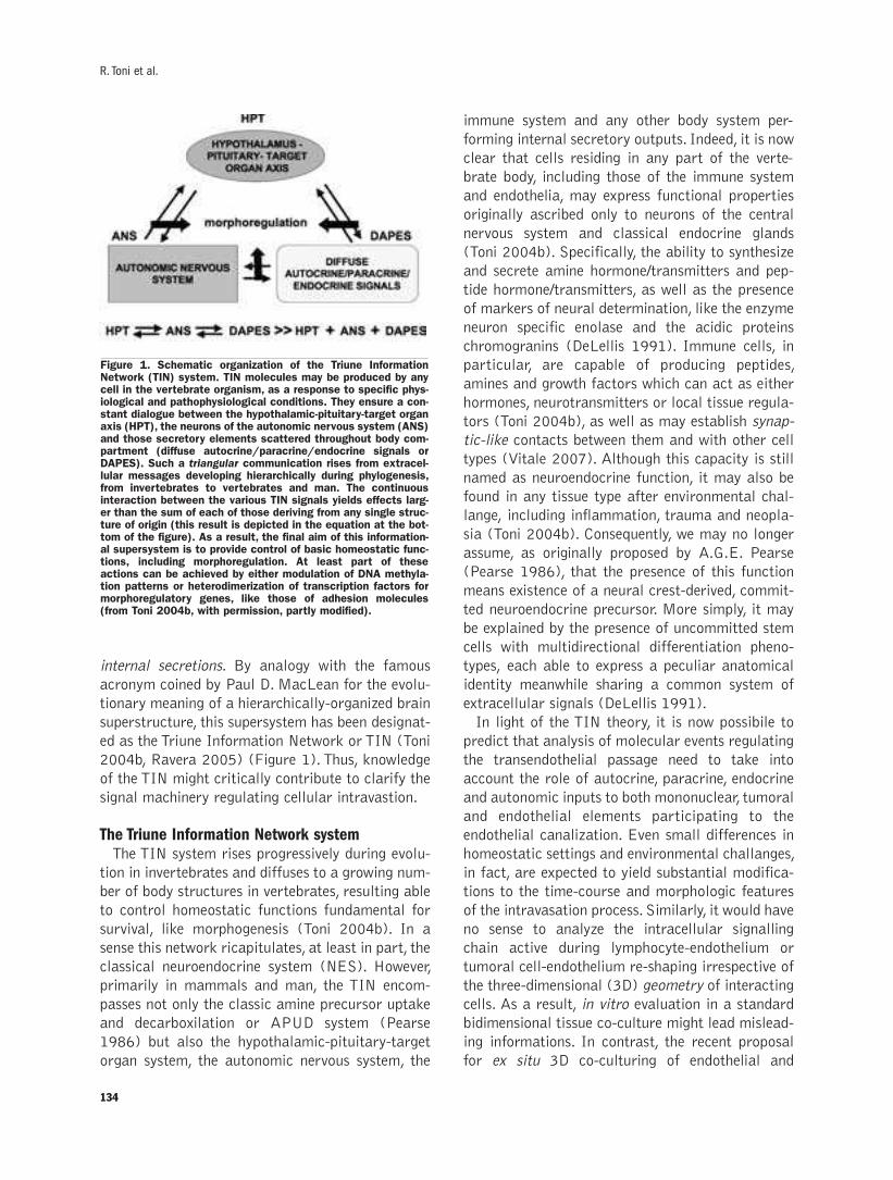
internal secretions. By analogy with the famous
acronym coined by Paul D. MacLean for the evolu-
tionary meaning of a hierarchically-organized brain
superstructure, this supersystem has been designat-
ed as the Triune Information Network or TIN (Toni
2004b, Ravera 2005) (Figure 1). Thus, knowledge
of the TIN might critically contribute to clarify the
signal machinery regulating cellular intravastion.
The Triune Information Network systemThe TIN system rises progressively during evolu-
tion in invertebrates and diffuses to a growing num-
ber of body structures in vertebrates, resulting able
to control homeostatic functions fundamental for
survival, like morphogenesis (Toni 2004b). In a
sense this network ricapitulates, at least in part, the
classical neuroendocrine system (NES). However,
primarily in mammals and man, the TIN encom-
passes not only the classic amine precursor uptake
and decarboxilation or APUD system (Pearse
1986) but also the hypothalamic-pituitary-target
organ system, the autonomic nervous system, the
immune system and any other body system per-
forming internal secretory outputs. Indeed, it is now
clear that cells residing in any part of the verte-
brate body, including those of the immune system
and endothelia, may express functional properties
originally ascribed only to neurons of the central
nervous system and classical endocrine glands
(Toni 2004b). Specifically, the ability to synthesize
and secrete amine hormone/transmitters and pep-
tide hormone/transmitters, as well as the presence
of markers of neural determination, like the enzyme
neuron specific enolase and the acidic proteins
chromogranins (DeLellis 1991). Immune cells, in
particular, are capable of producing peptides,
amines and growth factors which can act as either
hormones, neurotransmitters or local tissue regula-
tors (Toni 2004b), as well as may establish synap-
tic-like contacts between them and with other cell
types (Vitale 2007). Although this capacity is still
named as neuroendocrine function, it may also be
found in any tissue type after environmental chal-
lange, including inflammation, trauma and neopla-
sia (Toni 2004b). Consequently, we may no longer
assume, as originally proposed by A.G.E. Pearse
(Pearse 1986), that the presence of this function
means existence of a neural crest-derived, commit-
ted neuroendocrine precursor. More simply, it may
be explained by the presence of uncommitted stem
cells with multidirectional differentiation pheno-
types, each able to express a peculiar anatomical
identity meanwhile sharing a common system of
extracellular signals (DeLellis 1991).
In light of the TIN theory, it is now possibile to
predict that analysis of molecular events regulating
the transendothelial passage need to take into
account the role of autocrine, paracrine, endocrine
and autonomic inputs to both mononuclear, tumoral
and endothelial elements participating to the
endothelial canalization. Even small differences in
homeostatic settings and environmental challanges,
in fact, are expected to yield substantial modifica-
tions to the time-course and morphologic features
of the intravasation process. Similarly, it would have
no sense to analyze the intracellular signalling
chain active during lymphocyte-endothelium or
tumoral cell-endothelium re-shaping irrespective of
the three-dimensional (3D) geometry of interacting
cells. As a result, in vitro evaluation in a standard
bidimensional tissue co-culture might lead mislead-
ing informations. In contrast, the recent proposal
for ex situ 3D co-culturing of endothelial and
134
R. Toni et al.
Figure 1. Schematic organization of the Triune InformationNetwork (TIN) system. TIN molecules may be produced by anycell in the vertebrate organism, as a response to specific phys-iological and pathophysiological conditions. They ensure a con-stant dialogue between the hypothalamic-pituitary-target organaxis (HPT), the neurons of the autonomic nervous system (ANS)and those secretory elements scattered throughout body com-partment (diffuse autocrine/paracrine/endocrine signals orDAPES). Such a triangular communication rises from extracel-lular messages developing hierarchically during phylogenesis,from invertebrates to vertebrates and man. The continuousinteraction between the various TIN signals yields effects larg-er than the sum of each of those deriving from any single struc-ture of origin (this result is depicted in the equation at the bot-tom of the figure). As a result, the final aim of this information-al supersystem is to provide control of basic homeostatic func-tions, including morphoregulation. At least part of theseactions can be achieved by either modulation of DNA methyla-tion patterns or heterodimerization of transcription factors formorphoregulatory genes, like those of adhesion molecules(from Toni 2004b, with permission, partly modified).
epithelial progenitors on biocompatible scaffolds
(Toni 2007) could offer a new perspetive. In such a
frame, in fact, it would be possible to analyze the
TIN signals regulatiing the 3D arrangement of lym-
phocytes and neoplastic cells during both their
transendothelial passage and reciprocal recogni-
tion, like in the case of Natural Killer cells actions.
Natural killer cells and tumor immunityNatural Killer (NK) cells represent the 10-20%
of peripheral blood mononuclear cells, but they can
be also present in lymph nodes, spleen and bone
marrow, and can be induced to migrate towards
inflammation sites by different chemoattractants.
NK cells are able to kill target cells by a lytic
machinery in an activation-independent way, sug-
gesting a role in the control of tumor growth. NK
cells are not an homogenous population. In fact,
they express CD56 at different levels (dim or
bright) and also the CD16 antigen (Ag) can be
present or not on their surface. CD56bright NK cells
have been recently defined as the cytokine respon-
sive NK subset that may not require licensing by
host MHC-I molecules (Anfossi 2006). NK cells
express on their surface both inhibitory and activa-
tory receptors (Bottino 2004). The several types of
inhibitory receptors show different specificities for
alleles of class I molecule. In particular, the killer
Ig-like receptors (KIRs) bind HLA-class I, and the
heterodimeric receptors CD94-NKG2A/B recognize
HLA-E (Braud 1998). Cancer cells frequently lack
a MHC-I allele, and therefore are susceptible to NK
cell lysis. In the absence of inhibitory signals, NK
cell cytotoxicity must however be activated by a set
of triggering receptors. Spontaneous cytotoxic
activity is mainly triggered by NKG2D, leukocyte
adhesion molecule DNAM-1 (CD226), and Natural
cytotoxicity receptors (NCRs), while CD16, by
binding the Fc portion of IgG, binds to opsonized
cells mediating antibody dependent cellular cyto-
toxicity (ADCC) (Moretta 2004). NKG2D and
DNAM-1 recognize stress-induced ligands
expressed by several tumor cell lines, while NCRs
mediate cell lysis of many cancer cells.
Upon cytokine stimulation, NK cells become lym-
phokine activated killer cells (LAK) that prolifer-
ate, produce cytokines and up-regulate effector
molecules such as adhesion molecules, perforin,
granzymes, FasL and TRAIL (Figure 2). LAK cells
became able to induce perforin/granzymes-depen-
dent necrosis of target cell and TNF ligand family
members-induced apoptosis of the target cell. Given
the ability of TRAIL to kill many cancer cell types,
while sparing normal tissues, the use of recombi-
nant TRAIL has been proposed in clinical trials
(Smyth 2003). TRAIL is present in the BM, a site
of NK cell as well as erythro-myeloid differentia-
tion. Since it has been demonstrated that erythroid
cell differentiation is affected in vitro and in vivo by
recombinant TRAIL (Zamai 2000, Mirandola
2006, Ashkenazi 1999), its use in therapy should be
cautious. Activated NK cells themselves express dif-
ferent death receptors, such as TRAIL-R2 and
CD95, that are generally seen as implicated in the
termination of NK cell response and in tumor
responses to specific immune activities (immune
counterattacks). However, differently from erythro-
myeloid cells, NK cells are usually protected from
TRAIL-induced apoptosis thanks to cytokine-
dependent c-FLIP induction (Mirandola 2004).
Among the activatory cytokines, IL-15 is believed
to be responsible for NK cell development in vivo,
and is a survival factor that protects lymphocytes
from IL-2-activation-induced cell death (AICD).
Recent evidences suggest a nonreduntant unique
role for IL-15 in the differentiation, proliferation,
survival and activation of natural killer (NK) cells
(Rodella 2001). IL-2 acts as growth factor for NK
cell progenitors and mature NK cells, and induces
the production of NK effector molecules, enhancing
NK lytic activity. IL-12 and IL-18, NK activatory
cytokines active during late NK cell differentiation,
have been demonstrated to synergistically enhance
cytotoxicity against tumor targets and IFN-γ pro-duction by NK cells (Golab 2000). IFN-γ inducestype 1 immune response and directly acts on cancer
cells. Finally, IL-21, another cytokine binding the
common γ chain (shared with IL-2, IL-4, IL-7, IL-9 and IL-15), has been demonstrated to favour the
onset of the most cytotoxic CD56dimCD16+ NK cell
subset and to enhance its cytotoxicity (Parrish-
Novak 2000).
IL-2 activated NK cells were used in clinical tri-
als for the treatment of solid primary or metasta-
tized cancers (Rosenberg 1993). Subcutaneous
injections of NK-stimulating doses of IL-2 or
administration of pre-activated NK cells (adoptive
transfer of LAK cells), showed a 15-30% positive
effects in patients with advanced renal cell carcino-
ma (RCC) or melanoma (MEL) (Rosenberg 1993).
Unfortunately, IL-2 treatment is associated with
life-threatening toxicity, essentially represented by
Review
135
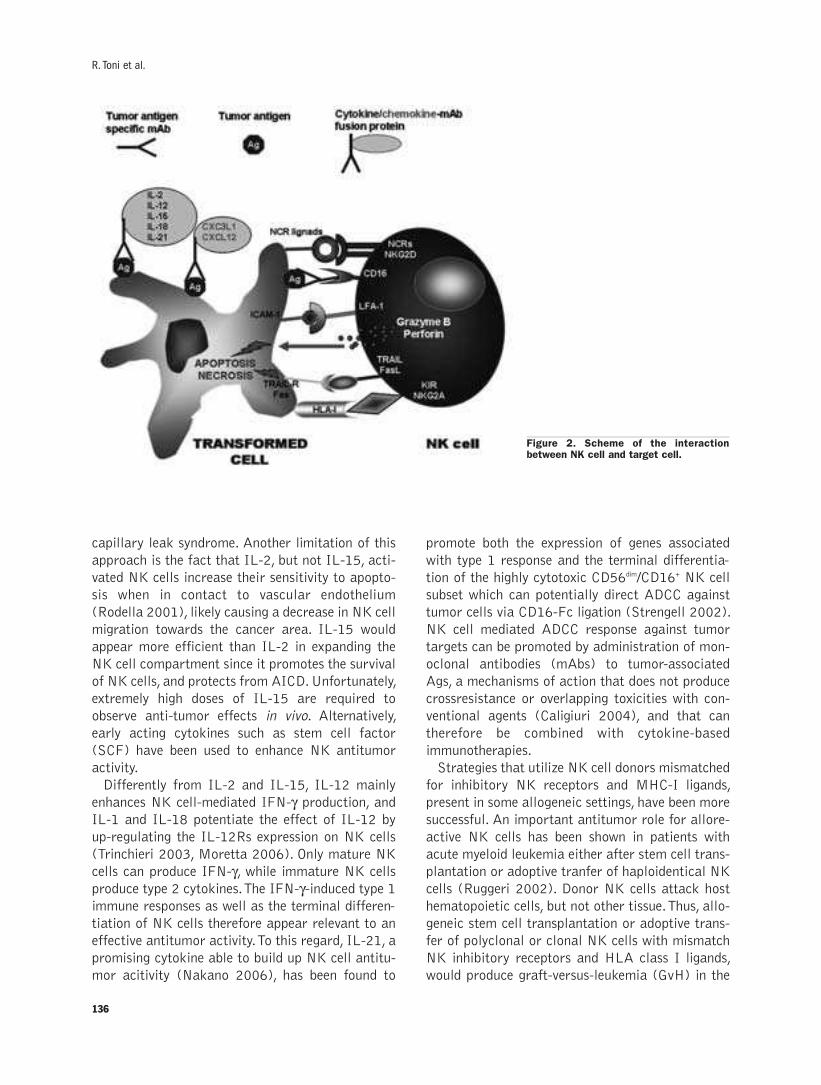
136
capillary leak syndrome. Another limitation of this
approach is the fact that IL-2, but not IL-15, acti-
vated NK cells increase their sensitivity to apopto-
sis when in contact to vascular endothelium
(Rodella 2001), likely causing a decrease in NK cell
migration towards the cancer area. IL-15 would
appear more efficient than IL-2 in expanding the
NK cell compartment since it promotes the survival
of NK cells, and protects from AICD. Unfortunately,
extremely high doses of IL-15 are required to
observe anti-tumor effects in vivo. Alternatively,
early acting cytokines such as stem cell factor
(SCF) have been used to enhance NK antitumor
activity.
Differently from IL-2 and IL-15, IL-12 mainly
enhances NK cell-mediated IFN-γ production, andIL-1 and IL-18 potentiate the effect of IL-12 by
up-regulating the IL-12Rs expression on NK cells
(Trinchieri 2003, Moretta 2006). Only mature NK
cells can produce IFN-γ, while immature NK cellsproduce type 2 cytokines. The IFN-γ-induced type 1immune responses as well as the terminal differen-
tiation of NK cells therefore appear relevant to an
effective antitumor activity. To this regard, IL-21, a
promising cytokine able to build up NK cell antitu-
mor acitivity (Nakano 2006), has been found to
promote both the expression of genes associated
with type 1 response and the terminal differentia-
tion of the highly cytotoxic CD56dim/CD16+ NK cell
subset which can potentially direct ADCC against
tumor cells via CD16-Fc ligation (Strengell 2002).
NK cell mediated ADCC response against tumor
targets can be promoted by administration of mon-
oclonal antibodies (mAbs) to tumor-associated
Ags, a mechanisms of action that does not produce
crossresistance or overlapping toxicities with con-
ventional agents (Caligiuri 2004), and that can
therefore be combined with cytokine-based
immunotherapies.
Strategies that utilize NK cell donors mismatched
for inhibitory NK receptors and MHC-I ligands,
present in some allogeneic settings, have been more
successful. An important antitumor role for allore-
active NK cells has been shown in patients with
acute myeloid leukemia either after stem cell trans-
plantation or adoptive tranfer of haploidentical NK
cells (Ruggeri 2002). Donor NK cells attack host
hematopoietic cells, but not other tissue. Thus, allo-
geneic stem cell transplantation or adoptive trans-
fer of polyclonal or clonal NK cells with mismatch
NK inhibitory receptors and HLA class I ligands,
would produce graft-versus-leukemia (GvH) in the
R. Toni et al.
Figure 2. Scheme of the interactionbetween NK cell and target cell.
Review
absence of graft-versus-host desease (GvHD). The
signals transduced by MHC-I inhibitory receptors
become superfluous and likely exploited by some
tumor cells to elude NK immunosurveillance. TNF-
receptor mediated apoptosis of sensitive tumor
cells should be NKR-independent, suggesting that
this mechanism should however work upon NK cell
activation, independently from the KIR/MHC-I set-
ting. Mouse models of leukemia have demonstrated
efficacy of anti-KIR blocking antibodies without
adverse effects on normal cells, indicating the fea-
sibility of treatments with antibody fragments to
prevent KIR/NKG2A-MHC-I interactions in cancer
therapy (Koh 2001) (Figure 2).
ConclusionsInteractions between solid tumor cells and the
microenvironment in vivo create a context that pro-
motes tumor growth, selection and protection from
immune attack, suggesting that the tridimensional
architecture of solid cancer lesions is likely one of
the tumor mechanisms to escape immunosurveil-
lance. To this regard, another important mechanism
to control NK cell activity is their ability to traffic
to tumor sites. Chemokines are key regulators of
NK cell migration and are required to drive NK
cells to tumor sites. NK cells express chemokine
receptors on the cell surface and migrate vigorous-
ly in response to CXCL12 and CXC3L1 (Robertson
2002).
Finally, both conventional therapies and immu-
notherapy kill tumor cells inducing programmed
cell death, thus selection of tumor cells resistant to
apoptosis would be the reason of cross-resistance
of cancer cells to chemotherapy and immunothera-
py. Therefore, sensitization of tumor cells to acti-
vated cytotoxic lymphocytes by up-regulating either
TNF family death receptors or effector activating
ligands on tumor cells combined with immunother-
apy have been pursued in order to overcome tumor
cell resistance and establish an effective antitumor
response. Today, the potential ability to manipulate
not only the balance of activating and inhibitory
receptors on NK cells but also their cognate ligands
as well as the sensitivity of tumor cells to apoptosis
opens new perspectives in NK cell based immuno-
therapy. Thus, detailed knowlede of the humoral
environment involved, like that expected on the
basis of the TIN system theory, could become criti-
cal to design any future intelligent, cell-mediated
antitumoral therapy.
AcknowledgementsThis work has been supported by the University of
Parma Scientific Research Local Funds (FIL06),
by Fondazione Cariparma and Fondazione G.B.
Morgagni grants.
References
Anfossi N, Andre P, Guia S, Falk CS, Roetynck S, Stewart CA, et al.Human NK cell education by inhibitory receptors for MHC class I.Immunity 2006; 25:331-42.
Asahara T, Murohara T, Sullivan A, Silver M, van der Zee R, Li T, et al.Isolation of putative endothelial progenitor cells for angiogenesis.Science 1997; 275:965-7.
Ashkenazi A, Pai RC, Fong S, Leung S, Lawrence DA, Marsters SA, etal. Safety and antitumor activity of recombinant soluble Apo2 lig-and. J Clin Invest 1999; 104:155-62.
Bottino C, Moretta L, Pende D, Vitale M, Moretta A. Learning how todiscriminate between friends and enemies, a lesson from NaturalKiller cells. Mol Immunol 2004; 41:569-75.
Braud VM, Allan DS, O'Callaghan CA, Soderstrom K, D'Andrea A, OggGS, et al. HLA-E binds to natural killer cell receptorsCD94/NKG2A, B and C. Nature 1998; 391:795-9.
Caligiuri MA, Velardi A, Scheinberg DA, Borrello IM.Immunotherapeutic approaches for hematologic malignancies.Hematology 2004; 337-53.
DeLellis RA, Tischler AS. The dispersed neuroendocrine cell system. In:Kovacs K, Asa SL, eds. Functional Endocrine Pathology. Boston,Blackwell Scientific Publications, 1991, vol 1, 493-508.
Edelman GM. Topobiology, an Introduction to Molecular Embryology.New New York, Basic Books Inc 1988.
Fuchs E, Tumbar T, Guasch G. Socializing with the neighbors: stemcells and their niche. Cell 2004; 116:769-78.
Golab J. Interleukin 18-interferon gamma inducing factor--a novelplayer in tumour immunotherapy? Cytokine 2000; 12:332-8.
Ishikawa M, Asahara T. Endothelial progenitor cell culture for vascu-lar regeneration. Stem Cells Dev 2004; 13:344-9.
Koh CY, Blazar BR, George T, Welniak LA, Capitini CM, Raziuddin A,et al. Augmentation of antitumor effects by NK cell inhibitory recep-tor blockade in vitro and in vivo. Blood 2001; 97:3132-7.
Mirandola P, Gobbi G, Ponti C, Sponzilli I, Cocco L, Vitale M.PKCepsilon controls protection against TRAIL in erythroid progen-itors. Blood 2006; 107:508-13.
Mirandola P, Ponti C, Gobbi G, Sponzilli I, Vaccarezza M, Cocco L, etal. Activated human NK and CD8+ T cells express both TNF-relat-ed apoptosis-inducing ligand (TRAIL) and TRAIL receptors but areresistant to TRAIL-mediated cytotoxicity. Blood 2004; 104:2418-24.
Moretta L, Bottino C, Pende D, Vitale M, Zingari MC, Moretta A.Different checkpoints in human NK-cell activation. TrendsImmunol2004; 25:670-6.
Moretta L, Ferlazzo G, Bottino C, Vitale M, Pende D, Zingari MC, etal. Effector and regulatory events during natural killer-dendritic cellinteractions. Immunol Rev 2006; 214:219-28.
Nakano H, Kishida T, Asada H, Shin-Ya M, Shinomiya T, Imanishi J, etal. Interleukin-21 triggers both cellular and humoral immuneresponses leading to therapeutic antitumor effects against head andneck squamous cell carcinoma. J Gene Med 2006; 8:90-9.
Parrish-Novak J, Dillon SR, Nelson A, Hammond A, Sprecher C, GrossJA, et al. Interleukin 21 and its receptor are involved in NK cellexpansion and regulation of lymphocyte function. Nature 2000;408:57-63.
Pearse AGE. The diffuse neuroendocrine systems: peptides, amines, pla-codes and the APUD theory. Prog. Brain Res 1986, 68:25-31.
Ravera S, Morigi FP, Coiro IM, Della Casa C, Bondi A, Toni R.Chromogranin A as an early marker of neuroendocrine differentia-tion in the human embryo: evidence for feasibility of the “TriuneInformation Network” concept in man. It J Anat Embryol 2005;
137
138
110 (suppl 1 to 3): 275 (abstract).
Robertson MJ. Role of chemokines in the biology of natural killer cells.J Leukoc Biol 2002; 71:173-83.
Rodella L, Zamai L, Rezzani R, Artico M, Peri G, Falconi M, et al.Interleukin 2 and interleukin 15 differentially predispose naturalkiller cells to apoptosis mediated by endothelial and tumour cells. BrJ Haematol 2001; 115:442-50.
Rosenberg SA, Lotze MT, Yang JC, Topalian SL, Chang AE,Schwartzentruber DJ, et al. Prospective randomized trial of high-dose interleukin-2 alone or in conjunction with lymphokine-activat-ed killer cells for the treatment of patients with advanced cancer. JNatl Cancer Inst 1993; 85:622-32.
Ruggeri L, Capanni M, Urbani E, Perruccio K, Shlomchik WD, Tosti A,et al. Effectiveness of donor natural killer cell alloreactivity in mis-matched hematopoietic transplants. Science 2002; 295:2097-100.
Smyth MJ, Takeda K, Hayakawa Y, Peschon JJ, van den Brink MR,Yagita H. Nature's TRAIL - On a Path to Cancer Immunotherapy.Immunity 2003; 18:1-6.
Strengell M, Sareneva T, Foster D, Julkunen I, Matikainen S. IL-21Up-Regulates the Expression of Genes Associated with Innate
Immunity and Th1 Response. J Immunol 2002; 169:3600-05.
Toni R, Della Casa C, Spaletta G, Marchetti G, Mazzoni P, Bodria M,et al. The bioartificial thyroid: a biotechnological perspective inendocrine organ engineering for transplantation replacement. ActaBiomed 2007; suppl 1:129-55.
Toni R. The neuroendocrine system: organization and homeostatic role.J Endocrinol Invest 2004b; 27 (suppl to n.6), 35-47.
Toni R. Topobiology: epistemological implications of an ontic theory inbiomorphology. Epistemologia 2004a; 27:83-106.
Toni R. Ontic conception and scientific explanation in biomedicine: thecase of the anatomical doctrine. Epistemologia 2003; 26:285-316.
Trinchieri G. Interleukin-12 and the regulation of innate resistance andadaptive immunity. Nat Rev Immunol 2003; 3:133-46.
Vitale M. Time for integration: communication in the immune system.Acta Biomed 2007; suppl 1:227-30
Zamai L, Secchiero P, Pierpaoli S, Bassini A, Papa S, Alnemri ES, etal. TNF-related apoptosis-inducing ligand (TRAIL) as a negativeregulator of normal human erythropoiesis. Blood 2000; 95:3716-24.
R. Toni et al.

Abatangelo G. 1
Adamo S. 35
Ambrosi G. 59
Anastasi G. 29
Antonucci A. 117
Anzalone R. 101
Azzali G. 73
Bagnara G.P. 1
Barberi L. 35
Baroni T. 105
Becchetti E. 105
Bellafiore M. 101
Benagiano V. 59
Bentivoglio M. 65
Boccaccio C. 79
Bodo M. 105
Bosco L. 59
Bucchieri F. 101
Calligaro A. 93
Capitani S. 125
Cappello F. 101
Carinci F. 105
Carinci P. 105
Casasco M. 93
Castellucci M. 65
Cinti S. 65
Cocco L. 125
Coletti D. 35
Comoglio P. 79
Conti G. 53
Cortivo R. 1
Cutroneo G. 29
David S. 101
De Pasquale V. 9
Di Felice V. 101
Di Primio R. 117
Dobrowolny G. 35
Errede M. 59
Farina F. 101
Favaloro A. 29
Ferrario V.F. 45
Flace P. 59
Formigli L. 21
Franchi M. 9
Giacinti C. 35
Giorgi C. 1
Girolamo F. 59
Gobbi G. 133
Guidotti L. 117
Icaro Cornaglia A. 93
Isola M. 53
Isola R. 53
La Rocca G. 101
Locci P. 105
Lorusso L. 59
Loy F. 53
Macciocca M. 9
Manzoli F.A. 125
Maraldi N.M. 117
Marotti G. 15
Martelli A.M. 125
Mazzotti G. 117
Meacci E. 21
Mirandola P. 133
Miscia S. 125
Molinaro M. 35
Musarò A. 35
Nardis C. 35
Orlandini G.E. 21
Orsini E. 9
Osculati F. 65
Ottani V. 9
Palma A. 101
Palumbo C. 15
Papa S. 125
Pelosi L. 35
Peri G. 101
Perra A. 53
Pezzetti F. 105
Quaranta M. 9
Rana R. 117
Riva A. 53
Riva F. 93
Rizzi A. 59
Rizzo G. 29
Roncali L. 59
Ruggeri A. 9
Sbarbati A. 65
Scapoli L. 105
Scicchitano B.M. 35
Sforza C. 45
Solinas P. 53
Stabellini G. 105
Testa Riva F. 53
Tognon M. 105
Toni R. 133
Trirè A. 9
Trusolino L. 79
Vindigni V. 1
Virgintino D. 59
Vitale M. 133
Volinia S. 105
Zancanaro C. 65
Zavan B. 1
Zecchi-Orlandini S. 21
Zummo G. 101
INDEX OF AUTHORS

ejhNOTICE TO AUTHORS
The European Journal of Histochemistry - a journal offunctional cytology publishes Original papers, Brief notes,Technical reports, Letters to the Editor, Minireviews,Editorials, Book reviews, Views and Comments andAnnouncements. Contributions are encouraged from investiga-tors in any country.
TextThe text of papers should be typewritten and double spaced
throughout. It is strongly encouraged to send papers asattachments to email messages at this e-mail address:[email protected]. Otherwise, three complete copies of the text,figures and tables should be sent to the following address:
European Journal of Histochemistry, Editorial Office, piazzaBotta, 10, 27100 Pavia (Italy). Phone: +39.0382.986420.Fax +39.0382.986325.
Manuscript formatFor Original papers, Brief notes, and Technical reports, each
manuscript should be preceded by: a) a title page containingthe following information: Title of the paper; Initials of thename and full surname of author(s); Full name and address ofthe Institution(s) where the work was done; Complete address(phone and fax numbers, and E-mail address) of the corre-sponding author; Key words (not more than seven); b) a pageconsisting, of an analytically informative Summary. The textshould normally be subdivided into: Introduction, Materialsand Methods, Results, Discussion and References. In theIntroduction, which should be concise, the purpose of theresearch must be clearly explained. Materials and Methodsshould report all information useful for repetition of the exper-iments. Generic and specific names should be underlined ortyped in italics. When chemicals are quoted, well defined, reg-istered names should be used following the subject index ofchemical abstracts. When the text refers to enzymes, the triv-ial names should be given as published in Enzyme nomencla-ture by Academic Press, 1984. Units of measurements shouldbe those recommended by the International Committee for theStandardization of Units of Measurements published in theAmerican Journal of Clinical Pathology 1979; 71:405-68.Results should include the presentation of all experimentaldata. The description of the original observations must be con-cise, avoiding the use of both tables and graphs to illustratethe same results. The author(s) should indicate in the manu-script the points where illustrations and tables are to be insert-ed. An adequate statistical analysis of quantitative datashould be provided. General considerations and conclusionsshould be reported only in the Discussion. Acknowledgementswill be placed at the end of the text, before the References,which should be arranged in alphabetical order, as follows:
Artico M, Cavallotti C, Iannetti G, Cavallotti D. Effect ofinterleukin 1ß on rat thymus microenvironment. Eur JHistochem 2001; 45:357-66.
Beridze T. Satellite DNA. Springer-Verlag, Berlin, 1982.
Mc Conkey DJ, Orrenius S. Cellular signaling in thymocyteapoptosis. In: Tomei LD, Cope FO, eds. Apoptosis: TheMolecular Basis of Cell Death. Curr Comm Cell and MolBiol, vol. 3. Cold Spring Harbor Laboratory Press, NewYork, 1991, pp. 227-46.
Paper listed under the same author must be in chronologi-cal order; in the case of more than one work published in the
same year, a suffix (a, b, c, etc.) is to be added. In the text, lit-erature reference quoted in brackets should contain theauthor’s surname, the year of publication, and the suffix if any.If the reference immediately follows the surname in the text,the date only is to be quoted in brackets. In the case of morethan six authors, write first six authors, then add et al.,Footnotes must be avoided.
IllustrationsDrawings and well contrasted photographs must be directly
reproducible. The figures should either match the size of the col-umn width (7.8 cm) or the printing width (16.4 cm). Severalfigures should be grouped into a plate not exceeding the pageprinting area (16.4x21 cm). Colour photographs should be usedonly when essential for demonstration purposes, and must besupplied as slides. The cost of colour reproduction will becharged to the author. The authors are also encouraged tosubmit their illustrations as.TIF or.JPG files, with a resolu-tion of at least 300 dpi. Please do not send images embed-ded in Microsoft Word files: they will be rejected as unus-able. Tables should be numbered in Roman numerals, andFigures in Arabic numerals; micrographs contained in the samefigure should be marked with letters. Captions of Tables andFigures should be presented on separate sheets, and should beinformative and concise without duplicating information pre-sented within the body of the text. Remarks such as see com-ments in the text must be avoided.
Acceptance of papersMinireviews (not exceeding 15 typewritten pages, including
illustrations) are, as a rule, expressly invited; author(s) inter-ested in contributing a minireview should contact the Editor inChief before submitting a manuscript. The decision of theEditorial Board to accept full-length articles (Original papersand Technical reports, not exceeding 20 typewritten pages) andBrief notes (not exceeding 10 pages, including figures) is basedon the opinion expressed by two or more of its members or con-sultants; possible reviewers may be proposed by the author(s)too. Letters to the Editor (not exceeding 3 typewritten pages)will be accepted solely by the Editorial Board. To ensure thatthe English used is of the appropriate standard, manuscriptswill be judged for language by an English-speaking consultant;this service will be charged to the author(s), who may be askedto revise the text. Upon acceptance of the paper, authors willbe asked to send their text by e-mail to [email protected] or storedon 3.5 inch discs, using the main word processors forMacintosh, IBM or 100% compatible personal computers.Published papers will bear the date of final acceptance.
CopyrightThe Copyright of published papers and articles is the prop-
erty of the Journal. It is an absolute condition that submittedmanuscripts have not been published, and will not concomi-tantly be submitted or published, elsewhere. By submitting amanuscript, the authors agree that the copyright to their arti-cle is transferred to the Journal, effective if and when the arti-cle is accepted for publication.
Page chargeThe first three pages will be free of charge. A charge (€ 37
per page) will be made for all the following pages. The authorswill receive 25 reprints free of charge; authors may requestadditional reprints. Charges and terms of payment will becommunicated to the authors on a special form attached totheir proofs. Please send corrected proofs and Reprint OrderForm via fax at the number +39.0382.1751762.
Related Documents