ANATOMY OF BONE AND FRACTURE HEALING MODERATOR :DR.PRAMOD B ITAGI PROFESSOR & UNIT HEAD DEPARTMENT OF ORTHOPAEDICS PRESENTER :DR.RAMACHANDRA Dr. SREE KRISHNA PATURI.

Anatomy of bone & fracture healing related to orthopaedics.
Jul 15, 2015
Welcome message from author
This document is posted to help you gain knowledge. Please leave a comment to let me know what you think about it! Share it to your friends and learn new things together.
Transcript
ANATOMY OF BONE AND FRACTURE HEALING
MODERATOR :DR.PRAMOD B ITAGI PROFESSOR & UNIT HEAD
DEPARTMENT OF ORTHOPAEDICS
PRESENTER :DR.RAMACHANDRA Dr. SREE KRISHNA PATURI.
ANATOMY OF BONE
• INTRODUCTION • GENERAL FEATURES OF BONE • CLASSIFICATION OF BONE • MACROSCOPIC ANATOMY OF BONE • MICROSCOPIC STRUCTURE OF BONE • COMPOSITION OF BONE • HISTIOGENESIS OF BONE
INTRODUCTION • The basic unit of human skeleton is BONE. • Human body contains 206 bones. • Bone is essenIally a highly vascular,living constantly changing mineralized connecIve Issue.
• It is remarkable for its hardeness, resilience & regeneraIve Issue.
• Bone matrix composed of organic materials,mainly collagen fibres & inorganic salts rich in calcium & phosphate.
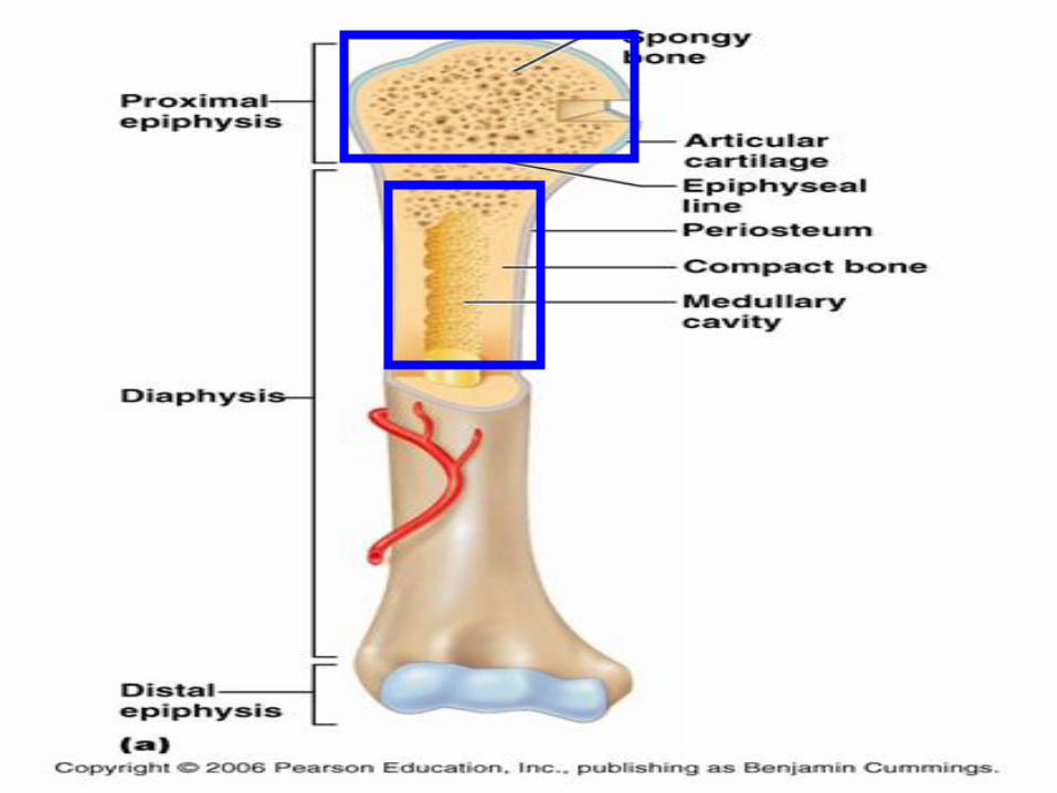
GENERAL FEATURES OF BONE • Typical long bone has
• DIAPHYSIS
• EPIPHYSIS
• METAPHYSIS
:
DIAPHYSIS: • The porIon of long bone between two carIlaginous ends is known as DIAPHYSIS.
• It ossifies from primary centre of ossificaIon which develops first in early foetal life in hyaline carIlage model of future bone.
• Primary centre & process of bone formaIon extends towards two ends.
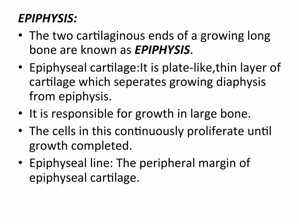
EPIPHYSIS: • The two carIlaginous ends of a growing long bone are known as EPIPHYSIS.
• Epiphyseal carIlage:It is plate-‐like,thin layer of carIlage which seperates growing diaphysis from epiphysis.
• It is responsible for growth in large bone. • The cells in this conInuously proliferate unIl growth completed.
• Epiphyseal line: The peripheral margin of epiphyseal carIlage.
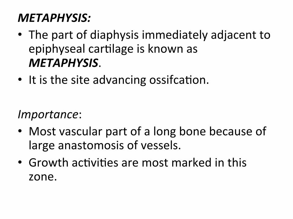
METAPHYSIS: • The part of diaphysis immediately adjacent to epiphyseal carIlage is known as METAPHYSIS.
• It is the site advancing ossifcaIon.
Importance: • Most vascular part of a long bone because of large anastomosis of vessels.
• Growth acIviIes are most marked in this zone.

• It is site of inserIon of muscles, thus it is liable to be injured due to muscular strain.
• SomeImes metaphysis lies within capsular ligament.So infecIon from diaphysis may spread to the joint.
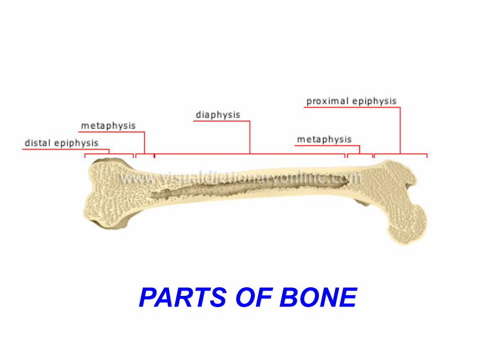
PARTS OF BONE
CLASSIFICATION OF BONE: • A)According to PosiIon: Axial:Bones forming axis of body. Ex:skull,ribs,sternum,vertebrae. Appendicular Bones: forming skeleton of limbs. • B)According to Size& Shape: Long bones:Present in upper & lower limbs. Ex.Femur,radius Act as levers for movements & locomoIon.
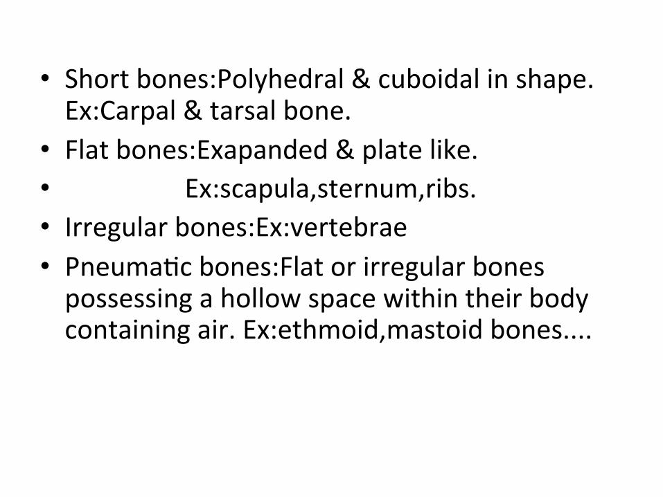
• Short bones:Polyhedral & cuboidal in shape. Ex:Carpal & tarsal bone.
• Flat bones:Exapanded & plate like. • Ex:scapula,sternum,ribs. • Irregular bones:Ex:vertebrae • PneumaIc bones:Flat or irregular bones possessing a hollow space within their body containing air. Ex:ethmoid,mastoid bones....
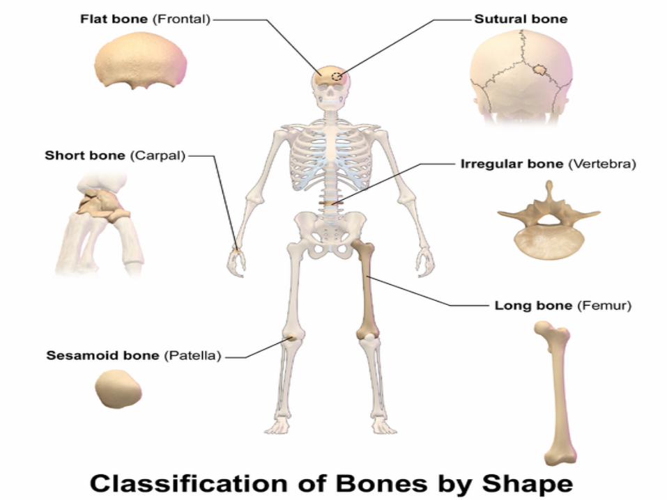
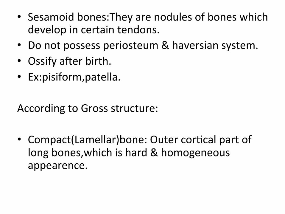
• Sesamoid bones:They are nodules of bones which develop in certain tendons.
• Do not possess periosteum & haversian system. • Ossify aZer birth. • Ex:pisiform,patella.
According to Gross structure:
• Compact(Lamellar)bone: Outer corIcal part of long bones,which is hard & homogeneous appearence.
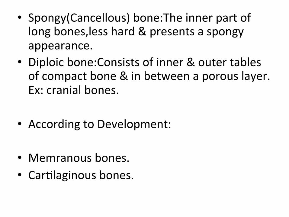
• Spongy(Cancellous) bone:The inner part of long bones,less hard & presents a spongy appearance.
• Diploic bone:Consists of inner & outer tables of compact bone & in between a porous layer. Ex: cranial bones.
• According to Development:
• Memranous bones. • CarIlaginous bones.
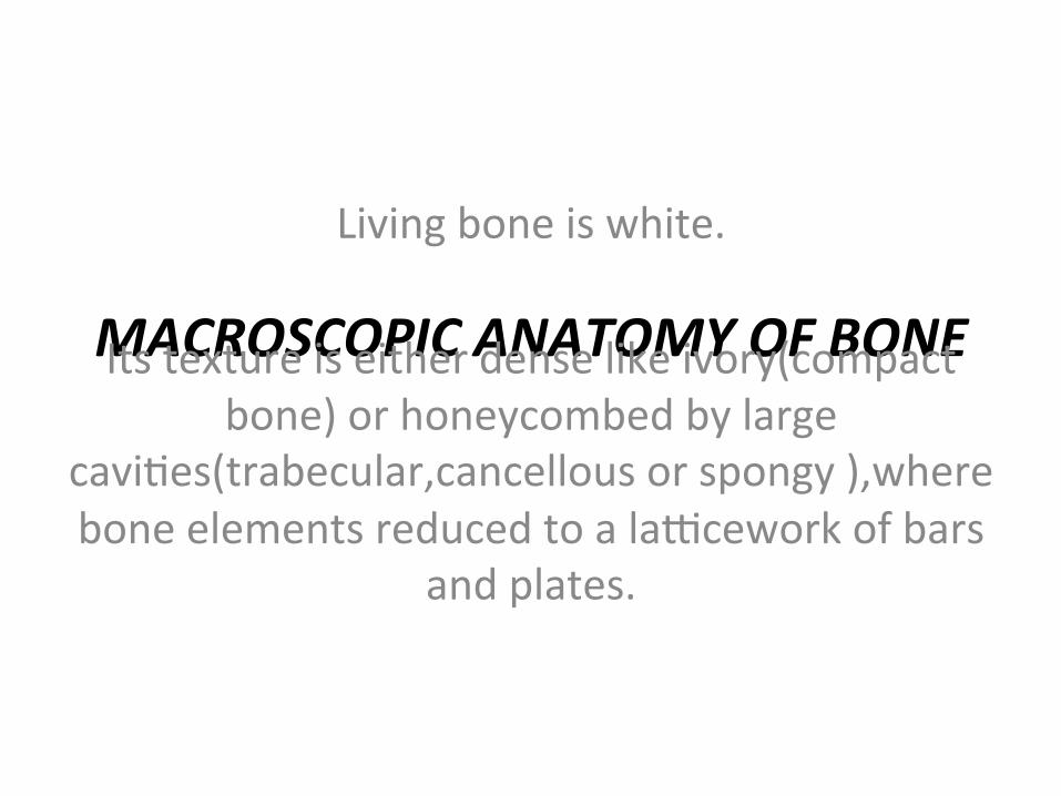
MACROSCOPIC ANATOMY OF BONE
Living bone is white.
Its texture is either dense like ivory(compact bone) or honeycombed by large
caviIes(trabecular,cancellous or spongy ),where bone elements reduced to a la\cework of bars
and plates.
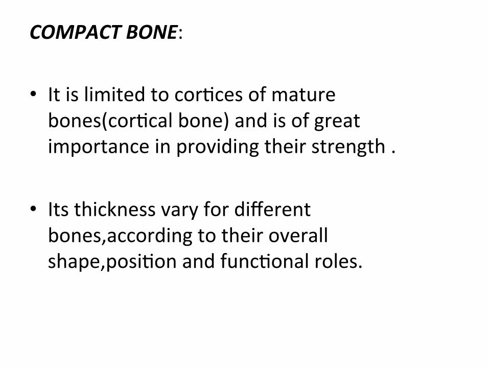
COMPACT BONE:
• It is limited to corIces of mature bones(corIcal bone) and is of great importance in providing their strength .
• Its thickness vary for different bones,according to their overall shape,posiIon and funcIonal roles.
COMPACT BONE
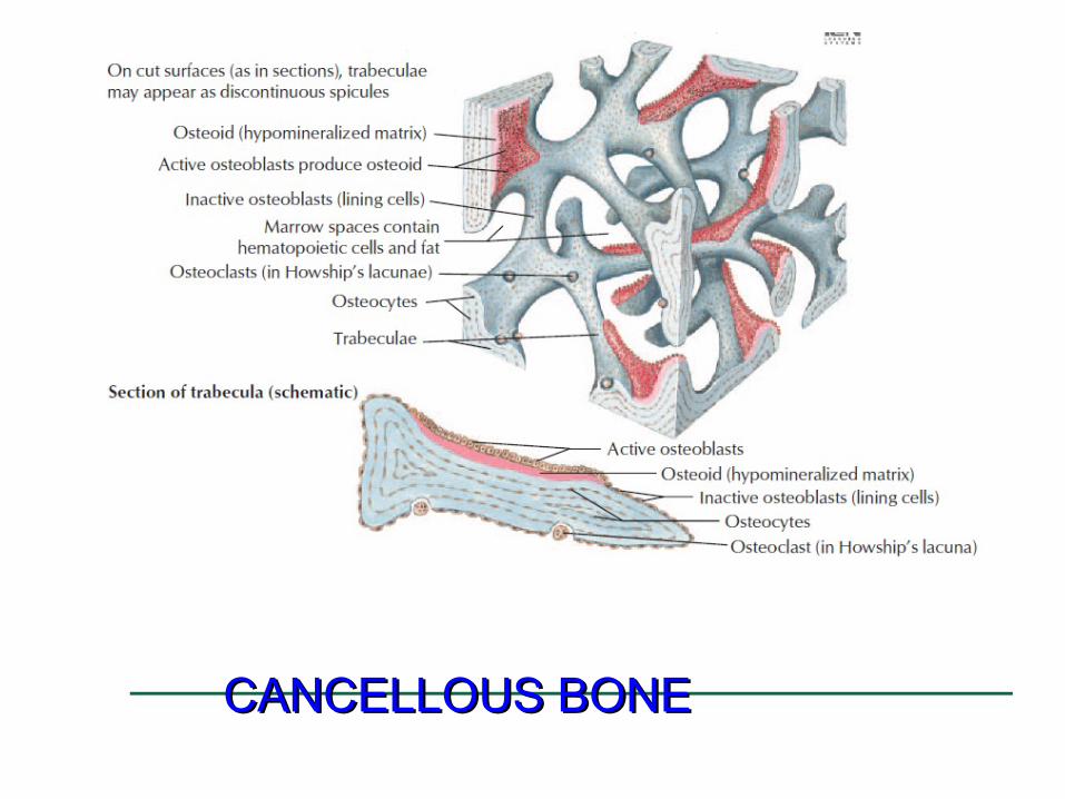
CANCELLOUS BONE
• It is usually internal, giving addiIonal strength to corIces and supporIng the bone marrow.
• Bone forms a reservoir of metabolic calcium(99% of calcium is in the bony skeleton) and phosphate which is under hormonal and cytokine control.
CANCELLOUS BONE CANCELLOUS BONE
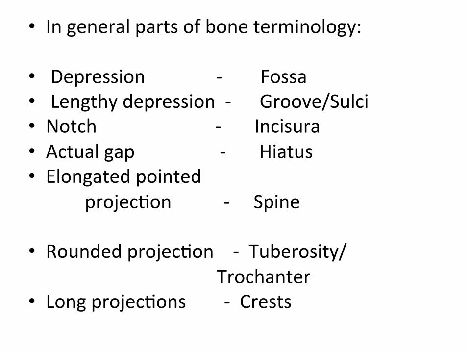
• In general parts of bone terminology:
• Depression -‐ Fossa • Lengthy depression -‐ Groove/Sulci • Notch -‐ Incisura • Actual gap -‐ Hiatus • Elongated pointed projecIon -‐ Spine • Rounded projecIon -‐ Tuberosity/ Trochanter • Long projecIons -‐ Crests
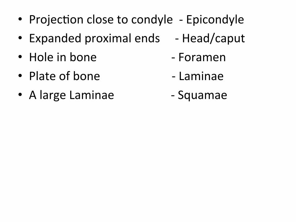
• ProjecIon close to condyle -‐ Epicondyle • Expanded proximal ends -‐ Head/caput • Hole in bone -‐ Foramen • Plate of bone -‐ Laminae • A large Laminae -‐ Squamae
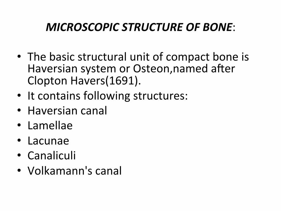
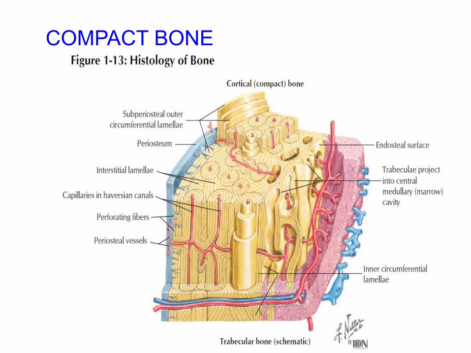
MICROSCOPIC STRUCTURE OF BONE:
• The basic structural unit of compact bone is Haversian system or Osteon,named aZer Clopton Havers(1691).
• It contains following structures: • Haversian canal • Lamellae • Lacunae • Canaliculi • Volkamann's canal
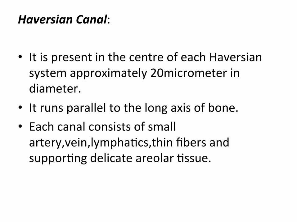
Haversian Canal: • It is present in the centre of each Haversian system approximately 20micrometer in diameter.
• It runs parallel to the long axis of bone. • Each canal consists of small artery,vein,lymphaIcs,thin fibers and supporIng delicate areolar Issue.
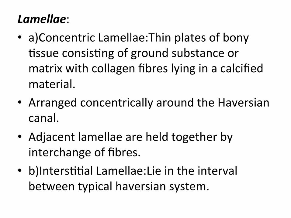
Lamellae: • a)Concentric Lamellae:Thin plates of bony Issue consisIng of ground substance or matrix with collagen fibres lying in a calcified material.
• Arranged concentrically around the Haversian canal.
• Adjacent lamellae are held together by interchange of fibres.
• b)IntersIIal Lamellae:Lie in the interval between typical haversian system.
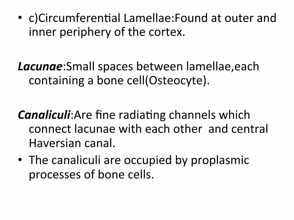
• c)CircumferenIal Lamellae:Found at outer and inner periphery of the cortex.
Lacunae:Small spaces between lamellae,each containing a bone cell(Osteocyte).
Canaliculi:Are fine radiaIng channels which connect lacunae with each other and central Haversian canal.
• The canaliculi are occupied by proplasmic processes of bone cells.
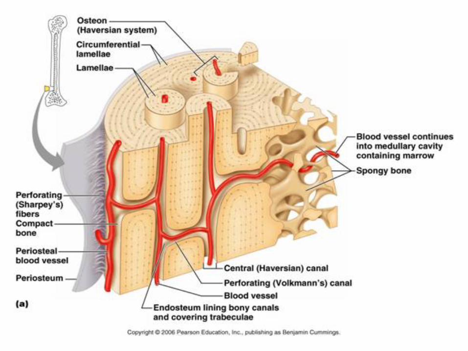
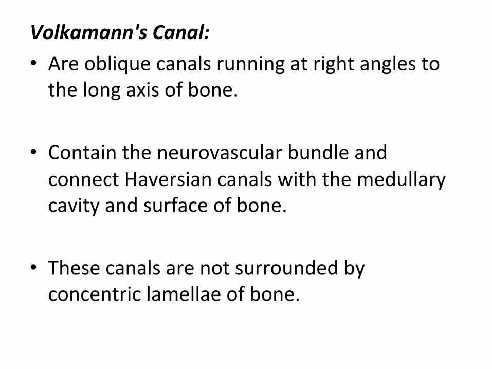
Volkamann's Canal: • Are oblique canals running at right angles to the long axis of bone.
• Contain the neurovascular bundle and connect Haversian canals with the medullary cavity and surface of bone.
• These canals are not surrounded by concentric lamellae of bone.
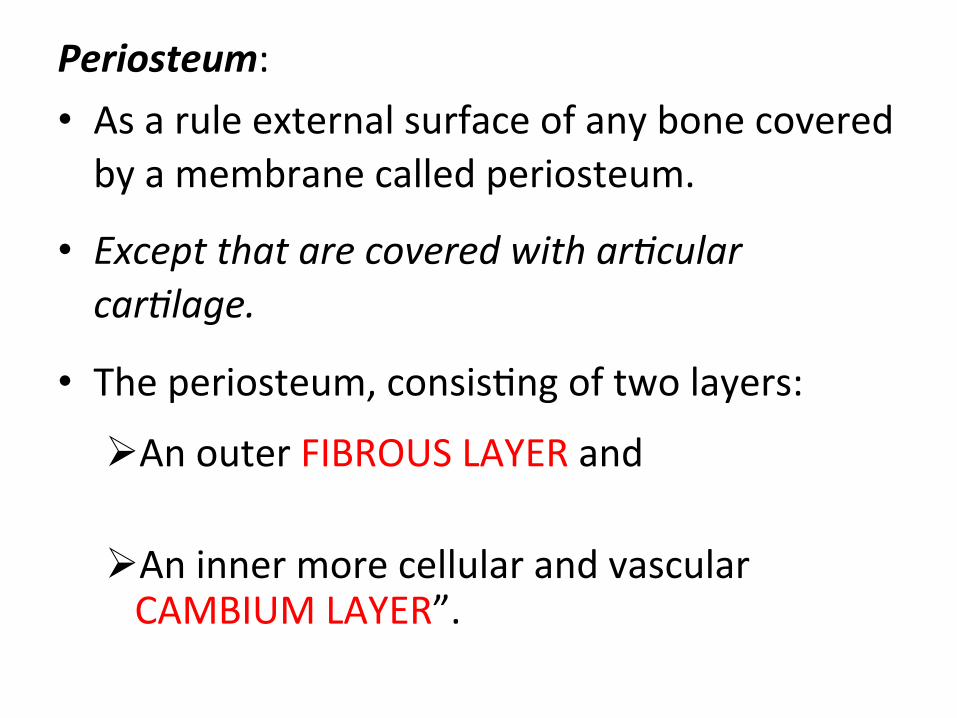
Periosteum: • As a rule external surface of any bone covered by a membrane called periosteum.
• Except that are covered with ar3cular car3lage.
• The periosteum, consisIng of two layers:
Ø An outer FIBROUS LAYER and
Ø An inner more cellular and vascular CAMBIUM LAYER”.
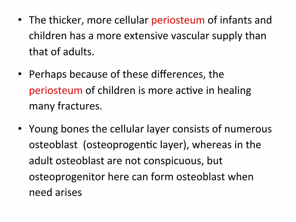
• The thicker, more cellular periosteum of infants and children has a more extensive vascular supply than that of adults.
• Perhaps because of these differences, the periosteum of children is more acIve in healing many fractures.
• Young bones the cellular layer consists of numerous osteoblast (osteoprogenIc layer), whereas in the adult osteoblast are not conspicuous, but osteoprogenitor here can form osteoblast when need arises
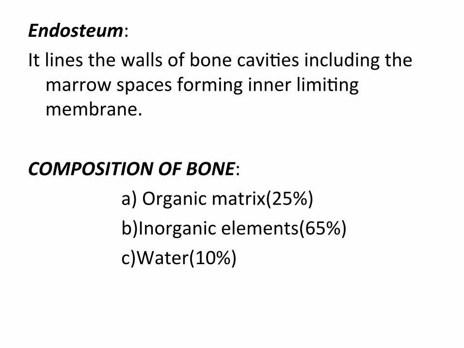
Endosteum: It lines the walls of bone caviIes including the marrow spaces forming inner limiIng membrane.
COMPOSITION OF BONE: a) Organic matrix(25%) b)Inorganic elements(65%) c)Water(10%)
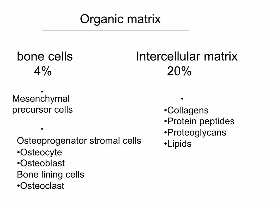
Organic matrix
bone cells 4%
Intercellular matrix 20%
• Collagens • Protein peptides • Proteoglycans • Lipids
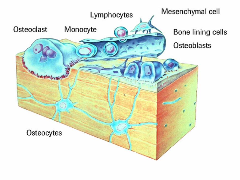
• Osteocyte • Osteoblast Bone lining cells • Osteoclast
Mesenchymal precursor cells
Osteoprogenator stromal cells
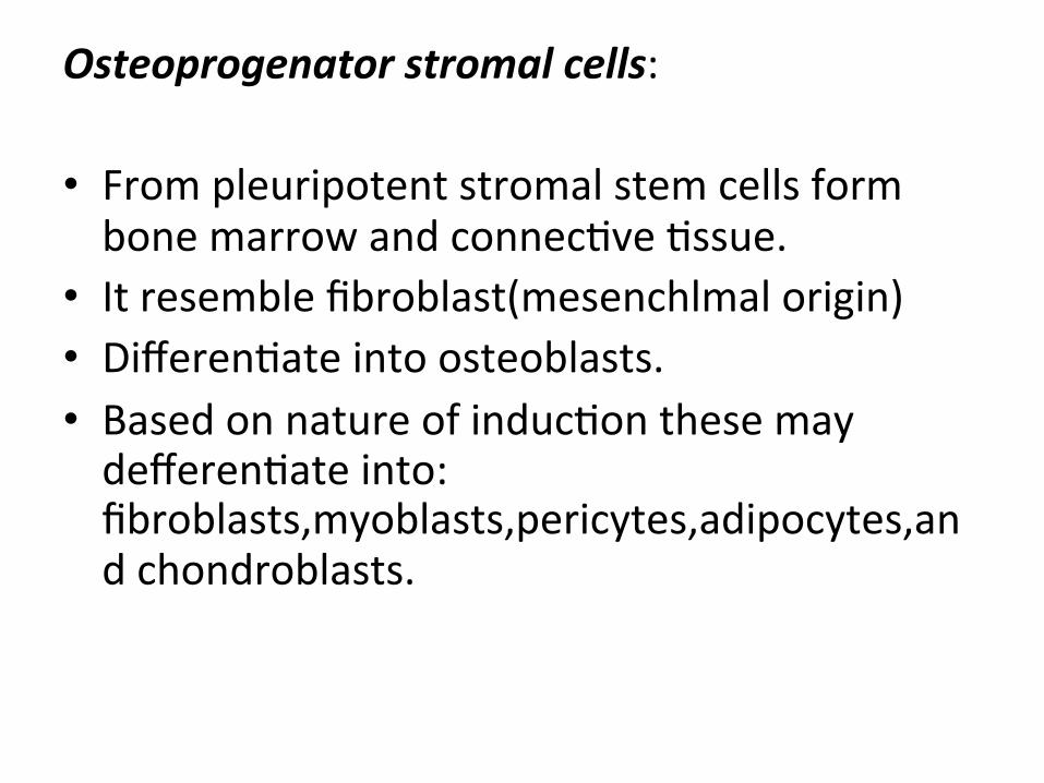
Osteoprogenator stromal cells:
• From pleuripotent stromal stem cells form bone marrow and connecIve Issue.
• It resemble fibroblast(mesenchlmal origin) • DifferenIate into osteoblasts. • Based on nature of inducIon these may defferenIate into: fibroblasts,myoblasts,pericytes,adipocytes,and chondroblasts.
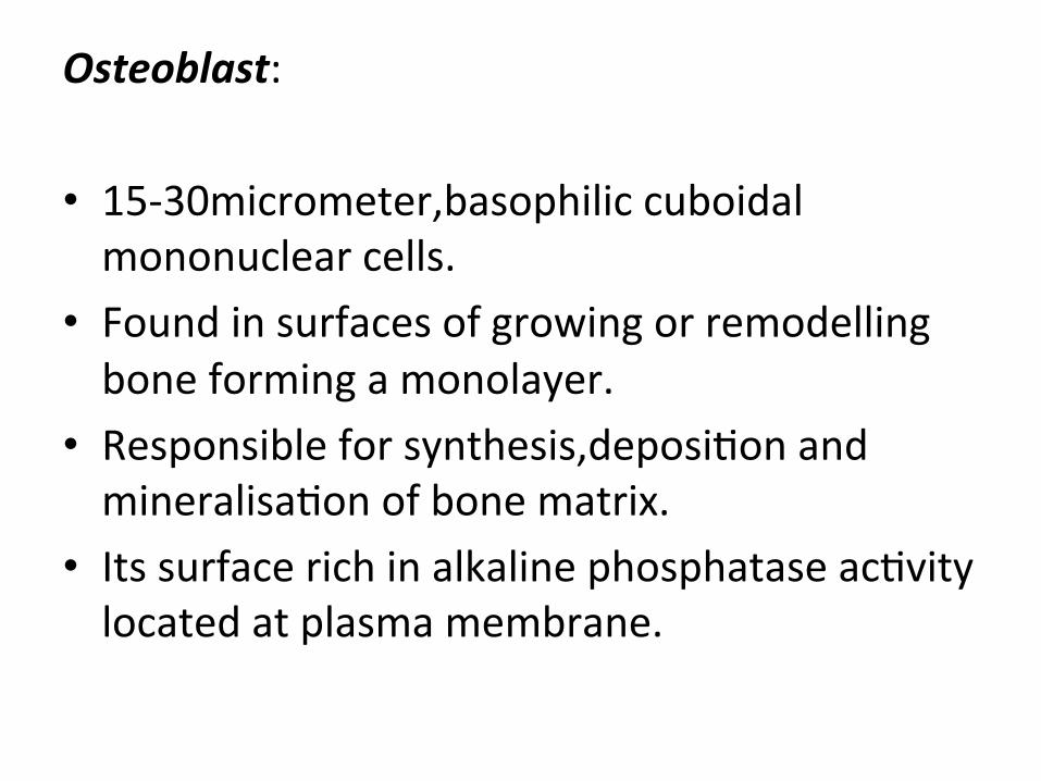
Osteoblast:
• 15-‐30micrometer,basophilic cuboidal mononuclear cells.
• Found in surfaces of growing or remodelling bone forming a monolayer.
• Responsible for synthesis,deposiIon and mineralisaIon of bone matrix.
• Its surface rich in alkaline phosphatase acIvity located at plasma membrane.
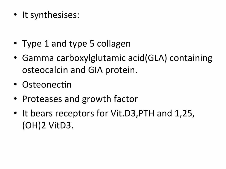
• It synthesises:
• Type 1 and type 5 collagen • Gamma carboxylglutamic acid(GLA) containing osteocalcin and GIA protein.
• OsteonecIn • Proteases and growth factor • It bears receptors for Vit.D3,PTH and 1,25,(OH)2 VitD3.
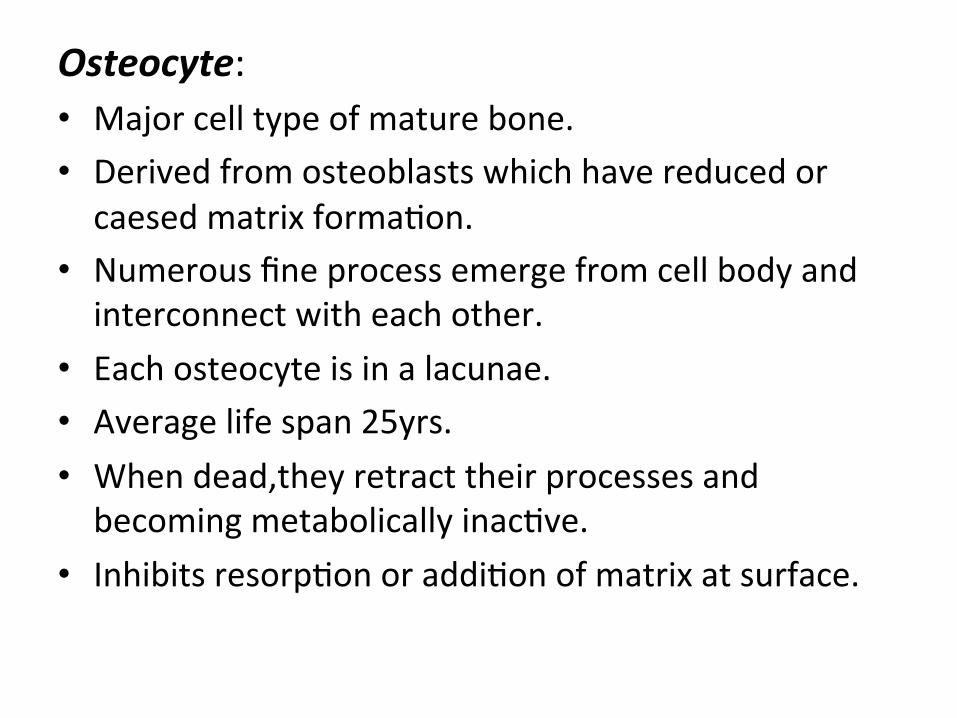
Osteocyte: • Major cell type of mature bone. • Derived from osteoblasts which have reduced or caesed matrix formaIon.
• Numerous fine process emerge from cell body and interconnect with each other.
• Each osteocyte is in a lacunae. • Average life span 25yrs. • When dead,they retract their processes and becoming metabolically inacIve.
• Inhibits resorpIon or addiIon of matrix at surface.
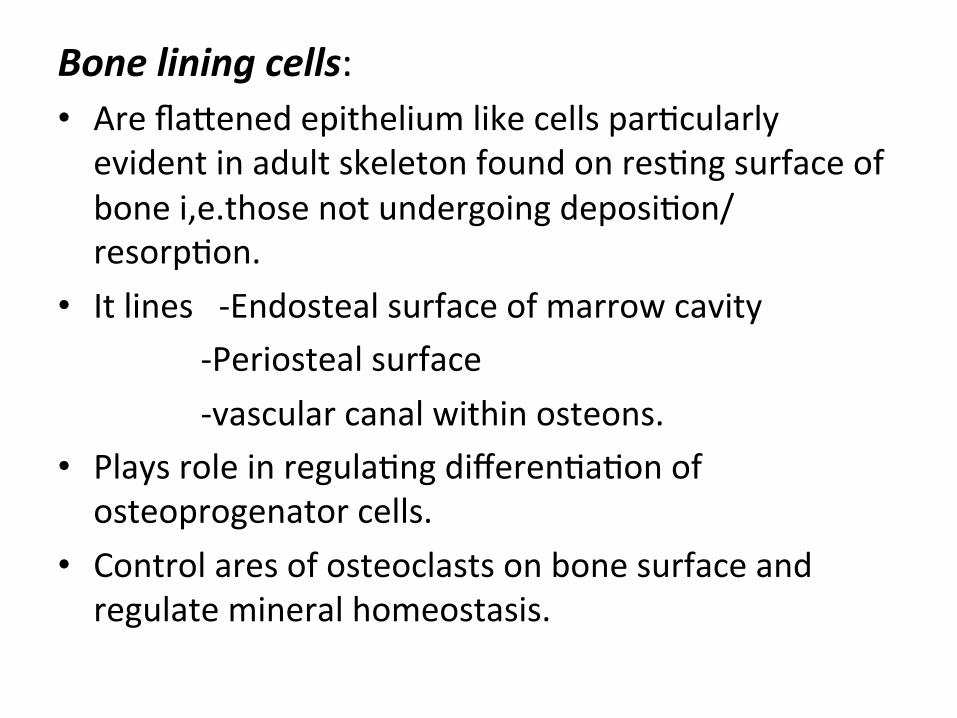
Bone lining cells: • Are flajened epithelium like cells parIcularly evident in adult skeleton found on resIng surface of bone i,e.those not undergoing deposiIon/resorpIon.
• It lines -‐Endosteal surface of marrow cavity -‐Periosteal surface -‐vascular canal within osteons. • Plays role in regulaIng differenIaIon of osteoprogenator cells.
• Control ares of osteoclasts on bone surface and regulate mineral homeostasis.
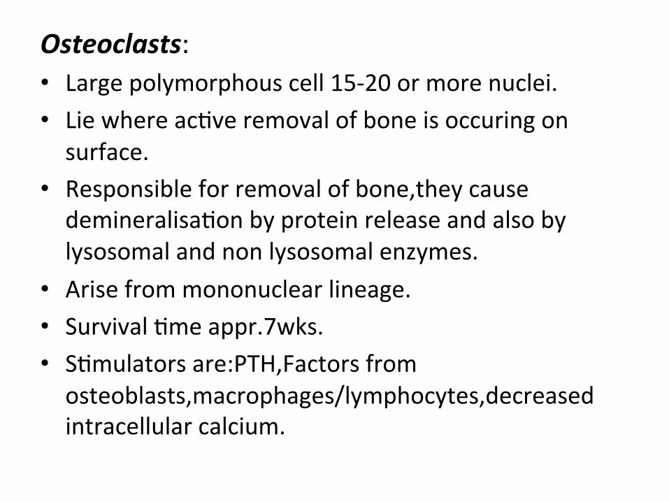
Osteoclasts: • Large polymorphous cell 15-‐20 or more nuclei. • Lie where acIve removal of bone is occuring on surface.
• Responsible for removal of bone,they cause demineralisaIon by protein release and also by lysosomal and non lysosomal enzymes.
• Arise from mononuclear lineage. • Survival Ime appr.7wks. • SImulators are:PTH,Factors from osteoblasts,macrophages/lymphocytes,decreased intracellular calcium.
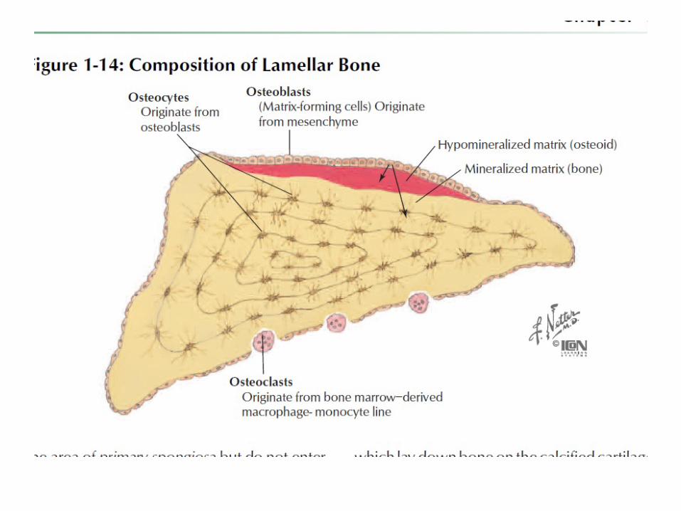
Bone Matrix: • It is the extracellular mineralized material of bone and consists of a ground substance in which are embeded numerous collagen fibres.
• In early stages of bone formaIon,before mineralizaIon,the matrix is Osteiod.
• In adult bone amount of osteiod is very small,reflecIng local remodelling of bone in which mineralizaIon follows deposiIon of organic matrix.
Collagen: • Bone contains type 1 and type 5 which is thought to regulate fibrillogenesis.
• It is synthesized from osteoblasts. • Other organic components of matrix like OsteonecIn is phosphorylated glycoprotein secreated by osteoblasts and bound mainly to minerals.
• Osteocalcin :Glycoprotein synthesized by osteoblasts.it is bound to mineral and is used as a marker of bone formaIon.
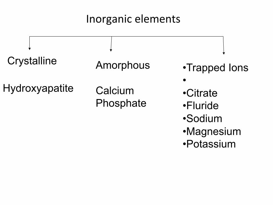
Inorganic elements
Hydroxyapatite
Crystalline Amorphous Calcium Phosphate
• Trapped Ions • • Citrate • Fluride • Sodium • Magnesium • Potassium

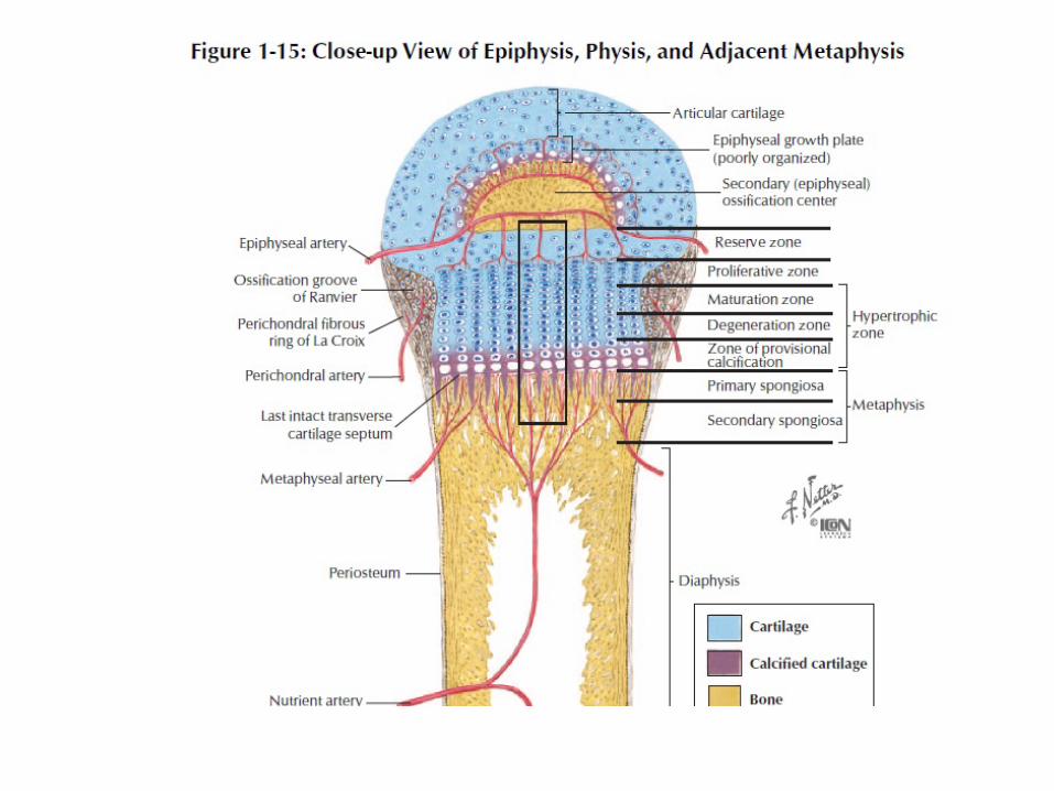
Blood Supply: • One or two main diaphyseal nutrient arteries enter shaZ obliquely through nutrient foramina leading into nutrient canals.
• Entry is directed away from dominant growing epiphysis.
• Nutrient arteries divided into ascending and descending branches in medullary cavity.
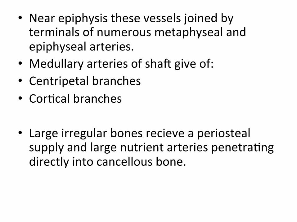
• Near epiphysis these vessels joined by terminals of numerous metaphyseal and epiphyseal arteries.
• Medullary arteries of shaZ give of: • Centripetal branches • CorIcal branches
• Large irregular bones recieve a periosteal supply and large nutrient arteries penetraIng directly into cancellous bone.
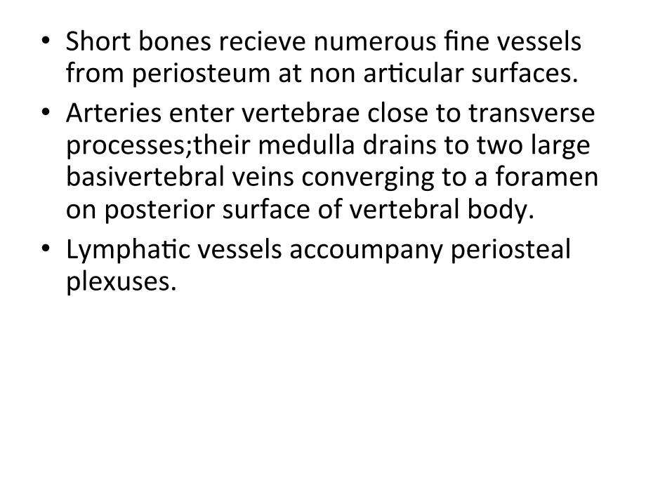
• Short bones recieve numerous fine vessels from periosteum at non arIcular surfaces.
• Arteries enter vertebrae close to transverse processes;their medulla drains to two large basivertebral veins converging to a foramen on posterior surface of vertebral body.
• LymphaIc vessels accoumpany periosteal plexuses.
Nerve Supply: • These are most numerous in arIcular extremiIes of longbones,vertebrae and larger flat bones.
• Nerves occur widely in periosteum, fine myelinated and non-‐myelinated fibres accoumpany nutrient vessels into bone marrow and lie in perivascular spaces of Haversian canals.
HISTIOGENESIS OF BONE:
• Bone first appears aZer 7th embryonic week. • They develop from embyonic mesenchymal Issue.
• The process of gradual bone formaIon is called OssificaIon.
• These are of two types: • 1)Endochondral OssificaIon • 2)Membranous OssificaIon
• 1)Endochondral OssificaIon: • In embryonic life most of skeleton is composed of carIlage, which is absorbed & replaced by bone.
• This process known as Endochondral OssificaIon.
• It begins prenatally & conInous throughout postnatal period unIl growth is complete.
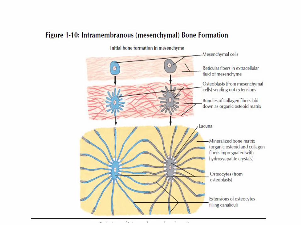
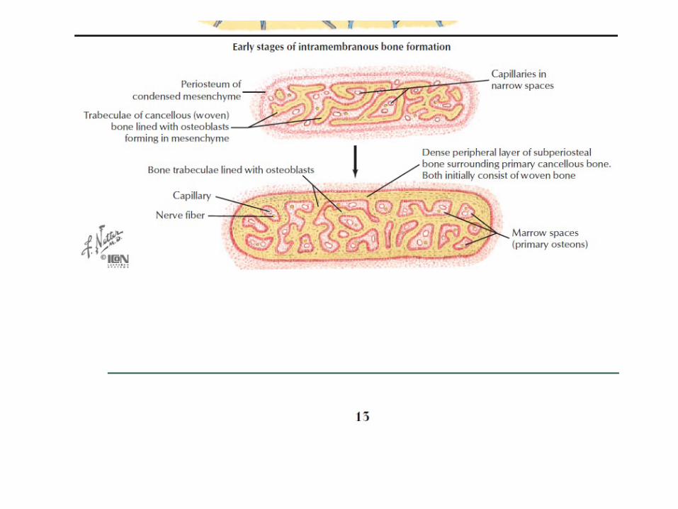
• 2)Membranous OssificaIon: • When bone is formed directly from a loose form of connecIve Issue without intervening stages of carIlage formaIon, calcificaIon and resorpIon and process is known as Membranous OssificaIon.
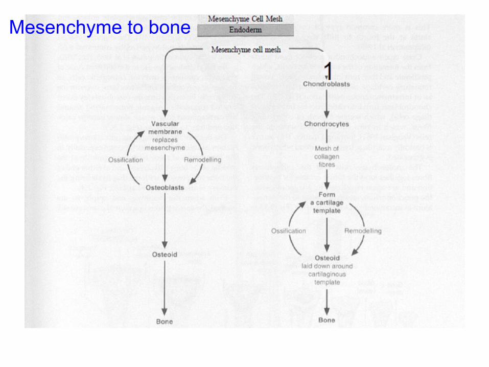
Mesenchyme to bone
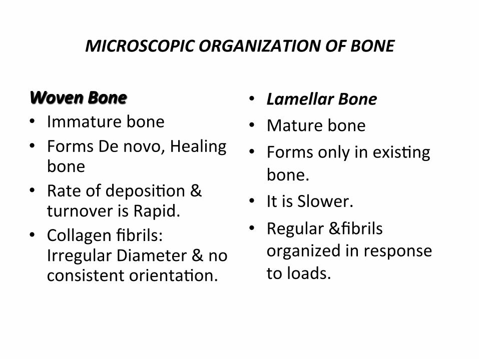
MICROSCOPIC ORGANIZATION OF BONE
Woven Bone • Immature bone • Forms De novo, Healing bone
• Rate of deposiIon & turnover is Rapid.
• Collagen fibrils: Irregular Diameter & no consistent orientaIon.
• Lamellar Bone • Mature bone • Forms only in exisIng bone.
• It is Slower. • Regular &fibrils organized in response to loads.
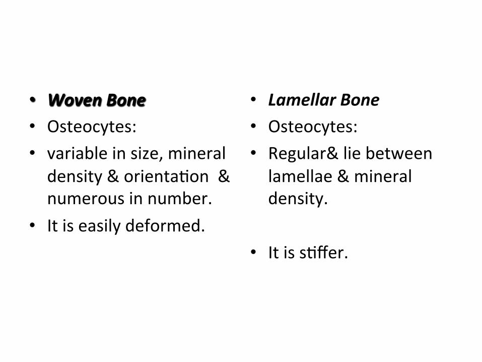
• Woven Bone • Osteocytes: • variable in size, mineral density & orientaIon & numerous in number.
• It is easily deformed.
• Lamellar Bone • Osteocytes: • Regular& lie between lamellae & mineral density.
• It is sIffer.
FRACTURE HEALING
• HISTORY
• INTRODUCTION
• STAGES OF FRACTURE HEALING
• VARIABLES INFLUENCE IN # HEALING
HISTORY: • Bones have broken since begining of humanity and have been recognised as long as recorded history.
• John Hunter,a pupil of Haller described morphologic sequence of fracture healing.
• In 1917,Bier reported sImulaIon factor for new bone formaIon was present in organized blood clot of the fracture haematoma.
INTRODUCTION: • A fracture is defined as a break in conInuity of bone.
• Fracture in man heal and unite by two main ways:
• 1)Primary/Osteonal/Direct Healing: • Bone formaIon occurs directly without any callus formaIon.This occurs parIcularly in stable,aligned,closely apposed fracture.
2)Secondary/Indirect Healing: • It is usual type consisIng of formaIon of callus either of carIlaginous or fibrous.
• This callus is later converted into lamellar bone.
• When fracture is not rigidly fixed and movements occur,in such cases callus is replaced by bone healing.
• On x ray charecterised by abundant callus formaIon,temporary widening of fracture gap and slow disappearance of radiolucent fracture line due to fibrocarIlage mineralisaIon.
STAGES OF FRACTURE HEALING: • OsteoinducIon is a first step in bone healing. • It causes mesenchymal cells to differenIate into various cells which then proliferate & produce messenger substances which further sImulate mesenchymal cells to differenIate.
• OsteconducIon a scaffold of collagenous network has developed upon which reparaIve cells produce callus & bone.
The various stages of # Healing includes:
• Stage of Haematoma FormaIon. • Stage of GranulaIon Issue. • Stage of Repair/Callus. • Stage of ConsolidaIon. • Stage of Remodelling.
Stage of Haematoma FormaTon: • Begins immediately following injury and followed rapidly by repair.
• The Haematoma provides 3 imp. factors:
• It immobilizes # and swellings hydrostaIcally splints the # and thus provides small amount of mechanically stability of # site.
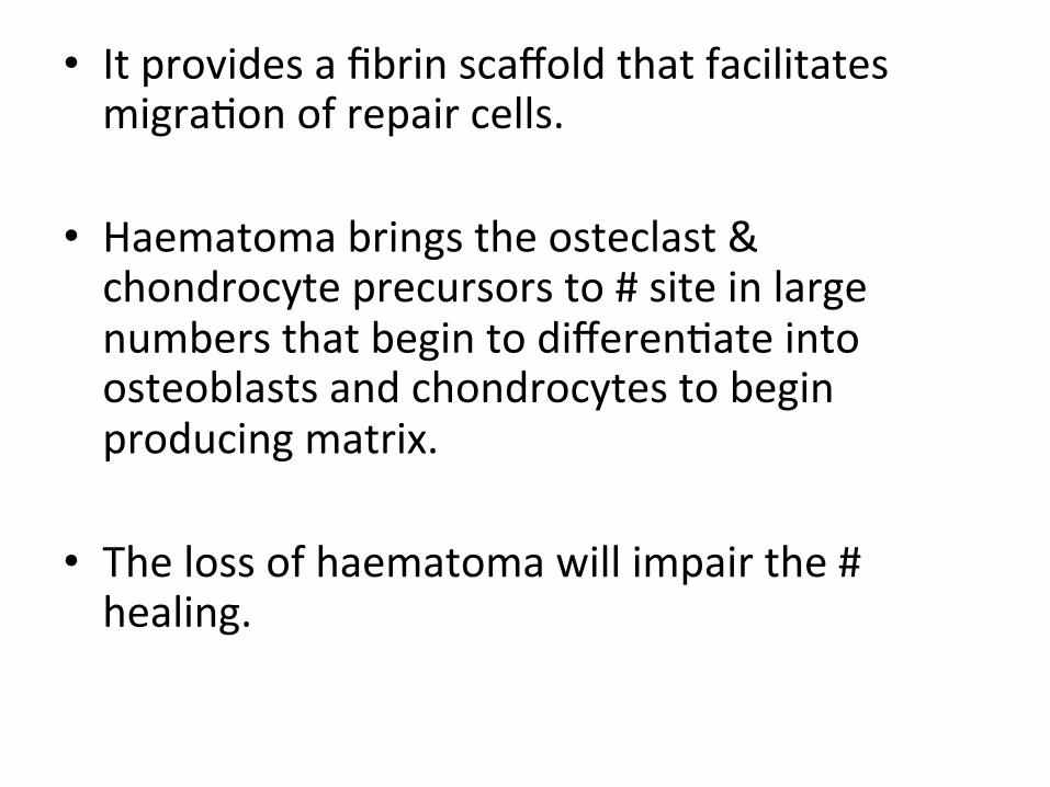
• It provides a fibrin scaffold that facilitates migraIon of repair cells.
• Haematoma brings the osteclast & chondrocyte precursors to # site in large numbers that begin to differenIate into osteoblasts and chondrocytes to begin producing matrix.
• The loss of haematoma will impair the # healing.

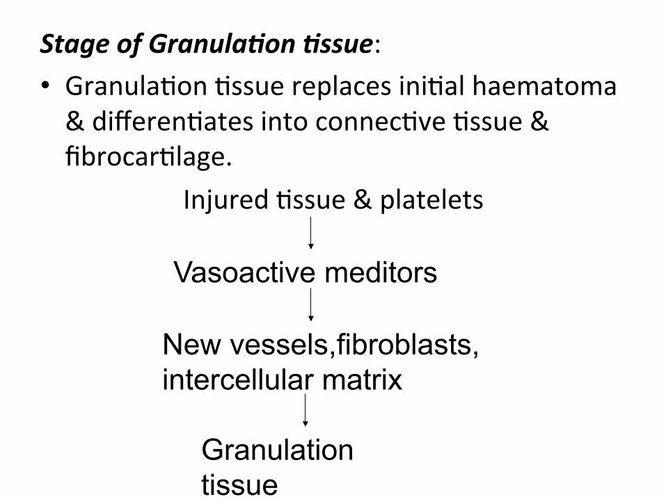
Stage of GranulaTon Tssue: • GranulaIon Issue replaces iniIal haematoma & differenIates into connecIve Issue & fibrocarIlage.
Injured Issue & platelets
Vasoactive meditors
New vessels,fibroblasts, intercellular matrix
Granulation tissue
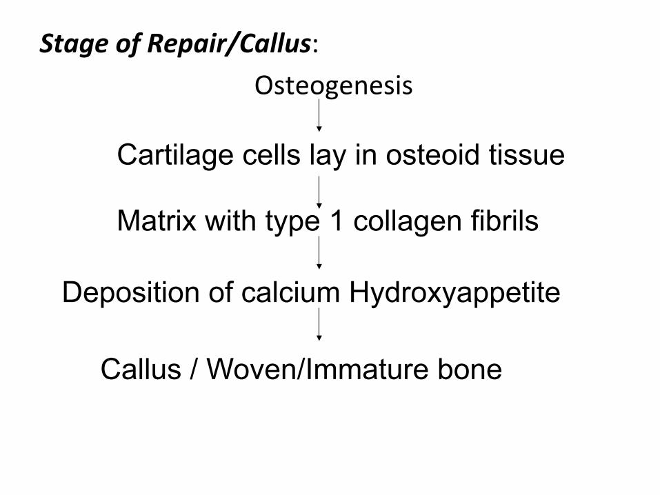
Stage of Repair/Callus: Osteogenesis
Cartilage cells lay in osteoid tissue
Matrix with type 1 collagen fibrils
Deposition of calcium Hydroxyappetite
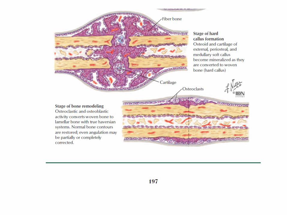
Callus / Woven/Immature bone
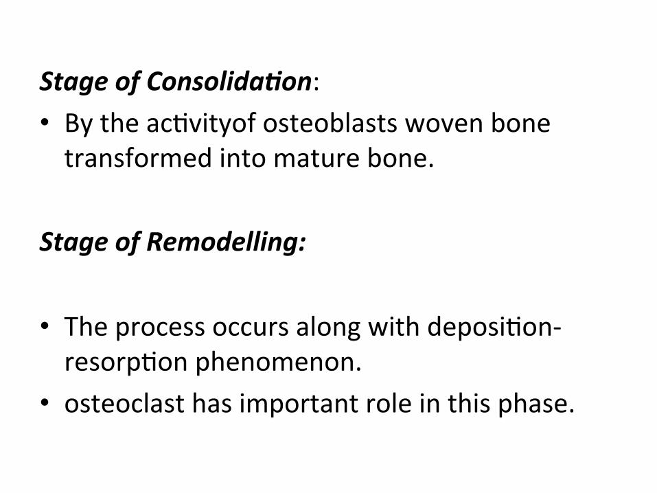
Stage of ConsolidaTon: • By the acIvityof osteoblasts woven bone transformed into mature bone.
Stage of Remodelling: • The process occurs along with deposiIon-‐resorpIon phenomenon.
• osteoclast has important role in this phase.
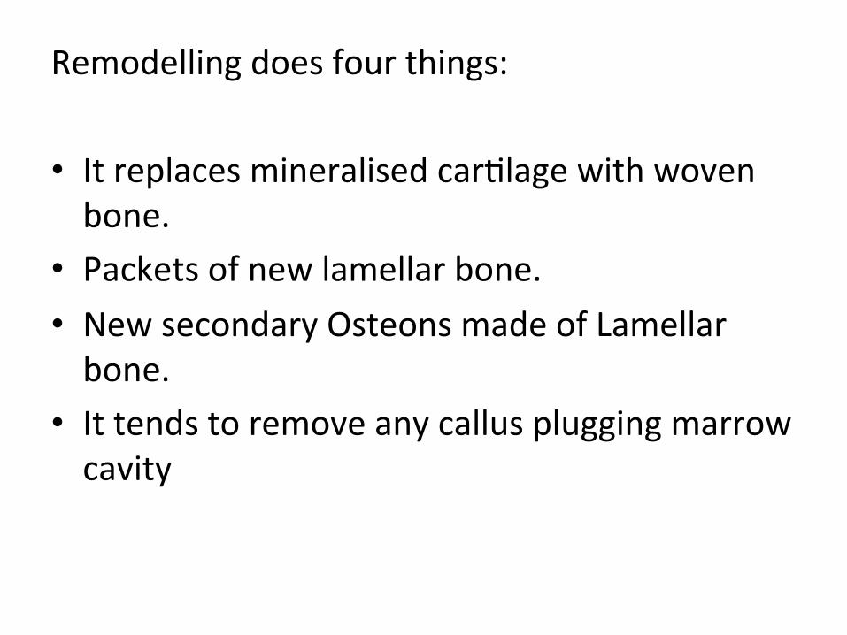
Remodelling does four things:
• It replaces mineralised carIlage with woven bone.
• Packets of new lamellar bone. • New secondary Osteons made of Lamellar bone.
• It tends to remove any callus plugging marrow cavity
FRACTURE HEALING IN CANCELLOUS BONE: • The extent of bone & marrow necrosis following cancellous bone # is much less than in compact bone,because of good circulaIon.
• Primary healing takes place in this,secondary healing is rare and endochondral bone formaIon excepIonal.
VARIABLES INFLUENCE IN # HEALING: • Cruses and Buck Walter have divided variarles into four groups
1)INJURY VARIABLES: Ø Open Fractures:Delays repair by soZ Issue disrupIon &disturbed blood supply to # site.
Ø severity of injury:Extensive soZ Issue & • bone damage leads to delayed # healing.
Ø IntraarTcular fracture: It requires reconstrucIon of joint surface,stable fixaIon & early mobilisaIon.
Ø Segmental fracture:It leads to delayed union/non union due to disrupted intramedullary blood supply of middle fragments.
Ø SoX Tssue interposiTon:Open reducIon to extricate interposed Issue will enhance # healing process.
Ø Damage to blood supply:Delay # healing.
2)PATIENT VARIABLES: Ø Age:Extremes of age have influenes on # healing.
Ø Nutri3on:Poor nutriIonal status affects # healing & can lead to mortality & surgical complicaIons.
Ø Systemic Hormones: Steroids,anIcoagulants,anIinflammatory drugs inhibit whereas GH,insulin thyroid hormone enhance # healing.
Ø Nico3ne
3)TISSUE VARIABLES: Ø Form of bone:Cancellous bone healing is rapid due to larger surface,rich in cells & blood supply.
Ø Bone Necrosis Ø Bone diseases:Osteoporosis,Primary malignant bone tumours,metastasis,bone cysts etc... all cause pathological bone # and delay bone healing.
Ø Infec3on:It slows down/prevents healing.
4)TREATMENT VARIABLES: Ø Apposi3on of # Fragments:Decreasing # gap decreases volume of repair Issue needed to heal #.
Ø Loading & Micrimo3on:Loading a # site & induced micrimoIon along bone # sites promotes healing but too much moIon lead to non union.
Ø Fracture Stabilisa3on:It will prevents repeated disrupIon of repair & enhances # callus.
Ø Rigid Fixa3on:Stable fixaIon allows early mobilisaIon of joints & hence prevents sIffness.
Ø Bone GraIing:It is osteoinducIve & osteoconducIve.
Ø Demineralised Bone marrow:The factors in bone marrow sImulate bone formaIon,by migraIon of undifferenIed mesenchymal cells to implanted matrix & differenIaIon into mesenchymal cells.

DHANYAVAAD
Related Documents