Anatomy of a Structural Pathway for Activation of the Catalytic Domain of Src Kinase Hck Nilesh K. Banavali and Benoı ˆt Roux * Department of Physiology and Biophysics, Weill Medical College of Cornell University, New York, New York 10021 ABSTRACT Src kinase activity is implicated in the regulation of downstream signal transduc- tion pathways involved in cell growth processes. Crystallographic studies indicate that activation of Hematopoietic cell kinase (Hck), a member of the Src kinase family, is accompanied structurally by a large conformational change in two specific parts of its catalytic domain: the a-C helix and the activation loop. In the present study, molecular dynamics (MD) simulations are used to character- ize the transformation pathway from the inactive to the active state. Four different conditions are considered: the presence or absence of Tyr416 phosphorylation in the activation loop, and the presence or absence of substrate ATP-2Mg 12 in the active site. Effective free energy landscapes for local residues are determined using a combination of restrained MD simulations with a Root Mean Square Distance (RMSD) biasing potential to enforce the change followed by free MD simula- tions to allow relaxation from artificially enforced intermediates. A conceptual subdivision of the kinase catalytic domain into four moving parts: the flexible activation loop segment, the buried activation loop segment, the a-C helix, and the N- terminal end linker, leads to a concise hypothesis in which each of the moving parts are only re- quired to be coupled to their nearest neighbor to ensure bidirectional allostery in the regulation of protein tyrosine kinases. Both Tyr416 phosphoryl- ation and ATP-2Mg 12 affect the local backbone tor- sional free energy landscapes accompanying the structural transition. When these two factors are present together, a metastable coordinated state of ATP-2Mg 12 and the phosphorylated Tyr416 is observed that offers a possible explanation for the inhibition of protein kinase activity due to increase in Mg 12 ion concentration. Proteins 2007;67:1096–1112. V V C 2007 Wiley-Liss, Inc. Key words: molecular dynamics; RMSD restraint; mechanism; Mg-ATP; free energy land- scape; allostery; phosphorylation INTRODUCTION Src kinases are membrane-associated nonreceptor ty- rosine kinases that phosphorylate specific tyrosine resi- dues in other proteins, thereby modifying their activity and affecting various downstream signal transduction pathways in which these proteins participate. 1 The over- all architecture of all nine members of the Src kinase family is conserved, and mainly consists of the domain that carries out the catalytic activity, and the SH2 and SH3 domains that regulate the activity of the catalytic domain. 2 The SH2 domain recognizes and binds to a phosphorylated tyrosine residue (Tyr 527, chicken c-Src numbering) in the C-terminal tail of the catalytic do- main, which is a critical regulatory intramolecular inter- action. 3 Dephosphorylation of this residue by phospha- tases results in Src kinase activation. 4 The SH3 domain recognizes and binds to the proline-containing linker between the catalytic and SH2 domains. 5 Both these interactions can be disrupted by external peptides with recognition sequences containing a phosphorylated tyro- sine or multiple proline residues that bind preferentially to the SH2 and SH3 domains, respectively, allowing the catalytic domain to shift to its activated form. 6 Phospho- rylation of Tyr416 in the catalytic domain also results in the activation of the catalytic domain. 7 The downregula- tion of catalytic domain activity thus primarily depends on a combination of three stabilizing factors: the SH2 do- main interacting intramolecularly with the C-terminal tail, the SH3 domain remaining bound to the proline- containing linker between SH2 and catalytic domains, and Tyr416 in the activation loop remaining unphospho- rylated. Disruption in any of these three factors causes a shift in equilibrium to the active state of the kinase. 8 Structural information about the downregulated inac- tive and upregulated active states of Src kinases is avail- able from X-ray crystallographic studies of three differ- The Supplementary Material referred to in this article can be found at http://www.interscience.wiley.com/jpages/0887-3585/suppmat/ Grant sponsor: National Institutes of Health; Grant number: CA93577-01. Nilesh K. Banavali’s current address is Division of Molecular Medicine, Wadsworth Center, Empire State Plaza, Albany, NY 12201. Benoı ˆt Roux current address is Institute of Molecular Pediatric Science, Gordon Center of Integrative Science, The University of Chicago, 929 East 57th Street, Chicago, IL 60637, E-mail: roux@ uchicago.edu *Correspondence to: Benoı ˆt Roux, Department of Physiology and Biophysics, Weill Medical College of Cornell University, New York, NY 10021. E-mail: [email protected] Received 1 May 2006; Revised 19 October 2006; Accepted 8 November 2006 Published online 22 March 2007 in Wiley InterScience (www. interscience.wiley.com). DOI: 10.1002/prot.21334 V V C 2007 WILEY-LISS, INC. PROTEINS: Structure, Function, and Bioinformatics 67:1096–1112 (2007)

Welcome message from author
This document is posted to help you gain knowledge. Please leave a comment to let me know what you think about it! Share it to your friends and learn new things together.
Transcript
Anatomy of a Structural Pathway for Activation of theCatalytic Domain of Src Kinase Hck
Nilesh K. Banavali and Benoıt Roux*
Department of Physiology and Biophysics, Weill Medical College of Cornell University, New York, New York 10021
ABSTRACT Src kinase activity is implicatedin the regulation of downstream signal transduc-tion pathways involved in cell growth processes.Crystallographic studies indicate that activationof Hematopoietic cell kinase (Hck), a member ofthe Src kinase family, is accompanied structurallyby a large conformational change in two specificparts of its catalytic domain: the a-C helix and theactivation loop. In the present study, moleculardynamics (MD) simulations are used to character-ize the transformation pathway from the inactiveto the active state. Four different conditions areconsidered: the presence or absence of Tyr416phosphorylation in the activation loop, and thepresence or absence of substrate ATP-2Mg12 in theactive site. Effective free energy landscapes forlocal residues are determined using a combinationof restrained MD simulations with a Root MeanSquare Distance (RMSD) biasing potential toenforce the change followed by free MD simula-tions to allow relaxation from artificially enforcedintermediates. A conceptual subdivision of thekinase catalytic domain into four moving parts:the flexible activation loop segment, the buriedactivation loop segment, the a-C helix, and the N-terminal end linker, leads to a concise hypothesisin which each of the moving parts are only re-quired to be coupled to their nearest neighbor toensure bidirectional allostery in the regulation ofprotein tyrosine kinases. Both Tyr416 phosphoryl-ation and ATP-2Mg12 affect the local backbone tor-sional free energy landscapes accompanying thestructural transition. When these two factors arepresent together, a metastable coordinated state ofATP-2Mg12 and the phosphorylated Tyr416 isobserved that offers a possible explanation for theinhibition of protein kinase activity due toincrease in Mg12 ion concentration. Proteins2007;67:1096–1112. VVC 2007 Wiley-Liss, Inc.
Key words: molecular dynamics; RMSD restraint;mechanism; Mg-ATP; free energy land-scape; allostery; phosphorylation
INTRODUCTION
Src kinases are membrane-associated nonreceptor ty-rosine kinases that phosphorylate specific tyrosine resi-dues in other proteins, thereby modifying their activity
and affecting various downstream signal transductionpathways in which these proteins participate.1 The over-all architecture of all nine members of the Src kinasefamily is conserved, and mainly consists of the domainthat carries out the catalytic activity, and the SH2 andSH3 domains that regulate the activity of the catalyticdomain.2 The SH2 domain recognizes and binds to aphosphorylated tyrosine residue (Tyr 527, chicken c-Srcnumbering) in the C-terminal tail of the catalytic do-main, which is a critical regulatory intramolecular inter-action.3 Dephosphorylation of this residue by phospha-tases results in Src kinase activation.4 The SH3 domainrecognizes and binds to the proline-containing linkerbetween the catalytic and SH2 domains.5 Both theseinteractions can be disrupted by external peptides withrecognition sequences containing a phosphorylated tyro-sine or multiple proline residues that bind preferentiallyto the SH2 and SH3 domains, respectively, allowing thecatalytic domain to shift to its activated form.6 Phospho-rylation of Tyr416 in the catalytic domain also results inthe activation of the catalytic domain.7 The downregula-tion of catalytic domain activity thus primarily dependson a combination of three stabilizing factors: the SH2 do-main interacting intramolecularly with the C-terminaltail, the SH3 domain remaining bound to the proline-containing linker between SH2 and catalytic domains,and Tyr416 in the activation loop remaining unphospho-rylated. Disruption in any of these three factors causes ashift in equilibrium to the active state of the kinase.8
Structural information about the downregulated inac-tive and upregulated active states of Src kinases is avail-able from X-ray crystallographic studies of three differ-
The Supplementary Material referred to in this article can be foundat http://www.interscience.wiley.com/jpages/0887-3585/suppmat/
Grant sponsor: National Institutes of Health; Grant number:CA93577-01.
Nilesh K. Banavali’s current address is Division of MolecularMedicine, Wadsworth Center, Empire State Plaza, Albany, NY12201.
Benoıt Roux current address is Institute of Molecular PediatricScience, Gordon Center of Integrative Science, The University ofChicago, 929 East 57th Street, Chicago, IL 60637, E-mail: [email protected]
*Correspondence to: Benoıt Roux, Department of Physiology andBiophysics, Weill Medical College of Cornell University, New York,NY 10021. E-mail: [email protected]
Received 1 May 2006; Revised 19 October 2006; Accepted 8November 2006
Published online 22 March 2007 in Wiley InterScience (www.interscience.wiley.com). DOI: 10.1002/prot.21334
VVC 2007 WILEY-LISS, INC.
PROTEINS: Structure, Function, and Bioinformatics 67:1096–1112 (2007)
ent members of the Src family: Hematopoietic cell kinase(Hck), c-Src, and Lck. Hck is found in myeloid andlymphoid cells9 that play a role in development of can-cer.10,11 The crystal structures of Hck in complex withtwo different inhibitors12,13 show the structural detailsof its inactive state, which are also very similar to thosefor the highly homologous c-Src.14,15 The molecular basisof the regulatory interactions between SH2 and C-termi-nal tail and SH3 and proline-containing linker is clari-fied in these fully assembled structures of the downregu-lated inactive form of the kinase. The catalytic domainitself is divided into two lobes, the N-lobe and the C-lobe, between which lies the catalytic site where ATPand peptide substrates bind. The overall structuralarchitecture of the catalytic domain is very similar tothat of other protein kinases, such as c-AMP activatedprotein kinase A (PKA)16 and the insulin receptor kinase(IRK).17 The inactive state of the catalytic domain ofHck has the activation loop with Tyr416 (approximatelyresidues 410–430, chicken Src numbering) folded backonto the catalytic site, and the a-C helix (residues 304–316) rotated outward away from the catalytic site. Thehydrogen bond between Lys295 and Glu310, which isknown to be critical for catalysis,12 is completely dissoci-ated in this inactive conformation. These features,absent in the active state structures of both PKA andIRK, are represented in the unphosphorylated inactiveIRK structure.18 Structural studies on other protein ki-nases have shown that their catalytic domains adoptactive state conformations similar to PKA, but may varywidely in their inactive state conformations.19
The crystal structure of the lymphocyte kinase Lck,another closely related member of the Src kinase familyfound in white blood cells, reveals an active state confor-mation reminiscent of that of PKA.20 In this conformation,the phosphorylated Tyr416 containing activation loop islocated away from the catalytic site and the a-C helix isrotated inwards such that the hydrogen bond betweenLys295 and Glu310 is formed. Phosphorylation of the tyro-sine residue in the activation loop of the catalytic domainclearly results in substantial conformational changes.These changes are communicated to the spatially
remote regulatory domains, as is characteristic of a long-range allosteric coupling mechanism.21 The peptide link-ers between the SH2 and SH3 domains (residues 140–147), and the SH2 and catalytic domains (residues 246–259) are both shown to play a part in this long-range al-losteric coupling. Three separate modifications related tothese linkers are able to supersede the inhibitory effectof SH2 domain interaction with the phosphorylatedTyr527: (a) introduction of structurally destabilizingmultiple glycine mutations in the short linker betweenthe SH2 and SH3 domains in c-Src22; (b) introduction ofpolyproline-rich HIV 1-Nef protein disrupting the intra-molecular binding of the proline-rich region (residues250–254 in the linker between the SH2 and catalyticdomains) to the SH3 domain6; and (c) alanine mutationof the strictly conserved Trp260 residue at the end of thelinker between the SH2 and catalytic domains.23 This
Trp260 residue at the N-terminal end of the catalytic do-main is also in close contact with the a-C helix, which inturn is closely associated with several residues of a bur-ied segment of the activation loop (residues 407–411), inclose proximity to the critical Tyr416 residue in the acti-vation loop.
All these local interactions trace an allosteric pathwaythrough the kinase that seems to functionally connectthe distant parts of the enzyme. The bidirectional natureof this allosteric pathway is suggested by biochemicalexperiments showing that Tyr416 phosphorylationcauses greater availability of the SH2 and SH3 domainsto interact with the phosphotyrosine containing p-YEEIpeptide and polyproline-containing HIV 1-Nef protein,respectively.8 However, the mechanism by which infor-mation about conformational state of one part of thekinase is allosterically transmitted to the other parts,and whether this process is concerted or not, remainsunclear.
Computer simulations based on atomic models canhelp in understanding the complex dynamics of Src ki-nases that governs such bidirectional allostery. A previ-ous computational study, focused on identifying the basisof long-range allostery in Hck activation throughrestrained Targeted Molecular Dynamics (TMD),22 sug-gested that the structural effects of activation on the N-lobe were transmitted as ‘‘instantaneous correlations’’ tothe regulatory domains. A more recent TMD studyfocused on the hinge-bending separation of the two inter-nally restrained lobes of the Src kinase and the responseof the activation loop to this hinge-bending motion.24
With the lobes artificially restrained with dihedral con-straints, the activation loop showed spontaneous openingonly in the presence of ADP with two Mgþ2 ions andphosphorylated Tyr416, which was attributed to phos-phate–phosphate repulsion and hydration requirements.The results were also interpreted to suggest the possibil-ity of an intramolecular (‘‘cis’’) phosphorylation of Tyr416by its own catalytic site. In spite of the qualitativeinsight provided by all these experimental and theoreti-cal studies, there is clearly a need to progress towards amore detailed understanding of the conformational path-ways linking Tyr416 phosphorylation to structural acti-vation of Src kinases.
In the present study, structural pathways for the acti-vation of the Hck catalytic domain are obtained usingrestrained MD simulations in explicit solvent. The pro-cess is examined under four different conditions concern-ing the presence of neutral ligand ATP-2Mgþ2 andTyr416 phosphorylation. To reduce the main drawback ofrestrained TMD simulations, unbiased (free) MD simula-tions are subsequently used to allow spontaneous relaxa-tion of the initially biased pathways. Conformational sta-tistics gathered are characterized through residue-levelbackbone torsion conformational analysis and sidechaininteraction analysis to quantitatively dissect the large-scale structural transformation into its local components.A metastable coordination state between ATP-2Mgþ2
and the phosphorylated Tyr416 is also observed and the
1097ANATOMY OF A STRUCTURAL PATHWAY OF HCK ACTIVATION
PROTEINS: Structure, Function, and Bioinformatics DOI 10.1002/prot
consequences of its existence are explored. The implica-tions of the details of the structural activation pathwaysin the catalytic function and bidirectional allosteric regu-lation of Hck are discussed.
MATERIALS AND METHODS
The c30a1 academic version of the CHARMM pro-gram25 and the all-atom PARAM22 CHARMM force fieldfor ATP-2Mgþ2 and proteins26,27 was used for all calcula-tions. The Hematopoietic cell kinase (Hck) catalyticdomain inactive state structure was obtained from the2.0 A resolution crystal structure (PDB ID: 1QCF) in thepresence of the inhibitor PP1; the initial active statestructure of the Hck catalytic domain was obtained fromthe 1.7 A resolution Lck active form structure (PDB ID:3LCK)20 by sequence alignment using CLUSTALW28 andhomology modeling using the program MODELLER.29
ATP was modeled into the active site by placing the ade-nine base moiety of ATP in the same orientation as theadenine base moiety of the bound inhibitor PP1 in theinactive state Hck crystal structure. The initial internalconformation of ATP and its associated Mgþ2 ions wasobtained from the Mn-ATP structure in the PKA crystalstructure (PDB ID: 1ATP).30 The phosphorylated form ofthe catalytic domain was generated by adding a phos-phate on the Tyr416 side chain with a chemically rea-sonable initial orientation with respect to the six mem-bered tyrosine phenyl ring. A 150 mM KCl solution boxwith dimensions 77.6 3 62.1 3 52.4 A3 was used to sol-vate all structures (catalytic domain, catalytic domainwith Tyr416 phosphorylated, catalytic domain with ATP-2Mgþ2, catalytic domain with Mgþ2-ATP and Tyr416phosphorylated) such that the edge of the box from anyprotein non-hydrogen atom was at least 8 A. The sys-tems were then minimized with 5 kcal/mol harmonicrestraints on non-hydrogen solute atoms for 500 SteepestDescent steps.The CRYSTAL module in CHARMM31 was used to
generate images for periodic boundary conditions. Toenable an integration time step of 2 fs, covalent bondsinvolving hydrogen were restrained using SHAKE.32
Long-range electrostatic interactions were treated usingthe Particle Mesh Ewald (PME) approach33 with a B-spline order of 4 and a Fast Fourier Transform grid ofone point per A and a real-space gaussian-width kappaof 0.3 A21. Real space and Lennard-Jones (LJ) interac-tion cutoffs of 10 A were used with nonbond interactionlists maintained and heuristically updated out to 15 A.The migration of the solute protein outside the primarysolvent box was discouraged by weak (0.5 kcal/mol) cen-ter-of-mass translational and rotational restraints usingthe MMFP module of CHARMM.34 All four structureswere equilibrated using constant pressure, temperature(NPT) molecular dynamics (MD) simulation35 with har-monic restraints on nonhydrogen atoms released gradu-ally over the first 100 ps for a total of 2 ns each till theRoot Mean Square Distance (RMSD) from the originalsolute structure stabilized in each system.
The initial structures for the intermediate windows forthe transition between the inactive and active conforma-tions of the catalytic domain were generated using a 1DRMSD restraint pulling the structure gradually from theinactive to the active conformation. The region includedin the restraint was chosen by initially orienting all non-hydrogen atoms in the catalytic domain in the inactiveand active reference structures. The restrained regionwas chosen using Monte Carlo criteria such that, foreach continuous restrained region, the residues in thecenter had maximal pair wise RMSD and residues atboth ends had minimum pair wise RMSD. A total of 82residues were included in the restraint (residues 289–329 including the a-C helix and residues 397–437 includ-ing the activation loop). Each intermediate was gener-ated at 0.1 A RMSD intervals with 10 ps of NVT sam-pling before pulling it to the next intermediate value20.1 A RMSD away. The final structure was restrainedto a RMSD value of 0 A from the active reference struc-ture. Thus up to 76 intermediate windows were gener-ated spanning the separation of up to 7.5 A RMSD sepa-rating the two reference structures for all four systems.These initial windows were then used as starting pointsfor umbrella sampling MD simulations with therestraint wj on the DDRMSD order parameter imple-mented in CHARMM.36
wj ¼ KrmsðDDRMSD � DDminÞ2 ð1Þ
where Dmin specifies the minimum value for the har-monic potential. The DDRMSD order parameter is the dif-ference between the RMSD values of each structurefrom the two reference states:
DDRMSD ¼ RMSDðXt;XinactiveÞ � RMSDðXt;XinactiveÞ ð2Þ
where Xt is the instantaneous structure, Xinactive and Xactive
are the inactive and active reference structures, respec-tively. The 0.1 A intervals in the restraint used for pullingto generate initial structures in each window correspond to0.2 A intervals for the same structures in the DDRMSD orderparameter. The force constant for this harmonic restraintwas gradually reduced from 500 to 20 kcal/mol/A2 over aperiod of 70 ps constant volume and temperature (NVT)MD simulation. The final system in each window was thenallowed to evolve with the final weak relative 1D RMSDrestraint of 20 kcal/mol/A2 using an NVT MD simulationfor a further 140 ps. Each window was subsequentlyallowed to evolve without any restraint for 300–800 ps,depending on the specific system. The total amount of sam-pling for the four systems combined (each about 25000atoms) was> 0.2 ls.
The initial structures for the phosphorus–oxygen dis-tance PMF quantifying the stability of the Mgþ2-coordi-nated state between Tyr416 and the g-phosphate of ATPwere obtained by pulling the inactive state conformationof the nonphosphorylated activation loop and its sur-rounding regions (residues 399–439), in the presence ofATP-2Mgþ2, to its active state conformation. This pulling
1098 N.K. BANAVALI AND B. ROUX
PROTEINS: Structure, Function, and Bioinformatics DOI 10.1002/prot
was enforced using a 1D RMSD restraint with 10 psequilibration at each 0.1 A interval as described above.Random structures from this pulling run distributed allalong the distance coordinate were chosen as startingstructures for individual umbrella sampling windows inthe distance PMF calculation. Each window wasrestrained using a 2 kcal/mol/A2 distance restraint usingthe MMFP module in CHARMM and allowed to evolveunder this restraint. A total of 42 windows were used forthis calculation, which was carried out in two conditionsfor the same starting structures: with and withoutTyr416 phosphorylation. Converged free energy profileswere obtained after about 200 ps of sampling in eachwindow amounting to a total amount of sampling forthis specific calculation of about 17 ns.One special note is that, in all figures and for all four
systems, the effective free energy surface for the panelscorresponding to the restrained TMD simulations (con-sistently labeled ‘R’) only represent the structural confor-mations observed for ease of comparison with the otherunrestrained MD simulation panels; that is, unbiasingby the Weighted Histogram Analysis Method (WHAM)algorithm37,38 is not carried out for the DDRMSD restraintimposed during the sampling. Residue numbering isalways according to the chicken c-Src nomenclature. Allmolecular pictures were produced using DINO (http://www.dino3d.org). All figures were made using theOPENDX program version 4.2.0 (http://www.opendx.org)or gnuplot version 3.7 (www.gnuplot.info) and the GnuImage Manipulation Program (GIMP) version 1.2 (http://www.gimp.org).
RESULTSRelative Motion of N- and C-Lobes ofthe Catalytic Domain
In the conserved architecture of protein kinases, theactive site is sandwiched between the small b-sheet con-taining N-lobe and large, predominantly helical C-lobe ofthe catalytic domain.16,12,14 Kinase activation results ina change in the relative orientation of the two lobes,which has been associated with the proper formation ofthe catalytic site.39–41 To understand the flexibility anddynamics of the catalytic domain at a coarse level, it isuseful to quantify the extent of the relative motion ofthe two lobes. In Figure 1, two-dimensional plots showingthe translation distance and rotation angle of the twolobes are plotted for the four different systems studied.The procedure used to obtain these values is to ini-
tially best-fit orient42,43 each structure saved during theMD simulations relative to the equilibrated referenceinactive structure using C-a atoms of the C-lobe, fol-lowed by best-fit orientation of the same structure usingthe C-a atoms of the N-lobe. The translation distance,the rotation angle, and the vector axis of rotation duringthe second orientation characterize the relative rigid-body dynamics of the two lobes. To avoid confusion aris-ing from internal dynamics, the C-a atoms chosenexclude residues forming part of the activation loop in
the C-lobe (i.e., residues 345–520 chosen excluding resi-dues 400–430), as well as those from the a-C helix in theN-lobe (residues 260–342 chosen excluding residues 300–320). This procedure allows quantification of the relativemotion of the N-lobe between the equilibrated referenceand instantaneous MD structures. In reality, six varia-bles (three for translation and three for rotation) arerequired to completely describe the relative orientationof the two domains. However, only two key variables areconsidered here for simplicity. The three translations arereduced to a single scalar distance of translation, andonly the scalar angle of rotation around the rotation axisis plotted to show the extent of the individual rotations.To enhance visualization of stability and variability inpopulation density along the chosen dimensions, the 2Dprobability distribution <q(d1,y1)> is plotted along theaforementioned distance (d1) and the rotation angle (y1)is converted into free energy-like values through theexpression:
Wðd1; u1Þ ¼ �kBT ln < qðd1; u1Þ >
Each system is unique with respect to the presence ofTyr416 phosphorylation and ATP-2Mgþ2. In all theanalysis carried out in the present study, the dynamicbehavior of each system with respect to any chosenorder parameter is classified into four sets: (a) the last2 ns of equilibration of the inactive state structure(labeled ‘I’), (b) the restrained TMD run going from theinactive to active state structure (labeled ‘R’), (c) theunrestrained relaxation of the TMD run structures(labeled ‘UR’), and (d) the last 2 ns of equilibration ofthe active state structure (labeled ‘A’). This classifica-tion shows the starting and ending points of the acti-vation pathway for the specific order parameters, thevariability of the order parameter in these end-pointbasins, the pathway followed along these order parame-ters during the restrained TMD run, and the relaxationaround this pathway upon removal of the restraint.This analysis for the two order parameters d1 and y1characterizing the relative dynamic motion of the twolobes is portrayed in Figure 1.
The general trend seen for all four systems is that theactive state equilibration structures show larger changesin d1 and y1 than the inactive state equilibration struc-tures. The restrained TMD runs and their correspondingunrestrained relaxation structures spread between thetwo end-point simulations, and appear to even go beyondin some cases. At this coarse level of structural descrip-tion, the presence or absence of ATP-2Mgþ2 gives rise tovisible differences in the range of conformations visited.This is indicated by a greater separation between theinactive and active state equilibrations, and the appear-ance of a clear, bistable distribution in the unrestrainedrelaxation simulations, especially in the systems whereATP-2Mgþ2 is present. In the presence of both Tyr416phosphorylation and ATP-2Mgþ2, the two minima areseparate and connected by a broad and shallow barrier,suggesting that lobe motion during Hck activation con-
1099ANATOMY OF A STRUCTURAL PATHWAY OF HCK ACTIVATION
PROTEINS: Structure, Function, and Bioinformatics DOI 10.1002/prot
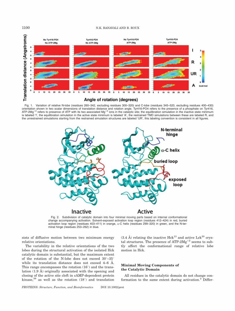
sists of diffusive motion between two minimum energyrelative orientations.The variability in the relative orientations of the two
lobes during the structural activation of the isolated Hckcatalytic domain is substantial, but the maximum extentof the rotation of the N-lobe does not exceed 308–358while its translation distance does not exceed 4–6 A.This range encompasses the rotation (168) and the trans-lation (1.9 A) originally associated with the opening andclosing of the active site cleft in cAMP-dependent proteinkinase,39 as well as the rotation (188) and translation
(3.4 A) relating the inactive Hck13 and active Lck20 crys-tal structures. The presence of ATP-2Mgþ2 seems to sub-tly affect the conformational range of relative lobemotion in Hck.
Minimal Moving Components ofthe Catalytic Domain
All residues in the catalytic domain do not change con-formation to the same extent during activation.2 Differ-
Fig. 2. Subdivision of catalytic domain into four minimal moving parts based on internal conformationalchange accompanying activation. Solvent-exposed activation loop region (residues 412–424) in red, buriedactivation loop region (residues 403–411) in orange, a-C helix (residues 299–320) in green, and the N-ter-minal hinge (residues 253–262) in blue.
Fig. 1. Variation of relative N-lobe (residues 260–342, excluding residues 300–320) and C-lobe (residues 345–520, excluding residues 400–430)orientation shown in two scalar dimensions of translation distance and rotation angle. Tyr416-PO4 refers to the presence of a phosphate on Tyr416,ATP-2Mgþ2 refers to presence of ATP with its two associated Mgþ2 ions in the catalytic site; the equilibration simulation in the inactive state minimumis labeled ‘I’, the equilibration simulation in the active state minimum is labeled ‘A’, the restrained TMD simulations between these are labeled R, andthe unrestrained simulations starting from the restrained simulation structures are labeled ‘UR’, this labeling convention is consistent in all figures.
1100 N.K. BANAVALI AND B. ROUX
PROTEINS: Structure, Function, and Bioinformatics DOI 10.1002/prot
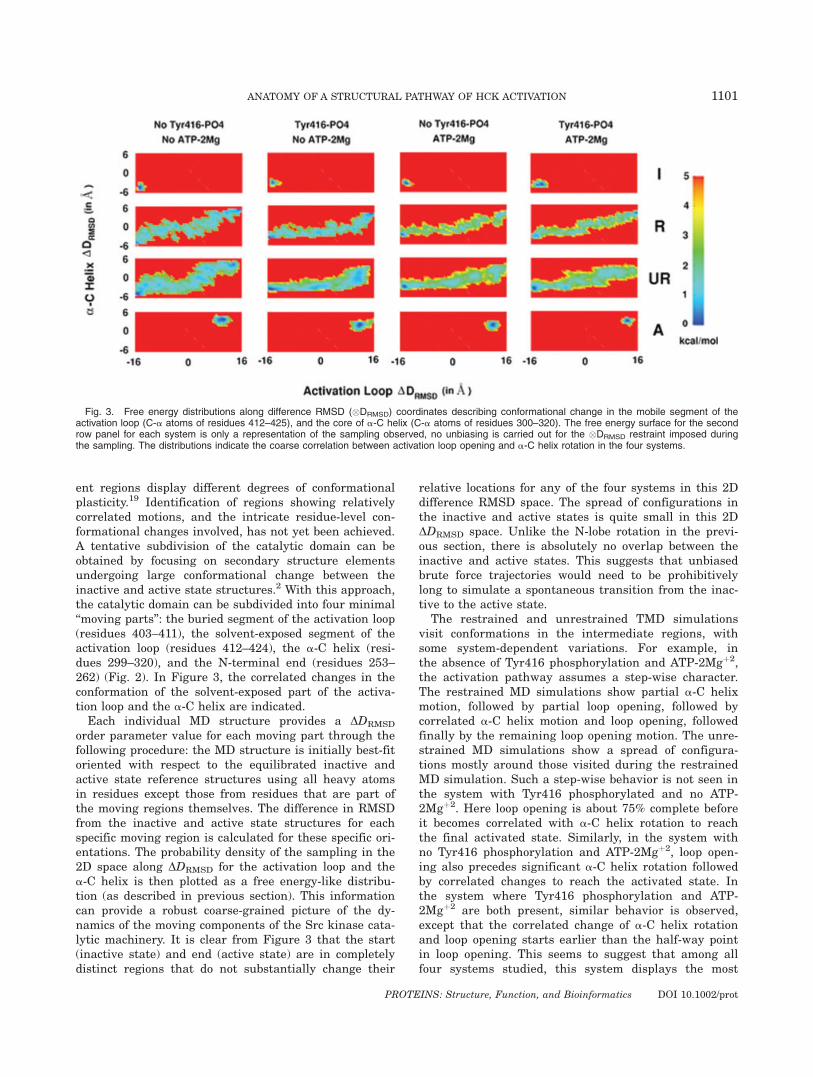
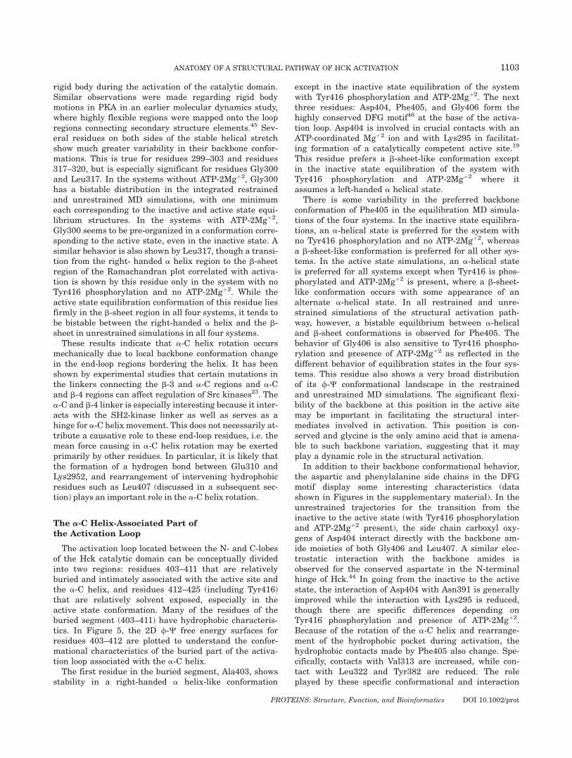
ent regions display different degrees of conformationalplasticity.19 Identification of regions showing relativelycorrelated motions, and the intricate residue-level con-formational changes involved, has not yet been achieved.A tentative subdivision of the catalytic domain can beobtained by focusing on secondary structure elementsundergoing large conformational change between theinactive and active state structures.2 With this approach,the catalytic domain can be subdivided into four minimal‘‘moving parts’’: the buried segment of the activation loop(residues 403–411), the solvent-exposed segment of theactivation loop (residues 412–424), the a-C helix (resi-dues 299–320), and the N-terminal end (residues 253–262) (Fig. 2). In Figure 3, the correlated changes in theconformation of the solvent-exposed part of the activa-tion loop and the a-C helix are indicated.Each individual MD structure provides a DDRMSD
order parameter value for each moving part through thefollowing procedure: the MD structure is initially best-fitoriented with respect to the equilibrated inactive andactive state reference structures using all heavy atomsin residues except those from residues that are part ofthe moving regions themselves. The difference in RMSDfrom the inactive and active state structures for eachspecific moving region is calculated for these specific ori-entations. The probability density of the sampling in the2D space along DDRMSD for the activation loop and thea-C helix is then plotted as a free energy-like distribu-tion (as described in previous section). This informationcan provide a robust coarse-grained picture of the dy-namics of the moving components of the Src kinase cata-lytic machinery. It is clear from Figure 3 that the start(inactive state) and end (active state) are in completelydistinct regions that do not substantially change their
relative locations for any of the four systems in this 2Ddifference RMSD space. The spread of configurations inthe inactive and active states is quite small in this 2DDDRMSD space. Unlike the N-lobe rotation in the previ-ous section, there is absolutely no overlap between theinactive and active states. This suggests that unbiasedbrute force trajectories would need to be prohibitivelylong to simulate a spontaneous transition from the inac-tive to the active state.
The restrained and unrestrained TMD simulationsvisit conformations in the intermediate regions, withsome system-dependent variations. For example, inthe absence of Tyr416 phosphorylation and ATP-2Mgþ2,the activation pathway assumes a step-wise character.The restrained MD simulations show partial a-C helixmotion, followed by partial loop opening, followed bycorrelated a-C helix motion and loop opening, followedfinally by the remaining loop opening motion. The unre-strained MD simulations show a spread of configura-tions mostly around those visited during the restrainedMD simulation. Such a step-wise behavior is not seen inthe system with Tyr416 phosphorylated and no ATP-2Mgþ2. Here loop opening is about 75% complete beforeit becomes correlated with a-C helix rotation to reachthe final activated state. Similarly, in the system withno Tyr416 phosphorylation and ATP-2Mgþ2, loop open-ing also precedes significant a-C helix rotation followedby correlated changes to reach the activated state. Inthe system where Tyr416 phosphorylation and ATP-2Mgþ2 are both present, similar behavior is observed,except that the correlated change of a-C helix rotationand loop opening starts earlier than the half-way pointin loop opening. This seems to suggest that among allfour systems studied, this system displays the most
Fig. 3. Free energy distributions along difference RMSD (�DRMSD) coordinates describing conformational change in the mobile segment of theactivation loop (C-a atoms of residues 412–425), and the core of a-C helix (C-a atoms of residues 300–320). The free energy surface for the secondrow panel for each system is only a representation of the sampling observed, no unbiasing is carried out for the �DRMSD restraint imposed duringthe sampling. The distributions indicate the coarse correlation between activation loop opening and a-C helix rotation in the four systems.
1101ANATOMY OF A STRUCTURAL PATHWAY OF HCK ACTIVATION
PROTEINS: Structure, Function, and Bioinformatics DOI 10.1002/prot

direct activation trajectory from the inactive to theactive state.The fourth moving part of the catalytic domain, which
corresponds to the N-terminal end (residues 253–262),was not restrained in the activation trajectories. Thebehavior of this region has been characterized in a previ-ous study44 using umbrella sampling MD simulations indetail. Its spontaneous behavior in response to enforcedchange in the activation loop and a-C helix regions isconsistent with previous observations44 and is describedin more detail in the supplementary material.
Rotation of the a-C Helix
The coarse description of the four moving regions ofthe catalytic domain based on DDRMSD illustrates thebroad aspects of the conformational transition. The over-
all structural change, however, can be traced back to acombination of conformational transitions in backbonetorsions in individual residues. To understand the fea-tures of a-C helix rotation at this localized level, the 2D/-C free energy surfaces for residues 299–320 are plot-ted in Figure 4. The residues that form the a-C helixitself (residues 304–316) displays no significant internalchange between the inactive state and active states.They reside in a narrow conformational minimum in thestandard right-handed a helix region of the Ramachan-dran plot. Distortions from this a helix region to the b-sheet region of the Ramachandran plot occur in therestrained and unrestrained MD simulations only for theresidues 311, 312, and 314, and even these distortionsare not consistently seen in all four systems.
The lack of conformational variability for the residues304–316 indicates that this region essentially moves as a
Fig. 4. Ramachandran free energy maps for the a-C helix and its neighboring loops (residues 299–320), for four systems considered. All torsionsare in degrees, each 2D torsional surface panel ranges from 21808 to 1808 in both axes.
1102 N.K. BANAVALI AND B. ROUX
PROTEINS: Structure, Function, and Bioinformatics DOI 10.1002/prot
rigid body during the activation of the catalytic domain.Similar observations were made regarding rigid bodymotions in PKA in an earlier molecular dynamics study,where highly flexible regions were mapped onto the loopregions connecting secondary structure elements.45 Sev-eral residues on both sides of the stable helical stretchshow much greater variability in their backbone confor-mations. This is true for residues 299–303 and residues317–320, but is especially significant for residues Gly300and Leu317. In the systems without ATP-2Mgþ2, Gly300has a bistable distribution in the integrated restrainedand unrestrained MD simulations, with one minimumeach corresponding to the inactive and active state equi-librium structures. In the systems with ATP-2Mgþ2,Gly300 seems to be pre-organized in a conformation corre-sponding to the active state, even in the inactive state. Asimilar behavior is also shown by Leu317, though a transi-tion from the right- handed a helix region to the b-sheetregion of the Ramachandran plot correlated with activa-tion is shown by this residue only in the system with noTyr416 phosphorylation and no ATP-2Mgþ2. While theactive state equilibration conformation of this residue liesfirmly in the b-sheet region in all four systems, it tends tobe bistable between the right-handed a helix and the b-sheet in unrestrained simulations in all four systems.These results indicate that a-C helix rotation occurs
mechanically due to local backbone conformation changein the end-loop regions bordering the helix. It has beenshown by experimental studies that certain mutations inthe linkers connecting the b-3 and a-C regions and a-Cand b-4 regions can affect regulation of Src kinases23. Thea-C and b-4 linker is especially interesting because it inter-acts with the SH2-kinase linker as well as serves as ahinge for a-C helix movement. This does not necessarily at-tribute a causative role to these end-loop residues, i.e. themean force causing in a-C helix rotation may be exertedprimarily by other residues. In particular, it is likely thatthe formation of a hydrogen bond between Glu310 andLys2952, and rearrangement of intervening hydrophobicresidues such as Leu407 (discussed in a subsequent sec-tion) plays an important role in the a-C helix rotation.
The a-C Helix-Associated Part ofthe Activation Loop
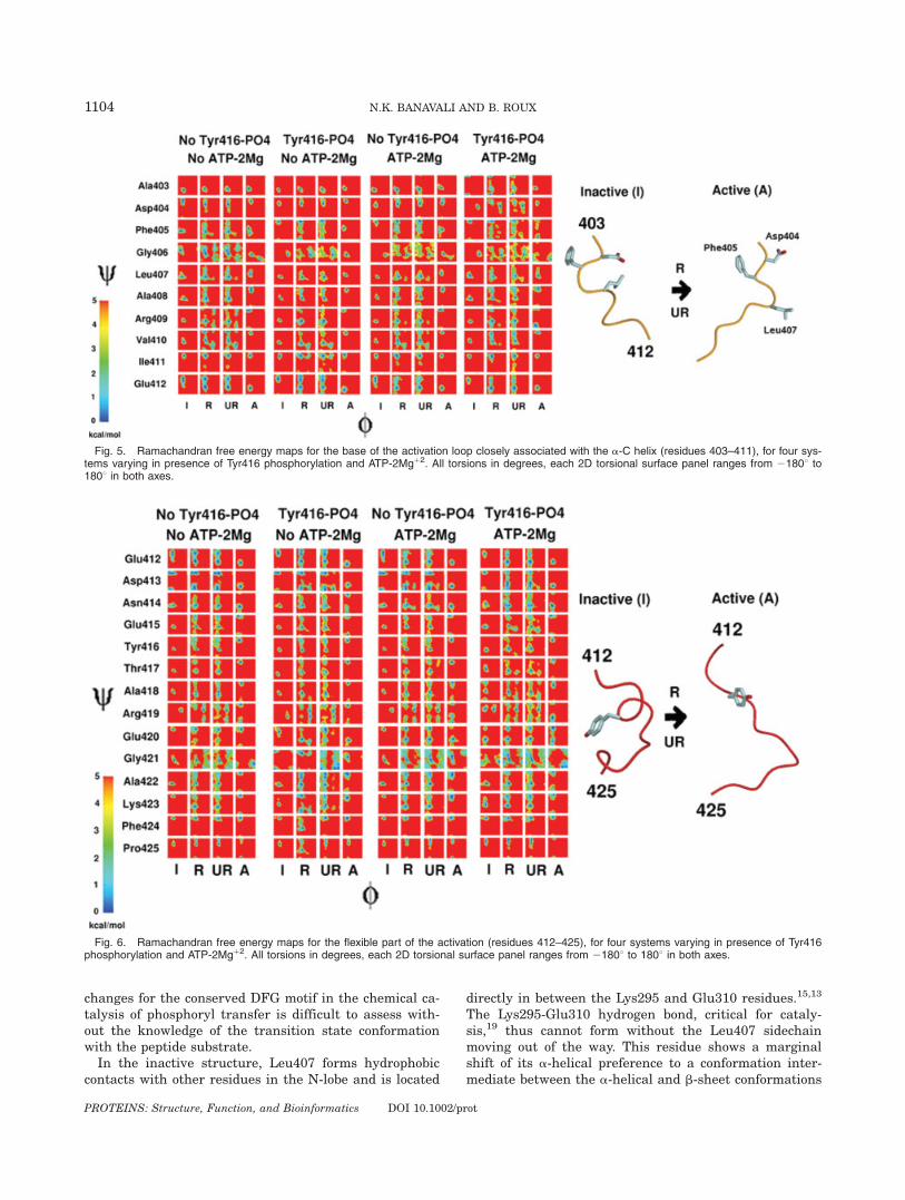
The activation loop located between the N- and C-lobesof the Hck catalytic domain can be conceptually dividedinto two regions: residues 403–411 that are relativelyburied and intimately associated with the active site andthe a-C helix, and residues 412–425 (including Tyr416)that are relatively solvent exposed, especially in theactive state conformation. Many of the residues of theburied segment (403–411) have hydrophobic characteris-tics. In Figure 5, the 2D /-C free energy surfaces forresidues 403–412 are plotted to understand the confor-mational characteristics of the buried part of the activa-tion loop associated with the a-C helix.The first residue in the buried segment, Ala403, shows
stability in a right-handed a helix-like conformation
except in the inactive state equilibration of the systemwith Tyr416 phosphorylation and ATP-2Mgþ2. The nextthree residues: Asp404, Phe405, and Gly406 form thehighly conserved DFG motif46 at the base of the activa-tion loop. Asp404 is involved in crucial contacts with anATP-coordinated Mgþ2 ion and with Lys295 in facilitat-ing formation of a catalytically competent active site.19
This residue prefers a b-sheet-like conformation exceptin the inactive state equilibration of the system withTyr416 phosphorylation and ATP-2Mgþ2 where itassumes a left-handed a helical state.
There is some variability in the preferred backboneconformation of Phe405 in the equilibration MD simula-tions of the four systems. In the inactive state equilibra-tions, an a-helical state is preferred for the system withno Tyr416 phosphorylation and no ATP-2Mgþ2, whereasa b-sheet-like conformation is preferred for all other sys-tems. In the active state simulations, an a-helical stateis preferred for all systems except when Tyr416 is phos-phorylated and ATP-2Mgþ2 is present, where a b-sheet-like conformation occurs with some appearance of analternate a-helical state. In all restrained and unre-strained simulations of the structural activation path-way, however, a bistable equilibrium between a-helicaland b-sheet conformations is observed for Phe405. Thebehavior of Gly406 is also sensitive to Tyr416 phospho-rylation and presence of ATP-2Mgþ2 as reflected in thedifferent behavior of equilibration states in the four sys-tems. This residue also shows a very broad distributionof its /-C conformational landscape in the restrainedand unrestrained MD simulations. The significant flexi-bility of the backbone at this position in the active sitemay be important in facilitating the structural inter-mediates involved in activation. This position is con-served and glycine is the only amino acid that is amena-ble to such backbone variation, suggesting that it mayplay a dynamic role in the structural activation.
In addition to their backbone conformational behavior,the aspartic and phenylalanine side chains in the DFGmotif display some interesting characteristics (datashown in Figures in the supplementary material). In theunrestrained trajectories for the transition from theinactive to the active state (with Tyr416 phosphorylationand ATP-2Mgþ2 present), the side chain carboxyl oxy-gens of Asp404 interact directly with the backbone am-ide moieties of both Gly406 and Leu407. A similar elec-trostatic interaction with the backbone amides isobserved for the conserved aspartate in the N-terminalhinge of Hck.44 In going from the inactive to the activestate, the interaction of Asp404 with Asn391 is generallyimproved while the interaction with Lys295 is reduced,though there are specific differences depending onTyr416 phosphorylation and presence of ATP-2Mgþ2.Because of the rotation of the a-C helix and rearrange-ment of the hydrophobic pocket during activation, thehydrophobic contacts made by Phe405 also change. Spe-cifically, contacts with Val313 are increased, while con-tact with Leu322 and Tyr382 are reduced. The roleplayed by these specific conformational and interaction
1103ANATOMY OF A STRUCTURAL PATHWAY OF HCK ACTIVATION
PROTEINS: Structure, Function, and Bioinformatics DOI 10.1002/prot
changes for the conserved DFG motif in the chemical ca-talysis of phosphoryl transfer is difficult to assess with-out the knowledge of the transition state conformationwith the peptide substrate.In the inactive structure, Leu407 forms hydrophobic
contacts with other residues in the N-lobe and is located
directly in between the Lys295 and Glu310 residues.15,13
The Lys295-Glu310 hydrogen bond, critical for cataly-sis,19 thus cannot form without the Leu407 sidechainmoving out of the way. This residue shows a marginalshift of its a-helical preference to a conformation inter-mediate between the a-helical and b-sheet conformations
Fig. 6. Ramachandran free energy maps for the flexible part of the activation (residues 412–425), for four systems varying in presence of Tyr416phosphorylation and ATP-2Mgþ2. All torsions in degrees, each 2D torsional surface panel ranges from 21808 to 1808 in both axes.
Fig. 5. Ramachandran free energy maps for the base of the activation loop closely associated with the a-C helix (residues 403–411), for four sys-tems varying in presence of Tyr416 phosphorylation and ATP-2Mgþ2. All torsions in degrees, each 2D torsional surface panel ranges from 21808 to1808 in both axes.
1104 N.K. BANAVALI AND B. ROUX
PROTEINS: Structure, Function, and Bioinformatics DOI 10.1002/prot
in the Ramachandran plot. The next three residues,Ala408, Arg409, and Val410 show consistent behavior inall four systems, with transitions from an a-helical stateto a b-sheet state accompanying the structural activa-tion. The last residue in this region of the activationloop, Ile411, mostly remains anchored in a b-sheet con-formation throughout the structural activation in allfour systems, and is the residue separating the relativelyhydrophobic and hydrophilic parts of the activation loop.To summarize, the structural transition occurring in
the buried part of the activation loop thus involves a com-plex mixture of hydrophobic rearrangements (Phe405,Leu407, Val410, Ile411), and exchange of hydrogen bondsbetween electrostatic residues (Glu310-Lys295). The sig-nificant flexibility of Gly406 in the conserved DFG motifmay be important in facilitating the activation transition.
The Solvent-Exposed Part of Activation Loop
The residues 412–425 (containing the critical Tyr416)form the most flexible region of the activation loop. A partof this region was not resolved in an earlier crystal struc-ture of Hck, underlining its conformational variability.12
In Figure 6, the 2D /-C free energy surfaces for residues412–425 are plotted to assess the flexibility of this loopregion. This region shows the greatest conformationalchange during activation in the entire catalytic domain.Some residues can be grouped together depending on thetypes of conformational transitions during activation.Glu412 and Asp413 show a b-sheet to a-helical transitionin all four systems. Asn414, Phe424, and Pro425 show nosubstantial differences between the inactive and activestate equilibrations, but show a conformational variabili-ty during the structural activation process itself, signify-ing a possible role in stabilizing one or more metastableintermediates. Glu415, Tyr416, Thr417, Ala418, Ala422,and Lys423 all undergo a transition from a-helical to b-sheet conformation during activation.Arg419 is unique in that it populates a left-handed a-
helical conformation in the inactive state, which convertsinto a b-sheet conformation during activation. Glu420shows a dynamic equilibrium between a-helical and b-sheet conformations, independent of the activation pro-cess, when Tyr416 is phosphorylated and ATP-2Mgþ2 ispresent. In the system with no Tyr416 phosphorylationand no ATP-2Mgþ2, Glu420 prefers a b-sheet conforma-tion, with some transitions to an a-helical conformationduring the activation process. In the presence of Tyr416phosphorylation alone or ATP-2Mgþ2 alone, this residueshows a clear b-sheet to a-helical transition during acti-vation. The backbone behavior of Gly421 is highly de-pendent on Tyr416 phosphorylation and ATP-2Mgþ2
presence, however, it consistently shows a large confor-mational variability during the structural activation.To summarize, the solvent-exposed segment of the
activation loop displays a wide conformational heteroge-neity in the activation transition of the catalytic domain.This variability is probably related to the electrostaticperturbation created by the phosphorylation of Tyr416.
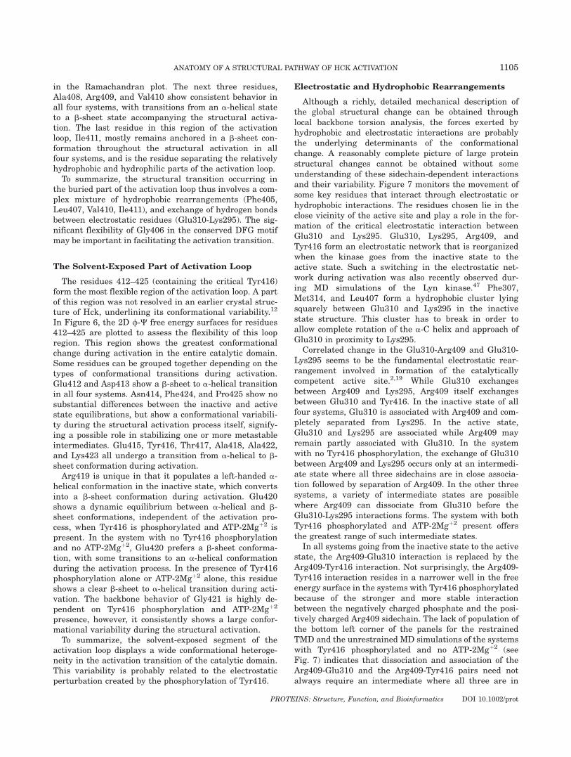
Electrostatic and Hydrophobic Rearrangements
Although a richly, detailed mechanical description ofthe global structural change can be obtained throughlocal backbone torsion analysis, the forces exerted byhydrophobic and electrostatic interactions are probablythe underlying determinants of the conformationalchange. A reasonably complete picture of large proteinstructural changes cannot be obtained without someunderstanding of these sidechain-dependent interactionsand their variability. Figure 7 monitors the movement ofsome key residues that interact through electrostatic orhydrophobic interactions. The residues chosen lie in theclose vicinity of the active site and play a role in the for-mation of the critical electrostatic interaction betweenGlu310 and Lys295. Glu310, Lys295, Arg409, andTyr416 form an electrostatic network that is reorganizedwhen the kinase goes from the inactive state to theactive state. Such a switching in the electrostatic net-work during activation was also recently observed dur-ing MD simulations of the Lyn kinase.47 Phe307,Met314, and Leu407 form a hydrophobic cluster lyingsquarely between Glu310 and Lys295 in the inactivestate structure. This cluster has to break in order toallow complete rotation of the a-C helix and approach ofGlu310 in proximity to Lys295.
Correlated change in the Glu310-Arg409 and Glu310-Lys295 seems to be the fundamental electrostatic rear-rangement involved in formation of the catalyticallycompetent active site.2,19 While Glu310 exchangesbetween Arg409 and Lys295, Arg409 itself exchangesbetween Glu310 and Tyr416. In the inactive state of allfour systems, Glu310 is associated with Arg409 and com-pletely separated from Lys295. In the active state,Glu310 and Lys295 are associated while Arg409 mayremain partly associated with Glu310. In the systemwith no Tyr416 phosphorylation, the exchange of Glu310between Arg409 and Lys295 occurs only at an intermedi-ate state where all three sidechains are in close associa-tion followed by separation of Arg409. In the other threesystems, a variety of intermediate states are possiblewhere Arg409 can dissociate from Glu310 before theGlu310-Lys295 interactions forms. The system with bothTyr416 phosphorylated and ATP-2Mgþ2 present offersthe greatest range of such intermediate states.
In all systems going from the inactive state to the activestate, the Arg409-Glu310 interaction is replaced by theArg409-Tyr416 interaction. Not surprisingly, the Arg409-Tyr416 interaction resides in a narrower well in the freeenergy surface in the systems with Tyr416 phosphorylatedbecause of the stronger and more stable interactionbetween the negatively charged phosphate and the posi-tively charged Arg409 sidechain. The lack of population ofthe bottom left corner of the panels for the restrainedTMD and the unrestrained MD simulations of the systemswith Tyr416 phosphorylated and no ATP-2Mgþ2 (seeFig. 7) indicates that dissociation and association of theArg409-Glu310 and the Arg409-Tyr416 pairs need notalways require an intermediate where all three are in
1105ANATOMY OF A STRUCTURAL PATHWAY OF HCK ACTIVATION
PROTEINS: Structure, Function, and Bioinformatics DOI 10.1002/prot
close association. In the presence of ATP-2Mgþ2, however,there are more configurations in this region indicating theincreased likelihood of a state where all three charged res-idues are in close proximity. Amongst all four systems, itis again the unrestrained MD simulations during activa-tion of the system with both Tyr416 phosphorylated andATP-2Mgþ2 present that shows the greatest amount ofpermissibility in allowing multiple conformational combi-nations.
The hydrophobic triad formed by Phe307 and Met314 (inthe a-C helix) and Leu407 (in the activation loop) is stablein the inactive state equilibration simulations of all foursystems. The distances between these associated pairs ofhydrophobic residues are typically larger than the effectiverange of electrostatic interactions (more so because of thepresent center-of-mass description of the sidechain loca-tion of each residue). It is also interesting to note that thespread of the distances around the minimum is quite large
Fig. 7. Free energy surfaces along distance coordinates quantifying various electrostatic or hydrophobic pair interactions crucial to the rotation ofthe a-C helix and catalytic activation of Hck. The specific distances chosen are shown pictorially on the left. The specific atoms chosen for distancecalculations are C-d of Glu310, C-n of Arg409, hydroxyl oxygen of Tyr416, and N-n of Lys295. For hydrophobic interactions, distances were calcu-lated between the center of mass of all sidechain non-hydrogen atoms of Phe307, Met314, and Leu407, all distances shown are in A.
1106 N.K. BANAVALI AND B. ROUX
PROTEINS: Structure, Function, and Bioinformatics DOI 10.1002/prot
(about 4 A), suggesting that such hydrophobic clusters aredynamical at room temperature. In the active state equili-brations, the Leu407-Phe307 and Leu407-Met314 pairsare both disrupted, creating a passageway used by Glu310to move towards Lys295. There seem to be two coarse path-ways for this disruption, one in which the Leu407-Phe307pair is disrupted to a greater extent than the Leu407-Met314 pair, and the other in which the opposite is true.The system with both Tyr416 phosphorylation and ATP-2Mgþ2 seems to prefer the former, whereas the systemwith no Tyr416 phosphorylation and no ATP-2Mgþ2
seems to prefer the latter. The other two systems displaycorrelated disruption of both pairs. In all cases, theeffective free energy surfaces obtained for the re-strained and unrestrained simulations during activa-tion show at least one broad and continuous pathwaybetween the inactive and active state equilibration stateextremes.
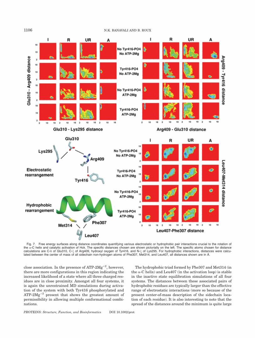
ATP Dynamics and Coordination WithPhosphorylated Tyr416
A number of studies point to the possibility thatchanges in the ligand at the ATP-binding site affect theconformational properties of regions involved in activa-tion. Kinetic studies of another protein tyrosine kinase,Csk, have indicated that nucleotide release could be therate-limiting step in the phosphoryl-transfer reaction.48
Deuterium-protection experiments indicate that theremight be conformational changes in Csk that varydepending on whether the bound nucleotide is ADP orATP.49 Time-resolved fluorescence anisotropy decaymethods show that binding of substrates have a differenteffect on the dynamic properties of PKA than binding ofproducts.50 In Hck, a part of the activation loop is in adisordered state when quercetin is bound in the ATP-binding site12; it is ordered when the bound inhibitor isPP1.13 These results suggest that the presence of ATPwith its associated Mgþ2 ions might therefore be an im-portant variable when considering the activation of thecatalytic domain.
The flexibility and preferred conformational state ofATP can be assessed from its dynamics in the presentsimulations. The starting conformation of ATP-2Mgþ2
assumed here is the same as the conformation of ATPbound to PKA, which is generally considered to be mostrepresentative of kinase catalytic function.30 The ab-sence of the substrate peptide means that no link can bedrawn between the conformation of the ATP observedhere and its catalytically competent conformation. Theassumption of the presence of 2 Mgþ2 ions also arisesfrom the same structure but it should be noted that thebehavior in the presence of Mnþ2 or a single Mgþ2 ioncannot be assumed to be the same as the behaviorobserved here. Since there is no conformational restraintimposed on ATP itself, any change in its structure is in
Fig. 8. A condensed view of the conformational dynamics of ATP bound to the catalytic site of Hck, with Tyr416 phosphorylated or unphosphoryl-ated. Pairwise torsional free energy maps (sugar pseudorotation angle P vs. v, g vs. P, a vs. b, | 2 vs. s1, and s4 vs. s3) provide a clear picture ofthe preferred minima and the torsional landscapes surrounding them that govern ATP flexibility.
1107ANATOMY OF A STRUCTURAL PATHWAY OF HCK ACTIVATION
PROTEINS: Structure, Function, and Bioinformatics DOI 10.1002/prot
response to its environment during the structural activa-tion process. In Figure 8, the torsional energy landscapeof ATP is mapped through the 2D free energy surfacesalong adjacent torsion pairs. The conformational state ofATP can be described quite accurately by knowing thevalues of 9 torsional variables: v, pseudorotation angle P,g, b, a, s1, s2, s3, and s4. In the absence of Tyr416phosphorylation, ATP shows conformational variabilitymostly in the a, s3, and s4 torsions associated with thephosphate moieties. In the presence of Tyr416 phospho-rylation, all torsional degrees of freedom show muchgreater variability due to the formation of an explicitinteraction between the g-phosphate of ATP and thephosphate on Tyr416 that is bridged by a Mgþ2 ion (seebelow). This interaction must be broken for the loop toassume its fully open state. The presence of single min-ima in the torsional degrees of freedom describing theconformational behavior of sugar and base componentsof ATP indicate that most of the conformational variabil-ity in ATP can be attributed to its triphosphate region.In the inactive state equilibration of the system con-
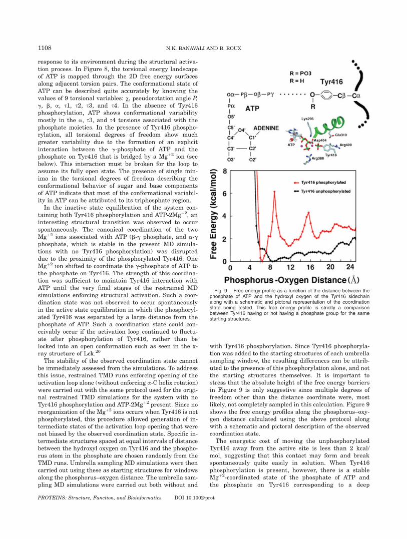
taining both Tyr416 phosphorylation and ATP-2Mgþ2, aninteresting structural transition was observed to occurspontaneously. The canonical coordination of the twoMgþ2 ions associated with ATP (b-g phosphate, and a-gphosphate, which is stable in the present MD simula-tions with no Tyr416 phosphorylation) was disrupteddue to the proximity of the phosphorylated Tyr416. OneMgþ2 ion shifted to coordinate the g-phosphate of ATP tothe phosphate on Tyr416. The strength of this coordina-tion was sufficient to maintain Tyr416 interaction withATP until the very final stages of the restrained MDsimulations enforcing structural activation. Such a coor-dination state was not observed to occur spontaneouslyin the active state equilibration in which the phosphoryl-ated Tyr416 was separated by a large distance from thephosphate of ATP. Such a coordination state could con-ceivably occur if the activation loop continued to fluctu-ate after phosphorylation of Tyr416, rather than belocked into an open conformation such as seen in the x-ray structure of Lck.20
The stability of the observed coordination state cannotbe immediately assessed from the simulations. To addressthis issue, restrained TMD runs enforcing opening of theactivation loop alone (without enforcing a-C helix rotation)were carried out with the same protocol used for the origi-nal restrained TMD simulations for the system with noTyr416 phosphorylation and ATP-2Mgþ2 present. Since noreorganization of the Mgþ2 ions occurs when Tyr416 is notphosphorylated, this procedure allowed generation of in-termediate states of the activation loop opening that werenot biased by the observed coordination state. Specific in-termediate structures spaced at equal intervals of distancebetween the hydroxyl oxygen on Tyr416 and the phospho-rus atom in the phosphate are chosen randomly from theTMD runs. Umbrella sampling MD simulations were thencarried out using these as starting structures for windowsalong the phosphorus–oxygen distance. The umbrella sam-pling MD simulations were carried out both without and
with Tyr416 phosphorylation. Since Tyr416 phosphoryla-tion was added to the starting structures of each umbrellasampling window, the resulting differences can be attrib-uted to the presence of this phosphorylation alone, and notthe starting structures themselves. It is important tostress that the absolute height of the free energy barriersin Figure 9 is only suggestive since multiple degrees offreedom other than the distance coordinate were, mostlikely, not completely sampled in this calculation. Figure 9shows the free energy profiles along the phosphorus–oxy-gen distance calculated using the above protocol alongwith a schematic and pictoral description of the observedcoordination state.
The energetic cost of moving the unphosphorylatedTyr416 away from the active site is less than 2 kcal/mol, suggesting that this contact may form and breakspontaneously quite easily in solution. When Tyr416phosphorylation is present, however, there is a stableMgþ2-coordinated state of the phosphate of ATP andthe phosphate on Tyr416 corresponding to a deep
Fig. 9. Free energy profile as a function of the distance between thephosphate of ATP and the hydroxyl oxygen of the Tyr416 sidechainalong with a schematic and pictoral representation of the coordinationstate being tested. This free energy profile is strictly a comparisonbetween Tyr416 having or not having a phosphate group for the samestarting structures.
1108 N.K. BANAVALI AND B. ROUX
PROTEINS: Structure, Function, and Bioinformatics DOI 10.1002/prot
energy well at a phosphorus–oxygen distance of 6 Aextending up to a distance of more than 8 A. Threeadditional wells stabilizing non-ATP-coordinated confor-mations of phosphorylated Tyr416 are also observed atdistances of about 12 A, 17 A, and 24 A, respectively.Although the formation of a strong interaction betweenphosphorylated Tyr416 and ATP seems counterintui-tive, the present results suggest that bridging by adivalent cation could counterbalance the electrostaticrepulsion between the two negatively charged groups.According to the free energy profile in presence ofTyr416 phosphorylation, spontaneous trajectories start-ing from the fully activated open loop conformationwould have to traverse at least three minima before thephosphorylated Tyr416 could coordinate to the phos-phate of ATP. Though the process would be slow, tran-sient visits to such intermediate states during the ls toms timescale of protein kinase catalytic turnover51 can-not be ruled out. Although a reorganization of divalentcations around ATP has not been observed in proteinkinase crystal structures, some structural similaritiesdo exist in crystal structures of the small molecule ki-nases phosphoglycerate kinase, phosphofructokinase,and phosphoenolpyruvate carboxykinase (see Fig. 3 inthe supplementary material).Interestingly, it is known that protein kinase activity
decreases upon increase in Mgþ2 ion concentrationbeyond a stoichiometry of 1 Mgþ2 per ATP.51 However,the mechanism behind this decrease in catalytic activityis not well-understood.51 A catalytically unproductiveMgþ2-mediated interaction between phosphorylatedTyr416 and ATP could provide a reasonable structuralexplanation for the inhibitory effect of an additional stoi-chiometric Mgþ2 ion. In addition to its stability, the for-mation of this state also enables proximity of the resi-dues that require to undergo pair wise electrostatic ex-changes (Glu310, Lys295, Arg409, and Tyr416).Although the present results are only suggestive, suchmetastable intermediate states may need to be consid-ered when formulating the complete catalytic cycle ofprotein tyrosine kinases.
Concluding Discussion
The present study uses all-atom MD simulations withexplicit solvent to shed light on the conformational tran-sition between the inactive and active states of Hck. Adetailed characterization of the behavior before, during(with and without restraints), and after the transforma-tion was carried out on three different length scales. Inthe coarsest description of the dynamics of the Hck cata-lytic domain, the relative orientation of the N-lobe withrespect to the C-lobe displays a significant amount ofconformational variability, presumably due to the flexi-bility of the hinge region between the two lobes. Thepresence of ATP-2Mgþ2 has a discernable impact in mak-ing the relative lobe motion bistable.The second scale of description uses a difference in
RMSD to describe correlations in rotation of the a-C he-
lix and opening of the activation loop in going from theinactive state to the active state. At this level, theimpact of ATP-2Mgþ2 and Tyr416 phosphorylation startsto become clearer, especially in terms of the order inwhich the different motions can occur. The third andhighest resolution scale describes the dynamic backbonebehavior of individual residues, which brings into starkfocus the specific conformational transformations thatoccur for each amino acid position involved in the struc-tural change accompanying activation. This descriptionclarifies the atomistic details of the specific structuralactivation processes, such as the rigidity of the a-C helixwith respect to the hinge flexibility displayed by specificresidues bordering the helical region. It also provides in-formation about the allowed backbone conformationalpathways for these local equilibria for each individualresidue.
These results highlight the multiscale character andcomplexity of the structural activation process of tyro-sine kinases. The existence of multiple local conforma-tional changes unavoidably raises questions about theorder in which they occur and the possibility of varia-tions in this order, which will be the focus of future stud-ies. Strict traditional views of structural properties ofproteins implicitly assume an architectural stability,which presume narrow global free energy basins corre-sponding to crystal structures. While this may be true ofvery stable domains, the present simulations show howmuch variation can occur within the inactive state andactive state minima for specific residues in Hck, withoutessentially leaving the basin of a given conformationalstate.
A plausible scenario explaining bidirectional allosteryin Src kinase regulation can be proposed based on thepresent results. Four distinct conformational motifs (thetwo activation loop segments, the a-C helix, and the N-terminal end) coupled to each other through their spe-cific nearest-neighbor contacts were chosen to monitorthe allosteric transition. Phosphorylation of Tyr416 inthe flexible, solvent-exposed region of the activation loopcauses a shift in the conformational equilibrium for thisloop region. The adjacent region (including the buriedsegment of the activation loop and the a-C helix)responds to this activation loop opening through changesin specific interactions, which can be remodeled to stabi-lize the inactive or active states. An example of such spe-cific changes is the exchange of Arg409 between Glu310and phosphorylated Tyr416 during activation. Thesechanged interactions result in a coupled shift of the con-formational equilibrium for both regions, due to achange in their underlying conformational free energylandscape. Activation loop opening can thus be coupledto rotation of the a-C helix. Rotation of the a-C helix inturn affects the conformational equilibrium describingthe motion of the N-terminal end hinge44 also throughspecific nearest-neighbor interactions between the a-Chelix and the N-terminal end, thereby transmitting theeffect of Tyr416 phosphorylation to the regulatory SH2and SH3 domains.
1109ANATOMY OF A STRUCTURAL PATHWAY OF HCK ACTIVATION
PROTEINS: Structure, Function, and Bioinformatics DOI 10.1002/prot
There are two noteworthy features in this model. Thefirst is that since the coupling is only necessary be-tween adjacent conformational motifs, there is no needto invoke long range interactions to explain allostery.For example, the N-terminal end need not have anydirect interactions with the activation loop to eventu-ally change its conformation in response to Tyr416phosphorylation, a shift in the conformation of the bur-ied segment of the loop and the a-C helix mediates theresponse. The second feature is that since the couplingbetween adjacent secondary structure motifs is postu-lated to occur through changes in their underlying con-formational free energy landscape, the coupling neednot have any inherent directionality. Hinge motion inthe N-terminal end can therefore cause rotation of thea-C helix, just the same as the rotation of the a-C helixcan result in hinge motion in the N-terminal end. Thislack of directionality is compatible with the bidirec-tional nature of the allostery that regulates Src kinasefunction.23
The existence of a metastable Mgþ2-coordination statebetween phosphorylated Tyr416 and the g-phosphate ofATP is suggested by the present calculations. Thoughcounterintuitive, such a state is chemically reasonableand was shown to be stable using umbrella sampling cal-culations. It is presently difficult to predict the signifi-cance of such intermediate states in the catalytic cycle ofHck and more work will be needed to further character-ize it.Inhibitors of protein tyrosine kinases52,53 mostly bind
through hydrophobic interactions in the ATP-bindingsite and have considerable therapeutic and thereforecommercial value. The structural activation process in-volves hydrophobic rearrangements, which can probablyaffect the binding of the small molecule inhibitors. Therobustness of the binding during such dynamic struc-tural changes, and indeed the ability to hamper suchchanges, is probably important to the inhibition capacityof these small molecule inhibitors. The present study ofconformational variability in kinase activation is there-fore likely to suggest alternate conformations againstwhich the binding affinity of these inhibitors could betested.The SH2 and SH3 domains were not included in the
present simulations. This choice is motivated by theneed to first characterize the specific moving partswithin the catalytic domain. The SH2 and SH3 domainsconstitute additional degrees of freedom in the Src ki-nase machinery that can be coupled to the various mov-ing parts in the catalytic domain. The importance ofsuch coupling is not completely clear and remains to beassessed. For example, the isolated Lck catalytic domainhas been demonstrated to be active without the SH2 andSH3 domains20 and SH2 or SH3 deletion mutants showincreased kinase activity.54 However, a recent X-raystructure of a c-Src complex in an active conformationsuggests possible interactions of the SH2 and SH3 withthe catalytic domain,55 and the SH2 and SH3 domainsinteract directly with the catalytic domain to stabilize
the active state structure of Csk, a closely related kinase(35% homology to Src).56 The SH2 domain also interactswith the C-lobe of the catalytic domain in the inactiveassembled structure of Hck and c-Src.12–14 Clearly, fur-ther studies will be needed to better understand theinfluence of direct interactions of the catalytic domainwith the regulatory domains.
The present results, together with those from a previ-ous study focused on the N-terminal end linker region ofthe catalytic domain44 suggest a dynamic view of alloste-ric regulation of the various functional states of theenzyme. A number of structural elements maintain theirinternal conformation and behave almost as rigid mod-ules that are re-organized relative to one another (e.g.,the SH2 and SH3 domains, the a-C helix in the N-lobe).In contrast, other elements can transform their internalconformation significantly (e.g., the N-terminal end ofcatalytic domain, the activation loop, the linker betweenthe SH2 and SH3 domains). Such ‘‘transformative’’ struc-tural elements (typically corresponding to relativelyshort peptide regions) display a bi-stable (or multi-sta-ble) character44—the conformational free energy surfaceis a useful concept to display this feature.44 The abilityof the transformative elements to switch between differ-ent stable conformational states can lead to long-rangespatial rearrangement of the (relatively stable) ‘‘modu-lar’’ elements with respect to one another. This view isconsistent with the accepted understanding of allosteryas a shift in equilibrium between two overall minima21
and provides a useful physical framework to seek thecausative factors in the long-range allosteric response ofa complicated macromolecular assembly.
ACKNOWLEDGMENTS
We would like to thank Carol Post, John Kuriyan,Susan Taylor, Guillaume Lamoureux, Simon Berneche,Toby Allen, Hyung-June Woo, Yuqing Deng, Sergei Nos-kov, Jose Faraldo-Gomez, and Deniz Sezer for helpfuldiscussions. This work was supported financially by NIHgrant CA093577 and by Keck Postdoctoral Fellowshipfor NKB. Computational support from the PittsburghSupercomputing Center (PSC) obtained through theNational Resource Allocation Committee (NRAC) wasused for the calculations.
REFERENCES
1. Brown M, Cooper J. Regulation, substrates and functions of src.Biochim Biophys Acta 1996;1287;121–149.
2. Sicheri F, Kuriyan J. Structures of Src-family tyrosine kinases.Curr Opin Struct Biol 1997;7:777–785.
3. Waksman G, Shoelson SE, Pant N, Cowburn D, Kuriyan J.Binding of a high affinity phosphotyrosyl peptide to the Src SH2domain: crystal structures of the complexed and peptide-freeforms. Cell 1993;72:779–790.
4. Zheng XM, Wang Y, Pallen CJ. Cell transformation and activa-tion of pp60c-src by overexpression of a protein tyrosine phos-phatase. Nature 1992;359:336–339.
5. Wu X, Knudsen B, Feller SM, Zheng J, Sali A, Cowburn D,Hanafusa H, Kuriyan J. Structural basis for the specific interac-tion of lysine-containing proline-rich peptides with the N-termi-nal SH3 domain of c-Crk. Structure 1995;3:215–226.
1110 N.K. BANAVALI AND B. ROUX
PROTEINS: Structure, Function, and Bioinformatics DOI 10.1002/prot
6. Moarefi I, LaFevre-Bernt M, Sicheri F, Huse M, Lee C, KuriyanJ, Miller W. Activation of the Src-family tyrosine kinase Hck bySH3 domain displacement. Nature 1997;385:650–653.
7. Azarnia R, Reddy S, Kmiecik TE, Shalloway D, LoewensteinWR. The cellular src gene product regulates junctional cell-to-cell communication. Science 1988;239:398–401.
8. Porter M, Schindler T, Kuriyan J, Miller W. Reciprocal regula-tion of Hck activity by phosphorylation of Tyr(527) and Tyr(416).Effect of introducing a high affinity\intramolecular SH2 ligand.J Biol Chem 2000;275:2721–2726.
9. Ziegler SF, Marth JD, Lewis DB, Perlmutter RM. Novel protein-tyrosine kinase gene (hck) preferentially expressed in cells of he-matopoietic origin. Mol Cell Biol 1987;7:2276–2285.
10. Hu Y, Liu Y, Pelletier S, Buchdunger E, Warmuth M, Fabbro D,Hallek M, Etten RAV, Li S. Requirement of Src kinases Lyn,Hck and Fgr for BCR-ABL1-induced B-lymphoblastic leukemiabut not chronic myeloid leukemia. Nat Genet 2004;36:453–461.
11. Podar K, Mostoslavsky G, Sattler M, Tai YT, Hayashi T, CatleyLP, Hideshima T, Mulligan RC, Chauhan D, Anderson KC. Criti-cal role for hematopoietic cell kinase (Hck)-mediated phosphoryl-ation of Gab1 and Gab2 docking proteins in interleukin 6-induced proliferation and survival of multiple myeloma cells.Biol J Chem 2004;279:21658–21665.
12. Sicheri F, Moarefi I, Kuriyan J. Crystal structure of the Src fam-ily tyrosine kinase Hck. Nature 1997;385:602–609.
13. Schindler T, Sicheri F, Pico A, Gazit A, Levitzki A, Kuriyan J.Crystal structure of Hck in complex with a Src family-selectivetyrosine kinase inhibitor. Mol Cell 1999;3:639–648.
14. Xu W, Harrison S, Eck M. Three-dimensional structure of thetyrosine kinase c-Src. Nature 1997;385:595–602.
15. Xu W, Doshi A, Lei M, Eck M, Harrison S. Crystal structures ofc-Src reveal features of its autoinhibitory mechanism. Mol Cell1999;3:629–638.
16. Taylor SS, Radzio-Andzelm E. 3 Protein-kinase structures definea common motif. Structure 1994;2:345–355.
17. Hubbard S. Crystal structure of the activated insulin receptortyrosine kinase in complex with peptide substrate and ATP ana-log. EMBO J 1997;16:5573–5581.
18. Hubbard SR, Wei L, Ellis L, Hendrickson WA. Crystal structureof the tyrosine kinase domain of the human insulin receptor.Nature 1994;372:746–754.
19. Huse M, Kuriyan J. The conformational plasticity of protein ki-nases. Cell 2002;109:275–282.
20. Yamaguchi H, Hendrickson W. Structural basis for activation ofhuman lymphocyte kinase Lck upon tyrosine phosphorylation.Nature 1996;384:484–489.
21. Kern D, Zuiderweg ER. The role of dynamics in allosteric regu-lation. Curr Opin Struct Biol 2003;13:748–757.
22. Young M, Gonfloni S, Superti G-Furga, Roux B, Kuriyan J.Dynamic coupling between the SH2 and SH3 domains of c-Srcand Hck underlies their inactivation by C-terminal tyrosinephosphorylation. Cell 2001;105:115–126.
23. Gonfloni S, Williams J, Hattula K, Weijland A, Wierenga R,Superti-Furga G. The role of the linker between the SH2 do-main and catalytic domain in the regulation and function of Src.EMBO J 1997;16:7261–7271.
24. Mendieta J, Gago F. In Silico activation of Src tyrosine kinasereveals the molecular basis for intromolecular phosphorylation.J Mol Graph Model 2004;23:189–198.
25. Brooks B, Bruccoleri R, Olafson B, States D, Swaminathan S,Karplus M. CHARMM: a program for macromolecular energyminimization and dynamics calculations. J Comput Chem 1983;4:187–217.
26. MacKerell AD, Jr, Bashford D, Bellot M, Dunbrack R, EvanseckJ, Field M, Fischer S, Gao J, Guo H, Ha DJ-MS, Kuchnir L,Kuczera K, Lau F, Mattos C, Michnick S, Ngo T, Nguyen D,Prodhom B, Reiher W III, Roux B, Schlenkrich M, Smith J,Stote R, Straub J, Watanabe M, Wiorkiewicz-Kuczera J, Kar-plus M. All-atom empirical potential for molecular modelingand dynamics studies of proteins. J Phys Chem B 1998;102:3586–3616.
27. Pavelites JJ, Gao JL, Bash PA, Mackerell AD. A molecularmechanics force field for NAD(þ), NADH, and the pyrophos-phate groups of nucleotides. J Comp Chem 1997;18:221–239.
28. Higgins D, Thompson J, Gibson T, Thompson JD, Higgins DG,Gibson TJ. CLUSTAL W: improving the sensitivity of progres-
sive multiple sequence alignment through sequence weighting,position-specific gap penalties and weight matrix choice. NucleicAcids Res 1994;22:4673–4680.
29. Marti-Renom MA, Stuart A, Fiser A, Sanchez R, Melo F, Sali A.Comparative protein structure modeling of genes and genomes.Ann Rev Biophys Biomol Struct 2000;29:291–325.
30. Zheng JH, Trafny EA, Knighton DR, Xuong NH, Taylor SS, Ten-eyck LF, Sowadski JM. 2.2-Angstrom refined crystal-structure ofthe catalytic subunit of CAMP-dependent protein-kinase com-plexed with MnATP and a peptide inhibitor. Acta Crystallog-raphica 1993;49:362–365.
31. Field MJ, Karplus M. CRYSTAL: program for crystal calcula-tions in CHARMM. Cambridge, MA: Harvard University; 1992.
32. Ryckaert JP, Ciccotti G, Berendsen HJC. Numerical integrationof the cartesian equations of motion of a system with con-straints: molecular dynamics of n-alkanes. J Comp Phys 1977;23:327–341.
33. Darden T, York D, Pedersen L. Particle Mesh Ewald—anNllog(N) method for ewald sums in large systems. J Chem Phys1993;98:10089–10092.
34. Beglov D, Roux B. Finite representation of an infinite bulk sys-tem: solvent boundary potential for computer simulations.J Chem Phys 1994;100:9050–9063.
35. Feller SE, Zhang YH, Pastor RW, Brooks BR. Constant pressuremolecular dynamics simulation—the Langevin piston method.J Chem Phys 1995;103:4613–4621.
36. Banavali NK, Roux B. Free energy landscape of A-DNA to B-DNA conversion in aqueous solution J Am Chem Soc 2005;127:6866–6876.
37. Kumar S, Bouzida D, Swendsen RH, Kollman PA, RosenbergJM. The weighted histogram analysis method for free-energycalculations on biomolecules. I. The method. J Comput Chem1992;13:1011–1021.
38. Souaille M, Roux B. Extension to the weighted histogram analy-sis method: combining umbrella sampling with free energy cal-culations. Comput Phys Commun 2001;135:40–57.
39. Karlsson R, Zheng JH, Xuong NH, Taylor SS, Sowadski JM.Structure of the mammalian catalytic subunit of cAMP-depend-ent protein-kinase and an inhibitor peptide displays an openconformation. Acta Crystallographica D 1993;49:381–388.
40. Zheng J, Knighton D, Xuong N, Taylor S, Sowadski J, Eyck LT.Crystal structures of the myristylated catalytic subunit ofcAMP-dependent protein kinase reveal open and closed confor-mations. Protein Sci 1993;2:1559–1573.
41. Johnson DA, Akamine P, Radio E-Andzelm, Madhusudan M,Taylor SS. Dynamics of cAMP-dependent protein kinase. ChemRev 2001;101:2243–2270.
42. Kabsch W. A solution for the best rotation to relate two sets ofvectors. Acta Crystallogr A 1976;32:922–923.
43. Kabsch W. A discussion of the solution for the best rotation torelate two sets of vectors. Acta Crystallogr A 1978;34:827–828.
44. Banavali NK, Roux B. The N-terminal end of the Catalytic Do-main of Src kinase Hck is a conformational switch implicated inlong-range allosteric regulation. Structure 2005;13:1715–1723.
45. Tsigelny I, Greenberg JP, Cox S, Nichols WL, Taylor SS, TenEyck LF. 600 ps molecular dynamics reveals stable substruc-tures and flexible hinge points in cAMP dependent protein ki-nase. Biopolymers 1999;50:513–524.
46. Johnson L, Lewis RJ. Structural basis for control by phosphoryl-ation. Chem Rev 2001;101:2209–2242.
47. Ozrikirimli E, Post CB. Src kinase activation: a switched elec-trostatic network. Protein Sci 2006;15:1051–1062.
48. Shaffer J, Sun G, Adams JA. Nucleotide release and associatedconformational changes regulate function in the COOH-terminalSrc kinase, Csk. Biochemistry 2001;40:11149–11155.
49. Hamuro Y, Wong L, Shaffer J, Kim JS, Stranz DD, JenningsPA, Woods VL, Jr, Adams JA. Phosphorylation driven motionsin the COOH-terminal Src kinase, CSK, revealed throughenhanced hydrogen-deuterium exchange and mass spectrometry(DXMS). J Mol Biol 2002;323:871–881.
50. Li F, Gangal M, Juliano C, Gorfain E, Taylor SS, Johnson DA.Evidence for an internal entropy contribution to phosphoryltransfer: a study of domain closure, backbone flexibility, and thecatalytic cycle of cAMP-dependent protein kinase. J Mol Biol 2002;315:459–469.
1111ANATOMY OF A STRUCTURAL PATHWAY OF HCK ACTIVATION
PROTEINS: Structure, Function, and Bioinformatics DOI 10.1002/prot
51. Adams JA. Kinetic and catalytic mechanisms of protein kinases.Chem Rev 2001;101:2271–2290.
52. Bridges AJ. Chemical Inhibitors of Protein Kinases. Chem Rev2001;101:2541–2571.
53. Nagar B, Bornmann WG, Pellicena P, Schindler T, Veach DR,Miller WT, Clarkson B, Kuriyan J. Crystal structures of the kinasedomain of c-Abl in complex with the small molecule inhibitorsPD173955 and imatinib (STI-571)1. Cancer Res 2002;62:4236–4243.
54. Seidel-Dugan C, Meyer BE, Thomas SM, Brugge JS. Effects ofSH2 and SH3 deletions on the functional activities of wild-type
and transforming variants of c-Src. Mol Cell Biol 1992;12:1835–1845.
55. Cowan-Jacob SW, Fendrich G, Manley PW, Jahnke W, FabbroD, Liebetanz J, Meyer T. The crystal structure of a c-Src com-plex in an active conformation suggests possible steps in c-Srcactivation. Structure 2005;13:861–871.
56. Ogawa A, Takayama Y, Sakai H, Chong KT, Takeuchi S, Naga-kawa A, Nada S, Okada M, Tsukihara T. Structure of the car-boxy-terminal Src kinase, Csk. J Biol Chem 2002;277:14351–14354.
1112 N.K. BANAVALI AND B. ROUX
PROTEINS: Structure, Function, and Bioinformatics DOI 10.1002/prot
Related Documents


