The Brain’s Default Network Anatomy, Function, and Relevance to Disease RANDY L. BUCKNER, a,b,c,d,e JESSICA R. ANDREWS-HANNA, a,b,c AND DANIEL L. SCHACTER a a Department of Psychology, Harvard University, Cambridge, Massachusetts, USA b Center for Brain Science, Harvard University, Cambridge, Massachusetts, USA c Athinoula A. Martinos Center for Biomedical Imaging, Massachusetts General Hospital, Boston, Massachusetts, USA d Department of Radiology, Harvard Medical School, Boston, Massachusetts, USA e Howard Hughes Medical Institute, Chevy Chase, Maryland 20815, USA Thirty years of brain imaging research has converged to define the brain’s default network—a novel and only recently appreciated brain system that participates in internal modes of cog- nition. Here we synthesize past observations to provide strong evidence that the default net- work is a specific, anatomically defined brain system preferentially active when individuals are not focused on the external environment. Analysis of connectional anatomy in the monkey sup- ports the presence of an interconnected brain system. Providing insight into function, the default network is active when individuals are engaged in internally focused tasks including autobio- graphical memory retrieval, envisioning the future, and conceiving the perspectives of oth- ers. Probing the functional anatomy of the network in detail reveals that it is best understood as multiple interacting subsystems. The medial temporal lobe subsystem provides informa- tion from prior experiences in the form of memories and associations that are the building blocks of mental simulation. The medial prefrontal subsystem facilitates the flexible use of this information during the construction of self-relevant mental simulations. These two sub- systems converge on important nodes of integration including the posterior cingulate cortex. The implications of these functional and anatomical observations are discussed in relation to possible adaptive roles of the default network for using past experiences to plan for the fu- ture, navigate social interactions, and maximize the utility of moments when we are not oth- erwise engaged by the external world. We conclude by discussing the relevance of the default network for understanding mental disorders including autism, schizophrenia, and Alzheimer’s disease. Key words: default mode; default system; default network; fMRI; PET; hippocampus; memory; schizophrenia; Alzheimer Introduction A common observation in brain imaging research is that a specific set of brain regions—referred to as the default network—is engaged when individuals are left to think to themselves undisturbed (Shulman et al. 1997, Mazoyer et al. 2001, Raichle et al. 2001). Prob- ing this phenomenon further reveals that other kinds of situations, beyond freethinking, engage the default net- work. For example, remembering the past, envisioning Address for correspondence: Dr. Randy Buckner, Harvard University, William James Hall, 33 Kirkland Drive, Cambridge, MA 02148. [email protected] future events, and considering the thoughts and per- spectives of other people all activate multiple regions within the default network (Buckner & Carroll 2007). These observations prompt one to ask such questions as: What do these tasks and spontaneous cognition share in common? and what is the significance of this network to adaptive function? The default net- work is also disrupted in autism, schizophrenia, and Alzheimer’s disease, further encouraging one to con- sider how the functions of the default network might be important to understanding diseases of the mind (e.g., Lustig et al. 2003, Greicius et al. 2004, Kennedy et al. 2006, Bluhm et al. 2007). Motivated by these questions, we provide a com- prehensive review and synthesis of findings about the Ann. N.Y. Acad. Sci. 1124: 1–38 (2008). C 2008 New York Academy of Sciences. doi: 10.1196/annals.1440.011 1

Welcome message from author
This document is posted to help you gain knowledge. Please leave a comment to let me know what you think about it! Share it to your friends and learn new things together.
Transcript
The Brain’s Default NetworkAnatomy, Function, and Relevance to Disease
RANDY L. BUCKNER,a,b,c,d,e JESSICA R. ANDREWS-HANNA,a,b,c
AND DANIEL L. SCHACTERa
aDepartment of Psychology, Harvard University, Cambridge, Massachusetts, USAbCenter for Brain Science, Harvard University, Cambridge, Massachusetts, USA
cAthinoula A. Martinos Center for Biomedical Imaging, Massachusetts General Hospital,Boston, Massachusetts, USA
dDepartment of Radiology, Harvard Medical School, Boston, Massachusetts, USAeHoward Hughes Medical Institute, Chevy Chase, Maryland 20815, USA
Thirty years of brain imaging research has converged to define the brain’s default network—anovel and only recently appreciated brain system that participates in internal modes of cog-nition. Here we synthesize past observations to provide strong evidence that the default net-work is a specific, anatomically defined brain system preferentially active when individuals arenot focused on the external environment. Analysis of connectional anatomy in the monkey sup-ports the presence of an interconnected brain system. Providing insight into function, the defaultnetwork is active when individuals are engaged in internally focused tasks including autobio-graphical memory retrieval, envisioning the future, and conceiving the perspectives of oth-ers. Probing the functional anatomy of the network in detail reveals that it is best understoodas multiple interacting subsystems. The medial temporal lobe subsystem provides informa-tion from prior experiences in the form of memories and associations that are the buildingblocks of mental simulation. The medial prefrontal subsystem facilitates the flexible use ofthis information during the construction of self-relevant mental simulations. These two sub-systems converge on important nodes of integration including the posterior cingulate cortex.The implications of these functional and anatomical observations are discussed in relation topossible adaptive roles of the default network for using past experiences to plan for the fu-ture, navigate social interactions, and maximize the utility of moments when we are not oth-erwise engaged by the external world. We conclude by discussing the relevance of the defaultnetwork for understanding mental disorders including autism, schizophrenia, and Alzheimer’sdisease.
Key words: default mode; default system; default network; fMRI; PET; hippocampus; memory;schizophrenia; Alzheimer
Introduction
A common observation in brain imaging researchis that a specific set of brain regions—referred to asthe default network—is engaged when individuals areleft to think to themselves undisturbed (Shulman et al.1997, Mazoyer et al. 2001, Raichle et al. 2001). Prob-ing this phenomenon further reveals that other kinds ofsituations, beyond freethinking, engage the default net-work. For example, remembering the past, envisioning
Address for correspondence: Dr. Randy Buckner, Harvard University,William James Hall, 33 Kirkland Drive, Cambridge, MA 02148.
future events, and considering the thoughts and per-spectives of other people all activate multiple regionswithin the default network (Buckner & Carroll 2007).These observations prompt one to ask such questionsas: What do these tasks and spontaneous cognitionshare in common? and what is the significance ofthis network to adaptive function? The default net-work is also disrupted in autism, schizophrenia, andAlzheimer’s disease, further encouraging one to con-sider how the functions of the default network mightbe important to understanding diseases of the mind(e.g., Lustig et al. 2003, Greicius et al. 2004, Kennedyet al. 2006, Bluhm et al. 2007).
Motivated by these questions, we provide a com-prehensive review and synthesis of findings about the
Ann. N.Y. Acad. Sci. 1124: 1–38 (2008). C! 2008 New York Academy of Sciences.doi: 10.1196/annals.1440.011 1
2 Annals of the New York Academy of Sciences
brain’s default network. This review covers both ba-sic science and clinical observations, with its contentorganized across five sections. We begin with a briefhistory of our understanding of the default network(section I). Next, a detailed analysis of the anatomyof the default network is provided including evidencefrom humans and monkeys (section II). The follow-ing sections concern the role of the default network inspontaneous cognition, as commonly occurs in passivetask settings (section III), as well as its functions in activetask settings (section IV). While recognizing alterna-tive possibilities, we hypothesize that the fundamentalfunction of the default network is to facilitate flexi-ble self-relevant mental explorations—simulations—that provide a means to anticipate and evaluate up-coming events before they happen. The final sectionof the review discusses emerging evidence that relatesthe default network to cognitive disorders, includingthe possibility that activity in the default network aug-ments a metabolic cascade that is conducive to thedevelopment of Alzheimer’s disease (section V).
I. A Brief History
The discovery of the brain’s default network wasentirely accidental. Evidence for the default networkbegan accumulating when researchers first measuredbrain activity in humans during undirected mentalstates. Even though no early studies were explicitly de-signed to explore such unconstrained states, relevantdata were nonetheless acquired because of the com-mon practice of using rest or other types of passiveconditions as an experimental control. These stud-ies revealed that activity in specific brain regions in-creased during passive control states as compared tomost goal-directed tasks. In almost all cases, the explo-ration of activity during the control states occurred asan afterthought—as part of reviews and meta-analysesperformed subsequent to the original reports, whichfocused on the goal-directed tasks.
Early ObservationsA clue that brain activity persists during undirected
mentation emerged from early studies of cerebralmetabolism. It was already known by the late 19thcentury that mental activity modulated local bloodflow (James 1890). Louis Sokoloff and colleagues (1955)used the Kety-Schmidt nitrous oxide technique (Kety& Schmidt 1948) to ask whether cerebral metabolismchanges globally when one goes from a quiet rest stateto performing a challenging arithmetic problem—atask that demands focused cognitive effort. To theirsurprise, metabolism remained constant. While not
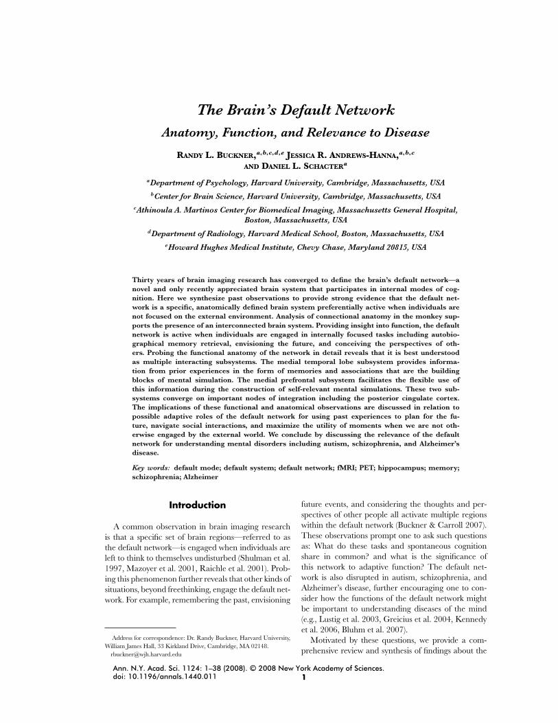
FIGURE 1. An early image of regional cerebral bloodflow (rCBF) at rest made by David Ingvar and colleaguesusing the nitrous oxide technique. The image shows data av-eraged over eight individuals to reveal a “hyperfrontal” ac-tivity pattern that Ingvar proposed reflected “spontaneous,conscious mentation” (Ingvar 1979). Ingvar’s ideas antici-pate many of the themes discussed in this review (see Ingvar1974, 1979, 1985).
their initial conclusion, the unchanged global rateof metabolism suggests that the rest state containspersistent brain activity that is as vigorous as thatwhen individuals solve externally administered mathproblems.
The Swedish brain physiologist David Ingvar wasthe first to aggregate imaging findings from rest taskstates and note the importance of consistent, region-ally specific activity patterns (Ingvar 1974, 1979, 1985).Using the xenon 133 inhalation technique to measureregional cerebral blood flow (rCBF), Ingvar and hiscolleagues observed that frontal activity reached highlevels during rest states (FIG. 1). To explain this unex-pected phenomenon, Ingvar proposed that the “hy-perfrontal” pattern of activity corresponded “to undi-rected, spontaneous, conscious mentation, the ‘brainwork,’ which we carry out when left alone undisturbed”(Ingvar 1974). Two lasting insights emerged from Ing-var’s work. First, echoing ideas of Hans Berger (1931),his work established that the brain is not idle when leftundirected. Rather, brain activity persists in the ab-sence of external task direction. Second, Ingvar’s ob-servations suggested that increased activity during restis localized to specific brain regions that prominentlyinclude prefrontal cortex.
The Era of Task-Induced DeactivationIngvar’s ideas about resting brain activity remained
largely unexplored for the next decade until positronemission tomography (PET) methods for brain imag-ing gained prominence. PET had finer resolution and
Buckner et al.: The Brain’s Default Network 3
sensitivity to deep-brain structures than earlier meth-ods and, owing to the development of isotopes withshort half-lives (Raichle 1987), typical PET studies in-cluded many task and control conditions for compar-ison. By the mid-1990s several dozen imaging studieswere completed that examined perception, language,attention, and memory. Scans of rest-state brain ac-tivitya were often acquired across these studies for acontrol comparison, and researchers began routinelynoticing brain regions more active in the passive con-trol conditions than the active target tasks—what atthe time was referred to as “deactivation.”
The term “deactivation” was used because analysesand image visualization were referenced to the target,experimental task. Within this nomenclature, regionsrelatively more active in the target condition (e.g., read-ing, classifying pictures) compared to the control task(e.g., passive fixation, rest) were labeled “activations”;regions less active in the target condition than the con-trol were labeled “deactivations.” Deactivations werepresent and often the most robust effect in many earlyPET studies. One form of deactivation for which earlyinterest emerged was activity reductions in unattendedsensory modalities because of its theoretical relevanceto mechanisms of attention (e.g., Haxby et al. 1994,Kawashima et al. 1994, Buckner et al. 1996). A secondform of commonly observed deactivation was along thefrontal and posterior midline during active, as com-pared to passive, task conditions. There was no initialexplanation for these mysterious midline deactivations(e.g., Ghatan et al. 1995, Baker et al. 1996).
A particularly informative early study was con-ducted while exploring brain regions supportingepisodic memory. Confronted with the difficult issue ofdefining a baseline state for an autobiographical mem-ory task, Andreasen and colleagues (1995) exploredthe possibility that spontaneous cognition makes animportant contribution to rest states. Much like otherstudies at the time, the researchers included a rest con-dition as a baseline for comparison to their target con-ditions. However, unlike other contemporary studies,they hypothesized that autobiographical memory (theexperimental target of the study) inherently involves in-ternally directed cognition, much like the spontaneouscognition that occurs during “rest” states. For this rea-son, Andreasen and colleagues explored both the rest
aPET and functional MRI (fMRI) both measure neural activity indi-rectly through local vascular (blood flow) changes that accompany neu-ronal activity. PET is sensitive to changes in blood flow directly (Raichle1987). fMRI is sensitive to changes in oxygen concentration in the bloodwhich tracks blood flow (Heeger and Ress 2002). For simplicity, we referto these methods as measuring brain activity in this review.
and memory tasks referenced to a third control con-dition that involved neither rest nor episodic memory.Their results showed that similar brain regions wereengaged during rest and memory as compared to thenonmemory control. In addition, to better understandthe cognitive processes associated with the rest state,they informally asked their participants to subjectivelydescribe their mental experiences.
Two insights originated from this work that fore-shadow much of the present review’s content. First,Andreasen et al. (1995) noted that the resting state“is in fact quite vigorous and consists of a mixtureof freely wandering past recollection, future plans, andother personal thoughts and experiences.” Second, theanalysis of brain activity during the rest state revealedprefrontal midline regions as well as a distinct poste-rior pattern that included the posterior cingulate andretrosplenial cortex. As later studies would confirm,these regions are central components of the core brainsystem that is consistently activated in humans duringundirected mental states.
Broad awareness of the common regions that be-come active during passive task states emerged witha pair of meta-analyses that pooled extensive data toreveal the functional anatomy of unconstrained cogni-tion. In the first study, Shulman and colleagues (1997)conducted meta-analysis of task-induced deactivationsto explicitly determine if there were common brain re-gions active during undirected (passive) mental states.They pooled data from 132 normal adults for which anactive task (word reading, active stimulus classification,etc.) could be directly compared to a passive task thatpresented the same visual words or pictures but con-tained no directed task goals. Using a similar approach,Mazoyer et al. (2001) aggregated data across 63 nor-mal adults that included both visually and aurally cuedactive tasks as compared to passive rest conditions.
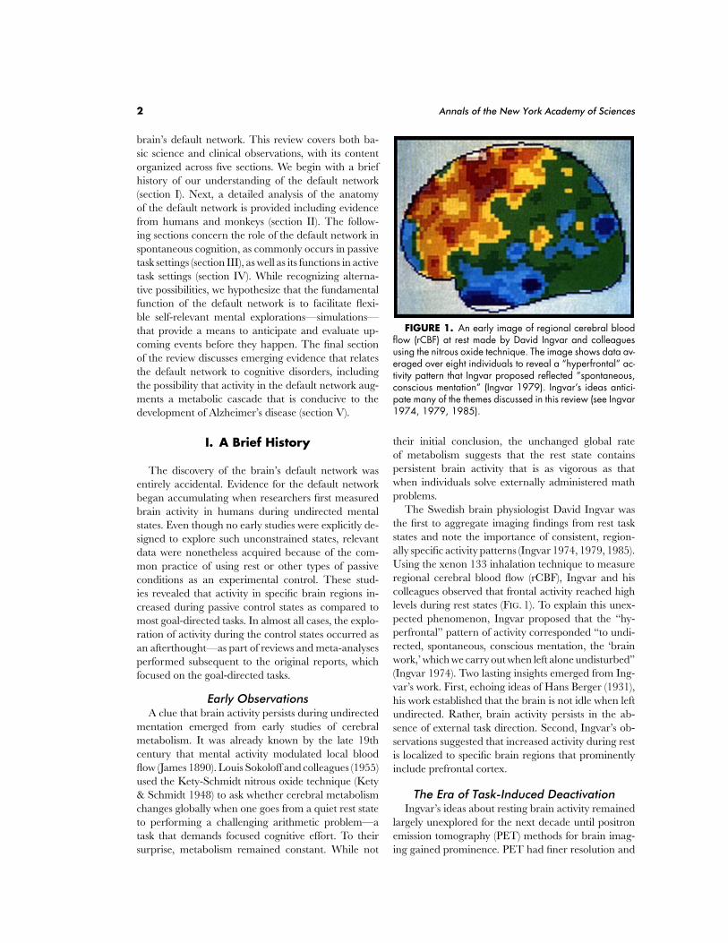
These two analyses revealed a remarkably consis-tent set of brain regions that were more active duringpassive task conditions than during numerous goal-directed task conditions (spanning both verbal andnonverbal domains and visual and auditory condi-tions). The results of the Shulman et al. (1997) meta-analysis are shown in FIGURE 2. This image displaysthe full cortical extent of the brain’s default network.The broad generality of the rest activity pattern acrossso many diverse studies reinforced the intriguing pos-sibility that a common set of cognitive processes wasused spontaneously during the passive-task states. Mo-tivated by this idea, Mazoyer et al. (2001) exploredthe content of spontaneous thought by asking partici-pants to describe their musings following the scannedrest periods. Paralleling the informal observations by
4 Annals of the New York Academy of Sciences
FIGURE 2. The brain’s default network was originallyidentified in a meta-analysis that mapped brain regionsmore active in passive as compared to active tasks (of-ten referred to as task-induced deactivation). The displayedpositron emission tomography (PET) data include nine stud-ies (132 participants) from Shulman et al. (1997; rean-alyzed in Buckner et al. 2005). Images show the me-dial and lateral surface of the left hemisphere using apopulation-averaged surface representation to take into ac-count between-subject variability in sulcal anatomy (Van Es-sen 2005). Blue represents regions most active in passivetask settings.
Ingvar and Andreasen et al., they noted that the im-aged rest state is associated with lively mental activitythat includes “generation and manipulation of men-tal images, reminiscence of past experiences based onepisodic memory, and making plans” and further notedthat the subjects of their study “preferentially reportedautobiographical episodes.”
Emergence of the Default Network as ItsOwn Research Area
The definitive recent event in the explication ofthe default network came with the a series of publi-cations by Raichle, Gusnard, and colleagues (Raichleet al. 2001, Gusnard & Raichle 2001, Gusnard et al.2001). A dominant theme in the field during the pre-vious decade concerned how to define an appropriatebaseline condition for neuroimaging studies. This focuson the baseline state was central to the evolving con-cept of a default network. Many argued that passive
conditions were simply too unconstrained to be usefulas control states. Richard Frackowiak summarized thiswidely held concern: “To call a ‘free-wheeling’ state,or even a state where you are fixating on a cross anddreaming about anything you like, a ‘control’ state,is to my mind quite wrong” (Frackowiak 1991). (Forrecent discussion of this ongoing debate see Morcomand Fletcher 2007, Buckner & Vincent 2007, Raichle& Snyder 2007). As a result of this uneasiness in inter-preting passive task conditions, beyond the few earlierstudies mentioned, there was a general trend not tothoroughly report or discuss the meaning of rest stateactivity.
Raichle, Gusnard, and colleagues reversed this trenddramatically with three papers in 2001 (Raichle et al.2001, Gusnard & Raichle 2001, Gusnard et al. 2001).Their papers directly considered the empirical andtheoretical implications of defining baseline states andwhat the specific pattern of activity in the default net-work might represent. Several lasting consequences onthe study of the default network emerged. First, theydistinguished between various forms of task-induceddeactivation and separated deactivations defining thedefault network from other forms of deactivation (in-cluding attenuation of activity in unattended sensoryareas). Second, they compiled a considerable array offindings that drew attention to the specific anatomicregions linked to the default network and what theirpresence might suggest about its function. A key in-sight was that the medial prefrontal regions consistentlyidentified as part of the default network are associatedwith self-referential processing (Gusnard et al. 2001,Gusnard & Raichle 2001). Most importantly, the pa-pers brought to the forefront the exploration of thedefault network as its own area of study (including pro-viding its name, which, as of late 2007, has appeared asa keyword in 237 articles). Our use of the label “defaultnetwork” in this review stems directly from their label-ing the baseline rest condition as the “default mode.”b
Their reviews made clear that the default network isto be studied as a fundamental neurobiological systemwith physiological and cognitive properties that distin-guish it from other systems.
The default network is a brain system much like themotor system or the visual system. It contains a setof interacting brain areas that are tightly functionally
bReferences to the default mode appear in the literature on cognitionprior to the introduction of the concept as an explanation for neural andmetabolic phenomena. Giambra (1995), for example, noted that “Task-unrelated images and thoughts may represent the normal default modeof operation of the self-aware.” Thus, the concept of a default mode isconverged upon from both cognitive and neurobiological perspectives.
Buckner et al.: The Brain’s Default Network 5
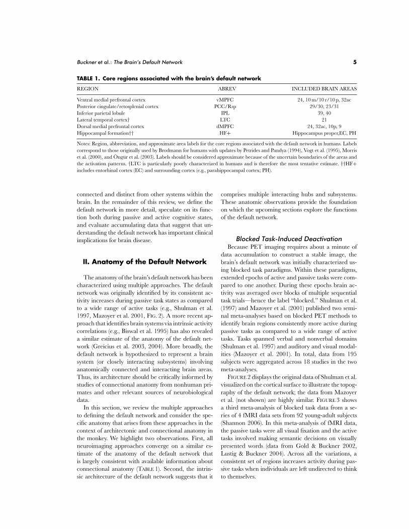
TABLE 1. Core regions associated with the brain’s default network
REGION ABREV INCLUDED BRAIN AREAS
Ventral medial prefrontal cortex vMPFC 24, 10 m/10 r/10 p, 32acPosterior cingulate/retosplenial cortex PCC/Rsp 29/30, 23/31Inferior parietal lobule IPL 39, 40Lateral temporal cortex† LTC 21Dorsal medial prefrontal cortex dMPFC 24, 32ac, 10p, 9Hippocampal formation†† HF+ Hippocampus proper,EC, PH
Notes: Region, abbreviation, and approximate area labels for the core regions associated with the default network in humans. Labelscorrespond to those originally used by Brodmann for humans with updates by Petrides and Pandya (1994), Vogt et al. (1995), Morriset al. (2000), and Ongur et al. (2003). Labels should be considered approximate because of the uncertain boundaries of the areas andthe activation patterns. †LTC is particularly poorly characterized in humans and is therefore the most tentative estimate. ††HF+includes entorhinal cortex (EC) and surrounding cortex (e.g., parahippocampal cortex; PH).
connected and distinct from other systems within thebrain. In the remainder of this review, we define thedefault network in more detail, speculate on its func-tion both during passive and active cognitive states,and evaluate accumulating data that suggest that un-derstanding the default network has important clinicalimplications for brain disease.
II. Anatomy of the Default Network
The anatomy of the brain’s default network has beencharacterized using multiple approaches. The defaultnetwork was originally identified by its consistent ac-tivity increases during passive task states as comparedto a wide range of active tasks (e.g., Shulman et al.1997, Mazoyer et al. 2001, FIG. 2). A more recent ap-proach that identifies brain systems via intrinsic activitycorrelations (e.g., Biswal et al. 1995) has also revealeda similar estimate of the anatomy of the default net-work (Greicius et al. 2003, 2004). More broadly, thedefault network is hypothesized to represent a brainsystem (or closely interacting subsystems) involvinganatomically connected and interacting brain areas.Thus, its architecture should be critically informed bystudies of connectional anatomy from nonhuman pri-mates and other relevant sources of neurobiologicaldata.
In this section, we review the multiple approachesto defining the default network and consider the spe-cific anatomy that arises from these approaches in thecontext of architectonic and connectional anatomy inthe monkey. We highlight two observations. First, allneuroimaging approaches converge on a similar es-timate of the anatomy of the default network thatis largely consistent with available information aboutconnectional anatomy (TABLE 1). Second, the intrin-sic architecture of the default network suggests that it
comprises multiple interacting hubs and subsystems.These anatomic observations provide the foundationon which the upcoming sections explore the functionsof the default network.
Blocked Task-Induced DeactivationBecause PET imaging requires about a minute of
data accumulation to construct a stable image, thebrain’s default network was initially characterized us-ing blocked task paradigms. Within these paradigms,extended epochs of active and passive tasks were com-pared to one another. During these epochs brain ac-tivity was averaged over blocks of multiple sequentialtask trials—hence the label “blocked.” Shulman et al.(1997) and Mazoyer et al. (2001) published two semi-nal meta-analyses based on blocked PET methods toidentify brain regions consistently more active duringpassive tasks as compared to a wide range of activetasks. Tasks spanned verbal and nonverbal domains(Shulman et al. 1997) and auditory and visual modal-ities (Mazoyer et al. 2001). In total, data from 195subjects were aggregated across 18 studies in the twometa-analyses.
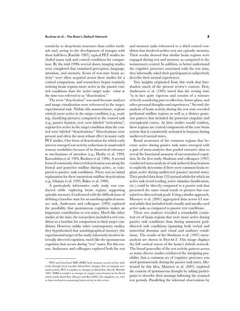
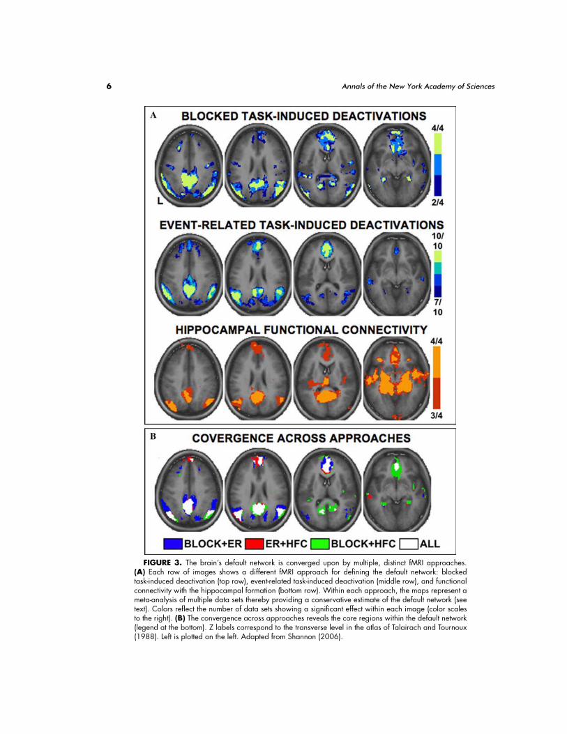
FIGURE 2 displays the original data of Shulman et al.visualized on the cortical surface to illustrate the topog-raphy of the default network; the data from Mazoyeret al. (not shown) are highly similar. FIGURE 3 showsa third meta-analysis of blocked task data from a se-ries of 4 fMRI data sets from 92 young-adult subjects(Shannon 2006). In this meta-analysis of fMRI data,the passive tasks were all visual fixation and the activetasks involved making semantic decisions on visuallypresented words (data from Gold & Buckner 2002,Lustig & Buckner 2004). Across all the variations, aconsistent set of regions increases activity during pas-sive tasks when individuals are left undirected to thinkto themselves.
6 Annals of the New York Academy of Sciences
FIGURE 3. The brain’s default network is converged upon by multiple, distinct fMRI approaches.(A) Each row of images shows a different fMRI approach for defining the default network: blockedtask-induced deactivation (top row), event-related task-induced deactivation (middle row), and functionalconnectivity with the hippocampal formation (bottom row). Within each approach, the maps represent ameta-analysis of multiple data sets thereby providing a conservative estimate of the default network (seetext). Colors reflect the number of data sets showing a significant effect within each image (color scalesto the right). (B) The convergence across approaches reveals the core regions within the default network(legend at the bottom). Z labels correspond to the transverse level in the atlas of Talairach and Tournoux(1988). Left is plotted on the left. Adapted from Shannon (2006).
Buckner et al.: The Brain’s Default Network 7
Event-Related, Task-Induced DeactivationAn alternative to defining the anatomy of the de-
fault network based on blocked tasks is to perform asimilar analysis on individual task events. Rapid event-related fMRI makes possible such an analysis by pre-senting task trials at randomly jittered time intervals,typically 2 to 10 seconds apart. The reason to performsuch an analysis is the possibility that extended epochsare required to elicit activity during passive epochs, asmight be the case if blocked task-induced deactivationsarise from slowly evolving signals or sustained task setsthat are not modulated on a rapid time frame (e.g.,Dosenbach et al. 2006).
FIGURE 3 illustrates the results of a meta-analysis ofstudies from Shannon (2006) that uses event-relatedfMRI data to define the default network. In total, datafrom 49 subjects were pooled for this analysis. Thedata are based on semantic and phonological classifi-cation tasks from Kirchhoff et al. (2005; n = 28) as wellas a second sample of event-related data that also in-volved semantic classification (Shannon 2006; n = 21).As can be appreciated visually, the default network de-fined based on event-related data is highly similar tothat previously reported using blocked data. Thus, thedifferential activity in the default network between pas-sive and active task states can emerge rapidly, on theorder of seconds or less.
Functional Connectivity AnalysisA final approach to defining the functional anatomy
of the default network is based on the measurement ofthe brain’s intrinsic activity. At all levels of the ner-vous system from individual neurons (Tsodyks et al.1999) and cortical columns (Arieli et al. 1995) to whole-brain systems (Biswal et al. 1995, De Luca et al. 2006),there exists spontaneous activity that tracks the func-tional and anatomic organization of the brain. Thepatterns of spontaneous activity are believed to re-flect direct and indirect anatomic connectivity (Vincentet al. 2007a) although additional contributions mayarise from spontaneous cognitive processes (as will bedescribed in a later section). In humans, low-frequency,spontaneous correlations are detectable across thebrain with fMRI and can be used to characterizethe intrinsic architecture of large-scale brain systems,an approach often referred to as functional connec-tivity MRI (Biswal et al. 1995, Haughton & Biswal1998; see Fox & Raichle 2007 for a recent review).Motor (Biswal et al. 1995), visual (Nir et al. 2006),auditory (Hunter et al. 2006), and attention (Fox etal. 2006) systems have been characterized using func-tional connectivity analysis (see also De Luca et al.2006).
Greicius and colleagues (2003, 2004) used such ananalysis to map the brain’s default network (see also Foxet al. 2005, Fransson 2005, Damoiseaux et al. 2006,Vincent et al. 2006). Functional connectivity analysisis particularly informative because it provides a meansto assess locations of interacting brain regions withinthe default network in a manner that is independentof task-induced deactivation. In their initial studies,Greicius et al. measured spontaneous activity from theposterior cingulate cortex, a core region in the defaultnetwork, and showed that activity levels in the remain-ing distributed regions of the system are all correlatedtogether. Their map of the default network, based onintrinsic functional correlations, is remarkably similarto that originally generated by Shulman et al. (1997)based on PET deactivations.
An important further observation from analyses ofintrinsic activity is that the default network includesthe hippocampus and adjacent areas in the medialtemporal lobe that are associated with episodic mem-ory function (Greicius et al. 2004). In fact, many ofthe major neocortical regions constituting the defaultnetwork can be revealed by placing a seed region inthe hippocampal formation and mapping those corti-cal regions that show spontaneous correlation (Vincentet al. 2006). FIGURE 3 shows a map of the default net-work as generated from intrinsic functional correla-tions with the hippocampal formation in four inde-pendent data sets.
Convergence across Approaches forDefining the Default Network
Is there convergence between the three distinct ap-proaches for defining the anatomy of the default net-work described above? To answer this question, theoverlap among the multiple methods for defining de-fault network anatomy is displayed on the bottom panelof FIGURE 3. The convergence reveals that the defaultnetwork comprises a distributed set of regions thatincludes association cortex and spares sensory andmotor cortex. In particular, medial prefrontal cortex(MPFC), posterior cingulate cortex/retrosplenial cor-tex (PCC/Rsp), and the inferior parietal lobule (IPL)show nearly complete convergence across the 18 datasets.
Several more specific observations are apparentfrom this analysis of overlap. First, the hippocampalformation (HF) is shown to be involved in the de-fault network regardless of which approach is used(task-induced deactivation or functional connectivityanalysis) but, relative to the robust posterior mid-line and prefrontal regions, the HF is less promi-nent using the approach of task-induced deactivations.
8 Annals of the New York Academy of Sciences
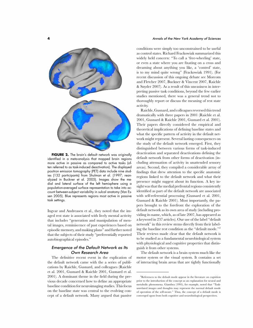
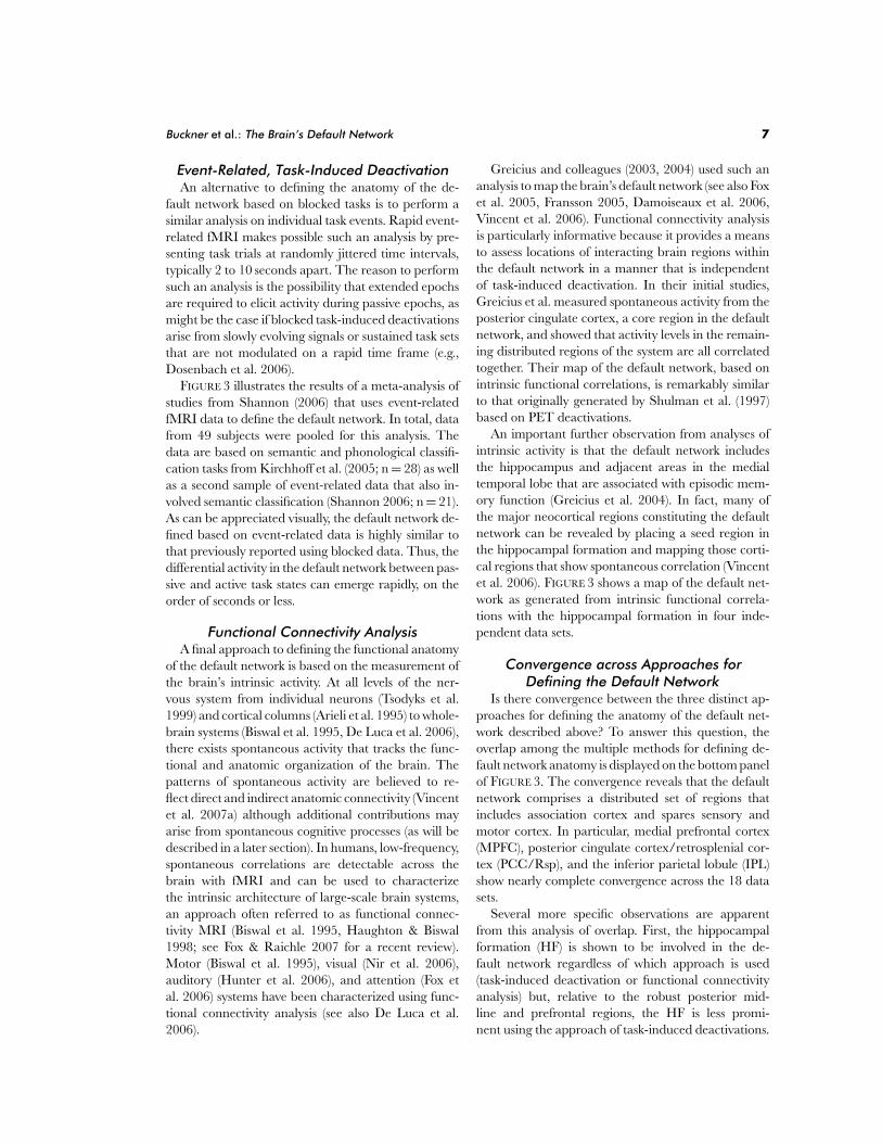
FIGURE 4. The default network in the monkey defined using functional connectivity analysis. A seedwas placed in the posterior midline (indicated by asterisk) and the regions showing correlated activitywere mapped. The left image shows the medial surface, the middle image a transverse section throughparietal cortex, and the right image a coronal section through the hippocampal formation. Left is plottedon the left. Adapted from Vincent et al. (2007a).
Second, multiple default network regions are function-ally correlated with the HF, reinforcing the notion thatthe medial temporal lobe is included in the network.Overlap is not perfect, however, with some indicationsof more extensive recruitment during passive cogni-tive states, including both in posterior parietal cortexand in prefrontal cortex. These details will be shownto be informative when subsystems within the defaultnetwork are discussed. Third, lateral temporal cortex(LTC) extending into the temporal pole is consistentlyobserved across approaches but, like the HF, is lessrobust. Together these observations tentatively definethe core anatomical components of the default network(TABLE 1).
Insights from Comparative AnatomyImportant insights into the organization of human
brain systems have been provided by comparative stud-ies in the monkey. Vincent et al. (2007a) recently usedfunctional connectivity analysis to show that the majordefault network regions in posterior cortex have pu-tative monkey homologues including PCC/Rsp, IPL,and the HF (FIG. 4, see also Rilling et al. 2007). Inaddition, architectonic maps reveal many similaritiesbetween human and monkey anatomy in the vicinityof the default network (e.g., Petrides & Pandya 1994,Morris et al. 2000, Ongur & Price 2000, Vogt et al.2001). Motivated by these recent observations, we pro-vide here a detailed analysis of the architectonics andconnectional anatomy of the default network, whilerecognizing that there may be fundamental differencesin humans. As a means to simplify our analysis, we fo-cus on areas that fall within PCC/Rsp and MPFC andtheir anatomic relationships with other cortical regions
and the HF. Potentially important subcortical connec-tions, such as to the striatal reward pathway and theamygdala, are not covered. Even with this simplifica-tion, the details of the anatomy are complex and one isimmediately confronted with the observation that eachof the activated regions, as defined based on humanfunctional neuroimaging data, extends across multiplebrain areas that have distinct architecture and connec-tivity. Progress will require significantly more detailedanalysis of the anatomic extent and locations of defaultnetwork regions in humans. Nonetheless, using avail-able data we provide an initial analysis of the anatomyrecognizing that it is provisional and incomplete.
Posterior cingulate cortex (PCC) and restrosple-nial cortex (Rsp) have been extensively studied in themacaque monkey and recently so with focus on di-rect comparison to human anatomy (e.g., Morris et al.2000, Vogt et al. 2001). The PCC and Rsp fall alongthe posterior midline and exist within a region thatcontains at least three contiguous, but distinct, sets ofareas: Rsp (areas 29/30), PCC (areas 23/31), and pre-cuneus (area 7m). Rsp is just posterior to the corpuscallosum and, in humans, extends along the ventralbank of the cingulate gyrus (Morris et al. 2000, Vogtet al. 2001). In macaques, Rsp is much smaller anddoes not encroach onto the cingulate gyrus (Morriset al. 1999, Kobayashi & Amaral 2000). Just poste-rior to Rsp, along the main portion of the cingulategyrus, is PCC. The precuneus, a region often cited asbeing involved in the default network, comprises theposterior and dorsal portion of the medial parietallobe and includes area 7m (Cavanna & Trimble 2006,Parvizi et al. 2006). As an ensemble, these three struc-tures are sometimes referred to as “posteriomedial
Buckner et al.: The Brain’s Default Network 9
cortex,” and each structure is interconnected with theothers (e.g., Parvizi et al. 2006, Kobayashi & Amaral2003).
The predominant extrinsic connections to and fromthe posteriomedial cortex differ by area. Collectively,the connections are widespread and, much like otherassociation areas, are consistent with a role in infor-mation integration. Specifically, Rsp is heavily inter-connected with the HF and parahippocampal cortex,receiving nearly 40% of its extrinsic input from the me-dial temporal lobe (Kobayashi & Amaral 2003, see alsoSuzuki & Amaral 1994, Morris et al. 1999). Rsp alsoprojects back to the medial temporal lobe as well asprominently to multiple prefrontal regions (Kobayashi& Amaral 2007, FIG. 5). PCC area 23 has reciprocalconnections with the medial temporal lobe and robustconnections with prefrontal cortex and parietal cortexarea 7a—an area at or near the putative homologue ofthe human default network region IPL (Kobayashi &Amaral 2003, 2007, FIG. 5). The medial temporal lobealso has modest, but consistent, connections with area7a (Suzuki & Amaral 1994, Clower et al. 2001, Lavenexet al. 2002). Thus, PCC/Rsp provides a key hub foroverlapping connections between themselves, the me-dial temporal lobe, and IPL—three of the distributedregions that constitute the major posterior extent ofthe default network.
An unresolved issue is whether the lateral projectionzone of PCC/Rsp is restricted to area 7a in humansor extends to areas 39/40. Macaque PCC has recipro-cal projections to superior temporal sulcus (STS) andthe superior temporal gyrus (STG; see also Kobayashi& Amaral 2003). Analysis of the default network inmacaques provides indication that the network’s lat-eral extent includes STG (Vincent et al. 2007a). Com-plicating the picture, IPL is greatly expanded in hu-mans, including areas 39/40 (Culham & Kanwisher2001, Simon et al. 2002, Orban et al. 2006) that areclosely localized to the lateral parietal region identifiedby human neuroimaging as being within the defaultnetwork (see Caspers et al. 2006). A recent analysis ofcortical expansion between the macaque and humanbrain based on mapping of 23 presumed homologiesrevealed that IPL is among the regions of greatest in-crease (Van Essen & Dieker 2007). Thus, these lateralparietal and temporo-parietal areas, which are not aswell characterized as PCC/Rsp, are extremely interest-ing in light of their anatomic connections, involvementin the default network, and potential evolutionary ex-pansion in humans.
The connectional anatomy of area 7m in the pre-cuneus is difficult to understand in relation to thedefault network even though it is often included in
the default network. One possibility is that area 7m issimply not a component of the default network. Ref-erences to precuneus in the neuroimaging literatureare often used loosely to label the general region thatincludes PCC area 29/30. Precuneus area 7m pre-dominantly connects with occipital and parietal areaslinked to visual processing and frontal areas associatedwith motor planning (Cavada & Goldman-Rakic 1989,Leichnetz 2001). Moreover, medial temporal lobe re-gions that have extensive projections to PCC and Rspshow minimal connections to area 7m. Connectionsdo exist between area 7m and the PCC, which maybe the basis for the extensive activation patterns some-times observed along the posterior midline, but wesuspect that area 7m is not a core component of thenetwork.
Reinforcing this impression, close examination ofthe many maps that define the human default net-work in this review shows that the posterior medialextent of the network usually does not encroach on theedge of the parietal midline (where area 7m is located,Scheperjans et al. 2007). This boundary is labeled ex-plicitly in FIGURE 7 by an asterisk. The middle panelof FIGURE 18 shows a particularly clear example of theseparation between task-induced deactivation of PCCand its dissociation from the region at or near area 7m.Another example of dissociation between the defaultnetwork and area 7m can be found in Vogeley et al.(2004; their Figure 2A versus 2B). For all these reasons,we provisionally conclude that area 7m in precuneusis not part of the default network.
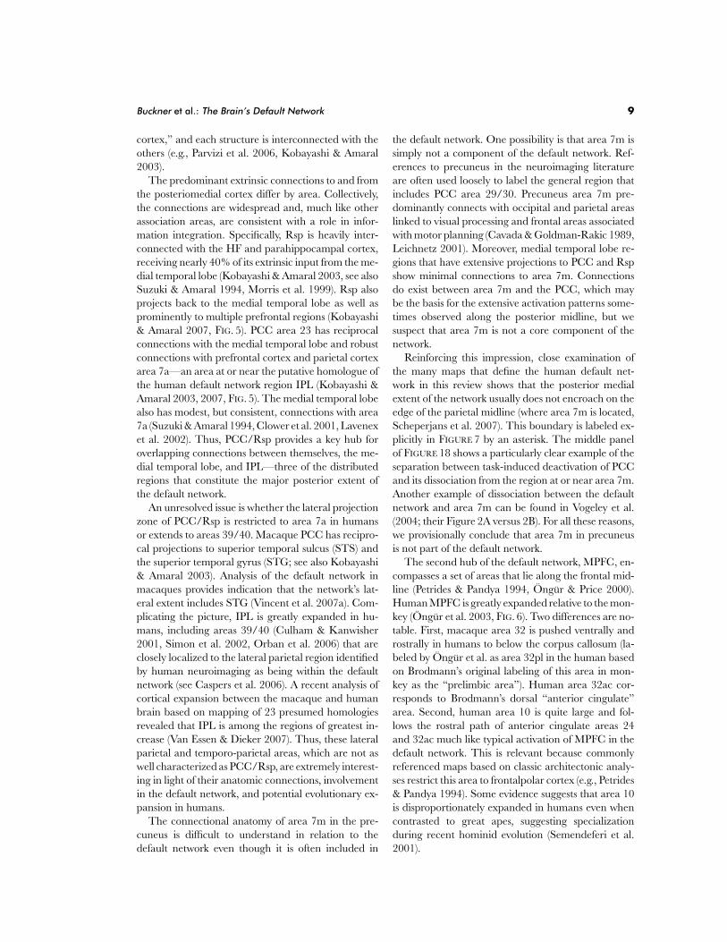
The second hub of the default network, MPFC, en-compasses a set of areas that lie along the frontal mid-line (Petrides & Pandya 1994, Ongur & Price 2000).Human MPFC is greatly expanded relative to the mon-key (Ongur et al. 2003, FIG. 6). Two differences are no-table. First, macaque area 32 is pushed ventrally androstrally in humans to below the corpus callosum (la-beled by Ongur et al. as area 32pl in the human basedon Brodmann’s original labeling of this area in mon-key as the “prelimbic area”). Human area 32ac cor-responds to Brodmann’s dorsal “anterior cingulate”area. Second, human area 10 is quite large and fol-lows the rostral path of anterior cingulate areas 24and 32ac much like typical activation of MPFC in thedefault network. This is relevant because commonlyreferenced maps based on classic architectonic analy-ses restrict this area to frontalpolar cortex (e.g., Petrides& Pandya 1994). Some evidence suggests that area 10is disproportionately expanded in humans even whencontrasted to great apes, suggesting specializationduring recent hominid evolution (Semendeferi et al.2001).

10 Annals of the New York Academy of Sciences
FIGURE 5. Monkey anatomy suggests that the default network includes multiple, distinct associationareas, each of which is connected to other areas within the network. Illustrated are two examples of output(efferent) and input (afferent) connections for posterior cingulate/retrosplenial cortex (PCC/Rsp) andparahippocampal cortex (PH). (A) Output connections from Rsp (areas 29 and 30) and PCC (area 23)are displayed. Lines show connections to distributed areas; thickness represents the connection strength.Rsp and PCC are heavily connected with the medial temporal lobe (HF, hippocampal formation; PH,parahippocampal cortex), the inferior parietal lobule (IPL) extending into superior temporal gyrus (STG),and prefrontal cortex (PFC). Numbers in the diagram indicate brain areas. Adapted from Kobayashi andAmaral (2007). (B) Input and output connections to and from PH to medial prefrontal cortex (MPFC) aredisplayed. Adapted from Kondo et al. (2005).
Given these details, MPFC activation within thedefault network is estimated to encompass humanareas 10 (10 m, 10 r, and 10 p), anterior cingu-late (area 24/32ac), and area 9 in prefrontal cor-tex. The closest homologues to these areas in the
monkey—the medial prefrontal network—show re-ciprocal connections with the PCC, Rsp, STG, HF,and the perirhinal/parahippocampal cortex; sen-sory inputs are nearly absent (Barbas et al. 1999,Price 2007). These connectivity patterns closely
Buckner et al.: The Brain’s Default Network 11
FIGURE 6. Architectonic areas within medial prefrontal cortex (MPFC) are illustrated for the monkeyand human. The human MPFC is greatly expanded relative to the macaque monkey. This expansion isdepicted by the triangle and asterisk that plot putative homologous areas between species based onOngur et al. (2003). Area 32 in the macaque is homologous with area 32pl in the human. Area 24c isexpanded and homologous to the caudal part of area 32ac in human. The MPFC region activated withinthe human default network likely corresponds to frontalpolar cortex and its rostral expansion (areas 10m,10r, and 10p), anterior cingulate (areas 24 and 32ac), and the rostral portion of prefrontal area 9.Because of differences in functional properties, we sometimes differentiate in this review between dorsaland ventral portions of MPFC (dMPFC and vMPFC). Adapted with permission from Ongur et al. (2003).
parallel areas implicated as components of the defaultnetwork.
At the broadest level, an important principleemerges from considering these anatomic details: thedefault network is not made up of a single monosynap-tically connected brain system. Rather, the architec-ture reveals a series of interconnected subsystems thatconverge on key “hubs,” in particular the PCC, thatare connected with the medial temporal lobe memorysystem. In the next section, we explore evidence forthese subsystems from functional connectivity analysisin humans.
The Default Network Comprises InteractingSubsystems
The default network comprises a set of brain regionsthat are coactivated during passive task states, show in-trinsic functional correlation with one another, and areconnected via direct and indirect anatomic projectionsas estimated from comparison to monkey anatomy.However, there is also clear evidence that the brainregions within the default network contribute special-ized functions that are organized into subsystems thatconverge on hubs.
One way to gain further insight into the organiza-tion of the default network is through detailed anal-
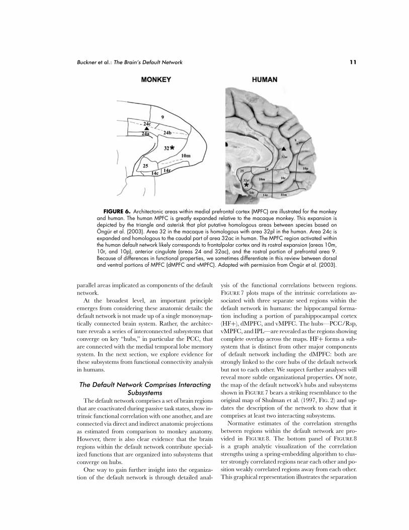
ysis of the functional correlations between regions.FIGURE 7 plots maps of the intrinsic correlations as-sociated with three separate seed regions within thedefault network in humans: the hippocampal forma-tion including a portion of parahippocampal cortex(HF+), dMPFC, and vMPFC. The hubs—PCC/Rsp,vMPFC, and IPL—are revealed as the regions showingcomplete overlap across the maps. HF+ forms a sub-system that is distinct from other major componentsof default network including the dMPFC: both arestrongly linked to the core hubs of the default networkbut not to each other. We suspect further analyses willreveal more subtle organizational properties. Of note,the map of the default network’s hubs and subsystemsshown in FIGURE 7 bears a striking resemblance to theoriginal map of Shulman et al. (1997, FIG. 2) and up-dates the description of the network to show that itcomprises at least two interacting subsystems.
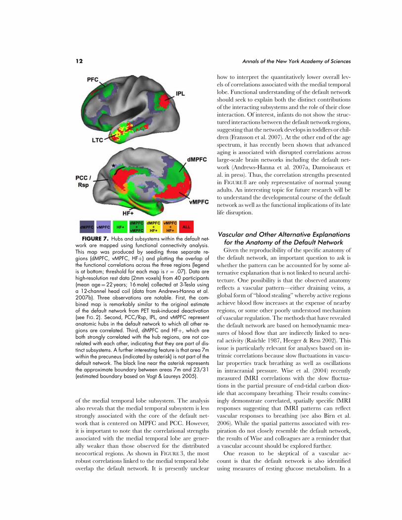
Normative estimates of the correlation strengthsbetween regions within the default network are pro-vided in FIGURE 8. The bottom panel of FIGURE 8is a graph analytic visualization of the correlationstrengths using a spring-embedding algorithm to clus-ter strongly correlated regions near each other and po-sition weakly correlated regions away from each other.This graphical representation illustrates the separation
12 Annals of the New York Academy of Sciences
FIGURE 7. Hubs and subsystems within the default net-work are mapped using functional connectivity analysis.This map was produced by seeding three separate re-gions (dMPFC, vMPFC, HF+) and plotting the overlap ofthe functional correlations across the three regions (legendis at bottom; threshold for each map is r = .07). Data arehigh-resolution rest data (2mm voxels) from 40 participants(mean age = 22 years; 16 male) collected at 3-Tesla usinga 12-channel head coil (data from Andrews-Hanna et al.2007b). Three observations are notable. First, the com-bined map is remarkably similar to the original estimateof the default network from PET task-induced deactivation(see FIG. 2). Second, PCC/Rsp, IPL, and vMPFC representanatomic hubs in the default network to which all other re-gions are correlated. Third, dMPFC and HF+, which areboth strongly correlated with the hub regions, are not cor-related with each other, indicating that they are part of dis-tinct subsystems. A further interesting feature is that area 7mwithin the precuneus (indicated by asterisk) is not part of thedefault network. The black line near the asterisk representsthe approximate boundary between areas 7m and 23/31(estimated boundary based on Vogt & Laureys 2005).
of the medial temporal lobe subsystem. The analysisalso reveals that the medial temporal subsystem is lessstrongly associated with the core of the default net-work that is centered on MPFC and PCC. However,it is important to note that the correlational strengthsassociated with the medial temporal lobe are gener-ally weaker than those observed for the distributedneocortical regions. As shown in FIGURE 3, the mostrobust correlations linked to the medial temporal lobeoverlap the default network. It is presently unclear
how to interpret the quantitatively lower overall lev-els of correlations associated with the medial temporallobe. Functional understanding of the default networkshould seek to explain both the distinct contributionsof the interacting subsystems and the role of their closeinteraction. Of interest, infants do not show the struc-tured interactions between the default network regions,suggesting that the network develops in toddlers or chil-dren (Fransson et al. 2007). At the other end of the agespectrum, it has recently been shown that advancedaging is associated with disrupted correlations acrosslarge-scale brain networks including the default net-work (Andrews-Hanna et al. 2007a, Damoiseaux etal. in press). Thus, the correlation strengths presentedin FIGURE 8 are only representative of normal youngadults. An interesting topic for future research will beto understand the developmental course of the defaultnetwork as well as the functional implications of its latelife disruption.
Vascular and Other Alternative Explanationsfor the Anatomy of the Default NetworkGiven the reproducibility of the specific anatomy of
the default network, an important question to ask iswhether the pattern can be accounted for by some al-ternative explanation that is not linked to neural archi-tecture. One possibility is that the observed anatomyreflects a vascular pattern—either draining veins, aglobal form of “blood stealing” whereby active regionsachieve blood flow increases at the expense of nearbyregions, or some other poorly understood mechanismof vascular regulation. The methods that have revealedthe default network are based on hemodynamic mea-sures of blood flow that are indirectly linked to neu-ral activity (Raichle 1987, Heeger & Ress 2002). Thisissue is particularly relevant for analyses based on in-trinsic correlations because slow fluctuations in vascu-lar properties track breathing as well as oscillationsin intracranial pressure. Wise et al. (2004) recentlymeasured fMRI correlations with the slow fluctua-tions in the partial pressure of end-tidal carbon diox-ide that accompany breathing. Their results convinc-ingly demonstrate correlated, spatially specific fMRIresponses suggesting that fMRI patterns can reflectvascular responses to breathing (see also Birn et al.2006). While the spatial patterns associated with res-piration do not closely resemble the default network,the results of Wise and colleagues are a reminder thata vascular account should be explored further.
One reason to be skeptical of a vascular ac-count is that the default network is also identifiedusing measures of resting glucose metabolism. In a
Buckner et al.: The Brain’s Default Network 13
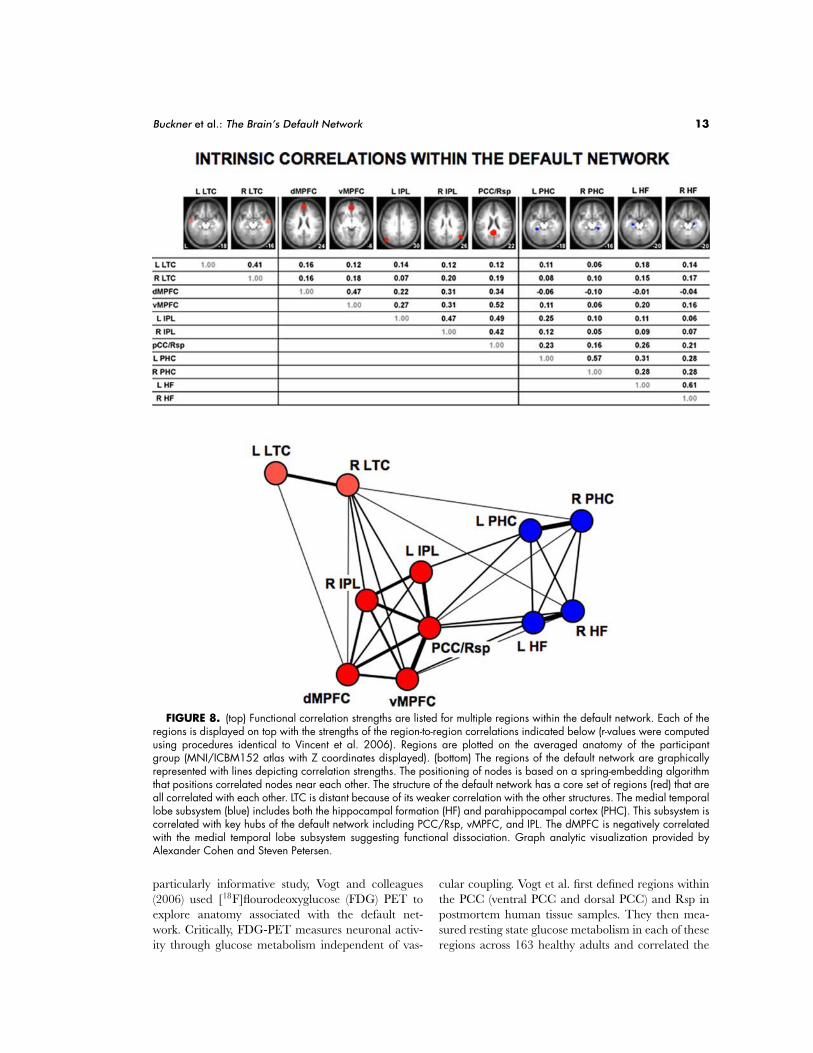
FIGURE 8. (top) Functional correlation strengths are listed for multiple regions within the default network. Each of theregions is displayed on top with the strengths of the region-to-region correlations indicated below (r-values were computedusing procedures identical to Vincent et al. 2006). Regions are plotted on the averaged anatomy of the participantgroup (MNI/ICBM152 atlas with Z coordinates displayed). (bottom) The regions of the default network are graphicallyrepresented with lines depicting correlation strengths. The positioning of nodes is based on a spring-embedding algorithmthat positions correlated nodes near each other. The structure of the default network has a core set of regions (red) that areall correlated with each other. LTC is distant because of its weaker correlation with the other structures. The medial temporallobe subsystem (blue) includes both the hippocampal formation (HF) and parahippocampal cortex (PHC). This subsystem iscorrelated with key hubs of the default network including PCC/Rsp, vMPFC, and IPL. The dMPFC is negatively correlatedwith the medial temporal lobe subsystem suggesting functional dissociation. Graph analytic visualization provided byAlexander Cohen and Steven Petersen.
particularly informative study, Vogt and colleagues(2006) used [18F]flourodeoxyglucose (FDG) PET toexplore anatomy associated with the default net-work. Critically, FDG-PET measures neuronal activ-ity through glucose metabolism independent of vas-
cular coupling. Vogt et al. first defined regions withinthe PCC (ventral PCC and dorsal PCC) and Rsp inpostmortem human tissue samples. They then mea-sured resting state glucose metabolism in each of theseregions across 163 healthy adults and correlated the
14 Annals of the New York Academy of Sciences
obtained values across the brain to yield metabolism-based maps of functional correlation. A quite remark-able pattern emerged: ventral PCC showed correlationwith the main components of the default network in-cluding vMPFC and IPL (see their Figure 7, panel B).Moreover, this pattern was preferential to ventral PCC,suggesting that the posterior hub of the default networkmay be even more circumscribed than the fMRI datasuggest, which have implicated the broader region in-cluding dorsal PCC and Rsp. Directly relevant to thequestion of whether a vascular explanation can ac-count for the default network’s anatomy, these resultswere obtained without relying on vascular coupling.
Glucose Metabolism and the OxygenExtraction Fraction
Metabolic properties of the default network also setthe network apart from other brain systems (Raichleet al. 2001). In particular, regions within the defaultnetwork show disproportionately high resting glucosemetabolism relative to other brain regions as mea-sured using FDG-PET (e.g., Minoshima et al. 1997,Gusnard & Raichle 2001, see FIG. 17) as well as highregional blood flow (Raichle et al. 2001). For example,Minoshima et al. (1997, see their Figure 1) mappedresting glucose metabolism in healthy older adults ref-erenced to the pons, allowing visualization of regionalvariation across the cortex. Along the midline, normal-ized glucose metabolism in PCC was about 20% higherthan in most other brain regions. However, high glu-cose metabolism was not selective only to the defaultnetwork—a region at or near primary visual cortexalso showed high resting metabolism. To our knowl-edge, there has been no systematic investigation of rest-ing glucose metabolism within default network regionsas contrasted to regions outside the network; however,all reported exploratory maps of glucose metabolismconverge on the observation that the posterior mid-line near PCC is a region of disproportionately highmetabolism (e.g., Minoshima et al. 1997, Figure 1, Gus-nard & Raichle 2001, Figure 1). Intriguingly, the re-gions within the default network that show high restingmetabolism are also those affected in Alzheimer’s dis-ease, something that will be discussed extensively inthe final section of this review. To foreshadow this fi-nal discussion, the possibility will be raised that highlevels of baseline activity and metabolism (glycolysis)in the default network are conducive to the forma-tion of pathology associated with Alzheimer’s disease(Buckner et al. 2005).
A second metabolic property that has been exploredin connection with the default network is regional oxy-gen utilization. In their seminal paper that drew at-
tention to the default network, Raichle et al. (2001)mapped the ratio of oxygen used locally to oxygen de-livered by blood flow. This ratio, referred to as the oxy-gen extraction fraction (OEF), decreases during height-ened neural activity because the increased flow ofblood into a region exceeds oxygen use (see Raichle &Mintun 2006). Raichle and colleagues (2001) hypoth-esized that an absolute physiological baseline couldbe shown to exist if OEF remained constant duringpassive (rest) task states, suggesting that task-induceddeactivations within the default network are physiolog-ically dissimilar from other forms of transient neuronalactivity increase. While an intriguing possibility, thereare several observations that suggest OEF within thedefault network does change at rest. First, OEF de-creases were noted by Raichle et al. (2001) in severaldefault network regions at rest when each was testedindividually at the p < 0.05 level of statistical signifi-cance. Second, regional variation in OEF across thedefault network was correlated from one data set tothe next (r = .89) indicating systematic modulation; aconstant OEF across regions would show zero corre-lation from one data set to the next. The modulationwas quantitatively small, however, with OEF valuesof most regions falling within 5 to 10% of the otherregions. Further exploration will be required to deter-mine if there is an absolute metabolic state that definesa baseline within the default network or whether thereare meaningful variations across regions. In the nextsection, we will specifically explore the possibility thatthe special properties that arise in the default networkassociate with its role in spontaneous cognition duringfreethinking.
III. Spontaneous Cognition
Human beings spend nearly all of their time in some kindof mental activity, and much of the time their activity con-sists not of ordered thought but of bits and snatches of in-ner experience: daydreams, reveries, wandering interiormonologues, vivid imagery, and dreams. These desultoryconcoctions, sometimes unobtrusive but often moving,contribute a great deal to the style and flavor of beinghuman. Their very humanness lends them great intrinsicinterest; but beyond that, surely so prominent a set ofactivities cannot be functionless. (Klinger 1971 p. 347)
A shared human experience is our active inter-nal mental life. Left without an immediate task thatdemands full attention, our minds wander jumpingfrom one passing thought to next—what William James(1890) called the “stream of consciousness.” We museabout past happenings, envision possible future events,and lapse into ideations about worlds that are far from
Buckner et al.: The Brain’s Default Network 15
our immediate surroundings. In lay terms, these arethe mental processes that make up fantasy, imagina-tion, daydreams, and thought. A central issue for ourpresent purposes is to understand to what degree, ifany, the default network mediates these forms of spon-taneous cognition. The observation that the defaultnetwork is most active during passive cognitive states,when thought is directed toward internal channels,encourages serious consideration of the possibility thatthe default network is the core brain system associatedwith spontaneous cognition, and further that peoplehave a strong tendency to engage the default networkduring moments when they are not otherwise occupiedby external tasks. In considering the relationship be-tween the default network and spontaneous cognition,it is worth beginning with a short review of spontaneouscognition itself.
Descriptions of human nature have alluded to theprominence of private mental experience since theclassical period. In a whimsical description, Plato por-trayed Socrates as “capable of standing all day in themarket place lost in thought and oblivious of the ex-ternal world,” leading Aristophanes to coin the phrase“his head is in the clouds” (Singer 1966). Experimentalstudy of internal mental life originated within the psy-chological movement of introspection in the late 19thcentury. Developed by Wilhelm Wundt and continuedby the American psychologist Edward Titchener, in-trospective methods required participants to describethe contents of their internal mental experience. Thepremise of introspection was that conscious elementsand attributes are sufficient to describe the mind. Thefocus on behaviorism during much of the 20th century,which emphasized measurement of the external factorsthat control behavior, caused a marked decline in thestudy of thought in mainstream science. The behavior-ists rejected the methods of introspection because theyrelied on subjective report leading to a global “morato-rium on the study of inner experience” (Klinger 1971).
The dark ages of spontaneous cognition ended in1966 with a seminal publication by Jerome Singer thatdescribed an extensive empirical research program onthe topic of daydreaming (see also Antrobus et al.1970, Klinger 1971, Singer 1974). Several importantadvances emerged from this work. First, behavioralinstruments were developed for the measurement ofspontaneous cognition that correlated with such fac-tors as individual differences in cognition, physiologicalmeasures and eye movements, and were also predic-tive of response patterns on varied tasks (e.g., Singer& Schonbar 1961, Singer et al. 1963, Antrobus et al.1966, Antrobus 1968, Antrobus et al. 1970). Second,spontaneous cognition was observed to be quite com-
mon: 96% of individuals report daydreaming daily.Moreover, the contents of daydreams were found toinclude everything from mundane recounts of recenthappenings to plans and expectations about the future.Finally, this work emphasized that spontaneous cogni-tion is healthy and adaptive, and not simply a set of dis-tracting processes or fantasies. Singer (1966), Antrobuset al. (1966) and later Klinger (1971) specifically sug-gested that internal mental activity is important foranticipating and planning the future. We will return tothis important idea later.
In the past decade, the study of spontaneous cogni-tion has built upon these foundations and introducednovel experimental approaches to explore the contentof people’s internal mental states (see Smallwood &Schooler 2006 for review). Critical to understandingthe relationship between the default network and spon-taneous cognition, measures of sampled thoughts trackdefault network activity. Moreover, individual differ-ences in tendencies to engage spontaneous cognitiveprocesses parallel differences in default network activ-ity. In the following section, we review these findingsand discuss their implications.
Stimulus-Independent ThoughtsA number of brain imaging studies have explored
stimulus-independent thoughts (SITs).c SITs are oper-ationally defined as thoughts about something otherthan events originating from the environment; theyare covert and not directed toward performance of thetask at hand. The most common method for measuringSITs involves periodically probing trained participantsto indicate whether they are experiencing a SIT. Careis taken to minimize the intrusiveness of the probe, al-though a limitation of this approach is that the probenonetheless does interfere with the SIT, most typicallyto terminate its occurrence (Giambra 1995). Antrobusand colleagues (1966, 1968, 1970) showed that SITsoccur quite pervasively—during both resting epochsand also during the performance of concurrent tasks.Even under heavy loads of external information, mostindividuals still report the presence of some SITs al-though the number of SITs correlates inversely withthe demands of the external task.
cVarious labels have been used in the reviewed papers to describeself-reported thought content including task-irrelevant thoughts (Antrobuset al. 1966), stimulus-independent thoughts (SITs, Antrobus et al. 1970,Teasdale et al. 1995), task-unrelated thoughts (TUTs, Giambra 1989), andtask-unrelated images and thoughts (TUITs, Giambra 1995). For simplic-ity, we use the term “stimulus-independent thoughts” or SITs throughoutthe text.
16 Annals of the New York Academy of Sciences
Extending from these behavioral observations, sev-eral imaging studies have correlated the number ofreported SITs with brain activity. In an early study,McGuire et al. (1996) demonstrated that the frequencyof SITs estimated following various PET scans cor-related with MPFC activity. Following a similar ap-proach, Binder and colleagues (Binder et al. 1999,McKiernan et al. 2003, 2006) conducted two fMRIstudies that explored the relationship between SITs andbrain activity. In both studies they measured brain ac-tivity during rest and various tasks using typical fMRIprocedures. Then, within a mock scanning environ-ment, they had participants perform the same taskswhile periodically probing for the presence of SITs.This procedure allowed them to sort the fMRI tasksbased on their propensity to elicit SITs. The first study(Binder et al. 1999) revealed that rest, as comparedto an externally oriented tone detection task, was as-sociated with both increased default network activity,and nearly six times more SITs. The second studyparametrically varied task difficulty across six separatetasks such that the easiest task (easy to detect target,slow presentation rate) produced about twice as manySITs as the most difficult task (McKiernan et al. 2003,2006). Referenced to rest, there was a strong corre-lation between SITs and activity within the defaultnetwork.
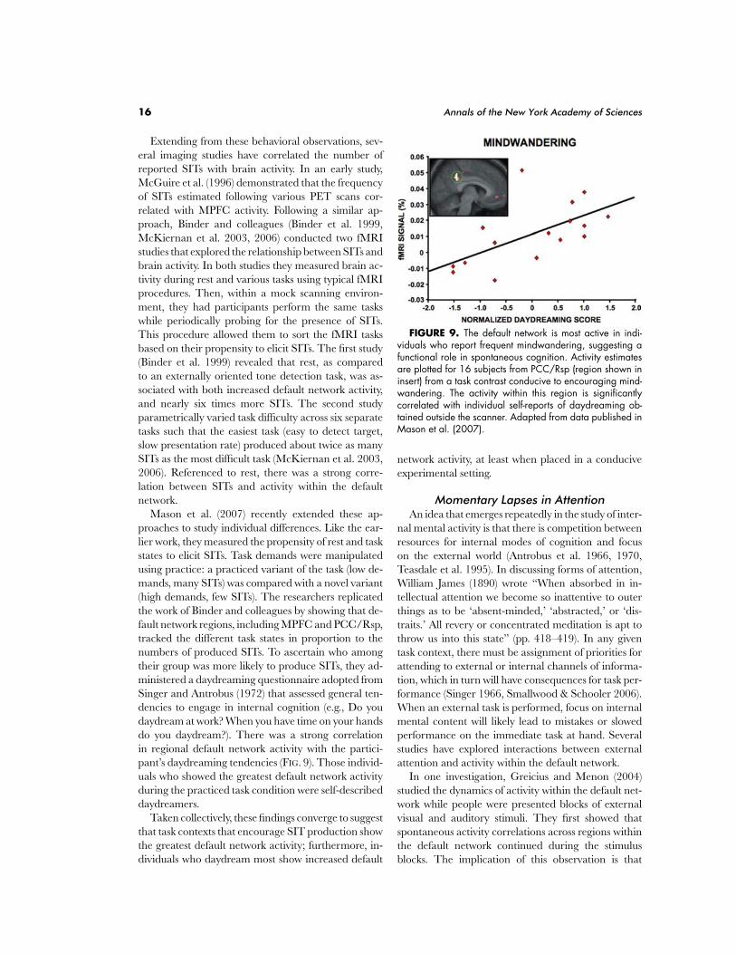
Mason et al. (2007) recently extended these ap-proaches to study individual differences. Like the ear-lier work, they measured the propensity of rest and taskstates to elicit SITs. Task demands were manipulatedusing practice: a practiced variant of the task (low de-mands, many SITs) was compared with a novel variant(high demands, few SITs). The researchers replicatedthe work of Binder and colleagues by showing that de-fault network regions, including MPFC and PCC/Rsp,tracked the different task states in proportion to thenumbers of produced SITs. To ascertain who amongtheir group was more likely to produce SITs, they ad-ministered a daydreaming questionnaire adopted fromSinger and Antrobus (1972) that assessed general ten-dencies to engage in internal cognition (e.g., Do youdaydream at work? When you have time on your handsdo you daydream?). There was a strong correlationin regional default network activity with the partici-pant’s daydreaming tendencies (FIG. 9). Those individ-uals who showed the greatest default network activityduring the practiced task condition were self-describeddaydreamers.
Taken collectively, these findings converge to suggestthat task contexts that encourage SIT production showthe greatest default network activity; furthermore, in-dividuals who daydream most show increased default
FIGURE 9. The default network is most active in indi-viduals who report frequent mindwandering, suggesting afunctional role in spontaneous cognition. Activity estimatesare plotted for 16 subjects from PCC/Rsp (region shown ininsert) from a task contrast conducive to encouraging mind-wandering. The activity within this region is significantlycorrelated with individual self-reports of daydreaming ob-tained outside the scanner. Adapted from data published inMason et al. (2007).
network activity, at least when placed in a conduciveexperimental setting.
Momentary Lapses in AttentionAn idea that emerges repeatedly in the study of inter-
nal mental activity is that there is competition betweenresources for internal modes of cognition and focuson the external world (Antrobus et al. 1966, 1970,Teasdale et al. 1995). In discussing forms of attention,William James (1890) wrote “When absorbed in in-tellectual attention we become so inattentive to outerthings as to be ‘absent-minded,’ ‘abstracted,’ or ‘dis-traits.’ All revery or concentrated meditation is apt tothrow us into this state” (pp. 418–419). In any giventask context, there must be assignment of priorities forattending to external or internal channels of informa-tion, which in turn will have consequences for task per-formance (Singer 1966, Smallwood & Schooler 2006).When an external task is performed, focus on internalmental content will likely lead to mistakes or slowedperformance on the immediate task at hand. Severalstudies have explored interactions between externalattention and activity within the default network.
In one investigation, Greicius and Menon (2004)studied the dynamics of activity within the default net-work while people were presented blocks of externalvisual and auditory stimuli. They first showed thatspontaneous activity correlations across regions withinthe default network continued during the stimulusblocks. The implication of this observation is that
Buckner et al.: The Brain’s Default Network 17
spontaneous activity within the default network per-sists through both experimental and rest epochs. Theyfurther observed evidence for competition betweensensory processing and spontaneous default networkfluctuations: sensory-evoked responses were attenu-ated in those individuals who showed the strongestspontaneous activity correlations within the defaultnetwork.
Momentary lapses in external attention were ex-plored directly by Weissman and colleagues (2006)during a demanding perceptual task. Lapses in atten-tion were defined as occurring when participants wereslow to respond. Two observations were made. First,just prior to a lapse in attention, activity within brainregions associated with control of attention was di-minished, including dorsal anterior cingulate and pre-frontal cortex. Second, during the lapse of attentionitself, activity within the default network was increasedprominently in the PCC/Rsp. These findings suggestthat transient lapses in the control of attention maylead to a shift in attention from the external world tointernal mentation.
A related observation was made in the context ofmemory encoding by Otten and Rugg (2001). Brain ac-tivity was measured in two studies during the inciden-tal encoding of words. The researchers found that in-creased activity in the posterior midline near PCC/Rspand lateral parietal regions near IPL, among other re-gions, predicted which words would be later forgotten.This observation is consistent with the possibility thattransient activity increases in the default network markthose trials on which the memorizers were distractedfrom their primary task, perhaps lapsing into privatechannels of thought.
Recently Li et al. (2007) tackled this possibility acrosstwo studies using a go/no-go paradigm. In their task,cues signaling participants to make speeded responseswere intermixed with infrequent stop signals that man-dated the responses should be withheld. Errors oc-curred when participants responded to stop signals.Exploring brain activity on the trials that preceded er-rors revealed that regions within the default network(MPFC and PCC/Rsp, but not IPL) augmented activ-ity just prior to errors, an effect replicated in a secondstudy. While again correlational, these data suggestthat when the default network is active, lapses in fo-cused external attention occur in ways that affect taskperformance.
However not all studies have found such relation-ships. Hahn et al. (2007), for example, noted that fastresponses in a target-detection task were associatedwith increased default network activity (see Figure 3in Hahn et al. 2007). Gilbert et al. (2006, 2007) hy-
pothesized that the default network is associated witha broadly tuned form of outward attention (“watch-fulness”). This idea, as will be discussed more ex-tensively in the upcoming section, is reminiscent ofShulman and colleagues’ (1997) suggestion that thedefault network participates in monitoring the ex-ternal environment. While difficult to reconcile withthe studies discussed earlier, the hypothesis put for-ward by Gilbert and colleagues is a reminder that ev-idence to date is limited and correlational, and fur-ther that opposing possibilities should be carefullyexplored. Thus, while an accumulating set of obser-vations suggest that mindwandering is linked to in-creased activity in regions within the default network,further exploration is warranted to determine if thesystem is directly supporting the processes underlyingthe stimulus-independent thoughts that accompanymindwandering.
Spontaneous Activity DynamicsThe default network spontaneously exhibits slow
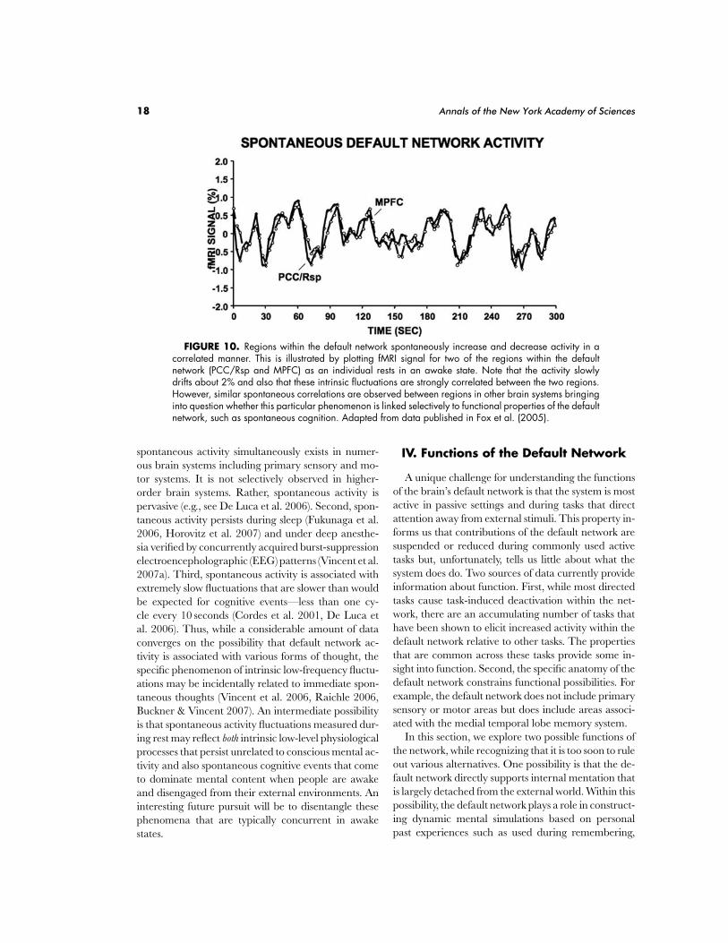
waxing and waning of activity during rest that is corre-lated across its distributed regions (Greicius et al. 2003,Fox et al. 2005, Fransson 2005, Damoiseaux et al.2006, Vincent et al. 2006). FIGURE 10 illustrates this ro-bust phenomenon for a 5-minute epoch during which ayoung adult passively viewed a small fixation crosshair.As can be seen, activity within MPFC and PCC/Rsp—two of the most prominent components of the defaultnetwork—spontaneously modulates over time. Criti-cally, these two regions, which are anatomically distantfrom one another and supplied by separate vascularterritories, show strong correlation, thereby indicatingthat the fMRI-measured activity swings arise from co-ordinated neural activity and not from measurementnoise. The presence of fluctuations at rest—when SITsare at their peak—raises the question of whether theseunprompted modulations reflect individual thoughtsand musings (e.g., Greicius & Menon 2004, Fox et al.2005, Fransson 2006). In a particularly thoughtful ap-proach to this question, Fransson (2006) showed thatcorrelated spontaneous activity within the default net-work attenuates when people perform a concurrent de-manding cognitive task (see also Shannon et al. 2006).Such forms of tasks are known to reduce the frequencyof SITs as discussed above (Antrobus et al. 1966,1970).
While these observations are intriguing, there areseveral reasons to be cautious of presuming a sim-ple relationship between spontaneous low-frequencyactivity modulations and cognitive processes (seeVincent et al. 2006, Fox & Raichle 2007). First,
18 Annals of the New York Academy of Sciences
FIGURE 10. Regions within the default network spontaneously increase and decrease activity in acorrelated manner. This is illustrated by plotting fMRI signal for two of the regions within the defaultnetwork (PCC/Rsp and MPFC) as an individual rests in an awake state. Note that the activity slowlydrifts about 2% and also that these intrinsic fluctuations are strongly correlated between the two regions.However, similar spontaneous correlations are observed between regions in other brain systems bringinginto question whether this particular phenomenon is linked selectively to functional properties of the defaultnetwork, such as spontaneous cognition. Adapted from data published in Fox et al. (2005).
spontaneous activity simultaneously exists in numer-ous brain systems including primary sensory and mo-tor systems. It is not selectively observed in higher-order brain systems. Rather, spontaneous activity ispervasive (e.g., see De Luca et al. 2006). Second, spon-taneous activity persists during sleep (Fukunaga et al.2006, Horovitz et al. 2007) and under deep anesthe-sia verified by concurrently acquired burst-suppressionelectroencepholographic (EEG) patterns (Vincent et al.2007a). Third, spontaneous activity is associated withextremely slow fluctuations that are slower than wouldbe expected for cognitive events—less than one cy-cle every 10 seconds (Cordes et al. 2001, De Luca etal. 2006). Thus, while a considerable amount of dataconverges on the possibility that default network ac-tivity is associated with various forms of thought, thespecific phenomenon of intrinsic low-frequency fluctu-ations may be incidentally related to immediate spon-taneous thoughts (Vincent et al. 2006, Raichle 2006,Buckner & Vincent 2007). An intermediate possibilityis that spontaneous activity fluctuations measured dur-ing rest may reflect both intrinsic low-level physiologicalprocesses that persist unrelated to conscious mental ac-tivity and also spontaneous cognitive events that cometo dominate mental content when people are awakeand disengaged from their external environments. Aninteresting future pursuit will be to disentangle thesephenomena that are typically concurrent in awakestates.
IV. Functions of the Default Network
A unique challenge for understanding the functionsof the brain’s default network is that the system is mostactive in passive settings and during tasks that directattention away from external stimuli. This property in-forms us that contributions of the default network aresuspended or reduced during commonly used activetasks but, unfortunately, tells us little about what thesystem does do. Two sources of data currently provideinformation about function. First, while most directedtasks cause task-induced deactivation within the net-work, there are an accumulating number of tasks thathave been shown to elicit increased activity within thedefault network relative to other tasks. The propertiesthat are common across these tasks provide some in-sight into function. Second, the specific anatomy of thedefault network constrains functional possibilities. Forexample, the default network does not include primarysensory or motor areas but does include areas associ-ated with the medial temporal lobe memory system.
In this section, we explore two possible functions ofthe network, while recognizing that it is too soon to ruleout various alternatives. One possibility is that the de-fault network directly supports internal mentation thatis largely detached from the external world. Within thispossibility, the default network plays a role in construct-ing dynamic mental simulations based on personalpast experiences such as used during remembering,
Buckner et al.: The Brain’s Default Network 19
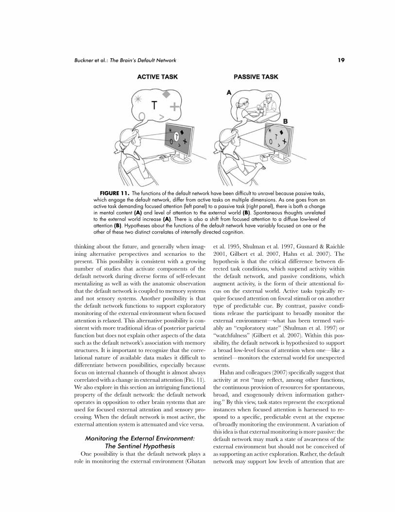
FIGURE 11. The functions of the default network have been difficult to unravel because passive tasks,which engage the default network, differ from active tasks on multiple dimensions. As one goes from anactive task demanding focused attention (left panel) to a passive task (right panel), there is both a changein mental content (A) and level of attention to the external world (B). Spontaneous thoughts unrelatedto the external world increase (A). There is also a shift from focused attention to a diffuse low-level ofattention (B). Hypotheses about the functions of the default network have variably focused on one or theother of these two distinct correlates of internally directed cognition.
thinking about the future, and generally when imag-ining alternative perspectives and scenarios to thepresent. This possibility is consistent with a growingnumber of studies that activate components of thedefault network during diverse forms of self-relevantmentalizing as well as with the anatomic observationthat the default network is coupled to memory systemsand not sensory systems. Another possibility is thatthe default network functions to support exploratorymonitoring of the external environment when focusedattention is relaxed. This alternative possibility is con-sistent with more traditional ideas of posterior parietalfunction but does not explain other aspects of the datasuch as the default network’s association with memorystructures. It is important to recognize that the corre-lational nature of available data makes it difficult todifferentiate between possibilities, especially becausefocus on internal channels of thought is almost alwayscorrelated with a change in external attention (FIG. 11).We also explore in this section an intriguing functionalproperty of the default network: the default networkoperates in opposition to other brain systems that areused for focused external attention and sensory pro-cessing. When the default network is most active, theexternal attention system is attenuated and vice versa.
Monitoring the External Environment:The Sentinel Hypothesis
One possibility is that the default network plays arole in monitoring the external environment (Ghatan
et al. 1995, Shulman et al. 1997, Gusnard & Raichle2001, Gilbert et al. 2007, Hahn et al. 2007). Thehypothesis is that the critical difference between di-rected task conditions, which suspend activity withinthe default network, and passive conditions, whichaugment activity, is the form of their attentional fo-cus on the external world. Active tasks typically re-quire focused attention on foveal stimuli or on anothertype of predictable cue. By contrast, passive condi-tions release the participant to broadly monitor theexternal environment—what has been termed vari-ably an “exploratory state” (Shulman et al. 1997) or“watchfulness” (Gilbert et al. 2007). Within this pos-sibility, the default network is hypothesized to supporta broad low-level focus of attention when one—like asentinel—monitors the external world for unexpectedevents.
Hahn and colleagues (2007) specifically suggest thatactivity at rest “may reflect, among other functions,the continuous provision of resources for spontaneous,broad, and exogenously driven information gather-ing.” By this view, task states represent the exceptionalinstances when focused attention is harnessed to re-spond to a specific, predictable event at the expenseof broadly monitoring the environment. A variation ofthis idea is that external monitoring is more passive: thedefault network may mark a state of awareness of theexternal environment but should not be conceived ofas supporting an active exploration. Rather, the defaultnetwork may support low levels of attention that are
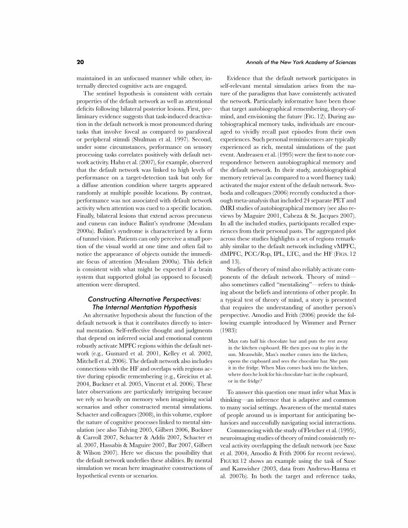
20 Annals of the New York Academy of Sciences
maintained in an unfocused manner while other, in-ternally directed cognitive acts are engaged.
The sentinel hypothesis is consistent with certainproperties of the default network as well as attentionaldeficits following bilateral posterior lesions. First, pre-liminary evidence suggests that task-induced deactiva-tion in the default network is most pronounced duringtasks that involve foveal as compared to parafovealor peripheral stimuli (Shulman et al. 1997). Second,under some circumstances, performance on sensoryprocessing tasks correlates positively with default net-work activity. Hahn et al. (2007), for example, observedthat the default network was linked to high levels ofperformance on a target-detection task but only fora diffuse attention condition where targets appearedrandomly at multiple possible locations. By contrast,performance was not associated with default networkactivity when attention was cued to a specific location.Finally, bilateral lesions that extend across precuneusand cuneus can induce Balint’s syndrome (Mesulam2000a). Balint’s syndrome is characterized by a formof tunnel vision. Patients can only perceive a small por-tion of the visual world at one time and often fail tonotice the appearance of objects outside the immedi-ate focus of attention (Mesulam 2000a). This deficitis consistent with what might be expected if a brainsystem that supported global (as opposed to focused)attention were disrupted.
Constructing Alternative Perspectives:The Internal Mentation Hypothesis
An alternative hypothesis about the function of thedefault network is that it contributes directly to inter-nal mentation. Self-reflective thought and judgmentsthat depend on inferred social and emotional contentrobustly activate MPFC regions within the default net-work (e.g., Gusnard et al. 2001, Kelley et al. 2002,Mitchell et al. 2006). The default network also includesconnections with the HF and overlaps with regions ac-tive during episodic remembering (e.g., Greicius et al.2004, Buckner et al. 2005, Vincent et al. 2006). Theselater observations are particularly intriguing becausewe rely so heavily on memory when imagining socialscenarios and other constructed mental simulations.Schacter and colleagues (2008), in this volume, explorethe nature of cognitive processes linked to mental sim-ulation (see also Tulving 2005, Gilbert 2006, Buckner& Carroll 2007, Schacter & Addis 2007, Schacter etal. 2007, Hassabis & Maguire 2007, Bar 2007, Gilbert& Wilson 2007). Here we discuss the possibility thatthe default network underlies these abilities. By mentalsimulation we mean here imaginative constructions ofhypothetical events or scenarios.
Evidence that the default network participates inself-relevant mental simulation arises from the na-ture of the paradigms that have consistently activatedthe network. Particularly informative have been thosethat target autobiographical remembering, theory-of-mind, and envisioning the future (FIG. 12). During au-tobiographical memory tasks, individuals are encour-aged to vividly recall past episodes from their ownexperiences. Such personal reminiscences are typicallyexperienced as rich, mental simulations of the pastevent. Andreasen et al. (1995) were the first to note cor-respondence between autobiographical memory andthe default network. In their study, autobiographicalmemory retrieval (as compared to a word fluency task)activated the major extent of the default network. Svo-boda and colleagues (2006) recently conducted a thor-ough meta-analysis that included 24 separate PET andfMRI studies of autobiographical memory (see also re-views by Maguire 2001, Cabeza & St. Jacques 2007).In all the included studies, participants recalled expe-riences from their personal pasts. The aggregated plotacross these studies highlights a set of regions remark-ably similar to the default network including vMPFC,dMPFC, PCC/Rsp, IPL, LTC, and the HF (FIGS. 12and 13).
Studies of theory of mind also reliably activate com-ponents of the default network. Theory of mind—also sometimes called “mentalizing”—refers to think-ing about the beliefs and intentions of other people. Ina typical test of theory of mind, a story is presentedthat requires the understanding of another person’sperspective. Amodio and Frith (2006) provide the fol-lowing example introduced by Wimmer and Perner(1983):
Max eats half his chocolate bar and puts the rest awayin the kitchen cupboard. He then goes out to play in thesun. Meanwhile, Max’s mother comes into the kitchen,opens the cupboard and sees the chocolate bar. She putsit in the fridge. When Max comes back into the kitchen,where does he look for his chocolate bar: in the cupboard,or in the fridge?
To answer this question one must infer what Max isthinking—an inference that is adaptive and commonto many social settings. Awareness of the mental statesof people around us is important for anticipating be-haviors and successfully navigating social interactions.
Commencing with the study of Fletcher et al. (1995),neuroimaging studies of theory of mind consistently re-veal activity overlapping the default network (see Saxeet al. 2004, Amodio & Frith 2006 for recent reviews).FIGURE 12 shows an example using the task of Saxeand Kanwisher (2003, data from Andrews-Hanna etal. 2007b). In both the target and reference tasks,
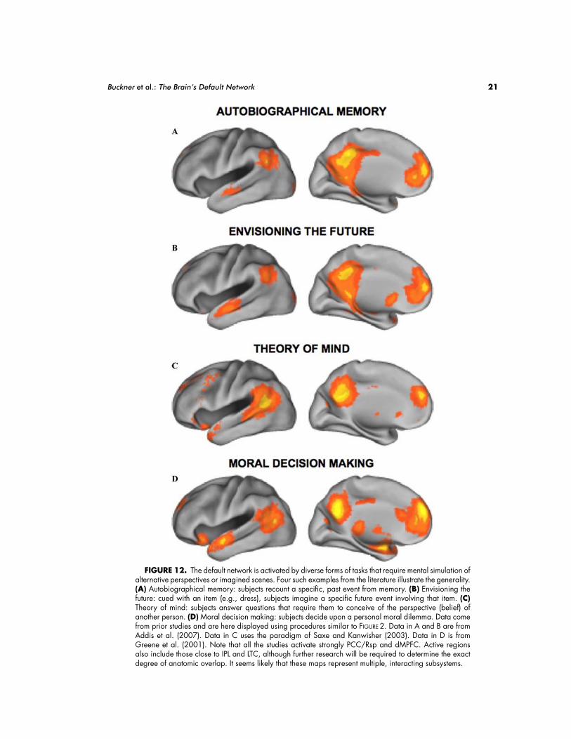
Buckner et al.: The Brain’s Default Network 21
FIGURE 12. The default network is activated by diverse forms of tasks that require mental simulation ofalternative perspectives or imagined scenes. Four such examples from the literature illustrate the generality.(A) Autobiographical memory: subjects recount a specific, past event from memory. (B) Envisioning thefuture: cued with an item (e.g., dress), subjects imagine a specific future event involving that item. (C)Theory of mind: subjects answer questions that require them to conceive of the perspective (belief) ofanother person. (D) Moral decision making: subjects decide upon a personal moral dilemma. Data comefrom prior studies and are here displayed using procedures similar to FIGURE 2. Data in A and B are fromAddis et al. (2007). Data in C uses the paradigm of Saxe and Kanwisher (2003). Data in D is fromGreene et al. (2001). Note that all the studies activate strongly PCC/Rsp and dMPFC. Active regionsalso include those close to IPL and LTC, although further research will be required to determine the exactdegree of anatomic overlap. It seems likely that these maps represent multiple, interacting subsystems.
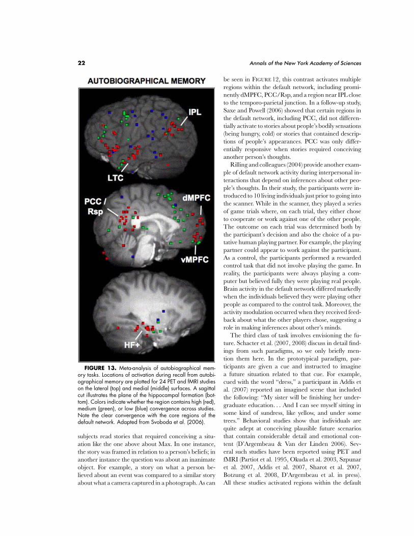
22 Annals of the New York Academy of Sciences
FIGURE 13. Meta-analysis of autobiographical mem-ory tasks. Locations of activation during recall from autobi-ographical memory are plotted for 24 PET and fMRI studieson the lateral (top) and medial (middle) surfaces. A sagittalcut illustrates the plane of the hippocampal formation (bot-tom). Colors indicate whether the region contains high (red),medium (green), or low (blue) convergence across studies.Note the clear convergence with the core regions of thedefault network. Adapted from Svoboda et al. (2006).
subjects read stories that required conceiving a situ-ation like the one above about Max. In one instance,the story was framed in relation to a person’s beliefs; inanother instance the question was about an inanimateobject. For example, a story on what a person be-lieved about an event was compared to a similar storyabout what a camera captured in a photograph. As can
be seen in FIGURE 12, this contrast activates multipleregions within the default network, including promi-nently dMPFC, PCC/Rsp, and a region near IPL closeto the temporo-parietal junction. In a follow-up study,Saxe and Powell (2006) showed that certain regions inthe default network, including PCC, did not differen-tially activate to stories about people’s bodily sensations(being hungry, cold) or stories that contained descrip-tions of people’s appearances. PCC was only differ-entially responsive when stories required conceivinganother person’s thoughts.
Rilling and colleagues (2004) provide another exam-ple of default network activity during interpersonal in-teractions that depend on inferences about other peo-ple’s thoughts. In their study, the participants were in-troduced to 10 living individuals just prior to going intothe scanner. While in the scanner, they played a seriesof game trials where, on each trial, they either choseto cooperate or work against one of the other people.The outcome on each trial was determined both bythe participant’s decision and also the choice of a pu-tative human playing partner. For example, the playingpartner could appear to work against the participant.As a control, the participants performed a rewardedcontrol task that did not involve playing the game. Inreality, the participants were always playing a com-puter but believed fully they were playing real people.Brain activity in the default network differed markedlywhen the individuals believed they were playing otherpeople as compared to the control task. Moreover, theactivity modulation occurred when they received feed-back about what the other players chose, suggesting arole in making inferences about other’s minds.
The third class of task involves envisioning the fu-ture. Schacter et al. (2007, 2008) discuss in detail find-ings from such paradigms, so we only briefly men-tion them here. In the prototypical paradigm, par-ticipants are given a cue and instructed to imaginea future situation related to that cue. For example,cued with the word “dress,” a participant in Addis etal. (2007) reported an imagined scene that includedthe following: “My sister will be finishing her under-graduate education. . . And I can see myself sitting insome kind of sundress, like yellow, and under sometrees.” Behavioral studies show that individuals arequite adept at conceiving plausible future scenariosthat contain considerable detail and emotional con-tent (D’Argembeau & Van der Linden 2006). Sev-eral such studies have been reported using PET andfMRI (Partiot et al. 1995, Okuda et al. 2003, Szpunaret al. 2007, Addis et al. 2007, Sharot et al. 2007,Botzung et al. 2008, D’Argembeau et al. in press).All these studies activated regions within the default
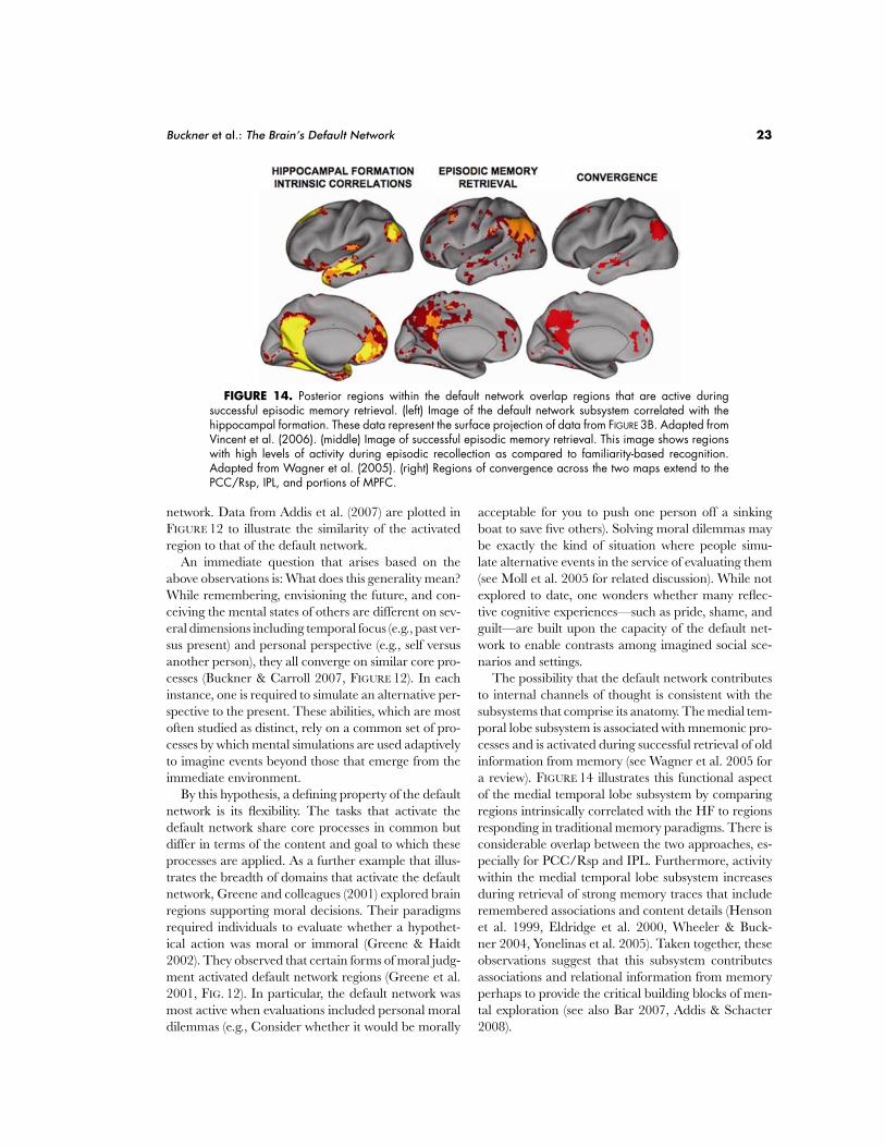
Buckner et al.: The Brain’s Default Network 23
FIGURE 14. Posterior regions within the default network overlap regions that are active duringsuccessful episodic memory retrieval. (left) Image of the default network subsystem correlated with thehippocampal formation. These data represent the surface projection of data from FIGURE 3B. Adapted fromVincent et al. (2006). (middle) Image of successful episodic memory retrieval. This image shows regionswith high levels of activity during episodic recollection as compared to familiarity-based recognition.Adapted from Wagner et al. (2005). (right) Regions of convergence across the two maps extend to thePCC/Rsp, IPL, and portions of MPFC.
network. Data from Addis et al. (2007) are plotted inFIGURE 12 to illustrate the similarity of the activatedregion to that of the default network.
An immediate question that arises based on theabove observations is: What does this generality mean?While remembering, envisioning the future, and con-ceiving the mental states of others are different on sev-eral dimensions including temporal focus (e.g., past ver-sus present) and personal perspective (e.g., self versusanother person), they all converge on similar core pro-cesses (Buckner & Carroll 2007, FIGURE 12). In eachinstance, one is required to simulate an alternative per-spective to the present. These abilities, which are mostoften studied as distinct, rely on a common set of pro-cesses by which mental simulations are used adaptivelyto imagine events beyond those that emerge from theimmediate environment.
By this hypothesis, a defining property of the defaultnetwork is its flexibility. The tasks that activate thedefault network share core processes in common butdiffer in terms of the content and goal to which theseprocesses are applied. As a further example that illus-trates the breadth of domains that activate the defaultnetwork, Greene and colleagues (2001) explored brainregions supporting moral decisions. Their paradigmsrequired individuals to evaluate whether a hypothet-ical action was moral or immoral (Greene & Haidt2002). They observed that certain forms of moral judg-ment activated default network regions (Greene et al.2001, FIG. 12). In particular, the default network wasmost active when evaluations included personal moraldilemmas (e.g., Consider whether it would be morally
acceptable for you to push one person off a sinkingboat to save five others). Solving moral dilemmas maybe exactly the kind of situation where people simu-late alternative events in the service of evaluating them(see Moll et al. 2005 for related discussion). While notexplored to date, one wonders whether many reflec-tive cognitive experiences—such as pride, shame, andguilt—are built upon the capacity of the default net-work to enable contrasts among imagined social sce-narios and settings.
The possibility that the default network contributesto internal channels of thought is consistent with thesubsystems that comprise its anatomy. The medial tem-poral lobe subsystem is associated with mnemonic pro-cesses and is activated during successful retrieval of oldinformation from memory (see Wagner et al. 2005 fora review). FIGURE 14 illustrates this functional aspectof the medial temporal lobe subsystem by comparingregions intrinsically correlated with the HF to regionsresponding in traditional memory paradigms. There isconsiderable overlap between the two approaches, es-pecially for PCC/Rsp and IPL. Furthermore, activitywithin the medial temporal lobe subsystem increasesduring retrieval of strong memory traces that includeremembered associations and content details (Hensonet al. 1999, Eldridge et al. 2000, Wheeler & Buck-ner 2004, Yonelinas et al. 2005). Taken together, theseobservations suggest that this subsystem contributesassociations and relational information from memoryperhaps to provide the critical building blocks of men-tal exploration (see also Bar 2007, Addis & Schacter2008).
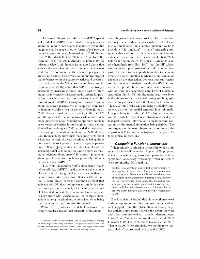
24 Annals of the New York Academy of Sciences
The second subsystem is linked to the MPFC, specif-ically dMPFC. dMPFC is activated by many task situ-ations that require participants to make self-referentialjudgments and engage in other forms of self-relevantmental exploration (e.g., Gusnard et al. 2001, Kelleyet al. 2002, Mitchell et al. 2006, see Adolphs 2003,Ramnani & Owen 2004, Amodio & Frith 2006 forrelevant reviews). All the task forms noted above thatactivate the complete, or near complete, default net-work share in common that the imagined perspectivesare self-referenced. Moreover, several findings suggestthat reference to the self causes selective and preferen-tial activity within the MPFC subsystem. For example,Szpunar et al. (2007) noted that MPFC was stronglyactivated by envisioning oneself in the past or futurebut not so for considering a personally unfamiliar pub-lic figure in a future setting. Saxe and Kanwisher (2003)showed greater dMPFC activity for making decisionsabout conceived perspectives of people as comparedto inanimate objects (e.g., a camera). Guroglu et al.(2008) demonstrated increased activity in the dMPFCand throughout the default network when individualsmade judgments about whether to approach familiarpeers versus celebrities in an imagined social setting.Mitchell and colleagues (2006) provided a particularlyclear example of modulation along the “self” dimen-sion. In their study, individuals made judgments abouta fictitious person who was described as being eitherquite similar in sociopolitical views to the participant orquite different. Judgments made about similar othersactivated dMPFC to about the same degree as mak-ing a judgment about oneself. In contrast, judgmentsabout people perceived as being politically differentdid not activate dMPFC.d
Thus, while it is admittedly difficult to define what isself or self-like, dMPFC is activated when the contentof an imagined setting involves social agents that arebeing considered as such. Note that a subtle distinc-tion is being drawn here: the common element thatactivates dMPFC does not appear to simply be refer-ence to a person or oneself, which can occur devoidof elaborated context. The common element appearsto align more with thinking about the complex inter-actions among people that are conceived of as beingsocial, interactive, and emotive like oneself.
Within this hypothesis, the default network thuscomprises at least two distinct interacting subsystems—
d Dorsal and ventral are relative terms and are used variably dependingon which regions are being compared. This paper defines dMPFC andvMPFC differently than did Mitchell et al. (2006). The region labeled hereas dMPFC is the region Mitchell et al. describe as being ventral.
one subsystem functions to provide information frommemory; the second participates to derive self-relevantmental simulations. The adaptive function may be toprovide a “life simulator”—a set of interacting sub-systems that can use past experiences to explore andanticipate social and event scenarios (Gilbert 2006,Gilbert & Wilson 2007). This idea is similar to a re-cent hypothesis from Bar (2007) that the HF subsys-tem serves to supply associations and analogies frompast experience to make predictions about upcomingevents. An open question is when mental simulationdepends on the interactions between both subsystems.As the functional analysis reveals, the dMPFC andmedial temporal lobe are not intrinsically correlatedwith one another, suggesting some level of functionalseparation (FIG. 8). Certain situations draw heavily onboth subsystems such as elicited during autobiograph-ical memory tasks and when thinking about the future.Theory-of-mind tasks, while utilizing the dMPFC sub-system, activate the medial temporal lobe minimally.One possibility is that the dMPFC subsystem interactswith the medial temporal lobe subsystem to the degreethat past episodic information is an important con-straint on the mental simulation being derived. Theconvergence of the two subsystems on common hubs,in particular PCC, may serve to prepare the system forthese critical interactions.
Competitive Functional InteractionsWhen initially considering the possibility of a brain
system for internal mentation, Ingvar (1979) proposedthat such a system might work in opposition to thosespecialized for sensory processing, which he termed“sensory-gnostic.” He noted that
the low flow/activity in postcentral sensory-gnostic re-gions appears to agree with a low general awareness ofthe sensory input from the immediate surroundings, whenone is left to oneself, undisturbed, resting awake. Possiblythe lower postcentral [flow] signals that the resting con-sciousness implies an active global inhibition of a sensoryinput, as if the brain filtered out trivial information inorder to let the mind be busy with its own consciousness(p. 20).
The idea that the brain’s default network may workin direct opposition to other systems has received re-cent support from the observation of strong nega-tive activity correlations between the default networkand other systems—coined variably “dynamic equi-librium” and “anticorrelations” (Greicius et al. 2003,Fransson 2005, Fox et al. 2005, Golland et al. 2007,Tian et al. 2007). For simplicity, we use the term “an-ticorrelation” as proposed by Fox et al. (2005).
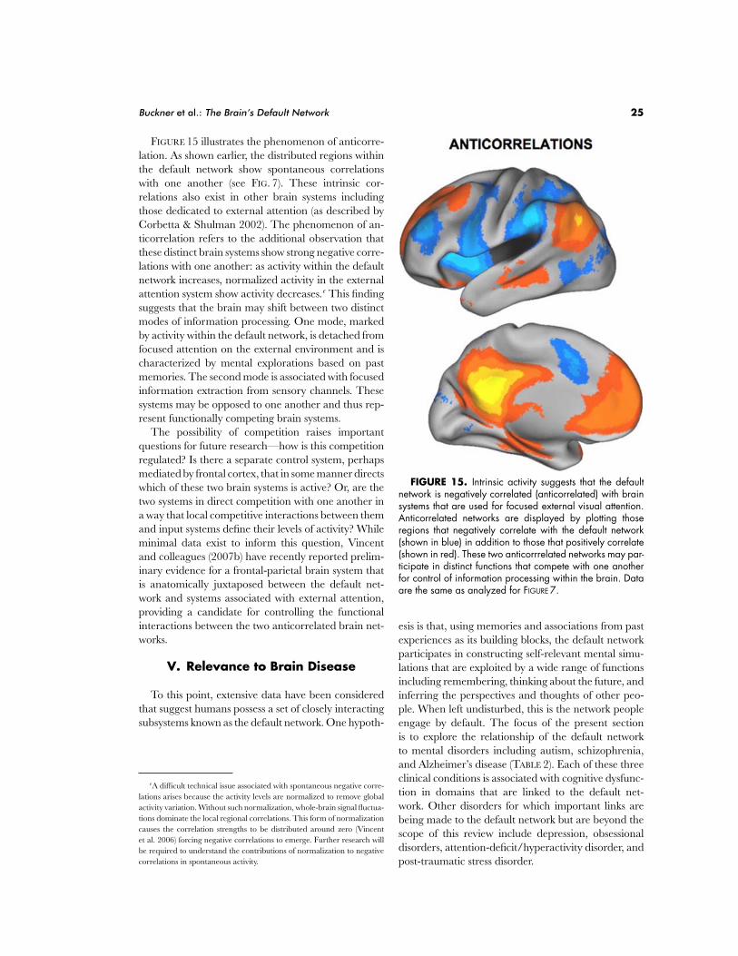
Buckner et al.: The Brain’s Default Network 25
FIGURE 15 illustrates the phenomenon of anticorre-lation. As shown earlier, the distributed regions withinthe default network show spontaneous correlationswith one another (see FIG. 7). These intrinsic cor-relations also exist in other brain systems includingthose dedicated to external attention (as described byCorbetta & Shulman 2002). The phenomenon of an-ticorrelation refers to the additional observation thatthese distinct brain systems show strong negative corre-lations with one another: as activity within the defaultnetwork increases, normalized activity in the externalattention system show activity decreases.e This findingsuggests that the brain may shift between two distinctmodes of information processing. One mode, markedby activity within the default network, is detached fromfocused attention on the external environment and ischaracterized by mental explorations based on pastmemories. The second mode is associated with focusedinformation extraction from sensory channels. Thesesystems may be opposed to one another and thus rep-resent functionally competing brain systems.
The possibility of competition raises importantquestions for future research—how is this competitionregulated? Is there a separate control system, perhapsmediated by frontal cortex, that in some manner directswhich of these two brain systems is active? Or, are thetwo systems in direct competition with one another ina way that local competitive interactions between themand input systems define their levels of activity? Whileminimal data exist to inform this question, Vincentand colleagues (2007b) have recently reported prelim-inary evidence for a frontal-parietal brain system thatis anatomically juxtaposed between the default net-work and systems associated with external attention,providing a candidate for controlling the functionalinteractions between the two anticorrelated brain net-works.
V. Relevance to Brain Disease
To this point, extensive data have been consideredthat suggest humans possess a set of closely interactingsubsystems known as the default network. One hypoth-
eA difficult technical issue associated with spontaneous negative corre-lations arises because the activity levels are normalized to remove globalactivity variation. Without such normalization, whole-brain signal fluctua-tions dominate the local regional correlations. This form of normalizationcauses the correlation strengths to be distributed around zero (Vincentet al. 2006) forcing negative correlations to emerge. Further research willbe required to understand the contributions of normalization to negativecorrelations in spontaneous activity.
FIGURE 15. Intrinsic activity suggests that the defaultnetwork is negatively correlated (anticorrelated) with brainsystems that are used for focused external visual attention.Anticorrelated networks are displayed by plotting thoseregions that negatively correlate with the default network(shown in blue) in addition to those that positively correlate(shown in red). These two anticorrrelated networks may par-ticipate in distinct functions that compete with one anotherfor control of information processing within the brain. Dataare the same as analyzed for FIGURE 7.
esis is that, using memories and associations from pastexperiences as its building blocks, the default networkparticipates in constructing self-relevant mental simu-lations that are exploited by a wide range of functionsincluding remembering, thinking about the future, andinferring the perspectives and thoughts of other peo-ple. When left undisturbed, this is the network peopleengage by default. The focus of the present sectionis to explore the relationship of the default networkto mental disorders including autism, schizophrenia,and Alzheimer’s disease (TABLE 2). Each of these threeclinical conditions is associated with cognitive dysfunc-tion in domains that are linked to the default net-work. Other disorders for which important links arebeing made to the default network but are beyond thescope of this review include depression, obsessionaldisorders, attention-deficit/hyperactivity disorder, andpost-traumatic stress disorder.
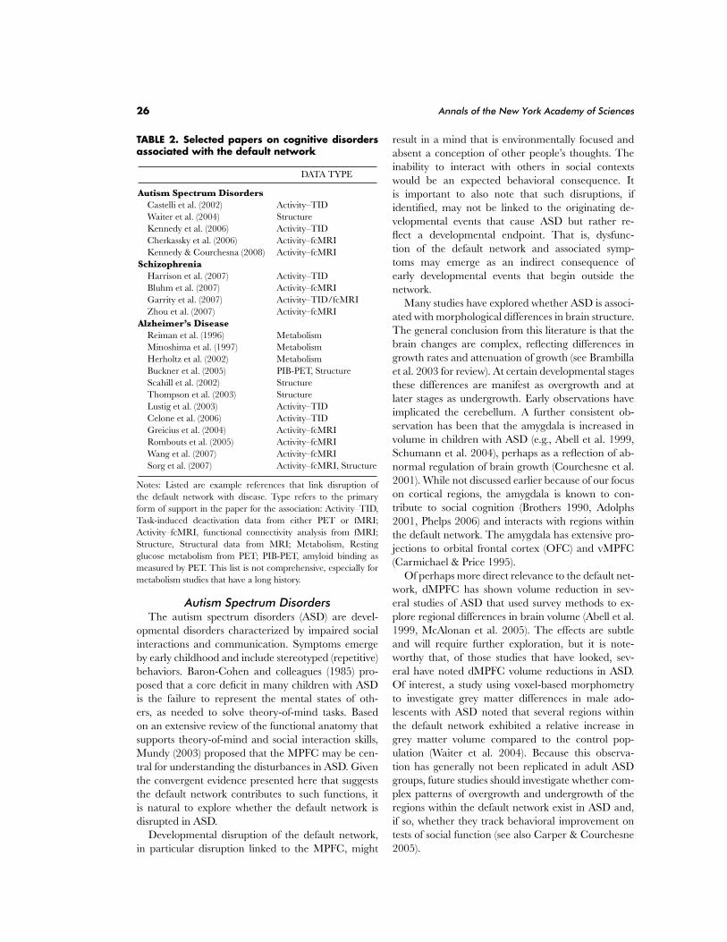
26 Annals of the New York Academy of Sciences
TABLE 2. Selected papers on cognitive disordersassociated with the default network
DATA TYPE
Autism Spectrum DisordersCastelli et al. (2002) Activity–TIDWaiter et al. (2004) StructureKennedy et al. (2006) Activity–TIDCherkassky et al. (2006) Activity–fcMRIKennedy & Courchesna (2008) Activity–fcMRI
SchizophreniaHarrison et al. (2007) Activity–TIDBluhm et al. (2007) Activity–fcMRIGarrity et al. (2007) Activity–TID/fcMRIZhou et al. (2007) Activity–fcMRI
Alzheimer’s DiseaseReiman et al. (1996) MetabolismMinoshima et al. (1997) MetabolismHerholtz et al. (2002) MetabolismBuckner et al. (2005) PIB-PET, StructureScahill et al. (2002) StructureThompson et al. (2003) StructureLustig et al. (2003) Activity–TIDCelone et al. (2006) Activity–TIDGreicius et al. (2004) Activity–fcMRIRombouts et al. (2005) Activity–fcMRIWang et al. (2007) Activity–fcMRISorg et al. (2007) Activity–fcMRI, Structure
Notes: Listed are example references that link disruption ofthe default network with disease. Type refers to the primaryform of support in the paper for the association: Activity–TID,Task-induced deactivation data from either PET or fMRI;Activity–fcMRI, functional connectivity analysis from fMRI;Structure, Structural data from MRI; Metabolism, Restingglucose metabolism from PET; PIB-PET, amyloid binding asmeasured by PET. This list is not comprehensive, especially formetabolism studies that have a long history.
Autism Spectrum DisordersThe autism spectrum disorders (ASD) are devel-
opmental disorders characterized by impaired socialinteractions and communication. Symptoms emergeby early childhood and include stereotyped (repetitive)behaviors. Baron-Cohen and colleagues (1985) pro-posed that a core deficit in many children with ASDis the failure to represent the mental states of oth-ers, as needed to solve theory-of-mind tasks. Basedon an extensive review of the functional anatomy thatsupports theory-of-mind and social interaction skills,Mundy (2003) proposed that the MPFC may be cen-tral for understanding the disturbances in ASD. Giventhe convergent evidence presented here that suggeststhe default network contributes to such functions, itis natural to explore whether the default network isdisrupted in ASD.
Developmental disruption of the default network,in particular disruption linked to the MPFC, might
result in a mind that is environmentally focused andabsent a conception of other people’s thoughts. Theinability to interact with others in social contextswould be an expected behavioral consequence. Itis important to also note that such disruptions, ifidentified, may not be linked to the originating de-velopmental events that cause ASD but rather re-flect a developmental endpoint. That is, dysfunc-tion of the default network and associated symp-toms may emerge as an indirect consequence ofearly developmental events that begin outside thenetwork.
Many studies have explored whether ASD is associ-ated with morphological differences in brain structure.The general conclusion from this literature is that thebrain changes are complex, reflecting differences ingrowth rates and attenuation of growth (see Brambillaet al. 2003 for review). At certain developmental stagesthese differences are manifest as overgrowth and atlater stages as undergrowth. Early observations haveimplicated the cerebellum. A further consistent ob-servation has been that the amygdala is increased involume in children with ASD (e.g., Abell et al. 1999,Schumann et al. 2004), perhaps as a reflection of ab-normal regulation of brain growth (Courchesne et al.2001). While not discussed earlier because of our focuson cortical regions, the amygdala is known to con-tribute to social cognition (Brothers 1990, Adolphs2001, Phelps 2006) and interacts with regions withinthe default network. The amygdala has extensive pro-jections to orbital frontal cortex (OFC) and vMPFC(Carmichael & Price 1995).
Of perhaps more direct relevance to the default net-work, dMPFC has shown volume reduction in sev-eral studies of ASD that used survey methods to ex-plore regional differences in brain volume (Abell et al.1999, McAlonan et al. 2005). The effects are subtleand will require further exploration, but it is note-worthy that, of those studies that have looked, sev-eral have noted dMPFC volume reductions in ASD.Of interest, a study using voxel-based morphometryto investigate grey matter differences in male ado-lescents with ASD noted that several regions withinthe default network exhibited a relative increase ingrey matter volume compared to the control pop-ulation (Waiter et al. 2004). Because this observa-tion has generally not been replicated in adult ASDgroups, future studies should investigate whether com-plex patterns of overgrowth and undergrowth of theregions within the default network exist in ASD and,if so, whether they track behavioral improvement ontests of social function (see also Carper & Courchesne2005).
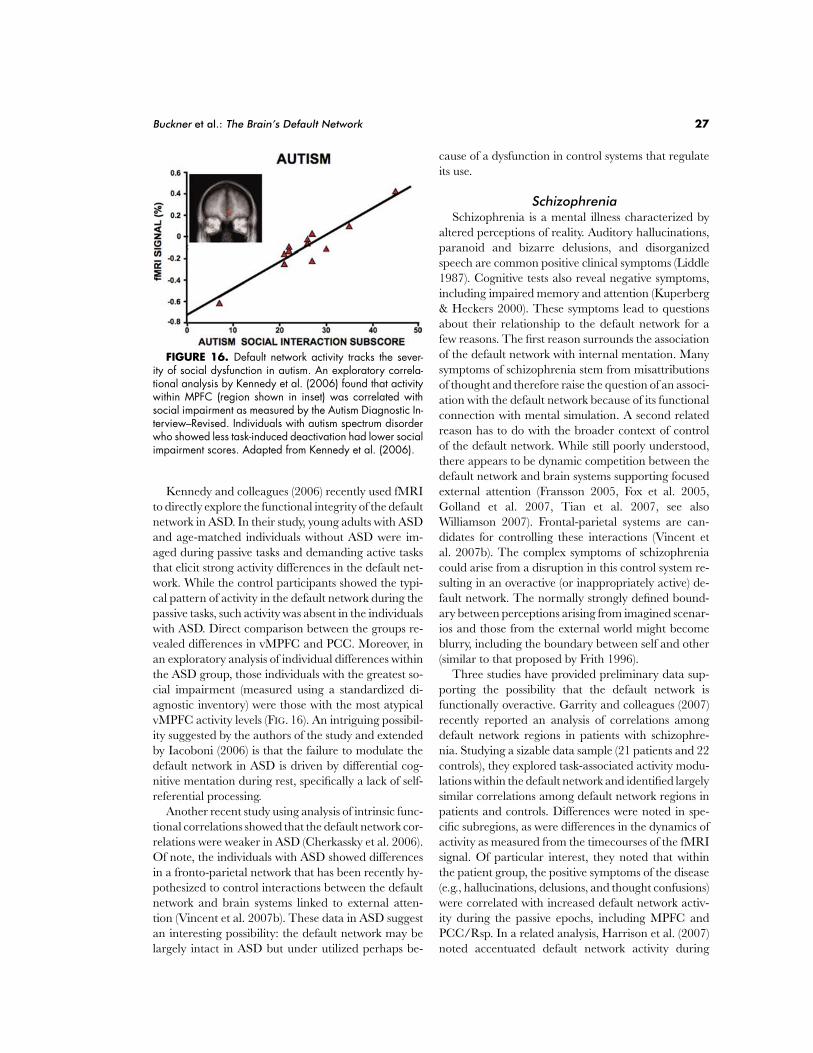
Buckner et al.: The Brain’s Default Network 27
FIGURE 16. Default network activity tracks the sever-ity of social dysfunction in autism. An exploratory correla-tional analysis by Kennedy et al. (2006) found that activitywithin MPFC (region shown in inset) was correlated withsocial impairment as measured by the Autism Diagnostic In-terview–Revised. Individuals with autism spectrum disorderwho showed less task-induced deactivation had lower socialimpairment scores. Adapted from Kennedy et al. (2006).
Kennedy and colleagues (2006) recently used fMRIto directly explore the functional integrity of the defaultnetwork in ASD. In their study, young adults with ASDand age-matched individuals without ASD were im-aged during passive tasks and demanding active tasksthat elicit strong activity differences in the default net-work. While the control participants showed the typi-cal pattern of activity in the default network during thepassive tasks, such activity was absent in the individualswith ASD. Direct comparison between the groups re-vealed differences in vMPFC and PCC. Moreover, inan exploratory analysis of individual differences withinthe ASD group, those individuals with the greatest so-cial impairment (measured using a standardized di-agnostic inventory) were those with the most atypicalvMPFC activity levels (FIG. 16). An intriguing possibil-ity suggested by the authors of the study and extendedby Iacoboni (2006) is that the failure to modulate thedefault network in ASD is driven by differential cog-nitive mentation during rest, specifically a lack of self-referential processing.
Another recent study using analysis of intrinsic func-tional correlations showed that the default network cor-relations were weaker in ASD (Cherkassky et al. 2006).Of note, the individuals with ASD showed differencesin a fronto-parietal network that has been recently hy-pothesized to control interactions between the defaultnetwork and brain systems linked to external atten-tion (Vincent et al. 2007b). These data in ASD suggestan interesting possibility: the default network may belargely intact in ASD but under utilized perhaps be-
cause of a dysfunction in control systems that regulateits use.
SchizophreniaSchizophrenia is a mental illness characterized by
altered perceptions of reality. Auditory hallucinations,paranoid and bizarre delusions, and disorganizedspeech are common positive clinical symptoms (Liddle1987). Cognitive tests also reveal negative symptoms,including impaired memory and attention (Kuperberg& Heckers 2000). These symptoms lead to questionsabout their relationship to the default network for afew reasons. The first reason surrounds the associationof the default network with internal mentation. Manysymptoms of schizophrenia stem from misattributionsof thought and therefore raise the question of an associ-ation with the default network because of its functionalconnection with mental simulation. A second relatedreason has to do with the broader context of controlof the default network. While still poorly understood,there appears to be dynamic competition between thedefault network and brain systems supporting focusedexternal attention (Fransson 2005, Fox et al. 2005,Golland et al. 2007, Tian et al. 2007, see alsoWilliamson 2007). Frontal-parietal systems are can-didates for controlling these interactions (Vincent etal. 2007b). The complex symptoms of schizophreniacould arise from a disruption in this control system re-sulting in an overactive (or inappropriately active) de-fault network. The normally strongly defined bound-ary between perceptions arising from imagined scenar-ios and those from the external world might becomeblurry, including the boundary between self and other(similar to that proposed by Frith 1996).
Three studies have provided preliminary data sup-porting the possibility that the default network isfunctionally overactive. Garrity and colleagues (2007)recently reported an analysis of correlations amongdefault network regions in patients with schizophre-nia. Studying a sizable data sample (21 patients and 22controls), they explored task-associated activity modu-lations within the default network and identified largelysimilar correlations among default network regions inpatients and controls. Differences were noted in spe-cific subregions, as were differences in the dynamics ofactivity as measured from the timecourses of the fMRIsignal. Of particular interest, they noted that withinthe patient group, the positive symptoms of the disease(e.g., hallucinations, delusions, and thought confusions)were correlated with increased default network activ-ity during the passive epochs, including MPFC andPCC/Rsp. In a related analysis, Harrison et al. (2007)noted accentuated default network activity during
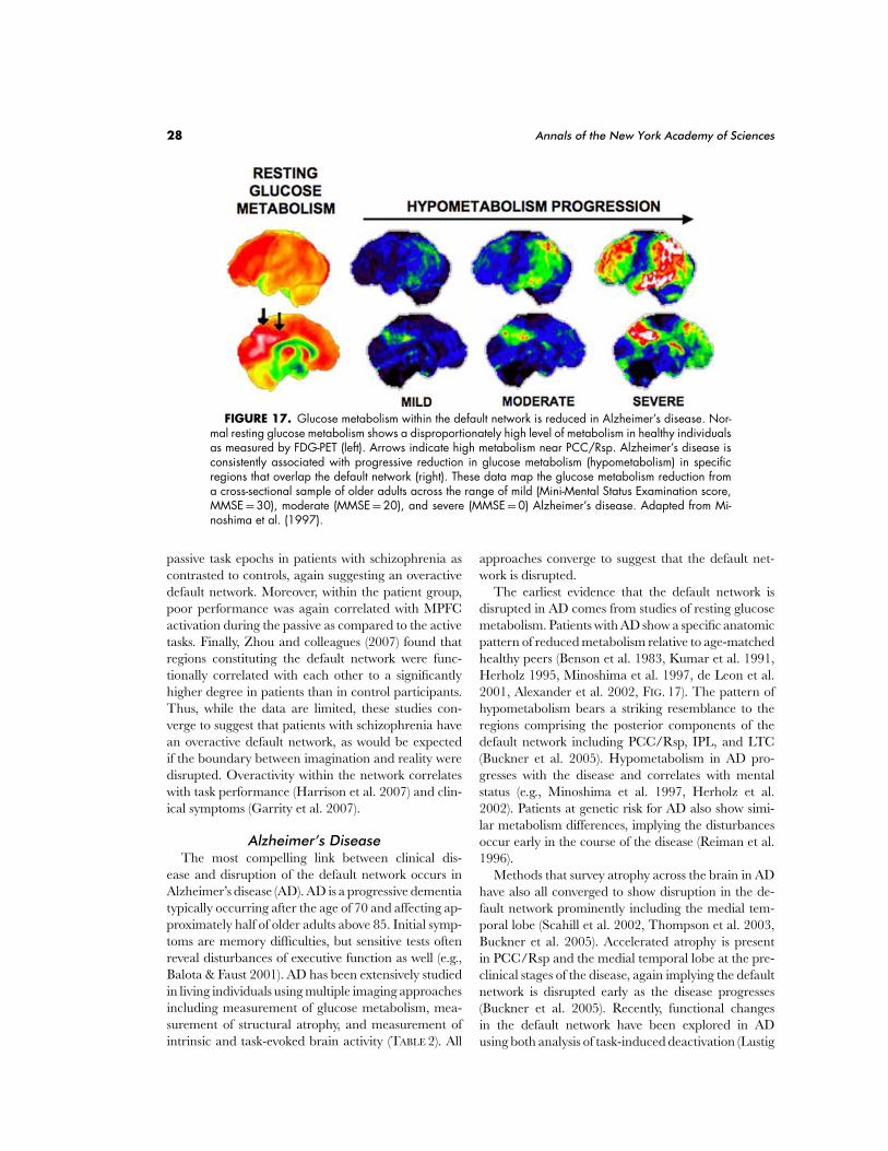
28 Annals of the New York Academy of Sciences
FIGURE 17. Glucose metabolism within the default network is reduced in Alzheimer’s disease. Nor-mal resting glucose metabolism shows a disproportionately high level of metabolism in healthy individualsas measured by FDG-PET (left). Arrows indicate high metabolism near PCC/Rsp. Alzheimer’s disease isconsistently associated with progressive reduction in glucose metabolism (hypometabolism) in specificregions that overlap the default network (right). These data map the glucose metabolism reduction froma cross-sectional sample of older adults across the range of mild (Mini-Mental Status Examination score,MMSE = 30), moderate (MMSE = 20), and severe (MMSE = 0) Alzheimer’s disease. Adapted from Mi-noshima et al. (1997).
passive task epochs in patients with schizophrenia ascontrasted to controls, again suggesting an overactivedefault network. Moreover, within the patient group,poor performance was again correlated with MPFCactivation during the passive as compared to the activetasks. Finally, Zhou and colleagues (2007) found thatregions constituting the default network were func-tionally correlated with each other to a significantlyhigher degree in patients than in control participants.Thus, while the data are limited, these studies con-verge to suggest that patients with schizophrenia havean overactive default network, as would be expectedif the boundary between imagination and reality weredisrupted. Overactivity within the network correlateswith task performance (Harrison et al. 2007) and clin-ical symptoms (Garrity et al. 2007).
Alzheimer’s DiseaseThe most compelling link between clinical dis-
ease and disruption of the default network occurs inAlzheimer’s disease (AD). AD is a progressive dementiatypically occurring after the age of 70 and affecting ap-proximately half of older adults above 85. Initial symp-toms are memory difficulties, but sensitive tests oftenreveal disturbances of executive function as well (e.g.,Balota & Faust 2001). AD has been extensively studiedin living individuals using multiple imaging approachesincluding measurement of glucose metabolism, mea-surement of structural atrophy, and measurement ofintrinsic and task-evoked brain activity (TABLE 2). All
approaches converge to suggest that the default net-work is disrupted.
The earliest evidence that the default network isdisrupted in AD comes from studies of resting glucosemetabolism. Patients with AD show a specific anatomicpattern of reduced metabolism relative to age-matchedhealthy peers (Benson et al. 1983, Kumar et al. 1991,Herholz 1995, Minoshima et al. 1997, de Leon et al.2001, Alexander et al. 2002, FIG. 17). The pattern ofhypometabolism bears a striking resemblance to theregions comprising the posterior components of thedefault network including PCC/Rsp, IPL, and LTC(Buckner et al. 2005). Hypometabolism in AD pro-gresses with the disease and correlates with mentalstatus (e.g., Minoshima et al. 1997, Herholz et al.2002). Patients at genetic risk for AD also show simi-lar metabolism differences, implying the disturbancesoccur early in the course of the disease (Reiman et al.1996).
Methods that survey atrophy across the brain in ADhave also all converged to show disruption in the de-fault network prominently including the medial tem-poral lobe (Scahill et al. 2002, Thompson et al. 2003,Buckner et al. 2005). Accelerated atrophy is presentin PCC/Rsp and the medial temporal lobe at the pre-clinical stages of the disease, again implying the defaultnetwork is disrupted early as the disease progresses(Buckner et al. 2005). Recently, functional changesin the default network have been explored in ADusing both analysis of task-induced deactivation (Lustig
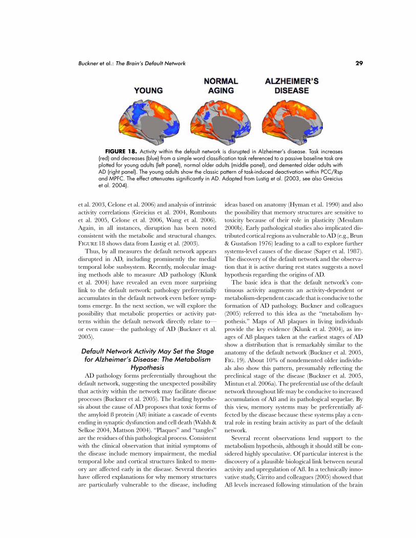
Buckner et al.: The Brain’s Default Network 29
FIGURE 18. Activity within the default network is disrupted in Alzheimer’s disease. Task increases(red) and decreases (blue) from a simple word classification task referenced to a passive baseline task areplotted for young adults (left panel), normal older adults (middle panel), and demented older adults withAD (right panel). The young adults show the classic pattern of task-induced deactivation within PCC/Rspand MPFC. The effect attenuates significantly in AD. Adapted from Lustig et al. (2003, see also Greiciuset al. 2004).
et al. 2003, Celone et al. 2006) and analysis of intrinsicactivity correlations (Greicius et al. 2004, Romboutset al. 2005, Celone et al. 2006, Wang et al. 2006).Again, in all instances, disruption has been notedconsistent with the metabolic and structural changes.FIGURE 18 shows data from Lustig et al. (2003).
Thus, by all measures the default network appearsdisrupted in AD, including prominently the medialtemporal lobe susbsystem. Recently, molecular imag-ing methods able to measure AD pathology (Klunket al. 2004) have revealed an even more surprisinglink to the default network: pathology preferentiallyaccumulates in the default network even before symp-toms emerge. In the next section, we will explore thepossibility that metabolic properties or activity pat-terns within the default network directly relate to—or even cause—the pathology of AD (Buckner et al.2005).
Default Network Activity May Set the Stagefor Alzheimer’s Disease: The Metabolism
HypothesisAD pathology forms preferentially throughout the
default network, suggesting the unexpected possibilitythat activity within the network may facilitate diseaseprocesses (Buckner et al. 2005). The leading hypothe-sis about the cause of AD proposes that toxic forms ofthe amyloid ß protein (Aß) initiate a cascade of eventsending in synaptic dysfunction and cell death (Walsh &Selkoe 2004, Mattson 2004). “Plaques” and “tangles”are the residues of this pathological process. Consistentwith the clinical observation that initial symptoms ofthe disease include memory impairment, the medialtemporal lobe and cortical structures linked to mem-ory are affected early in the disease. Several theorieshave offered explanations for why memory structuresare particularly vulnerable to the disease, including
ideas based on anatomy (Hyman et al. 1990) and alsothe possibility that memory structures are sensitive totoxicity because of their role in plasticity (Mesulam2000b). Early pathological studies also implicated dis-tributed cortical regions as vulnerable to AD (e.g., Brun& Gustafson 1976) leading to a call to explore furthersystems-level causes of the disease (Saper et al. 1987).The discovery of the default network and the observa-tion that it is active during rest states suggests a novelhypothesis regarding the origins of AD.
The basic idea is that the default network’s con-tinuous activity augments an activity-dependent ormetabolism-dependent cascade that is conducive to theformation of AD pathology. Buckner and colleagues(2005) referred to this idea as the “metabolism hy-pothesis.” Maps of Aß plaques in living individualsprovide the key evidence (Klunk et al. 2004), as im-ages of Aß plaques taken at the earliest stages of ADshow a distribution that is remarkably similar to theanatomy of the default network (Buckner et al. 2005,FIG. 19). About 10% of nondemented older individu-als also show this pattern, presumably reflecting thepreclinical stage of the disease (Buckner et al. 2005,Mintun et al. 2006a). The preferential use of the defaultnetwork throughout life may be conducive to increasedaccumulation of Aß and its pathological sequelae. Bythis view, memory systems may be preferentially af-fected by the disease because these systems play a cen-tral role in resting brain activity as part of the defaultnetwork.
Several recent observations lend support to themetabolism hypothesis, although it should still be con-sidered highly speculative. Of particular interest is thediscovery of a plausible biological link between neuralactivity and upregulation of Aß. In a technically inno-vative study, Cirrito and colleagues (2005) showed thatAß levels increased following stimulation of the brain
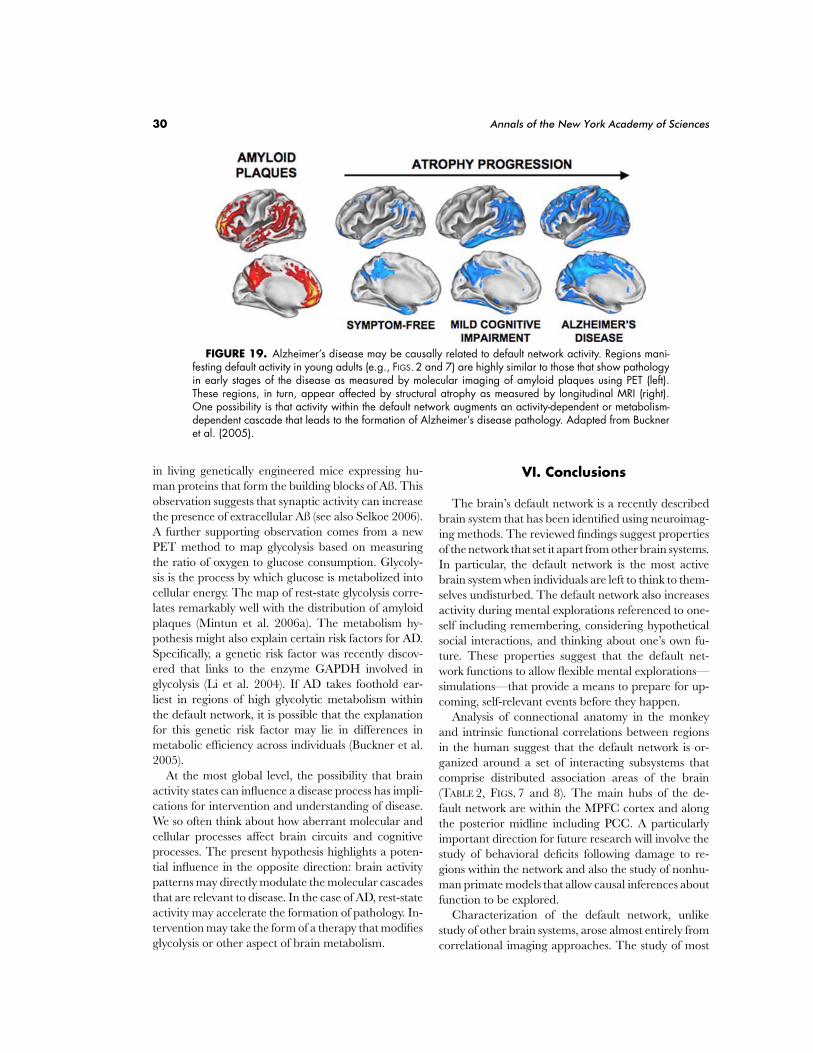
30 Annals of the New York Academy of Sciences
FIGURE 19. Alzheimer’s disease may be causally related to default network activity. Regions mani-festing default activity in young adults (e.g., FIGS. 2 and 7) are highly similar to those that show pathologyin early stages of the disease as measured by molecular imaging of amyloid plaques using PET (left).These regions, in turn, appear affected by structural atrophy as measured by longitudinal MRI (right).One possibility is that activity within the default network augments an activity-dependent or metabolism-dependent cascade that leads to the formation of Alzheimer’s disease pathology. Adapted from Buckneret al. (2005).
in living genetically engineered mice expressing hu-man proteins that form the building blocks of Aß. Thisobservation suggests that synaptic activity can increasethe presence of extracellular Aß (see also Selkoe 2006).A further supporting observation comes from a newPET method to map glycolysis based on measuringthe ratio of oxygen to glucose consumption. Glycoly-sis is the process by which glucose is metabolized intocellular energy. The map of rest-state glycolysis corre-lates remarkably well with the distribution of amyloidplaques (Mintun et al. 2006a). The metabolism hy-pothesis might also explain certain risk factors for AD.Specifically, a genetic risk factor was recently discov-ered that links to the enzyme GAPDH involved inglycolysis (Li et al. 2004). If AD takes foothold ear-liest in regions of high glycolytic metabolism withinthe default network, it is possible that the explanationfor this genetic risk factor may lie in differences inmetabolic efficiency across individuals (Buckner et al.2005).
At the most global level, the possibility that brainactivity states can influence a disease process has impli-cations for intervention and understanding of disease.We so often think about how aberrant molecular andcellular processes affect brain circuits and cognitiveprocesses. The present hypothesis highlights a poten-tial influence in the opposite direction: brain activitypatterns may directly modulate the molecular cascadesthat are relevant to disease. In the case of AD, rest-stateactivity may accelerate the formation of pathology. In-tervention may take the form of a therapy that modifiesglycolysis or other aspect of brain metabolism.
VI. Conclusions
The brain’s default network is a recently describedbrain system that has been identified using neuroimag-ing methods. The reviewed findings suggest propertiesof the network that set it apart from other brain systems.In particular, the default network is the most activebrain system when individuals are left to think to them-selves undisturbed. The default network also increasesactivity during mental explorations referenced to one-self including remembering, considering hypotheticalsocial interactions, and thinking about one’s own fu-ture. These properties suggest that the default net-work functions to allow flexible mental explorations—simulations—that provide a means to prepare for up-coming, self-relevant events before they happen.
Analysis of connectional anatomy in the monkeyand intrinsic functional correlations between regionsin the human suggest that the default network is or-ganized around a set of interacting subsystems thatcomprise distributed association areas of the brain(TABLE 2, FIGS. 7 and 8). The main hubs of the de-fault network are within the MPFC cortex and alongthe posterior midline including PCC. A particularlyimportant direction for future research will involve thestudy of behavioral deficits following damage to re-gions within the network and also the study of nonhu-man primate models that allow causal inferences aboutfunction to be explored.
Characterization of the default network, unlikestudy of other brain systems, arose almost entirely fromcorrelational imaging approaches. The study of most
Buckner et al.: The Brain’s Default Network 31
other brain systems has been initiated by a neurologicalsyndrome and then probed further using animal mod-els and neuroimaging approaches. On the one hand,the discovery of the brain’s default network representsa unique contribution of neuroimaging to cognitiveneuroscience. On the other hand, there have been nolesion studies that motivate their behavioral probesbased on the recent characterization of the network,leaving a large number of questions unanswered. Pro-viding some information, studies of patients with le-sions to regions overlapping the default network arenoted in the present review and also discussed in thecompanion paper of Schacter et al. (2008). However,considerably more work needs to be conducted.
A further open issue is how the default network inter-acts with the distributed brain systems that contributecontent to the process of mental exploration. Studiesof episodic memory retrieval have shown that visualcortex and auditory cortex are preferentially activatedduring the recollection of visual objects and sounds(e.g., Nyberg et al. 2000, Wheeler et al. 2000). Imagin-ing the personal future, which activates the default net-work under many contexts, has also been demonstratedto additionally recruit the anterior temporal cortex(Partiot et al. 1995) and the amygdala (Sharot et al.2007, see also Guroglu et al. 2008) when strong emo-tional context is a component of the upcoming episode.Judgments about inferred emotions have been linkedto regions within the default network (e.g., Ochsneret al. 2004, see also Maddock 1999). One possibility isthat the regions within the default network transientlyinteract with sensory, motor, and emotional systems torepresent the content of the imagined event.
Germane to this possibility, Hassabis and Maguire(2007) recently proposed that interactions among re-gions within the default network may “facilitate the re-trieval and integration of relevant informational com-ponents, stored in their modality-specific cortical areas,the product of which has a coherent spatial context,and can then later be manipulated and visualized.”They refer to this process as “scene construction,” aterm emphasizing that mental simulation often un-folds in one’s mind as an imagined scene with rich vi-sual and spatial content (see also Hassabis et al. 2007).Vogeley and colleagues (2004) have also noted that re-gions within the default network are differentially ac-tive depending on the perspective taken when imaginga scene. The default network is most active when onetakes a first-person perspective centered upon one’sown body as opposed to a third-person perspective.
Perhaps the most intriguing avenue for future ex-ploration surrounds the implication that specific brainsystems are devoted to internal modes of cognition.
To date, cognitive and systems neuroscience has con-cerned itself primarily with how information is ex-tracted from sensory inputs and integrated over timeto make decisions and plan actions. Knowledge thatthe default network exists reminds us that there maybe specialized brain systems that underlie our abilitiesto mentally explore and anticipate future situations.Such constructive processes may be adaptive becausethey allow the brain to preexperience upcoming eventsand to derive prospectively useful forms of representa-tion that are many steps removed from their originallyencoded sources.
Relevant to this possibility, studies of neural activ-ity in the rat hippocampus have recently revealed thatfuture event sequences are the beginnings of journeys(Diba & Buzaki 2007) and choice points (Johhnson& Redish 2007) providing a candidate neural mecha-nism for evaluating the consequences of upcoming ac-tions before they happen (see also Shapiro et al. 2006,Buckner & Carroll 2007). In a series of recent studies,Johnson and Redish (2007) focused on the behaviorof rats at a critical choice point in a maze where theywere confronted with a high-cost decision. The ratshad to follow a path to the right or left, and the in-correct choice required an extended journey to obtainanother chance for reward. By recording from ensem-bles of cells with place fields in the hippocampus, theywere able to visualize the representation of space inthe rat brain at these critical decision junctures. Whatemerged was quite remarkable: when the rats pausedbefore their decision, the neurons fired in patterns thatswept ahead of the location, first down one choice andthen the other. This prospective coding occurred, onaverage, for about 10% of the time the rats were atthe choice point. Moreover, on some trials where therats made decision errors, the representations of spaceswept back toward the choice point and down the pathof the correct journey. Although a direct causal link tothe decision choice has yet to be uncovered, these find-ings suggest a candidate neural mechanism by whichpotential future choices can be simulated in the ratbrain in the service of planning.
The default network’s prominent use during passiveepochs may contribute adaptive function by allowingevent scenarios to be constructed, replayed, and ex-plored to enrich the remnants of past events in orderto derive expectations about the future. This functionalrole may explain why the default network increases itsactivity during passive moments when the demands forprocessing external information are minimal. Ratherthan let the moments pass with idle brain activity, wecapitalize on them to consolidate past experience inways that are adaptive for our future needs.
32 Annals of the New York Academy of Sciences
Acknowledgments
We thank Justin Vincent, Avi Snyder, Peter Fransson,Michael Greicius, Daniel Gilbert, Cindy Lustig, MosheBar, Daphne Holt, Britta Hahn, Marcus Raichle, MikeFox, Jason Mitchell, Michael Miller, and two reviewersfor valuable discussion and comments on the paper.Avi Snyder and Itamar Kahn provided assistance withcomputational techniques for constructing the figures.Data for figures were generously provided by MikeFox, Joshua Greene, Malia Mason, Jason Mitchell,Marcus Raichle, Ben Shannon, Avi Snyder, SatoshiMinoshima, and Justin Vincent. Ben Shannon com-piled the data illustrated in FIGURE 3. Steve Petersenand Alex Cohen contributed the graph analytic visu-alization displayed in FIGURE 8. Katie Powers assistedwith manuscript preparation and Haderer & MullerBiomedical Art illustrated FIGURES 5 and 11. Fund-ing was provided by the National Institute on Aging(AG021910, JAG08441), National Institute of MentalHealth (MH060941), and the Howard Hughes Medi-cal Institute.
Conflict of Interest
The authors declare no conflicts of interest.
References
Abell, F., Krams, M., Ashburner, J., Passingham, R., Friston, K.,et al. (1999). The neuroanatomy of autism: a voxel-basedwhole brain analysis of structural scans. Neuroreport, 10,1647–51.
Addis, D. R., & Schacter, D. L. (2008). Constructive episodic sim-ulation: Temporal distance and detail of past and futureevents modulate hippocampal engagement. Hippocam-pus, 18, 227–237.
Addis, D. R., Wong, A. T., & Schacter, D. L. (2007). Remem-bering the past and imagining the future: common anddistinct neural substrates during event construction andelaboration. Neuropsychologia, 45, 1363–77.
Adolphs, R. (2001). The neurobiology of social cognition. Curr.Opin. Neurobiol., 11, 231–9.
Adolphs, R. (2003). Cognitive neuroscience of human social be-haviour. Nat. Rev. Neurosci., 4, 165–78.
Amodio, D. M., & Frith, C. D. (2006). Meeting of minds: the me-dial frontal cortex and social cognition. Nat. Rev. Neurosci.,7, 268–77.
Andreasen, N. C., O’Leary, D. S., Cizadlo, T., Arndt, S., Rezai,K., et al. (1995). Remembering the past: two facets ofepisodic memory explored with positron emission tomog-raphy. Am. J. Psychiatry, 152, 1576–85.
Andrews-Hanna, J. R., Snyder, A. Z., Vincent, J. L., Lustig, C.,Head, D., et al. (2007a). Disruption of large-scale brainsystems in advanced aging. Neuron, 56, 924–35.
Andrews-Hanna, J. R., Saxe, R., Poulin, R., & Buckner, R. L.(2007b). The default system overlaps activation during
theory of mind and episodic memory retrieval tasks. Soc.Neurosci. Abstr.
Antrobus, J. S. (1968). Information theory and stimulus-independent thought. Br. J. Psychol., 59, 423–30.
Antrobus, J. S., Singer, J. L., & Greenberg, S. (1966). Studies in thestream of consciousness: experimental enhancement andsuppression of spontaneous cognitive processes. Perceptualand Motor Skills, 23, 399–417.
Antrobus, J. S., Singer, J. L., Goldstein, S., & Fortgang, M. (1970).Mindwandering and cognitive structure. Trans. N. Y. Acad.Sci., 32, 242–52.
Alexander, G. E., Chen, K., Pietrini, P., Rapoport, S. I., &Reiman, E. M. (2002). Longitudinal PET evaluation ofcerebral metabolic decline in dementia: a potential out-come measure in Alzheimer’s disease treatment studies.Am. J. Psychiatry, 159, 738–45.
Arieli, A., Shoham, D., Hildesheim, R., & Grinvald, A. (1995).Coherent spatiotemporal patterns of ongoing activity re-vealed by real-time optical imaging coupled with single-unit recording in the cat visual cortex. J. Neurophysiol., 73,2072–93.
Baker, S. C., Rogers, R. D., Owen, A. M., Frith, C. D., Dolan,R. J., et al. (1996). Neural systems engaged by planning: aPET study of the Tower of London task. Neuropsychologia,34, 515–26.
Balota, D. A., & Faust, M. E. (2001). Attention in dementia of theAlzheimer’s type. In F. Boller & S. Cappa (Eds.) Handbookof Neuropsychology VI: Aging and Dementia (pp. 51–80).Amsterdam: Elsevier Science B.V.
Bar, M. (2007). The proactive brain: using analogies and associ-ations to generate predictions. Trends Cogn. Sci., 11, 280–9.
Barbas, H., Ghashghaei, H., Dombrowski, S. M., & Rempel-Clower, N. L. (1999). Medial prefrontal cortices are uni-fied by common connections with superior temporal cor-tices and distinguished by input from memory-related ar-eas in the rhesus monkey. J. Comp. Neurol., 410, 343–67.
Baron-Cohen, S., Leslie, A. M., & Frith, U. (1985). Does theautistic child have a “theory of mind”? Cognition, 21, 37–46.
Benson, D. F., Kuhl, D. E., Hawkins, R. A., Phelps, M. E., Cum-mings, J. L., & Tsai, S. Y. (1983). The fluorodeoxyglucose18F scan in Alzheimer’s disease and multi-infarct demen-tia. Arch. Neurol., 40, 711–4.
Berger, H. (1931/1969). On the electroencephalogram of man:third report. Electroenceph. Clin. Neurophysiol. Supplement No.,28, 95–132.
Binder, J. R., Frost, J. A., Hammeke, T. A., Bellgowan, P. S.,Rao, S. M., & Cox, R. W. (1999). Conceptual processingduring the conscious resting state. a functional MRI study.J. Cogn. Neurosci., 11, 80–95.
Birn, R. M., Diamond, J. B., Smith, M. A., & Bandettini, P. A.(2006). Separating respiratory-variation-related fluctua-tions from neuronal-activity-related fluctuations in fMRI.Neuroimage, 31, 1536–48.
Biswal, B., Yetkin, F. Z., Haughton, V. M., & Hyde, J. S. (1995).Functional connectivity in the motor cortex of restinghuman brain using echo-planar MRI. Magn. Reson. Med.,34, 537–41.
Bluhm, R. L., Miller, J., Lanius, R. A., Osuch, E. A., Boks-man, K., et al. (2007). Spontaneous low-frequency fluc-tuations in the BOLD signal in schizophrenic patients:
Buckner et al.: The Brain’s Default Network 33
anomalies in the default network. Schizophr. Bull., 33, 1004–12.
Botzung, A., Denkova, E., & Manning, L. (2008). Experiencingpast and future personal events: functional neuroimagingevidence on the neural bases of mental time travel. Brainand Cognition, 66 , 202–12.
Brambilla, P., Hardan, A., di Nemi, S. U., Perez, J., Soares, J. C.,& Barale, F. (2003). Brain anatomy and development inautism: review of structural MRI studies. Brain. Res. Bull.,61, 557–69.
Brothers, L. (1990). The social brain: A project for integratingprimate behavior and neurophysiology in a new domain.Concepts Neurosci., 1, 27–51.
Brun, A., & Gustafson, L. (1976). Distribution of cerebral de-generation in Alzheimer’s disease: A clinico-pathologicalstudy. Arch. Psychiat. NervKrankh., 223, 15–33.
Buckner, R. L., Raichle, M. E., Miezin, F. M., & Petersen, S. E.(1996). Functional anatomic studies of memory retrievalfor auditory words and visual pictures. J. Neurosci., 16 ,6219–35.
Buckner, R. L., Snyder, A. Z., Shannon, B. J., LaRossa, G.,Sachs, R., et al. (2005). Molecular, structural, and func-tional characterization of Alzheimer’s disease: evidencefor a relationship between default activity, amyloid, andmemory. J. Neurosci., 25, 7709–17.
Buckner, R. L., & Carroll, D. C. (2007). Self-projection and thebrain. Trends Cogn. Sci., 11, 49–57.
Buckner, R. L., & Vincent, J. L. (2007). Unrest at rest: defaultactivity and spontaneous network correlations. Neuroimage,37, 1091–6.
Cabeza, R., & St Jacques, P. (2007). Functional neuroimaging ofautobiographical memory. Trends Cogn. Sci., 11, 219–27.
Carmichael, S. T., & Price, J. L. (1995). Limbic connections of theorbital and medial prefrontal cortex in macaque monkeys.J. Comp. Neurol., 363, 615–41.
Carper, R. A., & Courchesne, E. (2005). Localized enlargementof the frontal cortex in early autism. Biol. Psychiatry., 57,126–33.
Caspers, S., Geyer, S., Schleicher, A., Mohlberg, H., Amunts, K.,& Zilles, K. (2006). The human inferior parietal cortex: cy-toarchitectonic parcellation and interindividual variabil-ity. Neuroimage, 33, 430–48.
Castelli, F., Frith, C., Happe, F., & Frith, U. (2002). Autism,Asperger syndrome and brain mechanisms for the attri-bution of mental states to animated shapes. Brain, 125,1839–49.
Cavada, C., & Goldman-Rakic, P. S. (1989). Posterior parietalcortex in rhesus monkey: I. parcellation of areas based ondistinctive limbic and sensory corticocortical connections.J. Comp. Neurol., 287, 393–421.
Cavanna, A. E., & Trimble, M. R. (2006). The precuneus: a re-view of its functional anatomy and behavioural correlates.Brain, 129, 564–83.
Celone, K. A., Calhoun, V. D., Dickerson, B. C., Atri, A., Chua,E. F., et al. (2006). Alterations in memory networks inmild cognitive impairment and Alzheimer’s disease: anindependent component analysis. J. Neurosci., 26 , 10222–31.
Cherkassky, V. L., Kana, R. K., Keller, T. A., & Just, M. A.(2006). Functional connectivity in a baseline resting-statenetwork in autism. Neuroreport, 17, 1687–90.
Cirrito, J. R., Yamada, K. A., Finn, M. B., Sloviter, R. S., Bales,K. R., et al. (2005). Synaptic activity regulates interstitialfluid amyloid-beta levels in vivo. Neuron, 48, 913–22.
Clower, D. M., West, R. A., Lynch, J. C., & Strick, P. L. (2001).The inferior parietal lobule is the target of output fromthe superior colliculus, hippocampus, and cerebellum. J.Neurosci., 21, 6283–91.
Corbetta, M., & Shulman, G. L. (2002). Control of goal-directedand stimulus-driven attention in the brain. Nat. Rev. Neu-rosci., 3, 201–15.
Cordes, D., Haughton, V. M., Arfanakis, K., Carew, J. D., Turski,P. A., et al. (2001). Frequencies contributing to functionalconnectivity in the cerebral cortex in “resting-state” data.Am. J. Neuroradiol., 22, 1326–33.
Courchesne, E., Karns, C. M., Davis, H. R., Ziccardi, R.,Carper, R. A., et al. (2001). Unusual brain growth pat-terns in early life in patients with autistic disorder: an MRIstudy. Neurology, 57, 245–54.
Culham, J. C., & Kanwisher, N. G. (2001). Neuroimaging ofcognitive functions in human parietal cortex. Curr. Opin.Neurobiol., 11, 157–63.
D’Argembeau, A., & Van der Linden, M. (2006). Individualdifferences in the phenomenology of mental time travel:the effect of vivid visual imagery and emotion regulationstrategies. Conscious Cogn., 15, 342–50.
D’Argembeau, A., Xue, G., Lu, Z.-L., Van der Linden, M., &Bechara, A. (In press). Neural correlates of envisioningemotional events in the year and far future. Neuroimage.
Damoiseaux, J. S., Beckmann, C. F., Arigita, E. J., Barkhof, F.,Scheltens, P., et al. (In press). Reduced resting-state brainactivity in the “default network" in normal aging. Cereb.Cortex.
Damoiseaux, J. S., Rombouts, S. A., Barkhof, F., Scheltens, P., &Stam, C. J., et al. (2006). Consistent resting-state networksacross healthy subjects. Proc. Natl. Acad. Sci. U.S.A., 103,13848–53.
de Leon, M. J., Convit, A., Wolf, O. T., Tarshish, C.Y., DeSanti, S., et al. (2001). Prediction of cogni-tive decline in normal elderly subjects with 2-[(18)F]fluoro-2-deoxy-D-glucose/positron-emission tomogra-phy (FDG/PET). Proc. Natl. Acad. Sci. U.S.A., 98, 10966–71.
De Luca, M., Beckmann, C. F., De Stefano, N., Matthews, P. M.,& Smith, S. M. (2006). fMRI resting state networks definedistinct modes of long-distance interactions in the humanbrain. Neuroimage, 29, 1359–67.
Diba, K., & Buzsaki, G. (2007). Forward and reverse hippocam-pal place-cell sequences during ripples. Nat. Neurosci., 10,1241–2.
Dosenbach, N. U., Visscher, K. M., Palmer, E. D., Miezin,F. M., Wenger, K. K., et al. (2006). A core systemfor the implementation of task sets. Neuron, 50, 799–812.
Eldridge, L. L., Knowlton, B. J., Furmanski, C. S., Bookheimer,S. Y., & Engel, S. A. (2000). Remembering episodes: aselective role for the hippocampus during retrieval. Nat.Neurosci., 3, 1149–52.
Fletcher, P. C., Happe, F. Frith, U., Baker, S. C., Dolan, R. J., etal. (1995). Other minds in the brain: A functional imag-ing study of "theory of mind" in story comprehension.Cognition, 57, 109–28.
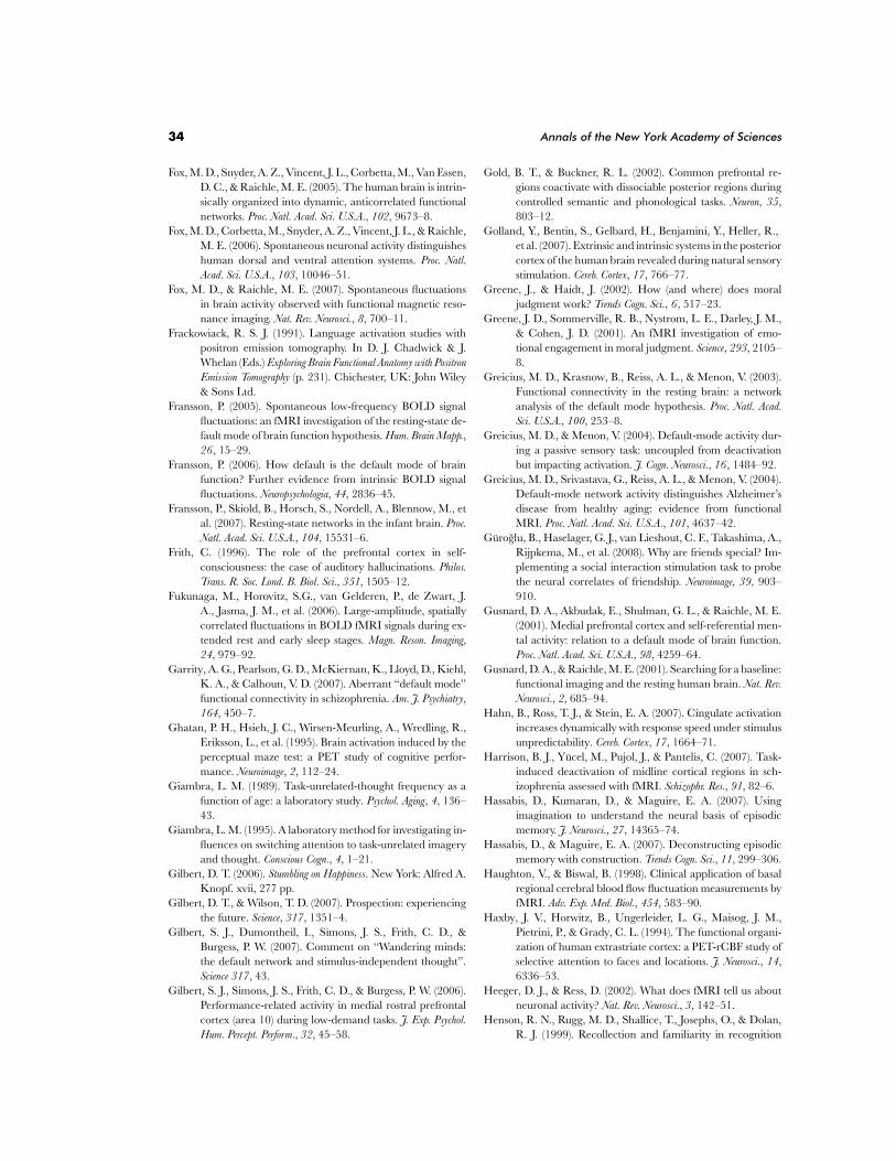
34 Annals of the New York Academy of Sciences
Fox, M. D., Snyder, A. Z., Vincent, J. L., Corbetta, M., Van Essen,D. C., & Raichle, M. E. (2005). The human brain is intrin-sically organized into dynamic, anticorrelated functionalnetworks. Proc. Natl. Acad. Sci. U.S.A., 102, 9673–8.
Fox, M. D., Corbetta, M., Snyder, A. Z., Vincent, J. L., & Raichle,M. E. (2006). Spontaneous neuronal activity distinguisheshuman dorsal and ventral attention systems. Proc. Natl.Acad. Sci. U.S.A., 103, 10046–51.
Fox, M. D., & Raichle, M. E. (2007). Spontaneous fluctuationsin brain activity observed with functional magnetic reso-nance imaging. Nat. Rev. Neurosci., 8, 700–11.
Frackowiack, R. S. J. (1991). Language activation studies withpositron emission tomography. In D. J. Chadwick & J.Whelan (Eds.) Exploring Brain Functional Anatomy with PositronEmission Tomography (p. 231). Chichester, UK: John Wiley& Sons Ltd.
Fransson, P. (2005). Spontaneous low-frequency BOLD signalfluctuations: an fMRI investigation of the resting-state de-fault mode of brain function hypothesis. Hum. Brain Mapp.,26 , 15–29.
Fransson, P. (2006). How default is the default mode of brainfunction? Further evidence from intrinsic BOLD signalfluctuations. Neuropsychologia, 44, 2836–45.
Fransson, P., Skiold, B., Horsch, S., Nordell, A., Blennow, M., etal. (2007). Resting-state networks in the infant brain. Proc.Natl. Acad. Sci. U.S.A., 104, 15531–6.
Frith, C. (1996). The role of the prefrontal cortex in self-consciousness: the case of auditory hallucinations. Philos.Trans. R. Soc. Lond. B. Biol. Sci., 351, 1505–12.
Fukunaga, M., Horovitz, S.G., van Gelderen, P., de Zwart, J.A., Jasma, J. M., et al. (2006). Large-amplitude, spatiallycorrelated fluctuations in BOLD fMRI signals during ex-tended rest and early sleep stages. Magn. Reson. Imaging,24, 979–92.
Garrity, A. G., Pearlson, G. D., McKiernan, K., Lloyd, D., Kiehl,K. A., & Calhoun, V. D. (2007). Aberrant “default mode”functional connectivity in schizophrenia. Am. J. Psychiatry,164, 450–7.
Ghatan, P. H., Hsieh, J. C., Wirsen-Meurling, A., Wredling, R.,Eriksson, L., et al. (1995). Brain activation induced by theperceptual maze test: a PET study of cognitive perfor-mance. Neuroimage, 2, 112–24.
Giambra, L. M. (1989). Task-unrelated-thought frequency as afunction of age: a laboratory study. Psychol. Aging, 4, 136–43.
Giambra, L. M. (1995). A laboratory method for investigating in-fluences on switching attention to task-unrelated imageryand thought. Conscious Cogn., 4, 1–21.
Gilbert, D. T. (2006). Stumbling on Happiness. New York: Alfred A.Knopf. xvii, 277 pp.
Gilbert, D. T., & Wilson, T. D. (2007). Prospection: experiencingthe future. Science, 317, 1351–4.
Gilbert, S. J., Dumontheil, I., Simons, J. S., Frith, C. D., &Burgess, P. W. (2007). Comment on “Wandering minds:the default network and stimulus-independent thought”.Science 317, 43.
Gilbert, S. J., Simons, J. S., Frith, C. D., & Burgess, P. W. (2006).Performance-related activity in medial rostral prefrontalcortex (area 10) during low-demand tasks. J. Exp. Psychol.Hum. Percept. Perform., 32, 45–58.
Gold, B. T., & Buckner, R. L. (2002). Common prefrontal re-gions coactivate with dissociable posterior regions duringcontrolled semantic and phonological tasks. Neuron, 35,803–12.
Golland, Y., Bentin, S., Gelbard, H., Benjamini, Y., Heller, R.,et al. (2007). Extrinsic and intrinsic systems in the posteriorcortex of the human brain revealed during natural sensorystimulation. Cereb. Cortex, 17, 766–77.
Greene, J., & Haidt, J. (2002). How (and where) does moraljudgment work? Trends Cogn. Sci., 6 , 517–23.
Greene, J. D., Sommerville, R. B., Nystrom, L. E., Darley, J. M.,& Cohen, J. D. (2001). An fMRI investigation of emo-tional engagement in moral judgment. Science, 293, 2105–8.
Greicius, M. D., Krasnow, B., Reiss, A. L., & Menon, V. (2003).Functional connectivity in the resting brain: a networkanalysis of the default mode hypothesis. Proc. Natl. Acad.Sci. U.S.A., 100, 253–8.
Greicius, M. D., & Menon, V. (2004). Default-mode activity dur-ing a passive sensory task: uncoupled from deactivationbut impacting activation. J. Cogn. Neurosci., 16 , 1484–92.
Greicius, M. D., Srivastava, G., Reiss, A. L., & Menon, V. (2004).Default-mode network activity distinguishes Alzheimer’sdisease from healthy aging: evidence from functionalMRI. Proc. Natl. Acad. Sci. U.S.A., 101, 4637–42.
Guroglu, B., Haselager, G. J., van Lieshout, C. F., Takashima, A.,Rijpkema, M., et al. (2008). Why are friends special? Im-plementing a social interaction stimulation task to probethe neural correlates of friendship. Neuroimage, 39, 903–910.
Gusnard, D. A., Akbudak, E., Shulman, G. L., & Raichle, M. E.(2001). Medial prefrontal cortex and self-referential men-tal activity: relation to a default mode of brain function.Proc. Natl. Acad. Sci. U.S.A., 98, 4259–64.
Gusnard, D. A., & Raichle, M. E. (2001). Searching for a baseline:functional imaging and the resting human brain. Nat. Rev.Neurosci., 2, 685–94.
Hahn, B., Ross, T. J., & Stein, E. A. (2007). Cingulate activationincreases dynamically with response speed under stimulusunpredictability. Cereb. Cortex, 17, 1664–71.
Harrison, B. J., Yucel, M., Pujol, J., & Pantelis, C. (2007). Task-induced deactivation of midline cortical regions in sch-izophrenia assessed with fMRI. Schizophr. Res., 91, 82–6.
Hassabis, D., Kumaran, D., & Maguire, E. A. (2007). Usingimagination to understand the neural basis of episodicmemory. J. Neurosci., 27, 14365–74.
Hassabis, D., & Maguire, E. A. (2007). Deconstructing episodicmemory with construction. Trends Cogn. Sci., 11, 299–306.
Haughton, V., & Biswal, B. (1998). Clinical application of basalregional cerebral blood flow fluctuation measurements byfMRI. Adv. Exp. Med. Biol., 454, 583–90.
Haxby, J. V., Horwitz, B., Ungerleider, L. G., Maisog, J. M.,Pietrini, P., & Grady, C. L. (1994). The functional organi-zation of human extrastriate cortex: a PET-rCBF study ofselective attention to faces and locations. J. Neurosci., 14,6336–53.
Heeger, D. J., & Ress, D. (2002). What does fMRI tell us aboutneuronal activity? Nat. Rev. Neurosci., 3, 142–51.
Henson, R. N., Rugg, M. D., Shallice, T., Josephs, O., & Dolan,R. J. (1999). Recollection and familiarity in recognition
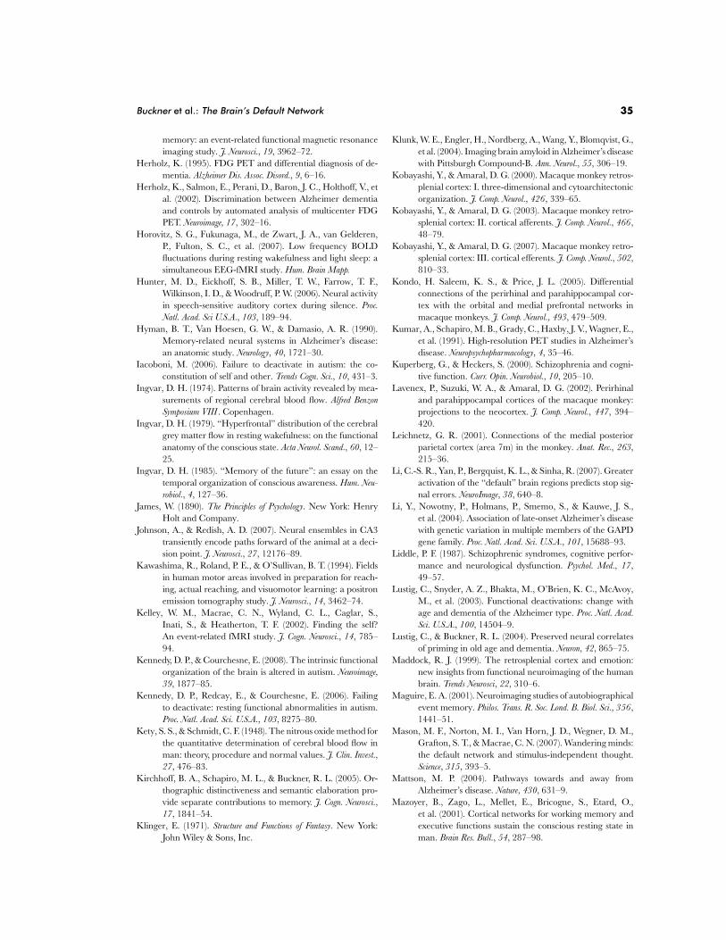
Buckner et al.: The Brain’s Default Network 35
memory: an event-related functional magnetic resonanceimaging study. J. Neurosci., 19, 3962–72.
Herholz, K. (1995). FDG PET and differential diagnosis of de-mentia. Alzheimer Dis. Assoc. Disord., 9, 6–16.
Herholz, K., Salmon, E., Perani, D., Baron, J. C., Holthoff, V., etal. (2002). Discrimination between Alzheimer dementiaand controls by automated analysis of multicenter FDGPET. Neuroimage, 17, 302–16.
Horovitz, S. G., Fukunaga, M., de Zwart, J. A., van Gelderen,P., Fulton, S. C., et al. (2007). Low frequency BOLDfluctuations during resting wakefulness and light sleep: asimultaneous EEG-fMRI study. Hum. Brain Mapp.
Hunter, M. D., Eickhoff, S. B., Miller, T. W., Farrow, T. F.,Wilkinson, I. D., & Woodruff, P. W. (2006). Neural activityin speech-sensitive auditory cortex during silence. Proc.Natl. Acad. Sci U.S.A., 103, 189–94.
Hyman, B. T., Van Hoesen, G. W., & Damasio, A. R. (1990).Memory-related neural systems in Alzheimer’s disease:an anatomic study. Neurology, 40, 1721–30.
Iacoboni, M. (2006). Failure to deactivate in autism: the co-constitution of self and other. Trends Cogn. Sci., 10, 431–3.
Ingvar, D. H. (1974). Patterns of brain activity revealed by mea-surements of regional cerebral blood flow. Alfred BenzonSymposium VIII . Copenhagen.
Ingvar, D. H. (1979). “Hyperfrontal” distribution of the cerebralgrey matter flow in resting wakefulness: on the functionalanatomy of the conscious state. Acta Neurol. Scand., 60, 12–25.
Ingvar, D. H. (1985). “Memory of the future”: an essay on thetemporal organization of conscious awareness. Hum. Neu-robiol., 4, 127–36.
James, W. (1890). The Principles of Psychology. New York: HenryHolt and Company.
Johnson, A., & Redish, A. D. (2007). Neural ensembles in CA3transiently encode paths forward of the animal at a deci-sion point. J. Neurosci., 27, 12176–89.
Kawashima, R., Roland, P. E., & O’Sullivan, B. T. (1994). Fieldsin human motor areas involved in preparation for reach-ing, actual reaching, and visuomotor learning: a positronemission tomography study. J. Neurosci., 14, 3462–74.
Kelley, W. M., Macrae, C. N., Wyland, C. L., Caglar, S.,Inati, S., & Heatherton, T. F. (2002). Finding the self?An event-related fMRI study. J. Cogn. Neurosci., 14, 785–94.
Kennedy, D. P., & Courchesne, E. (2008). The intrinsic functionalorganization of the brain is altered in autism. Neuroimage,39, 1877–85.
Kennedy, D. P., Redcay, E., & Courchesne, E. (2006). Failingto deactivate: resting functional abnormalities in autism.Proc. Natl. Acad. Sci. U.S.A., 103, 8275–80.
Kety, S. S., & Schmidt, C. F. (1948). The nitrous oxide method forthe quantitative determination of cerebral blood flow inman: theory, procedure and normal values. J. Clin. Invest.,27, 476–83.
Kirchhoff, B. A., Schapiro, M. L., & Buckner, R. L. (2005). Or-thographic distinctiveness and semantic elaboration pro-vide separate contributions to memory. J. Cogn. Neurosci.,17, 1841–54.
Klinger, E. (1971). Structure and Functions of Fantasy. New York:John Wiley & Sons, Inc.
Klunk, W. E., Engler, H., Nordberg, A., Wang, Y., Blomqvist, G.,et al. (2004). Imaging brain amyloid in Alzheimer’s diseasewith Pittsburgh Compound-B. Ann. Neurol., 55, 306–19.
Kobayashi, Y., & Amaral, D. G. (2000). Macaque monkey retros-plenial cortex: I. three-dimensional and cytoarchitectonicorganization. J. Comp. Neurol., 426 , 339–65.
Kobayashi, Y., & Amaral, D. G. (2003). Macaque monkey retro-splenial cortex: II. cortical afferents. J. Comp. Neurol., 466 ,48–79.
Kobayashi, Y., & Amaral, D. G. (2007). Macaque monkey retro-splenial cortex: III. cortical efferents. J. Comp. Neurol., 502,810–33.
Kondo, H. Saleem, K. S., & Price, J. L. (2005). Differentialconnections of the perirhinal and parahippocampal cor-tex with the orbital and medial prefrontal networks inmacaque monkeys. J. Comp. Neurol., 493, 479–509.
Kumar, A., Schapiro, M. B., Grady, C., Haxby, J. V., Wagner, E.,et al. (1991). High-resolution PET studies in Alzheimer’sdisease. Neuropsychopharmacology, 4, 35–46.
Kuperberg, G., & Heckers, S. (2000). Schizophrenia and cogni-tive function. Curr. Opin. Neurobiol., 10, 205–10.
Lavenex, P., Suzuki, W. A., & Amaral, D. G. (2002). Perirhinaland parahippocampal cortices of the macaque monkey:projections to the neocortex. J. Comp. Neurol., 447, 394–420.
Leichnetz, G. R. (2001). Connections of the medial posteriorparietal cortex (area 7m) in the monkey. Anat. Rec., 263,215–36.
Li, C.-S. R., Yan, P., Bergquist, K. L., & Sinha, R. (2007). Greateractivation of the “default” brain regions predicts stop sig-nal errors. NeuroImage, 38, 640–8.
Li, Y., Nowotny, P., Holmans, P., Smemo, S., & Kauwe, J. S.,et al. (2004). Association of late-onset Alzheimer’s diseasewith genetic variation in multiple members of the GAPDgene family. Proc. Natl. Acad. Sci. U.S.A., 101, 15688–93.
Liddle, P. F. (1987). Schizophrenic syndromes, cognitive perfor-mance and neurological dysfunction. Psychol. Med., 17,49–57.
Lustig, C., Snyder, A. Z., Bhakta, M., O’Brien, K. C., McAvoy,M., et al. (2003). Functional deactivations: change withage and dementia of the Alzheimer type. Proc. Natl. Acad.Sci. U.S.A., 100, 14504–9.
Lustig, C., & Buckner, R. L. (2004). Preserved neural correlatesof priming in old age and dementia. Neuron, 42, 865–75.
Maddock, R. J. (1999). The retrosplenial cortex and emotion:new insights from functional neuroimaging of the humanbrain. Trends Neurosci, 22, 310–6.
Maguire, E. A. (2001). Neuroimaging studies of autobiographicalevent memory. Philos. Trans. R. Soc. Lond. B. Biol. Sci., 356 ,1441–51.
Mason, M. F., Norton, M. I., Van Horn, J. D., Wegner, D. M.,Grafton, S. T., & Macrae, C. N. (2007). Wandering minds:the default network and stimulus-independent thought.Science, 315, 393–5.
Mattson, M. P. (2004). Pathways towards and away fromAlzheimer’s disease. Nature, 430, 631–9.
Mazoyer, B., Zago, L., Mellet, E., Bricogne, S., Etard, O.,et al. (2001). Cortical networks for working memory andexecutive functions sustain the conscious resting state inman. Brain Res. Bull., 54, 287–98.
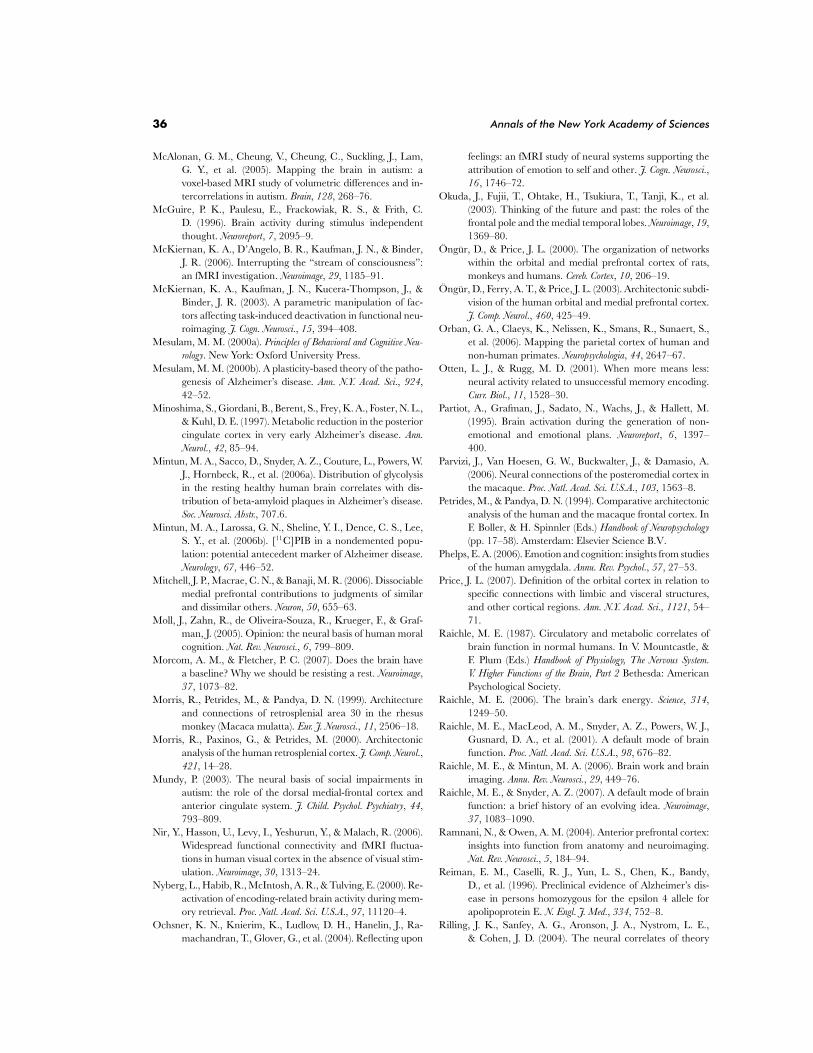
36 Annals of the New York Academy of Sciences
McAlonan, G. M., Cheung, V., Cheung, C., Suckling, J., Lam,G. Y., et al. (2005). Mapping the brain in autism: avoxel-based MRI study of volumetric differences and in-tercorrelations in autism. Brain, 128, 268–76.
McGuire, P. K., Paulesu, E., Frackowiak, R. S., & Frith, C.D. (1996). Brain activity during stimulus independentthought. Neuroreport, 7, 2095–9.
McKiernan, K. A., D’Angelo, B. R., Kaufman, J. N., & Binder,J. R. (2006). Interrupting the “stream of consciousness”:an fMRI investigation. Neuroimage, 29, 1185–91.
McKiernan, K. A., Kaufman, J. N., Kucera-Thompson, J., &Binder, J. R. (2003). A parametric manipulation of fac-tors affecting task-induced deactivation in functional neu-roimaging. J. Cogn. Neurosci., 15, 394–408.
Mesulam, M. M. (2000a). Principles of Behavioral and Cognitive Neu-rology. New York: Oxford University Press.
Mesulam, M. M. (2000b). A plasticity-based theory of the patho-genesis of Alzheimer’s disease. Ann. N.Y. Acad. Sci., 924,42–52.
Minoshima, S., Giordani, B., Berent, S., Frey, K. A., Foster, N. L.,& Kuhl, D. E. (1997). Metabolic reduction in the posteriorcingulate cortex in very early Alzheimer’s disease. Ann.Neurol., 42, 85–94.
Mintun, M. A., Sacco, D., Snyder, A. Z., Couture, L., Powers, W.J., Hornbeck, R., et al. (2006a). Distribution of glycolysisin the resting healthy human brain correlates with dis-tribution of beta-amyloid plaques in Alzheimer’s disease.Soc. Neurosci. Abstr., 707.6.
Mintun, M. A., Larossa, G. N., Sheline, Y. I., Dence, C. S., Lee,S. Y., et al. (2006b). [11C]PIB in a nondemented popu-lation: potential antecedent marker of Alzheimer disease.Neurology, 67, 446–52.
Mitchell, J. P., Macrae, C. N., & Banaji, M. R. (2006). Dissociablemedial prefrontal contributions to judgments of similarand dissimilar others. Neuron, 50, 655–63.
Moll, J., Zahn, R., de Oliveira-Souza, R., Krueger, F., & Graf-man, J. (2005). Opinion: the neural basis of human moralcognition. Nat. Rev. Neurosci., 6 , 799–809.
Morcom, A. M., & Fletcher, P. C. (2007). Does the brain havea baseline? Why we should be resisting a rest. Neuroimage,37, 1073–82.
Morris, R., Petrides, M., & Pandya, D. N. (1999). Architectureand connections of retrosplenial area 30 in the rhesusmonkey (Macaca mulatta). Eur. J. Neurosci., 11, 2506–18.
Morris, R., Paxinos, G., & Petrides, M. (2000). Architectonicanalysis of the human retrosplenial cortex. J. Comp. Neurol.,421, 14–28.
Mundy, P. (2003). The neural basis of social impairments inautism: the role of the dorsal medial-frontal cortex andanterior cingulate system. J. Child. Psychol. Psychiatry, 44,793–809.
Nir, Y., Hasson, U., Levy, I., Yeshurun, Y., & Malach, R. (2006).Widespread functional connectivity and fMRI fluctua-tions in human visual cortex in the absence of visual stim-ulation. Neuroimage, 30, 1313–24.
Nyberg, L., Habib, R., McIntosh, A. R., & Tulving, E. (2000). Re-activation of encoding-related brain activity during mem-ory retrieval. Proc. Natl. Acad. Sci. U.S.A., 97, 11120–4.
Ochsner, K. N., Knierim, K., Ludlow, D. H., Hanelin, J., Ra-machandran, T., Glover, G., et al. (2004). Reflecting upon
feelings: an fMRI study of neural systems supporting theattribution of emotion to self and other. J. Cogn. Neurosci.,16 , 1746–72.
Okuda, J., Fujii, T., Ohtake, H., Tsukiura, T., Tanji, K., et al.(2003). Thinking of the future and past: the roles of thefrontal pole and the medial temporal lobes. Neuroimage, 19,1369–80.
Ongur, D., & Price, J. L. (2000). The organization of networkswithin the orbital and medial prefrontal cortex of rats,monkeys and humans. Cereb. Cortex, 10, 206–19.
Ongur, D., Ferry, A. T., & Price, J. L. (2003). Architectonic subdi-vision of the human orbital and medial prefrontal cortex.J. Comp. Neurol., 460, 425–49.
Orban, G. A., Claeys, K., Nelissen, K., Smans, R., Sunaert, S.,et al. (2006). Mapping the parietal cortex of human andnon-human primates. Neuropsychologia, 44, 2647–67.
Otten, L. J., & Rugg, M. D. (2001). When more means less:neural activity related to unsuccessful memory encoding.Curr. Biol., 11, 1528–30.
Partiot, A., Grafman, J., Sadato, N., Wachs, J., & Hallett, M.(1995). Brain activation during the generation of non-emotional and emotional plans. Neuroreport, 6 , 1397–400.
Parvizi, J., Van Hoesen, G. W., Buckwalter, J., & Damasio, A.(2006). Neural connections of the posteromedial cortex inthe macaque. Proc. Natl. Acad. Sci. U.S.A., 103, 1563–8.
Petrides, M., & Pandya, D. N. (1994). Comparative architectonicanalysis of the human and the macaque frontal cortex. InF. Boller, & H. Spinnler (Eds.) Handbook of Neuropsychology(pp. 17–58). Amsterdam: Elsevier Science B.V.
Phelps, E. A. (2006). Emotion and cognition: insights from studiesof the human amygdala. Annu. Rev. Psychol., 57, 27–53.
Price, J. L. (2007). Definition of the orbital cortex in relation tospecific connections with limbic and visceral structures,and other cortical regions. Ann. N.Y. Acad. Sci., 1121, 54–71.
Raichle, M. E. (1987). Circulatory and metabolic correlates ofbrain function in normal humans. In V. Mountcastle, &F. Plum (Eds.) Handbook of Physiology, The Nervous System.V. Higher Functions of the Brain, Part 2 Bethesda: AmericanPsychological Society.
Raichle, M. E. (2006). The brain’s dark energy. Science, 314,1249–50.
Raichle, M. E., MacLeod, A. M., Snyder, A. Z., Powers, W. J.,Gusnard, D. A., et al. (2001). A default mode of brainfunction. Proc. Natl. Acad. Sci. U.S.A., 98, 676–82.
Raichle, M. E., & Mintun, M. A. (2006). Brain work and brainimaging. Annu. Rev. Neurosci., 29, 449–76.
Raichle, M. E., & Snyder, A. Z. (2007). A default mode of brainfunction: a brief history of an evolving idea. Neuroimage,37, 1083–1090.
Ramnani, N., & Owen, A. M. (2004). Anterior prefrontal cortex:insights into function from anatomy and neuroimaging.Nat. Rev. Neurosci., 5, 184–94.
Reiman, E. M., Caselli, R. J., Yun, L. S., Chen, K., Bandy,D., et al. (1996). Preclinical evidence of Alzheimer’s dis-ease in persons homozygous for the epsilon 4 allele forapolipoprotein E. N. Engl. J. Med., 334, 752–8.
Rilling, J. K., Sanfey, A. G., Aronson, J. A., Nystrom, L. E.,& Cohen, J. D. (2004). The neural correlates of theory
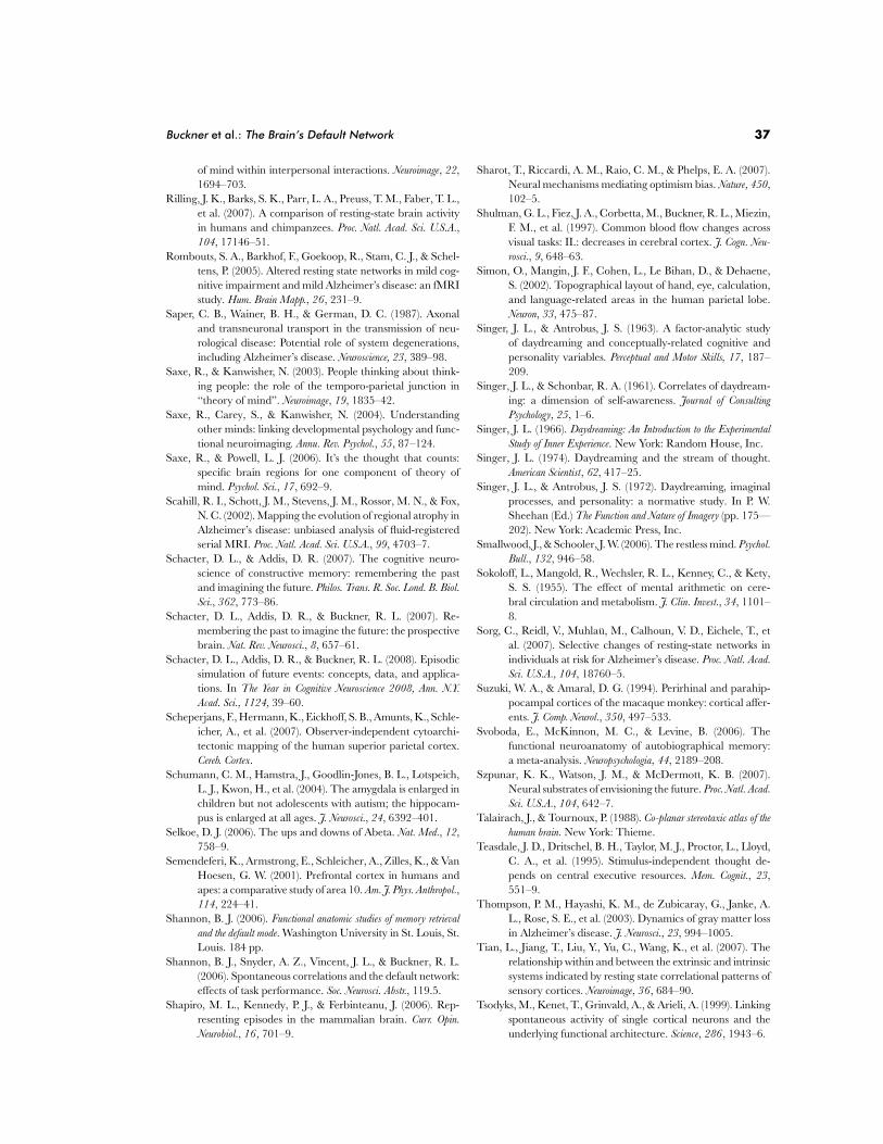
Buckner et al.: The Brain’s Default Network 37
of mind within interpersonal interactions. Neuroimage, 22,1694–703.
Rilling, J. K., Barks, S. K., Parr, L. A., Preuss, T. M., Faber, T. L.,et al. (2007). A comparison of resting-state brain activityin humans and chimpanzees. Proc. Natl. Acad. Sci. U.S.A.,104, 17146–51.
Rombouts, S. A., Barkhof, F., Goekoop, R., Stam, C. J., & Schel-tens, P. (2005). Altered resting state networks in mild cog-nitive impairment and mild Alzheimer’s disease: an fMRIstudy. Hum. Brain Mapp., 26 , 231–9.
Saper, C. B., Wainer, B. H., & German, D. C. (1987). Axonaland transneuronal transport in the transmission of neu-rological disease: Potential role of system degenerations,including Alzheimer’s disease. Neuroscience, 23, 389–98.
Saxe, R., & Kanwisher, N. (2003). People thinking about think-ing people: the role of the temporo-parietal junction in“theory of mind”. Neuroimage, 19, 1835–42.
Saxe, R., Carey, S., & Kanwisher, N. (2004). Understandingother minds: linking developmental psychology and func-tional neuroimaging. Annu. Rev. Psychol., 55, 87–124.
Saxe, R., & Powell, L. J. (2006). It’s the thought that counts:specific brain regions for one component of theory ofmind. Psychol. Sci., 17, 692–9.
Scahill, R. I., Schott, J. M., Stevens, J. M., Rossor, M. N., & Fox,N. C. (2002). Mapping the evolution of regional atrophy inAlzheimer’s disease: unbiased analysis of fluid-registeredserial MRI. Proc. Natl. Acad. Sci. U.S.A., 99, 4703–7.
Schacter, D. L., & Addis, D. R. (2007). The cognitive neuro-science of constructive memory: remembering the pastand imagining the future. Philos. Trans. R. Soc. Lond. B. Biol.Sci., 362, 773–86.
Schacter, D. L., Addis, D. R., & Buckner, R. L. (2007). Re-membering the past to imagine the future: the prospectivebrain. Nat. Rev. Neurosci., 8, 657–61.
Schacter, D. L., Addis, D. R., & Buckner, R. L. (2008). Episodicsimulation of future events: concepts, data, and applica-tions. In The Year in Cognitive Neuroscience 2008, Ann. N.Y.Acad. Sci., 1124, 39–60.
Scheperjans, F., Hermann, K., Eickhoff, S. B., Amunts, K., Schle-icher, A., et al. (2007). Observer-independent cytoarchi-tectonic mapping of the human superior parietal cortex.Cereb. Cortex.
Schumann, C. M., Hamstra, J., Goodlin-Jones, B. L., Lotspeich,L. J., Kwon, H., et al. (2004). The amygdala is enlarged inchildren but not adolescents with autism; the hippocam-pus is enlarged at all ages. J. Neurosci., 24, 6392–401.
Selkoe, D. J. (2006). The ups and downs of Abeta. Nat. Med., 12,758–9.
Semendeferi, K., Armstrong, E., Schleicher, A., Zilles, K., & VanHoesen, G. W. (2001). Prefrontal cortex in humans andapes: a comparative study of area 10. Am. J. Phys. Anthropol.,114, 224–41.
Shannon, B. J. (2006). Functional anatomic studies of memory retrievaland the default mode. Washington University in St. Louis, St.Louis. 184 pp.
Shannon, B. J., Snyder, A. Z., Vincent, J. L., & Buckner, R. L.(2006). Spontaneous correlations and the default network:effects of task performance. Soc. Neurosci. Abstr., 119.5.
Shapiro, M. L., Kennedy, P. J., & Ferbinteanu, J. (2006). Rep-resenting episodes in the mammalian brain. Curr. Opin.Neurobiol., 16 , 701–9.
Sharot, T., Riccardi, A. M., Raio, C. M., & Phelps, E. A. (2007).Neural mechanisms mediating optimism bias. Nature, 450,102–5.
Shulman, G. L., Fiez, J. A., Corbetta, M., Buckner, R. L., Miezin,F. M., et al. (1997). Common blood flow changes acrossvisual tasks: II.: decreases in cerebral cortex. J. Cogn. Neu-rosci., 9, 648–63.
Simon, O., Mangin, J. F., Cohen, L., Le Bihan, D., & Dehaene,S. (2002). Topographical layout of hand, eye, calculation,and language-related areas in the human parietal lobe.Neuron, 33, 475–87.
Singer, J. L., & Antrobus, J. S. (1963). A factor-analytic studyof daydreaming and conceptually-related cognitive andpersonality variables. Perceptual and Motor Skills, 17, 187–209.
Singer, J. L., & Schonbar, R. A. (1961). Correlates of daydream-ing: a dimension of self-awareness. Journal of ConsultingPsychology, 25, 1–6.
Singer, J. L. (1966). Daydreaming: An Introduction to the ExperimentalStudy of Inner Experience. New York: Random House, Inc.
Singer, J. L. (1974). Daydreaming and the stream of thought.American Scientist, 62, 417–25.
Singer, J. L., & Antrobus, J. S. (1972). Daydreaming, imaginalprocesses, and personality: a normative study. In P. W.Sheehan (Ed.) The Function and Nature of Imagery (pp. 175—202). New York: Academic Press, Inc.
Smallwood, J., & Schooler, J. W. (2006). The restless mind. Psychol.Bull., 132, 946–58.
Sokoloff, L., Mangold, R., Wechsler, R. L., Kenney, C., & Kety,S. S. (1955). The effect of mental arithmetic on cere-bral circulation and metabolism. J. Clin. Invest., 34, 1101–8.
Sorg, C., Reidl, V., Muhlau, M., Calhoun, V. D., Eichele, T., etal. (2007). Selective changes of resting-state networks inindividuals at risk for Alzheimer’s disease. Proc. Natl. Acad.Sci. U.S.A., 104, 18760–5.
Suzuki, W. A., & Amaral, D. G. (1994). Perirhinal and parahip-pocampal cortices of the macaque monkey: cortical affer-ents. J. Comp. Neurol., 350, 497–533.
Svoboda, E., McKinnon, M. C., & Levine, B. (2006). Thefunctional neuroanatomy of autobiographical memory:a meta-analysis. Neuropsychologia, 44, 2189–208.
Szpunar, K. K., Watson, J. M., & McDermott, K. B. (2007).Neural substrates of envisioning the future. Proc. Natl. Acad.Sci. U.S.A., 104, 642–7.
Talairach, J., & Tournoux, P. (1988). Co-planar stereotaxic atlas of thehuman brain. New York: Thieme.
Teasdale, J. D., Dritschel, B. H., Taylor, M. J., Proctor, L., Lloyd,C. A., et al. (1995). Stimulus-independent thought de-pends on central executive resources. Mem. Cognit., 23,551–9.
Thompson, P. M., Hayashi, K. M., de Zubicaray, G., Janke, A.L., Rose, S. E., et al. (2003). Dynamics of gray matter lossin Alzheimer’s disease. J. Neurosci., 23, 994–1005.
Tian, L., Jiang, T., Liu, Y., Yu, C., Wang, K., et al. (2007). Therelationship within and between the extrinsic and intrinsicsystems indicated by resting state correlational patterns ofsensory cortices. Neuroimage, 36 , 684–90.
Tsodyks, M., Kenet, T., Grinvald, A., & Arieli, A. (1999). Linkingspontaneous activity of single cortical neurons and theunderlying functional architecture. Science, 286 , 1943–6.
38 Annals of the New York Academy of Sciences
Tulving, E. (2005). Episodic memory and autonoesis: Uniquelyhuman? In H. Terrance, & J. Metcalfe (Eds.) The missinglink in cognition: Origins of self-reflective consciousness (pp. 3–56).New York: Oxford University Press.
Van Essen, D. C. (2005). A Population-Average, Landmark- andSurface-based (PALS) atlas of human cerebral cortex. Neu-roimage, 28, 635–62.
Van Essen, D. C., & Dieker, D. L. (2007). Surface-based andprobabilistic atlases of primate cerebral cortex. Neuron,56 , 209–25.
Vincent, J. L., Snyder, A. Z., Fox, M. D., Shannon, B. J., Andrews,J. R., et al. (2006). Coherent spontaneous activity identifiesa hippocampal-parietal memory network. J. Neurophysiol.,96 , 3517–31.
Vincent, J. L., Patel, G. H., Fox, M. D., Snyder, A. Z., Baker, J.T., et al. (2007a). Intrinsic functional architecture in theanaesthetized monkey brain. Nature, 447, 83–6.
Vincent, J. L., Kahn, I., Snyder, A. Z., Fox, M. D., Raichle, M.E., & Buckner, R. L. (2007b). Evidence for three distinct,bilateral frontoparietal associative brain systems revealedby spontaneous fMRI correlations. Soc. Neurosci. Abstr.
Vogeley, K., May, M., Ritzl, A., Falkai, P., Zilles, K., & Fink,G. R. (2004). Neural correlates of first-person perspectiveas one constituent of human self-consciousness. J. Cogn.Neurosci., 16 , 817–27.
Vogt, B. A., Vogt, L. J., Perl, D. P., & Hof, P. R. (2001). Cytologyof human caudomedial cingulate, retrosplenial, and cau-dal parahippocampal cortices. J. Comp. Neurol., 438, 353–76.
Vogt, B. A., & Laureys, S. (2005). Posterior cingulate, precunealand retrosplenial cortices: cytology and components ofthe neural network correlates of consciousness. Prog. BrainRes., 150, 205–17.
Vogt, B. A., Nimchinsky, E. A., Vogt, L. J., & Hof, P. R. (1995).Human cingulate cortex: Surface features, flat maps, andcytoarchitecture. J. Comp. Neurol., 359, 490–506.
Vogt, B. A., Vogt, L., & Laureys, S. (2006). Cytology and func-tionally correlated circuits of human posterior cingulateareas. Neuroimage, 29, 452–66.
Wagner, A. D., Shannon, B. J., Kahn, I., & Buckner, R. L. (2005).Parietal lobe contributions to episodic memory retrieval.Trends Cogn. Sci., 9, 445–53.
Waiter, G. D., Williams, J. H., Murray, A. D., Gilchrist, A., Perrett,D. I., & Whiten, A. (2004). A voxel-based investigation ofbrain structure in male adolescents with autistic spectrumdisorder. Neuroimage, 22, 619–25.
Walsh, D. M., & Selkoe, D. J. (2004). Deciphering the molecularbasis of memory failure in Alzheimer’s disease. Neuron, 44,181–93.
Wang, K., Liang, M., Wang, L., Tian, L., Zhang, X., et al. (2006).Altered functional connectivity in early Alzheimer’s dis-ease: a resting-state fMRI study. Hum. Brain Mapp., 28,967–78.
Weissman, D. H., Roberts, K. C., Visscher, K. M., & Woldorff,M. G. (2006). The neural bases of momentary lapses inattention. Nat. Neurosci., 9, 971–8.
Wheeler, M. E., Petersen, S. E., & Buckner, R. L. (2000). Mem-ory’s echo: vivid remembering reactivates sensory-specificcortex. Proc. Natl. Acad. Sci. U.S.A., 97, 11125–9.
Wheeler, M. E., & Buckner, R. L. (2004). Functional-anatomiccorrelates of remembering and knowing. Neuroimage, 21,1337–49.
Williamson, P. (2007). Are anticorrelated networks in the brainrelevant to schizophrenia? Schizophr. Bull., 33, 994–1003.
Wimmer, H., & Perner, J. (1983). Beliefs about beliefs: represen-tation and constraining function of wrong beliefs in youngchildren’s understanding of deception. Cognition, 13, 103–28.
Wise, R. G., Ide, K., Poulin, M. J., & Tracey, I. (2004). Restingfluctuations in arterial carbon dioxide induce significantlow frequency variations in BOLD signal. Neuroimage, 21,1652–64.
Yonelinas, A. P., Otten, L. J., Shaw, K. N., & Rugg, M. D. (2005).Separating the brain regions involved in recollection andfamiliarity in recognition memory. J. Neurosci., 25, 3002–8.
Zhou, Y., Liang, M., Tian, L., Wang, K., Hao, Y., et al. (2007).Functional disintegration in paranoid schizophrenia usingresting-state fMRI. Schizophr. Res, 97, 194–205.
Related Documents