Anatomy and Physical Exam Yibing Li 01/07/2004 MSK-HIP MSK-HIP (Part I) (Part I)

Anatomy and Physical Exam Yibing Li 01/07/2004 MSK-HIP (Part I)
Dec 17, 2015
Welcome message from author
This document is posted to help you gain knowledge. Please leave a comment to let me know what you think about it! Share it to your friends and learn new things together.
Transcript

Anatomy and Physical Exam
Yibing Li
01/07/2004
MSK-HIPMSK-HIP(Part I)(Part I)

Hip JointHip Joint
The hip joint is a synovial ball and socket joint, formed by the reception of the head of femur into the cavity of acetabulum. It is the largest weight bearing joint in the body and is surrounded by strong ligaments and muscles.
Due to high mobility, hip joint pathology can be manifested during weight bearing, ambulation or motion.

Hip BonesHip Bones
The ilium, ischium, pubis and femur are the main bones at the hip joints. The ilium, ischium and pubis are fused together to form the pelvis girdle (L+R).

Ligaments of the Hip JointsLigaments of the Hip Joints
Acetabular labrum forms a complete ring around the head of the femur. Its function is to hold the femoral head in place.
The ligament of the head of femur attaches the head of femur to the acetabulum.

Ligaments (cont.)Ligaments (cont.)
There are 3 ligaments that hold the head of femur to the pelvis:
1. Iliofemoral lig.: the strongest ligament; Its function is to prevent hyperextension, abduction and lateral rotation.
2. Pubofemoral lig. : limits abduction
3. Ischiofemoral lig. : limits medial rotation.

The Capsule of Hip JointThe Capsule of Hip Joint
The articular fibrous capsule extends from the acetabular rim to the intertrochanteric crest forming a sleeve that encloses the hip joint and most of the femoral neck.
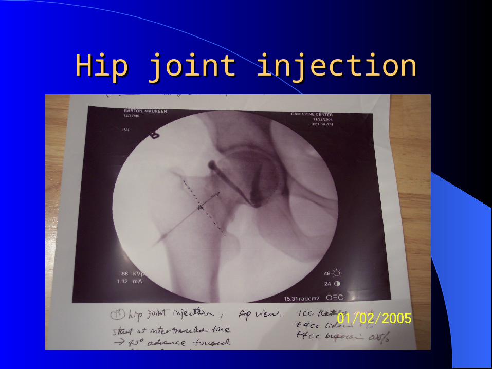
Hip joint injectionHip joint injection

Muscles of the hip and thighMuscles of the hip and thighMuscles of the Hip & Thigh
Inferior GemelliGluteal Region (Hip-Joint Stability)Deep Ischial Tuberosity Greater Trochanter Femur: Lateral Rotation
Directly below the obturator internus
Obturator ExternusGluteal Region (Hip-Joint Stability)Deep
Obturator Membrane (external surface) Greater Trochanter Femur: Lateral Rotation
Obturator InternusGluteal Region (Hip-Joint Stability)Deep
Obturator Membrane (internal surface) Greater Trochanter Femur: Lateral Rotation
Goes through Lesser Sciatic Foramen
PiriformisGluteal Region (Hip-Joint Stability)Deep Anterior aspect of the Sacrum
Greater Trochanter (Superior aspect)
Femur: Lateral Rotation | Femur: Abduction
Quadratus FemorisGluteal Region (Hip-Joint Stability)Deep Ischial Tuberosity
Intertrochanteric Crest (Quadrate Tubercle) Femur: Lateral Rotation
The big muscle in the gluteal group
Superior GemelliGluteal Region (Hip-Joint Stability)Deep Ischial Spine Greater Trochanter Femur: Lateral Rotation
Directly above the obturator internus
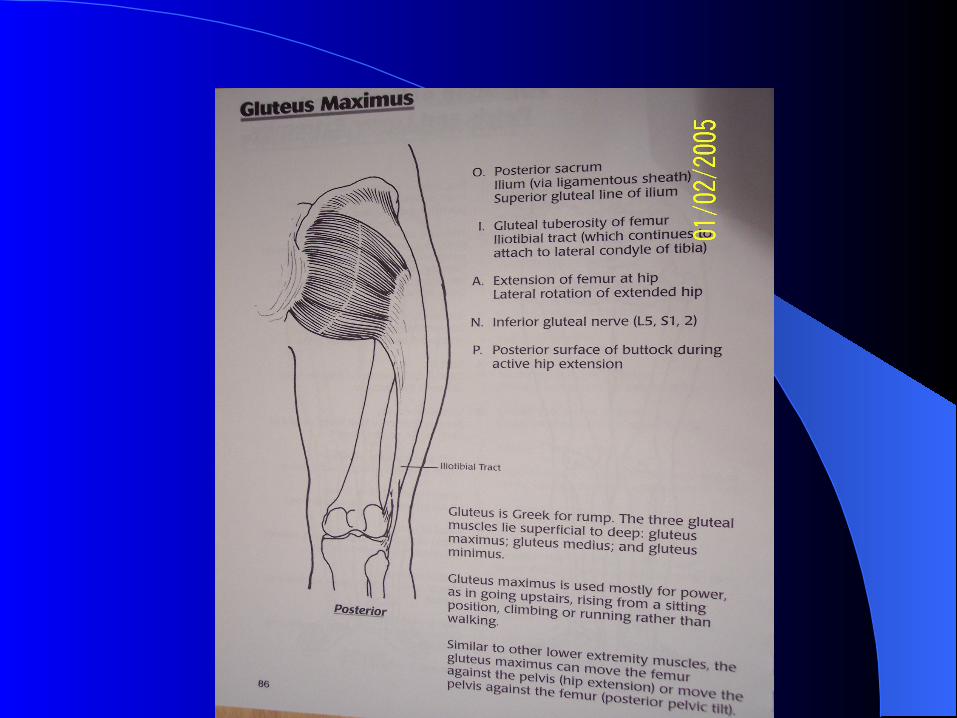
Gluteus MaximusGluteal Region (Hip-Joint Stability)Superficial Ilium, Sacrum, and Coccyx
Gluteal Tuberosity of Femur and Iliotibial Tract Hip: Extension
Gluteus MediusGluteal Region (Hip-Joint Stability)Superficial
Outer surface of il eum, between top two gluteal lines
Greater Trochanter (Lateral)
Femur: Abduction and medial rotation
Gluteus MinimusGluteal Region (Hip-Joint Stability)Superficial
Outer surface of Ili um, between bottom two gluteal lines
Greater Trochanter (anterior)
Femur: Abduction and medial rotation
Iliopsoas Thigh, Anterior Lumbar spine and Iliac crest Lesser Trochanter of Femur Femur: Flexion at hip Floor of the Femoral Triangle
Sartorius Thigh, Anterior Anterior Superior Iliac Spine Posteromedial aspect of proximal tibia
Hip: Flexion; Knee: Extension | Femur: Lateral Rotation
Inferolateral base of Femoral Triangle
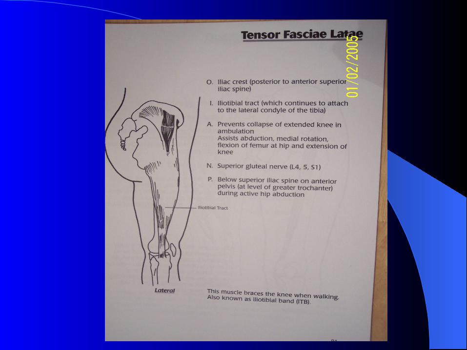
Tensor Fasciae Latae Thigh, Anterior Proximal femur Iliotibial Tract Hip: Flexion; Femur: Medial Rotation
Increases tension of fascia lata and iliotibial tract, esp. when standing upright; holds knee in place
Rectus Femoris Thigh, AnteriorQuadriceps Femoralis Anterior Inferior Iliac Spine
Quadriceps Ten don on the superior Patella Knee: Extension
Acts on the hip joint; Crucial role in knee stability
Vastus Intermedius Thigh, AnteriorQuadriceps Femoralis Proximal femur
Quadriceps Ten don on the superior Patella Knee: Extension Crucial role in knee stability
Vastus Lateralis Thigh, AnteriorQuadriceps Femoralis Proximal femur
Quadriceps Ten don on the superior Patella Knee: Extension Crucial role in knee stability
Vastus Medialis Thigh, AnteriorQuadriceps Femoralis Proximal femur
Quadriceps Ten don on the superior Patella Knee: Extension Crucial role in knee stability
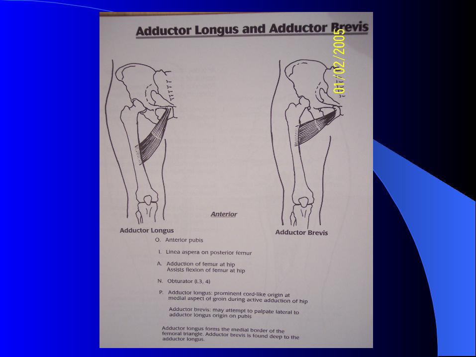
Adductor Brevis Thigh, MedialAdductor Group Pubic Bone Thigh: Adduction
Adductor Longus Thigh, MedialAdductor Group Pubic Bone Thigh: Adduction Lateral floor of Femoral Triangle
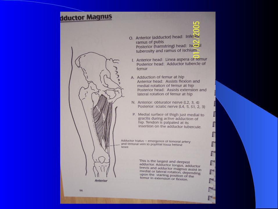
Adductor Magnus Thigh, MedialAdductor Group Pubic Bone Thigh: Adduction | Thigh: Extension Adductor "Hybrid" Muscle
Gracilis Thigh, MedialAdductor Group Pubic Bone NONE | Thigh: Adduction A good spare part; adduction can occur without it.
Pectineus Thigh, MedialAdductor Group Pubic Bone Thigh: Adduction | Hip: Flexion
Adductor "Hybrid" Muscle; Floor of the Femoral Triangle
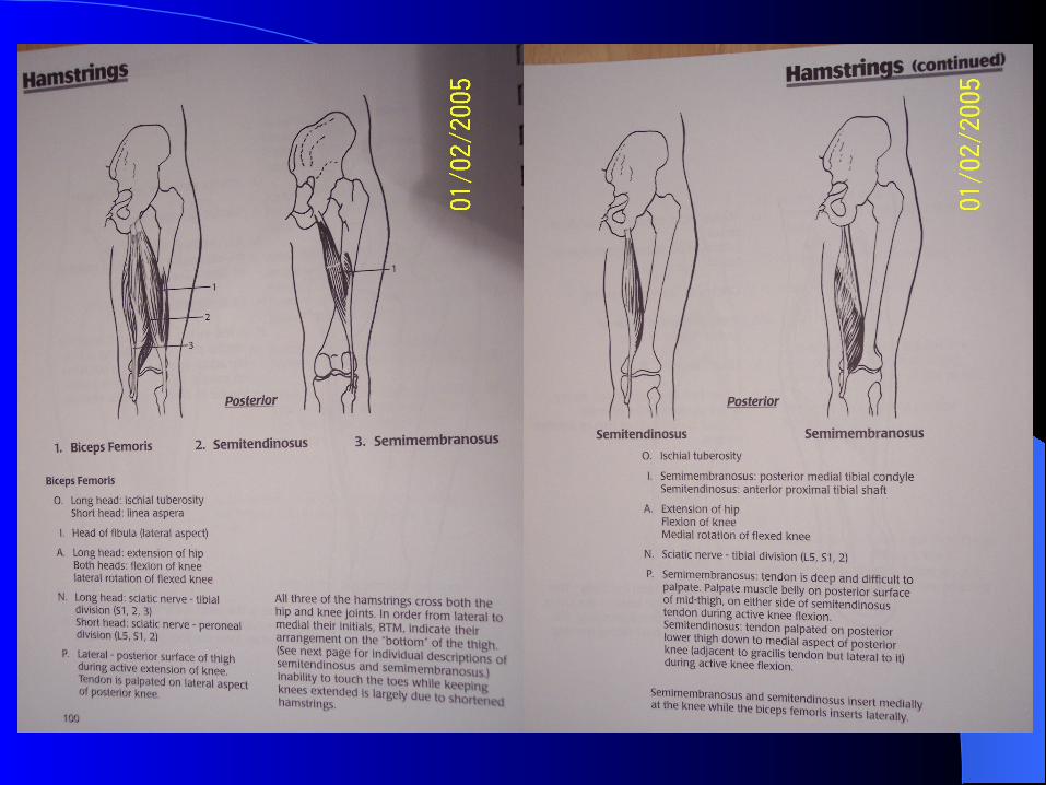
Biceps Femoris Thigh, PosteriorHamstring Muscles
Long Head: Ischial Tuberosity | Short Head: Femur (Linea Aspera) Head of the Fib ula
Hip: Extension; Knee: Flexion
Long head crosses both hip and knee joints
Semimembra nosus Thigh, PosteriorHamstring Muscles Ischial Tuberosity
Medial condyle of proximal tibia
Hip: Extension; Knee: Flexion
Cross both hip and knee joints
Semitendinos us Thigh, PosteriorHamstring Muscles Ischial Tuberosity
Medial condyle of proximal tibia
Hip: Extension; Knee: Flexion
Cross both hip and knee joints; medial to semimembranosus

Movements at the HipMovements at the Hip
1. Flexion / Extension2. Adduction / Abduction3. Lateral (external)Rotation / Medial
(internal) Rotation

Hip FlexorsHip Flexors
Iliopsoas (prime hip flexor)Pectineus Sartorius Rectus femoris Pectineus Tensor fsaciae latae Adductor brevis Adductor longus Adductor magnus (anterior head) Rectus femoris

P90-iliopsoasP90-iliopsoas

P96-pectineusP96-pectineus

Hip ExtensorsHip Extensors
Gluteus maximusBiceps femoris (long head)SemitendinosusSemimembranosus Adductor magnus (postrior head)
P86P86
P100-101 HamstringP100-101 Hamstring

Hip AbductorsHip Abductors
Gluteus mediusGluteus minimus Tensor fasciae latae Sartorius

p87p87

p88p88
Hip AdductorsHip Adductors
Adductor brevisAdductor longusAdductor magnusGracilis Pectineus
p97p97
p98p98

p99p99

Internal Rotators of the HipInternal Rotators of the Hip
Gluteus mediusGluteus minimusTensor fasciae latae Adductor magnus (anterior head)

p91p91
p89p89

Physical Exam of the Hip and Physical Exam of the Hip and PelvisPelvis
Inspection & PalpationROMNeurologic exam Special tests

InspectionInspection
Observe gaitCheck hip and pelvis area for skin
abrasions,abnormal swelling, etc.Check if the anterior superior iliac spines
are in the same horizontal plane or tilted pelvis
Observe the two discernible dimples to check PSIS for pelvic obliquity

GaitGait
Antalgic gait: prolonged double support period, decreased stance phase and step length on the unaffected side to reduce pain and avoid weight bearing on the affected side.
Trendelenburg gait (hip abductor weakness) ---uncompensated gait: contralateral pelvic drop. ---compensated: lateral lurch over the affected side.(Tx with cane)
Extensor lurch gait(gluteus maximus weakness): secondary to inferior gluteal N.injury or subtrochanteric hip fx. Unable to decelerate the hip flexion moment at heel strike due to hip extensor weakness. To compensate, pt lean upper body backward to keep the center of gravity. Tx with two crutches or canes.
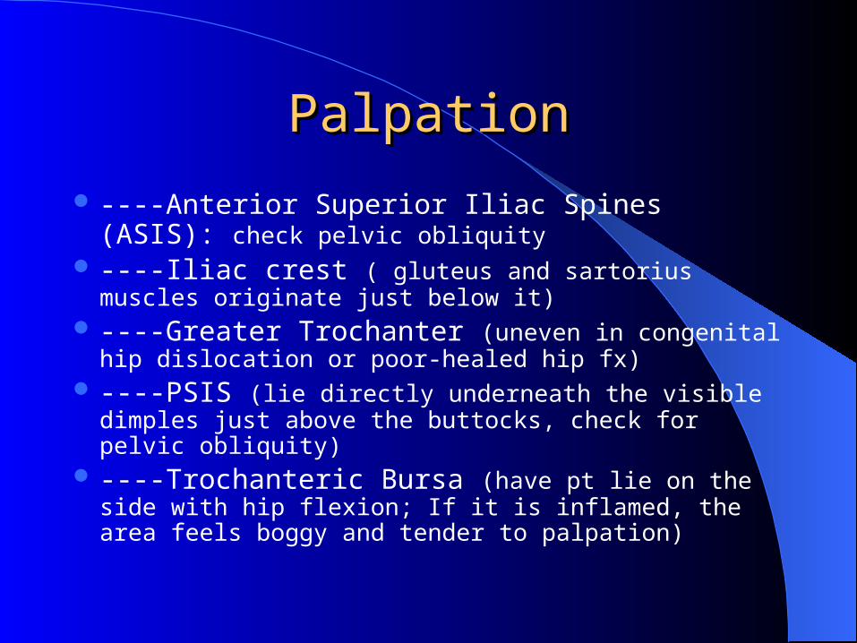
PalpationPalpation
----Anterior Superior Iliac Spines (ASIS): check pelvic obliquity
----Iliac crest ( gluteus and sartorius muscles originate just below it)
----Greater Trochanter (uneven in congenital hip dislocation or poor-healed hip fx)
----PSIS (lie directly underneath the visible dimples just above the buttocks, check for pelvic obliquity)
----Trochanteric Bursa (have pt lie on the side with hip flexion; If it is inflamed, the area feels boggy and tender to palpation)
ROMROM
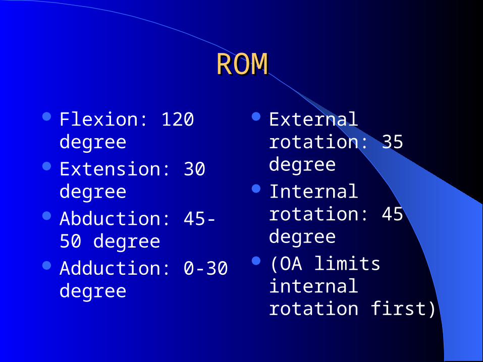
Flexion: 120 degree Extension: 30 degree Abduction: 45-50
degree Adduction: 0-30
degree
External rotation: 35 degree
Internal rotation: 45 degree
(OA limits internal rotation first)

Neurologic ExamNeurologic Exam
Muscle testing: test muscle strength in functional groups.
Primary flexor: Iliopsoas (femoral N. L1,2,3) Primary extensors: Gluteus Maximus (inferior gluteus N. S1) Primary adductors: Adductor longus (obturator N. L2,3,4) Primary Abductor: Gluteus medius( superior gluteal N., L5)
Sensation testing: for example, dermatomes ( T10-L3)

Special Hip TestsSpecial Hip Tests
1. Patrick (Fabere) test 2. Thomas test3. Ober test4. Trendelenburg test5. Leg length discrepancy

Patrick testPatrick test
This test is to assess Flexion, Abduction, External Rotation
Perform with pt supine, passively flex and externally rotate and abduct the hip.
Ipsilateral inguinal pain indicates pathology in the hip joint or the surrounding muscles.
Contralateral pain occurs in the dysfunctional SI joint.
Thomas TestThomas Test
To assess hip flexion contractures Perform the test with the pt supine, flex one hip
fully reducing the lumbar spine lordosis, stabilizing the spine and pelvis, extend the opposite hip.A flexion contracture is present if the hip cannot fully extend. The degree of flexion contracture can be done by estimating the angle between the table and pt’s leg.

Ober testOber test
To test for contraction of the fascia lata.Have pt lie on the side with involved leg
uppermost. Abduct the leg as far as possible and flex the knee to 90 degree. If the thigh remains abducted, there may be a contracture of the tensor fascia lata or ITB.
Trendelenburg testTrendelenburg test
Test for gluteus medius weakness Perform with the pt standing erect, one foot is raised
off the floor, strength of the gluteus medius of the supported side is assessed. A positive test occurs when the pelvis on the unsupported side descends or remains level.
Conditions with gluteus medius weakness:--- radiculopathies,poliomyelitis,meningomyelocele, fx of the greater trochanter, slipped capital femoral epiphysis, congenital hip dislocation.

Leg Length Discrepancy (LLD)Leg Length Discrepancy (LLD)
Leg Length Discrepancy (LLD) True LLD: measure from ASIS to the medial malleoli. The
shortening may be due to fx crossing the epiphyseal plate in children or poliomyelitis.
To determine the discrepancy from femur or the tibia: (with pt supine, flex the knees to 90 and place feet flat on the table) If the knee is higher than the other, that tibial is longer ; if one knee projects further anteriorly, then that femur is longer.

Leg length discrepancyLeg length discrepancy
Apparent LLD: (determine no TLLD first)with pt supine, measure from umbilicus to the medial malleoli. Apparent discrepancy may be caused by pelvic obliquities or flexion or adduction deformity of the hip.
Related Documents