Anatomy & Physiology 2019 Instructor’s Manual

Welcome message from author
This document is posted to help you gain knowledge. Please leave a comment to let me know what you think about it! Share it to your friends and learn new things together.
Transcript
Anatomy & Physiology 2019
Instructor’s Manual

Anatomy & Physiology 2019: Instructor’s Manual 2
Contents
1 Learning objectives (page 3) ❱ A complete list of each unit’s learning
objectives
2 Practice Quizzes (page 11) ❱ Multiple choice quizzes and answers
(page 11)
❱ Dissection quizzes (page 40)
3 Syllabus correlations (page 47) ❱ Tables that link chapters in the most
popular textbooks with Anatomy & Physiology’s content, featuring:
• Marieb, 9th edition
• Martini, 9th edition
• McKinley
• Saladin, 6th edition
• Tortora, 13th edition
• Anatomy and Physiology (Open Stax)
• Seeley’s Anatomy & Physiology, 11th edition
• Hole’s Human Anatomy & Physiology, 14th edition
• Amerman, 2nd edition
4 Content List (page 67) ❱ Browse our list of content in Anatomy &
Physiology. It’s easy to assign each lesson or chapter by adding to your course page.
5 Additional Resources ❱ Helpful hints and tips: A tutorial that
explains step by step how to use the software
WATCH THE TUTORIAL
❱ Download detailed correlations:
MARIEB, 9TH EDITION
MARTINI, 9TH EDITION
TORTORA, 13TH EDITION
HAPS
ANATOMY AND PHYSIOLOGY (OPEN STAX)
AMERMAN, 2ND EDITION
❱ HISTOLOGY SLIDE STAIN INFORMATION
❱ See how one professor integrates Anatomy & Physiology into her lecture on the respiratory system.
WATCH THE VIDEO
6 Links To Share with Students ❱ ANATOMY & PHYSIOLOGY EBOOK LIBRARY
❱ YOUTUBE
❱ VISIBLE BODY’S BLOG
❱ SUPPORT FORUM
Anatomy & Physiology 2019: Instructor’s Manual 3
Learning Objectives for Anatomy & Physiology
Anatomy & Physiology 2019: Instructor’s Manual 4
Anatomy & Physiology by Visible Body contains 12 units. Below is a listing of the units, including the chapters within them and their associated learning objectives.
1. Cells and Tissue
This unit contains chapters on Cell Structure and Function, Cell Life Cycle, and Tissues. At the conclusion of this unit, students will be able to:
❱ Identify different types of cells and describe their functions.
❱ Identify the parts of a cell.
❱ Describe the structure and functions of the plasma membrane.
❱ Explain how substances cross the plasma membrane.
❱ Identify the organelles of a typical cell and describe their functions.
❱ Describe the process of osmosis.
❱ Describe the process of cellular respiration.
❱ Explain how DNA is used to synthesize proteins.
❱ Explain how the process of replication allows cells to multiply.
❱ Describe the cell life cycle.
❱ Describe the processes of mitosis and meiosis.
❱ Describe the production and role of gametes.
❱ Identify the major tissue types and locate examples of each in the body.
❱ Describe the structure and locations of epithelial tissue.
❱ Describe the structure and locations of connective tissue.
❱ Describe the structure and locations of muscle tissue.
❱ Describe the structure and locations of nervous tissue.
❱ Describe the process of tissue repair.
❱ Explain how tissue repair can result in scarring.
2. Integumentary System
This unit contains a chapter on the Integumentary System. At the conclusion of this unit, students will be able to:
❱ Identify the major components of the integumentary system and describe their functions.
❱ Identify the major structures of the skin and describe their functions.
❱ Identify the four types of epidermal cells and describe their functions.
❱ Describe the role of dermal circulation.
❱ Explain how vitamin D is synthesized.
❱ Describe the sensory innervation of the skin.
❱ Describe the structure, functions, and growth process of hair.
❱ Describe the structure and growth process of nails.
❱ Explain why the mammary glands are considered specialized integumentary glands.
❱ Describe the process of tissue repair and explain why scarring occurs.
3. Skeletal System and Joints
This unit contains chapters on Types of Bones, Bone Tissue, Axial Skeleton, Appendicular Skeleton, and Joints. At the conclusion of this unit, students will be able to:
❱ Identify the major components of the skeletal system and describe their functions.
❱ Describe the different types of bones and provide an example of each type.
❱ Identify the parts of a long bone.
❱ Identify the major types of bone cells and describe their functions.
❱ Describe the structure and function of compact and spongy bone tissue.
❱ Describe the role of calcium in the skeletal system.
❱ Describe the processes of long and flat bone formation.
❱ Describe the internal structure of a long bone.
❱ Describe the components and functions of yellow and red bone marrow.
❱ Describe the process of bone repair.
❱ Identify the different types of fractures.
❱ Describe how bone tissue changes with advancing age.
❱ Locate and identify the structures that make up the axial skeleton.
❱ Locate and identify the bones and major landmarks of the skull.
❱ Describe the structure and function of skull sutures and fontanelles.
❱ Locate and identify the auditory ossicles.
❱ Locate and identify the bones, major landmarks, and ligaments of the vertebral column.
❱ Describe the cross-sectional structure of a vertebra.
❱ Locate and identify bones of the thoracic cage.

Anatomy & Physiology 2019: Instructor’s Manual 5
3. Skeletal System and Joints (continued)
❱ Locate and identify the structures that make up the appendicular skeleton.
❱ Locate and identify the bones and major landmarks of the shoulder girdle.
❱ Describe how some bones are stabilized by muscles.
❱ Locate and identify the bones and major landmarks of the upper limb.
❱ Describe the structure of the carpal tunnel and its role in carpal tunnel syndrome.
❱ Locate and identify the bones and major landmarks of the pelvic girdle.
❱ Describe the differences between the male pelvis and female pelvis, and explain why these differences exist.
❱ Locate and identify the bones and major landmarks of the lower limb.
❱ Describe the structure and function of the arches of the foot.
❱ Identify and describe the different types of joints, explain their functions, and provide an example of each type.
❱ Identify and describe the six major types of synovial joints, and provide an example of each type.
❱ Explain how ligaments reinforce joints and contribute to movement.
❱ Describe how joints can degenerate with advancing age.
4. Muscle Tissue and Muscular System
This unit contains chapters on Skeletal Muscle Tissue, Smooth and Cardiac Muscle Tissue, and the Muscular System. At the conclusion of this unit, students will be able to:
❱ Identify the three types of muscle and describe the muscular system’s functions.
❱ Describe the location and function of skeletal muscles.
❱ Locate and identify smooth muscle in the body.
❱ Locate and identify the blood vessels and conduction system that supply and innervate cardiac muscle.
❱ Describe the distinguishing features of each of the three types of muscle.
❱ Locate and identify the major skeletal muscle regions of the body.
❱ Describe the blood supply and innervation of skeletal muscles.
❱ Describe the microscopic structure of skeletal muscle tissue.
❱ Explain how an impulse generated by the central nervous system results in the contraction of a skeletal muscle.
❱ Locate and identify smooth muscle in the body.
❱ Describe the location and function of smooth muscle.
❱ Locate and identify smooth muscle layers of the stomach.
❱ Locate and identify the blood vessels and conduction system that supply and innervate cardiac muscle.
❱ Describe the location and function of cardiac muscle.
❱ Describe the roles of agonists and antagonists in muscle movement and identify at least one example of paired muscles that oppose each other’s action.
❱ Explain the meaning of the terms insertion and origin and describe how skeletal muscles attach to the bony skeleton.
❱ Explain how the skeletal and muscular systems work together to produce leverage and identify and describe examples of first, second, and third-class levers in the body.
❱ Locate, identify, and describe the functions of the following muscles or muscle groups.
• Facial expression.
• Extrinsic eye.
• Mastication.
• Tongue.
• Suprahyoid.
• Infrahyoid.
• Vertebral column.
• Abdomen.
• Pelvis.
• Diaphragm and intercostals.
• Shoulder girdle.
• Arm.
• Rotator cuff.
• Elbow flexors and extensors.
• Forearm pronators and supinators.
• Wrist/hand flexors and extensors.
• Thenar, hypothenar, midpalmar.
• Iliopsoas.
• Gluteal.
• Lateral rotators.
• Anterior thigh.
• Medial thigh.
• Posterior thigh.
• Anterior lower leg.
• Lateral lower leg.
• Posterior lower leg.
• Foot, dorsum.
• Foot, plantar layers.

Anatomy & Physiology 2019: Instructor’s Manual 6
5. Nervous System and Special Senses
This unit contains chapters on Nervous Tissue, Spinal Cord and Spinal Nerves, Brain, Cranial Nerves, Somatic and Autonomic Nervous Systems, and Special Senses. At the conclusion of this unit, students will be able to:
❱ Identify the major components of the nervous system and describe their functions.
❱ Describe the composition and location of nervous tissue.
❱ Locate and identify the parts of a neuron.
❱ Describe the structural types of neurons.
❱ Describe the types of neuroglia and their functions.
❱ Explain how resting and action potentials contribute to nerve function.
❱ Describe the process of neurotransmission.
❱ Identify major neurotransmitters and describe their functions.
❱ Locate and identify the spinal cord and its meninges.
❱ Locate and identify the cross-sectional structures of the spinal column.
❱ Describe the distribution and function of gray and white matter in the spinal cord.
❱ Explain how sensory signals and motor commands are relayed through the spinal cord and spinal nerves.
❱ Locate and identify the spinal nerves and nerve plexuses.
❱ Explain what a dermatome is and identify skin regions innervated by each spinal nerve.
❱ Locate and identify major spinal nerves and structures they innervate.
❱ Describe the somatic reflex arc.
❱ Locate and identify anatomical regions of the brain.
❱ Locate and identify anatomical structures that surround and protect the brain.
❱ Identify the ventricles of the brain and describe their functions.
❱ Locate and identify blood vessels that supply the brain.
❱ Identify structures of the brain stem and describe their functions.
❱ Identify the parts of the cerebellum and describe their functions.
❱ Identify structures of the diencephalon and describe their functions.
❱ Identify structures of the limbic system and describe their functions.
❱ Identify structures of the cerebrum and describe their functions.
❱ Locate and identify the anatomical features of the cerebrum.
❱ Locate and identify functional regions of the cerebral cortex.
❱ Locate and identify the 12 paired cranial nerves by name and number.
❱ Locate and identify the cranial nerves that transmit special sensory signals.
❱ Locate and identify the cranial nerves that transmit motor signals.
❱ Locate and identify the cranial nerves that transmit both sensory and motor signals.
❱ Describe the pathway and functions of each cranial nerve.
❱ Describe the functions of the somatic and autonomic nervous systems.
❱ Identify structures of somatic sensation and describe their functions.
❱ Describe the motor functions of the somatic nervous system.
❱ Describe the sensory and motor pathways of the somatic nervous system.
❱ Describe the roles of the basal ganglia and cerebellum in somatic nervous system function.
❱ Describe the functions of the somatic and autonomic nervous systems.
❱ Describe the structure of the autonomic nervous system.
❱ Describe the roles of the sympathetic and parasympathetic nervous systems.
❱ Locate and identify anatomical structures of the special senses.
❱ Describe the process of olfaction.
❱ Identify cranial nerves and describe the pathway of sensory impulses for each special sense.
❱ Describe the process of taste.
❱ Describe the process of vision.
❱ Explain how eye shape affects vision.
❱ Describe the role of the optic chiasm in binocular vision.
❱ Describe the process of hearing.
❱ Describe the process of equilibrium.
Anatomy & Physiology 2019: Instructor’s Manual 7
6. Endocrine System
This unit contains chapters on Hormone Action and Regulation and Endocrine Organs and Functions. At the conclusion of this unit, students will be able to:
❱ Identify the major components of the endocrine system and describe their functions.
❱ Locate and identify the primary and secondary endocrine organs.
❱ Describe the mechanisms of hormone action and the role hormones play in body functions.
❱ Identify the hypothalamus and pituitary gland and describe their roles in hormone production.
❱ Identify hormones produced by the hypothalamus and describe their functions.
❱ Identify hormones produced by the anterior lobe of the pituitary gland and describe their functions.
❱ Identify hormones stored and secreted by the posterior lobe of the pituitary gland and describe their functions.
❱ Locate and identify target organs of pituitary hormones.
❱ Locate and identify the primary and secondary endocrine organs.
❱ Locate and identify the thyroid gland.
❱ Identify hormones produced by the thyroid gland and describe their functions.
❱ Locate and identify the parathyroid glands.
❱ Identify hormones produced by the parathyroid glands and describe their functions.
❱ Locate and identify the adrenal glands.
❱ Identify hormones produced by the adrenal glands and describe their functions.
❱ Locate and identify the pineal gland and describe its functions.
❱ Locate and identify the pancreas.
❱ Describe the location and function of pancreatic islets, and identify hormones they produce.
❱ Describe how pancreas hormones regulate blood glucose level.
❱ Identify hormones produced by secondary endocrine organs and describe their functions.
❱ Describe how hormones regulate the stress response.
7. Circulatory System
This unit contains chapters on Blood, Heart, and Blood Vessels and Circulation. At the conclusion of this unit, students will be able to:
❱ Identify the major components of the circulatory system and describe their functions.
❱ Describe the exchange of gases between the lungs and bloodstream.
❱ Identify the components of blood.
❱ Describe the components and functions of plasma.
❱ Describe the production of red blood cells and their role in oxygen transport.
❱ Identify the different types of white blood cells and describe their functions.
❱ Explain how platelets contribute to the formation of blood clots.
❱ Describe the production of platelets.
❱ Describe the functions of the heart and describe the functions of the pericardium.
❱ Describe the heart’s location relative to the lungs, diaphragm, thoracic cage, and ribs.
❱ Identify the layers of the heart wall and describe each layer’s function.
❱ Locate and identify the four chambers of the heart.
❱ Describe the flow of blood through the heart and the role of each atrium, ventricle, and valve in this process.
❱ Locate and identify the four valves of the heart.
❱ Locate and identify the internal structures of the heart.
❱ Locate and identify the systemic and pulmonary vessels that enter and exit the heart.
❱ Locate the arteries and veins of coronary circulation and describe their function.
❱ Describe the function of the conduction system.
❱ Describe the steps of electrical conduction that lead to ventricular contraction.
❱ Locate and identify the major structures of the conduction system.
❱ Describe the purpose of an electrocardiogram.
❱ Describe the steps of the cardiac cycle.
❱ Describe systole and diastole and explain their place in the cardiac cycle.
❱ Explain how cardiac output is determined.
❱ Locate and identify the autonomic nervous system structures that control and innervate the heart.
❱ Identify the five major types of blood vessels and describe their functions.
❱ Describe the structure and function of arteries, veins, arterioles, venules, and capillaries.
Anatomy & Physiology 2019: Instructor’s Manual 8
7. Circulatory System (continued)
❱ Describe the structural differences between arteries and veins.
❱ Describe the relationship between blood pressure and resistance.
❱ Explain how arterial blood pressure is measured.
❱ Describe systolic and diastolic pressure.
❱ Identify the major routes of circulation and describe their functions.
❱ Locate and identify the vessels of pulmonary circulation and explain how pulmonary veins and arteries differ from systemic veins and systemic arteries.
❱ Locate and identify structures of the lower respiratory system that contribute to gas exchange.
❱ Describe the functions of pulmonary arteries and pulmonary veins.
❱ Describe the flow of blood through systemic circulation.
❱ Locate and identify the great vessels of the circulatory system.
❱ Locate and identify arteries and veins of the head and neck.
❱ Locate and identify arteries and veins of the Circle of Willis
❱ Locate and identify the venous sinuses.
❱ Locate and identify arteries and veins of the upper limb.
❱ Locate and identify arteries and veins of the thorax.
❱ Locate and identify arteries and veins of the azygos system.
❱ Locate and identify arteries and veins of the hepatic portal system.
❱ Locate and identify branches of the abdominal aorta.
❱ Locate and identify arteries and veins of the abdomen.
❱ Locate and identify arteries and veins of the intestines.
❱ Locate and identify arteries and veins of the pelvis.
❱ Locate and identify arteries and veins of the leg and foot.
8. Lymphatic System
This unit contains chapters on Lymphatic System and Immunity. At the conclusion of this unit, students will be able to:
❱ Identify the major components of the lymphatic system and describe their functions.
❱ Describe the circulation of lymph throughout the body.
❱ Locate and identify the major vessels of the lymphatic system.
❱ Locate and identify lymphatic tissues and describe their functions.
❱ Describe the internal structure of a lymph node.
❱ Describe the body’s innate immune defenses.
❱ Describe the process of phagocytosis.
❱ Identify the different types of white blood cells, including lymphocytes.
❱ Describe the body’s adaptive immune defenses.
❱ Describe the functions of B cells and T cells.
9. Respiratory System
This unit contains chapters on the Upper Respiratory System, Lower Respiratory System, and Respiration. At the conclusion of this unit, students will be able to:
❱ Identify and describe the basic functions of respiratory system structures.
❱ Describe pulmonary ventilation and identify the structures involved.
❱ Describe external respiration and identify the structures involved.
❱ Describe internal respiration and identify the structures involved.
❱ Locate and identify structures that make up the upper respiratory system.
❱ Locate and identify structures of the nose and nasal cavity.
❱ Describe the functions of the nasal mucosa.
❱ Describe the process and function of sneezing.
❱ Describe the process of olfaction.
❱ Locate and identify structures of the pharynx.
❱ Describe the function of the epiglottis.
❱ Locate and identify structures of the larynx.
❱ Describe the process of phonation.
❱ Describe the relationship between vocal fold tension and sound pitch.
❱ Locate and identify structures that make up the lower respiratory system.
❱ Locate and identify the airways of the lower respiratory system.
❱ Describe the structure and function of the trachea.
❱ Describe bronchodilation and bronchoconstriction.

Anatomy & Physiology 2019: Instructor’s Manual 9
9. Respiratory System (continued)
❱ Describe the location and shape of the lungs in relation to surrounding organs.
❱ Locate and identify each lobe and external feature of the lungs.
❱ Describe the location and structure of alveoli.
❱ Describe the location and functions of type I alveolar cells, type II alveolar cells, and alveolar macrophages.
❱ Describe the internal structures of the lungs.
❱ Locate and identify the vessels of pulmonary circulation.
❱ Explain how Boyle’s Law relates to breathing.
❱ Describe pulmonary ventilation and identify the structures involved.
❱ Locate and identify the muscles used during normal and forced inhalation.
❱ Locate and identify the muscles used during normal and forced exhalation.
❱ Explain how the respiratory and circulatory systems work together during external respiration.
❱ Describe external respiration and identify the structures involved.
❱ Using Dalton’s Law, explain why oxygen and carbon dioxide are exchanged between the lungs and the bloodstream.
❱ Describe internal respiration and identify the structures involved.
❱ Explain how imbalances of oxygen and carbon dioxide in the bloodstream affect respiratory rate.
❱ Locate and identify the nervous system structures that regulate respiration.
10. Digestive System
This unit contains chapters on Oral Cavity, Esophagus and Stomach, Accessory Organs of Digestion, and Small and Large Intestines. At the conclusion of this unit, students will be able to:
❱ Identify the major components of the digestive system and describe their functions.
❱ Describe the overall structure, sections, and layers of the alimentary canal.
❱ Describe the components and functions of major digestive juices and explain where they are produced.
❱ Explain how oral cavity structures contribute to the digestive process.
❱ Locate and identify major structures of the oral cavity.
❱ Describe the process of chewing and swallowing.
❱ Locate and identify the upper and lower arches of teeth.
❱ Identify the five types of teeth and describe each type’s function.
❱ Identify the parts of a tooth.
❱ Locate and identify the tongue and its extrinsic muscles.
❱ Explain how the tongue contributes to the sense of taste.
❱ Locate and identify the salivary glands and ducts.
❱ Locate and identify the muscles of mastication.
❱ Identify the epiglottis and describe its function during swallowing.
❱ Describe the process of peristalsis.
❱ Describe the location and pathway of the esophagus.
❱ Locate and identify the regions of the stomach.
❱ Identify the muscular layers of the stomach wall and explain how they differ from those of the rest of the alimentary canal.
❱ Locate and identify the sphincters through which food enters and exits the stomach.
❱ Locate and identify the major blood vessels supplying and draining the stomach wall.
❱ Locate and identify the accessory digestive organs of the abdominal cavity.
❱ Locate and identify the lobes of the liver.
❱ Locate and identify the ligaments of the liver.
❱ Identify major veins of the hepatic portal system and describe the hepatic portal system’s function.
❱ Describe the role of the liver, gall bladder, and pancreas in producing, transporting, and storing digestive juices.
❱ Identify the bile ducts and describe their function.
❱ Identify the pancreatic ducts and duodenal papillae and describe their function.
❱ Locate and identify the major arteries supplying the liver, gall bladder, and pancreas.
❱ Describe the process of absorption that occurs in the small intestine.
❱ Describe the function of circular folds and villi in the small intestine.
❱ Locate and identify the regions of the small intestine.
❱ Describe the digestive processes that occur in the large intestine, including the role of bacteria.
Anatomy & Physiology 2019: Instructor’s Manual 10
❱ Locate and identify the major structures of the large intestine.
❱ Locate and identify the regions of the colon.
❱ Describe the function of the taenia coli.
❱ Locate and identify the major blood vessels that supply and drain the intestines.
❱ Explain how the defecation reflex occurs.
11. Urinary System
This unit contains chapters on Kidney Anatomy and Physiology, Urine Production, and Urine Storage and Information. At the conclusion of this unit, students will be able to:
❱ Identify the major components of the urinary system and describe their functions.
❱ Describe the anatomical differences between the male and female urinary systems.
❱ Describe the position of the kidneys relative to other anatomical structures.
❱ Locate and identify structures of the kidneys and describe their functions.
❱ Locate and identify blood vessels that supply the kidneys.
❱ Describe the path of blood flow through the nephron.
❱ Describe the location, structure, and function of a nephron.
❱ Locate and identify structures involved in glomerular filtration.
❱ Describe the process of glomerular filtration.
❱ Explain how the filtration membrane filters blood plasma to create filtrate.
❱ Describe the processes of reabsorption and secretion, and explain why they are important.
❱ Describe the composition of normal urine.
❱ Explain how urine concentration is hormonally regulated.
❱ Locate and identify the structures involved in urine storage and elimination, and trace the pathway of urine from the kidneys out of the body.
❱ Describe the position of the bladder relative to other structures in the male and female.
❱ Describe the internal anatomy of the bladder.
❱ Describe the process of micturition.
❱ Explain how micturition is controlled by the nervous system.
❱ Locate and identify urinary system structures involved in maintaining urinary continence.
❱ Describe the anatomical differences between the male and female urethra.
12. Reproductive SystemThis unit contains chapters on the Male Reproductive System, Female Reproductive System, and Sexual Reproduction and Development. At the conclusion of this unit, students will be able to:
❱ Identify the major components of the male and female reproductive systems and describe their functions.
❱ Locate and identify the structures that make up the male reproductive system.
❱ Describe the role of each male reproductive structure in producing, storing, and transporting semen.
❱ Describe blood supply and innervation of the testes.
❱ Describe the process of spermatogenesis.
❱ Locate and identify the regions of the male urethra.
❱ Describe the composition and functions of semen.
❱ Describe the physiological changes that occur during erection and ejaculation.
❱ Identify the hormones involved in male reproductive functions.
❱ Locate and identify the structures that make up the female reproductive system.
❱ Identify the hormones involved in female reproductive functions.
❱ Describe the process of oogenesis.
❱ Locate and identify blood vessels that supply the uterus and ovaries.
❱ Describe the phases of the female reproductive cycle.
❱ Describe the role of each female reproductive structure in sexual reproduction.
❱ Locate and identify structures involved in lactation.
❱ Describe the process of lactation.
❱ Describe the events that occur during fertilization and the role of each gamete in the process.
❱ Describe the earliest stages of zygote development after fertilization and where these stages occur.
❱ Describe the primary hormones that come into play during pregnancy.
❱ Describe the development of reproductive anatomy in utero.
❱ Explain how fraternal and identical twins occur.
❱ Describe the stages of fetal development during pregnancy.
❱ Describe the process of birth.
❱ Explain how the reproductive system changes over the course of life.

Anatomy & Physiology 2019: Instructor’s Manual 11
Practice Quizzes: Multiple Choice
Anatomy & Physiology 2019: Instructor’s Manual 12
Cells and Tissue
2.a. Cell Structure and Function Multiple Choice
1. All of the following substances move in and out of cells, except:a. Nutrientsb. Gases c. Waste
✔ d. Blood
2. The nucleus contains DNA molecules arranged in bundles called:a. Proteinsb. Gametesc. Cytoplasm
✔ d. Chromosomes
3. During osmosis, if there is a hypotonic solution present around the cell then:
✔ a. There is a greater concentration of water outside the cell than inside it
b. There is a greater concentration of water inside the cell than outside it
c. There is no water inside the celld. There is an equal amount of water both inside
and outside the cell
4. When the concentration of a substance is higher on one side of the cell’s selectively permeable membrane, certain molecules may move by osmosis or diffusion through the membrane without the cell using any energy. This process is called:
✔ a. Passive transportb. Active transportc. Mitosisd. Replication
5. When cells divide and multiply in the embryo and change in shape and structure, the process is called:a. Duplication
✔ b. Differentiationc. Replicationd. Osmosis
6. The following are examples of somatic cells, except:a. Red blood cells b. Skeletal muscle cells
✔ c. Sex cells d. Osteocytes
7. In the cell cycle, which phase follows the S phase, or DNA replication?a. Mitosisb. Cytokinesis
✔ c. G2 phase, or protein synthesisd. Meiosis
8. Which of the following about the plasma membrane is not true?a. It is made of lipid moleculesb. It protects the cell’s cytoplasmc. It contains proteins
✔ d. It contains most of a cell’s DNA
9. Within the plasma membrane, the heads of the lipids:a. Do not dissolve in water
✔ b. Are attracted to waterc. Take in waterd. Release water
10. The information in DNA in the nucleus is used to produce:
✔ a. Proteinsb. Amino acidsc. Lipidsd. All of the above
11. What is the first step of cellular respiration?a. Two 3-carbon pyruvic acid molecules enter the
mitochondria ✔ b. Glycolysis breaks down a 6-carbon glucose
molecule into two 3-carbon pyruvic acid molecules
c. Two ATP molecules are created and carbon dioxide is released as a waste product
d. High-energy electrons interact with enzymes to create ATP molecules
12. _____ is located in the mitochondrial membrane.a. The citric acid cycleb. Glycolysis
✔ c. The electron transport chaind. Pyruvic acid
13. In _____, DNA’s information is encoded into mRNA, whereas in _____, an mRNA template is decoded to create a protein.a. Translation, transcriptionb. Transcription, replicationc. Translation, replication
✔ d. Transcription, translation
14. The mRNA transcript is a complementary copy of the _____ sequence.
✔ a. DNAb. tRNAc. Animo acidd. Polymerase
Anatomy & Physiology 2019: Instructor’s Manual 13
15. Which step begins the process of transcription?a. The mRNA template enters the cytoplasm
✔ b. RNA polymerase opens the double helix of DNAc. The tRNA molecule binds to the “start” codon on
the mRNA strandd. The tRNA molecule recognizes the mRNA
codon through its anticodon
16. Which molecule has a sequence that is complementary to the mRNA codon?a. The mRNA anticodonb. The tRNA codon
✔ c. The tRNA anticodond. The “stop” codon
17. All of the following occur when the amino acids of two tRNA molecules are joined, except:a. The tRNA in the first position is released to get a
new amino acid ✔ b. The mRNA molecules bring amino acids to
the ribosome to be joined to the growing polypeptide
c. The growing polypeptide is attached to the tRNA in the second position
d. The mRNA is pulled through the ribosome so that the tRNA with the protein chain goes into the first position
3.a. Cell Life Cycle Multiple Choice
1. Cells reproduce themselves during ________ which includes ________ or ________.
✔ a. Cell division, mitosis, meiosisb. Cell division, osmosis, meiosisc. Protein creation, mitosis, meiosisd. Gamete production, sperm, ova
2. Before cells divide, DNA is copied through the process of replication. The double helix is unzipped and new nucleotides bind to their complementary bases on the free strands, forming ____ duplicates of the original.
✔ a. Twob. Threec. Fourd. Five
3. During DNA replication, each tRNA molecule carries ________. As the tRNAs bind to mRNA, these link together, creating ________.a. A chromosome, a double helixb. Adenine, replication
✔ c. An amino acid, a peptide chaind. A DNA molecule, protein
4. Mitosis begins in the: ✔ a. Cell nucleus
b. Peptide chainc. Double helixd. Cytoplasm
5. During mitosis, identical copies of DNA molecules organize into chromatid pairs within the chromosome structure. These pairs are connected to each other at the chromosome’s centromere. This phase is called:a. Prometaphaseb. Metaphase
✔ c. Prophased. Telephase
6. ________ are produced through meiosis.a. Muscle cellsb. Skin cellsc. Blood cells
✔ d. Sex cells
7. Meiosis differs from mitosis for the following reasons, except:a. It involves two cell divisions instead of oneb. It produces four genetically unique cells rather
than two identical clones of the parentc. Sex cells can combine with another sex cell
during fertilization to create offspring with genetic variation
✔ d. It is a type of diffusion
8. Cells produced by meiosis are haploid (________ chromosomes) and those produced by mitosis are diploid (________ chromosomes).
✔ a. 23, 46b. 25, 50c. 10, 20d. 52, 104
9. The male and female sex cells are called:a. Zygotes
✔ b. Gametesc. Hormonesd. Chromosomes
10. Cytokinesis is defined as:a. Reproductive cell division
✔ b. Cytoplasmic divisionc. Somatic cell divisiond. Stage of cell division when replication of DNA
occurs
4.a. Tissues Multiple Choice
1. The following are major types of body tissue, except: a. Epithelial tissue b. Connective tissue
✔ c. Lymphatic tissue d. Nervous tissue
Anatomy & Physiology 2019: Instructor’s Manual 14
2. Tissues develop from ________ primary germ layers.a. Oneb. Two
✔ c. Three d. Four
3. The following are examples of connective tissue, except:a. Bones b. Tendons
✔ c. Skeletal muscled. Cartilages
4. ________ build new tissue by secreting collagen that takes the shape of the original tissue.
✔ a. Fibroblastsb. White blood cellsc. Plasma cellsd. Adipocytes
5. Epithelial tissue consists of sheets of cells that are not covered by other tissues. It can be found in the ________ and the ________.a. Muscles, skin
✔ b. Skin, linings of internal tractsc. Blood, tendonsd. Neuroglia cells, cartilages
6. The following are examples of the function of nervous tissue, except:a. Exhibits sensitivity to different stimulib. Converts stimuli into nerve impulses
✔ c. Strengthens nerve impulsesd. Conducts nerve impulses to other neurons
7. When tissue repair begins, ________ work to form a meshlike clot that prevents blood loss.a. Mast cells
✔ b. Plateletsc. Macrophagesd. Fibroblasts
8. Blood vessels carry ________ to the site of tissue damage to assist in the repair process.a. Red blood cellsb. Oxygen
✔ c. Plateletsd. All of the above
9. White blood cells called ________ work to consume bacteria and remove damaged tissue and debris.
✔ a. Neutrophils and macrophagesb. Macrophages and mast cellsc. Platelets and fibroblastsd. Mast cells and platelets
10. The final phase of wound healing is called:a. Reconstructionb. Repairc. Restoration
✔ d. Remodeling
Integumentary System
6.a. Integumentary System Multiple Choice
1. The dermis is the ________ layer of skin.a. Superficial
✔ b. Middlec. Deepd. None of these
2. These produce an oily substance that lubricates skin and provides protection from bacteria:
✔ a. Sebaceous glandsb. Mammary glandsc. Collagen fibersd. Sweat glands
3. ________ detect touch stimuli and transmit these signals to sensory nerves.a. Melanocytes
✔ b. Merkel cellsc. Keratinocytes d. Langerhans cells
4. All of the following are functions of skin, except:a. Vitamin D synthesisb. Protectionc. Temperature regulation
✔ d. Vitamin C synthesis
5. Ultraviolet radiation from sunlight causes skin cells to produce ________, which the liver and kidneys modify to promote bone development.a. Vitamin Cb. Vitamin A
✔ c. Vitamin Dd. Vitamin B12
6. Hair growth occurs when cells in the ________, at the base of the bulb, divide and push upwards.a. Hair follicleb. Rootc. Shaft
✔ d. Hair matrix
7. Nails are hard plates of dead epidermal cells that have been converted into:
✔ a. Keratinb. Melaninc. Collagend. Calcium
Anatomy & Physiology 2019: Instructor’s Manual 15
8. When scarring occurs after a deep wound, healed tissue: a. Loses all function
✔ b. Loses its normal functionc. Maintains its normal functiond. Creates new functionality
9. During integumentary innervation, sensory receptors in the skin pass signals to: a. Glandsb. Nerves of the autonomic system
✔ c. Nerves of the peripheral nervous systemd. All of the above
10. Blood vessels carry ________ to the site of tissue damage, causing a fibrous clot to form.
✔ a. Plateletsb. Melaninc. Epithelial cellsd. Fibroblasts
Skeletal System and Joints
8.a. Types of Bones Multiple Choice
1. Long bones are adapted for all of the following, except:
✔ a. Protecting internal organsb. Absorbing stressc. Supporting body weightd. Facilitating movement
2. Which of the following is not a flat bone?a. Ribb. Frontal bonec. Scapula
✔ d. Vertebra
3. The carpals of the wrist are examples of which bone type?a. Irregularb. Sesamoid
✔ c. Shortd. Flat
4. The medullary cavity of a long bone is located inside the:a. Proximal epiphysis
✔ b. Diaphysisc. Distal epiphysisd. Articular cartilage
5. The articular cartilage of a long bone covers the:a. Distal epiphysisb. Proximal epiphysisc. Diaphysis
✔ d. a and b
6. Moving from deep to superficial, the layers covering bone marrow are:a. Compact bone, spongy bone, periosteum
✔ b. Spongy bone, compact bone, periosteumc. Periosteum, spongy bone, compact boned. Spongy bone, periosteum, compact bone
7. The patella is an example of which bone type? ✔ a. Sesamoid
b. Irregularc. Shortd. Flat
8. Flat bones lack which of the following? ✔ a. Medullary cavity
b. Spongy bonec. Periosteumd. Bone marrow
9. All of the following are long bones, except:a. Humerus
✔ b. Ribc. Phalangesd. Fibula
10. Which is an example of an irregular bone? ✔ a. Vertebra
b. Patellac. Scapulad. Metacarpal
9.a. Bone Tissue Multiple Choice
1. The function of osteoclasts is to:a. Synthesize bone matrixb. Maintain bone tissue structurec. Absorb nutrients
✔ d. Break down bone matrix
2. Which of the following structural elements are unique to compact bone?a. Lamellae
✔ b. Osteonsc. Canaliculid. Osteocytes
3. In a long bone, yellow bone marrow is found in the ________, and red bone marrow is found in the ________.
✔ a. Medullary cavity, spongy boneb. Compact bone, trabeculaec. Canaliculi, spongy boned. Central canal, medullary cavity
Anatomy & Physiology 2019: Instructor’s Manual 16
4. Which of the following is not true about the formation of flat bones?
✔ a. They develop through endochondral ossification
b. Osteoblasts secrete bone matrixc. Osteoblasts develop into osteocytes and form
trabeculaed. A layer of compact bone replaces the upper
layers of spongy bone
5. In the embryonic development of long bones, ________ secrete and form a shaft of ________.a. Osteoblasts, articular cartilageb. Osteoclasts, compact bone
✔ c. Chondroblasts, hyaline cartilaged. Osteocytes, trabeculae
6. Place the following steps of bone repair in order:i. Formation of a bony callusii. Formation of a fibrocartilaginous callusiii. Blood clotting and formation of a fracture
hematomaiv. Remodeling of bone at the sitev. Removal of dead bone cells by osteoclastsa. ii, v, iii, i, ivb. v, ii, iii, iv, ic. iii, i, v, ii, iv
✔ d. iii, v, ii, i, iv
7. Which best describes a comminuted fracture?a. The broken bone pierces the skin
✔ b. The bone is crushed or shatteredc. The bone is partially fracturedd. One end of the broken bone is driven into the
other end
8. Osteoporosis results from a higher rate of bone ________ relative to ________.
✔ a. Reabsorption, depositionb. Deposition, reabsorptionc. Growth, remodelingd. Fracturing, growth
9. If the end of a broken bone pierces the skin, the fracture is considered a(n):a. Comminuted fractureb. Greenstick fracture
✔ c. Compound fractured. Impacted fracture
10. The cells that build up bone tissue are called:a. Osteoclasts
✔ b. Osteoblastsc. Chondroblastsd. Osteocytes
10.a. Axial Skeleton Multiple Choice
1. Which of the following is not a bone of the axial skeleton?a. Occipitalb. Vertebrac. Rib
✔ d. Clavicle
2. All of the following bones are cranial bones, except:a. Occipital
✔ b. Maxillac. Sphenoidd. Temporal
3. Which of the following facial bones is unpaired?a. Maxillab. Zygomaticc. Lacrimal
✔ d. Vomer
4. The joint between each parietal bone and occipital bone is called the ________ suture.
✔ a. Lambdoidb. Sagittalc. Coronald. Squamous
5. Which describes the order of the auditory ossicles, from outer to inner?a. Incus, malleus, stapesb. Stapes, incus, malleus
✔ c. Malleus, incus, stapesd. Incus, stapes, malleus
6. Which foramen does the spinal cord pass through?
✔ a. Foramen magnumb. Foramen ovalec. Mental foramend. Condyloid foramen
7. The function of fontanelles is to:a. Tightly bind the cranial bones together
✔ b. Allow the cranium to expandc. Protect the braind. Prevent motion of the cranial bones
8. All joints in the skull are sutures, except for the joint between the:a. Sphenoid and temporal bonesb. Occipital and parietal bonesc. Maxillae and zygomatic bones
✔ d. Mandible and temporal bones
9. Cervical vertebrae differ from other vertebrae in what way?a. They have bifid spinous processesb. They have transverse foraminac. They have large vertebral bodies
✔ d. a and b
Anatomy & Physiology 2019: Instructor’s Manual 17
10. The ligament running down the surface of the vertebral bodies is called the ________ ligament.a. Posterior longitudinalb. Supraspinous
✔ c. Anterior longitudinald. Intertransverse
11.a. Appendicular Skeleton Multiple Choice
1. Which of the following is true about the scapula?a. It articulates with the axial skeleton
✔ b. It is stabilized by musclesc. It forms part of the upper limbd. It tends not to be mobile
2. The carpal bones articulate with all of the following except:a. Ulnab. Radiusc. Metacarpals
✔ d. Phalanges
3. The carpal tunnel is bordered by the ________ and the ________.
✔ a. Flexor retinaculum, carpal bonesb. Extensor retinaculum, carpal bonesc. Carpal bones, flexor tendonsd. Metacarpal bones, extensor tendons
4. The knuckles are created by the articulation between the ________ and the ________.a. Metacarpals, carpals
✔ b. Phalanges, metacarpalsc. Phalanges, carpalsd. Carpals, ulna
5. The pelvic bones articulate with the femur at the:a. Pubic symphysisb. Obturator foramen
✔ c. Acetabulumd. Sacrum
6. The male pelvis is ________ and ________ than the female pelvis.
✔ a. Deeper, narrowerb. Wider, deeperc. Shallower, widerd. Narrower, shallower
7. The fibula articulates with which of the following? ✔ a. Tibia
b. Femurc. Calcaneusd. a and b
8. Arches of the foot do all of the following except:a. Distribute stressb. Support body weightc. Absorb shock
✔ d. Facilitate eversion and inversion
9. The bone that makes up the majority of the heel is the:a. Talus
✔ b. Calcaneusc. Naviculard. Cuboid
10. Which of the following is not a tarsal bone?a. Calcaneusb. Cuboidc. Talus
✔ d. None; they are all tarsals
12.a. Joints Multiple Choice
1. Which of the following would not be considered a fibrous joint?a. The joint between the parietal bone and the
occipital boneb. The joint between a tooth and the mandible
✔ c. The pubic symphysisd. The interosseous membrane of the leg
2. The distal joint between the tibia and fibula is an example of a:
✔ a. Syndesmosisb. Suturec. Synchrondosisd. Synovial joint
3. Which of the following is an example of a gliding joint?
✔ a. Intervertebral jointb. Elbow jointc. Temporomandibular jointd. Wrist joint
4. Ball-and-socket joints allow for movement along:a. Two axes
✔ b. All axesc. Three axesd. One axis
5. The joint between the axis and atlas that allows for rotation of the head is which kind of joint?a. Ball-and-socketb. Hingec. Condyloid
✔ d. Pivot
6. Which motions are allowed by the wrist joint?a. Flexionb. Extensionc. Circumduction
✔ d. All of these
7. The purpose of articular cartilage is to:a. Connect articulating bones
✔ b. Prevent the contact of articulating bone surfaces
c. Secrete synovial fluidd. Provide flexibility for growth
Anatomy & Physiology 2019: Instructor’s Manual 18
8. Osteoarthritis is caused primarily by:a. Degeneration of bone at the jointb. Excess bone at the joint
✔ c. Degeneration of articular cartilaged. Degeneration of ligaments
9. The joint between the metacarpal bone of the thumb and the carpus is which type of joint?
✔ a. Saddleb. Hingec. Ball-and-socketd. Condyloid
10. Which of the following characteristics is unique to synovial joints?a. They are supported by ligamentsb. They join long bones togetherc. They include cartilage
✔ d. They contain joint cavities
Muscle Tissue and Muscular System
14.a. Skeletal Muscle Tissue Multiple Choice
1. The three types of muscle tissue are: a. Skeletal, digestive, vascular
✔ b. Smooth, cardiac, skeletalc. Skeletal, smooth, striatedd. Cardiac, skeletal, tendinous
2. The place where an impulse is transmitted from a motor neuron to a skeletal muscle is called the:a. Intercalated discb. Myofibrilc. Origin point
✔ d. Neuromuscular junction
3. The neurotransmitter released by motor neurons that stimulates skeletal muscle is called:
✔ a. Acetylcholineb. Norepinephrine c. Acetylcholinesterased. Epinephrine
4. Which best describes the structural components of skeletal muscle from largest to smallest?
✔ a. Fascicle, fiber, myofibril, thick and thin filamentsb. Fiber, fascicle, thick and thin filaments,
myofibrilsc. Thick filaments, thin filaments, myofibrils,
fascicle, fiberd. Myofibril, thick filament, fascicle, thin filament
5. The binding of _____ to the receptor on an ion channel causes the channel to open, allowing an influx of _____ ions into the muscle fiber.a. Calcium, sodiumb. Acetylcholine, calcium
✔ c. Acetylcholine, sodiumd. Sodium, acetylcholine
6. Which step of muscle contraction is called the “power stroke”?a. Troponin slides to reveal actin-binding sitesb. Myosin filaments pull together and slide across
one anotherc. The sarcoplasmic reticulum within a muscle
fiber releases calcium ions ✔ d. Myosin heads move the actin filaments inward,
shortening the sarcomere
7. Sarcomeres contain: ✔ a. Actin and myosin filaments
b. Actin filaments and epimysiumc. Fascicles and motor neuronsd. Myofibrils and capillaries
8. All of the following are characteristics of skeletal muscles, except:a. They are under voluntary controlb. They are striated
✔ c. They are under involuntary controld. Their ability to contract depends on the
structure of their fibers and the surrounding nerves
9. Which of the following occurs when the myosin heads bind to actin?a. The myosin heads are cocked for the next
stroke ✔ b. The cross bridges are established
c. The sarcoplasmic reticulum within a muscle fiber releases calcium ions
d. Calcium ions bind to a protein complex
10. The proteins within a muscle cell are organized into large cells called:a. Sarcomeresb. Filamentsc. Fascicles
✔ d. Myofibrils

Anatomy & Physiology 2019: Instructor’s Manual 19
15.a. Smooth and Cardiac Muscle Tissue Multiple Choice
1. Which of the following statements about muscle tissue are true?i. Skeletal muscle tissue is the only striated type
of muscle tissueii. Cardiac and smooth muscle respond to
involuntary nervous signalsiii. Cardiac muscle is found only in the heartiv. Smooth muscle of the esophagus contracts in
peristaltic waves
a. i, ii, iii, and ivb. iii and iv
✔ c. ii, iii, and iv d. iii only
2. Smooth muscle does all of the following, except:a. Move food along the digestive tractb. Generate peristalsis
✔ c. Contract voluntarily to move blood through the vasculature
d. Form part of the walls of the airways of the respiratory system
3. Smooth muscle can be found in which of the following systems?a. Circulatoryb. Respiratoryc. Digestive
✔ d. All of the above
4. The outermost smooth muscle layer of the stomach is the:
✔ a. Longitudinal layerb. Oblique layerc. Circular layerd. Spiral layer
5. All of the following statements about cardiac muscle are true, except:a. It is found only in the myocardiumb. It responds to involuntary impulses from the
conduction system ✔ c. It is not striated
d. It contracts in a constant rhythm to make the heart beat
6. All of the following are part of the cardiac conduction system, except:a. The Purkinje fibersb. The sinoatrial node
✔ c. The chordae tendineaed. The atrioventricular node
7. Which layer of the heart wall lines the heart’s internal structures and is continuous with the lining of blood vessels that attach to the heart?a. The epicardiumb. The visceral pericardiumc. The myocardium
✔ d. The endocardium
8. The _____ is a conical pouch formed by the upper and left portion of the right ventricle that leads to the pulmonary trunk.a. Interventricular septum
✔ b. Conus arteriosusc. Chordae tendineaed. Moderator band
9. Which of the following is the layer of the blood vessel wall that consists of smooth muscle and elastic fibers?
✔ a. The tunica mediab. The tunica adventitiac. The tunica intimad. The internal elastic membrane
10. Contractions of smooth muscle in the _____ wall propel urine into the bladder.a. Urethrab. Kidneys
✔ c. Ureterd. Renal pelvis
16.a. Muscular System Multiple Choice
1. In elbow flexion, the biceps brachii is the ________ and the triceps brachii is the ________.a. Antagonist, agonist
✔ b. Agonist, antagonistc. Origin, insertiond. Agonist, prime mover
2. Which of the following are paired and opposing muscle actions?a. Extension, rotation
✔ b. Supination, pronationc. Rotation, supinationd. Flexion, bending
3. The points at which the tendons of a skeletal muscle attach to two articulating bones are called the:a. Agonist and antagonistb. Aponeuroses
✔ c. Origin and insertiond. Bursae
4. Which of the following actions is an example of a first-class lever?
✔ a. Lifting the chinb. Standing on tip-toec. Flexing the elbowd. All of the above

Anatomy & Physiology 2019: Instructor’s Manual 20
5. Which muscles contract to produce the main effort required to stand on your toes?a. Quadriceps
✔ b. Gastrocnemiusc. Biceps femorisd. Tibialis anterior
6. When the biceps brachii contract, which bone is pulled upward?a. Humerusb. Scapula
✔ c. Radiusd. Clavicle
7. When you lift your chin when nodding, which muscles are contracting?a. Prevertebral musclesb. Platysmac. Mastication muscles
✔ d. Upper back muscles
8. Which of the following is not a muscle of mastication?a. Temporalis
✔ b. Zygomaticus majorc. Deep masseterd. Lateral pterygoid
9. Which muscles are not prime movers of back extension?a. Spinalis musclesb. Longissimus musclesc. Iliocostalis muscles
✔ d. Latissimus dorsi muscles
10. When breathing normally (not forced), which of the following muscles are you using?a. Internal intercostalsb. Abdominals
✔ c. Diaphragmd. a and c
11. Contraction of the abdominal muscles results in: ✔ a. Flexion of the vertebral column
b. Extension of the vertebral columnc. Rotation of the hipd. Forced inspiration
12. Which is a prime mover of the humerus?a. Teres major
✔ b. Pectoralis majorc. Infraspinatusd. Biceps brachii
13. The ________ muscles are located on the posterior side of the forearm.a. Flexorb. Pronatorc. Adductor
✔ d. Extensor
14. Muscles in the anterior compartment of the thigh ________ the knee, and muscles in the posterior compartment of the thigh ________ the knee.a. Flex, extendb. Rotate medially, rotate laterally
✔ c. Extend, flexd. Pronate, supinate
15. The Achilles or calcaneal tendon is the common tendon of insertion for which muscles?a. Soleus and tibialis posteriorb. Fibularis longus and fibularis brevisc. Tibialis anterior and tibialis posterior
✔ d. Gastrocnemius and soleus
Nervous System and Special Senses
18.a. Nervous Tissue Multiple Choice
1. A neuron receives signals at its:a. Axon terminal
✔ b. Dendritesc. Nucleusd. Axon
2. The cells that create a myelin sheath around peripheral nerve axons are called:
✔ a. Schwann cellsb. Satellite cellsc. Oligodendrocytesd. Astrocytes
3. In a resting state, the plasma membrane of a neuron is:a. Depolarized
✔ b. Polarizedc. Hyperpolarizedd. Impermeable
4. Signals are passed through the nervous system:a. Electricallyb. Chemicallyc. Mechanically
✔ d. a and b
5. The wave of depolarization that is propagated down an axon is known as the:a. Graded potentialb. Resting potential
✔ c. Action potential d. Refractory period
Anatomy & Physiology 2019: Instructor’s Manual 21
6. Which of the following statements about neurotransmitters is false?a. Excitatory neurotransmitters may generate an
action potential in the neuron they reach ✔ b. At a neuromuscular junction, acetylcholine has
inhibitory effectsc. Dopamine helps regulate muscle toned. Norepinephrine is found in both the central
and peripheral nervous systems
7. A signal moves through the parts of a single neuron in what order?
✔ a. Dendrites, cell body, axon, axon terminalsb. Axon terminals, axon, cell body, dendritesc. Cell body, dendrites, axon, axon terminalsd. Axon, dendrites, axon terminals, cell body
8. A myelinated axon transmits a signal ________ a non-myelinated axon.a. More slowly than
✔ b. More quickly thanc. At the same rate asd. More accurately than
9. When a neuron is not transmitting a signal, which of the following is true?a. The cell membrane is depolarizedb. The cell contains an action potentialc. The cell cannot be stimulated by
neurotransmitters ✔ d. The net charge inside the cell is negative
10. Which type of cells phagocytize debris in the central nervous system?a. Ependymal cellsb. Astrocytes
✔ c. Microgliad. Oligodendrocytes
19.a. Spinal Cord and Spinal Nerves Multiple Choice
1. The innermost layer of the meninges of the spinal cord is the:a. Dura mater
✔ b. Pia materc. Arachnoid materd. Subarachnoid space
2. All of the following are true about gray and white matter in the spinal cord, except:
✔ a. Gray matter passes information up and down the spinal cord
b. Regions called horns contain gray matterc. White matter is arranged in columnsd. Tracts consist of white matter
3. Dorsal roots carry ________ signals ________ the spinal cord, while ventral roots carry ________ signals ________ the spinal cord.a. Motor, into; sensory, out ofb. Motor, out of; sensory, into
✔ c. Sensory, into; motor, out ofd. Sensory, out of; motor, into
4. Each dermatome sends sensory signals through:a. A single left spinal nerve or right spinal nerve,
but not bothb. A single nerve plexusc. A segment of the spinal cord corresponding to
a plexus ✔ d. A single pair of spinal nerves
5. The axillary nerve is a major nerve of the ________ plexus.a. Cervical
✔ b. Brachialc. Lumbard. Sacral
6. The major nerve that passes from the level of the sacrum down the posterior leg is the ________ nerve.
✔ a. Sciaticb. Femoralc. Genitofemorald. Obturator
7. Which plexus innervates muscles of the neck? ✔ a. Cervical
b. Brachialc. Lumbard. Sacral
8. Which spinal nerve roots form the brachial plexus?a. C1–C4
✔ b. C5–C8c. T1–T12d. L1–L5
9. Why can spinal reflexes occur more quickly than premeditated actions?a. Reflexes utilize different motor neuronsb. Reflex actions do not involve the central
nervous system ✔ c. The signal for a spinal reflex is processed in the
spinal cord rather than the cerebrumd. Sensory information travels faster during a
reflex action
10. What function does gray matter serve in spinal reflexes?a. It transmits the reflex signal to the brainb. It receives the signal at the point of external
stimulusc. It transmits a command from the spinal cord to
a skeletal muscle ✔ d. It acts as a processing center for the reflex
signal
Anatomy & Physiology 2019: Instructor’s Manual 22
20.a. Brain Multiple Choice
1. Which of the following statements about cerebrospinal fluid is false?a. It provides shock absorption during impactb. It passes substances between the blood and
the nervous system ✔ c. It only circulates through the ventricles of the
braind. It is absorbed into venous blood
2. Which structure is not part of the brainstem? ✔ a. Cerebellum
b. Medulla oblongatac. Ponsd. Thalamus
3. The primary function of the cerebellum is to:a. Process sensory input
✔ b. Coordinate movement and muscle tonec. Issue motor commands directly to musclesd. Relay reflex signals
4. The hypothalamus does all of the following, except:a. Regulate ANS activityb. Produce hormonesc. Control body temperature
✔ d. Control voluntary muscle contraction
5. Which part of the brain is responsible for establishing emotional states?a. Thalamus
✔ b. Limbic systemc. Cerebellumd. Medulla oblongata
6. The function of the thalamus is to:a. Relay sensory information to the cerebral
cortexb. Maintain consciousnessc. Relay motor commands to the brainstem
✔ d. a and b
7. The primary motor cortex is located on which surface feature of the brain?
✔ a. Precentral gyrusb. Postcentral gyrusc. Cingulate gyrusd. Parieto-occipital sulcus
8. Which of the following functions would typically be associated with the right hemisphere of the cerebrum?a. Language interpretationb. Mathematical calculationc. Control of muscles of the right side of the body
✔ d. Recognizing emotions
9. Which best describes the pathway of circulation for cerebrospinal fluid?a. Lateral ventricles, third ventricle, fourth
ventricle ✔ b. Lateral ventricles, third ventricle, fourth
ventricle, central canalc. Central canal, fourth ventricle, third ventricle,
lateral ventriclesd. Fourth ventricle, third ventricle, lateral
ventricles
10. Which substance(s) cannot usually cross the blood–brain barrier?a. Glucoseb. Carbon dioxide
✔ c. Proteinsd. Ions
21.a. Cranial Nerves Multiple Choice
1. Which cranial nerve connects directly to the cerebrum?a. Optic
✔ b. Olfactoryc. Trigeminald. Oculomotor
2. The ophthalmic, maxillary, and mandibular nerves are all branches of the:
✔ a. Trigeminal nerveb. Facial nervec. Abducens nerved. Glossopharyngeal nerve
3. Impulses for hearing and equilibrium are carried through which cranial nerve?a. VIIb. IV
✔ c. VIIId. V
4. Cranial nerve X innervates which body part(s)?a. Earsb. Trapezius muscle
✔ c. Stomachd. Tongue
5. The optic nerve ends in the ________. ✔ a. Thalamus
b. Cerebrumc. Ponsd. Medulla oblongata
6. Which of the following nerves has only motor functions?a. Olfactoryb. Facialc. Glossopharyngeal
✔ d. Hypoglossal
Anatomy & Physiology 2019: Instructor’s Manual 23
7. The eyeball is moved by the:a. Optic nerveb. Oculomotor nervec. Abducens nerve
✔ d. b and c
8. Which nerve carries sensory signals from taste buds?a. XI
✔ b. IXc. VIIId. VI
9. Which of the following statements about cranial nerves is false?a. They arise from the brainb. They are part of the peripheral nervous system
✔ c. They innervate only the head and neckd. They are numbered based on where they
originate along the brain’s long axis
10. The cranial nerves that are purely sensory nerves are:a. III, IV, VI, XI, and XIIb. I, II, and IIIc. I, II, V, and VII
✔ d. I, II, and VIII
22.a. Somatic and Autonomic Nervous Systems Multiple Choice
1. Which of the following body functions is controlled by the somatic nervous system?a. Heart rateb. Peristalsis
✔ c. Skeletal muscle movementd. Respiration
2. All of the following are sympathetic responses, except:
✔ a. Digestionb. Pupil dilationc. Increase of blood glucose leveld. Dilation of airways
3. Which of the following is not a somatic sensory pathway?a. Anterolateral (spinothalamic)
✔ b. Anterior corticospinalc. Posterior column-medial lemniscusd. Posterior spinocerebellar
4. Touch receptors in the skin that carry signals for vibration are known as ________.a. Baroreceptorsb. Chemoreceptors
✔ c. Pacinian corpusclesd. Free nerve endings
5. If you are being chased by a bear, your ________ nervous system functions have likely been put on hold.a. Autonomicb. Sympathetic
✔ c. Parasympatheticd. Somatic
6. Sympathetic nerves arise from the: ✔ a. Thoracic and lumbar spinal cord
b. Brain stem and sacral spinal cordc. Cervical and thoracic spinal cordd. Cerebrum and brain stem
7. Meissner corpuscles can detect all of the following, except:a. Touchb. Pressurec. Vibration
✔ d. Temperature
8. The somatic nervous system (SNS) is different from the autonomic nervous system in which way?a. SNS nerves carry only motor signals
✔ b. SNS motor neurons do not synapse at gangliac. The SNS is responsible for all muscle tissue
contraction in the bodyd. The SNS does not relay tactile sensory
information
9. The cranial nerves that have autonomic functions are:a. I, III, and Xb. VII, IX, II, and XI
✔ c. III, VII, IX, and Xd. I, II, VIII, and IX
10. Parasympathetic neurons release which neurotransmitter?
✔ a. Acetylcholine (ACh)b. Norepinephrine (NE)c. Epinephrine (E)d. All of the above
23.a. Special Senses Multiple Choice
1. In vision, light passing through the ________ is refracted and projected onto the ________.a. Vitreous chamber, corneab. Retina, lens
✔ c. Lens, retinad. Lens, cornea
2. When the lens focuses incoming light at a point within the vitreous chamber, which occurs?
✔ a. Nearsightednessb. Farsightednessc. Normal visiond. Better than normal vision
Anatomy & Physiology 2019: Instructor’s Manual 24
3. Vibrations are transferred through the ear in which order?a. Stapes, incus, malleus, tympanic membrane,
cochleab. Malleus, incus, stapes, tympanic membrane,
cochlea ✔ c. Tympanic membrane, malleus, incus, stapes,
cochlead. Tympanic membrane, incus, stapes, malleus,
cochlea
4. One function of the optic chiasm is to:a. Adjust the refraction of light in the eye
✔ b. Allow for depth perceptionc. Transmit signals from the optic nerve to the
cerebellumd. Prevent overlap of the visual field from each
eye
5. Equilibrium is sensed through:a. The tympanic membraneb. The oval windowc. Auditory ossicles
✔ d. Hair cells
6. Which of the following is a chemical sense?a. Equilibrium
✔ b. Gustationc. Visiond. Hearing
7. The largest papillae, found at the back of the tongue, are the ________ papillae.
✔ a. Vallate (circumvallate)b. Foliatec. Filiformd. Fungiform
8. The olfactory nerve passes through which of the following structures?a. Sphenoid boneb. Nasal cavity
✔ c. Cribriform plated. Frontal sinus
9. The Organ of Corti is contained within the:a. Scala vestibuli
✔ b. Cochlear ductc. Scala tympanid. Semicircular canals
10. Which of the following is not one of the primary tastes?a. Bitterb. Salty
✔ c. Spicyd. Sour
Endocrine System
25.a. Hormone Action and Regulation Multiple Choice
1. The following body functions are regulated by glands in the endocrine system, except:
✔ a. All of these are regulated by hormonesb. Sexual development and functionc. Metabolism and growthd. Immune responses
2. The ________ gland oversees metabolism and growth, while the ________ oversees immune responses.a. Parathyroid, thymusb. Gonads, thyroidc. Adrenals, thyroid
✔ d. Thyroid, thymus
3. The hypothalamus releases regulatory hormones into the hypophyseal portal system, a closed capillary bed around the: a. Adrenal gland
✔ b. Anterior pituitary glandc. Hypothalamusd. Thyroid
4. What does the pituitary gland produce?a. Sweat
✔ b. Hormonesc. Sex cellsd. Blood cells
5. One type of hormone produced by the anterior lobe of the pituitary gland regulates: a. Blood pressureb. Urine production
✔ c. Growthd. Uterine contractions
6. Hormones do all of the following, except:a. Bind to receptors on the surface of the target
cellb. Pass through the cell membrane
✔ c. Get secreted by glands through hollow ductsd. Attach to receptors in the cytoplasm or nucleus
7. Which hormones are produced in the hypothalamus and stored in the posterior pituitary?
✔ a. Antidiuretic hormone (ADH) and Oxytocin (OXT)
b. Adrenocorticotropic hormone (ACTH) and antidiuretic hormone (ADH)
c. Luteinizing hormone (LH) and oxytocin (OXT)d. Melanocyte-stimulating hormone (MSH) and
thyroid-stimulating hormone (TSH)
Anatomy & Physiology 2019: Instructor’s Manual 25
8. Which hormone causes the adrenal glands to produce steroid hormones that influence the metabolism of glucose?a. Melanocyte-stimulating hormone (MSH)
✔ b. Adrenocorticotropic hormone (ACTH)c. Oxytocin (OXT)d. Luteinizing hormone (LH)
9. Which hormone is responsible for milk production in a new mother?a. Thyroid-stimulating hormone (TSH)b. Melanocyte-stimulating hormone (MSH)c. Adrenocorticotropic hormone (ACTH)
✔ d. Prolactin (PRL)
10. All of the following are functions of human growth hormone, except:a. Growth of skeletal muscles
✔ b. Regulation of urine outputc. Lipid metabolism d. Growth of skeletal tissues
26.a. Endocrine Organs and Functions Multiple Choice
1. The thyroid gland is located ________ the trachea and ________ the larynx.
✔ a. Anterior to, inferior tob. Inferior to, anterior toc. Posterior to, inferior tod. Inferior to, posterior to
2. The thyroid gland releases the hormones thyroxine (T4) and triiodothyronine (T3), which do the following, except:a. Increase metabolism b. Increase nervous system development c. Increase glucose use
✔ d. Prohibit protein synthesis
3. The parathyroid glands secrete ________ which increases calcium levels in the blood by stimulating the bones, intestines, and kidneys.a. Thyroxine (T4)b. Melatonin
✔ c. Parathyroid hormoned. Epinephrine (E)
4. Corticosteroids are hormones that affect the breakdown of proteins and the reabsorption of water and sodium. They are produced by the:
✔ a. Adrenal cortexb. Parathyroid glandc. Thyroid glandd. Kidneys
5. The adrenal glands are located superior to the kidneys on either side of the: a. Liverb. Stomach
✔ c. Vertebral columnd. Thyroid gland
6. The pineal gland produces the hormone ________, which protects nervous tissue and regulates sleeping patterns.a. Glucagon
✔ b. Melatoninc. Corticosteroidsd. Estrogen and progesterone
7. Low blood glucose causes alpha cells of the pancreas to release ________, which triggers the release of glucose by the liver.
✔ a. Glucagonb. Insulinc. Somatostatind. Progesterone
8. The pancreatic islets are clusters of cells in the pancreas that secrete the following hormones, except: a. Insulinb. Glucagon
✔ c. Testosteroned. Somatostatin
9. One of the hormones released by the kidneys is: a. Natriuretic peptides
✔ b. Erythropoietinc. Estrogend. Melatonin
10. Stress stimulates the ________ to produce hormones that ramp up body activity in the fight-or-flight response. a. Pancreas
✔ b. Adrenal glandsc. Thyroidd. Pineal gland
Circulatory System
28.a. Blood Multiple Choice
1. The main components of blood are: ✔ a. Platelets, red blood cells, plasma, white blood
cellsb. Red blood cells, plateletsc. Proteins, plasma, neutrophilsd. White blood cells, red blood cells, oxygen
2. Blood does all of the following, except:a. Destroy invading pathogensb. Transport oxygen and carbon dioxidec. Transport endocrine hormones
✔ d. Produce stem cells

Anatomy & Physiology 2019: Instructor’s Manual 26
3. Which of the following are true?i. Mature red blood cells do not contain a
nucleusii. Red blood cells contain hemoglobin, which
binds to oxygeniii. Red blood cells transport oxygen to body cells
and transport some carbon dioxide from body cells
iv. Hemocytoblasts give rise to all types of blood cells
a. i, ii, and iiib. ii and iii onlyc. ii, iii, and iv
✔ d. i, ii, iii, and iv
4. Where are red blood cells produced?a. Lymphatic vesselsb. Heart chambers
✔ c. Red bone marrowd. Yellow bone marrow
5. All of the following types are white blood cells, except:a. Neutrophilsb. Lymphocytesc. T cells
✔ d. Platelets
6. Neutrophils perform which of the following functions?a. Produce antibodies
✔ b. Phagocytize bacteriac. Destroy infected body cellsd. Deliver carbon dioxide to the lungs
7. Although plasma is ________ percent water, it also transports ________ and ________.a. 70%, cells, proteinsb. 50%, oxygen, carbon dioxide
✔ c. 90%, nutrients, wastesd. 20%, lymphocytes, bone particles
8. Platelets stop blood loss by:a. Collecting and adhering at the site of damageb. Triggering a reaction that promotes the
formation of fibrin threadsc. Forming a platelet plug
✔ d. All of the above
9. B and T cells spend most of their time in the ________.a. Bloodstream
✔ b. Lymphatic systemc. Heart chambersd. Capillaries
10. Blood traveling from the lungs is ________, and blood traveling to the lungs is ________.
✔ a. Oxygenated, deoxygenatedb. Deoxygenated, oxygenatedc. High-pressure, low-pressured. Nitrogen-rich, nitrogen-poor
29.a. Heart Multiple Choice
1. The heart is located ________ the thoracic cage, ________ the lungs, and ________ to the diaphragm.a. Lateral to, within, inferior tob. Inside, inferior to, between
✔ c. Within, between, superior tod. To the left of, beneath, behind
2. The flow of blood through the heart and pulmonary circulation occurs in which sequence?a. Left atrium, mitral valve, left ventricle, aortic
valve, right atrium, tricuspid valve, right ventricle, pulmonary valve
✔ b. Right atrium, tricuspid valve, right ventricle, pulmonary valve, pulmonary circulation, left atrium, mitral valve, left ventricle, aortic valve
c. Aortic valve, right ventricle, mitral valve, right atrium, superior vena cava
d. Right atrium, left atrium, mitral valve, right ventricle, left ventricle, tricuspid valve, pulmonary circulation, pulmonary valve, aorta
3. Blood moving from the atria into the ventricles flow through which two valves?a. Pulmonary and mitral (bicuspid or left AV)b. Aortic and pulmonary
✔ c. Tricuspid (right AV) and mitral (bicuspid or left AV)
d. Tricuspid (right AV) and aortic
4. Which part of the heart’s conduction system sends the impulse that begins the process of conduction?a. Atrioventricular (AV) node
✔ b. Sinoatrial (SA) nodec. Bundle of Hisd. Purkinje fibers

Anatomy & Physiology 2019: Instructor’s Manual 27
5. An impulse travels through the heart’s conduction system in which of the following sequences?a. Atrioventricular (AV) node, bundle of His,
sinoatrial (SA) node, bundle branches, Purkinje fibers
b. Bundle of His, sinoatrial (SA) node, bundle branches, Purkinje fibers, atrioventricular (AV) node
c. Purkinje fibers, bundle branches, bundle of His, sinoatrial (SA) node, atrioventricular (AV) node
✔ d. Sinoatrial (SA) node, atrioventricular (AV) node, bundle of His, bundle branches, Purkinje fibers
6. The function of coronary circulation is to:a. Regulate the cardiac cycleb. Deliver oxygenated blood from the lungs into
systemic circulation ✔ c. Supply cardiac muscle with oxygenated blood
and drain deoxygenated blood from itd. Drain excess blood from the ventricles
7. The layer of the heart wall primarily responsible for the heart’s pumping action is the:
✔ a. Myocardiumb. Endocardiumc. Epicardiumd. Pericardium
8. During ________, the ventricles contract and blood pumps out of the heart. During ________, the ventricles relax and blood flows into the heart.a. Diastole, systole
✔ b. Systole, diastolec. Inhalation, exhalationd. Cardiac circulation, pulmonary circulation
9. Cardiac output is determined from which of the following factors?a. Heart rate and blood pressureb. Oxygen consumption and carbon dioxide
secretion ✔ c. Stroke volume and heart rate
d. Ventricular contraction and venous return
10. The volume of blood, in liters, that each ventricle of the heart ejects every minute is known as:a. stroke volume
✔ b. cardiac outputc. heart rated. blood pressure
30.a. Blood Vessels and Circulation Multiple Choice
1. Oxygenated blood flows from the heart through systemic circulation in which order?a. Arteries, veins, capillaries, arterioles, venulesb. Veins, venules, capillaries, arterioles, arteries
✔ c. Arteries, arterioles, capillaries, venules, veinsd. Capillaries, veins, venules, arterioles, arteries
2. Arteries are structurally different from veins in which way?a. They have thicker and stretchier walls to
accommodate higher pressuresb. They lack valvesc. They have a tunica media
✔ d. a and b
3. The purpose of valves is to:a. Filter debris from the bloodstream
✔ b. Ensure unidirectional blood flowc. Move blood through arteriesd. All of the above
4. When blood pressure increases, blood flow ________. When resistance increases, blood flow ________.a. Slows down, speeds upb. Stops, reverses
✔ c. Speeds up, slows downd. Reaches the extremities, moves into veins
5. The point of highest blood pressure is ________ pressure, and the point of lowest blood pressure is ________ pressure.a. Cardiac, systemicb. Diastolic, systolicc. Arterial, venous
✔ d. Systolic, diastolic
6. 120 mmHg, or millimeters of mercury, is the average ________ pressure for an adult.
✔ a. Systolicb. Diastolicc. Arteriald. Cardiac
7. Pulmonary veins carry ________, and pulmonary arteries carry ________.a. Deoxygenated blood, oxygenated bloodb. More blood, less bloodc. Nitrogenous wastes, oxygenated blood
✔ d. Oxygenated blood, deoxygenated blood
8. Which arteries supply the brain?a. Subclavianb. Intercostal
✔ c. Carotidd. Jugular
Anatomy & Physiology 2019: Instructor’s Manual 28
9. The ________ arteries supply the upper limbs, and the ________ arteries supply the lower limbs.a. Radial, mesenteric
✔ b. Axillary, femoralc. Iliac, gastricd. Aortic, popliteal
10. The superior and inferior mesenteric arteries primarily supply:a. The lungsb. The stomach
✔ c. The intestinesd. The heart
11. The kidneys are supplied by the:a. Celiac trunk
✔ b. Renal arteriesc. Tibial arteriesd. Pancreaticoduodenal arteries
12. The major veins draining the head are the:a. Cephalic veins
✔ b. Jugular veinsc. Brachiocephalic veinsd. Facial veins
13. The iliac veins are located in which area of the body?a. Upper limbsb. Backc. Abdominal viscera
✔ d. Pelvic region
14. The first and last steps of systemic circulation are: ✔ a. Blood is pumped from the left ventricle into
the aorta, blood drains from the superior and inferior venae cavae into the right atrium
b. Blood is pumped from the right atrium into the superior vena cava, blood drains from the aorta into the left ventricle
c. Blood drains into the venae cavae, blood leaves the aorta
d. Blood flows through the pulmonary valve, blood returns through the bicuspid valve
15. In systemic circulation, oxygenated blood is pumped from the heart into ________, which carry it to body tissues. ________ carry deoxygenated blood back to the heart.a. Vessels, Cellsb. Veins, Arteries
✔ c. Arteries, Veinsd. Valves, Capillaries
Lymphatic System
32.a. Lymphatic System Multiple Choice
1. As water and substances are exchanged between tissues and the bloodstream, unwanted substances enter the lymphatic network and travel towards:
✔ a. Nodesb. The thymusc. Kidneysd. The liver
2. The following are examples of lymphatic vessels and tissues, except: a. Thoracic duct
✔ b. Thyroidc. Spleend. Thymus
3. The thoracic duct, or left lymphatic duct, begins at the ________ and collects lymph from the left upper body and the entire body beneath the ribs.
✔ a. Cisterna chylib. Lymphatic capillariesc. Jugular trunkd. Thymus
4. Lymph trunks are major lymphatic vessels that empty into the thoracic duct and ________a. Lymphatic capillaries
✔ b. Right lymphatic ductc. Left lymphatic ductd. Cisterna chyli
5. The stem cells that give rise to B lymphocytes are produced in: a. Thymus glandb. Compact bone
✔ c. Red bone marrowd. Spleen
6. ________ are lymphocytes that develop and mature in the thymus. After maturing, they leave the thymus and colonize lymphatic tissues like the spleen and lymph nodes.
✔ a. T cellsb. Stem cellsc. Macrophagesd. B cells
7. Inside the spleen, abnormal blood cells are consumed by ________, and lymphocytes carry out immune responses. a. Lymph vesselsb. Stem cellsc. T cells
✔ d. Macrophages
Anatomy & Physiology 2019: Instructor’s Manual 29
8. Lymph nodes are capsules of tissue that filter lymph and contain lymphocytes that destroy:a. Tissueb. Antibodies
✔ c. Pathogensd. Platelets
9. Lymph nodes are clustered in areas where the head and limbs meet the torso and near thea. Diaphragm
✔ b. Intestinesc. Pelvic girdled. Kidneys
10. Lymph enters lymph nodes through the afferent vessels, and passes through the following structures inside the node, except: a. Subscapular sinusb. Trabeculac. Medullary sinus
✔ d. Efferent vessels
33.a. Immunity Multiple Choice
1. Innate immunity provides a fast and general defense against invading:
✔ a. Pathogensb. Lymphocytesc. T cellsd. B cells
2. Inflammation around the site of injury releases chemicals that attract macrophages and other ________ from the bloodstream. a. Red blood cellsb. Lymph
✔ c. White blood cellsd. Pathogens
3. The innate immune response is a general response involving the following, except: a. Physical defenses
✔ b. Interstitial fluidc. Antimicrobial substancesd. Fever and inflammation
4. ________ is an example of something that provides a physical barrier to invading pathogens.a. Red blood cellsb. Lymphocytesc. Spongy bone
✔ d. Skin
5. The following are examples of white blood cells, except:a. Basophilsb. Monocytesc. Neutrophils
✔ d. Erythrocytes
6. White blood cells called ________ work to consume bacteria.
✔ a. Neutrophilsb. Eosinophilsc. Basophilsd. Lymphocytes
7. When bacteria or other pathogens are present in the body, cells called ________ consume the microorganisms to protect the body from infection. a. Lysosomes
✔ b. Phagocytesc. Basophilsd. T cells
8. The adaptive immune response is a targeted response in which ________ lymphocytes recognize and neutralize invading microbes in the lymphatic system and bloodstream. a. B and NKb. NK and Tc. A and T
✔ d. B and T
9. B cells produce ________, substances that recognize the antigens on foreign microbes and act as tags that identify the invaders.
✔ a. Antibodiesb. Antigensc. Macrophagesd. Monocytes
10. Once activated by ________, the substances on foreign microbes, T cells seek out and destroy infected cells.a. Pathogensb. Lymph
✔ c. Antigensd. Antimicrobial substances
Respiratory System
35.a. Upper Respiratory System Multiple Choice
1. The components of the upper respiratory system are:a. Nasal cavity, larynx
✔ b. Nasal cavity, pharynx, larynxc. Pharynx, trachea, bronchid. Larynx, lungs, nose
Anatomy & Physiology 2019: Instructor’s Manual 30
2. All of the following can be found in the nasal cavity, except:a. Olfactory receptorsb. Mucosac. Conchae
✔ d. Cartilaginous rings
3. Which parts of the pharynx are shared with the digestive system?a. Nasopharynx, laryngopharynxb. Oropharynx onlyc. Oropharynx, nasopharynx
✔ d. Oropharynx, laryngopharynx
4. Olfactory receptors are activated by which of the following?
✔ a. Chemicals in the airb. Nerve impulses from the medullac. Inhalation and exhalationd. Nasal mucus
5. Which best describes the pathway of an olfactory nerve impulse?a. Olfactory tracts, olfactory bulb, olfactory
receptors, cerebral cortex ✔ b. Olfactory receptors, olfactory bulb, olfactory
tracts, cerebral cortexc. Olfactory bulb, olfactory tracts, cerebral cortexd. Olfactory tracts, olfactory bulb, nasal mucosa
6. How is air modified as it passes over the nasal mucosa?a. Particles are filtered out by mucus and coarse
hairsb. Bacteria are destroyed by antibiotics secreted
by seromucous glandsc. Air is warmed by capillaries
✔ d. All of the above
7. The buildup of pressure in the lungs during a sneeze functions to:
✔ a. Propel irritants forcefully out of the nasal cavityb. Draw mucus into the pharynxc. Facilitate inhalationd. Inhibit the reflex arc
8. The main function of the epiglottis is to:a. Produce sound during phonationb. Filter air passing through the oropharynx
✔ c. Close off the trachea to direct food into the esophagus
d. Begin the process of peristalsis
9. Which of the following statements about phonation are true?i. Sound is produced by vibration of the vocal
cordsii. Muscles move the arytenoid cartilages to
control the vocal cordsiii. Pitch is determined only by vocal cord lengthiv. Higher air pressure creates louder sound
✔ a. i, ii, and ivb. i and ivc. i, ii, iii, and ivd. i only
10. Which would you expect to produce the highest pitched sound?a. Long vocal cords, low air pressureb. Short vocal cords, low vocal cord tension, high
air pressurec. Long vocal cords, high vocal cord tension
✔ d. Short vocal cords, high vocal cord tension
36.a. Lower Respiratory System Multiple Choice
1. The components of the lower respiratory system are:
✔ a. Trachea, bronchi, bronchioles, lungsb. Pharynx, larynx, trachea, lungsc. Larynx, trachea, bronchi, lungsd. Bronchi and lungs
2. What is the purpose of the trachea’s cartilaginous rings?a. Support the trachea and keep it from
collapsing or overexpandingb. Push air along the length of the trachea by
peristalsisc. Give the trachea flexibility to allow passage of
food through the esophagus ✔ d. a and c
3. When smooth muscles in the bronchi relax, ________ occurs and ventilation usually ________. a. Bronchodilation, decreasesb. Bronchoconstriction, decreases
✔ c. Bronchodilation, increasesd. Bronchoconstriction, increases
4. The branches of the bronchial tree, from widest to narrowest, are:a. Bronchioles, primary bronchi, secondary
bronchi, tertiary bronchi ✔ b. Primary bronchi, secondary bronchi, tertiary
bronchi, bronchiolesc. Tertiary bronchi, secondary bronchi, primary
bronchi, bronchiolesd. Secondary bronchi, tertiary bronchi, primary
bronchi, bronchioles
Anatomy & Physiology 2019: Instructor’s Manual 31
5. The right lung has ________ lobes, and the left lung has ________ lobes.a. 2, 3b. 3, 3c. 2, 2
✔ d. 3, 2
6. Which best describes the path that deoxygenated blood travels from the heart into the lungs?a. Pulmonary trunk, pulmonary arteries,
pulmonary veins ✔ b. Right ventricle, pulmonary trunk, pulmonary
arteries, capillariesc. Pulmonary arteries, capillaries, pulmonary
veins, left atriumd. Right ventricle, pulmonary veins, capillaries
7. In gas exchange (external respiration): ✔ a. Oxygen diffuses from alveoli into capillaries,
carbon dioxide diffuses from capillaries into alveoli
b. Carbon dioxide diffuses from alveoli into capillaries, oxygen diffuses from capillaries into alveoli
c. Oxygen and carbon dioxide is carried from alveoli into the bronchioles
d. Oxygen is chemically transformed into carbon dioxide within the alveoli
8. The function of Type II alveolar cells is to:a. Exchange oxygen and carbon dioxide
✔ b. Produce alveolar fluidc. Remove particulate debrisd. All of the above
9. Why is surfactant in alveolar fluid important?a. It facilitates particle absorptionb. It exchanges oxygen and carbon dioxidec. It produces antibiotics that clean the alveolar
surface ✔ d. It reduces surface tension so that alveoli can
maintain their shape
10. Gas exchange in external respiration occurs in which cells?
✔ a. Type I alveolar cellsb. Type II alveolar cellsc. Alveolar macrophagesd. None of the above
37.a. Respiration Multiple Choice
1. Which muscles are used during normal inhalation?a. Internal intercostals, external intercostalsb. Scalenes, pectoralis minorc. Abdominals, transversus thoracis
✔ d. Diaphragm, external intercostals
2. All of the following muscles are used during forced inhalation, except:
✔ a. Internal obliqueb. External intercostalsc. Sternocleidomastoidd. Scalenes
3. During normal exhalation, which of the following muscles contract?a. Diaphragm, external intercostalsb. Internal intercostals, transversus thoracis
✔ c. None; the muscles of inhalation relax in normal exhalation
d. Abdominals
4. In forced exhalation, the ________ muscles compress the trunk.a. External intercostalsb. Pectoralis
✔ c. Abdominald. Serratus
5. Respiratory rhythm is regulated by which of the following brain structures?
✔ a. Medulla oblongatab. Limbic systemc. Cerebral cortexd. Thalamus
6. Why does air move out of the lungs during exhalation?a. Smooth muscle forces the air out through the
tracheab. Relaxation of the diaphragm makes the lungs
expand ✔ c. Pressure inside the lungs is higher than
atmospheric pressured. b and c
7. Which accurately describes Boyle’s Law?a. Carbon dioxide and oxygen exert their
pressures independentlyb. Pressure inside the lungs remains constantc. When the volume of a container decreases,
pressure of a gas inside decreases ✔ d. When the volume of a container increases,
pressure of a gas inside decreases
Anatomy & Physiology 2019: Instructor’s Manual 32
8. According to Dalton’s Law, oxygen and carbon dioxide are exchanged between the alveoli and the bloodstream because:
✔ a. Each gas diffuses to the area of its lower partial pressure
b. The changing volume of the lungs creates a pressure differential
c. Oxygen is attracted to hemoglobin in the bloodd. Waste carbon dioxide is expelled more
forcefully
9. The circulatory system works with the respiratory system to maintain body function by doing which of the following?i. Chemically filtering incoming airii. Transporting oxygen in hemoglobiniii. Pressurizing the alveolar membraneiv. Delivering waste carbon dioxide to pulmonary
capillariesa. i and ii
✔ b. ii and ivc. ii, iii, and ivd. ii only
10. Which of the following would cause the medulla oblongata to increase respiratory rate?a. Too much oxygen in the bloodstream
✔ b. Too much carbon dioxide in the bloodstreamc. Decrease in metabolic needs d. None of the above
Digestive System
39.a. Oral Cavity Multiple Choice
1. Food is broken down mechanically in the oral cavity by: a. Teethb. Tonguec. Saliva
✔ d. a and b
2. Which of the following muscles is not involved in mastication?a. Lateral pterygoidb. Temporalis
✔ c. Mentalisd. Deep masseter
3. When you bite the tip off of a carrot, you use the sharp, front teeth adapted for cutting. These would be the: a. Premolars
✔ b. Incisorsc. Molarsd. Canines
4. If the epiglottis fails to perform its main function, which of the following is likely to occur?
✔ a. Chokingb. Vomitingc. Acid refluxd. Drooling
5. The substance that encloses the pulp cavity inside a tooth is called:a. Cementum
✔ b. Dentinc. Enameld. Gingiva
6. Which of the following is not true about the tongue?a. Its surface contains papillaeb. It is anchored by extrinsic tongue muscles
✔ c. It depresses during swallowingd. It manipulates the bolus
7. What percentage of saliva is water?a. 85%b. 90%c. 95%
✔ d. Over 99%
8. The four layers of the digestive tract, from innermost to outermost, are the:
✔ a. Mucosa, submucosa, muscularis, serosab. Serosa, muscularis, submucosa, mucosac. Submucosa, mucosa, muscularis, serosad. Muscularis, submucosa, serosa, mucosa
9. The alimentary canal consists of the:a. Esophagus, stomach, small intestine, large
intestineb. Stomach, small intestine, large intestine
✔ c. Oral cavity, pharynx, esophagus, stomach, small intestine, large intestine
d. Pharynx, esophagus, stomach, small intestine, large intestine
10. The hard palate is located at the base of the:a. Zygomaticb. Ethmoidc. Mandible
✔ d. Maxilla

Anatomy & Physiology 2019: Instructor’s Manual 33
40.a. Esophagus and Stomach Multiple Choice
1. The smooth muscle of the digestive tract pushes food forward in contractile waves called:a. Churning
✔ b. Peristalsisc. Deglutitiond. Haustral contractions
2. The esophagus lies ________ to the ________ and extends from the ________ to the ________.a. Anterior, trachea, oral cavity, pyloric sphincterb. Lateral, trachea, laryngopharynx, cardiac
sphincter ✔ c. Posterior, trachea, laryngopharynx, cardiac
sphincterd. Anterior, trachea, oropharynx, duodenum
3. The only muscle the esophagus passes through is the:a. Transversus thoracisb. Sternothyroidc. Internal oblique
✔ d. Diaphragm
4. The bulge in the superior region of the stomach is known as the:
✔ a. Fundusb. Cardiac. Bodyd. Pylorus
5. The third muscular layer found in the stomach wall (but not in the rest of the alimentary canal) is the:a. Longitudinalb. Circular
✔ c. Obliqued. Smooth
6. Gastric juice contains which type of acid?a. Lacticb. Sulfuric
✔ c. Hydrochloricd. Phosphoric
7. Food enters the stomach through the ________ sphincter, and chyme exits through the ________ sphincter.a. Pyloric, cardiac
✔ b. Cardiac, pyloricc. Iliocecal, pyloricd. Cardiac, iliocecal
8. Which arteries supply the lesser curvature of the stomach?
✔ a. Gastricb. Gastroepiploicc. Hepaticd. Cystic
9. The mixing process that takes place in the stomach is known as:a. Peristalsisb. Deglutition
✔ c. Churningd. Mass movements
10. The greater curvature of the stomach is drained by which veins?a. Gastric
✔ b. Gastroepiploicc. Hepaticd. Cystic
41.a. Accessory Organs Multiple Choice
1. All of the following are segments of the liver, except:a. The caudate lobeb. The left posterolateral segment
✔ c. The right quadrate segmentd. The anteromedial segment
2. Bile flows from the liver to the gall bladder along which pathway?a. Hepatic duct, common hepatic duct, common
bile duct, gall bladderb. Common hepatic duct, common bile duct,
cystic ductc. Common bile duct, hepatic duct, common
hepatic duct, gall bladder ✔ d. Hepatic duct, common hepatic duct, cystic
duct, gall bladder
3. The main pancreatic duct empties into the duodenum at the same place as the:
✔ a. Common bile ductb. Accessory pancreatic ductc. Common hepatic ductd. Cystic duct
4. The main role of bile salts in digestion is to:a. Break down proteins
✔ b. Emulsify fats c. Lubricate the digestive tractd. Buffer gastric juice
5. The main function of the hepatic portal system is to:a. Drain deoxygenated blood from the liverb. Supply the liver with oxygenated blood
✔ c. Drain blood from the digestive tract into the liver for processing
d. Supply blood into the digestive tract
Anatomy & Physiology 2019: Instructor’s Manual 34
6. Bile is produced by the ________ and stored by the ________ until it drains into the ________.a. Gall bladder, liver, duodenumb. Pancreas, liver, gall bladder
✔ c. Liver, gall bladder, duodenumd. Liver, gall bladder, stomach
7. The ligament that separates the right and left lobes on the anterior surface of the liver is the:
✔ a. Falciform ligamentb. Ligamentum teresc. Coronary ligamentd. Ligamentum venosum
8. The gall bladder is located on the ________ and ________ side of the liver.a. Superior, right
✔ b. Inferior, rightc. Inferior, leftd. Posterior, left
9. Pancreatic juice contains ________ and ________ that aid digestion in the ________.a. Enzymes, ions, pancreasb. Acids, enzymes, small intestinec. Bicarbonate salts, acids, stomach
✔ d. Ions, enzymes, small intestine
10. Branches of the ________ and ________ arteries supply the liver and gall bladder with oxygenated blood, while branches of the ________ and ________ arteries supply the pancreas.a. Gastroduodenal, proper hepatic, cystic, splenic
✔ b. Proper hepatic, cystic, splenic, gastroduodenal c. Cystic, splenic, proper hepatic, gastroduodenald. Splenic, gastroduodenal, cystic, proper hepatic
42.a. Small and Large Intestines Multiple Choice
1. Which structural features of the small intestine facilitate nutrient absorption?a. Circular foldsb. Villic. Greater length relative to other gastrointestinal
tract regions ✔ d. All of the above
2. The function of mucus in intestinal juices is to:a. Break down all components of chyme
✔ b. Protect the intestinal liningc. Emulsify fatsd. Dissolve carbohydrates for absorption
3. From the stomach to the large intestine, the regions of the small intestine are the:a. Jejunum, ileum, duodenumb. Ileum, jejunum, duodenumc. Duodenum, ileum, jejunum
✔ d. Duodenum, jejunum, ileum
4. From the small intestine to the anal canal, the regions of the large intestine are the:
✔ a. Cecum, ascending colon, transverse colon, descending colon, sigmoid colon, rectum
b. Ascending colon, descending colon, transverse colon, sigmoid colon, cecum, rectum
c. Cecum, ascending colon, descending colon, transverse colon, sigmoid colon, rectum
d. Transverse colon, sigmoid colon, ascending colon, descending colon, rectum
5. The ________ are bulges in the large intestine formed by the ________.a. Taenia coli, haustrab. Circular folds, taenia coli
✔ c. Haustra, taenia colid. Haustral contractions, mass movements
6. Chyme passes from the small intestine into the large intestine through the ________ valve.a. Pyloricb. Cardiacc. Duodenal
✔ d. Ileocecal
7. Chyme is moved through the large intestine by:a. Deglutitionb. Peristalsisc. Haustral churning
✔ d. b and c
8. Which of the following vitamins is released by bacteria in the colon?a. Cb. A
✔ c. Bd. D
9. The main component of chyme absorbed in the large intestine is:a. Vitamin K
✔ b. Waterc. Proteind. Carbohydrates
10. Relaxation of the ________ is an involuntary result of the defecation reflex.
✔ a. Internal anal sphincterb. Rectumc. External anal sphincterd. Anal canal
Anatomy & Physiology 2019: Instructor’s Manual 35
Urinary System
44.a. Kidney Multiple Choice
1. The kidneys are located approximately between the levels of which two vertebrae?a. T6 and T8b. T7 and T12
✔ c. T12 and L3d. C3 and C7
2. Because of their position between the posterior abdominal wall and the peritoneum, the kidneys are said to be:a. Intraperitoneal
✔ b. Retroperitonealc. Subperitoneald. Infraperitoneal
3. The renal pyramids of the kidneys are contained within the:
✔ a. Renal medullab. Renal cortexc. Renal pelvisd. Renal sinuses
4. The primary function of the renal pelvis is to:a. Produce urine through filtrationb. Supply blood to the kidneyc. Reabsorb water and nutrients
✔ d. Funnel urine into the ureter
5. Blood enters each kidney through the ________.a. Glomerulus
✔ b. Renal arteryc. Renal veind. Capillaries
6. A nephron is a tiny structure in the kidneys that ________.a. Produces urineb. Stimulates the pituitary gland
✔ c. Filters bloodd. Moves urine
7. Which of the following is not part of a nephron? ✔ a. Collecting duct
b. Glomerulusc. Distal convoluted tubuled. Nephron loop
8. Filtration occurs in which part of the nephron?a. Proximal convoluted tubuleb. Nephron loop
✔ c. Glomerulusd. b and c
9. The nephron does all of the following except:a. Reabsorb waterb. Produce urine
✔ c. Filter solutesd. Secrete waste
10. Blood from the branches of the renal artery is filtered by nephrons in the ________.a. Proximal convoluted tubuleb. Glomerulus
✔ c. Renal pyramidsd. Distal convoluted tubule
45.a. Urine Production Multiple Choice
1. Glomerular filtration occurs because the blood pressure inside glomerular capillaries is ________ the pressure in the surrounding capsule.a. Lower than
✔ b. Higher thanc. Equal tod. Controlled by
2. Which of the following does not typically pass through the glomerular filtration membrane?a. Waterb. Solutes
✔ c. Blood cellsd. All pass through
3. As the filtrate passes out of the glomerular capsule and through the renal tubule, substances like the following are reabsorbed into the body through cells along the tube wall, except:a. Glucoseb. Amino acids
✔ c. Bloodd. Proteins
4. Water is conserved through the process of:a. Secretionb. Filtrationc. Micturition
✔ d. Reabsorption
5. Waste products pass from the bloodstream into urine through:a. Glomerular filtration onlyb. Reabsorption
✔ c. Glomerular filtration and secretiond. Secretion only
6. Normal urine is composed of about 95%:a. Urea
✔ b. Waterc. Nitrogenous wastesd. Uric acid
7. In males, the urethra is divided into how many regions?a. 1b. 2
✔ c. 3d. 4

Anatomy & Physiology 2019: Instructor’s Manual 36
8. When water intake is high, excess water is filtered from blood into urine and expelled from the body in what?
✔ a. Diluted urineb. Concentrated urinec. Sweatd. None of the above
9. Concentrated urine forms as a result of:a. Greater secretionb. Higher glomerular filtration rate
✔ c. Increased reabsorptiond. Decreased reabsorption
10. Which would be most likely to cause impaired kidney function?
✔ a. Acute dehydrationb. High blood pressure c. Anemiad. Concussion
46.a. Urine Storage and Elimination Multiple Choice
1. Which best describes the pathway of urine from the kidneys out of the body?a. Renal pelvis, bladder, ureters, urethrab. Ureters, renal pelvis, bladder, urethra
✔ c. Renal pelvis, ureters, bladder, urethrad. Bladder, ureters, renal pelvis, urethra
2. Which of the following does not form one of the angles of the trigone?a. Left ureteral orificeb. Right ureteral orificec. Internal urethral orifice
✔ d. External urethral orifice
3. During the micturition reflex, the internal urethral sphincter ________ and the detrusor muscle ________.a. Contracts, relaxes
✔ b. Relaxes, contractsc. Contracts, contractsd. Relaxes, relaxes
4. Which of the following statements about micturition is not true?a. It is a reflexb. Both urethral sphincters must be relaxed for it
to take place ✔ c. The internal urethral sphincter can be
voluntarily controlledd. Stretch receptors in the bladder wall signal the
need to micturate
5. From proximal to distal, the regions of the male urethra are:
✔ a. Prostatic, membranous, spongyb. Membranous, prostatic, spongyc. Spongy, prostatic, membranousd. Prostatic, spongy, membranous
6. In females, the bladder is located ________ to the uterus and ________ to the vagina.a. Superior, anterior
✔ b. Inferior, anteriorc. Superior, laterald. Inferior, medial
7. The male urethra extends from the ________ through the prostate and out the penis.
✔ a. Bladder neckb. Kidneysc. Detrusor muscled. Glomerulus
8. Voluntary control of micturition involves which nervous system structure(s)?a. Spinal cord onlyb. Spinal cord and thalamusc. Stretch receptors and spinal cord
✔ d. Spinal cord, thalamus, and cerebral cortex
9. The male bladder is located in front of the rectum and:
✔ a. Superior to the prostate glandb. Inferior to the prostate glandc. Superior to the kidneysd. Inferior to the deep transverse perineal
10. Incontinence is usually caused by lack of control over which structure?a. Internal urethral sphincter
✔ b. External urethral sphincterc. Detrusor muscled. Trigone
Reproductive System
48.a. Male Reproductive System Multiple Choice
1. Sperm are produced in the ________ and stored in the ________.a. Epididymis, testesb. Seminal vesicles, prostatec. Testes, seminal vesicles
✔ d. Testes, epididymis

Anatomy & Physiology 2019: Instructor’s Manual 37
2. In spermatogenesis, sperm cells develop from stem cells through a series of intermediate steps. Which best describes the order of development?a. Spermatogonium, spermatids, secondary
spermatocytes, primary spermatocytes ✔ b. Spermatogonium, primary spermatocytes,
secondary spermatocytes, spermatidsc. Spermatids, secondary spermatocytes, primary
spermatocytes, spermatogoniumd. Spermatogonium, secondary spermatocytes,
primary spermatocytes, spermatids
3. Mature sperm cells have ________ chromosomes, because they have undergone ________ during their development.a. 46, mitosisb. 23, only mitosisc. 46, meiosis
✔ d. 23, meiosis
4. A sperm cell’s genetic information is contained in the ________.a. Acrosome
✔ b. Headc. Midpieced. Tail
5. Semen is composed of spermatozoa and secretions from which of the following?a. Prostate gland onlyb. Seminal vesicles only
✔ c. Prostate gland, seminal vesicles, and bulbourethral glands
d. Seminal vesicles and bulbourethral glands
6. Which best describes the path of sperm from the testes to the exterior of the body?
✔ a. Testes, epididymis, vas deferens, ejaculatory ducts, prostatic urethra, membranous urethra, spongy urethra
b. Testes, ejaculatory ducts, epididymis, vas deferens, prostatic urethra, membranous urethra, spongy urethra
c. Testes, epididymis, ejaculatory ducts, spongy urethra, prostatic urethra, membranous urethra
d. Testes, epididymis, vas deferens, ejaculatory ducts, membranous urethra, prostatic urethra, spongy urethra
7. During an erection, the penis becomes and stays rigid because arteries in the penis ________ and veins in the penis ________.a. Constrict, dilate
✔ b. Dilate, are compressedc. Contract, expandd. Compress veins, dilate
8. The erectile tissues of the penis are the ________ and the ________.a. Corpora cavernosa, tunica albugineab. Dartos, glans
✔ c. Corpus spongiosum, corpora cavernosad. Bulbospongiosus, corpus spongiosum
9. The three sections of the male urethra, from innermost to outermost, are the:
✔ a. Prostatic, membranous, spongyb. Membranous, prostatic, spongyc. Spongy, membranous, prostaticd. Prostatic, spongy, membranous
10. Which of the following is not a male reproductive gland?a. Vas deferensb. Seminal vesiclec. Epididymis
✔ d. a and c
49.a. Female Reproductive System Multiple Choice
1. Female sex cells develop in the ________ and are released into the ________.a. Uterus, ovaries
✔ b. Ovaries, uterusc. Ovaries, uterine tubesd. Uterine tubes, uterus
2. In oogenesis, stem cells develop into female sex cells in this order:a. Primary oocytes, oogonium, secondary oocytesb. Primary oocytes, secondary oocytes, oogonium
✔ c. Oogonium, primary oocytes, secondary oocytes
d. Ovum, primary oocytes, oogonium
3. A secondary oocyte has ________ chromosomes. ✔ a. 23
b. 46c. 64d. 92
4. When is meiosis II completed in female sex cells?a. Birthb. Menarche
✔ c. Fertilizationd. Implantation
Anatomy & Physiology 2019: Instructor’s Manual 38
5. A spike in LH levels triggers which of the following?a. Menstruation
✔ b. Ovulationc. Implantationd. Follicular phase
6. A follicle in the ovary develops and ________ at the same time that the lining of the uterus thickens.
✔ a. Releases a secondary oocyteb. Triggers the secretory phasec. Triggers the menstrual phased. Extends the uterine tube
7. When fertilization and implantation occur, which of the following things happen in the next cycle?a. Menstrual phase
✔ b. Maintenance of estrogen and progesterone levels
c. Ovulationd. a and c
8. Lactation is controlled by all of the following hormones, except:a. Progesterone
✔ b. Luteinizing hormonec. Prolactind. Oxytocin
9. Higher levels of estrogen and progesterone during pregnancy stimulate the:a. Secretory phaseb. Proliferative phasec. Release of a secondary oocyte
✔ d. Thickening of the uterine lining
10. The ________ contains erectile tissue and is homologous to the penis in males.a. Vagina
✔ b. Clitorisc. Cervixd. Labia minora
50.a. Sexual Reproduction and Development Multiple Choice
1. Fertilization usually occurs in the:a. Uterusb. Vagina
✔ c. Uterine tubed. Cervix
2. A newly fertilized oocyte is called a:a. Gameteb. Morulac. Blastocyst
✔ d. Zygote
3. After fertilization, the oocyte undergoes mitosis and develops from a _____ into a _____ as it moves toward the uterus.a. Blastocyst, zygote
✔ b. Zygote, morulac. Morula, zygoted. Zygote, blastocyst
4. When the fertilized oocyte has developed into _____, it implants on the uterine wall.a. A zygoteb. A morulac. An embryo
✔ d. A blastocyst
5. If a fertilized egg has a Y chromosome, all of the following are true except:a. The sperm that fertilized the egg was carrying a
Y chromosome ✔ b. The embryo will develop male characteristics in
the first 2–3 weeksc. The production of testosterone will trigger
genital developmentd. At birth, the infant will be male
6. The primary hormone that regulates labor contractions is:a. Estrogenb. Progesteronec. Relaxin
✔ d. Oxytocin
7. By week ________ the embryo is a fetus.a. 12
✔ b. 10c. 20d. 4
8. The term menarche refers to:a. The onset of puberty
✔ b. The first reproductive cyclec. The end of the uterine cycled. The period of female fertility
9. A woman’s ovarian cycle lasts for about ________ days.a. 5b. 15
✔ c. 28d. 38
Anatomy & Physiology 2019: Instructor’s Manual 39
10. For men, testosterone decline usually begins after the age of:a. 40b. 60
✔ c. 50d. 70
11. It takes about _____ days for the fertilized egg to reach the uterus from the ovary.a. 10b. 15c. 20
✔ d. 5
12. When the fertilized egg has become a hollow ball containing a few hundred cells, it is called:
✔ a. A blastocystb. A fetusc. A zygoted. An oocyte
13. About 15 days after conception, some of the blastocyst cells will begin to develop into part of the:
✔ a. Placentab. Endometriumc. Zygoted. Ovary
14. Implantation occurs when the:a. Egg is fertilized
✔ b. Blastocyst attaches to the uterine liningc. Cells of the zygote divide and multiplyd. Fertilized zygote passes from the uterine tube
to the uterus
15. All of the following statements accurately describe neurulation, except:a. It occurs when a portion of the dorsal ectoderm
thickensb. It is the first major event of organogenesis
✔ c. It occurs when the blastocyst attaches to the uterine lining
d. It occurs in week 5 of embryonic growth
16. The anterior end of the neural tube forms the _____ and the rest forms the _____.a. Heart, brain
✔ b. Brain, spinal cordc. Lungs, spinal cordd. Heart, lungs
17. Growth of the fetus is supplied by nutrients passing from the mother’s blood in the placenta into the:a. Uterus
✔ b. Umbilical cordc. Uterine tubed. Cervix
18. By the fourth week of development, the embryo is an oblong body, and before becoming a fetus, it further develops all of the following, except:a. Limbs
✔ b. Vertebraec. Organsd. Skin
Anatomy & Physiology 2019: Instructor’s Manual 40
Practice Quizzes: Dissection Quizzes
Anatomy & Physiology 2019: Instructor’s Manual 41
Cells and Tissue
Quiz 2.b. Cell Structure and Function
❱ Select any part of the plasma membrane.
❱ Select any part of the cytosol.
❱ Select any part of the nucleus.
❱ Select any part of a mitochondrion.
❱ Select any part of the smooth endoplasmic reticulum.
❱ Select a ribosome.
❱ Select any part of the golgi complex.
❱ Select any part of the rough endoplasmic reticulum.
❱ Select the pericentriolar material.
❱ Select a lysosome or peroxisome.
❱ Select the nucleolus.
❱ Select the nuclear envelope.
❱ Select any part of a centriole.
❱ Select any part of the cytoskeleton.
Integumentary System
Quiz 6.b. Integumentary System
❱ Select the dermis.
❱ Select a sweat gland.
❱ Select any tactile sensory receptor.
❱ Select a hair follicle or root.
❱ Select a sebaceous (oil) gland.
❱ Select any part of the hypodermis.
❱ Select the stratum basale.
❱ Select the stratum spinosum.
❱ Select the stratum granulosum.
❱ Select the stratum corneum.
Skeletal System and Joints
Quiz 10.b. Axial Skeleton
❱ Select a cranial bone.
❱ Select the right or left zygomatic bone.
❱ Select any part of the sphenoid.
❱ Select the right or left temporal bone.
❱ Select the occipital bone.
❱ Select the right or left maxilla.
❱ Select any part of the ethmoid.
❱ Select the hyoid.
❱ Select the atlas.
❱ Select a lumbar vertebra.
❱ Select a thoracic vertebra.
❱ Select a cervical vertebra.
❱ Select a true rib.
❱ Select a false rib.
❱ Select the manubrium.
❱ Select the body of the sternum.
Quiz 11.b. Appendicular Skeleton
❱ Select the right or left scapula.
❱ Select a bone of the arm.
❱ Select one of the carpal bones.
❱ Select the right or left first proximal phalanx of the hand.
❱ Select the right or left ilium.
❱ Select the right or left pubis.
❱ Select the right or left tibia.
❱ Select the right or left femur.
❱ Select the right or left patella.
❱ Select the right or left fibula.
❱ Select a metatarsal bone.
❱ Select the right or left radius.
❱ Select the right or left ulna.
❱ Select the right or left ischium.
❱ Select the right or left clavicle.
Quiz 12.b. Joints
❱ Select a bone that makes up part of a ball-and-socket joint.
❱ Select the bone that articulates with the atlas in a pivot joint.
❱ Select the pubic symphysis.
❱ Select the bone that articulates with the carpals in a condyloid joint.
❱ Select the anterior cruciate ligament of the knee.
❱ Select one of the collateral ligaments of the knee.
❱ Select a bone in the wrist that is part of a gliding joint.
❱ Select a ligament of the hip joint.
❱ Select a ligament of the shoulder joint.
❱ Select a bone that makes up part of the skull’s only synovial joint.
Muscle Tissue and Muscular System
Quiz 15.b. Smooth and Cardiac Muscle Tissue
❱ Select the esophagus.
❱ Select any part of the small or large intestine.
❱ Select the trachea.
❱ Select the bladder.

Anatomy & Physiology 2019: Instructor’s Manual 42
❱ Select the oblique muscle layer of the stomach.
❱ Select the myocardium.
❱ Select any part of the cardiac conduction system.
❱ Select an artery that supplies cardiac muscle.
Quiz 16.b. Muscular System
❱ Select the muscle that acts as the primary agonist of elbow flexion.
❱ Select any part of the muscle that acts as the primary antagonist of elbow flexion.
❱ Select the bone that serves as the insertion for the biceps brachii.
❱ Select one of the muscles of mastication.
❱ Select any part of the right or left longissimus.
❱ Select any part of the right or left levator ani.
❱ Select the right or left trapezius.
❱ Select any muscle of the rotator cuff.
❱ Select the right or left extensor digitorum of the hand.
❱ Select the right or left internal oblique.
❱ Select the right or left gluteus medius.
❱ Select any of the lateral rotators of the thigh.
❱ Select the right or left rectus femoris.
❱ Select the right or left adductor magnus.
❱ Select any muscle of the hamstrings.
❱ Select the right or left gastrocnemius.
Nervous System and Special Senses
Quiz 19.b. Spinal Cord and Spinal Nerves
❱ Select a dorsal root ganglion of a thoracic nerve.
❱ Select a ventral root of a cervical nerve.
❱ Select a dorsal root of a lumbar nerve.
❱ Select the right or left sciatic nerve.
❱ Select the right or left axillary nerve.
❱ Select the right or left phrenic nerve.
❱ Select a cord of the brachial plexus.
❱ Select the right or left femoral nerve.
❱ Select a nerve of the cervical plexus.
❱ Select any part of the right or left radial nerve.
❱ Select the right or left medial or lateral pectoral nerve.
❱ Select a trunk of the brachial plexus.
❱ Select the right or left ulnar nerve.
❱ Select a root of a sacral nerve.
Quiz 20.b. Brain
❱ Select any part of the medulla oblongata.
❱ Select any part of the pons.
❱ Select any part of the midbrain.
❱ Select any part of the cerebellum.
❱ Select the right or left frontal lobe of the cerebral cortex.
❱ Select the right or left precentral gyrus.
❱ Select a ventricle of the brain.
❱ Select the hypothalamus.
❱ Select the right or left thalamus.
❱ Select any part of the limbic system.
❱ Select any part of the basal ganglia.
❱ Select the right or left temporal lobe of the cerebral cortex.
❱ Select any part of the pituitary gland.
❱ Select the right or left central sulcus.
❱ Select the right or left occipital lobe of the cerebral cortex.
Quiz 21.b. Cranial Nerves
❱ Select the right or left optic nerve (II).
❱ Select the right or left olfactory nerve (I), bulb, or tract.
❱ Select the right or left oculomotor nerve (III).
❱ Select the right or left trochlear (IV) nerve.
❱ Select any part of the right or left trigeminal (V) nerve.
❱ Select the right or left semilunar (trigeminal) ganglion.
❱ Select the right or left abducens (VI) nerve.
❱ Select any part of the right or left facial (VII) nerve.
❱ Select the right or left vestibulocochlear (VIII) nerve.
❱ Select any part of the right or left glossopharyngeal (IX) nerve.
❱ Select any part of the right or left vagus (X) nerve.
❱ Select any part of the right or left accessory (XI) nerve.
❱ Select any part of the right or left hypoglossal (XII) nerve.
Anatomy & Physiology 2019: Instructor’s Manual 43
Quiz 23.b. Eye
❱ Select the sclera.
❱ Select the choroid.
❱ Select any part of the retina.
❱ Select the cornea.
❱ Select the lens.
❱ Select a lacrimal gland.
❱ Select the vitreous body.
❱ Select the optic disc.
❱ Select the iris.
❱ Select the lacrimal sac.
Quiz 23.c. Ear
❱ Select any part of the auricle.
❱ Select the external acoustic meatus.
❱ Select any part of the cochlea.
❱ Select any part of the malleus.
❱ Select any part of the incus.
❱ Select any part of the stapes.
❱ Select the oval window.
❱ Select the tympanic membrane.
❱ Select any part of a semicircular canal.
Quiz 23.d. Cochlea
❱ Select the scala vestibuli.
❱ Select the scala tympani.
❱ Select the cochlear duct.
❱ Select the vestibular membrane.
❱ Select the basilar membrane.
❱ Select the tectorial membrane.
❱ Select a hair cell.
❱ Select a supporting epithelial cell.
❱ Select a hair.
Quiz 23.e. Tongue
❱ Select the root of the tongue.
❱ Select the palatine tonsils.
❱ Select the lingual tonsils.
❱ Select the frenulum.
❱ Select the body of the tongue.
❱ Select the median sulcus.
❱ Select the apex of the tongue.
Quiz 23.f. Papillae
❱ Select the circumvallate (vallate) papillae.
❱ Select the filiform papillae.
❱ Select the fungiform papillae.
❱ Select any papillae that contain taste buds.
❱ Select the lingual tonsils.
Endocrine System
Quiz 26.b. Endocrine Organs and Functions
❱ Select the thyroid gland.
❱ Select any of the parathyroid glands.
❱ Select the right or left adrenal gland.
❱ Select any part of the pineal gland.
❱ Select any part of the anterior pituitary.
❱ Select any part of the posterior pituitary.
❱ Select the hypothalamus.
❱ Select any part of the pancreas.
❱ Select any part of the right or left kidney.
❱ Select any part of the right or left ovary.
Circulatory System
Quiz 29.b. Heart
❱ Select the left atrium.
❱ Select the right ventricle.
❱ Select the heart chamber that receives deoxygenated blood from veins.
❱ Select a papillary muscle.
❱ Select any of the chordae tendineae.
❱ Select any part of the right AV (tricuspid) valve.
❱ Select any part of the aortic valve.
❱ Select any part of the pulmonary valve.
❱ Select any part of the left AV (mitral or bicuspid) valve.
❱ Select the interventricular septum.
❱ Select any part of a semilunar valve.
❱ Select the sinoatrial (SA) node.
❱ Select the atrioventricular bundle (bundle of His).
❱ Select the atrioventricular (AV) node.
❱ Select a coronary artery.
❱ Select a coronary vein.
Quiz 30.b. Blood Vessels and Circulation
❱ Select a pulmonary vessel that carries deoxygenated blood.
❱ Select any part of the pulmonary trunk.
❱ Select the right or left common carotid artery.
❱ Select an artery of the circle of Willis.
❱ Select the right or left axillary artery.
❱ Select the right or left brachial artery.
❱ Select the right or left radial artery.
Anatomy & Physiology 2019: Instructor’s Manual 44
❱ Select the brachiocephalic trunk (innominate artery).
❱ Select the celiac trunk.
❱ Select the superior mesenteric artery.
❱ Select the inferior mesenteric artery.
❱ Select the right or left renal artery.
❱ Select the common hepatic artery.
❱ Select the right or left common iliac artery.
❱ Select the right or left femoral artery.
❱ Select the right or left external jugular vein.
❱ Select the right or left internal jugular vein.
❱ Select one of the venous sinuses.
❱ Select the right or left subclavian vein.
❱ Select the right or left basilic vein.
❱ Select the inferior vena cava.
❱ Select a vein of the azygos system.
❱ Select a vein of the hepatic portal system.
❱ Select the superior mesenteric vein.
❱ Select the inferior mesenteric vein.
❱ Select the right or left femoral vein.
❱ Select the right or left great saphenous vein.
Lymphatic System
Quiz 32.b. Lymphatic System
❱ Select the spleen.
❱ Select the thymus.
❱ Select the cisterna chyli.
❱ Select the thoracic duct (left lymphatic duct).
❱ Select a vessel or node that drains lymph into the right lymphatic duct.
❱ Select the left or right lumbar trunk.
❱ Select the right or left subclavian trunk.
❱ Select the right or left subclavian vein.
❱ Select the right or left internal jugular vein.
Respiratory System
Quiz 35.b. Upper Respiratory System
❱ Select the nasal cavity.
❱ Select any of the nasal conchae.
❱ Select any of the nasal cartilages.
❱ Select the nasopharynx.
❱ Select the oropharynx.
❱ Select the laryngopharynx.
❱ Select any part of the larynx.
❱ Select the epiglottis.
❱ Select the thyroid cartilage.
❱ Select the cricoid cartilage.
❱ Select the right or left arytenoid cartilage.
❱ Select the right or left corniculate cartilage.
❱ Select the right or left vocal ligament.
❱ Select the vocal folds.
❱ Select the vestibular folds.
Quiz 36.b. Lower Respiratory System
❱ Select the trachea.
❱ Select the tracheal cartilaginous rings.
❱ Select the right or left primary bronchus.
❱ Select the right or left secondary bronchi.
❱ Select any of the tertiary bronchi.
❱ Select any of the bronchioles.
❱ Select the right or left hilum.
❱ Select the middle lobe of the right lung.
❱ Select the horizontal fissure of the right lung.
❱ Select the oblique fissure of the left lung.
❱ Select the inferior lobe of the left lung.
❱ Select any of the pulmonary arteries.
❱ Select any of the pulmonary veins.
❱ Select the pulmonary trunk.
Quiz 37.b. Respiration
❱ Select the diaphragm.
❱ Select the right or left external intercostals.
❱ Select the right or left internal intercostals.
❱ Select any of the muscles that contract in forced inhalation.
❱ Select any of the muscles that contract in forced exhalation.
❱ Select the right or left common carotid artery.
❱ Select the aortic arch.
❱ Select any part of the right or left vagus nerve (CN X).
❱ Select any part of the medulla oblongata.
❱ Select any part of the right or left glossopharyngeal nerve (CN IX).
Digestive System
Quiz 39.b. Oral Cavity
❱ Select the tongue.
❱ Select the hard palate.
❱ Select a palatine tonsil.
❱ Select the uvula.
❱ Select a canine (cuspid).
❱ Select an incisor.
❱ Select the right or left parotid gland.
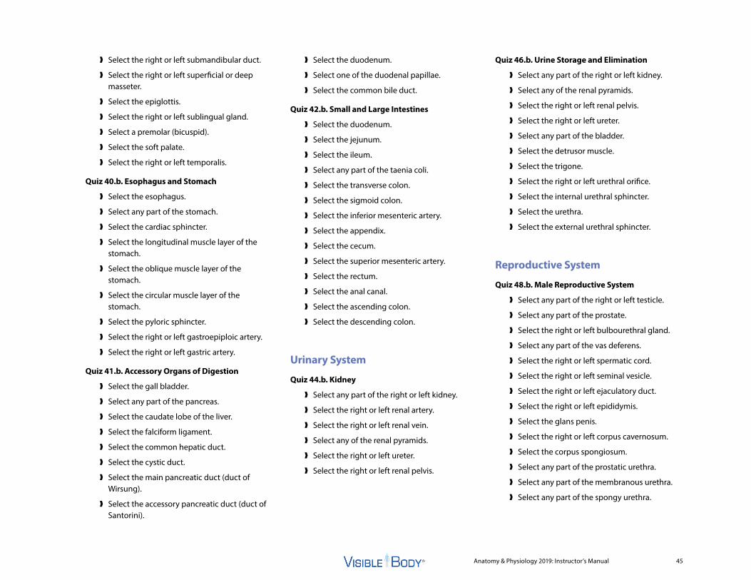
Anatomy & Physiology 2019: Instructor’s Manual 45
❱ Select the right or left submandibular duct.
❱ Select the right or left superficial or deep masseter.
❱ Select the epiglottis.
❱ Select the right or left sublingual gland.
❱ Select a premolar (bicuspid).
❱ Select the soft palate.
❱ Select the right or left temporalis.
Quiz 40.b. Esophagus and Stomach
❱ Select the esophagus.
❱ Select any part of the stomach.
❱ Select the cardiac sphincter.
❱ Select the longitudinal muscle layer of the stomach.
❱ Select the oblique muscle layer of the stomach.
❱ Select the circular muscle layer of the stomach.
❱ Select the pyloric sphincter.
❱ Select the right or left gastroepiploic artery.
❱ Select the right or left gastric artery.
Quiz 41.b. Accessory Organs of Digestion
❱ Select the gall bladder.
❱ Select any part of the pancreas.
❱ Select the caudate lobe of the liver.
❱ Select the falciform ligament.
❱ Select the common hepatic duct.
❱ Select the cystic duct.
❱ Select the main pancreatic duct (duct of Wirsung).
❱ Select the accessory pancreatic duct (duct of Santorini).
❱ Select the duodenum.
❱ Select one of the duodenal papillae.
❱ Select the common bile duct.
Quiz 42.b. Small and Large Intestines
❱ Select the duodenum.
❱ Select the jejunum.
❱ Select the ileum.
❱ Select any part of the taenia coli.
❱ Select the transverse colon.
❱ Select the sigmoid colon.
❱ Select the inferior mesenteric artery.
❱ Select the appendix.
❱ Select the cecum.
❱ Select the superior mesenteric artery.
❱ Select the rectum.
❱ Select the anal canal.
❱ Select the ascending colon.
❱ Select the descending colon.
Urinary System
Quiz 44.b. Kidney
❱ Select any part of the right or left kidney.
❱ Select the right or left renal artery.
❱ Select the right or left renal vein.
❱ Select any of the renal pyramids.
❱ Select the right or left ureter.
❱ Select the right or left renal pelvis.
Quiz 46.b. Urine Storage and Elimination
❱ Select any part of the right or left kidney.
❱ Select any of the renal pyramids.
❱ Select the right or left renal pelvis.
❱ Select the right or left ureter.
❱ Select any part of the bladder.
❱ Select the detrusor muscle.
❱ Select the trigone.
❱ Select the right or left urethral orifice.
❱ Select the internal urethral sphincter.
❱ Select the urethra.
❱ Select the external urethral sphincter.
Reproductive System
Quiz 48.b. Male Reproductive System
❱ Select any part of the right or left testicle.
❱ Select any part of the prostate.
❱ Select the right or left bulbourethral gland.
❱ Select any part of the vas deferens.
❱ Select the right or left spermatic cord.
❱ Select the right or left seminal vesicle.
❱ Select the right or left ejaculatory duct.
❱ Select the right or left epididymis.
❱ Select the glans penis.
❱ Select the right or left corpus cavernosum.
❱ Select the corpus spongiosum.
❱ Select any part of the prostatic urethra.
❱ Select any part of the membranous urethra.
❱ Select any part of the spongy urethra.

Anatomy & Physiology 2019: Instructor’s Manual 46
Quiz 49.b. Female Reproductive System
❱ Select any part of the uterus.
❱ Select any part of the right or left ovary.
❱ Select any part of the cervix.
❱ Select any part of the right or left uterine duct.
❱ Select the vagina.
❱ Select any part of the right or left mammary gland.
❱ Select the right or left lactiferous ducts.
❱ Select the right or left mammary gland lobules.
❱ Select the vestibule.
❱ Select the prepuce.
❱ Select the clitoris.
❱ Select the labia minora.
❱ Select the urethral orifice

Anatomy & Physiology 2019: Instructor’s Manual 47
Syllabus Correlations
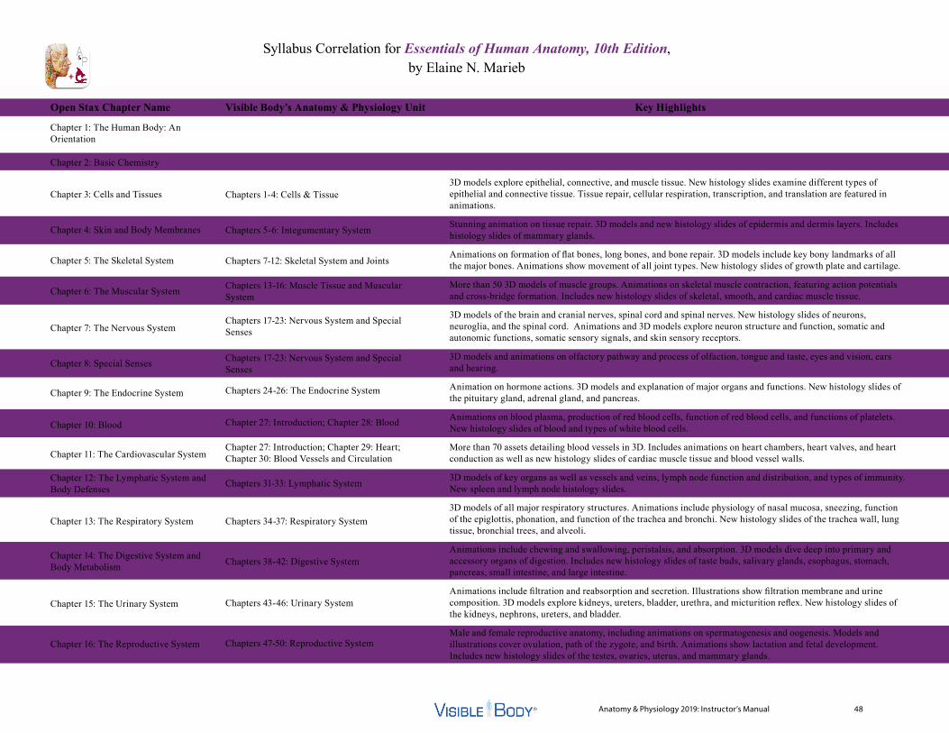
Anatomy & Physiology 2019: Instructor’s Manual 48
Syllabus Correlation for Essentials of Human Anatomy, 10th Edition, by Elaine N. Marieb
Open Stax Chapter Name
Chapter 1: The Human Body: An Orientation
Chapter 2: Basic Chemistry
Chapter 3: Cells and Tissues
Chapter 4: Skin and Body Membranes
Chapter 5: The Skeletal System
Chapter 6: The Muscular System
Chapter 7: The Nervous System
Chapter 8: Special Senses
Chapter 9: The Endocrine System
Chapter 10: Blood
Chapter 11: The Cardiovascular System
Chapter 12: The Lymphatic System and Body Defenses
Chapter 13: The Respiratory System
Chapter 14: The Digestive System and Body Metabolism
Chapter 15: The Urinary System
Chapter 16: The Reproductive System
Chapters 1-4: Cells & Tissue
Chapters 5-6: Integumentary System
Chapters 7-12: Skeletal System and Joints
Chapters 13-16: Muscle Tissue and Muscular System
Chapters 17-23: Nervous System and Special Senses
Chapters 17-23: Nervous System and Special Senses
Chapters 24-26: The Endocrine System
Chapter 27: Introduction; Chapter 28: Blood
Chapter 27: Introduction; Chapter 29: Heart; Chapter 30: Blood Vessels and Circulation
Chapters 31-33: Lymphatic System
Chapters 34-37: Respiratory System
Chapters 38-42: Digestive System
Chapters 43-46: Urinary System
Chapters 47-50: Reproductive System
3D models explore epithelial, connective, and muscle tissue. New histology slides examine different types of epithelial and connective tissue. Tissue repair, cellular respiration, transcription, and translation are featured in animations.
Stunning animation on tissue repair. 3D models and new histology slides of epidermis and dermis layers. Includes histology slides of mammary glands.
Animations on formation of flat bones, long bones, and bone repair. 3D models include key bony landmarks of all the major bones. Animations show movement of all joint types. New histology slides of growth plate and cartilage.
More than 50 3D models of muscle groups. Animations on skeletal muscle contraction, featuring action potentials and cross-bridge formation. Includes new histology slides of skeletal, smooth, and cardiac muscle tissue.
3D models of the brain and cranial nerves, spinal cord and spinal nerves. New histology slides of neurons, neuroglia, and the spinal cord. Animations and 3D models explore neuron structure and function, somatic and autonomic functions, somatic sensory signals, and skin sensory receptors.
3D models and animations on olfactory pathway and process of olfaction, tongue and taste, eyes and vision, ears and hearing.
Animation on hormone actions. 3D models and explanation of major organs and functions. New histology slides of the pituitary gland, adrenal gland, and pancreas.
Animations on blood plasma, production of red blood cells, function of red blood cells, and functions of platelets. New histology slides of blood and types of white blood cells.
More than 70 assets detailing blood vessels in 3D. Includes animations on heart chambers, heart valves, and heart conduction as well as new histology slides of cardiac muscle tissue and blood vessel walls.
3D models of key organs as well as vessels and veins, lymph node function and distribution, and types of immunity. New spleen and lymph node histology slides.
3D models of all major respiratory structures. Animations include physiology of nasal mucosa, sneezing, function of the epiglottis, phonation, and function of the trachea and bronchi. New histology slides of the trachea wall, lung tissue, bronchial trees, and alveoli.
Animations include chewing and swallowing, peristalsis, and absorption. 3D models dive deep into primary and accessory organs of digestion. Includes new histology slides of taste buds, salivary glands, esophagus, stomach, pancreas, small intestine, and large intestine.
Animations include filtration and reabsorption and secretion. Illustrations show filtration membrane and urine composition. 3D models explore kidneys, ureters, bladder, urethra, and micturition reflex. New histology slides of the kidneys, nephrons, ureters, and bladder.
Male and female reproductive anatomy, including animations on spermatogenesis and oogenesis. Models and illustrations cover ovulation, path of the zygote, and birth. Animations show lactation and fetal development. Includes new histology slides of the testes, ovaries, uterus, and mammary glands.
Visible Body’s Anatomy & Physiology Unit Key Highlights

Anatomy & Physiology 2019: Instructor’s Manual 49
Syllabus Correlation for Human Anatomy & Physiology, 9th Edition, by Elaine N. Marieb and Katja Hoehn
Marieb Chapter Name
Chapter 1: The Human Body: An Orientation
Chapter 2: Chemistry Comes Alive
Chapter 3: Cells: The Living Units
Chapter 4: Tissue: The Living Fabric
Chapter 5: The Integumentary System
Chapter 6: Bones and Skeletal Tissue
Chapter 7: The Skeleton
Chapter 8: Joints
Chapter 9: Muscles and Muscle Tissue
Chapter 10: The Muscular System
Chapter 11: The Fundamentals of the Nervous System and Nervous Tissue
Chapter 12: The Central Nervous System
Chapter 13: The Peripheral Nervous System and Reflex Activity
Chapter 14: The Autonomic Nervous System
Chapter 15: The Special Senses
Chapter 16: The Endocrine System
Chapter 17: Blood
Chapter 18: The Cardiovascular System: The HeartChapter 19: The Cardiovascular System: Blood VesselsChapter 20: The Lymphatic System and Lymphoid Organs and TissuesChapter 21: The Immune System: Innate and Adaptive Body Defenses
Chapters 1-4: Cells & Tissue
Chapters 1-4: Cells & Tissue
Chapters 5-6: Integumentary System
Chapters 7-12: Skeletal System and Joints
Chapters 7-12: Skeletal System and Joints
Chapter 12: JointsChapters 13-16: Muscle Tissue and Muscular SystemChapters 13-16: Muscle Tissue and Muscular System
Chapters 17-23: Nervous System and Special Senses
Chapters 17-23: Nervous System and Special Senses
Chapters 17-23: Nervous System and Special Senses
Chapters 17-23: Nervous System and Special Senses
Chapter 23: Special Senses
Chapters 24-26: The Endocrine System
Chapter 27: Introduction; Chapter 28: Blood
Chapter 27: Introduction; Chapter 29: Heart
Chapter 27: Introduction; Chapter 30: Blood Vessels and Circulation
Chapters 31-33: Lymphatic System
Chapters 31-33: Lymphatic System
Animations on cellular respiration, transcription, and translation.
3D models explore epithelial, connective, and muscle tissue. New histology slides examine different types of epithelial and connective tissue. Tissue repair and scarring are featured in an animation and 3D model.
Stunning animation on tissue repair. 3D models and new histology slides of epidermis and dermis layers. Includes histology slides of mammary glands.
Animations on formation of flat bones, long bones, and bone repair. New histology slides of growth plate and cartilage.
Now with 3D models that include key bony landmarks of all of the major bones.
Animations showing movement of all joint types.
Animations on skeletal muscle contraction, featuring action potentials and cross-bridge formation.
More than 50 3D models of muscle groups. Includes new histology slides of skeletal, smooth, and cardiac muscle tissue.
Illustrations of types of neurons and neuron structure. Includes animation of neuron function and new histology slides of neurons and neuroglia.
3D models of the brain, spinal cord, and spinal nerves showing anatomy and innervation. Includes new histology slides of spinal cord.
Includes animations and 3D models of somatic and autonomic functions, as well as somatic sensory signals and skin sensory receptors.
3D model conveying autonomic nervous functions.
3D models and animations on olfactory pathway and process of olfaction, tongue and taste, eyes and vision, and ears and hearing.
Animation on hormone actions, as well as 3D models and explanation of major organs and functions. New histology slides of the pituitary gland, adrenal gland, and pancreas.
Animations on blood plasma, production of red blood cells, function of red blood cells, and function of platelets. Includes new histology slides of blood and types of white blood cells.
Animations on heart chambers, heart valves, and heart conduction. Contains new histology slides of cardiac muscle tissue.
More than 55 assets detailing arteries and veins in 3D. Includes new histology slides of blood vessel walls.
3D models of key organs, as well as vessels and veins and lymph node function and distribution. Includes new spleen and lymph node histology slides.
Phagocytosis animation and illustrations on innate immunity, adaptive immunity, and types of white blood cells. 3D models of B and T cells.
Visible Body’s Anatomy & Physiology Unit Key Highlights
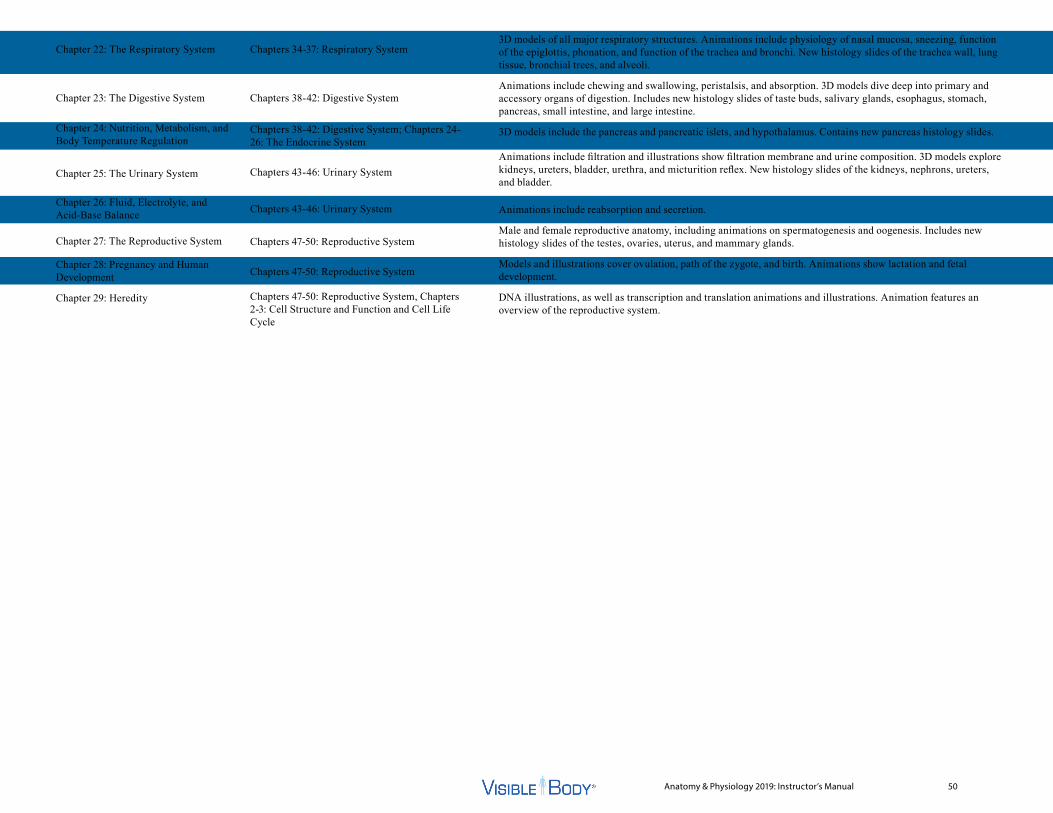
Anatomy & Physiology 2019: Instructor’s Manual 50
Chapter 22: The Respiratory System
Chapter 23: The Digestive System
Chapter 24: Nutrition, Metabolism, and Body Temperature Regulation
Chapter 25: The Urinary System
Chapter 26: Fluid, Electrolyte, and Acid-Base Balance
Chapter 27: The Reproductive System
Chapter 28: Pregnancy and Human Development
Chapter 29: Heredity
Chapters 34-37: Respiratory System
Chapters 38-42: Digestive System
Chapters 38-42: Digestive System; Chapters 24-26: The Endocrine System
Chapters 43-46: Urinary System
Chapters 43-46: Urinary System
Chapters 47-50: Reproductive System
Chapters 47-50: Reproductive System
Chapters 47-50: Reproductive System, Chapters 2-3: Cell Structure and Function and Cell Life Cycle
3D models of all major respiratory structures. Animations include physiology of nasal mucosa, sneezing, function of the epiglottis, phonation, and function of the trachea and bronchi. New histology slides of the trachea wall, lung tissue, bronchial trees, and alveoli.
Animations include chewing and swallowing, peristalsis, and absorption. 3D models dive deep into primary and accessory organs of digestion. Includes new histology slides of taste buds, salivary glands, esophagus, stomach, pancreas, small intestine, and large intestine.
3D models include the pancreas and pancreatic islets, and hypothalamus. Contains new pancreas histology slides.
Animations include filtration and illustrations show filtration membrane and urine composition. 3D models explore kidneys, ureters, bladder, urethra, and micturition reflex. New histology slides of the kidneys, nephrons, ureters, and bladder.
Animations include reabsorption and secretion.
Male and female reproductive anatomy, including animations on spermatogenesis and oogenesis. Includes new histology slides of the testes, ovaries, uterus, and mammary glands.
Models and illustrations cover ovulation, path of the zygote, and birth. Animations show lactation and fetal development.
DNA illustrations, as well as transcription and translation animations and illustrations. Animation features an overview of the reproductive system.
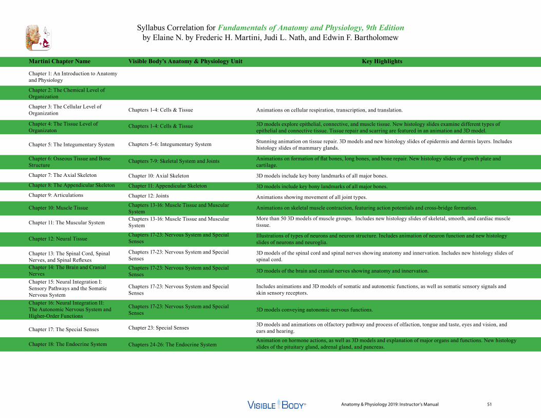
Anatomy & Physiology 2019: Instructor’s Manual 51
Syllabus Correlation for Fundamentals of Anatomy and Physiology, 9th Edition by Elaine N. by Frederic H. Martini, Judi L. Nath, and Edwin F. Bartholomew
Martini Chapter Name
Chapter 1: An Introduction to Anatomy and Physiology
Chapter 2: The Chemical Level of Organization
Chapter 3: The Cellular Level of Organization
Chapter 4: The Tissue Level of Organizaton
Chapter 5: The Integumentary System
Chapter 6: Osseous Tissue and Bone Structure
Chapter 7: The Axial Skeleton
Chapter 8: The Appendicular Skeleton
Chapter 9: Articulations
Chapter 10: Muscle Tissue
Chapter 11: The Muscular System
Chapter 12: Neural Tissue
Chapter 13: The Spinal Cord, Spinal Nerves, and Spinal ReflexesChapter 14: The Brain and Cranial NervesChapter 15: Neural Integration I: Sensory Pathways and the Somatic Nervous SystemChapter 16: Neural Integration II: The Autonomic Nervous System and Higher-Order Functions
Chapter 17: The Special Senses
Chapter 18: The Endocrine System
Chapters 1-4: Cells & Tissue
Chapters 1-4: Cells & Tissue
Chapters 5-6: Integumentary System
Chapters 7-9: Skeletal System and Joints
Chapter 10: Axial Skeleton
Chapter 11: Appendicular Skeleton
Chapter 12: Joints
Chapters 13-16: Muscle Tissue and Muscular SystemChapters 13-16: Muscle Tissue and Muscular System
Chapters 17-23: Nervous System and Special Senses
Chapters 17-23: Nervous System and Special Senses
Chapters 17-23: Nervous System and Special Senses
Chapters 17-23: Nervous System and Special Senses
Chapters 17-23: Nervous System and Special Senses
Chapter 23: Special Senses
Chapters 24-26: The Endocrine System
Animations on cellular respiration, transcription, and translation.
3D models explore epithelial, connective, and muscle tissue. New histology slides examine different types of epithelial and connective tissue. Tissue repair and scarring are featured in an animation and 3D model.
Stunning animation on tissue repair. 3D models and new histology slides of epidermis and dermis layers. Includes histology slides of mammary glands.
Animations on formation of flat bones, long bones, and bone repair. New histology slides of growth plate and cartilage.
3D models include key bony landmarks of all major bones.
3D models include key bony landmarks of all major bones.
Animations showing movement of all joint types.
Animations on skeletal muscle contraction, featuring action potentials and cross-bridge formation.
More than 50 3D models of muscle groups. Includes new histology slides of skeletal, smooth, and cardiac muscle tissue.
Illustrations of types of neurons and neuron structure. Includes animation of neuron function and new histology slides of neurons and neuroglia.
3D models of the spinal cord and spinal nerves showing anatomy and innervation. Includes new histology slides of spinal cord.
3D models of the brain and cranial nerves showing anatomy and innervation.
Includes animations and 3D models of somatic and autonomic functions, as well as somatic sensory signals and skin sensory receptors.
3D models conveying autonomic nervous functions.
3D models and animations on olfactory pathway and process of olfaction, tongue and taste, eyes and vision, and ears and hearing.
Animation on hormone actions, as well as 3D models and explanation of major organs and functions. New histology slides of the pituitary gland, adrenal gland, and pancreas.
Visible Body’s Anatomy & Physiology Unit Key Highlights
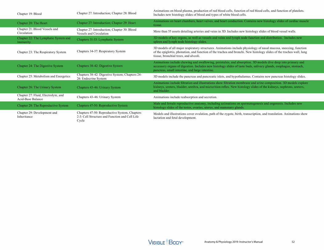
Anatomy & Physiology 2019: Instructor’s Manual 52
Chapter 19: Blood
Chapter 20: The Heart
Chapter 21: Blood Vessels and CirculationChapter 22: The Lymphatic System and Immunity
Chapter 23: The Respiratory System
Chapter 24: The Digestive System
Chapter 25: Metabolism and Energetics
Chapter 26: The Urinary System
Chapter 27: Fluid, Electrolyte, and Acid-Base Balance
Chapter 28: The Reproductive System
Chapter 29: Development and Inheritance
Chapter 27: Introduction; Chapter 28: Blood
Chapter 27: Introduction; Chapter 29: Heart
Chapter 27: Introduction; Chapter 30: Blood Vessels and Circulation
Chapters 31-33: Lymphatic System
Chapters 34-37: Respiratory System
Chapters 38-42: Digestive System
Chapters 38-42: Digestive System; Chapters 24-26: Endocrine System
Chapters 43-46: Urinary System
Chapters 43-46: Urinary System
Chapters 47-50: Reproductive System
Chapters 47-50: Reproductive System, Chapters 2-3: Cell Structure and Function and Cell Life Cycle
Animations on blood plasma, production of red blood cells, function of red blood cells, and function of platelets. Includes new histology slides of blood and types of white blood cells.
Animations on heart chambers, heart valves, and heart conduction. Contains new histology slides of cardiac muscle tissue.
More than 55 assets detailing arteries and veins in 3D. Includes new histology slides of blood vessel walls.
3D models of key organs, as well as vessels and veins and lymph node function and distribution. Includes new spleen and lymph node histology slides.
3D models of all major respiratory structures. Animations include physiology of nasal mucosa, sneezing, function of the epiglottis, phonation, and function of the trachea and bronchi. New histology slides of the trachea wall, lung tissue, bronchial trees, and alveoli.
Animations include chewing and swallowing, peristalsis, and absorption. 3D models dive deep into primary and accessory organs of digestion. Includes new histology slides of taste buds, salivary glands, esophagus, stomach, pancreas, small intestine, and large intestine.
3D models include the pancreas and pancreatic islets, and hypothalamus. Contains new pancreas histology slides.
Animations include filtration and illustrations show filtration membrane and urine composition. 3D models explore kidneys, ureters, bladder, urethra, and micturition reflex. New histology slides of the kidneys, nephrons, ureters, and bladder.
Animations include reabsorption and secretion.
Male and female reproductive anatomy, including animations on spermatogenesis and oogenesis. Includes new histology slides of the testes, ovaries, uterus, and mammary glands.
Models and illustrations cover ovulation, path of the zygote, birth, transcription, and translation. Animations show lactation and fetal development.
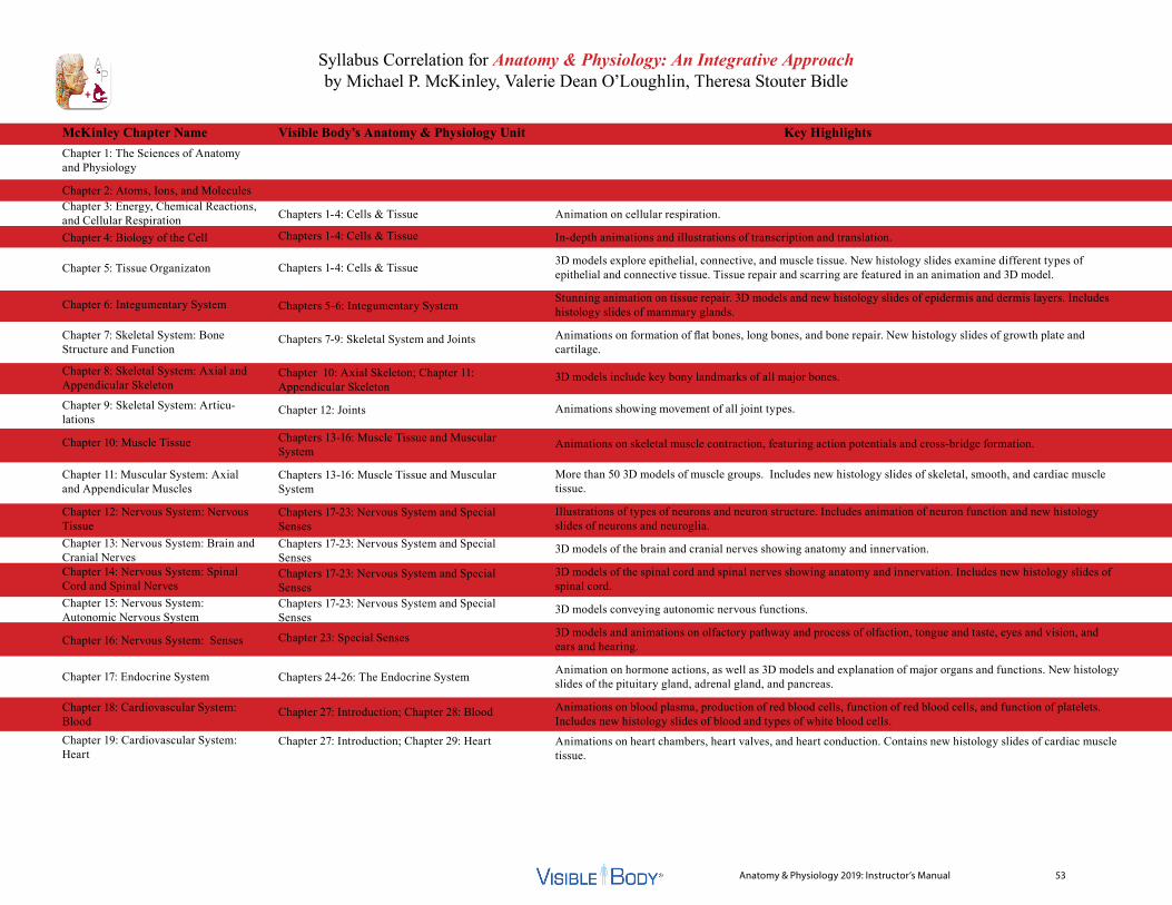
Anatomy & Physiology 2019: Instructor’s Manual 53
Syllabus Correlation for Anatomy & Physiology: An Integrative Approach by Michael P. McKinley, Valerie Dean O’Loughlin, Theresa Stouter Bidle
McKinley Chapter NameChapter 1: The Sciences of Anatomy and Physiology
Chapter 2: Atoms, Ions, and MoleculesChapter 3: Energy, Chemical Reactions, and Cellular RespirationChapter 4: Biology of the Cell
Chapter 5: Tissue Organizaton
Chapter 6: Integumentary System
Chapter 7: Skeletal System: Bone Structure and Function
Chapter 8: Skeletal System: Axial and Appendicular Skeleton
Chapter 9: Skeletal System: Articu-lations
Chapter 10: Muscle Tissue
Chapter 11: Muscular System: Axial and Appendicular Muscles
Chapter 12: Nervous System: Nervous TissueChapter 13: Nervous System: Brain and Cranial NervesChapter 14: Nervous System: Spinal Cord and Spinal NervesChapter 15: Nervous System: Autonomic Nervous System
Chapter 16: Nervous System: Senses
Chapter 17: Endocrine System
Chapter 18: Cardiovascular System: BloodChapter 19: Cardiovascular System: Heart
Chapters 1-4: Cells & Tissue
Chapters 1-4: Cells & Tissue
Chapters 1-4: Cells & Tissue
Chapters 5-6: Integumentary System
Chapters 7-9: Skeletal System and Joints
Chapter 10: Axial Skeleton; Chapter 11: Appendicular Skeleton
Chapter 12: Joints
Chapters 13-16: Muscle Tissue and Muscular System
Chapters 13-16: Muscle Tissue and Muscular System
Chapters 17-23: Nervous System and Special SensesChapters 17-23: Nervous System and Special SensesChapters 17-23: Nervous System and Special SensesChapters 17-23: Nervous System and Special Senses
Chapter 23: Special Senses
Chapters 24-26: The Endocrine System
Chapter 27: Introduction; Chapter 28: Blood
Chapter 27: Introduction; Chapter 29: Heart
Animation on cellular respiration.
In-depth animations and illustrations of transcription and translation.
3D models explore epithelial, connective, and muscle tissue. New histology slides examine different types of epithelial and connective tissue. Tissue repair and scarring are featured in an animation and 3D model.
Stunning animation on tissue repair. 3D models and new histology slides of epidermis and dermis layers. Includes histology slides of mammary glands.
Animations on formation of flat bones, long bones, and bone repair. New histology slides of growth plate and cartilage.
3D models include key bony landmarks of all major bones.
Animations showing movement of all joint types.
Animations on skeletal muscle contraction, featuring action potentials and cross-bridge formation.
More than 50 3D models of muscle groups. Includes new histology slides of skeletal, smooth, and cardiac muscle tissue.
Illustrations of types of neurons and neuron structure. Includes animation of neuron function and new histology slides of neurons and neuroglia.
3D models of the brain and cranial nerves showing anatomy and innervation.
3D models of the spinal cord and spinal nerves showing anatomy and innervation. Includes new histology slides of spinal cord.
3D models conveying autonomic nervous functions.
3D models and animations on olfactory pathway and process of olfaction, tongue and taste, eyes and vision, and ears and hearing.
Animation on hormone actions, as well as 3D models and explanation of major organs and functions. New histology slides of the pituitary gland, adrenal gland, and pancreas.
Animations on blood plasma, production of red blood cells, function of red blood cells, and function of platelets. Includes new histology slides of blood and types of white blood cells.
Animations on heart chambers, heart valves, and heart conduction. Contains new histology slides of cardiac muscle tissue.
Visible Body’s Anatomy & Physiology Unit Key Highlights
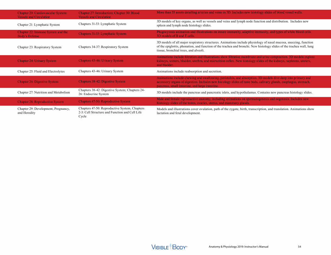
Anatomy & Physiology 2019: Instructor’s Manual 54
Chapter 20: Cardiovascular System: Vessels and Circulation
Chapter 21: Lymphatic System
Chapter 22: Immune System and the Body’s Defense
Chapter 23: Respiratory System
Chapter 24: Urinary System
Chapter 25: Fluid and Electrolytes
Chapter 26: Digestive System
Chapter 27: Nutrition and Metabolism
Chapter 28: Reproductive System
Chapter 29: Development, Pregnancy, and Heredity
Chapter 27: Introduction; Chapter 30: Blood Vessels and Circulation
Chapters 31-33: Lymphatic System
Chapters 31-33: Lymphatic System
Chapters 34-37: Respiratory System
Chapters 43-46: Urinary System
Chapters 43-46: Urinary System
Chapters 38-42: Digestive System
Chapters 38-42: Digestive System; Chapters 24-26: Endocrine System
Chapters 47-50: Reproductive System
Chapters 47-50: Reproductive System, Chapters 2-3: Cell Structure and Function and Cell Life Cycle
More than 55 assets detailing arteries and veins in 3D. Includes new histology slides of blood vessel walls.
3D models of key organs, as well as vessels and veins and lymph node function and distribution. Includes new spleen and lymph node histology slides.
Phagocytosis animation and illustrations on innate immunity, adaptive immunity, and types of white blood cells. 3D models of B and T cells.
3D models of all major respiratory structures. Animations include physiology of nasal mucosa, sneezing, function of the epiglottis, phonation, and function of the trachea and bronchi. New histology slides of the trachea wall, lung tissue, bronchial trees, and alveoli.
Animations include filtration and illustrations show filtration membrane and urine composition. 3D models explore kidneys, ureters, bladder, urethra, and micturition reflex. New histology slides of the kidneys, nephrons, ureters, and bladder.
Animations include reabsorption and secretion.
Animations include chewing and swallowing, peristalsis, and absorption. 3D models dive deep into primary and accessory organs of digestion. Includes new histology slides of taste buds, salivary glands, esophagus, stomach, pancreas, small intestine, and large intestine.
3D models include the pancreas and pancreatic islets, and hypothalamus. Contains new pancreas histology slides.
Male and female reproductive anatomy, including animations on spermatogenesis and oogenesis. Includes new histology slides of the testes, ovaries, uterus, and mammary glands.
Models and illustrations cover ovulation, path of the zygote, birth, transcription, and translation. Animations show lactation and fetal development.
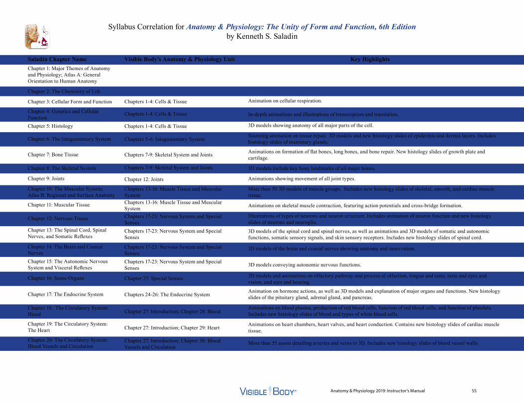
Anatomy & Physiology 2019: Instructor’s Manual 55
Syllabus Correlation for Anatomy & Physiology: The Unity of Form and Function, 6th Edition by Kenneth S. Saladin
Saladin Chapter NameChapter 1: Major Themes of Anatomy and Physiology; Atlas A: General Orientation to Human Anatomy
Chapter 2: The Chemistry of Life
Chapter 3: Cellular Form and Function
Chapter 4: Genetics and Cellular FunctionChapter 5: Histology
Chapter 6: The Integumentary System
Chapter 7: Bone Tissue
Chapter 8: The Skeletal System
Chapter 9: Joints
Chapter 10: The Muscular System; Atlas B: Regional and Surface Anatomy
Chapter 11: Muscular Tissue
Chapter 12: Nervous Tissue
Chapter 13: The Spinal Cord, Spinal Nerves, and Somatic Reflexes
Chapter 14: The Brain and Cranial NervesChapter 15: The Autonomic Nervous System and Visceral Reflexes
Chapter 16: Sense Organs
Chapter 17: The Endocrine System
Chapter 18: The Circulatory System: Blood
Chapter 19: The Circulatory System: The Heart
Chapter 20: The Circulatory System: Blood Vessels and Circulation
Chapters 1-4: Cells & Tissue
Chapters 1-4: Cells & Tissue
Chapters 1-4: Cells & Tissue
Chapters 5-6: Integumentary System
Chapters 7-9: Skeletal System and Joints
Chapters 7-9: Skeletal System and Joints
Chapter 12: Joints
Chapters 13-16: Muscle Tissue and Muscular SystemChapters 13-16: Muscle Tissue and Muscular SystemChapters 17-23: Nervous System and Special SensesChapters 17-23: Nervous System and Special Senses
Chapters 17-23: Nervous System and Special SensesChapters 17-23: Nervous System and Special Senses
Chapter 23: Special Senses
Chapters 24-26: The Endocrine System
Chapter 27: Introduction; Chapter 28: Blood
Chapter 27: Introduction; Chapter 29: Heart
Chapter 27: Introduction; Chapter 30: Blood Vessels and Circulation
Animation on cellular respiration.
In-depth animations and illustrations of transcription and translation.
3D models showing anatomy of all major parts of the cell.
Stunning animation on tissue repair. 3D models and new histology slides of epidermis and dermis layers. Includes histology slides of mammary glands.
Animations on formation of flat bones, long bones, and bone repair. New histology slides of growth plate and cartilage.
3D models include key bony landmarks of all major bones.
Animations showing movement of all joint types.
More than 50 3D models of muscle groups. Includes new histology slides of skeletal, smooth, and cardiac muscle tissue.
Animations on skeletal muscle contraction, featuring action potentials and cross-bridge formation.
Illustrations of types of neurons and neuron structure. Includes animation of neuron function and new histology slides of neurons and neuroglia.
3D models of the spinal cord and spinal nerves, as well as animations and 3D models of somatic and autonomic functions, somatic sensory signals, and skin sensory receptors. Includes new histology slides of spinal cord.
3D models of the brain and cranial nerves showing anatomy and innervation.
3D models conveying autonomic nervous functions.
3D models and animations on olfactory pathway and process of olfaction, tongue and taste, taste and eyes and vision, and ears and hearing.
Animation on hormone actions, as well as 3D models and explanation of major organs and functions. New histology slides of the pituitary gland, adrenal gland, and pancreas.
Animations on blood plasma, production of red blood cells, function of red blood cells, and function of platelets. Includes new histology slides of blood and types of white blood cells.
Animations on heart chambers, heart valves, and heart conduction. Contains new histology slides of cardiac muscle tissue.
More than 55 assets detailing arteries and veins in 3D. Includes new histology slides of blood vessel walls.
Visible Body’s Anatomy & Physiology Unit Key Highlights
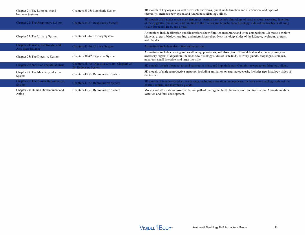
Anatomy & Physiology 2019: Instructor’s Manual 56
Chapter 21: The Lymphatic and Immune Systems
Chapter 22: The Respiratory System
Chapter 23: The Urinary System
Chapter 24: Water, Electrolyte, and Acid-Base Balance
Chapter 25: The Digestive System
Chapter 26: Nutrition and Metabolism
Chapter 27: The Male Reproductive System
Chapter 28: The Female Reproductive System
Chapter 29: Human Development and Aging
Chapters 31-33: Lymphatic System
Chapters 34-37: Respiratory System
Chapters 43-46: Urinary System
Chapters 43-46: Urinary System
Chapters 38-42: Digestive System
Chapters 38-42: Digestive System; Chapters 24-26: Endocrine System
Chapters 47-50: Reproductive System
Chapters 47-50: Reproductive System
Chapters 47-50: Reproductive System
3D models of key organs, as well as vessels and veins, lymph node function and distribution, and types of immunity. Includes new spleen and lymph node histology slides.
3D models of all major respiratory structures. Animations include physiology of nasal mucosa, sneezing, function of the epiglottis, phonation, and function of the trachea and bronchi. New histology slides of the trachea wall, lung tissue, bronchial trees, and alveoli.
Animations include filtration and illustrations show filtration membrane and urine composition. 3D models explore kidneys, ureters, bladder, urethra, and micturition reflex. New histology slides of the kidneys, nephrons, ureters, and bladder.
Animations include reabsorption and secretion.
Animations include chewing and swallowing, peristalsis, and absorption. 3D models dive deep into primary and accessory organs of digestion. Includes new histology slides of taste buds, salivary glands, esophagus, stomach, pancreas, small intestine, and large intestine.
3D models include the pancreas and pancreatic islets, and hypothalamus. Contains new pancreas histology slides.
3D models of male reproductive anatomy, including animation on spermatogenesis. Includes new histology slides of the testes.
3D models of female reproductive anatomy, including animation on oogenesis. Includes new histology slides of the ovaries, uterus, and mammary glands.
Models and illustrations cover ovulation, path of the zygote, birth, transcription, and translation. Animations show lactation and fetal development.

Anatomy & Physiology 2019: Instructor’s Manual 57
Syllabus Correlation for Principles of Anatomy and Physiology, 13th Edition by Gerard J. Tortora and Bryan Derrickson
Tortora Chapter NameChapter 1: An Introduction to the Human BodyChapter 2: The Chemical Level of Organization
Chapter 3: The Cellular Level of Organization
Chapter 4: The Tissue Level of Organizaton
Chapter 5: The Integumentary System
Chapter 6: The Skeletal System: Bone TissueChapter 7: The Skeletal System: The Axial SkeletonChapter 8: The Skeletal System: The Appendicular SkeletonChapter 9: Joints
Chapter 10: Muscular Tissue
Chapter 11: The Muscular System
Chapter 12: Nervous Tissue
Chapter 13: The Spinal Cord and Spinal Nerves
Chapter 14: The Brain and Cranial NervesChapter 15: The Autonomic Nervous SystemChapter 16: Sensory, Motor, and Integrative Systems
Chapter 17: The Special Senses
Chapter 18: The Endocrine System
Chapter 19: The Cardiovascular System: The Blood
Chapters 1-4: Cells & Tissue
Chapters 1-4: Cells & Tissue
Chapters 5-6: Integumentary System
Chapters 7-9: Skeletal System and Joints
Chapter 10: Axial Skeleton
Chapter 11: Appendicular Skeleton
Chapter 12: Joints
Chapters 13-16: Muscle Tissue and Muscular System
Chapters 13-16: Muscle Tissue and Muscular SystemChapters 17-23: Nervous System and Special SensesChapters 17-23: Nervous System and Special Senses
Chapters 17-23: Nervous System and Special SensesChapters 17-23: Nervous System and Special SensesChapters 17-23: Nervous System and Special Senses
Chapter 23: Special Senses
Chapters 24-26: Endocrine System
Chapter 27: Introduction; Chapter 28: Blood
Animations on cellular respiration, transcription, and translation.
3D models explore epithelial, connective, and muscle tissue. New histology slides examine different types of epithelial and connective tissue. Tissue repair and scarring are featured in an animation and 3D model.
Stunning animation on tissue repair. 3D models and new histology slides of epidermis and dermis layers. Includes histology slides of mammary glands.
Animations on formation of flat bones, long bones, and bone repair. New histology slides of growth plate and cartilage.
3D models include key bony landmarks of all major bones.
3D models include key bony landmarks of all major bones.
Animations showing movement of all joint types.
Animations on skeletal muscle contraction, featuring action potentials and cross-bridge formation.
More than 50 3D models of muscle groups. Includes new histology slides of skeletal, smooth, and cardiac muscle tissue.Illustrations of types of neurons and neuron structure. Includes animation of neuron function and new histology slides of neurons and neuroglia.3D models of the spinal cord and spinal nerves showing anatomy and innervation. Includes new histology slides of spinal cord.
3D models of the brain and cranial nerves showing anatomy and innervation.
3D models conveying autonomic nervous functions.
3D models of somatic and autonomic functions, somatic sensory signals, and skin sensory receptors.
3D models and animations on olfactory pathway and process of olfaction, tongue and taste, eyes and vision, and ears and hearing.
Animation on hormone actions, as well as 3D models and explanation of major organs and functions. New histology slides of the pituitary gland, adrenal gland, and pancreas.
Animations on blood plasma, production of red blood cells, function of red blood cells, and function of platelets. Includes new histology slides of blood and types of white blood cells.
Visible Body’s Anatomy & Physiology Unit Key Highlights
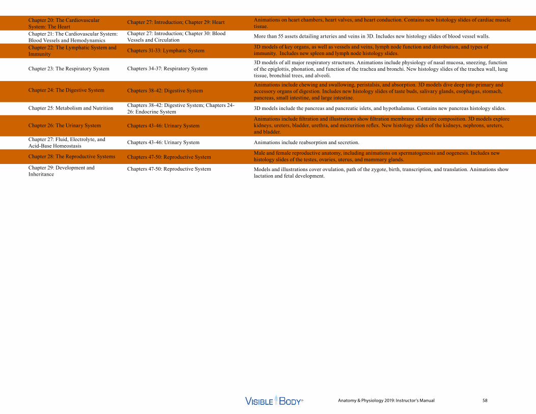
Anatomy & Physiology 2019: Instructor’s Manual 58
Chapter 20: The Cardiovascular System: The HeartChapter 21: The Cardiovascular System: Blood Vessels and HemodynamicsChapter 22: The Lymphatic System and Immunity
Chapter 23: The Respiratory System
Chapter 24: The Digestive System
Chapter 25: Metabolism and Nutrition
Chapter 26: The Urinary System
Chapter 27: Fluid, Electrolyte, and Acid-Base Homeostasis
Chapter 28: The Reproductive Systems
Chapter 29: Development and Inheritance
Chapter 27: Introduction; Chapter 29: Heart
Chapter 27: Introduction; Chapter 30: Blood Vessels and Circulation
Chapters 31-33: Lymphatic System
Chapters 34-37: Respiratory System
Chapters 38-42: Digestive System
Chapters 38-42: Digestive System; Chapters 24-26: Endocrine System
Chapters 43-46: Urinary System
Chapters 43-46: Urinary System
Chapters 47-50: Reproductive System
Chapters 47-50: Reproductive System
Animations on heart chambers, heart valves, and heart conduction. Contains new histology slides of cardiac muscle tissue.
More than 55 assets detailing arteries and veins in 3D. Includes new histology slides of blood vessel walls.
3D models of key organs, as well as vessels and veins, lymph node function and distribution, and types of immunity. Includes new spleen and lymph node histology slides.
3D models of all major respiratory structures. Animations include physiology of nasal mucosa, sneezing, function of the epiglottis, phonation, and function of the trachea and bronchi. New histology slides of the trachea wall, lung tissue, bronchial trees, and alveoli.
Animations include chewing and swallowing, peristalsis, and absorption. 3D models dive deep into primary and accessory organs of digestion. Includes new histology slides of taste buds, salivary glands, esophagus, stomach, pancreas, small intestine, and large intestine.
3D models include the pancreas and pancreatic islets, and hypothalamus. Contains new pancreas histology slides.
Animations include filtration and illustrations show filtration membrane and urine composition. 3D models explore kidneys, ureters, bladder, urethra, and micturition reflex. New histology slides of the kidneys, nephrons, ureters, and bladder.
Animations include reabsorption and secretion.
Male and female reproductive anatomy, including animations on spermatogenesis and oogenesis. Includes new histology slides of the testes, ovaries, uterus, and mammary glands.
Models and illustrations cover ovulation, path of the zygote, birth, transcription, and translation. Animations show lactation and fetal development.

Syllabus Correlation for Anatomy and Physiology, from Open Stax
Open Stax Chapter Name Chapter 1: An Introduction to the Human BodyChapter 2: The Chemical Level of Organization
Chapter 3: The Cellular Level of Organization
Chapter 4: The Tissue Level of Organization
Chapter 5: The Integumentary System
Chapter 6: Bone Tissue and the Skeletal System
Chapter 7: Axial Skeleton
Chapter 8: The Appendicular Skeleton
Chapter 9: Joints
Chapter 10: Muscle Tissue
Chapter 11: The Muscular System
Chapter 12: The Nervous System and Nervous Tissue
Chapter 13: Anatomy of the Nervous System
Chapter 14: The Somatic Nervous System
Chapter 15: The Autonomic Nervous System
Chapter 16: The Neurological Exam
Chapter 17: The Endocrine System
Chapter 18: The Cardiovascular System: Blood
Chapter 1: Introduction: Cells and Tissues; Chapter 2: Cell Structure and Function; Chapter 3: Cell Life Cycle
Chapter 4: Tissues
Chapters 5-6: Integumentary System
Chapter 7: Introduction: Skeletal System/ Joints; Chapter 8: Types of Bones; Chapter 9: Bone Tissue
Chapter 10: Axial Skeleton
Chapter 11: Appendicular Skeleton
Chapter 12: Joints
Chapter 13: Introduction: Muscular System; Chapter 14: Skeletal Muscle Tissue; Chapter 15: Smooth and Cardiac Muscle Tissue
Chapter 16: Muscular System
Chapter 17: Introduction: Nervous System/ Special Senses; Chapter 18: Nervous TissueChapter 19: Spinal Cord and Spinal Nerves; Chapter 20: Brain; Chapter 21: Cranial Nerves
Chapter 22: Somatic and Autonomic Nervous Systems; Chapter 23: Special Senses
Chapter 22: Somatic and Autonomic Nervous Systems
Chapters 24-26: Endocrine System
Chapter 27: Introduction: Circulatory System; Chapter 28: Blood
Animations on cellular respiration, transcription, and translation.
3D models explore epithelial, connective, and muscle tissue. New histology slides examine different types of epithelial and connective tissue. Tissue repair and scarring are featured in an animation and 3D model.
Stunning animation on tissue repair. 3D models and new histology slides of epidermis and dermis layers. Includes histology slides of mammary glands.
Animations on formation of flat bones, long bones, and bone repair. 3D models include key bony landmarks of all the major bones. New histology slides of growth plate and cartilage.
3D models include key bony landmarks of all the major bones.
3D models include key bony landmarks of all the major bones.
Animations showing movement of all joint types.
Animations on skeletal muscle contraction, featuring action potentials and cross-bridge formation. Includes new histology slides of skeletal, smooth, and cardiac muscle tissue.
More than 50 3D models of muscle groups.
Illustrations of types of neurons and neuron structure. Includes animation of neuron function and new histology slides of neurons and neuroglia.
3D models of the spinal cord and spinal nerves showing anatomy and innervation. Includes new histology slides of spinal cord.
3D models of somatic and autonomic functions, somatic sensory signals, and skin sensory receptors; as well as 3D models and animations on olfactory pathway and process of olfaction, tongue and taste, eyes and vision, and ears and hearing.
3D models conveying autonomic nervous functions.
Animation on hormone actions, as well as 3D models and explanation of major organs and functions. New histology slides of the pituitary gland, adrenal gland, and pancreas.Animations, 3D models, and illustrations explain the production and functions of blood plasma, red blood cells, white blood cells, and platelets. Includes new histology slides of blood and types of white blood cells.
Visible Body’s Anatomy & Physiology Unit Key Highlights
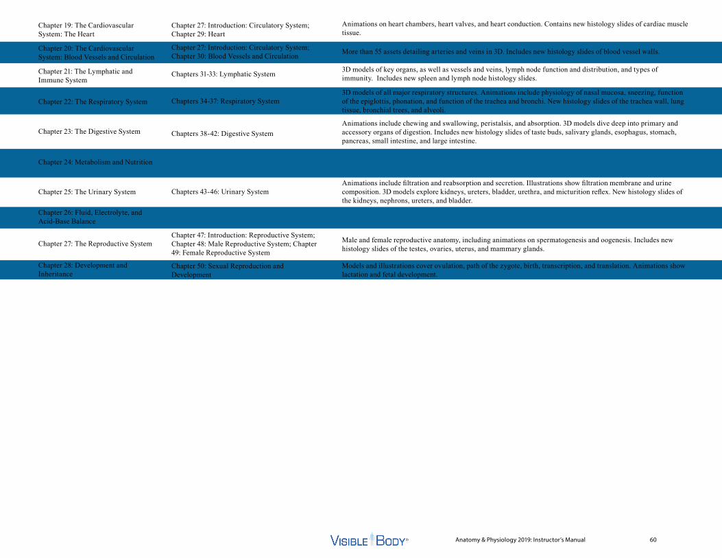
Anatomy & Physiology 2019: Instructor’s Manual 60
Chapter 19: The Cardiovascular System: The Heart
Chapter 20: The Cardiovascular System: Blood Vessels and Circulation
Chapter 21: The Lymphatic and Immune System
Chapter 22: The Respiratory System
Chapter 23: The Digestive System
Chapter 24: Metabolism and Nutrition
Chapter 25: The Urinary System
Chapter 26: Fluid, Electrolyte, and Acid-Base Balance
Chapter 27: The Reproductive System
Chapter 28: Development and Inheritance
Chapter 27: Introduction: Circulatory System; Chapter 29: Heart
Chapter 27: Introduction: Circulatory System; Chapter 30: Blood Vessels and Circulation
Chapters 31-33: Lymphatic System
Chapters 34-37: Respiratory System
Chapters 38-42: Digestive System
Chapters 43-46: Urinary System
Chapter 47: Introduction: Reproductive System; Chapter 48: Male Reproductive System; Chapter 49: Female Reproductive System
Chapter 50: Sexual Reproduction and Development
Animations on heart chambers, heart valves, and heart conduction. Contains new histology slides of cardiac muscle tissue.
More than 55 assets detailing arteries and veins in 3D. Includes new histology slides of blood vessel walls.
3D models of key organs, as well as vessels and veins, lymph node function and distribution, and types of immunity. Includes new spleen and lymph node histology slides.
3D models of all major respiratory structures. Animations include physiology of nasal mucosa, sneezing, function of the epiglottis, phonation, and function of the trachea and bronchi. New histology slides of the trachea wall, lung tissue, bronchial trees, and alveoli.
Animations include chewing and swallowing, peristalsis, and absorption. 3D models dive deep into primary and accessory organs of digestion. Includes new histology slides of taste buds, salivary glands, esophagus, stomach, pancreas, small intestine, and large intestine.
Animations include filtration and reabsorption and secretion. Illustrations show filtration membrane and urine composition. 3D models explore kidneys, ureters, bladder, urethra, and micturition reflex. New histology slides of the kidneys, nephrons, ureters, and bladder.
Male and female reproductive anatomy, including animations on spermatogenesis and oogenesis. Includes new histology slides of the testes, ovaries, uterus, and mammary glands.
Models and illustrations cover ovulation, path of the zygote, birth, transcription, and translation. Animations show lactation and fetal development.

Anatomy & Physiology 2019: Instructor’s Manual 61
Syllabus Correlation for Seeley’s Anatomy & Physiology, 11th Edition by Cinnamon VanPutte, Jennifer Regan, and Andrew Russ
Seeley’s Chapter NameChapter 1: The Human Organism
Chapter 2: The Chemical Basis of Life
Chapter 3: Cell Biology
Chapter 4: Tissues
Chapter 5: Integumentary System
Chapter 6: Skeletal System: Bones and Bone Tissue
Chapter 7: Skeletal System: Gross Anatomy
Chapter 8: Joints and Movement
Chapter 9: Muscular System: Histology and Physiology
Chapter 10: Muscular System: Gross Anatomy
Chapter 11: Functional Organization of Nervous Tissue
Chapter 12: Spinal Cord and Spinal Nerves
Chapter 13: Brain and Cranial Nerves
Chapter 14: Integration of Nervous System Functions
Chapter 15: The Special Senses
Chapters 1-4: Cells & Tissue
Chapter 4: Tissues
Chapters 5–6: Integumentary System
Chapter 8: Types of Bones; Chapter 9: Bone Tissue
Chapter 10: Axial Skeleton; Chapter 11: Appendicular Skeleton
Chapter 12: Joints
Chapters 13–16: Muscle Tissue and Muscular System
Chapter 16: Muscular System
Chapter 17: Introduction; Chapter 18: Nervous Tissue; Chapter 22: Somatic and Autonomic Nervous Systems
Chapter 19: Spinal Cord and Spinal Nerves
Chapter 20: Brain; Chapter 21: Cranial Nerves
Chapter 22: Somatic and Autonomic Nervous Systems
Chapter 23: Special Senses
Animations show passive and active transport, mitosis, cellular respiration, transcription, and translation. 3D models and illustrations explain cell structures and functions, cell types, osmosis, the cell cycle, meiosis, protein synthesis, and DNA replication.
An animation and 3D model show tissue repair and scarring. 3D models explain epithelial, connective, muscular, and nervous tissue. New histology slides examine different types of epithelial and connective tissue.
An animation and 3D model show tissue repair and scarring. 3D models explain the epidermis and dermis layers, skin circulation and innervation, and the structure and function of skin, hair, nails, and glands. Includes new histology slides of epidermis, dermis, and mammary glands.
Animations on formation of flat bones, long bones, and bone repair. 3D models and illustrations explain the bone types, bone tissue, and types of fractures. New histology slides of growth plate and cartilage.
3D models and illustrations explain the structures of the axial and appendicular skeleton with key bony landmarks on all the major bones.
Animations and 3D models show all the joint types and their movements. An illustration explains the effects of aging on joints.
7 different animations, as well as several 3D models, illustrations, and new histology slides, cover the structure and functions of skeletal, smooth, and cardiac muscle.
3D models explore over 50 different groups of muscle structures. An animation shows muscle interactions. Illustrations explain skeletal muscle attachments and the three classes of levers.
3D models, animations, and illustrations explain the functions of the central, peripheral, somatic, autonomic, sympathetic, and parasympathetic divisions of the nervous system; neuron types, structure, and function; neuroglia in the central and peripheral nervous systems; resting and action potentials; and neurotransmitters. New histology slides explore neurons and neuroglia.
3D models and illustrations explain spinal cord anatomy, sensory signals, motor commands, and spinal nerve structure and regions. An animation and 3D model cover the somatic reflex arc. Includes new histology slides of the spinal cord.
3D models show the brain’s structures, functions, and blood supply as well as the cranial nerves. Animations explain olfaction and vision.
3D models explain somatic nervous system functions, somatic sensory and motor signals, and skin sensory receptors. An illustration shows somatic sensory and motor pathways. An animation and 3D model explain skeletal muscle contraction and the role of the basal ganglia and cerebellum.
3D models explain the olfactory, taste, vision, auditory, and equilibrium pathways. Animations show the processes of olfaction, vision, and hearing. 3D models and illustrations identify the structures of the tongue, eyes, and ears.
Visible Body’s Anatomy & Physiology Unit Key Highlights
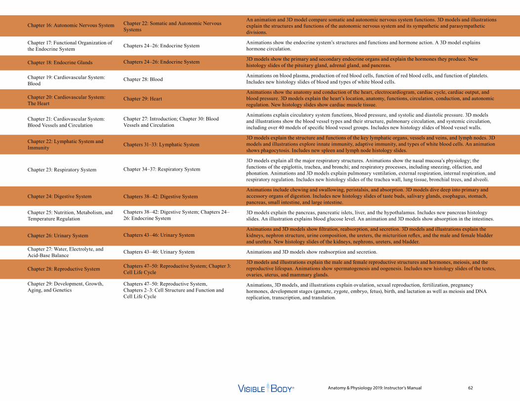
Anatomy & Physiology 2019: Instructor’s Manual 62
Chapter 16: Autonomic Nervous System
Chapter 17: Functional Organization of the Endocrine System
Chapter 18: Endocrine Glands
Chapter 19: Cardiovascular System: Blood
Chapter 20: Cardiovascular System: The Heart
Chapter 21: Cardiovascular System: Blood Vessels and Circulation
Chapter 22: Lymphatic System and Immunity
Chapter 23: Respiratory System
Chapter 24: Digestive System
Chapter 25: Nutrition, Metabolism, and Temperature Regulation
Chapter 26: Urinary System
Chapter 27: Water, Electrolyte, and Acid-Base Balance
Chapter 28: Reproductive System
Chapter 29: Development, Growth, Aging, and Genetics
Chapter 22: Somatic and Autonomic Nervous Systems
Chapters 24–26: Endocrine System
Chapters 24–26: Endocrine System
Chapter 28: Blood
Chapter 29: Heart
Chapter 27: Introduction; Chapter 30: Blood Vessels and Circulation
Chapters 31–33: Lymphatic System
Chapter 34–37: Respiratory System
Chapters 38–42: Digestive System
Chapters 38–42: Digestive System; Chapters 24–26: Endocrine System
Chapters 43–46: Urinary System
Chapters 43–46: Urinary System
Chapters 47–50: Reproductive System; Chapter 3: Cell Life Cycle
Chapters 47–50: Reproductive System, Chapters 2–3: Cell Structure and Function and Cell Life Cycle
An animation and 3D model compare somatic and autonomic nervous system functions. 3D models and illustrations explain the structures and functions of the autonomic nervous system and its sympathetic and parasympathetic divisions.
Animations show the endocrine system’s structures and functions and hormone action. A 3D model explains hormone circulation.
3D models show the primary and secondary endocrine organs and explain the hormones they produce. New histology slides of the pituitary gland, adrenal gland, and pancreas.
Animations on blood plasma, production of red blood cells, function of red blood cells, and function of platelets. Includes new histology slides of blood and types of white blood cells.
Animations show the anatomy and conduction of the heart, electrocardiogram, cardiac cycle, cardiac output, and blood pressure. 3D models explain the heart’s location, anatomy, functions, circulation, conduction, and autonomic regulation. New histology slides show cardiac muscle tissue.
Animations explain circulatory system functions, blood pressure, and systolic and diastolic pressure. 3D models and illustrations show the blood vessel types and their structure, pulmonary circulation, and systemic circulation, including over 40 models of specific blood vessel groups. Includes new histology slides of blood vessel walls.
3D models explain the structure and functions of the key lymphatic organs, vessels and veins, and lymph nodes. 3D models and illustrations explore innate immunity, adaptive immunity, and types of white blood cells. An animation shows phagocytosis. Includes new spleen and lymph node histology slides.
3D models explain all the major respiratory structures. Animations show the nasal mucosa’s physiology; the functions of the epiglottis, trachea, and bronchi; and respiratory processes, including sneezing, olfaction, and phonation. Animations and 3D models explain pulmonary ventilation, external respiration, internal respiration, and respiratory regulation. Includes new histology slides of the trachea wall, lung tissue, bronchial trees, and alveoli.
Animations include chewing and swallowing, peristalsis, and absorption. 3D models dive deep into primary and accessory organs of digestion. Includes new histology slides of taste buds, salivary glands, esophagus, stomach, pancreas, small intestine, and large intestine.
3D models explain the pancreas, pancreatic islets, liver, and the hypothalamus. Includes new pancreas histology slides. An illustration explains blood glucose level. An animation and 3D models show absorption in the intestines.
Animations and 3D models show filtration, reabsorption, and secretion. 3D models and illustrations explain the kidneys, nephron structure, urine composition, the ureters, the micturition reflex, and the male and female bladder and urethra. New histology slides of the kidneys, nephrons, ureters, and bladder.
Animations and 3D models show reabsorption and secretion.
3D models and illustrations explain the male and female reproductive structures and hormones, meiosis, and the reproductive lifespan. Animations show spermatogenesis and oogenesis. Includes new histology slides of the testes, ovaries, uterus, and mammary glands.
Animations, 3D models, and illustrations explain ovulation, sexual reproduction, fertilization, pregnancy hormones, development stages (gamete, zygote, embryo, fetus), birth, and lactation as well as meiosis and DNA replication, transcription, and translation.
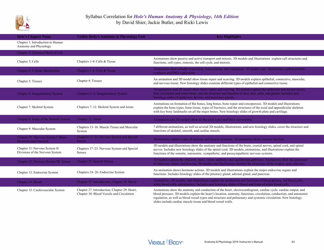
Anatomy & Physiology 2019: Instructor’s Manual 63
Syllabus Correlation for Hole’s Human Anatomy & Physiology, 14th Edition by David Shier, Jackie Butler, and Ricki Lewis
Hole’s Chapter NameChapter 1: Introduction to Human Anatomy and Physiology
Chapter 2: Chemical Basis of Life
Chapter 3: Cells
Chapter 4: Cellular Metabolism
Chapter 5: Tissues
Chapter 6: Integumentary System
Chapter 7: Skeletal System
Chapter 8: Joints of the Skeletal System
Chapter 9: Muscular System
Chapter 10: Nervous System I: Basic Structure and Function
Chapter 11: Nervous System II: Divisions of the Nervous System
Chapter 12: Nervous System III: Senses
Chapter 13: Endocrine System
Chapter 14: Blood
Chapter 15: Cardiovascular System
Chapters 1-4: Cells & Tissue
Chapters 1-4: Cells & Tissue
Chapter 4: Tissues
Chapters 5–6: Integumentary System
Chapters 7–12: Skeletal System and Joints
Chapter 12: Joints
Chapters 13–16: Muscle Tissue and Muscular System
Chapters 17–23: Nervous System and Special Senses
Chapters 17–23: Nervous System and Special Senses
Chapter 23: Special Senses
Chapters 24–26: Endocrine System
Chapter 27: Introduction; Chapter 28: Blood
Chapter 27: Introduction; Chapter 29: Heart; Chapter 30: Blood Vessels and Circulation
Animations show passive and active transport and mitosis. 3D models and illustrations explain cell structures and functions, cell types, osmosis, the cell cycle, and meiosis.
Animations show cellular respiration, transcription, and translation. 3D models and illustrations explain protein synthesis and DNA replication.
An animation and 3D model show tissue repair and scarring. 3D models explain epithelial, connective, muscular, and nervous tissue. New histology slides examine different types of epithelial and connective tissue.
An animation and 3D model show tissue repair and scarring. 3D models explain the epidermis and dermis layers, skin circulation and innervation, and the structure and function of skin, hair, nails, and glands. Includes new histology slides of epidermis, dermis, and mammary glands.
Animations on formation of flat bones, long bones, bone repair and osteoporosis. 3D models and illustrations explain the bone types, bone tissue, types of fractures, and the structures of the axial and appendicular skeleton with key bony landmarks on all the major bones. New histology slides of growth plate and cartilage.
Animations and 3D models show all the joint types and their movements.
7 different animations, as well as several 3D models, illustrations, and new histology slides, cover the structure and functions of skeletal, smooth, and cardiac muscle.
Illustrations explain types of neurons and neuron structure. An animation shows neuron function.
3D models and illustrations show the anatomy and functions of the brain, cranial nerves, spinal cord, and spinal nerves. Includes new histology slides of the spinal cord. 3D models, animations, and illustrations explain the functions of the somatic, autonomic, sympathetic, and parasympathetic nervous systems.
3D models explain the olfactory, taste, vision, auditory, and equilibrium pathways. Animations show the processes of olfaction, vision, and hearing. 3D models and illustrations identify the structures of the tongue, eyes, and ears.
An animation shows hormone actions. 3D models and illustrations explain the major endocrine organs and functions. Includes histology slides of the pituitary gland, adrenal gland, and pancreas.
Animations, 3D models, and illustrations explain the production and functions of blood plasma, red blood cells, white blood cells, and platelets. Includes new histology slides of blood and types of white blood cells.
Animations show the anatomy and conduction of the heart, electrocardiogram, cardiac cycle, cardiac output, and blood pressure. 3D models explain the heart’s location, anatomy, functions, circulation, conduction, and autonomic regulation, as well as blood vessel types and structure and pulmonary and systemic circulation. New histology slides include cardiac muscle tissue and blood vessel walls.
Visible Body’s Anatomy & Physiology Unit Key Highlights
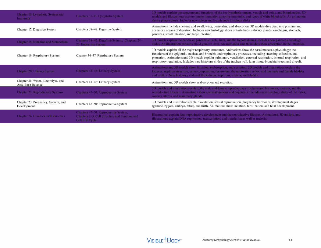
Anatomy & Physiology 2019: Instructor’s Manual 64
Chapter 16: Lymphatic System and Immunity
Chapter 17: Digestive System
Chapter 18: Nutrition and Metabolism
Chapter 19: Respiratory System
Chapter 20: Urinary System
Chapter 21: Water, Electrolyte, and Acid-Base Balance
Chapter 22: Reproductive Systems
Chapter 23: Pregnancy, Growth, and Development
Chapter 24: Genetics and Genomics
Chapters 31–33: Lymphatic System
Chapters 38–42: Digestive System
Chapters 38–42: Digestive System; Chapters 24–26: Endocrine System
Chapter 34–37: Respiratory System
Chapters 43–46: Urinary System
Chapters 43–46: Urinary System
Chapters 47–50: Reproductive System
Chapters 47–50: Reproductive System
Chapters 47–50: Reproductive System, Chapters 2–3: Cell Structure and Function and Cell Life Cycle
3D models explain the structure and functions of the key lymphatic organs, vessels and veins, and lymph nodes. 3D models and illustrations explore innate immunity, adaptive immunity, and types of white blood cells. An animation shows phagocytosis. Includes new spleen and lymph node histology slides.
Animations include chewing and swallowing, peristalsis, and absorption. 3D models dive deep into primary and accessory organs of digestion. Includes new histology slides of taste buds, salivary glands, esophagus, stomach, pancreas, small intestine, and large intestine.
3D models explain the pancreas, pancreatic islets, liver, and the hypothalamus. Includes new pancreas histology slides. An illustration explains blood glucose level. An animation and 3D models show absorption in the intestines.
3D models explain all the major respiratory structures. Animations show the nasal mucosa’s physiology; the functions of the epiglottis, trachea, and bronchi; and respiratory processes, including sneezing, olfaction, and phonation. Animations and 3D models explain pulmonary ventilation, external respiration, internal respiration, and respiratory regulation. Includes new histology slides of the trachea wall, lung tissue, bronchial trees, and alveoli.
Animations and 3D models show filtration, reabsorption, and secretion. 3D models and illustrations explain the kidneys, nephron structure, urine composition, the ureters, the micturition reflex, and the male and female bladder and urethra. New histology slides of the kidneys, nephrons, ureters, and bladder.
Animations and 3D models show reabsorption and secretion.
3D models and illustrations explain the male and female reproductive structures and hormones, meiosis, and the reproductive lifespan. Animations show spermatogenesis and oogenesis. Includes new histology slides of the testes, ovaries, uterus, and mammary glands.
3D models and illustrations explain ovulation, sexual reproduction, pregnancy hormones, development stages (gamete, zygote, embryo, fetus), and birth. Animations show lactation, fertilization, and fetal development.
Illustrations explain fetal reproductive development and the reproductive lifespan. Animations, 3D models, and illustrations explain DNA replication, transcription, and translation as well as meiosis.
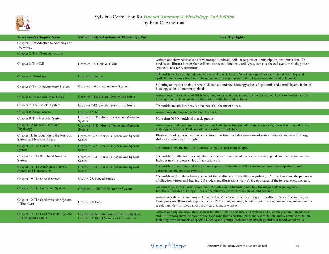
Anatomy & Physiology 2019: Instructor’s Manual 65
Syllabus Correlation for Human Anatomy & Physiology, 2nd Edition by Erin C. Amerman
Amerman’s Chapter NameChapter 1: Introduction to Anatomy and Physiology
Chapter 2: The Chemistry of Life
Chapter 3: The Cell
Chapter 4: Histology
Chapter 5: The Integumentary System
Chapter 6: Bones and Bone Tissue
Chapter 7: The Skeletal System
Chapter 8: Articulations
Chapter 9: The Muscular System
Chapter 10: Muscle Tissue and Physiology
Chapter 11: Introduction to the Nervous System and Nervous Tissue
Chapter 12: The Central Nervous System
Chapter 13: The Peripheral Nervous System
Chapter 14: The Autonomic Nervous System and Homeostasis
Chapter 15: The Special Senses
Chapter 16: The Endocrine System
Chapter 17: The Cardiovascular System I: The Heart
Chapter 18: The Cardiovascular System II: The Blood Vessels
Chapters 1-4: Cells & Tissue
Chapter 4: Tissues
Chapters 5-6: Integumentary System
Chapters 7-12: Skeletal System and Joints
Chapters 7-12: Skeletal System and Joints
Chapter 12: JointsChapters 13-16: Muscle Tissue and Muscular SystemChapters 13-16: Muscle Tissue and Muscular System
Chapters 17-23: Nervous System and Special Senses
Chapters 17-23: Nervous System and Special Senses
Chapters 17-23: Nervous System and Special Senses
Chapters 17-23: Nervous System and Special Senses
Chapter 23: Special Senses
Chapters 24-26: The Endocrine System
Chapter 29: Heart
Chapter 27: Introduction: Circulatory System; Chapter 30: Blood Vessels and Circulation
Animations show passive and active transport, mitosis, cellular respiration, transcription, and translation. 3D models and illustrations explain cell structures and functions, cell types, osmosis, the cell cycle, meiosis, protein synthesis, and DNA replication.
3D models explore epithelial, connective, and muscle tissue. New histology slides examine different types of epithelial and connective tissue. Tissue repair and scarring are featured in an animation and 3D model.
Stunning animation on tissue repair. 3D models and new histology slides of epidermis and dermis layers. Includes histology slides of mammary glands.
Animations on formation of flat bones, long bones, and bone repair. 3D models include key bony landmarks of all the major bones. New histology slides of growth plate and cartilage.
3D models include key bony landmarks of all the major bones.
Animations showing movement of all joint types.
More than 50 3D models of muscle groups.
Animations on skeletal muscle contraction, featuring action potentials and cross-bridge formation. Includes new histology slides of skeletal, smooth, and cardiac muscle tissue.
Illustrations of types of neurons and neuron structure. Includes animation of neuron function and new histology slides of neurons and neuroglia.
3D models show the brain’s structures, functions, and blood supply.
3D models and illustrations show the anatomy and functions of the cranial nerves, spinal cord, and spinal nerves. Includes new histology slides of the spinal cord.
3D models, animations, and illustrations explain the functions of the somatic, autonomic, sympathetic, and parasympathetic nervous systems.
3D models explain the olfactory, taste, vision, auditory, and equilibrium pathways. Animations show the processes of olfaction, vision, and hearing. 3D models and illustrations identify the structures of the tongue, eyes, and ears.
An animation shows hormone actions. 3D models and illustrations explain the major endocrine organs and functions. Includes histology slides of the pituitary gland, adrenal gland, and pancreas.
Animations show the anatomy and conduction of the heart, electrocardiogram, cardiac cycle, cardiac output, and blood pressure. 3D models explain the heart’s location, anatomy, functions, circulation, conduction, and autonomic regulation. New histology slides show cardiac muscle tissue.
Animations explain circulatory system functions, blood pressure, and systolic and diastolic pressure. 3D models and illustrations show the blood vessel types and their structure, pulmonary circulation, and systemic circulation, including over 40 models of specific blood vessel groups. Includes new histology slides of blood vessel walls.
Visible Body’s Anatomy & Physiology Unit Key Highlights
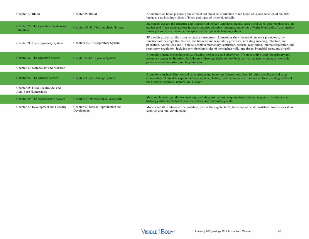
Anatomy & Physiology 2019: Instructor’s Manual 66
Chapter 19: Blood
Chapter 20: The Lymphatic System and Immunity
Chapter 21: The Respiratory System
Chapter 22: The Digestive System
Chapter 23: Metabolism and Nutrition
Chapter 24: The Urinary System
Chapter 25: Fluid, Electrolyte, and Acid-Base Homeostasis
Chapter 26: The Reproductive System
Chapter 27: Development and Heredity
Chapter 28: Blood
Chapters 31-33: The Lymphatic System
Chapters 34-37: Respiratory System
Chapter 38-42: Digestive System
Chapters 43-46: Urinary System
Chapters 47-50: Reproductive System
Chapter 50: Sexual Reproduction and Development
Animations on blood plasma, production of red blood cells, function of red blood cells, and function of platelets. Includes new histology slides of blood and types of white blood cells.
3D models explain the structure and functions of the key lymphatic organs, vessels and veins, and lymph nodes. 3D models and illustrations explore innate immunity, adaptive immunity, and types of white blood cells. An animation shows phagocytosis. Includes new spleen and lymph node histology slides.
3D models explain all the major respiratory structures. Animations show the nasal mucosa’s physiology; the functions of the epiglottis, trachea, and bronchi; and respiratory processes, including sneezing, olfaction, and phonation. Animations and 3D models explain pulmonary ventilation, external respiration, internal respiration, and respiratory regulation. Includes new histology slides of the trachea wall, lung tissue, bronchial trees, and alveoli.
Animations include chewing and swallowing, peristalsis, and absorption. 3D models dive deep into primary and accessory organs of digestion. Includes new histology slides of taste buds, salivary glands, esophagus, stomach, pancreas, small intestine, and large intestine.
Animations include filtration and reabsorption and secretion. Illustrations show filtration membrane and urine composition. 3D models explore kidneys, ureters, bladder, urethra, and micturition reflex. New histology slides of the kidneys, nephrons, ureters, and bladder.
Male and female reproductive anatomy, including animations on spermatogenesis and oogenesis. Includes new histology slides of the testes, ovaries, uterus, and mammary glands.
Models and illustrations cover ovulation, path of the zygote, birth, transcription, and translation. Animations show lactation and fetal development.

Anatomy & Physiology 2019: Instructor’s Manual 68
Cells and Tissue1 Introduction: Cells and Tissue
1.1 Cells and Tissue Overview1.2 Cell Functions1.3 Cell Types
2 Cell Structure and Function2.1 Parts of a Cell2.2 Plasma Membrane2.3 Structure of the Plasma Membrane2.4 Passive and Active Transport2.5 Transport Vesicles2.6 Osmosis2.7 Organelles2.8 Cytoskeleton and Centrosome2.9 Endoplasmic Reticulum
2.10 Golgi Complex2.11 Lysosomes and Peroxisomes2.12 Nucleus2.13 Mitochondria2.14 Ribosomes2.15 Cellular Respiration Definition2.16 Cellular Respiration2.17 Proteins2.18 Protein Synthesis2.19 Transcription Definition2.20 Transcription2.21 Translation Definition2.22 Codons and the Ribosome2.23 Translation
3 Cell Life Cycle3.1 Replication3.2 Cell Cycle3.3 Mitosis3.4 Mitotic Spindle3.5 Meiosis3.6 Meiosis and Gametes
4 Tissues4.1 Tissue Types in the Body4.2 Epithelial Tissue4.3 Types of Epithelial Tissue4.4 Simple Epithelium4.5 Stratified Epithelium4.6 Connective Tissue4.7 Connective Tissue Proper: Loose4.8 Types of Loose Connective Tissue4.9 Connective Tissue Proper: Dense
4.10 Types of Dense Connective Tissue4.11 Special Connective Tissue: Bone and
Cartilage4.12 Bone Tissue4.13 Cartilage4.14 Cartilage Histology4.15 Special Connective Tissue: Blood
and Lymph4.16 Blood4.17 Muscle Tissue4.18 Types of Muscle Tissue4.19 Nervous Tissue4.20 Neuron Structure4.21 Tissue Repair4.22 Tissue Repair and Scarring
Integumentary System5 Introduction:
Integumentary System5.1 Integumentary System Overview5.2 Integumentary Structures5.3 Integumentary Functions
6 Integumentary System6.1 Skin Layers6.2 Epidermis Layers6.3 Epidermis Histology6.4 Skin Cells6.5 Epidermal Cells6.6 Dermis Layers6.7 Dermis Histology6.8 Thick vs. Thin Skin Histology6.9 Sebaceous Glands
6.10 Sudoriferous Glands6.11 Dermal Circulation6.12 Vitamin D Synthesis6.13 Integumentary Innervation6.14 Dermatomes6.15 Hair6.16 Hair Structure6.17 Hair Functions6.18 Nails6.19 Mammary Glands6.20 Mammary Gland Histology 6.21 Tissue Repair6.22 Tissue Repair and Scarring

Anatomy & Physiology 2019: Instructor’s Manual 69
Skeletal System and Joints7 Introduction:
Skeletal System/Joints7.1 Skeletal System Overview7.2 Axial and Appendicular Skeleton7.3 Function of the Skeleton
8 Types of Bones8.1 Bone Types8.2 Flat Bones8.3 Long Bones8.4 Short Bones8.5 Irregular Bones8.6 Sesamoid Bones
9 Bone Tissue9.1 Bone Marrow9.2 Interior of a Long Bone9.3 Parts of a Long Bone9.4 Bone Tissue Histology9.5 Osteon9.6 Bone Cells9.7 Flat Bone Formation9.8 Flat Bones in the Skull9.9 Long Bone Formation and Growth
9.10 Long Bones in the Limbs9.11 Growth Plate Histology9.12 Cartilage Histology9.13 Types of Fractures9.14 Bone Repair9.15 Calcium in the Skeleton
9.16 Osteoporosis9.17 Healthy and Osteoporotic Bone
10 Axial Skeleton10.1 Axial Skeleton10.2 Skull10.3 Cranial Bones10.4 Occipital Bone Landmarks10.5 Temporal Bone Landmarks10.6 Frontal Bone Landmarks10.7 Sphenoid Bone Landmarks10.8 Ethmoid Bone Landmarks10.9 Skull Sutures
10.10 Fontanelles10.11 Facial Skeleton I10.12 Maxilla Landmarks10.13 Mandible Landmarks10.14 Facial Skeleton II10.15 Palatine Bone Landmarks10.16 Lacrimal Bone Landmarks10.17 Auditory Ossicles10.18 Foramina of Skull10.19 Bones of the Orbit10.20 Nasal Septum10.21 Hyoid10.22 Vertebral Column10.23 Vertebra10.24 Ligaments of the Spine10.25 Intervertebral Joints10.26 Cervical Spine10.27 Cervical Spine Landmarks10.28 Thoracic Spine10.29 Thoracic Spine Landmarks
10.30 Lumbar Spine10.31 Lumbar Spine Landmarks10.32 Sacral Spine10.33 Sacrum Landmarks10.34 Thoracic Cage10.35 Sternum10.36 Ribs
11 Appendicular Skeleton11.1 Appendicular Skeleton11.2 Pectoral (Shoulder) Girdle11.3 Scapula Landmarks11.4 Clavicle Landmarks11.5 Muscular Stabilization11.6 Upper Limb11.7 Arm and Forearm11.8 Humerus Landmarks11.9 Radius Landmarks
11.10 Ulna Landmarks11.11 Carpus (Wrist) I11.12 Carpus (Wrist) II11.13 Carpal Tunnel11.14 Hand11.15 Pelvic (Hip) Girdle11.16 Ilium Landmarks11.17 Ischium Landmarks11.18 Pubis Landmarks11.19 Male and Female Pelvis11.20 Lower Limb11.21 Thigh and Leg11.22 Femur Landmarks11.23 Tibia Landmarks11.24 Fibula Landmarks
Anatomy & Physiology 2019: Instructor’s Manual 70
11.25 Tarsus11.26 Foot Arches11.27 Foot
12 Joints12.1 Joints12.2 Joints in the Skull12.3 Synarthroses (No Movement): Skull
Sutures12.4 Synarthroses (No Movement):
Gomphoses12.5 Synarthroses (No Movement):
Synchrondoses12.6 Syndesmoses (Slight Movement)12.7 Symphyses (Slight Movement)12.8 Synovial Joints (Full Movement)12.9 Structure of Synovial Joints
12.10 Gliding (Plane) Joints Movement12.11 Gliding Joints in the Skeleton12.12 Hinge Joints Movement12.13 Hinge Joints in the Skeleton12.14 Pivot Joints Movement12.15 Pivot Joints in the Skeleton12.16 Condyloid Joints Movement12.17 Condyloid Joints in the Skeleton12.18 Saddle Joints Movement12.19 Saddle Joints in the Skeleton12.20 Ball and Socket Joints Movement12.21 Ball and Socket Joints
in the Skeleton12.22 Ligaments and Synovial Joints12.23 Joints and Aging
Muscle Tissue and Muscular System13 Introduction: Muscular System
13.1 Muscular System Overview13.2 Muscle Types13.3 Types of Muscle Tissue13.4 Skeletal Muscle13.5 Smooth Muscle13.6 Cardiac Muscle13.7 Muscular Anatomy
14 Skeletal Muscle Tissue14.1 Nerve and Blood Supply
to Skeletal Muscle14.2 Skeletal Muscle Function Overview14.3 Muscle Contraction: Action Potential14.4 Muscle Contraction:
Cross Bridge Formation14.5 Skeletal Muscle Innervation14.6 Skeletal Muscle Tissue Histology
15 Smooth and Cardiac Muscle Tissue
15.1 Smooth Muscle of the Body15.2 Smooth Muscle Function15.3 Smooth Muscle Tissue Histology15.4 Alimentary Canal15.5 Stomach Wall15.6 Smooth Muscle in the Stomach Wall15.7 Trachea and Bronchi15.8 Blood Vessel Walls
15.9 Smooth Muscle in Blood Vessel Walls
15.10 Urinary / Reproductive Systems (Female)
15.11 Urinary / Reproductive Systems (Male)
15.12 Cardiac Muscle15.13 Heart Wall15.14 Myocardium15.15 Cardiac Muscle Tissue Histology
16 Muscular System16.1 Muscle Interactions16.2 Skeletal Muscle Attachments16.3 Chin Raise: Muscles as
First‑Class Levers16.4 Calf Raise: Muscles as
Second‑Class Levers16.5 Elbow Flexion: Muscles as
Third‑Class Levers16.6 Facial Expression:
Scalp and Eyebrow16.7 Facial Expression: Upper Mouth16.8 Facial Expression: Lower Mouth16.9 Extrinsic Eye Muscles
16.10 Mastication16.11 Tongue16.12 Neck: Suprahyoid16.13 Neck: Infrahyoid16.14 Neck: Head Movements16.15 Vertebral Column: Splenius16.16 Vertebral Column: Erector Spinae16.17 Erector Spinae: Spinalis16.18 Erector Spinae: Longissimus
Anatomy & Physiology 2019: Instructor’s Manual 71
16.19 Erector Spinae: Iliocostalis16.20 Vertebral Column:
Transversospinales16.21 Transversospinales: Semispinalis16.22 Transversospinales: Rotatores16.23 Vertebral Column: Segmental16.24 Vertebral Column: Scalenes16.25 Vertebral Column: Spinal Flexors16.26 Abdomen16.27 Pelvis: Pelvic Diaphragm16.28 Pelvis: Superficial Perineal16.29 Pelvis: Deep Perineal16.30 Thorax: Breathing16.31 Thorax: Shoulder Girdle
Movements, Anterior16.32 Thorax: Shoulder Girdle
Movements, Posterior16.33 Arm: Prime Movers16.34 Arm: Synergists / Fixators16.35 Arm: Rotator Cuff16.36 Forearm: Elbow Flexors16.37 Forearm: Elbow Extensors16.38 Forearm: Pronation / Supination16.39 Hand: Superficial Flexors16.40 Hand: Deep Flexors16.41 Hand: Superficial Extensors16.42 Hand: Deep Extensors16.43 Fingers: Thenar16.44 Fingers: Hypothenar16.45 Fingers: Midpalmar16.46 Thigh: Iliopsoas16.47 Thigh: Gluteal16.48 Thigh: Lateral Rotators
16.49 Thigh: Anterior Compartment16.50 Anterior Compartment: Quadriceps16.51 Thigh: Medial Compartment16.52 Medial Compartment: Adductors16.53 Thigh: Posterior Compartment
(Hamstrings)16.54 Lower Leg: Anterior Compartment16.55 Lower Leg: Lateral Compartment16.56 Lower Leg: Posterior Compartment
(Superficial)16.57 Lower Leg: Posterior Compartment
(Deep)16.58 Foot: Dorsum16.59 Foot: Plantar Layer 116.60 Foot: Plantar Layer 216.61 Foot: Plantar Layer 316.62 Foot: Plantar Layer 4
Nervous System and Special Senses17 Introduction: Nervous System/
Special Senses17.1 Nervous System Overview17.2 Nervous System Anatomy17.3 Central Nervous System17.4 Peripheral Nervous System17.5 Nervous System Functions17.6 Special Sensory Organs
18 Nervous Tissue18.1 Nervous Tissue in the Body
18.2 Neuron Histology18.3 Types of Neurons18.4 CNS Neuroglia Histology18.5 PNS Neuroglia Histology18.6 Neuron Function18.7 Resting and Action Potentials18.8 Neurotransmitters18.9 Acetylcholine (ACh)
18.10 Norepinephrine (NE) and Epinephrine (E)
18.11 Dopamine
19 Spinal Cord and Spinal Nerves19.1 Spinal Cord19.2 Spine Cross Section19.3 Spinal Nerve Roots19.4 Spinal Cord Histology19.5 Sensory Signals19.6 Motor Commands19.7 Spinal Nerves19.8 Spinal Nerve Regions19.9 Dermatomes
19.10 Cervical Plexus19.11 Cervical Innervation19.12 Brachial Plexus19.13 Brachial Innervation I19.14 Brachial Innervation II19.15 Brachial Innervation III19.16 Lumbosacral Plexus I19.17 Lumbosacral Plexus II19.18 Lumbosacral Innervation19.19 Somatic Reflex Arc19.20 Path of Reflex Arc

Anatomy & Physiology 2019: Instructor’s Manual 72
20 Brain20.1 The Brain20.2 Brain Regions20.3 Brain Cross Section20.4 Ventricles of the Brain20.5 Cerebrospinal Fluid20.6 Blood Supply to the Brain20.7 Brain Stem20.8 Medulla and Pons20.9 Midbrain
20.10 Cerebellum Anatomy20.11 Cerebellum Function20.12 Diencephalon20.13 Thalamus20.14 Hypothalamus20.15 Pituitary Gland20.16 Limbic System20.17 Cerebrum20.18 Cerebral Hemispheres20.19 Lobes of the Cerebral Cortex20.20 Features of the Cerebral Cortex20.21 Basal Ganglia20.22 Functional Regions
of the Cerebral Cortex20.23 Functional Regions
and Cortex Anatomy20.24 Hemispheric Lateralization
21 Cranial Nerves21.1 Cranial Nerves21.2 Special Sensory Cranial Nerves21.3 Motor Cranial Nerves21.4 Mixed Cranial Nerves
21.5 Olfactory Nerves (I)21.6 Olfaction21.7 Optic Nerves (II)21.8 Vision21.9 Oculomotor Nerves (III)
21.10 Trochlear Nerves (IV)21.11 Trigeminal Nerves (V)21.12 Ophthalmic Branch
of Trigeminal Nerve21.13 Maxillary Branch
of Trigeminal Nerve21.14 Mandibular Branch
of Trigeminal Nerve21.15 Abducens Nerves (VI)21.16 Facial Nerves (VII)21.17 Vestibulocochlear Nerves (VIII)21.18 Glossopharyngeal Nerves (IX)21.19 Vagus Nerves (X)21.20 Accessory Nerves (XI)21.21 Hypoglossal Nerves (XII)
22 Somatic and Autonomic Nervous Systems
22.1 Somatic and Autonomic Functions22.2 Somatic and Autonomic
Nervous Functions22.3 Somatic Sensory Signals22.4 Skin Sensory Receptors22.5 Somatic Motor Signals22.6 Skeletal Muscle Contraction22.7 Somatic Sensory and
Motor Pathways22.8 Basal Ganglia and Cerebellum
22.9 Autonomic Nervous Functions22.10 Sympathetic Nervous System22.11 Sympathetic Nervous Functions22.12 Parasympathetic Nervous System22.13 Parasympathetic Nervous Functions
23 Special Senses23.1 Special Sensory Organs23.2 Olfaction23.3 Olfactory Pathway23.4 Tongue23.5 Taste Buds23.6 Papillae23.7 Taste Pathway23.8 Eyes in Context23.9 Eye
23.10 Eye Layers23.11 Lacrimal Apparatus23.12 Eye Interior23.13 Retina23.14 Rods and Cones23.15 Vision23.16 Types of Vision23.17 Vision Pathway23.18 Optic Chiasm23.19 Ear23.20 Outer Ear23.21 Middle Ear23.22 Inner Ear23.23 Cochlea Cross Section23.24 Organ of Corti23.25 Hearing23.26 Auditory Pathway
Anatomy & Physiology 2019: Instructor’s Manual 73
23.27 Equilibrium23.28 Equilibrium Pathway
Endocrine System24 Introduction: Endocrine System
24.1 Endocrine System Overview24.2 Primary Endocrine Organs24.3 Secondary Endocrine Organs
25 Hormone Action and Regulation25.1 Hormone Action25.2 Hormone Circulation25.3 Hypothalamus and Pituitary25.4 Pituitary Gland Histology25.5 Hypothalamus and Pituitary
Hormones25.6 Hypothalamus Hormones25.7 Hypophyseal Portal System25.8 Anterior Pituitary Hormones25.9 Posterior Pituitary Hormones
25.10 Target Organs of Pituitary Hormones
25.11 Human Growth Hormone (hGH)25.12 Thyroid‑Stimulating Hormone (TSH)25.13 Follicle‑Stimulating Hormone (FSH)25.14 Luteinizing Hormone (LH)25.15 Melanocyte‑Stimulating Hormone
(MSH)25.16 Prolactin (PRL)25.17 Adrenocorticotropic Hormone
(ACTH)
25.18 Melanocyte‑Stimulating Hormone (MSH)
25.19 Antidiuretic Hormone (ADH)25.20 Oxytocin (OXT)
26 Endocrine Organs and Functions26.1 Primary and Secondary
Endocrine Organs26.2 Thyroid Gland26.3 Thyroid Hormones26.4 Parathyroid Glands26.5 Parathyroid Hormones26.6 Adrenal Glands26.7 Adrenal Gland Histology26.8 Adrenal Cortex Hormones26.9 Adrenal Medulla Hormones
26.10 Pineal Gland and Hormones26.11 Pancreas26.12 Pancreatic Islets26.13 Pancreas Histology26.14 Blood Glucose Level26.15 Ovaries26.16 Testes26.17 Kidneys26.18 Heart26.19 Thymus26.20 Stress Response
Circulatory System27 Introduction: Circulatory System
27.1 Circulatory System Overview27.2 Circulatory Anatomy
27.3 Arteries and Veins27.4 Oxygen and Carbon Dioxide
Exchange
28 Blood28.1 Blood Histology28.2 Blood Plasma28.3 Plasma Protein Production28.4 Production of Red Blood Cells28.5 Erythropoietin and RBC Production28.6 Function of Red Blood Cells28.7 Red Blood Cells and Oxygen
Transport28.8 White Blood Cell Histology28.9 Function of Platelets
28.10 Locations of Platelet Production
29 Heart29.1 Heart Overview29.2 Location of Heart in the Thoracic
Cavity29.3 Location of Heart in the Thoracic
Cage29.4 Inferior View of Thoracic Cavity,
Transverse Section29.5 Heart Wall29.6 Myocardium29.7 Cardiac Muscle Tissue Histology29.8 Heart Chamber Anatomy29.9 Heart Chambers
29.10 Heart Chamber Functions29.11 Heart Valve Anatomy29.12 Heart Valves29.13 Heart Valve Function
Anatomy & Physiology 2019: Instructor’s Manual 74
29.14 Heart Internal Anatomy29.15 Blood Flow Through the Heart29.16 Blood Flow Out of Heart29.17 Blood Flow Into Heart29.18 Coronary Circulation29.19 Coronary Veins29.20 Left Coronary Arteries29.21 Right Coronary Arteries29.22 Conduction System29.23 Heart Conduction29.24 Path of Nerve Impulse
in Conduction System29.25 Electrocardiogram29.26 Cardiac Cycle29.27 Systole and Diastole29.28 Cardiac Output29.29 Autonomic Regulation of Heart
30 Blood Vessels and Circulation30.1 Types of Blood Vessels30.2 Blood Vessel Walls30.3 Artery Structure30.4 Vein Structure30.5 Blood Vessel Wall Histology30.6 Capillaries30.7 Blood Pressure30.8 Systolic and Diastolic Pressure30.9 Systolic and Diastolic Pressure
30.10 Circulatory Routes30.11 Pulmonary Circulation30.12 Pulmonary Circulation and Bronchi30.13 Pulmonary Arteries and Veins30.14 Pulmonary Arteries30.15 Pulmonary Veins30.16 Systemic Circulation30.17 Great Vessels and Branches30.18 Superficial Arteries
of the Head and Neck
30.19 Deep Arteries of the Head and Neck30.20 Circle of Willis I30.21 Circle of Willis II30.22 Circle of Willis III30.23 Arteries of the Brain30.24 Arteries of the Upper Limb I30.25 Arteries of the Upper Limb II30.26 Arteries of the Forearm and Hand30.27 Arteries of the Thorax I30.28 Arteries of the Thorax II30.29 Branches of the Abdominal Aorta I30.30 Branches of the Abdominal Aorta II30.31 Arteries of the Abdomen I30.32 Arteries of the Abdomen II30.33 Arteries of the Intestines I30.34 Arteries of the Intestines II30.35 Arteries of the Pelvis I30.36 Arteries of the Pelvis II30.37 Arteries of the Upper Leg30.38 Arteries of the Lower Leg30.39 Dorsal Arteries of the Foot30.40 Plantar Arteries of the Foot30.41 Superficial Veins of the Head and
Neck30.42 Deep Veins of the Head and Neck30.43 Venous Sinuses I30.44 Venous Sinuses II30.45 Veins of the Upper Limb I30.46 Veins of the Upper Limb II30.47 Veins of the Hand30.48 Veins of the Thorax I30.49 Veins of the Thorax II30.50 Azygos System30.51 Veins of the Abdomen30.52 Hepatic Portal System30.53 Veins of the Intestines I30.54 Veins of the Intestines II30.55 Veins of the Pelvis I
30.56 Veins of the Pelvis II30.57 Veins of the Upper Leg30.58 Veins of the Lower Leg30.59 Dorsal Veins of the Foot30.60 Plantar Veins of the Foot
Lymphatic System31 Introduction: Lymphatic System
31.1 Lymphatic System Overview31.2 Lymphatic System Structures31.3 Lymphatic Functions
32 Lymphatic System32.1 Lymph Circulation32.2 Lymphatic Vessels and Veins32.3 Thoracic Duct32.4 Right Lymphatic Duct32.5 Lymph Trunks32.6 Lymphocyte Production32.7 Thymus32.8 Spleen32.9 Spleen Histology
32.10 Lymph Node Function32.11 Lymph Node Distribution32.12 Lymph Node Histology
33 Immunity33.1 Physical Defenses33.2 Innate Immunity33.3 Innate Immune Response33.4 Phagocytosis33.5 Types of White Blood Cells33.6 Adaptive Immunity33.7 Adaptive Immune Response33.8 B Cells33.9 T Cells
Anatomy & Physiology 2019: Instructor’s Manual 75
Respiratory System34 Introduction: Respiratory
System34.1 Respiratory System Overview34.2 Functions of Respiratory Structures34.3 Pulmonary Ventilation34.4 External Respiration34.5 Internal Respiration
35 Upper Respiratory System35.1 Upper Respiratory Structures35.2 Nasal Cavity35.3 Nasal Septum35.4 Bones of the Nasal Cavity35.5 Nasal Cartilages35.6 Upper Respiratory Tract35.7 Nasal Mucosa35.8 Filtration of Air35.9 Respiratory Defenses
35.10 Sneezing35.11 Olfaction35.12 Olfaction Structures35.13 Pharynx35.14 Function of the Epiglottis35.15 Larynx, Posterior35.16 Larynx, Anterior35.17 Phonation35.18 Vocal Folds and Musculature
36 Lower Respiratory System36.1 Lower Respiratory Structures36.2 Location of the Trachea36.3 Cartilaginous Rings36.4 Trachea Wall Histology
36.5 The Trachea and Bronchi36.6 Lungs in Anatomical Context36.7 Right Lung, Gross Anatomy36.8 Left Lung, Gross Anatomy36.9 Lung Tissue vs. Epidermis Histology
36.10 Lung Hilum36.11 Bronchial Tree36.12 Bronchial Tree Histology36.13 Alveoli Histology36.14 Respiratory Membrane36.15 Blood Supply to the Lungs36.16 Pulmonary Circulation
37 Respiration37.1 Pulmonary Ventilation and Boyle’s
Law37.2 Pulmonary Ventilation Structures37.3 Muscles of Inhalation37.4 Muscles of Exhalation37.5 External Respiration37.6 External Respiration Structures37.7 Dalton’s Law37.8 Internal Respiration37.9 Internal Respiration Structures
37.10 Breathing Rate and Regulation37.11 Respiratory Regulation
Digestive System38 Introduction: Digestive System
38.1 Digestive Overview38.2 Digestive Anatomy38.3 Digestive Process38.4 Alimentary Canal38.5 Digestive Juices38.6 Production of Digestive Juices
39 Oral Cavity39.1 Structures of the Oral Cavity39.2 Oral Cavity39.3 Chewing and Swallowing39.4 Function of Oral Cavity Structures39.5 Teeth39.6 Upper Arch39.7 Lower Arch39.8 Tooth Cross‑Section39.9 Tongue
39.10 Tongue39.11 Taste Bud Histology39.12 Salivary Glands39.13 Salivary Gland Histology39.14 Mastication Muscles39.15 Epiglottis and Larynx
40 Esophagus and Stomach40.1 Peristalsis40.2 Peristalsis of Alimentary Canal40.3 Esophagus40.4 Esophagus Histology40.5 Stomach Regions40.6 Stomach Wall40.7 Stomach Histology40.8 Stomach Sphincters40.9 Stomach Vasculature
41 Accessory Organs of Digestion41.1 Accessory Organs41.2 Liver Segments41.3 Liver Ligaments41.4 Hepatic Portal System41.5 Pancreas, Liver, Gall Bladder41.6 Bile Ducts41.7 Pancreatic Ducts
Anatomy & Physiology 2019: Instructor’s Manual 76
41.8 Pancreas Histology41.9 Blood Supply to Accessory Organs
42 Small and Large Intestines42.1 Absorption42.2 Absorption Structures42.3 Intestinal Vasculature42.4 Small Intestine42.5 Small Intestine Histology42.6 Large Intestine42.7 Colon Regions42.8 Taenia Coli42.9 Large Intestine
42.10 Large Intestine Histology42.11 Defecation Reflex
Urinary System43 Introduction: Urinary System
43.1 Urinary System Overview43.2 Male Urinary Anatomy43.3 Female Urinary Anatomy43.4 Urinary Functions
44 Kidney Anatomy and Physiology44.1 Position of the Kidneys44.2 Kidney Anatomy44.3 Kidney Histology44.4 Blood Supply to the Kidneys44.5 Blood Flow Through the Nephron44.6 Nephron Structure44.7 Nephron Histology
45 Urine Production45.1 Filtration Process45.2 Filtration45.3 Filtration Membrane
45.4 Reabsorption and Secretion45.5 Reabsorption and Secretion Process45.6 Urine Composition45.7 Function of ADH
46 Urine Storage and Elimination46.1 Pathway of Urine46.2 Ureters46.3 Ureter Histology46.4 Bladder in Female46.5 Bladder in Male46.6 Bladder Anatomy46.7 Bladder Histology46.8 Micturition Reflex46.9 Nervous Control of Micturition
46.10 Urethral Sphincters46.11 Female Urethra46.12 Male Urethra
Reproductive System47 Introduction: Reproductive
System47.1 Reproductive System Overview47.2 Male Reproductive Anatomy47.3 Female Reproductive Anatomy
48 Male Reproductive System48.1 Male Reproductive Organs
in Context48.2 Testes and Spermatic Cords48.3 Seminiferous Tubules48.4 Testis Histology48.5 Spermatogenesis48.6 Epididymis48.7 Vas Deferens48.8 Urethra
48.9 Accessory Glands48.10 Semen Production48.11 Penis Internal Anatomy48.12 Blood Supply to Male Genitalia48.13 Male Sexual Response48.14 Male Sex Hormones
49 Female Reproductive System49.1 Female Reproductive Organs in
Context49.2 Pelvic Ligaments49.3 Ovaries49.4 Ovary Internal Anatomy49.5 Ovary Histology49.6 Oogenesis49.7 Relationship of Ovaries to Uterus49.8 Blood Supply to Uterus and Ovaries49.9 Uterus Histology
49.10 Vagina49.11 External Genitalia49.12 Female Reproductive Cycle49.13 Ovulation49.14 Female Sex Hormones49.15 Mammary Glands49.16 Mammary Gland Histology49.17 Lactation49.18 Lactation Hormones
50 Sexual Reproduction and Development
50.1 Gametes to Zygote50.2 Sexual Reproduction50.3 Fertilization to Implantation50.4 Path of Zygote50.5 Two Weeks After Conception50.6 A Developing Embryo50.7 Hormones During Pregnancy50.8 Hormones and Fetal Development
Anatomy & Physiology 2019: Instructor’s Manual 77
50.9 How Twins Develop50.10 Taking Shape50.11 Fetal Reproductive Development50.12 Fetal Development50.13 Birth50.14 Stages of Birth50.15 Reproductive Lifespan
Related Documents