Anatomical Analysis of an Aye-Aye Brain (Daubentonia madagascariensis, Primates: Prosimii) Combining Histology, Structural Magnetic Resonance Imaging, and Diffusion- Tensor Imaging JASON A. KAUFMAN, 1 ERIC T. AHRENS, 2 DAVID H. LAIDLAW, 3 SONG ZHANG, 3 AND JOHN M. ALLMAN 1 * 1 Division of Biology, California Institute of Technology, Pasadena, California 2 Department of Biological Sciences and the Pittsburgh NMR Center for Biomedical Research, Carnegie Mellon University, Pittsburgh, Pennsylvania 3 Department of Computer Science, Brown University, Providence, Rhode Island ABSTRACT This report presents initial results of a multimodal analysis of tissue volume and microstructure in the brain of an aye-aye (Daubentonia madagascariensis). The left hemi- sphere of an aye-aye brain was scanned using T2-weighted structural magnetic resonance imaging (MRI) and diffusion-tensor imaging (DTI) prior to histological processing and stain- ing for Nissl substance and myelinated fibers. The objectives of the experiment were to estimate the volume of gross brain regions for comparison with published data on other prosimians and to validate DTI data on fiber anisotropy with histological measurements of fiber spread. Measurements of brain structure volumes in the specimen are consistent with those reported in the literature: the aye-aye has a very large brain for its body size, a reduced volume of visual structures (V1 and LGN), and an increased volume of the olfactory lobe. This trade-off between visual and olfactory reliance is likely a reflection of the nocturnal extractive foraging behavior practiced by Daubentonia. Additionally, frontal cortex volume is large in the aye-aye, a feature that may also be related to its complex foraging behavior and senso- rimotor demands. Analysis of DTI data in the anterior cingulum bundle demonstrates a strong correlation between fiber spread as measured from histological sections and fiber spread as measured from DTI. These results represent the first quantitative comparison of DTI data and fiber-stained histology in the brain. © 2005 Wiley-Liss, Inc. Key words: aye-aye; Daubentonia; brain volume; histology; magnetic reso- nance imaging; diffusion-tensor imaging The aye-aye (Daubentonia madagascariensis) is a re- markable primate in many respects. A Malagasy prosim- ian, Daubentonia is the largest nocturnal primate and the most highly encephalized of the Strepsirhini (Stephan et al., 1981, 1988). Its ecological niche is distinct from any other primate; though they consume a wide range of gums, nectars, and insects, Daubentonia is a specialist in extrac- tive foraging, subsisting largely on insect larvae that lie burrowed beneath tree bark (Ganzhorn and Rabesoa, 1986; Sterling, 1993; Sterling et al., 1994). The aye-aye has several morphological features that are clearly adap- tive for extractive foraging: large mobile pinnae, perpetu- ally growing incisors for gnawing wood, and an elongated third digit that functions as a probe for extracting larvae from cavities (Petter et al., 1977; Tattersall, 1982). The modified finger is also used to tap the wood surface, ap- parently to generate acoustical cues that are used for prey capture (Milliken et al., 1991; Erickson, 1994). *Correspondence to: John M. Allman, Division of Biology, Cal- ifornia Institute of Technology, MC 216-76, Pasadena, CA 91125. Fax: 626-449-0679. E-mail: [email protected] Received 16 August 2005; Accepted 17 August 2005 DOI 10.1002/ar.a.20264 Published online 7 October 2005 in Wiley InterScience (www.interscience.wiley.com). THE ANATOMICAL RECORD PART A 287A:1026 –1037 (2005) © 2005 WILEY-LISS, INC.

Welcome message from author
This document is posted to help you gain knowledge. Please leave a comment to let me know what you think about it! Share it to your friends and learn new things together.
Transcript
Anatomical Analysis of an Aye-AyeBrain (Daubentonia madagascariensis,
Primates: Prosimii) CombiningHistology, Structural Magnetic
Resonance Imaging, and Diffusion-Tensor Imaging
JASON A. KAUFMAN,1 ERIC T. AHRENS,2 DAVID H. LAIDLAW,3
SONG ZHANG,3 AND JOHN M. ALLMAN1*1Division of Biology, California Institute of Technology, Pasadena, California
2Department of Biological Sciences and the Pittsburgh NMR Center for BiomedicalResearch, Carnegie Mellon University, Pittsburgh, Pennsylvania
3Department of Computer Science, Brown University, Providence, Rhode Island
ABSTRACTThis report presents initial results of a multimodal analysis of tissue volume and
microstructure in the brain of an aye-aye (Daubentonia madagascariensis). The left hemi-sphere of an aye-aye brain was scanned using T2-weighted structural magnetic resonanceimaging (MRI) and diffusion-tensor imaging (DTI) prior to histological processing and stain-ing for Nissl substance and myelinated fibers. The objectives of the experiment were toestimate the volume of gross brain regions for comparison with published data on otherprosimians and to validate DTI data on fiber anisotropy with histological measurements offiber spread. Measurements of brain structure volumes in the specimen are consistent withthose reported in the literature: the aye-aye has a very large brain for its body size, a reducedvolume of visual structures (V1 and LGN), and an increased volume of the olfactory lobe. Thistrade-off between visual and olfactory reliance is likely a reflection of the nocturnal extractiveforaging behavior practiced by Daubentonia. Additionally, frontal cortex volume is large inthe aye-aye, a feature that may also be related to its complex foraging behavior and senso-rimotor demands. Analysis of DTI data in the anterior cingulum bundle demonstrates astrong correlation between fiber spread as measured from histological sections and fiberspread as measured from DTI. These results represent the first quantitative comparison ofDTI data and fiber-stained histology in the brain. © 2005 Wiley-Liss, Inc.
Key words: aye-aye; Daubentonia; brain volume; histology; magnetic reso-nance imaging; diffusion-tensor imaging
The aye-aye (Daubentonia madagascariensis) is a re-markable primate in many respects. A Malagasy prosim-ian, Daubentonia is the largest nocturnal primate and themost highly encephalized of the Strepsirhini (Stephan etal., 1981, 1988). Its ecological niche is distinct from anyother primate; though they consume a wide range of gums,nectars, and insects, Daubentonia is a specialist in extrac-tive foraging, subsisting largely on insect larvae that lieburrowed beneath tree bark (Ganzhorn and Rabesoa,1986; Sterling, 1993; Sterling et al., 1994). The aye-ayehas several morphological features that are clearly adap-tive for extractive foraging: large mobile pinnae, perpetu-ally growing incisors for gnawing wood, and an elongatedthird digit that functions as a probe for extracting larvae
from cavities (Petter et al., 1977; Tattersall, 1982). Themodified finger is also used to tap the wood surface, ap-parently to generate acoustical cues that are used for preycapture (Milliken et al., 1991; Erickson, 1994).
*Correspondence to: John M. Allman, Division of Biology, Cal-ifornia Institute of Technology, MC 216-76, Pasadena, CA 91125.Fax: 626-449-0679. E-mail: [email protected]
Received 16 August 2005; Accepted 17 August 2005DOI 10.1002/ar.a.20264Published online 7 October 2005 in Wiley InterScience(www.interscience.wiley.com).
THE ANATOMICAL RECORD PART A 287A:1026–1037 (2005)
© 2005 WILEY-LISS, INC.
Little is known about the brain of Daubentonia, butthere is reason to believe that its large relative brain sizemay be related to its distinct ecological niche. Gibson
(1986) argued that the four most-encephalized primatespecies (Homo, Pan, Cebus, and Daubentonia) all shareomnivorous diets and are all adapted for exploiting cryp-tic, defended resources. Gibson (1986) and Sterling (1994)have suggested that the high degree of encephalizationseen in Daubentonia could be related to its complex for-aging behaviors and associated sensorimotor activities.Recently, Bush and Allman (2004) examined frontal cor-tex volume in a variety of primates and carnivores, includ-ing the right hemisphere of the Daubentonia individualused in this study, and found that Daubentonia has alarge frontal cortex for its brain size.
There is also a possibility that Daubentonia’s largebrain size is in part a consequence of a secondary reduc-tion in body mass from a larger-bodied ancestor (Lamber-ton, 1934; Bauchot and Stephan, 1969; Frahm et al.,1982). The only other known species of Daubentoniidae isthe subfossil Daubentonia robusta (Lamberton, 1934), amember of a diverse array of recently extinct giant lemurs(Godfrey and Jungers, 2003) that is estimated to havebeen approximately 13.5 kg—three to five times the size ofthe extant D. madagascariensis (Simons, 1994).
We recently had the opportunity to begin analyzing afixed, intact aye-aye brain. Our goal has been to apply amultimodal approach to this unique brain specimen, in-cluding high-resolution conventional magnetic resonanceimaging (MRI), diffusion-tensor imaging (DTI), and histo-logical staining for Nissl and myelinated fibers. DTI is avariant of MRI that is used to measure the orientation andcoherence of white matter fiber tracts in vivo or in isolatedtissue specimens (Basser et al., 1994b). Our initial analy-
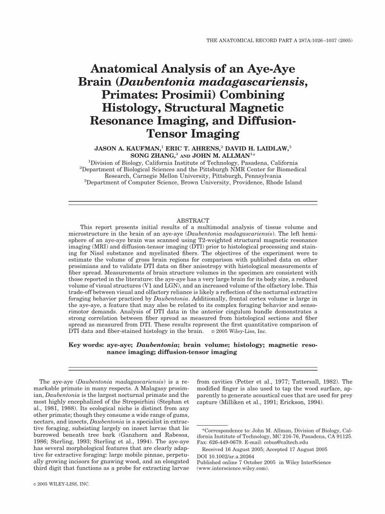
Fig. 1. Prosimian phylogeny used for independent contrast analysis.Adapted from Yoder (1994), Yoder and Yang (2004), and Karnath et al.(2005).
Fig. 2. Lateral view of the reconstructed lefthemisphere of the aye-aye brain. Sulcal terminol-ogy from Connolly (1950). Names in parenthesesrefer to conventional terminology used for pri-mates.
1027ANATOMICAL ANALYSIS OF AN AYE-AYE BRAIN
sis focuses first on digital reconstruction of the MR-im-aged left hemisphere and on volumetric estimates of grossbrain subdivisions for comparison with the aye-aye datapoint of Stephan et al. (1981). We begin by illustratingsome features of the cortical morphology and then presentsome preliminary results on Daubentonia brain volume in
comparison with 16 other prosimian species representedin the data set of Stephan et al. (1981). Using both con-ventional least-squares regression and phylogeneticallyindependent contrasts, we analyze the allometric relation-ships of individual brain components across prosimians.Since we do not have body mass data for our Daubentonia
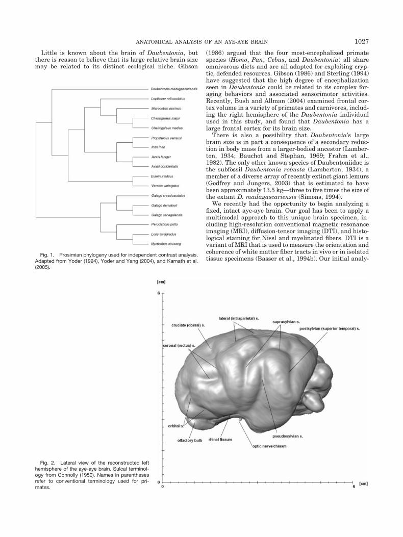
Fig. 3. Coronal sections through the T2-weighted MRI scan of the Daubentonia left hemi-sphere.
1028 KAUFMAN ET AL.
individual, we model our regression using structure vol-ume as the dependent variable and whole brain volumeminus structure volume (i.e., the rest of the brain) as theindependent variable (Barton, 1999).
The DTI technique assays the magnitude and spatialdirectionality of Brownian water diffusion in tissue (LeBihan and Breton, 1985; Le Bihan et al., 2001). Water thatdiffuses more readily in some directions compared to oth-ers due to features in the tissue microstructure is said tohave anisotropic diffusion. In the brain, water diffusesmore readily along axon tracts compared to transversedirections (Beaulieu and Allen, 1994a, 1994b). DTI can beused to measure this anisotropy, which in turn can beused to visualize the fiber’s trajectory and coherence (LeBihan et al., 2001). The magnitude and direction of thewater diffusion is modeled by a second-order diffusion-tensor at each point in a volume image, and the relativecoherence or uniformity of spatial diffusion can be quan-tified using the fractional anisotropy (FA), a ratio of theeigenvalues of the tensor. Fractional anisotropy rangesfrom 0 (isotropic diffusion) to 1 (highly anisotropic diffu-sion). Nominally, the value of FA is � 0 in gray matter, �0 in white matter, and approaches unity in large coherenttracts.
DTI and its associated tractography techniques havebeen successfully applied in a variety of clinical andresearch settings (reviewed by Le Bihan et al., 2001;Michael, 2002; Moseley et al., 2002; Neil et al., 2002;Ramnani et al., 2004; Sundgren et al., 2004), but therelationship between DTI data and brain microstruc-ture is not fully understood. In isolated rabbit myocar-dium, DTI data on muscle fiber orientation have beenshown to correlate well with orientation measurementsperformed directly on histological preparations subse-quent to scanning (Hsu et al., 1998; Scollan et al., 1998;Holmes et al., 2000). Here we apply a similar analysisby estimating a histological analogue of FA, which wedefined as the variance of axon orientations within amicroscopic volume. We calculated axon orientation dis-tributions from a series of sites within the anteriorcingulum bundle using stereologic microscopy and com-pared these values with fiber spread data as measuredby the DTI-derived FA parameter. We chose the cingu-lum bundle for this analysis since the hemisphere wassectioned in the sagittal plane and the fibers of thecingulum bundle run primarily within the plane of sec-tioning.
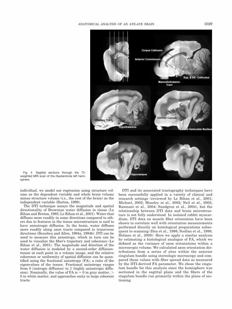
Fig. 4. Sagittal sections through the T2-weighted MRI scan of the Daubentonia left hemi-sphere.
1029ANATOMICAL ANALYSIS OF AN AYE-AYE BRAIN

MATERIALS AND METHODSThe aye-aye died of natural causes at the Duke Univer-
sity Primate Center and was immediately frozen at�80°C. Later it was thawed and the brain removed andfixed in 4% paraformaldehyde. Before imaging, the lefthemisphere was rinsed in PBS, immersed in Fluorinert(Sigma-Aldrich, Inc.), and positioned in a 35 mm innerdiameter birdcage MRI coil. Images were acquired usingan 11.7 T, 89 mm vertical-bore Bruker microimaging sys-tem. The three-dimensional (3D) DTI data were built fromthree overlapping packets of 32 contiguous slices acquiredusing a pulsed-gradient spin-echo protocol (Basser et al.,1994a). Each slice packet was translated along the slicedirection by one-third of the slice thickness. Postprocess-ing interleaved the slices for a final isotropic voxel size ofapproximately 70 �m. A total of 29 volumetric imageswere used in fitting the diffusion-tensors. The diffusiongradients were applied along seven different directions,and four values of b-matrix magnitude were used alongeach direction (maximum b � 1,500 mm2/sec). Imageswere acquired with TR/TE � 2000/19 msec and 512 �256 � 96 image points. The tensors were fit using a lab-oratory-written nonlinear fitting algorithm (Zhang et al.,2003). The anatomical T2 images were a by-product of theDTI fitting process. The brain hemisphere was scanned intwo volumes, one for the rostral portion and a second forthe caudal portion of the brain, and then they were digi-tally merged.
We also prepared a complete histological series from theleft hemisphere. The hemisphere was immersion-fixed informalin and embedded in celloidin. We have found that
celloidin provides better staining results, with less tissueshrinkage, than other embedding media. The hemispherewas sectioned in the parasagittal plane at a thickness of50 �m, and the complete series was alternately stained forNissl substance (thionin stain) and for myelinated axons(Gallyas, 1979). To help ensure accurate registration ofthe MRI with histological sections, the cut face of thecelloidin block was photographed with a digital cameramounted in a stationary position above the microtome.The mounted sections were digitized with a flatbed scan-ner, and each image was registered with its correspondingphotograph of the cut face. A 3D rendering of the hemi-sphere, assembled from the registered images, was used toselect sites on the histological sections that correspondedto regions that could be identified on the MRI.
Fig. 5. Axial sections through the T2-weightedMRI scan of the Daubentonia left hemisphere.
TABLE 1. Size of the Daubentonia brain and itscomponents as calculated in this study comparedwith the values calculated by Stephan et al. (1981)
PresentStudy
Stephan etal., 1981
Whole Brain (mg) 38130 45150Neocortex (mm3) 22029 22127Cerebellum (mm3) 4602 6461Thalamus (mm3) 1512 -Hippocampus (mm3) 1561 1776Striatum (mm3) 2450 2765Visual Striate Cortex (mm3) 1028 1355Lateral Geniculate Nucleus (mm3) 48 59Neocortical White (mm3) 4347 5053
1030 KAUFMAN ET AL.
Volumetric measurements were calculated by manuallysegmenting the T2-weighted MRI scans or, in the case ofvisual striate cortex, a combination of Nissl and fiberstains using the software package Amira (TGS, San Diego,CA). Allometric regressions were calculated using conven-tional least-squares regression (hereafter termed “starphylogeny”) as well as phylogenetically independent con-trasts. For Daubentonia, data points represent the meanof our individual and Stephan’s individual, and Dauben-tonia was excluded from the sample when calculatingregression equations. For independent contrast regres-sion, the assumed prosimian branching order (Fig. 1) wasadapted from previous studies (Yoder, 1994; Yoder and
Yang, 2004; Karanth et al., 2005). Independent contrastregression statistics were calculated using the softwarepackage PDAP (Garland et al., 1999; Garland and Ives,2000). To satisfy the statistical requirements of indepen-dent contrast regression, the branch lengths were trans-formed using Pagel’s (1992) method, which sets all branchlengths equal to 1 with the condition that tips are contem-poraneous. For graphical illustration, we use the methodof Garland and Ives (2000) to map the independent con-trast regression line back into raw data space.
Using the Gallyas stain, it was possible to trace individ-ual axons at high magnification and fiber bundles at lowmagnification. Fiber tracings were performed at 14 sites
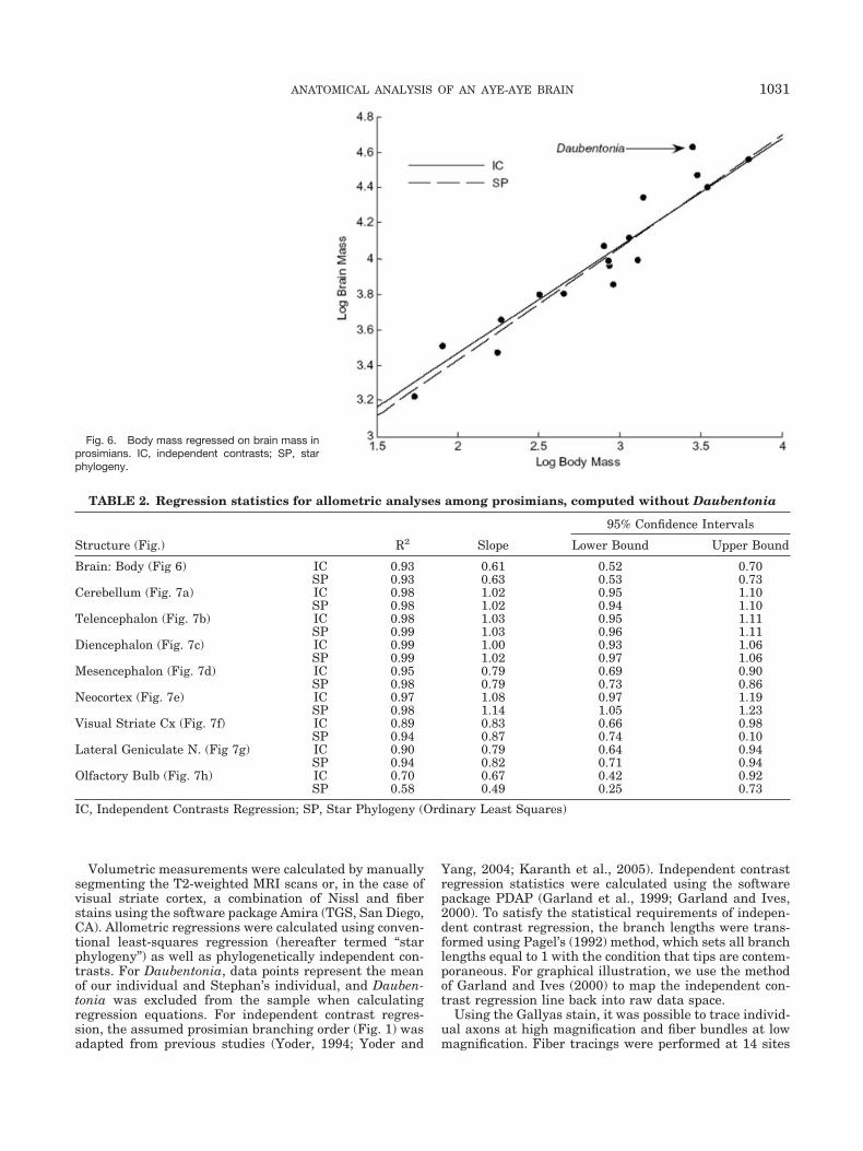
Fig. 6. Body mass regressed on brain mass inprosimians. IC, independent contrasts; SP, starphylogeny.
TABLE 2. Regression statistics for allometric analyses among prosimians, computed without Daubentonia
Structure (Fig.) R2 Slope
95% Confidence Intervals
Lower Bound Upper Bound
Brain: Body (Fig 6) IC 0.93 0.61 0.52 0.70SP 0.93 0.63 0.53 0.73
Cerebellum (Fig. 7a) IC 0.98 1.02 0.95 1.10SP 0.98 1.02 0.94 1.10
Telencephalon (Fig. 7b) IC 0.98 1.03 0.95 1.11SP 0.99 1.03 0.96 1.11
Diencephalon (Fig. 7c) IC 0.99 1.00 0.93 1.06SP 0.99 1.02 0.97 1.06
Mesencephalon (Fig. 7d) IC 0.95 0.79 0.69 0.90SP 0.98 0.79 0.73 0.86
Neocortex (Fig. 7e) IC 0.97 1.08 0.97 1.19SP 0.98 1.14 1.05 1.23
Visual Striate Cx (Fig. 7f) IC 0.89 0.83 0.66 0.98SP 0.94 0.87 0.74 0.10
Lateral Geniculate N. (Fig 7g) IC 0.90 0.79 0.64 0.94SP 0.94 0.82 0.71 0.94
Olfactory Bulb (Fig. 7h) IC 0.70 0.67 0.42 0.92SP 0.58 0.49 0.25 0.73
IC, Independent Contrasts Regression; SP, Star Phylogeny (Ordinary Least Squares)
1031ANATOMICAL ANALYSIS OF AN AYE-AYE BRAIN
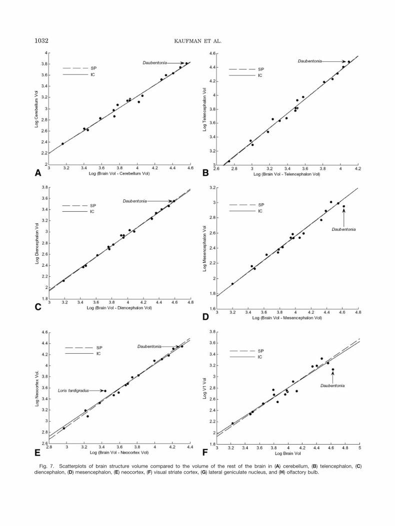
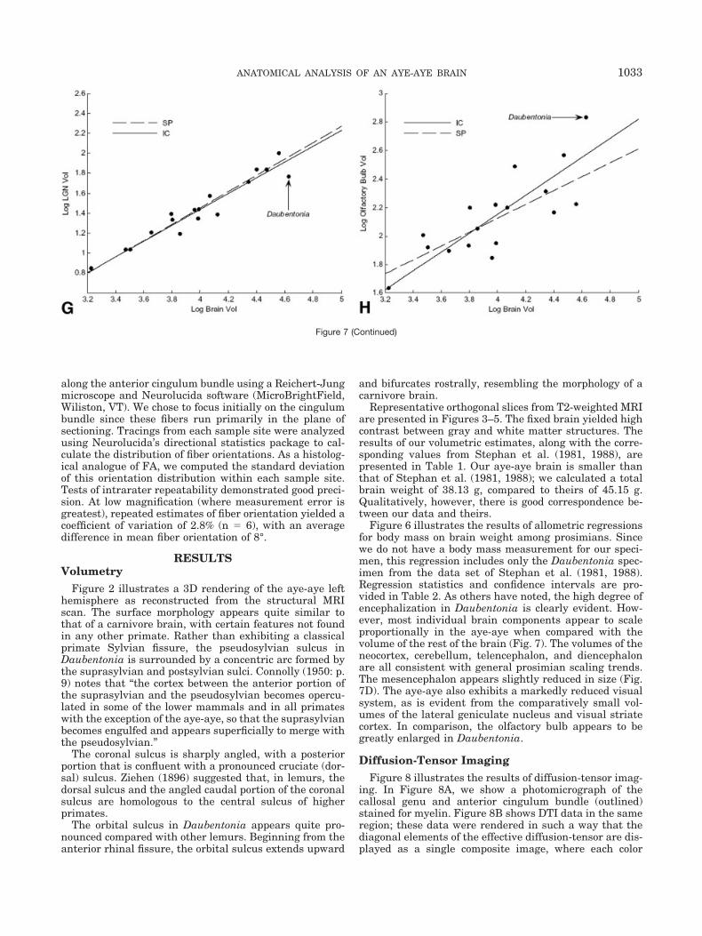
Fig. 7. Scatterplots of brain structure volume compared to the volume of the rest of the brain in (A) cerebellum, (B) telencephalon, (C)diencephalon, (D) mesencephalon, (E) neocortex, (F) visual striate cortex, (G) lateral geniculate nucleus, and (H) olfactory bulb.
1032 KAUFMAN ET AL.
along the anterior cingulum bundle using a Reichert-Jungmicroscope and Neurolucida software (MicroBrightField,Wiliston, VT). We chose to focus initially on the cingulumbundle since these fibers run primarily in the plane ofsectioning. Tracings from each sample site were analyzedusing Neurolucida’s directional statistics package to cal-culate the distribution of fiber orientations. As a histolog-ical analogue of FA, we computed the standard deviationof this orientation distribution within each sample site.Tests of intrarater repeatability demonstrated good preci-sion. At low magnification (where measurement error isgreatest), repeated estimates of fiber orientation yielded acoefficient of variation of 2.8% (n � 6), with an averagedifference in mean fiber orientation of 8°.
RESULTSVolumetry
Figure 2 illustrates a 3D rendering of the aye-aye lefthemisphere as reconstructed from the structural MRIscan. The surface morphology appears quite similar tothat of a carnivore brain, with certain features not foundin any other primate. Rather than exhibiting a classicalprimate Sylvian fissure, the pseudosylvian sulcus inDaubentonia is surrounded by a concentric arc formed bythe suprasylvian and postsylvian sulci. Connolly (1950: p.9) notes that “the cortex between the anterior portion ofthe suprasylvian and the pseudosylvian becomes opercu-lated in some of the lower mammals and in all primateswith the exception of the aye-aye, so that the suprasylvianbecomes engulfed and appears superficially to merge withthe pseudosylvian.”
The coronal sulcus is sharply angled, with a posteriorportion that is confluent with a pronounced cruciate (dor-sal) sulcus. Ziehen (1896) suggested that, in lemurs, thedorsal sulcus and the angled caudal portion of the coronalsulcus are homologous to the central sulcus of higherprimates.
The orbital sulcus in Daubentonia appears quite pro-nounced compared with other lemurs. Beginning from theanterior rhinal fissure, the orbital sulcus extends upward
and bifurcates rostrally, resembling the morphology of acarnivore brain.
Representative orthogonal slices from T2-weighted MRIare presented in Figures 3–5. The fixed brain yielded highcontrast between gray and white matter structures. Theresults of our volumetric estimates, along with the corre-sponding values from Stephan et al. (1981, 1988), arepresented in Table 1. Our aye-aye brain is smaller thanthat of Stephan et al. (1981, 1988); we calculated a totalbrain weight of 38.13 g, compared to theirs of 45.15 g.Qualitatively, however, there is good correspondence be-tween our data and theirs.
Figure 6 illustrates the results of allometric regressionsfor body mass on brain weight among prosimians. Sincewe do not have a body mass measurement for our speci-men, this regression includes only the Daubentonia spec-imen from the data set of Stephan et al. (1981, 1988).Regression statistics and confidence intervals are pro-vided in Table 2. As others have noted, the high degree ofencephalization in Daubentonia is clearly evident. How-ever, most individual brain components appear to scaleproportionally in the aye-aye when compared with thevolume of the rest of the brain (Fig. 7). The volumes of theneocortex, cerebellum, telencephalon, and diencephalonare all consistent with general prosimian scaling trends.The mesencephalon appears slightly reduced in size (Fig.7D). The aye-aye also exhibits a markedly reduced visualsystem, as is evident from the comparatively small vol-umes of the lateral geniculate nucleus and visual striatecortex. In comparison, the olfactory bulb appears to begreatly enlarged in Daubentonia.
Diffusion-Tensor ImagingFigure 8 illustrates the results of diffusion-tensor imag-
ing. In Figure 8A, we show a photomicrograph of thecallosal genu and anterior cingulum bundle (outlined)stained for myelin. Figure 8B shows DTI data in the sameregion; these data were rendered in such a way that thediagonal elements of the effective diffusion-tensor are dis-played as a single composite image, where each color
Figure 7 (Continued)
1033ANATOMICAL ANALYSIS OF AN AYE-AYE BRAIN
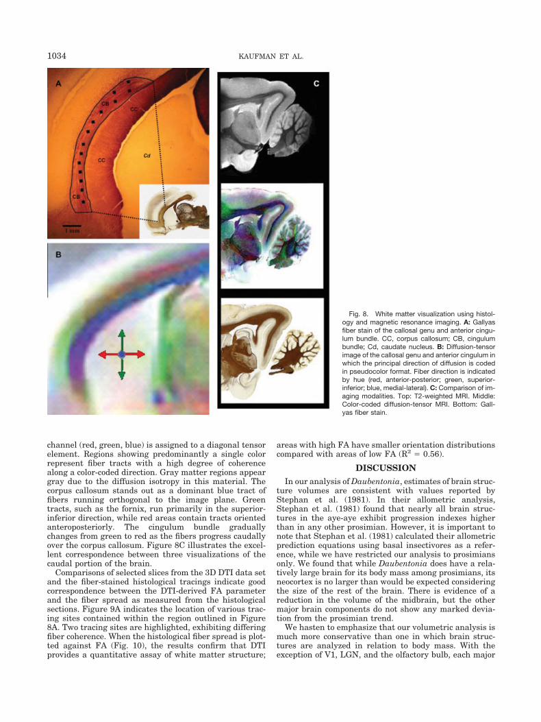
channel (red, green, blue) is assigned to a diagonal tensorelement. Regions showing predominantly a single colorrepresent fiber tracts with a high degree of coherencealong a color-coded direction. Gray matter regions appeargray due to the diffusion isotropy in this material. Thecorpus callosum stands out as a dominant blue tract offibers running orthogonal to the image plane. Greentracts, such as the fornix, run primarily in the superior-inferior direction, while red areas contain tracts orientedanteroposteriorly. The cingulum bundle graduallychanges from green to red as the fibers progress caudallyover the corpus callosum. Figure 8C illustrates the excel-lent correspondence between three visualizations of thecaudal portion of the brain.
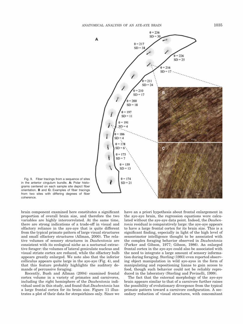
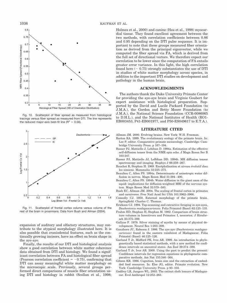
Comparisons of selected slices from the 3D DTI data setand the fiber-stained histological tracings indicate goodcorrespondence between the DTI-derived FA parameterand the fiber spread as measured from the histologicalsections. Figure 9A indicates the location of various trac-ing sites contained within the region outlined in Figure8A. Two tracing sites are highlighted, exhibiting differingfiber coherence. When the histological fiber spread is plot-ted against FA (Fig. 10), the results confirm that DTIprovides a quantitative assay of white matter structure;
areas with high FA have smaller orientation distributionscompared with areas of low FA (R2 � 0.56).
DISCUSSIONIn our analysis of Daubentonia, estimates of brain struc-
ture volumes are consistent with values reported byStephan et al. (1981). In their allometric analysis,Stephan et al. (1981) found that nearly all brain struc-tures in the aye-aye exhibit progression indexes higherthan in any other prosimian. However, it is important tonote that Stephan et al. (1981) calculated their allometricprediction equations using basal insectivores as a refer-ence, while we have restricted our analysis to prosimiansonly. We found that while Daubentonia does have a rela-tively large brain for its body mass among prosimians, itsneocortex is no larger than would be expected consideringthe size of the rest of the brain. There is evidence of areduction in the volume of the midbrain, but the othermajor brain components do not show any marked devia-tion from the prosimian trend.
We hasten to emphasize that our volumetric analysis ismuch more conservative than one in which brain struc-tures are analyzed in relation to body mass. With theexception of V1, LGN, and the olfactory bulb, each major
Fig. 8. White matter visualization using histol-ogy and magnetic resonance imaging. A: Gallyasfiber stain of the callosal genu and anterior cingu-lum bundle. CC, corpus callosum; CB, cingulumbundle; Cd, caudate nucleus. B: Diffusion-tensorimage of the callosal genu and anterior cingulum inwhich the principal direction of diffusion is codedin pseudocolor format. Fiber direction is indicatedby hue (red, anterior-posterior; green, superior-inferior; blue, medial-lateral). C: Comparison of im-aging modalities. Top: T2-weighted MRI. Middle:Color-coded diffusion-tensor MRI. Bottom: Gall-yas fiber stain.
1034 KAUFMAN ET AL.
brain component examined here constitutes a significantproportion of overall brain size, and therefore the twovariables are highly intercorrelated. At the same time,there are strong indications of a trade-off in visual andolfactory reliance in the aye-aye that is quite differentfrom the typical primate pattern of large visual structuresand small olfactory structures (Allman, 2000). The rela-tive volumes of sensory structures in Daubentonia areconsistent with its ecological niche as a nocturnal extrac-tive forager: the volumes of lateral geniculate nucleus andvisual striate cortex are reduced, while the olfactory bulbappears greatly enlarged. We note also that the inferiorcolliculus appears quite large in the aye-aye (Fig. 4), andthat this feature probably highlights the auditory de-mands of percussive foraging.
Recently, Bush and Allman (2004) examined frontalcortex volume in a variety of primates and carnivores,including the right hemisphere of the Daubentonia indi-vidual used in this study, and found that Daubentonia hasa large frontal cortex for its brain size. Figure 11 illus-trates a plot of their data for strepsirhines only. Since we
have an a priori hypothesis about frontal enlargement inthe aye-aye brain, the regression equations were calcu-lated without the aye-aye data point. Indeed, the Dauben-tonia residual is comparatively large: the aye-aye appearsto have a large frontal cortex for its brain size. This is asignificant finding, especially in light of the high level ofsensorimotor intelligence thought to be associated withthe complex foraging behavior observed in Daubentonia(Parker and Gibson, 1977; Gibson, 1986). An enlargedfrontal cortex in the aye-aye could also be associated withthe need to integrate a large amount of sensory informa-tion during foraging. Sterling (1993) even reported observ-ing object manipulation in wild aye-ayes in the form ofmanipulating and repositioning lianas to gain access tofood, though such behavior could not be reliably repro-duced in the laboratory (Sterling and Povinelli, 1999).
The fact that the external morphology of the aye-ayebrain appears similar to that of a carnivore further raisesthe possibility of evolutionary divergence from the typicalprimate pattern toward a carnivore configuration. A sec-ondary reduction of visual structures, with concomitant
Fig. 9. Fiber tracings from a sequence of sitesin the anterior cingulum bundle. A: Polar histo-grams centered on each sample site depict fiberorientation. B and C: Examples of fiber tracingsfrom two sites with differing degrees of fibercoherence.
1035ANATOMICAL ANALYSIS OF AN AYE-AYE BRAIN
expansion of auditory and olfactory structures, may con-tribute to the atypical morphology illustrated here. It isalso possible that craniodental features, such as the con-tinually growing incisors, have an effect on brain shape inthe aye-aye.
Finally, the results of our DTI and histological analysisshow a good correlation between white matter coherencedata obtained from DTI and histology. We found a signif-icant correlation between FA and histological fiber spread(Pearson correlation coefficient � �0.75), confirming thatDTI can assay meaningful white matter morphology onthe microscopic scale. Previously, several groups per-formed direct comparisons of muscle fiber orientation us-ing DTI and histology in rabbit (Scollan et al., 1998;
Holmes et al., 2000) and canine (Hsu et al., 1998) myocar-dial tissue. They found excellent agreement between thetwo methods, with correlation coefficients between 0.86and 0.95 depending on the DTI pulse sequence. It is im-portant to note that these groups measured fiber orienta-tion as derived from the principal eigenvector, while wecomputed the fiber spread via FA, which is derived fromthe full set of directional vectors. We therefore expect ourcorrelation to be lower since the computation of FA entailsgreater error variance. In this light, the high correlationfound here (� 0.75) strongly substantiates the use of DTIin studies of white matter morphology across species, inaddition to the important DTI studies on development andpathology in the human brain.
ACKNOWLEDGMENTSThe authors thank the Duke University Primate Center
for providing the aye-aye brain and Virginie Goubert forexpert assistance with histological preparation. Sup-ported by the David and Lucile Packard Foundation (toJ.M.A.), the Gordon and Betty Moore Foundation (toJ.M.A.), the National Science Foundation (CCR-0086065to D.H.L.), and the National Institutes of Health (RO1-EB003453, P41-EB001977, and P50-ES049617 to E.T.A.).
LITERATURE CITEDAllman JM. 2000. Evolving brains. New York: W.H. Freeman.Barton RA. 1999. The evolutionary ecology of the primate brain. In:
Lee P, editor. Comparative primate socioecology. Cambridge: Cam-bridge University Press. p 167–194.
Basser PJ, Mattiello J, Lebihan D. 1994a. Estimation of the effectiveself-diffusion tensor from the NMR spin echo. J Magn Reson Ser B103:247.
Basser PJ, Mattiello JJ, LeBihan DD. 1994b. MR diffusion tensorspectroscopy and imaging. Biophys J 66:259–267.
Bauchot R, Stephan H. 1969. Encephalisation et niveau evolutif chezles simiens. Mammalia 33:225–275.
Beaulieu C, Allen PS. 1994a. Determinants of anisotropic water dif-fusion in nerves. Magn Reson Med 31:394–400.
Beaulieu C, Allen PS. 1994b. Water diffusion in the giant axon of thesquid: implications for diffusion-weighted MRI of the nervous sys-tem. Magn Reson Med 32:579–583.
Bush EC, Allman JM. 2004. The scaling of frontal cortex in primatesand carnivores. Proc Natl Acad Sci USA 101:3962–3966.
Connolly CJ. 1950. External morphology of the primate brain.Springfield: Charles C. Thomas.
Erickson CJ. 1994. Tap-scanning and extractive foraging in aye-ayes,Daubentonia madagascariensis. Folia Primatol (Basel) 62:125–135.
Frahm HD, Stephan H, Stephan M. 1982. Comparison of brain struc-ture volumes in Insectivora and Primates: I, neocortex. J Hirnfor-sch 23:375–389.
Gallyas F. 1979. Silver staining of myelin by means of physical de-velopment. Neurol Res 1:203–209.
Ganzhorn JU, Rabesoa J. 1986. The aye-aye (Daubentonia madagas-cariensis) found in the eastern rainforest of Madagascar. FoliaPrimatol (Basel) 46:125–126.
Garland T Jr, Midford PE, Ives AR. 1999. An introduction to phylo-genetically based statistical methods, with a new method for confi-dence intervals on ancestral states. Am Zool 39:374–388.
Garland T Jr, Ives AR. 2000. Using the past to predict the present:Confidence intervals for regression equations in phylogenetic com-parative methods. Am Nat 155:346–364.
Gibson KR. 1986. Cognition, brain size and the extraction of embed-ded food resources. In: Else JG, editor. Primate evolution. NewYork: Cambridge University Press. p 93–103.
Godfrey LR, Jungers WL. 2003. The extinct sloth lemurs of Madagas-car. Evol Anthropol 12:252–263.
Fig. 10. Scatterplot of fiber spread as measured from histologicaltracings versus fiber spread as measured from DTI. The line representsthe reduced major-axis best-fit line (R2 � 0.56).
Fig. 11. Scatterplot of frontal cortex volume versus volume of therest of the brain in prosimians. Data from Bush and Allman (2004).
1036 KAUFMAN ET AL.
Holmes AA, Scollan DF, Winslow RL. 2000. Direct histological vali-dation of diffusion tensor MRI in formaldehyde-fixed myocardium.Magn Reson Med 44:157–161.
Hsu EW, Muzikant AL, Matulevicius SA, Penland RC, Henriquez CS.1998. Magnetic resonance myocardial fiber-orientation mappingwith direct histological correlation. Am J Physiol Heart Circ Physiol274:H1627–H1634.
Karanth KP, Delefosse T, Rakotosamimanana B, Parsons TJ, YoderAD. 2005. Ancient DNA from giant extinct lemurs confirms singleorigin of Malagasy primates. Proc Natl Acad Sci USA 102:5090–5095.
Lamberton C. 1934. Contribution a la connaissance de la faune sub-fossile de Madagascar. Mem Acad Malagache 17:40–46.
Le Bihan D, Breton E. 1985. Imagerie de diffusion in-vivo par reso-nance magnetique nucleaire. CR Acad Sci (Paris) 301:1109–1112.
Le Bihan D, Mangin JF, Poupon C, Clark CA, Pappata S, Molko N,Chabriat H. 2001. Diffusion tensor imaging: concepts and applica-tions. J Magn Reson Imaging 13:534–546.
Michael M. 2002. Diffusion tensor imaging and aging: a review. NMRBiomed 15:553–560.
Milliken GW, Ward JP, Erickson CJ. 1991. Independent digit controlin foraging by the aye-aye (Daubentonia madagascariensis). FoliaPrimatol (Basel) 56:219–224.
Moseley M, Bammer R, Illes J. 2002. Diffusion-tensor imaging ofcognitive performance. Brain Cogn 50:396.
Neil J, Miller J, Mukherjee P, Huppi PS. 2002. Diffusion tensorimaging of normal and injured developing human brain: a technicalreview. NMR Biomed 15:543–552.
Pagel MD. 1992. A method for the analysis of comparative data. JTheor Biol 156:431–442.
Parker ST, Gibson KR. 1977. Object manipulation, tool use and sen-sorimotor intelligence as feeding adaptations in cebus monkeys andgreat apes. J Hum Evol 6:623–641.
Petter J-J, Albignac R, Rumpler Y. 1977. Mammiferes Lemuriens(Primates Prosimiens). Paris: ORSTOM and CNRS.
Ramnani N, Behrens TEJ, Penny W, Matthews PM. 2004. New ap-proaches for exploring anatomical and functional connectivity inthe human brain. Biol Psychiatry 56:613.
Scollan DF, Holmes A, Winslow R, Forder J. 1998. Histological vali-dation of myocardial microstructure obtained from diffusion tensormagnetic resonance imaging. Am J Physiol 275:H2308–H2318.
Simons EL. 1994. The giant aye-aye Daubentonia robusta. Folia Pri-matol (Basel) 62:14–21.
Stephan H, Frahm H, Baron G. 1981. New and revised data onvolumes of brain structures in insectivores and primates. FoliaPrimatol (Basel) 35:1–29.
Stephan H, Baron G, Frahm H. 1988. Comparative size of brains andbrain components. In: Steklis HD, Erwin J, editors. Comparativeprimate biology, vol. 4, neurosciences. New York: Alan R. Liss. p1–38.
Sterling EJ. 1993. Behavioral ecology of the aye-aye (Daubentoniamadagascariensis) on nosy mangabe, Madagascar. PhD thesis. NewHaven, CT: Yale University.
Sterling EJ. 1994. Aye-ayes: specialists on structurally defended re-sources. Folia Primatol (Basel) 62:142–154.
Sterling EJ, Dierenfeld ES, Ashbourne CJ, Feistner AT. 1994. Dietaryintake, food composition and nutrient intake in wild and captivepopulations of Daubentonia madagascariensis. Folia Primatol(Basel) 62:115–124.
Sterling EJ, Povinelli DJ. 1999. Tool use, aye-ayes, and sensorimotorintelligence. Folia Primatol (Basel) 70:8–16.
Sundgren PC, Dong Q, Gomez-Hassan D, Mukherji SK, Maly P,Welsh R. 2004. Diffusion tensor imaging of the brain: review ofclinical applications. Neuroradiology 46:339–350.
Tattersall I. 1982. The primates of Madagascar. New York: ColumbiaUniversity Press.
Yoder AD. 1994. Relative position of the cheirogaleidae in strep-sirhine phylogeny: a comparison of morphological and molecularmethods and results. Am J Phys Anthropol 94:25–46.
Yoder AD, Yang Z. 2004. Divergence dates for Malagasy lemurs esti-mated from multiple gene loci: geological and evolutionary context.Mol Ecol 13:757–773.
Zhang S, Demiralp C, Laidlaw DH. 2003. Visualizing diffusion tensorMR images using streamtubes and streamsurfaces. IEEE T VisComput Gr 9:454–462.
Ziehen T. 1896. Ueber die Grosshirnfurchung der Halbaffen und dieDeutungeiniger Furchen des menschliched Gehirns. Arch PsyciatNervenkr 28:898–930.
1037ANATOMICAL ANALYSIS OF AN AYE-AYE BRAIN
Related Documents