Analyzing brain structural differences among undergraduates with different grades of self‑esteem using multiple anatomical brain network Bo Peng 1,2,3 , Gaofeng Pang 4 , Aditya Saxena 5 , Yan Liu 1,2,3 , Baohua Hu 1,2,3 , Suhong Wang 6* and Yakang Dai 1,2,3* Background Self-esteem is regarded as self-affirmation and self-identification about oneself [1]. Peo- ple with good mental health have a higher self-esteem grade and think of themselves as valuable persons [2]. Researchers found that undergraduates with different self-esteem Abstract Background: Self-esteem is the individual evaluation of oneself. People with high self-esteem grade have mental health and can bravely cope with the threats from the environment. With the development of neuroimaging techniques, researches on cognitive neural mechanisms of self-esteem are increased. Existing methods based on brain morphometry and single-layer brain network cannot characterize the subtle structural differences related to self-esteem. Method: To solve this issue, we proposed a multiple anatomical brain network based on multi-resolution region of interest (ROI) template to study the brain structural con- nections of self-esteem. The multiple anatomical brain network consists of ROI features and hierarchal brain network features that are extracted from structural MRI. For each layer, we calculated the correlation relationship between pairs of ROIs. In order to solve the high-dimensional problem caused by the large amount of network features, feature selection methods (t-test, mRMR, and SVM-RFE) are adopted to reduce the number of features while retaining discriminative information to the maximum extent. Multi-kernel SVM is employed to integrate the various types of features by appropriate weight coefficient. Result: The experimental results show that the proposed method can improve clas- sification accuracy to 97.26% compared with single-layer brain network. Conclusions: The proposed method provides a new perspective for the analysis of brain structural differences of self-esteem, which also has potential guiding signifi- cance in other researches involved brain cognitive activity and brain disease diagnosis. Keywords: Structural magnetic resonance imaging, Self-esteem, Multi-resolution ROI, Hierarchal brain network, Machine learning method Open Access © The Author(s) 2021. This article is licensed under a Creative Commons Attribution 4.0 International License, which permits use, sharing, adaptation, distribution and reproduction in any medium or format, as long as you give appropriate credit to the original author(s) and the source, provide a link to the Creative Commons licence, and indicate if changes were made. The images or other third party material in this article are included in the article’s Creative Commons licence, unless indicated otherwise in a credit line to the material. If material is not included in the article’s Creative Commons licence and your intended use is not permitted by statutory regulation or exceeds the permitted use, you will need to obtain permission directly from the copyright holder. To view a copy of this licence, visit http://creativecommons.org/ licenses/by/4.0/. The Creative Commons Public Domain Dedication waiver (http://creativecommons.org/publicdomain/zero/1.0/) applies to the data made available in this article, unless otherwise stated in a credit line to the data. RESEARCH Peng et al. BioMed Eng OnLine (2021) 20:20 https://doi.org/10.1186/s12938‑021‑00853‑z BioMedical Engineering OnLine *Correspondence: [email protected]; [email protected] 1 Suzhou Institute of Biomedical Engineering and Technology, Chinese Academy of Sciences, Suzhou, China 6 Department of Clinical Psychology, The Third Affiliated Hospital of Soochow University, Changzhou, China Full list of author information is available at the end of the article

Welcome message from author
This document is posted to help you gain knowledge. Please leave a comment to let me know what you think about it! Share it to your friends and learn new things together.
Transcript

Analyzing brain structural differences among undergraduates with different grades of self‑esteem using multiple anatomical brain networkBo Peng1,2,3, Gaofeng Pang4, Aditya Saxena5, Yan Liu1,2,3, Baohua Hu1,2,3, Suhong Wang6* and Yakang Dai1,2,3*
BackgroundSelf-esteem is regarded as self-affirmation and self-identification about oneself [1]. Peo-ple with good mental health have a higher self-esteem grade and think of themselves as valuable persons [2]. Researchers found that undergraduates with different self-esteem
Abstract
Background: Self-esteem is the individual evaluation of oneself. People with high self-esteem grade have mental health and can bravely cope with the threats from the environment. With the development of neuroimaging techniques, researches on cognitive neural mechanisms of self-esteem are increased. Existing methods based on brain morphometry and single-layer brain network cannot characterize the subtle structural differences related to self-esteem.
Method: To solve this issue, we proposed a multiple anatomical brain network based on multi-resolution region of interest (ROI) template to study the brain structural con-nections of self-esteem. The multiple anatomical brain network consists of ROI features and hierarchal brain network features that are extracted from structural MRI. For each layer, we calculated the correlation relationship between pairs of ROIs. In order to solve the high-dimensional problem caused by the large amount of network features, feature selection methods (t-test, mRMR, and SVM-RFE) are adopted to reduce the number of features while retaining discriminative information to the maximum extent. Multi-kernel SVM is employed to integrate the various types of features by appropriate weight coefficient.
Result: The experimental results show that the proposed method can improve clas-sification accuracy to 97.26% compared with single-layer brain network.
Conclusions: The proposed method provides a new perspective for the analysis of brain structural differences of self-esteem, which also has potential guiding signifi-cance in other researches involved brain cognitive activity and brain disease diagnosis.
Keywords: Structural magnetic resonance imaging, Self-esteem, Multi-resolution ROI, Hierarchal brain network, Machine learning method
Open Access
© The Author(s) 2021. This article is licensed under a Creative Commons Attribution 4.0 International License, which permits use, sharing, adaptation, distribution and reproduction in any medium or format, as long as you give appropriate credit to the original author(s) and the source, provide a link to the Creative Commons licence, and indicate if changes were made. The images or other third party material in this article are included in the article’s Creative Commons licence, unless indicated otherwise in a credit line to the material. If material is not included in the article’s Creative Commons licence and your intended use is not permitted by statutory regulation or exceeds the permitted use, you will need to obtain permission directly from the copyright holder. To view a copy of this licence, visit http://creat iveco mmons .org/licen ses/by/4.0/. The Creative Commons Public Domain Dedication waiver (http://creat iveco mmons .org/publi cdoma in/zero/1.0/) applies to the data made available in this article, unless otherwise stated in a credit line to the data.
RESEARCH
Peng et al. BioMed Eng OnLine (2021) 20:20 https://doi.org/10.1186/s12938‑021‑00853‑z BioMedical Engineering
OnLine
*Correspondence: [email protected]; [email protected] 1 Suzhou Institute of Biomedical Engineering and Technology, Chinese Academy of Sciences, Suzhou, China 6 Department of Clinical Psychology, The Third Affiliated Hospital of Soochow University, Changzhou, ChinaFull list of author information is available at the end of the article

Page 2 of 15Peng et al. BioMed Eng OnLine (2021) 20:20
grades have brain structural differences. These people feel that they deserve to be respected by others, and are more able to accept individual’s deficiencies [3]. However, people with low self-esteem have low self-confidence, and the outside world will have a great impact on them, resulting in low socioeconomic status and poor physical health. Neurophysiology researchers found that self-esteem may be composed of multiple sub-systems that are structurally separated from each other, but functionally interact [4]. Brain imaging studies suggest that self-esteem involves multiple psychological processes, including self in perception, memory, and introspection. These psychological processes have their own corresponding brain regions. For example, self-face recognition occurs in right brain, and autobiographical memory is mainly related to hippocampus, and self-reference is related to medial prefrontal lobe [5]. In addition to these independent brain regions, the difference in self-esteem is also reflected in brain network connec-tion. Medial prefrontal cortex is activated during the process of social, self, and affective events [6]. Therefore, in this study, we focus on exploring the brain structural differences among undergraduates with different levels of self-esteem.
Brain network aims to study the interaction of various brain regions as a whole, which has an important role to deeply understand brain structures and cognitive neural pro-cesses. The anatomical brain network mainly uses region of interest (ROI) of the brain as the node, and the correlation between brain regions as edge [7, 8]. The definition of ROI is a key step in anatomical brain network analysis. Most existing methods use ROI-based brain network analysis methods to study brain structure and functional connec-tions related to self-esteem. Kelly et al. use the cerebral blood flow imaging method to estimate the hemodynamic response function of each ROI, in order to study the brain networks that are activated during the processing of self-esteem related information [9]. Goldin et al. used functional magnetic resonance imaging (fMRI) technology to meas-ure changes in the brain network between self-esteem group and the self-confidence group by measuring the BOLD response in the ROI [10]. Chavez et al. conducted a psy-chophysiological interaction analysis to calculate the correlation between specific ROIs related to self-esteem [11]. Although a variety of neuroimaging methods can be used to explore the cognitive mechanism of the brain, structural magnetic resonance imag-ing (sMRI) is widely used in the analysis of brain anatomical networks due to its high resolution of brain soft tissue imaging [12]. Studies based on sMRI show that self-esteem involves multiple networks related to self-reference processing, autobiographical mem-ory, and social cognition, including default mode networks and social cognition net-works [13]. In addition, self-esteem shows the brain network mechanism dominated by bilateral brain and mainly controlled by right brain [14]. Although the above researches have initially revealed the brain network representation of self-esteem, it only used sin-gle-layer network that cannot fully identify the subtle differences in network connectiv-ity caused by self-esteem.
The motivation of this study is to use enhanced feature representation method to bet-ter analyze brain structural connectivity related to self-esteem. In recent years, machine learning techniques become a research hotspot in the field of brain network analysis due to its ability to learn pattern from data and predict unknown data [8]. Brain net-work analysis can help us fully understand the cognitive psychological activity of self-esteem. However, there are few studies using machine learning methods to construct

Page 3 of 15Peng et al. BioMed Eng OnLine (2021) 20:20
self-esteem-related brain network, especially for the construction of hierarchal brain networks. In this article, we propose multiple anatomical brain network construction method based on multi-resolution ROIs. The innovation of this method is to use the in-layer and between-layer connections to better describe the correlation between small brain regions and large brain functional areas, which improves the feature expression ability of single-layer brain network.
ResultsClassification performance
Various indexes can be used to evaluate the classification performance of the proposed method. The evaluation indicators including accuracy (ACC), sensitivity (SEN), speci-ficity (SPE), area under the receiver operating characteristic curve (AUC), F score, bal-anced accuracy, Youden’s index are listed in Table 1. The results show that the multiple brain network features have the highest classification accuracy of 97.26%, and the AUC is also greater than other feature types. This indicates that the multiple brain network features have advantages in characterizing structural differences at the global level. In addition, the higher specificity and sensitivity also show that the multiple brain network features have better recognition capabilities in exploring the subtle differences in brain structure caused by self-esteem (Fig. 1).
Weight coefficient
The role of the weight coefficient is to determine the proportion of the various types of features in the multi-kernel classifier (Fig. 2). Appropriate weight coefficient helps achieve the best classifier performance. A smaller weight coefficient indicates that the contribution of ROI features is lower, while the contribution of the hierarchical brain network features is higher. Through experiments, we can find the most suitable weight coefficient in the range of 0–1.
The weight coefficient has an important influence on the performance of the clas-sifier. It is proved that the weight coefficient makes the classifier perform well in the relatively large range from 0.05 to 0.35, which can decline the difficulty of determin-ing the ratio of the two types of features, which reflects the robustness of our pro-posed method. The best results are obtained at 0.05. At this time, the hierarchical
Table 1 Classification performance using different feature types
ACC: accuracy; AUC: area under receiver operating characteristic curve; SEN: sensitivity; SPE: specificity; Y: Youden’s index; F: F‑score; BAC: balanced accuracy
ACC (%) AUC (%) SEN (%) SPE (%) Y (%) F (%) BAC (%)
Network features in the 4th layer 90.69 96.63 87.72 90.65 86.74 77.38 88.77
Network features in the 3rd layer 88.31 84.27 85.32 84.29 88.33 82.62 85.74
Network features in the 2nd layer 89.59 76.65 78.53 75.94 73.24 69.18 67.77
Network features in all layers 92.59 91.93 91.51 90.91 93.27 87.18 91.49
ROI features in the 4th layer 88.69 85.63 87.72 87.65 86.74 77.38 88.77
ROI features and network features in the 4th layer
94.41 95.58 94.42 93.41 92.47 92.82 92.64
Multilevel (ROI features in the 4th layer and network features in all layers)
97.26 99.88 97.27 97.41 97.12 94.53 97.27

Page 4 of 15Peng et al. BioMed Eng OnLine (2021) 20:20
brain network features contributed more to the classification than the ROI features. This is because the hierarchical brain network can fully express the differences in brain structure between the high self-esteem group and low self-esteem group.
Top discriminative features
We use the proposed method to select the most discriminative ROI features (Fig. 3). These ROIs include occipital lobe (superior and middle occipital gyrus, cuneus), frontal lobe (supplementary motor area, middle frontal gyrus), temporal lobe (mid-dle temporal gyrus), parietal lobe (precuneus, angular gyrus), limbic lobe (posterior cingulate gyrus), and central region (precentral gyrus). The experimental results also
Fig. 1 Boxplot of classification accuracy for different feature types. (1) Network features in the 4th layer; (2) network features in the 3rd layer; (3) network features in the 2nd layer; (4) network features in all layers; (5) ROI features in the 4th layer; (6) ROI features and network features in the 4th layer; (7) multilevel features. ACC accuracy, SEN sensitivity, SPE specificity
Fig. 2 Classification performance with ROI features using different weighting factors. The weight for the ROI features decrease from left to right (range from 0 to 1)

Page 5 of 15Peng et al. BioMed Eng OnLine (2021) 20:20
show that differences in brain structure related to self-esteem are mainly in white matter and cortical thickness (Table 2).
The top 15 network features selected from all four layers (Table 3). The most discrimi-native hierarchical network features are mainly distributed in limbic lobe and parietal lobe (Fig. 4).
DiscussionWe studied multiple anatomical brain network related to self-esteem. Our results dem-onstrate that the proposed method is superior to the single-layer network method. The multiple networks enhance the representation of the specific brain structure related to self-esteem, thereby providing an effective and novel method to detect self-esteem-related biomarkers.
Fig. 3 The most discriminating ROI features projected onto the cortical surface. HSG means high self-esteem group and LSG means low self-esteem group

Page 6 of 15Peng et al. BioMed Eng OnLine (2021) 20:20
Improvement of the proposed method
It is difficult to fully understand the functional organization of the brain using only a single-layer network framework since the brain is a complex system. In this study, we construct a multiple anatomical brain network in multi-resolution ROIs to
Table 2 Top 15 most discriminating regional features that were selected using the proposed classification framework
L: left hemisphere; R: right hemisphere; GM: gray matter volume; WM: white matter volume; CSF: cerebrospinal volume; Thickness: cortical thickness; Area: cortical surface area; Frequency: selected frequency over 100 repetitions of twofold cross‑validation
No. Name of ROI L/R Tissue Brain lobe Frequency
1 Middle frontal gyrus R WM Frontal lobe 185
2 Superior occipital gyrus R GM Occipital lobe 144
3 Precentral gyrus R Thickness Central region 141
4 Middle occipital gyrus L GM Occipital lobe 102
5 Supplementary motor area R WM Frontal lobe 86
6 Posterior cingulate gyrus L CSF Limbic lobe 75
7 Middle frontal gyrus L WM Frontal lobe 73
8 Posterior cingulate gyrus L Thickness Limbic lobe 70
9 Middle occipital gyrus R Thickness Occipital lobe 68
10 Angular gyrus R WM Parietal lobe 64
11 Precuneus R Thickness Parietal lobe 58
12 Cuneus L WM Occipital lobe 58
13 Middle temporal gyrus L Area Temporal lobe 54
14 Precuneus L Thickness Parietal lobe 53
15 Middle occipital gyrus L Thickness Occipital lobe 53
Table 3 Top 15 similarity features that were selected using the proposed classification framework
L: left hemisphere; R: right hemisphere; Frequency: selected frequency over 100 repetitions of twofold cross‑validation
Network Name of ROI L/R Name of ROI L/R No. Frequency
Network 4 Orbitofrontal cortex (inferior) L Superior parietal gyrus L 15 45
Rectus gyrus L Precuneus L 12 48
Orbitofrontal cortex (inferior) L Paracentral lobule R 10 54
Orbitofrontal cortex (inferior) R Precuneus L 3 95
Network 3 Parietal lobe: lateral surface R Limbic lobe: Temporal pole (superior)
R 1 118
Frontal lobe: lateral surface L Parietal lobe: lateral surface L 2 114
Temporal lobe: lateral surface L Parietal lobe: lateral surface R 4 93
Frontal lobe: lateral surface R Temporal lobe: lateral surface R 5 93
Frontal lobe: lateral surface R Parietal lobe: lateral surface L 6 92
Central region: Rolandic oper-culum
L Limbic lobe: temporal pole (superior)
L 7 74
Central region: postcentral gyrus R Parietal lobe: lateral surface R 9 61
Temporal lobe: lateral surface R Limbic lobe: temporal pole (superior)
R 13 47
Parietal lobe: lateral surface L Limbic lobe: temporal pole (superior)
L 14 47
Network 2 Central region L Limbic lobe L 8 73
Central region R Limbic lobe L 11 54

Page 7 of 15Peng et al. BioMed Eng OnLine (2021) 20:20
improve the classification performance. Compared with the single-layer network-based method, multiple networks enhance the classification performance by using supplementary information from different networks. Compared with the best results obtained using a single-layer network, our proposed method can improve the classifi-cation accuracy by 8.95% (Table 1).
Analysis of discriminative features
The discriminative ROI features discovered by our method are distributed in multi-ple regions of the brain. Because few current studies employ automatic classification method to study the brain structure of self-esteem, we only compare brain regions found through our machine learning method with existing morphological based studies. Compared with previous studies, our results showed consistency in departmental brain regions, including precuneus [4], precentral gyrus [15], middle frontal gyrus [16], cuneus [4], posterior cingulate [17]. This indicates the effectiveness of our classification method in revealing brain regions related to self-esteem. In addition to these consistent regions, we also found that the middle occipital, superior occipital, and supplementary motor are related to self-esteem. These brain regions have not been reported in previous studies, which is worth paying attention to in the follow-up study.
The discriminative network features are mainly located on frontal, parietal and limbic lobe. After a comprehensive analysis of existing research on neuropsychological mecha-nisms related to self-esteem, we found that the frontal region is an important part of the neural basis related to self-esteem. The frontal lobe is responsible for self-evaluation,
Fig. 4 Correlative matrix: a high self-esteem group, b low self-esteem group, c differences between the two groups
Page 8 of 15Peng et al. BioMed Eng OnLine (2021) 20:20
self-regulation, and emotion management. Individuals with low self-esteem have a stronger emotional response to social evaluations, while high self-esteem individuals show stronger self-positivity in the process of self-evaluation. These findings indicate that frontal lobe plays an important role in generating positive self-information.
Comparison with other methods
Since few studies have used machine learning to analyze the relationship between self-esteem and brain structure, we compare our results with the current morphological studies related to self-esteem. At present, most studies have found a correlation between self-esteem and frontal lobe [6, 16, 17]. The frontal lobe is mainly responsible for the cognitive activities of the brain, and self-esteem involves cognitive processing and emo-tional response. The results of this paper are generally consistent with those of previous studies. In addition, some specific brain regions, such as the cuneus, have been found to be related to many self-related functions, such as self-related information processing and various aspects of consciousness [16]. In addition, we also found a correlation between cingulate cortex and self-esteem. Studies have shown that when individuals are accepted by society, individuals activate the ventral anterior cingulate cortex and medial prefron-tal cortex, thus enhancing self-esteem [17]. Therefore, the study of brain structure with different self-esteem grades is helpful to understand the neurophysiological mechanism of self-esteem.
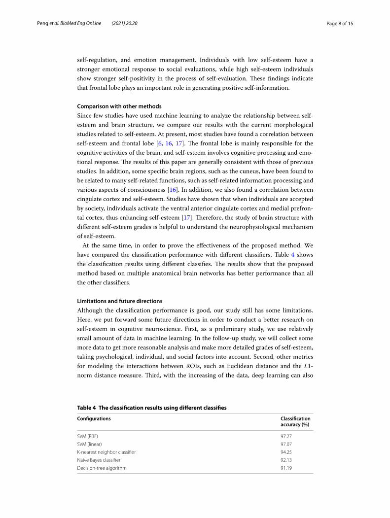
At the same time, in order to prove the effectiveness of the proposed method. We have compared the classification performance with different classifiers. Table 4 shows the classification results using different classifies. The results show that the proposed method based on multiple anatomical brain networks has better performance than all the other classifiers.
Limitations and future directions
Although the classification performance is good, our study still has some limitations. Here, we put forward some future directions in order to conduct a better research on self-esteem in cognitive neuroscience. First, as a preliminary study, we use relatively small amount of data in machine learning. In the follow-up study, we will collect some more data to get more reasonable analysis and make more detailed grades of self-esteem, taking psychological, individual, and social factors into account. Second, other metrics for modeling the interactions between ROIs, such as Euclidean distance and the L1-norm distance measure. Third, with the increasing of the data, deep learning can also
Table 4 The classification results using different classifies
Configurations Classification accuracy (%)
SVM (RBF) 97.27
SVM (linear) 97.07
K-nearest neighbor classifier 94.25
Naive Bayes classifier 92.13
Decision-tree algorithm 91.19

Page 9 of 15Peng et al. BioMed Eng OnLine (2021) 20:20
be used to automatically extract features to find the discriminative features of multiple brain network, such as depth automatic encoder. Fourth, due to the multi-types of fea-tures involved in this study, multiple weight factors can be used for better feature fusion.
ConclusionIn this study, we have presented how multiple anatomical brain networks can be used to analyze brain structural differences among undergraduates with different grades of self-esteem. Several feature selection methods are adopted to reduce the number of features, and multi-kernel SVM was employed to integrate various types of features by appropriate weight coefficient. The features extracted from these networks can be used to improve the defects that the traditional single-layer brain network contains insuffi-cient information. The experiments show that our method has improved performance compared with the single-layer network structure, which can provide a new perspective for the analysis of brain structure differences of self-esteem. It also has potential guiding significance of out method in other researches involved in brain cognitive activity and brain disease diagnosis.
MethodsSubjects
The structural MRI data used in our study were acquired from Soochow Univer-sity, which is composed of 68 undergraduates. The study was approved by the Ethics Committee of the Third Affiliated Hospital of Soochow University. Written informed consents were obtained from all subjects. All subjects did not receive stimulants or hyp-notics before acquisition in order to keep them awake and let the brain work normally. All participants’ vision were normal or corrected to normal, and they were right-handed. After the test, each participant will receive a small gift or financial reward. All subjects are required to perform Rosenberg Self-esteem Scale (RSES) test. The RSES is originally developed by Rosenberg in 1965 to assess the overall feelings of undergraduates about self-worth and self-acceptance. It is the most used self-esteem measurement tool in the psychology community [18]. We ranked the RSES test scores from highest to lowest, and then divided them into two groups: high self-esteem group and low self-esteem group. Table 5 provides detailed information of all participants.
Table 5 Demographic information of all subjects
The p‑value of gender was obtained by Chi‑squared test
The p‑values of age and Rosenberg scale were obtained by t‑test
Significance level was set to 0.05
High self-esteem group Low self-esteem group p value
Subjects 34 34
Male/female 19/15 16/18 0.83
Age (mean ± SD) 21.90 ± 1.16 22.53 ± 1.42 0.77
Rosenberg Scale (mean ± SD) 25.35 ± 0.81 17.86 ± 3.35 < 0.001

Page 10 of 15Peng et al. BioMed Eng OnLine (2021) 20:20
Imaging acquisition and preprocessing
All images were collected on a 3T Siemens Medical Systems equipment. The acquisi-tion parameters are set as: echo time (TE) = 2.98 ms, repetition time (TR) = 2300 ms, flip angle (FA) = 9 °, voxel size = 1 × 1 × 1 mm3, slice thickness = 1 mm, field of view (FoV) = 256 mm.
We use an automatic pipeline for sMRI image processing. Firstly, we adjusted the image orientation (axial, coronal, and sagittal) to match the template image, and per-formed offset field correction to remove the gray-scale unevenness of the image [19]. Secondly, the brain image was extracted by removing the skull and cerebellum [20]. Thirdly, gray matter (GM), white matter (WM) and cerebrospinal fluid (CSF) were seg-mented from the background [21]. Fourth, the segmented image was registered to the template labeled with the Automated Anatomical Labeling (AAL) template [22]. Fifth, in order to calculate the morphological features based on the cortex, the middle layer of the cerebral cortex was depicted [23]. After the whole processing, the morphological measurements of GM volume, WM volume, CSF volume, cortical thickness, and corti-cal surface area of each ROI were obtained for each subject. It should be noted that we removed 12 subcortical ROIs from AAL template considering that the cerebral cortex contains more neurons.
Framework of the proposed method
The framework of the proposed method based on multiple brain network is shown in Fig. 5, mainly including image processing, feature extraction, feature selection, and clas-sification. Multiple brain network were constructed based on morphological features (volume of different brain tissue, cortical thickness, and cortical surface area). Feature selection can reduce the dimensionality of high-dimensional brain network features, only retaining the features that can maximize the specificity of the subjects. The optimal feature subset can be trained by the classifier as neuroimaging markers representing dif-ferent self-esteem levels.
Fig. 5 Framework of the classification method using multilevel network features

Page 11 of 15Peng et al. BioMed Eng OnLine (2021) 20:20
Construction of multiple anatomical networks
Through the above image processing steps, GM volume, WM volume, CSF volume, cortical thickness, and cortical surface area of each ROI can be obtained from the MRI image of each subject. In order to reduce individual differences, standardization was performed, dividing the measured value of each ROI by the total intracranial volume, mean cortical thickness, and whole cerebral cortical surface area of the subject. There-fore, we used normalized volume and cortical features to provide a more appropriate representation. More objective measurements can be received by such processing. In order to improve the performance of the classifier, we propose a four-layer hierarchical network in this paper. We used brain templates with different ROI resolution in each layer to construct brain network nodes and edges.
Specifically, the bottommost template containing 78 ROIs is defined as L4 , the remain-ing three layers are defined as Ll , where l = 1, 2, 3 . A larger l value indicates a higher-resolution ROI, which is located in the brain network layer closer to the bottom of the hierarchy. By merging small brain regions into large brain functional areas, the number of ROIs is reduced. In the layer L3 , there are 36 ROIs by dividing the whole brain into lat-eral, medial and inferior surfaces. In the layer L2 , 14 ROIs are defined reefing to the ana-tomical brain structure of central, frontal, parietal, occipital, temporal, limbic, and insula lobe. The specific definition rules of these ROIs can be found in Table 6. It is worth not-ing that in the first layer L1 , we study the brain as a whole.
For each layer, correlation between ROIs can be calculated using brain template defined above. Its node correspond to the ROIs in different resolution, and the edge corresponds to the interaction between pairs of ROIs. Take the bottom layer L4 as an example, a 78 × 78 matrix C4 can be calculated by computing the Pearson correlation coefficient between the ith ROI and jth ROI. We define
where t(i) and t(j) represent the mean thickness of the cerebral cortex corresponding to the i th and j th ROIs.σ is defined as σ =
√
σi2 + σj
2 , where σi and σj represent the standard deviation of
cortex for the i th and j th ROI. Due to the symmetry of the correlation matrix, we only use the upper triangular elements of the matrix C4 to construct the feature vector. We connect the 3003 upper triangular elements to form the corresponding feature vector for L4 . Since the ROIs in the remaining three layers are obtained by merging ROIs in the bottommost layer, the mean and standard deviation of these compound ROIs can be obtained by calculating the average value of all ROIs. The definition of correlation matrix Cl for other layers is similar to C4 . The union of the hierarchical networks is constructed by junction of the four upper triangular correlation matrix into a long vector.
Feature selection
In order to reduce the feature dimension and filter out the most discriminative features, we adopted several feature selection methods. First, we preliminarily select the features by comparing the statistics of different features. The statistical t-test (p < 0.05) is adopted
(1)C4(
i, j)
= exp
{
−
[
t(i)− t(
j)]2
2σ 2
}
,

Page 12 of 15Peng et al. BioMed Eng OnLine (2021) 20:20
to remove features with small differences (the features with small differences are diffi-cult to distinguish the two groups). Then, another filter-based feature selection method called minimum redundancy and maximum correlation (mRMR) is used to remove the redundant features [24]. The core idea of mRMR is to maximize the correlation between features and classification variables, and minimize the correlation between different
Table 6 Regions of interest (ROIs) defined in the automated anatomical labeling (AAL) template
Network 2 Network 3 Network 4
No. Name of ROI No. Name of ROI No. Name of ROI
1, 2 Central region 1, 2 Central region: precentral gyrus 1, 2 Precentral gyrus
3, 4 Central region: postcentral gyrus 53, 54 Postcentral gyrus
5, 6 Central region: Rolandic operculum 17, 18 Rolandic operculum left
3, 4 Frontal lobe 7, 8 Frontal lobe: lateral surface 3, 4 Superior frontal gyrus (dorsal)
7, 8 Middle frontal gyrus
11, 12 Inferior frontal gyrus (opercular)
13, 14 Inferior frontal gyrus (triangular)
9, 10 Frontal lobe: medial surface 19, 20 Supplementary motor area
23, 24 Superior frontal gyrus (medial)
65, 66 Paracentral lobule
11, 12 Frontal lobe: orbital surface 5, 6 Orbitofrontal cortex (superior)
9, 10 Orbitofrontal cortex (middle)
15, 16 Orbitofrontal cortex (inferior)
21, 22 Olfactory
25, 26 Orbitofrontal cortex (medial)
27, 28 Rectus gyrus
5, 6 Temporal lobe 13, 14 Temporal lobe: lateral surface 67, 68 Heschl gyrus
69, 70 Superior temporal gyrus
73, 74 Middle temporal gyrus
77, 78 Inferior temporal gyrus
7, 8 Parietal lobe 15, 16 Parietal lobe: lateral surface 55, 56 Superior parietal gyrus
57, 58 Inferior parietal lobule
59, 60 Supramarginal gyrus
61, 62 Angular gyrus
17, 18 Parietal lobe: medial surface 63, 64 Precuneus
9, 10 Occipital lobe 19, 20 Occipital lobe: lateral surface 45, 46 Superior occipital gyrus
47, 48 Middle occipital gyrus
49, 50 Inferior occipital gyrus
21, 22 Occipital lobe: medial and inferior surfaces
39, 40 Calcarine cortex
41, 42 Cuneus
43, 44 Lingual gyrus
51, 52 Fusiform gyrus
11, 12 Limbic lobe 23, 24 Limbic lobe: temporal pole (superior) 71, 72 Temporal pole (superior)
25, 26 Limbic lobe: temporal pole (middle) 75, 76 Temporal pole (middle)
27, 28 Limbic lobe: anterior cingulate gyrus 31, 32 Anterior cingulate gyrus
29, 30 Limbic lobe: middle cingulate gyrus 33, 34 Middle cingulate gyrus
31, 32 Limbic lobe: posterior cingulate gyrus 35, 36 Posterior cingulate gyrus
33, 34 Limbic lobe: Parahippocampal gyrus 37,38 Parahippocampal gyrus
13, 14 Insula 35, 36 Insula: insula 29, 30 Insula

Page 13 of 15Peng et al. BioMed Eng OnLine (2021) 20:20
features. After the above two filter-based feature selections, the machine learning recur-sive feature elimination (SVM-RFE) method [25] is used to further reduce the feature dimension. SVM-RFE is proposed in classification of cancer, and has good performance and strong generalization ability. It is the combination of SVM and subsequent search strategy. It trains samples through the model, and then ranks the scores of each feature to remove the feature with the smallest score, and then trains the model again with the remaining features for the next iteration, and finally selects the number of features that are needed. After completing the entire feature selection steps, the optimal feature sub-set is obtained.
Classification using multi-kernel SVM
There are various types of features in the multiple brain network, one is the high-reso-lution ROI features in the fourth layer, and the other is the brain network features cor-responding to different layers. Multi-kernel machine learning method can integrate these various types of features into a final classifier. Firstly, a Gaussian Radial Basis Func-tion (RBF) kernel function is used to construct a kernel matrix for each type of feature. Secondly, the two kernel matrices are integrated into the multi-kernel matrix through appropriate weight coefficients [25]. Comparing the results of using linear kernel func-tion and using RBF function (non-linear), we discover that the RBF kernel can signifi-cantly improve the classification performance. Therefore, we choose the RBF kernel function to construct the multi-kernel classifier. Finally, the optimal features subset can be obtained.
Cross-validation
The nested cross-validation method has been applied in our previous research [26]. In the inner loop, the training set are used to determine the parameters of the classifier. In the outer loop, the testing set is used to evaluate the generalization ability of the classi-fier. It should be noted that at the beginning of the experiment, the entire data set was randomly divided into two parts, one for training and the other one for testing. The training set and testing set can be exchanged throughout the verification process, while the processing steps remain unchanged.
AbbreviationsROI: Region of interest; fMRI: Functional magnetic resonance imaging; sMRI: Structural magnetic resonance imaging; RSES: Rosenberg Self-esteem Scale; TE: Echo time; TR: Repetition time; FA: Flip angle; FoV: Field of view; GM: Gray matter; WM: White matter; CSF: Cerebrospinal fluid; AAL: Automated Anatomical Labeling; mRMR: The minimum redundancy and maximum correlation; SVM-RFE: The machine learning recursive feature elimination; RBF: Radial Basis Function.
AcknowledgementsThe authors would like to thank the physicians in department of neuroscience, the Third Affiliated Hospital of Soochow University for their help collecting the brain MR images of the young adults with different self-esteem scales.
Authors’ contributionsSW and GP contributed to the conception of the study. AS and JW performed the experiment. YD and BP contributed significantly to analysis and manuscript preparation. BP performed the data analyses and wrote the manuscript. BP and CP performed the analysis with constructive discussions. All authors read and approved the final manuscript.
FundingThis work was supported by National Key Research and Development Plan (2018YFC0116904), National Nature Science Foundation of China under Grant (61971413, 61801476), Jiangsu Key Technology Research Development Program (BE2018610), Jiangsu Natural Science Foundation (BK20192004, BK20180221), Shandong Natural Science Foundation (ZR2020QF018, ZR2020QF019, and ZR2020QF022), Suzhou Municipal Health and Family Planning Commission’s Key Diseases Diagnosis and Treatment Program (LCZX201930), Suzhou Science & Technology Projects for People’s Livelihood

Page 14 of 15Peng et al. BioMed Eng OnLine (2021) 20:20
(SS202054, SS202065, SS201866), Jinan Innovation Team, Quancheng 5150 Project, Lishui Medical Key Discipline Construction Project (2020ZDYF09, 2017ZDXK09), Suzhou Health Science & Technology Project (2020Q003),and Suzhou Health Personnel Training Program.
Availability of data and materialsThe datasets used and/or analyzed during the current study are available from the corresponding author on reasonable request.
Ethics approval and consent to participateThe study is approved by the Ethics Committee of the Third Affiliated Hospital of Soochow University.
Consent for publicationAll subjects gave written informed consent in accordance with the Declaration of Helsinki.
Competing interestsThe authors declare that they have no competing interests.
Author details1 Suzhou Institute of Biomedical Engineering and Technology, Chinese Academy of Sciences, Suzhou, China. 2 Suzhou Key Laboratory of Medical and Health Information Technology, Suzhou, China. 3 Jinan Guoke Medical Engineering Technology Development Co., LTD, Jinan, China. 4 Department of Pediatrics, The Third Affiliated Hospital of Soochow University, Changzhou, China. 5 Trauma Center, Khandwa District Hospital, Khandwa, India. 6 Department of Clinical Psychology, The Third Affiliated Hospital of Soochow University, Changzhou, China.
Received: 11 August 2020 Accepted: 23 January 2021
References 1. Saiphoo AN, Halevi LD, Vahedi Z. Social networking site use and self-esteem: a meta-analytic review. Personal
Individ Differ. 2020;153:109639. 2. Rieger S, Göllner R, Trautwein U, et al. Low self-esteem prospectively predicts depression in the transition to
young adulthood: a replication of Orth, Robins, and Roberts. J Pers Soc Psychol. 2016;110(1):e16. 3. Chavez RS, Heatherton TF. Structural integrity of frontostriatal connections predicts longitudinal changes in self-
esteem. Soc Neurosci. 2017;12(3):280–6. 4. Zilverstand A, Huang AS, Alia-Klein N, et al. Neuroimaging impaired response inhibition and salience attribution
in human drug addiction: a systematic review. Neuron. 2018;98(5):886–903. 5. Wang Y, Zhang L, Kong X, et al. Pathway to neural resilience: Self-esteem buffers against deleterious effects of
poverty on the hippocampus. Hum Brain Mapp. 2016;37(11):3757–66. 6. Lieberman MD, Straccia MA, Meyer ML, et al. Social, self,(situational), and affective processes in medial prefrontal
cortex (MPFC): Causal, multivariate, and reverse inference evidence. Neurosci Biobehav Rev. 2019;99:311–28. 7. Bede P, Hardiman O. Longitudinal structural changes in ALS: a three time-point imaging study of white and gray
matter degeneration. Amyotroph Later Scler Frontotemporal Degener. 2018;19(3–4):232–41. 8. Solé-Casals J, Serra-Grabulosa JM, Romero-Garcia R, et al. Structural brain network of gifted children has a more
integrated and versatile topology. Brain Struct Funct. 2019;224(7):2373–83. 9. Kelley WM, Macrae CN, Wyland CL, et al. Finding the self? An event-related fMRI study. J Cogn Neurosci.
2002;14(5):785–94. 10. Goldin P, Ziv M, Jazaieri H, et al. Randomized controlled trial of mindfulness-based stress reduction versus
aerobic exercise: effects on the self-referential brain network in social anxiety disorder. Front Hum Neurosci. 2012;6:295.
11. Cuingnet R, Gerardin E, Tessieras J, et al. Automatic classification of patients with Alzheimer’s disease from struc-tural MRI: a comparison of ten methods using the ADNI database. Neuroimage. 2011;56(2):766–81.
12. Schmitgen MM, Niedtfeld I, Schmitt R, et al. Individualized treatment response prediction of dialectical behavior therapy for borderline personality disorder using multimodal magnetic resonance imaging. Brain Behav. 2019;9(9):e01384.
13. Erol RY, Orth U. Self-esteem development from age 14 to 30 years: a longitudinal study. J Pers Soc Psychol. 2011;101(3):607.
14. Peng B, Saxena A, Wang S, et al. Enhancing the representation of multiple anatomical network for young adults with self-esteem difference. In: 2019 12th international congress on image and signal processing, BioMedical Engineering and Informatics (CISP-BMEI). IEEE. 2019;1–5.
15. Cheng W, Rolls ET, Qiu J, et al. Functional connectivity of the precuneus in unmedicated patients with depres-sion. Biol Psychiatry Cogn Neurosci Neuroimaging. 2018;3(12):1040–9.
16. Yang J, Xu X, Chen Y, et al. Trait self-esteem and neural activities related to self-evaluation and social feedback. Sci Rep. 2016;6(1):1–10.
17. Van Schie CC, Chiu CD, Rombouts SARB, et al. When compliments do not hit but critiques do: an fMRI study into self-esteem and self-knowledge in processing social feedback. Soc Cogn Affect Neurosci. 2018;13(4):404–17.
18. García JA, y Olmos FC, Matheu ML, et al. Self esteem levels vs global scores on the Rosenberg self-esteem scale. Heliyon. 2019;5(3):e01378.
19. Thirion JP. Image matching as a diffusion process: an analogy with Maxwell’s demons. Med Image Anal. 1998;2:243–60.
20. Smith SM. Fast robust automated brain extraction. Hum Brain Mapp. 2002;17(3):143–55.

Page 15 of 15Peng et al. BioMed Eng OnLine (2021) 20:20
• fast, convenient online submission
•
thorough peer review by experienced researchers in your field
• rapid publication on acceptance
• support for research data, including large and complex data types
•
gold Open Access which fosters wider collaboration and increased citations
maximum visibility for your research: over 100M website views per year •
At BMC, research is always in progress.
Learn more biomedcentral.com/submissions
Ready to submit your researchReady to submit your research ? Choose BMC and benefit from: ? Choose BMC and benefit from:
21. Wang L, Shi F, Li G, et al. 4D segmentation of brain MR images with constrained cortical thickness variation. PLoS ONE. 2013;8(7):e64207.
22. Tzourio-Mazoyer N, Landeau B, Papathanassiou D, et al. Automated anatomical labeling of activations in SPM using a macroscopic anatomical parcellation of the MNI MRI single-subject brain. Neuroimage. 2002;15(1):273–89.
23. Li G, Wang L, Yap PT, et al. Computational neuroanatomy of baby brains: a review. NeuroImage. 2019;185:906–25.
24. Peng H, Xie P, Liu L, et al. Brain-wide single neuron reconstruction reveals morphological diversity in molecularly defined striatal, thalamic, cortical and claustral neuron types. bioRxiv. 2020;675280.
25. Guyon I, Weston J, Barnhill S, et al. Gene selection for cancer classification using support vector machines. Mach Learn. 2002;46(1–3):389–422.
26. Peng B, Lu J, Saxena A, et al. Examining brain morphometry associated with self-esteem in young adults using multilevel-ROI-features-based classification method. Front Comput Neurosci. 2017;11:37.
Publisher’s NoteSpringer Nature remains neutral with regard to jurisdictional claims in published maps and institutional affiliations.
Related Documents