ANALYTICAL TECHNIQUES INTRODUCTION Spectroscopy is one of the valuable technique used to study atomic and molecular structure. It is the study of interaction of electromagnetic radiation with matter and the important consequence of this interaction is that energy gets absorbed or emitted by the matter in discrete amounts called quanta. The absorption or emission takes place throught the electromagnetic spectrum. The study of spectroscopy can be discussed under the following headings. 1. Atomic spectroscopy: It deals with the interaction of emr (electromagnetic radiation) with atoms. 2. Molecular spectroscopy: It deals with the interaction of electromagnetic radiation with molecules. Electromagnetic radiation: Light is a form of energy and it is a example of emr. All the properties of light can be explained by considering the two complimentary theories Corpuscular and Wave theory. According to wave theory, light travel in the form of waves. Like light there are various forms of emr such as Ultraviolet (UV), Infrared (IR), X-ray, radio waves etc. Characteristics of emr: 1

Analytical Techniques
Nov 16, 2014
Welcome message from author
This document is posted to help you gain knowledge. Please leave a comment to let me know what you think about it! Share it to your friends and learn new things together.
Transcript

ANALYTICAL TECHNIQUES
INTRODUCTION
Spectroscopy is one of the valuable technique used to study atomic and molecular
structure. It is the study of interaction of electromagnetic radiation with matter and the
important consequence of this interaction is that energy gets absorbed or emitted by the
matter in discrete amounts called quanta. The absorption or emission takes place
throught the electromagnetic spectrum.
The study of spectroscopy can be discussed under the following headings.
1. Atomic spectroscopy: It deals with the interaction of emr (electromagnetic
radiation) with atoms.
2. Molecular spectroscopy: It deals with the interaction of electromagnetic radiation
with molecules.
Electromagnetic radiation:
Light is a form of energy and it is a example of emr. All the properties of light can be
explained by considering the two complimentary theories Corpuscular and Wave theory.
According to wave theory, light travel in the form of waves. Like light there are various
forms of emr such as Ultraviolet (UV), Infrared (IR), X-ray, radio waves etc.
Characteristics of emr:
1. Electromagnetic wave is an alternating electrical and associated magnetic force in
space. Emr has an electric component and magnetic component. The two components
oscillate in planes perpendicular to each other and perpendicular to the direction of
propagation.
2.Emr are characterized by their wavelength , frequency or wave number.
Wavelength (λ) : It is the distance between the adjacent crest or troughs. It is generally expressed in nm, Ao (Angstrom) or in mμ (millimicron). A beam of emr carrying radiation of one discrete wavelength is said to be monochromatic and a beam having radiation of several wavelength is called polychromatic or heterochromatic.
1 nm = 10 -9 m = 10Ao = 1 mμ 1 Ao =10-8cm = 10-10 m
1

Frequency (υ): The no. of complete wavelength units passing through a given point in unit time is called frequency. It is expressed id cycles / second or hertz.1 CPS = 1 hertz
Wave number ( ): It is the reciprocal of wavelength expressed in cm-1 or it is the total no. of waves which can pass through a space of one cm.
3.The energy carried by an electromagnetic radiation is directly proportional to its frequency . E = hυ = h. c / λ where h is Planck's constant = 6.626 x 10 -27 ergs sec. υ is frequency in cps , c is velocity of emr. λ is wavelength of emr.
4. All types of radiation travel with the same velocity and no medium is required for their propagation.
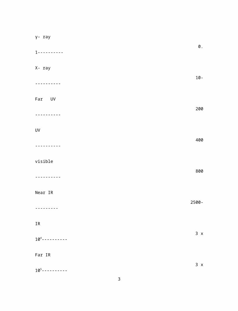
Electromagnetic Spectrum: The arrangement of all types of emr in order of their increasing wavelength or decreasing frequency is known as electromagnetic spectrum.
Wavelength (nm) EMR
γ- ray
0.1----------
X- ray
10-----------
Far UV
200----------
UV
400 ----------
visible
800----------
Near IR
2500----------
IR
3 x 10⁴----------
Far IR
3 x 10⁵----------
Micro wave.
1 x 10⁹-----------2
Radio wave
1 x 10¹³-----------
Electromagnetic Spectrum
Types of molecular energy:.
The total energy of the molecule, E tot = E trans + Erot + E vib +E ele
Where Etrans is the energy due to translational motion. This energy is associated with the
uniform motion of a molecule as a whole .This form of energy is of no significance in
spectroscopy. It is not quantized and it can be neglected.
Erot is the energy associated with overall rotation of the molecule with atoms
considered as fixed point masses.
E vib is the energy associated with the oscillation of atom of the molecule.
E ele is the energy associated with the motion of electrons while considering the
nucleus of the atom.
Etot = E rot + E vib + E ele
Eele > Evib >E rot.
All these energy components are quantized
Interaction of electromagnetic radiation with molecule:
Spectroscopy is the study of interaction of emr with matter. After interaction there may
occur variation in the intensity of emr with frequency or wavelength.
The instrument which is used to record the variation in the intensity of radiation is known
as spectrometer and the plot of variation in intensity with frequency or wavelength is
called as spectrum.
Types of spectra.
3

1. Emission spectrum: Molecules give emission spectra when subjected to intense heat
or electric discharge.The molecule obtain necessary energy to become excited . On
returning to lower energy state molecule may emit radiation which is the result of a
transition of molecule form an excited state to one of lower usually the ground state.
2.Absorbtion spectrum: When emr interacts with molecules , molecules undergo
change in its energy state by absorbing photons of appropriate energy.(the molecule is
raised from GS to ES )
GS- Ground state- Lowest possible energy level ES- Excited state -Higher energy state.
This process is known is absorption and the resultant spectrum is called absorption
spectrum.
The energy absorption occurs only when the energy difference between the ground state
and excited state exactly matches the energy of the incident radiation. The energy
absorbed (∆E) by a molecule may bring about changes in one or more of its energy level
such as rotational , vibrational and electronic.
4

The order of energy levels is schematically shown below.
From the above scheme it is clear that rotational energy levels are closely packed
(spaced).Thus little amount of energy is required for pure rotational transition. Such
transition occur in the microwave region of electromagnetic spectrum. Similarly
vibrational transition occurs in the Infrared region and electronic transition occurs in
Visible and Ultraviolet region.
Types of molecular spectra:
Rotational (Micro wave )spectra: These spectra arises due to transition between rotational
energy levels on absorption of radiation in microwave region.
Vibrational (Infra red) spectra: These spectra arises due to transition between vibrational
energy level of a molecule on absorption of radiation in infrared region.
Electronic spectra (UV-Visible) spectra: These spectra arise due to transition induced
between the electronic energy level of a molecule on absorption of radiation falling in the
UV- Visible region.5

Selection rule (Allowed and forbidden transition)
The molecular spectra is governed by certain set of rules called selection rules.
E.g. SR for pure vib spectra (transition) is ∆v = ± 1 Where V is vibrational quantum no.
SR for pure rotation transition is ∆j = ± 1 Where j is rotational quantum no.
The selection rule are however are not always obeyed strictly .The spectral transition
which obey a given selection rule is allowed transition. and those which violate a
selection rule are called forbidden transition. Intensity of absorption due to allowed
transition is strong. But it is very feeble in the case of forbidden transitions .
Photophysical law Beer- Lambert 's law :
Beer-Lambert law is a combination of two laws namely Beer law and Lambert law.
Lambert’s law : When a beam of monochromatic radiation is passed through a
homogeneous absorbing medium, the rate of decrease of intensity of radiation "dI" with
thickness of the absorbing solution " dx " is proportional to the intensity of incident
radiation "I".
-dI /dx α I
Beer’s law(Beer –Lambert law): Beer extended the above law and stated that When a
beam of monochromatic radiation is passed through a homogeneous absorbing medium,
the rate of decrease of intensity of radiation "dI" with thickness of the absorbing solution
" dx " is proportional to the intensity of incident radiation as well as concentration of the
solution “C”.
-dI /dx IC
-dI /dx = kIC
-dI / I = kC dx
dI / I = - k C dx
I0 ∫ I dI / I = - x =0∫x=x kC dx
ln I / Io = -kC x6

log I /Io = -kCx / 2.303.
log I / Io = -ε Cx where ε = k /2.303 is molar absorbtivity coefficient (dm 3.mol -1 cm-1 )
log Io / I = ε Cx
A = ε C x where A = log Io /I is called absorbance.
Absorbance is directly proportional to molar concentration and thickness or path length
The above equation is called Beer-Lambert ‘s law
I /Io is called Transmittance = T
log T = -ε Cx = -A or A = - log T
Deviations from Beer - Lambert's law. Deviation occurs
1. when the structure of coloured ion or of the coloured non-electrolyte changes with con.
2. due to presence of impurities that absorb at the absorption wavelength..
3. If monochromatic light is not used.
4. If the coloured species ionizes, dissociates or associates in solution.
5. if the concentration of the solution is high(>10 -3 m)
Problem based on Beer-Lambert law.
Example 1: If the transmittance of a solution is 19.4% ,what is its absorbance?
Solution:
Percentage transmittance (%T)= 19.4 or T = 0.194
Absorbance or optical density = - log T = log 1/T
= 0.712
Example 2: The transmittance of 2 X 10 -4 M solution of a substance was found to be 76.2% at
wavelength 480 nm when place in a cell of 1 cm length. Calculate i) the absorbance and ii) the
molar absorbtivity.
7

Solution :
Percentage transmittance (%T)=76.2 or 0.762
Absorbance or optical density = - log T = log 1/T
= log 1 / 0.762
=0.118
Molar absrbtivity, ε = A / Cb
= 0.118/(1 X 2X10-4 )
= 0.059X10 -4 mol -1 cm -1 dm 3
Example 3: An aqueous solution which is 10 -3 M absorbs 10% of incident radiation in a path
length of 1 cm. Calaculte the concentration of the solution that will absorb 90% of the same
incident radiation in the same cell.
Solution : The solution absorbs 10 % of incident light so T = 90 % or 0.9
A = - log T
A= - log 0.9 = 0.0458
We know A = ε Cb
0.0458 = ε 10-3 X 1
ε = 0.0458 /(10-3 X 1)
=0.0458 X 10 3 dm3 mol-1 cm -1
When 90 % of the light is absorbed ,transmittance T = 10 % or 0.1
A = - log T
A = - log 0.1 = 1
Therefore A = ε CT
1 = 0.0458 X 10 3 X C X 1
C = 1/(0.0458 X 10 3 X 1)
= 2.183 X 10 -2 M
8

ELECTRONIC (UV -VISIBLE ) SPECTROSCOPY:
Electronic spectrum arises from the excitation of electrons in a molecule from one energy
level to other .The energy required to cause this transition is provided by radiation in
UV -Visible region of electromagnetic spectrum .Thus electronic spectroscopy is also
called UV- Visible spectroscopy.
Instead of sharp peaks we get band in electronic spectrum because electronic transitions
are always accompanied by rotational transition and vibrational transitions, thus there are
large no. of transitions possible. A large no. of wavelength which are close enough will
be absorbed resulting in the formation of bands.
Range: 10 - 750nm 10 -200 - Far UV
200- 400 - UV
400 -750 - Visible.
Origin of absorption bands in UV- Visible spectrum.
According to Molecular Orbital theory ,Linear combination of atomic orbitals leads to the
formation of bonding and antibonding orbitals. Depending on the nature of AO and MO
may be σ and π type Antibonding(Higher energy) corresponding to σ and π are σ* and
π*.
Types of electrons in organic molecule.
σ electrons - present in single bond(saturated bond)
π electrons - Present in double and triple bond (unsaturated bond)
n electrons - unshared or non bonded electrons present in organic compounds containing
N,O or halogens.
9

Electronic transition occurs when the electrons are excited from the bonding σ and π and
non bonding n orbitals to antibonding σ* and π*
The energy level diagram for a molecule is shown below.
The energy for different transitions are in the following order.
σ → σ * > n → σ * > π→ π * > n →π*
Types of transition in organic molecules.
1. σ → σ * transition: The energy required for σ → σ * transition is very high. The
absorptions occurs in far UV region (120 -136 cm-1)
Ex. CH4 : σ → σ * λ max = 121.9 nm
2. n → σ * transition: This type of transition takes place in saturated compounds
containing one heteroatom with unshared pair of electrons.
Ex. Saturated halide , alcohols, esters, amines etc
The absorption occurs at longer wavelength in the near UV region (180 -
200 nm)
3. π→ π * transition:
This type of transition occurs in the unsaturated centres of the molecule. i.e. compound
containing double and triple bond and also in aromatics.
The excitatation of π→ π * electron requires lesser energy when compared to . n → σ *
transition.
Ex. ethylene 174 cm-1
10
σ
n
σ *

4. n →π*: In this type of transition, electron of unshaired electron pair on heteroatom
gets excited to antibonding orbital. This type of transition requires least amount of
energy ou of all transitions. According to selection rule this type transition is forbidden.
The intensity of absorption is very week.
Ex. aldehyde and ketone. CH3COCH3 270 -300 nm
Chromophores and Auxochromes:
The presence of one or more unsaturated linkages (π electron) in a compound is
responsible for the colour of the compound. These linkages are called chromophores.
Ex. = C=C = ; - C = C - ; - C = N
An auxochrome can be defined as any group which does not itself act as chromophore
but whose presence brings about a shift in the absorption towards the red end of the
spectrum(longer wavelength) Ex. - OH ,-NH2 ,- Cl etc.
λ max = 255 nm λ max = 280 nm
Terms related to change in intensity / wavelength.
Batho chromic shift -towards higher wavelength.
Hypsochromic shift - towards lower wavelength.
Hyperchromic shift -Increase in intensity. Hypochromic shift - Decrease in intensity.
11
Instrumentation:
1.Radiation source : Tungsten filament lamp and hydrogen - Deuterium lamp are
commonly used radiation source.
2.Monochromator: The monochromator is used to disperse the radiation according to
wavelength with a help of a rotating prism.
3. Sample holder or cell or cuvette : The sample holder should fulfill the following main
conditions i)They must be uniform in construction .ii ) It should be transparent to
wavelength of UV-Visible region. iii )The material of construction should be inert to
solvents. Silica or quartz cells are commonly used
4. Detectors: the detector converts radiation into current .Detector may be photomultiplier
tube , photo cell etc.
5. Recorder: The recorder plots absorbance against wavelength depending upon the signal
from the detector.
BLOCK DIAGRAM OF UV-VISIBLE SPECTROPHOTOMETER
12

Working : The radiation from the source is split into two identical beams having equal
intensity. One of the beam (sample beam) is allowed to pass through sample and the other
(reference beam) is made to pass through reference. Due to absorption of radiation by the
sample the intensity of radiation coming out of sample get decreased Intensities of the
two beams are converted into and measured as electrical energies with the help of
detector. The signal from the detector is passed to the recording unit and
recorded.Generally absorption is plotted against wavelength.
Applications
1 Identification of organic compounds: Electronic spectroscopy can be used for
identifying organic compounds especially aromatic compounds and conjugated olefins
2. Detection of impurities: The spectrum of the compound is compared with that of pure
compound. If impurities are present then extra absorption bands are found. Ex. benzene
(255nm) in cyclohexane.
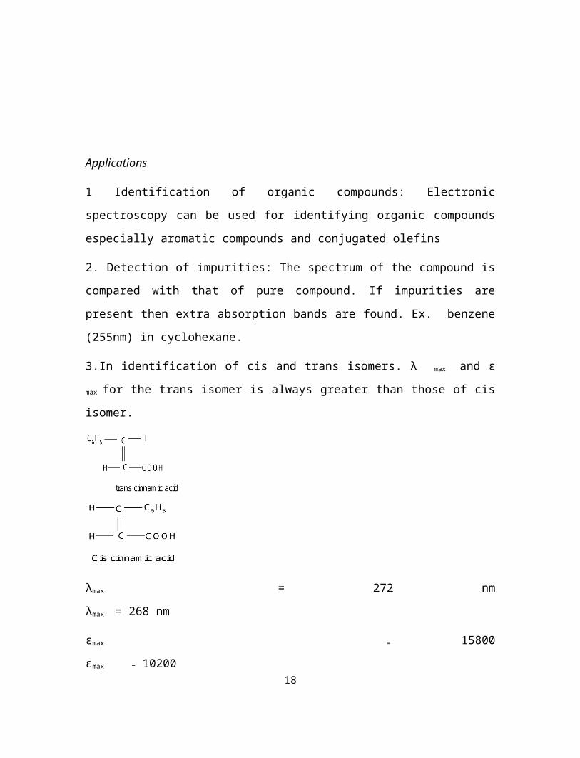
3.In identification of cis and trans isomers. λ max and ε max for the trans isomer is always
greater than those of cis isomer.
λmax = 272 nm λmax = 268 nm
εmax = 15800 εmax = 10200
4. In quantitative analysis to determine the concentration in solution by usi
Beer -Lambert's law. A = ε c l
13

5. To study chemical kinetics: Kinetics of chemical reaction can be studied by following
the change in concentration of reactant or product with time during the reaction.
6. Quantitative analysis: UV-Vis spectroscopy is used for qualitative determination of
substances which absorbs UV-VIS light and the determination is based of Beer- Lambert
law.
7. UV-Vis spectroscopy is used to calculate the percentage of different forms in
tautomeric system.
VIBRATIONAL (INFRARED ) SPECTROSCOPY
Absorption of radiation with energy equal to difference between two vibrational levels
(∆E vib) will cause a vibrational transition to occur .Radiation with energy sufficient to
cause vibrational transition is found in IR region of the spectrum. So this is called Infra
red spectroscopy. Since vibrational transition is accompanied by changes in rotational
level also the IR spectroscopy is also called Vibrational -Rotational Spectroscopy.
Region of IR.
0.8 - 2.5 µ Near IR 12500 – 4000 cm-1
2.5 - 15 µ IR 4000 – 667 cm-1
15 - 20 µ Far IR 667 - 50 cm-1
Kinds of fundamental vibrations: There are two types of fundamental vibrations.
1. Stretching 2.Bending.
Stretching: In this type of vibrations the distance between the atoms increases or
decreases but the atoms remain in the same bond axis.
14

Bending : In this type of vibration , the position of the atoms change with respect to
original bond axes.
Stretching Bending
Stretching vibration requires higher energy than bending vibrations (Stretching
absorptions occur at higher frequency)
Types of Stretching vibrations:
Symmetric stretching asymmetric stretching
Types of bending vibrations:
15

The various stretching and bending vibrations of a bond occur at certain quantized
frequencies.
Essential requirements for a substance to exhibit IR spectroscopy is that there should be
change in dipole moment during vibrations.
Number of fundamental vibrations:
1. For nonlinear molecule with ' n' no. of atoms 3n - 6 modes of vibration.
eg. CH4 3x 5 - 6 =9 :H20 3 x 3 -6 =3
H2O is a non linear triatomic molecule :
2.For linear molecule with 'n' no of atoms, 3n- 5 modes of vibrations
eg.CO2
16

Vibrational frequency:
The value of stretching vibrational frequency of a bond can be calculated by the application of Hook’s law which may be represented as
υ= 1/2π √ (k /μ) where μ is the reduced mass = (m1m2)/(m1+ m2)
m1 and m2 are the massed of the atom concerned in grams in a particular bond.
k = force constant of the bond and relates to the strength of the bond. For single bond it is approximately 5 x 10⁵ gm sec -2. It becomes double and triple for double and triple bonds respectively.
Wave number ( ) = υ/c OR 1/2πc √ (k /μ)
Where c is Velocity of the radiation = 3 x 10 8 m sec-1
Problems:
1.Calculate the frequeny of C-H stretching vibration from the following data.
k=5 x 10⁵ gm sec -2.
Mass of C atom= 20 x 10-24 : Mass of H atom= 1.6 x 10⁻²⁴
Solution:
υ = 1/2π √ (k /μ) where μ is the reduced mass = (m1m2)/(m1+ m2)
= 9.3 x 10¹³ sec -1
= υ/c
= (9.3 x 10¹³ sec -1 ) / (3 x 10 8 m sec-1)
= 3.1 x 10⁵ m-1 or 3100 cm-1
2. Calculate the reduced mass of CN molecule given atomic weights of C =12.011 and
N =14.0037 amu
Solution:
17
The reduced mass μ = (m1m2)/(m1+ m2)
=(12.011)(14.0067)/(12.011+14.0037) x (6.023 x 1023 )
=1.0737 x 10 -23 g
3.Calculate the force constant for carbon monoxide if the compound absorbs 2.143 x 10⁵
m-1 and its reduced mass is 1.139 x 10⁻²⁶ Kg
Solution:
= υ/c OR 1/2πc √ (k /μ)
k = 4 2π2C2 μ
= 4 x (3.14)2 x (3x10 8 ms-1)2 x (2.14 x 10 5 m-1)2 x 1.0139 x10-26
= 1857 Kg m2 s-2 (m -2) (1 Kg ms-2 = 1 N )
= 1857 N m-1
Instrumentation:
Components:
1 Radiation Source: The radiation source must emit IR radiation which must be
i) intense for detection ii ) steady iii ) extended over desired wavelength.Nernst glower
is one of the commonly used radiation source which is rod of sintered mixture of oxides
of Zirconium, Ytterbium and Erbium. when this rod is heated to1000 - 1800 ˚C produce
IR radiation.
2. Monochromator : It allows the light of required wavelength to pass through but
absorbs the light of other wavelength. Alkali halide (NaCl) prism is used as
monochromator.
3. Sample cell: The cell holding the sample must be transparent to IR. Depending on the
state of sample, the appropriate sampling technique is adopted
For solid samples mull or pressed pellet technique is commonly used.In mull
technique solid sample is mixed with Nujol to make a thick paste .then it is spread
between IR transmitting windows. In pressed pellet technique a small amount of
18
sample is powered with KBr (in the ratio of 1:100 by weight) The mixture is
pressed into pellets under high pressure.This pellet is placed in the path of IR
Liquid samples or solutions of solid can be taken in rectangular cell made up of
NaCl ,KBr or ThBr. For making solutions CHCl3,CCl4,CS2 are commonly used as
solvent
Gaseous sample is taken in glass or metal cylinder with NaCl windows.
4. Detectors: IR detectors convert the thermal radiant energy into electrical signals. The
detector may be bolometer, thermocouple, Photoconductivity cell or thermistor etc.
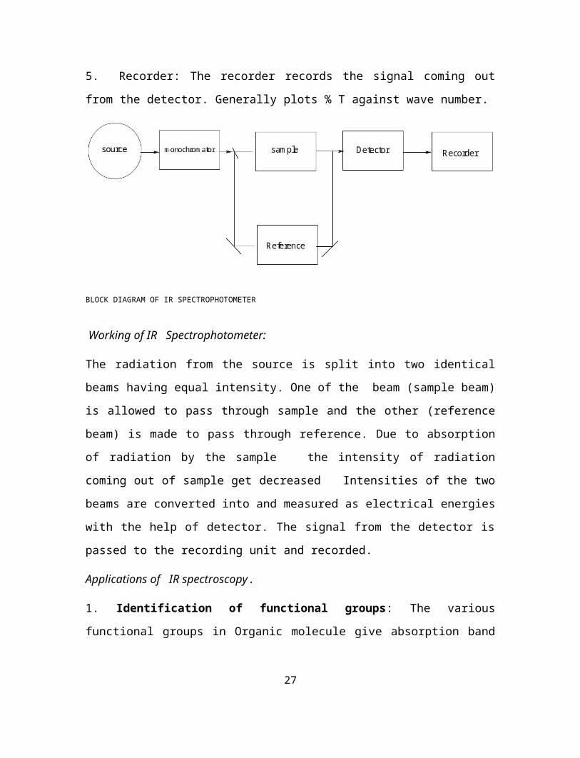
5. Recorder: The recorder records the signal coming out from the detector. Generally
plots % T against wave number.
BLOCK DIAGRAM OF IR SPECTROPHOTOMETER
Working of IR Spectrophotometer:
The radiation from the source is split into two identical beams having equal intensity.
One of the beam (sample beam) is allowed to pass through sample and the other
(reference beam) is made to pass through reference. Due to absorption of radiation by the
sample the intensity of radiation coming out of sample get decreased Intensities of the
two beams are converted into and measured as electrical energies with the help of
detector. The signal from the detector is passed to the recording unit and recorded.
Applications of IR spectroscopy.
19

1. Identification of functional groups: The various functional groups in Organic
molecule give absorption band of definite frequencies. Thus IR spectroscopy is highly
useful in detecting the functional group.
Group frequency cm-1
- OH 3600
- NH2 3400
>C= O 1750 -1600
> C= N - 1600
The region 4000 -1430 cm-1 is known as group frequency region.
Finger print region: The region below 1500 cm-1 is rich in many absorptions gives intense
bands is called finger print region. Some molecules containing the same functional
group show similar absorption above 1500 cm-1. Finger print region is used to detect and
characterize the molecule.
2. Studying the progress of reactions: Progress of chemical reaction can be readily
followed by examining spectra of small portion of reaction mixture withdrawn from time
to time. The rate of disappearance of characteristic absorption bands due to the formation
of some product helps in studying the progress of chemical reactions.example. the
gradual disappearance of - OH stretching (3600 - 3650 cm-1) and the consequent
appearance of - CO stretching 1680 -1760 cm-1 can be use to study the oxidation of an
alcohol to carbonyl compound.
3.Detection of impurities :Ex. Ketone as impurity in alcohol shows absorption due
to = C = O in the absorption of alcohol.
4.To ascertain hydrogen bonding: Hydrogen bonding shows appreciable downward
frequency shifts. We can also differentiate Intermolecular H bonding and Intra molecular
H bonding. Inter molecular H bond is concentration dependent .Intra molecular is
concentration independent. Ex. The fundamental frequency associated with -OH is found
at 3600cm -1 If the -OH forms inter molecular H bonding ,and additional band appears at
3300cm -1.Increase in dilution decreases the intensity of 3300cm-1 ant intensity at
20

3600cm-1 increases. If an intramolecular H bonding is formed as in the case of
salicylaldehyde,the band at 3600cm-1 is absent.The only band is observed is at 3300cm-1.
5.IR spectroscopy is used to identify the type of tautomer.
Ex .Keto - enol tautomerismis well established by the characteristic frequency of C= O an
– OH groups.
Colorimetry
1. Colorimetry is concerned with the visible region(400-800 nm) of the spectrum.
2. Variation of intensity in colour of a system with change in concentration of some
component forms basis of colorimetric analysis.
3. The colour formation may be due to the component itself or by the addition of suitable
reagent. Cu 2+ gives blue coloured complex with ammonium hydroxide ; Ni 2+ gives
scarlet coloured complex with dimethyl glyoxime. Fe 3+ gives deep red complex with
thiocyanate.
4.The intensity of colour of the sample may be compared with that of intensity of colour
obtained by treating known amount of he substance (standard).
5. Colorimetry is concerned with the determination of the concentration of substance by
measurement of relative absorption of light with respect to known concentration of the
substance.
6. The instrument used for measuring absorbance or transmittance in the visible region is
called colorimeter.
7. Instrumentation:
Components:
a) Radiation source : Tungsten filament lamp is widely used source which emits light in visible region.( 400 -800nm)
b) Filter or Monochromator: It allows the radiation of the required wavelength to pass through..
c) Slits : It provides narrow beam of light.
d) Cell or sample holder or cuvette : The cell holding the solution should be transparent. to UV -Visible light.
21

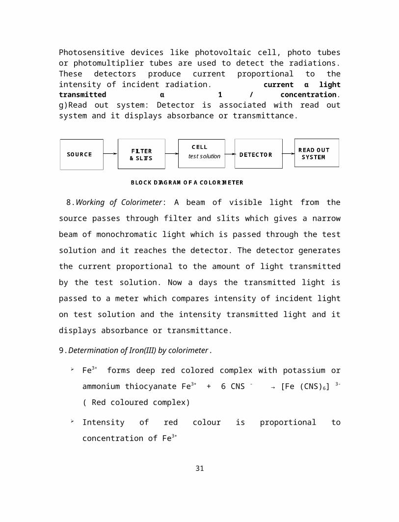
f) Detector: It is used for measuring radiant energy transmitted through the sample .
Photosensitive devices like photovoltaic cell, photo tubes or photomultiplier tubes are used to detect the radiations. These detectors produce current proportional to the intensity of incident radiation. current α light transmitted α 1 / concentration. g)Read out system: Detector is associated with read out system and it displays absorbance or transmittance.
8.Working of Colorimeter: A beam of visible light from the source passes through filter
and slits which gives a narrow beam of monochromatic light which is passed through the
test solution and it reaches the detector. The detector generates the current proportional to
the amount of light transmitted by the test solution. Now a days the transmitted light is
passed to a meter which compares intensity of incident light on test solution and the
intensity transmitted light and it displays absorbance or transmittance.
9.Determination of Iron(III) by colorimeter.
Fe3+ forms deep red colored complex with potassium or ammonium thiocyanate
Fe3+ + 6 CNS - → [Fe (CNS)6] 3- ( Red coloured complex)
Intensity of red colour is proportional to concentration of Fe3+
Colorimeter is calibrated using blank solution(it is adjusted for zero abdorbance
and 100% transmittance )with proper filter( it allows light of 480 nm).
Series of standard solutions ( Fe3+ solution of known concentration) were
prepared and their absorbance or transmittance is measured.
Absorbance is ploted against concentration which gives a straight line passing
through the origin (Due to Beer -Lambert 's law A = ε Cx) x - path length is
constant for a given cell.
Absorbance is measured for sample .From the calibration plot,concentration of the
unknown Fe 3+ can be estimated.
22

10.Application of colorimetry:
It is used to determine molar composition of complexes.
It is used in elucidation of structure of organic molecules.
It is used in the determination of dissociation of an acid base indicator.
It is used in the study of structure of inorganic complexes.
It is used in the quantitative analysis like the estimation of Cu2+, Ni 2+,Fe3+, Mn 2+,
Al3+ etc.
FLAME PHOTOMETRY OR FLAME EMISSION SPECTROSCOPY.
Flame photometry is based on the measurement of intensity of the light emitted when a
metal is introduced into flame.
If a solution containing a metallic salt is aspirated into a flame, a vapour which contains
atoms of the metal may be formed, some of these gaseous atoms may be raised to an
energy level which is as sufficiently high to permit the emission of radiation of
characteristic of the metal.
The wavelength of light tells what the element is?
Intensity of color tells about how much the element is present.
When metal salt solution is introduced into a flame, the following takes place.
1. Evaporation of solvent, leaving behind a solid residue.
23

2. Vaporization of solid with dissociation into its constituent atoms initially in the
ground state.
3. Some of the atoms may be excited by the thermal energy of the flame to higher energy
levels.
4. The excited atoms radiate energy and returns to ground state.
The emitted radiation is passed through the filter, which permits the characteristic
wavelength of the metal under analysis. It is then passed into the detector and finally into
the recorder.
24

Instrumentation:
1. Nebuliser - burner system: It produces gaseous metal atoms using a suitable
combustion flame involving fuel gas -Oxidant gas mixture. An essential requirement of
flame spectroscopy is flame temperature greater than 2000 K. So attain this mixture of
fuel gas and oxidant gas is used.
Ex. H2 and O2 (2800 C): Acetylene and N2O (3000 C).
The flame should 1. Evaporate the solvent 2.decompose the solid into atom and 3. Excite
the atoms and cause them to emit radiant energy.
2. Mirrors: A concave mirror is set behind the flame (burner) to increase the amount of
radiation reaching the detector.
3. Monochromator or Filter: It allows the light of required wavelength to pass.
4. Detector: The radiation from the monochromator or filter is allowed to pass on the
detector which measured the intensity of the radiation falling on it. It converts the
radiation into an electrical current.
5. Amplifier and recorder: The current coming from the detector is amplified and
recorded.
Working: Air at a given pressure is passed into atomizer and suction produced draws of
the sample into the atomizer. Where it combines the air and passes into burner. The air
meets the fuel gas and it is burnt. Radiation from the resulting flame passes through a
25

lens and finally through a optical filter which permits only the radiation characteristic of
the element under investigation to pass through the photo detector.
A series of standard solution were prepared and intensity of various solution were
recorded. A graph is constructed between intensity and concentration.( calibration graph )
The experiment is repeated with the sample solution and the concentration of sample is
obtained using calibration plot.
Application of flame photometry
1. Quantitative analysis: -
The amount of elements (group I and II) in a sample can be determined.
- It is used in soil analysis
- Used in the analysis of biological fluids and tisues.
2. Qualitative analysis:
- Elements of group I and II can be identified visually from the colour of the flame
Ca - brick red ; K - red: Na - Yellow ;Li - scarlet red.
Estimation of sodium in water sample.
The instrument is switched on.Flow of gas and air are regulated.First distilled water is
sent and ignition is started. After the instrument is warmed up, it is adjusted for zero
reading.The instrument is set at λ= 589 since sodium produces yellow emission at 589
nm
26
Liquid sample
hNa + (Gas)
Cl (Gas)
Thermal ExcitationNa * (gas)
Flame emission
NaCl (solution)
EvaporationNaCl (solid)
VaporisationNaCl (gas)
Dissociation
A series of standard NaCl solution (1 ,2,3,4,----10 ppm) is prepared and intensity was
measured .The calibration graph is drawn between the con. and the intensity of emitted
light .A straight line is obtained .
Intensity of the (unknown) sample is measured using the calibration curve the
concentration of sodium in water is determined.
ATOMIC ABSORPTION SPECTRSCOPY.
AAS is a method of elemental analysis and it is useful for determining trace metals in
liquids. It is independent of molecular form of the metal in the sample. This method is
very sensitive (Con. even lower than 1 ppm can be determined)
The absorption of energy by ground state atoms in gaseous state forms the basis of AAS
When a solution containing metallic species is introduced into flame, the metallic species
is converted to vapour. Some of the metal atoms may be raised to an energy level
sufficiently high to emit characteristic radiation of the metal. But large number of metal
atoms will remain in the ground state. These ground state atoms of a particular element
absorb light radiation of specific wavelength. Thus when a light of this wavelength is
allowed to pass through a flame having atoms of metallic species part of that light will be
absorbed and the absorption will be proportional to the density of the atoms in the flame.
From the value of absorption , concentration can be determined.
27

Components
1.Radiation source: It should emit stable , intense, characteristic radiation of the element
to be determined. Hollow cathode lamp is used as source.
Cathode : hollow cup in which element to be determined is kept
Anode : tungsten wire.
Cathode and anode is enclosed in a cyclinder filled with inert gas like argon.
28

2.Chopper :A rotating wheel which breaks steady light to pulsating light. Only the
pulsating current is amplified and recorded and thus the absorption of light will be
measured without interference of light emitted by the flame.
3. Burner or flame .The flame is used to convert liquid sample into gaseous state. It also
converts molecule into atom.
Two types of burners are commonly used 1.Total consumption burner 2.Premixed burner.
4. Nebuliser : It produces droplets of liquid sample
5. Monochromator : Prism or grating element is used as monochromator.
The function of monochromator is to select a given absorbing line from spectral lines emitted from the hollow cathode.
6.Detector: Photo multiplier tube is used as detector. It converts falling radiant energy into an electric current .
7.Amplifier: The current coming from the detector is so feeble that it should be amplified.
8.Read out device or Meter. Which gives absorbance or transmittance.
Working: The meter is adjusted to zero absorbance or 100 % transmittance when blank solution is sprayed into the flame. When solution containing the absorbing species is introduced a part from the light is absorbed resulting in a decrease in the intensity of light falling on the detector and the meter shows a value of absorption .Standard solutions of the metal to be determined are used to make a calibration curve using which concentration of the test solution can be measured.
Estimation of Ni.
Series of standard nickel solution is prepared
The instrument is calibrated using blank solution with operating wavelength (232 nm)and appropriate source -. HCL( Ni) .
Absorbance is measured for standard solutions.
Calibration plot (graph plotted between absorbance and concentration )is made.
Absorbance for sample solution is measured under same condition.
Using calibration plot con. of the sample Ni solution can be determined.
29

Applications
Determination of element s like Cu, Zn ,Ni in food industry.
Determination of Ca , Mg , Na , K in blood sample.
Determination in lead in petrol
Review Questions:
1. Define the term spectroscopy and explain the various type of transitions involved in spectroscopic method.
2. Derive Beer-Lambert’s law and mention its limitations.
3. Explain the principle of colorimetric method of analysis. and how Iron (III) estimated by colorimetry?
4.Explain the various components and working of colorimeter.
5. Explain briefly the principle and instrumentation of flame photometry.
6. Explain how sodium estimated by flame photometry.
7. Explain the various component and working of UV spectrometer.
8. Explain the principle and applications of UV-Visible spectroscopy.
9. Explain the various components and working of infrared spectrophotometer.
10.Explain the principle and applications of IR spectroscopy.
11.Discuss the principle, instrumentation and working of Atomic absorption spectrophotometer.
12.How nickel estimated by AAS.
Problems for practice.
1.Find out the absorbance of a solution ,if the transmittance of solution is 32.5%
2.A solution shows a transmittance of 20 % .When taken in cell of 2.5 cm thickness. Calculate its concentration if the molar absorption coefficient is 12000 dm3 mol -1 cm-1
30
3.A solution of thickness 2 cm transmits 40 % incident light. Calculate the concentration of the solution .Given ε =6000 dm3 mol -1 cm-1
4.A monochromatic radiation is incident on a solution of 0.05 M concentration of an absorbing solution. The intensity of radiation is reduced to one fourth of the initial value after passing through 10 cm length of the solution. Calculate the value of molar extinction coefficient of the absorbing substance.
5.A solution containing 30.1 g/l of a dye in a l cm cell,absorbs 50% of blue light.Under the same condition what percentage of light will be absorbed by 15.05 g/l of dye.
6. If the intensity of radiation is reduced by 20% in passing through 2 cm of solution i) what will the intensity after passing 10 cm of the same solution? ii ) What thickness would a beam of light has to pass before the intensity is reduced by 50 %.
7.The internuclear distance of NaCl is 2.36 x 10⁻¹⁰ m. Calculate the reduced mass and moment of inertia of NaCl. given masses of Cl =35 x10-3 Kg mol-1 and Na = 23 x10-3 Kg mol -1.
8.Calculate the frequency of O-H band, if the force constant and reduced mass of the atom pair are 770 nm-1 and 10463 x10-27 kg respectively.
9.The wave number of the fundamental vibration of 79 Br -81 Br is 323.2 cm-1 .Calculate the force constant of the bond(Mass of 79 Br = 78.9183 amu and 81 Br = 80.9463 amu).
31
Related Documents


