Analysis of the xplAB-Containing Gene Cluster Involved in the Bacterial Degradation of the Explosive Hexahydro-1,3,5-Trinitro-1,3,5- Triazine Chun Shiong Chong, a,b Dana Khdr Sabir, a Astrid Lorenz, a Cyril Bontemps, a Peter Andeer, c David A. Stahl, c Stuart E. Strand, c Elizabeth L. Rylott, a Neil C. Bruce a Centre for Novel Agricultural Products, Department of Biology, University of York, York, United Kingdom a ; Department of Biotechnology and Medical Engineering, Faculty of Biosciences and Medical Engineering, Universiti Teknologi Malaysia, Skudai, Johor, Malaysia b ; Department of Civil and Environmental Engineering, University of Washington, Seattle, Washington, USA c Repeated use of the explosive compound hexahydro-1,3,5-trinitro-1,3,5-triazine (RDX) on military land has resulted in signifi- cant soil and groundwater pollution. Rates of degradation of RDX in the environment are low, and accumulated RDX, which the U.S. Environmental Protection Agency has determined is a possible human carcinogen, is now threatening drinking water sup- plies. RDX-degrading microorganisms have been isolated from RDX-contaminated land; however, despite the presence of these species in contaminated soils, RDX pollution persists. To further understand this problem, we studied RDX-degrading species belonging to four different genera (Rhodococcus, Microbacterium, Gordonia, and Williamsia) isolated from geographically dis- tinct locations and established that the xplA and xplB (xplAB) genes, which encode a cytochrome P450 and a flavodoxin redox partner, respectively, are nearly identical in all these species. Together, the xplAB system catalyzes the reductive denitration of RDX and subsequent ring cleavage under aerobic and anaerobic conditions. In addition to xplAB, the Rhodococcus species stud- ied here share a 14-kb region flanking xplAB; thus, it appears likely that the RDX-metabolizing ability was transferred as a genomic island within a transposable element. The conservation and transfer of xplAB-flanking genes suggest a role in RDX me- tabolism. We therefore independently knocked out genes within this cluster in the RDX-degrading species Rhodococcus rhodo- chrous 11Y. Analysis of the resulting mutants revealed that XplA is essential for RDX degradation and that XplB is not the sole contributor of reducing equivalents to XplA. While XplA expression is induced under nitrogen-limiting conditions and further enhanced by the presence of RDX, MarR is not regulated by RDX. T he explosive hexahydro-1,3,5-trinitro-1,3,5-triazine (RDX) is widely used in both military and commercial applications. Following decades of use on military training ranges, along with manufacturing and decommissioning processes, RDX is now a significant environmental contaminant (1). The low rates of bio- degradation of RDX in the environment, coupled with its poor soil-binding capacity, mean that RDX readily leaches through soil and groundwater, with the potential to contaminate drinking wa- ter supplies. This is of concern; the U.S. Environmental Protection Agency has determined RDX to be a possible human carcinogen, and the use of RDX on some ranges has been restricted as a result of groundwater contamination. A number of Corynebacterineae spanning four different genera (Rhodococcus, Microbacterium, Gordonia, and Williamsia) isolated from RDX-contaminated soils are able to use RDX as a sole nitro- gen source for growth (2–8). These species originate from geo- graphically distinct locations, and all of those species tested have been shown to encode the cytochrome P450 XplA and the associ- ated reductase XplB; indeed, no aerobic RDX-degrading isolates lacking xplA have yet been isolated by selective enrichments (using RDX as a sole nitrogen source). Together, the encoded P450 sys- tem catalyzes the reductive denitration of RDX, resulting in sub- sequent ring cleavage under aerobic and anaerobic conditions (9, 10). Structural analyses, including X-ray studies (11), have re- vealed features not previously seen in cytochromes P450, includ- ing the fusion of the heme domain to a flavodoxin redox partner (12). The highly conserved nature of the xplAB gene sequences be- tween two phylogenetically disparate species, Rhodococcus rhodo- chrous 11Y and Microbacterium sp. strain MA1, isolated from RDX-contaminated soil in the United Kingdom and the United States, respectively, indicates that these genes have been rapidly distributed across the world by horizontal gene transfer (1). In support of this, the xplAB sequence was found to be located on a plasmid of approximately 145.5 kb in Microbacterium sp. MA1. Sequencing revealed that an open reading frame (ORF) upstream of xplAB, pMA1.030, and the neighboring downstream ORFs pMA1.033 and pMA1.034 were nearly identical between Micro- bacterium sp. MA1 and R. rhodochrous 11Y (6). The xplAB genes are also present on an extrachromosomal element in Gordonia sp. strain KTR9, although in this species, glutamine synthetase is fused to the N terminus of xplB (13). pMA1.028 in Microbacterium sp. MA1 encodes a putative per- mease transporter, termed AroP (6), which shares 72% identity with a putative amino acid permease from Mycobacterium van- baalenii PYR-1 (NCBI accession number YP_952672.1). Two Received 4 June 2014 Accepted 9 August 2014 Published ahead of print 15 August 2014 Editor: F. E. Löffler Address correspondence to Neil C. Bruce, [email protected]. C.S.C. and D.K.S. are joint first authors. Copyright © 2014, American Society for Microbiology. All Rights Reserved. doi:10.1128/AEM.01818-14 November 2014 Volume 80 Number 21 Applied and Environmental Microbiology p. 6601– 6610 aem.asm.org 6601

Welcome message from author
This document is posted to help you gain knowledge. Please leave a comment to let me know what you think about it! Share it to your friends and learn new things together.
Transcript
Analysis of the xplAB-Containing Gene Cluster Involved in theBacterial Degradation of the Explosive Hexahydro-1,3,5-Trinitro-1,3,5-Triazine
Chun Shiong Chong,a,b Dana Khdr Sabir,a Astrid Lorenz,a Cyril Bontemps,a Peter Andeer,c David A. Stahl,c Stuart E. Strand,c
Elizabeth L. Rylott,a Neil C. Brucea
Centre for Novel Agricultural Products, Department of Biology, University of York, York, United Kingdoma; Department of Biotechnology and Medical Engineering, Facultyof Biosciences and Medical Engineering, Universiti Teknologi Malaysia, Skudai, Johor, Malaysiab; Department of Civil and Environmental Engineering, University ofWashington, Seattle, Washington, USAc
Repeated use of the explosive compound hexahydro-1,3,5-trinitro-1,3,5-triazine (RDX) on military land has resulted in signifi-cant soil and groundwater pollution. Rates of degradation of RDX in the environment are low, and accumulated RDX, which theU.S. Environmental Protection Agency has determined is a possible human carcinogen, is now threatening drinking water sup-plies. RDX-degrading microorganisms have been isolated from RDX-contaminated land; however, despite the presence of thesespecies in contaminated soils, RDX pollution persists. To further understand this problem, we studied RDX-degrading speciesbelonging to four different genera (Rhodococcus, Microbacterium, Gordonia, and Williamsia) isolated from geographically dis-tinct locations and established that the xplA and xplB (xplAB) genes, which encode a cytochrome P450 and a flavodoxin redoxpartner, respectively, are nearly identical in all these species. Together, the xplAB system catalyzes the reductive denitration ofRDX and subsequent ring cleavage under aerobic and anaerobic conditions. In addition to xplAB, the Rhodococcus species stud-ied here share a 14-kb region flanking xplAB; thus, it appears likely that the RDX-metabolizing ability was transferred as agenomic island within a transposable element. The conservation and transfer of xplAB-flanking genes suggest a role in RDX me-tabolism. We therefore independently knocked out genes within this cluster in the RDX-degrading species Rhodococcus rhodo-chrous 11Y. Analysis of the resulting mutants revealed that XplA is essential for RDX degradation and that XplB is not the solecontributor of reducing equivalents to XplA. While XplA expression is induced under nitrogen-limiting conditions and furtherenhanced by the presence of RDX, MarR is not regulated by RDX.
The explosive hexahydro-1,3,5-trinitro-1,3,5-triazine (RDX) iswidely used in both military and commercial applications.
Following decades of use on military training ranges, along withmanufacturing and decommissioning processes, RDX is now asignificant environmental contaminant (1). The low rates of bio-degradation of RDX in the environment, coupled with its poorsoil-binding capacity, mean that RDX readily leaches through soiland groundwater, with the potential to contaminate drinking wa-ter supplies. This is of concern; the U.S. Environmental ProtectionAgency has determined RDX to be a possible human carcinogen,and the use of RDX on some ranges has been restricted as a resultof groundwater contamination.
A number of Corynebacterineae spanning four different genera(Rhodococcus, Microbacterium, Gordonia, and Williamsia) isolatedfrom RDX-contaminated soils are able to use RDX as a sole nitro-gen source for growth (2–8). These species originate from geo-graphically distinct locations, and all of those species tested havebeen shown to encode the cytochrome P450 XplA and the associ-ated reductase XplB; indeed, no aerobic RDX-degrading isolateslacking xplA have yet been isolated by selective enrichments (usingRDX as a sole nitrogen source). Together, the encoded P450 sys-tem catalyzes the reductive denitration of RDX, resulting in sub-sequent ring cleavage under aerobic and anaerobic conditions (9,10). Structural analyses, including X-ray studies (11), have re-vealed features not previously seen in cytochromes P450, includ-ing the fusion of the heme domain to a flavodoxin redox partner(12).
The highly conserved nature of the xplAB gene sequences be-
tween two phylogenetically disparate species, Rhodococcus rhodo-chrous 11Y and Microbacterium sp. strain MA1, isolated fromRDX-contaminated soil in the United Kingdom and the UnitedStates, respectively, indicates that these genes have been rapidlydistributed across the world by horizontal gene transfer (1). Insupport of this, the xplAB sequence was found to be located on aplasmid of approximately 145.5 kb in Microbacterium sp. MA1.Sequencing revealed that an open reading frame (ORF) upstreamof xplAB, pMA1.030, and the neighboring downstream ORFspMA1.033 and pMA1.034 were nearly identical between Micro-bacterium sp. MA1 and R. rhodochrous 11Y (6). The xplAB genesare also present on an extrachromosomal element in Gordonia sp.strain KTR9, although in this species, glutamine synthetase isfused to the N terminus of xplB (13).
pMA1.028 in Microbacterium sp. MA1 encodes a putative per-mease transporter, termed AroP (6), which shares 72% identitywith a putative amino acid permease from Mycobacterium van-baalenii PYR-1 (NCBI accession number YP_952672.1). Two
Received 4 June 2014 Accepted 9 August 2014
Published ahead of print 15 August 2014
Editor: F. E. Löffler
Address correspondence to Neil C. Bruce, [email protected].
C.S.C. and D.K.S. are joint first authors.
Copyright © 2014, American Society for Microbiology. All Rights Reserved.
doi:10.1128/AEM.01818-14
November 2014 Volume 80 Number 21 Applied and Environmental Microbiology p. 6601– 6610 aem.asm.org 6601
studies have documented permeases located close to genes withrelated function (14, 15). The mode of RDX removal by R. rhodo-chrous 11Y is unknown, but given the proximity to xplA, it ispossible that the aroP-encoded permease is involved in the trans-port of RDX or its breakdown products.
The ORF pMA1.027 in Microbacterium sp. MA1 encodes atranscriptional regulator that shares 62% amino acid identity withthe regulatory protein MarR (multiple-antibiotic-resistance reg-ulator) from Gordonia bronchialis DSM 43247 (NCBI accessionnumber YP_003274556). The MarR family of proteins is involvedin environmental stress responses and in the catabolic pathways ofaromatic compounds (16) and is known to bind a number ofanionic lipophilic, and often phenolic, effector molecules (17).MarR is also reported to negatively regulate a small cluster ofnaphthalene catabolism-related genes called nagR2-ORF2I�KL inthe naphthalene-degrading bacterium Polaromonas naphthaleniv-orans CJ2 (18) and the marRAB operon in Escherichia coli (19).Additionally, MarR is part of the phn island in polycyclic aromatichydrocarbon (PAH)-degrading bacteria and is possibly involvedin the regulation of phn genes (20).
Despite the presence of RDX-degrading microorganisms inRDX-contaminated soil, the rate of RDX degradation in soils isinsufficient to remediate polluted environments resulting ingroundwater contamination. It is known that the expression ofxplAB is repressed by the presence of other nitrogenous com-pounds (13, 21), and previous studies suggest that XplA activity isregulated and induced by the presence of RDX (3).
We have isolated RDX-degrading bacteria from different geo-graphical regions and sequenced them for xplAB and neighboringgenes. The identification of a highly conserved, nearly identicalgene cluster harboring xplAB in Microbacterium sp. MA1 and R.rhodochrous 11Y indicates the involvement, within this cluster, ofthese genes in RDX metabolism. To investigate the potential roleof genes adjacent to xplA, we created and characterized R. rhodo-chrous 11Y knockouts in aroP, marR, xplA, and xplB and testedRDX metabolism following growth in a range of nitrogen sources.
MATERIALS AND METHODSEnrichment and phylogenetic analysis. Soil samples were obtained frommilitary sites in the United Kingdom, Belgium, and Ukraine. Enrichmentcultures for soil from the United Kingdom, Belgium, and Ukraine weredeveloped as described previously by Binks et al. (2).
Taxonomic characterization of isolates was performed by amplifying
and sequencing a nearly full-length (1,500-nucleotide) sequence of the16S rRNA gene. PCR amplifications were performed with Phusion high-fidelity DNA polymerase (NEB), according to the manufacturer’s recom-mendations, by using primers 7f (5=-AGAGTTTGATYMTGGCTCAG-3=) and 1510r (5=-ACGGYTACCTTGTTACGACTT-3=) (22). The 16SrRNA gene sequences were submitted to the NCBI database (Table 1).Alignment and tree construction were performed as described previouslyby Andeer et al. (6), using MEGA software version 6 (23).
Analysis of the xplAB gene cluster. A cosmid library of R. rhodochrous11Y DNA was constructed by using the pWEB cosmid vector kit (Epicen-tre) and phage T1-resistant Escherichia coli EPI100-TR according to theinstructions provided by the manufacturer. Primers used for sequencingare listed in Table 2.
Construction of knockout strains. Using the primers listed in Table 3,regions of arop, marR, xplB, and xplA were amplified from R. rhodochrous11Y to produce PCR products with EcoRI and NdeI restriction sites forthe upstream regions and NdeI and HindIII restriction sites for the down-stream regions. Each pair of upstream and downstream regions wascloned together into EcoRI- and HindIII-digested pK18mobsacB to pro-duce the mutagenic plasmids p11Yarop, p11YmarR, p11YxplB, andp11YxplA. The mutagenic plasmids were transformed into E. coli S17-1and knockouts in the marR, aroP, xplA, and xplB genes created followingrecombination with homologous regions on either side of each gene ofinterest in R. rhodochrous 11Y (24). Sucrose-sensitive transconjugantswere grown in Luria-Bertani (LB) medium (1% [wt/vol] tryptone, 0.5%[wt/vol] yeast extract, 1% [wt/vol] NaCl) overnight (16 h) to induce thesecond crossover and then screened on agar containing 10% sucrose. Su-crose-sensitive Rhodococcus mutants were identified following screeningon complex medium agar containing 10% (wt/vol) sucrose. Incompletecrossover events were removed following screening on sucrose with 50
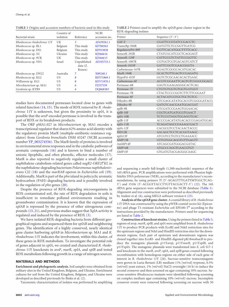
TABLE 1 Origins and accession numbers of bacteria used in this study
Bacterial strainCountry ofisolation Reference
NCBIaccession no.
Rhodococcus rhodochrous 11Y UK 3 AF439261.1Rhodococcus sp. BL1 Belgium This study KF700363Rhodococcus sp. DS1 Belgium This study KF933858Rhodococcus sp. 5U Ukraine This study KJ566616Rhodococcus sp. 9UK UK This study KJ566615Rhodococcus sp. YH1 Israel Unpublished
data (Z.Ronen)
AF103733.1
Rhodococcus sp. DN22 Australia 7 X89240.1Rhodococcus sp. EG2 US 8 KF571869.1Williamsia sp. EG1 US 8 KF571870.1Microbacterium sp. MA1 US 6 FJ357539Gordonia sp. KTR9 US 4 DQ068383
TABLE 2 Primers used to amplify the xplAB gene cluster region in theRDX-degrading isolates
Primer Sequence (5=–3=)GAF-F CGATTCCGTATCCGACCTCTranscRp-504R GATGTTCTCCGGTTGATCGRegulatoryPrt-89F GGTTCACATGGCTTTTCACCSensorK-292R CTATCGCATCGCTCAGGAGTSensorK-129F CATTCCGTCTATTCCGGTTCSensorK-1087R CGTGGTCGTGACAGTCATCTSensork-1035F GATTCGGTCGAACGGGTACarbesterase-167R AGAGTCGGGCACATGACACMarR-194R GCACTGTTGAGTCCGAGATGHypoPrt-453F GGTCTCGCAACACACTGAACCarbesterase-6F ACGTCGGAATTCAGTGTCGGGCGGACCPermease-6R GAGTCGAAGAGGGCACTCAGPermease-7F CTGTGTGGTGTGGTGATGGTPermease-7R CTACTCCCCGGTCTTCTTCGGAATPermease-8F ACTACATCGTGCTGCTGTCCDihydro-8R GTCGAGCATATGCACGTCGGGGATACGDihydro-9F GATGTCAACGAATGGACGATxplB-9R GTCGATCCGAAGTGGAACACxplB-10F CGGTGATGATGCGTGAACTxplA-10R TCTCCGTAGGTGGAGGTGACxplB-11F ACCCGACATATGACACCCGAGATGGAGxplA-11R TCAGATAGCCGAAAGCGACTxplA-12F CACGCGTCCTACAACTACCCSSD12R AACAGCTCCTCACGGTAAGCxplA13F ATCGTCCTGTCCTGAAAACCAMP13R GTTGTGGCAGGTGTTGAGTTAmMP14F ATCAGCGATGGGACGATACAMP14R ATGCCCAGGTGAACGTGTAMP15F GTGATGTACGAGGGCAAGC
Chong et al.
6602 aem.asm.org Applied and Environmental Microbiology
�g/ml kanamycin. Gene deletions in sucrose-resistant, kanamycin-sensi-tive mutants were confirmed by PCR using gene-specific primers listed inTable 3 and spanning each putatively deleted gene.
Growth and RDX uptake measurements. Bacteria were initiallygrown for 1 to 3 days at 30°C on RDX dispersion agarose plates containing2 mM RDX, minimal medium (1.55 g/liter KH2PO4, 4.95 g/liter K2HPO4,10 mM glycerol, 5 mM glucose, 5 mM succinate, trace elements [pH 7.2][25]), and 1.5% (wt/vol) agarose. Colonies were then transferred ontoagar plates of LB medium and grown at 30°C for 2 days. Cell biomass washarvested, and 5 mg in 50 �l of phosphate buffer (pH 7.2) was inoculatedinto each 250-ml conical flask containing 50 ml minimal medium with100 �M RDX as the sole source of nitrogen. The flasks were incubated at30°C with shaking at 180 rpm. Samples were taken at intervals, the absor-bance at an optical density at 600 nm (OD600) was recorded, and RDXlevels were measured by using high-performance liquid chromatography(HPLC) (9).
Resting-cell assays. Bacteria were grown at 30°C to an OD600 of 0.3 inminimal medium supplemented with 5 mM KNO3, 750 �M KNO2, 750�M KNO3, or 250 �M RDX as a nitrogen source, as described in Table 4,and the cells were collected by centrifugation. The cells were then resus-pended in minimal medium containing 5 mM KNO3, 450 �M KNO2, 450�M KNO3, no nitrogen, or 150 �M RDX, as described in Table 4, andgrown for a further 3 h; cells were then harvested by centrifugation. Cellswere washed twice in 40 mM phosphate buffer (pH 7.2) and resuspendedat a concentration of 0.1 g (wet weight)/ml. Resting-cell assays were per-formed at 30°C with shaking at 180 rpm, using 50 �l of cells in a totalvolume of 700 �l containing 40 mM phosphate buffer (pH 7.2), 100 �MRDX, and 100 �g/ml kanamycin. Samples of 100 �l were taken at eachtime point, each reaction was then stopped by the addition of 10 �l 1 Mtrichloroacetic acid, and RDX levels were measured by using HPLC (9).
Western analysis and activity assays. Cells were prepared as de-scribed above for resting-cell assays and then disrupted by sonication andcentrifuged (10,000 � g) at 4°C for 30 min. For Western blot analysis, 25�g of supernatant protein was loaded per lane. Anti-rabbit polyclonalantibodies to the XplA and XplB proteins were prepared by using purifiedXplA and XplB proteins, respectively, in rabbit.
Activity of XplA in cell extracts was determined by using 75 �l of cellextract, 300 �M NADPH, 0.1 U/ml spinach ferredoxin reductase (Sigma),and 100 �M RDX, in a final volume of 150 �l. Reactions were performedaerobically at 20°C, and NADPH oxidation was measured by the A340.
Gene expression analysis. Total RNA was extracted by using anRNeasy kit (Qiagen). cDNA synthesis and quantitative PCR (qPCR) anal-
ysis were conducted as described previously (9). Primer sequences forxplA were 5=-CCGAGTGGGCCAAACAGT-3= and 5=-TCCTCCTCGTCGAGTTCGAT-3=, and those for DNA gyrase subunit B (gyrB) were 5=-GCCGAGGAGCAGGAACAG-3= and 5=-TAGTGGTAGACGCGGGTCTTG-3=. Relative expression values were calculated by using gyrB as aninternal reference.
Testing of putative transcription initiation sites. Rapid amplifica-tion of cDNA ends (RACE) PCR was performed by using a SMARTerRACE kit (Clontech Laboratories, Inc.). Total RNA was isolated from R.rhodochrous 11Y by using an RNeasy kit (Qiagen), and first-strand cDNAsynthesis was carried out according to the manufacturer’s protocol. RACEPCR was performed by using Universal Primer Mix (Clontech) in com-bination with the Rhodococcus-specific primer 5=-GGCGACCATCACCACACCACACAG-3=. Nested PCR was then performed by using UniversalPrimer A (Clontech) and the nested Rhodococcus-specific primer 5=-ATGGTGACGAGCAGCGCGATCAGATAC-3=. The PCR products werecloned into TOPO pCR2.1 (Invitrogen) and sequenced. Primer extensionanalysis was performed as described previously (16). Reverse transcrip-tion was carried out with a volume of 20 �l using 20 �g total RNA, 400 USuperscript III reverse transcriptase (Invitrogen), 5 nM primer (5=-FAM[6-carboxyfluorescein]-CATAGGCGAAGACCGTGTAGATCCCGAC-3=), and 750 �M dATP, dCTP, dTTP, and dGTP at 55°C for 1.5 h. Thereverse transcription reaction was repeated as described previously (26).Primer extension products were mixed with 0.3 �l the Genescan LIZ600ladder and run on the AB3130XL instrument (Applied Biosystems), usingdefault parameters. Results were analyzed by using Peak Scanner Software(Applied Biosystems).
Nucleotide sequence accession numbers. The GenBank accessionnumbers for Rhodococcus sp. BL1, DS1, 5U, and 9UK sequences areKF700363, KF933858, KJ566616, and KJ566615, respectively.
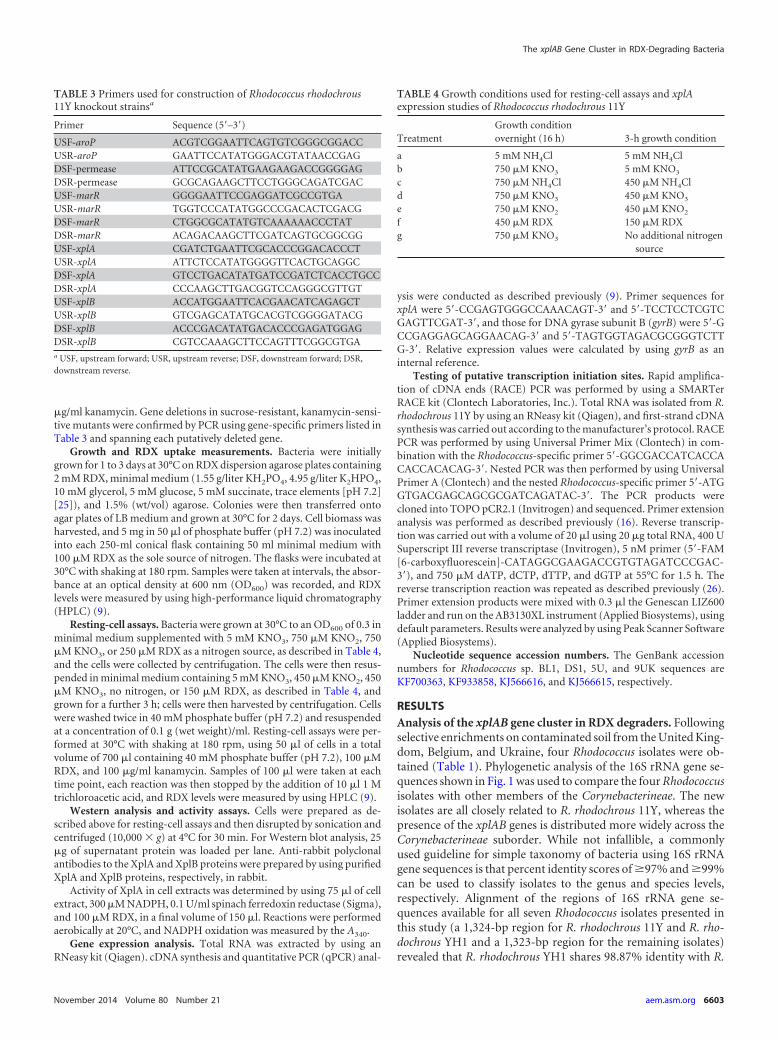
RESULTSAnalysis of the xplAB gene cluster in RDX degraders. Followingselective enrichments on contaminated soil from the United King-dom, Belgium, and Ukraine, four Rhodococcus isolates were ob-tained (Table 1). Phylogenetic analysis of the 16S rRNA gene se-quences shown in Fig. 1 was used to compare the four Rhodococcusisolates with other members of the Corynebacterineae. The newisolates are all closely related to R. rhodochrous 11Y, whereas thepresence of the xplAB genes is distributed more widely across theCorynebacterineae suborder. While not infallible, a commonlyused guideline for simple taxonomy of bacteria using 16S rRNAgene sequences is that percent identity scores of �97% and �99%can be used to classify isolates to the genus and species levels,respectively. Alignment of the regions of 16S rRNA gene se-quences available for all seven Rhodococcus isolates presented inthis study (a 1,324-bp region for R. rhodochrous 11Y and R. rho-dochrous YH1 and a 1,323-bp region for the remaining isolates)revealed that R. rhodochrous YH1 shares 98.87% identity with R.
TABLE 3 Primers used for construction of Rhodococcus rhodochrous11Y knockout strainsa
Primer Sequence (5=–3=)USF-aroP ACGTCGGAATTCAGTGTCGGGCGGACCUSR-aroP GAATTCCATATGGGACGTATAACCGAGDSF-permease ATTCCGCATATGAAGAAGACCGGGGAGDSR-permease GCGCAGAAGCTTCCTGGGCAGATCGACUSF-marR GGGGAATTCCGAGGATCGCCGTGAUSR-marR TGGTCCCATATGGCCCGACACTCGACGDSF-marR CTGGCGCATATGTCAAAAAACCCTATDSR-marR ACAGACAAGCTTCGATCAGTGCGGCGGUSF-xplA CGATCTGAATTCGCACCCGGACACCCTUSR-xplA ATTCTCCATATGGGGTTCACTGCAGGCDSF-xplA GTCCTGACATATGATCCGATCTCACCTGCCDSR-xplA CCCAAGCTTGACGGTCCAGGGCGTTGTUSF-xplB ACCATGGAATTCACGAACATCAGAGCTUSR-xplB GTCGAGCATATGCACGTCGGGGATACGDSF-xplB ACCCGACATATGACACCCGAGATGGAGDSR-xplB CGTCCAAAGCTTCCAGTTTCGGCGTGAa USF, upstream forward; USR, upstream reverse; DSF, downstream forward; DSR,downstream reverse.
TABLE 4 Growth conditions used for resting-cell assays and xplAexpression studies of Rhodococcus rhodochrous 11Y
TreatmentGrowth conditionovernight (16 h) 3-h growth condition
a 5 mM NH4Cl 5 mM NH4Clb 750 �M KNO3 5 mM KNO3
c 750 �M NH4Cl 450 �M NH4Cld 750 �M KNO3 450 �M KNO3
e 750 �M KNO2 450 �M KNO2
f 450 �M RDX 150 �M RDXg 750 �M KNO3 No additional nitrogen
source
The xplAB Gene Cluster in RDX-Degrading Bacteria
November 2014 Volume 80 Number 21 aem.asm.org 6603
rhodochrous 11Y, whereas the remaining 16S sequences share be-tween 95.31 and 95.62% identity with R. rhodochrous 11Y. Thesealignments suggest that all seven Rhodococcus isolates are separatespecies.
The xplAB gene cluster sequence was determined for these spe-cies as well as for previously isolated RDX-degrading bacteria,including Microbacterium sp. MA1 (6); Williamsia sp. strain EG1(8); Rhodococcus sp. strain EG2 (8); Gordonia sp. KTR9 (4); andthree Rhodococcus species isolates from the United Kingdom (3),Israel (Rhodococcus sp. YH1), and Australia (7). Sequence analysisrevealed that these 11 species all contained near-identical copies ofthe xplAB genes. Further sequencing of R. rhodochrous 11Y and
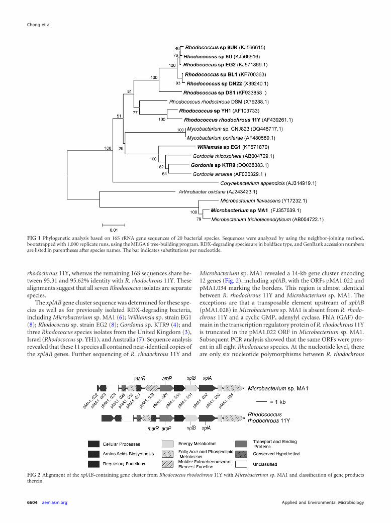
Microbacterium sp. MA1 revealed a 14-kb gene cluster encoding12 genes (Fig. 2), including xplAB, with the ORFs pMA1.022 andpMA1.034 marking the borders. This region is almost identicalbetween R. rhodochrous 11Y and Microbacterium sp. MA1. Theexceptions are that a transposable element upstream of xplAB(pMA1.028) in Microbacterium sp. MA1 is absent from R. rhodo-chrous 11Y and a cyclic GMP, adenylyl cyclase, FhlA (GAF) do-main in the transcription regulatory protein of R. rhodochrous 11Yis truncated in the pMA1.022 ORF in Microbacterium sp. MA1.Subsequent PCR analysis showed that the same ORFs were pres-ent in all eight Rhodococcus species. At the nucleotide level, thereare only six nucleotide polymorphisms between R. rhodochrous
FIG 1 Phylogenetic analysis based on 16S rRNA gene sequences of 20 bacterial species. Sequences were analyzed by using the neighbor-joining method,bootstrapped with 1,000 replicate runs, using the MEGA 6 tree-building program. RDX-degrading species are in boldface type, and GenBank accession numbersare listed in parentheses after species names. The bar indicates substitutions per nucleotide.
FIG 2 Alignment of the xplAB-containing gene cluster from Rhodococcus rhodochrous 11Y with Microbacterium sp. MA1 and classification of gene productstherein.
Chong et al.
6604 aem.asm.org Applied and Environmental Microbiology
11Y and Microbacterium sp. MA1 across the 14-kb region, four ofwhich are located in the xplAB region. Across all 11 species, thexplAB region is highly conserved (Rhodococcus sp. DS1 and YH1are identical, and the remaining nine species share 99.87 to99.99% identity with R. rhodochrous 11Y). The encoded aminoacids that differ from the xplAB region in R. rhodochrous 11Y arepresented in Table 5. The glutamine synthetase-xplB fusion foundin Gordonia sp. KTR9 (4) was not found in any of the other RDX-degrading bacteria examined in this study.
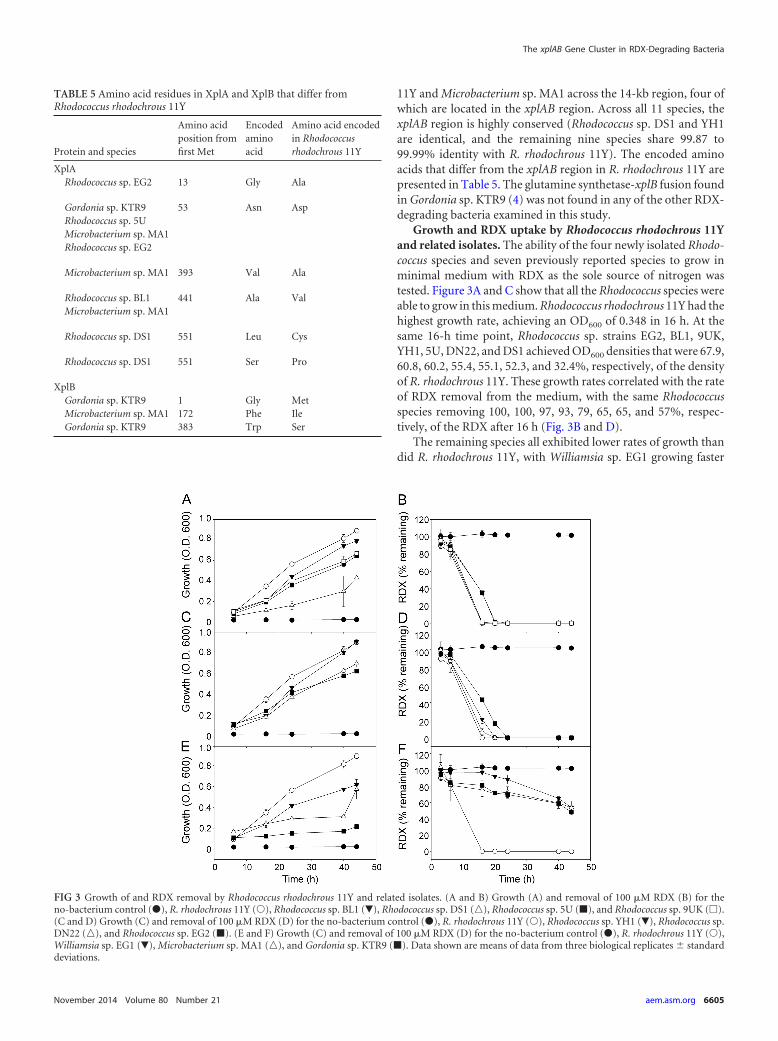
Growth and RDX uptake by Rhodococcus rhodochrous 11Yand related isolates. The ability of the four newly isolated Rhodo-coccus species and seven previously reported species to grow inminimal medium with RDX as the sole source of nitrogen wastested. Figure 3A and C show that all the Rhodococcus species wereable to grow in this medium. Rhodococcus rhodochrous 11Y had thehighest growth rate, achieving an OD600 of 0.348 in 16 h. At thesame 16-h time point, Rhodococcus sp. strains EG2, BL1, 9UK,YH1, 5U, DN22, and DS1 achieved OD600 densities that were 67.9,60.8, 60.2, 55.4, 55.1, 52.3, and 32.4%, respectively, of the densityof R. rhodochrous 11Y. These growth rates correlated with the rateof RDX removal from the medium, with the same Rhodococcusspecies removing 100, 100, 97, 93, 79, 65, 65, and 57%, respec-tively, of the RDX after 16 h (Fig. 3B and D).
The remaining species all exhibited lower rates of growth thandid R. rhodochrous 11Y, with Williamsia sp. EG1 growing faster
TABLE 5 Amino acid residues in XplA and XplB that differ fromRhodococcus rhodochrous 11Y
Protein and species
Amino acidposition fromfirst Met
Encodedaminoacid
Amino acid encodedin Rhodococcusrhodochrous 11Y
XplARhodococcus sp. EG2 13 Gly Ala
Gordonia sp. KTR9 53 Asn AspRhodococcus sp. 5UMicrobacterium sp. MA1Rhodococcus sp. EG2
Microbacterium sp. MA1 393 Val Ala
Rhodococcus sp. BL1 441 Ala ValMicrobacterium sp. MA1
Rhodococcus sp. DS1 551 Leu Cys
Rhodococcus sp. DS1 551 Ser Pro
XplBGordonia sp. KTR9 1 Gly MetMicrobacterium sp. MA1 172 Phe IleGordonia sp. KTR9 383 Trp Ser
FIG 3 Growth of and RDX removal by Rhodococcus rhodochrous 11Y and related isolates. (A and B) Growth (A) and removal of 100 �M RDX (B) for theno-bacterium control (�), R. rhodochrous 11Y (Œ), Rhodococcus sp. BL1 (�), Rhodococcus sp. DS1 (o), Rhodococcus sp. 5U (�), and Rhodococcus sp. 9UK (�).(C and D) Growth (C) and removal of 100 �M RDX (D) for the no-bacterium control (�), R. rhodochrous 11Y (Œ), Rhodococcus sp. YH1 (�), Rhodococcus sp.DN22 (o), and Rhodococcus sp. EG2 (�). (E and F) Growth (C) and removal of 100 �M RDX (D) for the no-bacterium control (�), R. rhodochrous 11Y (Œ),Williamsia sp. EG1 (�), Microbacterium sp. MA1 (o), and Gordonia sp. KTR9 (�). Data shown are means of data from three biological replicates � standarddeviations.
The xplAB Gene Cluster in RDX-Degrading Bacteria
November 2014 Volume 80 Number 21 aem.asm.org 6605
than Microbacterium sp. MA1 and Gordonia sp. KTR9 having thelowest growth rate (Fig. 3E). Over the first 16 h of incubation,Williamsia sp. EG1, Microbacterium sp. MA1, and Gordonia sp.KTR9 reached OD600 densities that were 69.4, 67.9, and 35.1%,respectively, of that reached by R. rhodochrous 11Y. As with theRhodococcus species, these growth rates correlated approximatelywith the rate of RDX uptake (Fig. 3F): after 16 h, the level of RDXin the medium containing Williamsia sp. EG1 was not signifi-cantly different from the level of RDX in the no-bacterium controlsamples, and Microbacterium sp. MA1 and Gordonia sp. KTR9 hadremoved only 25 and 19%, respectively, of the RDX.
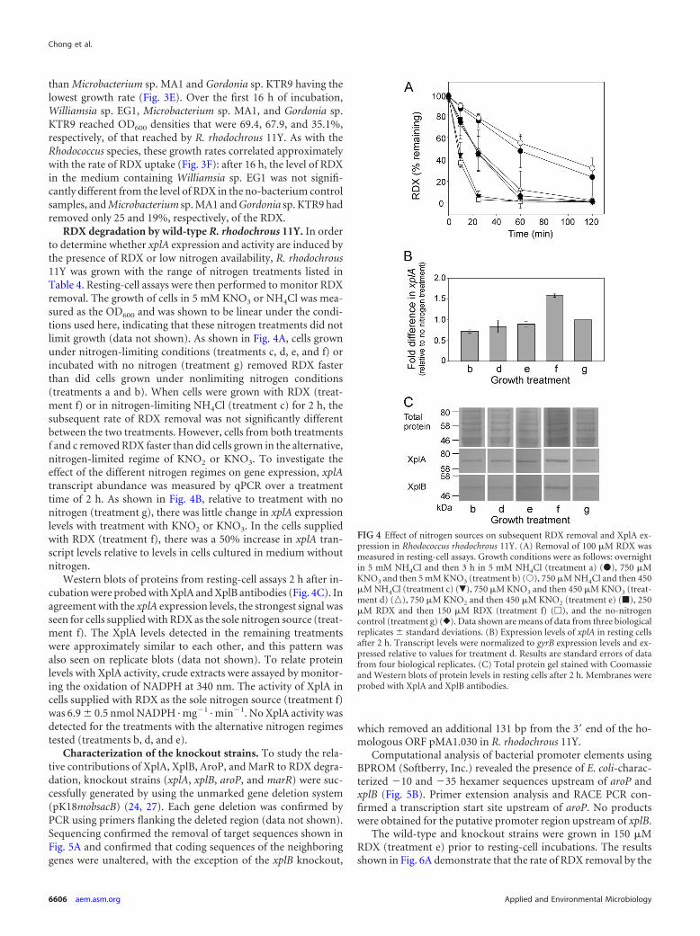
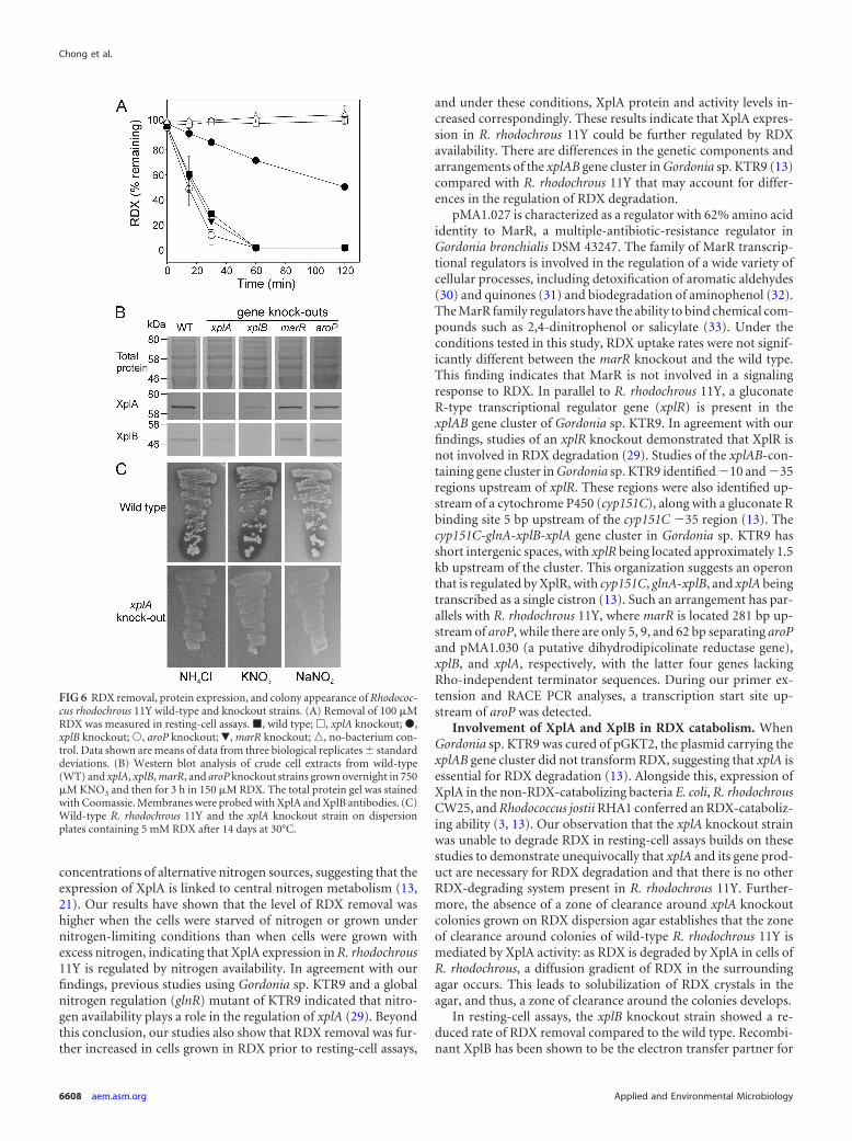
RDX degradation by wild-type R. rhodochrous 11Y. In orderto determine whether xplA expression and activity are induced bythe presence of RDX or low nitrogen availability, R. rhodochrous11Y was grown with the range of nitrogen treatments listed inTable 4. Resting-cell assays were then performed to monitor RDXremoval. The growth of cells in 5 mM KNO3 or NH4Cl was mea-sured as the OD600 and was shown to be linear under the condi-tions used here, indicating that these nitrogen treatments did notlimit growth (data not shown). As shown in Fig. 4A, cells grownunder nitrogen-limiting conditions (treatments c, d, e, and f) orincubated with no nitrogen (treatment g) removed RDX fasterthan did cells grown under nonlimiting nitrogen conditions(treatments a and b). When cells were grown with RDX (treat-ment f) or in nitrogen-limiting NH4Cl (treatment c) for 2 h, thesubsequent rate of RDX removal was not significantly differentbetween the two treatments. However, cells from both treatmentsf and c removed RDX faster than did cells grown in the alternative,nitrogen-limited regime of KNO2 or KNO3. To investigate theeffect of the different nitrogen regimes on gene expression, xplAtranscript abundance was measured by qPCR over a treatmenttime of 2 h. As shown in Fig. 4B, relative to treatment with nonitrogen (treatment g), there was little change in xplA expressionlevels with treatment with KNO2 or KNO3. In the cells suppliedwith RDX (treatment f), there was a 50% increase in xplA tran-script levels relative to levels in cells cultured in medium withoutnitrogen.
Western blots of proteins from resting-cell assays 2 h after in-cubation were probed with XplA and XplB antibodies (Fig. 4C). Inagreement with the xplA expression levels, the strongest signal wasseen for cells supplied with RDX as the sole nitrogen source (treat-ment f). The XplA levels detected in the remaining treatmentswere approximately similar to each other, and this pattern wasalso seen on replicate blots (data not shown). To relate proteinlevels with XplA activity, crude extracts were assayed by monitor-ing the oxidation of NADPH at 340 nm. The activity of XplA incells supplied with RDX as the sole nitrogen source (treatment f)was 6.9 � 0.5 nmol NADPH · mg�1 · min�1. No XplA activity wasdetected for the treatments with the alternative nitrogen regimestested (treatments b, d, and e).
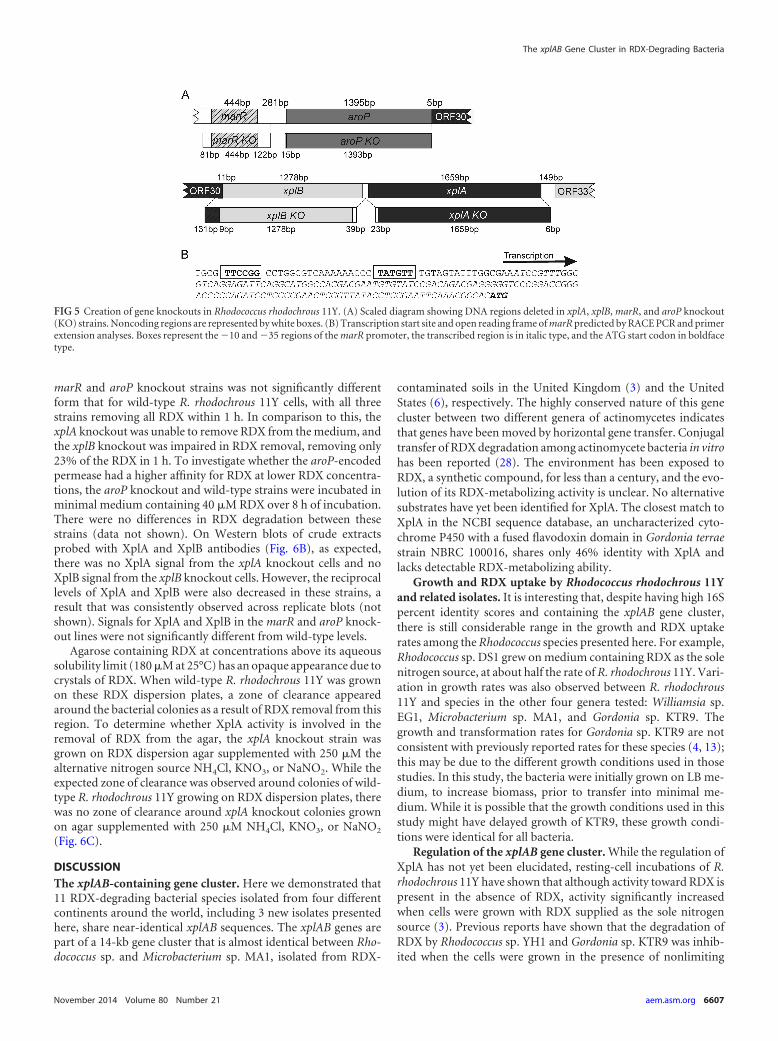
Characterization of the knockout strains. To study the rela-tive contributions of XplA, XplB, AroP, and MarR to RDX degra-dation, knockout strains (xplA, xplB, aroP, and marR) were suc-cessfully generated by using the unmarked gene deletion system(pK18mobsacB) (24, 27). Each gene deletion was confirmed byPCR using primers flanking the deleted region (data not shown).Sequencing confirmed the removal of target sequences shown inFig. 5A and confirmed that coding sequences of the neighboringgenes were unaltered, with the exception of the xplB knockout,
which removed an additional 131 bp from the 3= end of the ho-mologous ORF pMA1.030 in R. rhodochrous 11Y.
Computational analysis of bacterial promoter elements usingBPROM (Softberry, Inc.) revealed the presence of E. coli-charac-terized �10 and �35 hexamer sequences upstream of aroP andxplB (Fig. 5B). Primer extension analysis and RACE PCR con-firmed a transcription start site upstream of aroP. No productswere obtained for the putative promoter region upstream of xplB.
The wild-type and knockout strains were grown in 150 �MRDX (treatment e) prior to resting-cell incubations. The resultsshown in Fig. 6A demonstrate that the rate of RDX removal by the
FIG 4 Effect of nitrogen sources on subsequent RDX removal and XplA ex-pression in Rhodococcus rhodochrous 11Y. (A) Removal of 100 �M RDX wasmeasured in resting-cell assays. Growth conditions were as follows: overnightin 5 mM NH4Cl and then 3 h in 5 mM NH4Cl (treatment a) (�), 750 �MKNO3 and then 5 mM KNO3 (treatment b) (Œ), 750 �M NH4Cl and then 450�M NH4Cl (treatment c) (�), 750 �M KNO3 and then 450 �M KNO3 (treat-ment d) (o), 750 �M KNO2 and then 450 �M KNO2 (treatment e) (�), 250�M RDX and then 150 �M RDX (treatment f) (�), and the no-nitrogencontrol (treatment g) (}). Data shown are means of data from three biologicalreplicates � standard deviations. (B) Expression levels of xplA in resting cellsafter 2 h. Transcript levels were normalized to gyrB expression levels and ex-pressed relative to values for treatment d. Results are standard errors of datafrom four biological replicates. (C) Total protein gel stained with Coomassieand Western blots of protein levels in resting cells after 2 h. Membranes wereprobed with XplA and XplB antibodies.
Chong et al.
6606 aem.asm.org Applied and Environmental Microbiology
marR and aroP knockout strains was not significantly differentform that for wild-type R. rhodochrous 11Y cells, with all threestrains removing all RDX within 1 h. In comparison to this, thexplA knockout was unable to remove RDX from the medium, andthe xplB knockout was impaired in RDX removal, removing only23% of the RDX in 1 h. To investigate whether the aroP-encodedpermease had a higher affinity for RDX at lower RDX concentra-tions, the aroP knockout and wild-type strains were incubated inminimal medium containing 40 �M RDX over 8 h of incubation.There were no differences in RDX degradation between thesestrains (data not shown). On Western blots of crude extractsprobed with XplA and XplB antibodies (Fig. 6B), as expected,there was no XplA signal from the xplA knockout cells and noXplB signal from the xplB knockout cells. However, the reciprocallevels of XplA and XplB were also decreased in these strains, aresult that was consistently observed across replicate blots (notshown). Signals for XplA and XplB in the marR and aroP knock-out lines were not significantly different from wild-type levels.
Agarose containing RDX at concentrations above its aqueoussolubility limit (180 �M at 25°C) has an opaque appearance due tocrystals of RDX. When wild-type R. rhodochrous 11Y was grownon these RDX dispersion plates, a zone of clearance appearedaround the bacterial colonies as a result of RDX removal from thisregion. To determine whether XplA activity is involved in theremoval of RDX from the agar, the xplA knockout strain wasgrown on RDX dispersion agar supplemented with 250 �M thealternative nitrogen source NH4Cl, KNO3, or NaNO2. While theexpected zone of clearance was observed around colonies of wild-type R. rhodochrous 11Y growing on RDX dispersion plates, therewas no zone of clearance around xplA knockout colonies grownon agar supplemented with 250 �M NH4Cl, KNO3, or NaNO2
(Fig. 6C).
DISCUSSIONThe xplAB-containing gene cluster. Here we demonstrated that11 RDX-degrading bacterial species isolated from four differentcontinents around the world, including 3 new isolates presentedhere, share near-identical xplAB sequences. The xplAB genes arepart of a 14-kb gene cluster that is almost identical between Rho-dococcus sp. and Microbacterium sp. MA1, isolated from RDX-
contaminated soils in the United Kingdom (3) and the UnitedStates (6), respectively. The highly conserved nature of this genecluster between two different genera of actinomycetes indicatesthat genes have been moved by horizontal gene transfer. Conjugaltransfer of RDX degradation among actinomycete bacteria in vitrohas been reported (28). The environment has been exposed toRDX, a synthetic compound, for less than a century, and the evo-lution of its RDX-metabolizing activity is unclear. No alternativesubstrates have yet been identified for XplA. The closest match toXplA in the NCBI sequence database, an uncharacterized cyto-chrome P450 with a fused flavodoxin domain in Gordonia terraestrain NBRC 100016, shares only 46% identity with XplA andlacks detectable RDX-metabolizing ability.
Growth and RDX uptake by Rhodococcus rhodochrous 11Yand related isolates. It is interesting that, despite having high 16Spercent identity scores and containing the xplAB gene cluster,there is still considerable range in the growth and RDX uptakerates among the Rhodococcus species presented here. For example,Rhodococcus sp. DS1 grew on medium containing RDX as the solenitrogen source, at about half the rate of R. rhodochrous 11Y. Vari-ation in growth rates was also observed between R. rhodochrous11Y and species in the other four genera tested: Williamsia sp.EG1, Microbacterium sp. MA1, and Gordonia sp. KTR9. Thegrowth and transformation rates for Gordonia sp. KTR9 are notconsistent with previously reported rates for these species (4, 13);this may be due to the different growth conditions used in thosestudies. In this study, the bacteria were initially grown on LB me-dium, to increase biomass, prior to transfer into minimal me-dium. While it is possible that the growth conditions used in thisstudy might have delayed growth of KTR9, these growth condi-tions were identical for all bacteria.
Regulation of the xplAB gene cluster. While the regulation ofXplA has not yet been elucidated, resting-cell incubations of R.rhodochrous 11Y have shown that although activity toward RDX ispresent in the absence of RDX, activity significantly increasedwhen cells were grown with RDX supplied as the sole nitrogensource (3). Previous reports have shown that the degradation ofRDX by Rhodococcus sp. YH1 and Gordonia sp. KTR9 was inhib-ited when the cells were grown in the presence of nonlimiting
FIG 5 Creation of gene knockouts in Rhodococcus rhodochrous 11Y. (A) Scaled diagram showing DNA regions deleted in xplA, xplB, marR, and aroP knockout(KO) strains. Noncoding regions are represented by white boxes. (B) Transcription start site and open reading frame of marR predicted by RACE PCR and primerextension analyses. Boxes represent the �10 and �35 regions of the marR promoter, the transcribed region is in italic type, and the ATG start codon in boldfacetype.
The xplAB Gene Cluster in RDX-Degrading Bacteria
November 2014 Volume 80 Number 21 aem.asm.org 6607
concentrations of alternative nitrogen sources, suggesting that theexpression of XplA is linked to central nitrogen metabolism (13,21). Our results have shown that the level of RDX removal washigher when the cells were starved of nitrogen or grown undernitrogen-limiting conditions than when cells were grown withexcess nitrogen, indicating that XplA expression in R. rhodochrous11Y is regulated by nitrogen availability. In agreement with ourfindings, previous studies using Gordonia sp. KTR9 and a globalnitrogen regulation (glnR) mutant of KTR9 indicated that nitro-gen availability plays a role in the regulation of xplA (29). Beyondthis conclusion, our studies also show that RDX removal was fur-ther increased in cells grown in RDX prior to resting-cell assays,
and under these conditions, XplA protein and activity levels in-creased correspondingly. These results indicate that XplA expres-sion in R. rhodochrous 11Y could be further regulated by RDXavailability. There are differences in the genetic components andarrangements of the xplAB gene cluster in Gordonia sp. KTR9 (13)compared with R. rhodochrous 11Y that may account for differ-ences in the regulation of RDX degradation.
pMA1.027 is characterized as a regulator with 62% amino acididentity to MarR, a multiple-antibiotic-resistance regulator inGordonia bronchialis DSM 43247. The family of MarR transcrip-tional regulators is involved in the regulation of a wide variety ofcellular processes, including detoxification of aromatic aldehydes(30) and quinones (31) and biodegradation of aminophenol (32).The MarR family regulators have the ability to bind chemical com-pounds such as 2,4-dinitrophenol or salicylate (33). Under theconditions tested in this study, RDX uptake rates were not signif-icantly different between the marR knockout and the wild type.This finding indicates that MarR is not involved in a signalingresponse to RDX. In parallel to R. rhodochrous 11Y, a gluconateR-type transcriptional regulator gene (xplR) is present in thexplAB gene cluster of Gordonia sp. KTR9. In agreement with ourfindings, studies of an xplR knockout demonstrated that XplR isnot involved in RDX degradation (29). Studies of the xplAB-con-taining gene cluster in Gordonia sp. KTR9 identified �10 and �35regions upstream of xplR. These regions were also identified up-stream of a cytochrome P450 (cyp151C), along with a gluconate Rbinding site 5 bp upstream of the cyp151C �35 region (13). Thecyp151C-glnA-xplB-xplA gene cluster in Gordonia sp. KTR9 hasshort intergenic spaces, with xplR being located approximately 1.5kb upstream of the cluster. This organization suggests an operonthat is regulated by XplR, with cyp151C, glnA-xplB, and xplA beingtranscribed as a single cistron (13). Such an arrangement has par-allels with R. rhodochrous 11Y, where marR is located 281 bp up-stream of aroP, while there are only 5, 9, and 62 bp separating aroPand pMA1.030 (a putative dihydrodipicolinate reductase gene),xplB, and xplA, respectively, with the latter four genes lackingRho-independent terminator sequences. During our primer ex-tension and RACE PCR analyses, a transcription start site up-stream of aroP was detected.
Involvement of XplA and XplB in RDX catabolism. WhenGordonia sp. KTR9 was cured of pGKT2, the plasmid carrying thexplAB gene cluster did not transform RDX, suggesting that xplA isessential for RDX degradation (13). Alongside this, expression ofXplA in the non-RDX-catabolizing bacteria E. coli, R. rhodochrousCW25, and Rhodococcus jostii RHA1 conferred an RDX-cataboliz-ing ability (3, 13). Our observation that the xplA knockout strainwas unable to degrade RDX in resting-cell assays builds on thesestudies to demonstrate unequivocally that xplA and its gene prod-uct are necessary for RDX degradation and that there is no otherRDX-degrading system present in R. rhodochrous 11Y. Further-more, the absence of a zone of clearance around xplA knockoutcolonies grown on RDX dispersion agar establishes that the zoneof clearance around colonies of wild-type R. rhodochrous 11Y ismediated by XplA activity: as RDX is degraded by XplA in cells ofR. rhodochrous, a diffusion gradient of RDX in the surroundingagar occurs. This leads to solubilization of RDX crystals in theagar, and thus, a zone of clearance around the colonies develops.
In resting-cell assays, the xplB knockout strain showed a re-duced rate of RDX removal compared to the wild type. Recombi-nant XplB has been shown to be the electron transfer partner for
FIG 6 RDX removal, protein expression, and colony appearance of Rhodococ-cus rhodochrous 11Y wild-type and knockout strains. (A) Removal of 100 �MRDX was measured in resting-cell assays. �, wild type; �, xplA knockout; �,xplB knockout; Œ, aroP knockout; �, marR knockout;o, no-bacterium con-trol. Data shown are means of data from three biological replicates � standarddeviations. (B) Western blot analysis of crude cell extracts from wild-type(WT) and xplA, xplB, marR, and aroP knockout strains grown overnight in 750�M KNO3 and then for 3 h in 150 �M RDX. The total protein gel was stainedwith Coomassie. Membranes were probed with XplA and XplB antibodies. (C)Wild-type R. rhodochrous 11Y and the xplA knockout strain on dispersionplates containing 5 mM RDX after 14 days at 30°C.
Chong et al.
6608 aem.asm.org Applied and Environmental Microbiology
XplA, transferring electrons from NADPH to XplA for RDX deg-radation (9) and accounting for the reduced level of RDX removal.However, significant XplA activity was observed for the xplBknockout, demonstrating that XplA is able to obtain reducingequivalents from another source in R. rhodochrous 11Y. Consis-tent with this, expression of XplA in non-RDX-catabolizing bac-teria conferred an RDX-catabolizing ability (3, 13), demonstrat-ing that one or more surrogate reductases present in the non-RDXdegraders are able to partner with XplA. Moreover, the expressionof xplA in Arabidopsis confers the ability to catabolize RDX (12),demonstrating that XplA can also utilize plant reductases. Theseobservations also show that the transfer of xplA alone from donorto recipient naturally in the microbial environment would be suf-ficient to confer an RDX-degrading ability to the recipient.
Western blot analysis of crude extracts probed with XplA andXplB antibodies (Fig. 5C) demonstrated that levels of XplA andXplB were decreased in the xplB and xplA knockouts, respectively.When the xplA knockout was created, the open reading frame ofxplB was not disrupted, and vice versa for the creation of the xplBknockout. It is possible that the mutations affected transcript sta-bility or translational efficiency. Measurement of transcript andprotein levels of the surrounding ORFs would reveal if this was thecase. Alternatively, posttranslational coregulation of XplA andXplB could occur.
Route of RDX uptake. The aroP gene shares 76% sequenceidentity with an amino acid permease from Mycobacterium van-baalenii PYR-1. Although studies of Gordonia sp. KTR9 show thatbacteria do not require a dedicated transporter for RDX uptake(13), bacterial genes encoding related functions are frequently sit-uated close to each other (14, 34, 35). Therefore, we speculatedthat the aroP-encoded permease might also be involved in thetransport of RDX. However, resting cell assays performed on thearoP knockout strain showed that the rate of RDX removal wasnot significantly different from that of the wild-type (data notshown).
In summary, we have shown that xplAB genes are highly con-served among all four known genera of RDX-degrading bacteriaisolated from different geographical locations. These genes arepart of a larger gene cluster that is highly conserved between R.rhodochrous 11Y and Microbacterium MA1. Expression of XplA isinduced under nitrogen-limiting conditions and can be furtherenhanced by the presence of RDX. We established that MarR mustbe controlled by nitrogen availability and is unlikely to sense RDXlevels, as there was no difference in RDX removal between themarR knockout and wild-type cells when grown in RDX. Furtherexperiments are required to fully understand the regulation ofXplA by RDX and the involvement of RDX in the regulation ofnitrogen metabolism.
ACKNOWLEDGMENTS
This work was funded by the Strategic Environmental Research and De-velopment Program of the U.S. Department of Defense. Chun ShiongChong acknowledges funding from the Ministry of Higher Education,Malaysia. Dana Khdr Sabir recognizes the HCDP program, KurdistanRegional Government, Iraq, for funding.
We thank Zeev Ronen (Ben-Gurion University of the Negev, Israel)for providing Rhodococcus sp. YH1, Karl Indest (U.S. Army Engineer Re-search and Development Center) for providing Gordonia sp. KTR9, andthe National Institute of Technology and Evaluation (NITE) BiologicalResource Centre (NBRC), Japan, for supplying Gordonia terrae strainNBRC 100016.
REFERENCES1. Rylott EL, Lorenz A, Bruce NC. 2011. Biodegradation and biotransfor-
mation of explosives. Curr. Opin. Biotechnol. 22:434 – 440. http://dx.doi.org/10.1016/j.copbio.2010.10.014.
2. Binks PR, Nicklin S, Bruce NC. 1995. Degradation of hexahydro-1,3,5-trinitro-1,3,5-triazine (RDX) by Stenotrophomonas maltophilia PB1. Appl.Environ. Microbiol. 61:1318 –1322.
3. Seth-Smith HM, Rosser SJ, Basran A, Travis ER, Dabbs ER, Nicklin S,Bruce NC. 2002. Cloning, sequencing, and characterization of the hexa-hydro-1,3,5-trinitro-1,3,5-triazine degradation gene cluster from Rhodo-coccus rhodochrous. Appl. Environ. Microbiol. 68:4764 – 4771. http://dx.doi.org/10.1128/AEM.68.10.4764-4771.2002.
4. Thompson KT, Crocker FH, Fredrickson HL. 2005. Mineralization ofthe cyclic nitramine explosive hexahydro-1,3,5-trinitro-1,3,5-triazine byGordonia and Williamsia spp. Appl. Environ. Microbiol. 71:8265– 8272.http://dx.doi.org/10.1128/AEM.71.12.8265-8272.2005.
5. Seth-Smith H, Edwards J, Rosser S, Rathbone D, Bruce N. 2008. Theexplosive-degrading cytochrome P450 system is highly conserved amongstrains of Rhodococcus spp. Appl. Environ. Microbiol. 74:4550 – 4552.http://dx.doi.org/10.1128/AEM.00391-08.
6. Andeer P, Stahl D, Bruce N, Strand S. 2009. Lateral transfer of genes forhexahydro-1,3,5-trinitro-1,3,5-triazine (RDX) degradation. Appl. Envi-ron. Microbiol. 75:3258 –3262. http://dx.doi.org/10.1128/AEM.02396-08.
7. Coleman NV, Nelson DR, Duxbury T. 1998. Aerobic biodegradation ofhexahydro-1,3,5-trinitro-1,3,5-triazine (RDX) as a nitrogen source by aRhodococcus sp., strain DN22. Soil Biol. Biochem. 30:1159 –1167. http://dx.doi.org/10.1016/S0038-0717(97)00172-7.
8. Andeer P, Stahl DA, Lillis L, Strand SE. 2013. Identification of microbialpopulations assimilating nitrogen from RDX in munitions contaminatedmilitary training range soils by high sensitivity stable isotope probing. Envi-ron. Sci. Technol. 47:10356–10363. http://dx.doi.org/10.1021/es401729c.
9. Jackson R, Rylott E, Fournier D, Hawari J, Bruce N. 2007. Exploring thebiochemical properties and remediation applications of the unusual ex-plosive-degrading P450 system XpIA/B. Proc. Natl. Acad. Sci. U. S. A.104:16822–16827. http://dx.doi.org/10.1073/pnas.0705110104.
10. Rylott E, Bruce N. 2009. Plants disarm soil: engineering plants for thephytoremediation of explosives. Trends Biotechnol. 27:73– 81. http://dx.doi.org/10.1016/j.tibtech.2008.11.001.
11. Sabbadin F, Jackson R, Haider K, Tampi G, Turkenburg JP, Hart S,Bruce NC, Grogan G. 2009. The 1.5-A structure of XplA-heme, an un-usual cytochrome P450 heme domain that catalyzes reductive biotrans-formation of royal demolition explosive. J. Biol. Chem. 284:28467–28475.http://dx.doi.org/10.1074/jbc.M109.031559.
12. Rylott EL, Jackson RG, Edwards J, Womack GL, Seth-Smith HM,Rathbone DA, Strand SE, Bruce NC. 2006. An explosive-degradingcytochrome P450 activity and its targeted application for the phytoreme-diation of RDX. Nat. Biotechnol. 24:216 –219. http://dx.doi.org/10.1038/nbt1184.
13. Indest K, Jung C, Chen H, Hancock D, Florizone C, Eltis L, CrockerF. 2010. Functional characterization of pGKT2, a 182-kilobase plasmidcontaining the xplAB genes, which are involved in the degradation ofhexahydro-1,3,5-trinitro-1,3,5-triazine by Gordonia sp. strain KTR9.Appl. Environ. Microbiol. 76:6329 – 6337. http://dx.doi.org/10.1128/AEM.01217-10.
14. Yu M, Faan Y, Chung W, Tsang J. 2007. Isolation and characteriza-tion of a novel haloacid permease from Burkholderia cepacia MBA4.Appl. Environ. Microbiol. 73:4874 – 4880. http://dx.doi.org/10.1128/AEM.00576-07.
15. Wehrmann A, Morakkabati S, Kramer R, Sahm H, Eggeling L. 1995.Functional analysis of sequences adjacent to dapE of Corynebacterium glu-tamicum reveals the presence of aroP, which encodes the aromatic aminoacid transporter. J. Bacteriol. 177:5991–5993.
16. Bussmann M, Baumgart M, Bott M. 2010. RosR (Cg1324), a hydrogenperoxide-sensitive MarR-type transcriptional regulator of Corynebacte-rium glutamicum. J. Biol. Chem. 285:29305–29318. http://dx.doi.org/10.1074/jbc.M110.156372.
17. Wilkinson S, Grove A. 2006. Ligand-responsive transcriptional regula-tion by members of the MarR family of winged helix proteins. Curr. IssuesMol. Biol. 8:51– 62.
18. Jeon CO, Park M, Ro HS, Park W, Madsen EL. 2006. The naphthalenecatabolic (nag) genes of Polaromonas naphthalenivorans CJ2: evolutionaryimplications for two gene clusters and novel regulatory control. Appl.
The xplAB Gene Cluster in RDX-Degrading Bacteria
November 2014 Volume 80 Number 21 aem.asm.org 6609
Environ. Microbiol. 72:1086 –1095. http://dx.doi.org/10.1128/AEM.72.2.1086-1095.2006.
19. Alekshun M, Levy S. 1997. Regulation of chromosomally mediated mul-tiple antibiotic resistance: the mar regulon. Antimicrob. Agents Che-mother. 41:2067–2075.
20. Hickey WJ, Chen S, Zhao J. 2012. The phn island: a new genomic islandencoding catabolism of polynuclear aromatic hydrocarbons. Front. Mi-crobiol. 3:125. http://dx.doi.org/10.3389/fmicb.2012.00125.
21. Nejidat A, Kafka L, Tekoah Y, Ronen Z. 2008. Effect of organic andinorganic nitrogenous compounds on RDX degradation and cytochromeP-450 expression in Rhodococcus strain YH1. Biodegradation 19:313–320.http://dx.doi.org/10.1007/s10532-007-9137-3.
22. Satokari RM, Vaughan EE, Akkermans AD, Saarela M, de Vos WM.2001. Bifidobacterial diversity in human feces detected by genus-specificPCR and denaturing gradient gel electrophoresis. Appl. Environ. Micro-biol. 67:504 –513. http://dx.doi.org/10.1128/AEM.67.2.504-513.2001.
23. Tamura K, Dudley J, Nei M, Kumar S. 2007. MEGA4: Molecular Evo-lutionary Genetics Analysis (MEGA) software version 4.0. Mol. Biol. Evol.24:1596 –1599. http://dx.doi.org/10.1093/molbev/msm092.
24. van der Geize R, Hessels G, van Gerwen R, van der Meijden P,Dijkhuizen L. 2001. Unmarked gene deletion mutagenesis of kstD, en-coding 3-ketosteroid delta(1)-dehydrogenase, in Rhodococcus erythropolisSQ1 using sacB as counter-selectable marker. FEMS Microbiol. Lett. 205:197–202. http://dx.doi.org/10.1111/j.1574-6968.2001.tb10947.x.
25. Rosenberger RF, Elsden SR. 1960. The yields of Streptococcus faecalisgrown in continuous culture. J. Gen. Microbiol. 22:726 –739. http://dx.doi.org/10.1099/00221287-22-3-726.
26. Rylott EL, Budarina MV, Barker A, Lorenz A, Strand SE, Bruce NC.2011. Engineering plants for the phytoremediation of RDX in the presenceof the co-contaminating explosive TNT. New Phytol. 192:405– 413. http://dx.doi.org/10.1111/j.1469-8137.2011.03807.x.
27. Schafer A, Tauch A, Jager W, Kalinowski J, Thierbach G, Puhler A.1994. Small mobilizable multipurpose cloning vectors derived from theEscherichia coli plasmids pK18 and pK19 —selection of defined deletions
in the chromosome of Corynebacterium glutamicum. Gene 145:69 –73.http://dx.doi.org/10.1016/0378-1119(94)90324-7.
28. Jung CM, Crocker FH, Eberly JO, Indest KJ. 2011. Horizontal genetransfer (HGT) as a mechanism of disseminating RDX-degrading activityamong actinomycete bacteria. J. Appl. Microbiol. 110:1449 –1459. http://dx.doi.org/10.1111/j.1365-2672.2011.04995.x.
29. Indest KJ, Hancock DE, Jung CM, Eberly JO, Mohn WW, Eltis LD,Crocker FH. 2013. Role of nitrogen limitation in transformation of RDX(hexahydro-1,3,5-trinitro-1,3,5-triazine) by Gordonia sp. strain KTR9.Appl. Environ. Microbiol. 79:1746 –1750. http://dx.doi.org/10.1128/AEM.03905-12.
30. Fiorentino G, Ronca R, Cannio R, Rossi M, Bartolucci S. 2007. MarR-like transcriptional regulator involved in detoxification of aromatic com-pounds in Sulfolobus solfataricus. J. Bacteriol. 189:7351–7360. http://dx.doi.org/10.1128/JB.00885-07.
31. Leelakriangsak M, Huyen N, Towe S, van Duy N, Becher D, Hecker M,Antelmann H, Zuber P. 2008. Regulation of quinone detoxification bythe thiol stress sensing DUF24/MarR-like repressor, YodB in Bacillus sub-tilis. Mol. Microbiol. 67:1108 –1124. http://dx.doi.org/10.1111/j.1365-2958.2008.06110.x.
32. Tropel D, van der Meer J. 2004. Bacterial transcriptional regulators fordegradation pathways of aromatic compounds. Microbiol. Mol. Biol. Rev.68:474 –500. http://dx.doi.org/10.1128/MMBR.68.3.474-500.2004.
33. Cohen SP, Hachler H, Levy SB. 1993. Genetic and functional analysis ofthe multiple antibiotic resistance (mar) locus in Escherichia coli. J. Bacte-riol. 175:1484 –1492.
34. Williamson N, Fineran P, Leeper F, Salmond G. 2006. The biosynthesisand regulation of bacterial prodiginines. Nat. Rev. Microbiol. 4:887– 899.http://dx.doi.org/10.1038/nrmicro1531.
35. Govantes F, Garcia-Gonzalez V, Porrua O, Platero A, Jimenez-Fernandez A, Santero E. 2010. Regulation of the atrazine-degradativegenes in Pseudomonas sp strain ADP. FEMS Microbiol. Lett. 310:1– 8.http://dx.doi.org/10.1111/j.1574-6968.2010.01991.x.
Chong et al.
6610 aem.asm.org Applied and Environmental Microbiology
Related Documents