Journal of Visualized Experiments www.jove.com Copyright © 2014 Journal of Visualized Experiments June 2014 | 88 | e51668 | Page 1 of 10 Video Article Analysis of the Epithelial Damage Produced by Entamoeba histolytica Infection Abigail Betanzos 1 , Michael Schnoor 2 , Rosario Javier-Reyna 1 , Guillermina García-Rivera 1 , Cecilia Bañuelos 3 , Jonnatan Pais-Morales 1 , Esther Orozco 1 1 Department of Infectomics and Molecular Pathogenesis, Center for Research and Advanced Studies of the National Polytechnic Institute 2 Department of Molecular Biomedicine, Center for Research and Advanced Studies of the National Polytechnic Institute 3 Agency for Knowledge Commercialization, Center for Research and Advanced Studies of the National Polytechnic Institute Correspondence to: Abigail Betanzos at [email protected], Esther Orozco at [email protected] URL: http://www.jove.com/video/51668 DOI: doi:10.3791/51668 Keywords: Immunology, Issue 88, Entamoeba histolytica, EhCPADH112, cell adhesion, MDCK, Caco-2, tight junction disruption, amoebiasis, host- pathogen interaction, infection model, actin cytoskeleton Date Published: 6/12/2014 Citation: Betanzos, A., Schnoor, M., Javier-Reyna, R., García-Rivera, G., Bañuelos, C., Pais-Morales, J., Orozco, E. Analysis of the Epithelial Damage Produced by Entamoeba histolytica Infection. J. Vis. Exp. (88), e51668, doi:10.3791/51668 (2014). Abstract Entamoeba histolytica is the causative agent of human amoebiasis, a major cause of diarrhea and hepatic abscess in tropical countries. Infection is initiated by interaction of the pathogen with intestinal epithelial cells. This interaction leads to disruption of intercellular structures such as tight junctions (TJ). TJ ensure sealing of the epithelial layer to separate host tissue from gut lumen. Recent studies provide evidence that disruption of TJ by the parasitic protein EhCPADH112 is a prerequisite for E. histolytica invasion that is accompanied by epithelial barrier dysfunction. Thus, the analysis of molecular mechanisms involved in TJ disassembly during E. histolytica invasion is of paramount importance to improve our understanding of amoebiasis pathogenesis. This article presents an easy model that allows the assessment of initial host-pathogen interactions and the parasite invasion potential. Parameters to be analyzed include transepithelial electrical resistance, interaction of EhCPADH112 with epithelial surface receptors, changes in expression and localization of epithelial junctional markers and localization of parasite molecules within epithelial cells. Video Link The video component of this article can be found at http://www.jove.com/video/51668/ Introduction Entamoeba histolytica is a single cell protozoan responsible of human amoebiasis, an intestinal infection causing inflammation and diarrhea. E. histolytica infects up to 50 million individuals yearly, but only about 10% of infected people develop the symptoms associated to amoebiasis 1 . Infection occurs upon ingestion of contaminated food or water containing E. histolytica cysts. In the intestine, cysts produce live trophozoites that adhere to colon mucin and proliferate 2 . Trophozoites usually form cysts that are excreted via stools. In other cases and for yet unknown reasons, trophozoites break the intestinal epithelial layer and invade underlying tissues. In worst cases, they enter the blood stream and affect other organs such as the liver 3 . Breaking the epithelial barrier requires disruption of epithelial transmembranal structures that maintain cells joined. Epithelial cell contacts are formed by the apical junctional complex consisting of tight (TJ) and adherens junctions (AJ), and desmosomes 4 . The most apical junctions are TJ, and therefore, they are the first barrier affronted by E. histolytica and some other pathogens during host invasion. TJ are comprised of transmembranal adhesion receptors such as claudins, occludin and junctional adhesion molecules (JAM) that engage in homo- or heterophilic interactions with receptors of the neighboring cell. They are intracellularly bound by scaffold molecules of the zonula occludens (ZO) family that connect adhesion receptors to actin cytoskeleton to provide further mechanical strength to the epithelium. TJ are responsible for sealing intestinal tissue from the gut lumen, preventing excessive water and solute leakage. Thus, after TJ are disrupted by the parasite, tissues are invaded. E. histolytica secretes several molecules such as: (i) those involved in adhesion of amoebae to target cells 5 ; (ii) membrane- active factors participating in killing of host cells by exocytosis, for example the ion channel-forming peptides termed amoebapores 6,7 ; and (iii) proteinases that degrade extracellular matrix proteins and mediate tissue disintegration 5,8,9 . The cysteine protease EhCP112 and the adhesion molecule EhADH112 that together form the EhCPADH112 complex are two E. histolytica virulence proteins that play a major role in the disassembly of TJ 10 . Live trophozoites, their total lysates and secreted products induce molecular changes in the TJ complex and functional disturbance of the epithelial barrier. In this study, it is shown that EhCP112 and EhADH112 interact with occludin and claudin-1 proteins leading to internalization and degradation of cell proteins, thus facilitating E. histolytica entrance through the paracellular pathway. Our data and those of other groups 11-17 strongly suggest the necessity of specific host-pathogen interactions that allow parasite invasion. Unraveling the molecular basis of these interactions is of utmost importance for a better understanding of amoebiasis pathogenesis. Selective

Welcome message from author
This document is posted to help you gain knowledge. Please leave a comment to let me know what you think about it! Share it to your friends and learn new things together.
Transcript
Journal of Visualized Experiments www.jove.com
Copyright © 2014 Journal of Visualized Experiments June 2014 | 88 | e51668 | Page 1 of 10
Video Article
Analysis of the Epithelial Damage Produced by Entamoeba histolyticaInfectionAbigail Betanzos1, Michael Schnoor2, Rosario Javier-Reyna1, Guillermina García-Rivera1, Cecilia Bañuelos3, Jonnatan Pais-Morales1,Esther Orozco1
1Department of Infectomics and Molecular Pathogenesis, Center for Research and Advanced Studies of the National Polytechnic Institute2Department of Molecular Biomedicine, Center for Research and Advanced Studies of the National Polytechnic Institute3Agency for Knowledge Commercialization, Center for Research and Advanced Studies of the National Polytechnic Institute
Correspondence to: Abigail Betanzos at [email protected], Esther Orozco at [email protected]
URL: http://www.jove.com/video/51668DOI: doi:10.3791/51668
Keywords: Immunology, Issue 88, Entamoeba histolytica, EhCPADH112, cell adhesion, MDCK, Caco-2, tight junction disruption, amoebiasis, host-pathogen interaction, infection model, actin cytoskeleton
Date Published: 6/12/2014
Citation: Betanzos, A., Schnoor, M., Javier-Reyna, R., García-Rivera, G., Bañuelos, C., Pais-Morales, J., Orozco, E. Analysis of the EpithelialDamage Produced by Entamoeba histolytica Infection. J. Vis. Exp. (88), e51668, doi:10.3791/51668 (2014).
Abstract
Entamoeba histolytica is the causative agent of human amoebiasis, a major cause of diarrhea and hepatic abscess in tropical countries. Infectionis initiated by interaction of the pathogen with intestinal epithelial cells. This interaction leads to disruption of intercellular structures such as tightjunctions (TJ). TJ ensure sealing of the epithelial layer to separate host tissue from gut lumen. Recent studies provide evidence that disruptionof TJ by the parasitic protein EhCPADH112 is a prerequisite for E. histolytica invasion that is accompanied by epithelial barrier dysfunction.Thus, the analysis of molecular mechanisms involved in TJ disassembly during E. histolytica invasion is of paramount importance to improve ourunderstanding of amoebiasis pathogenesis. This article presents an easy model that allows the assessment of initial host-pathogen interactionsand the parasite invasion potential. Parameters to be analyzed include transepithelial electrical resistance, interaction of EhCPADH112 withepithelial surface receptors, changes in expression and localization of epithelial junctional markers and localization of parasite molecules withinepithelial cells.
Video Link
The video component of this article can be found at http://www.jove.com/video/51668/
Introduction
Entamoeba histolytica is a single cell protozoan responsible of human amoebiasis, an intestinal infection causing inflammation and diarrhea. E.histolytica infects up to 50 million individuals yearly, but only about 10% of infected people develop the symptoms associated to amoebiasis1.Infection occurs upon ingestion of contaminated food or water containing E. histolytica cysts. In the intestine, cysts produce live trophozoitesthat adhere to colon mucin and proliferate2. Trophozoites usually form cysts that are excreted via stools. In other cases and for yet unknownreasons, trophozoites break the intestinal epithelial layer and invade underlying tissues. In worst cases, they enter the blood stream and affectother organs such as the liver3.
Breaking the epithelial barrier requires disruption of epithelial transmembranal structures that maintain cells joined. Epithelial cell contacts areformed by the apical junctional complex consisting of tight (TJ) and adherens junctions (AJ), and desmosomes4. The most apical junctionsare TJ, and therefore, they are the first barrier affronted by E. histolytica and some other pathogens during host invasion. TJ are comprised oftransmembranal adhesion receptors such as claudins, occludin and junctional adhesion molecules (JAM) that engage in homo- or heterophilicinteractions with receptors of the neighboring cell. They are intracellularly bound by scaffold molecules of the zonula occludens (ZO) familythat connect adhesion receptors to actin cytoskeleton to provide further mechanical strength to the epithelium. TJ are responsible for sealingintestinal tissue from the gut lumen, preventing excessive water and solute leakage. Thus, after TJ are disrupted by the parasite, tissuesare invaded. E. histolytica secretes several molecules such as: (i) those involved in adhesion of amoebae to target cells5; (ii) membrane-active factors participating in killing of host cells by exocytosis, for example the ion channel-forming peptides termed amoebapores6,7; and (iii)proteinases that degrade extracellular matrix proteins and mediate tissue disintegration5,8,9.
The cysteine protease EhCP112 and the adhesion molecule EhADH112 that together form the EhCPADH112 complex are two E. histolyticavirulence proteins that play a major role in the disassembly of TJ 10. Live trophozoites, their total lysates and secreted products induce molecularchanges in the TJ complex and functional disturbance of the epithelial barrier. In this study, it is shown that EhCP112 and EhADH112 interactwith occludin and claudin-1 proteins leading to internalization and degradation of cell proteins, thus facilitating E. histolytica entrance through theparacellular pathway.
Our data and those of other groups11-17strongly suggest the necessity of specific host-pathogen interactions that allow parasite invasion.Unraveling the molecular basis of these interactions is of utmost importance for a better understanding of amoebiasis pathogenesis. Selective
Journal of Visualized Experiments www.jove.com
Copyright © 2014 Journal of Visualized Experiments June 2014 | 88 | e51668 | Page 2 of 10
disturbance of TJ by trophozoites, characterized by increased paracellular permeability, can be measured by a decrease in transepithelialelectrical resistance (TER). The transference of parasitic proteins towards host epithelia can be determined by immunofluorescence staining andconfocal laser microscopy, a method that can also reveal co-localization of amoeba virulence factors with epithelial junctional markers indicatingpossible direct interactions. In this article, we describe in detail how epithelial cells and trophozoites are cultivated, harvested and manipulated toexamine host-pathogen interactions and their consequences.
Protocol
1. Establishment and Maintenance of E. histolytica Cultures
1. Grow axenically (entirely free of all other contaminating organisms) 1 x 10⁵ trophozoites of Entamoeba histolytica strain HMl:IMSS clone A18
in 16 x 125 mm culture tubes with Teflon liner screw caps (or 1 x 10⁶ trophozoites in a disposable T-25 flask) and 15 ml (or 50 ml in T-25flask) of TYI-S-33 medium (TYI broth supplemented with 3% Diamond vitamin mixture, 10% heat inactivated adult bovine serum, 0.5 IU/mlpenicillin and 35 µg/ml streptomycin)19 in an incubator at 37 °C.
2. Harvest trophozoites during the logarithmic growth phase usually at 48-96 hr intervals (Figure 1) by chilling the culture tubes for 5-10 min inan ice-water bath to release trophozoites attached to the glass culture tube.
3. Transfer the culture into a conical tube and invert it several times to disperse the cells. Determine cell number using a hemocytometer(Neubauer chamber), and transfer an inoculum into a culture tube containing fresh TYI-S-33 medium.
1. Use low numbers of amoebas for longer incubation periods (~3 x 10⁵ cells for ~5 days) and a higher number for shorter periods (~1x 10⁶ cells for ~1 day). While counted inocula are desirable, established cultures become predictable so that estimated volumes ofinocula are feasible.
2. Titrate the amount of trophozoites to optimize cell numbers for each experiment.3. Maintain a parallel duplicate culture to have a back-up in case of inadvertent contamination or tube breakage.
4. Cap tubes tightly and incubate them at 37 °C in a 5° tilted horizontal angle.5. Check cultures by visual inspection regularly, since an excessive growth of the culture may contain many lysed cells.
2. Establishment and Maintenance of MDCK Culture
1. Grow MDCK (Madin Darby Canine Kidney) cells type I (high resistance) in disposable T-75 flasks with 10 ml of DMEM media supplementedwith penicillin (100 IU/ml), streptomycin (100 mg/ml), insulin (0.08 U/ml) and 10% neonate calf serum in a humidified atmosphere of 5% CO2and 95% air at 37 °C.
2. To split the cells, check them on an inverted microscope. Split cells when they are ~ 85-100% confluent.3. Use a vacuum aspirator in a sterile hood and a sterile glass Pasteur pipette to remove media from the culture. To avoid scraping cells, turn
the flask upside down and aspirate media from the bottom corner.4. Dispense 10 ml of prewarmed PBS (140 mM NaCl, 2.7 mM KCl, 10 mM Na2HPO4, 1.8 mM KH2PO4, pH 7.4) into a T-75 flask (5 ml in the
case of a T-25 flask) and gently rock the flask to wash the cells. Remove PBS by vacuum aspiration. Extensive washing is required sinceserum inhibits trypsin.
5. Dispense 1.5 ml of 0.05% trypsin into a T-75 flask (for T-25 flask use 0.5 ml) and gently rock the flask to cover the cell layer with trypsin.6. Incubate for 5-10 min in the incubator (exact time depends on trypsin activity).7. Check for cell detachment by holding flask against the light and look for flowing cells. Cell detachment (rounded cells) can also be checked
using a microscope. Often, cells release faster with a quick, gentle slap to the side of the flask.8. Add 15 ml of supplemented DMEM to a T-75 flask (5 ml for a T-25 flask). Resuspend cells by pipetting up and down 4 or 5 times.9. To propagate cells, dilute cell suspension 1:5 with fresh supplemented DMEM. Cells number can also be determined at this point using a
hemocytometer but this is usually only necessary if splitting cells into special formats for certain assays.
3. Preparation of Trophozoite Total Lysates
1. Detach trophozoites from culture tubes by chilling the tube for 5-10 min in an ice-water bath. Transfer the culture aseptically into a conicaltube and centrifuge at 360 x g for 10 min at 4 °C. Carefully discard the supernatant by decantation, add ice-cold PBS to the pellet, invert thetube several times to disperse the cells and centrifuge again at 360 x g for 10 min. Repeat this step twice to remove all traces of TYI-S-33medium.
2. Determine trophozoites number using a hemocytometer.3. Lyse trophozoites by freeze-thaw cycles. Snap-freeze a measured inoculum of trophozoites (600,000 trophozoites/ml) diluted in ice-cold PBS,
in liquid nitrogen for 1 min. Unfreeze the sample at 4 °C and vortex vigorously. Repeat freezing and thawing 3 times to complete cell lysis.4. Activate proteases present in the lysates with 0.02% β-mercaptoethanol.
4. Preparation of Trophozoite Secreted Products
1. Perform steps 3.1 to 3.2.2. Determine cells number and viability at this point using a hemocytometer and applying a trypan blue exclusion test20:
1. Dilute 9 x 10⁶ trophozoites in 3 ml ice-cold PBS and transfer cells into a 50 ml conical tube. Take 10 µl of this suspension and add 1 µlof 0.4% trypan blue stock solution pH 7.2.
2. Use a Neubauer chamber to count cells at low magnification.3. Count all cells and separate the number of blue cells, since blue cells have taken up the dye and must be considered dead.4. Calculate the percentage of viable cells by dividing the number of viable cells by the total number of cells and multiplying by 100.
Journal of Visualized Experiments www.jove.com
Copyright © 2014 Journal of Visualized Experiments June 2014 | 88 | e51668 | Page 3 of 10
3. Transfer the conical tube containing the trophozoite suspension into the incubator at 37 °C for 2 hr in a 5° tilted horizontal angle.4. To collect the secreted products, centrifuge tubes at 360 x g for 10 min. Use a disposable and sterile syringe and carefully collect the
supernatant, avoiding contamination of samples with trophozoites from the pellet. Eliminate any transferred cells by passing the supernatantthrough a 0.22 μm cellulose acetate membrane. Activate proteases with 0.02% β-mercaptoethanol.
5. To discard putative unwanted contamination with molecules released from dead cells, reapply the trypan blue exclusion test for cell viability.Viable and total number of cells should be the same as obtained in step 4.2. If this is not the case, the collected supernatant may not onlycontain secreted proteins and should be discarded.
5. Interaction of MDCK Cells with Trophozoites, Trophozoite Lysates or Secreted Products
1. Incubate confluent MDCK cell monolayers with live trophozoites (an MDCK-to-amoeba ratio of 1:1), trophozoite lysates (an MDCK-to-amoebaratio of 1:2) or secreted products (an MDCK-to-amoeba ratio of 1:10) at 37 °C for 2 and 30 min (Figure 2A).
2. Wash epithelial cells five times with ice-cold PBS to eliminate unbound molecules or trophozoites.
6. Preparation of Samples for Immunofluorescence
1. Culture MDCK cells on sterile glass coverslips placed inside a 24-multiwell cell culture dish. Inspect the cells on the inverted microscope untilthey reach 100% of confluence. Then, replace medium with 1 ml of fresh supplemented DMEM medium and transfer the plate to a 37 °Cincubator for 24 hr more.
2. Remove the medium using a vacuum aspirator and a sterile glass Pasteur pipette and add 1 ml of warm PBS to each well. Remove PBS andrepeat the wash with fresh PBS. Take care not to scrape cells from the bottom of the well with the glass pipette.
3. Add different conditions of trophozoites diluted in 1 ml of warm PBS: live trophozoites (T), trophozoite total lysates (TL) and secreted products(SP).
1. Put the culture plate in the incubator for 2 or 30 min.2. Prepare two control conditions: one named time 0 min, where MDCK cells should be not incubated with E. histolytica but only with 1 ml
of warm PBS; and another condition as secondary antibodies control, in this case incubate MDCK with T, TL or SP for 2 or 30 min andomit incubation with primary antibodies.
4. Wash epithelial cells five times with cold PBS to eliminate unbound molecules or trophozoites.5. Fix and permeabilize the cells with 1 ml of 96% ethanol for 30 min at -20 °C.6. To remove ethanol, perform three quick washes with 1 ml of PBS at room temperature.7. Perform the following steps in a humid chamber to avoid drying of samples:
1. Use any available flat box that can be closed and filled with a wet paper towel on which samples can be safely placed. For example, anempty pipette tip box serves this purpose well.
2. Inside the chamber, evenly place a Parafilm layer and pipette 25 μl of blocking solution (0.5% BSA) for each coverslip.3. Take coverslips out of the wells with fine forceps, carefully remove excess liquid from the edge with a filter paper and put coverslip
into the drop containing blocking solution (0.5% BSA)with the cells facing the blocking solution. Incubate the samples for 1 hr at roomtemperature.
8. Prepare a mixture of primary antibodies in PBS: IgM mouse anti-EhCPADH112 (mα-EhCPADH112; 1:10 dilution) and IgG rabbit anti-ZO-1(pα-ZO-1; 1:100 dilution).
9. Place a fresh sheet of Parafilm in the humid box and pipette 25 μl of antibodies solution for each coverslip.10. Lift the coverslips from the blocking solution, dry any excess liquid at the edges using a filter paper, and place them into the drops with the
cells facing the antibody solution. Incubate the samples overnight at 4 °C.11. Put each coverslip into a well of a 24-multiwell (cell side on top) and add 1 ml of PBS.
1. Wash five times with 1 ml of PBS to remove unbound molecules.
12. Prepare a mixture of fluorescent secondary antibodies in PBS: goat anti-IgM mouse coupled to FITC (1:100 dilution) and goat anti-IgG rabbitcoupled to TRITC (1:50 dilution).
13. Place a fresh sheet of Parafilm into the humid box and pipette 25 µl of the secondary antibodies solution for each coverslip.14. Transfer the samples as in 6.10 and incubate for 1 hr at room temperature in the dark to avoid bleaching of the fluorescent dyes. Repeat step
6.11.1.15. For nuclear staining, incubate for 3 min with 200 μl of 0.05 mM DAPI solution at room temperature and protect from light. Repeat step 6.11.1.16. To conserve the fluorescent dyes, place the coverslips (cells facing down) into 5 μl Vecta Shield antifade mounting solution on glass
microscope slides. Seal the cover slips using nail polish to avoid sample drying.17. For long term storage, keep slides in a microscope slide box at -20 °C.18. Analyze preparations by confocal microscopy through Z-stack sections and xz-planes (Figure 2A).
7. Incubation of Trophozoites with Protease Inhibitors or Specific Antibody
1. Repeat steps 3.1 to 3.2. For each experimental condition, incubate 1 x 10⁵ trophozoites (resuspended in 50 μl PBS) with proteaseinhibitors (1 mM Complete and 40 µg/ml E-64) or 30 µg monoclonal antibody against EhCPADH112 (mαEhCPADH112)21 for 20 min at 4 °C(Figure 2B). Use these preparations for co-incubation assays as described in step 8.5.
8. Measurement of Transepithelial Electrical Resistance (TER)
1. Culture MDCK cells on sterile transwell permeable supports (0.4 μm pore size). In the upper compartment, place 100 μl of cellularsuspension and in the lower compartment place 600 μl of supplemented DMEM medium.
Journal of Visualized Experiments www.jove.com
Copyright © 2014 Journal of Visualized Experiments June 2014 | 88 | e51668 | Page 4 of 10
1. Check the medium level periodically. Fresh medium can be added as required.2. Inspect the cells on an inverted microscope until they reach 100% of confluence. Change medium every 2-3 days.3. To improve cell attachment (if necessary), equilibrate the transwell filters overnight at 37 °C in DMEM before adding the cell
suspension.
2. Sterilize STX2 electrodes in the hood by immersion in ethanol and then in sterile PBS for 15 min each, under UV light. Connect electrodes toan EVOM epithelial voltohmmeter and select resistance mode.
3. Collect medium from the upper compartment of each transwell and pool them. Take 50 μl of this media mixture and combine it with 50 μl oftrophozoites suspension (1 x 10⁵ trophozoites in PBS) or with only PBS (control). Use a similar mixture for each transwell.
4. Add mixtures to the upper chamber of each transwell containing the MDCK cells. Experimental conditions include MDCK cells withouttrophozoites (control), a co-culture with trophozoites or with trophozoites pre-incubated with protease inhibitors or mαEhCPADH112. Toeliminate the resistance provided by filters, use transwells incubated with medium only. For each experimental condition use at least threetranswells.
5. Immediately, measure TER by dipping the STX2 electrodes into each transwell.1. Ensure that the longer electrode touches the bottom of the lower chamber and that the shorter electrode is covered by medium in the
upper compartment (see Figure 2B).2. Make sure to hold the electrodes perpendicularly each time. This will improve reproducibility, significantly. Avoid moving or tilting the
electrode during the measurements as this will lead to variation of data.
6. This measurement should be considered the initial TER value. Then, monitor the TER during the following 30 min.7. To calculate the final TER value, subtract the TER value of filters only. To compare results from different experiments normalize each data
point to the initial values, taking these as 100% (Figure 2B).
Representative Results
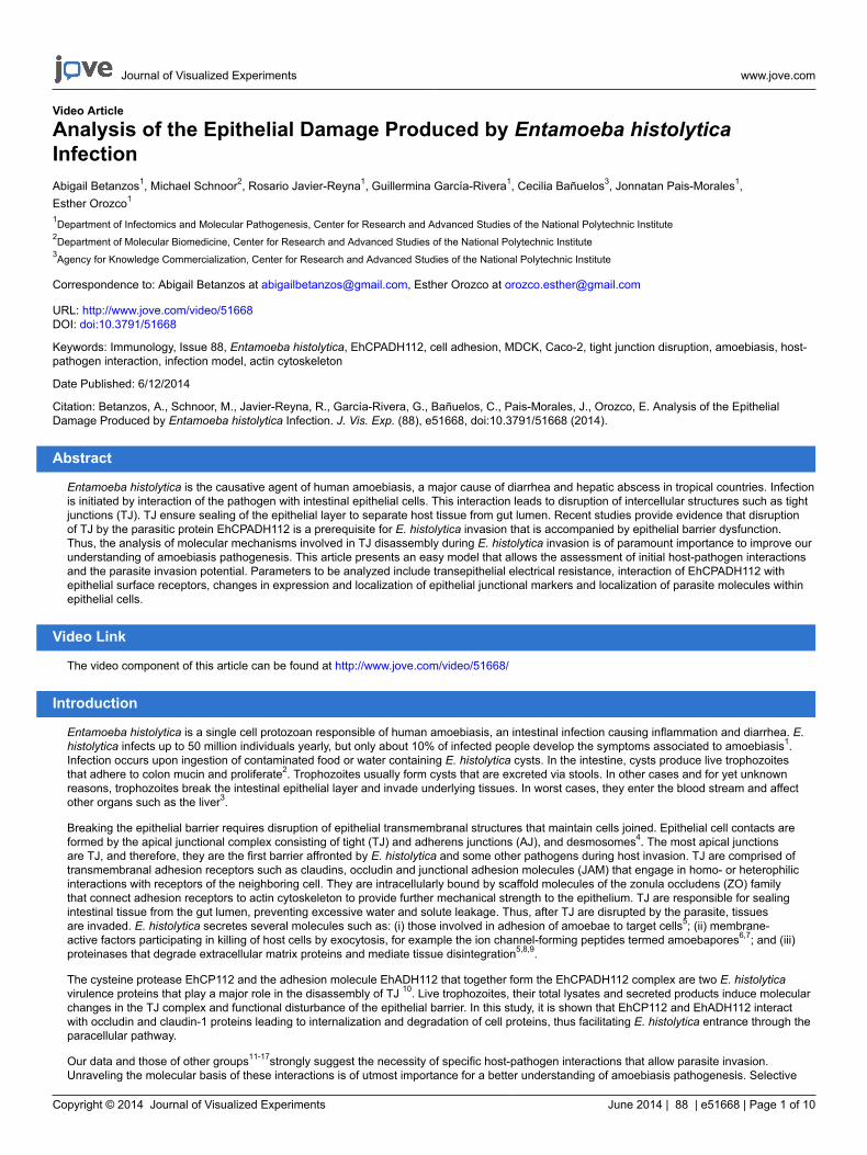
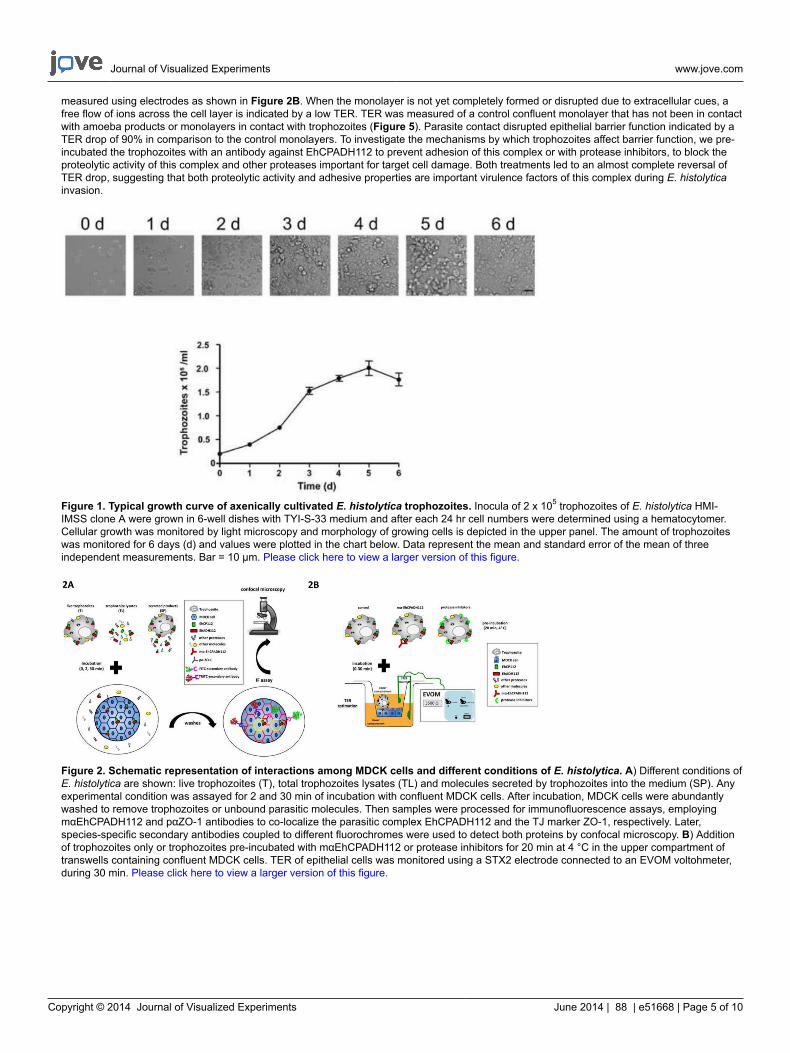
For a successful E. histolytica culture, two important conditions must be fulfilled: growth in axenic conditions and harvest in logarithmic phase.Previously, cultures of E. histolytica were readily established in association with certain species of bacteria or trypanosomatids22. However,nowadays it is common to have axenic cultivation of this parasite meaning an indefinite subcultivation of amoebae in an environment freeof metabolizing bacteria, fungi, protozoa, or metazoan cells. Additionally, harvesting the trophozoites during the late logarithmic to earlystationary phase of growth is crucial to have viable and proliferating cells23. Therefore, it is important to distinguish this phase by quantification oftrophozoites along several days and also by examining their intact morphology and rapid locomotion (Figure 1).
For interaction studies, epithelial cells need to be in a confluent monolayer to represent physiological conditions that amoebas would normallyencounter in the body. Several epithelial cell lines, easy to cultivate, are available. While a more physiological cell model system would derivefrom the intestines such as Caco-2 or T84, these cell lines grow rather slowly. A faster growing cell line that has extensively been studied areMDCK cells24. These epithelial cells are of kidney origin and are characterized by high TER, strong TJ and easy cultivation. This cell line hasbeen used for decades to study cell biology of junctions and TJ composition as well as mechanisms of TJ assembly and disassembly. Forthese reasons, MDCK cells are often chosen by researchers when studying TJ functions. MDCK cells have also been widely used as a modelfor the study of mechanisms induced during amoebiasis5,8,10,25-28. In a recent study, we reported that interaction of strain I MDCK cells andintestinal epithelial Caco-2 cells with live amoebas, lysates and amoeba products caused similar effects on TJ composition and TER10. Thus,MDCK cells are a suitable model to study mechanisms of amoeba-epithelia interactions. Furthermore, the use of several E. histolytica products(intact trophozoites, trophozoite total lysates or secreted products harvested as trophozoites culture supernatant) is crucial to provide additionaland relevant information about molecules involved in these interactions, such as availability, mode of action, secretion, participation of othermolecules and triggering of signaling pathways.
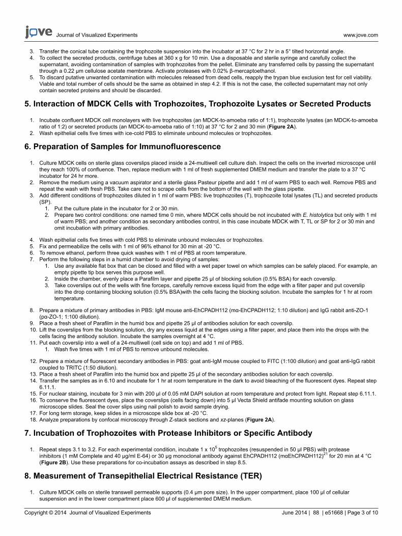
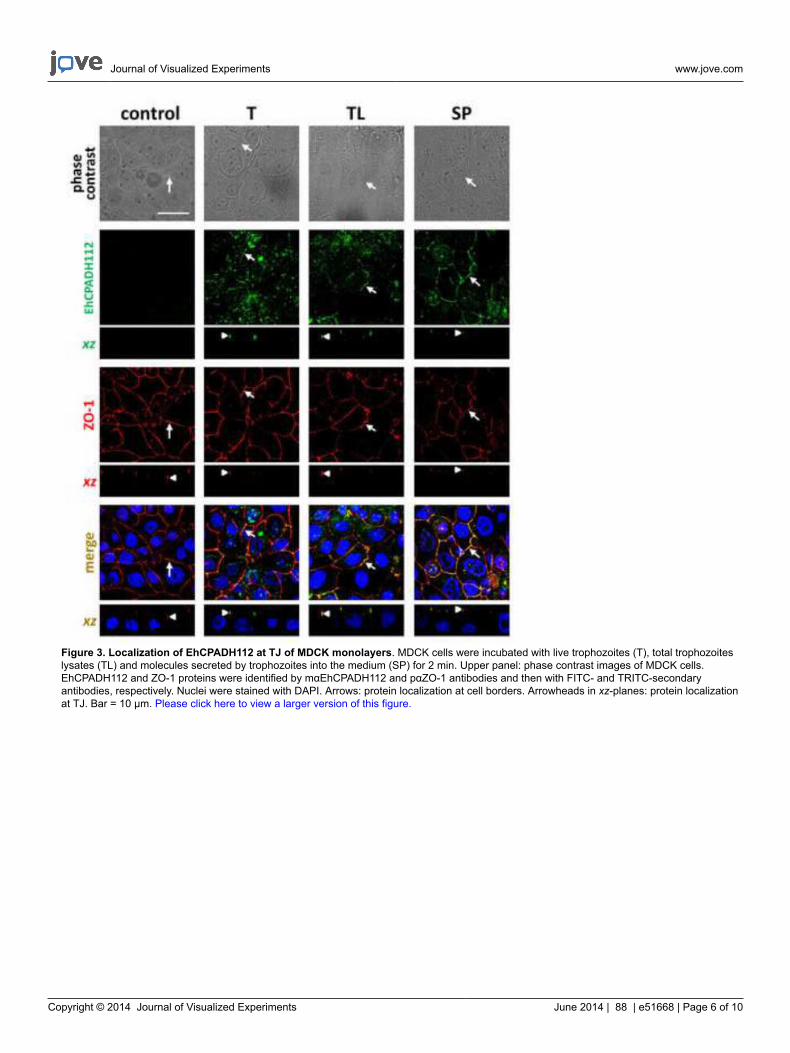
While reaching confluency, epithelial cells form contacts that are stabilized by TJ and AJ. These structures are composed of certain moleculesthat can be visualized by using specific antibodies in immunofluorescence stainings. For example, in Figure 3, cell contacts are visualized byan antibody against the TJ marker ZO-1 and a red fluorescently labeled secondary antibody. As it can be seen, cells gather in close proximityto form a dense monolayer without any hole. In this figure, the amoeba-derived complex EhCPADH112 has been stained using a monoclonalantibody that is detected by a green fluorescently labeled secondary antibody to study the interaction of this complex with the epithelial surface.Three different sources of this complex have been applied to the epithelial monolayer: live trophozoites (T), trophozoite total lysates (TL) andsecreted products (SP). Strikingly, in all cases, co-localization of EhCPADH112 with ZO-1 can be observed (Figure 3, arrows), indicating that thiscomplex could be required to facilitate E. histolytica penetration into the epithelial monolayer.
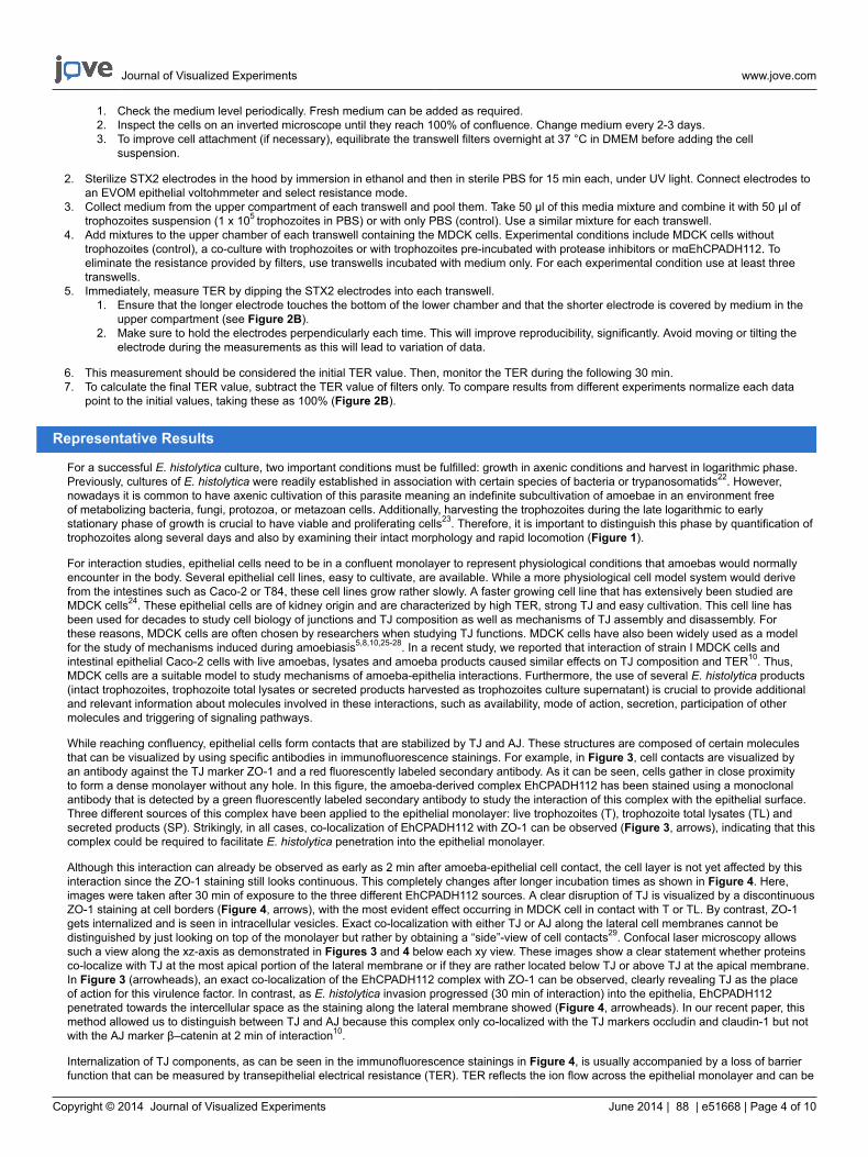
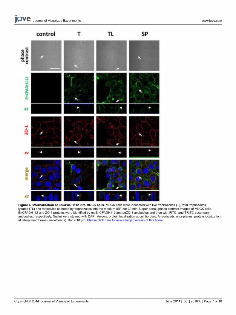
Although this interaction can already be observed as early as 2 min after amoeba-epithelial cell contact, the cell layer is not yet affected by thisinteraction since the ZO-1 staining still looks continuous. This completely changes after longer incubation times as shown in Figure 4. Here,images were taken after 30 min of exposure to the three different EhCPADH112 sources. A clear disruption of TJ is visualized by a discontinuousZO-1 staining at cell borders (Figure 4, arrows), with the most evident effect occurring in MDCK cell in contact with T or TL. By contrast, ZO-1gets internalized and is seen in intracellular vesicles. Exact co-localization with either TJ or AJ along the lateral cell membranes cannot bedistinguished by just looking on top of the monolayer but rather by obtaining a “side”-view of cell contacts29. Confocal laser microscopy allowssuch a view along the xz-axis as demonstrated in Figures 3 and 4 below each xy view. These images show a clear statement whether proteinsco-localize with TJ at the most apical portion of the lateral membrane or if they are rather located below TJ or above TJ at the apical membrane.In Figure 3 (arrowheads), an exact co-localization of the EhCPADH112 complex with ZO-1 can be observed, clearly revealing TJ as the placeof action for this virulence factor. In contrast, as E. histolytica invasion progressed (30 min of interaction) into the epithelia, EhCPADH112penetrated towards the intercellular space as the staining along the lateral membrane showed (Figure 4, arrowheads). In our recent paper, thismethod allowed us to distinguish between TJ and AJ because this complex only co-localized with the TJ markers occludin and claudin-1 but notwith the AJ marker β–catenin at 2 min of interaction10.
Internalization of TJ components, as can be seen in the immunofluorescence stainings in Figure 4, is usually accompanied by a loss of barrierfunction that can be measured by transepithelial electrical resistance (TER). TER reflects the ion flow across the epithelial monolayer and can be
Journal of Visualized Experiments www.jove.com
Copyright © 2014 Journal of Visualized Experiments June 2014 | 88 | e51668 | Page 5 of 10
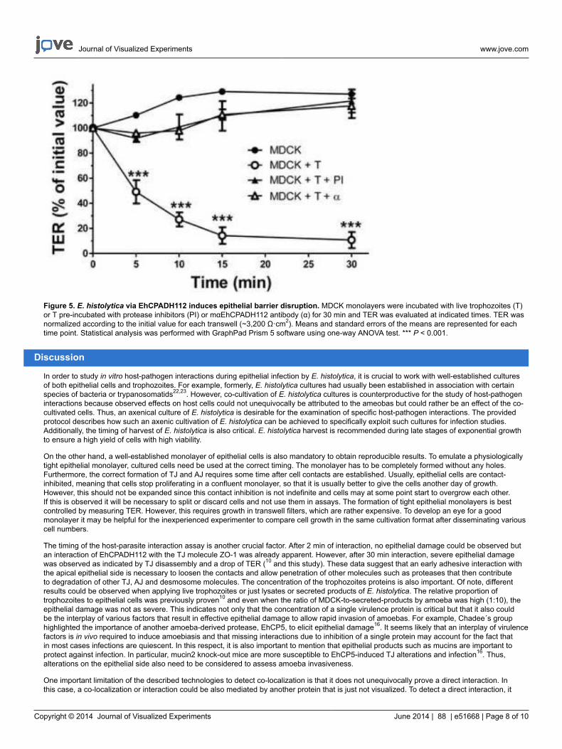
measured using electrodes as shown in Figure 2B. When the monolayer is not yet completely formed or disrupted due to extracellular cues, afree flow of ions across the cell layer is indicated by a low TER. TER was measured of a control confluent monolayer that has not been in contactwith amoeba products or monolayers in contact with trophozoites (Figure 5). Parasite contact disrupted epithelial barrier function indicated by aTER drop of 90% in comparison to the control monolayers. To investigate the mechanisms by which trophozoites affect barrier function, we pre-incubated the trophozoites with an antibody against EhCPADH112 to prevent adhesion of this complex or with protease inhibitors, to block theproteolytic activity of this complex and other proteases important for target cell damage. Both treatments led to an almost complete reversal ofTER drop, suggesting that both proteolytic activity and adhesive properties are important virulence factors of this complex during E. histolyticainvasion.
Figure 1. Typical growth curve of axenically cultivated E. histolytica trophozoites. Inocula of 2 x 10⁵ trophozoites of E. histolytica HMI-IMSS clone A were grown in 6-well dishes with TYI-S-33 medium and after each 24 hr cell numbers were determined using a hematocytomer.Cellular growth was monitored by light microscopy and morphology of growing cells is depicted in the upper panel. The amount of trophozoiteswas monitored for 6 days (d) and values were plotted in the chart below. Data represent the mean and standard error of the mean of threeindependent measurements. Bar = 10 µm. Please click here to view a larger version of this figure.
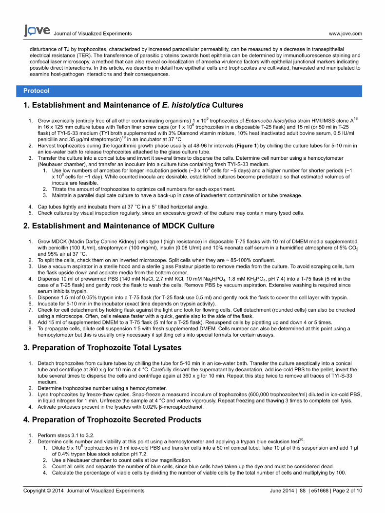
Figure 2. Schematic representation of interactions among MDCK cells and different conditions of E. histolytica. A) Different conditions ofE. histolytica are shown: live trophozoites (T), total trophozoites lysates (TL) and molecules secreted by trophozoites into the medium (SP). Anyexperimental condition was assayed for 2 and 30 min of incubation with confluent MDCK cells. After incubation, MDCK cells were abundantlywashed to remove trophozoites or unbound parasitic molecules. Then samples were processed for immunofluorescence assays, employingmαEhCPADH112 and pαZO-1 antibodies to co-localize the parasitic complex EhCPADH112 and the TJ marker ZO-1, respectively. Later,species-specific secondary antibodies coupled to different fluorochromes were used to detect both proteins by confocal microscopy. B) Additionof trophozoites only or trophozoites pre-incubated with mαEhCPADH112 or protease inhibitors for 20 min at 4 °C in the upper compartment oftranswells containing confluent MDCK cells. TER of epithelial cells was monitored using a STX2 electrode connected to an EVOM voltohmeter,during 30 min. Please click here to view a larger version of this figure.
Journal of Visualized Experiments www.jove.com
Copyright © 2014 Journal of Visualized Experiments June 2014 | 88 | e51668 | Page 6 of 10
Figure 3. Localization of EhCPADH112 at TJ of MDCK monolayers. MDCK cells were incubated with live trophozoites (T), total trophozoiteslysates (TL) and molecules secreted by trophozoites into the medium (SP) for 2 min. Upper panel: phase contrast images of MDCK cells.EhCPADH112 and ZO-1 proteins were identified by mαEhCPADH112 and pαZO-1 antibodies and then with FITC- and TRITC-secondaryantibodies, respectively. Nuclei were stained with DAPI. Arrows: protein localization at cell borders. Arrowheads in xz-planes: protein localizationat TJ. Bar = 10 μm. Please click here to view a larger version of this figure.
Journal of Visualized Experiments www.jove.com
Copyright © 2014 Journal of Visualized Experiments June 2014 | 88 | e51668 | Page 7 of 10
Figure 4. Internalization of EhCPADH112 into MDCK cells. MDCK cells were incubated with live trophozoites (T), total trophozoiteslysates (TL) and molecules secreted by trophozoites into the medium (SP) for 30 min. Upper panel: phase contrast images of MDCK cells.EhCPADH112 and ZO-1 proteins were identified by mαEhCPADH112 and pαZO-1 antibodies and then with FITC- and TRITC-secondaryantibodies, respectively. Nuclei were stained with DAPI. Arrows: protein localization at cell borders. Arrowheads in xz-planes: protein localizationat lateral membrane (arrowheads). Bar = 10 μm. Please click here to view a larger version of this figure.
Journal of Visualized Experiments www.jove.com
Copyright © 2014 Journal of Visualized Experiments June 2014 | 88 | e51668 | Page 8 of 10
Figure 5. E. histolytica via EhCPADH112 induces epithelial barrier disruption. MDCK monolayers were incubated with live trophozoites (T)or T pre-incubated with protease inhibitors (PI) or mαEhCPADH112 antibody (α) for 30 min and TER was evaluated at indicated times. TER wasnormalized according to the initial value for each transwell (~3,200 Ω·cm2). Means and standard errors of the means are represented for eachtime point. Statistical analysis was performed with GraphPad Prism 5 software using one-way ANOVA test. *** P < 0.001.
Discussion
In order to study in vitro host-pathogen interactions during epithelial infection by E. histolytica, it is crucial to work with well-established culturesof both epithelial cells and trophozoites. For example, formerly, E. histolytica cultures had usually been established in association with certainspecies of bacteria or trypanosomatids22,23. However, co-cultivation of E. histolytica cultures is counterproductive for the study of host-pathogeninteractions because observed effects on host cells could not unequivocally be attributed to the ameobas but could rather be an effect of the co-cultivated cells. Thus, an axenical culture of E. histolytica is desirable for the examination of specific host-pathogen interactions. The providedprotocol describes how such an axenic cultivation of E. histolytica can be achieved to specifically exploit such cultures for infection studies.Additionally, the timing of harvest of E. histolytica is also critical. E. histolytica harvest is recommended during late stages of exponential growthto ensure a high yield of cells with high viability.
On the other hand, a well-established monolayer of epithelial cells is also mandatory to obtain reproducible results. To emulate a physiologicallytight epithelial monolayer, cultured cells need be used at the correct timing. The monolayer has to be completely formed without any holes.Furthermore, the correct formation of TJ and AJ requires some time after cell contacts are established. Usually, epithelial cells are contact-inhibited, meaning that cells stop proliferating in a confluent monolayer, so that it is usually better to give the cells another day of growth.However, this should not be expanded since this contact inhibition is not indefinite and cells may at some point start to overgrow each other.If this is observed it will be necessary to split or discard cells and not use them in assays. The formation of tight epithelial monolayers is bestcontrolled by measuring TER. However, this requires growth in transwell filters, which are rather expensive. To develop an eye for a goodmonolayer it may be helpful for the inexperienced experimenter to compare cell growth in the same cultivation format after disseminating variouscell numbers.
The timing of the host-parasite interaction assay is another crucial factor. After 2 min of interaction, no epithelial damage could be observed butan interaction of EhCPADH112 with the TJ molecule ZO-1 was already apparent. However, after 30 min interaction, severe epithelial damagewas observed as indicated by TJ disassembly and a drop of TER (10 and this study). These data suggest that an early adhesive interaction withthe apical epithelial side is necessary to loosen the contacts and allow penetration of other molecules such as proteases that then contributeto degradation of other TJ, AJ and desmosome molecules. The concentration of the trophozoites proteins is also important. Of note, differentresults could be observed when applying live trophozoites or just lysates or secreted products of E. histolytica. The relative proportion oftrophozoites to epithelial cells was previously proven10 and even when the ratio of MDCK-to-secreted-products by amoeba was high (1:10), theepithelial damage was not as severe. This indicates not only that the concentration of a single virulence protein is critical but that it also couldbe the interplay of various factors that result in effective epithelial damage to allow rapid invasion of amoebas. For example, Chadee´s grouphighlighted the importance of another amoeba-derived protease, EhCP5, to elicit epithelial damage16. It seems likely that an interplay of virulencefactors is in vivo required to induce amoebiasis and that missing interactions due to inhibition of a single protein may account for the fact thatin most cases infections are quiescent. In this respect, it is also important to mention that epithelial products such as mucins are important toprotect against infection. In particular, mucin2 knock-out mice are more susceptible to EhCP5-induced TJ alterations and infection16. Thus,alterations on the epithelial side also need to be considered to assess amoeba invasiveness.
One important limitation of the described technologies to detect co-localization is that it does not unequivocally prove a direct interaction. Inthis case, a co-localization or interaction could be also mediated by another protein that is just not visualized. To detect a direct interaction, it
Journal of Visualized Experiments www.jove.com
Copyright © 2014 Journal of Visualized Experiments June 2014 | 88 | e51668 | Page 9 of 10
will be necessary to produce recombinant tagged (such as GST or 10xHis) proteins and perform immunoprecipitation for one tag and Westernblot for the other. Anyways, immunofluorescence stainings have the advantage of revealing the exact cellular location of the protein complexand give the experimenter an idea of the proteins that should be tested in such an in vitro interaction assay. Another drawback is that there areonly few amoeba-specific antibodies available. Thus, if one wants to study certain amoeba proteins in such interaction studies, it will most likelyrequire the generation of an antibody. However, if antibodies are available, the described methods can be applied to study the relevance of otheramoeba proteins (secreted or surface-bound) for host-pathogen interactions.
This paper describes an easy model to detect co-localization and interactions between molecules of the host and parasite. The knowledgegained from these cell-based experiments can be applied in in vivo infection models using hamsters or mice to corroborate the physiologicalrelevance for host invasion by the parasite. These data could then be used for the development of novel treatment strategies.
In summary, we provide detailed protocols for cultivating E. histolytica and epithelial cells for the use in host-pathogen interaction studies, inparticular, assays that permit evaluation of co-localization and TJ disassembly. The timing of the cultures as well as that of each assay is crucialto ensure reproducibility of obtained results. While timing of the cultures can be influenced by varying the number of disseminated cells, thetiming of the experiments themselves depends on the type of assay and the type of proteins investigated.
Disclosures
The authors have nothing to disclose.
Acknowledgements
This work was supported by grants from the Institute of Science and Technology of the Federal District (ICyTDF, 64/2012 to EO) and the MexicanCouncil for Science and Technology (Conacyt, 179895 to MS).
References
1. Faust, D. M., & Guillen, N. Virulence and virulence factors in Entamoeba histolytica, the agent of human amoebiasis. Microbes Infect. 14,1428-1441, doi:S1286-4579(12)00143-8 [pii] 10.1016/j.micinf.2012.05.013 (2012).
2. Stauffer, W., & Ravdin, J. I. Entamoeba histolytica: an update. Curr Opin Infect Dis. 16, 479-485, doi:10.1097/01.qco.0000092821.42392.ca(2003).
3. Santi-Rocca, J., Rigothier, M. C., & Guillen, N. Host-microbe interactions and defense mechanisms in the development of amoebic liverabscesses. Clin Microbiol Rev. 22, 65-75, Table of Contents, doi:22/1/65 [pii] 10.1128/CMR.00029-08 (2009).
4. Turner, J. R. Intestinal mucosal barrier function in health and disease. Nat Rev Immunol. 9, 799-809, doi:nri2653 [pii] 10.1038/nri2653 (2009).5. Garcia-Rivera, G. et al. Entamoeba histolytica : a novel cysteine protease and an adhesin form the 112 kDa surface protein. Mol Microbiol.
33, 556-568, doi:mole1500 [pii] (1999).6. Leippe, M. Amoebapores. Parasitol Today. 13, 178-183, doi:S0169-4758(97)01038-7 [pii] (1997).7. Leippe, M., Bruhn, H., Hecht, O., & Grotzinger, J. Ancient weapons: the three-dimensional structure of amoebapore A. Trends Parasitol. 21,
5-7, doi:S1471-4922(04)00281-8 [pii] 10.1016/j.pt.2004.10.009 (2005).8. Ocadiz, R. et al. EhCP112 is an Entamoeba histolytica secreted cysteine protease that may be involved in the parasite-virulence. Cell
Microbiol. 7, 221-232, doi:CMI453 [pii] 10.1111/j.1462-5822.2004.00453.x (2005).9. Sajid, M., & McKerrow, J. H. Cysteine proteases of parasitic organisms. Mol Biochem Parasitol. 120, 1-21, doi:S0166685101004388 [pii]
(2002).10. Betanzos, A. et al. The EhCPADH112 complex of Entamoeba histolytica interacts with tight junction proteins occludin and claudin-1 to
produce epithelial damage. PLoS One. 8, e65100, doi:10.1371/journal.pone.0065100 PONE-D-12-35362 [pii] (2013).11. Lauwaet, T. et al. Proteinase inhibitors TPCK and TLCK prevent Entamoeba histolytica induced disturbance of tight junctions and microvilli in
enteric cell layers in vitro. Int J Parasitol. 34, 785-794, doi:10.1016/j.ijpara.2004.03.007 S0020751904000748 [pii] (2004).12. Leroy, A. et al. Contact-dependent transfer of the galactose-specific lectin of Entamoeba histolytica to the lateral surface of enterocytes in
culture. Infect Immun. 63, 4253-4260 (1995).13. Leroy, A., Lauwaet, T., De Bruyne, G., Cornelissen, M., & Mareel, M. Entamoeba histolytica disturbs the tight junction complex in human
enteric T84 cell layers. FASEB J. 14, 1139-1146 (2000).14. Leroy, A. et al. Disturbance of tight junctions by Entamoeba histolytica: resistant vertebrate cell types and incompetent trophozoites. Arch
Med Res. 31, S218-220, doi:S0188440900001454 [pii] (2000).15. Lauwaet, T. et al. Entamoeba histolytica trophozoites transfer lipophosphopeptidoglycans to enteric cell layers. Int J Parasitol. 34, 549-556,
doi:10.1016/j.ijpara.2003.11.013 S0020751903003345 [pii] (2004).16. Kissoon-Singh, V., Moreau, F., Trusevych, E., & Chadee, K. Entamoeba histolytica Exacerbates Epithelial Tight Junction Permeability and
Proinflammatory Responses in Muc2(-/-) Mice. Am J Pathol. 182, 852-865, doi:S0002-9440(13)00003-5 [pii] 10.1016/j.ajpath.2012.11.035(2013).
17. Lejeune, M., Moreau, F., & Chadee, K. Prostaglandin E2 produced by Entamoeba histolytica signals via EP4 receptor and alters claudin-4 toincrease ion permeability of tight junctions. Am J Pathol. 179, 807-818, doi:S0002-9440(11)00408-1 [pii] 10.1016/j.ajpath.2011.05.001 (2011).
18. Orozco, M. E., Martinez Palomo, A., Gonzalez Robles, A., Guarneros, G., & Galindo, J. M. [Interactions between lectin and receptor mediatethe adhesion of E. histolytica to epithelial cells. Relation of adhesion to the virulence of the strains]. Arch Invest Med (Mex). 13 Suppl 3,159-167 (1982).
19. Diamond, L. S., Harlow, D. R., & Cunnick, C. C. A new medium for the axenic cultivation of Entamoeba histolytica and other Entamoeba.Trans R Soc Trop Med Hyg. 72, 431-432 (1978).
20. Strober, W. Trypan blue exclusion test of cell viability. Curr Protoc Immunol. Appendix 3, Appendix 3B, doi:10.1002/0471142735.ima03bs21(2001).
Journal of Visualized Experiments www.jove.com
Copyright © 2014 Journal of Visualized Experiments June 2014 | 88 | e51668 | Page 10 of 10
21. Arroyo, R., & Orozco, E. Localization and identification of an Entamoeba histolytica adhesin. Mol Biochem Parasitol. 23, 151-158,doi:0166-6851(87)90150-2 [pii] (1987).
22. Diamond, L. S. Axenic cultivation of Entamoeba hitolytica. Science. 134, 336-337 (1961).23. Diamond, L. S. Techniques of axenic cultivation of Entamoeba histolytica Schaudinn, 1903 and E. histolytica-like amebae. J Parasitol. 54,
1047-1056 (1968).24. Dukes, J. D., Whitley, P., & Chalmers, A. D. The MDCK variety pack: choosing the right strain. BMC Cell Biol. 12, 43, doi:1471-2121-12-43
[pii] 10.1186/1471-2121-12-43 (2011).25. Espinosa-Cantellano, M., & Martinez-Palomo, A. Pathogenesis of intestinal amebiasis: from molecules to disease. Clin Microbiol Rev. 13,
318-331 (2000).26. Martinez-Palomo, A. et al. Structural bases of the cytolytic mechanisms of Entamoeba histolytica. J Protozool. 32, 166-175 (1985).27. Martinez-Lopez, C. et al. The EhADH112 recombinant polypeptide inhibits cell destruction and liver abscess formation by Entamoeba
histolytica trophozoites. Cell Microbiol. 6, 367-376, doi:363 [pii] (2004).28. Ocadiz-Ruiz, R. et al. Effect of the silencing of the Ehcp112 gene on the in vitro virulence of Entamoeba histolytica. Parasit Vectors. 6, 248,
doi:1756-3305-6-248 [pii] 10.1186/1756-3305-6-248 (2013).29. Johnson, L. G. Applications of imaging techniques to studies of epithelial tight junctions. Adv Drug Deliv Rev. 57, 111-121,
doi:S0169-409X(04)00188-7 [pii] 10.1016/j.addr.2004.08.004 (2005).
Related Documents