Analysis of the Conformation and Function of the Plasmodium falciparum Merozoite Proteins MTRAP and PTRAMP Onyinyechukwu Uchime, a Raul Herrera, a Karine Reiter, a Svetlana Kotova, b Richard L. Shimp, Jr., a Kazutoyo Miura, c Dominique Jones, a Jacob Lebowitz, b Xavier Ambroggio, d Darrell E. Hurt, d Albert J. Jin, b Carole Long, c Louis H. Miller, c and David L. Narum a Laboratory of Malaria Immunology and Vaccinology, National Institute of Allergy and Infectious Diseases, National Institutes of Health, Rockville, Maryland, USA a ; Laboratory of Cellular Imaging and Macromolecular Biophysics, National Institute of Biomedical Imaging and Bioengineering, National Institutes of Health, Bethesda, Maryland, USA b ; Laboratory of Malaria and Vector Research, National Institutes of Health, Rockville, Maryland, USA c ; and Bioinformatics and Computational Biosciences Branch, Office of Cyber Infrastructure and Computational Biology, National Institute of Allergy and Infectious Diseases, National Institutes of Health, Bethesda, Maryland, USA d Thrombospondin repeat (TSR)-like domains are structures involved with cell adhesion. Plasmodium falciparum proteins con- taining TSR domains play crucial roles in parasite development. In particular, the preerythrocytic P. falciparum circumsporozo- ite protein is involved in hepatocyte invasion. The importance of these domains in two other malaria proteins, the merozoite- specific thrombospondin-related anonymous protein (MTRAP) and the thrombospondin-related apical membrane protein (PTRAMP), were assessed using near-full-length recombinant proteins composed of the extracellular domains produced in Esch- erichia coli. MTRAP is thought to be released from invasive organelles identified as micronemes during merozoite invasion to mediate motility and host cell invasion through an interaction with aldolase, an actin binding protein involved in the moving junction. PTRAMP function remains unknown. In this study, the conformation of recombinant MTRAP (rMTRAP) appeared to be a highly extended protein (2 nm by 33 nm, width by length, respectively), whereas rPTRAMP had a less extended structure. Using an erythrocyte binding assay, rMTRAP but not rPTRAMP bound human erythrocytes; rMTRAP binding was mediated through the TSR domain. MTRAP- and in general PTRAMP-specific antibodies failed to inhibit P. falciparum development in vitro. Altogether, MTRAP is a highly extended bifunctional protein that binds to an erythrocyte receptor and the merozoite motor. M alaria caused by Plasmodium falciparum remains a serious global disease resulting in nearly 800,000 deaths annually, principally in children in sub-Saharan Africa (43). Various con- trol efforts, including insecticide-impregnated bed nets and artemisinin combination therapy, are currently impacting the morbidity and mortality of the disease; however, a long-term, cost-effective strategy requires an effective malaria vaccine (5, 43). Recently, a major step toward this end was reported for an inves- tigative malaria vaccine identified as RTS,S, which is comprised of the carboxyl-terminal half of the circumsporozoite protein (CSP) fused with the hepatitis B surface protein and biophysically pre- sented as a virus-like particle formulated with the adjuvant ASO1 (3). RTS,S in a phase 3 trial reduced the number of clinical epi- sodes by 50.4% compared to the number in a control group for 14 months following the final dose (30). A unique protein motif within P. falciparum CSP (PfCSP) and RTS,S is the presence of a thrombospondin repeat (TSR)-like domain within the carboxyl terminus, which may be involved in cellular adhesion (8–10, 34), similar to other members within the TSR superfamily (42). Anal- ysis of the P. falciparum genome using conserved TSR amino acid motifs identified additional malaria parasite proteins which are also a part of this superfamily (2, 24, 39). Two such members, identified in blood-stage P. falciparum parasites, are the merozoite-specific thrombospondin-related anonymous protein (MTRAP) and the thrombospondin-related apical membrane protein (PTRAMP). MTRAP is named after the sporozoite thrombospondin-re- lated anonymous protein (TRAP) (2). TRAP and MTRAP are both type 1 membrane proteins with a cytoplasmic domain. TRAP has an essential biological role in sporozoite gliding motility by linking the actin-myosin motor through its cytoplasmic domain while binding to its receptor on the hepatocyte surface through its extracellular domain, which contains a TSR domain and a von Willebrand factor-like A domain (17, 21, 24, 37). TRAP is redis- tributed from micronemes to the sporozoite surface and subse- quently cleaved near the membrane by a rhomboid protease (1, 29). The cytoplasmic domain of TRAP interacts with actin-myo- sin via aldolase as a bridge (7, 16). Gene disruption of TRAP, as well as mutation analysis of specific amino acid residues within the cytoplasmic domain, impairs sporozoite motility (17, 37). In comparison, no adhesive function for the ectodomain of MTRAP has been identified, although the cytoplasmic domain is reported to interact with aldolase and processing by a rhomboid protease near the membrane surface appears possible (1, 2). Interestingly, MTRAP-specific antibodies have failed to inhibit parasite inva- sion of erythrocytes in vitro, despite the fact that it has not been possible to knock out this gene by standard genetic techniques, which suggests an essential role for MTRAP in asexual blood-stage development (2, 24). PTRAMP, another type 1 merozoite protein, is initially local- Received 6 February 2012 Accepted 21 March 2012 Published ahead of print 30 March 2012 Address correspondence to David L. Narum, [email protected]. O.U. and R.H. contributed equally to this article. Supplemental material for this article may be found at http://ec.asm.org/. Copyright © 2012, American Society for Microbiology. All Rights Reserved. doi:10.1128/EC.00039-12 1535-9778/12/$12.00 Eukaryotic Cell p. 615– 625 ec.asm.org 615 on May 5, 2020 by guest http://ec.asm.org/ Downloaded from

Welcome message from author
This document is posted to help you gain knowledge. Please leave a comment to let me know what you think about it! Share it to your friends and learn new things together.
Transcript
Analysis of the Conformation and Function of the Plasmodiumfalciparum Merozoite Proteins MTRAP and PTRAMP
Onyinyechukwu Uchime,a Raul Herrera,a Karine Reiter,a Svetlana Kotova,b Richard L. Shimp, Jr.,a Kazutoyo Miura,c Dominique Jones,a
Jacob Lebowitz,b Xavier Ambroggio,d Darrell E. Hurt,d Albert J. Jin,b Carole Long,c Louis H. Miller,c and David L. Naruma
Laboratory of Malaria Immunology and Vaccinology, National Institute of Allergy and Infectious Diseases, National Institutes of Health, Rockville, Maryland, USAa;Laboratory of Cellular Imaging and Macromolecular Biophysics, National Institute of Biomedical Imaging and Bioengineering, National Institutes of Health, Bethesda,Maryland, USAb; Laboratory of Malaria and Vector Research, National Institutes of Health, Rockville, Maryland, USAc; and Bioinformatics and Computational BiosciencesBranch, Office of Cyber Infrastructure and Computational Biology, National Institute of Allergy and Infectious Diseases, National Institutes of Health, Bethesda, Maryland,USAd
Thrombospondin repeat (TSR)-like domains are structures involved with cell adhesion. Plasmodium falciparum proteins con-taining TSR domains play crucial roles in parasite development. In particular, the preerythrocytic P. falciparum circumsporozo-ite protein is involved in hepatocyte invasion. The importance of these domains in two other malaria proteins, the merozoite-specific thrombospondin-related anonymous protein (MTRAP) and the thrombospondin-related apical membrane protein(PTRAMP), were assessed using near-full-length recombinant proteins composed of the extracellular domains produced in Esch-erichia coli. MTRAP is thought to be released from invasive organelles identified as micronemes during merozoite invasion tomediate motility and host cell invasion through an interaction with aldolase, an actin binding protein involved in the movingjunction. PTRAMP function remains unknown. In this study, the conformation of recombinant MTRAP (rMTRAP) appeared tobe a highly extended protein (2 nm by 33 nm, width by length, respectively), whereas rPTRAMP had a less extended structure.Using an erythrocyte binding assay, rMTRAP but not rPTRAMP bound human erythrocytes; rMTRAP binding was mediatedthrough the TSR domain. MTRAP- and in general PTRAMP-specific antibodies failed to inhibit P. falciparum development invitro. Altogether, MTRAP is a highly extended bifunctional protein that binds to an erythrocyte receptor and the merozoitemotor.
Malaria caused by Plasmodium falciparum remains a seriousglobal disease resulting in nearly 800,000 deaths annually,
principally in children in sub-Saharan Africa (43). Various con-trol efforts, including insecticide-impregnated bed nets andartemisinin combination therapy, are currently impacting themorbidity and mortality of the disease; however, a long-term,cost-effective strategy requires an effective malaria vaccine (5, 43).Recently, a major step toward this end was reported for an inves-tigative malaria vaccine identified as RTS,S, which is comprised ofthe carboxyl-terminal half of the circumsporozoite protein (CSP)fused with the hepatitis B surface protein and biophysically pre-sented as a virus-like particle formulated with the adjuvant ASO1(3). RTS,S in a phase 3 trial reduced the number of clinical epi-sodes by 50.4% compared to the number in a control group for 14months following the final dose (30). A unique protein motifwithin P. falciparum CSP (PfCSP) and RTS,S is the presence of athrombospondin repeat (TSR)-like domain within the carboxylterminus, which may be involved in cellular adhesion (8–10, 34),similar to other members within the TSR superfamily (42). Anal-ysis of the P. falciparum genome using conserved TSR aminoacid motifs identified additional malaria parasite proteinswhich are also a part of this superfamily (2, 24, 39). Two suchmembers, identified in blood-stage P. falciparum parasites, arethe merozoite-specific thrombospondin-related anonymousprotein (MTRAP) and the thrombospondin-related apicalmembrane protein (PTRAMP).
MTRAP is named after the sporozoite thrombospondin-re-lated anonymous protein (TRAP) (2). TRAP and MTRAP areboth type 1 membrane proteins with a cytoplasmic domain. TRAPhas an essential biological role in sporozoite gliding motility by
linking the actin-myosin motor through its cytoplasmic domainwhile binding to its receptor on the hepatocyte surface through itsextracellular domain, which contains a TSR domain and a vonWillebrand factor-like A domain (17, 21, 24, 37). TRAP is redis-tributed from micronemes to the sporozoite surface and subse-quently cleaved near the membrane by a rhomboid protease (1,29). The cytoplasmic domain of TRAP interacts with actin-myo-sin via aldolase as a bridge (7, 16). Gene disruption of TRAP, aswell as mutation analysis of specific amino acid residues within thecytoplasmic domain, impairs sporozoite motility (17, 37). Incomparison, no adhesive function for the ectodomain of MTRAPhas been identified, although the cytoplasmic domain is reportedto interact with aldolase and processing by a rhomboid proteasenear the membrane surface appears possible (1, 2). Interestingly,MTRAP-specific antibodies have failed to inhibit parasite inva-sion of erythrocytes in vitro, despite the fact that it has not beenpossible to knock out this gene by standard genetic techniques,which suggests an essential role for MTRAP in asexual blood-stagedevelopment (2, 24).
PTRAMP, another type 1 merozoite protein, is initially local-
Received 6 February 2012 Accepted 21 March 2012
Published ahead of print 30 March 2012
Address correspondence to David L. Narum, [email protected].
O.U. and R.H. contributed equally to this article.
Supplemental material for this article may be found at http://ec.asm.org/.
Copyright © 2012, American Society for Microbiology. All Rights Reserved.
doi:10.1128/EC.00039-12
1535-9778/12/$12.00 Eukaryotic Cell p. 615–625 ec.asm.org 615
on May 5, 2020 by guest
http://ec.asm.org/
Dow
nloaded from
ized within micronemes and then may be redistributed onto themerozoite surface (39). PTRAMP contains a TSR domain withinits ectodomain and has a cytoplasmic domain which has beenshown to weakly interact with aldolase (2). PTRAMP is a con-served protein within the Plasmodium genus and appears to havean essential and conserved unknown biological function consid-ering that the gene that encodes it cannot be disrupted (24, 39).
In order to better understand the functions of MTRAP andPTRAMP and investigate their roles during merozoite invasion oferythrocytes, including susceptibility to antibody blockade, wegenerated full-length recombinant MTRAP (rMTRAP) andPTRAMP (rPTRAMP) of the extracellular sequence, using Esche-richia coli expression and protein refolding. Both rMTRAP andrPTRAMP were extensively biochemically and biophysically char-acterized and used to produce antigen-specific antisera. The re-sults collated have enabled the identification that MTRAP is ahighly extended protein that binds human erythrocytes mediatedby the TSR domain. Neither antigen appears to be a robust targetfor antibody-mediated blockade of erythrocyte invasion.
MATERIALS AND METHODSExpression and production of recombinant proteins rMTRAP andrPTRAMP. The amino acid sequence of rPTRAMP (PFL0870w; GenBankaccession no. XP_001350582) was used to generate a codon-optimizedsynthetic gene for expression in E. coli (GeneArt, Regensburg, Germany).The construct, corresponding to amino acids (aa) 25 to 309 of the full-length gene, was subcloned into the E. coli pET-24a� expression vectordownstream of the T7 promoter using the NdeI and XhoI restriction sites(EMD Chemicals, Inc., Gibbstown, NJ). Similarly, the amino acid se-quence of rMTRAP (PF10_0281; GenBank protein accession no.XP_001347565.1) was used to generate a codon-optimized synthetic genefor expression in E. coli (GeneArt, Regensburg, Germany). The construct,corresponding to aa 23 to 433 of the full-length gene, was subcloned intothe E. coli pET-24a� expression vector using the NdeI and XhoI restric-tion sites (EMD Chemicals, Inc., Gibbstown, NJ). The resulting tran-scribed genes incorporate the additional amino acid sequence LEHHHHHH. Both constructs were transformed into E. coli BL21(DE3) cells(Novagen, San Diego, CA) and used for the expression of rPTRAMP andrMTRAP. Expression of both rPTRAMP and rMTRAP was performed ina 1-liter culture shake flask, using Luria-Bertani medium (KD Medical,Columbia, MD) containing 50 �g/ml kanamycin at 37°C. Once the opti-cal density at 550 nm reached 0.6 to 0.8, the culture was induced with 1mM isopropyl-1-thio-�-D-1-galactopyranoside (IPTG) for 3 h. Followinginduction, cells were harvested by centrifugation, and the cell pellet wasstored at �80°C.
To break the bacterial cells, the cell pellet was resuspended in 10 vol-umes of lysis buffer (10 mM Tris-HCl, 10 mM EDTA, 10 mM NaCl, 5 mMdithiothreitol [DTT] [pH 8.0]) and then mechanically disrupted by pas-sage through a Microfluidizer (M110Y; Microfluidics Corp., Newton,MA) five times operating at 19,000 lb/in2. The rPTRAMP and rMTRAPlysates were centrifuged for 45 min at 10,000 � g to separate the solublefraction from the inclusion bodies and cellular debris. rPTRAMP wassubsequently captured from the inclusion body fraction, whereasrMTRAP was captured from the soluble fraction as described below.
Capture, refolding, and purification. rMTRAP was captured fromthe soluble fraction under denatured and reduced conditions using anickel Sepharose 6 Fast Flow column (GE Healthcare, Piscataway, NJ).Load material was first dialyzed in a mixture containing 10 mM Tris, 100mM sodium phosphate, and 14.1 mM �-mercaptoethanol (pH 8.0). Priorto column loading, urea and imidazole were added so that final concen-trations of 8 M and 20 mM, respectively, were achieved. A preequilibratedaffinity column using a similar buffered solution was loaded, and thebound protein was eluted by an imidazole gradient (20 to 400 mM) in 20column volumes. Fractions containing rMTRAP were pooled and re-
folded by rapid dilution at 50 �g/ml protein into a refold buffer contain-ing a 10 mM glutathione oxidized (GSSG) and 2 mM glutathione (GSH)redox system overnight at 10°C. The refolded protein was dialyzed in 50mM Tris-HCl (pH 7.4), loaded onto a preequilibrated Q Sepharose FastFlow ion-exchange chromatography column (GE Healthcare), and elutedwith an NaCl gradient (0 to 1 M NaCl) in 20 column volumes. The re-folded rMTRAP was polished using size exclusion chromatography in aHiLoad 16/60 Superdex 200 prep-grade column (GE Healthcare) equili-brated with phosphate-buffered saline (PBS) at pH 7.4. The final refoldedand purified protein was filtered with a 0.22-�m-pore filter and stored atless than �70°C. Similar conditions were used for producing rPTRAMP,with two exceptions. First, the GSH and GSSG concentrations in the re-folding buffer were 5 mM and 1 mM, respectively. Second, the final pol-ishing step required a HiLoad 16/60 Superdex 75 prep-grade column(GE Healthcare). Endotoxin levels of the various antigens were measuredusing Limulus amoebocyte lysate in a 96-well plate with chromogenicreagents and PyroSoft software (Associates of Cape Cod, Inc., East Fal-mouth, MA).
Amino-terminal sequencing. Automated Edman sequencing wasperformed using a Sequenator model Procise 494 HT connected to anonline UV detector (Applied Biosystems). Samples were immobilizedonto polyvinylidene difluoride (PVDF) using a ProSorb sample prepara-tive cartridge. A 2- by 220-mm C18 microbore column (Applied Biosys-tems) was used for separation of the phenylthiohydantoin (PTH) aminoacids using an acetonitrile–2-propanol gradient elution.
Reversed-phase HPLC. For reversed-phase high-performance liquidchromatography (HPLC), samples were analyzed on a 2-mm by 250-mm,5-�m particle size, C4 column (Phenomenex, Torrance, CA). The columnwas equilibrated in 95% mobile phase A (0.1% [wt/vol] trifluoroaceticacid [TFA] in water) and 5% mobile phase B (0.1% [wt/vol] TFA inacetonitrile), and elution was performed by increasing the percentage ofmobile phase B at a constant flow rate (0.2 ml/min). Elution peaks werecollected and vacuum dried in a Vacufuge concentrator (Eppendorf,Westbury, NY). Theses samples were submitted to the Research Technol-ogy Branch (NIAID/NIH, Rockville, MD) for mass spectrometry (MS)and amino-terminal sequencing.
CD spectroscopy. Both rMTRAP and rPTRAMP were analyzed aspreviously described (26). In brief, circular dichroism (CD) spectra of theproteins were recorded on a Jasco J-815 CD spectropolarimeter over thewavelength range of 185 to 260 nm. Data were collected using a data pitchof 0.2 nm, a slit bandwidth of 1.0 nm, and a signal averaging time of 1.0 sin a 1-mm quartz cuvette. Secondary structure content was calculatedusing the DICHROWEB web server (http://dichroweb.cryst.bbk.ac.uk)and CDPro software.
Analytical SEC-MALS-QELS HPLC. To evaluate the identity, purity,and solution aggregation state of purified rMTRAP and rPTRAMP, ana-lytical size exclusion chromatography with in-line multiangle light scat-tering (SEC-MALS) and quasielastic light scatter (QELS) detection wasperformed. Chromatography was performed on an Alliance HPLC system(Waters, Milford, MA) connected in series to a multiangle DAWN EOSlight-scattering detector and a QELS detector (Wyatt Technology, SantaBarbara CA). Samples were prepared and analyzed as described by Tsai etal. (40). In brief, protein separation of rPTRAMP was performed on aTSKgel G2000SWxl column, whereas separation of rMTRAP was accom-plished on a TSKgel G3000SWxl column (Tosoh Bioscience, Montgom-eryville, PA). The column was equilibrated in the mobile phase consistingof 1.04 mM KH2PO4, 2.97 mM Na2P04 · 7H2O, 308 mM NaCl, and 0.02%azide (pH 7.4), and the samples were run using an isocratic elution at 0.5ml/min. A gel filtration standard (Bio-Rad, Hercules, CA) was run for sizecomparison as well as a 25-�l injection of bovine serum albumin (BSA) at5 mg/ml for configuration of the MALS data.
MS. Intact mass analyses were performed by electrospray ionizationmass spectrometry (ESI-MS). Samples were analyzed by coupling an au-tomated chip-based nanoelectrospray unit, TriVersa Nanomate (AdvionBioSciences), to a 4000 Q Trap liquid chromatography-tandem MS
Uchime et al.
616 ec.asm.org Eukaryotic Cell
on May 5, 2020 by guest
http://ec.asm.org/
Dow
nloaded from
(LC-MS/MS) device (Applied Biosystems/Sciex). Sample solutions werefirst adjusted to low pH (0.1% acetic acid) and low acetonitrile (2%) andthen desalted through a microprotein trap cartridge (Michrom BioResources) at 20 ml/min. In LC-MS mode, a Tempo multidimensionalliquid chromatography (MDLC) device designed for HPLC applicationswas used to automate trap desalting of protein samples. Bound proteinwas step eluted with 60% acetonitrile and 0.1% acetic acid at 800 nl/mininto the 4000 Q Trap via a TriVersa capillary coupler.
Polyclonal antibody production. Rat polyclonal sera were generatedat the National Institutes of Health in compliance with guidelines of theNational Institutes of Health Institutional Animal Care and Use Commit-tee. In brief, two groups of six rats were immunized with 10 �g ofrMTRAP or rPTRAMP formulated in Montanide ISA 720 (Seppic, Inc.,Fairfield, NJ) on days 0, 28, and 42 and exsanguinated on day 56. Controlrats were immunized with the adjuvant alone. Rabbit polyclonal sera weregenerated against rMTRAP and rPTRAMP by Spring Valley Laboratories.In brief, 10 healthy female New Zealand White rabbits (Orytolagus cunic-ulus) were randomly selected into 3 groups and housed individually.Group 1 contained 6 rabbits which were immunized with 20 �g ofrPTRAMP formulated in Montanide ISA 720 on day 0 and day 28. Therabbits were prebled on day 0 and then exsanguinated on day 42. Group 2contained 2 rabbits which were immunized with 20 �g of rMTRAP in thesame fashion as rPTRAMP. Group 3 contained 2 rabbits which were im-munized with adjuvant alone in the same fashion as rPTRAMP.
IFA and confocal microscopy. Immunofluorescence assay (IFA)analysis was performed as per the standard protocol (26, 33). In brief,schizont-enriched parasites were smeared on slides and stored at �80°C.Slides were thawed, acetone fixed, and reacted with primary and second-ary antibodies diluted in blocking buffer (1� PBS, 3% BSA) at 4°C (25).All primary and secondary antibody incubations were carried out at roomtemperature and separated by extensive washing with PBS containing0.05% Tween 20 (Bio-Rad). The samples were mounted under coverslipsusing Vectashield hard-set mounting medium and stored at 4°C untilimages were acquired by confocal microscopy as described previously(33). In brief, a Leica SP2 confocal microscope using Leica confocal soft-ware was used for image acquisition. All images were collected by using aPL APO �100/1.4 oil immersion objective and a confocal zoom of �6.Secondary reagents used Alexa 488 or Alexa 568 species-specific conju-gated antibodies (Molecular Probes). All images were collected as three-dimensional (3D) data sets (z-stacks) with a step size of 0.1221 nm be-tween the successive optical sections. Deconvolution of all image stackswas performed using Huygens Essential (Scientific Volume Imaging) toimprove the maximum resolution of the data, as well as to minimizebackground noise. Deconvolved images were saved and analyzed throughImaris image analysis software (version 5.7.2; Bitplane). Confocal imagesin this study are displayed as maximum projection of the 3D image stacks.
GIA assay. The standard methodology for the growth inhibition ac-tivity (GIA) assay has been described previously (19). The assay was per-formed with purified IgGs at the indicated concentrations against the 3D7strain of P. falciparum parasites.
Erythrocyte binding assay. The erythrocyte binding assay was per-formed as previously described (20), with the following modifications.One hundred microliters of human erythrocytes with a 50% hematocritwas washed three times in incomplete RPMI 1640 medium (Invitrogen)and resuspended in 500 �l of RPMI containing 200 �M rMTRAP,rPTRAMP, PpPfs25 (41), EcMSP142 (32), or BAEBL RII (D. L. Narum,unpublished data) and 10% fetal bovine serum (Invitrogen). The eryth-rocyte suspension was incubated at 37°C for 1 h with constant shaking andthen layered over 300 �l of oil (dibutyl phthalate; Sigma) and centrifugedat 13,000 � g for 30 s. Bound protein was eluted from the erythrocytepellet by the addition of 100 �l of RPMI with 1 M NaCl. Erythrocyteeluates were analyzed by Western blotting using antigen-specific rabbitantisera produced against the same recombinant antigen and thendetected with alkaline phosphatase-conjugated goat anti-rabbit IgG(Kirkegaard & Perry Laboratories, MD).
Analytical ultracentrifugation characterization of rMTRAP. Boundarysedimentation velocity measurements were made in an Optima XLA an-alytical ultracentrifuge (Beckman-Coulter Instruments). Sedimentationvelocity analysis was performed at 20°C at a rotor speed of 58,000 rpm.The centrifuge cell was filled with 400 �l of protein solution at a concen-tration of 1.0 mg/ml of rMTRAP. Absorbance scans were obtained at 235nm. Sedimentation coefficient distribution analysis to deconvolute theboundary velocity data into sedimenting species was performed as previ-ously described (31), using the public domain software Sedfit developedby Peter Schuck (http://www.analyticalultracentrifugation.com/). The re-sult of this computational analysis is a presentation of the distribution ofsedimenting species in the form of a c(s) versus s plot. In this computa-tional treatment, the sedimentation boundary velocity data were sub-jected to maximum entropy regularization statistical analysis for the mostparsimonious distribution of sedimenting species (31) that best fits thedata. A weight average shape factor (f/fo frictional ratio) is extracted fromthe boundary spreading, and this allows for the conversion of the c(s)versus s distribution to a c(M) versus M graphical presentation, therebyproviding the molar mass of each sedimenting peak. The software pro-gram Sednterp developed by Hayes, Laue, and Philo (http://www.bbri.org/RASMB/rasmb.html) was also used to determine the partial specific vol-ume of the rMTRAP and the viscosity (�) and density (�) for the PBSsolution used in the sedimentation analyses and for the hydrodynamicanalysis to be reported below.
AFM. Samples for atomic force microscopy (AFM) were prepared andanalyzed basically as described by Plassmeyer et al. (26) and Tsai et al.(40), with modifications as noted. Briefly, biological AFM imaging of theprotein products was carried out under a range of conditions, in both fluidand air, using gentle-tapping-mode AFM, mostly with a PicoForce mul-timode atomic force microscope (Veeco, CA) consisting of a Nanoscope Vcontroller, a type E scanner head, and a sharpened TESP-SS (Veeco, CA)or similar AFM cantilever (26, 40). For rMTRAP visualization, suitableprotein attachment was readily achieved by a 1-min incubation of 7 �l of�1 �g/ml solution of rMTRAP in PBS buffer (pH 7.4) on freshly peeledmica substrates, followed by rinsing with �1 ml of deionized water andcomplete drying under an inert gas flow. The sample was then sealed intothe instrument compartment dehumidified by Drierite. AFM images wereevaluated within the Nanoscope software (version 7.2; Veeco, CA) andexported to Image J version 1.41o (National Institutes of Health, Be-thesda, MD) and Mathcad version 14 (Mathsoft, Needham, MA) for fur-ther analyses and display.
Computational modeling. The homology model of the MTRAP TSRdomains was constructed using HHPRED (35) and Rosetta (18). Residues23 to 105 of MTRAP were submitted to the HHPRED server to query theProtein Data Bank (PDB) (6). The crystal structure of human thrombos-pondin-1 (PDB identification [ID] 1LSL) (38) was identified as the besttemplate (probability, 98.3; E value, 3E�07) and was used for subsequentmodeling. For the modeling procedure, the Homo sapiens TSP-1(HsTSP-1) TSR2 domain spanning residues 434 to 490 was used as thetemplate for both TSR domains of MTRAP (residues 28 to 61 and 62 to94). Three blocks of residues were restrained to the coordinates ofHsTSP-1 residues 434 to 490, with the intervening loop regions modeledusing the Rosetta loop relax protocol (27). Specifically, residues 32 to 38and 66 to 72, 41 to 48 and 75 to 82, and 55 to 61 and 88 to 94 of MTRAPwere restrained to residues 441 to 447, 454 to 461, and 479 to 485, respec-tively, of HsTSP-1. The final alignment was created through iterativemodeling and manual adjustments.
Assessment of human antibody responses. Plasma samples were col-lected from volunteers (n 93, 18 to 60 years old) in the village of Keni-eroba, Mali. The approval of the human study was obtained from theEthical Review Committees of the Faculty of Medicine, Pharmacy, andDentistry at the University of Bamako (Mali) and the National Institute ofAllergy and Infectious Diseases (IRB no. 08-I-N120). Individual writteninformed consent was obtained from all participants.
All 93 of the plasma samples were tested at a 1:500 dilution against
Analysis of Plasmodium falciparum MTRAP and PTRAMP
May 2012 Volume 11 Number 5 ec.asm.org 617
on May 5, 2020 by guest
http://ec.asm.org/
Dow
nloaded from
MTRAP, PTRAMP, apical membrane antigen 1 (AMA1), and merozoitesurface protein 1, 42 kDa (MSP142), enzyme-linked immunosorbent as-say (ELISA) plate antigens. The standardized methodology for perform-ing the ELISA has been described previously (22).
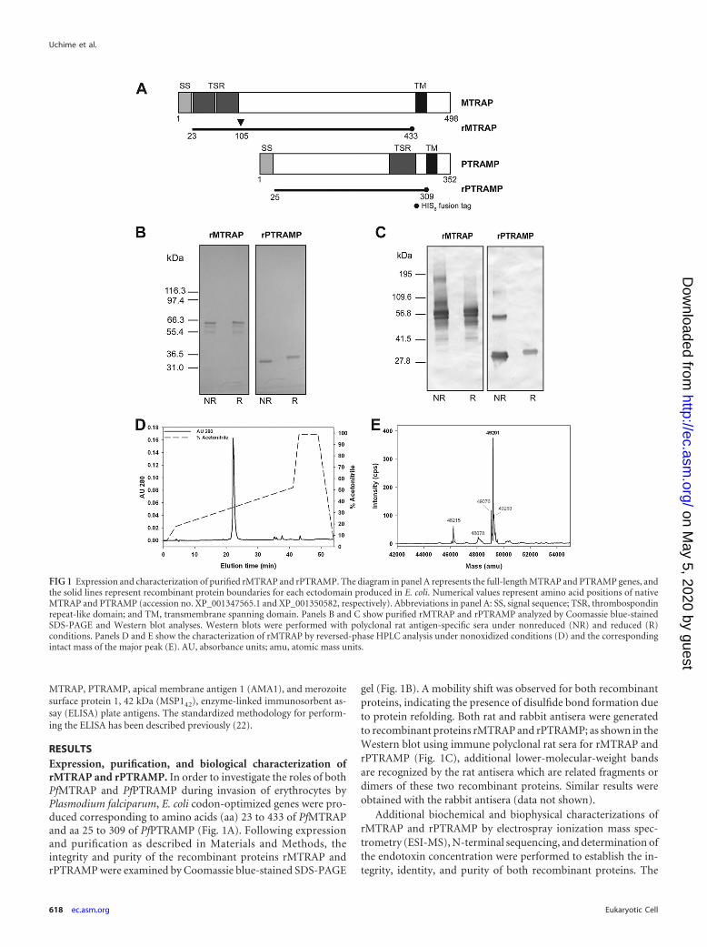
RESULTSExpression, purification, and biological characterization ofrMTRAP and rPTRAMP. In order to investigate the roles of bothPfMTRAP and PfPTRAMP during invasion of erythrocytes byPlasmodium falciparum, E. coli codon-optimized genes were pro-duced corresponding to amino acids (aa) 23 to 433 of PfMTRAPand aa 25 to 309 of PfPTRAMP (Fig. 1A). Following expressionand purification as described in Materials and Methods, theintegrity and purity of the recombinant proteins rMTRAP andrPTRAMP were examined by Coomassie blue-stained SDS-PAGE
gel (Fig. 1B). A mobility shift was observed for both recombinantproteins, indicating the presence of disulfide bond formation dueto protein refolding. Both rat and rabbit antisera were generatedto recombinant proteins rMTRAP and rPTRAMP; as shown in theWestern blot using immune polyclonal rat sera for rMTRAP andrPTRAMP (Fig. 1C), additional lower-molecular-weight bandsare recognized by the rat antisera which are related fragments ordimers of these two recombinant proteins. Similar results wereobtained with the rabbit antisera (data not shown).
Additional biochemical and biophysical characterizations ofrMTRAP and rPTRAMP by electrospray ionization mass spec-trometry (ESI-MS), N-terminal sequencing, and determination ofthe endotoxin concentration were performed to establish the in-tegrity, identity, and purity of both recombinant proteins. The
FIG 1 Expression and characterization of purified rMTRAP and rPTRAMP. The diagram in panel A represents the full-length MTRAP and PTRAMP genes, andthe solid lines represent recombinant protein boundaries for each ectodomain produced in E. coli. Numerical values represent amino acid positions of nativeMTRAP and PTRAMP (accession no. XP_001347565.1 and XP_001350582, respectively). Abbreviations in panel A: SS, signal sequence; TSR, thrombospondinrepeat-like domain; and TM, transmembrane spanning domain. Panels B and C show purified rMTRAP and rPTRAMP analyzed by Coomassie blue-stainedSDS-PAGE and Western blot analyses. Western blots were performed with polyclonal rat antigen-specific sera under nonreduced (NR) and reduced (R)conditions. Panels D and E show the characterization of rMTRAP by reversed-phase HPLC analysis under nonoxidized conditions (D) and the correspondingintact mass of the major peak (E). AU, absorbance units; amu, atomic mass units.
Uchime et al.
618 ec.asm.org Eukaryotic Cell
on May 5, 2020 by guest
http://ec.asm.org/
Dow
nloaded from
primary elution peak obtained from reversed-phase HPLC ofrMTRAP (Fig. 1D) was analyzed by ESI-MS. The observed mass ofthe nonreduced form of rMTRAP was 49,201 Da (Fig. 1E), whichis within 1 Da of the nonreduced theoretical mass, 49,199.8 Da,demonstrating excellent integrity of rMTRAP. The identity ofrMTRAP was confirmed by N-terminal sequencing. The expectedand observed amino acid sequences were in complete agreementfor rMTRAP (MISTHDTXDE). Recombinant MTRAP was alsoanalyzed by reduced reversed-phase HPLC. A minor peak wasisolated and identified by N-terminal sequencing (MNNSYIY)which corresponded to a truncated form with an amino-terminaldeletion of 84 amino acids, which we identified as rMTRAP�84
(see Fig. S1 in the supplemental material). Due to this deletion (aa105 relative to the native MTRAP sequence), this truncated pro-tein lacks the TSR domain. Additionally, the primary elution peakobtained from reversed-phase HPLC of rPTRAMP was analyzedby ESI-MS and Edman degradation. The observed mass for thenonreduced form was 34,444.2 Da, which is within 1 Da of thenonreduced theoretical mass of 34,443.1 Da, demonstrating ex-cellent integrity of rPTRAMP. The expected and observed N-ter-minal amino acid sequences were in complete agreement forrPTRAMP (MNDLXSXNLK) as well. The concentrations of en-dotoxin in the rMTRAP and rPTRAMP stocks were less than 30and 10 endotoxin units per mg, respectively.
To characterize both the rat and rabbit antisera for biologicalsignificance, the anti-rMTRAP and -rPTRAMP polyclonal anti-bodies were examined for their reactivity to parasitized red bloodcells by confocal microscopy. Given the prior localization ofMTRAP and PTRAMP to the micronemes (2, 39), we aimed tocorroborate the localization to the micronemes and confirm theintegrity of the MTRAP- and PTRAMP-specific antibodies. Colo-calization of rat anti-rMTRAP polyclonal antibodies with anti-AMA1, -EBA-175, and -RON4 antibodies corroborates thatPfMTRAP and PfPTRAMP are located in the micronemes (see Fig.S2A and B in the supplemental material), consistent with the pre-viously published data (2, 39).
The anti-rMTRAP and -rPTRAMP rat and rabbit polyclonalantibodies were further examined for their ability to providegrowth inhibition activity (GIA) (see Table S1 in the supple-mental material). Only anti-rPTRAMP antibody from two offive rats demonstrated GIA—about 60 or 80% at 10 mg/ml. Theinhibition was partially reversible by preincubation with 800�M rPTRAMP protein. In contrast, no GIA was observed foreither rat or rabbit polyclonal antibodies generated to rMTRAP(data not shown).
Biological characterization of rMTRAP and rPTRAMP byerythrocyte binding assay. Using an erythrocyte binding assay,rMTRAP was observed to bind to human erythrocytes, while nobinding was observed for rPTRAMP (Fig. 2). The erythrocytebinding assay was performed using two recombinant negativecontrol proteins, the 25-kDa sexual-stage-specific protein Pfs25and the 42-kDa fragment of MSP1 and a recombinant positivecontrol protein, BAEBL RII (Fig. 2). A common background bandwas observed in all five Western blots, as noted by an asterisk inFig. 2. Further analysis of rMTRAP human erythrocyte bind-ing indicated that only the full-length form bound, whilerMTRAP�84, an amino-terminal truncation product which waspresent in the purified protein sample with an N-terminal 84-aadeletion, did not. (For characterization of the truncated product,see Fig. S1 in the supplemental material.)
Biophysical and biochemical characterization by SEC-MALS-QELS HPLC. rMTRAP and rPTRAMP were examined byanalytical SEC-MALS-QELS HPLC to determine their conforma-tion, molar mass and hydrodynamic radius. Analysis of rMTRAPby SEC-HPLC indicated two populations of protein existed insolution: a monomeric population at 94% and an oligomer pop-ulation at 6%. Similarly, analysis of rPTRAMP by SEC-HPLC in-dicated two populations of protein existed in solution: a mono-meric population at 94% and a dimer population at 6%.Examination of the monomer peaks from SEC-HPLC revealedrMTRAP had a retention time (RT) of 17 to 18 min, similar to theRT of immunoglobulin G (158 kDa) at approximately 17 min(data not shown). In contrast to rMTRAP, rPTRAMP had a longerretention time, approximately 22 min. Further analysis by in-lineMALS indicated the weight average molar masses of the mono-meric peaks for rMTRAP and rPTRAMP of 53 kDa and 38 kDacorresponded to the theoretical masses of 49.2 kDa and 34.5 kDa,respectively (Fig. 3A; see Fig. S3A in the supplemental material).To further characterize the recombinant proteins, the hydrody-namic radii (Rh) of the monomeric recombinant proteins wereexamined by QELS. The Rh of rMTRAP was 5.1 nm (Fig. 3B),indicating the monomeric form of rMTRAP (49.2 kDa) is highly
FIG 2 Erythrocyte-binding ability of rMTRAP. Purified recombinant pro-teins were added to human erythrocytes, and the eluates were separated bySDS-PAGE and tested by Western blotting with antigen (Ag)-specific rabbitantibodies. Arrows indicate expected migration by SDS-PAGE of individualrecombinant protein. The asterisks indicate background bands. MWM, mo-lecular weight marker lane.
Analysis of Plasmodium falciparum MTRAP and PTRAMP
May 2012 Volume 11 Number 5 ec.asm.org 619
on May 5, 2020 by guest
http://ec.asm.org/
Dow
nloaded from
extended compared to a typical globular protein like BSA (66.4kDa), which has an Rh value of �3.4 nm (40). In contrast torMTRAP, the Rh of rPTRAMP was 3.1 nm (see Fig. S3B in thesupplemental material), indicating the monomeric form ofrPTRAMP is more globular.
Biophysical and biochemical characterization by boundarysedimentation velocity. In order to further characterize the re-combinant proteins, an orthogonal approach was utilized to de-termine their mass and hydrodynamic radius. Boundary sedimen-tation velocity was used to obtain the sedimentation coefficientdistribution of rMTRAP and rPTRAMP. The absorbance scans ofthe sedimenting boundaries versus time (data not shown) wereanalyzed by the computational program Sedfit to give the sedi-mentation distribution of protein components shown in Fig. 3Cand Fig. S3C in the supplemental material. For rMTRAP, theweight average frictional ratio (f/fo) of 1.93 was extracted in thefitting process to account for boundary diffusion. Integration ofthe entire c(s) peak gives a weight average uncorrected s value of2.66S. Correcting for the effects of the density and viscosity of PBS,we obtain an s20,w value of 2.74S. The weight average sedimenta-tion coefficient coupled with the weight average f/fo provides forthe conversion of the c(s) distribution into a c(M) versus M distri-bution, as shown in the inset in Fig. 3C. The weight average molarmass of the major peak is 45,233. This is in good agreement withthe sequence molar mass of 49,191.8. Using the latter and the s20,w
value of 2.74S, we obtain the following hydrodynamic results: acalculated f/fo of 2.04 and a Stokes radius of 4.9 nm. There is anexcellent agreement between the weight average frictional ratio(f/fo) of 1.93 extracted from the boundary fitting process and thef/fo of 2.04 determined from the s20,w value and the sequence molarmass. Based on the large f/fo for MTRAP, it is reasonable to modelthe shape of this protein as a cylinder. Using this hydrodynamicmodel with an estimated hydration of 0.3 g of water per g of pro-tein, the MTRAP cylinder or rod would have length and diameterdimensions of 33.4 nm and 2.15 nm, respectively. The AFM re-sults that are presented below support the rod/ribbon shape mod-eling for MTRAP. A similar analysis was performed for rPTRAMP(see Fig. S3C in the supplemental material). Integration of theentire c(s) peak gives a weight average uncorrected s value of 2.46S.Correcting for the effects of the density and viscosity of PBS, weobtain an s20,w value of 2.54S. A weight average frictional ratio(f/fo) of 1.50 was extracted in the fitting process to account forboundary diffusion. As discussed above, the weight average sedi-mentation coefficient coupled with the weight average f/fo pro-vides for the conversion of the c(s) distribution into a c(M) versusM distribution in comparable fashion to the rMTRAP results thatwere shown in the inset in Fig. 3C. The weight average molar massof the major peak for rPTRAMP is 34,759. This is in excellentagreement with the sequence molar mass of 34,443.1. Using thelatter and the s20,w value of 2.54S, we obtain the following hydro-dynamic results: a calculated f/fo of 1.49 (in excellent agreementwith the fitted f/fo of 1.50) and a Stokes radius of 3.2 nm. Using aprolate model, with an estimated hydration of 0.3 g of water per gof protein, the moderately asymmetric rPTRAMP would havelength and diameter dimensions of 16.35 nm and 2.65 nm, respec-tively.
Biophysical and biochemical characterization of rMTRAPby AFM. AFM images directly reveal that the recombinant proteinrMTRAP is an extended linear and flexible molecule with variabletwisted-ribbon-like appearances (Fig. 4A). In agreement with the
FIG 3 Biochemical characterization of rMTRAP by SEC-MALS-QELS HPLCand analytical ultracentrifugation. (A) Analysis of rMTRAP by SEC-MALSHPLC provided the molar mass distribution of the main peak (molar massindicated by thick line) compared to the absorbance at 280 nm. (B) QELSgoodness of fit of the autocorrelation function plot at the apex of the peak. (C)Sedimentation coefficient distributions and molar mass obtained from theboundary sedimentation velocity data (not shown) of the rMTRAP sample bythe computational analysis described in Materials and Methods. The molarmass distribution c(M) versus M shown in the inset is calculated from the c(s)versus s distribution using the fitted weight average f/fo. The differential c(s)scale is in units of absorbance (AU) per Svedberg unit, and the differential c(M)scale is in units of absorbance per molar mass multiplied by 100,000.
Uchime et al.
620 ec.asm.org Eukaryotic Cell
on May 5, 2020 by guest
http://ec.asm.org/
Dow
nloaded from
solution studies, AFM images suggest over 80% of the rMTRAPmass exists in a monomeric state with the measured protein vol-ume of around 50 nm3 (Fig. 4B). However, the balance of theprotein mass suggests the presence of a minute dimer populationand a very sporadic occurrence of a small oligomer population.The protein particles are varied in shape, although all shapes areconsistent with extended twisted-ribbon forms when quantifiedby the number histogram of the circularity, defined as 4 · area/
(perimeter)2 (Fig. 4C). Assuming a straight ribbon shape for afully extended molecule, the measured circularity values suggestthe typical length/width ratio of between 2 and 6. Furthermore, byexamination of the more typical rMTRAP monomers with a mea-sured protein volume of between 40 and 60 nm3, the numberhistogram of the molecular area is tightly distributed and fallswithin the range of approximately 150 to 220 nm2 (Fig. 4D). Onthe whole, AFM images of rMTRAP suggest an “average” ribbon
FIG 4 AFM shows rMTRAP as a flexible “rod”-like filamentous protein. (A) Panel of representative images, in three-dimensional plots (50-nm square viewedperpendicular to the scan direction, with a colored scale bar for height up to 0.8 nm bottom left and 30-nm bars representing the x-y scale), showing rMTRAPprotein monomer shapes as seen in AFM topographies of uniformly dispersed particles on a mica surface. (B) Mass distribution histogram from �3,800computed particles reveals that over 80% of rMTRAP is seen in a monomeric state (monomer-dimer decompositions illustrated under the blue curve centeredat the monomer volume of 50 nm3). (C) A histogram of the circularity of these particles, defined as 4 · area/(perimeter)2, suggests flexible rod-like moleculesshowing twisted-ribbon-like morphologies with a typical length/width ratio of between 2 and 6. (D) A histogram of the molecular area for 955 rMTRAPmonomers, having a more typical measured protein volume between 0.8 and 1.2 of the monomer value in panel B, reveals a distribution range of �150 to220 nm2.
Analysis of Plasmodium falciparum MTRAP and PTRAMP
May 2012 Volume 11 Number 5 ec.asm.org 621
on May 5, 2020 by guest
http://ec.asm.org/
Dow
nloaded from
shape of the dimensions around 28 nm by 7 nm by 0.3 nm forrMTRAP monomers on mica surfaces. Consequently, the high-resolution AFM images of rMTRAP on a solid surface fully sup-port the interpretations of the structures deduced from the solu-tion state measurements.
Biophysical and biochemical characterization by CD spec-troscopy and thermal stability. The circular dichroism (CD)spectrum of MTRAP showed a negative peak maxima at 199 nm(Fig. 5A), indicating significant random coil structure within theprotein (13). Analysis with the DICHROWEB server and CDProsoftware gave a composition of 44% � 15% random coil, 29% �7% �-sheet, 19% � 6% �-turn, and 8% � 6% �-helix. The spec-trum of MTRAP was remarkably similar to that of CSP (Fig. 5A),which has a composition of 49% � 13% random coil, 23% � 7%�-sheet, 17% � 5% �-turn, and 10% � 4% �-helix (26). Uponheating of MTRAP to 80°C, the peak maxima at 200 nm decreasedand ellipticity above 210 nm had a negative shift (see Fig. S4A inthe supplemental material). The protein was able to revert back toits original structure upon cooling. The circular dichroism spec-trum of PTRAMP had a low negative ellipticity from 205 to 220nm (see Fig. S4B), indicating significant antiparallel �-pleatedsheets (13). Analysis with the DICHROWEB server and CDProsoftware gave a composition of 41% � 4% �-sheet, 35% � 5%random coil, 20% � 5% �-turn, and 5% � 2% �-helix. Circulardichroism thermal melt data showed a loss of �-sheet contentsince the negative molar ellipticity decreased with increasing tem-perature (see Fig. S4B). The denatured protein was not able torevert to its original folded structure upon cooling.
Computational modeling of TSR domains. The MTRAP TSRdomains are predicted to be considerably more compact thanthose of the HsTSP-1 (roughly half the length, �25 Å) (Fig. 5B andC) with a fold similar to that of the TRAP TSR domain (38). Incontrast to HsTSP-1, the MTRAP TSR domains are predicted tohave eight fewer amino acids between the domains, which wouldresult in a more compact structure and suggests that the TSRdomains may function together as a single unit (Fig. 5B and C). Asmodeled, the MTRAP TSR folds are predicted to deviate from theTRAP TSR fold in two ways: (i) in the cation-pi stacking coremade up of repeating tryptophan and arginine residues, theMTRAP cores contain a single W-R-W stack, as opposed to aW-R-W-R-W stack, and (ii) the cysteine at the N terminus of theA-loop is predicted to form a disulfide with a cysteine at the fol-lowing strand B (MTRAP TSR residues 28 to 61) or in the B-Cloop (MTRAP TSR residues 62 to 94). The latter predicted disul-fide linkage would be a variant of the group 2 classification by Tanet al. (38). The MTRAP TSR domains contain only four cysteines,as opposed to the six found in HsTSP-1 and TRAP TSR domains.
Assessment of human antibody responses. Plasma sampleswere collected from healthy volunteers, 18 to 60 years old, in thevillage of Kenieroba, Mali, and were evaluated for their serumreactivity to rMTRAP, rPTRAMP, AMA1, and MSP142. Compar-ing the relative percentages of individual serum reactivity at asingle dilution of 1:500, the levels of antibody recognition ofMTRAP and PTRAMP were markedly less than those of AMA1and MSP142 (Fig. 6). Even though the reactivity of the sera waslow, significant correlations were observed between AMA1 andMSP1 titers and MTRAP or PTRAMP titers (Spearman rank testrange, P 0.0156 to P 0.0001; r2 range, 0.250 to 0.410).
DISCUSSION
A general conservation of the mechanisms and components of amotor complex involved in gliding motility and cell invasionacross the Apicomplexan parasites has been previously suggested(4, 11, 14, 17). Recently Baum et al. (2) demonstrated the presenceof a conserved motor complex in Plasmodium falciparum blood-stage merozoites. Essential to demonstrating conservation of thismotor complex in blood-stage merozoites was the identificationof a TRAP-like functional protein, MTRAP, in blood-stage mero-zoites. However, Baum et al. (2) were unable to demonstrate di-rect binding of MTRAP to the host cells. To that end, we produceda high-quality recombinant protein (rMTRAP) containing theTSR domain in order to begin to investigate the role in invasion ofred blood cells. Furthermore, we also generated an additionalTSR-containing protein, PTRAMP, which has demonstrated ex-pression in and on the surface of merozoites and which undergoesproteolytic processing upon invasion of erythrocytes (12, 39).Here we describe the production and biological, biochemical, andbiophysical characterization of both MTRAP and PTRAMP.
In order to biologically characterize both rMTRAP andrPTRAMP, antisera were generated to recombinant proteinsrMTRAP and rPTRAMP in two different species, rats and rabbits.Confocal microscopy using the antisera generated in both rats andrabbits demonstrated colocation of signal in parasitized red bloodcells for the respective recombinant proteins (see Fig. S2 in thesupplemental material). Furthermore, localization of MTRAPand PTRAMP to the micronemes was consistent with previously published data (2, 39), indicating the anti-rMTRAP and-rPTRAMP polyclonal antibodies generated in both rats and rab-bits specifically recognized PfMTRAP and PfPTRAMP. Next, theanti-rMTRAP and -rPTRAMP polyclonal antibodies were exam-ined for GIA. Only antibodies from two rats generated towardrPTRAMP demonstrated GIA. Differences in GIA between specieshave previously been observed (23). Additionally, GIA for the an-ti-rPTRAMP polyclonal antibodies was partially reversed byrPTRAMP, indicating specificity to PTRAMP. Taken altogether,these native proteins do not appear to be effective antimalariavaccine targets.
Biochemical characterization of purified rMTRAP andrPTRAMP demonstrated that the N-terminal sequence and themass spectrum directly corresponded to the expected resultsfor each protein. Western blot analysis of rMTRAP was consis-tent with a good-quality purified recombinant protein. Dimersand multimers of rPTRAMP were observed by Western blot-ting of nonreduced rPTRAMP but were not observed uponreduction of rPTRAMP (Fig. 1), consistent with the findings ofGreen et al. (12). The observed dimerization of rPTRAMP islikely due to the presence of an odd number of cysteine residuesin the recombinant protein.
The biophysical studies of rMTRAP and rPTRAMP by SEC-MALS and QELS indicated a predominant monomeric conforma-tion for both proteins. The weight average molar mass for bothrMTRAP and rPTRAMP is similar to the theoretical mass of ourdesigned product within the error of the assay. Furthermore,QELS data indicated rMTRAP was an extended molecule based onthe observed hydrodynamic radii (Fig. 3B), which was corrobo-rated by sedimentation velocity (Fig. 3C). Consequently, two or-thogonal approaches, QELS and sedimentation velocity analysis,support the concept that MTRAP acquires a nonglobular highly
Uchime et al.
622 ec.asm.org Eukaryotic Cell
on May 5, 2020 by guest
http://ec.asm.org/
Dow
nloaded from
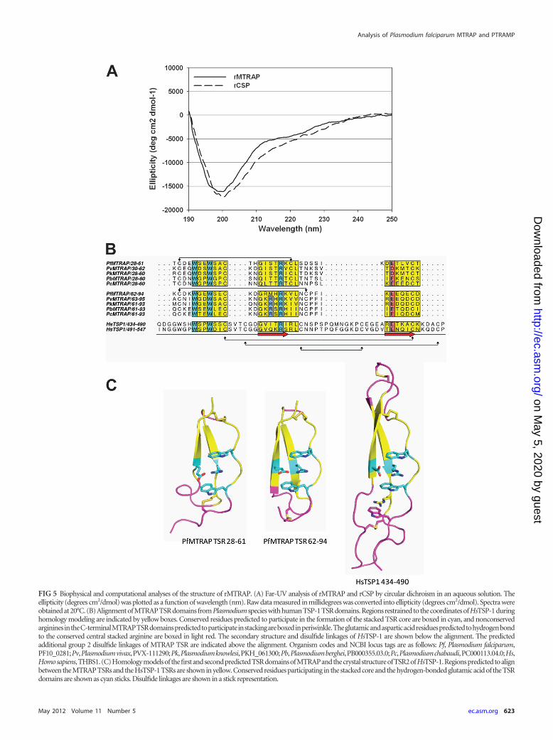
FIG 5 Biophysical and computational analyses of the structure of rMTRAP. (A) Far-UV analysis of rMTRAP and rCSP by circular dichroism in an aqueous solution. Theellipticity (degrees cm2/dmol) was plotted as a function of wavelength (nm). Raw data measured in millidegrees was converted into ellipticity (degrees cm2/dmol). Spectra wereobtained at 20°C. (B) Alignment of MTRAP TSR domains from Plasmodium species with human TSP-1 TSR domains. Regions restrained to the coordinates of HsTSP-1 duringhomology modeling are indicated by yellow boxes. Conserved residues predicted to participate in the formation of the stacked TSR core are boxed in cyan, and nonconservedargininesintheC-terminalMTRAPTSRdomainspredictedtoparticipateinstackingareboxedinperiwinkle.Theglutamicandasparticacidresiduespredictedtohydrogenbondto the conserved central stacked arginine are boxed in light red. The secondary structure and disulfide linkages of HsTSP-1 are shown below the alignment. The predictedadditional group 2 disulfide linkages of MTRAP TSR are indicated above the alignment. Organism codes and NCBI locus tags are as follows: Pf, Plasmodium falciparum,PF10_0281;Pv,Plasmodiumvivax,PVX-111290;Pk,Plasmodiumknowlesi,PKH_061300;Pb,Plasmodiumberghei,PB000355.03.0;Pc,Plasmodiumchabaudi,PC000113.04.0;Hs,Homosapiens,THBS1.(C)Homologymodelsof thefirstandsecondpredictedTSRdomainsofMTRAPandthecrystal structureofTSR2ofHsTSP-1.Regionspredictedtoalignbetween the MTRAP TSRs and the HsTSP-1 TSRs are shown in yellow. Conserved residues participating in the stacked core and the hydrogen-bonded glutamic acid of the TSRdomains are shown as cyan sticks. Disulfide linkages are shown in a stick representation.
Analysis of Plasmodium falciparum MTRAP and PTRAMP
May 2012 Volume 11 Number 5 ec.asm.org 623
on May 5, 2020 by guest
http://ec.asm.org/
Dow
nloaded from

extended structure. To further investigate and begin to resolve thestructural features of MTRAP, we employed an additional orthogo-nal technique, high-resolution AFM, which permitted visualizationof individual rMTRAP molecules. The AFM images and the subse-quent measurements via direct visualizations on mica substrate indi-cate that rMTRAP is an extended linear, rod-like flexible moleculewith measurements centered around 33 nm by 2 nm. In contrast,QELS data indicated rPTRAMP was a less-extended molecule (�16nm by 2.6 nm) based on the observed hydrodynamic radii (see Fig.S3B in the supplemental material), which was corroborated by sedi-mentation velocity (see Fig. S3C). Consequently, two orthogonal ap-proaches, QELS and sedimentation velocity analysis, support theconcept that PTRAMP acquires a less-extended structure.
The TSR domains, when modeled end to end, could at mostaccount for approximately 6 of the 33 nm of rMTRAP, leavingroughly 330 amino acids to account for the remaining length andwidth. Interpreting the far-UV spectrum within the constraints ofthose dimensions, the �330 amino acids are likely ordered withsignificant �-strand content. MTRAP, like CSP, contains a do-main with internal repeats (KNDDD in MTRAP, residues �124 to168; NANP in CSP); however, the length of the domain is shorterby about a factor of 3. These observations, taken together with thesimilarity in the far-UV spectra between MTRAP and CSP, suggestthat the extracellular domain of MTRAP may be composed of adomain with regularly repeating units of mixed sheet-coil charac-ter along with a larger domain (residues �169 to 360) with lessinternal symmetry of similar secondary structure content.
The predicted disulfide linkage between loop A and strand B inthe MTRAP TSR-like domains puts these domains in a new group(group 3), as they do not fit into either group defined by Tan et al.(38). The model presented here is consistent with substitutionsseen within the N- and C-terminal domains and between ho-mologs of these TSR-like domains (see Fig S5 in the supplementalmaterial). For example, in a homologous domain from a hypo-thetical protein in the Plasmodium-related organism Theileria an-nulata, in the N-terminal domain (residues 22 to 55), both cys-teines in this A-B linkage are replaced by small amino acids. In theC-terminal domain of this homolog, residues in the cation-pi
stacking interaction and the proposed Arg-Glu hydrogen bondinginteraction are all replaced by hydrophobic resides. Experimentalevidence will be necessary to confirm the modeling predictions.
These data are the first direct evidence using orthogonal tech-niques with good agreement demonstrating the relative shapes ofboth MTRAP and PTRAMP. Both proteins have different, ex-tended, and multidomain shapes: MTRAP has a long flexible rod-like shape versus PTRAMP, which has a more globular structure.Moreover, determination of the relative structure notwithstand-ing, we desired to examine binding of both proteins to erythro-cytes. Using an erythrocyte binding assay, we were unable to dem-onstrate direct binding of PTRAMP to erythrocytes; however,there may be an indirect interaction between an erythrocytereceptor via interactions with other parasite proteins. Indirectinteractions with host cell receptors via multiprotein com-plexes are common biological occurrences. Such an interactionfor a TRAP homolog is not unprecedented given that the Toxo-plasma TRAP homolog, Toxoplasma gondii MIC2 (TgMIC2), in-teracts with TgM2AP (28). Furthermore, disruption of this asso-ciation, TgMIC2-M2AP, adversely affects invasion (15). Given thefact that only two of the rats immunized with rPTRAMP producedpolyclonal antibodies capable of GIA, in addition to incompleteGIA reversal by rPTRAMP, it is plausible that these two polyclonalantibodies contain antibody which recognizes a low-immunoge-nicity site involved in protein complex formation.
Conversely, while we were unable to demonstrate direct bind-ing of PTRAMP to erythrocytes, we were able to provide the firstevidence demonstrating the direct binding of rMTRAP to eryth-rocytes (Fig. 2). The eluted rMTRAP from the erythrocytes corre-sponded to the full-length rMTRAP, not the truncated versionrMTRAP�84, in which the TSR domain is missing, localizing thefunctional region to the TSR domain. As demonstrated by West-ern blotting, there is an obvious discrepancy between the quantityof rMTRAP incubated with the erythrocytes and the quantity ofrMTRAP eluted by the high-salt elution buffer. This could be dueto a low number of host receptors or possibly due to a low affinitybetween the host cells’ receptor and rMTRAP. While we were ableto demonstrate direct binding to erythrocytes, we were unable toshow GIA with either rat or rabbit anti-rMTRAP polyclonal anti-bodies, consistent with the findings of Baum et al. (2). Given thelack of GIA and MTRAP’s apparent lack of positive selection alongwith the absence of an excess of nonsynonymous polymorphismswithin PfMTRAP (2), it is feasible that MTRAP is concealed from thehost immune response by its sequestration within the tight junction.MTRAP and PTRAMP are not markedly immunogenic since theprevalence of naturally induced human antibodies is low comparedto those of other merozoite antigens, such as AMA1 and MSP142.
MTRAP appears to be a bifunctional molecule that is involvedin merozoite invasion. It is interesting to speculate that due to thehighly extended structure of MTRAP, with an adhesive TSR do-main located at the amino terminus, it is involved in early attach-ment and reorientation through the interaction with aldolase andthe motor complex. There are two challenges to this model, whichare that antibodies against rMTRAP do not block merozoite inva-sion, as reported here and in a previous work (2), and that theprotein sequence of MTRAP appears conserved, indicating no im-mune selection. The amino acid sequence conservation suggests abiological function that occurs later than AMA1, as an example,since AMA1 is subject to immune pressure. However, other adhesionmolecules, such as the EBLs (EBA-175, and BAEBL) are relatively
FIG 6 Reverse cumulative distribution plot of antimalarial antibody observedin Malian adults. The antibody levels in 93 Malian adults were determined byELISA. All samples were tested at a 1:500 dilution, and any optical density at405 (OD405) values more than 3 were assigned as 3 in this figure. Each ODvalue (x axis) is plotted against the percentage of subjects who have at least thatlevel of antibody (y axis).
Uchime et al.
624 ec.asm.org Eukaryotic Cell
on May 5, 2020 by guest
http://ec.asm.org/
Dow
nloaded from
conserved and function prior to AMA1. Finally, it is easier to under-stand the necessity of a long filamentous protein of 25 nm in lengthbefore a moving junction has formed between AMA1 and RON2 (36)since the molecular distance inside the junction is likely to be quitesmall ( 10 nm). While others have demonstrated MTRAP is ex-pressed on the surface of the merozoite following secretion from themicronemes (2), the ability of MTRAP to interact with aldolase invitro (2), and the possibility of PfROM4 cleavage of MTRAP (1), wehave provided evidence that MTRAP is a long flexible rod-like pro-tein capable of binding erythrocytes and consequently tethering themotor complex to an erythrocyte receptor.
ACKNOWLEDGMENTS
We are thankful to Matthew Plassmeyer for expertise in recombinantprotein production and confocal microscopy and Carl Hammer andMark Garfield for mass spectrometry and protein sequencing, respec-tively. We appreciate the contributions of Mahamadou Diakite and RickFairhurst for the Malian samples, Ababacar Diouf and Hong Zhou for theGIA and ELISA data, and the villagers of Kenieroba, Mali, for their par-ticipation. We appreciate helpful discussions with Prakash Srinivasan re-garding the function of these proteins during merozoite invasion.
This research was supported by the Intramural Research Program ofthe NIH, including NIAID and NIBIB, and the GIA Reference Center issupported by the PATH Malaria Vaccine Initiative.
REFERENCES1. Baker RP, Wijetilaka R, Urban S. 2006. Two Plasmodium rhomboid
proteases preferentially cleave different adhesins implicated in all invasivestages of malaria. PLoS Pathog. 2:e113.
2. Baum J, et al. 2006. A conserved molecular motor drives cell invasion andgliding motility across malaria life cycle stages and other apicomplexanparasites. J. Biol. Chem. 281:5197–5208.
3. Bejon P, et al. 2008. Efficacy of RTS,S/AS01E vaccine against malaria inchildren 5 to 17 months of age. N. Engl. J. Med. 359:2521–2532.
4. Bergman LW, et al. 2003. Myosin A tail domain interacting protein(MTIP) localizes to the inner membrane complex of Plasmodium sporo-zoites. J. Cell Sci. 116:39 – 49.
5. Bhattarai A, et al. 2007. Impact of artemisinin-based combination ther-apy and insecticide-treated nets on malaria burden in Zanzibar. PLoSMed. 4:e309.
6. Bourne PE, et al. 2004. The distribution and query systems of the RCSBProtein Data Bank. Nucleic Acids Res. 32:D223–D225.
7. Buscaglia CA, Coppens I, Hol WG, Nussenzweig V. 2003. Sites ofinteraction between aldolase and thrombospondin-related anonymousprotein in Plasmodium. Mol. Biol. Cell 14:4947– 4957.
8. Cerami C, et al. 1992. The basolateral domain of the hepatocyte plasmamembrane bears receptors for the circumsporozoite protein of Plasmo-dium falciparum sporozoites. Cell 70:1021–1033.
9. Coppi A, et al. 2011. The malaria circumsporozoite protein has twofunctional domains, each with distinct roles as sporozoites journey frommosquito to mammalian host. J. Exp. Med. 208:341–356.
10. Frevert U, et al. 1993. Malaria circumsporozoite protein binds to heparansulfate proteoglycans associated with the surface membrane of hepato-cytes. J. Exp. Med. 177:1287–1298.
11. Gaskins E, et al. 2004. Identification of the membrane receptor of a classXIV myosin in Toxoplasma gondii. J. Cell Biol. 165:383–393.
12. Green JL, Hinds L, Grainger M, Knuepfer E, Holder AA. 2006. Plasmo-dium thrombospondin related apical merozoite protein (PTRAMP) isshed from the surface of merozoites by PfSUB2 upon invasion of erythro-cytes. Mol. Biochem. Parasitol. 150:114 –117.
13. Greenfield NJ. 2006. Using circular dichroism spectra to estimate proteinsecondary structure. Nat. Protoc. 1:2876 –2890.
14. Herm-Gotz A, et al. 2002. Toxoplasma gondii myosin A and its light chain:a fast, single-headed, plus-end-directed motor. EMBO J. 21:2149 –2158.
15. Huynh MH, et al. 2003. Rapid invasion of host cells by Toxoplasmarequires secretion of the MIC2-M2AP adhesive protein complex. EMBO J.22:2082–2090.
16. Jewett TJ, Sibley LD. 2003. Aldolase forms a bridge between cell surface
adhesins and the actin cytoskeleton in apicomplexan parasites. Mol. Cell11:885– 894.
17. Kappe S, et al. 1999. Conservation of a gliding motility and cell invasionmachinery in Apicomplexan parasites. J. Cell Biol. 147:937–944.
18. Leaver-Fay A, et al. 2011. ROSETTA3: an object-oriented software suite forthe simulation and design of macromolecules. Methods Enzymol. 487:545–574.
19. Malkin EM, et al. 2005. Phase 1 clinical trial of apical membrane antigen1: an asexual blood-stage vaccine for Plasmodium falciparum malaria.Infect. Immun. 73:3677–3685.
20. Mayer DC, et al. 2006. The glycophorin C N-linked glycan is a criticalcomponent of the ligand for the Plasmodium falciparum erythrocyte re-ceptor BAEBL. Proc. Natl. Acad. Sci. U. S. A. 103:2358 –2362.
21. Menard R. 2000. The journey of the malaria sporozoite through its hosts:two parasite proteins lead the way. Microbes Infect. 2:633– 642.
22. Miura K, et al. 2008. Development and characterization of a standardizedELISA including a reference serum on each plate to detect antibodiesinduced by experimental malaria vaccines. Vaccine 26:193–200.
23. Miura K, et al. 2007. In immunization with Plasmodium falciparum apicalmembrane antigen 1, the specificity of antibodies depends on the speciesimmunized. Infect. Immun. 75:5827–5836.
24. Morahan BJ, Wang L, Coppel RL. 2009. No TRAP, no invasion. TrendsParasitol. 25:77– 84.
25. Ozaki LS, Gwadz RW, Godson GN. 1984. Simple centrifugation method forrapid separation of sporozoites from mosquitoes. J. Parasitol. 70:831–833.
26. Plassmeyer ML, et al. 2009. Structure of the Plasmodium falciparumcircumsporozoite protein, a leading malaria vaccine candidate. J. Biol.Chem. 284:26951–26963.
27. Qian B, et al. 2007. High-resolution structure prediction and the crystal-lographic phase problem. Nature 450:259 –264.
28. Rabenau KE, et al. 2001. TgM2AP participates in Toxoplasma gondiiinvasion of host cells and is tightly associated with the adhesive proteinTgMIC2. Mol. Microbiol. 41:537–547.
29. Rogers WO, et al. 1992. Characterization of Plasmodium falciparumsporozoite surface protein 2. Proc. Natl. Acad. Sci. U. S. A. 89:9176 –9180.
30. RTS,S Clinical Trials Partnership. 2011. First results of phase 3 trial ofRTS,S/AS01 malaria vaccine in African children. N. Engl. J. Med. 365:1863–1875.
31. Schuck P. 2000. Size-distribution analysis of macromolecules by sedi-mentation velocity ultracentrifugation and lamm equation modeling.Biophys. J. 78:1606 –1619.
32. Shimp RL, Jr et al. 2006. Production and characterization of clinicalgrade Escherichia coli derived Plasmodium falciparum 42 kDa merozoitesurface protein 1 (MSP1(42)) in the absence of an affinity tag. Prot. Expr.Purif. 50:58 – 67.
33. Singh S, Plassmeyer M, Gaur D, Miller LH. 2007. Mononeme: a newsecretory organelle in Plasmodium falciparum merozoites identified bylocalization of rhomboid-1 protease. Proc. Natl. Acad. Sci. U. S. A. 104:20043–20048.
34. Sinnis P, et al. 1994. Structural and functional properties of region II-plusof the malaria circumsporozoite protein. J. Exp. Med. 180:297–306.
35. Soding J. 2005. Protein homology detection by HMM-HMM compari-son. Bioinformatics 21:951–960.
36. Srinivasan P, et al. 2011. Binding of Plasmodium merozoite proteinsRON2 and AMA1 triggers commitment to invasion. Proc. Natl. Acad. Sci.U. S. A. 108:13275–13280.
37. Sultan AA, et al. 1997. TRAP is necessary for gliding motility and infec-tivity of Plasmodium sporozoites. Cell 90:511–522.
38. Tan K, et al. 2002. Crystal structure of the TSP-1 type 1 repeats: a novellayered fold and its biological implication. J. Cell Biol. 159:373–382.
39. Thompson J, et al. 2004. PTRAMP; a conserved Plasmodium thrombospon-din-related apical merozoite protein. Mol. Biochem. Parasitol. 134:225–232.
40. Tsai CW, et al. 2009. Characterization of a protective Escherichia coli-expressed Plasmodium falciparum merozoite surface protein 3 indicates anon-linear, multi-domain structure. Mol. Biochem. Parasitol. 164:45–56.
41. Tsai CW, Duggan PF, Shimp RL, Jr, Miller LH, Narum DL. 2006. Over-production of Pichia pastoris or Plasmodium falciparum protein disulfideisomerase affects expression, folding and O-linked glycosylation of a malariavaccine candidate expressed in P. pastoris. J. Biotechnol. 121:458–470.
42. Tucker RP. 2004. The thrombospondin type 1 repeat superfamily. Int. J.Biochem. Cell Biol. 36:969 –974.
43. World Health Organization. 2010. World malaria report. World HealthOrganization Press, Geneva, Switzerland.
Analysis of Plasmodium falciparum MTRAP and PTRAMP
May 2012 Volume 11 Number 5 ec.asm.org 625
on May 5, 2020 by guest
http://ec.asm.org/
Dow
nloaded from
Related Documents