Official Journal of the European Paediatric Neurology Society Original article Analysis of status epilepticus related presumed encephalitis in children Jainn-Jim Lin a,b , Kuang-Lin Lin a, , Huei-Shyong Wang a , Shao-Hsuan Hsia b , Chang-Teng Wu b a Division of Pediatric Neurology, Chang Gung Children’s Hospital and Chang Gung Memorial Hospital, Chang Gung University College of Medicine, #5 Fu-Shin Street, Kwei-Shan, Taoyuan 333, Taiwan b Divisions of Pediatric Critical Care and Emergency Medicine, Chang Gung Children’s Hospital and Chang Gung Memorial Hospital, Chang Gung University College of Medicine, Taoyuan, Taiwan article info Article history: Received 5 February 2007 Received in revised form 18 April 2007 Accepted 14 May 2007 Keywords: Presumed encephalitis Status epilepticus abstract Objective: Encephalitis is an acute infection of brain parenchyma characterized clinically by fever, headache, and an altered level of consciousness. There may also be focal or multifocal neurologic deficits, and focal or generalized seizure activity. Here we report an analysis of status epilepticus (SE) related presumed encephalitis in a series of children. Methods: We retrospectively reviewed cases of SE related presumed encephalitis treated in the pediatric intensive care unit, between February 2002 and June 2006. Factors evaluated included age, sex, clinical symptoms, seizure type, presence of SE or refractory status epilepticus (RSE), initial electroencephalogram (EEG) finding, neuroimaging study, cere- brospinal fluid (CSF) and outcome. Results: There were 46 patients (19 girls and 27 boys), aged 8 months to 16 years. Twenty (43.4%) of 46 children developed RSE. The major clinical symptoms included fever (100%), upper respiratory symptoms (56.5%) and altered level of consciousness (45.6%). The initial seizure type was categorized as focal (23.9%), generalized (34.8%), primary focal and secondary generalized (41.3%). Initial EEG revealed a focal (30.8%), or multifocal (19.2%) epileptiform discharge in the SE group and a focal (5%), or multifocal (70%) or generalized (25%) epileptiform discharge in the RSE group. The time of follow-up for this study was 6 months to 51 months. In the SE group, 4 died, 16 developed epilepsy and/or neurologic deficits, and 6 returned to baseline. In the RSE group, 6 died, 13 developed epilepsy and/or neurologic deficits, and none returned to baseline. All survivors were discharged on antiepileptic medications. Conclusions: Our data indicated that children of SE related presumed encephalitis had a high mortality and morbidity. Outcome was related to multifocal or generalized abnormalities of the initial EEG and presence of RSE. & 2007 European Paediatric Neurology Society. Published by Elsevier Ltd. All rights reserved. ARTICLE IN PRESS 1090-3798/$ - see front matter & 2007 European Paediatric Neurology Society. Published by Elsevier Ltd. All rights reserved. doi:10.1016/j.ejpn.2007.05.007 Corresponding author. Tel.: +886 3 3281200x8200; fax: +886 3 3288957. E-mail address: [email protected] (K.-L. Lin). EUROPEAN JOURNAL OF PAEDIATRIC NEUROLOGY 12 (2008) 32– 37

Welcome message from author
This document is posted to help you gain knowledge. Please leave a comment to let me know what you think about it! Share it to your friends and learn new things together.
Transcript
ARTICLE IN PRESS
E U R O P E A N J O U R N A L O F PA E D I AT R I C N E U R O L O G Y 1 2 ( 2 0 0 8 ) 3 2 – 3 7
1090-3798/$ - see frodoi:10.1016/j.ejpn.20
�Corresponding autE-mail address: l
Official Journal of the European Paediatric Neurology Society
Original article
Analysis of status epilepticus related presumedencephalitis in children
Jainn-Jim Lina,b, Kuang-Lin Lina,�, Huei-Shyong Wanga,Shao-Hsuan Hsiab, Chang-Teng Wub
aDivision of Pediatric Neurology, Chang Gung Children’s Hospital and Chang Gung Memorial Hospital, Chang Gung University College of
Medicine, #5 Fu-Shin Street, Kwei-Shan, Taoyuan 333, TaiwanbDivisions of Pediatric Critical Care and Emergency Medicine, Chang Gung Children’s Hospital and Chang Gung Memorial Hospital,
Chang Gung University College of Medicine, Taoyuan, Taiwan
a r t i c l e i n f o
Article history:
Received 5 February 2007
Received in revised form
18 April 2007
Accepted 14 May 2007
Keywords:
Presumed encephalitis
Status epilepticus
nt matter & 2007 Europe07.05.007
hor. Tel.: +886 3 [email protected]
a b s t r a c t
Objective: Encephalitis is an acute infection of brain parenchyma characterized clinically
by fever, headache, and an altered level of consciousness. There may also be focal or
multifocal neurologic deficits, and focal or generalized seizure activity. Here we report an
analysis of status epilepticus (SE) related presumed encephalitis in a series of children.
Methods: We retrospectively reviewed cases of SE related presumed encephalitis treated
in the pediatric intensive care unit, between February 2002 and June 2006. Factors evaluated
included age, sex, clinical symptoms, seizure type, presence of SE or refractory status
epilepticus (RSE), initial electroencephalogram (EEG) finding, neuroimaging study, cere-
brospinal fluid (CSF) and outcome.
Results: There were 46 patients (19 girls and 27 boys), aged 8 months to 16 years. Twenty
(43.4%) of 46 children developed RSE. The major clinical symptoms included fever (100%),
upper respiratory symptoms (56.5%) and altered level of consciousness (45.6%). The initial
seizure type was categorized as focal (23.9%), generalized (34.8%), primary focal and
secondary generalized (41.3%). Initial EEG revealed a focal (30.8%), or multifocal (19.2%)
epileptiform discharge in the SE group and a focal (5%), or multifocal (70%) or generalized
(25%) epileptiform discharge in the RSE group. The time of follow-up for this study was 6
months to 51 months. In the SE group, 4 died, 16 developed epilepsy and/or neurologic
deficits, and 6 returned to baseline. In the RSE group, 6 died, 13 developed epilepsy and/or
neurologic deficits, and none returned to baseline. All survivors were discharged on
antiepileptic medications.
Conclusions: Our data indicated that children of SE related presumed encephalitis had a
high mortality and morbidity. Outcome was related to multifocal or generalized
abnormalities of the initial EEG and presence of RSE.
& 2007 European Paediatric Neurology Society. Published by Elsevier Ltd. All rights reserved.
an Paediatric Neurology Society. Published by Elsevier Ltd. All rights reserved.
8200; fax: +886 3 3288957.(K.-L. Lin).
ARTICLE IN PRESS
E UR O P EA N J O UR NA L O F PA ED I ATR I C N E U RO L O G Y 12 (2008) 32 – 37 33
1. Introduction
Acute viral encephalitis most commonly affects children and
young adults. Most viral infections of the central nervous
system (CNS) either involve the meninges, leading to aseptic
meningitis, or cause mild meningoencephalitis rather than
encephalitis. The prognosis of viral encephalitis in pediatric
patients is usually excellent. Although the outcome can range
from complete recovery to severe neurologic sequelae and
death, most children recover without significant residua and
require only brief hospitalization. However viral encephalitis
remains a significant source of neurologic morbidity and
mortality worldwide. Although some previous studies report
predictive factors of neurologic outcome in children with
encephalitis,1–5 little information is available in the literature
about the outcome of status epilepticus (SE) related presumed
encephalitis in children. Here, we report an analysis of SE
related presumed encephalitis in a series of children.
2. Methods
Presumed encephalitis was defined as having symptoms of an
acute febrile illness prior to, or at the time of, the onset of
SE,6,7 and no positive findings in cerebral spinal fluid (CSF)
culture. All children were previous healthy and none had
prior seizures including febrile seizure. Any child with a
family history of epilepsy or febrile seizure, a prior neurologic
insult, progressive neurologic disorder, electrolyte imbalance
and hypoglycemia was excluded.
SE was defined as continuous seizure activity lasting 30 min
or as two or more discrete seizures between which con-
sciousness was not fully regained.8,9 RSE was defined as
seizures lasting 42 h, despite treatment with conventional
antiepileptic drugs including initial therapy with a benzodia-
zepine (BZD), followed by therapeutic dosage of both pheny-
toin (PHT) and phenobarbital (PB).8,10 The goal of treatment
was to achieve complete clinical seizure control or burst-
suppression (BS) pattern on electroencephalogram (EEG),
aiming for an interburst interval of 45 s.11 BS coma was
induced by high-dose suppressive therapy consisting of
midazolam (MDL), lidocaine, propofol, thiopental, citosol or
high-dose PB. Weaning of the high-dose suppressive therapy
was guided by EEG monitoring. The weaning process was
aborted if there was return of SE or frequent clinical or
electrographic seizures. All children were cared for in the
pediatric intensive care unit in Chang Gung Children’s
Hospital. EEG in SE group was performed within 24 h.
However, in the RSE group, 24 h EEG monitor was performed
immediately. All EEGs were interpreted by pediatric neurol-
ogist.
From February 2002 to August 2006, 46 children with
presumed encephalitis complicated with SE were enrolled.
Information was collected by retrospective chart review
regarding: age, sex, etiology, initial seizure type, presence of
SE or RSE, initial EEG, neuroimaging, CSF analysis and
outcome. The diagnosis of initially seizure types was based
upon clinical impression by attending neurologists. The
initial EEGs were categorized as having either negative, or a
focal/diffuse cortical dysfunction, or a focal, or multifocal or
generalized epileptiform discharge. The cortical dysfunction
was defined as slow waves in EEG. Patient outcome including
mortality, a new neurologic deficit, or return to baseline was
determined from last clinic visit, which was a minimum of 6
months after the episode of SE. The neurological deficits were
categorized as seizure, neurological outcome and cognition
outcome. The neurological outcome was according to the
Glasgow outcome scale classification. GOS ¼ 1 (good recovery)
was capacity to resume normal occupational and social
activities, although there may be minor physical or mental
deficits or symptoms. GOS ¼ 2 (moderate disability) was
independent and can resume almost all activities of daily
living. Disabled to the extent that they cannot participate in a
variety of social and work activities. GOS ¼ 3 (severe dis-
ability) was no longer capable of engaging in most previous
personal, social or work activities. Limited communication
skills and have abnormal behavioral or emotional responses.
Typically are partially or totally dependent on assistance from
others in daily living. GOS ¼ 4 (persistent vegetative state)
was not aware of surroundings or purposely responsive to
stimuli. The cognition outcomes were classified as normal,
learning disability, mild mental retardation (MR) and other
MR. Normal indicates that a child had an intelligence
quotient of more than 70 without learning disabilities.
Learning disability was defined as significant difficulties in
the acquisition and use of certain skills such as listening,
speaking, reading, writing, reasoning or mathematical abil-
ities in individuals with normal intelligence. Mild MR was
defined as intelligence quotient scores of between 55 and 80
and moderate MR was defined as intelligence quotient scores
of less than 55. The time of follow-up for the study group was
6 months to 51 months.
3. Data analysis
Clinical and laboratory factors obtained upon admission were
analyzed as possible predictors of neurologic outcome,
defined at last clinic visit, which was a minimum of 6 months
after the episode of SE. These factors included age, seizure
pattern, presence of SE or RSE, EEG finding, Neuroimaging
study, CSF leukocyte and protein measurement. The associa-
tion between each of these variables and neurologic outcome
was examined.
Statistical analysis was performed using the SPSS (10.0)
statistical software. The relationships between parameters
were analyzed using Pearson product moment correlation.
Statistical significance was set at po0.05.
4. Results
Forty-six children with presumed encephalitis complicated
with SE were enrolled. The age at presentation of the children
ranged from 8 months to 16 years. There were 19 (41%)
girls and 27 (59%) boys. Twenty (43.4%) of 46 children
developed RSE. The clinical symptoms included fever (46/46,
100%), upper respiratory symptoms (26/46, 56.5%), altered
level of consciousness (21/46, 45.6%), vomiting (20/46, 43.4%),

ARTICLE IN PRESS
Table 3 – Positive result of serologic test and/or virusculture in 18 patients
Patient no. Serologic test Virus culture
1 Mycoplasma IgM
2 EV (rectal)
3 HSV IgM HSV-1 (throat)
4 EV (throat, rectal)
5 Mycoplasma IgM
6 Influenza A (throat)
7 VZV IgM
8 HSV IgM HSV-1 (throat)
9 EV (throat, rectal)
10 EV (throat, rectal)
11 EV (throat)
12 Influenza A (throat)
13 Mycoplasma IgM
14 Mycoplasma IgM
15 Mycoplasma IgM
16 Mycoplasma IgM HSV-1 (throat)
17 Adenovirus (throat)
18 Mycoplasma IgM
Mycoplasma, mycoplasma pneumoniae; HSV, herpes simplex virus;
EBV, epstein-barr virus; VZV, varicella-zoster virus, EV, enterovirus.
Table 4 – The initial EEG pattern in 46 children withpresumed encephalitis
EEG pattern SE(n ¼ 26)
RSE(n ¼ 20)
Negative 4 (15.4%)
Focal/diffuse cortical 9 (34.6%)
E U R O P E A N J O U R N A L O F PA E D I AT R I C N E U R O L O G Y 1 2 ( 2 0 0 8 ) 3 2 – 3 734
headache (13/46, 28.2%), behavioral disturbances (2/46, 4.3%).
The initial seizure type was categorized as focal (11/46, 23.9%),
generalized (16/46, 34.8%), primary focal and secondary
generalized (19/46, 41.3%). The clinical characteristics and
initial seizure pattern of all 46 children are summarized in
Tables 1 and 2.
All patients underwent a thorough investigation during
their hospitalization, including serologic tests for viruses,
polymerase chain reaction (PCR) in CSF for herpes simplex virus
(HSV) DNA (n ¼ 14) and virus culture of throat (n ¼ 34), CSF
(n ¼ 33), and rectal (n ¼ 28) specimens. Most children under-
went serologic tests for HSV (n ¼ 31), epstein-barr virus (EBV)
(n ¼ 28), mycoplasma pneumoniae (n ¼ 27), human herpesvirus 6
(HHV-6) (n ¼ 4) and varicella-zoster virus (VZV) (n ¼ 1). Positive
result for serologic tests or/and virus culture was noted in 18
patients. In all other cases, no organism was identified.
Positive IgM result of serologic tests was VZV 1 (100%),
mycoplasma pneumonia 7 (25.9%), and HSV 3 (9.6%). The result
of virus culture was throat 10 (29%), rectal 4 (14.2%) and CSF 0
(0%). Positive result for serologic tests or/and virus culture in
18 patients is shown in Table 3.
Lumbar puncture was performed in 33 patients. White
blood cell (WBC) count in the CSF studies ranged from 0 to
109 cells/uL. There was a predominance of monocytes
(60–100%). The glucose level in the CSF ranged from 48 to
128 mg/dL and protein level was 16.6–179.5 mg/dL.
Neuroimaging studies of brain computed tomography (CT)
or magnetic resonance imaging (MRI) were divided into acute
phase (o1 weeks after disease onset), non-acute phase
(1 weeks to 3 month after disease onset) and follow-up
neuroimages (43 month after disease onset). The first brain
CT (n ¼ 30) or MRI (n ¼ 8) performed during acute phase was
no specific including increased letomeningeal enhancement
or brain swelling, whereas the non-acute MRI (n ¼ 15)
Table 1 – Clinical symptoms of 46 children with pre-sumed encephalitis
Fever 46/46 (100%)Upper respiratory tract infection 26/46 (56.5%)
Altered level of consciousness 21/46 (45.6%)
Vomiting 20/46 (43.4%)
Headache 13/46 (28.2%)
Behavioral disturbances 2/46 (4.3%)
Table 2 – The initial seizure patterns of 46 children withpresumed encephalitis
SE(n ¼ 26)
RSE(n ¼ 20)
Focal (n ¼ 9, 19.5%) 4 5
Generalized (n ¼ 33, 71.7%) 21 12
Primary focal and secondary
generalized (n ¼ 4, 8.7%)
1 3
SE, status epilepticus; RSE, refractory status epilepticus.
dysfunction
Epileptiform discharge
Focal 8 (30.8%) 1 (5%)
Multifocal 5 (19.2%) 14 (70%)
Generalized 5 (25%)
revealed mild atrophy in 1 patients. Three patients had
unilateral or bilateral hippocampal hyperintensity in T2
suggestive of focal edema, 5 had multiple focal hyperintensity
in T2 suggestive of encephalitis or acute disseminated
encephalomyelitis (ADEM), 2 had global hypoxic ischemic
encephalopathy and 4 had no specific finding. A follow-up
brain CT was performed for seven patients and brain atrophy
was found in all seven patients.
In the SE group, initial EEG was revealed to be negative in 4
(15.4%), focal/diffuse cortical dysfunction in 9 (34.6%), and a
focal epileptiform discharge in 8 (30.8%), or multifocal in 5
(19.2%). In the RSE group, initial EEG revealed a focal in 1 (5%),
or multifocal in 14 (70%) or generalized epileptiform discharge
in 5 (25%) (Table 4).
In RSE group, the duration from seizure onset to complete
clinical seizure control or appearance of BS pattern on EEG
was ranged from 1 to 20 days (mean7SD ¼ 6.6575.60). If the
duration was less than 3 days, there was less cognition delay
ARTICLE IN PRESS
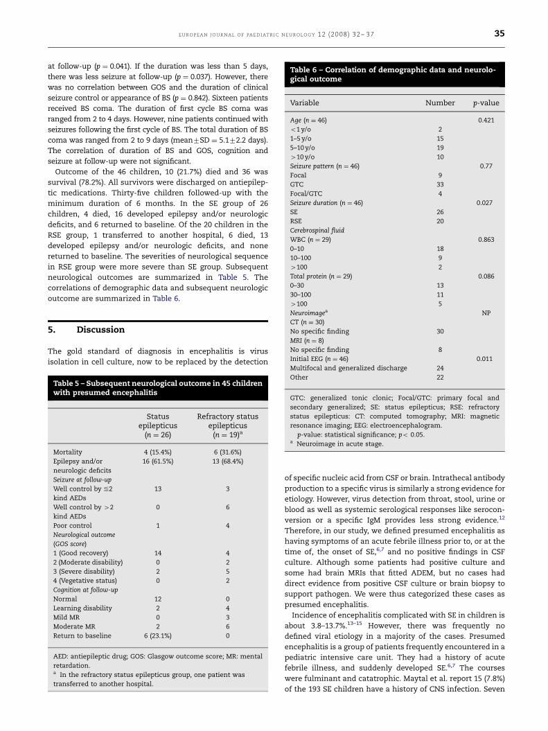
Table 6 – Correlation of demographic data and neurolo-gical outcome
Variable Number p-value
Age (n ¼ 46) 0.421
o1 y/o 2
1–5 y/o 15
5–10 y/o 19
410 y/o 10
Seizure pattern (n ¼ 46) 0.77
Focal 9
GTC 33
Focal/GTC 4
Seizure duration (n ¼ 46) 0.027�
SE 26
RSE 20
Cerebrospinal fluid
WBC (n ¼ 29) 0.863
0–10 18
10–100 9
4100 2
Total protein (n ¼ 29) 0.086
0–30 13
30–100 11
E UR O P EA N J O UR NA L O F PA ED I ATR I C N E U RO L O G Y 12 (2008) 32 – 37 35
at follow-up (p ¼ 0.041). If the duration was less than 5 days,
there was less seizure at follow-up (p ¼ 0.037). However, there
was no correlation between GOS and the duration of clinical
seizure control or appearance of BS (p ¼ 0.842). Sixteen patients
received BS coma. The duration of first cycle BS coma was
ranged from 2 to 4 days. However, nine patients continued with
seizures following the first cycle of BS. The total duration of BS
coma was ranged from 2 to 9 days (mean7SD ¼ 5.172.2 days).
The correlation of duration of BS and GOS, cognition and
seizure at follow-up were not significant.
Outcome of the 46 children, 10 (21.7%) died and 36 was
survival (78.2%). All survivors were discharged on antiepilep-
tic medications. Thirty-five children followed-up with the
minimum duration of 6 months. In the SE group of 26
children, 4 died, 16 developed epilepsy and/or neurologic
deficits, and 6 returned to baseline. Of the 20 children in the
RSE group, 1 transferred to another hospital, 6 died, 13
developed epilepsy and/or neurologic deficits, and none
returned to baseline. The severities of neurological sequence
in RSE group were more severe than SE group. Subsequent
neurological outcomes are summarized in Table 5. The
correlations of demographic data and subsequent neurologic
outcome are summarized in Table 6.
4100 5Neuroimagea NP
CT (n ¼ 30)
No specific finding 30
MRI (n ¼ 8)
No specific finding 8
Initial EEG (n ¼ 46) 0.011�
5. Discussion
The gold standard of diagnosis in encephalitis is virus
isolation in cell culture, now to be replaced by the detection
Table 5 – Subsequent neurological outcome in 45 childrenwith presumed encephalitis
Statusepilepticus
(n ¼ 26)
Refractory statusepilepticus
(n ¼ 19)a
Mortality 4 (15.4%) 6 (31.6%)
Epilepsy and/or
neurologic deficits
16 (61.5%) 13 (68.4%)
Seizure at follow-up
Well control by %2
kind AEDs
13 3
Well control by 42
kind AEDs
0 6
Poor control 1 4
Neurological outcome
(GOS score)
1 (Good recovery) 14 4
2 (Moderate disability) 0 2
3 (Severe disability) 2 5
4 (Vegetative status) 0 2
Cognition at follow-up
Normal 12 0
Learning disability 2 4
Mild MR 0 3
Moderate MR 2 6
Return to baseline 6 (23.1%) 0
AED: antiepileptic drug; GOS: Glasgow outcome score; MR: mental
retardation.a In the refractory status epilepticus group, one patient was
transferred to another hospital.
Multifocal and generalized discharge 24
Other 22
GTC: generalized tonic clonic; Focal/GTC: primary focal and
secondary generalized; SE: status epilepticus; RSE: refractory
status epilepticus: CT: computed tomography; MRI: magnetic
resonance imaging; EEG: electroencephalogram.� p-value: statistical significance; po 0.05.a Neuroimage in acute stage.
of specific nucleic acid from CSF or brain. Intrathecal antibody
production to a specific virus is similarly a strong evidence for
etiology. However, virus detection from throat, stool, urine or
blood as well as systemic serological responses like serocon-
version or a specific IgM provides less strong evidence.12
Therefore, in our study, we defined presumed encephalitis as
having symptoms of an acute febrile illness prior to, or at the
time of, the onset of SE,6,7 and no positive findings in CSF
culture. Although some patients had positive culture and
some had brain MRIs that fitted ADEM, but no cases had
direct evidence from positive CSF culture or brain biopsy to
support pathogen. We were thus categorized these cases as
presumed encephalitis.
Incidence of encephalitis complicated with SE in children is
about 3.8–13.7%.13–15 However, there was frequently no
defined viral etiology in a majority of the cases. Presumed
encephalitis is a group of patients frequently encountered in a
pediatric intensive care unit. They had a history of acute
febrile illness, and suddenly developed SE.6,7 The courses
were fulminant and catatrophic. Maytal et al. report 15 (7.8%)
of the 193 SE children have a history of CNS infection. Seven
ARTICLE IN PRESS
E U R O P E A N J O U R N A L O F PA E D I AT R I C N E U R O L O G Y 1 2 ( 2 0 0 8 ) 3 2 – 3 736
(46.7%) of 15 children present status epilepticus 41 h.13 In our
study, 46 children with presumed encephalitis complicated
with SE are enrolled. Twenty (43.4%) of 46 children develop
RSE. So SE related presumed encephalitis etiology has a
higher rate to develop RSE.
All of our patients had either continuous EEG monitoring or
routine EEG recordings.
The neurologic outcome among patients with multifocal or
generalized epileptiform discharge on the initial EEG are
poorer than others (p ¼ 0.011). The existence of more than
one epileptic focus owing to encephalitis is not surprising and
probably plays a part in the severity of the epileptic process.
Previous studies show multifocal or generalized abnormal-
ities on initial EEG had higher mortality6 and poor neurologic
outcome.16 Besides, EEG abnormality at 1 h predicts seizure
recurrence within 24-h.17 Our data demonstrates that multi-
focal or generalized epileptiform discharges on the initial EEG
are the factors associated with a worse neurologic outcome.
In RSE group, the duration from seizure onset to complete
clinical seizure control or appearance of BS pattern on EEG
was correlated with morbidity. In our study, if the duration
was less than 3 days, there was less cognition delay at follow-
up (p ¼ 0.041). If the duration was less than 5 days, there was
less seizure at follow-up (p ¼ 0.037). Therefore, prolonged
uncontrolled seizure may cause permanent neurologic da-
mage in encephalitis. However, the duration of BS pattern on
EEG was not correlation with morbidity included GOS,
cognition and seizure at follow-up. Aggressive treatment to
achieve complete clinical seizure control or appearance of BS
pattern on EEG was emphasized.
It is not unexpected that there is higher morbidity in the
survivors of RSE than in SE. In our RSE group, all survivors
develop epilepsy and/or neurologic deficits, and none return
to baseline. Besides, the severities of neurological sequence
were more severe in RSE than SE group (Table 5). In a recent
series of children with seizures lasting 430 min, only 23% of
the survivors are normal at follow-up; 34% show develop-
mental deterioration, and 36% develop new-onset epilepsy.18
However, in two other small retrospective reviews of severe
RSE children with a presumed encephalitic etiology, who were
refractory to initial therapy with a BZD, follow therapeutic
levels of either PHT or PB, for 460 min; 83.3% of the survivors
develop epilepsy and/or neurologic deficits and 16.6% re-
turned to baseline.5,6 Our data demonstrate that the RSE
owing to presumed encephalitis carries a high morbidity in
the survivors of RSE (p ¼ 0.027).
The overall mortality rate in pediatric SE is zero to
10%.13,14,18,19 However, there is high mortality in the RSE group
with a presumed encephalitic etiology. Maytal et al. report low
morbidity and mortality of SE in children, except in acute or
progressive neurologic insults group. The mortality rate in this
group was 12.5%.13 In another two studies, the mortality rate in
the RSE group owing to presumed encephalitis in children was
about 25%.5,8 In our study, the mortality rate also was higher in
RSE group than in SE group (31.6% vs. 15.4%).
Taken together, these studies suggest that mortality
and morbidity associated with pediatric RSE owing to
presumed encephalitis may be higher than that previously
thought. There are several possible explanations for this
situation. First, the complications of SE include cardiac
dysrhythmia, derangements of metabolic and autonomic
function, neurogenic pulmonary edema, hyperthermia, rhab-
domyolysis, and pulmonary aspiration. Prolonged SE in-
creases risk for these complications. Second, permanent
neurologic damage in encephalitis with prolonged uncon-
trolled convulsive activity may occur by direct virus invasion,
stimulating host dependent immune response or SE-induced
neuronal death.20 Long duration of SE is associated with
markedly poor neurologic outcome, in particular, the devel-
opment of post-SE symptomatic epilepsy.21,22 Third, because
our patients are treated in a major tertiary care referral
center, and several cases had been transferred from other
hospitals, they may represent the more severe cases.
Factors that are not correlated with adverse outcomes
included age, any type of seizure occurrence, initial brain CT/
MRI or abnormal CSF findings. In conclusion, our data
demonstrate a high mortality and morbidity in the RSE group
of presumed encephalitis in children. Neurologic outcome is
related to multifocal or generalized abnormalities on the
initial EEG and the presence of RSE.
R E F E R E N C E S
1. Kennedy CR, Duffy SW, Smith R, Robinson RO. Clinicalpredictors of outcome in encephalitis. Arch Dis Child1987;62:1156–62.
2. Rantala H, Uhari M, Uhari M, Saukkonen A, Sorri M. Outcomeafter childhood encephalitis. Dev Med Child Neurol1991;33:858–67.
3. Klein SK, Hom DL, Anderson MR, Latrizza AT, Toltzis P.Predictive factors of short-term neurologic outcome inchildren with encephalitis. Pediatr Neurol 1994;11:308–12.
4. Bhutto E, Naim M, Ehtesham M, et al. Prognostic indicators ofchildhood acute viral encephalitis. J Pak Med Assoc1999;49:311–6.
5. Kramer U, Shorer Z, Ben-Zeev B, et al. Severe refractory statusepilepticus owing to presumed encephalitis. J Child Neurol2005;20:184–7.
6. Sahin M, Menache CC, Holmes GL, Riviello Jr. JJ. Outcome ofsevere refractory status epilepticus in children. Epilepsia2001;42:1461–7.
7. Sahin M, Menache CC, Holmes GL, Riviello Jr. JJ. Prolongedtreatment for acute symptomatic refractory status epilepti-cus: outcome in children. Neurology 2003;61:398–401.
8. Lowenstein DH, Alldredge BK. Status epilepticus. N Engl J Med1998;338:970–6.
9. Chapman MG, Smith M, Hirsch NP. Status epilepticus.Anaesthesia 2001;56:648–59.
10. Prasad A, Worrall BB, Bertram EH, Bleck TP. Propofol andmidazolam in the treatment of refractory status epilepticus.Epilepsia 2001;42:380–6.
11. Sahin M, Riviello JJ. Prolonged treatment of refractory statusepilepticus in a child. J Child Neurol 2001;16:147–50.
12. Steiner I, Budka H, Chaudhuri A, Koskiniemi M, Sainio K,Salonen O, et al. Viral encephalitis: a review of diagnosticmethods and guidelines for management. Eur J Neurol2005;12:331–43.
13. Maytal J, Shinnar S, Moshe SL, Alvarez LA. Low morbidity andmortality of status epilepticus in children. Pediatrics1989;83:323–31.
14. Lacroix J, Deal C, Gauthier M, Rousseau E, Farrell A. Admis-sions to a pediatric intensive care unit for status epilepticus: a10-year experience. Crit Care Med 1994;22:827–32.

ARTICLE IN PRESS
E UR O P EA N J O UR NA L O F PA ED I ATR I C N E U RO L O G Y 12 (2008) 32 – 37 37
15. Chin RF, Neville BG, Peckham C, et al. Incidence, cause, andshort-term outcome of convulsive status epilepticus in child-hood: prospective population-based study. Lancet 2006;368:222–9.
16. Trinka E, Dubeau F, Andermann F, et al. Clinical findings,imaging characteristics and outcome in catastrophic post-encephalitic epilepsy. Epileptic Disord 2000;2:153–62.
17. Kalita J, Misra UK, Patel R. Initial EEG in status epilepticus ishelpful in predicting seizure recurrence. Electromyogr ClinNeurophysiol 2006;46:139–44.
18. Barnard C, Wirrell E. Does status epilepticus in children causedevelopmental deterioration and exacerbation of epilepsy? JChild Neurol 1999;14:787–94.
19. Eriksson KJ, Koivikko MJ. Status epilepticus in children:aetiology, treatment, and outcome. Dev Med Child Neurol1997;39:652–8.
20. Fujikawa DG. Prolonged seizures and cellular injury: under-standing the connection. Epilepsy Behav 2005;7:S3–S11.
21. Holtkamp M, Othman J, Buchheim K, Meierkord H. Predictorsand prognosis of refractory status epilepticus treated in aneurological intensive care unit. J Neurol Neurosurg Psychiatry2005;76:534–9.
22. Maegaki Y, Kutozawa Y, Hanaki Koch Ohno K. Risk factors forfatality and neurological sequelae after status epilepticus inchildren. Neuropediatrics 2005;36:186–92.
Related Documents