Analysis of Genotype-Phenotype Correlations in Human Holoprosencephaly Benjamin D. Solomon 1 , Sandra Mercier 2,3 , Jorge I. Vélez 1 , Daniel E. Pineda-Alvarez 1 , Adrian Wyllie 1 , Nan Zhou 1 , Christèle Dubourg 2 , Veronique David 2 , Sylvie Odent 2,3 , Erich Roessler 1 , and Maximilian Muenke 1,* 1 Medical Genetics Branch, National Human Genome Research Institute, Bethesda, MD, USA 2 CNRS Génétique et Développement, Université de Rennes, 35043 Rennes Cedex, France 3 Service de génétique clinique, CHU Hôpital Sud, 35043 Rennes Cedex, France Abstract Since the discovery of the first gene causing holoprosencephaly (HPE), over 500 patients with mutations in genes associated with non-chromosomal, non-syndromic HPE have been described, with detailed descriptions available in over 300. Comprehensive clinical analysis of these individuals allows examination for the presence of genotype-phenotype correlations. These correlations allow a degree of differentiation between patients with mutations in different HPE- associated genes and for the application of functional studies to determine intragenic correlations. These early correlations are an important advance in the understanding of the clinical aspects of this disease, and in general argue for continued analysis of the genetic and clinical findings of large cohorts of patients with rare diseases in order to better inform both basic biological insight and care and counseling for affected patients and families. Keywords Holoprosencephaly; HPE Introduction Holoprosencephaly (HPE) can be caused by chromosomal anomalies, result from teratogenic exposure, occur as part of a syndrome, or be due to mutations in one of over 12 known HPE-associated genes. Mutations in SHH, the first such gene identified, were shown to cause HPE in 1996 [Roessler et al., 1996]. Currently, 4 genes (SHH, ZIC2, SIX3, and TGIF) are routinely analyzed on a clinical basis in patients with HPE [Brown et al., 1998; Wallis et al., 1999; Gripp et al., 2000; Dubourg et al., 2007]. In the approximately one-and-a-half decades since the discovery of SHH as a cause of HPE, advances in the understanding of the molecular pathogenesis of the condition, combined with comprehensive clinical analyses of patients allows for analysis of genotype-phenotype correlations. Such correlations are still in the early phases, but it is possible to begin to distinguish between groups of patients with HPE due to mutations in different genes. Additionally, within cohorts of patients with mutations in the same gene, the use of functional analyses allows exploration of intragenic genotype-phenotype correlations. Corresponding Author: Maximilian Muenke, MD, National Institutes of Health, MSC 3717, Building 35, Room 1B-203, Bethesda, MD, 20892, Phone: (301)594-7487, Fax: (301)496-7184, [email protected]. NIH Public Access Author Manuscript Am J Med Genet C Semin Med Genet. Author manuscript; available in PMC 2011 February 15. Published in final edited form as: Am J Med Genet C Semin Med Genet. 2010 February 15; 154C(1): 133–141. doi:10.1002/ajmg.c.30240. NIH-PA Author Manuscript NIH-PA Author Manuscript NIH-PA Author Manuscript

Welcome message from author
This document is posted to help you gain knowledge. Please leave a comment to let me know what you think about it! Share it to your friends and learn new things together.
Transcript

Analysis of Genotype-Phenotype Correlations in HumanHoloprosencephaly
Benjamin D. Solomon1, Sandra Mercier2,3, Jorge I. Vélez1, Daniel E. Pineda-Alvarez1,Adrian Wyllie1, Nan Zhou1, Christèle Dubourg2, Veronique David2, Sylvie Odent2,3, ErichRoessler1, and Maximilian Muenke1,*
1Medical Genetics Branch, National Human Genome Research Institute, Bethesda, MD, USA2CNRS Génétique et Développement, Université de Rennes, 35043 Rennes Cedex, France3Service de génétique clinique, CHU Hôpital Sud, 35043 Rennes Cedex, France
AbstractSince the discovery of the first gene causing holoprosencephaly (HPE), over 500 patients withmutations in genes associated with non-chromosomal, non-syndromic HPE have been described,with detailed descriptions available in over 300. Comprehensive clinical analysis of theseindividuals allows examination for the presence of genotype-phenotype correlations. Thesecorrelations allow a degree of differentiation between patients with mutations in different HPE-associated genes and for the application of functional studies to determine intragenic correlations.These early correlations are an important advance in the understanding of the clinical aspects ofthis disease, and in general argue for continued analysis of the genetic and clinical findings oflarge cohorts of patients with rare diseases in order to better inform both basic biological insightand care and counseling for affected patients and families.
KeywordsHoloprosencephaly; HPE
IntroductionHoloprosencephaly (HPE) can be caused by chromosomal anomalies, result fromteratogenic exposure, occur as part of a syndrome, or be due to mutations in one of over 12known HPE-associated genes. Mutations in SHH, the first such gene identified, were shownto cause HPE in 1996 [Roessler et al., 1996]. Currently, 4 genes (SHH, ZIC2, SIX3, andTGIF) are routinely analyzed on a clinical basis in patients with HPE [Brown et al., 1998;Wallis et al., 1999; Gripp et al., 2000; Dubourg et al., 2007].
In the approximately one-and-a-half decades since the discovery of SHH as a cause of HPE,advances in the understanding of the molecular pathogenesis of the condition, combinedwith comprehensive clinical analyses of patients allows for analysis of genotype-phenotypecorrelations. Such correlations are still in the early phases, but it is possible to begin todistinguish between groups of patients with HPE due to mutations in different genes.Additionally, within cohorts of patients with mutations in the same gene, the use offunctional analyses allows exploration of intragenic genotype-phenotype correlations.
Corresponding Author: Maximilian Muenke, MD, National Institutes of Health, MSC 3717, Building 35, Room 1B-203, Bethesda,MD, 20892, Phone: (301)594-7487, Fax: (301)496-7184, [email protected].
NIH Public AccessAuthor ManuscriptAm J Med Genet C Semin Med Genet. Author manuscript; available in PMC 2011 February 15.
Published in final edited form as:Am J Med Genet C Semin Med Genet. 2010 February 15; 154C(1): 133–141. doi:10.1002/ajmg.c.30240.
NIH
-PA Author Manuscript
NIH
-PA Author Manuscript
NIH
-PA Author Manuscript
Finally, while the individual genes are considered alone here, it is critical to keep in mindthat the complex pathogenesis of HPE appears to involve multiple interacting factors.
In the following discussion, after a brief description of common clinical findings, the knownHPE-associated genes will be discussed in turn, and then analyzed as a group. Analysis ofpatients with mutations in each of these genes raises salient points regarding patients withHPE as a whole, including those in whom the genetic cause is unknown. Further, analysis ofthe clinical features of affected patients makes three specific conclusions quite clear. First,there is a pressing need for functional analyses in order to better inform clinicians, families,and diagnostic laboratories of the nature of identified variants in order to determine if theyare truly mutations. Second, the establishment of robust genotype-phenotype correlationsdepends on thorough genetic and clinical analyses of a large number of individuals at bothends of the phenotypic spectrum, as well as the active research participation of relativesthrough their primary care-givers. In the case of a rare disorder such as HPE, theestablishment of cooperative international research groups is imperative to increase ourunderstanding of the disease to better inform clinicians and to optimally treat affectedpatients and families.
General CharacteristicsNon-chromosomal, non-syndromic HPE is classically considered an autosomal dominantcondition with incomplete penetrance and highly variable expressivity. Studies attempting toexplain the wide spectrum of the effects of a single mutation even within a single kindredare still pending, but many lines of analysis, including extrapolation from animal models,point to a complex pattern of inheritance combining multiple interacting genetic andenvironmental factors [Reviewed in Krauss, 2007 and Schacter and Krauss, 2008; Ming andMuenke, 2001; Lacbawan et al., 2009].
HPE may be recognized in utero because of abnormalities on fetal imaging [reviewed inVolpe et al., 2009]. After birth, HPE is more commonly recognized due to facial findingsconsistent with the diagnosis and/or abnormalities on neurological examination.Neuroradiological or pathological studies confirm the diagnosis [Plawner et al., 2002;Lazaro et al., 2004; Stashinko et al., 2004; Dubourg et al., 2007; see Hahn and Barnesreview on neuroimaging, this issue]. A common facial appearance occurs in most patientswith HPE (the notable exception is patients with ZIC2 mutations) [Solomon et al., underreview]. In general, the long-standing observation that “the face predicts the brain” holdstrue: in patients with typical facial abnormalities, the severity of facial findings generallycorrelates with the degree of brain anomalies and with survival [DeMyer et al., 1964;Muenke and Beachy, 2001; Stashinko et al., 2004; Lacbawan et al., 2009].
At the most severe end of the spectrum, patients may have pronounced microcephaly andcyclopia or synophthalmia below a proboscis. Less-severely affected infants havemicrocephaly (though hydrocephalus can lead to macrocephaly), hypotelorism, midfacehypoplasia with a flat nasal bridge, cleft lip and or/palate, and a single maxillary centralincisor (SMCI). Individuals with no abnormalities appreciated on conventionalneuroimaging may have subtle facial findings including hypotelorism, a sharp and narrownasal bridge, and SMCI [Muenke & Beachy, 2001; Lacbawan et al., 2009; Solomon et al.,2009]. These individuals, who are diagnosed with “microform” HPE, are identified due to aseverely-affected relative (Fig. 1). It is important to note that even those who havepathogenic mutations in HPE-causing genes and who have clear microform HPE may haveno cognitive impairment. Similarly, not all carriers of deleterious mutations manifestclinically detectable facial or cognitive abnormalities (see Table I, Fig. 2) [Muenke lab,unpublished data].
Solomon et al. Page 2
Am J Med Genet C Semin Med Genet. Author manuscript; available in PMC 2011 February 15.
NIH
-PA Author Manuscript
NIH
-PA Author Manuscript
NIH
-PA Author Manuscript

In addition to the facial appearance, a characteristic clinical pattern occurs in patients withnon-syndromic, non-chromosomal HPE. The most pressing clinical issue commonly issevere neurological impairment, which is universal in patients with structural brainanomalies of the HPE type. The range of impairment is wide; some patients are among themost-severely impaired of any disorder compatible with life, while other patients may beable to walk, eat by mouth, and communicate. To some extent, the degree of impairment canbe predicted by the degree and type of brain anomalies, though exceptions abound [Plawneret al., 2002; Hahn et al., 2006; Roesler et al., 2006].
Seizures are frequent in patients with HPE. Seizures may be underappreciated, especially incognitively very impaired patients, and can be difficult to control [Hahn et al., 2003]. Thediagnosis and management of seizures may be further complicated by electrolyte imbalancessecondary to diabetes insipidus. Autonomic instability affecting temperature control andcardiac and respiratory function can also extremely challenging to manage.
Posterior pituitary insufficiency manifesting primarily as diabetes insipidus (DI) is common.DI may rarely be the presenting sign of HPE, especially in patients with a relatively normalfacial appearance. Anterior pituitary insufficiency and other endocrinological disorders maymanifest, but tend to be far less frequent and linked to a narrower subset of genes includingGLI2 [Roessler et al., 2003; Lazaro et al., 2004; Hahn et al., 2005; Roessler et al., 2005].
SHH (Sonic Hedgehog; OMIM# 600725)More families with mutations in SHH have been reported than with mutations in any othergene, accounting for up to 12% of propositi [Roessler et al., 2009a]. Hence, patients withmutations in SHH are sometimes considered the “prototypical” HPE patient. A number ofvery large families segregating mutations in SHH have been identified, typically only afterthe diagnosis of a severely-affected propositus. These families epitomize the highly variableexpressivity described in many autosomal dominant disorders [Muenke et al., 1994].Approximately 10-30% of mutations in SHH occur de novo. The presence of structural brainanomalies in patients with SHH mutations is estimated to be approximately 45%, while thepenetrance of any manifestations (including microform HPE) is estimated to beapproiximately 90% [Muenke lab, unpublished data]. However, penetrance estimates maybe skewed for a variety of reasons. For example, penetrance may be underestimated: inmultiple separate kindreds, many mutation-positive individuals were noticed to havemicroform holoprosencephaly, including microcephaly, hypotelorism, and SMCI, only aftermutation testing demonstrated the presence of the same mutations found in a severely-affected relative [Ardinger et al., 1988; Roessler et al., 1996]. Conversely, testing of onlyobviously affected patients would lead to a disproportionate underestimation of mutationprevalence in individuals on the milder end of the phenotypic spectrum.
ZIC2 (Zinc Finger Protein of Cerebellum 2; OMIM# 603073)Mutations in ZIC2 are estimated to occur in up to 9% of propositi [Roessler et al., 2009b;Solomon et al., under review]. ZIC2 mutations tend to occur de novo much more frequentlythan mutations in other HPE-associated genes, with mutations occuring de novo inapproximately 72% of proposite. In contrast to SHH and SIX3, the other two genes mostcommonly associated with non-chromosomal, non-syndromic HPE, there are no familiesidentified with mutations in ZIC2 in which the mutation has been ascertained in more than 2generations. As with other HPE-associated genes, germline mosaicism occurs notinfrequently, and must be considered in genetic counseling scenarios when a mutation foundin a propositus does not occur in DNA samples obtained from the parents [Lacbawan et al.,2009; Solomon et al., under review]. Unlike other genes, mutations in ZIC2 tend to result infull HPE, and patients with mutations much less frequently demonstrate mild HPE signs.
Solomon et al. Page 3
Am J Med Genet C Semin Med Genet. Author manuscript; available in PMC 2011 February 15.
NIH
-PA Author Manuscript
NIH
-PA Author Manuscript
NIH
-PA Author Manuscript
Almost 90% of patients with mutations in ZIC2 have structural brain anomalies, and it israre that a parent with a mutation will not show clear signs of cognitive impairment[Solomon et al., under review].
Mutations in ZIC2 also give one of two clear examples in which the face does not predict thebrain, as opposed to the prevailing idea regarding HPE for over four decades [DeMyer et al.,1964]. Recent analysis of a large cohort of patients with mutations in ZIC2 shows a commonfacial phenotype consisting of bitemporal narrowness, upslanting palpebral fissures, a flatnasal bridge, a short nose with anteverted nares, a broad and deep philtrum, and large ears[Solomon et al., under review].
SIX3 (Sine Oculis Homeobox, Drosophila, Homolog of, 3; OMIM# 603714)Mutations in SIX3 are estimated to occur in up to 5% of propositi with HPE [Dubourg et al.,2007; Lacbawan et al., 2009; Muenke lab, unpublished data]. Similar to SHH, large familiessegregating mutations are typically ascertained only after the identification of a severelyaffected patient. Also like SHH, multiple kindreds have been described in which manyindividuals with mutations have subtle microform HPE [Lacbawan et al., 2009; Solomon etal., 2009]. The majority of mutations are inherited, with a de novo mutation rate estimated at14%. Interestingly, of the 4 genes most commonly associated with HPE, SIX3 is the onlyone with a statistically significant over-representation of affected females [Keaton et al.,2009; Lacbawan et al., 2009; Solomon et al., under review, Muenke Lab, unpublished data].
While the presence of structural brain anomalies due to mutations in SIX3, at approximately65% of mutation-positive patients, is intermediate between SHH and ZIC2, it has beenposited that mutations in SIX3 result in relatively severe HPE [Ribeiro et al., 2006]. Whenapplied to propositi only, this observation was borne out by a recent large-scale analysis ofall known patients with mutations in SIX3 [Lacbawan et al., 2009]. Unlike HPE due tomutations in other genes, patients with HPE brain anomalies due to SIX3 mutations are mostlikely to have alobar, rather than semilobar HPE.
Finally, SIX3 shows an early instance of the use of functional studies to analyze potentialgenotype-phenotype correlations. Using results from a functional assay in zebrafish, it waspossible to show that predictions of mutation severity in the animal model correlates withhuman severity of HPE [Domené et al., 2008; Lacbawan et al., 2009]. This study shows thepower of functional studies in the interpretation of human mutation data; similar studies ofmutations in other genes are critical for improved understanding of the causative effects ofspecific mutations, which will also directly translate to the clinical realm.
TGIF (Transforming Growth Factor-Beta Induced Factor; OMIM# 602630)Of the 4 genes commonly tested in clinical laboratories, mutations in TGIF are by far theleast common, occurring in approximately 1 to 2% of propositi [Dubourg et al., 2007;Keaton et al., in preparation]. TGIF is the only gene in which a family has been describedwhere two clinically normal parents each contributed a mutant allele of the same gene to aseverely affected propositus, though it must be said that only one of these alleles was clearlypathogenic [El-Jaick et al., 2007].
This family raises an important point. In the clinical scenario, finding variations in TGIF andother HPE-associated genes can be challenging to interpret, highlighting a common issue ingenetic conditions in which most genetic variations are family-specific. While thedeleterious effects of nonsense and frameshift mutations are usually clear, other variationsmay or may not have functional effects [El-Jaick et al., 2007]. Additionally, the role thatalterations in TGIF play in causing HPE may be less completely understood than with othergenes, which also muddies issues.
Solomon et al. Page 4
Am J Med Genet C Semin Med Genet. Author manuscript; available in PMC 2011 February 15.
NIH
-PA Author Manuscript
NIH
-PA Author Manuscript
NIH
-PA Author Manuscript

GLI2 (Gli-Krüppel Family Member 2, OMIM# *165230)GLI2 is a large gene (>4,500 bp coding) in which many uncommon variants have beendetected, but relatively few cases have been described in which loss-of-function has beenclearly demonstrated. Patients with mutations shown to be loss-of-function (by formalexperiments) indicate the presence of a common spectrum of microform HPE (SMCI ormidline clefting) and anterior pituitary dysfunction, as well as polydactyly. Reportedexceptions to this common GLI2 phenotype highlight the problem of the lack of acommonly accepted mechanism to test the functionality of new variants in a large gene[Roessler et al., 2003; Roessler et al., 2005; Rahimov et al., 2006; Muenke lab, unpublisheddata]. The fact that patients with mutations in GLI2 appear to have abnormalities (eg,anterior pituitary insufficiency and polydactyly) not seen in most patients with non-chromosomal, non-syndromic HPE emphasizes the importance of a thorough clinicalevaluation that takes into account features not typically seen as part of the HPE spectrum.
Other GenesPatients with mutations in other genes have been described, though these genes are nottypically sequenced except on a research basis. Of note, the recent advent of oligonucleotidemicroarray analysis may reveal more about the consequence of copy number changesaffecting these loci, though it may be challenging to interpret the consequences of gain orloss of nearby genes and regulatory regions [Bendavid et al., 2009; Muenke lab, unpublisheddata].
Patients with HPE-spectrum anomalies and mutations in PTCH1 do not appear todemonstrate findings outside typical HPE [Ming et al., 2002]. Mutations in NODAL mayresult in HPE, but are more commonly associated with cardiac and laterality defects[Roessler et al 2009d]. Similarly, mutations in FOXH1 (part of the NODAL signalingpathway) may result in cardiac defects or overt HPE [Roessler et al 2008]. Two patientshave been reported with mutations in TDGF1 (CRIPTO), one with a midline brain anomalyand one with HPE [de la Cruz et al., 2002]. Finally, patients with loss-of-function mutationsin DISP1 may have normal brain structure and development, but may have facial featuresusually seen in conjunction with frank HPE [Roessler et al 2009c].
Further Statistical Analysis of Combined Results1) Analysis of structural brain anomalies in all mutation-positive individuals—A statistically significant association was found between the involved gene and whether ornot structural brain anomalies (SBA) were present in all patients with mutations (χ2
(3) =42.8, p<0.0001) (Figure 2). After aggregating the information by gene, we compared theproportion of patients with SBA to those without known SBA. This latter group, in whomneuroimaging was not typically indicated nor performed, includes patients with clearmicroform features and individuals described as phenotypically normal. Two statisticallysignificant differences were found, one in ZIC2 (χ2
(1) = 108.4, p<0.0001) and the other inSIX3 (χ2
(1) = 13.59, p<0.001), showing that when mutations in these genes are present, it ismore likely to find patients with SBA than without SBA. We further compared theproportion of patients among all genes and found that the proportion of patients with SBA isnot statistically the same for all genes (χ2
(3) = 79.32, p<0.0001). Specifically, mutations inZIC2 are responsible for ∼3% of SBA cases overall. Likewise, the proportion of patientswithout SBA differs among genes (χ2
(3) = 79.32, p<0.0001), and mutations in SHH areresponsible for 48% of patients without SBA.
2) Analysis of variable expressivity—We used the absolute number of patients withmolecular changes to present, analyze and plot our data (Tables I and II). Unless specified,χ2-based tests were performed and based-on-simulation p-values were calculated to compare
Solomon et al. Page 5
Am J Med Genet C Semin Med Genet. Author manuscript; available in PMC 2011 February 15.
NIH
-PA Author Manuscript
NIH
-PA Author Manuscript
NIH
-PA Author Manuscript

the distribution of HPE types attributed to each gene for both all individuals with mutations(Fig. 3a) and separately for propositi only (Fig. 3b). We considered potential differences inmutation distribution among HPE patients with SBA (thus excluding non-penetrant carriersand microform patients), and looked for differences in the distributional pattern in patientswith SBA and those with each HPE type. To do this, we assumed the total SBA group to bethe true distribution among the HPE genes and the distribution of each HPE type to besimilar, or different, from this core pattern. We used R 2.9.2 Patched (R Development CoreTeam, 2009) for statistical analysis and graphical representation of data. Patient data wereused from the following sources: [TGIF: Keaton et al., in preparation; SIX3: Lacbawan etal., 2009; ZIC2: Solomon et al., under review, SHH: Muenke Lab, unpublished data]
Side-by-side comparison: We performed a side-by-side comparison of the distribution ofHPE types (excluding MIHV due to information paucity) among patients with SBA, plottedseparately for all mutation-positive individuals (Fig. 3a) and propositi (Fig. 3b). Thedistribution of anatomical HPE types among the genes is statistically the same whencalculated separately for both all mutation-positive individuals and for propositi alone.
Distribution of mutation-positive patients among HPE typesAll mutation-positive individuals: The contribution of each gene to a particular anatomicaltype was not equal among the three major genes (SHH, ZIC2 and SIX3). This likely reflectsthe fact that the mutation detection rate in prospective cases is not the same for each gene.Interestingly, ZIC2 was more likely to be associated with semilobar HPE, and patients withmicroform findings were more likely to have SHH mutations. Statistically significantdifferences in mutation distribution among the HPE genes (assuming that these genes areequivalent risk factors) were found for the SBA category (χ2
(3) = 59.5, p<0.0001) as well asfor patients with alobar (χ2
(3) = 21.5, p<0.0001), semilobar (χ2(3) = 35.5, p<0.0001) and
microform (χ2(3) = 55.4, p<0.0001) HPE. We note that this assumption of equivalence is not
supported by the empirical data and mutation detection rate of prospective molecularstudies. With the exception of microform HPE, in which mutations in SHH are morecommon (45/72 patients), mutations in ZIC2, followed by mutations in SIX3, were morefrequently observed than mutations in any other gene for the rest of the HPE cohort. Thissuggests that whereas patients with alobar, semilobar or lobar HPE are likely to havemutations in either ZIC2 or SIX3, patients with microform HPE are more likely to havemutations in SHH.
Propositi: Similarly, the distribution of mutation-positive patients among the HPE genes inthe SBA group (χ2
(3) = 67.1, p<0.0001) as well as those with alobar (χ2(3) = 19.6, p<0.0001)
and semilobar (χ2(3) = 40.9, p<0.0001) HPE are statistically different from the hypothesis
that all genes are equal contributors to these disease types. In other words, the contributionto each anatomical class is not equal for the four genes. Whereas propositi with SBA whohave either alobar or semilobar HPE are more likely to have a ZIC2 mutation, it is notpossible to determine which HPE genes are associated with lobar HPE based on this data.Further, despite the low numbers, SHH mutations seem to be responsible for mostmicroform HPE (7/10 patients).
The spectrum of mutations seen in the structural brain anomalies (SBA) vs. HPEanatomical types: Using the SBA information as the expected number of HPE patients withmutations in each of the four principal HPE genes, we looked for discrepancies as follows:(1) based upon SBA data, we calculated the observed percentage of cases with mutations ineach gene; then, (2) we used this percentage to estimate the expected number of mutationswithin each HPE type; and (3) we performed a χ2-based test to detect any pattern changes.We found that in both all mutation-positive individuals (Figure 3a) and for propositi alone
Solomon et al. Page 6
Am J Med Genet C Semin Med Genet. Author manuscript; available in PMC 2011 February 15.
NIH
-PA Author Manuscript
NIH
-PA Author Manuscript
NIH
-PA Author Manuscript

(considered as a group, Figure 3b), the distribution of cases with microform HPE is the onlygroup that significantly differs from these expectations (mutation carriers: χ2
(3) = 76.6,p<0.0001; propositi: χ2
(3) = 20.3, p<0.001).
Genotype-phenotype correlation: After excluding MIHV due to the paucity ofinformation, we fit a uniform association model (Agresti, 1996; section 7.2.1) for our allmutation-positive and propositi-only data sets separately, considering the HPE types and theHPE genes as ordinal and nominal variables, respectively. For the HPE types, a unit-spacedscores ranging from 1 to 4 were used (1 = microform, 2 = lobar, 3 = semilobar and 4 =alobar HPE). Our results suggest that patients in both groups, patients with the most severeHPE phenotypes are more likely to have mutations in either SHH or ZIC2 (all mutationpositive patients: G2 = 80.7, df = 8, p<0.0001; propositi: G2 = 50.7, df = 8, p<0.0001).
DiscussionWe present here the first systematic analysis of possible genotype-phenotype correlations inhuman HPE. The results described above demonstrate that progress has been made inunderstanding the clinical correlations of HPE-associated mutations. However, this progressis still certainly just a start. Much work remains to more fully understand HPE on a numberof levels, including the way in which multiple genetic and environmental mechanismsinteract to result in disease.
First, the identification of a mutation does not yet allow satisfying prediction of outcome,either in a research setting or in the clinical realm. Continued work on the molecular basis ofHPE-spectrum defects, including robust functional analyses will hopefully address thebiological meaning of variations in HPE-associated genes.
Second, in order to better understand the broad HPE spectrum, it is critical to expand ourdiagnostic approach to include a more thorough analysis of both propositi and familymembers, especially including mildly-affected individuals. Areas in which data are lackingshould be addressed by: a careful examination including attention to features nottraditionally seen as part of the HPE spectrum; a robust family history; molecularcharacterization to include both parents as well as other relatives when applicable, completephysical examination, neuroimaging (with analysis by those familiar with HPE) andneuropsychological evaluation of affected family members.
Third, to accomplish the previous point, it is vital to recognize that HPE is a rare disorder,and most clinical geneticists will only encounter a few affected patients. In order to fullyunderstand the clinical features of this disorder, clinicians must continue to collaborate withresearchers studying the condition. Along these lines, international testing centers whocommonly perform genetic testing of patients with HPE must continue to recognize theimportance of collaboration with other groups. With focused collaborative work betweenclinicians and researchers who encounter these patients, our understanding of the complexpathogenesis of HPE will directly translate to better care for affected patients and families.
AcknowledgmentsThe authors would like to extend their deepest gratitude to all patients and families who took part in our research onHPE. JIV would like to thank to Juan Carlos Correa and Ehidy Karime García from the School of Statistics of theNational University of Colombia at Medellín, for their helpful comments and suggestions that improved thestatistical analysis. This research was supported by the Division of Intramural Research, National Human GenomeResearch Institute, National Institutes of Health and Human Services, United States of America and GIS MaladiesRares GISMR0701/DHOS, France.
Solomon et al. Page 7
Am J Med Genet C Semin Med Genet. Author manuscript; available in PMC 2011 February 15.
NIH
-PA Author Manuscript
NIH
-PA Author Manuscript
NIH
-PA Author Manuscript
ReferencesAgresti, A. An Introduction to Categorical Data Analysis. New York: John Wiley & Sons; 1996.Ardinger HH, Bartley JA. Microcephaly in familial holoprosencephaly. J Craniofac Genet Dev Biol.
1988; 8:53–61. [PubMed: 3209679]Bendavid C, Rochard L, Dubourg C, Seguin J, Gicquel I, Pasquier L, Vigneron J, Laquerrière A,
Marcorelles P, Jeanne-Pasquier C, Rouleau C, Jaillard S, Mosser J, Odent S, David V. Array-CGHanalysis indicates a high prevalence of genomic rearrangements in holoprosencephaly: an updatedmap of candidate loci. Hum Mutat. 2009; 30:1175–1182. [PubMed: 19431187]
Brown SA, Warburton D, Brown LY, Yu CY, Roeder ER, Stengel-Rutkowski S, Hennekam RC,Muenke M. Holoprosencephaly due to mutations in ZIC2, a homologue of Drosophila odd-paired.Nat Genet. 1998; 20:180–183. [PubMed: 9771712]
de la Cruz JM, Bamford RN, Burdine RD, Roessler E, Barkovich AJ, Donnai D, Schier AF, MuenkeM. A loss-of-function mutation in the CFC domain of TDGF1 is associated with human forebraindefects. Hum Genet. 2002; 110:422–428. [PubMed: 12073012]
DeMeyer W, Zeman W, Palmer CG. The face predicts the brain: diagnostic significance of medianfacial anomalies for holoprosencephaly (arhinencephaly). Pediatrics. 1964; 34:256–263. [PubMed:14211086]
Domené S, Roessler E, El-Jaick K, Boorech J, Vélez JI, Bale S, Lacbawan F, Muenke M, Feldman B.Mutations in the human SIX3 gene in holoprosencephaly are loss-of-function. Hum Mol Genet.2008; 17:3919–3928. [PubMed: 18791198]
Dubourg C, Bendavid C, Pasquier L, Henry C, Odent S, David V. Holoprosencephaly. Orphanet JRare Dis. 2007; 2:8. [PubMed: 17274816]
El-Jaick KB, Powers SE, Bartholin L, Myers KR, Hahn J, Orioli IM, Ouspenskaia M, Lacbawan F,Roessler E, Wotton D, Muenke M. Functional analysis of mutations in TGIF associated withholoprosencephaly. Mol Genet Metab. 2007; 90:97–111. [PubMed: 16962354]
Gripp KW, Wotton D, Edwards MC, Roessler E, Ades L, Meinecke P, Richieri-Costa A, Zackai EH,Massagué J, Muenke M, Elledge SJ. Mutations in TGIF cause holoprosencephaly and linkNODAL signaling to human neural axis determination. Nat Genet. 2000; 25:205–208. [PubMed:10835638]
Hahn JS, Barkovich AJ, Stashinko EE, Kinsman SL, Delgado MR, Clegg NJ. Factor analysis ofneuroanatomical and clinical characteristics of holoprosencephaly. Brain Dev. 2006; 28:413–9.[PubMed: 16503393]
Hahn JS, Delgado MR, Clegg NJ, Sparagana SP, Gerace KL, Barkovich AJ, Olson DM.Electroencephalography in holoprosencephaly: findings in children without epilepsy. ClinNeurophysiol. 2003; 114:1908–1917. [PubMed: 14499753]
Hahn JS, Hahn SM, Kammann H, Barkovich AJ, Clegg NJ, Delgado MR, Levey E. Endocrinedisorders associated with holoprosencephaly. J Pediatr Endocrinol Metab. 2005; 18:935–941.[PubMed: 16355806]
Keaton A, Solomon BD, Pineda-Alvarez DE, El-Jaick KB, Gropman AL, Zafer Y, Meck JM, Bale SJ,Lacbawan F, Roessler E, Muenke M. Correlation between genotype and phenotype in patientswith holoprosencephaly and alterations in TGIF. 2009 In Preparation.
Krauss RS. Holoprosencephaly: new models, new insights. Expert Rev Mol Med. 2007; 9:1–17.[PubMed: 17888203]
Lacbawan F, Solomon BD, Roessler E, El-Jaick K, Domené S, Vélez JI, Zhou N, Hadley D, Balog JZ,Long R, Fryer A, Smith W, Omar S, McLean SD, Clarkson K, Lichty A, Clegg NJ, Delgado MR,Levey E, Stashinko E, Potocki L, Vanallen MI, Clayton-Smith J, Donnai D, Bianchi DW,Juliusson PB, Njølstad PR, Brunner HG, Carey JC, Hehr U, Müsebeck J, Wieacker PF, Postra A,Hennekam RC, van den Boogaard MJ, van Haeringen A, Paulussen A, Herbergs J, Schrander-Stumpel CT, Janecke AR, Chitayat D, Hahn J, McDonald-McGinn DM, Zackai EH, Dobyns WB,Muenke M. Clinical spectrum of SIX3-associated mutations in holoprosencephaly: correlationbetween genotype, phenotype and function. J Med Genet. 2009; 46:389–98. [PubMed: 19346217]
Solomon et al. Page 8
Am J Med Genet C Semin Med Genet. Author manuscript; available in PMC 2011 February 15.
NIH
-PA Author Manuscript
NIH
-PA Author Manuscript
NIH
-PA Author Manuscript

Lazaro L, Dubourg C, Pasquier L, Le Duff F, Blayau M, Durou MR, de la Pintière AT, Aguilella C,David V, Odent S. Phenotypic and molecular variability of the holoprosencephalic spectrum. Am JMed Genet A. 2004; 129A:21–24. [PubMed: 15266610]
Ming JE, Kaupas ME, Roessler E, Brunner HG, Golabi M, Tekin M, Stratton RF, Sujansky E, Bale SJ,Muenke M. Mutations in PATCHED-1, the receptor for SONIC HEDGEHOG, are associated withholoprosencephaly. Hum Genet. 2002; 110:297–301. [PubMed: 11941477]
Ming JE, Muenke M. Multiple hits during early embryonic development: digenic diseases andholoprosencephaly. Am J Hum Genet. 2002; 71:1017–1032. [PubMed: 12395298]
Muenke, M.; Beachy, PA. Holoprosencephaly. In: Scriver, CR.; Beaudet, AL.; Sly, WS., et al., editors.The Metabolic & Molecular Bases of Inherited Disease. Vol. 8e. New York: McGraw-Hill; 2001.p. 6203-6230.
Muenke M, Gurrieri F, Bay C, Yi DH, Collins AL, Johnson VP, Hennekam RC, Schaefer GB, Weik L,Lubinsky MS, Daack-Hirsch S, Moore CA, Dobyns WB, Murray JC, Price RA. Linkage of ahuman brain malformation, familial holoprosencephaly, to chromosome 7 and evidence for geneticheterogeneity. Proc Natl Acad Sci U S A. 1994; 91:8102–8106. [PubMed: 8058764]
Münke M, Page DC, Brown LG, Armson BA, Zackai EH, Mennuti MT, Emanuel BS. Moleculardetection of a Yp/18 translocation in a 45,X holoprosencephalic male. Hum Genet. 1988; 80:219–223. [PubMed: 3192211]
Plawner LL, Delgado MR, Miller VS, Levey EB, Kinsman SL, Barkovich AJ, Simon EM, Clegg NJ,Sweet VT, Stashinko EE, Hahn JS. Neuroanatomy of holoprosencephaly as predictor of function:beyond the face predicting the brain. Neurology. 2002; 59:1058–1066. [PubMed: 12370462]
R Development Core Team. R Foundation for Statistical Computing; Vienna, Austria: 2009. R: Alanguage and environment for statistical computing. URL http://www.R-project.org
Rahimov F, Ribeiro LA, de Miranda E, Richieri-Costa A, Murray JC. GLI2 mutations in four Brazilianpatients: how wide is the phenotypic spectrum? Am J Med Genet A. 2006; 140:2571–2576.[PubMed: 17096318]
Ribeiro LA, El-Jaick KB, Muenke M, Richieri-Costa A. SIX3 mutations with holoprosencephaly. Am JMed Genet. 2006; 140:2577–2583. [PubMed: 17001667]
Roesler CP, Paterson SJ, Flax J, Hahn JS, Kovar C, Stashinko EE, Jing H, Benasich AA. Linksbetween abnormal brain structure and cognition in holoprosencephaly. Pediatr Neurol. 2006;35:387–394. [PubMed: 17138007]
Roessler E, Belloni E, Gaudenz K, Jay P, Berta P, Scherer SW, Tsui LC, Muenke M. Mutations in thehuman Sonic Hedgehog gene cause holoprosencephaly. Nat Genet. 1996; 14:357–360. [PubMed:8896572]
Roessler E, Du YZ, Mullor JL, Casas E, Allen WP, Gillessen-Kaesbach G, Roeder ER, Ming JE, Ruizi Altaba A, Muenke M. Loss-of-function mutations in the human GLI2 gene are associated withpituitary anomalies and holoprosencephaly-like features. Proc Natl Acad Sci U S A. 2003;100:13424–13429. [PubMed: 14581620]
Roessler E, El-Jaick KB, Dubourg C, Vélez JI, Solomon BD, Pineda-Álvarez DE, Lacbawan F, ZhouN, Ouspenskaia M, Paulussen A, Smeets HJ, Hehr U, Bendavid C, Bale S, Odent S, David V,Muenke M. The mutational spectrum of holoprosencephaly-associated changes within the SHHgene in humans predicts loss-of-function through either key structural alterations of the ligand orits altered synthesis. Hum Mutat. 2009; 30:E921–35. [PubMed: 19603532]
Roessler E, Ermilov AN, Grange DK, Wang A, Grachtchouk M, Dlugosz AA, Muenke M. Apreviously unidentified amino-terminal domain regulates transcriptional activity of wild-type anddisease-associated human GLI2. Hum Mol Genet. 2005; 14:2181–2188. [PubMed: 15994174]
Roessler E, Lacbawan F, Dubourg C, Paulussen A, Herbergs J, Hehr U, Bendavid C, Zhou N,Ouspenskaia M, Bale S, Odent S, David V, Muenke M. The full spectrum of holoprosencephaly-associated mutations within the ZIC2 gene in humans predicts loss-of-function as the predominantdisease mechanism. Hum Mutat. 2009; 30:E541–554. [PubMed: 19177455]
Roessler E, Ma Y, Ouspenskaia MV, Lacbawan F, Bendavid C, Dubourg C, Beachy PA, Muenke M.Truncating loss-of-function mutations of DISP1 contribute to holoprosencephaly-like microformfeatures in humans. Hum Genet. 2009; 125:393–400. [PubMed: 19184110]
Solomon et al. Page 9
Am J Med Genet C Semin Med Genet. Author manuscript; available in PMC 2011 February 15.
NIH
-PA Author Manuscript
NIH
-PA Author Manuscript
NIH
-PA Author Manuscript
Roessler E, Ouspenskaia MV, Karkera JD, Vélez JI, Kantipong A, Lacbawan F, Bowers P, BelmontJW, Towbin JA, Goldmuntz E, Feldman B, Muenke M. Reduced NODAL signaling strength viamutation of several pathway members including FOXH1 is linked to human heart defects andholoprosencephaly. Am J Hum Genet. 2008; 83:18–29. [PubMed: 18538293]
Roessler E, Pei W, Ouspenskaia MV, Karkera JD, Veléz JI, Banerjee-Basu S, Gibney G, Lupo PJ,Mitchell LE, Towbin JA, Bowers P, Belmont JW, Goldmuntz E, Baxevanis AD, Feldman B,Muenke M. Cumulative ligand activity of NODAL mutations and modifiers are linked to humanheart defects and holoprosencephaly. Mol Genet Metab. 2009; 98:225–234. [PubMed: 19553149]
Schachter KA, Krauss RS. Murine models of holoprosencephaly. Curr Top Dev Biol. 2008; 84:139–70. [PubMed: 19186244]
Solomon BD, Lacbawan F, Jain M, Domené S, Roessler E, Moore C, Dobyns WB, Muenke M. Anovel SIX3 mutation segregates with holoprosencephaly in a large family. Am J Med Genet A.2009; 149A:919–925. [PubMed: 19353631]
Solomon BD, Lacbawan F, Mercier S, Clegg NJ, Delgado MR, Rosenbaum K, Dubourg C, David V,Olney AH, Wehner LE, Hehr U, Bale S, Paulussen A, Smeets HJ, Hardisty E, Tylki-SzymanskaA, Pronicka E, Clemens M, McPherson E, Hennekam RCM, Hahn J, Stashinko E, Levey E,Wieczorek D, Roeder E, Imaizumi K, Schell-Apacik CC, Booth CW, Thomas RL, Kenwrick S,Keaton A, Balog JZ, Hadley D, Zhou N, Long R, Vélez JI, Pineda-Alvarez DE, Odent S, RoesslerE, Muenke M. Mutations in ZIC2 in human holoprosencephaly: description of a novel ZIC2-specific phenotype and comprehensive analysis of 157 individuals. Under review.
Stashinko EE, Clegg NJ, Kammann HA, Sweet VT, Delgado MR, Hahn JS, Levey EB. A retrospectivesurvey of perinatal risk factors of 104 living children with holoprosencephaly. Am J Med Genet A.2004; 128A:114–119. [PubMed: 15213999]
Volpe P, Campobasso G, De Robertis V, Rembouskos G. Disorders of prosencephalic development.Prenat Diagn. 2009; 29:340–354. [PubMed: 19184971]
Wallis DE, Roessler E, Hehr U, Nanni L, Wiltshire T, Richieri-Costa A, Gillessen-Kaesbach G, ZackaiEH, Rommens J, Muenke M. Mutations in the homeodomain of the human SIX3 gene causeholoprosencephaly. Nat Genet. 1999; 22:196–198. [PubMed: 10369266]
BiographiesBenjamin D. Solomon, M.D. is a fellow in the Combined Pediatrics and Medical GeneticsResidency Program, based at the National Human Genome Research Institute, NationalInstitutes of Health, Bethesda, MD, USA. He is involved in research on holoprosencephalyand VACTERL association.
Sandra Mercier, M.D. is a researcher in the holoprosencephaly group in the “Génétique desPathologies Liées au Développement” branch of UMR 6061 CNRS, IGDR, at the Universityof Rennes 1, France. She is also a senior registrar in the Clinical Genetics Service at CHUHôpital Sud in Rennes.
Jorge I. Vélez is a fellow in the Medical Genetics Branch of the National Human GenomeResearch Institute, National Institutes of Health, Bethesda, MD, USA, and a graduatestudent in the Department of Statistics of The George Washington University, Washington,DC, USA. His research interests include applied statistical genetics, statistical computing,and bioinformatics.
Daniel E. Pineda-Alvarez, M.D. is a medical graduate trained in Colombia and is currently aClinical Molecular Genetics fellow in the Medical Genetics Branch of the National HumanGenome Research Institute, National Institutes of Health, Bethesda, MD, USA. He is amember of the Muenke lab and is involved in investigating the genetics behindholoprosencephaly and attention deficit hyperactivity disorder.
Solomon et al. Page 10
Am J Med Genet C Semin Med Genet. Author manuscript; available in PMC 2011 February 15.
NIH
-PA Author Manuscript
NIH
-PA Author Manuscript
NIH
-PA Author Manuscript

Adrian Wyllie is a senior at the University of the District of Columbia, Washington, DC,USA. He is also a Special Volunteer at the National Human Genome Research Institute,National Institutes of Health, Bethesda, MD, USA. He is interested in holoprosencephalyresearch and will be attending medical school in 2010.
Nan Zhou is a Biologist in the Medical Genetics Branch of the National Human GenomeResearch Institute, National Institutes of Health, Bethesda, MD, USA. She specializes inscreening four genes which are associated with holoprosencephaly in the CLIA Lab.
Dr. Dubourg is a faculty member in the branch “Génétique des Pathologies Liées auDéveloppement”, UMR 6061 CNRS, IGDR, University of Rennes 1, Faculty of Medicine,Rennes, France, and a hospital member in the Laboratory of Molecular Genetics, CHUPontchaillou, Rennes, France. Her diagnostic and research interests includeholoprosencephaly and mental retardation.
Professor David is the chief of Holoprosencephaly group in the branch of “Genetics ofDevelopmental Pathologies”, UMR 6061 CNRS IGDR, Faculty of Medicine, University ofRennes 1, France. She is the chief of the Molecular Diagnosis laboratory, CHUPontchaillou, Rennes, France. Her research interest mainly concerns HPE, and her clinicalresearch also focuses on iron overload genetic diseases.
Sylvie Odent, M.D., Ph.D. is a professor of genetics and a member of the holoprosencephalygroup in the “Génétique des Pathologies Liées au Développement” branch of UMR 6061CNRS, IGDR, at the University of Rennes 1, France. She is the chief of the ClinicalGenetics Service at CHU Hôpital Sud in Rennes and is a coordinator of a Center ofReference for Rare Diseases, focusing on developmental abnormalities and dysmorphology.Her clinical and research interests mainly concern holoprosencephaly and mentalretardation.
Erich Roessler, M.D., Ph.D. is a faculty member of the Medical Genetics Branch of theNational Human Genome Research Institute, National Institutes of Health, Bethesda, MD,USA. His research interests include holoprosencephaly and disturbances of organ sidedness,or laterality.
Maximilian Muenke, M.D. is the chief of the Medical Genetics Branch at the Division ofIntramural Research in the National Human Genome Research Institute, National Institutesof Health, Bethesda, MD, USA. He has a longstanding interest in elucidating the geneticsbehind holoprosencephaly, craniofacial malformation syndromes, and attention deficithyperactivity disorder, as well as an interest in improving knowledge about the formation ofthe central nervous system.
Solomon et al. Page 11
Am J Med Genet C Semin Med Genet. Author manuscript; available in PMC 2011 February 15.
NIH
-PA Author Manuscript
NIH
-PA Author Manuscript
NIH
-PA Author Manuscript

Figure 1.Facial findings in patients with mutations in HPE-associated genes, showing the range ofanomalies. Clockwise from top left, photos show: a patient with alobar HPE due to adeletion of TGIF, with synophthalmia and a proboscis [Münke et al., 1988; Roessler et al.,1996]; a patient with alobar HPE due to a mutation in SIX3 demonstrating hypotelorism, aflat nasal bridge, colobomata, and a facial cleft [Lacbawan et al., 2009]; a patient with lobarHPE due to a mutation in SIX3, demonstrating relatively mild but clear signs of HPE,including hypotelorism and a flat nasal bridge [Lacbawan et al., 2009]; a patient withmicroform HPE due to a mutation in SHH, who has a hypotelorism and a single centralincisor [Roessler et al., 1996]. All images reprinted with permission Reproduced from:Roessler et al., Mutations in the human Sonic Hedgehog gene cause holoprosencephaly,14:357, Copyright (1996), with permission from Nature Publishing Group; Lacbawan et al.,Clinical spectrum of SIX3-associated mutations in holoprosencephaly: correlation betweengenotype, phenotype and function, 46:390, copyright notice 2009, with permission fromBMJ Publishing Group, Ltd.
Solomon et al. Page 12
Am J Med Genet C Semin Med Genet. Author manuscript; available in PMC 2011 February 15.
NIH
-PA Author Manuscript
NIH
-PA Author Manuscript
NIH
-PA Author Manuscript
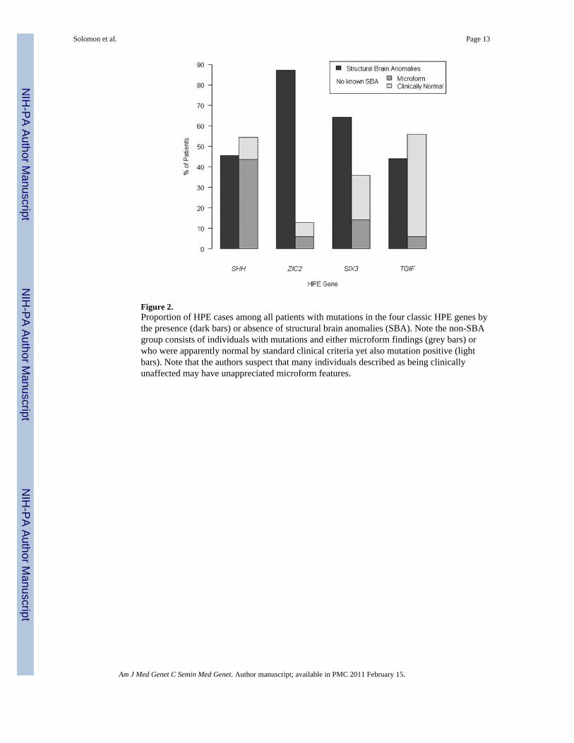
Figure 2.Proportion of HPE cases among all patients with mutations in the four classic HPE genes bythe presence (dark bars) or absence of structural brain anomalies (SBA). Note the non-SBAgroup consists of individuals with mutations and either microform findings (grey bars) orwho were apparently normal by standard clinical criteria yet also mutation positive (lightbars). Note that the authors suspect that many individuals described as being clinicallyunaffected may have unappreciated microform features.
Solomon et al. Page 13
Am J Med Genet C Semin Med Genet. Author manuscript; available in PMC 2011 February 15.
NIH
-PA Author Manuscript
NIH
-PA Author Manuscript
NIH
-PA Author Manuscript
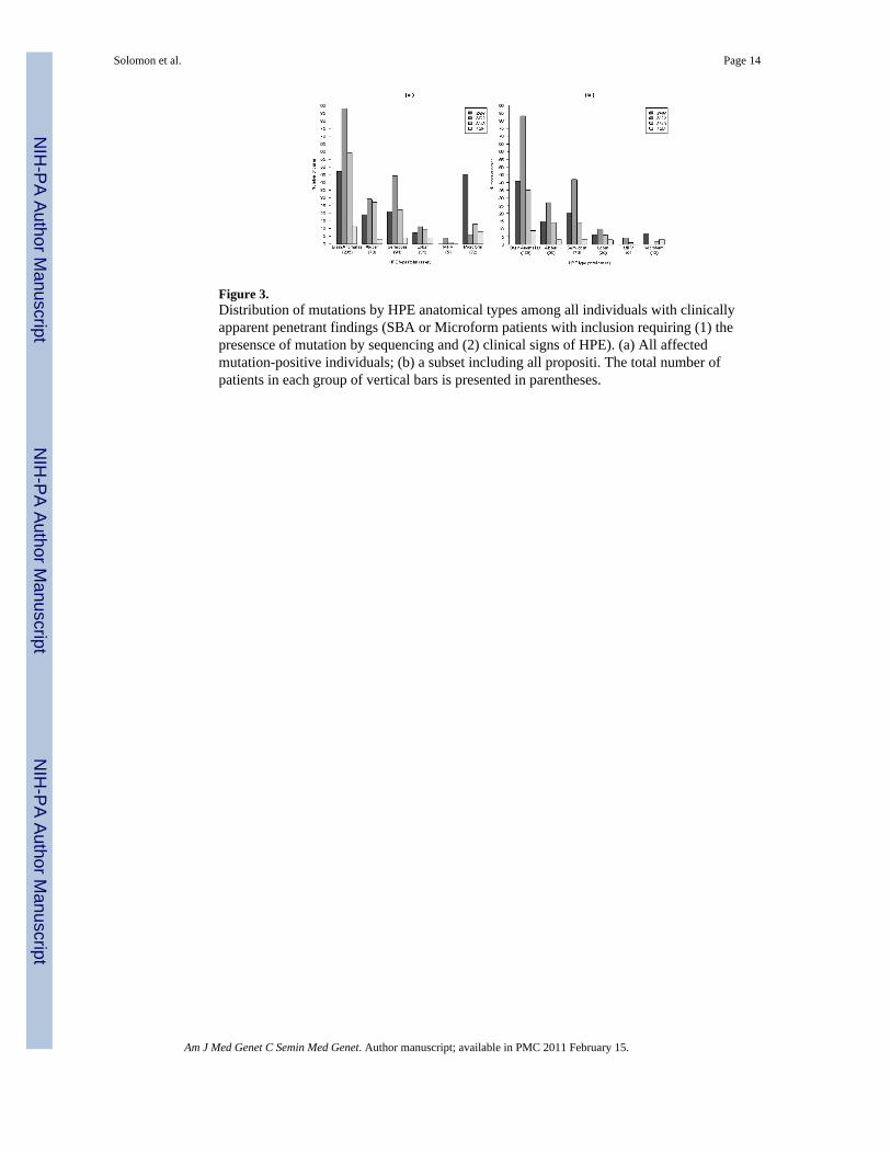
Figure 3.Distribution of mutations by HPE anatomical types among all individuals with clinicallyapparent penetrant findings (SBA or Microform patients with inclusion requiring (1) thepresensce of mutation by sequencing and (2) clinical signs of HPE). (a) All affectedmutation-positive individuals; (b) a subset including all propositi. The total number ofpatients in each group of vertical bars is presented in parentheses.
Solomon et al. Page 14
Am J Med Genet C Semin Med Genet. Author manuscript; available in PMC 2011 February 15.
NIH
-PA Author Manuscript
NIH
-PA Author Manuscript
NIH
-PA Author Manuscript

NIH
-PA Author Manuscript
NIH
-PA Author Manuscript
NIH
-PA Author Manuscript
Solomon et al. Page 15
Tabl
e I
All
indi
vidu
als w
ith id
entif
ied
mut
atio
ns in
the
4 m
ost c
omm
on H
PE-a
ssoc
iate
d ge
nes i
n w
hom
the
HPE
type
(or l
ack
ther
eof)
is k
now
n. In
divi
dual
spr
esen
ted
in th
is ta
ble
incl
ude
180
prop
ositi
and
141
rela
tives
; not
e th
at fu
ll tri
o an
alys
is w
as n
ot a
vaila
ble
for a
ll pr
opos
iti. I
ndiv
idua
ls in
who
m c
linic
alde
scrip
tions
did
not
incl
ude
HPE
type
or c
linic
al c
hara
cter
izat
ion
wer
e no
t inc
lude
d in
the
anal
ysis
.
HPE
Gen
eB
rain
Ano
mal
ies
No
Stru
ctur
al B
rain
Ano
mal
ies
Tot
alA
SL
MM
icPh
enot
ypic
ally
Nor
mal
*
SHH
1921
70
4511
103
ZIC
229
4411
46
710
1
SIX3
2722
91
1320
92
TGIF
34
40
86
25
Tota
l78
9131
572
4432
1
* Indi
vidu
als a
re d
escr
ibed
as p
heno
typi
cally
nor
mal
her
e w
hen
no m
icro
form
feat
ures
are
repo
rted
and
in w
hom
ther
e w
as n
o cl
inic
al in
dica
tion
for n
euro
imag
ing.
All
phen
otyp
ical
ly n
orm
al in
divi
dual
sw
ere
rela
tives
of p
ropo
siti
(the
vast
maj
ority
of w
hich
wer
e pa
rent
s of s
ever
ely
affe
cted
pat
ient
s).
A: A
loba
r; S:
Sem
iloba
r; L:
Lob
ar; M
: MIH
V; M
ic: M
icro
form
Am J Med Genet C Semin Med Genet. Author manuscript; available in PMC 2011 February 15.

NIH
-PA Author Manuscript
NIH
-PA Author Manuscript
NIH
-PA Author Manuscript
Solomon et al. Page 16
Tabl
e II
HPE
type
am
ong
prop
ositi
with
mut
atio
ns in
the
four
mos
t com
mon
HPE
-ass
ocia
ted
gene
s.
HPE
Gen
eH
PE T
ype
Tot
alA
loba
rSe
milo
bar
Lob
arM
IHV
Mic
rofo
rm
SHH
1520
60
748
ZIC
227
4210
40
83
SIX3
1414
61
237
TGIF
33
30
312
Tot
al59
7925
512
180
Am J Med Genet C Semin Med Genet. Author manuscript; available in PMC 2011 February 15.
Related Documents


