Citation: Mazurek, M.; Szczepanek, D.; Orzylowska, A.; Rola, R. Analysis of Factors Affecting 5-ALA Fluorescence Intensity in Visualizing Glial Tumor Cells—Literature Review. Int. J. Mol. Sci. 2022, 23, 926. https://doi.org/10.3390/ ijms23020926 Academic Editor: Kiryl D. Piatkevich Received: 15 December 2021 Accepted: 13 January 2022 Published: 15 January 2022 Publisher’s Note: MDPI stays neutral with regard to jurisdictional claims in published maps and institutional affil- iations. Copyright: © 2022 by the authors. Licensee MDPI, Basel, Switzerland. This article is an open access article distributed under the terms and conditions of the Creative Commons Attribution (CC BY) license (https:// creativecommons.org/licenses/by/ 4.0/). International Journal of Molecular Sciences Review Analysis of Factors Affecting 5-ALA Fluorescence Intensity in Visualizing Glial Tumor Cells—Literature Review Marek Mazurek *, Dariusz Szczepanek, Anna Orzylowska and Radoslaw Rola Chair and Department of Neurosurgery and Pediatric Neurosurgery, Medical University of Lublin, 20-954 Lublin, Poland; [email protected] (D.S.); [email protected] (A.O.); [email protected] (R.R.) * Correspondence: [email protected]; Tel.: +48-81-724-41-76 Abstract: Glial tumors are one of the most common lesions of the central nervous system. Despite the implementation of appropriate treatment, the prognosis is not successful. As shown in the literature, maximal tumor resection is a key element in improving therapeutic outcome. One of the methods to achieve it is the use of fluorescent intraoperative navigation with 5-aminolevulinic acid. Unfortunately, often the level of fluorescence emitted is not satisfactory, resulting in difficulties in the course of surgery. This article summarizes currently available knowledge regarding differences in the level of emitted fluorescence. It may depend on both the histological type and the genetic profile of the tumor, which is reflected in the activity and expression of enzymes involved in the intracellular metabolism of fluorescent dyes, such as PBGD, FECH, UROS, and ALAS. The transport of 5-aminolevulinic acid and its metabolites across the blood–brain barrier and cell membranes mediated by transporters, such as ABCB6 and ABCG2, is also important. Accompanying therapies, such as antiepileptic drugs or steroids, also have an impact on light emission by tumor cells. Accurate determination of the factors influencing the fluorescence of 5-aminolevulinic acid-treated cells may contribute to the improvement of fluorescence navigation in patients with highly malignant gliomas. Keywords: 5-aminolevulinic acid; high-grade glioma; glioblastoma; intraoperative navigation 1. Introduction Gliomas are among the most common tumors found in neurosurgery. It is estimated that they constitute 30% of all brain tumors and as much as 80% of malignant lesions [1]. The standard of care for malignant gliomas is maximal tumor resection, followed by radio- and chemotherapy [2]. Unfortunately, despite appropriate treatment, these tumors are prone to recurrences and have an unfavorable prognosis [3–5]. One of the reasons for this phenomenon is the high migratory ability of glioma cells, which renders the gross total resection (GTR) highly unlikely [6,7]. The analyses performed showed the presence of tumor cells up to 4 cm from macroscopically visible tumor margins [8,9]. An incomplete resection results in higher risk of neoplasm recurrence and poorer effectiveness of adjuvant therapies, such as radio- and chemotherapy [10–15]. For this reason, much attention is paid to the improvement of surgical techniques that help to maximize the percentage of achieved GTR. One of them is intraoperative fluorescence navigation that facilitates intraoperative visualization of the neoplastic tissue through the administration of substances that make it fluoresce [16,17]. Currently, a few substances are used as fluorescent indicators in brain tumor surgery. The most commonly used dye is 5-aminolevulinic acid (5-ALA) [18–20]. This compound participates in the heme metabolic pathway [21–23]. Usually, it is administered orally to patients in the form of an aqueous solution at a dose of 20 mg/kg body weight, about 3 h before the planned surgery [24,25]. Thereafter, in the body it is metabolized to the heme precursor—protoporphyrin IX (PpIX) [25–27]. This compound has two unique features Int. J. Mol. Sci. 2022, 23, 926. https://doi.org/10.3390/ijms23020926 https://www.mdpi.com/journal/ijms

Welcome message from author
This document is posted to help you gain knowledge. Please leave a comment to let me know what you think about it! Share it to your friends and learn new things together.
Transcript
�����������������
Citation: Mazurek, M.; Szczepanek,
D.; Orzyłowska, A.; Rola, R. Analysis
of Factors Affecting 5-ALA
Fluorescence Intensity in Visualizing
Glial Tumor Cells—Literature
Review. Int. J. Mol. Sci. 2022, 23, 926.
https://doi.org/10.3390/
ijms23020926
Academic Editor: Kiryl D. Piatkevich
Received: 15 December 2021
Accepted: 13 January 2022
Published: 15 January 2022
Publisher’s Note: MDPI stays neutral
with regard to jurisdictional claims in
published maps and institutional affil-
iations.
Copyright: © 2022 by the authors.
Licensee MDPI, Basel, Switzerland.
This article is an open access article
distributed under the terms and
conditions of the Creative Commons
Attribution (CC BY) license (https://
creativecommons.org/licenses/by/
4.0/).
International Journal of
Molecular Sciences
Review
Analysis of Factors Affecting 5-ALA Fluorescence Intensity inVisualizing Glial Tumor Cells—Literature ReviewMarek Mazurek *, Dariusz Szczepanek, Anna Orzyłowska and Radosław Rola
Chair and Department of Neurosurgery and Pediatric Neurosurgery, Medical University of Lublin,20-954 Lublin, Poland; [email protected] (D.S.); [email protected] (A.O.);[email protected] (R.R.)* Correspondence: [email protected]; Tel.: +48-81-724-41-76
Abstract: Glial tumors are one of the most common lesions of the central nervous system. Despitethe implementation of appropriate treatment, the prognosis is not successful. As shown in theliterature, maximal tumor resection is a key element in improving therapeutic outcome. One of themethods to achieve it is the use of fluorescent intraoperative navigation with 5-aminolevulinic acid.Unfortunately, often the level of fluorescence emitted is not satisfactory, resulting in difficulties inthe course of surgery. This article summarizes currently available knowledge regarding differencesin the level of emitted fluorescence. It may depend on both the histological type and the geneticprofile of the tumor, which is reflected in the activity and expression of enzymes involved in theintracellular metabolism of fluorescent dyes, such as PBGD, FECH, UROS, and ALAS. The transportof 5-aminolevulinic acid and its metabolites across the blood–brain barrier and cell membranesmediated by transporters, such as ABCB6 and ABCG2, is also important. Accompanying therapies,such as antiepileptic drugs or steroids, also have an impact on light emission by tumor cells. Accuratedetermination of the factors influencing the fluorescence of 5-aminolevulinic acid-treated cells maycontribute to the improvement of fluorescence navigation in patients with highly malignant gliomas.
Keywords: 5-aminolevulinic acid; high-grade glioma; glioblastoma; intraoperative navigation
1. Introduction
Gliomas are among the most common tumors found in neurosurgery. It is estimatedthat they constitute 30% of all brain tumors and as much as 80% of malignant lesions [1].The standard of care for malignant gliomas is maximal tumor resection, followed by radio-and chemotherapy [2]. Unfortunately, despite appropriate treatment, these tumors areprone to recurrences and have an unfavorable prognosis [3–5]. One of the reasons for thisphenomenon is the high migratory ability of glioma cells, which renders the gross totalresection (GTR) highly unlikely [6,7]. The analyses performed showed the presence oftumor cells up to 4 cm from macroscopically visible tumor margins [8,9]. An incompleteresection results in higher risk of neoplasm recurrence and poorer effectiveness of adjuvanttherapies, such as radio- and chemotherapy [10–15]. For this reason, much attention is paidto the improvement of surgical techniques that help to maximize the percentage of achievedGTR. One of them is intraoperative fluorescence navigation that facilitates intraoperativevisualization of the neoplastic tissue through the administration of substances that make itfluoresce [16,17].
Currently, a few substances are used as fluorescent indicators in brain tumor surgery.The most commonly used dye is 5-aminolevulinic acid (5-ALA) [18–20]. This compoundparticipates in the heme metabolic pathway [21–23]. Usually, it is administered orally topatients in the form of an aqueous solution at a dose of 20 mg/kg body weight, about 3 hbefore the planned surgery [24,25]. Thereafter, in the body it is metabolized to the hemeprecursor—protoporphyrin IX (PpIX) [25–27]. This compound has two unique features
Int. J. Mol. Sci. 2022, 23, 926. https://doi.org/10.3390/ijms23020926 https://www.mdpi.com/journal/ijms
Int. J. Mol. Sci. 2022, 23, 926 2 of 27
that allow it to be used in fluorescence navigation. One of them is the ability to emit a lightwave after excitation by blue-violet light with a wavelength of 375–440 nm. This allows forintraoperative imaging of its deposits thanks to the use of an operating microscope with aspecial set of filters [19,21,23,28–30]. The second unique feature of the dye is its selectiveaccumulation in high-grade glioma cells compared with normal brain tissue. This allowsthe surgeon to determine the likely margins of the operated lesion while the procedureis still ongoing. Data available in the literature indicate high specificity (83.8–93.9%) andsensitivity (73.9–91.4%) presented by 5-aminolevulinic acid [31–40].
The first studies on the usefulness of 5-ALA in high-grade glioma surgery were carriedout in the 1990s [21,22]. Since then, the use of this dye has been repeatedly demonstrated toimprove the percentage of GTR achieved in glioblastoma patients by using intraoperativefluorescence navigation [19,24,29,41–44]. Additionally, as a meta-analysis by Gandhi et al.shows, this also results in progression-free survival and survival rates of the patients [18].However, the presence of fluorescence and its nature are not the same for all glial tumors.Many times, despite the supply of an appropriate dose of dye in the appropriate timewindow, no fluorescence is observed after the tumor is visualized, which significantlyhinders the course of the procedure. The goal of this study is a literature review on thetopic of the factors affecting the mechanism of fluorescence induced by the supply of5-aminolevulinic acid.
2. Intracellular Metabolism of 5-Aminolevulinic Acid
When considering the metabolism of 5-ALA and its potential influence on the strengthof fluorescence, one should remember that the cell can derive 5-ALA from two sources.The first one is the exogenous supply. The dye is administered orally to patients; then it isabsorbed from the digestive tract and distributed throughout the body. Exogenous 5-ALA isdelivered to cells via special transporter proteins. This group includes peptide transporter 1(PEPT1) and peptide transporter 2 (PEPT2) [45,46]. The other source of the dye is itsendogenous fraction, produced from succinyl-CoA and glycine. This process takes place inthe mitochondria and is catalyzed by ALA synthase (ALAS) [46–49]. Then, endogenous5-ALA is transported to the cytoplasm, where it can undergo further transformationstogether with the exogenous fraction.
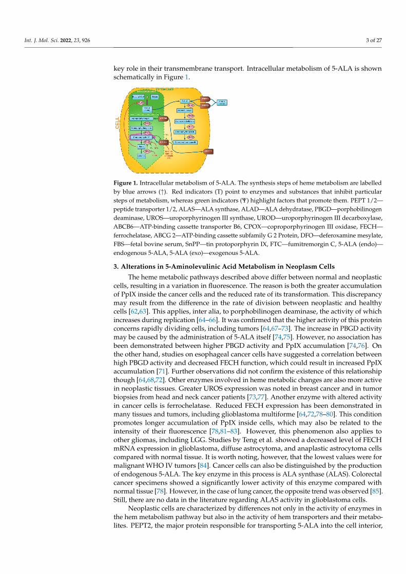
The first steps occur in the cytoplasm, while the final transformations take place inthe mitochondria [50]. In the first act of metabolism, two 5-ALA molecules condense toporphobilinogen (PBG) in a reaction catalyzed by ALA dehydratase (ALAD). Anothername for this enzyme is porphobilinogen synthase (PBGS). Then, porphobilinogen deami-nase (PBGD), also known as hydroxymethylbilane synthase (HMBS), catalyzes the fusionreaction of four PBG molecules, resulting in the formation of hydroxymethylbilane (HMB).Its structure is later closed by uroporphyrinogen III synthase (UROS) to form the cyclicuroporphyrinogen III. The next step is its decarboxylation. This reaction is catalyzed byuroporphyrinogen III decarboxylase (UROD), and its product is coproporphyrinogen III.The next stages of metabolism take place already in the mitochondria. The transport ofmetabolites is mediated by ATP-binding cassette transporter B6 (ABCB6). In mitochon-dria, coproporphyrinogen III oxidase (CPOX) catalyzes the oxidative decarboxylation ofcoproporphyrinogen III, resulting in the formation of protoporphyrinogen III. It is furtheroxidized to protoporphyrinogen IX (PpIX) by protoporphyrinogen III oxidase (PPOX). Itis protoporphyrinogen IX that is the main source of fluorescence used in intraoperative5-ALA navigation. In the last step, Fe2+ is included in the pyrrole ring of PpIX. As a result,nonfluorescent heme is formed. This process also takes place in the mitochondria andis catalyzed by ferrochelatase (FECH). Additionally, reaction can be accelerated by hemeoxygenase-1 (HO-1) [48,50–57]. If heme and free porphyrin metabolites remain insidethe cells for a long time, they can induce oxidative stress and damage them. For thisreason, a well-coordinated mechanism of their further transport and degradation is veryimportant [58]. The ATP-binding cassette subfamily G (ABCG) 2 protein [59–61] plays a
Int. J. Mol. Sci. 2022, 23, 926 3 of 27
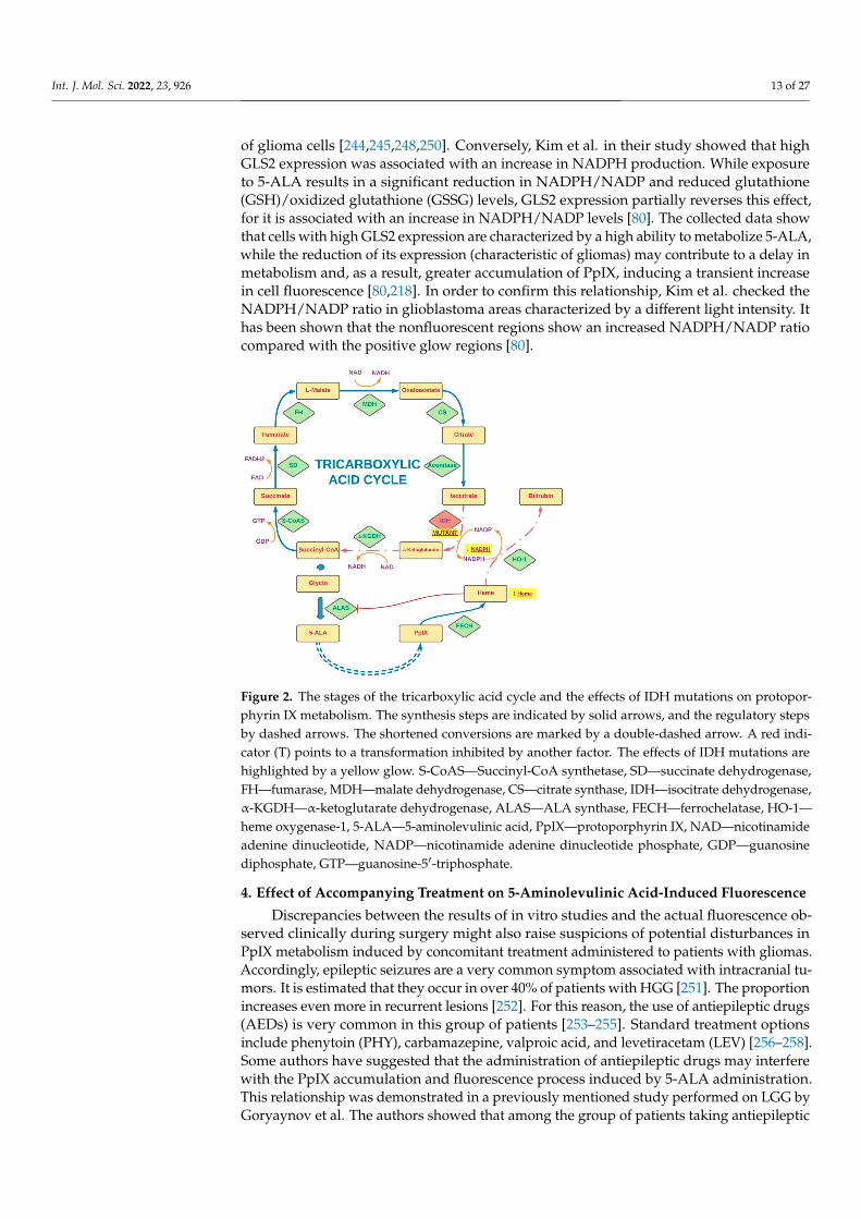
key role in their transmembrane transport. Intracellular metabolism of 5-ALA is shownschematically in Figure 1.
Figure 1. Intracellular metabolism of 5-ALA. The synthesis steps of heme metabolism are labelledby blue arrows (↑). Red indicators (T) point to enzymes and substances that inhibit particularsteps of metabolism, whereas green indicators (Ψ) highlight factors that promote them. PEPT 1/2—peptide transporter 1/2, ALAS—ALA synthase, ALAD—ALA dehydratase, PBGD—porphobilinogendeaminase, UROS—uroporphyrinogen III synthase, UROD—uroporphyrinogen III decarboxylase,ABCB6—ATP-binding cassette transporter B6, CPOX—coproporphyrinogen III oxidase, FECH—ferrochelatase, ABCG 2—ATP-binding cassette subfamily G 2 Protein, DFO—deferoxamine mesylate,FBS—fetal bovine serum, SnPP—tin protoporphyrin IX, FTC—fumitremorgin C, 5-ALA (endo)—endogenous 5-ALA, 5-ALA (exo)—exogenous 5-ALA.
3. Alterations in 5-Aminolevulinic Acid Metabolism in Neoplasm Cells
The heme metabolic pathways described above differ between normal and neoplasticcells, resulting in a variation in fluorescence. The reason is both the greater accumulationof PpIX inside the cancer cells and the reduced rate of its transformation. This discrepancymay result from the difference in the rate of division between neoplastic and healthycells [62,63]. This applies, inter alia, to porphobilinogen deaminase, the activity of whichincreases during replication [64–66]. It was confirmed that the higher activity of this proteinconcerns rapidly dividing cells, including tumors [64,67–73]. The increase in PBGD activitymay be caused by the administration of 5-ALA itself [74,75]. However, no association hasbeen demonstrated between higher PBGD activity and PpIX accumulation [74,76]. Onthe other hand, studies on esophageal cancer cells have suggested a correlation betweenhigh PBGD activity and decreased FECH function, which could result in increased PpIXaccumulation [71]. Further observations did not confirm the existence of this relationshipthough [64,68,72]. Other enzymes involved in heme metabolic changes are also more activein neoplastic tissues. Greater UROS expression was noted in breast cancer and in tumorbiopsies from head and neck cancer patients [73,77]. Another enzyme with altered activityin cancer cells is ferrochelatase. Reduced FECH expression has been demonstrated inmany tissues and tumors, including glioblastoma multiforme [64,72,78–80]. This conditionpromotes longer accumulation of PpIX inside cells, which may also be related to theintensity of their fluorescence [78,81–83]. However, this phenomenon also applies toother gliomas, including LGG. Studies by Teng et al. showed a decreased level of FECHmRNA expression in glioblastoma, diffuse astrocytoma, and anaplastic astrocytoma cellscompared with normal tissue. It is worth noting, however, that the lowest values were formalignant WHO IV tumors [84]. Cancer cells can also be distinguished by the productionof endogenous 5-ALA. The key enzyme in this process is ALA synthase (ALAS). Colorectalcancer specimens showed a significantly lower activity of this enzyme compared withnormal tissue [78]. However, in the case of lung cancer, the opposite trend was observed [85].Still, there are no data in the literature regarding ALAS activity in glioblastoma cells.
Neoplastic cells are characterized by differences not only in the activity of enzymes inthe hem metabolism pathway but also in the activity of hem transporters and their metabo-lites. PEPT2, the major protein responsible for transporting 5-ALA into the cell interior,
Int. J. Mol. Sci. 2022, 23, 926 4 of 27
has been shown to be overexpressed in glioblastoma cells [84]. Analogue discrepancyapplies to ABCG2, which is also an intermembrane transporter of hem metabolites [86–88].ABCB6 is another transporter belonging to the same family of proteins. Zhao et al. notedthat it may also affect the distribution of PpIX. Their studies on glioblastoma cells showedhigher expression of this protein in glioma cells compared with normal brain tissue, butthe intracellular localization of ABCB6 does not provide unambiguous evidence regardingits influence on the degree of PpIX accumulation [89–92]. However, the above-presenteddiscrepancies in the metabolism and transport of heme metabolites result in greater accumu-lation of PpIX in some tumor cells, which is a key aspect used in fluorescence intraoperativenavigation [22,93]. Additionally, studies by Stummer et al. in a rat model showed thatglioblastoma cells metabolize 5-ALA and collect its excess both in vivo and in vitro, whichmay interfere with the detection of fluorescence [22]. The heme metabolism is subject tonatural regulatory processes. The availability of substrates and intermediates plays animportant role in it. An important regulating point is, inter alia, inhibition of ALAS in thefeedback mechanism [94]. It can be inhibited directly by the intracellular heme level [95].However, this compound also influences other stages of metabolism, including the rate ofits degradation by heme oxidase [50,83].
However, PpIX fluorescence after exogenous 5-ALA supply is not a common featureof all neoplastic cells. It has been shown many times that it concerns mainly cells exhibitingmarked features of malignancy. It is clear that the use of fluorescent intraoperative navi-gation shows the significant effectiveness of this method in the treatment of patients withhigh-grade glioma (HGG) [19,24,29,41–43,96–99]. However, for low-grade glioma (LGG),the statistics are not that promising. One of the first reports on the use of 5-ALA in thesurgery of low-grade gliomas comes from Ishihara et al. The authors, examining 65 slicesfrom six resected tumors, showed that diffuse astrocytomas exhibit noticeably weaker PpIXfluorescence compared with anaplastic astrocytomas and glioblastoma [100]. This was inline with the later work of Widhalm et al., who showed no light emission for 100% (8/8) ofthe samples from patients with WHO grade II diffusely infiltrating gliomas [101]. Theirlater work confirmed these initial observations—of the 215 tumor specimens analyzedfrom 59 patients with diffusely infiltrating gliomas, only 19% (4/33) of the WHO II tumorsamples showed noticeable fluorescence. For comparison, in WHO grade III gliomas, focalPpIX fluorescence was visible in 85% (23/26) of the cases [102]. A similar inconsistency wasvisible in the insights of Ewelt et al., who detected visible PpIX fluorescence only in 7.7%(1/13) of WHO II glioma and 70.6% (12/17) of higher-grade tumors [32]. Observationson a larger group of patients were conducted by Wadiur et al. The authors analyzed thepresence of fluorescence in 110 patients with low-grade glioma tumors in imaging studiesand recorded it in 36% (40/110) of the cases. Moreover, subsequent analysis of the resultsof histopathological examinations revealed that of the WHO II tumors, only 11% (7/65)showed noticeable luminosity. In the case of WHO III and WHO IV tumors, it was 68%(26/38) and 100% (7/7), respectively [103]. Similarly, Jaber et al. in their study proved thatof 166 eligible tumors (82 WHO II, 76 WHO III, and 8 WHO IV), PpIX fluorescence waspresent in more malignant tumors. The authors concluded that if the lesion fluoresces, it isHGG 85% of the time. For LGG, the percentage of glowing tumors was 16% (13/82) [104].The problem with visualizing pure low-grade glioma fluorescence was also reported byother authors [27,105–110]. However, there are case reports of LGG patients showing visiblefluorescence [111,112]. Moreover, Marbacher et al. confirmed the presence of detected glowin 40% (8/20) of the analyzed LGGs [108]. Similar value results were shown by Valdés et al.Goryaynov et al. showed even greater fluorescence when out of 27 histologically confirmedtumors (14 diffuse astrocytomas, 6 oligodendrogliomas, 4 pilocytic astrocytomas, 2 gemisto-cytic astrocytomas, and 1 desmoplastic infantile ganglioglioma), 52% showed fluorescenceafter exogenous 5-ALA supply at a dose of 20 mg/kg. However, the quality of lighting wasdiverse—50% of the tumors showed diffuse fluorescence, and 50% focal [113,114]. Someauthors suggested that such foci of increased fluorescence in low-grade glioma tissue mayindicate local malignancy [114,115].
Int. J. Mol. Sci. 2022, 23, 926 5 of 27
Interestingly, fluorescence heterogeneity is also frequently observed in tumors with ahigh proliferation rate. Many have authors shown significant discrepancy in the fluorescenceintensity between different glioma cell lines subjected to the same conditions [46,57,116,117].The phenomenon has also been observed intraoperatively. Stummer et al. noted significantregional heterogeneity in fluorescence intensity for glioblastoma tissue. It has been sug-gested that the reason for the different intensity of light may be a various cell density withinthe tumor [101,102,118]. However, it is probably caused by the significant genetic polymor-phism of neoplastic cells resulting from the rate of their proliferation. This was very welloutlined in a study by Kim et al., in which the authors classified samples from five patientswith glioblastoma in terms of luminosity and then subjected them to a thorough RNAsequencing analysis. A total of 585 genes that influence the PpIX accumulation and fluo-rescence intensity were identified [80]. As previously mentioned, other authors have alsosuggested a relationship between the rate of cell proliferation and the luminosity [18–20].Additionally, Widhalm et al. analyzed the role of 5-ALA in identifying anaplasia foci indiffusely infiltrating gliomas with nonsignificant contrast enhancement [101]. The resultsobtained by the authors showed that Ki-67/MIB-1 is significantly higher in the areas ofthe tumor showing PpIX fluorescence (20% vs. 10%) [101]. In their following work, theauthors analyzed features of 215 tumor specimens collected from 59 patients with diffuselyinfiltrating gliomas with nonsignificant contrast enhancement on MRI. They showed thatin fluorescent areas, mitotic rate, cell density, nuclear pleomorphism, and proliferationrate were significantly higher than in nonfluorescing areas. Similar results were presentedby Ohba et al., who in their observations on 104 patients with glioma showed that con-trast enhancement, WHO malignancy, IDH status, and the Ki-67/MIB-1 index influenceintraoperative tumor glow assessed by the surgeon [106]. Likewise, Jaber et al. exam-ined the influence of tumor volume, 18F-FET PET uptake, contrast enhancement, grade,IDH1 mutation status, O6-methylguanine DNA methyltransferase (MGMT) promotermethylation status, 1p/19q codeletion, and Ki-67/MIB-1 and proved that the intensity offluorescence correlates with the expression of Ki-67 and with the grades of histologicalmalignancy. No similar relationship was found for MGMT status, IDH1 mutation status,or 1p19q codeletion status, however [104]. Equally, Saito et al., on the basis of univariateanalysis, also showed a relationship between the intensity of cell proliferation measuredwith the Ki-67/MIB-1 index and the luminosity induced by 5-ALA supply. It should benoted, though, that a similar relationship also occurred in the case of 1p19q codeletionstatus in contrast to the previously mentioned work. A work by Ishihara et al. is alsoworth mentioning, in which on top of the similar relationship between the Ki-67/MIB-1index and the fluorescence intensity, it was shown that this parameter is also influenced byCD31-microvessel density and larger VEGF expression. However, a more detailed multipleregression analysis showed that only the Ki-67/MIB-1 index was significantly related to thefluorescence intensity [19]. It has been suggested that modifications of tumor metabolismrelated to increased cell proliferation might affect the activity of enzymes responsible for theintensity of PpIX accumulation [80,119]. Mechanisms affecting the visibility of fluorescencewere induced by the supply of 5-aminolevulinic acid.
3.1. Blood–Brain Barrier
One of the factors that may influence the accumulation of dyes in cells, and hencethe intensity of light, is the structure of the blood–brain barrier (BBB). It is the primaryborder separating the brain environment from the rest of the body. However, brain tumorscan cause dysfunction and degradation, simultaneously affecting tumor growth and theeffectiveness of therapeutic strategies. This is also true for fluorescent intraoperative nav-igation [120]. The potential for 5-ALA penetration into the brain was considered in thepast, explaining the presence of neuropsychiatric symptoms in hepatic porphyria [121].However, observations of the distribution of radiolabeled ALA by Terr and Weiner showedno penetration of the dye into the brain tissue [122]. This was in line with the conclu-sions of Stummer et al., who did not show the presence of fluorescence caused by 5-ALA
Int. J. Mol. Sci. 2022, 23, 926 6 of 27
administration in patients with a normal blood brain barrier structure [123]. It thereforeappears that under normal conditions, it is impermeable to 5-ALA [84]. On the other hand,transport via BBB was noted by McGillion et al., and a slight displacement of 5-ALA withinthe blood–brain barrier itself has also been shown in other studies [124,125]. Few authorshave described the ability of choroid plexus to transport 5-ALA, but it has not been shownthat it is able to penetrate the brain tissue [123,126,127]. Therefore, Ennis et al. analyzedthe distribution of radiolabeled ALA across the blood–brain and blood–cerebrospinal fluidbarrier in rats. The authors showed that in adults, the spread of the dye in the brain tissuewas low and suggested that this probably took place by means of passive diffusion sincethe increase in plasma ALA concentration was not associated with the increase in its distri-bution to the brain tissue [128]. The lack of active transporters was also consistent with theresults of García et al., who analyzed the structure of brain microvessels without provingthe presence of transporters enabling the distribution of the dye to the brain tissue [125].The above observations suggest that despite the possibility of interaction of 5-ALA withmembrane transporters in various tissues, the blood–brain barrier in the normal state ispractically impermeable to it [129–132]. Consequently, the condition necessary for theaccumulation of PpIX after the exogenous supply of the dye is the state of disturbance ofits structure. The severity of BBB dysfunction depends on the location, volume, type, andmalignancy of the tumor [133]. In the case of gliomas, the difference in barrier permeabilitybetween LGG and HGG has been repeatedly demonstrated. While in tumors of low malig-nancy BBB disruption is relatively minor, in malignant gliomas it results in edema and theformation of areas with impaired vascular density and integrity [134–137]. Additionally,astrocytes migrate away from vascular endothelial cells, resulting in the disruption of abarrier structure, thereby affecting BBB permeability [138]. This may provide an explana-tion for the disparity in PpIX accumulation and fluorescence of lesions depending on theirmalignancy [28,93,120,134,139].
3.2. ABCG2 and ABCB6 Transporters
Intracellular 5-ALA transport may also have an influence on the PpIX accumulationand fluorescence intensity. The ABCG2 transporters, which as previously mentioned arecrucial in the distribution of heme metabolites, play an important role in this issue. Theseare proteins belonging to the larger family of the ATP-binding cassette superfamily locatedin the plasma membrane [1,61]. Their function is not only to remove porphyrins but also totransport xenobiotics and harmful toxins outside the cell [140]. The presence of ABCG2was first demonstrated in a study of doxorubicin-resistant breast cancer cells, which gave itits second name, breast cancer resistance protein (BCRP) [141]. Later they were found inmany tissues of the body, including the blood–brain barrier, and their overexpression is acharacteristic feature of many cancers [30,87,88,142–149]. Moreover, their expression levelcorresponds to the histological grade of neoplastic lesions [88]. It has been shown that highactivity of ABCG2 is associated with a decrease in intracellular accumulation of PpIX afterALA administration, which results in a lower intensity of fluorescence [60,149]. Moreover,the fact that blocking ABCG2 receptors causes the accumulation of porphyrins and ABCG2blocking with imatinib and gefitinib increases the effectiveness of photodynamic therapyhave already been shown [150–153]. This type of transporter can also be blocked bygenistein [154]. Reiner et al. observed the fluorescence intensity in three different GBMcell lines and found no effect of genistein on the intensity of endogenous PpIX glow.However, after 5-ALA was added to the cell lines, there were significant discrepanciesdue to dye accumulation. The simultaneous application of genistein and 5-ALA increasedPpIX fluorescence by 42% for U87MG cells, by 31% for U87wtEGFR cells, and by 54% forU87vIII cells compared with the use of 5-ALA alone [155]. This phenomenon was alsoobserved in studies by Piffaretti et al., which showed the effect of increasing genisteinconcentrations on the viability of the analyzed glioblastoma cell lines. Moreover, theyalso noticed an increase in fluorescence of cells incubated in genistein and 5-ALA mediacompared with 5-ALA alone [46]. A number of other compounds are also known that
Int. J. Mol. Sci. 2022, 23, 926 7 of 27
may affect the expression level and function of ABCG2 transporters, thereby affecting theintensity of PpIX fluorescence, such as Ko143 or flavonoids, including 6-prenylchrysis andtectochrysin [156–159]. Currently, there are no studies of the impact of their application onthe fluorescence intensity in gliomas though. It is worth mentioning that the fluorescenceintensity and transport activity can be influenced not only by chemical but also by physicalfactors. Recent studies on breast cancer stem cells have shown that ultrasound has theeffect of reversing chemoresistance by altering the expression of ABCG2 [160]. Interestingconclusions were also brought by a research conducted by Higuchi et al. The authorsanalyzed the effect of ultrasound on the intensity of fluorescence of cells and on theexpression of ABCG2 after 5-ALA administration. It was shown that exposure of thecells to 5-ALA caused a slight increase in the expression of the transporters; however,this effect was suppressed by ultrasounds that reduced the expression of ABCG2. Theincrease in the intracellular accumulation of PpIX in glioblastoma cells was confirmedby a spectrometer. This applied to all analyzed cell lines, and the effect lasted for over2 h with some differences in the intensity and dynamics of the increase in glow betweendifferent cell populations [161]. It should be mentioned, however, that the inhibition ofABCG2 was not associated with an increase in PpIX accumulation in all observations.Wang et al. used reserpine to lower ABCG2 activity in their study on glioma cancerstem cells (GSCs) and showed that it did not improve the PpIX fluorescence in both thegroup of GSCs and the control group. Moreover, in the case of GSCs, the effect was evenlower accumulation of PpIX [162]. This may be due to the influence of reserpine on otherABC family transporters present in the cell: ABCB6, ABCB7, and ABCB10. Disruptionof their function may impair PpIX metabolism, contributing to its lower accumulation incells [162,163]. However, the abovementioned data show that the regulation of the functionof ABCG2 transporters has a great potential to modify PpIX accumulation and tumor cellfluorescence during intraoperative navigation. One should remember yet that long-termadministration of ABCG2 inhibitors may be associated with phototoxicity reactions andthe disruption of transporters in other parts of the body, such as kidneys, which may havenegative consequences for patients [164,165]. Therefore, further observations in this matterare needed in order to develop an optimal strategy for a potential treatment.
Other proteins from the ATP-binding cassette superfamily family may also influenceheme transformation processes. In recent years, the role of the ABCB6 protein transporterin the regulation of PpIX metabolism in leukemic cells has been demonstrated [166]. Asmentioned before, this protein is responsible for the transport of coproporphyrinogenfrom the cytoplasm to the mitochondria in order to carry out further changes in the hemesynthesis pathway [166]. Zhao et al. in their study analyzed ABCB6 expression in surgicalglioma samples and proved a much higher expression of ABCB6 mRNA compared withnormal brain tissue. Moreover, this increase correlated with the histological grade of thetumor. WHO IV cells showed the highest expression, but a statistically significant differencewas already visible in WHO II cells, compared with normal brain tissue. In the next step,the authors showed that cells with high abundance of ABCB6 were also characterized byhigher dye accumulation and higher fluorescence intensity and proved that incubationwith exogenous ALA resulted in a further increase in ABCB6 expression [89]. Additionally,cells overexpressing ABCB6 showed a higher level of PpIX accumulation compared withcontrol cells. In the last stage of the study, the authors checked whether inhibition of ABCB6with a specific siRNA would cause a change in the intensity of dye accumulation. Theresults showed that ABCB6 expression silencing was associated with a significant decreasein accumulated PpIX in comparison with control cells. The data obtained from the studyshowed that the above-described phenomena apply only to cells exposed to exogenous5-ALA [89]. The exact mechanism by which the increase in ABCB6 expression leads toincreased PpIX accumulation has not been described to date.
Interestingly, it is not only ABCG2 transporters that can influence the efflux of PpIXfrom cells. Kitajima et al. conducted a study in which they analyzed the distribution ofPpIX in cells derived from the JFCR39 panel. It is a panel consisting 39 human cancer cell
Int. J. Mol. Sci. 2022, 23, 926 8 of 27
lines from nine different tissues (lung, colon, gastric, breast, ovarian, brain, renal, prostatecancer, and melanoma) established by the Japanese Foundation for Cancer Research. Theanalysis studied the effect of the known ABCG2 inhibitor fumitremorgin C (FTC). Inmost of the cells of the JFCR39 panel, FTC administration increased the intracellularaccumulation of PpIX and decreased its extracellular fraction, which was related to theinhibition of ABCG2 [167]. These results were consistent with the observations of otherauthors [59,168,169]. Nevertheless, this trend was not true for all of the lines included in thestudy since some of them showed no strong correlation between the level of dye excretionand ABCG2 expression. Surprisingly, a much stronger correlation was found in the caseof the expression of the protein involved in exocytosis, which is dynamin 2. Additionally,inhibition of dynamin 2 significantly increased the accumulation of PpIX by limiting theexcretion of the dye [167]. It may be beneficial to perform subsequent observations onglioblastoma cells in order to better understand this mechanism.
3.3. Activity of Ferrochelatase (FECH)
Another point of key importance in the accumulation of PpIX is the incorporation ofFe2+ into its pyrrole ring, resulting in the formation of nonfluorescent heme. This reaction iscatalyzed by ferrochelatase (FECH), a homodimer composed of two amino acid polypeptidechains, located in the mitochondrial membrane [55,78,170,171]. It has been shown thatmolecular defects or low expression of FECH results in a lower dye content [62,172]. Asmentioned earlier, the cells of many neoplasms are characterized by reduced activity ofthis enzyme [53,80]. This results in a lower conversion rate of PpIX to nonfluorescent hemeand hence a higher glow intensity [81,83]. Importantly, the reduction of the activity ofFECH may then contribute to an increase in PpIX accumulation inside cells, which has beenproven in colorectal and cancer cells. A similar issue, therefore, became the subject of aresearch by Teng et al. conducted on both human glioma cell lines and surgical specimens.The authors proved that glioblastoma has a prominent downregulation of FECH mRNAexpression when compared with normal brain. Other types of gliomas also showed loweralbeit less pronounced expression. In addition, inconsistency between different cell lineswas also found. The G112 line had the highest expression of ferrochelatase mRNA, whilethe U87 line had the lowest. This reflected the disproportion in fluorescence after the supplyof exogenous 5-ALA with the highest intensity in the case of the U87 line, and the lowest forG112 and SNB19 lines. Moreover, the authors affected FECH activity by siRNA in two celllines with silencing efficacy greater than 50% for both lines. This resulted in the intracellularaccumulation of PpIX and increased intensity of fluorescence in cells exposed to 5-ALA,proving that the use of small RNA interference may allow for a significant increase in thequality of fluorescence achieved [84]. Similar studies on the silencing of the FECH genewere also conducted on other neoplasms [78,84,172,173].
It is worth remembering that other factors may also influence ferrochelatase activity.The natural form of its regulation is the availability of free Fe2+ ions [57,63]. Deferoxaminemesylate (DFO) is a compound commonly used in clinical practice for the treatment ofcutaneous porphyria. It exerts its effect on blocking FECH activity by chelating iron ions sothat they cannot be introduced into the pyrrole ring of PpIX [46,174–176]. The result is anincrease in the intracellular volume of PpIX and a greater intensity of fluorescence. Hence,an increase in dye accumulation under the influence of DFO has been demonstrated invarious neoplastic cells, including glial ones [177–183]. Reiner et al. analyzed the effect ofdeferoxamine on three glioblastoma cell lines. The results showed that the supply of DFOin the absence of 5-ALA did not affect the luminance. However, among cells previouslyexposed to the dye, significant increase in glow intensity was observed in all analyzed celllines and varied from 6% for U87wtEGFR to 22% for U87vIII lines, respectively, comparedwith cells treated alone [155]. It was consistent with the observations of other authors.Valdes et al. analyzed the effect of DFO supply in studies conducted on mice implantedwith xenograft U251-GFP glioma tumor cells. The animals took a dose of deferoxaminefor 3 days and then were given 5-ALA to induce fluorescence. The results showed a 50%
Int. J. Mol. Sci. 2022, 23, 926 9 of 27
increase in PpIX fluorescence intensity in the group of animals receiving DFO comparedwith the control group. The reported glow level after dye administration was 2.9 timesgreater than the background in mice treated with DFO, and only 1.9 times greater in thecontrol group [184]. Another interesting conclusion can be drawn from studies by Wanget al. on glioma cancer stem cells (GCSs). The authors showed that the addition of DFO tocells exposed to ALA increased the accumulation of PpIX. This effect was visible both inGCS cells and in the control group [162]. These observations show the great potential ofdeferoxamine in increasing the fluorescence intensity. However, it should be mentioned thatfor some authors the results were not as promising. A study by Choudry et al. showed nogreater accumulation of PpIX in basal cell carcinoma patients after exposure to DFO [185].
3.4. Function of Heme Oxygenase (HO-1)
Another compound involved in the heme homeostasis is heme oxygenase-1 (HO-1),which is responsible for the conversion of heme into biliverdin, carbon monoxide, andFe2+ ions [186–189]. The high activity of HO-1 results in a large production of Fe2+ ions,which, as we mentioned earlier, are the regulating point of FECH efficiency. Moreover,the rapid depletion of heme itself may alter the enzymatic activity in favor of increasedPpIX metabolism by FECH. The effect is an increase in the rate of heme synthesis fromFe2+ and PpIX, which reduces the intensity of the fluorescence of cells [57,106,162,190,191].Importantly, this enzyme has been shown to be overexpressed in many conditions thataffect the CNS, such as ischemia, brain injuries, or Alzheimer’s disease [192–194]. There arealso data in the literature showing the upregulation of HO-1 in many neoplasms, such askidney, prostate, and lung cancer; squamous cell carcinoma of the oral cavity; melanoma;Kaposi’s sarcoma; lymphosarcoma; hepatoma; and chronic myeloid leukemia [195–203].This also applies to brain tumors, including gliomas [204,205]. Moreover, an associationwas noted between HO-1 expression and tumor progression and its histological grade.Paradoxically, Andaloussi et al. showed that an increase in tumor malignancy is associatedwith a higher expression of HO-1, which should translate into lower accumulation ofPpIX [204,206]. Convergent conclusions were also seen in the work of Gandini et al. Theauthors showed a significant difference in the expression of HO-1 between glioblastomaand normal brain tissue (54% vs. 22%). The expression level was increased in all analyzedgrades and histological subtypes. However, no changes in HO-1 levels were shown withincreasing tumor grade. In addition, the authors also noted a significant reduction insurvival in patients with WHO grade II and III astrocytoma with high HO-1 expression. Itconcerned only the cytoplasmic localization of the enzyme [207].
The previously mentioned studies by Wang et al. focused on the observation ofenzyme expression in a population of glioma cancer stem cells (GCSs) [162]. These are cellscharacterized by properties responsible for the initiation of the neoplastic process, oftenrelated to the tumor resistance to conventional treatments [208,209]. The observations ofWang et al. showed that the GCSs showed a lower level of fluorescence compared with thecells from the control group following exposure to 5-ALA (34.9% ± 5.4% of the GCSs vs.68.1% ± 12.6% cells in the control group). When the authors analyzed the level of HO-1expression, they found that the level of expression of this enzyme in GSCs is high, whichresults in a lower accumulation of PpIX and, hence, a lower intensity of fluorescence [162].Data linking high HO-1 expression with decreased PpIX accumulation were provided,inter alia, through studies carried out on melanoma cells. Moreover, the authors showedthat enzyme inhibition causes an increase in fluorescence intensity [210]. Similar attemptsto enhance the fluorescence of tumor cells by inhibiting the activity of HO-1 were alsomade in gliomas. The studies conducted by Piffarett and Reiner et al. investigated theeffect of tin protoporphyrin IX (SnPP, a synthetic heme analogue with a tin atom in thecore) on the intensity of fluorescence induced by 5-ALA supply in glioblastoma cell lines.Due to its structure, SnPP inhibits the activity of HO-1 and is commonly used in pediatricpatients suffering from hyperbilirubinemia [211]. Fluorescence intensity analysis showedthat the simultaneous use of SnPP and 5-ALA allows for increasing the glow intensity.
Int. J. Mol. Sci. 2022, 23, 926 10 of 27
Moreover, the increase differs between cell lines from 39% for U87wtEGFR to 81% forU87MG. Interestingly, an even greater difference in the light intensity was present withthe supply of SnPP alone [46,155]. This relationship may be related to EGFR expression.U87MG cells have normal EGFR expression, while U87wtEGFR overexpresses EGFR, andU87vIII cells express the EGFR version III mutation (EGFRvIII) [46,155].
EGFR, a member of the ErbB receptor family, is an important element in the regulationof cell growth in tissues of epithelial origin [44,212]. Recent studies have shown its keyrole in tumorigenesis, cell migration, and angiogenesis. Upon binding of a specific ligand,intracellular tyrosine kinase is activated, which leads to the activation of signaling alongthe Akt, PI3 kinase, and nuclear factor (NF-kβ) proteins pathways. The result is a decreasein cell apoptosis, an increase in proliferation and angiogenesis, and a greater tendency tomigrate [2,44,212–214]. The mutation present in EGFRvIII cells is characteristic of GBMsince 40% of glioblastoma cells have a mutation that overexpresses EGFR, of which about50% is the EGFRvIII variant, which results in uninterrupted activity of the receptor withoutthe need for external stimulation [215–220]. The abovementioned experiments clearlyindicate a relationship between the function of HO-1 and the activity of the epidermalgrowth factor receptor. The presence of a similar relationship has already been observedin non-small cell lung cancer and colon cancer. Studies have shown that EGF stimulation,through NF-kβ activation, contributes to an increase in HO-1 and can be induced by variouspathways, such as PI3K, IKK, and protein kinase C (PKC) [221–223]. Likewise, Fontana et al.focused their research on the analysis of the influence of EGFR activity on HO-1 functionin glioma cell lines: U87MG (low EGFR expression), LN229EGFR (EGFR overexpression),and BS153 (EGFRvIII mutation). Initially, all lines were exposed to 5-ALA, and theirfluorescence was checked. BS151 cells were characterized by the weakest intensity of light,which was probably related to the constitutively active EGFRvIII +. In the next step, EGFwas added to the samples. The result was a significant decrease in fluorescence for U87MGand LN229EGFR, while no significant change was observed in BS153 culture. This effectwas reversed by EGFR-specific siRNA, which reduced protein expression by approximately80% in U87MG. The authors suggested that this was related to EGFR receptor activation,which resulted in the promotion of HO-1 transcription and expression in a concentration-dependent manner. The mutant BS153 cell receptor remained uninterruptedly activewith no effect upon the addition of EGF. To test this theory, the authors inhibited theeffect of HO-1 by using SnPP (HO-1 inhibitor) and HO-1-specific siRNA. In both cases,the effect was to restore fluorescence in all cell lines, independent of EGFR expression.Additionally, gefitinib, which is a selective inhibitor of the EGF tyrosine kinase receptor,was added to lines previously exposed to EGF. As a result, the fluorescence was restoredin U87MG cells, but no effect was obtained in the case of BS153 [57]. These data clearlyindicate a strong relationship between EGFR and HO-1 activity. It also suggests a potentialcause of inhomogeneous fluorescence in some tumors composed of cells characterizedby intratumoral heterogeneity of EGFR/EGFRvIII, which may make it very difficult todetermine the extent of resection [116,224].
3.5. Significance of Isocitrate Dehydrogenase (IDH) Status
Other genetic features of tumors may also influence their susceptibility to PpIXaccumulation-dependent fluorescence. The new classification of tumors of CNS createdby the World Health Organization (WHO), in addition to the standard four-step divisionaccording to the degree of histological malignancy, also included a group of neoplasms inwhich the key role is played by the mutant isocitrate dehydrogenase (IDH) 1/2 [225]. It iswidely believed that the IDH1/2 mutation occurs at one of the early stages of gliomagenesis.As a consequence, there are two different pathways of neoplastic cell progression dependingon the mutation status [226]. Some authors have even suggested that these discrepanciesin the origin of the cells may also affect their ability to fluoresce. It should be noted that,physiologically, isocitrate dehydrogenase 1 is one of the enzymes involved in the Krebscycle. Its role is to catalyze the oxidative decarboxylation of isocitrate to α-ketoglutarate
Int. J. Mol. Sci. 2022, 23, 926 11 of 27
(α-KG) with simultaneous conversion of NADP (+) to NADPH in the cytoplasm andperoxisomes [227,228]. As mentioned earlier, endogenous ALA synthesis depends onthe availability of glycine and succinyl-coenzyme A being the reactants [46–49,229,230].This indicates a possible role for IDH in regulating PpIX metabolism. IDH1 mutationsare present in 55% of WHO III gliomas and 6% of WHO IV [231]. Their presence causesa decrease in the physiological activity of the enzyme, with the simultaneous produc-tion of R-2-hydroxyglutarate (2-HG) through the consumption of NADPH, which is anoncometabolite favoring neoplastic transformation [232–235].
In their research, Ohba et al. analyzed 104 patients operated on for glioma for fac-tors that could affect the fluorescence of the tumor tissue exposed to 5-ALA. Analysisof the collected data showed that among glial tumors, cells with the IDH1/2 mutationshowed less fluorescence compared with cells without this mutation. Interestingly, thisrelationship concerned both high- and low-grade glial tumors. Additionally, in order tofind out about the nature of this phenomenon, the authors analyzed the glow intensityof two glioma cell lines in vitro: NHAE6E7hTERTIDH1mut, which was transformed bymutant IDH1, and NHAE6E7hTERTRas, which was transformed by H-Ras (wild-typeIDH1 model). Both lines were exposed to 5-ALA. Then, the accumulation of PpIX in bothcell types was assessed. The authors showed that the concentration of the dye was lowerin NHAE6E7hTERTIDH1mut than in NHAE6E7hTERTRas. This confirmed the geneticbasis for the difference in cell fluorescence depending on the IDH1/2 mutation status. Theauthors suggested that a potential reason for this relationship is the greater activity ofFECH and HO-1 in mutant cells, which results in an increase in PpIX metabolism andits lower accumulation [106]. Similar conclusions were drawn by Hickman et al. Theyanalyzed 58 patients with HGG operated under the guidance of intraoperative fluorescencenavigation using 5-ALA. The results showed a statistically significant predominance oftumors with an IDH mutation in the group of nonfluorescent lesions—70.6% of tumors withand 100.0% without IDH mutation showed PpIX fluorescence [236]. The presented datawere consistent with the results of other authors. In their observations on 60 patients withastrocytic or oligodendroglial tumors, Saito et al. studied the effect of IDH1 status, 1p19qloss of heterozygosity (LOH), the MIB-1 labelling index, the tumor margin, heterogeneity,and contrast enhancement on MRI scans. The authors showed that only the status ofisocitrate dehydrogenase 1 allowed for predicting the fluorescence of the tested cells toa statistically significant degree. Their data indicated that only 15% of the cells with theIDH1 mutation showed intraoperative florescence induced by 5-ALA administration [109].On the other hand, Jaber et al., who also studied the factors influencing the fluorescenceof cells after 5-ALA administration, did not show any correlation between the intensityof fluorescence and IDH1, 1p/19q, and MGMT promoter methylation status. It shouldbe noted, however, that the study was deliberately selected for glial tumors not showingradiological features characteristic of glioblastoma multiforme. Out of 166 samples, postop-erative histopathological examination revealed 82 WHO II, 76 WHO III, and 8 WHO IVtumors [104].
The role of IDH1 in PpIX metabolism was also investigated by Kim et al., whose cohortincluded tumor samples from 5-ALA fluorescence-guided surgeries in 35 patients withWHO III gliomas. Postoperative examination of tumor samples revealed the presence ofIDH1 mutations in 24 of them, of which 16 showed luminosity. The authors showed astatistically significant relationship between the presence of a mutation and tumor fluores-cence. In the next stage of the experiment, PpIX analysis was performed in glioblastomacell lines. The lines U87MG-IDH1WT harboring wild-type IDH1 and U87MG-IDH1R132Hrepresenting the mutant gene variant were used in the experiment. The authors showeda significant delay in PpIX metabolism due to the IDH mutation. In the case of U87MG-IDH1WT cells, an intense increase in fluorescence was noted as early as 1 h after incubation.In U87MG-IDH1R132H cells, the increase in fluorescence was noticeably later. In order tounderstand the exact nature of this relationship, the authors assessed tricarboxylic acid(TCA) cycle-related metabolite changes using LC–MS. After administration of 5-ALA, a
Int. J. Mol. Sci. 2022, 23, 926 12 of 27
significant increase in the concentration of citrate and a decrease in the concentration ofα-ketoglutarate (α-KG) in cells presenting the mutated gene variant were noted. A sim-ilar relationship was not observed in the U87MG-IDH1WT line. Additionally, a higherproduction of R-2-hydroxyglutarate (2-HG) was demonstrated in mutant cells both withand without exposure to 5-ALA. The authors also observed a decrease in baseline NADPHlevels in cells bearing the IDH1 mutation compared with WT. After, exposure to 5-ALANADPH was almost depleted in both cell lines [218]. It has therefore been hypothesizedthat it is the balance of this compound that may be crucial for heme metabolism andPpIX accumulation.
NADPH is involved in many metabolic chains of the organism and plays an im-portant role in neoplastic cells in which the pathways of its transformation are oftendisturbed [237,238]. As mentioned before, physiologically, ALA is produced from the Krebscycle succinyl-CoA and glycine in a reaction catalyzed by ALA synthase [45,93,190,230].This enzyme is the natural regulatory point of heme metabolism. The excess of hemeinhibits the synthase function (inhibiting endogenous ALA production) and at the sametime stimulates the action of HO-1, which in the feedback mechanism enhances its degra-dation [230]. HO-1 breaks down heme together with NADPH reductase. It has been shownthat HO-1 activity can be increased also in the presence of NADPH and NADH [239].The presence of the IDH1/2 mutation greatly influences the availability of NADPH. Dataavailable in the literature suggest that its primary source in human brain cells and gliomasis the pentose phosphate pathway regulated by the activity of isocitrate dehydrogenase,which catalyzes the conversion of isocitrate to α-KG [240]. The IDH1/2 mutation causesa decrease in enzyme activity, leading to impaired NADPH production. In addition, itsconsumption is also increasing in the pathological production of 2-HG from a-KG in thereduction process dependent on NADPH [233]. The effect may be a significant deficiencyof NADPH, resulting in an insufficiently effective work of HO-1. It has been shown thatin glioblastoma cells with an IDH1 mutation, the production of the compound is reducedby more than 40% [235,241]. The addition of exogenous ALA overactivates the hememetabolic pathway to break down 5-ALA excess. With concomitant NADPH deficiency,HO-1 function is limited, resulting in low FECH expression and consequently increasedPpIX accumulation. It is this mechanism that likely causes the temporary increase influorescence in IDH-1 mutant cells demonstrated by Kim et al. [218].
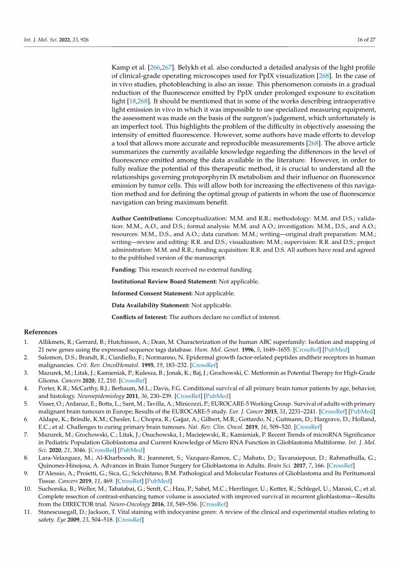
The authors explored the role of NADPH in the regulation of PpIX accumulation andfluorescence with another paper in which three types of glioblastoma tissue (collectedfrom five patients) were carefully scrutinized and characterized in terms of luminosity asstrong, weak, and absent. Then, using RNA sequencing, they showed that the expressionof 77 genes was directly and that of 508 genes inversely proportional to the intensity of thefluorescence. The effects of IDH mutations on protoporphyrin IX metabolism are shown inFigure 2. In the next stage of the experiment, Kim et al. examined the role of glutaminase 2(GLS2) in the heterogeneity of the tumor tissue fluorescence [80]. Glutaminase is anenzyme that converts glutamine to glutamate and ammonium in the mitochondria [242].Importantly, it has been proven that glutamine is the basic metabolic fuel for neoplasticcells, including glial ones [80,119,243–245]. The glutamate derived from the transformationis then converted to α-KG and incorporated into the Krebs cycle [242]. Glutaminase hastwo isoforms: the renal type (GLS), which is expressed in most tissues, and the hepatictype (GLS2), which is present in the brain, liver, and pancreas [246]. They differ in termsof regulating factors and their role in the oncogenesis process [235,242,244,247,248]. Inglioblastoma cells, silencing of GLS induces apoptosis, while overexpression of GLS2inhibits tumor growth [249]. An experiment by Kim et al. showed that GLS2 expressioninversely correlated with the intensity of cell fluorescence. Likewise, the transfection of theGLS2 gene construct into the glioblastoma cell lines (T98G, U87MG, and LN18) resultedin a decrease in PpIX accumulation and lower glow level in the samples. It was thusconfirmed that decreased GLS2 expression is associated with increased PpIX fluorescenceintensity [80]. Importantly, downregulation of GLS2 has been recognized as a hallmark
Int. J. Mol. Sci. 2022, 23, 926 13 of 27
of glioma cells [244,245,248,250]. Conversely, Kim et al. in their study showed that highGLS2 expression was associated with an increase in NADPH production. While exposureto 5-ALA results in a significant reduction in NADPH/NADP and reduced glutathione(GSH)/oxidized glutathione (GSSG) levels, GLS2 expression partially reverses this effect,for it is associated with an increase in NADPH/NADP levels [80]. The collected data showthat cells with high GLS2 expression are characterized by a high ability to metabolize 5-ALA,while the reduction of its expression (characteristic of gliomas) may contribute to a delay inmetabolism and, as a result, greater accumulation of PpIX, inducing a transient increasein cell fluorescence [80,218]. In order to confirm this relationship, Kim et al. checked theNADPH/NADP ratio in glioblastoma areas characterized by a different light intensity. Ithas been shown that the nonfluorescent regions show an increased NADPH/NADP ratiocompared with the positive glow regions [80].
Figure 2. The stages of the tricarboxylic acid cycle and the effects of IDH mutations on protopor-phyrin IX metabolism. The synthesis steps are indicated by solid arrows, and the regulatory stepsby dashed arrows. The shortened conversions are marked by a double-dashed arrow. A red indi-cator (T) points to a transformation inhibited by another factor. The effects of IDH mutations arehighlighted by a yellow glow. S-CoAS—Succinyl-CoA synthetase, SD—succinate dehydrogenase,FH—fumarase, MDH—malate dehydrogenase, CS—citrate synthase, IDH—isocitrate dehydrogenase,α-KGDH—α-ketoglutarate dehydrogenase, ALAS—ALA synthase, FECH—ferrochelatase, HO-1—heme oxygenase-1, 5-ALA—5-aminolevulinic acid, PpIX—protoporphyrin IX, NAD—nicotinamideadenine dinucleotide, NADP—nicotinamide adenine dinucleotide phosphate, GDP—guanosinediphosphate, GTP—guanosine-5′-triphosphate.
4. Effect of Accompanying Treatment on 5-Aminolevulinic Acid-Induced Fluorescence
Discrepancies between the results of in vitro studies and the actual fluorescence ob-served clinically during surgery might also raise suspicions of potential disturbances inPpIX metabolism induced by concomitant treatment administered to patients with gliomas.Accordingly, epileptic seizures are a very common symptom associated with intracranial tu-mors. It is estimated that they occur in over 40% of patients with HGG [251]. The proportionincreases even more in recurrent lesions [252]. For this reason, the use of antiepileptic drugs(AEDs) is very common in this group of patients [253–255]. Standard treatment optionsinclude phenytoin (PHY), carbamazepine, valproic acid, and levetiracetam (LEV) [256–258].Some authors have suggested that the administration of antiepileptic drugs may interferewith the PpIX accumulation and fluorescence process induced by 5-ALA administration.This relationship was demonstrated in a previously mentioned study performed on LGG byGoryaynov et al. The authors showed that among the group of patients taking antiepileptic
Int. J. Mol. Sci. 2022, 23, 926 14 of 27
drugs, tumor fluorescence was seen in only 27% of cases. In comparison, for those withoutthis form of treatment, the percentage was 83% [114]. In another study, Lawrence et al. usedthe glioblastoma cell line (U87MG) exposing the cells to antiepileptic drugs (phenytoin, val-proic acid, and levetiracetam) as well as other substances commonly used in patients withintracranial tumors, such as steroids (dexamethasone) and antidepressants (desipramine).The results showed that for all these substances, except levetiracetam, a reduction in PpIXsynthesis in GBM cells was present. What is more, combining dexamethasone with anyof the drugs (including levetiracetam) resulted in an even greater reduction in a dye pro-duction. It is worth noting, however, that the supply of the steroid was associated withgreater PpIX cell retention compared with the control sample. Concomitant use of dexam-ethasone with desipramine, valproic acid, or levetiracetam did not affect dye retention,while the combination of the steroid with phenytoin was associated with its significantreduction [259]. It has also been suggested that the supply of corticosteroids may sealoff the blood–brain barrier, leading to weaker 5-ALA penetration into tumor cells andsubsequently weaker PpIX fluorescence [84,260]. Interestingly, one of the first studies onintraoperative navigation using 5-ALA considered including preoperative dexamethasonein the standard operating protocol [24,261]. However, this is not a common practice atpresent in view of the available data. In another study, Hefti et al. analyzed the effect ofphenytoin and levetiracetam on the accumulation of PpIX in glioblastoma cell lines (U373MG and U-87 MG) and in tumor samples obtained from biopsies of patients who received5-ALA. The authors demonstrated a dose-dependent reduction in PpIX accumulation inall cell types exposed to phenytoin. Interestingly, no similar relationship was found forlevetiracetam [262]. Since the authors related this phenomenon to the disturbance of thePpIX mitochondrial synthesis, they assessed their function by measuring the mitochondrialmembrane potential (MMP). Again, the study showed a reduction in mitochondrial mem-brane potentials only after exposure to phenytoin. At the same time, no morphologicaland necrotic changes or disturbances in glutathione status, being an indicator of oxidativestress, were observed for both drugs [218]. It was suggested that the changes observed wereconsequences of the damage to protein and lipids induced by phenytoin and its metabolites,resulting in membrane disorders [262–264]. To conclude from studies by Hefti et al. andLawrence et al., one ought to remember that in patients operated with the use of 5-ALA forfluorescent intraoperative navigation, levetiracetam should be the preferred antiepilepticdrug [259,262].
Still the exact mechanism of the influence of concomitant pharmacotherapy on PpIXmetabolism remains unknown. Haust et al. in 1989 described the effects of antiepilepticdrugs (carbamazepine and valproic acid) on 5-aminolevulinic acid dehydratase and uro-porphyrinogen I synthetase activities in erythrocytes of a vitamin B6-deficient epileptic boy.The authors reported that long-term drug use led to decreased activities of 5-aminolevulinicacid dehydratase and uroporphyrinogen I synthetase, while increasing the concentrationof erythrocyte protoporphyrin. Pb poisoning, iron depletion, and erythropoietic protopor-phyria [265] were not observed. This suggests the direct influence of antiepileptic drugs onthe PpIX synthesis enzymes in the mitochondria; however, a detailed understanding of thisrelationship requires further observations. Not all authors have agreed about the effects ofAEDs and steroids on PpIX accumulation. Wadiura et al., who retrospectively analyzed thetreatment of 110 glioblastoma patients (WHO II–IV) operated on with 5-ALA navigation,found that visible fluorescence was noted only in 35% of the patients. Nonetheless, themajority of the group consisted of low-grade tumors (WHO II—59%). Of all patients, 66%of the group received preoperative premedication with antiepileptic drugs. The most fre-quently administered drug was levetiracetam alone or in combination with substances fromother groups. Analysis of the collected data showed no statistically significant correlationof the percentages of 5-ALA fluorescence between the groups of patients not taking AEDs(29%), taking LEV alone (43%), taking LEV with another AED (45%), and taking otherAED (32%). Still, some of the patients enrolled in the study were also premedicated withdexamethasone (24%). In those cases, univariate analysis showed a statistically significant
Int. J. Mol. Sci. 2022, 23, 926 15 of 27
difference in the percentages of tumors with positive fluorescence between patients receiv-ing preoperative steroid therapy (54%) and those without such therapy (31%). It shouldbe noted though that steroids were administered much more frequently in HGG, whichmay have biased the data to some extent. Comparison of data only for tumors with thesame WHO grading did not show a statistically significant relationship in the fluorescenceof tumors in patients receiving and not receiving dexamethasone (WHO II: 11% vs. 11%,WHO III/IV: 77% vs. 71% [103].
5. Conclusions
According to the data available in the literature, the use of 5-ALA in intraoperativefluorescence navigation may bring notable benefits in the surgical treatment of patientswith glial tumors, resulting in a better therapeutic outcome [18,42,43]. Unfortunately, asclinical practice shows, in many cases the operated tumors differ in the level of emittedfluorescence, which often complicates the course of resection. Moreover, this phenomenonremains largely unpredictable, introducing an element of randomness to the therapy ofpatients. For this reason, it is important to identify factors that may enhance or disruptthe phenomenon of PpIX fluorescence in glial tumor cells exposed to 5-ALA. In the lightof the reviewed publications, many authors suggest that these discrepancies may be dueto the different expression and activity of enzymes involved in the metabolism of 5-ALAdepending on the histopathological characteristics of the tumor. However, as some ofthe studies show, the different rate of cell division may also have a significant impact onthe expression of particular enzymes involved in the metabolism of protoporphyrin IX,such as PBGD, FECH, UROS, and ALAS [64,66,77,78]. In addition, the dynamics of theseprocesses depends also on the function of proteins involved in the transport of dyes andtheir metabolites. These include ABCB6 and ABCG2 [89,149,153]. The differences in theintensity of the emitted light between different tumors may also result from impairedpenetration of the dye through the blood–brain barrier. Most researchers agree that somedegree of damage to the BBB by the tumor is a necessary prerequisite for selective 5-ALAaccumulation in the tumor cells. This may be one of the differences observed in the intensityof fluorescence or lack of fluorescence in different brain tumors [122,125].
The use of substances and physical methods that potentiate the emitted fluorescencemay also be of great significance. Some authors have attempted to identify tumor profilesthat favor fluorescence. These studies include the Ki-67/MIB-1 index, MGMT status, IDHmutation, and 1p/19q codeletion. While in the case of the Ki-67/MIB-1 index and IDHmutation data available in the literature are mostly consistent, in other cases, furtherobservations are needed to assess their significance [19,101,106]. Moreover, the drugs usedin standard therapy for patients with CNS tumors may also affect 5-ALA metabolismpathways. Some authors have suggested the importance of certain antiepileptic drugs, suchas phenytoin, carbamazepine, valproic acid, and levetiracetam, in this regard [262]. Thiseffect was potentiated by the addition of dexamethasone [259]. The mechanism underlyingthis relationship is still not fully understood. It can take place both through the influence ofdrugs on the mitochondrial membrane potential and through the influence on the activityof some enzymes, such as UROS or ALAD. This draws attention to the possibility of anappropriate treatment setting in patients for whom intraoperative fluorescence navigationis used.
Furthermore, it is worth noting that the nature of tumor cell fluorescence is a complexphenomenon. This article focuses primarily on variations resulting from different accumu-lations of PpIX, the main source of excited light. This is a key aspect, especially in in vitroobservations, where light measurement is always performed under analogous conditionsusing the same instruments. However, it should be noted that the fluorescence intensityalso depends on both the equipment used to induce PpIX to emit fluorescence (operatingmicroscope, set of filters determining the light wavelength, or characteristics of the lampsused in the laboratory) and the sensitivity of the detection device. The relationship betweenthe fluorescence intensity of PpIX and the excitation light source is described in a paper by
Int. J. Mol. Sci. 2022, 23, 926 16 of 27
Kamp et al. [266,267]. Belykh et al. also conducted a detailed analysis of the light profileof clinical-grade operating microscopes used for PpIX visualization [268]. In the case ofin vivo studies, photobleaching is also an issue. This phenomenon consists in a gradualreduction of the fluorescence emitted by PpIX under prolonged exposure to excitationlight [18,268]. It should be mentioned that in some of the works describing intraoperativelight emission in vivo in which it was impossible to use specialized measuring equipment,the assessment was made on the basis of the surgeon’s judgement, which unfortunately isan imperfect tool. This highlights the problem of the difficulty in objectively assessing theintensity of emitted fluorescence. However, some authors have made efforts to developa tool that allows more accurate and reproducible measurements [268]. The above articlesummarizes the currently available knowledge regarding the differences in the level offluorescence emitted among the data available in the literature. However, in order tofully realize the potential of this therapeutic method, it is crucial to understand all therelationships governing protoporphyrin IX metabolism and their influence on fluorescenceemission by tumor cells. This will allow both for increasing the effectiveness of this naviga-tion method and for defining the optimal group of patients in whom the use of fluorescencenavigation can bring maximum benefit.
Author Contributions: Conceptualization: M.M. and R.R.; methodology: M.M. and D.S.; valida-tion: M.M., A.O., and D.S.; formal analysis: M.M. and A.O.; investigation: M.M., D.S., and A.O.;resources: M.M., D.S., and A.O.; data curation: M.M.; writing—original draft preparation: M.M.;writing—review and editing: R.R. and D.S.; visualization: M.M.; supervision: R.R. and D.S.; projectadministration: M.M. and R.R.; funding acquisition: R.R. and D.S. All authors have read and agreedto the published version of the manuscript.
Funding: This research received no external funding.
Institutional Review Board Statement: Not applicable.
Informed Consent Statement: Not applicable.
Data Availability Statement: Not applicable.
Conflicts of Interest: The authors declare no conflict of interest.
References1. Allikmets, R.; Gerrard, B.; Hutchinson, A.; Dean, M. Characterization of the human ABC superfamily: Isolation and mapping of
21 new genes using the expressed sequence tags database. Hum. Mol. Genet. 1996, 5, 1649–1655. [CrossRef] [PubMed]2. Salomon, D.S.; Brandt, R.; Ciardiello, F.; Normanno, N. Epidermal growth factor-related peptides andtheir receptors in human
malignancies. Crit. Rev. OncolHematol. 1995, 19, 183–232. [CrossRef]3. Mazurek, M.; Litak, J.; Kamieniak, P.; Kulesza, B.; Jonak, K.; Baj, J.; Grochowski, C. Metformin as Potential Therapy for High-Grade
Glioma. Cancers 2020, 12, 210. [CrossRef]4. Porter, K.R.; McCarthy, B.J.; Berbaum, M.L.; Davis, F.G. Conditional survival of all primary brain tumor patients by age, behavior,
and histology. Neuroepidemiology 2011, 36, 230–239. [CrossRef] [PubMed]5. Visser, O.; Ardanaz, E.; Botta, L.; Sant, M.; Tavilla, A.; Minicozzi, P.; EUROCARE-5 Working Group. Survival of adults with primary
malignant brain tumours in Europe; Results of the EUROCARE-5 study. Eur. J. Cancer 2015, 51, 2231–2241. [CrossRef] [PubMed]6. Aldape, K.; Brindle, K.M.; Chesler, L.; Chopra, R.; Gajjar, A.; Gilbert, M.R.; Gottardo, N.; Gutmann, D.; Hargrave, D.; Holland,
E.C.; et al. Challenges to curing primary brain tumours. Nat. Rev. Clin. Oncol. 2019, 16, 509–520. [CrossRef]7. Mazurek, M.; Grochowski, C.; Litak, J.; Osuchowska, I.; Maciejewski, R.; Kamieniak, P. Recent Trends of microRNA Significance
in Pediatric Population Glioblastoma and Current Knowledge of Micro RNA Function in Glioblastoma Multiforme. Int. J. Mol.Sci. 2020, 21, 3046. [CrossRef] [PubMed]
8. Lara-Velazquez, M.; Al-Kharboosh, R.; Jeanneret, S.; Vazquez-Ramos, C.; Mahato, D.; Tavanaiepour, D.; Rahmathulla, G.;Quinones-Hinojosa, A. Advances in Brain Tumor Surgery for Glioblastoma in Adults. Brain Sci. 2017, 7, 166. [CrossRef]
9. D’Alessio, A.; Proietti, G.; Sica, G.; Scicchitano, B.M. Pathological and Molecular Features of Glioblastoma and Its PeritumoralTissue. Cancers 2019, 11, 469. [CrossRef] [PubMed]
10. Suchorska, B.; Weller, M.; Tabatabai, G.; Senft, C.; Hau, P.; Sabel, M.C.; Herrlinger, U.; Ketter, R.; Schlegel, U.; Marosi, C.; et al.Complete resection of contrast-enhancing tumor volume is associated with improved survival in recurrent glioblastoma—Resultsfrom the DIRECTOR trial. Neuro-Oncology 2016, 18, 549–556. [CrossRef]
11. Stanescusegall, D.; Jackson, T. Vital staining with indocyanine green: A review of the clinical and experimental studies relating tosafety. Eye 2009, 23, 504–518. [CrossRef]

Int. J. Mol. Sci. 2022, 23, 926 17 of 27
12. Choi, M.; Chung, T.; Choi, K.; Choi, C. Dynamic fluorescence imaging for multiparametric measurement of tumor vasculature. J.Biomed. Opt. 2011, 16, 46008. [CrossRef] [PubMed]
13. Chaichana, K.L.; Chaichana, K.K.; Olivi, A.; Weingart, J.D.; Bennett, R.; Brem, H.; Quiñones-Hinojosa, A. Surgical outcomesfor older patients with glioblastoma multiforme: Preoperative factors associated with decreased survival: Clinical article. J.Neurosurg. 2011, 114, 587–594. [CrossRef]
14. Velde, E.T.; Veerman, T.; Subramaniam, V.; Ruers, T. The use of fluorescent dyes and probes in surgical oncology. Eur. J. Surg.Oncol. 2010, 36, 6–15. [CrossRef] [PubMed]
15. Stummer, W.; Meinel, T.; Ewelt, C.; Martus, P.; Jakobs, O.; Felsberg, J.; Reifenberger, G. Prospective cohort study of radiotherapywith concomitant and adjuvant temozolomide chemotherapy for glioblastoma patients with no or minimal residual enhancingtumor load after surgery. J. Neuro-Oncol. 2012, 108, 89–97. [CrossRef] [PubMed]
16. Castano, A.P.; Demidova, T.N.; Hamblin, M.R. Mechanisms in photodynamic therapy: Part two—cellular signaling, cellmetabolism and modes of cell death. Photodiagn. Photodyn. Ther. 2005, 2, 1–23. [CrossRef]
17. Akimoto, J. Photodynamic Therapy for Malignant Brain Tumors. Neurol. Med.-Chir. 2016, 56, 151–157. [CrossRef] [PubMed]18. Gandhi, S.; Meybodi, A.T.; Belykh, E.; Cavallo, C.; Zhao, X.; Syed, M.P.; Moreira, L.B.; Lawton, M.T.; Nakaji, P.; Preul, M.C.
Survival Outcomes among Patients With High-Grade Glioma Treated with 5-Aminolevulinic Acid–Guided Surgery: A SystematicReview and Meta-Analysis. Front. Oncol. 2019, 9, 620. [CrossRef]
19. Stummer, W.; Novotny, A.; Stepp, H.; Goetz, C.; Bise, K.; Reulen, H.J. Fluorescence-guided resection of glioblastoma multiformeby using 5-aminolevulinic acid-induced porphyrins: A prospective study in 52 consecutive patients. J. Neurosurg. 2000, 93,1003–1013. [CrossRef]
20. Valle, R.D.; Hadjipanayis, C.G.; Stummer, W. Established and emerging uses of 5-ALA in the brain: An overview. J. Neuro-Oncol.2019, 141, 487–494. [CrossRef]
21. Regula, J.; MacRobert, A.J.; Gorchein, A.; Buonaccorsi, G.A.; Thorpe, S.M.; Spencer, G.M.; Hatfield, A.R.; Bown, S.G. Photosensi-tisation and photodynamic therapy of oesophageal, duodenal, and colorectal tumours using 5 aminolaevulinic acid inducedprotoporphyrin IX-a pilot study. Gut 1995, 36, 67–75. [CrossRef]
22. Walter, S.; Susanne, S.; Simon, W.; Herbert, S.; Clemens, F.; Claudia, G.; Alwin, E.G.; Rainer, K.; Hans, J.R. Intraoperative detectionof malignant gliomas by 5-aminolevulinic acid-induced porphyrin fluorescence. Neurosurgery 1998, 42, 518–526. [CrossRef]
23. Esteves, S.; Alves, M.; Castel-Branco, M.; Stummer, W. A pilot cost-effectiveness analysis of treatments in newly diagnosedhigh-grade gliomas. Neurosurgery 2015, 76, 552–562. [CrossRef] [PubMed]
24. Stummer, W.; Pichlmeier, U.; Meinel, T.; Wiestler, O.D.; Zanella, F.; Reulen, H.-J.; ALA-Glioma Study Group. Fluorescence-guidedsurgery with 5-aminolevulinic acid for resection of malignant glioma: A randomised controlled multicentre phase III trial. LancetOncol. 2006, 7, 392–401. [CrossRef]
25. Belykh, E.; Martirosyan, N.L.; Yagmurlu, K.; Miller, E.J.; Eschbacher, J.M.; Izadyyazdanabadi, M.; Bardonova, L.A.; Byvaltsev, V.;Nakaji, P.; Preul, M.C. Intraoperative Fluorescence Imaging for Personalized Brain Tumor Resection: Current State and FutureDirections. Front. Surg. 2016, 3, 55. [CrossRef] [PubMed]
26. Stummer, W.; Stocker, S.; Novotny, A.; Heimann, A.; Sauer, O.; Kempski, O.; Plesnila, N.; Wietzorrek, J.; Reulen, H. In vitro andin vivo porphyrin accumulation by C6 glioma cells after exposure to 5-aminolevulinic acid. J. Photochem. Photobiol. B Biol. 1998,45, 160–169. [CrossRef]
27. Stummer, W.; Molina, E.S. Fluorescence imaging/agents in tumor resection. Neurosurg. Clin. N. Am. 2017, 28, 569–583. [CrossRef] [PubMed]28. Roberts, D.W.; Valdés, P.A.; Harris, B.T.; Hartov, A.; Fan, X.; Ji, S.; Leblond, F.; Tosteson, T.D.; Wilson, B.C.; Paulsen, K.D.
Glioblastoma multiforme treatment with clinical trials for surgical resection (aminolevulinic acid). Neurosurg. Clin. N. Am. 2012,23, 371–377. [CrossRef]
29. Idoate, M.A.; Valle, R.D.; Echeveste, J.; Tejada, S. Pathological characterization of the glioblastoma border as shown duringsurgery using 5-aminolevulinic acid-induced fluorescence. Neuropathology 2011, 31, 575–582. [CrossRef]
30. Stummer, W.; Stepp, H.; Möller, G.; Ehrhardt, A.; Leonhard, M.; Reulen, H.J. Technical principles for protoporphyrin-ix-fluorescence guided microsurgical resection of malignant glioma tissue. Acta Neurochir. 1998, 140, 995–1000. [CrossRef]
31. Coburger, J.; Engelke, J.; Scheuerle, A.; Thal, D.; Hlavac, M.; Wirtz, C.R.; König, R. Tumor detection with 5-aminolevulinic acidfluorescence and Gd-DTPA–enhanced intraoperative MRI at the border of contrast-enhancing lesions: A prospective study basedon histopathological assessment. Neurosurg. Focus 2014, 36, E3. [CrossRef] [PubMed]
32. Ewelt, C.; Floeth, F.W.; Felsberg, J.; Steiger, H.J.; Sabel, M.; Langen, K.-J.; Stoffels, G.; Stummer, W. Finding the anaplastic focus indiffuse gliomas: The value of Gd-DTPA enhanced MRI, FET-PET, and intraoperative, ALA-derived tissue fluorescence. Clin.Neurol. Neurosurg. 2011, 113, 541–547. [CrossRef] [PubMed]
33. Valdés, P.A.; Kim, A.; Leblond, F.; Conde, O.M.; Harris, B.T.; Paulsen, K.D.; Wilson, B.C.; Roberts, D.W. Combined fluorescenceand reflectance spectroscopy for in vivo quantification of cancer biomarkers in low- and high-grade glioma surgery. J. Biomed.Opt. 2011, 16, 116007–11600714. [CrossRef]
34. Su, X.; Huang, Q.-F.; Chen, H.-L.; Chen, J. Fluorescence-guided resection of high-grade gliomas: A systematic review andmeta-analysis. Photodiagn. Photodyn. Ther. 2014, 11, 451–458. [CrossRef]
35. Panciani, P.P.; Fontanella, M.; Schatlo, B.; Garbossa, D.; Agnoletti, A.; Ducati, A.; Lanotte, M. Fluorescence and image guidedresection in high grade glioma. Clin. Neurol. Neurosurg. 2012, 114, 37–41. [CrossRef] [PubMed]
Int. J. Mol. Sci. 2022, 23, 926 18 of 27
36. Zhao, S.; Wu, J.; Wang, C.; Liu, H.; Dong, X.; Shi, C.; Shi, C.; Liu, Y.; Teng, L.; Han, D.; et al. Intraoperative fluorescence-guided resection of high-grade malignant gliomas using 5-aminolevulinic acid–induced porphyrins: A systematic review andmeta-analysis of prospective studies. PLoS ONE 2013, 8, e63682. [CrossRef] [PubMed]
37. Hefti, M.; Von Campe, G.; Moschopulos, M.; Siegner, A.; Looser, H.; Landolt, H. 5-aminolevulinic acid induced protoporphyrinIX fluorescence in high-grade glioma surgery: A one-year experience at a single institutuion. Swiss. Med. Wkly. 2008, 138,180–185. [PubMed]
38. Valdés, P.A.; Leblond, F.; Kim, A.; Harris, B.T.; Wilson, B.C.; Fan, X.; Tosteson, T.D.; Hartov, A.; Ji, S.; Erkmen, K.; et al. Quantitativefluorescence in intracranial tumor: Implications for ALA-induced PpIX as an intraoperative biomarker. J. Neurosurg. 2011, 115,11–17. [CrossRef] [PubMed]
39. Floeth, F.W.; Sabel, M.; Ewelt, C.; Stummer, W.; Felsberg, J.; Reifenberger, G.; Steiger, H.J.; Stoffels, G.; Coenen, H.H.; Langen,K.-J. Comparison of 18F-FET PET and 5-ALA fluorescence in cerebral gliomas. Eur. J. Nucl. Med. Mol. Imaging 2010, 38,731–741. [CrossRef]
40. Kremer, P.; Fardanesh, M.; Ding, R.; Pritsch, M.; Zoubaa, S.; Frei, E. Intraoperative fluorescence staining of malignantbrain tumors using 5-aminofluorescein-labeled albumin. Oper. Neurosurg. 2009, 64 (Suppl. 3), ONS53–ONS60, discussionONS60–ONS61. [CrossRef]
41. Valle, R.D.; Slof, J.; Galván, J.; Arza, C.; Romariz, C.; Vidal, C.; VISIONA Study Researchers. Observational, retrospective studyof the effectiveness of 5-aminolevulinic acid in malignant glioma surgery in Spain (The VISIONA study). Neurologia 2014, 29,131–138. [CrossRef]
42. Diez-Valle, R.; Tejada-Solis, S.; Idoate-Gastearena, M.; Garcia-De-Eulate, R.; Dominguez, P.D.; Mendiroz, J.A. Surgery guidedby 5-aminolevulinic fluorescence in glioblastoma: Volumetric analysis of extent of resection in single-center experience. J.Neuro-Oncol. 2011, 102, 105–113. [CrossRef] [PubMed]
43. Della Puppa, A.; Ciccarino, P.; Lombardi, G.; Rolma, G.; Cecchin, D.; Rossetto, M. 5-Aminolevulinic acid fluorescence inhigh grade glioma surgery: Surgical outcome, intraoperative findings, and fluorescence patterns. BioMed Res. Int. 2014,2014, 232561. [PubMed]
44. Gan, H.K.; Cvrljevic, A.N.; Johns, T.G. The epidermal growth factor receptor variant III (EGFRvIII): Where wild things are altered.FEBS J. 2013, 280, 5350–5370. [CrossRef] [PubMed]
45. Ishizuka, M.; Abe, F.; Sano, Y.; Takahashi, K.; Inoue, K.; Nakajima, M.; Kohda, T.; Komatsu, N.; Ogura, S.-I.; Tanaka, T. Noveldevelopment of 5-aminolevurinic acid (ALA) in cancer diagnoses and therapy. Int. Immunopharmacol. 2011, 11, 358–365. [CrossRef]
46. Piffaretti, D.; Burgio, F.; Thelen, M.; Kaelin-Lang, A.; Paganetti, P.; Reinert, M.; D’Angelo, M.L. Protoporphyrin IX tracer fluores-cence modulation for improved brain tumor cell lines visualization. J. Photochem. Photobiol. B Biol. 2019, 201, 111640. [CrossRef]
47. Scotto, A.W.; Chang, L.F.; Beattie, D.S. The characterization and submitochondrial localization of delta-aminolevulinic acidsynthase and an associated amidase in rat liver mitochondria using an improved assay for both enzymes. J. Biol. Chem. 1983, 258,81–90. [CrossRef]
48. Yang, X.; Palasuberniam, P.; Kraus, D.; Chen, B. Aminolevulinic Acid-Based Tumor Detection and Therapy: Molecular Mechanismsand Strategies for Enhancement. Int. J. Mol. Sci. 2015, 16, 25865–25880. [CrossRef]
49. May, B.K.; Bawden, M.J. Control of heme biosynthesis in animals. Semin. Hematol. 1989, 26, 150–156.50. Ponka, P. Cell biology of heme. Am. J. Med Sci. 1999, 318, 241–256. [CrossRef]51. Moore, M.R.; McColl, K.E.L.; Rimington, C.; Goldberg, A. Disorders of Porphyrin Metabolism; Plenum Press: New York, NY, USA, 1987.52. Grandchamp, B.; Phung, N.; Nordmann, Y. The mitochondrial localization of coproporphyrinogen III oxidase. Biochem. J. 1978,
176, 97–102. [CrossRef]53. Teng, L.; Nakada, M.; Hayashi, Y.; Yoneyama, T.; Zhao, S.-G.; Hamada, S.-G.Z.A.J.-I. Current Applications of 5-ALA in Glioma
Diagnostics and Therapy. In Clinical Management and Evolving Novel Therapeutic Strategies for Patients with Brain Tumors; TerryLichtor, IntechOpen, 2013; Available online: https://www.intechopen.com/chapters/43910 (accessed on 15 November 2021).
54. Ferreira, G.C.; Andrew, T.L.; Karr, S.W.; Dailey, H.A. Organization of the terminal two enzymes of the heme biosynthetic pathway.Orientation of protoporphyrinogen oxidase and evidence for a membrane complex. J. Biol. Chem. 1988, 263, 3835–3839. [CrossRef]
55. Ferraro, N.; Barbarite, E.; Albert, T.R.; Berchmans, E.; Shah, A.H.; Bregy, A.; Ivan, M.E.; Brown, T.; Komotar, R.J. The role of5-aminolevulinic acid in brain tumor surgery: A systematic review. Neurosurg. Rev. 2016, 39, 545–555. [CrossRef]
56. Elder, G.H.; Evans, J.O. Evidence that the coproporphyrinogen oxidase activity of rat liver is situated in the intermembrane spaceof mitochondria. Biochem. J. 1978, 172, 345–347. [CrossRef] [PubMed]
57. Fontana, A.O.; Piffaretti, D.; Marchi, F.; Burgio, F.; Faia-Torres, A.B.; Paganetti, P.; Pinton, S.; Pieles, U.; Reinert, M. Ep-ithelial growth factor receptor expression influences 5-ALA induced glioblastoma fluorescence. J. Neuro-Oncol. 2017, 133,497–507. [CrossRef] [PubMed]
58. Khan, A.A.; Quigley, J.G. Control of intracellular heme levels: Heme transporters and heme oxygenases. Biochim. Biophys. Acta2011, 1813, 668–682. [CrossRef]
59. Hagiya, Y.; Endo, Y.; Yonemura, Y.; Takahashi, K.; Ishizuka, M.; Abe, F.; Tanaka, T.; Okura, I.; Nakajima, M.; Ishikawa, T.; et al.Pivotal roles of peptide transporter PEPT1 and ATP-binding cassette (ABC) transporter ABCG2 in 5-aminolevulinic acid (ALA)-based photocytotoxicity of gastric cancer cells in vitro. Photodiagn. Photodyn. Ther. 2012, 9, 204–214. [CrossRef] [PubMed]
60. Robey, R.W.; Steadman, K.; Polgar, O.; Bates, S.E. ABCG2-mediated transport of photosensitizers: Potential impact on photody-namic therapy. Cancer Biol. Ther. 2005, 4, 187–194. [CrossRef] [PubMed]
Int. J. Mol. Sci. 2022, 23, 926 19 of 27
61. Kobuchi, H.; Moriya, K.; Ogino, T.; Fujita, H.; Inoue, K.; Shuin, T.; Yasuda, T.; Utsumi, K.; Utsumi, T. Mitochondrial localization ofABC transporter ABCG2 and its function in 5-aminolevulinic acid-mediated protoporphyrin IX accumulation. PLoS ONE 2012,7, e50082. [CrossRef] [PubMed]
62. Peng, Q.; Warloe, T.; Berg, K.; Moan, J.; Kongshaug, M.; Giercksky, K.E.; Nesland, J.M. 5-Aminolevulinic acid-based photodynamictherapy. Clinical research and future challenges. Cancer 1997, 79, 2282–2308. [CrossRef]
63. Ogino, T.; Kobuchi, H.; Munetomo, K.; Fujita, H.; Yamamoto, M.; Utsumi, T.; Inoue, K.; Shuin, T.; Sasaki, J.; Inoue, M.; et al.Serum-dependent export of protoporphyrin IX by ATP-binding cassette transporter G2 in T24 cells. Mol. Cell. Biochem. 2011, 358,297–307. [CrossRef] [PubMed]
64. Krieg, R.C.; Messmann, H.; Rauch, J.; Seeger, S.; Knuechel, R. Metabolic characterization of tumor cell-specific protoporphyrin IXaccumulation after exposure to 5-aminolevulinic acid in human colonic cells. Photochem. Photobiol. 2002, 76, 518–525. [CrossRef]
65. Mamet, R.; Leibovici, L.; Teitz, Y.; Schoenfeld, N. Accelerated heme synthesis and degradation in transformed fibroblasts. Biochem.Med. Metab. Biol. 1990, 44, 175–180. [CrossRef]
66. Greenbaum, L.; Gozlan, Y.; Schwartz, D.; Katcoff, D.J.; Malik, Z. Nuclear distribution of porphobilinogen deaminase (PBGD) inglioma cells: A regulatory role in cancer transformation? Br. J. Cancer 2002, 86, 1006–1011. [CrossRef]
67. Schoenfeld, N.; Mamet, R.; Leibovici, L.; Epstein, O.; Teitz, Y.; Atsmon, A. Growth rate determines activity of porphobilinogendeaminase both in nonmalignant and malignant cell lines. Biochem. Med. Metab. Biol. 1988, 40, 213–217. [CrossRef]
68. Hinnen, P.; de Rooij, F.W.M.; Terlouw, E.M.; Edixhoven, A.; Van Dekken, H.; Van Hillegersberg, R.; Tilanus, H.W.; Wilson, J.H.P.;Siersema, P.D. Porphyrin biosynthesis in human Barrett’s oesophagus and adenocarcinoma after ingestion of 5-aminolaevulinicacid. Br. J. Cancer 2000, 83, 539–543. [CrossRef] [PubMed]
69. Gonçalves, T.L.; Erthal, F.; Corte, C.L.; Müller, L.G.; Piovezan, C.M.; Nogueira, C.W.; Rocha, J.B. Involvement of oxidative stress inthe pre-malignant and malignant states of cervical cancer in women. Clin. Biochem. 2005, 38, 1071–1075. [CrossRef] [PubMed]
70. Neslund-Dudas, C.; Levin, A.M.; Rundle, A.; Beebe-Dimmer, J.; Bock, C.H.; Nock, N.L.; Jankowski, M.; Datta, I.; Krajenta, R.;Dou, Q.P.; et al. Case-only gene-environment interaction between ALAD tagSNPs and occupational lead exposure in prostatecancer. Prostate 2014, 74, 637–646. [CrossRef] [PubMed]
71. Hinnen, P.; De Rooij, F.; van Velthuysen, M.-L.; Edixhoven, A.; Van Hillegersberg, R.; Tilanus, H.; Wilson, J.; Siersema, P.Biochemical basis of 5-aminolaevulinic acid-induced protoporphyrin IX accumulation: A study in patients with (pre)malignantlesions of the oesophagus. Br. J. Cancer 1998, 78, 679–682. [CrossRef]
72. Krieg, R.C.; Fickweiler, S.; Wolfbeis, O.S.; Knuechel, R. Cell-type specific protoporphyrin IX metabolism in human bladder cancerin vitro. Photochem. Photobiol. 2000, 72, 226–233. [CrossRef]
73. Navone, N.M.; Polo, C.F.; Frisardi, A.L.; Andrade, N.E. Heme biosynthesis in human breast cancer—Mimetic “in vitro” studiesand some heme enzymic activity levels. J. Biochem. 1990, 22, 1407–1411. [CrossRef]
74. Schauder, A.; Feuerstein, T.; Malik, Z. The centrality of PBGD expression levels on ALA-PDT efficacy. Photochem. Photobiol. Sci.2011, 10, 1310–1317. [CrossRef] [PubMed]
75. Gibson, S.; Cupriks, D.; Havens, J.; Nguyen, M.; Hilf, R. A regulatory role for porphobilinogen deaminase (PBGD) in δ-aminolaevulinic acid (δ-ALA)-induced photosensitization? Br. J. Cancer 1998, 77, 235–242. [CrossRef] [PubMed]
76. Hilf, R.; Havens, J.J.; Gibson, S.L. Effect of delta-aminolevulinic acid on protoporphyrin IX accumulation in tumor cells transfectedwith plasmids containing porphobilinogen deaminase DNA. Photochem. Photobiol. 1999, 70, 334–340. [PubMed]
77. Ito, E.; Yue, S.; Moriyama, E.H.; Hui, A.B.; Kim, I.; Shi, W.; Alajez, N.M.; Bhogal, N.; Li, G.; Datti, A.; et al. Uroporphyrinogendecarboxylase is a radiosensitizing target for head and neck cancer. Sci. Transl. Med. 2011, 3, 67ra7. [CrossRef] [PubMed]
78. Kemmner, W.; Wan, K.; Rüttinger, S.; Ebert, B.; Macdonald, R.; Klamm, U.; Moesta, K.T. Silencing of human ferrochelatase causesabundant protoporphyrin-IX accumulation in colon cancer. FASEB J. 2008, 22, 500–509. [CrossRef]
79. Dailey, H.A.; Smith, A. Differential interaction of porphyrins used in photoradiation therapy with ferrochelatase. Biochem. J. 1984,223, 441–445. [CrossRef]
80. Kim, S.; Kim, J.E.; Kim, Y.H.; Hwang, T.; Kim, S.K.; Xu, W.J.; Shin, J.-Y.; Kim, J.-I.; Choi, H.; Kim, H.C.; et al. Glutaminase2 expression is associated with regional heterogeneity of 5-aminolevulinic acid fluorescence in glioblastoma. Sci. Rep. 2017,7, 12221. [CrossRef]
81. Utsuki, S.; Oka, H.; Fujii, K. Intraoperative Photodynamic Diagnosis of Brain Tumors Using 5-Aminolevulinic Acid. In DiagnosticTechniques and Surgical Management of Brain Tumors; Abujamra, A.L., Ed.; InTech: Rijeka, Croatia, 2011; pp. 227–244. [CrossRef]
82. Ohgari, Y.; Nakayasu, Y.; Kitajima, S.; Sawamoto, M.; Mori, H.; Shimokawa, O.; Matsui, H.; Taketani, S. Mechanisms involvedin δ-aminolevulinic acid (ALA)-induced photosensitivity of tumor cells: Relation of ferrochelatase and uptake of ALA to theaccumulation of protoporphyrin. Biochem. Pharmacol. 2005, 71, 42–49. [CrossRef] [PubMed]
83. Peng, Q.; Berg, K.; Moan, J.; Kongshaug, M.; Nesland, J.M. 5-Aminolevulinic acid-based photodynamic therapy: Principles andexperimental research. Photochem. Photobiol. 1997, 65, 235–251. [CrossRef]
84. Teng, L.; Nakada, M.; Zhao, S.-G.; Endo, Y.; Furuyama, N.; Nambu, E.; Pyko, I.; Hayashi, Y.; Hamada, J.-I. Silencing offerrochelatase enhances 5-aminolevulinic acid-based fluorescence and photodynamic therapy efficacy. Br. J. Cancer 2011, 104,798–807. [CrossRef] [PubMed]
85. Hooda, J.; Cadinu, D.; Alam, M.; Shah, A.; Cao, T.M.; Sullivan, L.A.; Brekken, R.; Zhang, L. Enhanced heme function andmitochondrial respiration promote the progression of lung cancer cells. PLoS ONE 2013, 8, e63402. [CrossRef]
Int. J. Mol. Sci. 2022, 23, 926 20 of 27
86. Ishikawa, T.; Nakagawa, H. ABC transporter ABCG2 in cancer chemotherapy and pharmacogenomics. J. Exp. Ther. Oncol. 2009, 8,5–24. [PubMed]
87. Bleau, A.-M.; Huse, J.T.; Holland, E.C. The ABCG2 resistance network of glioblastoma. Cell Cycle 2009, 8, 2937–2945. [CrossRef]88. Jin, Y.; Bin, Z.Q.; Qiang, H.; Liang, C.; Hua, C.; Jun, D.; Dong, W.A.; Qing, L. ABCG2 is related with the grade of glioma and resis-
tance to mitoxantone, a chemotherapeutic drug for glioma. J. Cancer Res. Clin. Oncol. 2009, 135, 1369–1376. [CrossRef] [PubMed]89. Zhao, S.-G.; Chen, X.-F.; Wang, L.-G.; Yang, G.; Han, D.-Y.; Teng, L.; Yang, M.-C.; Wang, D.-Y.; Shi, C.; Liu, Y.-H.; et al. Increased
expression of ABCB6 enhances protoporphyrin IX accumulation and photodynamic effect in human glioma. Ann. Surg. Oncol.2012, 20, 4379–4388. [CrossRef]
90. Paterson, J.K.; Shukla, S.; Black, C.M.; Tachiwada, T.; Garfield, S.; Wincovitch, S.; Ernst, D.N.; Agadir, A.; Li, X.;Ambudkar, S.V.; et al. Human ABCB6 localizes to both the outer mitochondrial membrane and the plasma membrane.Biochemistry 2007, 46, 9443–9452. [CrossRef]
91. Matsumoto, K.; Hagiya, Y.; Endo, Y.; Nakajima, M.; Ishizuka, M.; Tanaka, T.; Ogura, S.-I. Effects of plasma membrane ABCB6 on5-aminolevulinic acid (ALA)-induced porphyrin accumulation in vitro: Tumor cell response to hypoxia. Photodiagn. Photodyn.Ther. 2015, 12, 45–51. [CrossRef]
92. Tsuchida, M.; Emi, Y.; Kida, Y.; Sakaguchi, M. Human ABC transporter isoform B6 (ABCB6) localizes primarily in the Golgiapparatus. Biochem. Biophys. Res. Commun. 2008, 369, 369–375. [CrossRef]
93. Colditz, M.J.; van Leyen, K.; Jeffree, R.L. Aminolevulinic acid (ALA)–protoporphyrin IX fluorescence guided tumour resection.Part 2: Theoretical, biochemical and practical aspects. J. Clin. Neurosci. 2012, 19, 1611–1616. [CrossRef] [PubMed]
94. Bottomley, S.S.; Muller-Eberhard, U. Pathophysiology of heme synthesis. Semin. Hematol. 1988, 25, 282–302. [PubMed]95. Rossi, E.; Attwood, P.V.; Garcia-Webb, P.; Costin, K.A. Inhibition of human lymphocyte ferrochelatase activity by hemin. Biochim.
Biophys. Acta 1990, 1038, 375–381. [CrossRef]96. Mazurek, M.; Kulesza, B.; Stoma, F.; Osuchowski, J.; Mandziuk, S.; Rola, R. Characteristics of Fluorescent Intraoperative Dyes
Helpful in Gross Total Resection of High-Grade Gliomas—A Systematic Review. Diagnostics 2020, 10, 1100. [CrossRef]97. Della Puppa, A.; Lombardi, G.; Rossetto, M.; Rustemi, O.; Berti, F.; Cecchin, D.; Gardiman, M.P.; Rolma, G.; Persano, L.;
Zagonel, V.; et al. Outcome of patients affected by newly diagnosed glioblastoma undergoing surgery assisted by 5-aminolevulinicacid guided resection followed by BCNU wafers implantation: A 3-year follow-up. J. Neuro-Oncol. 2017, 131, 331–340. [CrossRef]
98. Tejada-Solís, S.; Aldave-Orzaiz, G.; Pay-Valverde, E.; Marigil-Sánchez, M.; Idoate-Gastearena, M.A.; Díez-Valle, R. Prognosticvalue of ventricular wall fluorescence during 5-aminolevulinic-guided surgery for glioblastoma. Acta Neurochir. 2012, 154,1997–2002, discussion 2002. [CrossRef]
99. Schucht, P.; Beck, J.; Abu-Isa, J.; Andereggen, L.; Murek, M.; Seidel, K.; Stieglitz, L.; Raabe, A. Gross total resection rates incontemporary glioblastoma surgery. Neurosurgery 2012, 71, 927–935, discussion 935–936. [CrossRef] [PubMed]
100. Ishihara, R.; Katayama, Y.; Watanabe, T.; Yoshino, A.; Fukushima, T.; Sakatani, K. Quantitative spectroscopic analysis of 5-aminolevulinic acid-induced protoporphyrin IX fluorescence intensity in diffusely infiltrating astrocytomas. Neurol. Med.-Chir.2007, 47, 53–57, discussion 57. [CrossRef]
101. Widhalm, G.; Wolfsberger, S.; Minchev, G.; Woehrer, A.; Krssak, M.; Czech, T.; Prayer, D.; Asenbaum, S.; Hainfellner, J.A.; Knosp,E. 5-Aminolevulinic acid is a promising marker for detection of anaplastic foci in diffusely infiltrating gliomas with nonsignificantcontrast enhancement. Cancer 2010, 116, 1545–1552. [CrossRef] [PubMed]
102. Widhalm, G.; Kiesel, B.; Woehrer, A.; Traub-Weidinger, T.; Preusser, M.; Marosi, C.; Prayer, D.; Hainfellner, J.A.; Knosp, E.;Wolfsberger, S. 5-Aminolevulinic Acid Induced Fluorescence Is a Powerful Intraoperative Marker for Precise HistopathologicalGrading of Gliomas with Non-Significant Contrast-Enhancement. PLoS ONE 2013, 8, e76988. [CrossRef] [PubMed]
103. Wadiura, L.I.; Mischkulnig, M.; Hosmann, A.; Borkovec, M.; Kiesel, B.; Rötzer, T.; Mercea, P.A.; Furtner, J.; Hervey-Jumper, S.;Rössler, K.; et al. Influence of Corticosteroids and Antiepileptic Drugs on Visible 5-Aminolevulinic Acid Fluorescence in a Seriesof Initially Suspected Low-Grade Gliomas Including World Health Organization Grade II, III, and IV Gliomas. World Neurosurg.2020, 137, e437–e446. [CrossRef]
104. Jaber, M.; Wölfer, J.; Ewelt, C.; Holling, M.; Hasselblatt, M.; Niederstadt, T.; Zoubi, T.; Weckesser, M.; Stummer, W. The Value of5-Aminolevulinic Acid in Low-grade Gliomas and High-grade Gliomas Lacking Glioblastoma Imaging Features: An AnalysisBased on Fluorescence, Magnetic Resonance Imaging, 18F-Fluoroethyl Tyrosine Positron Emission Tomography, and TumorMolecular. Neurosurgery 2015, 78, 401–411. [CrossRef]
105. Zhang, C.; Boop, F.A.; Ruge, J. The use of 5-aminolevulinic acid in resection of pediatric brain tumors: A critical review. J.Neuro-Oncol. 2019, 141, 567–573. [CrossRef] [PubMed]
106. Ohba, S.; Murayama, K.; Kuwahara, K.; Pareira, E.S.; Nakae, S.; Nishiyama, Y.; Adachi, K.; Yamada, S.; Sasaki, H.; Yamamoto, N.;et al. The Correlation of Fluorescence of Protoporphyrinogen IX and Status of Isocitrate Dehydrogenase in Gliomas. Neurosurgery2020, 87, 408–417. [CrossRef] [PubMed]
107. McGirt, M.J.; Chaichana, K.L.; Attenello, F.J.; Weingart, J.D.; Than, K.; Burger, P.C.; Olivi, A.; Brem, H.; Quinoñes-Hinojosa, A.Extent of surgical resection is independently associated with survival in patients with hemispheric infiltrating low-grade gliomas.Neurosurgery 2008, 63, 700–708. [CrossRef] [PubMed]
108. Marbacher, S.; Klinger, E.; Schwyzer, L.; Fischer, I.; Nevzati, E.; Diepers, M.; Roelcke, U.; Fathi, A.-R.; Coluccia, D.; Fandino,J. Use of fluorescence to guide resection or biopsy of primary brain tumors and brain metastases. Neurosurg. Focus 2014,36, E10. [CrossRef] [PubMed]
Int. J. Mol. Sci. 2022, 23, 926 21 of 27
109. Saito, K.; Hirai, T.; Takeshima, H.; Kadota, Y.; Yamashita, S.; Ivanova, A.; Yokogami, K. Genetic factors affecting intraoperative5-aminolevulinic acid-induced fluorescence of diffuse gliomas. Radiol. Oncol. 2017, 51, 142–150. [CrossRef] [PubMed]
110. Widhalm, G.; Olson, J.; Weller, J.; Bravo, J.; Han, S.J.; Phillips, J.; Hervey-Jumper, S.L.; Chang, S.M.; Roberts, D.W.; Berger, M.S. Thevalue of visible 5-ALA fluorescence and quantitative protoporphyrin IX analysis for improved surgery of suspected low-gradegliomas. J. Neurosurg. 2019, 133, 79–88. [CrossRef] [PubMed]
111. Stockhammer, F.; Misch, M.; Horn, P.; Koch, A.; Fonyuy, N.; Plotkin, M. Association of F18-fluoro-ethyl-tyrosin uptake and5-aminolevulinic acid-induced fluorescence in gliomas. Acta Neurochir. 2009, 151, 1377–1383. [CrossRef]
112. Ruge, J.R.; Liu, J. Use of 5-aminolevulinic acid for visualization and resection of a benign pediatric brain tumor. J. Neurosurg.Pediatr. 2009, 4, 484–486. [CrossRef]
113. Valdés, P.A.; Jacobs, V.L.; Harris, B.T.; Wilson, B.C.; Leblond, F.; Paulsen, K.D.; Roberts, D.W. Quantitative fluorescence using5-aminolevulinic acid-induced protoporphyrin IX biomarker as a surgical adjunct in low-grade glioma surgery. J. Neurosurg.2015, 123, 771–780. [CrossRef]
114. Goryaynov, S.; Widhalm, G.; Goldberg, M.F.; Chelushkin, D.; Spallone, A.; Chernyshov, K.A.; Ryzhova, M.; Pavlova, G.; Revischin,A.; Shishkina, L.; et al. The Role of 5-ALA in Low-Grade Gliomas and the Influence of Antiepileptic Drugs on IntraoperativeFluorescence. Front. Oncol. 2019, 9, 423. [CrossRef]
115. Kiesel, B.; Millesi, M.; Woehrer, A.; Furtner, J.; Bavand, A.; Roetzer, T.; Mischkulnig, M.; Wolfsberger, S.; Preusser, M.;Knosp, E.; et al. 5-ALA–induced fluorescence as a marker for diagnostic tissue in stereotactic biopsies of intracranial lymphomas:Experience in 41 patients. Neurosurg. Focus 2018, 44, E7. [CrossRef] [PubMed]
116. Hauser, S.B.; Kockro, R.A.; Actor, B.; Sarnthein, J.; Bernays, R.-L. Combining 5-Aminolevulinic Acid Fluorescence and In-traoperative Magnetic Resonance Imaging in Glioblastoma Surgery: A Histology-Based Evaluation. Neurosurgery 2015, 78,475–483. [CrossRef]
117. Bruno, S.; Darzynkiewicz, Z. Cell cycle dependent expression and stability of the nuclear protein detected by Ki-67 antibody inHL-60 cells. Cell Prolif. 1992, 25, 31–40. [CrossRef]
118. Stummer, W.; Tonn, J.-C.; Goetz, C.; Ullrich, W.; Stepp, H.; Bink, A.; Pietsch, T.; Pichlmeier, U. 5-Aminolevulinic acid-derivedtumor fluorescence: The diagnostic accuracy of visible fluorescence qualities as corroborated by spectrometry and histology andpostoperative imaging. Neurosurgery 2014, 74, 310–319, discussion 319–320. [CrossRef]
119. Krall, A.S.; Christofk, H. Rethinking glutamine addiction. Nat. Cell Biol. 2015, 17, 1515–1517. [CrossRef]120. Belykh, E.; Shaffer, K.V.; Lin, C.; Byvaltsev, V.A.; Preul, M.C.; Chen, L. Blood-Brain Barrier, Blood-Brain Tumor Bar-
rier, and Fluorescence-Guided Neurosurgical Oncology: Delivering Optical Labels to Brain Tumors. Front. Oncol. 2020,10, 739. [CrossRef] [PubMed]
121. Laiwah, A.C.Y.; Goldberg, A.; Moore, M.R. Pathogenesis and treatment of acute intermittent porphyria: Discussion paper. J. R.Soc. Med. 1983, 76, 386–392. [CrossRef] [PubMed]
122. Terr, L.; Weiner, L. An autoradiographic study of δ-aminolevulinic acid uptake by mouse brain. Exp. Neurol. 1983,79, 564–568. [CrossRef]
123. Pallud, J.; Capelle, L.; Taillandier, L.; Fontaine, D.; Mandonnet, E.; Guillevin, R.; Bauchet, L.; Peruzzi, P.; Laigle-Donadey, F.;Kujas, M.; et al. Prognostic significance of imaging contrast enhancement for WHO grade II gliomas. Neuro-Oncology 2009, 11,176–182. [CrossRef] [PubMed]
124. McGillion, F.; Thompson, G.; Moore, M.; Goldberg, A. The passage of δ-aminolaevulinic acid across the blood brain barrier of therat: Effect of ethanol. Biochem. Pharmacol. 1974, 23, 472–474. [CrossRef]
125. Garcıía, S.C.; Moretti, M.B.; Garay, M.V.R.; Batlle, A. δ-Aminolevulinic acid transport through blood–brain barrier. Gen. Pharmacol.1998, 31, 579–582. [CrossRef]
126. Louis, D.N.; Ohgaki, H.; Wiestler, O.D.; Cavenee, W.K.; Burger, P.C.; Jouvet, A.; Scheithauer, B.W.; Kleihues, P. The 2007 WHOclassification of tumours of the central nervous system. Acta Neuropathol. 2007, 114, 97–109. [CrossRef] [PubMed]
127. Novotny, A.; Xiang, J.; Stummer, W.; Teuscher, N.S.; Smith, D.E.; Keep, R.F. Mechanisms of 5-Aminolevulinic acid uptake at thechoroid plexus. J. Neurochem. 2000, 75, 321–328. [CrossRef] [PubMed]
128. Ennis, S.; Novotny, A.; Xiang, J.; Shakui, P.; Masada, T.; Stummer, W.; Smith, D.; Keep, R. Transport of 5-aminolevulinic acidbetween blood and brain. Brain Res. 2003, 959, 226–234. [CrossRef]
129. Becker, D.M.; Kramer, S.; Viljoen, J.D. Delta-aminolevulinic acid uptake by rabbit brain cerebral cortex. J. Neurochem. 1974, 23,1019–1023. [CrossRef]
130. Cheeks, C.; Wedeen, R.P. Renal tubular transport of delta-aminolevulinic acid in rat. Proc. Soc. Exp. Biol. Med. 1986, 181,596–601. [CrossRef]
131. Döring, F.; Walter, J.; Will, J.; Föcking, M.; Boll, M.; Amasheh, S.; Clauss, W.; Daniel, H. Delta-aminolevulinic acid trans-port by intestinal and renal peptide transporters and its physiological and clinical implications. J. Clin. Investig. 1998, 101,2761–2767. [CrossRef]
132. McLoughlin, J.; Cantrill, R. The effect of delta-aminolaevulinic acid on the high affinity uptake of aspartic acid by rat brainsynaptosomes. Gen. Pharmacol. 1984, 15, 553–555. [CrossRef]
133. Nir, I.; Levanon, D.; Iosilevsky, G. Permeability of blood vessels in experimental gliomas: Uptake of 99mTc-glucoheptonateand alteration in blood-brain barrier as determined by cytochemistry and electron microscopy. Neurosurgery 1989, 25,523–531. [CrossRef]
Int. J. Mol. Sci. 2022, 23, 926 22 of 27
134. Nduom, E.; Yang, C.; Merrill, M.J.; Zhuang, Z.; Lonser, R.R. Characterization of the blood-brain barrier of metastatic and primarymalignant neoplasms. J. Neurosurg. 2013, 119, 427–433. [CrossRef]
135. Wesseling, P.; Van Der Laak, J.A.W.M.; De Leeuw, H.; Ruiter, D.J.; Burger, P.C. Quantitative immunohistological analysis of themicrovasculature in untreated human glioblastoma multiforme. Computer-assisted image analysis of whole-tumor sections. J.Neurosurg. 1994, 81, 902–909. [CrossRef]
136. Dhermain, F.G.; Hau, P.; Lanfermann, H.; Jacobs, A.H.; van den Bent, M.J. Advanced MRI and PET imaging for assessment oftreatment response in patients with gliomas. Lancet Neurol. 2010, 9, 906–920. [CrossRef]
137. Machein, M.R.; Kullmer, J.; Fiebich, B.L.; Plate, K.H.; Warnke, P.C. Vascular endothelial growth factor expression, vascular volume,and, capillary permeability in human brain tumors. Neurosurgery 1999, 44, 732–740, discussion 740-1. [CrossRef]
138. Watkins, S.; Robel, S.; Kimbrough, I.F.; Robert, S.M.; Ellisdavies, G.C.R.; Sontheimer, H. Disruption of astrocyte–vascular couplingand the blood–brain barrier by invading glioma cells. Nat. Commun. 2014, 5, 4196. [CrossRef] [PubMed]
139. Stewart, D.J. A critique of the role of the blood-brain barrier in the chemotherapy of human brain tumors. J. Neuro-Oncol. 1994, 20,121–139. [CrossRef] [PubMed]
140. Krishnamurthy, P.; Ross, D.D.; Nakanishi, T.; Bailey-Dell, K.; Zhou, S.; Mercer, K.E.; Sarkadi, B.; Sorrentino, B.P.; Schuetz,J.D. The stem cell marker bcrp/abcg2 enhances hypoxic cell survival through interactions with heme. J. Biol. Chem. 2004,279, 24218–24225. [CrossRef]
141. Doyle, L.A.; Yang, W.; Abruzzo, L.V.; Krogmann, T.; Gao, Y.; Rishi, A.K.; Ross, D.D. A multidrug resistance transporter fromhuman MCF-7 breast cancer cells. Proc. Natl. Acad. Sci. USA 1998, 95, 15665–15670. [CrossRef]
142. Wakabayashi, K.; Nakagawa, H.; Adachi, T.; Kii, I.; Kobatake, E.; Kudo, A.; Ishikawa, T. Identification of cysteine residuescritically involved in homodimer formation and protein expression of human ATP-binding cassette transporter ABCG2: A newapproach using the flp recombinase system. J. Exp. Ther. Oncol. 2006, 5, 205–222.
143. Bart, J.; Hollema, H.; Groen, H.J.; de Vries, E.; Hendrikse, N.; Sleijfer, D.; Wegman, T.; Vaalburg, W.; van der Graaf, W. Thedistribution of drug-efflux pumps, P-gp, BCRP, MRP1 and MRP2, in the normal blood–testis barrier and in primary testiculartumours. Eur. J. Cancer 2004, 40, 2064–2070. [CrossRef] [PubMed]
144. Wakabayashi, K.; Nakagawa, H.; Tamura, A.; Koshiba, S.; Hoshijima, K.; Komada, M.; Ishikawa, T. Intramolecular disulfide bondis a critical check point determining degradative fates of ATP-binding cassette (ABC) transporter ABCG2 protein. J. Biol. Chem.2007, 282, 27841–27846. [CrossRef] [PubMed]
145. Cooray, H.C.; Blackmore, C.G.; Maskell, L.; Barrand, M.A. Localisation of breast cancer resistance protein in microvesselendothelium of human brain. NeuroReport 2002, 13, 2059–2063. [CrossRef]
146. Basseville, A.; Hall, M.D.; Chau, C.H.; Robey, R.W.; Gottesman, M.; Figg, W.D.; Bates, S.E. The ABCG2 Multidrug Transporter. InABC Transporters—40 Years on; George, A., Ed.; Springer: Cham, Switzerland, 2015; pp. 195–226. [CrossRef]
147. Stummer, W.; Koch, R.; Valle, R.D.; Roberts, D.W.; Sanai, N.; Kalkanis, S.; Hadjipanayis, C.G.; Molina, E.S. Intraoperativefluorescence diagnosis in the brain: A systematic review and suggestions for future standards on reporting diagnostic accuracyand clinical utility. Acta Neurochir. 2019, 161, 2083–2098. [CrossRef] [PubMed]
148. Diestra, J.E.; Scheffer, G.L.; Català, I.; Maliepaard, M.; Schellens, J.H.M.; Scheper, R.J.; Germà-Lluch, J.R.; Izquierdo, M.A. Frequentexpression of the multi-drug resistance-associated protein BCRP/MXR/ABCP/ABCG2 in human tumours detected by theBXP-21 monoclonal antibody in paraffin-embedded material. J. Pathol. 2002, 198, 213–219. [CrossRef]
149. Barron, G.A.; Moseley, H.; Woods, J.A. Differential sensitivity in cell lines to photodynamic therapy in combination with ABCG2inhibition. J. Photochem. Photobiol. B: Biol. 2013, 126, 87–96. [CrossRef] [PubMed]
150. Tamura, A.; Onishi, Y.; An, R.; Koshiba, S.; Wakabayashi, K.; Hoshijima, K.; Priebe, W.; Yoshida, T.; Kometani, S.;Matsubara, T.; et al. In vitro evaluation of photosensitivity risk related to genetic polymorphisms of human abc transporter abcg2and inhibition by drugs. Drug Metab. Pharmacokinet. 2007, 22, 428–440. [CrossRef] [PubMed]
151. Kawai, N.; Hirohashi, Y.; Ebihara, Y.; Saito, T.; Murai, A.; Saito, T.; Shirosaki, T.; Kubo, T.; Nakatsugawa, M.; Kanaseki, T.; et al.ABCG2 expression is related to low 5-ALA photodynamic diagnosis (PDD) efficacy and cancer stem cell phenotype, andsuppression of ABCG2 improves the efficacy of PDD. PLoS ONE 2019, 14, e0216503. [CrossRef] [PubMed]
152. Sun, W.; Kajimoto, Y.; Inoue, H.; Miyatake, S.-I.; Ishikawa, T.; Kuroiwa, T. Gefitinib enhances the efficacy of photodynamic therapyusing 5-aminolevulinic acid in malignant brain tumor cells. Photodiagn. Photodyn. Ther. 2013, 10, 42–50. [CrossRef] [PubMed]
153. Liu, W.; Baer, M.R.; Bowman, M.J.; Pera, P.; Zheng, X.; Morgan, J.; Pandey, R.A.; Oseroff, A.R. The tyrosine kinase inhibitor imatinibmesylate enhances the efficacy of photodynamic therapy by inhibiting ABCG2. Clin. Cancer Res. 2007, 13, 2463–2470. [CrossRef]
154. Fujita, H.; Nagakawa, K.; Kobuchi, H.; Ogino, T.; Kondo, Y.; Inoue, K.; Shuin, T.; Utsumi, T.; Utsumi, K.; Sasaki, J.; et al.Phytoestrogen Suppresses Efflux of the Diagnostic Marker Protoporphyrin IX in Lung Carcinoma. Cancer Res. 2016, 76,1837–1846. [CrossRef]
155. Reinert, M.; Piffaretti, D.; Wilzbach, M.; Hauger, C.; Guckler, R.; Marchi, F.; D’Angelo, M.L. Quantitative Modulation of PpIXFluorescence and Improved Glioma Visualization. Front. Surg. 2019, 6, 41. [CrossRef] [PubMed]
156. Pick, A.; Wiese, M. Tyrosine kinase inhibitors influence ABCG2 Expression in EGFR-Positive MDCK BCRP cells via the PI3K/Aktsignaling pathway. ChemMedChem 2012, 7, 650–662. [CrossRef]
157. Ahmed-Belkacem, A.; Pozza, A.; Muñoz-Martínez, F.; Bates, S.E.; Castanys, S.; Gamarro, F.; Di Pietro, A.; Pérez-Victoria, J.M.Flavonoid structure-activity studies identify 6-Prenylchrysin and tectochrysin as potent and specific inhibitors of breast cancerresistance protein ABCG2. Cancer Res. 2005, 65, 4852–4860. [CrossRef]
Int. J. Mol. Sci. 2022, 23, 926 23 of 27
158. Elkind, N.K.; Szentpétery, Z.; Apáti, A.; Özvegy-Laczka, C.; Várady, G.; Ujhelly, O.; Szabó, K.; Homolya, L.; Váradi, A.;Buday, L.; et al. Multidrug Transporter ABCG2 Prevents Tumor Cell Death Induced by the Epidermal Growth Factor ReceptorInhibitor Iressa (ZD1839, Gefitinib). Cancer Res. 2005, 65, 1770–1777. [CrossRef] [PubMed]
159. Zhang, S.; Yang, X.; Coburn, R.A.; Morris, M.E. Structure activity relationships and quantitative structure activity relationshipsfor the flavonoid-mediated inhibition of breast cancer resistance protein. Biochem. Pharmacol. 2005, 70, 627–639. [CrossRef]
160. Guo, L.; Zheng, P.; Fan, H.; Wang, H.; Xu, W.; Zhou, W. Ultrasound reverses chemoresistance in breast cancer stem cell like cellsby altering ABCG2 expression. Biosci. Rep. 2017, 37, BSR20171137. [CrossRef]
161. Higuchi, T.; Yamaguchi, F.; Asakura, T.; Yoshida, D.; Oishi, Y.; Morita, A. Ultrasound Modulates Fluorescence Strength andABCG2 mRNA Response to Aminolevulinic Acid in Glioma Cells. J. Nippon. Med. Sch. 2021, 87, 310–317. [CrossRef] [PubMed]
162. Wang, W.; Tabu, K.; Hagiya, Y.; Sugiyama, Y.; Kokubu, Y.; Murota, Y.; Ogura, S.-I.; Taga, T. Enhancement of 5-aminolevulinicacid-based fluorescence detection of side population-defined glioma stem cells by iron chelation. Sci. Rep. 2017,7, 42070. [CrossRef] [PubMed]
163. Zutz, A.; Gompf, S.; Schägger, H.; Tampé, R. Mitochondrial ABC proteins in health and disease. Biochim. Biophys. Acta 2009, 1787,681–690. [CrossRef]
164. Tamura, A.; An, R.; Hagiya, Y.; Hoshijima, K.; Yoshida, T.; Mikuriya, K.; Ishikawa, T. Drug-induced phototoxicity evoked byinhibition of human ABC transporter ABCG2: Development ofin vitrohigh-speed screening systems. Expert Opin. Drug Metab.Toxicol. 2008, 4, 255–272. [CrossRef]
165. Matsuo, H.; Takada, T.; Ichida, K.; Nakamura, T.; Nakayama, A.; Ikebuchi, Y.; Ito, K.; Kusanagi, Y.; Chiba, T.; Tadokoro, S.; et al.Common defects of ABCG2, a high-capacity urate exporter, cause gout: A function-based genetic analysis in a Japanese population.Sci. Transl. Med. 2009, 1, 5ra11. [CrossRef] [PubMed]
166. Krishnamurthy, P.C.; Du, G.; Fukuda, Y.; Sun, D.; Sampath, J.; Mercer, K.E.; Wang, J.; Sosa-Pineda, B.; Murti, K.G.; Schuetz, J.D.Identification of a mammalian mitochondrial porphyrin transporter. Nature 2006, 443, 586–589. [CrossRef]
167. Kitajima, Y.; Ishii, T.; Kohda, T.; Ishizuka, M.; Yamazaki, K.; Nishimura, Y.; Tanaka, T.; Dan, S.; Nakajima, M. Mechanisticstudy of PpIX accumulation using the JFCR39 cell panel revealed a role for dynamin 2-mediated exocytosis. Sci. Rep. 2019,9, 8666. [CrossRef] [PubMed]
168. Zhou, S.; Zong, Y.; Ney, P.A.; Nair, G.; Stewart, C.F.; Sorrentino, B.P. Increased expression of the Abcg2 transporter duringerythroid maturation plays a role in decreasing cellular protoporphyrin IX levels. Blood 2005, 105, 2571–2576. [CrossRef] [PubMed]
169. Hagiya, Y.; Fukuhara, H.; Matsumoto, K.; Endo, Y.; Nakajima, M.; Tanaka, T.; Okura, I.; Kurabayashi, A.; Furihata, M.;Inoue, K.; et al. Expression levels of PEPT1 and ABCG2 play key roles in 5-aminolevulinic acid (ALA)-induced tumor-specificprotoporphyrin IX (PpIX) accumulation in bladder cancer. Photodiagn. Photodyn. Ther. 2013, 10, 288–295. [CrossRef] [PubMed]
170. Kaneko, S. Photodynamic Applications Using Aminolevulinic Acid in Neurosurgery. In 5-Aminolevulinic Acid Science, Technologyand Application; Okura, I., Tanaka, T.E., Eds.; SBI ALApromo Co., Ltd.: Tokyo, Japan; Tokyo Institute of Technology Press:Tokyo, Japan, 2012.
171. Ferreira, G.C.; Franco, R.; Lloyd, S.G.; Moura, I.; Moura, J.J.G.; Huynh, B.H. Structure and function of ferrochelatase. J. Bioenerg.Biomembr. 1995, 27, 221–229. [CrossRef]
172. Miyake, M.; Ishii, M.; Kawashima, K.; Kodama, T.; Sugano, K.; Fujimoto, K.; Hirao, Y. siRNA-mediated knockdown of the hemesynthesis and degradation pathways: Modulation of treatment effect of 5-aminolevulinic acid-based photodynamic therapy inurothelial cancer cell lines. Photochem. Photobiol. 2009, 85, 1020–1027. [CrossRef] [PubMed]
173. Yang, X.; Li, W.; Palasuberniam, P.; Myers, K.A.; Wang, C.; Chen, B. Effects of Silencing Heme Biosynthesis Enzymes on5-Aminolevulinic Acid-mediated Protoporphyrin IX Fluorescence and Photodynamic Therapy. Photochem. Photobiol. 2015, 91,923–930. [CrossRef]
174. Poggiali, E.; Cassinerio, E.; Zanaboni, L.; Cappellini, M.D. An update on iron chelation therapy. Blood Transfus. 2012, 10,411–422. [CrossRef]
175. Ballas, S.K.; Zeidan, A.M.; Duong, V.H.; Deveaux, M.; Heeney, M.M. The effect of iron chelation therapy on overall survival insickle cell disease and β-thalassemia: A systematic review. Am. J. Hematol. 2018, 93, 943–952. [CrossRef]
176. Iinuma, S.; Farshi, S.S.; Ortel, B.; Hasan, T.N. A mechanistic study of cellular photodestruction with 5-aminolaevulinic acid-induced porphyrin. Br. J. Cancer 1994, 70, 21–28. [CrossRef]
177. Juzenas, P.; Juzeniene, A.; Moan, J. Deferoxamine photosensitizes cancer cells in vitro. Biochem. Biophys. Res. Commun. 2005, 332,388–391. [CrossRef] [PubMed]
178. Lin, F.; Geiger, P.G.; Korytowski, W.; Girotti, A.W. Protoporphyrin IX-sensitized photoinactivation of 5-aminolevulinate-treatedleukemia cells: Effects of exogenous iron. Photochem. Photobiol. 1999, 69, 375–381. [CrossRef]
179. Uekusa, M.; Omura, K.; Nakajima, Y.; Hasegawa, S.; Harada, H.; Morita, K.-I.; Tsuda, H. Uptake and kinetics of 5-aminolevulinicacid in oral squamous cell carcinoma. Int. J. Oral Maxillofac. Surg. 2010, 39, 802–805. [CrossRef] [PubMed]
180. Tan, W.C.; Krasner, N.; O’Toole, P.; Lombard, M. Enhancement of photodynamic therapy in gastric cancer cells by removal of iron.Gut 1997, 41, 14–18. [CrossRef]
181. Berg, K.; Anholt, H.; Bech, O.; Moan, J. The influence of iron chelators on the accumulation of protoporphyrin IX in 5-aminolaevulinic acid-treated cells. Br. J. Cancer 1996, 74, 688–697. [CrossRef]
182. Yang, J.; Xia, Y.; Liu, X.; Jiang, S.; Xiong, L. Desferrioxamine shows different potentials for enhancing 5-aminolaevulinic acid-basedphotodynamic therapy in several cutaneous cell lines. Lasers Med. Sci. 2010, 25, 251–257. [CrossRef]
Int. J. Mol. Sci. 2022, 23, 926 24 of 27
183. Inoue, K.; Fukuhara, H.; Kurabayashi, A.; Furihata, M.; Tsuda, M.; Nagakawa, K.; Fujita, H.; Utsumi, K.; Shuin, T. Photodynamictherapy involves an antiangiogenic mechanism and is enhanced by ferrochelatase inhibitor in urothelial carcinoma. Cancer Sci.2013, 104, 765–772. [CrossRef] [PubMed]
184. Valdés, P.A.; Samkoe, K.; O’Hara, J.A.; Roberts, D.W.; Paulsen, K.D.; Pogue, B.W. Deferoxamine iron chelation increasesδ-aminolevulinic acid induced protoporphyrin IX in xenograft glioma model. Photochem. Photobiol. 2010, 86, 471–475. [CrossRef]
185. Choudry, K.; Brooke, R.; Farrar, W.; Rhodes, L. The effect of an iron chelating agent on protoporphyrin IX levels and phototoxicityin topical 5-aminolaevulinic acid photodynamic therapy. Br. J. Dermatol. 2003, 149, 124–130. [CrossRef]
186. De Matteis, F.; Dawson, S.J.; Pons, N.; Pipino, S. Bilirubin and uroporphyrinogen oxidation by induced cytochrome P4501A and cy-tochrome P4502B: Role of polyhalogenated biphenyls of different configuration. Biochem. Pharmacol. 2002, 63, 615–624. [CrossRef]
187. Hou, J.; Cai, S.; Kitajima, Y.; Fujino, M.; Ito, H.; Takahashi, K.; Abe, F.; Tanaka, T.; Ding, Q.; Li, X.-K. 5-Aminolevulinic acidcombined with ferrous iron induces carbon monoxide generation in mouse kidneys and protects from renal ischemia-reperfusioninjury. Am. J. Physiol. Renal Physiol. 2013, 305, F1149–F1157. [CrossRef] [PubMed]
188. Jozkowicz, A.; Was, H.; Dulak, J. Heme Oxygenase-1 in Tumors: Is It a False Friend? Antioxid. Redox Signal. 2007, 9,2099–2117. [CrossRef]
189. Tenhunen, R.; Marver, H.S.; Schmid, R. The enzymatic conversion of heme to bilirubin by microsomal heme oxygenase. Proc.Natl. Acad. Sci. USA 1968, 61, 748–755. [CrossRef] [PubMed]
190. Ajioka, R.S.; Phillips, J.D.; Kushner, J.P. Biosynthesis of heme in mammals. Biochim. Biophys. Acta 2006, 1763,723–736. [CrossRef] [PubMed]
191. Wilks, A.; Heinzl, G. Heme oxygenation and the widening paradigm of heme degradation. Arch. Biochem. Biophys. 2014, 544,87–95. [CrossRef]
192. Nimura, T.; Weinstein, P.R.; Massa, S.M.; Panter, S.; Sharp, F.R. Heme oxygenase-1 (HO-1) protein induction in rat brain followingfocal ischemia. Mol. Brain Res. 1996, 37, 201–208. [CrossRef]
193. Fukuda, K.; Panter, S.S.; Sharp, F.R.; Noble, L. Induction of heme oxygenase-1 (HO-1) after traumatic brain injury in the rat.Neurosci. Lett. 1995, 199, 127–130. [CrossRef]
194. Schipper, H.M.; Cissé, S.; Stopa, E.G. Expression of heme oxygenase-1 in the senescent and alzheimer-diseased brain. Ann. Neurol.1995, 37, 758–768. [CrossRef] [PubMed]
195. Sacca, P.A.; Meiss, R.; Casas, G.; Mazza, O.; Calvo, J.C.; Navone, N.M.; Vazquez, E. Nuclear translocation of haeme oxygenase-1 isassociated to prostate cancer. Br. J. Cancer 2007, 97, 1683–1689. [CrossRef]
196. Maines, M.D.; Abrahamsson, P.-A. Expression of heme oxygenase-1 (HSP32) in human prostate: Normal, hyperplastic, and tumortissue distribution. Urology 1996, 47, 727–733. [CrossRef]
197. Berberat, P.O.; Dambrauskas, Z.; Gulbinas, A.; Giese, T.; Giese, N.; Künzli, B.; Autschbach, F.; Meuer, S.; Büchler, M.W.; Friess, H.Inhibition of heme oxygenase-1 increases responsiveness of pancreatic cancer cells to anticancer treatment. Clin. Cancer Res. 2005,11, 3790–3798. [CrossRef]
198. Torisu-Itakura, H.; Furue, M.; Kuwano, M.; Ono, M. Co-expression of thymidine phosphorylase and heme oxygenase-1 inmacrophages in human malignant vertical growth melanomas. Jpn. J. Cancer Res. 2000, 91, 906–910. [CrossRef]
199. Mayerhofer, M.; Florian, S.; Krauth, M.-T.; Aichberger, K.J.; Bilban, M.; Marculescu, R.; Printz, D.; Fritsch, G.; Wagner, O.;Selzer, E.; et al. Identification of heme oxygenase-1 as a novel BCR/ABL-dependent survival factor in chronic myeloid leukemia.Cancer Res. 2004, 64, 3148–3154. [CrossRef]
200. Doi, K.; Akaike, T.; Fujii, S.; Tanaka, S.; Ikebe, N.; Beppu, T.; Shibahara, S.; Ogawa, M.; Maeda, H. Induction of haem oxygenase-1 by nitric oxide and ischaemia in experimental solid tumours and implications for tumour growth. Br. J. Cancer 1999, 80,1945–1954. [CrossRef]
201. Schacter, B.A.; Kurz, P. Alterations in hepatic and splenic microsomal electron transport system components, drug metabolism,heme oxygenase activity, and cytochrome P-450 turnover in Murphy-Sturm lymphosarcoma-bearing rats. Cancer Res. 1982, 42,3557–3564. [PubMed]
202. Degese, M.S.; Mendizabal, J.E.; Gandini, N.A.; Gutkind, J.S.; Molinolo, A.; Hewitt, S.; Curino, A.C.; Coso, O.A.; Facchinetti, M.M.Expression of heme oxygenase-1 in non-small cell lung cancer (NSCLC) and its correlation with clinical data. Lung Cancer 2012,77, 168–175. [CrossRef] [PubMed]
203. Goodman, A.I.; Choudhury, M.; Da Silva, J.-L.; Schwartzman, M.L.; Abraham, N.G. Overexpression of the heme oxygenase genein renal cell carcinoma. Proc. Soc. Exp. Biol. Med. 1997, 214, 54–75. [CrossRef]
204. Deininger, M.H.; Meyermann, R.; Trautmann, K.; Duffner, F.; Grote, E.H.; Wickboldt, J.; Schluesener, H.J. Heme oxygenase (HO)-1expressing macrophages/microglial cells accumulate during oligodendroglioma progression. Brain Res. 2000, 882, 1–8. [CrossRef]
205. Hara, E.; Takahashi, K.; Tominaga, T.; Kumabe, T.; Kayama, T.; Suzuki, H.; Fujita, H.; Yoshimoto, T.; Shirato, K.; Shibahara,S. Expression of heme oxygenase and inducible nitric oxide synthase mRNA in human brain tumors. Biochem. Biophys. Res.Commun. 1996, 224, 153–158. [CrossRef] [PubMed]
206. El Andaloussi, A.; Lesniak, M.S. CD4+CD25+FoxP3+ T-cell infiltration and heme oxygenase-1 expression correlate with tumorgrade in human gliomas. J. Neuro-Oncol. 2007, 83, 145–152. [CrossRef] [PubMed]
207. Gandini, N.A.; Fermento, M.E.; Salomón, D.G.; Obiol, D.J.; Andrés, N.C.; Zenklusen, J.C.; Arévalo, J.; Blasco, J.; Romero, A.L.;Facchinetti, M.M.; et al. Heme oxygenase-1 expression in human gliomas and its correlation with poor prognosis in patients withastrocytoma. Tumor Biol. 2014, 35, 2803–2815. [CrossRef] [PubMed]
Int. J. Mol. Sci. 2022, 23, 926 25 of 27
208. Abdullah, L.N.; Chow, E.K.-H. Mechanisms of chemoresistance in cancer stem cells. Clin. Transl. Med. 2013, 2, 3. [CrossRef] [PubMed]209. Baumann, M.; Krause, M.; Hill, R. Exploring the role of cancer stem cells in radioresistance. Nat. Rev. Cancer 2008,
8, 545–554. [CrossRef] [PubMed]210. Frank, J.; Lornejad-Schäfer, M.R.; Schöffl, H.; Flaccus, A.; Lambert, C.; Biesalski, H.K. Inhibition of heme oxygenase-1 increases
responsiveness of melanoma cells to ALA-based photodynamic therapy. Int. J. Oncol. 2007, 31, 1539–1545. [CrossRef] [PubMed]211. Stevenson, D.K.; Wong, R.J. Metalloporphyrins in the management of neonatal hyperbilirubinemia. Semin. Fetal Neonatal Med.
2010, 15, 164–168. [CrossRef]212. Zhang, H.; Berezov, A.; Wang, Q.; Zhang, G.; Drebin, J.; Murali, R.; Greene, M.I. ErbB receptors: From oncogenes to targeted
cancer therapies. J. Clin. Investig. 2007, 117, 2051–2058. [CrossRef]213. Arteaga, C.L. Epidermal growth factor receptordependence in human tumors: More than justexpression? Oncologist 2002, 7
(Suppl. 4), 31–39. [CrossRef] [PubMed]214. Burtness, B. The role of cetuximab in the treatment of squamous cell cancer of the head and neck. Expert Opin. Biol. Ther. 2005, 5,
1085–1093. [CrossRef]215. Hatanpaa, K.J.; Burma, S.; Zhao, D.; Habib, A.A. Epidermal growth factor receptor in glioma: Signal transduction, neuropathology,
imaging, and radioresistance. Neoplasia 2010, 12, 675–684. [CrossRef] [PubMed]216. Frederick, L.; Wang, X.Y.; Eley, G.; James, C.D. Diversity and frequency of epidermal growth factor receptor mutations in human
glioblastomas. Cancer Res. 2000, 60, 1383–1387. [PubMed]217. Motekallemi, A.; Jeltema, H.-R.; Metzemaekers, J.D.M.; Van Dam, G.M.; Crane, L.M.A.; Groen, R.J.M. The current sta-
tus of 5-ALA fluorescence-guided resection of intracranial meningiomas—A critical review. Neurosurg. Rev. 2015, 38,619–628. [CrossRef] [PubMed]
218. Kim, J.E.; Cho, H.R.; Xu, W.J.; Kim, J.Y.; Kim, S.K.; Kim, S.-K.; Park, S.-H.; Kim, H.; Lee, S.-H.; Choi, S.H.; et al. Mechanismfor enhanced 5-aminolevulinic acid fluorescence in isocitrate dehydrogenase 1 mutant malignant gliomas. Oncotarget 2015, 6,20266–20277. [CrossRef]
219. Voldborg, B.R.; Damstrup, L.; Spang-Thomsen, M.; Poulsen, H.S. Pidermal growth factor receptor(EGFR) and EGFR mutations,function and possiblerole in clinical trials. Ann. Oncol. 1997, 8, 1197–1206. [CrossRef]
220. Jungbluth, A.A.; Stockert, E.; Huang, H.J.; Collins, V.P.; Coplan, K.; Iversen, K.; Kolb, D.; Johns, T.J.; Scott, A.M.;Gullick, W.J.; et al. A monoclonal antibodyrecognizing human cancers with amplification/overexpression of the humanepidermal growth factorreceptor. Proc. Natl. Acad. Sci. USA 2003, 100, 639–644. [CrossRef] [PubMed]
221. Kuroda, H.; Takeno, M.; Murakami, S.; Miyazawa, N.; Kaneko, T.; Ishigatsubo, Y. Inhibition of heme oxygenase-1 with anepidermal growth factor receptor inhibitor and cisplatin decreases proliferation of lung cancer A549 cells. Lung Cancer 2010, 67,31–36. [CrossRef] [PubMed]
222. Lien, G.-S.; Wu, M.-S.; Bien, M.-Y.; Chen, C.-H.; Lin, C.-H.; Chen, B.-C. Epidermal growth factor stimulates nuclear factor-κBactivation and heme oxygenase-1 expression via c-Src, NADPH oxidase, PI3K, and Akt in human colon cancer cells. PLoS ONE2014, 9, e104891. [CrossRef]
223. Biswas, D.K.; Cruz, A.P.; Gansberger, E.; Pardee, A.B. Epidermal growth factor-induced nuclear factor kappa B activation: Amajor pathway of cell-cycle progression in estrogen-receptor negative breast cancer cells. Proc. Natl. Acad. Sci. USA 2000, 97,8542–8547. [CrossRef]
224. Padfield, E.; Ellis, H.P.; Kurian, K.M. Current Therapeutic Advances Targeting EGFR and EGFRvIII in Glioblastoma. Front. Oncol.2015, 5, 5. [CrossRef]
225. Louis, D.N.; Perry, A.; Wesseling, P.; Brat, D.J.; Cree, I.A.; Figarella-Branger, D.; Hawkins, C.; Ng, H.K.; Pfister, S.M.;Reifenberger, G.; et al. The 2021 WHO Classification of Tumors of the Central Nervous System: A summary. Neuro-Oncology2021, 23, 1231–1251. [CrossRef] [PubMed]
226. Ohba, S.; Hirose, Y. Biological Significance of Mutant Isocitrate Dehydrogenase 1 and 2 in Gliomagenesis. Neurol. Med. Chir. 2016,56, 170–179. [CrossRef] [PubMed]
227. Xu, X.; Zhao, J.; Xu, Z.; Peng, B.; Huang, Q.; Arnold, E.; Ding, J. Structures of human cytosolic NADP-dependent isocitratedehydrogenase reveal a novel self-regulatory mechanism of activity. J. Biol. Chem. 2004, 279, 33946–33957. [CrossRef]
228. Leonardi, R.; Subramanian, C.; Jackowski, S.; Rock, C.O. Cancer-associated isocitrate dehydrogenase mutations inactivatenadph-dependent reductive carboxylation. J. Biol. Chem. 2012, 287, 14615–14620. [CrossRef]
229. Collaud, S.; Juzeniene, A.; Moan, J.; Lange, N. On the Selectivity of 5-Aminolevulinic Acid-Induced Protoporphyrin IX Formation.Curr. Med. Chem. Agents 2004, 4, 301–316. [CrossRef] [PubMed]
230. Abraham, N.G.; Kappas, A. Pharmacological and clinical aspects of heme oxygenase. Pharmacol. Rev. 2008, 60, 79–127. [CrossRef]231. Sanson, M.; Marie, Y.; Paris, S.; Idbaih, A.; Laffaire, J.; Ducray, F.; El Hallani, S.; Boisselier, B.; Mokhtari, K.; Hoang-Xuan, K.; et al.
Isocitrate dehydrogenase 1 codon 132 mutation is an important prognostic biomarker in gliomas. J. Clin. Oncol. 2009, 27,4150–4154. [CrossRef] [PubMed]
232. Ward, P.; Cross, J.; Lu, C.; Weigert, O.; Abel-Wahab, O.; Levine, R.L.; Weinstock, D.M.; Sharp, K.A.; Thompson, C.B. Identifi-cation of additional IDH mutations associated with oncometabolite R(−)-2-hydroxyglutarate production. Oncogene 2011, 31,2491–2498. [CrossRef] [PubMed]
233. Dang, L.; White, D.W.; Gross, S.; Bennett, B.D.; Bittinger, M.A.; Driggers, E.M.; Fantin, V.R.; Jang, H.G.; Jin, S.; Keenan, M.C.; et al.Cancer-associated IDH1 mutations produce 2-hydroxyglutarate. Nature 2009, 462, 739–744. [CrossRef] [PubMed]
Int. J. Mol. Sci. 2022, 23, 926 26 of 27
234. Yano, H.; Nakayama, N.; Hirose, Y.; Ohe, N.; Shinoda, J.; Yoshimura, S.-I.; Iwama, T. Intraventricular glioneuronal tumor withdisseminated lesions at diagnosis—A case report. Diagn. Pathol. 2011, 6, 119. [CrossRef] [PubMed]
235. Bleeker, F.E.; Atai, N.A.; Lamba, S.; Jonker, A.; Rijkeboer, D.; Bosch, K.S.; Tigchelaar, W.; Troost, D.; Vandertop, W.P.;Bardelli, A.; et al. The prognostic IDH1 R132 mutation is associated with reduced NADP+-dependent IDH activity inglioblastoma. Acta Neuropathol. 2010, 119, 487–494. [CrossRef] [PubMed]
236. Hickmann, A.-K.; Nadji-Ohl, M.; Hopf, N.J. Feasibility of fluorescence-guided resection of recurrent gliomas using five-aminolevulinic acid: Retrospective analysis of surgical and neurological outcome in 58 patients. J. Neuro-Oncol. 2015, 122,151–160. [CrossRef] [PubMed]
237. Boroughs, L.K.; DeBerardinis, R.J. Metabolic pathways promoting cancer cell survival and growth. Nat. Cell Biol. 2015, 17,351–359. [CrossRef] [PubMed]
238. Fan, J.; Ye, J.; Kamphorst, J.; Shlomi, T.; Thompson, C.B.; Rabinowitz, J.D. Quantitative flux analysis reveals folate-dependentNADPH production. Nature 2014, 510, 298–302. [CrossRef] [PubMed]
239. Abraham, N.G.; Lin, J.H.; Dunn, M.W.; Schwartzman, M.L. Presence of heme oxygenase and NADPH cytochrome P-450 (c)reductase in human corneal epithelium. Investig. Ophthalmol. Vis. Sci. 1987, 28, 1464–1472.
240. Atai, N.A.; Renkema-Mills, N.A.; Bosman, J.; Schmidt, N.; Rijkeboer, D.; Tigchelaar, W.; Bosch, K.S.; Troost, D.; Jonker, A.;Bleeker, F.E.; et al. Differential activity of nadph-producing dehydrogenases renders rodents unsuitable models to study idh1r132mutation effects in human glioblastoma. J. Histochem. Cytochem. 2011, 59, 489–503. [CrossRef] [PubMed]
241. Wahl, D.R.; Dresser, J.; Wilder-Romans, K.; Parsels, J.D.; Zhao, S.G.; Davis, M.; Zhao, L.; Kachman, M.; Wernisch,S.; Burant, C.F.; et al. Glioblastoma Therapy Can Be Augmented by Targeting IDH1-Mediated NADPH Biosynthesis. Cancer Res.2017, 77, 960–970. [CrossRef]
242. Hu, W.; Zhang, C.; Wu, R.; Sun, Y.; Levine, A.; Feng, Z. Glutaminase 2, a novel p53 target gene regulating energy metabolism andantioxidant function. Proc. Natl. Acad. Sci. USA 2010, 107, 7455–7460. [CrossRef]
243. Suzuki, S.; Tanaka, T.; Poyurovsky, M.V.; Nagano, H.; Mayama, T.; Ohkubo, S.; Lokshin, M.; Hosokawa, H.; Nakayama, T.;Suzuki, Y.; et al. Phosphate-activated glutaminase (GLS2), a p53-inducible regulator of glutamine metabolism and reactive oxygenspecies. Proc. Natl. Acad. Sci. USA 2010, 107, 7461–7466. [CrossRef]
244. Szeliga, M.; Albrecht, J. Opposing roles of glutaminase isoforms in determining glioblastoma cell phenotype. Neurochem. Int.2015, 88, 6–9. [CrossRef] [PubMed]
245. Marcus, H.J.; Carpenter, K.L.H.; Price, S.J.; Hutchinson, P.J. In vivo assessment of high-grade glioma biochemistry usingmicrodialysis: A study of energy-related molecules, growth factors and cytokines. J. Neuro-Oncol. 2010, 97, 11–23. [CrossRef]
246. Xiang, L.; Xie, G.; Liu, C.; Zhou, J.; Chen, J.; Yu, S.; Li, J.; Pang, X.; Shi, H.; Liang, H. Knock-down of glutaminase 2 expres-sion decreases glutathione, NADH, and sensitizes cervical cancer to ionizing radiation. Biochim. Biophys. Acta 2013, 1833,2996–3005. [CrossRef] [PubMed]
247. DeBerardinis, R.J.; Cheng, T. Q’s next: The diverse functions of glutamine in metabolism, cell biology and cancer. Oncogene 2010,29, 313–324. [CrossRef]
248. Szeliga, M.; Sidoryk, M.; Matyja, E.; Kowalczyk, P.; Albrecht, J. Lack of expression of the liver-type glutaminase (LGA) mRNA inhuman malignant gliomas. Neurosci. Lett. 2005, 374, 171–173. [CrossRef] [PubMed]
249. Szeliga, M.; Bogacinska-Karas, M.; Kuzmicz-Kowalska, K.; Rola, R.; Albrecht, J. Downregulation of GLS2 in glioblastoma cells isrelated to DNA hypermethylation but not to the p53 status. Mol. Carcinog. 2016, 55, 1309–1316. [CrossRef] [PubMed]
250. Szeliga, M.; Obara-Michlewska, M.; Matyja, E.; Łazarczyk, M.; Lobo, C.; Hilgier, W.; Alonso, F.J.; Márquez, J.; Albrecht, J.Transfection with liver-type glutaminase cDNA alters gene expression and reduces survival, migration and proliferation of T98Gglioma cells. Glia 2009, 57, 1014–1023. [CrossRef] [PubMed]
251. Moots, P.L.; Maciunas, R.J.; Eisert, D.R.; Parker, R.A.; Laporte, K.; Abou-Khalil, B. The course of seizure disorders in patients withmalignant gliomas. Arch. Neurol. 1995, 52, 717–724. [CrossRef] [PubMed]
252. Nabavi, A.; Thurm, H.; Zountsas, B.; Pietsch, T.; Lanfermann, H.; Pichlmeier, U.; Mehdorn, M. Five-aminolevulinic acid forfluorescence-guided resection of recurrent malignant gliomas. Neurosurgery 2009, 65, 1070–1077. [CrossRef]
253. Kurzwelly, D.; Herrlinger, U.; Simon, M. Seizures in patients with low-grade gliomas—Incidence, pathogenesis, surgicalmanagement, and pharmacotherapy. Adv. Tech. Stand. Neurosurg. 2010, 35, 81–111. [CrossRef] [PubMed]
254. Hirsch, L. Seizures in patients undergoing resection of low-grade gliomas. Epilepsy Curr. 2009, 9, 98–100. [CrossRef]255. Rudà, R.; Bello, L.; Duffau, H.; Soffietti, R. Seizures in low-grade gliomas: Natural history, pathogenesis, and outcome after
treatments. Neuro-Oncology 2012, 14 (Suppl. 4), iv55–iv64. [CrossRef] [PubMed]256. Rosati, A.; Buttolo, L.; Stefini, R.; Todeschini, A.; Cenzato, M.; Padovani, A. Efficacy and safety of levetiracetam in patients with
glioma. Arch. Neurol. 2010, 67, 343–346. [CrossRef] [PubMed]257. Ständer, M.; Dichgans, J.; Weller, M. Anticonvulsant drugs fail to modulate chemotherapy-induced cytotoxicity and growth
inhibition of human malignant glioma cells. J. Neuro-Oncol. 1998, 37, 191–198. [CrossRef]258. Usery, J.B.; Michael, L.M.; Sills, A.K.; Finch, C. A prospective evaluation and literature review of levetiracetam use in patients
with brain tumors and seizures. J. Neuro-Oncol. 2010, 99, 251–260. [CrossRef]259. Lawrence, J.E.; Steele, C.J.; Rovin, R.A.; Belton, R.J.; Winn, R.J. Dexamethasone alone and in combination with desipramine,
phenytoin, valproic acid or levetiracetam interferes with 5-ALA-mediated PpIX production and cellular retention in glioblastomacells. J. Neuro-Oncol. 2016, 127, 15–21. [CrossRef] [PubMed]
Int. J. Mol. Sci. 2022, 23, 926 27 of 27
260. Underwood, J.L.; Murphy, C.G.; Chen, J.; Franse-Carman, L.; Wood, I.; Epstein, D.L.; Alvarado, J.A. Glucocorticoids regulatetransendothelial fluid flow resistance and formation of intercellular junctions. Am. J. Physiol. 1999, 277, C330–C342. [CrossRef]
261. Tonn, J.-C.; Stummer, W. Fluorescence-guided resection of malignant gliomas using 5-aminolevulinic acid: Practical use, risks,and pitfalls. Clin. Neurosurg. 2008, 55, 20–26.
262. Hefti, M.; Albert, I.; Luginbuehl, V. Phenytoin reduces 5-aminolevulinic acid-induced protoporphyrin IX accumulation inmalignant glioma cells. J. Neuro-Oncol. 2012, 108, 443–450. [CrossRef]
263. Santos, N.; Medina, W.; Martins, N.; Mingatto, F.; Curti, C.; Santos, A. Aromatic antiepileptic drugs and mitochondrial toxicity:Effects on mitochondria isolated from rat liver. Toxicol. In Vitro 2008, 22, 1143–1152. [CrossRef] [PubMed]
264. Meyer, R.P.; Knoth, R.; Schiltz, E.; Volk, B. Possible function of astrocyte cytochrome p450 in control of xenobiotic phenytoin inthe brain: In vitro studies on murine astrocyte primary cultures. Exp. Neurol. 2001, 167, 376–384. [CrossRef]
265. Haust, H.L.; Poon, H.C.; Carson, R.; Vandewetering, C.; Peter, F. Protoporphyrinaemia and decreased activities of 5-aminolevulinicacid dehydrase and uroporphyrinogen I synthetase in erythrocytes of a vitamin B6-deficient epileptic boy given valproic acid andcarbamazepine. Clin. Biochem. 1989, 22, 201–211. [CrossRef]
266. Kamp, M.A.; Knipps, J.; Neumann, L.M.; Mijderwijk, H.-J.; Dibué-Adjei, M.; Steiger, H.-J.; Slotty, P.J.; Rapp, M.; Cornelius, J.-F.;Sabel, M. Is the Intensity of 5-Aminolevulinic Acid–Derived Fluorescence Related to the Light Source? World Neurosurg. 2019,131, e271–e276. [CrossRef] [PubMed]
267. Belykh, E.; Nelson, L.Y.; Seibel, E.J.; Preul, M.C. Letter to the Editor: Factors that Influence Quantification of Fluorescent Signalduring the 5-ALA-Guided Surgery. World Neurosurg. 2020, 139, 700–702. [CrossRef] [PubMed]
268. Belykh, E.; Miller, E.J.; Patel, A.A.; Bozkurt, B.; Yagmurlu, K.; Robinson, T.R.; Nakaji, P.; Spetzler, R.F.; Lawton, M.T.;Nelson, L.Y.; et al. Optical Characterization of Neurosurgical Operating Microscopes: Quantitative Fluorescence and Assessmentof PpIX Photobleaching. Sci. Rep. 2018, 8, 12543. [CrossRef] [PubMed]
Related Documents


![Spektroskopie Teil 4 - Universität Bielefeld · fluorescence intensity [a.u.] Laser-induzierte Fluoreszenz (11) • Typisches LIF-Bild min max fluorescence intensity “hot-spot”](https://static.cupdf.com/doc/110x72/5d524d0988c9939b088b6df2/spektroskopie-teil-4-universitaet-bielefeld-fluorescence-intensity-au.jpg)