REVIEW An update on the variations of the orbital blood supply and hemodynamic Eugenio Bertelli 1 • Marı ` Regoli 1 • Sandra Bracco 2 Received: 24 June 2016 / Accepted: 31 October 2016 / Published online: 9 November 2016 Ó The Author(s) 2016. This article is published with open access at Springerlink.com Abstract Purpose Several variations of the arterial blood supply of the orbit have been reported over the years. This review is aimed to provide an update focusing on three important issues: (a) variations of the ophthalmic artery origin; (b) contribution of the external carotid artery to the orbital blood supply; (c) orbital hemodynamic. Methods A PubMed and Google search was carried out with the following keywords: ophthalmic artery origin, ophthalmic artery anastomoses and ophthalmic artery anatomy. Results The site of origin of the ophthalmic artery displays a limited number of variations. However they are important as they are also associated with course variations. Anas- tomoses between the ophthalmic artery and the external carotid artery are numerous and many of them can acquire clinical relevance. Records on their anatomic frequency are limited. Orbital hemodynamic variations are a poorly studied subject. Recent investigations in children have unveiled unexpected variability and instability in the way the blood flows through the orbit. Conclusions The orbit shows several possible arterial variations. Some of them have a profound influence on its hemodynamic at least in children. More studies are required to ascertain if the hemodynamic variability observed in children can be pinpointed also in adults. Keywords Orbit Á Ophthalmic artery Á Anastomosis Á Hemodynamic Á Visibility index Introduction The anatomic variations of the arterial blood supply can be challenging in several clinical settings and particularly for orbital surgeons, neurovascular interventionalists and neuroradiologists. New investigations have recently added a body of valuable information that we believe it has come the time to sum up. Novel data on the anastomoses occurring between the external carotid artery (ECA) and the internal carotid artery (ICA) via the ophthalmic artery (OA) have been produced. In addition to their angiographic demonstration, a survey on the frequency of visualization has been produced for the first time [10]. Knowledge of the incidence that characterizes a certain vascular pattern provides clinicians and neurovascular interventionalists with a useful reference when searching for connections between the internal and external carotid systems. At first sight, angiography looks the perfect tool for this purpose as it is usually considered the gold standard to visualize blood vessels. However, when it comes to anastomoses, it seems that its efficacy is suboptimal. Apparently, some of them may appear (i.e. become detectable) under particular hemodynamic circumstances [5, 16]. Thus, it is important to ascertain how powerful angiography is to unveil orbital anastomoses. A way to achieve this task is to compare angiographic studies, which provide the frequency of visualization, with dissection-based investigations, which provide information on the true anatomic frequency of the & Eugenio Bertelli [email protected] 1 Department of Molecular and Developmental Medicine, University of Siena, Via Aldo Moro 2, 53100 Siena, Italy 2 Unit of Neuroimaging and Neurointervention (NINT), Department of Neurological and Sensorineural Sciences, Azienda Ospedaliera Universitaria Senese, Policlinico ‘‘Santa Maria alle Scotte’’, Siena, Italy 123 Surg Radiol Anat (2017) 39:485–496 DOI 10.1007/s00276-016-1776-9

Welcome message from author
This document is posted to help you gain knowledge. Please leave a comment to let me know what you think about it! Share it to your friends and learn new things together.
Transcript

REVIEW
An update on the variations of the orbital blood supplyand hemodynamic
Eugenio Bertelli1 • Marı Regoli1 • Sandra Bracco2
Received: 24 June 2016 / Accepted: 31 October 2016 / Published online: 9 November 2016
� The Author(s) 2016. This article is published with open access at Springerlink.com
Abstract
Purpose Several variations of the arterial blood supply of
the orbit have been reported over the years. This review is
aimed to provide an update focusing on three important
issues: (a) variations of the ophthalmic artery origin;
(b) contribution of the external carotid artery to the orbital
blood supply; (c) orbital hemodynamic.
Methods A PubMed and Google search was carried out
with the following keywords: ophthalmic artery origin,
ophthalmic artery anastomoses and ophthalmic artery
anatomy.
Results The site of origin of the ophthalmic artery displays
a limited number of variations. However they are important
as they are also associated with course variations. Anas-
tomoses between the ophthalmic artery and the external
carotid artery are numerous and many of them can acquire
clinical relevance. Records on their anatomic frequency are
limited. Orbital hemodynamic variations are a poorly
studied subject. Recent investigations in children have
unveiled unexpected variability and instability in the way
the blood flows through the orbit.
Conclusions The orbit shows several possible arterial
variations. Some of them have a profound influence on its
hemodynamic at least in children. More studies are
required to ascertain if the hemodynamic variability
observed in children can be pinpointed also in adults.
Keywords Orbit � Ophthalmic artery � Anastomosis �Hemodynamic � Visibility index
Introduction
The anatomic variations of the arterial blood supply can be
challenging in several clinical settings and particularly for
orbital surgeons, neurovascular interventionalists and
neuroradiologists. New investigations have recently added
a body of valuable information that we believe it has come
the time to sum up. Novel data on the anastomoses
occurring between the external carotid artery (ECA) and
the internal carotid artery (ICA) via the ophthalmic artery
(OA) have been produced. In addition to their angiographic
demonstration, a survey on the frequency of visualization
has been produced for the first time [10]. Knowledge of the
incidence that characterizes a certain vascular pattern
provides clinicians and neurovascular interventionalists
with a useful reference when searching for connections
between the internal and external carotid systems. At first
sight, angiography looks the perfect tool for this purpose as
it is usually considered the gold standard to visualize blood
vessels. However, when it comes to anastomoses, it seems
that its efficacy is suboptimal. Apparently, some of them
may appear (i.e. become detectable) under particular
hemodynamic circumstances [5, 16]. Thus, it is important
to ascertain how powerful angiography is to unveil orbital
anastomoses. A way to achieve this task is to compare
angiographic studies, which provide the frequency of
visualization, with dissection-based investigations, which
provide information on the true anatomic frequency of the
& Eugenio Bertelli
1 Department of Molecular and Developmental Medicine,
University of Siena, Via Aldo Moro 2, 53100 Siena, Italy
2 Unit of Neuroimaging and Neurointervention (NINT),
Department of Neurological and Sensorineural Sciences,
Azienda Ospedaliera Universitaria Senese, Policlinico ‘‘Santa
Maria alle Scotte’’, Siena, Italy
123
Surg Radiol Anat (2017) 39:485–496
DOI 10.1007/s00276-016-1776-9

anastomoses. By the combination of these data a numerical
index can be generated which objectively measures the
angiographic power to demonstrate each anastomosis. This
parameter, referred to as the visibility index (VI) (see
materials and methods), has been recently introduced to
ascertain the ability of angiography to visualize and iden-
tify intraorbital arteries [9]. Unfortunately, as it will
become apparent in the next paragraphs, in spite of the fact
that orbital anastomoses are acknowledged of pivotal
importance in several clinical scenarios [5, 17, 34], only
limited information on their frequency are available, so that
the VI can be calculated just in a small number of cases.
Still on the subject of the orbital blood supply, a novel
field of interest that is opening up is the short-term
hemodynamic variations that may occur. This mostly
unexplored issue is the result of the many anastomoses
occurring between the OA and the ECA that provide
pathways for the internal and external carotid systems to
compete for the orbital blood supply [10]. A glimpse into
the matter was given in orbits of children affected by
intraocular retinoblastoma [5]. In these patients, the repe-
ated sessions of intra-arterial chemotherapy offered the
chance to explore the short-term hemodynamic changes
unlikely occurring secondary to the pathology still
restricted inside the eyeball [5]. Though tout-court expor-
tation of observations carried out on children to adults
would be certainly incorrect, this study should be consid-
ered an interesting starting point to induce investigators to
pursue the matter. To sum up, this review is focused on the
following points:
1. Variations of the OA origin,
2. Contribution of the ECA to the orbital blood supply via
anastomoses with the OA,
3. Orbital hemodynamic balance between ECA and ICA.
Materials and methods
A PubMed and Google search was carried out with the
following keywords: OA anatomy, OA origin, OA anas-
tomoses. The reference lists of the relevant articles were
carefully checked to extend the results of the electronic
search.
To measure the power of angiography to visualize and
identify vessels/anastomoses, a novel numerical index, the
VI, is employed [9]. The VI is the ratio between the fre-
quency of the angiographic visualization of a vascular
structure (vessel or anastomosis) and its true anatomic
incidence, the latter one picked up from the most reliable
(in terms of number of anatomic samples employed) pre-
viously published dissection-based studies. Only when the
frequency of angiographic visualization matches exactly
the true anatomical incidence, the VI is equal to 1. VI
values lower than 1 indicate that the identification by
angiography of a given vessel/anastomosis can be missed
in a variable number of cases in spite of its presence.
Therefore, the VI of a given vessel/anastomosis can be
calculated only when the frequency of detection by
angiography and the anatomic incidence are both known.
Development of the orbital blood supply
A brief account of Padget’s seminal work on the devel-
opment of the cranial arteries [51] is worth to better
understand the variations of the orbital blood supply. This
is even more important, since the OA development has
been frequently a matter of dispute [33] and it has recently
been re-evaluated in light of a more attentive reading of
Padget’s work [16, 18]. The OA as it is found postnatally
derives from the contribution of 4–5 embryonic arteries
which partially regress after anastomosing together: the
primitive maxillary artery (PMA), the primitive ventral
ophthalmic artery (PVOA), the primitive dorsal ophthalmic
artery (PDOA), the stapedial artery (StA), and, possibly,
the primitive olfactory artery (POlfA) (Fig. 1).
The PMA contributes with its lateral branch to the
blood supply of the optic vesicle in 4- to 5-mm
embryos. It starts regressing early, in 7- to 12-mm
embryo. The PMA arises from the ICA caudal to the
site origin of the adult OA. In the adult, the remnant of
its lateral branch may persist becoming the anastomosis
Fig. 1 Schematic drawing of the arteries of the forebrain with special
reference to the branches serving the optic vesicle in the embryo. The
StA is not shown as it does not contribute to the blood supply of the
optic vesicle. PMA primitive maxillary artery, rPOlfA recurrent
primitive olfactory artery, PVOD primitive ventral ophthalmic artery,
PDOA primitive dorsal ophthalmic artery, OA ophthalmic artery,
ACA anterior cerebral artery, POlfA primitive olfactory artery, ICA
internal carotid artery, MCA middle cerebral artery, OV optic vesicle,
AChoA anterior choroidal artery, PComA posterior communicating
artery (i.e., caudal division of the primitive ICA) Modified from
Padget [51]
486 Surg Radiol Anat (2017) 39:485–496
123

occurring between the deep recurrent OA and the
inferolateral trunk of the ICA [18].
The primitive ventral ophthalmic artery (PVOA) origi-
nates very high, opposite the anterior choroidal artery. It
first appears in 9-mm embryos and provides the medial
ciliary artery to the adult OA [51]. The PDOA forms even
earlier as it is detectable in 4-mm embryos. It arises from
the intracranial ICA, at the level of the posterior commu-
nicating artery and caudal to the PVOA. The PDOA sup-
plies the lateral ciliary artery and the hyaloid artery (future
central artery of the retina) to the adult OA [51]. At any
rate, as recently pointed out [16], since they both arise from
sites that are distal to the adult OA stemming place, the
primitive OAs cannot account for the occasional intracav-
ernous origin of the OA that, instead, should be referred to
the enlargement of a persistent lateral branch of the PMA.
According to Padget [51], the site of origin of the regular
OA as it is found in the adult is the result of the caudal
migration along the ICA of the PDOA. Indeed, the mech-
anism of such migration is not clear. Apparently, the stem
of the adult OA is a newly formed secondary branch of the
ICA that annexes the PDOA [51]. In 18-mm embryos, the
PVOA and the PDOA join together within the orbit,
beneath the optic nerve. By the subsequent regression of its
main stem, the distal part of the PVOA (medial ciliary
artery) is annexed to the OA. The PDOA, as above men-
tioned, migrates caudally along the ICA to reach the adult
position. In the adult, the remnants of the original stems of
the PDOA and of the PVOA are possibly retained as the
minute branches supplying the region of the optic chiasma
[51].
The POlfA is early located close to the optic vesicle.
Based on studies carried out on rats, a branch of the
POlfA, referred to as the recurrent POlfA, is supposed to
connect the parent vessel with the PMA supplying a
capillary network around the optic stalk [18]. When the
development proceeds regularly, this connection is
believed to last up to 12- to 14-mm stage. In the adult,
this branch will eventually become the small chiasmatic
rami of the anterior cerebral artery [18]. The rare
infraoptic course of the anterior cerebral artery occa-
sionally observed in the adult can be explained as the
persistence of the anastomosis between the recurrent
POlfA and the lateral branch of the PMA [18].
The StA makes a contribution to the orbital blood
supply with its supraorbital (upper) branch that enters
the orbit with its orbital end via the superior orbital
fissure already in 18-mm embryos. In 16- to 18-mm
embryos, the maxillomandibular (lower) division of the
StA anastomoses with the maxillary artery forming the
stem of the middle meningeal artery (MMA). Con-
comitantly, the segment of the StA medial to the stapes
regresses and, thanks to the newly formed anastomosis
with the maxillary artery, the StA is definitively annexed
to the ECA.
Variations of the OA origin
It is well known that the OA is the first extracavernous
branch of the ICA. This is certainly true in most cases.
However, some variations have been reported as the direct
result of small derangements from the normal develop-
mental program of the OA.
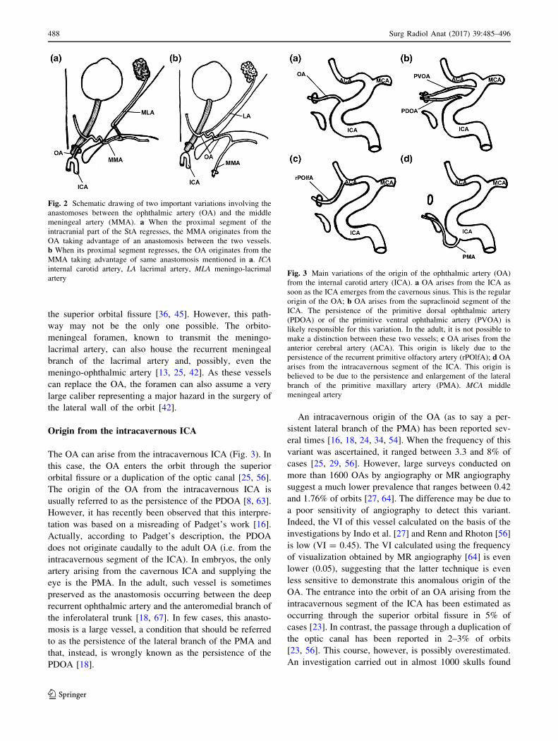
OA origin from the middle meningeal artery
The intracranial part of the StA becomes in the adult the
MMA. The orbital ramus of the StA is responsible for the
blood supply of the extraocular structures of the orbit and
enters the orbit through the superior orbital fissure. In the
orbit, this artery divides into two branches, a lateral one
directed to the lacrimal gland, and a medial one referred
to as ethmoido-nasal artery or naso-ciliary artery [30, 45].
Variations in the development of the StA lead to several
possible outcomes. When the orbital ramus fails to regress
at the level of the superior orbital fissure, a connection
between the MMA and the OA persists postnatally as the
recurrent meningeal branch of the lacrimal artery [36, 45].
In other cases, the division of the orbital ramus occurs
within the cranial cavity [45], one branch entering the
orbit through the orbitomeningeal foramen (also known as
Hyrtl’s foramen) and becoming the meningo-lacrimal
artery, the other branch (ethmoido-nasal artery), passing
through the superior orbital fissure and being annexed to
the OA. If the ethmoido-nasal artery does not regress
completely at the level of the superior orbital fissure, a
direct anastomosis between the MMA and the OA is
found in the adult and is referred to as meningo-oph-
thalmic artery [36, 45]. The occasional aberrant regression
of the stem of the MMA or that of the proximal segment
of the OA is compensated by the presence of the
meningo-ophthalmic artery or by the recurrent meningeal
branch of the lacrimal artery [16]: depending on the cir-
cumstances, these vessels may become an aberrant MMA
originating from the OA/lacrimal artery or, more impor-
tant to us, an aberrant OA arising from the MMA
(OAMMA) (Fig. 2) [36, 45]. The stemming from the MMA
is certainly the most frequently reported aberrant origin of
the OA [11, 23, 39, 40, 65, 66]. In large series of dis-
sections or radiological surveys, the OAMMA has been
reported in as much as 1.2–3.3% of orbits [24, 29, 64].
Sometimes, the OAMMA flanks a regular OA originating
from the ICA (OAICA). Such occurrence is dealt with in a
dedicated paragraph as it should be considered a double
OA. The OAMMA most frequently enters the orbit through
Surg Radiol Anat (2017) 39:485–496 487
123
the superior orbital fissure [36, 45]. However, this path-
way may not be the only one possible. The orbito-
meningeal foramen, known to transmit the meningo-
lacrimal artery, can also house the recurrent meningeal
branch of the lacrimal artery and, possibly, even the
meningo-ophthalmic artery [13, 25, 42]. As these vessels
can replace the OA, the foramen can also assume a very
large caliber representing a major hazard in the surgery of
the lateral wall of the orbit [42].
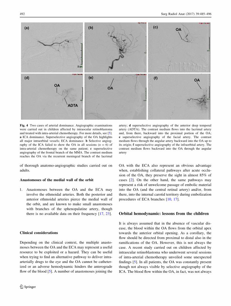
Origin from the intracavernous ICA
The OA can arise from the intracavernous ICA (Fig. 3). In
this case, the OA enters the orbit through the superior
orbital fissure or a duplication of the optic canal [25, 56].
The origin of the OA from the intracavernous ICA is
usually referred to as the persistence of the PDOA [8, 63].
However, it has recently been observed that this interpre-
tation was based on a misreading of Padget’s work [16].
Actually, according to Padget’s description, the PDOA
does not originate caudally to the adult OA (i.e. from the
intracavernous segment of the ICA). In embryos, the only
artery arising from the cavernous ICA and supplying the
eye is the PMA. In the adult, such vessel is sometimes
preserved as the anastomosis occurring between the deep
recurrent ophthalmic artery and the anteromedial branch of
the inferolateral trunk [18, 67]. In few cases, this anasto-
mosis is a large vessel, a condition that should be referred
to as the persistence of the lateral branch of the PMA and
that, instead, is wrongly known as the persistence of the
PDOA [18].
An intracavernous origin of the OA (as to say a per-
sistent lateral branch of the PMA) has been reported sev-
eral times [16, 18, 24, 34, 54]. When the frequency of this
variant was ascertained, it ranged between 3.3 and 8% of
cases [25, 29, 56]. However, large surveys conducted on
more than 1600 OAs by angiography or MR angiography
suggest a much lower prevalence that ranges between 0.42
and 1.76% of orbits [27, 64]. The difference may be due to
a poor sensitivity of angiography to detect this variant.
Indeed, the VI of this vessel calculated on the basis of the
investigations by Indo et al. [27] and Renn and Rhoton [56]
is low (VI = 0.45). The VI calculated using the frequency
of visualization obtained by MR angiography [64] is even
lower (0.05), suggesting that the latter technique is even
less sensitive to demonstrate this anomalous origin of the
OA. The entrance into the orbit of an OA arising from the
intracavernous segment of the ICA has been estimated as
occurring through the superior orbital fissure in 5% of
cases [23]. In contrast, the passage through a duplication of
the optic canal has been reported in 2–3% of orbits
[23, 56]. This course, however, is possibly overestimated.
An investigation carried out in almost 1000 skulls found
Fig. 2 Schematic drawing of two important variations involving the
anastomoses between the ophthalmic artery (OA) and the middle
meningeal artery (MMA). a When the proximal segment of the
intracranial part of the StA regresses, the MMA originates from the
OA taking advantage of an anastomosis between the two vessels.
b When its proximal segment regresses, the OA originates from the
MMA taking advantage of same anastomosis mentioned in a. ICAinternal carotid artery, LA lacrimal artery, MLA meningo-lacrimal
arteryFig. 3 Main variations of the origin of the ophthalmic artery (OA)
from the internal carotid artery (ICA). a OA arises from the ICA as
soon as the ICA emerges from the cavernous sinus. This is the regular
origin of the OA; b OA arises from the supraclinoid segment of the
ICA. The persistence of the primitive dorsal ophthalmic artery
(PDOA) or of the primitive ventral ophthalmic artery (PVOA) is
likely responsible for this variation. In the adult, it is not possible to
make a distinction between these two vessels; c OA arises from the
anterior cerebral artery (ACA). This origin is likely due to the
persistence of the recurrent primitive olfactory artery (rPOlfA); d OA
arises from the intracavernous segment of the ICA. This origin is
believed to be due to the persistence and enlargement of the lateral
branch of the primitive maxillary artery (PMA). MCA middle
meningeal artery
488 Surg Radiol Anat (2017) 39:485–496
123

that a duplication of the optic canal occurs only in 0.65% of
orbits [6]. When entering through the superior orbital fis-
sure, the OA passes through the posterior part of the fissure,
within the tendinous annulus of Zinn and medial to the
oculomotor nerve [24]. Then, running between the lateral
rectus and the optic nerve, the persistent PMA courses
forward and about 1 cm behind the eyeball, it turns
medially to cross over the optic nerve [8, 54]. This course
roughly coincides with that of the deep recurrent oph-
thalmic artery [35].
Origin from the supraclinoid ICA
In embryos, the PVOA and the PDOA take origin close to
the terminal division of the ICA. When developing regu-
larly, the stem of the PVOA regresses, while the stem of
the PDOA migrates caudally, so that the origin and the
intracranial course of the OA are established as they are
normally found in adults. Sometimes, however, the caudal
migration of the PDOA fails or the PVOA persists. In both
cases, the persistence of one primitive OA in the adult
results in the OA arising from the supraclinoid ICA with no
way to make a certain distinction between the two primi-
tive vessels (Fig. 3). At any rate, this is a very rare event
that has been reported only occasionally [18, 19, 46, 52].
Origin from the anterior cerebral artery
The aberrant stemming from the anterior cerebral artery is
another rare variant of the OA origin
[3, 4, 18, 20, 21, 26, 28, 37, 63]. This variation has been
frequently explained with the persistence of the PVOA
[3, 4, 26, 28, 37, 63], though it more probably involves a
persistent recurrent POlfA (Fig. 3) [18].
Origin from other intracranial arteries
Exceptionally, the OA has been reported to stem from
other intracranial arteries like the middle cerebral artery
[41], the posterior communicating artery [15, 47], the
basilar artery [57–59], and the contralateral ICA [50].
Double OAs
A double origin of the OA can be the result of a regular OA
co-existing with an OAMMA or with a persistent lateral
branch of the PMA. The former case seems to be the
commonest as in dissection-based studies, it has been
reported in 2.4–3.3% of cases [23, 29]. However, a recent
vast survey carried out on MR angiographies demonstrated
an OAMMA co-existing with an OAICA in only 0.18% of
cases [64]. Once again, MR angiography seems to be
scarcely sensitive to detect variations of the OA origin. The
VI calculated using the values determined by Uchino et al.
[64] and Hayreh [23] is in fact extremely low (0.075).
Interestingly, the larger OA has been consistently reported
to be the OAMMA [25, 29, 44]. The two OAs usually join
together, the meeting site being located either medial or
lateral to the optic nerve [24]. In other instances, however,
the eyeball is served by the OAICA, whereas the rest of the
orbit is supplied by the OAMMA, the two systems appar-
ently not having any connection [44, 45].
Two OAs arising from the ICA are really rare. To our
knowledge, 11 cases have been reported so far
[1, 8, 16, 18, 30, 34, 48, 49, 64, 67], though we believe that
the two deep recurrent ophthalmic arteries reported by
Lasjaunias [35] should be included in the count. It is fre-
quently said that the persistent lateral branch of the PMA
(previously referred to as persistent PDOA) enters the orbit
through the superior orbital fissure [1, 8, 30, 48, 54, 62].
However, in some cases, the diagnosis has been merely
angiographic [30, 34, 62, 64] and the course of the artery
should be considered presumptive. Indeed, the persistent
PMA can enter the orbit also through a duplication of the
optic canal [23, 56]. In most cases, the two OAs are inde-
pendent vessels with their own territory of distribution
[1, 48, 64]. However, a small anastomosis has been observed
between the two vessels in one case [30], whereas in few
other cases, the two OAs joined together to form, distally to
their anastomosis, a ‘‘common OA’’ [8, 16, 35]. In the latter
instance, the anatomic variant should be better described as a
double ICA origin of the OA. Interestingly, when two OAs
co-exist together, one can lie angiographically occult [8].
This observation legitimately raises the issue on the actual
frequency of this anatomic variant which, though rare, could
be less exceptional than previously thought.
In one case, to the best of our knowledge, the only one
so far reported, a double OA has been the result of the
simultaneous presence of a persistent recurrent branch of
the POlfA coming from the anterior cerebral artery and a
larger OAMMA. Unfortunately, no information is available
on the intraorbital distribution of the two vessels [4].
Clinical considerations
On the basis of 1643 selective angiographies, an anomalous
site of origin of the OA from the ICA has been associated
with a 50-fold higher risk of ICA anterior wall aneurysms
[27]. Ligation or embolization of the MMA or that of the
maxillary artery is a procedure that can endanger the eye if
the OA stems from the MMA [22, 61]. The same hazard as
well as a high risk of severe bleeding can be expected in
surgical procedures involving the lateral wall of the orbit
when an OA arising from the MMA or a large meningo-
ophthalmic artery runs through the orbitomeningeal fora-
men [42].
Surg Radiol Anat (2017) 39:485–496 489
123

Additional sources of blood supply to the orbitfrom the ECA
The blood supply of the orbit receives a contribution from
several branches of the ECA which anastomose with
variable frequencies with the OA [10, 23, 45]. A few
branches of the ECA can also supply part of the orbit
without making meaningful anastomoses with the OA.
Basically, all extraocular branches of the OA can have
connections with rami of the ECA. In particular circum-
stances, some of them represent major alternative pathways
for the blood supply to the orbit and may acquire clinical
relevance [42, 45]. The anastomoses between the OA and
the ECA are numerous. Some of them are quite common,
others are rare, and others can be considered exceptional.
Unfortunately, cadaver-based studies addressing the fre-
quency of OA-ECA anastomoses are few [7].
One recent report has shed some light on the matter,
though the investigation carried out by angiography on
children affected by intraocular retinoblastoma should not
be considered necessarily representative of the adult orbit
circulation [10]. Overall, at least one anastomosis (but
many children showed more than one) can be demonstrated
by angiography in 44.33% of orbits, the frequency of
visualization (angiographic incidence) being dependent,
however, on the technique employed. When angiography is
carried out either through the OA or through the ECA, the
angiographic incidence increases up to 91% of orbits
suggesting that, at least in children, one connection
between the OA and the ECA can be found almost always
if properly searched [10].
The list of anastomoses between the OA and the ECA is
long (Table 1). In general, they can be divided into anas-
tomoses located in the posterior or in the anterior orbit
[10]. A third minor group is represented by anastomoses
connecting the ethmoidal arteries with arteries of the nasal
cavity.
Anastomoses of the posterior orbit (always involving
the MMA)
1. The anastomosis most frequently visualized by
angiography (up to 37.77% of orbits) is the con-
nection between the MMA and the lacrimal artery
via its recurrent meningeal branch [10, 23, 55]. Its VI
is 0.80 (Table 1) as to say that angiography
demonstrates this anastomosis roughly in 4 cases out
of 5.
2. A second anastomosis frequently observed on
angiograms is the meningo-ophthalmic artery
[10, 45]. In spite of its recurrent angiographic
visualization (Table 1) and though the meningo-
ophthalmic artery can be exploited for drug delivery
of intra-arterial chemotherapy [5], to our knowledge,
no dissection-based study has ever investigated its
frequency.
3. Less frequently (5.71% of orbits), the OA is
connected with the MMA via the lateral muscular
artery [12], and exceptionally via the supraorbital
artery or the superior muscular artery [10, 12].
4. A novel anastomosis has recently been described
between the ECA and the OA. Referred to as ‘‘artery
of the superior orbital fissure’’, this very small
branch of the maxillary artery ascends from the
pterygopalatine fossa to join the anteromedial branch
of the inferolateral trunk [32]. It has been detected
by 3D rotational angiography in 31.25% of orbits.
This is a very small artery that becomes
detectable concomitantly with hypervascular
parasellar lesions [32]. Its frequency in regular
hemodynamic conditions is unknown. In our opin-
ion, the name ‘‘artery of the superior orbital fissure’’
conveys the wrong idea that the artery enters the
orbit through the superior orbital fissure. Actually, it
is the anteromedial branch of the inferolateral trunk
that enters the orbit to anastomose with the deep
recurrent ophthalmic artery [35]. However, in 3% of
orbits, the artery of the superior orbital fissure is
connected with the deep recurrent OA [32] realizing
an actual anastomosis between the ECA and the OA
through the superior orbital fissure.
The OA-ECA anastomoses located in the posterior orbit
are usually reported to pass through the superior orbital
fissure [36, 45]. However, this course is not constant [16].
For instance, the recurrent meningeal branch connecting
the lacrimal artery with the MMA leaves the orbit through
orbitomeningeal foramen in 10.53% of orbits [13]. The
demonstration that the orbitomeningeal foramen can be
double, triple, or even quadruple raises the possibility that
in some cases, more branches of the MMA, than just the
meningo-lacrimal artery or the anastomosis with the
lacrimal artery, supply the orbit through the orbito-
meningeal foramen [42]. In addition, orbitomeningeal
foramina can be larger than 1 mm in 12% of cases sug-
gesting the passage of arteries distributing to large portions
of the orbit [42].
Anastomoses in the anterior orbit
1. In the anterior orbit, the branch of the OA most
frequently anastomosed with the ECA is certainly
the lacrimal artery. The lacrimal artery is central to
the system of anastomoses connecting the OA with
the ECA either in terms of frequency or variety of
490 Surg Radiol Anat (2017) 39:485–496
123

connections. In addition to the above-recalled anas-
tomosis with the MMA in the posterior orbit, the
lacrimal artery can be connected with the anterior
deep temporal artery [10, 23, 45], the transverse
facial artery [23], the orbital branch of the infraor-
bital artery [23], the zygomaticoorbital artery [10],
and the meningo-lacrimal artery [12] (Table 1). In
children, the connection with the anterior deep
temporal artery is the anastomosis most often visu-
alized by angiography [10] (Table 1). In spite of its
frequent angiographic visualization (33.3% of cases)
and though this pathway can be exploited for drug
delivery in intra-arterial chemotherapy [5], to the
best of our knowledge, no dissection-based study has
ever addressed its actual anatomic incidence. The
anastomosis between the lacrimal artery and the
zygomaticoorbital artery has been detected by
angiography in up to 2.22% of cases [10]. Connec-
tions between the lacrimal artery and the meningo-
lacrimal artery are achieved through small
intraglandular rami [12]. Such intraglandular anas-
tomoses are possible only with the concomitant
presence of two lacrimal arteries and their impor-
tance is negligible [12]. The anastomoses between
the lacrimal artery and the transverse facial artery or
the orbital branch of the infraorbital artery are rare
[25].
2. The dorsal nasal artery frequently anastomoses with
the facial artery via the angular artery [7, 10, 12, 23]
or with the infraorbital artery [7, 10, 23]. These
connections have been detected in few cases by
angiography, though they can be observed with
higher frequencies by dissection. Accordingly, they
have very low VIs (Table 1).
3. While leaving the orbit, the supraorbital artery can
make connections with the frontal branch of the
superficial temporal artery [7, 10, 23], or with the
zygomaticoorbital artery [10] (Table 1). In adults,
the anastomosis with the frontal branch of the
superficial temporal artery has been demonstrated
in 1/3 of cases [7], whereas in children, the
frequency of its angiographic visualization is low
[10] (Table 1). The connection with the zygomati-
coorbital artery has been demonstrated only by
angiography [10].
4. The supratrochlear artery anastomoses with the
frontal branch of the superficial temporal artery
[10, 23]. This connection, detected in up to 2.22% of
angiographies, can be considered a variation of the
anastomosis described in 3, though it courses more
medially.
5. Anastomoses involving the palpebral arteries or the
muscular branches of the OA are rare and/or of
negligible clinical importance [12, 36].
The anastomoses located at the anterior orbit share very
low VIs (Table 1). Such values can be explained in two
ways: angiography is not an efficient technique for their
demonstration or they mostly develop with age. In the
latter case, a reliable VI could be produced only by the use
Table 1 Main anastomoses between the ECA and the OA
Name OA branch ECA branch Anat. F. Ang. F. VI
Lacrimal a. MMA 47.14% [12] 37.7% [10] 0.80
Meningo-ophthalmic a. OA itself MMA ND 31.1% [10] –
Lateral muscular a. MMA 5.71% [12] ND –
A. of the superior orbital fissure Deep recurrent OA Maxillary a. ND 3% [32] –
Lacrimal a. Anterior deep temporal a. ND 33.3% [10] –
Lacrimal a. Transverse facial a. ND ND –
Lacrimal a. Orbital branch of the infraorbital a. ND ND –
Lacrimal a. Zygomaticoorbital a. ND 2.22% [10] –
Dorsal nasal a. Facial a. 60% [7] 8.9% [10] 0.15
Dorsal nasal a. Orbital branch of the infraorbital a. 27% [7] 6.6% [10] 0.24
Supraorbital a. Superficial temporal a. 33% [7] 2.22% [10] 0.07
Supraorbital a. Zygomaticoorbital a. ND 2.22% [10] –
Supratrochlear a. Superficial temporal a. ND 2.22% [10] –
Anterior ethmoidal a. Sphenopalatine a. ND ND –
Posterior ethmoidal a. Sphenopalatine a. ND ND –
Anat. F. anatomic frequency, Ang. F. angiographic frequency, ND not determined
Surg Radiol Anat (2017) 39:485–496 491
123
of thorough anatomo-angiographic studies carried out on
adults.
Anastomoses of the medial wall of the orbit
1. Anastomoses between the OA and the ECA may
involve the ethmoidal arteries. Both the posterior and
anterior ethmoidal arteries pierce the medial wall of
the orbit, and are known to make small anastomoses
with branches of the sphenopalatine artery, though
there is no available data on their frequency [17, 23].
Clinical considerations
Depending on the clinical context, the multiple anasto-
moses between the OA and the ECA may represent a useful
resource to be exploited or a hazard. They can be useful
when trying to find an alternative pathway to deliver intra-
arterially drugs to the eye and the OA cannot be catheter-
ized or an adverse hemodynamic hinders the anterograde
flow of the blood [5]. A number of anastomoses joining the
OA with the ECA also represent an obvious advantage
when, establishing collateral pathways after acute occlu-
sion of the OA, they preserve the sight in almost 85% of
cases [2]. On the other hand, the same pathways may
represent a risk of unwelcome passage of embolic material
into the OA (and the central retinal artery) and/or, from
there, into the internal carotid territory during embolization
procedures of ECA branches [10, 17].
Orbital hemodynamic: lessons from the children
It is always assumed that in the absence of vascular dis-
ease, the blood within the OA flows from the orbital apex
towards the anterior orbital opening. As a corollary, the
flow should be directed from proximal to distal also in the
ramifications of the OA. However, this is not always the
case. A recent study carried out on children affected by
intraocular retinoblastoma who underwent several sessions
of intra-arterial chemotherapy unveiled some unexpected
findings [5]. In all patients, the OA was constantly present
though not always visible by selective angiography of the
ICA. The blood flow within the OA, in fact, was not always
Fig. 4 Two cases of arterial dominance. Angiographic examinations
were carried out in children affected by intraocular retinoblastoma
and treated with intra-arterial chemotherapy. For more details, see [5].
a ICA dominance. Superselective angiography of the OA highlights
all major intraorbital vessels; ECA dominance. b Selective angiog-
raphy of the ICA failed to show the OA in all sessions (n = 6) of
intra-arterial chemotherapy on the same patient; c superselective
angiography of the frontal branch of the MMA. The contrast medium
reaches the OA via the recurrent meningeal branch of the lacrimal
artery; d superselective angiography of the anterior deep temporal
artery (ADTA). The contrast medium flows into the lacrimal artery
and, from there, backward into the proximal portion of the OA;
e superselective angiography of the facial artery. The contrast
medium flows through the angular artery backward into the OA up to
its origin; f superselective angiography of the infraorbital artery. The
contrast medium flows backward into the OA through the angular
artery
492 Surg Radiol Anat (2017) 39:485–496
123

directed anterogradely. In greater detail, it has been
observed that orbits can be entirely supplied either by the
ICA, with the blood within the OA regularly flowing
anterogradely (ICA dominance) (Fig. 4a), or by the ECA
(ECA dominance), with the flow within the OA backward
directed towards the ICA (Fig. 4b–f) [5, 43]. Between
these two extreme situations (ICA or ECA dominance), a
variety of possible hemodynamically intermediate condi-
tions (balanced hemodynamic) can be found [4]. In such
cases, part of the orbit is supplied by branches of the ECA
and part from the ICA via the OA (Fig. 5). These three
possibilities should not be considered necessarily as
stable conditions [5]. When a series of angiographies is
carried out monthly on the same child, it is not unusual to
find different hemodynamic outlines (Fig. 5). The exten-
sion of the territories supplied by the ECA and ICA,
therefore, may change in a very short time. The reason for
this is unclear and it may depend just on the physiologic
fine tuning of the orbit circulation. At any rate, these
findings unveil that in children, a subtle balance exists
between ECA and ICA, the two vessels competing for the
orbital blood supply. The implications of such findings are
evident particularly to the neurovascular interventionalists
who desire to take advantage of the vascular flow to deliver
drugs to an intraorbital target. On the other hand, on the
ground of these results, one should also infer that a
reversed blood flow assessed by Doppler ultrasonography
within the OA of children does not necessarily mean a
pathologically altered hemodynamic.
A similar in-depth investigation has never been per-
formed by angiography on adult orbits. Indeed, most of
the current knowledge on orbital hemodynamic in adults
come from Doppler ultrasonography (US) studies [38].
According to such studies, in the absence of vascular
pathology, an ICA dominance in the orbit seems the
rule. However, though Doppler US provides valuable
information [38], it does not supply a comprehensive
view of the orbital circulation and some important
details may escape. In particular, the ultimate source of
the blood flowing within the orbit is difficult to estimate.
Fig. 5 Balanced hemodynamic. Two angiographic studies carried out
on the same patient demonstrate that the territory of the lacrimal
artery is supplied by the ECA via the anterior deep temporal artery
(ADTA). In addition, the extension of the territories of the orbit
served by the OA and the ECA change between the two examinations.
a First angiography. Superselective angiography of the OA. The
contrast medium flows in almost the entire vascular tree of the OA,
including the anterior ethmoidal artery (AEA). However, only a short
portion of the lacrimal artery (LA) can be seen; second angiography.
The hemodynamic balance between ECA and ICA is changed;
b superselective angiography of the OA. The contrast medium does
not diffuse into the OA as far as in b and the LA is not visible; c,d selective angiography of the ECA. The contrast medium reaches the
LA via the ADTA. In the LA, the flow is forward-directed to the
lacrimal gland and backward directed to the OA. In this examination,
the ECA also contributes to the blood flowing into the distal OA and
even into the AEA
Surg Radiol Anat (2017) 39:485–496 493
123

Indeed, the direction of the flow within the OA as an
indication to predict the source of the blood can be
misguiding. For instance, the US assessment of blood
flowing anterogradely in the distal OA does not imply
that it comes from the ICA. As a matter of fact, when
the OA arises directly from the MMA, the entire blood
supply of the orbit ultimately comes from the ECA
[25, 40]. On the other hand, even a large anastomosis
between the MMA and the OA can have the same
hemodynamic effects, with blood within the OA flowing
anterogradely though deriving from the ECA [5]. Actu-
ally, in such cases, a reversed course within the OA
might still occur in that part of the artery which is
proximal to the anastomosis, a deep portion difficult to
probe by US. At any rate, based also on the observations
carried out on children [5, 43], not necessarily affected
by retinoblastoma [43], the finding that the orbit is
entirely supplied by the ECA should not necessarily
imply an underlying vascular pathology, though experi-
ence says that this is highly probable.
Evidences in children show that the orbital hemody-
namic can vary over the time. The balance between ECA
and OA can shift in favor of one artery rather than the other
in a matter of days [5]. In some children, it is possible to
observe that OA-ECA anastomoses are not constantly
visible. Apparently, they can functionally close or open
depending on the hemodynamic requirements of the
moment [5]. If their opening/closure is the cause or the
consequence of the hemodynamic shift between OA and
ECA dominances (including all the intermediate gradations
of the balanced hemodynamic outlines) at present is
impossible to say. However, results in children show that
anastomoses are vascular channels that do not necessarily
develop secondarily to chronic vascular disorders. In the
adult, we do not have the same compelling evidences.
Indeed, as we lack investigations comparable to those
carried out on children (in terms of number of patients
undergoing repeated angiographic studies within a relative
short range of time), hemodynamic shifts between OA and
ECA dominances cannot be documented at present. Nev-
ertheless, even in adults, vascular channels may lie
angiographically hidden becoming conspicuous only under
particular hemodynamic circumstances [5, 16] and the
presence of functionally operational anastomoses in acute
settings has been demonstrated in several occasions. In
particular, the absence of visual deterioration in most cases
during balloon test occlusion of the ICA and/or OA before
endovascular treatment of carotid-ophthalmic aneurysms
confirms their ability to guarantee the blood flow to the eye
even after acute OA occlusion [14, 31, 53, 60]. Hemody-
namic shifts between OA and ECA dominances are,
therefore, potentially feasible also in adults.
Conclusions
If the clinician is not aware of the possible variations
occurring to the site of origin of the OA, some endovas-
cular or surgical interventions may put at risk the eye of the
patient. Anomalous origin of the OA is also associated with
a high risk of ICA anterior wall aneurysms.
The anastomoses that may occur between the OA and
the ECA represent an anatomic resource or a hazard. They
may provide alternative routes for the blood supply of the
orbit but also for the unwelcome passage of embolic
material during embolization procedures of ECA branches.
For this reason, a detailed knowledge of their anatomy and
frequency in the adults is required for the proper evaluation
of the risks of some endovascular procedures. Anastomoses
between the OA and ECA also influence orbital hemody-
namic which in children displays a certain degree of vari-
ability, including ECA dominance of the orbital blood
supply. A large-scale angiographic study of the orbital
blood supply should be carried out to verify if the concept
of ICA and ECA dominances as well as that of balanced
hemodynamic can be extended to grown-ups as well.
Finally, a comparison between children and adults is
mandatory to ascertain if arterial dominances (ECA and
ICA) and balanced hemodynamic are patterns that change
with growth and if adult hemodynamic has the same
potentials for the changes observed in children. A large-
scale angiographic study carried out on adults with the
same modalities previously employed on children [5]
would serve the purpose. However, clinical scenarios
requiring such vast investigation in adults are exceedingly
rare. In spite of this, the possibility that OA/ECA anasto-
moses are in place, though not always visible, should be
kept in mind.
Compliance with ethical standards
Conflict of interest We declare that we have no conflict of interest.
Open Access This article is distributed under the terms of the
Creative Commons Attribution 4.0 International License (http://crea
tivecommons.org/licenses/by/4.0/), which permits unrestricted use,
distribution, and reproduction in any medium, provided you give
appropriate credit to the original author(s) and the source, provide a
link to the Creative Commons license, and indicate if changes were
made.
References
1. Agarwal N, Singh PL, Karimi RJ, Gandhi CD, Prestigiacomo CJ
(2013) Persistent vestige of dorsal ophthalmic artery: a case
report. J Neurointerv Surg 5:e25. doi:10.1136/neurintsurg-2011-
010196
494 Surg Radiol Anat (2017) 39:485–496
123

2. Ahn JH, Cho YD, Kang JE, Kim JE, Cho W-S, Jung SC, Kim
CH, Han MH (2014) Endovascular treatment of ophthalmic
artery aneurysms: assessing balloon test occlusion and preserva-
tion of vision in coil embolization. AJNR Am J Neuroradiol
35:2146–2152
3. Baltsavias G, Turk Y, Valavanis A (2012) Persistent ventral
ophthalmic artery associated with supraclinoid internal carotid
artery aneurysm: case report and review of the literature. J Neu-
roradiol 39:186–189
4. Belotti F, Ferrari M, Doglietto F, Cocchi MA, Lancini D,
Buffoli B, Nicolai P, Fontanella MM, Maroldi R, Tsch-
abitscher M, Rodella LF (2016) Ophthalmic artery originating
from the anterior cerebral artery: anatomo-radiological study,
histological analysis, and literature review. Neurosurg Rev
39:483–493
5. Bertelli E, Leonini S, Galimberti D, Moretti S, Tinturini R,
Hadjistilianou T, De Francesco S, Romano DG, Vallone IM,
Cioni S, Gennari P, Galluzzi P, Grazzini I, Rossi S, Bracco S
(2016) Hemodynamic and anatomic variations require an adapt-
able approach during intra-arterial chemotherapy for intraocular
retinoblastoma: alternative routes, strategies and follow-up.
AJNR Am J Neuroradiol 37:1289–1295
6. Bertelli E (2014) Metoptic canal, duplication of the optic canal
and Warwick’s foramen in human orbits. Anat Sci Inter 89:34–45
7. Berthelot JL, Hureau J (1982) Clinical anatomy study of the
macroscopic anastomoses of the ophthalmic artery in the peri-
orbital region. Clin Anat 3:271–278
8. Bracco S, Gennari P, Vallone IM, Tassi R, Acampa M, Martini G,
Bertelli E (2016) Double ophthalmic arteries arising from
the internal carotid artery. A case report of a hidden second
ophthalmic artery. Surg Radiol Anat, doi:10.1007/s00276-016-
1672-3
9. Bracco S, Venturi C, Leonini S, Romano DG, Cioni S, Vallone
IM, Gennari P, Galluzzi P, Hadjistilianou T, De Francesco S,
Guglielmucci D, Tarantino F, Bertelli E (2015) Identification of
intraorbital arteries in pediatric age by high resolution superse-
lective angiography. Orbit 34:237–247
10. Bracco S, Venturi C, Leonini S, Romano DG, Cioni S, Vallone
IM, Gennari P, Hadjistilianou T, De Francesco S, Bertelli E
(2016) Transorbital anastomotic pathways between the external
and internal carotid systems in children affected by intraocular
retinoblastoma. Surg Radiol Anat 38:79–87
11. Cohen JE, Moscovici S, Halpert M, Itshayek E (2012) Selective
thrombolysis performed through meningo-ophthalmic artery in
central retinal occlusion. J Clin Neurosci 19:462–464
12. Ducasse A, Segal A, Delattre JF, Burette A, Flament JB (1985)
La partecipation de l’artere carotid externe a la vascularisation
orbitaire. J Fr Ophtalmol 8:333–339
13. Erdogmus S, Govsa F (2005) Importance of the anatomic features
of the lacrimal artery for orbital approaches. J Craniofac Surg
16:957–964
14. Ezura M, Takahashi A, Yoshimoto T (1997) Combined
intravascular parent artery and ophthalmic artery occlusion for
giant aneurysm of the supraclinoid internal carotid artery. Surg
Neurol 47:360–363
15. Fisher AG (1913) A case of complete absence of both internal
carotid arteries, with a preliminary note on the developmental
history of the stapedial artery. J Anat 48:37–46
16. Gailloud P, Gregg L, San Millan Ruiz D (2009) Developmental
anatomy, angiography, and clinical implications of orbital arterial
variations involving the stapedial artery. Neuroimag Clin N Am
19:169–179
17. Geibprasert S, Pongpech S, Armstrong D, Krings T (2009)
Dangerous extracranial-intracranial anastomoses and supply to
the cranial nerves: vessels the neurointerventionalist needs to
know. AJNR Am J Neuroradiol 30:1459–1468
18. Gregg L, San Millan D, Orru E, Tamargo RJ, Gailloud P (2016)
Ventral and dorsal persistent primitive ophthalmic artery. Oper-
ative Neurosurgery 12:141–152
19. Hamada J-I, Kitamura I, Kurino M, Sueyoshi N, Uemura S, Ushio
Y (1991) Abnormal origin of bilateral ophthalmic artery. J Neu-
roradiol 74:287–289
20. Hannequin P, Peltier J, Destrieux C, Velut S, Havet E, Le Gars D
(2013) The inter-optic course of a unique precommunicating
anterior cerebral artery with aberrant origin of an ophthalmic
artery: an anatomic case report. Surg Radiol Anat 35:269–271
21. Hassler W, Zentner J, Voigt K (1989) Abnormal origin of the
ophthalmic artery from the anterior cerebral artery: neuroradio-
logical and intraoperative findings. Neuroradiology 31:85–87
22. Hayashi N, Kubo M, Tsuboi Y, Nishimura S, Nishijima M,
Abdel-Aal MA, Endo S (2007) Impact of anomalous origin of the
ophthalmic artery from the middle meningeal artery on selection
of sirgical approach to skull base meningioma. Surg Neurol
68:568–572
23. Hayreh SS (2006) Orbital vascular anatomy. Eye 20:1130–1144
24. Hayreh SS, Dass R (1962) The ophthalmic artery. I. Origin and
intra-cranial and intra-canalicular course. Br J Ophthalmol
46:65–98
25. Hayreh SS (1962) The ophthalmic artery. III. Branches. Br J
Ophthalmol 46:212–247
26. Honma Y, Ogawa T, Nagao S (1997) Angiographically occult
anomalous ophthalmic artery arising from the anterior cerebral
artery. Acta Neurochir 139:480–481
27. Indo M, Oya S, Tanaka M, Matsui T (2014) High incidence of
ICA anterior wall aneurysms in patients with an anomalous origin
of the ophthalmic artery: possible relevance to the pathogenesis
of aneurysm formation. J Neurosurg 120:93–98
28. Islak C, Ogut G, Numan F, Cokyuksel O, Kuday C (1994) Per-
sistent nonmigrated ventral primitive artery. Report on one case.
J Neuroradiol 21:46–49
29. Jimenez-Castellanos J, Carmona A, Castellanos L, Catalina-
Herrera CJ (1985) Microsurgical anatomy of the human oph-
thalmic artery: a mesoscopic study of its origin, course and col-
lateral branches. Surg Radiol Anat 17:139–143
30. Kam CK, Alvarez H, Lasjaunias P (2003) Double internal carotid
origin of the ophthalmic artery with ruptured aneurysm of the
posterior communicating artery. Interv Neuroradiol 9:383–388
31. Kim B, Jeon P, Kim K, Yang N, Kim S, Kim H, Byun H, Jo K-II
(2016) Endovascular treatment of unruptured ophthalmic artery
aneurysms: clinical usefulness of the balloon occlusion test in
predicting vision outcomes after coil embolization. J NeuroInter-
vent Surg 8:696–701
32. Kiyosue H, Tanoue S, Hongo N, Sagara Y, Mori H (2015) Artery
of the superior orbital fissure: an undescribed branch from the
pterygopalatine segment of the maxillary artery to the apex
connecting with the anteromedial branch of the inferolateral
trunk. AJNR Am J Neuroradiol 36:1741–1747
33. Komiyama M (2009) Embryology of the ophthalmic artery: a
revived concept letter to the editor. INR 15:367–372
34. Lasjaunias P, Bereinstein A, ter Brugge KG (2001) Surgical
Neuroradiology, Vol 1: clinical vascular anatomy and variations,
2nd edition, Springer-Verlag, Berlin Heidelberg New York
35. Lasjaunias P, Brismar J, Moret J, Theron J (1978) Recurrent
cavernous branches of the ophthalmic artery. Acta Radiol Diagn
19:553–560
36. Lasjaunias P, Michotey P, Vignaud J, Clay C (1975) II-Ra-
dioanatomie de la vascularisation arterielle de l’orbite, a l’ex-
ception du tronc de l’artere ophthalmique. Ann Radiol
18:181–194
37. Li Y, Horiuchi T, Yako T, Ishizaka S, Hongo K (2011) Ano-
malous origin of the ophthalmic artery from the anterior cerebral
artery. Neurol Med Chir 51:579–581
Surg Radiol Anat (2017) 39:485–496 495
123

38. Lieb WE, Muller-Forell WS, Wichmann W (2006) Ophthalmo-
logic imaging methods. In: Muller-Forell WS (ed) Imaging of
orbital and visual pathway pathology. Springer-Verlag, Berlin
Heidelberg New York, pp 3–23
39. Liu Q, Rhoton AL Jr (2001) Middle meningeal origin of the
ophthalmic artery. Neurosurgery 48:401–406
40. Lombardi G (1969) Ophthalmic artery anomalies. Ophthalmo-
logica 157:321–327
41. Lowrey LG (1916) Anomaly of the circle of Willis due to the
absence of right internal carotid artery. Anat Rec 10:221–222
42. Macchi V, Regoli M, Bracco S, Nicoletti C, Morra A, Porzionato
A, De Caro R, Bertelli E (2016) Clinical anatomy of the orbit-
omeningeal foramens: variational anatomy of the canals con-
necting the orbit with the cranial cavity. Surg Radiol Anat
38:165–177
43. McIntosh DL, Douglas G, Lee K, Allen J, Mahadevan M (2007)
External carotid artery blood supply to the orbit. Int J Pediatr
Otolaryngol 71:1623–1626
44. Morandi X, Le Bourdon E, Darnault P, Brassier G, Duval JM
(1998) Unusual origin of the ophthalmic artery and occlusion of
the central retinal artery. Surg Radiol Anat 20:69–71
45. Moret J, Lasjaunias P, Theron J, Merland JJ (1977) The middle
meningeal artery. Its contribution to the vascularization of the
orbit. J Neuroradiol 4:225–248
46. N’da HA, Peltier J, Zunon-Kipre Y, Alsaiari S, Foulon P, Legars
D, Havet E (2014) An unusual superolaterla origin of ophthalmic
artery: ana anatomic case report. Surg Radiol Anat 36:95–97
47. Naeini RM, De J, Satow T, Benndorf G (2004) Unilateral age-
nesis of internal carotid artery with ophthalmic artery arising
from posterior communicating artery. Am J Radiol 184:571–573
48. Namba K, Nemoto S (2013) Double ophthalmic artery visualized
with new technology. Neuroradiol J 26:371–372
49. Ogawa T, Miyauchi T, Kato T, Tamakawa Y (1990) Internal
carotid origin of double ophthalmic arteries. Neuroradiology
32:508–510
50. Ogul H, Havan N, Gedikli Y, Pirimoglu B, Kantarci M (2016) A
new anatomic variation: coexistence of both Dandy-Walker
variant and ophthalmic artery originating from contralateral
internal carotid artery. J Craniofac Surg 27:e336–e338
51. Padget DH (1948) The development of the cranial arteries in the
human embryo. Contrib Embryol 32:205–261
52. Parlato C, di Nuzzo G, Luongo M, Tortora F, Briganti F (2011)
Anatomical variant of origin of ophthalmic artery: case report.
Surg Radiol Anat 33:275–278
53. Piche SL, Haw CS, Redekop GJ, Heran MKS (2005) Rare
intracanalicular ophthalmic aneurysm: endovascular treatment
and review of the literature. AJNR Am J Neuroradiol
26:1929–1931
54. Pretterklieber M, Schindler A, Krammer EB (1994) Unilateral
persistence of the dorsal ophthalmic artery in man. Acta Anat
149:300–305
55. Priman J, Christie DH (1959) A case of abnormal internal carotid
artery and associated vascular animalies. Anat Rec 134:87–95
56. Renn WH, Rhoton AL (1975) Microsurgical anatomy of the
sellar region. J Neurosurg 43:288–298
57. Rivera R, Choi IS, Sordo JG, Giacaman P, Badilla L, Bravo E,
Echeverria D (2015) Unusual origin of the left ophthalmic artery
from the basilar trunk. Surg Radiol Anat 37:399–401
58. Sade B, Tampieri D, Mohr G (2004) Ophthalmic artery origi-
nating from basilar artery: a rare variant. AJNR Am J Neuroradiol
25:1730–1731
59. Schumacher M, Wakhloo AK (1994) An orbital arteriovenous
malformation in a patient with origin of the ophthalmic artery
from the basilar artery. AJNR Am J Neuroradiol 15:550–553
60. Shaibani A, Khawar S, Bendok B, Walker M, Russell EJ, Batjer
HH (2004) Temporary balloon occlusion to test adequacy of
collateral flow to the retina and tolerance for endovascular
aneurysmal coiling. AJNR Am J Neuroradiol 25:1384–1386
61. Shima K, Kawasaki T, Shimizu A, Takiguchi H, Chigasaki H
(1995) an ophthalmic artery occlusion after craniotomy using the
pterional approach: a report of three cases, one resulting in
blindness. Jpn J Neurosurg 4:163–169
62. Tanaka M (2009) Persistent primitive dorsal ophthalmic artery
associated with paraclinoid internal carotid artery aneurysm.
JNET 3:39–41
63. Uchino A, Saito N, Ikeda S, Ishihara S (2015) Ophthalmic artery
arising from the anterior cerebral artery diagnosed by MR
angiography. Surg Radiol Anat 37:1009–1012
64. Uchino A, Saito N, Takahashi M, Kozawa E, Mizukoshi W,
Nakajima R, Okano N (2013) Persistent dorsal ophthalmic artery
and ophthalmic artery arising from the middle meningeal artery
diagnosed by MR angiography at 3D. Surg Radiol Anat
35:775–782
65. Watanabe A, Hirano K, Ishii R (1996) Dural caroticocavernous
fistula with both ophthalmic arteries arising from middle
meningeal arteries. Neuroradiology 38:806–808
66. Weinberg PE, Patronas NJ, Kim KS, Melen O (1981) Anomalous
origin of the ophthalmic artery in a patient with amaurosis fugax.
Arch Neurol 38:315–317
67. Willinsky R, Lasjaunias P, Berenstein A (1987) Intracavernous
branches of the internal carotid artery. Comprehensive review of
their variations. Surg Radiol Anat 9:201–215
496 Surg Radiol Anat (2017) 39:485–496
123
Related Documents