55ESPE Poster presented at: Maria Xatzipsalti¹ , Ioulia Polychroni¹, Elena Fryssira², Lela Stamogiannou¹ ¹First Department of Pediatrics, "P & A Kyriakou" Children's Hospital, Athens, Greece, ²Department of Medical Genetics, Athens University Medical School, Aghia Sophia Children’s Hospital, Athens, Greece OBJECTIVES CLINICAL PRESENTATION Fig 1. Bone age with exostoses on middle phalanx on 4 th finger, distal radius bone Fig 2 exostoses on thigh and both knees Fig 3. exostoses on right scapulae A 12.5 years old girl was referred to our Department because of short stature. She showed a stature of 132cm (-2,8SDS), weight of 24kg ((ΒΜΙ-13,8, -2,59z-score), reduced growth rate (2,3cm/year) and she was on pre-pubertal maturation stage. Genetic target was 155+/-4.5cm. On physical examination she had multiple exostoses on right scapulae, right thigh, right arm, right knee joint and very short fingers. The mobility of the joints adjacent to the exostoses remained free and muscle strength was normal. Her father had also multiple exostoses on scapulae and long bones and was under orthopedic follow up. Hypothyroidism, chronic diseases, malabsorption and skeletal dwarfism were excluded. GH deficiency was diagnosed by two stimulation tests: insulin (GH peak: 5.36 ng/ml) and L-dopa administration (GH peak: 7.35 ng/ml) and confirmed by low IGF-I values (214 ng/ml). Bone age was delayed (10.5years) with exostoses on distal radius bone and middle phalanx of fourth finger..(fig1) X-rays showed exostoses on right scapulae, right distal femurs, both knees (fig 2,3), right arm, right clavicles and pelvic bones. . She was referred to orthopedics who confirmed the diagnosis of HME. 1. Wicklund, C.L., et al., Natural history study of hereditary multiple exostoses. Am J Med Genet, 1995. 55(1): p. 43-6. 2 Schmale GA, Conrad EU III, Raskind WH. The natural history of hereditary multiple exostoses. J Bone Joint Surg Am 1994; 76:986–992. 3.Bovee, J.V., Multiple osteochondromas. Orphanet J Rare Dis, 2008. 3: p. 3. 4.Pedrini, E., et al., Genotype-phenotype correlation study in 529 patients with multiple hereditary exostoses: identification of "protective" and "risk" factors. J Bone Joint Surg Am, 2011. 93(24): p. 2294-302. 5. Bozzola, M., et al., Hereditary multiple exostoses and solitary osteochondroma associated with growth hormone deficiency: to treat or not to treat? Ital J Pediatr, 2015. 41: p. 53 6. Lazaro Martinez E, Leon Sanz M, Hawkinks Carranzo FH. Growth hormone deficiency associated with hereditary multiple exostoses. growth hormone treatment of one case. Acta Paediatr Scand. 1988;343:218–9. 7. Galasso C, Scirè G, Sanna ML, Carnazza S, Bonaiuto F, Boscherini B. Growthhormone therapy in two patients with hereditary multiple exostoses. Clin Pediatr. 1996;35:657–61. GH replacement therapy (0.025 mg/Kg/day) showed good response on linear growth: 6.1 cm (1.4 SDS) during the first year and 4.2 cm (3.0 SDS) the second year of therapy (fig 5). Her mother was negative for exostoses . Her sister (10years old) showed short stature (121.8cm, -2,5SDS), reduced growth rate 2.8cm/year and exostoses (right scapulae and knees) at x-ray examination. GH deficiency was diagnosed by insulin (GHpeak2.11 ng/ml), L-dopa stimulation tests (GHpeak2.35 ng/ml) and low IGF-1 207ng/ml. GH replacement treatment (0.025mg/kg/d) showed moderate response: 5.5cm/year (-0.8SDS) the first year and 3.8cm/year (-0.8SDS) the second. (fig 6) References CONCLUSIONS AN UNUSUAL CASE OF GROWTH HORMONE REPLACEMENT THERAPY IN A CHILD WITH HEREDITARY MULTIPLE EXOSTOSES AND GROWTH HORMONE DEFICIENCY Hereditary multiple exostoses (HME) is an autosomal dominant heritable disorder characterized by exostoses located mainly in the long bones of extremities. It is caused by mutations mainly in two genes: EXT1, EXT2(1,3) The prevalence of HME is reported to be 0,9-2/100,000 (2). Clinical expression of HME phenotype is variable in individuals. Many exostoses are asymptomatic, alternatively some may cause pain, deformity, arthritis and impingement on adjacent tendons, nerves, vessels(1) or undergo malignant transformation to chondrosarcoma in about 2-5% (3,4). Exostoses are rarely evident at birth (1)and the age of onset is variable, from 2 to 15 years (2). Growth hormone (GH) deficiency is very rare in children with HME and GH replacement therapy has not been well described(5,6,7) GH deficiency has rarely been found in HME patients. It is necessary to investigate GH secretion in patients with HME and short stature, because when GH deficiency is confirmed by clinical features and classic pharmacological tests, GH replacement therapy could be started to improve their stature. Moreover, a close follow-up of exostoses and HME before and during a long-term GH treatment is mandatory, as exostoses could transform into chondrosarcoma. However, studies with a longer follow-up are needed in order to define long-term effects of GH treatment on HME patients. Fig 5. Growth chart after GH replacement therapy Fig 6. Growth chart after GH replacement therapy 851--P2 MARIA CHATZIPSALTI DOI: 10.3252/pso.eu.55ESPE.2016 Syndromes : Mechanisms and Management

Welcome message from author
This document is posted to help you gain knowledge. Please leave a comment to let me know what you think about it! Share it to your friends and learn new things together.
Transcript
55
ESP
E
Poster
presented at:
Maria Xatzipsalti¹, Ioulia Polychroni¹, Elena Fryssira², Lela Stamogiannou¹
¹First Department of Pediatrics, "P & A Kyriakou" Children's Hospital, Athens, Greece, ²Department of Medical Genetics, Athens University Medical
School, Aghia Sophia Children’s Hospital, Athens, Greece
OBJECTIVES CLINICAL PRESENTATION
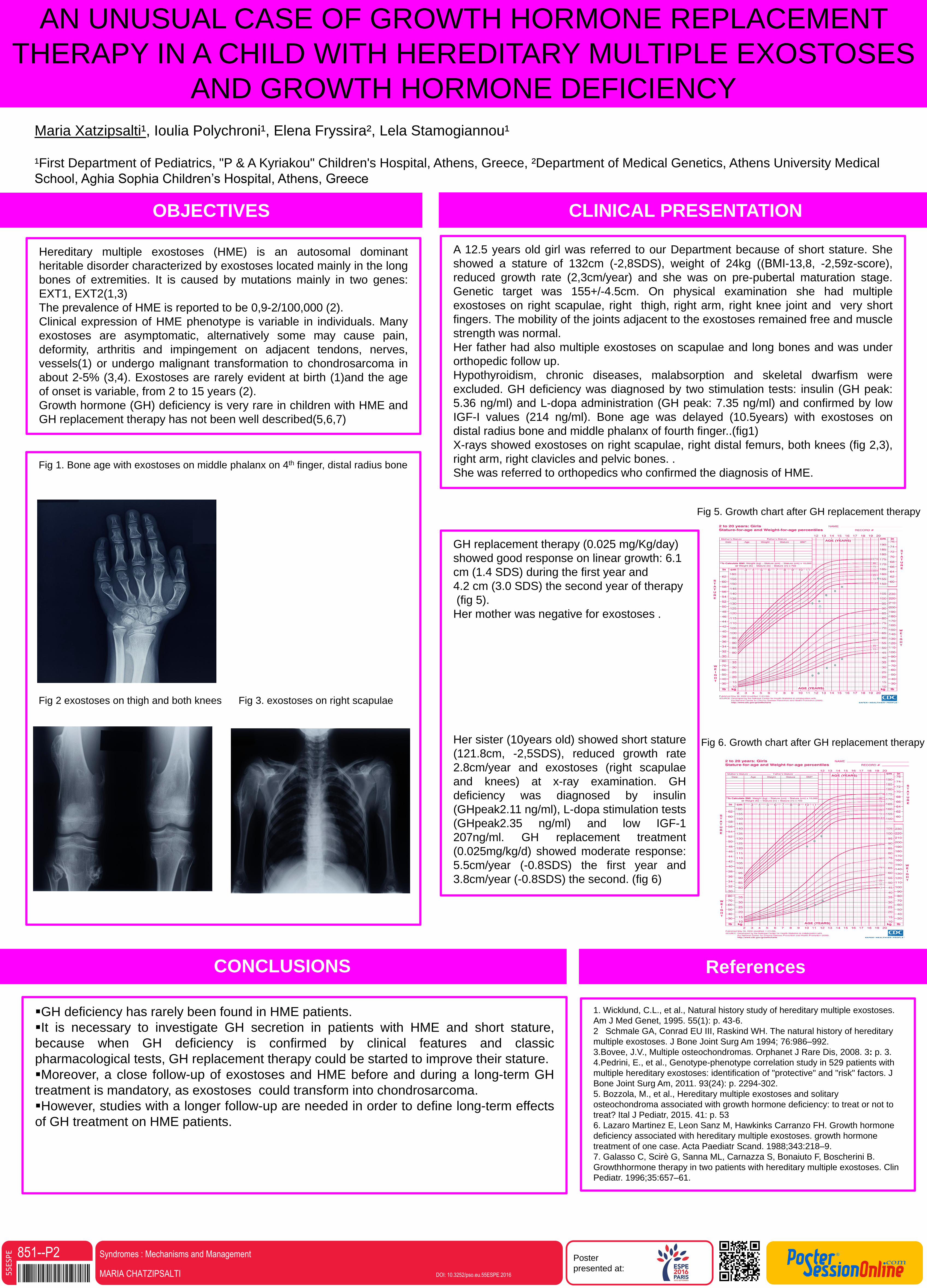
Fig 1. Bone age with exostoses on middle phalanx on 4th finger, distal radius bone
Fig 2 exostoses on thigh and both knees Fig 3. exostoses on right scapulae
A 12.5 years old girl was referred to our Department because of short stature. She
showed a stature of 132cm (-2,8SDS), weight of 24kg ((ΒΜΙ-13,8, -2,59z-score),
reduced growth rate (2,3cm/year) and she was on pre-pubertal maturation stage.
Genetic target was 155+/-4.5cm. On physical examination she had multiple
exostoses on right scapulae, right thigh, right arm, right knee joint and very short
fingers. The mobility of the joints adjacent to the exostoses remained free and muscle
strength was normal.
Her father had also multiple exostoses on scapulae and long bones and was under
orthopedic follow up.
Hypothyroidism, chronic diseases, malabsorption and skeletal dwarfism were
excluded. GH deficiency was diagnosed by two stimulation tests: insulin (GH peak:
5.36 ng/ml) and L-dopa administration (GH peak: 7.35 ng/ml) and confirmed by low
IGF-I values (214 ng/ml). Bone age was delayed (10.5years) with exostoses on
distal radius bone and middle phalanx of fourth finger..(fig1)
X-rays showed exostoses on right scapulae, right distal femurs, both knees (fig 2,3),
right arm, right clavicles and pelvic bones. .
She was referred to orthopedics who confirmed the diagnosis of HME.
1. Wicklund, C.L., et al., Natural history study of hereditary multiple exostoses.
Am J Med Genet, 1995. 55(1): p. 43-6.
2 Schmale GA, Conrad EU III, Raskind WH. The natural history of hereditary
multiple exostoses. J Bone Joint Surg Am 1994; 76:986–992.
3.Bovee, J.V., Multiple osteochondromas. Orphanet J Rare Dis, 2008. 3: p. 3.
4.Pedrini, E., et al., Genotype-phenotype correlation study in 529 patients with
multiple hereditary exostoses: identification of "protective" and "risk" factors. J
Bone Joint Surg Am, 2011. 93(24): p. 2294-302.
5. Bozzola, M., et al., Hereditary multiple exostoses and solitary
osteochondroma associated with growth hormone deficiency: to treat or not to
treat? Ital J Pediatr, 2015. 41: p. 53
6. Lazaro Martinez E, Leon Sanz M, Hawkinks Carranzo FH. Growth hormone
deficiency associated with hereditary multiple exostoses. growth hormone
treatment of one case. Acta Paediatr Scand. 1988;343:218–9.
7. Galasso C, Scirè G, Sanna ML, Carnazza S, Bonaiuto F, Boscherini B.
Growthhormone therapy in two patients with hereditary multiple exostoses. Clin
Pediatr. 1996;35:657–61.
GH replacement therapy (0.025 mg/Kg/day)
showed good response on linear growth: 6.1
cm (1.4 SDS) during the first year and
4.2 cm (3.0 SDS) the second year of therapy
(fig 5).
Her mother was negative for exostoses .
Her sister (10years old) showed short stature
(121.8cm, -2,5SDS), reduced growth rate
2.8cm/year and exostoses (right scapulae
and knees) at x-ray examination. GH
deficiency was diagnosed by insulin
(GHpeak2.11 ng/ml), L-dopa stimulation tests
(GHpeak2.35 ng/ml) and low IGF-1
207ng/ml. GH replacement treatment
(0.025mg/kg/d) showed moderate response:
5.5cm/year (-0.8SDS) the first year and
3.8cm/year (-0.8SDS) the second. (fig 6)
ReferencesCONCLUSIONS
AN UNUSUAL CASE OF GROWTH HORMONE REPLACEMENT
THERAPY IN A CHILD WITH HEREDITARY MULTIPLE EXOSTOSES
AND GROWTH HORMONE DEFICIENCY
Hereditary multiple exostoses (HME) is an autosomal dominant
heritable disorder characterized by exostoses located mainly in the long
bones of extremities. It is caused by mutations mainly in two genes:
EXT1, EXT2(1,3)
The prevalence of HME is reported to be 0,9-2/100,000 (2).
Clinical expression of HME phenotype is variable in individuals. Many
exostoses are asymptomatic, alternatively some may cause pain,
deformity, arthritis and impingement on adjacent tendons, nerves,
vessels(1) or undergo malignant transformation to chondrosarcoma in
about 2-5% (3,4). Exostoses are rarely evident at birth (1)and the age
of onset is variable, from 2 to 15 years (2).
Growth hormone (GH) deficiency is very rare in children with HME and
GH replacement therapy has not been well described(5,6,7)
GH deficiency has rarely been found in HME patients.
It is necessary to investigate GH secretion in patients with HME and short stature,
because when GH deficiency is confirmed by clinical features and classic
pharmacological tests, GH replacement therapy could be started to improve their stature.
Moreover, a close follow-up of exostoses and HME before and during a long-term GH
treatment is mandatory, as exostoses could transform into chondrosarcoma.
However, studies with a longer follow-up are needed in order to define long-term effects
of GH treatment on HME patients.
Fig 5. Growth chart after GH replacement therapy
Fig 6. Growth chart after GH replacement therapy
851--P2MARIA CHATZIPSALTI DOI: 10.3252/pso.eu.55ESPE.2016
Syndromes : Mechanisms and Management
Related Documents