Cancers 2021, 13, 3910. https://doi.org/10.3390/cancers13153910 www.mdpi.com/journal/cancers Review An Overview on the Histogenesis and Morphogenesis of Salivary Gland Neoplasms and Evolving Diagnostic Approaches Janaki Iyer 1 , Arvind Hariharan 1 , Uyen Minh Nha Cao 1,2 , Crystal To Tam Mai 1 , Athena Wang 1 , Parisa Khayambashi 1 , Bich Hong Nguyen 3 , Lydia Safi 1 and Simon D. Tran 1, * 1 McGill Craniofacial Tissue Engineering and Stem Cells Laboratory, Faculty of Dentistry, McGill University, 3640 University Street, Montreal, QC H3A 0C7, Canada; [email protected] (J.I.); [email protected] (A.H.); [email protected] (U.M.N.C.); [email protected] (C.T.T.M.); [email protected] (A.W.); [email protected] (P.K.); [email protected] (L.S.) 2 Department of Orthodontics, Faculty of Dentistry, Ho Chi Minh University of Medicine and Pharmacy, Ho Chi Minh City 700000, Vietnam 3 CHU Sainte Justine Hospital, Montreal, QC H3T 1C5, Canada; [email protected] * Correspondence: [email protected] Simple Summary: Diagnosing salivary gland neoplasms (SGN) remain a challenge, given their underlying biological nature and overlapping features. Evolving techniques in molecular pathol‐ ogy have uncovered genetic mutations resulting in these tumors. This review delves into the mo‐ lecular etiopathogenesis of SGN, highlighting advanced diagnostic protocols that may facilitate the identification and therapy of a variety of SGN. Abstract: Salivary gland neoplasms (SGN) remain a diagnostic dilemma due to their heterogenic complex behavior. Their diverse histomorphological appearance is attributed to the underlying cellular mechanisms and differentiation into various histopathological subtypes with overlapping features. Diagnostic tools such as fine needle aspiration biopsy, computerized tomography, mag‐ netic resonance imaging, and positron emission tomography help evaluate the structure and assess the staging of SGN. Advances in molecular pathology have uncovered genetic patterns and onco‐ genes by immunohistochemistry, fluorescent in situ hybridization, and next–generation sequenc‐ ing, that may potentially contribute to innovating diagnostic approaches in identifying various SGN. Surgical resection is the principal treatment for most SGN. Other modalities such as radio‐ therapy, chemotherapy, targeted therapy (agents like tyrosine kinase inhibitors, monoclonal anti‐ bodies, and proteasome inhibitors), and potential hormone therapy may be applied, depending on the clinical behaviors, histopathologic grading, tumor stage and location, and the extent of tissue invasion. This review delves into the molecular pathways of salivary gland tumorigenesis, high‐ lighting recent diagnostic protocols that may facilitate the identification and management of SGN. Keywords: salivary glands; salivary gland neoplasms; epithelial tumors; head and neck cancer; molecular pathology; diagnostic advances 1. Introduction Salivary glands are tubulo‐acinar exocrine organs that embryonically initiate in the sixth–eighth week of intrauterine life. The parotid gland is believed to arise from the oral ectoderm, while the submandibular and sublingual glands are from the embryonic en‐ doderm [1,2]. Their development is attributed to the physiologic process of ‘branching morphogenesis’, described as the rearrangement of a single epithelial bud to generate multiple acinar and ductal units, through continuous multi‐directional branching [3]. Citation: Iyer, J.; Hariharan, A.; Cao, U.M.N.; Mai, C.T.T.; Wang, A.; Khayambashi, P.; Nguyen, B.H.; Safi, L.; Tran, S.D. An Overview on the Histogenesis and Morphogenesis of Salivary Gland Neoplasms and Evolving Diagnostic Approaches. Cancers 2021, 13, 3910. https://doi.org/10.3390/ cancers13153910 Academic Editors: Paola Ferrari and Andrea Nicolini Received: 04 June 2021 Accepted: 29 July 2021 Published: 3 August 2021 Publisher’s Note: MDPI stays neutral with regard to jurisdictional claims in published maps and institutional affiliations. Copyright: © 2021 by the authors. Licensee MDPI, Basel, Switzerland. This article is an open access article distributed under the terms and conditions of the Creative Commons Attribution (CC BY) license (http://creativecommons.org/licenses /by/4.0/).

Welcome message from author
This document is posted to help you gain knowledge. Please leave a comment to let me know what you think about it! Share it to your friends and learn new things together.
Transcript

Cancers 2021, 13, 3910. https://doi.org/10.3390/cancers13153910 www.mdpi.com/journal/cancers
Review
An Overview on the Histogenesis and Morphogenesis of
Salivary Gland Neoplasms and Evolving
Diagnostic Approaches
Janaki Iyer 1, Arvind Hariharan 1, Uyen Minh Nha Cao 1,2, Crystal To Tam Mai 1, Athena Wang 1,
Parisa Khayambashi 1, Bich Hong Nguyen 3, Lydia Safi 1 and Simon D. Tran 1,*
1 McGill Craniofacial Tissue Engineering and Stem Cells Laboratory, Faculty of Dentistry,
McGill University, 3640 University Street, Montreal, QC H3A 0C7, Canada; [email protected] (J.I.);
[email protected] (A.H.); [email protected] (U.M.N.C.);
[email protected] (C.T.T.M.); [email protected] (A.W.);
[email protected] (P.K.); [email protected] (L.S.) 2 Department of Orthodontics, Faculty of Dentistry, Ho Chi Minh University of Medicine and Pharmacy,
Ho Chi Minh City 700000, Vietnam 3 CHU Sainte Justine Hospital, Montreal, QC H3T 1C5, Canada; [email protected]
* Correspondence: [email protected]
Simple Summary: Diagnosing salivary gland neoplasms (SGN) remain a challenge, given their
underlying biological nature and overlapping features. Evolving techniques in molecular pathol‐
ogy have uncovered genetic mutations resulting in these tumors. This review delves into the mo‐
lecular etiopathogenesis of SGN, highlighting advanced diagnostic protocols that may facilitate the
identification and therapy of a variety of SGN.
Abstract: Salivary gland neoplasms (SGN) remain a diagnostic dilemma due to their heterogenic
complex behavior. Their diverse histomorphological appearance is attributed to the underlying
cellular mechanisms and differentiation into various histopathological subtypes with overlapping
features. Diagnostic tools such as fine needle aspiration biopsy, computerized tomography, mag‐
netic resonance imaging, and positron emission tomography help evaluate the structure and assess
the staging of SGN. Advances in molecular pathology have uncovered genetic patterns and onco‐
genes by immunohistochemistry, fluorescent in situ hybridization, and next–generation sequenc‐
ing, that may potentially contribute to innovating diagnostic approaches in identifying various
SGN. Surgical resection is the principal treatment for most SGN. Other modalities such as radio‐
therapy, chemotherapy, targeted therapy (agents like tyrosine kinase inhibitors, monoclonal anti‐
bodies, and proteasome inhibitors), and potential hormone therapy may be applied, depending on
the clinical behaviors, histopathologic grading, tumor stage and location, and the extent of tissue
invasion. This review delves into the molecular pathways of salivary gland tumorigenesis, high‐
lighting recent diagnostic protocols that may facilitate the identification and management of SGN.
Keywords: salivary glands; salivary gland neoplasms; epithelial tumors; head and neck cancer;
molecular pathology; diagnostic advances
1. Introduction
Salivary glands are tubulo‐acinar exocrine organs that embryonically initiate in the
sixth–eighth week of intrauterine life. The parotid gland is believed to arise from the oral
ectoderm, while the submandibular and sublingual glands are from the embryonic en‐
doderm [1,2]. Their development is attributed to the physiologic process of ‘branching
morphogenesis’, described as the rearrangement of a single epithelial bud to generate
multiple acinar and ductal units, through continuous multi‐directional branching [3].
Citation: Iyer, J.; Hariharan, A.; Cao,
U.M.N.; Mai, C.T.T.; Wang, A.;
Khayambashi, P.; Nguyen, B.H.;
Safi, L.; Tran, S.D. An Overview on
the Histogenesis and Morphogenesis
of Salivary Gland Neoplasms and
Evolving Diagnostic Approaches.
Cancers 2021, 13, 3910.
https://doi.org/10.3390/
cancers13153910
Academic Editors: Paola Ferrari and
Andrea Nicolini
Received: 04 June 2021
Accepted: 29 July 2021
Published: 3 August 2021
Publisher’s Note: MDPI stays
neutral with regard to jurisdictional
claims in published maps and
institutional affiliations.
Copyright: © 2021 by the authors.
Licensee MDPI, Basel, Switzerland.
This article is an open access article
distributed under the terms and
conditions of the Creative Commons
Attribution (CC BY) license
(http://creativecommons.org/licenses
/by/4.0/).

Cancers 2021, 13, 3910 2 of 20
‘Epithelial–mesenchymal interaction’, described as a secondary induction of the epithe‐
lium by its underlying mesenchyme, is also essential for the normal development of sal‐
ivary glands [4]. This cascade of events ultimately forms multiple secretory units, each
consisting of a terminal acinar (serous/mucous) cell, myoepithelial cell, intercalated duct,
striated duct, and excretory duct, as elaborated in Figure 1 [1,2].
Figure 1. Schematic representation of the histology of salivary glands. Reprinted from [5,6] with permission. Ducto‐acinar
architecture of salivary glands is divided by capsular connective tissue septa into lobules. Each lobule consists of nu‐
merous serous/mucous/mixed (also known as serous demilune around mucous acini) secretory acini that are enveloped
by myoepithelial cells. The secretory acini unite to form intercalated ducts (lined by simple squamous to low cuboidal
epithelium and wrapped by myoepithelial cells). Saliva is secreted by acinar cells and drains into the striated ducts that
are lined by simple or pseudostratified columnar epithelial cells. This ductal lining transforms into stratified squamous
epithelium supported by basal cells. Ultimately, the striated ducts drain into the excretory ducts (also known as inter‐
lobular ducts) with tall columnar epithelial cells. Figure adapted from Tran et al., 2019 and Proctor and Shaalan, 2018
[5,6].
The architecture of salivary glands is two‐tiered, consisting of luminal (acinar and
ductal) and abluminal (myoepithelial and basal) cells [1,2]. These cells enter the cell cycle
rapidly, thus acting as potential targets for neoplastic transformation. The estimated
global incidence of salivary gland neoplasms (SGN) ranges from 0.4 to 13.5 cases per
100,000 annually, and constitutes approximately 3 to 6% of head and neck tumors [7]. The
diverse histomorphological appearance of SGN is attributed to their heterogenic and
complex cellular behavior. Differentiation into various histopathological subtypes with
overlapping features both within tumors and in different regions of the same tumor, re‐
sults in significant diagnostic challenge [1,2,8]. Advances in molecular pathology have
uncovered genetic patterns and biomarkers that may potentially contribute to innovating
diagnostic approaches in identifying various salivary gland pathologies [9]. In this paper,
we delve into the molecular pathways of salivary gland tumorigenesis, highlighting re‐
cent diagnostic protocols that may facilitate the identification and management of SGN.
Cancers 2021, 13, 3910 3 of 20
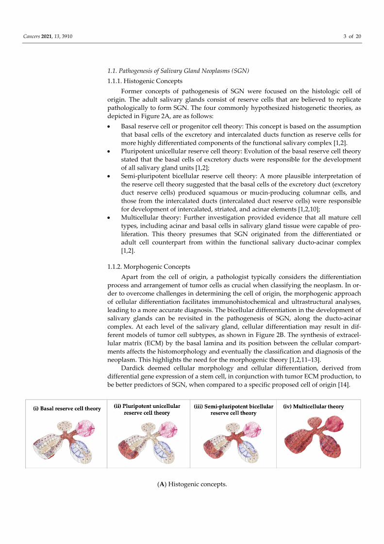
1.1. Pathogenesis of Salivary Gland Neoplasms (SGN)
1.1.1. Histogenic Concepts
Former concepts of pathogenesis of SGN were focused on the histologic cell of
origin. The adult salivary glands consist of reserve cells that are believed to replicate
pathologically to form SGN. The four commonly hypothesized histogenetic theories, as
depicted in Figure 2A, are as follows:
Basal reserve cell or progenitor cell theory: This concept is based on the assumption
that basal cells of the excretory and intercalated ducts function as reserve cells for
more highly differentiated components of the functional salivary complex [1,2].
Pluripotent unicellular reserve cell theory: Evolution of the basal reserve cell theory
stated that the basal cells of excretory ducts were responsible for the development
of all salivary gland units [1,2];
Semi‐pluripotent bicellular reserve cell theory: A more plausible interpretation of
the reserve cell theory suggested that the basal cells of the excretory duct (excretory
duct reserve cells) produced squamous or mucin‐producing columnar cells, and
those from the intercalated ducts (intercalated duct reserve cells) were responsible
for development of intercalated, striated, and acinar elements [1,2,10];
Multicellular theory: Further investigation provided evidence that all mature cell
types, including acinar and basal cells in salivary gland tissue were capable of pro‐
liferation. This theory presumes that SGN originated from the differentiated or
adult cell counterpart from within the functional salivary ducto‐acinar complex
[1,2].
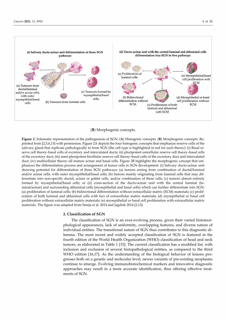
1.1.2. Morphogenic Concepts
Apart from the cell of origin, a pathologist typically considers the differentiation
process and arrangement of tumor cells as crucial when classifying the neoplasm. In or‐
der to overcome challenges in determining the cell of origin, the morphogenic approach
of cellular differentiation facilitates immunohistochemical and ultrastructural analyses,
leading to a more accurate diagnosis. The bicellular differentiation in the development of
salivary glands can be revisited in the pathogenesis of SGN, along the ducto‐acinar
complex. At each level of the salivary gland, cellular differentiation may result in dif‐
ferent models of tumor cell subtypes, as shown in Figure 2B. The synthesis of extracel‐
lular matrix (ECM) by the basal lamina and its position between the cellular compart‐
ments affects the histomorphology and eventually the classification and diagnosis of the
neoplasm. This highlights the need for the morphogenic theory [1,2,11–13].
Dardick deemed cellular morphology and cellular differentiation, derived from
differential gene expression of a stem cell, in conjunction with tumor ECM production, to
be better predictors of SGN, when compared to a specific proposed cell of origin [14].
(A) Histogenic concepts.
Cancers 2021, 13, 3910 4 of 20
(B) Morphogenic concepts.
Figure 2. Schematic representation of the pathogenesis of SGN. (A) Histogenic concepts. (B) Morphogenic concepts. Re‐
printed from [2,5,6,13] with permission. Figure 2A depicts the four histogenic concepts that emphasize reserve cells of the
salivary gland that replicate pathologically to form SGN (the cell type is highlighted in red for each theory): (i) Basal re‐
serve cell theory–basal cells of excretory and intercalated ducts; (ii) pluripotent unicellular reserve cell theory–basal cells
of the excretory duct; (iii) semi‐pluripotent bicellular reserve cell theory–basal cells of the excretory duct and intercalated
duct; (iv) multicellular theory–all mature acinar and basal cells. Figure 2B highlights the morphogenic concept that em‐
phasizes the differentiation process and arrangement of tumor cells in SGN development: (i) Salivary ducto‐acinar unit
showing potential for differentiation of three SGN pathways: (a) tumors arising from combination of ductal/luminal
and/or acinar cells, with outer myoepithelial/basal cells; (b) tumors mainly originating from luminal cells that may dif‐
ferentiate into non‐specific ductal, acinar or goblet cells, and/or combination of these cells; (c) tumors almost entirely
formed by myoepithelial/basal cells; or (ii) cross‐section of the ducto‐acinar unit with the central luminal (lu‐
minal/acinar) and surrounding abluminal cells (myoepithelial and basal cells) which can further differentiate into SGN:
(a) proliferation of luminal cells; (b) bidirectional differentiation without extracellular matrix (ECM) materials; (c) prolif‐
eration of both luminal and abluminal cells with foci of extracellular matrix materials; (d) myoepithelial or basal cell
proliferation without extracellular matrix materials; (e) myoepithelial or basal cell proliferation with extracellular matrix
materials. The figure was adapted from Sreeja et al. 2014 and Jagdish 2014 [2,13].
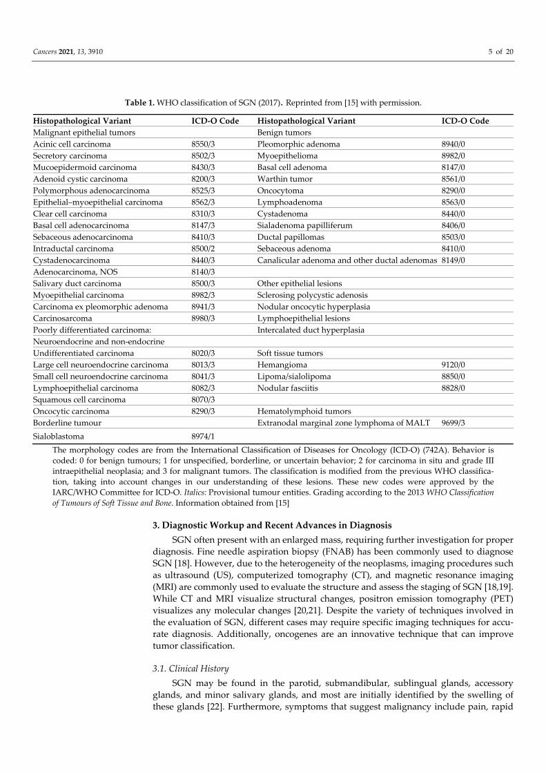
2. Classification of SGN
The classification of SGN is an ever‐evolving process, given their varied histomor‐
phological appearances, lack of uniformity, overlapping features, and diverse nature of
individual entities. The transitional nature of SGN thus contributes to this diagnostic di‐
lemma. The most recent and widely accepted classification of SGN is featured in the
fourth edition of the World Health Organization (WHO) classification of head and neck
tumors, as elaborated in Table 1 [15]. The current classification has a modified list, with
inclusion and exclusion of several histopathological entities, as compared to the third
WHO edition [16,17]. As the understanding of the biological behavior of lesions pro‐
gresses both on a genetic and molecular level, newer variants of pre‐existing neoplasms
continue to emerge. Evolving immunohistochemical markers and innovative diagnostic
approaches may result in a more accurate identification, thus offering effective treat‐
ments of SGN.
Cancers 2021, 13, 3910 5 of 20
Table 1. WHO classification of SGN (2017). Reprinted from [15] with permission.
Histopathological Variant ICD‐O Code Histopathological Variant ICD‐O Code
Malignant epithelial tumors Benign tumors
Acinic cell carcinoma 8550/3 Pleomorphic adenoma 8940/0
Secretory carcinoma 8502/3 Myoepithelioma 8982/0
Mucoepidermoid carcinoma 8430/3 Basal cell adenoma 8147/0
Adenoid cystic carcinoma 8200/3 Warthin tumor 8561/0
Polymorphous adenocarcinoma 8525/3 Oncocytoma 8290/0
Epithelial–myoepithelial carcinoma 8562/3 Lymphoadenoma 8563/0
Clear cell carcinoma 8310/3 Cystadenoma 8440/0
Basal cell adenocarcinoma 8147/3 Sialadenoma papilliferum 8406/0
Sebaceous adenocarcinoma 8410/3 Ductal papillomas 8503/0
Intraductal carcinoma 8500/2 Sebaceous adenoma 8410/0
Cystadenocarcinoma 8440/3 Canalicular adenoma and other ductal adenomas 8149/0
Adenocarcinoma, NOS 8140/3
Salivary duct carcinoma 8500/3 Other epithelial lesions
Myoepithelial carcinoma 8982/3 Sclerosing polycystic adenosis
Carcinoma ex pleomorphic adenoma 8941/3 Nodular oncocytic hyperplasia
Carcinosarcoma 8980/3 Lymphoepithelial lesions
Poorly differentiated carcinoma: Intercalated duct hyperplasia
Neuroendocrine and non‐endocrine
Undifferentiated carcinoma 8020/3 Soft tissue tumors
Large cell neuroendocrine carcinoma 8013/3 Hemangioma 9120/0
Small cell neuroendocrine carcinoma 8041/3 Lipoma/sialolipoma 8850/0
Lymphoepithelial carcinoma 8082/3 Nodular fasciitis 8828/0
Squamous cell carcinoma 8070/3
Oncocytic carcinoma 8290/3 Hematolymphoid tumors
Borderline tumour Extranodal marginal zone lymphoma of MALT 9699/3
Sialoblastoma 8974/1
The morphology codes are from the International Classification of Diseases for Oncology (ICD‐O) (742A). Behavior is
coded: 0 for benign tumours; 1 for unspecified, borderline, or uncertain behavior; 2 for carcinoma in situ and grade III
intraepithelial neoplasia; and 3 for malignant tumors. The classification is modified from the previous WHO classifica‐
tion, taking into account changes in our understanding of these lesions. These new codes were approved by the
IARC/WHO Committee for ICD‐O. Italics: Provisional tumour entities. Grading according to the 2013 WHO Classification
of Tumours of Soft Tissue and Bone. Information obtained from [15]
3. Diagnostic Workup and Recent Advances in Diagnosis
SGN often present with an enlarged mass, requiring further investigation for proper
diagnosis. Fine needle aspiration biopsy (FNAB) has been commonly used to diagnose
SGN [18]. However, due to the heterogeneity of the neoplasms, imaging procedures such
as ultrasound (US), computerized tomography (CT), and magnetic resonance imaging
(MRI) are commonly used to evaluate the structure and assess the staging of SGN [18,19].
While CT and MRI visualize structural changes, positron emission tomography (PET)
visualizes any molecular changes [20,21]. Despite the variety of techniques involved in
the evaluation of SGN, different cases may require specific imaging techniques for accu‐
rate diagnosis. Additionally, oncogenes are an innovative technique that can improve
tumor classification.
3.1. Clinical History
SGN may be found in the parotid, submandibular, sublingual glands, accessory
glands, and minor salivary glands, and most are initially identified by the swelling of
these glands [22]. Furthermore, symptoms that suggest malignancy include pain, rapid

Cancers 2021, 13, 3910 6 of 20
tissue growth, or loss of nerve function [23,24]. In clinical practice, it has been reported
that minor SGN account for less than 25% of SGN [25], and smaller salivary glands have a
higher incidence of malignancy. While only 20% of SGN are malignant [26], it is crucial to
accurately differentiate benign from malignant neoplasms in order to devise an appro‐
priate treatment plan.
3.2. Fine‐Needle Aspiration Biopsy (FNAB)
FNAB is one of the first line procedures used to diagnose SGN on account of its easy,
inexpensive, highly accurate, quick, and minimally invasive nature [27]. This technique
entails using a fine gauge needle to collect cells. After alcohol fixation and drying, the
cellular aspirate is stained with Papanicolaou stain and can be immediately evaluated
and diagnosed [27]. FNAB results are universally reported using the Milan’s system, as
seen in Table 2 [28]. Edizer et al. (2016), evaluated the ability of FNAB to differentially
diagnose salivary gland masses by comparing the preoperative FNAB results with the
postoperative definitive histopathological results of 285 patients. Their FNAB results
were 92.6% accurate compared to the definitive histopathological results. This demon‐
strated that FNAB is useful in benign and malignant tumor differentiation. However,
they do have some limitations, which involve relatively high non‐diagnostic results,
possibly due to bleeding, low cellularity, necrosis, or erroneous technique [27]. In addi‐
tion, some potential outcomes in the final histopathological examination include squa‐
mous metaplasia and fibrosis. However, these do not interfere with the definitive diag‐
nosis [27].
Table 2. Milan’s system of FNAB reporting for SGN [28].
Diagnostic Category Risk of Malignancy % Management
Non‐diagnostic 25 Clinical and radiologic
correlation/repeat FNAC
Non‐neoplastic 10 Clinical follow‐up and
radiological correlation
Atypia of undetermined
significance (AUS) 20 Repeat FNAC or surgery
Neoplasm: benign <5 Surgery or clinical follow‐up
Neoplasm: salivary gland
neoplasm of uncertain malignant
potential (SUMP) 35 Surgery
Suspicious for malignancy (SM) 60 Surgery
Malignant 90 Surgery
Information obtained from [28]
3.3. Ultrasound (US)
US is a highly effective non‐invasive technology that can be used in the differential
diagnosis of SGN [29]. The technology uses high‐frequency sound (ultrasonic) waves to
generate images of internal tissues and organs [30]. Modern USs have demonstrated
greater success in providing precise measurements, localization, and evaluation of the
structures of various SGN, as highlighted in Table 3 [29,31,32]. In a study conducted by
Bialek et al. (2003), the role of the US in the differentiation and diagnosis of Pleomorphic
Adenomas (PA) was analyzed. By using a modern US machine, in conjunction with
high‐resolution probes and tissue harmonic imaging, they were able to detect 96% of
malignant salivary glands in patients with solid lesions. Modern USs are considered
highly valuable, dependable, and useful in the differential diagnosis of SGN; however,
they possess some limitations [29]. USs are unable to properly assess lesions located in
obscure areas (i.e., deep lobe of the parotid gland, behind bones) and are inadequate in
differentially diagnosing small lesions [29].
Cancers 2021, 13, 3910 7 of 20
3.4. Computerized Tomography (CT)
In conjunction with the US, contrast‐enhanced computerized tomography (CT) is an
imaging technique often used to obtain a more detailed view of deeper masses (Table 3)
[31–34]. Given that CT scanning exposes patients to high levels of radiation, variations of
CT such as cone beam CT (CBCT) have been used as an alternative measure since it emits
relatively decreased levels of radiation [35]. Furthermore, in a study by Jung et al. (2020),
researchers found that single‐phase CT scanning may be a low‐radiation alternative in
the differentiation of tumors [18]. They compared the texture analysis parameters in sin‐
gle‐phase CT and conventional two‐phase CT, to differentiate between two common
types of benign tumors: Warthin tumor (WT) and PA. The authors found that the dif‐
ferential parameters between WT and PA from a single‐phase CT were similar to those of
a two‐phase scan. Moreover, they found that the patient was exposed to less radiation
during texture analysis via the single‐phase imaging. Thus, researchers concluded that
this tool could be a minimally invasive method in the investigation of benign SGN [18].
However, further testing is required to assess whether these findings may be extended to
the differentiation of malignant neoplasms.
3.5. Magnetic Resonance Imaging (MRI)
Despite MRI being relatively more costly and requiring more time to produce im‐
ages [36], its major benefit is that it is free of radiation. Additionally, researchers have
previously deemed MRI to be the most suitable imaging technique in the assessment of
parotid gland tumors and relation to adjacent vital structures as it offers a high contrast
resolution of the soft tissues (Table 3) [31,32,37]. Parameters investigating malignancy
include tumor border configuration, invasion of adjacent tissues, T1‐ and T2‐ weighted
signal intensity, and time–intensity curve with constant enhancement [38,39]. Common
MRI findings that favor malignancy include low T2 signal, heterogenous enhancement,
lesional growth with ill‐defined, or blurry tumor borders that may invade into the adja‐
cent structures and lymph nodes. Malignant SGN imaging may reveal cystic changes,
central necrosis, perineural infiltration, accompanied by regional or distant metastasis
[40]. Low‐grade malignant tumors may resemble benign lesions; however, the difference
of contents of the cystic component of benign lesions may be revealed as increased hy‐
perintense T1‐weighted images [40,41]. Although MRI is the preferred imaging technique
for SGN, this diagnostic approach cannot be employed among patients allergic to con‐
trast dyes. Therefore, in a study conducted by Takumi et al. (2021), they investigated a
combination of non‐contrast MRI techniques to enhance the diagnostic performance in
differentiating between benign and malignant SGN [36]. They focused on three
non‐contrast MRI parameters: apparent diffusion coefficient, tumor blood flow, and
amide proton transfer related signal intensity. Upon studying each parameter individu‐
ally, diagnostic performance was found to be limited. However, when these parameters
were combined, there was a significant increase in the accuracy of the diagnosis, thus
leading the authors to conclude that this multiparametric approach of using non‐contrast
MRI may improve the differentiation of the nature of the SGN [36].
3.6. Positron Emission Tomography (PET)
PET is a non‐invasive imaging technology that uses radioactive tracers to visualize
and evaluate tissues and organs for the presence of diseases, including cancer [42]. Once
these tracers are intravenously injected, they gather in areas of higher chemical activity,
often indicating areas of disease [42]. These tracers emit radiation that can be detected by
the PET scanner, which generates an image map for assessment [42]. Roh et al. (2007),
evaluated the role of PET, using 18F‐fluorodeoxyglucose (FDG) as tracers among patients
with salivary gland cancers (Table 3) [43]. They were able to detect 91.2% of primary
tumors in patients and concluded that 18FDG‐PET is clinically useful in histologic grad‐
ing and initial staging of salivary gland malignancies. However, the technology does
Cancers 2021, 13, 3910 8 of 20
have some limitations. Occasionally, normal physiologic uptake of radioactive tracers
occurs, which often mimics or hides existing neoplasms [43]. Additionally, low‐grade
malignancies frequently have lower tracer uptake than high‐grade malignancies [43].
Therefore, these limitations may lead to undetected SGN [43].
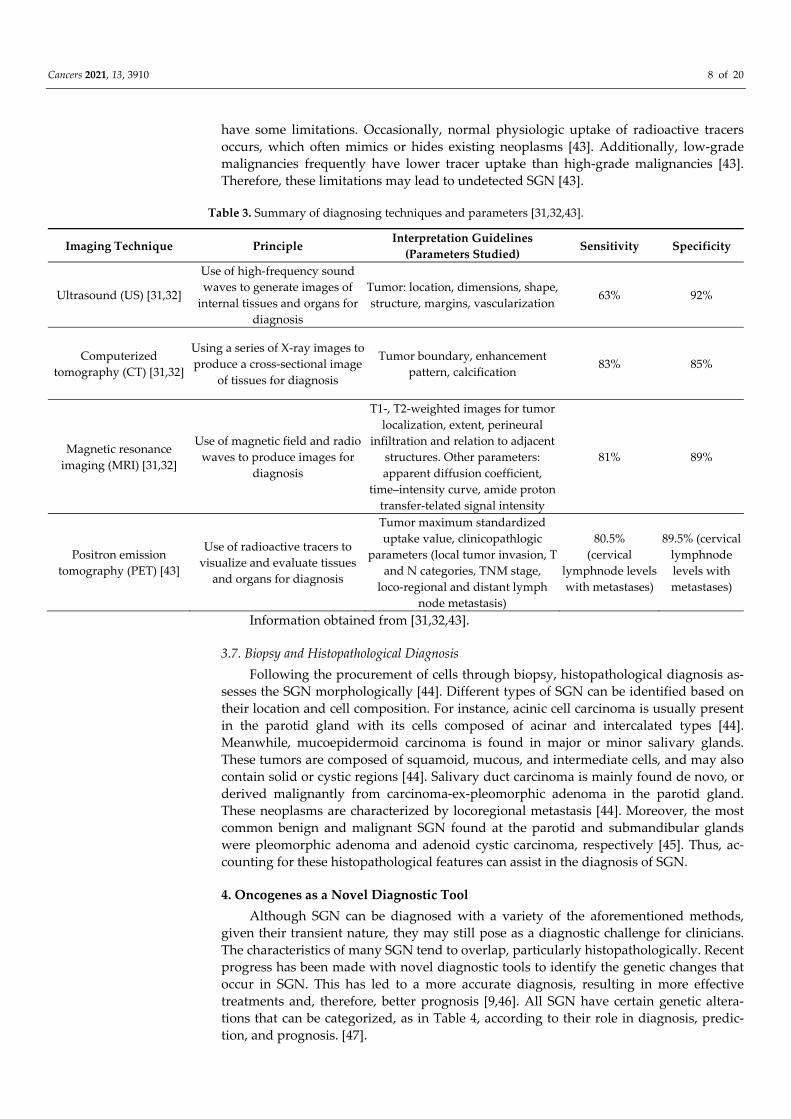
Table 3. Summary of diagnosing techniques and parameters [31,32,43].
Imaging Technique Principle Interpretation Guidelines
(Parameters Studied) Sensitivity Specificity
Ultrasound (US) [31,32]
Use of high‐frequency sound
waves to generate images of
internal tissues and organs for
diagnosis
Tumor: location, dimensions, shape,
structure, margins, vascularization 63% 92%
Computerized
tomography (CT) [31,32]
Using a series of X‐ray images to
produce a cross‐sectional image
of tissues for diagnosis
Tumor boundary, enhancement
pattern, calcification 83% 85%
Magnetic resonance
imaging (MRI) [31,32]
Use of magnetic field and radio
waves to produce images for
diagnosis
T1‐, T2‐weighted images for tumor
localization, extent, perineural
infiltration and relation to adjacent
structures. Other parameters:
apparent diffusion coefficient,
time–intensity curve, amide proton
transfer‐telated signal intensity
81% 89%
Positron emission
tomography (PET) [43]
Use of radioactive tracers to
visualize and evaluate tissues
and organs for diagnosis
Tumor maximum standardized
uptake value, clinicopathlogic
parameters (local tumor invasion, T
and N categories, TNM stage,
loco‐regional and distant lymph
node metastasis)
80.5%
(cervical
lymphnode levels
with metastases)
89.5% (cervical
lymphnode
levels with
metastases)
Information obtained from [31,32,43].
3.7. Biopsy and Histopathological Diagnosis
Following the procurement of cells through biopsy, histopathological diagnosis as‐
sesses the SGN morphologically [44]. Different types of SGN can be identified based on
their location and cell composition. For instance, acinic cell carcinoma is usually present
in the parotid gland with its cells composed of acinar and intercalated types [44].
Meanwhile, mucoepidermoid carcinoma is found in major or minor salivary glands.
These tumors are composed of squamoid, mucous, and intermediate cells, and may also
contain solid or cystic regions [44]. Salivary duct carcinoma is mainly found de novo, or
derived malignantly from carcinoma‐ex‐pleomorphic adenoma in the parotid gland.
These neoplasms are characterized by locoregional metastasis [44]. Moreover, the most
common benign and malignant SGN found at the parotid and submandibular glands
were pleomorphic adenoma and adenoid cystic carcinoma, respectively [45]. Thus, ac‐
counting for these histopathological features can assist in the diagnosis of SGN.
4. Oncogenes as a Novel Diagnostic Tool
Although SGN can be diagnosed with a variety of the aforementioned methods,
given their transient nature, they may still pose as a diagnostic challenge for clinicians.
The characteristics of many SGN tend to overlap, particularly histopathologically. Recent
progress has been made with novel diagnostic tools to identify the genetic changes that
occur in SGN. This has led to a more accurate diagnosis, resulting in more effective
treatments and, therefore, better prognosis [9,46]. All SGN have certain genetic altera‐
tions that can be categorized, as in Table 4, according to their role in diagnosis, predic‐
tion, and prognosis. [47].
Cancers 2021, 13, 3910 9 of 20
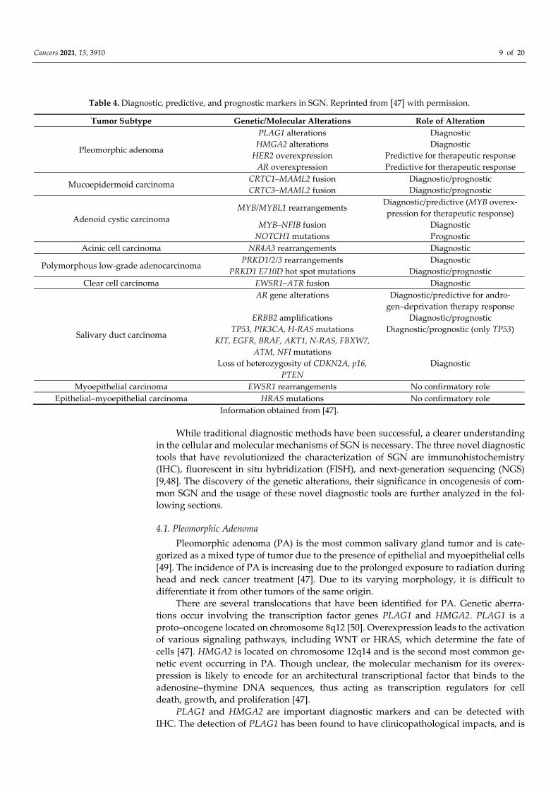
Table 4. Diagnostic, predictive, and prognostic markers in SGN. Reprinted from [47] with permission.
Tumor Subtype Genetic/Molecular Alterations Role of Alteration
Pleomorphic adenoma
PLAG1 alterations Diagnostic
HMGA2 alterations Diagnostic
HER2 overexpression
AR overexpression
Predictive for therapeutic response
Predictive for therapeutic response
Mucoepidermoid carcinoma CRTC1–MAML2 fusion Diagnostic/prognostic
CRTC3–MAML2 fusion Diagnostic/prognostic
Adenoid cystic carcinoma
MYB/MYBL1 rearrangements Diagnostic/predictive (MYB overex‐
pression for therapeutic response)
MYB–NFIB fusion
NOTCH1 mutations
Diagnostic
Prognostic
Acinic cell carcinoma NR4A3 rearrangements Diagnostic
Polymorphous low‐grade adenocarcinoma PRKD1/2/3 rearrangements
PRKD1 E710D hot spot mutations
Diagnostic
Diagnostic/prognostic
Clear cell carcinoma EWSR1–ATR fusion Diagnostic
Salivary duct carcinoma
AR gene alterations Diagnostic/predictive for andro‐
gen–deprivation therapy response
ERBB2 amplifications Diagnostic/prognostic
TP53, PIK3CA, H‐RAS mutations
KIT, EGFR, BRAF, AKT1, N‐RAS, FBXW7,
ATM, NFI mutations
Diagnostic/prognostic (only TP53)
Loss of heterozygosity of CDKN2A, p16,
PTEN
Diagnostic
Myoepithelial carcinoma EWSR1 rearrangements No confirmatory role
Epithelial–myoepithelial carcinoma HRAS mutations No confirmatory role
Information obtained from [47].
While traditional diagnostic methods have been successful, a clearer understanding
in the cellular and molecular mechanisms of SGN is necessary. The three novel diagnostic
tools that have revolutionized the characterization of SGN are immunohistochemistry
(IHC), fluorescent in situ hybridization (FISH), and next‐generation sequencing (NGS)
[9,48]. The discovery of the genetic alterations, their significance in oncogenesis of com‐
mon SGN and the usage of these novel diagnostic tools are further analyzed in the fol‐
lowing sections.
4.1. Pleomorphic Adenoma
Pleomorphic adenoma (PA) is the most common salivary gland tumor and is cate‐
gorized as a mixed type of tumor due to the presence of epithelial and myoepithelial cells
[49]. The incidence of PA is increasing due to the prolonged exposure to radiation during
head and neck cancer treatment [47]. Due to its varying morphology, it is difficult to
differentiate it from other tumors of the same origin.
There are several translocations that have been identified for PA. Genetic aberra‐
tions occur involving the transcription factor genes PLAG1 and HMGA2. PLAG1 is a
proto–oncogene located on chromosome 8q12 [50]. Overexpression leads to the activation
of various signaling pathways, including WNT or HRAS, which determine the fate of
cells [47]. HMGA2 is located on chromosome 12q14 and is the second most common ge‐
netic event occurring in PA. Though unclear, the molecular mechanism for its overex‐
pression is likely to encode for an architectural transcriptional factor that binds to the
adenosine–thymine DNA sequences, thus acting as transcription regulators for cell
death, growth, and proliferation [47].
PLAG1 and HMGA2 are important diagnostic markers and can be detected with
IHC. The detection of PLAG1 has been found to have clinicopathological impacts, and is

Cancers 2021, 13, 3910 10 of 20
supported by histopathological findings [50,51]. The overexpression of PLAG1 by IHC
has helped differentiate between PA and other SGN, such as adenoid cystic carcinoma
(ACC), with high specificity [52]. A study by Mito et al. (2017) also showed the im‐
portance of IHC in the detection of HMGA2. They found that it is a highly specific marker
for PA compared to other histologically mimicking tumors [53].
FISH is now at the forefront of SGN diagnosis due to the discovery of novel onco‐
genic fusions and gene translocations. It has been useful in diagnosing PA with the ex‐
pression of fusions involving PLAG1 and HMGA2 [54]. Evrard et al. (2017) showed how
the use of FISH facilitated salivary gland cytology and thus, the assessment of the extent
of surgery. They concluded that the addition of FISH in the detection of PLAG1 to con‐
ventional cytological analysis increased overall sensitivity and eliminated the need to use
frozen sections for a diagnosis [55].
PA has the potential to transform into carcinoma ex pleomorphic adenoma (Ca
ex–PA) adding to the diagnostic challenge. The expression of PLAG1 and HMGA2 is
common for both tumors [47]. In a molecular study that used FISH to determine the
similarities between PA and Ca ex–PA, the reviewed cases displayed evidence of metas‐
tasis. However, they appeared histologically benign which further complicates differen‐
tiation [56]. There is evidence that Ca ex–PA could be differentiated by overexpression of
TP53, AR, and HER2 genes. However, further research is required to confirm whether
mutations of these genes could signify malignant transformation and hence be used as
predictive biomarkers.
4.2. Mucoepidermoid Carcinoma
Mucoepidermoid carcinoma (MEC) is the most common malignancy of the salivary
glands and can occur in both children and adults. It is characterized by the increased
proliferation of the excretory cells [9]. While the etiology remains controversial, some
studies have shown the implications of viruses [9]. The diagnostic and prognostic mark‐
ers involve fusion proteins derived from chromosomal rearrangements [57].
The genetic aberrations involve CRTC1–MAML2 or CRTC3–MAML2 fusions, with
the latter being more important [58,59]. CRTC1 is located on chromosome 9 and it en‐
codes protein from the CREB family to enhance transcription. The CREB protein is re‐
sponsible for regulating all genes involved in proliferation and differentiation. The
MAML2 gene is located on chromosome 11 and it encodes for the nuclear proteins re‐
sponsible for the activation of the NOTCH pathway, which is one of the most common
signaling pathways activated during tumorigenesis [47].
While CRTC1–MAML2 fusion is detected in most cases of MEC, the molecular
mechanisms have yet to be clearly understood. Once fusion occurs, the protein activates
the transcription of CREB target genes to contribute to tumorigenesis. A study by Chen et
al. (2021) showed that CRTC1–MAML2 fusion could be modulated as a therapeutic target.
After its elimination in mice, MEC xenografts demonstrated no further growth [58]. Ear‐
lier studies expressed this fusion as a potential prognostic marker due to its tendency to
indicate a favorable prognosis in young patients [47]. Recent studies, however, have
disproved this theory due to increasingly strict MEC diagnostic guidelines, especially in
early‐stage MEC [60].
Expression of MAML2 using FISH has been acclaimed to be very useful. It is a rela‐
tively straightforward diagnosis considering that the expression of MAML2 is exclusive
to MEC [52]. It is particularly useful in diagnosing the oncocytic variants of MEC. These
variants are more problematic to diagnose since they mimic other SGN, such as acinic cell
carcinoma (AciCC) [52]. Although NGS has improved diagnostic accuracy, the MAML2,
FISH, may sometimes exhibit negative results, notably in the oncocytic variants of MEC
[61]. Case studies have shown that whenever FISH has failed to express fusion, NGS has
validated its potential as a confirmatory test [61]. The prognostic role of MAML2 can be
seen using IHC since it is thought that the CTRC1–MAML2 fusion is a downstream target
of the EGFR ligand, amphiregulin (AREG). A study by Shinomiya et al. (2016) supported
Cancers 2021, 13, 3910 11 of 20
this finding, where the overexpression of AREG and EGFR was characterized by IHC in
MEC samples, which played a role in tumor growth and survival [62].
4.3. Adenoid Cystic Carcinoma
Adenoid cystic carcinoma (ACC) is another common malignant tumor of the sali‐
vary glands. It is slow‐growing and composed of epithelial and myoepithelial cells of
different origins [9]. Given its high recurrence, it has a very poor prognosis due to its
metastatic capability and associated perineural invasion. Current treatment protocols
involve surgery, followed by post‐operative radiotherapy (PORT), which have been
fairly successful [63]. PORT has shown to be an effective adjuvant to surgery and to
minimize the incidence of recurrence [64]. However, studies have shown that it does not
really affect the overall survival rates of patients, therefore questioning its effectiveness
[65]. For more effective diagnosis and management strategies, it is necessary to under‐
stand the underlying genome alterations when studying recurrent ACC tumors [47].
Studies found that recurrent ACC showed alterations in the NOTCH pathway when
compared to primary ACC cases. These mutations in the NOTCH pathway are signifi‐
cant as they could lead to potential therapeutic targets [66]. Ferraroto et al. (2017) con‐
cluded that the NOTCH mutations were indicative of a more distinct form of ACC, ex‐
hibiting metastasis in bone and liver; however, this was minimized by NOTCH inhibitors
[67].
The main genomic alteration that characterizes ACC is the MYB–NFIB gene fusion.
Overexpression of MYB is a diagnostic characteristic feature of ACC. It is located on
chromosome 6q and it encodes for a transcription factor that regulates cell proliferation
and differentiation of hematopoietic, colonic, and neural progenitor cells [47]. NFIB is
located on chromosome 9q and is also a key regulator for hematopoietic and epithelial
cells. A study by Rettig et al. (2016) presented an overexpression of NFIB in ACC, sug‐
gestive of an alternative oncogenetic pathway [68]. Whole genome sequencing has also
revealed enhanced translocation, leading to the overexpression of MYB. This provided
another insight into the downstream process of MYB in different ACC lineages [69].
Overexpression of MYB is thought to impact DNA repair, apoptosis, cell migration,
and cell signaling for cell cycle control [47]. Xu et al. (2019) inferred that salivary ACC
tissue samples displayed a higher expression of MYB when compared to normal salivary
tissue and was associated with metastatic potential [70]. Detecting MYB–NFIB fusion can
be difficult using IHC since MYB overexpression is also seen in other SGN. Thus, this
MYB–NFIB fusion is detected using FISH [44]. NGS has also been proven useful in di‐
agnosing ACC. In a recent case study, the presence of a MYB–NFIB fusion was detected
by NGS in a suspected case of ameloblastoma with histopathological variations [61]. This
emphasizes the significance of introducing these novel diagnostic tools for a more accu‐
rate diagnosis.
4.4. Acinic Cell Carcinoma
Acinic cell carcinoma (AciCC) is a low‐grade malignancy consisting of both ductal
and acinar cells with the presence of basophilic cytoplasm. It is the third most common
malignancy of the salivary glands and is slow progressing but can metastasize to local
and distant sites [9]. Though the knowledge of its molecular aberrations remains limited,
it is commonly characterized by the expression of DOG1 (a membrane channel protein),
while the prominent genetic aberration is the translocation of SCPP–NR4A3 [47].
SCPP is a secretory phosphoprotein that contains several genes responsible for
producing salivary contents, bone, dentin, and enamel. It is located on chromosome 4q13
[36]. NR4A3 is an important nuclear receptor that is located on chromosome 9q31 and it
encodes for the steroid–thyroid hormone–retinoid receptor [47,71]. The upregulation of
NR4A3 increases the expression of target genes and influences cell proliferation. Another
rare genetic fusion that can be seen is MSANTD3–HTN3 translocation, which is charac‐
teristic in variants with a more serous nature [72]. MSANTD3 encodes for a poorly char‐
Cancers 2021, 13, 3910 12 of 20
acterized protein, whereas HTN3 is exclusively present in the saliva and functions as an
antimicrobial peptide [71].
Diagnosis is straightforward as the SCPP–NR4A3 is exclusive to AciCC. Moreover,
the immunoexpression of DOG1 is also a characteristic finding for AciCC [47]. IHC has
proven to be more specific than FISH for the expression of NR4A3 and has been found to
be a specific and sensitive novel marker [71]. IHC has also shown relatively high speci‐
ficity for the expression of MSANTD3. However, further studies are required to validate
its role as a diagnostic marker in AciCC.
4.5. Polymorphous Adenocarcinoma
Polymorphous Adenocarcinoma (PAC) is an epithelial tumor most commonly
found in the minor salivary glands. It is a relatively rare tumor and is usually associated
with a favorable prognosis [9]. It was previously named “polymorphous low‐grade car‐
cinoma,” and was renamed by the WHO (2017) due to its aggressive nature [47]. While
cribriform adenocarcinoma has recently been incorporated into the PAC group of SGN
due to their similar characteristics, it remains highly controversial whether they should
be referred to as separate entities [73].
PAC is characterized by the mutation of PRKD1, a protein–kinase gene located on
chromosome 14. It encodes a protein kinase that is involved in cellular processes in‐
cluding migration and differentiation, due to the signaling of the MAP kinase, RAS, and
other cell survival and adhesion pathways [47]. The PRKD1 E710D hot spot mutation is a
useful ancillary diagnostic marker along with the PRKD1 mutations to differentiate be‐
tween other SGN [73]. Although diagnosis is most likely done by visualizing the mor‐
phology, IHC can play a small role in certain instances [44]. Sebastiao et al. (2019) used
FISH to demonstrate the genetic alterations of PRKD1 as a diagnostic marker with rea‐
sonable success, particularly to identify nodal metastasis [74]. While PRKD1 E710D mu‐
tations as a prognostic marker has yet to be thoroughly researched, studies have ob‐
served its correlation with a metastasis–free tumor [73].
4.6. Clear Cell Carcinoma
Clear cell carcinoma (CCC) is a low‐grade salivary tumor found in minor salivary
glands and is characterized by the presence of clear cells [47]. It is identified by the ap‐
pearance of EWSR1–ATF1 fusion, which is a major genetic aberration [47]. EWSR1 is an
“Ewing’s sarcoma” gene and is a member of the TET family protein group, located on
chromosome 22q12. It encodes an RNA‐binding protein, which is involved in gene ex‐
pression, cell signaling as well as RNA processing, transport, and function [75]. ATF1 is a
transcription factor located on chromosome 12 and is an element of the CREB family of
proteins. Studies have shown that tumorigenesis could occur due to the aberrant activa‐
tion of ATF1 upon fusion with EWSR1 [75].
FISH is a very useful tool for diagnosing CCC as it can detect the EWSR1–ATF1 fu‐
sion. An early study by Shah et al. (2013) demonstrated that FISH had higher sensitivity
in detecting rearrangements of EWSR1 in hyalinizing CCC [76]. At times, it can be diffi‐
cult to differentiate between hyalinizing and odontogenic forms of CCC, as well as minor
forms of MEC since they all exhibit translocations involving EWSR1 [44]. NGS may be an
even more accurate tool to differentiate and specify the genetic alterations between forms
of CCC [44].
4.7. Salivary Duct Carcinoma
Salivary duct carcinoma (SDC) is a high‐grade malignant neoplasm that usually
arises from the parotid gland and is one of the most aggressive SGN. It is usually associ‐
ated with a poor prognosis and frequent metastasis [9,47]. It is normally characterized by
the expression of AR, an androgen receptor, located on chromosome Xq11‐12 [47]. Stud‐
ies have shown that treatment with androgen deprivation therapy may be effective with
Cancers 2021, 13, 3910 13 of 20
SDC since the expression of AR is equally seen in tumors of the prostate gland and
breasts [77]. However, SDCs are also associated with somatic mutations of many other
genes, including TP53, ERBB2, HRAS, and PTEN. This could be beneficial for more
therapeutic targets of the associated downstream signaling pathways, such as mTOR,
PI3K, Akt, and MAP kinase, which are major oncogenic drivers [78]. Multiple mutations
have also shown to interfere with androgen response therapy, further necessitating the
development of treatment strategies [79].
IHC and FISH have been useful in the detection of AR expression. AR immunoex‐
pression has proven effective as a diagnostic and predictive biomarker which can further
be treated with androgen deprivation therapies [77]. IHC detection of TP53 and ERBB2
mutations has been associated with a poor prognosis due to the activation of signaling
pathways [47]. NGS has established insights on new fusions involving ETV6–NTRK3
which allows for the possibility of new variants of SDC with different therapeutic targets
[61].
4.8. Myoepithelial Carcinoma
Myoepithelial carcinoma is a rare SGN, consisting mainly of myoepithelial cells. It is
characterized by EWSR1 gene aberrations, making it difficult to distinguish from CCC
[47]. However, FISH has helped with this distinction by demonstrating no evidence of
fusion involving EWSR1, as seen in the case of CCC [47]. While it is considered to be
chemo‐resistant, a study by Shenoy (2020) demonstrated that there is evidence of fusion
between EWSR1 and POU5F1, a feature in tumors arising from visceral organs [80]. He
stated that it can be treated with combination chemotherapy in the treatment of Ewing’s
sarcoma [80].
4.9. Epithelial–Myoepithelial Carcinoma
Epithelial‐myoepithelial carcinoma is a rare, bi‐phasic tumor with a very low ma‐
lignant potential and mainly characterized by HRAS mutations. However, other muta‐
tions involving PIK3CA, CTNNB1, and AKT1 have also been reported to occur alongside
HRAS mutations [47]. There is no concrete information regarding the molecular profile of
this condition, and the extent to which these mutations can be used as diagnostic, prog‐
nostic, or predictive markers is unknown. However, in some studies, HRAS mutations
have been seen as a diagnostic feature to differentiate it from other SGN mimickers.
Further research is required due to the varied histology [81].
The recent advances in molecular pathology have aided deeper understanding of
the etiopathogenesis of SGN. These varied histological subtypes also result in the need
for tailored treatment options to optimize prognosis. Translational medicine, novel di‐
agnostic tools, and improved technology promote newer and efficient therapeutic strat‐
egies for SGN.
5. The Management of SGN
SGN are abnormal tissue growths in the parotid, sublingual, submandibular, and
minor salivary glands. The neoplastic conditions in the salivary glands present a wide
variety of histological and clinical manifestations, ranging from benign to malignant and
aggressive cancers [26]. Although surgical resection is the principal treatment for most
SGN, the management of these tumors may vary depending on the clinical behaviors,
histopathologic grading, tumor stage and location, and the extent of tissue invasion
[82–84]. Thus, a thorough diagnostic and management plan should be made preopera‐
tively [84].

Cancers 2021, 13, 3910 14 of 20
5.1. Surgery
For noncancerous tumors, total surgical excision with a negative margin remains the
standard treatment [84]. Enucleation is not a recommended option for benign tumors, as
this technique may lead to higher incidences of recurrence and adjacent nerve damages
[85]. Irradiation treatment exclusively has rarely been deemed as an effective treatment
for SGNs. Moreover, postoperative radiotherapy is also inadvisable for benign tumors
due to the associated risks of morbidity outweighing local benefits. However, in recur‐
ring cases, adjuvant radiotherapy has been proven to enhance locoregional control and
reduce facial nerve damage [85]. With malignant neoplasms, the medical intervention
often depends on the stages of the tumor. When a tumor is in stage 1 (T1) or stage 2 (T2)
without any evidence of nodal invasion, complete removal of the cancerous mass with
optimal preservation of facial nerves is advisable. Long‐term follow‐up is crucial to pre‐
vent recurrences. In stage 3 (T3) and stage 4a (T4a), the primary tumors are greater than 4
cm and often infiltrate adjacent anatomical structures, resulting in bone invasion and/or
perineural spread. At this stage, radical surgical resection of the tumors with any in‐
volved tissues should be performed [84]. For parotid gland tumors, a partial or total pa‐
rotidectomy is often achieved at the advanced stage, and if there is any intraoperative
evidence of peri‐neural and connective tissue infiltration, the damaged tissues are also
profoundly excised [83,86]. In more severe cases, lateral temporal bone or pharyn‐
go‐maxillary space resection would also be required [87]. SGN in submandibular and
sublingual areas would need en‐bloc resection of the tumors and related structures such
as branches of facial nerves, the floor of the mouth, and a part of the mandible. A lym‐
phadenectomy would also be crucial for the complete elimination of gross disease [83].
Selective neck dissection should be carefully evaluated even in confirmed cases of clinical
N0 lymph node invasion. In the cases of clinical N+ neck invasion, a modified radical or
total radical neck dissection is often performed to ensure the total removal of cancerous
entities [84,88]. In stage 4b (T4b), the primary tumors become so extensive that they in‐
volve the craniofacial base and pterygoid plates. At this stage, total removal of the tu‐
mors may not be possible considering the risks of morbidity and the inability to achieve
microscopically negative margins. In these inoperative cases, definite radiotherapy or a
combination of chemotherapy and radiation would be implemented [84].
5.2. Radiotherapy
While surgical intervention with negative margins alone may be sufficient to ter‐
minate benign or small low‐grade salivary gland tumors, malignant neoplasms would
require adjuvant radiotherapy postoperatively. The application of adjuvant radiotherapy
is often prescribed to patients in the advanced or recurrent stages, with lymph node
metastasis, tissue infiltration, and undetermined margins [83,87,88]. Several studies have
demonstrated that adjuvant radiotherapy post‐surgery would lead to a more effective
outcome of locoregional and systemic tumor control, optimizing the survival rate of
cancer patients [89–91]. In severe unresectable tumors, definite radiotherapy is often
prescribed. Spratt et al. (2014) reported that the five‐year locoregional control rate of
definite radiation comprises 57–70% of cases [92]. Another study found that the use of
fast neutron radiotherapy may result in more control over the unresectable tumor than
the conventional electron or photon‐based therapy. However, the neutron‐based method
may cause more side effects and toxicity for the patients; therefore, this therapeutic in‐
tervention remains controversial [89]. Alternative therapies include carbon ion therapy,
altered fractionation schedule, brachytherapy, and hyperthermia [83].
5.3. Chemotherapy
The application of systemic chemotherapy has been occasionally seen in severe
stages of tumors with distant metastasis. A wide range of mono and polychemotherapy
Cancers 2021, 13, 3910 15 of 20
is used as a palliative treatment among patients for whom local therapy, such as surgery
or radiation, is no longer feasible [93]. A study by Hsieh et al. (2016) reported that post‐
operative chemotherapy improved locoregional tumor control more than radiation [94].
However, other studies have not found any significant differences in local control and
overall survival rates of chemotherapy versus radiotherapy [95,96]. Due to the absence of
consistent evidence‐based data, implementing chemotherapy in the treatment of salivary
gland cancers adjunctively or as a palliative agent should be evaluated cautiously on a
case‐by‐case basis.
5.4. Other Therapeutic Interventions
The profound comprehension of molecular behaviors in salivary gland cancers has
led to the invention of other potential therapies, such as targeted therapy and hormone
therapy. Tyrosine kinase inhibitors, monoclonal antibodies, and proteasome inhibitors
are some of the agents used in targeted therapy [93]. Regarding hormone therapy, it has
been reported that some salivary gland cancers responded well with hormonal receptors
such as estrogen, progesterone, and androgen. These findings have led hormonal agents
to be applied in several trial cases such as AciCC treated with tamoxifen, and both SDC
and adenocarcinoma treated with antiandrogen agents [93,97,98]. Several trials in phase
II are in progress to examine these new techniques; however, further investigation is
needed prior to implementing this technique in cancer patients [93].
5.5. Relative Problems of SGN Therapy
There are risks of morbidity in all therapeutic interventions of SGNs. First of all, the
complications after surgical therapy may include total or partial nerve damage, facial
numbness, loss of lingual sensation, sialoceles, and salivary fistula [99]. In some cases,
patients experience Frey’s syndrome or gustatory sweating, which is sweating in the fa‐
cial area while chewing. These complications usually take months to heal; however, in
rare cases, they can be permanent [99]. Regarding the application of radiotherapy in
treating SGNs, this method would also leave multiple complications to the patients. The
most commonly observed consequences are dry mouth (xerostomia) and salivary gland
hypofunction [100]. Multiple strategies have been proposed to lessen these manifesta‐
tions and improve the patient’s quality of life such as radioprotectors, preservation of
salivary stem cells, or acupuncture [100]. Other surrounding structures may also be
damaged by the radiotherapy, which would cause other morbidities for the cancer pa‐
tients such as pharyngitis, dysphagia, dysgeusia, and trismus. These issues are usually
short‐term and will disappear over time [100]. However, mandibular osteoradionecrosis
is a lifetime sequelae, which is often induced by prolonged and severe doses of radiation,
poor dental health, post‐treatment extraction, and oral trauma [101–103]. Other inter‐
ventions such as chemotherapy, targeted therapy and hormonal therapy have often been
used for treating recurrent and metastatic SGNs. The application of these systemic in‐
terventions is meant to relieve the cancerous‐related symptoms and slow down the dis‐
ease progression rate; however, there is still insufficient documentation on whether these
managements could minimize the mortality rate [104]. While chemotherapy has been
well‐documented to result in numerous side effects to the patients such as hair loss,
nausea, diarrhea, easy bleeding, and a high chance of infections [105], the results from
targeted and hormonal interventions are still too restricted to report any concomitant
effects [106]. In conclusion, further clinical trials with a combination of different therapies
are imperative in the search for optimum treatments of SGNs.
6. Conclusions
Technical advances in molecular biology have helped gain deeper insight into the
underlying histogenic, morphogenic, and genetic pathways responsible for various SGN.
This has resulted in improved diagnostic tools and thereby more competent therapeutic

Cancers 2021, 13, 3910 16 of 20
modalities. However, the innately dynamic nature of salivary gland pathologies results
in an everchanging classification protocol, and thus continues to challenge pathologists
and clinicians. This emphasizes the need for collaborative efforts among pathologists,
surgeons, medical, and radiation oncologists for personalized, case‐specific treatment
options with optimized prognosis.
Author Contributions: S.D.T., J.I., and A.H. designed and conceptualized the review. J.I., A.H.,
U.M.N.C., C.T.T.M., and A.W. collected the information from literature, and wrote the manuscript.
C.T.T.M., A.W., B.H.N., and L.S. reviewed and edited the manuscript. P.K. worked on the illustra‐
tions and references. S.D.T. supervised the paper. All authors have read and agreed to the pub‐
lished version of the manuscript.
Funding: This research received no external funding.
Conflicts of Interest: The authors declare no conflict of interest.
References
1. Dardick, I.; Burford‐Mason, A.P. Current Status of Histogenetic and Morphogenetic Concepts of Salivary Gland Tumorigene‐
sis. Crit. Rev. Oral Biol. Med. 1993, 4, 639–677, doi:10.1177/10454411930040050201.
2. Sreeja, C.; Shahela, T.; Aesha, S.; Satish, M.K. Taxonomy of salivary gland neoplasm. J. Clin. Diagn. Res. 2014, 8, 291–293.
3. Harunaga, J.; Hsu, J.; Yamada, K. Dynamics of Salivary Gland Morphogenesis. J. Dent. Res. 2011, 90, 1070–1077,
doi:10.1177/0022034511405330.
4. Denny, P.; Ball, W.; Redman, R. Salivary Glands: A Paradigm for Diversity of Gland Development. Crit. Rev. Oral Biol. Med.
1997, 8, 51–75, doi:10.1177/10454411970080010301.
5. Tran, O.N.; Wang, H.; Dean, D.D.; Chen, X.D.; Yeh, C.K. Chapter 14—Stem Cell–Based Restoration of Salivary Gland Function.
In A Roadmap to Non‐Hematopoietic Stem Cell‐Based Therapeutics, 1st ed.; Academic Press: Cambridge, MA, USA; Elsevier: Am‐
sterdam, The Netherlands, 2018; p. 544.
6. Proctor, G.B.; Shaalan, A.K. Chapter 37—Salivary Gland Secretion. In Physiology of the Gastrointestinal Tract, 6th ed.; Academic
Press: Cambridge, MA, USA; Elsevier: Amsterdam, The Netherlands, 2018.
7. Gontarz, M.; Bargiel, J.; Gąsiorowski, K.; Marecik, T.; Szczurowski, P.; Zapała, J.; Wyszyńska‐Pawelec, G. Epidemiology of
Primary Epithelial Salivary Gland Tumors in Southern Poland—A 26‐Year, Clinicopathologic, Retrospective Analysis. J. Clin.
Med. 2021, 10, 1663.
8. Ostović, K.T.; Luksić, I.; Virag, M.; Macan, D.; Müllers, D.; Manojlović, S. The importance of team work of cytologist and sur‐
geon in preoperative diagnosis of intraoral minor salivary gland tumours. Coll. Antropol. 2012, 36 (Suppl. 2), 151–157.
9. Porcheri, C.; Meisel, C.T.; Mitsiadis, T.A. Molecular and Cellular Modelling of Salivary Gland Tumors Open New Landscapes
in Diagnosis and Treatment. Cancers 2020, 12, 3107, doi:10.3390/cancers12113107.
10. Eversole, L.R. Histogenic classification of salivary tumors. Arch. Pathol. 1971, 92, 433–443.
11. Batsakis, J.G.; Ordonez, N.G.; Ro, J.; Meis, J.M.; Bruner, J.M. S‐100 protein and myoepithelial neoplasms. J. Laryngol. Otol. 1986,
100,687‐698.
12. Dardick, I.; Van Nostrand, A.W. Morphogenesis of salivary gland tumors. A prerequisite to improving classification. Pathol.
Annu. 1987, 22, 1–53.
13. Ajay Kumar Jagdish, J.J.; Parthasarathy, S.; Santosham, K. Histogenetic and Morphogenetic Concepts of Salivary Gland Neo‐
plasms. Int. J. Sci. Res. 2014, 3, 575–581.
14. Dardick, I.; van Nostrand, A.P.; Phillips, M.J. Histogenesis of salivary gland pleomorphic adenoma (mixed tumor) with an
evaluation of the role of the myoepithelial cell. Hum. Pathol. 1982, 13, 62–75, doi:10.1016/s0046‐8177(82)80140‐8.
15. El‐Naggar, A.K.; Chan, J.K.C.; Grandis, J.R.; Takata, T.; Slootweg, P.J. (Eds.) World Health Organization Classification of Tumours:
Pathology and Genetics of Head and Neck Tumours, 4th ed.; International Agency for Research on Cancer (IARC): Lyon, France,
2017.
16. Seethala, R.R.; Stenman, G. Update from the 4th Edition of the World Health Organization Classification of Head and Neck
Tumours: Tumors of the Salivary Gland. Head Neck Pathol. 2017, 11, 55–67, doi:10.1007/s12105‐017‐0795‐0.
17. Speight, P.M.; Barrett, A.W. Salivary gland tumours: Diagnostic challenges and an update on the latest WHO classification.
Diagn. Histopathol. 2020, 26, 147–158, doi:10.1016/j.mpdhp.2020.01.001.
18. Jung, Y.J.; Han, M.; Ha, E.J.; Choi, J.W. Differentiation of salivary gland tumors through tumor heterogeneity: A comparison
between pleomorphic adenoma and Warthin tumor using CT texture analysis. Neuroradiology 2020, 62, 1451–1458,
doi:10.1007/s00234‐020‐02485‐x.
19. Rudack, C.; Jörg, S.; Kloska, S.; Stoll, W.; Thiede, O. Neither MRI, CT nor US is superior to diagnose tumors in the salivary
glands‐‐an extended case study. Head Face Med. 2007, 3, 19.
20. Buchbender, C.; Heusner, T.A.; Lauenstein, T.C.; Bockisch, A.; Antoch, G. Oncologic PET/MRI, part 1: Tumors of the brain,
head and neck, chest, abdomen, and pelvis. J. Nucl. Med. 2012, 53, 928–938.

Cancers 2021, 13, 3910 17 of 20
21. Mouminah, A.; Borja, A.J.; Hancin, E.C.; Chang, Y.C.; Werner, T.J.; Swisher‐McClure, S.; Korostoff, J.; Alavi, A.; Revheim, M.‐E.
18F‐FDG‐PET/CT in radiation therapy‐induced parotid gland inflammation. Eur. J. Hybrid Imaging 2020, 4, 1–10,
doi:10.1186/s41824‐020‐00091‐x.
22. Khosravi, M.H.; Bagherihagh, A.; Saeedi, M.; Dabirmoghaddam, P.; Kouhi, A.; Amirzade‐Iranaq, M.H. Chapter 3—Salivary
Gland Cancers: A Survey through History, Classifications and Managements. In Diagnosis and Management of Head and Neck
Cancer; IntechOpen: London, UK, 2017.
23. Stodulski, D.; Mikaszewski, B.; Stankiewicz, C. Signs and symptoms of parotid gland carcinoma and their prognostic value.
Int. J. Oral Maxillofac. Surg. 2012, 41, 801–806, doi:10.1016/j.ijom.2011.12.020.
24. Son, E.; Panwar, A.; Mosher, C.H.; Lydiatt, D. Cancers of the Major Salivary Gland. J. Oncol. Pract. 2018, 14, 99–108,
doi:10.1200/jop.2017.026856.
25. Sarmento, D.J.; Morais, M.L.; Costa, A.L.; Silveira, É.J. Minor intraoral salivary gland tumors: A clinical‐pathological study.
Einstein 2016, 14, 508–512.
26. To, V.S.H.; Chan, J.Y.W.; Tsang, R.K.Y.; Wei, W.I. Review of Salivary Gland Neoplasms. ISRN Otolaryngol. 2012, 2012, 1–6,
doi:10.5402/2012/872982.
27. Edizer, D.T.; Server, E.A.; Yigit, O.; Yıldız, M. Role of Fine‐Needle Aspiration Biopsy in the Management of Salivary Gland
Masses. Turk. Arch. Otorhinolaryngol. 2016, 54, 105–111, doi:10.5152/tao.2016.1700.
28. Kala, C.; Kala, S.; Khan, L. Milan system for reporting salivary gland cytopathology: An experience with the implication for
risk of malignancy. J. Cytol. 2019, 36, 160–164, doi:10.4103/joc.joc_165_18.
29. Białek, E.J.; Jakubowski, W.; Karpińska, G. Role of ultrasonography in diagnosis and differentiation of pleomorphic adenomas:
Work in progress. Arch. Otolaryngol. Head Neck Surg. 2003, 129, 929–933.
30. Smith‐Francis, M.; Orr, P. Ultrasound studies. Crit. Care Nurs. Clin. N. Am. 2010, 22, 83–93.
31. Thoeny, H.C. Imaging of salivary gland tumours. Cancer Imaging 2007, 7, 52–62, doi:10.1102/1470‐7330.2007.0008.
32. Liu, Y.; Li, J.; Tan, Y.‐R.; Xiong, P.; Zhong, L.‐P. Accuracy of diagnosis of salivary gland tumors with the use of ultrasonogra‐
phy, computed tomography, and magnetic resonance imaging: A meta‐analysis. Oral Surg. Oral Med. Oral Pathol. Oral Radiol.
2015, 119, 238–245.e2, doi:10.1016/j.oooo.2014.10.020.
33. Burke, C.; Thomas, R.; Howlett, D. Imaging the major salivary glands. Br. J. Oral Maxillofac. Surg. 2011, 49, 261–269,
doi:10.1016/j.bjoms.2010.03.002.
34. Kim, T.‐Y.; Lee, Y. Contrast‐enhanced Multi‐detector CT Examination of Parotid Gland Tumors: Determination of the Most
Helpful Scanning Delay for Predicting Histologic Subtypes. J. Belg. Soc. Radiol. 2019, 103, doi:10.5334/jbsr.1596.
35. Abdel‐Wahed, N.; Amer, M.E.; Abo‐Taleb, N.S.M. Assessment of the role of cone beam computed sialography in diagnosing
salivary gland lesions. Imaging Sci. Dent. 2013, 43, 17–23, doi:10.5624/isd.2013.43.1.17.
36. Takumi, K.; Nagano, H.; Kikuno, H.; Kumagae, Y.; Fukukura, Y.; Yoshiura, T. Differentiating malignant from benign salivary
gland lesions: A multiparametric non‐contrast MR imaging approach. Sci. Rep. 2021, 11, 1–9, doi:10.1038/s41598‐021‐82455‐2.
37. Tartaglione, T.; Botto, A.; Sciandra, M.; Gaudino, S.; Danieli, L.; Parrilla, C.; Paludetti, G.; Colosimo, C. Differential diagnosis of
parotid gland tumours: Which magnetic resonance findings should be taken in account? Acta Otorhinolaryngol. Ital. 2015, 35,
314–320.
38. Davachi, B.; Imanimoghaddam, M.; Majidi, M.R.; Sahebalam, A.; Johari, M.; Langaroodi, A.J.; Shakeri, M.T. The Efficacy of
Magnetic Resonance Imaging and Color Doppler Ultrasonography in Diagnosis of Salivary Gland Tumors. J. Dent. Res. Dent.
Clin. Dent. Prospect. 2014, 8, 246–251, doi:10.5681/joddd.2014.044.
39. Bae, Y.J.; Choi, B.S.; Jeong, W.‐J.; Jung, Y.H.; Park, J.H.; Sunwoo, L.; Jung, C.; Kim, J.H. Amide Proton Transfer‐weighted MRI in
the Diagnosis of Major Salivary Gland Tumors. Sci. Rep. 2019, 9, 8349, doi:10.1038/s41598‐019‐44820‐0.
40. Freling, N.; Crippa, F.; Maroldi, R. Staging and follow‐up of high‐grade malignant salivary gland tumours: The role of tradi‐
tional versus functional imaging approaches—A review. Oral Oncol. 2016, 60, 157–166, doi:10.1016/j.oraloncology.2016.04.016.
41. Kato, H.; Kanematsu, M.; Watanabe, H.; Mizuta, K.; Aoki, M. Salivary gland tumors of the parotid gland: CT and MR imaging
findings with emphasis on intratumoral cystic components. Neuroradiology 2014, 56, 789–795, doi:10.1007/s00234‐014‐1386‐3.
42. Tai, Y.F.; Piccini, P.; Chinnery, P.F. Applications of Positron Emission Tomography (PET) in Neurology. J. Neurol. Neurosurg.
Psychiatry 2006, 377–399, doi:10.1142/9781860948961_0014.
43. Roh, J.‐L.; Ryu, C.H.; Choi, S.‐H.; Kim, J.S.; Lee, J.H.; Cho, K.‐J.; Nam, S.Y.; Kim, S.Y. Clinical utility of 18F‐FDG PET for patients
with salivary gland malignancies. J. Nucl. Med. 2007, 48, 240–246.
44. Moutasim, K.A.; Thomas, G.J. Salivary gland tumours: Update on molecular diagnostics. Diagn. Histopathol. 2020, 26, 159–164.
45. Bobati, S.S.; Patil, B.V.; Dombale, V.D. Histopathological study of salivary gland tumors. J. Oral Maxillofac. Pathol. 2017, 21,
46–50, doi:10.4103/0973‐029X.203762.
46. Thompson, L.D.; Lewis, J.S.; Skálová, A.; Bishop, J.A. Don’t stop the champions of research now: A brief history of head and
neck pathology developments. Hum. Pathol. 2020, 95, 1–23, doi:10.1016/j.humpath.2019.08.017.
47. Toper, M.H.; Sarioglu, S. Molecular Pathology of Salivary Gland Neoplasms: Diagnostic, Prognostic, and Predictive Perspec‐
tive. Adv. Anat. Pathol. 2021, 28, 81–93, doi:10.1097/pap.0000000000000291.
48. Nagao, T.; Sato, E.; Inoue, R.; Oshiro, H.; Takahashi, R.H.; Nagai, T.; Yoshida, M.; Suzuki, F.; Obikane, H.; Yamashina, M.; et al.
Immunohistochemical Analysis of Salivary Gland Tumors: Application for Surgical Pathology Practice. Acta Histochem. Cyto‐
chem. 2012, 45, 269–282, doi:10.1267/ahc.12019.
49. Bokhari, M.R.; Greene, J. Pleomorphic Adenoma. In StatPearls; StatPearls Publishing: Treasure Island, FL, USA, 2021.
Cancers 2021, 13, 3910 18 of 20
50. Asahina, M.; Saito, T.; Hayashi, T.; Fukumura, Y.; Mitani, K.; Yao, T. Clinicopathological effect of PLAG1 fusion genes in
pleomorphic adenoma and carcinoma ex pleomorphic adenoma with special emphasis on histological features. Histopathology
2018, 74, 514–525, doi:10.1111/his.13759.
51. Katabi, N.; Xu, B.; Jungbluth, A.A.; Zhang, L.; Shao, S.Y.; Lane, J.; Ghossein, R.; Antonescu, C.R. PLAG1 immunohistochemistry
is a sensitive marker for pleomorphic adenoma: A comparative study with PLAG1 genetic abnormalities. Histopathology 2017,
72, 285–293, doi:10.1111/his.13341.
52. Griffith, C.C.; Schmitt, A.C.; Little, J.L.; Magliocca, K.R. New Developments in Salivary Gland Pathology: Clinically Useful
Ancillary Testing and New Potentially Targetable Molecular Alterations. Arch. Pathol. Lab. Med. 2017, 141, 381–395,
doi:10.5858/arpa.2016‐0259‐sa.
53. Mito, J.K.; Jo, V.Y.; Chiosea, S.; Cin, P.D.; Krane, J.F. HMGA2 is a specific immunohistochemical marker for pleomorphic ad‐
enoma and carcinoma ex‐pleomorphic adenoma. Histopathology 2017, 71, 511–521, doi:10.1111/his.13246.
54. Darras, N.; Mooney, K.L.; Long, S.R. Diagnostic utility of fluorescence in situ hybridization testing on cytology cell blocks for
the definitive classification of salivary gland neoplasms. J. Am. Soc. Cytopathol. 2019, 8, 157–164, doi:10.1016/j.jasc.2019.01.006.
55. Evrard, S.M.; Meilleroux, J.; Daniel, G.; Basset, C.; Lacoste‐Collin, L.; Vergez, S.; Uro‐Coste, E.; Courtade‐Saidi, M. Use of flu‐
orescent in‐situ hybridisation in salivary gland cytology: A powerful diagnostic tool. Cytopathology 2017, 28, 312–320,
doi:10.1111/cyt.12427.
56. Wasserman, J.K.; Dickson, B.C.; Smith, A.; Swanson, D.; Purgina, B.M.; Weinreb, I. Metastasizing Pleomorphic Adenoma: Re‐
current PLAG1/HMGA2 Rearrangements and Identification of a Novel HMGA2‐TMTC2 Fusion. Am. J. Surg. Pathol. 2019, 43,
1145–1151.
57. Kaye, F.J. Mutation‐associated fusion cancer genes in solid tumors. Mol. Cancer Ther. 2009, 8, 1399–1408,
doi:10.1158/1535‐7163.mct‐09‐0135.
58. Chen, Z.; Ni, W.; Li, J.‐L.; Lin, S.; Zhou, X.; Sun, Y.; Li, J.W.; Leon, M.E.; Hurtado, M.D.; Zolotukhin, S.; et al. The
CRTC1‐MAML2 fusion is the major oncogenic driver in mucoepidermoid carcinoma. JCI Insight 2021, 6,
doi:10.1172/jci.insight.139497.
59. Cipriani, N.A.; Lusardi, J.J.; McElherne, J.; Pearson, A.T.; Olivas, A.D.; Fitzpatrick, C.; Lingen, M.W.; Blair, E.A. Mucoepider‐
moid Carcinoma: A Comparison of Histologic Grading Systems and Relationship to MAML2 Rearrangement and Prognosis.
Am. J. Surg. Pathol. 2019, 43, 885–897.
60. Okumura, Y.; Nakano, S.; Murase, T.; Ueda, K.; Kawakita, D.; Nagao, T.; Kusafuka, K.; Urano, M.; Yamamoto, H.; Kano, S.; et
al. Prognostic impact of CRTC1/3‐MAML2 fusions in salivary gland mucoepidermoid carcinoma: A multiinstitutional retro‐
spective study. Cancer Sci. 2020, 111, 4195–4204.
61. Todorovic, E.; Dickson, B.C.; Weinreb, I. Salivary Gland Cancer in the Era of Routine Next‐Generation Sequencing. Head Neck
Pathol. 2020, 14, 311–320, doi:10.1007/s12105‐020‐01140‐4.
62. Shinomiya, H.; Ito, Y.; Kubo, M.; Yonezawa, K.; Otsuki, N.; Iwae, S.; Inagaki, H.; Nibu, K.‐I. Expression of amphiregulin in
mucoepidermoid carcinoma of the major salivary glands: A molecular and clinicopathological study. Hum. Pathol. 2016, 57,
37–44, doi:10.1016/j.humpath.2016.06.016.
63. Ishida, E.; Ogawa, T.; Rokugo, M.; Ishikawa, T.; Wakamori, S.; Ohkoshi, A.; Usubuchi, H.; Higashi, K.; Ishii, R.; Nakanome, A.;
et al. Management of adenoid cystic carcinoma of the head and neck: A single‐institute study with over 25‐year follow‐up.
Head Face Med. 2020, 16, 1–9, doi:10.1186/s13005‐020‐00226‐2.
64. Dhouib, F.; Siala, W.; Hassine, S.B.; Fourati, N.; Mnejja, W.; Hammami, B.; Daoud, J. Adenoid cystic carcinoma of head and
neck. PAMJ Clin. Med. 2020, 3, 1–9.
65. Chen, Y.; Zheng, Z.‐Q.; Chen, F.‐P.; Yan, J.‐Y.; Huang, X.‐D.; Li, F.; Sun, Y.; Zhou, G.‐Q. Role of Postoperative Radiotherapy in
Nonmetastatic Head and Neck Adenoid Cystic Carcinoma. J. Natl. Compr. Cancer Netw. 2020, 18, 1476–1484,
doi:10.6004/jnccn.2020.7593.
66. Ho, A.S.; Ochoa, A.; Jayakumaran, G.; Zehir, A.; Mayor, C.V.; Tepe, J.; Makarov, V.; Dalin, M.G.; He, J.; Bailey, M.; et al. Genetic
hallmarks of recurrent/metastatic adenoid cystic carcinoma. J. Clin. Investig. 2019, 129, 4276–4289, doi:10.1172/jci128227.
67. Ferrarotto, R.; Mitani, Y.; Diao, L.; Guijarro, I.; Wang, J.; Zweidler‐McKay, P.; Bell, D.; Jr, W.N.W.; Glisson, B.S.; Wick, M.J.; et al.
Activating NOTCH1 Mutations Define a Distinct Subgroup of Patients With Adenoid Cystic Carcinoma Who Have Poor
Prognosis, Propensity to Bone and Liver Metastasis, and Potential Responsiveness to Notch1 Inhibitors. J. Clin. Oncol. 2017, 35,
352–360, doi:10.1200/jco.2016.67.5264.
68. Rettig, E.M.; Talbot, C.C.; Sausen, M.; Jones, S.; Bishop, J.A.; Wood, L.D.; Tokheim, C.; Niknafs, N.; Karchin, R.; Fertig, E.; et al.
Whole‐Genome Sequencing of Salivary Gland Adenoid Cystic Carcinoma. Cancer Prev. Res. 2016, 9, 265–274,
doi:10.1158/1940‐6207.capr‐15‐0316.
69. Drier, Y.; Cotton, M.J.; Williamson, K.E.; Gillespie, S.; Ryan, R.; Kluk, M.J.; Carey, C.D.; Rodig, S.J.; Sholl, L.M.; Afrogheh, A.H.;
et al. An oncogenic MYB feedback loop drives alternate cell fates in adenoid cystic carcinoma. Nat. Genet. 2016, 48, 265–272,
doi:10.1038/ng.3502.
70. Xu, L.; Zhao, F.; Yang, W.; Chen, C.; Du, Z.; Fu, M.; Ge, X.; Li, S. MYB promotes the growth and metastasis of salivary adenoid
cystic carcinoma. Int. J. Oncol. 2019, 54, 1579–1590, doi:10.3892/ijo.2019.4754.
71. Haller, F.; Skálová, A.; Ihrler, S.; Märkl, B.; Bieg, M.; Moskalev, E.A.; Erber, R.; Blank, S.; Winkelmann, C.; Hebele, S.; et al.
Nuclear NR4A3 Immunostaining Is a Specific and Sensitive Novel Marker for Acinic Cell Carcinoma of the Salivary Glands.
Am. J. Surg. Pathol. 2019, 43, 1264–1272, doi:10.1097/pas.0000000000001279.

Cancers 2021, 13, 3910 19 of 20
72. Andreasen, S.; Varma, S.; Barasch, N.; Thompson, L.D.; Miettinen, M.; Rooper, L.; Stelow, E.B.; Agander, T.K.; Seethala, R.R.;
Chiosea, S.I.; et al. The HTN3‐MSANTD3 Fusion Gene Defines a Subset of Acinic Cell Carcinoma of the Salivary Gland. Am. J.
Surg. Pathol. 2019, 43, 489–496, doi:10.1097/pas.0000000000001200.
73. Xu, B.; Barbieri, A.L.; Bishop, J.A.; Chiosea, S.I.; Dogan, S.; di Palma, S.; Faquin, W.C.; Ghossein, R.; Hyrcza, M.; Jo, V.Y.; et al.
Histologic Classification and Molecular Signature of Polymorphous Adenocarcinoma (PAC) and Cribriform Adenocarcinoma
of Salivary Gland (CASG): An International Interobserver Study. Am. J. Surg. Pathol. 2020, 44, 545–552.
74. Sebastiao, A.P.M.; Xu, B.; Lozada, J.; Pareja, F.; Geyer, F.C.; Paula, A.D.C.; Da Silva, E.M.; Ghossein, R.A.; Weinreb, I.; De No‐
ronha, L.; et al. Histologic spectrum of polymorphous adenocarcinoma of the salivary gland harbor genetic alterations affect‐
ing PRKD genes. Mod. Pathol. 2019, 33, 65–73, doi:10.1038/s41379‐019‐0351‐4.
75. Fisher, C. The diversity of soft tissue tumours withEWSR1gene rearrangements: A review. Histopathology 2013, 64, 134–150,
doi:10.1111/his.12269.
76. Shah, A.A.; LeGallo, R.D.; van Zante, A.; Frierson, H.F., Jr; Mills, S.E.; Berean, K.W.; Mentrikoski, M.J.; Stelow, E.B. EWSR1
genetic rearrangements in salivary gland tumors: A specific and very common feature of hyalinizing clear cell carcinoma. Am.
J. Surg. Pathol. 2013, 37, 571–578.
77. Udager, A.M.; Chiosea, S.I. Salivary Duct Carcinoma: An Update on Morphologic Mimics and Diagnostic Use of Androgen
Receptor Immunohistochemistry. Head Neck Pathol. 2017, 11, 288–294, doi:10.1007/s12105‐017‐0798‐x.
78. Shimura, T.; Tada, Y.; Hirai, H.; Kawakita, D.; Kano, S.; Tsukahara, K.; Shimizu, A.; Takase, S.; Imanishi, Y.; Ozawa, H.; et al.
Prognostic and histogenetic roles of gene alteration and the expression of key potentially actionable targets in salivary duct
carcinomas. Oncotarget 2017, 9, 1852–1867, doi:10.18632/oncotarget.22927.
79. Khoo, T.K.; Yu, B.; Smith, J.A.; Clarke, A.J.; Luk, P.P.; Selinger, C.I.; Mahon, K.L.; Kraitsek, S.; Palme, C.; Boyer, M.J.; et al. So‐
matic mutations in salivary duct carcinoma and potential therapeutic targets. Oncotarget 2017, 8, 75893–75903,
doi:10.18632/oncotarget.18173.
80. Shenoy, N. Aggressive myoepithelial carcinoma with EWSR1‐POU5F1 fusion highly responsive to Ewing sarcoma combina‐
tion chemotherapy. Cancer 2020, 126, 5198–5201, doi:10.1002/cncr.33220.
81. Urano, M.; Nakaguro, M.; Yamamoto, Y.; Hirai, H.; Tanigawa, M.; Saigusa, N.; Shimizu, A.; Tsukahara, K.; Tada, Y.; Sakurai,
K.; et al. Diagnostic Significance of HRAS Mutations in Epithelial‐Myoepithelial Carcinomas Exhibiting a Broad Histopatho‐
logic Spectrum. Am. J. Surg. Pathol. 2019, 43, 984–994, doi:10.1097/pas.0000000000001258.
82. Sood, S.; McGurk, M.; Vaz, F. Management of Salivary Gland Tumours: United Kingdom National Multidisciplinary Guide‐
lines. J. Laryngol. Otol. 2016, 130, S142–S149, doi:10.1017/s0022215116000566.
83. Adelstein, D.J.; Koyfman, S.A.; El‐Naggar, A.K.; Hanna, E. Biology and Management of Salivary Gland Cancers. Semin. Radiat.
Oncol. 2012, 22, 245–253, doi:10.1016/j.semradonc.2012.03.009.
84. Mantravadi, A.V.; Moore, M.G.; Rassekh, C.H. AHNS series: Do you know your guidelines? Diagnosis and management of
salivary gland tumors. Head Neck 2018, 41, 269–280, doi:10.1002/hed.25499.
85. Young, A.; Okuyemi, O.T. Benign Salivary Gland Tumors; StatPearls Publishing: Treasure Island, FL, USA, 2021.
86. Lewis, A.G.; Tong, T.; Maghami, E. Diagnosis and Management of Malignant Salivary Gland Tumors of the Parotid Gland.
Otolaryngol. Clin. N. Am. 2016, 49, 343–380, doi:10.1016/j.otc.2015.11.001.
87. Mifsud, M.J.; Burton, J.N.; Trotti, A.M.; Padhya, T.A. Multidisciplinary Management of Salivary Gland Cancers. Cancer Control
2016, 23, 242–248, doi:10.1177/107327481602300307.
88. Thielker, J.; Grosheva, M.; Ihrler, S.; Wittig, A.; Guntinas‐Lichius, O. Contemporary Management of Benign and Malignant
Parotid Tumors. Front. Surg. 2018, 5, 39, doi:10.3389/fsurg.2018.00039.
89. Panwar, A.; Kozel, J.A.; Lydiatt, W.M. Cancers of Major Salivary Glands. Surg. Oncol. Clin. N. Am. 2015, 24, 615–633,
doi:10.1016/j.soc.2015.03.011.
90. Chen, A.M.; Lau, V.H.; Farwell, D.G.; Luu, Q.; Donald, P.J. Mucoepidermoid carcinoma of the parotid gland treated by surgery
and postoperative radiation therapy: Clinicopathologic correlates of outcome. Laryngoscope 2013, 123, 3049–3055,
doi:10.1002/lary.24238.
91. Holtzman, A.; Morris, C.G.; Amdur, R.J.; Dziegielewski, P.T.; Boyce, B.; Mendenhall, W.M. Outcomes after primary or adju‐
vant radiotherapy for salivary gland carcinoma. Acta Oncol. 2016, 56, 484–489, doi:10.1080/0284186x.2016.1253863.
92. Spratt, D.E.; LSalgado, R.; Riaz, N.; Doran, M.G.; Tam, M.; Wolden, S.; Katsoulakis, E.; Rao, S.; Ho, A.; Wong, R.; et al. Results of
photon radiotherapy for unresectable salivary gland tumors: Is neutron radiotherapy’s local control superior? Radiol. Oncol.
2014, 48, 56–61.
93. Lagha, A.; Chraiet, N.; Ayadi, M.; Krimi, S.; Allani, B.; Rifi, H.; Raies, H.; Mezlini, A. Systemic therapy in the management of
metastatic or advanced salivary gland cancers. Head Neck Oncol. 2012, 4, 19, doi:10.1186/1758‐3284‐4‐19.
94. Hsieh, C.‐E.; Lin, C.‐Y.; Lee, L.‐Y.; Yang, L.‐Y.; Wang, C.‐C.; Wang, H.‐M.; Chang, J.T.‐C.; Fan, K.‐H.; Liao, C.‐T.; Yen, T.‐C.; et
al. Adding concurrent chemotherapy to postoperative radiotherapy improves locoregional control but Not overall survival in
patients with salivary gland adenoid cystic carcinoma‐a propensity score matched study. Radiat. Oncol. 2016, 11, 47,
doi:10.1186/s13014‐016‐0617‐7.
95. Gebhardt, B.J.; Ohr, J.P.; Ferris, R.L.; Duvvuri, U.; Kim, S.; Johnson, J.T.; Heron, D.E.; Clump, D.A. Concurrent Chemoradio‐
therapy in the Adjuvant Treatment of High‐risk Primary Salivary Gland Malignancies. Am. J. Clin. Oncol. 2018, 41, 888–893,
doi:10.1097/coc.0000000000000386.
Cancers 2021, 13, 3910 20 of 20
96. Mifsud, M.J.; Tanvetyanon, T.; McCaffrey, J.C.; Otto, K.J.; Padhya, T.A.; Kish, J.; Trotti, A.M.; Harrison, L.B.; Caudell, J.J. Ad‐
juvant radiotherapy versus concurrent chemoradiotherapy for the management of high‐risk salivary gland carcinomas. Head
Neck 2016, 38, 1628–1633, doi:10.1002/hed.24484.
97. Locati, L.D.; Quattrone, P.; Bossi, P.; Marchianò, A.V.; Cantù, G.; Licitra, L. A complete remission with androgen‐deprivation
therapy in a recurrent androgen receptor‐expressing adenocarcinoma of the parotid gland. Ann. Oncol. 2003, 14, 1327–1328.
98. Elkin, A.D.; Jacobs, C.D. Tamoxifen for salivary gland adenoid cystic carcinoma: Report of two cases. J. Cancer Res. Clin. Oncol.
2008, 134, 1151–1153, doi:10.1007/s00432‐008‐0377‐3.
99. Nahlieli, O. Complications of traditional and modern therapeutic salivary approaches. Acta Otorhinolaryngol. Ital. 2017, 37,
142–147, doi:10.14639/0392‐100X‐1604.
100. Jensen, S.B.; Vissink, A.; Limesand, K.H.; Reyland, M.E. Salivary Gland Hypofunction and Xerostomia in Head and Neck Ra‐
diation Patients. J. Natl. Cancer Inst. Monogr. 2019, 2019, doi:10.1093/jncimonographs/lgz016.
101. Aarup‐Kristensen, S.; Hansen, C.R.; Forner, L.; Brink, C.; Eriksen, J.G.; Johansen, J. Osteoradionecrosis of the mandible after
radiotherapy for head and neck cancer: Risk factors and dose‐volume correlations. Acta Oncol. 2019, 58, 1373–1377,
doi:10.1080/0284186x.2019.1643037.
102. Strojan, P.; Hutcheson, K.; Eisbruch, A.; Beitler, J.J.; Langendijk, J.A.; Lee, A.W.; Corry, J.; Mendenhall, W.M.; Smee, R.; Rinaldo,
A.; et al. Treatment of late sequelae after radiotherapy for head and neck cancer. Cancer Treat. Rev. 2017, 59, 79–92,
doi:10.1016/j.ctrv.2017.07.003.
103. Ben‐David, M.A.; Diamante, M.; Radawski, J.D.; Vineberg, K.A.; Stroup, C.; Murdoch‐Kinch, C.‐A.; Zwetchkenbaum, S.R.;
Eisbruch, A. Lack of Osteoradionecrosis of the Mandible After Intensity‐Modulated Radiotherapy for Head and Neck Cancer:
Likely Contributions of Both Dental Care and Improved Dose Distributions. Int. J. Radiat. Oncol. Biol. Phys. 2007, 68, 396–402,
doi:10.1016/j.ijrobp.2006.11.059.
104. Hamakawa, H.; Nakashiro, K.‐I.; Sumida, T.; Shintani, S.; Myers, J.N.; Takes, R.P.; Rinaldo, A.; Ferlito, A. Basic evidence of
molecular targeted therapy for oral cancer and salivary gland cancer. Head Neck 2008, 30, 800–809, doi:10.1002/hed.20830.
105. Lagha, A.; Chraiet, N.; Ayadi, M.; Krimi, S.; Allani, B.; Rifi, H.; Raies, H.; Mezlini, A. RETRACTED: Systemic therapy in the
management of metastatic or advanced salivary gland cancers. Oral Oncol. 2012, 48, 948–957,
doi:10.1016/j.oraloncology.2012.05.004.
106. Can, N.T.; Lingen, M.W.; Mashek, H.; McElherne, J.; Briese, R.; Fitzpatrick, C.; Van Zante, A.; Cipriani, N.A. Expression of
Hormone Receptors and HER‐2 in Benign and Malignant Salivary Gland Tumors. Head Neck Pathol. 2017, 12, 95–104,
doi:10.1007/s12105‐017‐0833‐y.
Related Documents