Genome Biology 2006, 7:216 comment reviews reports deposited research interactions information refereed research Review An overview of the serpin superfamily Ruby HP Law*, Qingwei Zhang* † , Sheena McGowan* †‡ , Ashley M Buckle* † , Gary A Silverman § , Wilson Wong* ‡ , Carlos J Rosado* ‡ , Chris G Langendorf* ‡ , Rob N Pike*, Philip I Bird* and James C Whisstock* †§ Addresses: *Department of Biochemistry and Molecular Biology, Monash University, Clayton Campus, Melbourne VIC 3800, Australia. † Victorian Bioinformatics Consortium, Monash University, Clayton Campus, Melbourne VIC 3800, Australia. ‡ ARC Centre for Structural and Functional Microbial Genomics, Monash University, Clayton Campus, Melbourne VIC 3800, Australia. § Magee-Womens Research Institute, Children’s Hospital of Pittsburgh, Department of Pediatrics, University of Pittsburgh School of Medicine, Pittsburgh, PA 15213, USA. Correspondence: James C Whisstock. Email: [email protected] Abstract Serpins are a broadly distributed family of protease inhibitors that use a conformational change to inhibit target enzymes. They are central in controlling many important proteolytic cascades, including the mammalian coagulation pathways. Serpins are conformationally labile and many of the disease-linked mutations of serpins result in misfolding or in pathogenic, inactive polymers. Published: 30 May 2006 Genome Biology 2006, 7:216 (doi:10.1186/gb-2006-7-5-216) The electronic version of this article is the complete one and can be found online at http://genomebiology.com/2006/7/5/216 © 2006 BioMed Central Ltd Serpins (serine protease inhibitors or classified inhibitor family I4) are the largest and most broadly distributed superfamily of protease inhibitors [1,2]. Serpin-like genes have been identified in animals, poxviruses, plants, bacteria and archaea, and over 1,500 members of this family have been identified to date. Analysis of the available genomic data reveals that all multicellular eukaryotes have serpins: humans, Drosophila, Arabidopsis thaliana and Caenorhab- ditis elegans have 36, 13, 29, and about 9 serpin-like genes, respectively [1,3]. In contrast, serpins in prokaryotes are sporadically distributed and most serpin-containing prokaryotes have only a single serpin gene [4]. The majority of serpins inhibit serine proteases, but serpins that inhibit caspases [5] and papain-like cysteine proteases [6,7] have also been identified. Rarely, serpins perform a non- inhibitory function; for example, several human serpins function as hormone transporters [8] and certain serpins function as molecular chaperones [9] or tumor suppressors [10]. A phylogenetic study of the superfamily divided the eukaryotic serpins into 16 ‘clades’ (termed A-P) [1]. The pro- teins are named SERPINXy, where X is the clade and y is the number within that clade; many serpins also have alterna- tive names from before this classification was proposed. Serpins are relatively large molecules (about 330-500 amino acids) in comparison with protease inhibitors such as basic pancreatic trypsin inhibitor (BPTI, which is about 60 amino acids) [11]. Over 70 serpin structures have been determined, and these data, along with a large amount of biochemical and biophysical information, reveal that inhibitory serpins are ‘suicide’ or ‘single use’ inhibitors that use a unique and extensive conformational change to inhibit proteases [12]. This conformational mobility renders serpins heat-labile and vulnerable to mutations that promote misfolding, sponta- neous conformational change, formation of inactive serpin polymers and serpin deficiency [13]. In humans, several con- formational diseases or ‘serpinopathies’ linked to serpin polymerization have been identified, including emphysema (SERPINA1 (antitrypsin) deficiency) [14], thrombosis (SERPINC1 (antithrombin) deficiency) [15] and angio- edema (SERPING1 (C1 esterase inhibitor) deficiency) [16]. Accumulation of serpin polymers in the endoplasmic reticu- lum of serpin-secreting cells can also result in disease, most notably cirrhosis (SERPINA1 polymerization) [14] and familial dementia (SERPINI1 (neuroserpin) polymerization) [17]. Other serpin-related diseases are caused by null muta- tions or (rarely) point mutations that alter inhibitory

Welcome message from author
This document is posted to help you gain knowledge. Please leave a comment to let me know what you think about it! Share it to your friends and learn new things together.
Transcript

Genome Biology 2006, 7:216
com
ment
reviews
reports
deposited research
interactions
inform
ation
refereed research
ReviewAn overview of the serpin superfamily Ruby HP Law*, Qingwei Zhang*†, Sheena McGowan*†‡, Ashley M Buckle*†,Gary A Silverman§, Wilson Wong*‡, Carlos J Rosado*‡, Chris GLangendorf*‡, Rob N Pike*, Philip I Bird* and James C Whisstock*†§
Addresses: *Department of Biochemistry and Molecular Biology, Monash University, Clayton Campus, Melbourne VIC 3800, Australia.†Victorian Bioinformatics Consortium, Monash University, Clayton Campus, Melbourne VIC 3800, Australia. ‡ARC Centre for Structural andFunctional Microbial Genomics, Monash University, Clayton Campus, Melbourne VIC 3800, Australia. §Magee-Womens Research Institute,Children’s Hospital of Pittsburgh, Department of Pediatrics, University of Pittsburgh School of Medicine, Pittsburgh, PA 15213, USA.
Correspondence: James C Whisstock. Email: [email protected]
Abstract
Serpins are a broadly distributed family of protease inhibitors that use a conformational change toinhibit target enzymes. They are central in controlling many important proteolytic cascades,including the mammalian coagulation pathways. Serpins are conformationally labile and many ofthe disease-linked mutations of serpins result in misfolding or in pathogenic, inactive polymers.
Published: 30 May 2006
Genome Biology 2006, 7:216 (doi:10.1186/gb-2006-7-5-216)
The electronic version of this article is the complete one and can befound online at http://genomebiology.com/2006/7/5/216
© 2006 BioMed Central Ltd
Serpins (serine protease inhibitors or classified inhibitor
family I4) are the largest and most broadly distributed
superfamily of protease inhibitors [1,2]. Serpin-like genes
have been identified in animals, poxviruses, plants, bacteria
and archaea, and over 1,500 members of this family have
been identified to date. Analysis of the available genomic
data reveals that all multicellular eukaryotes have serpins:
humans, Drosophila, Arabidopsis thaliana and Caenorhab-
ditis elegans have 36, 13, 29, and about 9 serpin-like genes,
respectively [1,3]. In contrast, serpins in prokaryotes are
sporadically distributed and most serpin-containing
prokaryotes have only a single serpin gene [4]. The majority
of serpins inhibit serine proteases, but serpins that inhibit
caspases [5] and papain-like cysteine proteases [6,7] have
also been identified. Rarely, serpins perform a non-
inhibitory function; for example, several human serpins
function as hormone transporters [8] and certain serpins
function as molecular chaperones [9] or tumor suppressors
[10]. A phylogenetic study of the superfamily divided the
eukaryotic serpins into 16 ‘clades’ (termed A-P) [1]. The pro-
teins are named SERPINXy, where X is the clade and y is the
number within that clade; many serpins also have alterna-
tive names from before this classification was proposed.
Serpins are relatively large molecules (about 330-500 amino
acids) in comparison with protease inhibitors such as basic
pancreatic trypsin inhibitor (BPTI, which is about 60 amino
acids) [11]. Over 70 serpin structures have been determined,
and these data, along with a large amount of biochemical
and biophysical information, reveal that inhibitory serpins
are ‘suicide’ or ‘single use’ inhibitors that use a unique and
extensive conformational change to inhibit proteases [12].
This conformational mobility renders serpins heat-labile and
vulnerable to mutations that promote misfolding, sponta-
neous conformational change, formation of inactive serpin
polymers and serpin deficiency [13]. In humans, several con-
formational diseases or ‘serpinopathies’ linked to serpin
polymerization have been identified, including emphysema
(SERPINA1 (antitrypsin) deficiency) [14], thrombosis
(SERPINC1 (antithrombin) deficiency) [15] and angio-
edema (SERPING1 (C1 esterase inhibitor) deficiency) [16].
Accumulation of serpin polymers in the endoplasmic reticu-
lum of serpin-secreting cells can also result in disease, most
notably cirrhosis (SERPINA1 polymerization) [14] and
familial dementia (SERPINI1 (neuroserpin) polymerization)
[17]. Other serpin-related diseases are caused by null muta-
tions or (rarely) point mutations that alter inhibitory

specificity or inhibitory function [18]. Here, we summarize
the evolution, structure and mechanism of serpin function
and dysfunction.
Broad organization of the serpin superfamilySerpins appear to be ubiquitous in multicellular higher
eukaryotes and in the poxviridae pathogens of mammals. In
humans, the two largest clades of the 36 serpins that have
been identified are the extracellular ‘clade A’ molecules (thir-
teen members found on chromosomes 1, 14 and X) and the
intracellular ‘clade B’ serpins (thirteen members on chromo-
somes 18 and 6) [3].
Recent bioinformatic and structural studies have also identi-
fied inhibitory serpins in the genomes of certain primitive
unicellular eukaryotes (such as Entamoeba histolytica [19])
as well as prokaryotes [4,20]. No fungal serpin has been
identified to date, and the majority of prokaryotes do not
contain clearly identifiable serpin-like genes. Phylogenetic
analyses have found no evidence for horizontal transfer
[1,21], and it is instead suggested that serpins are ancient
proteins and that most prokaryotes have lost the require-
ment for serpin-like activity [4].
Functional diversity of serpinsInhibitory serpins have been shown to function in processes
as diverse as DNA binding and chromatin condensation in
chicken erythrocytes [22,23], dorsal-ventral axis formation
and immunoregulation in Drosophila and other insects
[24,25], embryo development in nematodes [26], and
control of apoptosis [5].
In humans, the majority (27 out of 36) of serpins are
inhibitory (Table 1). Clade A serpins include inflammatory
response molecules such as SERPINA1 (antitrypsin) and
SERPINA3 (antichymotrypsin) as well as the non-inhibitory
hormone-transport molecules SERPINA6 (corticosteroid-
binding globulin) and SERPINA7 (thyroxine-binding globu-
lin). Clade B includes inhibitory molecules that function to
prevent inappropriate activity of cytotoxic apoptotic pro-
teases (SERPINB6, also called PI6, and SERPINB9, also
called PI9) and inhibit papain-like enzymes (SERPINB3,
squamous cell carcinoma antigen-1) as well as the non-
inhibitory molecule SERPINB5 (maspin). SERPINB5 does
not undergo the characteristic serpin-like conformational
change and functions to prevent metastasis in breast cancer
and other cancers through an incompletely characterized
mechanism [10,27]. The roles of several other well charac-
terized human serpins are also summarized in Table 1.
Numerous important branches of the serpin superfamily
remain to be functionally characterized. For example,
although plants have a large number of serpin genes, the
function of plant serpins remains obscure. Studies in vitro
clearly show that plant serpins can function as protease
inhibitors [28], but plants lack close relatives of chy-
motrypsin-like proteases, which would be the obvious
targets for these serpins. Thus, it has been suggested that
plant serpins may be involved in inhibiting proteases in plant
pathogens; for example, they may be targeting digestive pro-
teases in insects [29]. One study convincingly demonstrated a
close inverse correlation between the upregulation of Cucur-
bita maxima (squash) phloem serpin-1 (CmPS) and aphid
survival [30]. Feeding experiments in vitro showed, however,
that purified CmPS did not affect insect survival [30].
Together, these data suggest that rather than directly inter-
acting with the pathogen, plant serpins, like their insect coun-
terparts, may have a role in the complex pathways involved
in upregulating the host immune response.
Similarly, the role of serpins in prokaryotes remains to be
understood; again, these molecules are capable of inhibitory
activity in vitro [20], but their targets in vivo and their func-
tion remain to be characterized. Interestingly, several
inhibitory prokaryote serpins are found in extremophiles
that live at elevated temperatures (for example, Pyrobacu-
lum aerophilum, which lives at 100°C); these serpins use
novel strategies to function as inhibitors at elevated temper-
atures while resisting inappropriate conformational change
[4,20,31].
Structural biology of the serpins and themechanism of protease inhibition Serpins are made up of three � sheets (A, B and C) and 8-9 �
helices (termed hA-hI). Figure 1a shows the native structure
of the archetypal serpin SERPINA1 [32]. The region respon-
sible for interaction with target proteases, the reactive center
loop (RCL), forms an extended, exposed conformation above
the body of the serpin scaffold. The remarkable conforma-
tional change characteristic of inhibitory serpins is depicted
in Figure 1d; the structure of SERPINA1 with its RCL cleaved
[33] shows that, following proteolysis, the amino-terminal
portion of the RCL inserts into the center of �-sheet A to
form an additional (fourth) strand (s4A). This conforma-
tional transition is termed the ‘stressed (S) to relaxed (R)
transition’, as the cleavage of native inhibitory serpins
results in a dramatic increase in thermal stability. Native
serpins are therefore trapped in an intermediate, metastable
state, rather than their most stable conformation, and thus
represent a rare exception to Anfinsen’s conjecture, which
predicts that a protein sequence will fold to a single structure
that represents the lowest free-energy state [34].
Serpins use the S-to-R transition to inhibit target proteases.
Figure 1b shows the structure of an initial docking complex
between a serpin and a protease (SERPINA1 and trypsin
[35,36]) and Figure 1c shows the final serpin-enzyme
complex [12]. These structural studies [12,35,36], combined
with extensive biochemical data, revealed that RCL cleavage
216.2 Genome Biology 2006, Volume 7, Issue 5, Article 216 Law et al. http://genomebiology.com/2006/7/5/216
Genome Biology 2006, 7:216
com
ment
reviews
reports
deposited research
interactions
inform
ation
refereed research
http://genomebiology.com/2006/7/5/216 Genome Biology 2006, Volume 7, Issue 5, Article 216 Law et al. 216.3
Genome Biology 2006, 7:216
Table 1
Function and dysfunction of human serpins
Serpin Alternative name(s) Protease target or function Involvement in disease
SERPINA1 Antitrypsin Extracellular; inhibition of neutrophil elastase Deficiency results in emphysema: polymerization and retention in the ER resultsin cirrhosis [14,64,65]
SERPINA2 Antitrypsin-related protein Not characterized, probable pseudogene
SERPINA3 Antichymotrypsin Extracellular; inhibition of cathepsin G Deficiency results in emphysema (see [61] for a review)
SERPINA4 Kallistatin (PI4) Extracellular, inhibition of kallikrein [68]
SERPINA5 Protein C inhibitor (PAI-3) Extracellular; inhibition of active protein C Angioedema(see [69] for a review)
SERPINA6 Corticosteroid-binding globulin Extracellular; non-inhibitory; cortisol binding Deficiency linked to chronic fatigue [83,84]
SERPINA7 Thyroxine-binding globulin Extracellular; non-inhibitory, thyroxine binding Deficiency results in hypothyroidism [85]
SERPINA8 Angiotensinogen Extracellular; non-inhibitory; amino-terminal cleavage Certain variants linked to essential by the protease renin results in release of the hypertension [86]decapeptide angiotensin I
SERPINA9 Centerin Extracellular; maintenance of naive B cells [70]
SERPINA10 Protein Z-dependent proteinase Extracellular; inhibition of activated factor Z and XI Deficiency linked to venous thromboembolic inhibitor disease [87]
SERPINA11 XP_170754.3 Not characterized
SERPINA12 Vaspin Extracellular; insulin-sensitizing adipocytokine [71]
SERPINA13 XM_370772 Not characterized
SERPINB1 Monocyte neutrophil elastase Intracellular; inhibition of neutrophil elastase [72]inhibitor
SERPINB2 Plasminogen activator Intracellular; inhibition of uPA (see [73] for a review)inhibitor-2 (PAI2)
SERPINB3 Squamous cell carcinoma Intracellular; cross-class inhibition of cathepsins L antigen-1 and V [6]
SERPINB4 Squamous cell carcinoma Intracellular; cross-class inhibition of cathepsin G antigen-2 and chymase [74]
SERPINB5 Maspin Intracellular; non-inhibitory; inhibition of metastasis Downregulation and/or intracellular location through uncharacterized mechanism linked to tumor progression and overall
prognosis [10]
SERPINB6 Proteinase inhibitor-6 (PI6) Intracellular, inhibition of cathepsin G [75]
SERPINB7 Megsin Intracellular; megakaryocyte maturation [76] IgA nephropathy
SERPINB8 Cytoplasmic antiproteinase 8 (PI8) Intracellular; inhibition of furin [77]
SERPINB9 Cytoplasmic antiproteinase 9 (PI9) Intracellular, inhibition of granzyme B [78]
SERPINB10 Bomapin (PI10) Intracellular; inhibition of thrombin and trypsin [79]
SERPINB11 Epipin Intracellular
SERPINB12 Yukopin Intracellular; inhibition of trypsin [80]
SERPINB13 Headpin (PI13) Intracellular; inhibition of cathepsins L and K
SERPINC1 Antithrombin Extracellular; thrombin and factor Xa inhibitor Deficiency results in thrombosis (see [88] for review)
SERPIND1 Heparin cofactor II Extracellular; thrombin inhibitor May contribute to thrombotic risk when combined with other deficiencies [89]
SERPINE1 Plasminogen activator inhibitor 1 Extracellular; inhibitor of thrombin, uPA, tPA Abnormal bleeding [90](PAI1) and plasmin
SERPINE2 Protease nexin I (PI7) Extracellular; inhibition of uPA and tPA
SERPINE3 Hs.512272 Not characterized
SERPINF1 Pigment epithelium derived factor Non-inhibitory; potent anti-angiogenic molecule [81]
SERPINF2 Alpha-2-antiplasmin Extracellular; plasmin inhibitor Unrestrained fibrinolytic activity, bleeding [91]
SERPING1 C1 inhibitor C1 esterase inhibitor Angioedema [92]
SERPINH1 47kDa heat-shock protein Non-inhibitory molecular Chaperone for collagens [9]
SERPINI1 Neuroserpin (PI12) Extracellular; inhibitor of tPA, uPA and plasmin Polymerization results in dementia [17]
SERPINI2 Myoepithelium-derived serine Extracellular; inhibition of cancer metastasis [82]proteinase inhibitor (PI14)

and subsequent insertion is crucial for effective protease
inhibition. In the final serpin-protease complex, the protease
remains covalently linked to the serpin, the enzyme being
trapped at the acyl-intermediate stage of the catalytic cycle.
Structural comparisons show that the protease in the final
complex is severely distorted in comparison with the native
conformation, and that much of the enzyme is disordered
[12]. In addition, a fluorescence study demonstrated that the
protease was partially unfolded in the final complex [37].
These conformational changes lead to distortion at the active
site, which prevents efficient hydrolysis of the acyl interme-
diate and the subsequent release of the protease. These data
are consistent with the observation that buried or cryptic
cleavage sites within trypsin become exposed following
complex formation with a serpin [38]. It is possible that
cleavage of such cryptic sites within the protease occurs in
vivo and thus results in permanent enzyme inactivation. The
absolute requirement for RCL cleavage, however, means that
serpins are irreversible ‘suicide’ inhibitors.
A major advantage of the serpin fold over small protease
inhibitors such as BPTI is that the inhibitory activity of
serpins can be exquisitely controlled by specific cofactors.
For example, human SERPINC1 (antithrombin) is a rela-
tively poor inhibitor of the proteases thrombin and factor Xa
until it is activated by the cofactor heparin [39]. Structural
216.4 Genome Biology 2006, Volume 7, Issue 5, Article 216 Law et al. http://genomebiology.com/2006/7/5/216
Genome Biology 2006, 7:216
Figure 1The structure and mechanism of inhibitory serpins. (a) The structure of native SERPINA1 (Protein Data Bank (PDB) code 1QLP) [32]. The A sheet is inred, the B sheet in green and the C sheet in yellow; helices (hA-hI) are in blue. The reactive center loop (RCL) is at the top of the molecule, in magenta.The position of the breach and the shutter are labeled and the path of RCL insertion indicated (magenta dashed line). Both of these regions containseveral highly conserved residues, many of which are mutated in various serpinopathies. (b) The Michaelis or docking complex between SERPINA1 andinactive trypsin (PDB code 1OPH) [36], with the protease (multicolors) docked onto the RCL (magenta). Upon docking with an active protease (b), twopossible pathways are apparent. (c) The final serpin enzyme complex (PDB code 1EZX [12]). The serpin has undergone the S to R transition, and theprotease hangs distorted at the base of the molecule. (d) The structure of cleaved SERPINA1 is shown (PDB code 7API) [93]) with the RCL (magenta)forming the fourth strand of �-sheet A. The result of serpin substrate-like behavior can be seen where the protease has escaped the conformational trap,leaving active protease and inactive, cleaved serpin. Certain serpin mutations, particularly non-conservative substitutions within the hinge region of theRCL, result in substrate-like, rather than inhibitory, behavior [94].
+
Protease
Serpin+
Cleavedserpin
Active protease
RCL
RCL(a) (b)
(d)
Breach
Shutter
Final (covalent) complex
RCL(c)
Docking (Michaelis) complex
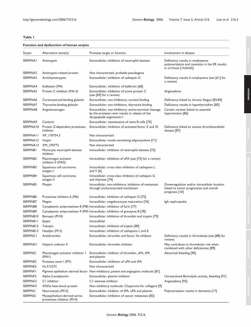
studies of SERPINC1 highlight the molecular basis for
heparin function. Figure 2a shows the structure of native
SERPINC1. Here, we use the convention of Schechter and
Berger, in which residues on the amino-terminal side of the
cleavage site (P1/P1�) are termed P2, P3, and so on, and
those carboxy-terminal are termed P2�, P3�, and so on; cor-
responding subsites in the enzyme are termed S1, S2, and so
on [40]. The RCL is partially inserted into the top of the �
sheet; the residue (P1-Arg) responsible for docking into the
primary specificity pocket (S1) of the protease is relatively
inaccessible to docking with thrombin, as it is pointing
towards and forming interactions with the body of the serpin
[41,42]. Figure 2b illustrates the ternary complex between
SERPINC1, thrombin and heparin [43]. Upon interaction
with a specific heparin pentasaccharide sequence present in
high-affinity heparin, SERPINC1 undergoes a substantial
conformational rearrangement whereby the RCL is expelled
from �-sheet A and the P1 residue flips to an exposed
protease-accessible conformation [44-46]. In addition to
loop expulsion and P1 exposure, long-chain heparin can bind
both enzyme and inhibitor and thus provides an additional
acceleration of the inhibitory interaction. Several other
serpins, including SERPIND1 (heparin cofactor II), also use
cofactor binding and conformational change to achieve
exquisite inhibitory control [47].
Structural studies on prokaryote and viral serpins have
revealed several interesting variations of the serpin scaffold.
Viral proteins are often ‘stripped down’ to a minimal scaffold
in order to minimize the size of the viral genome. Consistent
with this requirement, the structure of the viral serpin crmA,
one of the smallest members of the serpin superfamily
[48,49], shows that it lacks helix hD. More recently, the
structure of the prokaryote serpin thermopin from Ther-
mobifida fusca revealed the absence of helix hH [20,31].
These studies also showed that thermopin contains a
com
ment
reviews
reports
deposited research
interactions
inform
ation
refereed research
http://genomebiology.com/2006/7/5/216 Genome Biology 2006, Volume 7, Issue 5, Article 216 Law et al. 216.5
Genome Biology 2006, 7:216
Figure 2Modulation of serpin conformation by cofactors. (a) The structure of native SERPINC1 (PDB code 2ANT) [95]. The partial insertion of the RCL (tworesidues) into the top of �-sheet A is circled, and the position of the P1 residue is shown (magenta spheres). (b) The structure of the ternary complexbetween SERPINC1, inactive thrombin (the Ser195Ala mutant) and a synthetic long-chain heparin construct (PDB code 1TB6) [43]. A specific high-affinitypentasaccharide (green) on the heparin interacts with the heparin-binding site on SERPINC1 (on and around helix hD) and promotes expulsion of theRCL (blue arrow) and rearrangement of the P1 residue (magenta spheres).
P1
Serpin
Ternary complex
(a) (b)
Protease Heparin
+ +
4 amino-acid insertion at the carboxyl terminus that forms
extensive interactions with conserved residues at the top of
�-sheet A (called the ‘breach’; see later); biophysical data
suggest that this region is important for proper and efficient
folding of this unusual serpin.
The major conformational change that occurs within both
the protease and the serpin as a result of serpin-enzyme
complex formation provides an elegant mechanism for cells
to specifically detect and clear inactivated serpin-protease
complexes. Several studies have shown that the low density
lipoprotein-related protein (LRP) specifically binds to and
promotes internalization of the final complexes SERPINC1-
thrombin, SERPIND1-thrombin and SERPINA1-trypsin. In
contrast, native or cleaved serpin alone are not internalized
[50]. Additionally, recent studies on SERPINI1 show that
both SERPINI1-tissue plasminogen activator complexes and
native SERPINI1 are internalized in an LRP-dependent
manner. However, while SERPINI1-tissue plasminogen acti-
vator complexes can bind directly to LRP, native SERPINI1
requires the presence of an (as yet unidentified) cofactor
[51]. The structural basis for interaction of LRP with serpin-
enzyme complexes and the subsequent intracellular signal-
ing response remain to be fully understood. It is clear,
however, that native serpins and serpin-enzyme complexes
can induce powerful responses such as cell migration in an
LRP-dependent manner [52].
Inactivation of serpins: latency, polymerization,deficiency and diseaseThe metastability of serpins and their ability to undergo con-
trolled conformational change also renders these molecules
susceptible to spontaneous conformational rearrangements.
Most notably, the serpin SERPINE1 (plasminogen activator
inhibitor-1) uses spontaneous conformational change to
control inhibitory activity [53]. Structural and biochemical
studies show that, in the absence of the cofactor vitronectin,
native SERPINE1 (Figure 3a) rapidly converts to a latent
inactive state (Figure 3b). The transition to latency is accom-
panied by insertion of the RCL into �-sheet A, where it
cannot interact with the target protease. Interestingly, the
structure of SERPINE1 in complex with the somatomedin B
domain of vitronectin [54] shows that the cofactor-binding
site on SERPINE1 is located in a similar region to the
heparin-binding site of SERPINC1 (on and around helices hD
and hE; Figure 3c). Whereas heparin promotes conforma-
tional change in SERPINC1, however, vitronectin prevents
conformational change in SERPINE1. Several other serpins,
including SERPINC1, have been shown to spontaneously
undergo the transition to the latent state, and it is suggested
that this may be an important control mechanism [55].
Although the transition to latency could be an important
control mechanism in at least one serpin, an alternative spon-
taneous conformational change, serpin polymerization, results
in deficiency and disease (or serpinopathy) [14,56]. Serpin
polymerization is postulated to occur via a domain-swapping
event whereby the RCL of one molecule docks into �-sheet A
of another to form an inactive long-chain serpin polymer
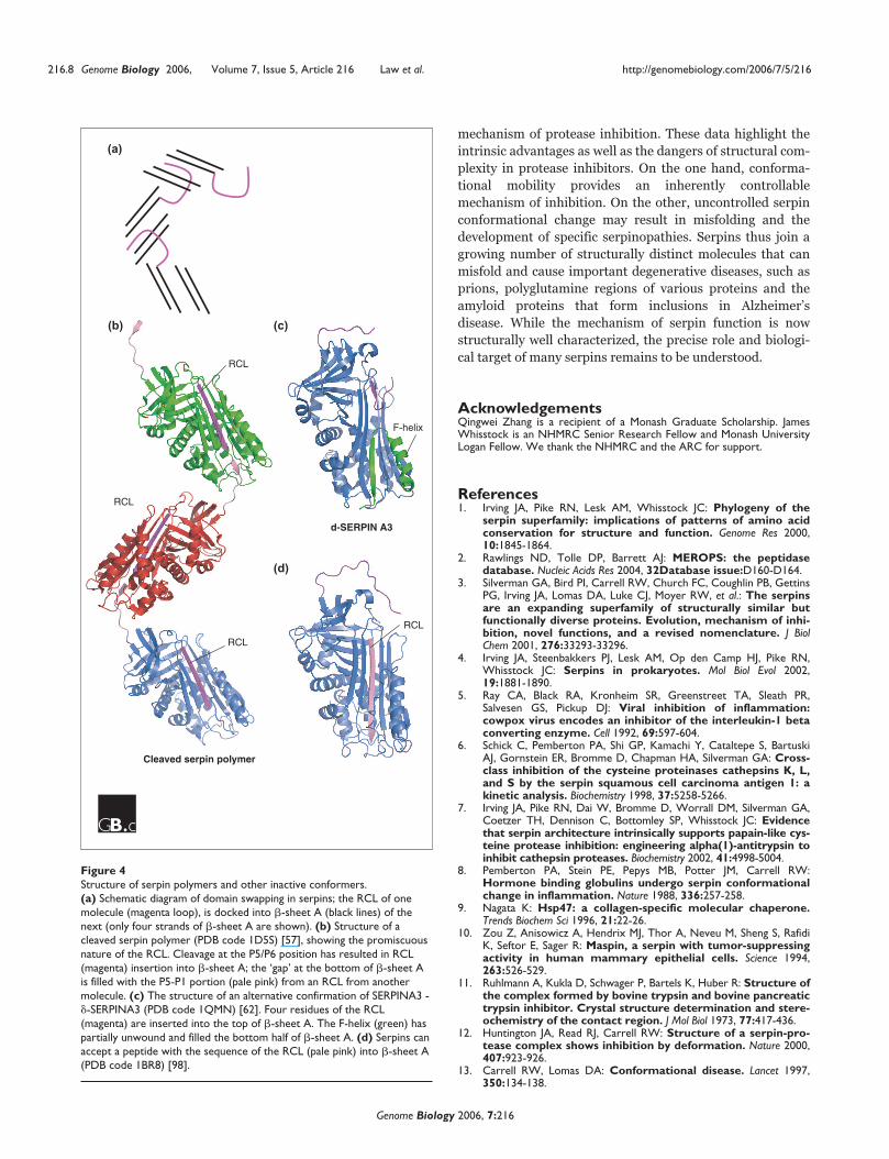
(Figure 4a,b) [14,57-59]. Several important human serpin
variants result in polymerization, the best studied and most
common of which is the Z allele (Glu342Lys) of SERPINA1
[14]. Here, failure to properly control the activity of neutrophil
elastase (the inhibitory target of SERPINA1) in the lung
during the inflammatory response results in the destruction of
lung tissue, leading to emphysema. Furthermore, in individu-
als homozygous for the Z-variant, the accumulation of serpin
aggregates or polymers in the endoplasmic reticulum of anti-
trypsin-producing cells, the hepatocytes, can eventually result
in cell death and liver cirrhosis [14]. Similarly, mutation of
SERPINI1 results in the formation of neural inclusion bodies
and in the disease ‘familial encephalopathy with neuroserpin
inclusion bodies’ (FENIB) [17,60,61].
In addition to promoting polymerization, several serpin
mutations have been identified that promote formation of
a disease-linked latent state. Notably, a mutation in
SERPINC1, the wibble variant (Thr85Met), results in forma-
tion of large amounts of circulating latent SERPINC1 (about
10% of total SERPINC1) [55]. An alternative ‘half-way house’
conformation of SERPINA3, termed �, has also been identi-
fied (Figure 4c) [62]. The structure of �-SERPINA3 also
highlights the extraordinary flexibility of the serpin scaf-
fold: in this conformation the RCL is partially inserted into
�-sheet A and helix hF has partially unwound and inserted
into the base of �-sheet A, completing the �-sheet hydrogen
bonding (Figure 4c). Finally, the promiscuity of �-sheet A is
highlighted by the ability of this region to readily accept
short peptides: several structural and biochemical studies
have demonstrated that peptides can bind to �-sheet A and
induce the S-to-R transition (Figure 4d).
Valuable insights into the mechanism of serpin function
have been gleaned from the structural location of variants
that promote serpin instability [18,63]. The majority of
serpinopathy-linked mutations (including antitrypsin
Siiyama [64] and Mmalton [65], antithrombin wibble [55]
and �-SERPINA3 [62]) cluster in the center of the serpin
molecule, underneath �-sheet A, in a region termed the
shutter (marked on Figure 1a). Interestingly, Glu342, the
position mutated in the Z allele of SERPINA1, is located at
the breach, which is just above the shutter at the top of �-
sheet A. This portion of the molecule is the point of initial
RCL insertion. It is suggested that destabilization of �-sheet
A in either the shutter or the breach is sufficient to favor the
transition to a polymeric or latent state over maintenance of
the monomeric metastable native state [14]. Interestingly,
analysis of conserved residues in the serpin superfamily also
reveals a striking distribution of highly conserved residues
stretching down the center of �-sheet A from the breach to
the base of the molecule [1].
216.6 Genome Biology 2006, Volume 7, Issue 5, Article 216 Law et al. http://genomebiology.com/2006/7/5/216
Genome Biology 2006, 7:216

Unsurprisingly, given the important proteolytic processes
they control, simple deficiencies such as those caused by
null mutations of a large number of human serpins are
linked to disease (some of these are summarized in Table 1).
Interestingly, however, several (rare) mutations have been
identified that do not promote instability but instead inter-
fere with the ability of the serpin to interact correctly with
proteases. These include the Enschede variant of SERPINF2
[66], in which insertion of an additional alanine in the RCL
results in predominantly substrate-like (rather than
inhibitory) behavior upon interaction with a protease. Muta-
tions that alter serpin specificity can also have a devastating
effect. For example, the Pittsburgh variant of SERPINA1
(antitrypsin) is an effective thrombin inhibitor as a result of
mutation of the P1 methionine to an arginine [67]. The
carrier of this variant died of a fatal bleeding disorder in
childhood.
Our knowledge of the functional biochemistry and cell
biology of serpins has been shaped by extensive contribu-
tions from structural biology and genomics. The structure of
six different serpin conformations, together with analysis of
numerous different dysfunctional serpin variants, has
allowed the characterization of a unique conformational
com
ment
reviews
reports
deposited research
interactions
inform
ation
refereed research
http://genomebiology.com/2006/7/5/216 Genome Biology 2006, Volume 7, Issue 5, Article 216 Law et al. 216.7
Genome Biology 2006, 7:216
Figure 3Spontaneous conformational change in serpins. (a) Structure of native SERPINE1 (PDB code 1B3K) [96]. The RCL is in magenta and strand s1c of �-sheet C is in yellow. (b) The structure of latent SERPINE1 (PDB code 1DVN) [53,97], which can form by spontaneous conversion from the nativeprotein. The RCL (magenta) is inserted into �-sheet A. In order to enable full insertion of the RCL, s1C of �-sheet C (pale yellow) has peeled off. Inaddition, conformational change in the strands s3C and s4C (pale green) is indicated. (c) Structure of SERPINE1 (blue) in complex with the somatomedinB domain (green) of vitronectin (PDB code 1OC0) [54]. The interaction with vitronectin locks SERPINE1 in the native, active conformation.
s1C
RCL
s4C
Formerly s1C
Somatomedin B domainof vitronectin
+
Spontaneousconformationalchange
Native SERPINE1
Latent SERPINE1
SERPINE1 -vitronectin complex
(a) (b)
(c)
s3C
mechanism of protease inhibition. These data highlight the
intrinsic advantages as well as the dangers of structural com-
plexity in protease inhibitors. On the one hand, conforma-
tional mobility provides an inherently controllable
mechanism of inhibition. On the other, uncontrolled serpin
conformational change may result in misfolding and the
development of specific serpinopathies. Serpins thus join a
growing number of structurally distinct molecules that can
misfold and cause important degenerative diseases, such as
prions, polyglutamine regions of various proteins and the
amyloid proteins that form inclusions in Alzheimer’s
disease. While the mechanism of serpin function is now
structurally well characterized, the precise role and biologi-
cal target of many serpins remains to be understood.
AcknowledgementsQingwei Zhang is a recipient of a Monash Graduate Scholarship. JamesWhisstock is an NHMRC Senior Research Fellow and Monash UniversityLogan Fellow. We thank the NHMRC and the ARC for support.
References1. Irving JA, Pike RN, Lesk AM, Whisstock JC: Phylogeny of the
serpin superfamily: implications of patterns of amino acidconservation for structure and function. Genome Res 2000,10:1845-1864.
2. Rawlings ND, Tolle DP, Barrett AJ: MEROPS: the peptidasedatabase. Nucleic Acids Res 2004, 32Database issue:D160-D164.
3. Silverman GA, Bird PI, Carrell RW, Church FC, Coughlin PB, GettinsPG, Irving JA, Lomas DA, Luke CJ, Moyer RW, et al.: The serpinsare an expanding superfamily of structurally similar butfunctionally diverse proteins. Evolution, mechanism of inhi-bition, novel functions, and a revised nomenclature. J BiolChem 2001, 276:33293-33296.
4. Irving JA, Steenbakkers PJ, Lesk AM, Op den Camp HJ, Pike RN,Whisstock JC: Serpins in prokaryotes. Mol Biol Evol 2002,19:1881-1890.
5. Ray CA, Black RA, Kronheim SR, Greenstreet TA, Sleath PR,Salvesen GS, Pickup DJ: Viral inhibition of inflammation:cowpox virus encodes an inhibitor of the interleukin-1 betaconverting enzyme. Cell 1992, 69:597-604.
6. Schick C, Pemberton PA, Shi GP, Kamachi Y, Cataltepe S, BartuskiAJ, Gornstein ER, Bromme D, Chapman HA, Silverman GA: Cross-class inhibition of the cysteine proteinases cathepsins K, L,and S by the serpin squamous cell carcinoma antigen 1: akinetic analysis. Biochemistry 1998, 37:5258-5266.
7. Irving JA, Pike RN, Dai W, Bromme D, Worrall DM, Silverman GA,Coetzer TH, Dennison C, Bottomley SP, Whisstock JC: Evidencethat serpin architecture intrinsically supports papain-like cys-teine protease inhibition: engineering alpha(1)-antitrypsin toinhibit cathepsin proteases. Biochemistry 2002, 41:4998-5004.
8. Pemberton PA, Stein PE, Pepys MB, Potter JM, Carrell RW:Hormone binding globulins undergo serpin conformationalchange in inflammation. Nature 1988, 336:257-258.
9. Nagata K: Hsp47: a collagen-specific molecular chaperone.Trends Biochem Sci 1996, 21:22-26.
10. Zou Z, Anisowicz A, Hendrix MJ, Thor A, Neveu M, Sheng S, RafidiK, Seftor E, Sager R: Maspin, a serpin with tumor-suppressingactivity in human mammary epithelial cells. Science 1994,263:526-529.
11. Ruhlmann A, Kukla D, Schwager P, Bartels K, Huber R: Structure ofthe complex formed by bovine trypsin and bovine pancreatictrypsin inhibitor. Crystal structure determination and stere-ochemistry of the contact region. J Mol Biol 1973, 77:417-436.
12. Huntington JA, Read RJ, Carrell RW: Structure of a serpin-pro-tease complex shows inhibition by deformation. Nature 2000,407:923-926.
13. Carrell RW, Lomas DA: Conformational disease. Lancet 1997,350:134-138.
216.8 Genome Biology 2006, Volume 7, Issue 5, Article 216 Law et al. http://genomebiology.com/2006/7/5/216
Genome Biology 2006, 7:216
Figure 4Structure of serpin polymers and other inactive conformers.(a) Schematic diagram of domain swapping in serpins; the RCL of onemolecule (magenta loop), is docked into �-sheet A (black lines) of thenext (only four strands of �-sheet A are shown). (b) Structure of acleaved serpin polymer (PDB code 1D5S) [57], showing the promiscuousnature of the RCL. Cleavage at the P5/P6 position has resulted in RCL(magenta) insertion into �-sheet A; the ‘gap’ at the bottom of �-sheet Ais filled with the P5-P1 portion (pale pink) from an RCL from anothermolecule. (c) The structure of an alternative confirmation of SERPINA3 -�-SERPINA3 (PDB code 1QMN) [62]. Four residues of the RCL(magenta) are inserted into the top of �-sheet A. The F-helix (green) haspartially unwound and filled the bottom half of �-sheet A. (d) Serpins canaccept a peptide with the sequence of the RCL (pale pink) into �-sheet A(PDB code 1BR8) [98].
Cleaved serpin polymer
d-SERPIN A3
RCL
RCL
RCL
RCL
F-helix
(c)(b)
(d)
(a)

14. Lomas DA, Evans DL, Finch JT, Carrell RW: The mechanism of Zalpha 1-antitrypsin accumulation in the liver. Nature 1992,357:605-607.
15. Bruce D, Perry DJ, Borg JY, Carrell RW, Wardell MR: Throm-boembolic disease due to thermolabile conformationalchanges of antithrombin Rouen-VI (187 Asn→→Asp). J ClinInvest 1994, 94:2265-2274.
16. Aulak KS, Pemberton PA, Rosen FS, Carrell RW, Lachmann PJ, Har-rison RA: Dysfunctional C1-inhibitor(At), isolated from atype II hereditary-angio-oedema plasma, contains a P1‘reactive centre’ (Arg444→→His) mutation. Biochem J 1988,253:615-618.
17. Davis RL, Shrimpton AE, Holohan PD, Bradshaw C, Feiglin D, CollinsGH, Sonderegger P, Kinter J, Becker LM, Lacbawan F, et al.: Familialdementia caused by polymerization of mutant neuroserpin.Nature 1999, 401:376-379.
18. Stein PE, Carrell RW: What do dysfunctional serpins tell usabout molecular mobility and disease? Nat Struct Biol 1995,2:96-113.
19. Riahi Y, Siman-Tov R, Ankri S: Molecular cloning, expressionand characterization of a serine proteinase inhibitor genefrom Entamoeba histolytica. Mol Biochem Parasitol 2004, 133:153-162.
20. Irving JA, Cabrita LD, Rossjohn J, Pike RN, Bottomley SP, WhisstockJC: The 1.5 Å crystal structure of a prokaryote serpin: con-trolling conformational change in a heated environment.Structure 2003, 11:387-397.
21. Roberts TH, Hejgaard J, Saunders NF, Cavicchioli R, Curmi PM:Serpins in unicellular Eukarya, Archaea, and Bacteria:sequence analysis and evolution. J Mol Evol 2004, 59:437-447.
22. Grigoryev SA, Woodcock CL: Chromatin structure in granulo-cytes. A link between tight compaction and accumulation ofa heterochromatin-associated protein (MENT). J Biol Chem1998, 273:3082-3089.
23. Irving JA, Shushanov SS, Pike RN, Popova EY, Bromme D, CoetzerTH, Bottomley SP, Boulynko IA, Grigoryev SA, Whisstock JC:Inhibitory activity of a heterochromatin-associated serpin(MENT) against papain-like cysteine proteinases affectschromatin structure and blocks cell proliferation. J Biol Chem2002, 277:13192-13201.
24. Ligoxygakis P, Roth S, Reichhart JM: A serpin regulates dorsal-ventral axis formation in the Drosophila embryo. Curr Biol2003, 13:2097-2102.
25. Levashina EA, Langley E, Green C, Gubb D, Ashburner M, HoffmannJA, Reichhart JM: Constitutive activation of toll-mediated anti-fungal defense in serpin-deficient Drosophila. Science 1999,285:1917-1919.
26. Pak SC, Kumar V, Tsu C, Luke CJ, Askew YS, Askew DJ, Mills DR,Bromme D, Silverman GA: SRP-2 is a cross-class inhibitor thatparticipates in postembryonic development of the nema-tode Caenorhabditis elegans: initial characterization of theclade L serpins. J Biol Chem 2004, 279:15448-15459.
27. Law RH, Irving JA, Buckle AM, Ruzyla K, Buzza M, Bashtannyk-Puhalovich TA, Beddoe TC, Nguyen K, Worrall DM, Bottomley SP,et al.: The high-resolution crystal structure of the humantumor suppressor maspin reveals a novel conformationalswitch in the G-helix. J Biol Chem 2005, 280:22356-22364.
28. Roberts TH, Marttila S, Rasmussen SK, Hejgaard J: Differentialgene expression for suicide-substrate serine proteinaseinhibitors (serpins) in vegetative and grain tissues of barley.J Exp Bot 2003, 54:2251-2263.
29. Hejgaard J: Inhibitory plant serpins with a sequence of threeglutamine residues in the reactive center. Biol Chem 2005,386:1319-1323.
30. Yoo BC, Aoki K, Xiang Y, Campbell LR, Hull RJ, Xoconostle-CazaresB, Monzer J, Lee JY, Ullman DE, Lucas WJ: Characterization ofCucurbita maxima phloem serpin-1 (CmPS-1). A develop-mentally regulated elastase inhibitor. J Biol Chem 2000,275:35122-35128.
31. Fulton KF, Buckle AM, Cabrita LD, Irving JA, Butcher RE, Smith I,Reeve S, Lesk AM, Bottomley SP, Rossjohn J, Whisstock JC: Thehigh-resolution crystal structure of a native thermostableserpin reveals the complex mechanism underpinning thestressed to relaxed transition. J Biol Chem 2005, 280:8435-8442.
32. Elliott PR, Lomas DA, Carrell RW, Abrahams JP: Inhibitory con-formation of the reactive loop of alpha 1-antitrypsin. NatStruct Biol 1996, 3:676-681.
33. Lobermann H, Lottspeich F, Bode W, Huber R: Interaction ofhuman alpha 1-proteinase inhibitor with chymotrypsino-gen A and crystallization of a proteolytically modifiedalpha 1-proteinase inhibitor. Hoppe Seylers Z Physiol Chem 1982,363:1377-1388.
34. Cabrita LD, Bottomley SP: How do proteins avoid becomingtoo stable? Biophysical studies into metastable proteins. EurBiophys J 2004, 33:83-88.
35. Ye S, Cech AL, Belmares R, Bergstrom RC, Tong Y, Corey DR,Kanost MR, Goldsmith EJ: The structure of a Michaelis serpin-protease complex. Nat Struct Biol 2001, 8:979-983.
36. Dementiev A, Simonovic M, Volz K, Gettins PG: Canonicalinhibitor-like interactions explain reactivity of alpha1-pro-teinase inhibitor Pittsburgh and antithrombin with pro-teinases. J Biol Chem 2003, 278:37881-37887.
37. Tew DJ, Bottomley SP: Intrinsic fluorescence changes andrapid kinetics of proteinase deformation during serpin inhi-bition. FEBS Lett 2001, 494:30-33.
38. Kaslik G, Patthy A, Balint M, Graf L: Trypsin complexed withalpha 1-proteinase inhibitor has an increased structural flex-ibility. FEBS Lett 1995, 370:179-183.
39. Rezaie AR: Calcium enhances heparin catalysis of theantithrombin-factor Xa reaction by a template mechanism.Evidence that calcium alleviates Gla domain antagonism ofheparin binding to factor Xa. J Biol Chem 1998, 273:16824-16827.
40. Schechter I, Berger A: On the active site of proteases. 3.Mapping the active site of papain; specific peptide inhibitorsof papain. Biochem Biophys Res Commun 1968, 32:898-902.
41. Carrell RW, Stein PE, Fermi G, Wardell MR: Biological implica-tions of a 3 Å structure of dimeric antithrombin. Structure1994, 2:257-270.
42. Schreuder H, de Boer B, Pronk S, Hol W, Dijkema R, Mulders J, The-unissen H: Crystallization and preliminary X-ray analysis ofhuman antithrombin III. J Mol Biol 1993, 229:249-250.
43. Li W, Johnson DJ, Esmon CT, Huntington JA: Structure of theantithrombin-thrombin-heparin ternary complex revealsthe antithrombotic mechanism of heparin. Nat Struct Mol Biol2004, 11:857-862.
44. Jin L, Abrahams JP, Skinner R, Petitou M, Pike RN, Carrell RW: Theanticoagulant activation of antithrombin by heparin. ProcNatl Acad Sci USA 1997, 94:14683-14688.
45. Pike RN, Potempa J, Skinner R, Fitton HL, McGraw WT, Travis J,Owen M, Jin L, Carrell RW: Heparin-dependent modification ofthe reactive center arginine of antithrombin and conse-quent increase in heparin binding affinity. J Biol Chem 1997,272:19652-19655.
46. Whisstock JC, Pike RN, Jin L, Skinner R, Pei XY, Carrell RW, LeskAM: Conformational changes in serpins: II. The mechanismof activation of antithrombin by heparin. J Mol Biol 2000,301:1287-1305.
47. Baglin TP, Carrell RW, Church FC, Esmon CT, Huntington JA:Crystal structures of native and thrombin-complexedheparin cofactor II reveal a multistep allosteric mechanism.Proc Natl Acad Sci USA 2002, 99:11079-11084.
48. Renatus M, Zhou Q, Stennicke HR, Snipas SJ, Turk D, Bankston LA,Liddington RC, Salvesen GS: Crystal structure of the apoptoticsuppressor CrmA in its cleaved form. Structure 2000, 8:789-797.
49. Simonovic M, Gettins PG, Volz K: Crystallization and prelimi-nary X-ray diffraction analysis of a recombinant cysteine-free mutant of crmA. Acta Crystallogr D Biol Crystallogr 2000,56:1440-1442.
50. Kounnas MZ, Church FC, Argraves WS, Strickland DK: Cellularinternalization and degradation of antithrombin III-throm-bin, heparin cofactor II-thrombin, and alpha 1-antitrypsin-trypsin complexes is mediated by the low densitylipoprotein receptor-related protein. J Biol Chem 1996,271:6523-6529.
51. Makarova A, Mikhailenko I, Bugge TH, List K, Lawrence DA, Strick-land DK: The low density lipoprotein receptor-relatedprotein modulates protease activity in the brain by mediat-ing the cellular internalization of both neuroserpin and neu-roserpin-tissue-type plasminogen activator complexes. J BiolChem 2003, 278:50250-50258.
52. Degryse B, Neels JG, Czekay RP, Aertgeerts K, Kamikubo Y, Loskut-off DJ: The low density lipoprotein receptor-related protein
com
ment
reviews
reports
deposited research
interactions
inform
ation
refereed research
http://genomebiology.com/2006/7/5/216 Genome Biology 2006, Volume 7, Issue 5, Article 216 Law et al. 216.9
Genome Biology 2006, 7:216

is a motogenic receptor for plasminogen activator inhibitor-1.J Biol Chem 2004, 279:22595-22604.
53. Mottonen J, Strand A, Symersky J, Sweet RM, Danley DE, GeogheganKF, Gerard RD, Goldsmith EJ: Structural basis of latency in plas-minogen activator inhibitor-1. Nature 1992, 355:270-273.
54. Zhou A, Huntington JA, Pannu NS, Carrell RW, Read RJ: How vit-ronectin binds PAI-1 to modulate fibrinolysis and cell migra-tion. Nat Struct Biol 2003, 10:541-544.
55. Beauchamp NJ, Pike RN, Daly M, Butler L, Makris M, Dafforn TR,Zhou A, Fitton HL, Preston FE, Peake IR, Carrell RW: Antithrom-bins Wibble and Wobble (T85M/K): archetypal conforma-tional diseases with in vivo latent-transition, thrombosis, andheparin activation. Blood 1998, 92:2696-2706.
56. Lomas DA, Carrell RW: Serpinopathies and the conforma-tional dementias. Nat Rev Genet 2002, 3:759-768.
57. Dunstone MA, Dai W, Whisstock JC, Rossjohn J, Pike RN, Feil SC,Le Bonniec BF, Parker MW, Bottomley SP: Cleaved antitrypsinpolymers at atomic resolution. Protein Sci 2000, 9:417-420.
58. Huntington JA, Pannu NS, Hazes B, Read RJ, Lomas DA, Carrell RW:A 2.6 Å structure of a serpin polymer and implications forconformational disease. J Mol Biol 1999, 293:449-455.
59. Mast AE, Enghild JJ, Salvesen G: Conformation of the reactivesite loop of alpha 1-proteinase inhibitor probed by limitedproteolysis. Biochemistry 1992, 31:2720-2728.
60. Davis RL, Shrimpton AE, Carrell RW, Lomas DA, Gerhard L,Baumann B, Lawrence DA, Yepes M, Kim TS, Ghetti B, et al.: Associ-ation between conformational mutations in neuroserpin andonset and severity of dementia. Lancet 2002, 359:2242-2247.
61. Lomas DA: Molecular mousetraps, alpha1-antitrypsin defi-ciency and the serpinopathies. Clin Med 2005, 5:249-257.
62. Gooptu B, Hazes B, Chang WS, Dafforn TR, Carrell RW, Read RJ,Lomas DA: Inactive conformation of the serpin alpha(1)-antichymotrypsin indicates two-stage insertion of the reac-tive loop: implications for inhibitory function andconformational disease. Proc Natl Acad Sci USA 2000, 97:67-72.
63. Carrell RW, Stein PE: The biostructural pathology of theserpins: critical function of sheet opening mechanism. BiolChem Hoppe Seyler 1996, 377:1-17.
64. Lomas DA, Finch JT, Seyama K, Nukiwa T, Carrell RW: Alpha 1-antitrypsin Siiyama (Ser53→→Phe). Further evidence forintracellular loop-sheet polymerization. J Biol Chem 1993,268:15333-15335.
65. Lomas DA, Elliott PR, Sidhar SK, Foreman RC, Finch JT, Cox DW,Whisstock JC, Carrell RW: alpha 1-Antitrypsin Mmalton(Phe52-deleted) forms loop-sheet polymers in vivo. Evi-dence for the C sheet mechanism of polymerization. J BiolChem 1995, 270:16864-16870.
66. Holmes WE, Lijnen HR, Nelles L, Kluft C, Nieuwenhuis HK, RijkenDC, Collen D: Alpha 2-antiplasmin Enschede: alanine inser-tion and abolition of plasmin inhibitory activity. Science 1987,238:209-211.
67. Owen MC, Brennan SO, Lewis JH, Carrell RW: Mutation of anti-trypsin to antithrombin, alpha 1-antitrypsin Pittsburgh (358Met→→Arg), leads to a fatal bleeding disorder. N Engl J Med1983, 309:694-698.
68. Chao J, Schmaier A, Chen LM, Yang Z, Chao L: Kallistatin, a novelhuman tissue kallikrein inhibitor: levels in body fluids, bloodcells, and tissues in health and disease. J Lab Clin Med 1996,127:612-620.
69. Suzuki K, Deyashiki Y, Nishioka J, Toma K: Protein C inhibitor:structure and function. Thromb Haemost 1989, 61:337-342.
70. Frazer JK, Jackson DG, Gaillard JP, Lutter M, Liu YJ, Banchereau J,Capra JD, Pascual V: Identification of centerin: a novel humangerminal center B cell-restricted serpin. Eur J Immunol 2000,30:3039-3048.
71. Hida K, Wada J, Eguchi J, Zhang H, Baba M, Seida A, Hashimoto I,Okada T, Yasuhara A, Nakatsuka A, et al.: Visceral adipose tissue-derived serine protease inhibitor: a unique insulin-sensitiz-ing adipocytokine in obesity. Proc Natl Acad Sci USA 2005,102:10610-10615.
72. Kordula T, Dubin A, Schooltink H, Koj A, Heinrich PC, Rose-John S:Molecular cloning and expression of an intracellular serpin:an elastase inhibitor from horse leucocytes. Biochem J 1993,293:187-193.
73. Medcalf RL, Stasinopoulos SJ: The undecided serpin. The ins andouts of plasminogen activator inhibitor type 2. FEBS J 2005,272:4858-4867.
74. Schick C, Kamachi Y, Bartuski AJ, Cataltepe S, Schechter NM,Pemberton PA, Silverman GA: Squamous cell carcinomaantigen 2 is a novel serpin that inhibits the chymotrypsin-like proteinases cathepsin G and mast cell chymase. J BiolChem 1997, 272:1849-1855.
75. Scott FL, Hirst CE, Sun J, Bird CH, Bottomley SP, Bird PI: Theintracellular serpin proteinase inhibitor 6 is expressed inmonocytes and granulocytes and is a potent inhibitor of theazurophilic granule protease, cathepsin G. Blood 1999,93:2089-2097.
76. Miyata T, Nangaku M, Suzuki D, Inagi R, Uragami K, Sakai H, OkuboK, Kurokawa K: A mesangium-predominant gene, megsin, is anew serpin upregulated in IgA nephropathy. J Clin Invest 1998,102:828-836.
77. Dahlen JR, Jean F, Thomas G, Foster DC, Kisiel W: Inhibition ofsoluble recombinant furin by human proteinase inhibitor 8. JBiol Chem 1998, 273:1851-1854.
78. Sun J, Bird CH, Sutton V, McDonald L, Coughlin PB, De Jong TA,Trapani JA, Bird PI: A cytosolic granzyme B inhibitor relatedto the viral apoptotic regulator cytokine response modifierA is present in cytotoxic lymphocytes. J Biol Chem 1996,271:27802-27809.
79. Riewald M, Schleef RR: Molecular cloning of bomapin (proteaseinhibitor 10), a novel human serpin that is expressed specifi-cally in the bone marrow. J Biol Chem 1995, 270:26754-26757.
80. Askew YS, Pak SC, Luke CJ, Askew DJ, Cataltepe S, Mills DR, KatoH, Lehoczky J, Dewar K, Birren B, Silverman GA: SERPINB12 is anovel member of the human ov-serpin family that is widelyexpressed and inhibits trypsin-like serine proteinases. J BiolChem 2001, 276: 49320-49330.
81. Dawson DW, Volpert OV, Gillis P, Crawford SE, Xu H, Benedict W,Bouck NP: Pigment epithelium-derived factor: a potentinhibitor of angiogenesis. Science 1999, 285:245-248.
82. Xiao G, Liu YE, Gentz R, Sang QA, Ni J, Goldberg ID, Shi YE: Sup-pression of breast cancer growth and metastasis by a serpinmyoepithelium-derived serine proteinase inhibitorexpressed in the mammary myoepithelial cells. Proc Natl AcadSci USA 1999, 96:3700-3705.
83. Torpy DJ, Bachmann AW, Gartside M, Grice JE, Harris JM, Clifton P,Easteal S, Jackson RV, Whitworth JA: Association between chronicfatigue syndrome and the corticosteroid-binding globulingene ALA SER224 polymorphism. Endocr Res 2004, 30:417-429.
84. Torpy DJ, Bachmann AW, Grice JE, Fitzgerald SP, Phillips PJ, Whit-worth JA, Jackson RV: Familial corticosteroid-binding globulindeficiency due to a novel null mutation: association withfatigue and relative hypotension. J Clin Endocrinol Metab 2001,86:3692-3700.
85. Refetoff S, Murata Y, Mori Y, Janssen OE, Takeda K, Hayashi Y: Thy-roxine-binding globulin: organization of the gene and vari-ants. Horm Res 1996, 45:128-138.
86. Kim HS, Krege JH, Kluckman KD, Hagaman JR, Hodgin JB, Best CF,Jennette JC, Coffman TM, Maeda N, Smithies O: Genetic controlof blood pressure and the angiotensinogen locus. Proc NatlAcad Sci USA 1995, 92:2735-2739.
87. Water N, Tan T, Ashton F, O’Grady A, Day T, Browett P, OckelfordP, Harper P: Mutations within the protein Z-dependent pro-tease inhibitor gene are associated with venous throm-boembolic disease: a new form of thrombophilia. Br JHaematol 2004, 127:190-194.
88. Perry DJ, Carrell RW: Molecular genetics of human antithrom-bin deficiency. Hum Mutat 1996, 7:7-22.
89. He L, Vicente CP, Westrick RJ, Eitzman DT, Tollefsen DM: Heparincofactor II inhibits arterial thrombosis after endothelialinjury. J Clin Invest 2002, 109:213-219.
90. Fay WP, Parker AC, Condrey LR, Shapiro AD: Human plasmino-gen activator inhibitor-1 (PAI-1) deficiency: characteriza-tion of a large kindred with a null mutation in the PAI-1gene. Blood 1997, 90:204-208.
91. Miles LA, Plow EF, Donnelly KJ, Hougie C, Griffin JH: A bleedingdisorder due to deficiency of alpha 2-antiplasmin. Blood 1982,59:1246-1251.
92. De Marchi M, Jacot-Guillarmod H, Ressa TG, Carbonara AO:Hereditary angioedema: report of a large kindred with arare genetic variant of C1-esterase inhibitor. Clin Genet 1973,4:229-236.
93. Loebermann H, Tokuoka R, Deisenhofer J, Huber R: Human alpha1-proteinase inhibitor. Crystal structure analysis of two
216.10 Genome Biology 2006, Volume 7, Issue 5, Article 216 Law et al. http://genomebiology.com/2006/7/5/216
Genome Biology 2006, 7:216
crystal modifications, molecular model and preliminaryanalysis of the implications for function. J Mol Biol 1984,177:531-557.
94. Hopkins PC, Carrell RW, Stone SR: Effects of mutations in thehinge region of serpins. Biochemistry 1993, 32:7650-7657.
95. Skinner R, Abrahams JP, Whisstock JC, Lesk AM, Carrell RW,Wardell MR: The 2.6 Å structure of antithrombin indicates aconformational change at the heparin binding site. J Mol Biol1997, 266:601-609.
96. Sharp AM, Stein PE, Pannu NS, Carrell RW, Berkenpas MB, GinsburgD, Lawrence DA, Read RJ: The active conformation of plas-minogen activator inhibitor 1, a target for drugs to controlfibrinolysis and cell adhesion. Structure 1999, 7:111-118.
97. Stout TJ, Graham H, Buckley DI, Matthews DJ: Structures ofactive and latent PAI-1: a possible stabilizing role for chlo-ride ions. Biochemistry 2000, 39:8460-8469.
98. Skinner R, Chang WS, Jin L, Pei X, Huntington JA, Abrahams JP,Carrell RW, Lomas DA: Implications for function and therapyof a 2.9 Å structure of binary-complexed antithrombin. J MolBiol 1998, 283:9-14.
com
ment
reviews
reports
deposited research
interactions
inform
ation
refereed research
http://genomebiology.com/2006/7/5/216 Genome Biology 2006, Volume 7, Issue 5, Article 216 Law et al. 216.11
Genome Biology 2006, 7:216
Related Documents