An Orally Administered Redox Nan Accumulates in the Colonic Muco Colitis in Mice 著者 Vong Long Binh, Tomita Tsutomu, Yos Toru, Matsui Hirofumi, Nagasaki Yu journal or publication title Gastroenterology volume 143 number 4 page range 1027-1036.e3 year 2012-10 権利 (C) 2012 by the AGA Institute. Publish Elsevier Inc. NOTICE: this is the author’s versi work that was accepted for publ Gastroenterology. Changes resul publishing process, such as pee editing, corrections, structur and other quality control mech be reflected in this document. have been made to this work sin submitted for publication. A de version was subsequently publi PUBLICATION, VOL143(4) 2012 DOI:10.1053/j.gastro.2012.06.043 URL http://hdl.handle.net/2241/117849 doi: 10.1053/j.gastro.2012.06.043

Welcome message from author
This document is posted to help you gain knowledge. Please leave a comment to let me know what you think about it! Share it to your friends and learn new things together.
Transcript
An Orally Administered Redox Nanoparticle ThatAccumulates in the Colonic Mucosa and ReducesColitis in Mice
著者 Vong Long Binh, Tomita Tsutomu, YoshitomiToru, Matsui Hirofumi, Nagasaki Yukio
journal orpublication title
Gastroenterology
volume 143number 4page range 1027-1036.e3year 2012-10権利 (C) 2012 by the AGA Institute. Published by
Elsevier Inc.NOTICE: this is the author’s version of awork that was accepted for publication inGastroenterology. Changes resulting from thepublishing process, such as peer review,editing, corrections, structural formatting,and other quality control mechanisms may notbe reflected in this document. Changes mayhave been made to this work since it wassubmitted for publication. A definitiveversion was subsequently published inPUBLICATION, VOL143(4) 2012DOI:10.1053/j.gastro.2012.06.043
URL http://hdl.handle.net/2241/117849doi: 10.1053/j.gastro.2012.06.043
1
An Orally Administered Redox Nanoparticle that Accumlates
in the Colonic Mucosa and Reduces Colitis in Mice
Long Binh Vong*, Tsutomu Tomita*, ‡, Toru Yoshitomi*, Hirofumi Matsui§,ǁ and Yukio
Nagasaki*,§,¶
*Department of Materials Science, Graduate School of Pure and Applied Sciences,
University of Tsukuba, Tsukuba, Japan; ‡Timelapse Vision Inc., Asaka, Saitama, Japan;
§Master’s School of Medical Sciences, Graduate School of Comprehensive Human
Sciences, University of Tsukuba, Tsukuba, Japan; ǁ Division of Gastroenterology,
Graduate School of Comprehensive Human Sciences, University of Tsukuba, Tsukuba,
Japan; and ¶Satellite Laboratory, International Center for Materials Nanoarchitectonics
(WPI-MANA), National Institute for Materials Science (NIMS), University of Tsukuba,
Tsukuba, Japan
Grant Support A part of this work was supported by Grant-in-Aid for Scientific Research A
(21240050) and Grant-in-Aid for Research Activity Start-up (22800004) and the World
Premier International Research Center Initiative (WPI Initiative) on Materials
Nanoarchitronics of the Ministry of Education, Culture, Sports, Science and
Technology (MEXT) of Japan.
2
Correspondence
Prof. Yukio Nagasaki, Department of Materials Science, Graduate School of Pure and
Applied Sciences, Master’s School of Medical Sciences, Graduate School of
Comprehensive Human Sciences, Satellite Laboratory, International Center for
Materials Nanoarchitectonics (WPI-MANA), National Institute for Materials Science
(NIMS), University of Tsukuba, Tennoudai 1-1-1, Tsukuba, Ibaraki, 305-8573, Japan
E-mail address: [email protected]
Phone: +81-29-853-5749
Fax: +81-29-853-5749
Conflicts of interest
The authors declare that they have no competing financial interests.
Author contributions
Y.N. designed the experiments and wrote the manuscript. L.B.V. and T.Y.
designed and carried out the experiments, analyzed the results and wrote the manuscript.
T.T. carried out the experiments of in vivo live imaging. H.M. designed colitis model in
mice.
3
BACKGROUND AND AIMS: Drugs used to treat patients with ulcerative colitis (UC)
are not always effective because of non-specific distribution, metabolism in
gastrointestinal tract, and side effects. We designed a nitroxide radical-containing
nanoparticle (RNPO) that accumulates specifically in the colon to suppress
inflammation and reduce the undesirable side effects of nitroxide radicals.
METHODS: RNPO was synthesized by assembly of an amphiphilic block copolymer
that contains stable nitroxide radicals in an ether-linked hydrophobic side chain.
Biodistribution of RNPO in mice was determined from radioisotope and electron spin
resonance measurements. The effects of RNPO were determined in mice with dextran
sodium sulfate (DSS)-induced colitis and compared with those of low-molecular-weight
drugs (4-hydroxyl-2,2,6,6-tetramethylpiperidine-1-oxyl [TEMPOL] or mesalamine).
RESULTS: RNPO, with a diameter of 40 nm and a shell of poly(ethylene glycol), had a
significantly greater level of accumulation in the colonic mucosa than
low-molecular-weight TEMPOL or polystyrene latex particles. RNPO was not absorbed
into the bloodstream through the intestinal wall, despite its long-term retention in the
colon, which prevented its distribution to other parts of the body. Mice with
DSS-induced colitis had significantly lower disease activity index and less
inflammation following 7 days of oral administration of RNPO, compared with
4
DSS-induced colitis mice or mice given low-molecular-weight TEMPOL or
mesalamine.
CONCLUSION: We designed an orally administered RNPO that accumulates
specifically in the colons of mice with colitis and is more effective in reducing
inflammation than low-molecular-weight TEMPOL or mesalamine. RNPO might be
developed for treatment of patients with UC.
Key words: Nitroxide Radical-containing Nanoparticles; Inflammatory Bowel Disease;
Reactive Oxygen Species; Nanotherapy.
Abbreviation used in this paper: AUC, area under the concentration-time curve; CD,
Crohn’s disease; CMS, chloromethylstyrene; DAI, disease activity index; DHE
dihydroethidium; DSS, dextran sodium sulfate; ESR, electron spin resonance; IBD,
inflammatory bowel disease; IL, interleukin; H&E, hematoxylin and eosin; GIT,
gastrointestinal tract; MeO-PEG-b-PCMS, methoxy-poly(ethylene
glycol)-b-poly(chloromethylstyrene); MeO-PEG-b-PMOT, methoxy-poly(ethylene
glycol)-b-poly[p-4-(2,2,6,6-tetramethylpiperidine-1-oxyl)oxymethylstyrene];
MeO-PEG-SH, methoxy-poly(ethylene glycol)-sulfanyl; MPO, myeloperoxidase; PEG,
poly(ethylene glycol); PEG-b-PMNT, poly(ethylene
5
glycol)-b-poly[p-4-(2,2,6,6-tetramethylpiperidine-1-oxyl)aminomethylstyrene]; RNPN,
nitroxide radical-containing nanoparticles prepared by PEG-b-PMNT; RNPO, nitroxide
radical-containing nanoparticles prepared by MeO-PEG-b-PMOT; TEMPOL,
4-hydroxyl-2,2,6,6-tetramethylpiperidine-1-oxyl; UC, ulcerative colitis.
6
Inflammatory bowel disease (IBD), including Crohn’s disease (CD) and ulcerative
colitis (UC), affects millions of patients worldwide. 1 – 4 Since the etiology and
pathogenesis of IBD are not well understood, it is considered an intractable disease. The
intestinal mucosa of patients with IBD is characterized by reactive oxygen species
(ROS) overproduction and an imbalance of important antioxidants, leading to oxidative
damage. Self-sustaining cycles of oxidant production may amplify inflammation and
mucosal injury.5– 8 In several experimental models, antioxidant compounds and free
radical scavengers have improved colitis. 9– 11
Nanoparticles such as liposome and polymeric micelles have gained
worldwide attention as a new medical technology, because they change biodistribution
of drugs to result in therapeutic effect of drugs significantly.
However, these compounds are not
completely effective due to a non-specific drug distribution, a low retention in the colon
and side effects. If antioxidant compounds are specifically targeted to the diseased sites
and effectively scavenge excessive generated ROS, they represent a safe and effective
treatment for IBD.
12,13 In particular, the
intratumoral microdistribution of nanoparticle has been studied for over two decades
that nanoparticles can accumulate in sites of tumor due to the increased vascular
permeability. 14 – 17 Recently, we have developed an amphiphilic block copolymer,
7
poly(ethylene
glycol)-b-poly[p-4-(2,2,6,6-tetramethylpiperidine-1-oxyl)aminomethylstyrene]
(PEG-b-PMNT), possessing stable nitroxide radicals in the hydrophobic segment as a
side chain via an amine linkage, which forms core-shell-type micelles in the
physiological environment with an average diameter of about 40 nm, and termed
nitroxide radical-containing nanoparticle (RNPN).18 Nitroxide radicals are confined in
the core of this micelle, which shows high biocompatibility, including long-term blood
circulation when administered intravenously and low toxicity. Therefore, RNPN has
been studied for therapy in oxidative stress injuries18 – 22 and bioimaging.23,24
In this study, we describe a novel nanotherapy for the treatment of UC via oral
administration. In order to target the nanoparticle to the colon area, its accumulation in
the colonic mucosa is optimized, preventing its uptake into the bloodstream. We
designed a new redox polymer, methoxy-poly(ethylene
glycol)-b-poly[p-4-(2,2,6,6-tetramethylpiperidine-1-oxyl)oxymethylstyrene]
For
example, pH-sensitive RNPN works effectively in acute renal injury18 and cerebral
ischemia-reperfusion19 because it disintegrates in acidic conditions of diseased area by
protonation of amino groups. However, pH-disintegrative character is not suitable for
the treatment of UC via oral administration.
8
(MeO-PEG-b-PMOT), which is an amphiphilic block copolymer with stable nitroxide
radicals in a hydrophobic segment as a side chain via an ether linkage and forms
40-nm-diameter core-shell-type micelles (RNPO) by self-assembly in the aqueous
environments regardless of pH (Figure 1A). Here, we investigate specific accumulation
of RNPO in colon after oral administration by comparison to low-molecular-weight
compound, 4-hydroxyl-2,2,6,6-tetramethylpiperidine-1-oxyl (TEMPOL) and
commercial available polystyrene latex particles with different sizes from 40 nm to 1
µm. Also, we examine the therapeutic effect of RNPO on dextran sodium sulfate
(DSS)-induced colitis model in mice, compared to low-molecular-weight TEMPOL and
mesalamine, a commercial anti-ulcer drug. Our results show that RNPO significantly
accumulates in colonic mucosa area, especially inflammatory sites, without absorption
into bloodstream and has an extremely high therapeutic efficiency in mice with
DSS-induced colitis (Figure 1B).
Materials and Methods
Preparation of RNPO
RNPO was prepared by a self-assembling MeO-PEG-b-PMOT block
copolymer, as previously reported.18 Briefly, methoxy-poly(ethylene
9
glycol)-b-poly(chloromethylstyrene) (MeO-PEG-b-PCMS) was synthesized by the
radical telomerization of chloromethylstyrene (CMS) using methoxy-poly(ethylene
glycol)-sulphanyl (MeO-PEG-SH; Mn = 5,000) as a telogen. The chloromethyl groups
were converted to TEMPOs via a Williamson ether synthesis of benzyl chloride in the
MeO-PEG-b-PCMS block copolymer with the alkoxide of TEMPOL, as previously
reported. RNPO was prepared from MeO-PEG-b-PMOT by dialysis method; Micelle
without nitroxide radicals was similarly prepared from MeO-PEG-b-PCMS as a control
and termed “micelle”.
Preparation of rhodamine-labeled RNPO, 125I-labeled RNPO, polystyrene latex
particles with nitroxide radicals
Detailed methods were described in Supplementary Materials and Methods
section.
Animals
All experiments were carried out using 7-week-old male ICR mice (32–35 g)
purchased from Charles River Japan, Inc. Mice were maintained in the experimental
animal facilities at the University of Tsukuba. All experiments were performed
according to the Guide for the Care and Use of Laboratory Animals at the University of
10
Tsukuba.
Localization of RNPO in the colon
Localization of RNPO in the colon was determined by fluorescent
rhodamine-labeled RNPO. Rhodamine-labeled RNPO was prepared via thiourethane
bond between MeO-PEG-b-PMOT possessing reduced TEMPO moieties and
rhodamine B isothiocyanatein (see Supplementary Materials and Methods). Mice were
killed 4 hours after oral administration of 1 mL of rhodamine-labeled RNPO (5 mg/mL).
Residues in the colon were gently removed with phosphate buffered saline (50 mM, pH
7.4), and 7-μm thick colon sections were prepared. Localization of rhodamine-labeled
RNPO was recorded using a fluorescent microscope.
Accumulation of RNPO in the colon
Accumulation of RNPO was determined by ESR assay. One mL of
low-molecular-weight TEMPOL, RNPO and different sized polystyrene latex particles
with an equivalent nitroxide concentration (1.33 mg; 7.5 μM) were orally administered
to mice. Mice were killed 1, 4, 12, 24, and 48 hours after oral administration. Whole
colons were homogenized in 1 mL of phosphate buffered saline (50 mM, pH 7.4)
11
containing potassium ferricyanide (50 mM). The ESR signal intensities in homogenized
samples were measured by an X-band ESR spectrometer (JES-TE25X, JEOL, Tokyo,
Japan) at room temperature. The amount of nitroxide radicals in the colon was
determined by ESR measurements under the following conditions: frequency, 9.41
GHz; power, 10.00 mW; center field, 333.3; sweep width, 5 mT; sweep time, 0.5 min;
modulation, 0.1 mT; time constant, 0.1 s.
Biodistribution of RNPO
125I-labebled RNPO was prepared via reaction between RNPO and Na[125I] with
present of chloramine-T as a catalyst (see Supplementary Materials and Methods). Mice
were fasted for 1 day before the experiment and 0.5 mL of 125I-labeled RNPO (20
mg/mL) was orally administered. Then, mice were sacrificed at 0.25, 0.5, 1, 2, 4, 8, 12,
and 24 hours after oral administration. The major digestive organs (small intestine,
cecum, and colon) and blood were isolated, and their radioactivities were measured by
a γ-counter (ARC-380, Aloka, Japan). The percentage of radioactivity in each organ
was determined based on the initial total radioactivity.
Induction of colitis by DSS and drug administration
12
Colitis in mice was induced by 3% (wt/vol) DSS (5,000 daltons; Wako Pure
Chemicals) supplemented in the drinking water for 7 days. The experiment was
designed to six groups: normal control group, DSS-injured group,
low-molecular-weight TEMPOL-treated group, micelle-treated group, RNPO-treated
group and mesalamine-treated group. The equivalent doses of drugs (0.2 mM/kg) were
orally administered daily during the 7 days of DSS treatment. The concentrations of
low-molecular-weight TEMPOL, micelle and RNPO were adjusted in distilled water,
and the solutions were filtered with a 0.25-μm cellulose acetate filter. Mesalamine was
suspended in 0.5% (wt/vol) carboxymethyl cellulose.
Evaluation of colitis severity by disease activity index (DAI) and colon length;
Histological assessment; Measurements of myeloperoxidase (MPO) activity,
interleukin (IL)-1 β and superoxide production
Detailed methods were described in Supplementary Materials and Methods
section.
Intravital observation by in vivo live imaging
Aqueous solution of DSS (3% wt/vol) was administered by free access for 7
days to induce colitis in mice. One mL of RNPO (10 mg/mL) was orally administered
13
daily. After 7 days of treatment, mice were anesthetized with urethane (15 g/kg,
Sigma-Aldrich) and an arc-shaped incision was made in the peritoneum to expose the
colon. Then, approximately 1 cm length incision was made to observe the colonic
mucosa and the remained contents in the colon were removed gently by physiological
saline. Mice were set on the stage of microscope and in vivo live imaging was acquired
after 2 hours with a microscope. Dead cells in colonic mucosa were identified by
staining of propidium iodide in physiological buffer (50 μg/mL; Wako Pure Chemicals)
under an excitation wavelength of 488 nm and an emission wavelength of 515 nm.
Survival rate experiment
The survival rate of mice was determined by replacing drinking water with a
3% (wt/vol) solution of DSS for 15 days. Starting on day 5, drugs were oral
administered daily until day 15, and the number of surviving mice was counted until
day 15.
Statistical analysis
All values are expressed as mean ± standard error of mean (SEM). Differences
between groups were examined for statistical significance using the one-way and
14
two-way ANOVA, followed by Bonferroni post-hoc test (SPSS software, IBM Corp.,
NY, USA). A P-value < .05 was considered significant for all statistical analyses.
Results
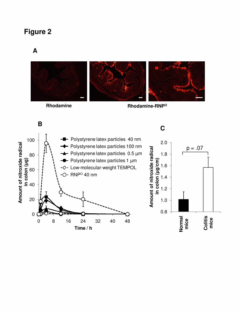
Specific accumulation of RNPO in colonic mucosa and inflamed colon area
The accumulation of nanoparticles in the colon area is one of the most important
features for an effective nanomedicine against UC. Firstly, we orally administered
fluorescently labeled nanoparticles, and analyzed the accumulation of these
nanoparticles in the colon by fluorescent microscopy. Here, we prepared
rhodamine-labeled RNPO (see Supplementary Materials and Methods). After oral
administration of rhodamine-labeled RNPO, there was a strong fluorescent signal at the
colonic mucosa area, as compared to oral administration of low-molecular-weight
fluorescein (Figure 2A). This result indicates effective accumulation of RNPO in the
colonic mucosa.
In order to quantify the accumulation of nanoparticles in the colon area, we
compared RNPO with different sizes of commercial available polystyrene latex particles
and low-molecular-weight compound, TEMPOL. Because we introduced nitroxide
radicals into the particles, their accumulation could be quantitatively monitored by
15
electron spin resonance (ESR) measurements. When we orally administered
low-molecular-weight TEMPOL to mice, almost no ESR signal was observed in the
colon, as shown in Figure 2B. In contrast, polystyrene latex particles showed a higher
accumulation in the colon compared to low-molecular-weight TEMPOL. From these
results, the size-dependent accumulation in colon was observed. Polystyrene latex
particles with 40 nm and 100 nm in size accumulated higher than large-sized particles
(0.5 µm and 1 µm), which is consistent with previous reports.25,26
Next, we investigated the specific accumulation of RNPO in the injured colon.
Interestingly, when
RNPO was administrated orally to mice, considerable high accumulation of RNPO in
colon was observed, as compared to polystyrene latex particles, even though the same
size (40 nm). The area under the concentration-time curve (AUC), an important
parameter in biopharmaceuticals and pharmacokinetics, of RNPO was 1223.3, which
was significantly higher than 27.8 of low-molecular-weight TEMPOL. The AUC of
polystyrene latex particles with sizes 40 nm, 100 nm, 0.5 µm and 1 µm were 249.5,
204.7, 83.7 and 32.9, respectively. High colloidal stability of RNPO due to the PEG
tethered chains on the surface might be effective to accumulate in colonic mucosa as
compared to polystyrene latex particles. The extremely high accumulation of RNPO in
colonic mucosa can be anticipated for high performance efficiency as a colitis therapy.
16
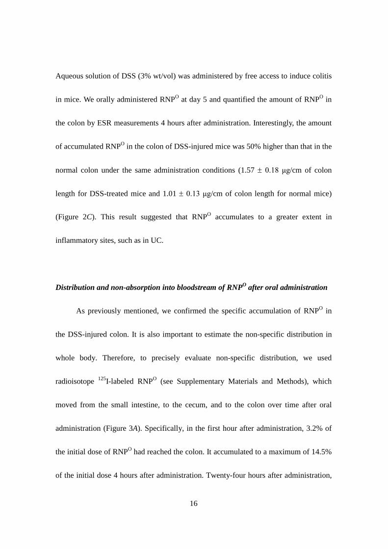
Aqueous solution of DSS (3% wt/vol) was administered by free access to induce colitis
in mice. We orally administered RNPO at day 5 and quantified the amount of RNPO in
the colon by ESR measurements 4 hours after administration. Interestingly, the amount
of accumulated RNPO in the colon of DSS-injured mice was 50% higher than that in the
normal colon under the same administration conditions (1.57 ± 0.18 μg/cm of colon
length for DSS-treated mice and 1.01 ± 0.13 μg/cm of colon length for normal mice)
(Figure 2C). This result suggested that RNPO accumulates to a greater extent in
inflammatory sites, such as in UC.
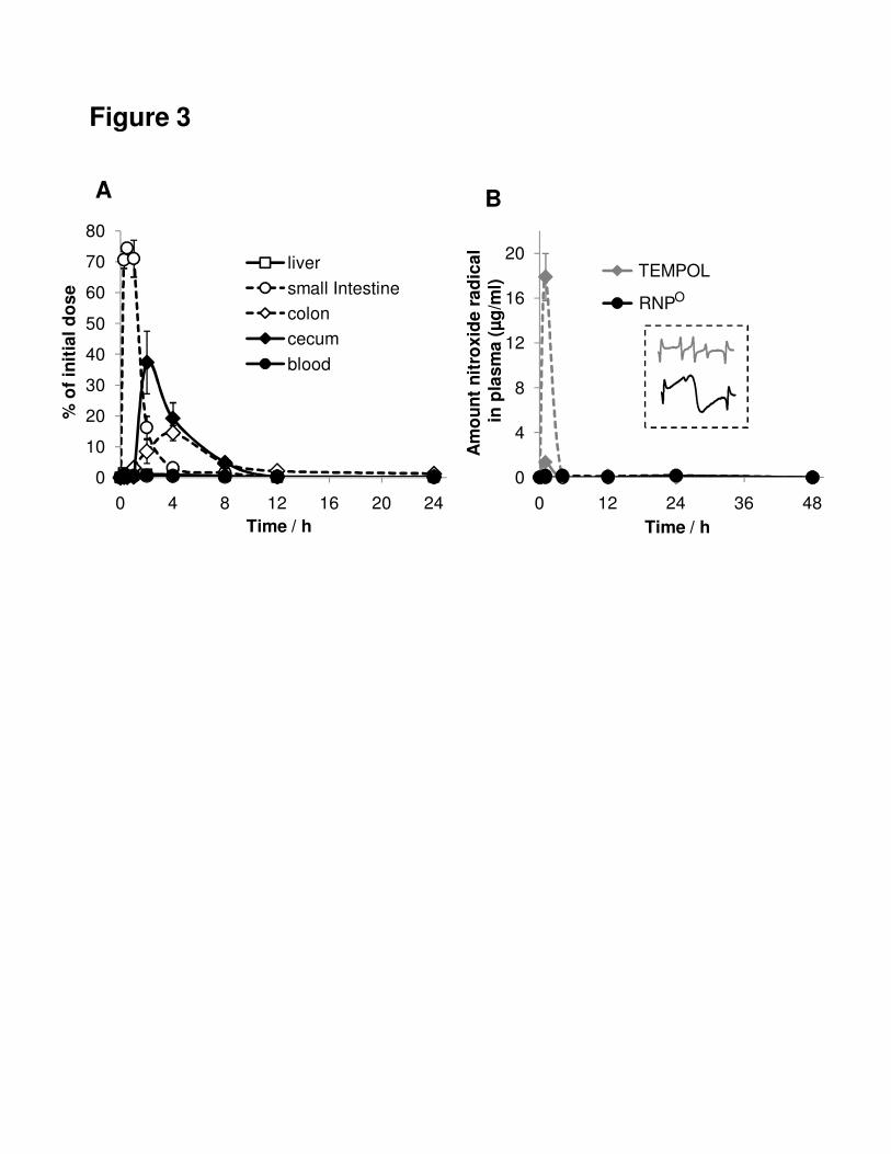
Distribution and non-absorption into bloodstream of RNPO after oral administration
As previously mentioned, we confirmed the specific accumulation of RNPO in
the DSS-injured colon. It is also important to estimate the non-specific distribution in
whole body. Therefore, to precisely evaluate non-specific distribution, we used
radioisotope 125I-labeled RNPO (see Supplementary Materials and Methods), which
moved from the small intestine, to the cecum, and to the colon over time after oral
administration (Figure 3A). Specifically, in the first hour after administration, 3.2% of
the initial dose of RNPO had reached the colon. It accumulated to a maximum of 14.5%
of the initial dose 4 hours after administration. Twenty-four hours after administration,
17
there was 0.5% of the initial dose of RNPO remaining in the colon. Importantly, we did
not observe the uptake of RNPO into the bloodstream (Figure 3A). This is in sharp
contrast to low-molecular-weight compounds, such as TEMPOL. This difference in
bloodstream uptake via the gastrointestinal tract (GIT) was further confirmed by ESR
measurements. Low-molecular-weight TEMPOL was absorbed into the bloodstream
through the GIT in normal mice and even more in DSS-treated mice (Figure 3B).
However, when RNPO was administered orally, there was no observable ESR signal in
the blood, which was consistent with the results from the experiments of 125I-labeled
RNPO. In the present study, oral nanotherapy with RNPO prevented uptake into the
bloodstream, suggesting a lack of systemic side effects.
Stability of RNPO in GIT
Next, we evaluated the stability of orally administered RNPO in the GIT using
ESR spectra of RNPO in the colon. The ESR signals of low-molecular-weight TEMPOL
in the colon showed a sharp triplet due to an interaction between the 14N nuclei and the
unpaired electron, as previously reported18 (Figure 3B, inset, grey spectrum). In contrast,
the ESR signals of RNPO in the colon were broad (Figure 3B, inset, black spectrum),
suggesting that RNPO remains as core-shell type micelle even in the GIT. The stability
18
of self-assembled RNPO with several tens of nanometers in GIT could prevent the
uptake into the bloodstream through the intestinal wall. After reaching colon, RNPO is
accumulated in inflamed and mucosal area, followed by effectively scavenging ROS. It
is noted that RNPN, which contains amino group as side chains in the hydrophobic
segment, is absorbed into the bloodstream when administered orally (data not shown).
It is likely that the disintegration of RNPN in the stomach facilitates its uptake into the
bloodstream through the intestinal wall, which was not observed in RNPO.
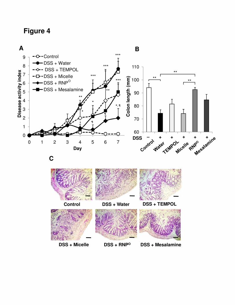
Therapeutic effect of RNPO on DSS-induced colitis in mice
Since orally administered RNPO accumulated in the colonic mucosa of
DSS-injured mice and was not absorbed into the bloodstream, it is anticipated to be an
ideal nanomedicine for UC treatment. Therefore, we investigated its therapeutic and
suppressive effects on DSS-induced colitis model in mice. RNPO was orally
administered daily to DSS-injured mice for 7 days. Additional DSS-injured mice were
treated with low-molecular-weight TEMPOL, commercially anti-ulcer mesalamine and
micelle without nitroxide radicals as controls. After 7 days of treatment, we assessed
the severity of colitis on the basis of DAI27 (see Supplementary Table 1), colon length,
and histological analysis. Mice treated with DSS had a significant increase in DAI and
19
shortening of the colon compared to control mice (Figure 4A,B). The treatments with
low-molecular-weight TEMPOL or mesalamine showed efficiency to decrease DAI as
compared to DSS-treated mice, though this efficiency was not significant. On the
contrary, RNPO-treated mice showed much lower DAI and preserved colon length
compared to DSS-treated mice (P < .01) and other low-molecular-weight drugs-treated
mice. It should be noted that no effect was observed when polymeric micelle without
nitroxide radicals was administered instead of RNPO. Additionally, histological analyses
showed that mucosal structures of DSS- and micelle-treated mice were significantly
damaged, viz., destruction of crypts and high levels of neutrophil invasion were
observed in these mice. Low-molecular-weight TEMPOL- or mesalamine-treated mice
showed moderately damaged mucosal structures. Contrary of those treatments,
RNPO-treated mice showed almost similar to that of control mice (Figure 4C),
indicating the significant therapeutic effect of RNPO on DSS-induced colitis in mice.
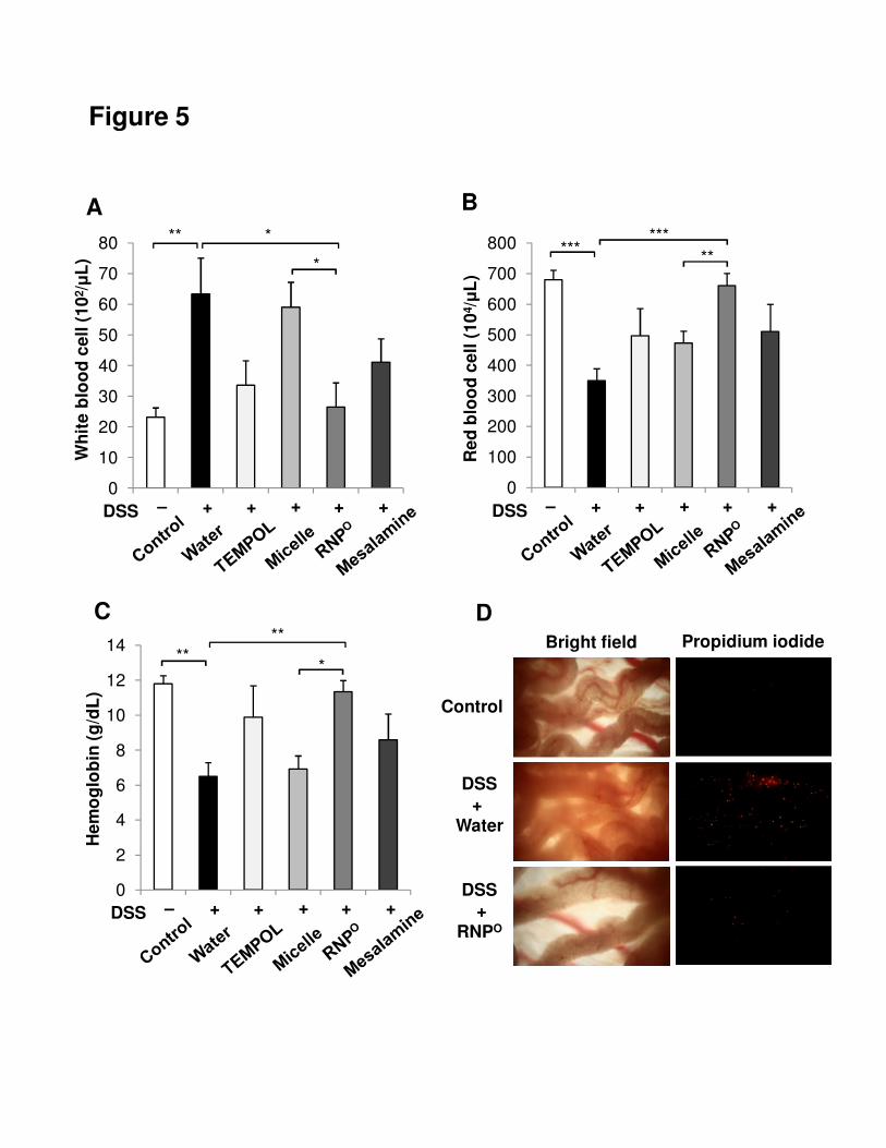
We then analyzed ability of RNPO to suppress systemic inflammation in
DSS-induced colitis. Hematological analyses were performed to reveal the massive
infiltration of leukocytes. Blood from RNPO-treated mice had a significant lower level
of white blood cells compared to DSS- and micelle-treated mice (P < .05), indicating
lower levels of neutrophil invasion in RNPO-treated mice (Figure 5A).
20
Low-molecular-weight TEMPOL and mesalamine showed the effect to suppress white
blood cells in DSS-treated mice; however, the significance was not observed.
Furthermore, results of the hematological analysis indicated higher levels of red blood
cells and hemoglobin in the blood of RNPO-treated mice (Figure 5B,C). This suggests
that the intestinal wall was protected from hemorrhage in RNPO-treated mice. We
further investigated the desquamation of impaired epithelial cells and cell death in
colonic mucosa by intravital observation using in vivo microscopic live imaging and
propidium iodide staining.28
The results showed that a great number of desquamated
cells and cell death existed in colonic mucosa of DSS-treated mice (Figure 5D,
Supplementary Video 1). In contrast, in colonic mucosa of RNPO-treated mice, the
desquamation and cell death was remarkably suppressed. On the basis of these results,
it was confirmed that the colonic injury is protected by the oral administration of RNPO.
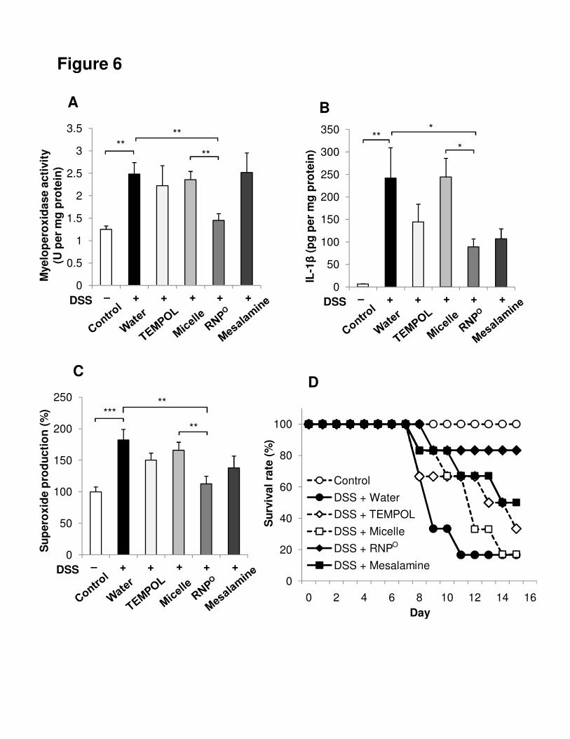
RNPO suppresses pro-inflammatory mediators and enhances survival rate in mice
In addition, after 7 days of treatment, we determined pro-inflammatory mediators
in the colonic mucosa, including MPO activity, IL-1β and superoxide. These
pro-inflammatory mediators are well-known markers of inflammation and play an
important role in UC. Low-molecular-weight TEMPOL and mesalamine did not
effectively suppress these pro-inflammatory mediators induced by DSS (Figure 6A–C).
21
On the other hand, RNPO-treated mice showed a significant suppression of
pro-inflammatory mediators in colonic tissue (P < .01). It should be noted that no
therapeutic effect was observed for polymeric micelle without nitroxide groups,
indicating that effective delivery of nitroxide groups in colonic mucosa area is one of
the most important factors for UC treatment. Because low-molecular-weight drugs tend
to be absorbed into the bloodstream via mesentery, sufficient dose of drugs might not
reach to target area to result in low therapeutic efficacy. Side effects in whole body
should also be considered such kind of low-molecular weight drugs. Finally, we
investigated the effect of orally administered RNPO on the survival rate of mice with
colitis induced by 5-day administration of DSS. After 15 days of treatment, orally
administered low-molecular-weight TEMPOL and mesalamine slightly increased the
survival rate (33.3% and 50%, respectively) compared with DSS- and micelle-treated
mice (16.7%) (Figure 6D). On the other hand, RNPO treatment significantly increased
the survival rate of DSS-treated mice to 83.3%. This indicates that RNPO has not only
suppressive but also therapeutic effects on mice with DSS-induced colitis.
Discussion
22
Despite significant advances in treatments, IBD remains a major clinical problem,
because no drug is entirely effective. For many years, there were only 2 treatment
options for IBD: corticosteroids and mesalamine.29,30 Although they are effective in
treating IBD in some extent, their severe side effects have raised significant concerns
among both physicians and patients, and limited their use. In addition, anti-TNF-α
antibody is employed to suppress inflammation of UC, which works well though it is
cost-oriented therapy with multiple side effects. 31 Recently, many promising
low-molecular-weight medications, such as antioxidants, have been found beneficial in
experimental models of UC.9–11,32 Unfortunately, results of clinical trials investigating
these promising drugs have been largely negative. The drawbacks of current
low-molecular-weight drugs are poor stability in stomach, low solubility and side
effects on whole body when they enter the bloodstream. In this study, we have
developed a novel nitroxide radical-containing nanoparticle RNPO that accumulates
specifically in colon area to suppress the inflammation in DSS-induced colitis mice. For
UC treatment via oral administration, this nanoparticle showed excellent properties,
including high accumulation in inflamed tissues of colon and non-absorption into the
bloodstream.
23
Here, we found that the accumulation in colon area depends on the sizes and
PEGylated character of particles. Both low-molecular-weight drugs, submicron- and
micron-sized polystyrene latex particles showed poor accumulation in colon, whereas
higher accumulation of particles with approximately several tens of nanometers was
observed. Optimal size of several tens of nanometers allowed easier diffusion in the
mucosa compared to larger sized particles.25,26,33 In addition, 40-nm-diameter RNPO
with PEG shell showed significantly high accumulation and long retention in colon area
compared to polystyrene latex particles with similar size of 40 nm. PEGylated character
of RNPO might protect nitroxide radicals in the hydrophobic core from hash conditions
of GIT after oral administration, resulting in the significant accumulation in colon
area.34 Furthermore, PEG chains of RNPO may achieve mucoadhesion due to their
ability to inter-diffuse among the mucus network and polymer entanglement with
mucin, which is composed of glycoprotein.35 Therefore, PEGylated character of RNPO
showed much significant effect on its accumulation in colonic mucosa. Eventually, we
observed the accumulation of RNPO in colon is almost 50 times higher than that of
low-molecular-weight TEMPOL. To deliver sufficient dose of anti-inflammatory drugs
for UC treatment, high dose of drugs is required, however it leads to undesirable side
effects, because almost all low-molecular-weight drugs tend to metabolize in upper GIT
24
or absorb into bloodstream.36,37 In case of RNPO, no absorption into bloodstream was
observed via oral administration route, which improves accumulation in colon region
and prevents side effects to whole body. Another interesting phenomenon in our study
is the higher accumulation of nanoparticles in inflammatory colon than healthy colon.
Mucus layer in colon area is significantly thicker than that in small intestine, which is
considered as a significant barrier to nanoparticle penetration.38 In colon of patients
with UC, the overall thickness of the adherent mucus layer is reduced due to the
reduction of goblet cells,38,39 resulting in the facile penetration of nanoparticles in
inflammatory tissues. In addition, the opening tight junction of epithelium cells in UC
is another explanation for higher accumulation of nanoparticles.40
After investigating the distribution of RNPO in GIT, we used DSS-induced colitis
model mice to compare suppressive effect of RNPO with low-molecular-weight
TEMPOL and mesalamine, a commercial medication for UC treatment. Our results
showed that low-molecular-weight TEMPOL and mesalamine did not clearly show
their effects, whereas RNPO effectively reduced the severity of colitis by suppression of
DAI and damage of colonic architecture. It is noted that micelle without nitroxide
radicals did not show any therapeutic effect at all on colitis mice, indicating that ROS
It should be noted
that no absorption of RNPO into bloodstream was observed even in colitis mice.
25
scavenging character of nitroxide radicals plays critical role in the effect of RNPO on
colitis mice. Further investigations, it is confirmed that RNPO did not simulate the
whole body immune system as well as effectively suppressed pro-inflammatory
mediators such as MPO, IL-1β and superoxide. The therapeutic efficiency of RNPO was
further confirmed by survival data, which showed higher survival rate of RNPO-treated
mice compared to low-molecular-weight TEMPOL- or mesalamine-treated mice.
In conclusion, we have developed a novel nitroxide radical-containing
nanoparticle, RNPO, which possesses anti-oxidative nitroxide radicals in the core for
treatment of DSS-induced colitis mice. RNPO significantly accumulated not only in the
mucosa but also higher in inflammatory sites of the colon, resulting in a high
therapeutic effect, which was not observed in low-molecular-weight drugs. In addition,
RNPO may lack the undesirable side effects of low-molecular-weight TEMPOL, since it
is not absorbed into the bloodstream. Our results indicated that the therapeutic
efficiency of nitroxide radicals could be successfully enhanced by using nanoparticles
to suppress inflammation in the colon area and reduce undesirable side effects.
Therefore, we believe that RNPO may become an important therapeutic agent for the
treatment of UC.
26
Acknowledgements
One of the authors, L.B.V., would like to express his sincere appreciation for the
research fellowship of The Japan-East Asia Network of Exchange for Students &
Youths (JENESYS) between University of Science Ho Chi Minh, Vietnam and
University of Tsukuba, Japan.
Figure Legends:
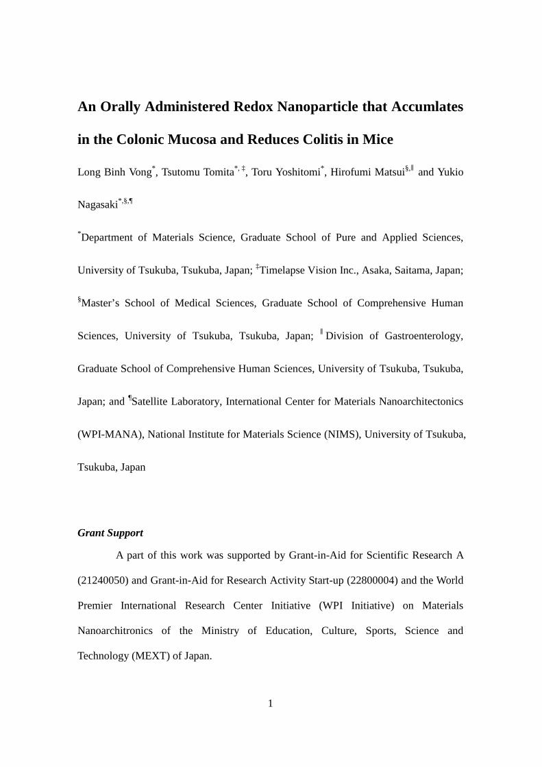
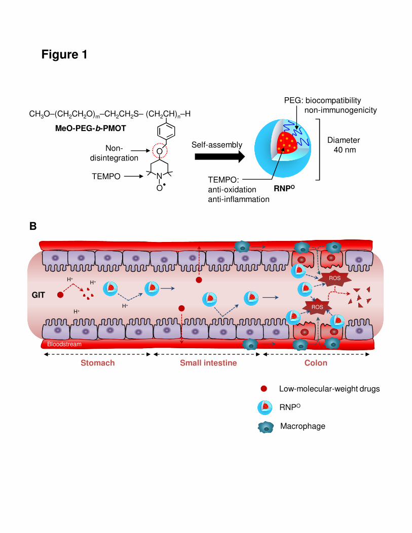
Figure 1. Schematic illustration of RNPO and nanotherapy for DSS-induced colitis in
mice. (A) RNPO is prepared by self-assembly of a poly(ethylene
glycol)-b-poly(4-methylstyrene) block copolymer possessing nitroxide radical TEMPO
moieties. (B) After oral administration, low-molecular-weight drugs, such as TEMPOL,
are degraded and absorbed into the bloodstream in stomach and small intestine before
reaching the colon. In contrast, RNPO is stable and withstands the harsh conditions of
the gastrointestinal tract (GIT), and reach the colon to scavenge ROS, especially sites of
inflammation.
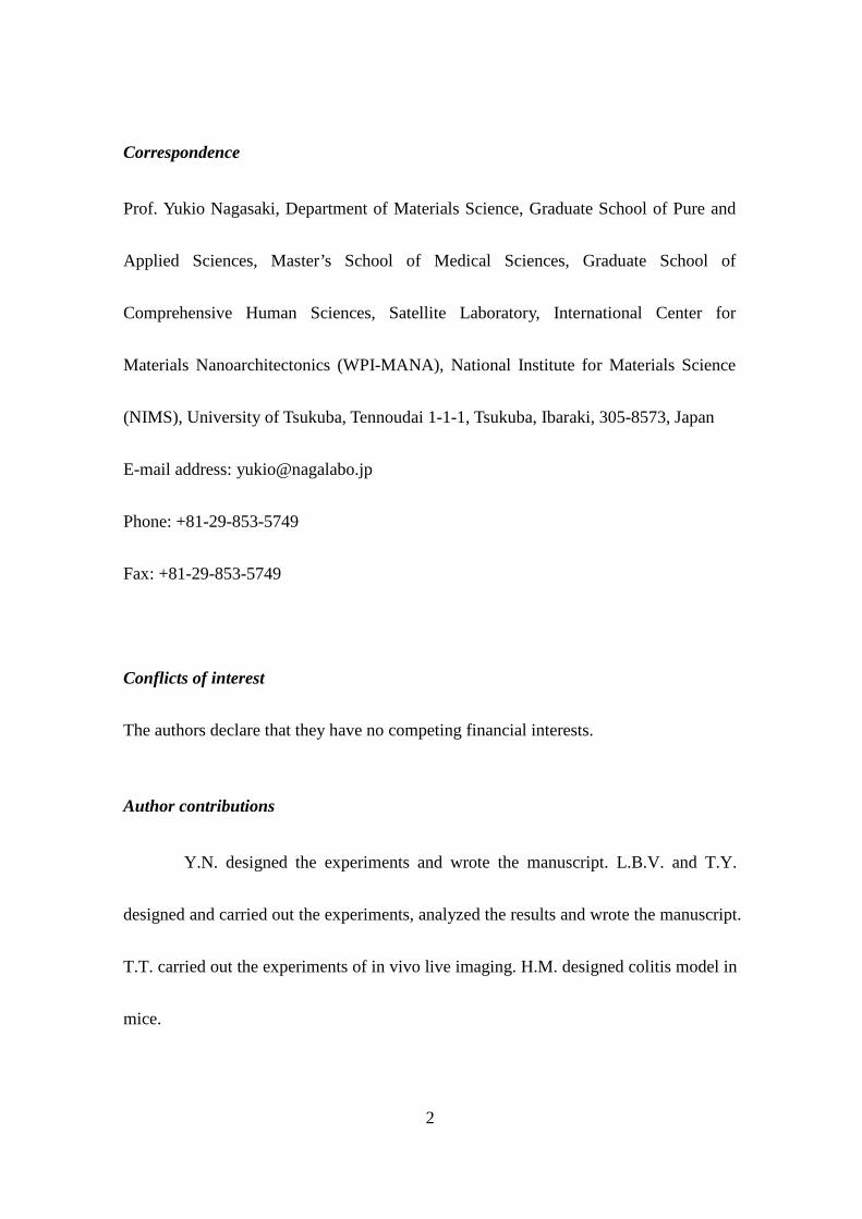
Figure 2. Specific accumulation of RNPO in mice with colitis. (A) Localization of
RNPO in the colon was determined with rhodamine-labeled RNPO. Mice were sacrificed
4 hours after oral administration of 1 mL of rhodamine-labeled RNPO at a dose of 5
27
mg/mL (n = 3), and colon sections were prepared. Localization of rhodamine-labeled
RNPO in the colon was analyzed by fluorescent microscopy. Scale bars, 200 μm. (B)
Accumulation of low-molecular-weight TEMPOL, RNPO and polystyrene latex
particles in the colon. After oral administration of low-molecular-weight TEMPOL,
RNPO and polystyrene latex particles with equivalent nitroxide radicals (1.33 mg; 7.5
μM), the amount of nitroxide radicals was measured by ESR. The data are expressed as
mean ± SEM, n = 3. (C) Specific accumulation of RNPO in the inflamed colon. Colitis
was induced in mice by supplementing the drinking water with DSS (3% wt/vol) for 5
days. The amount of nitroxide radicals in the normal colon and the inflamed colon was
determined by ESR measurement 4 hours after administration of RNPO. The data are
expressed as mean ± SEM, n = 3.
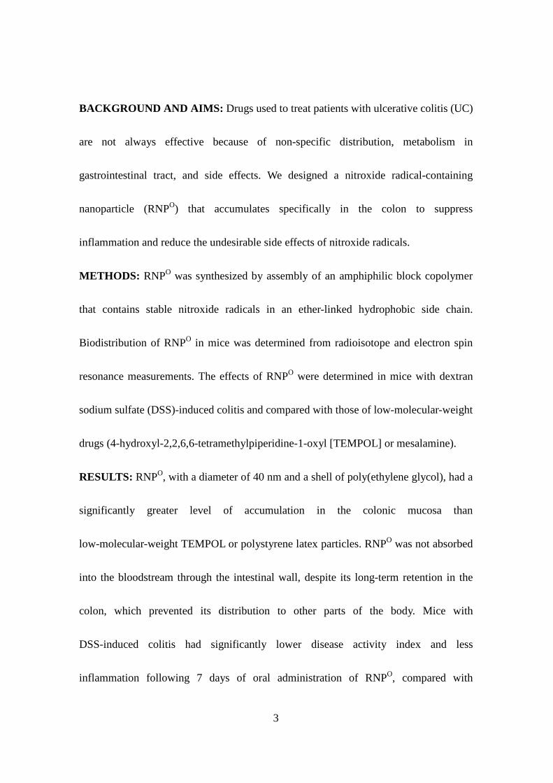
Figure 3. Biodistribution of RNPO in GIT and bloodstream. (A) The biodistribution of
RNPO was determined using 125I-labeled RNPO. The percentages of radioactivity in
each organ and in the blood were determined by comparison to the initial total
radioactivity. The data are expressed as mean ± SEM, n = 5. (B) Absorption of
low-molecular-weight TEMPOL and RNPO into the bloodstream of normal mice (solid
line) and colitis mice (dashed line). After administration of low-molecular-weight
28
TEMPOL and RNPO, the amount of nitroxide radicals in the plasma was determined by
ESR measurement. The data are expressed as mean ± SEM, n = 3. (Inset) The ESR
spectra of low-molecular-weight TEMPOL (grey spectrum) and RNPO (black spectrum)
in the colon homogenate after 4 h oral administration.
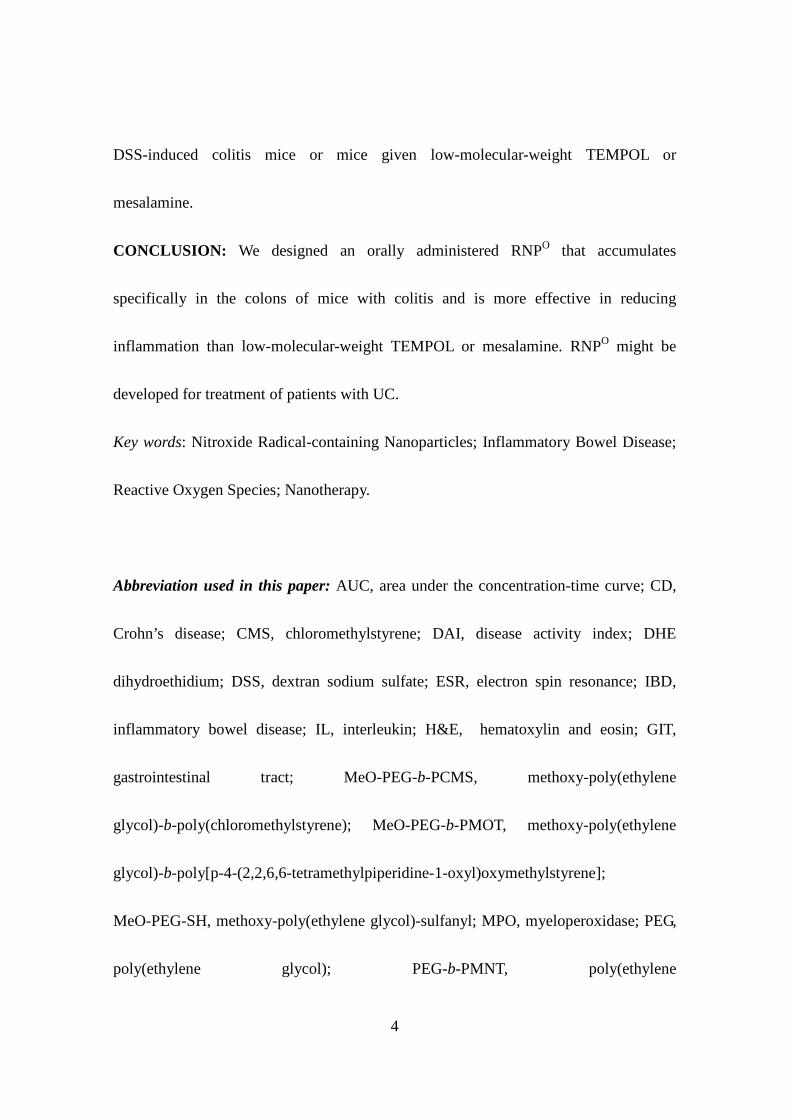
Figure 4. Therapeutic effect of RNPO on DSS-induced colitis in mice. (A) Changes in
disease activity index. Disease activity index is the summation of the stool consistency
index (0–3), fecal bleeding index (0–3), and weight loss index (0–4). The data are
expressed as mean ± SEM, *P < .05, **P < .01 and ***P < .001 vs. control group; ‡P
< .05 and ¶P < .001 vs. DSS groups, n = 6–7, two-way ANOVA, followed by
Bonferroni post-hoc test. (B) Preservation of colon length. After 7 days of treatment, the
colon was collected and measured. The data are expressed as mean ± SEM, **P < .01, n
= 6–7. (C) Protection of mucosal architecture. After 7 days of treatment, the colon was
collected, and 7-μm-thick sections of distal colon were prepared. Sections of the distal
colon were stained by hematoxylin and eosin (H&E), and assessed histologically. Scale
bars, 200 μm.
Figure 5. Hematological analyses in the peripheral blood and intravital observation of
29
colon. (A–C) After 7 days of treatment, blood was collected by intracardiac puncture
with a heparin-containing syringe, and hematological analyses were performed by
automatic hematology analyzer (Celltac α, MEK-6358; Nihon Kohden Co., Tokyo,
Japan). Blood samples were analyzed for white blood cells (A), red blood cells (B), and
hemoglobin (C). The data are expressed as mean ± SEM, *P < .05, **P < .01, ***P
< .001, n = 6. (D) The desquamation of impaired epithelial cells and cell death in
colonic mucosa were determined by in vivo microscopic live imaging and propidium
iodide staining. The bright field images were acquired 2 hours after removing remains
in the colon. The cell death images were recorded immediately after staining of
propidium iodide under an excitation wavelength of 488 nm and an emission
wavelength of 515 nm.
Figure 6. RNPO reduced pro-inflammatory mediators and increased survival rate in
colitis mice. (A–C) After 7 days of treatment, colon homogenates were prepared, and
MPO activity, superoxide, and IL-1β were measured. (A) MPO activity was determined
by a colorimetric assay using o-dianisidine hydrochloride and H2O2 as substrates. (B)
Measurement of IL-1β in colon homogenate was performed with an ELISA kit for mice.
Protein content in the colon homogenate was determined by a BCA kit. (C) Generation
30
of superoxide in colon homogenates was measured by dihydroethidium (DHE)
fluorescence. The fluorescence intensity was measured with an excitation wavelength
of 530 nm and an emission wavelength of 620 nm. Superoxide values were expressed
as intensity per mg of protein, and the superoxide value of the control group was
standardized to 100%. The data are expressed as mean ± SEM, *P < .05, **P < .01,
***P < .001, n = 6. (D) The survival rate of mice was determined after 15 days of 3%
(wt/vol) DSS treatment. Starting on day 5, test drugs were orally administered daily
until day 15. The number of surviving mice was counted until day 15, n = 6.
References
1. Khor B, Gardet A, Xavier RJ. Genetics and pathogenesis of inflammatory bowel
disease. Nat Rev 2011;474:307–317.
2. Podolsky DK. Inflammatory bowel disease. N Engl J Med 2002;347:417–429.
3. Abraham C, Cho HJ. Mechanism of disease inflammatory bowel disease. N Engl J
Med 2009;361:2066–2078.
4. Edward VL. Clinical epidemiology of inflammatory bowel disease: incidence,
prevalence, and environmental influences. Gastroenterology 2004;126:1504–1517.
5. Simmonds NJ, Rampton DS. Inflammatory bowel disease a radical view. Gut
31
1993;34:865–868.
6. Xavier RJ, Podolsky DK. Unravelling the pathogenesis of inflammatory bowel
disease. Nat Rev 2007;448:427–434.
7 . Babbs CF. Oxygen radicals in ulcerative colitis. Free Radic Biol Med
1992;13:169–182.
8. McCord JM. The evolution of free radicals and oxidative stress. Am J Med
2000;108:652–659.
9. Jin Y, Kotakadi VS, Ying L, et al. American ginseng suppresses inflammation and
DNA damage associated with mouse colitis. Carcinogenesis 2008;29:2351–2359.
10. Ju J, Hao X, Lee MJ, et al. A γ-tocopherol-rich mixture of tocopherols inhibits
colon inflammation and carcinogenesis in azoxymethane and dextran sulfate
sodium-treated mice. Cancer Prev Re 2009;2:143–152.
11. Aggarwal BB, Harikumar KB. Potential therapeutic effects of curcumin, the
anti-inflammatory agent, against neurodegenerative, cardiovascular, pulmonary,
metabolic, autoimmune and neoplastic diseases. Int J Biochem Cell Biol
2009;41:40–59.
12. Kim B, Rutka J, Chan W. Nanomedicine. N Engl J Med 2010;36:2434–2443.
32
13. Otsuka H, Nagasaki Y, Kataoka K. PEGylated nanoparticles for biological and
pharmaceutical applications. Adv Drug Deliv Rev 2003;55:403–419.
14. Maeda H, Fang J, Inutsuka T, et al. Vascular permeability enhancement in solid
tumor: various factors, mechanisms involved and its implications. Int
Immunopharmacol 2003;3:319–328.
15. Weis SM, Cheresh DA. Tumor angiogenesis: molecular pathways and therapeutic
targets. Nat Med 2011;17:1359–1370.
16. Davis ME, Chen ZG, Shin DM. Nanoparticle therapeutics: an emerging treatment
modality for cancer. Nat rev Drug dis 2008;7:771–782.
17. Cabral H, Matsumoto Y, Mizuno K, et al. Accumulation of sub-100 nm polymeric
micelles in poorly permeable tumours depends on size. Nat Nanotech 2011;6:815–823.
18. Yoshitomi T, Hirayama A, Nagasaki Y. The ROS scavenging and renal protective
effects of pH-responsive nitroxide radical-containing nanoparticles. Biomaterials
2011;32:8021–8028.
19. Marushima A, Suzuki K, Nagasaki Y, et al. Newly synthesized radical-containing
nanoparticles enhance neuroprotection after cerebral ischemia-reperfusion injury.
Neurosurgery 2011;68:1418–1426.
33
20. Chonpathompikunlert P, Yoshitomi T, Han J, et al. Chemical nanotherapy: Nitroxyl
radical-containing nanoparticle (RNP) protects neuroblastoma SH-SY5Y cells from a
β-induced oxidative stress. Ther Deliv 2011;2:585–597.
21 . Chonpathompikunlert P, Yoshitomi T, Han J, et al. The use of nitroxide
radical-containing nanoparticles coupled with piperine to protect neuroblastoma
SH-SY5Y cells from a β-induced oxidative stress. Biomaterials 2011;32:8605–8612.
22. Yoshitomi T, Nagasaki Y. Nitroxyl radical-containing nanoparticles for novel
nanomedicine against oxidative stress injury. Nanomedicine 2011;6:509–518.
23 . Yoshitomi T, Suzuki R, Mamiya T, et al. pH-sensitive
radical-containing-nanoparticle (RNP) for the L-band-EPR imaging of low pH
circumstances. Bioconjugate Chem 2009;20:1792–1798.
24. Yoshitomi T, Miyamoto D, Nagasaki Y. Design of core-shell-type nanoparticles
carrying stable radicals in the core. Biomacromolecules 2009;10:596–601.
25. Lamprecht A, Schafer U, Lehr CM. Size-dependent bioadhesion of micro- and
nanoparticulate carriers to the inflamed colonic mucosa. Pharm Res 2001;18:788–793.
26. Francis MF, Cristea M, Winnik FM. Polymeric micelles for oral drug delivery:
Why and how. Pure Appl Chem 2004;76:1321–1335.
34
27. Cooper HS, Murthy SN, Shah RS, et al. Clinicopathologic study of dextran sulfate
sodium experimental murine colitis. Lab Invest 1993;69:238–249.
28. Bryson GJ, Harmon BV, Collins RJ. A flow cytometric study of cell death: Failure
of some models to correlate with morphological assessment. Immunol Cell Biol
1994;72:35–41.
29. Friend DR, Sellin J. Drug delivery in advancing the treatment of inflammatory
bowel disease. Adv Drug Deliv Rev 2005;57:215–216.
30 . Stephen BH. Medical therapy for ulcerative colitis 2004. Gastroenterology
2004;126:1582–1592.
31. Singh K, Chaturvedi R, Barry DP, et al. The apolipoprotein E-mimetic peptide
COG112 inhibits NF-kappaB signaling, proinflammatory cytokine expression, and
disease activity in murine models of colitis. J Biol Chem 2011;286:3839–3850.
32. Helieh S, Theresa S, Craig J, et al. Antioxidants as novel therapy in a murine model
of colitis. J Nutr Biochem 2005;16:297–304.
33. Jiang W, Kim B, Rutka J, et al. Nanoparticle-mediated cellular response is
size-dependent. Nat Nanotech 2008;3:145–150.
34. Tobio M, Sanchez A, Vila A, et al. The role of PEG on the stability in digestive
35
fluids and in vivo fate of PEG-PLA nanoparticles following oral administration.
Colloids Surf B 2000;18:315–323.
35. Lai SK, Wang YY, Hanes J. Mucus-penetrating nanoparticles for drug and gene
delivery to mucosal tissues. Adv Drug Deliv Rev 2009;61:158–171.
36. Friend, D.R. New oral delivery systems for treatment of inflammatory bowel
disease. Adv Drug Deliv Rev 2005;57:247–265.
37. Laroui H, Dalmasso G, Thu Nguyen HT, et al. Drug-loaded nanoparticles targeted to
the colon with polysaccharide hydrogel reduce colitis in a mouse model.
Gastroenterology 2010;138:843–853.
38. Ensign LM, Cone R, Hanes J. Oral drug delivery with polymeric nanoparticles: The
gastrointestinal mucus barriers. Adv Drug Deliv Rev 2012;64:557–570.
39. Pullan RD, Thomas G, Rhodes M, et al. Thickness of adherent mucus gel on colonic
mucosa in humans and its relevance to colitis. Gut 1994;35:353–359.
40. Cereijido M, Contreras RG, Flores-Benítez D, et al. New diseases derived or
associated with the tight junction. Arch Med Res 2007;38:465–478.
B
TEMPO
MeO-PEG-b-PMOT
Non-disintegration
CH3O–(CH2CH2O)m–CH2CH2S– (CH2CH)n–H
O
N
O
H+
H+H+
ROS
ROS
Stomach Small intestine Colon
H+
Low-molecular-weight drugs
RNPO
Macrophage
Self-assemblyDiameter
40 nm
TEMPO: anti-oxidationanti-inflammation
RNPO
PEG: biocompatibilitynon-immunogenicity
GIT
Bloodstream
Figure 1
0.8
1.0
1.2
1.4
1.6
1.8
2.0
No
rma
l m
ice
Co
liti
s
mic
e
p = .07A
mo
un
t o
f n
itro
xid
era
dic
al
in c
olo
n (µ
g/c
m)
0
20
40
60
80
100
0 8 16 24 32 40 48
Am
ou
nt
of
nit
rox
ide
rad
ica
lin
co
lon
(µ
g)
Time / h
Polystyrene latex particles 40 nm
Polystyrene latex particles 100 nm
Polystyrene latex particles 0.5 µm
Polystyrene latex particles 1 µm
Low-molecular-weight TEMPOL
RNPO 40 nm
B
Rhodamine Rhodamine-RNPO
A
C
Figure 2
A B
Figure 3
0
10
20
30
40
50
60
70
80
0 4 8 12 16 20 24
liver
small Intestine
colon
cecum
blood
% o
f in
itia
ld
os
e
Time / h
0
4
8
12
16
20
0 12 24 36 48
TEMPOL
RNP
Am
ou
nt
nit
rox
ide
rad
ica
lin
pla
sm
a (
µg
/ml)
Time / h
O
60
70
80
90
100
110
Co
lon
le
ng
th(m
m)
**
**
**
– + + + +DSS +0
1
2
3
4
5
6
7
8
9
0 1 2 3 4 5 6 7
Control
DSS + Water
DSS + TEMPOL
DSS + Micelle
DSS + RNP
DSS + Mesalamine
Dis
ea
se
ac
tivit
y i
nd
ex
Day
O
Control DSS + Water DSS + TEMPOL
DSS + RNPO DSS + Mesalamine
C
A B
DSS + Micelle
Figure 4
***
*, ¶
***
***
**
***
**
*
* ¶
¶‡
0
10
20
30
40
50
60
70
80
Wh
ite
blo
od
ce
ll (
10
2/µ
L)
** *
*
– + + + +DSS +0
100
200
300
400
500
600
700
800
Re
d b
loo
d c
ell
(1
04/µ
L)
******
**
– + + + +DSS +
0
2
4
6
8
10
12
14
He
mo
glo
bin
(g
/dL
)
****
*
– + + + +DSS +
Control
DSS+
Water
DSS+
RNPO
Bright field Propidium iodide
DC
A B
Figure 5
0
0.5
1
1.5
2
2.5
3
3.5
Mye
lop
ero
xid
as
e a
cti
vit
y
(U p
er
mg
pro
tein
)
****
**
– + + + +DSS +0
50
100
150
200
250
300
350
IL-1β
(pg
pe
r m
g p
rote
in)
***
*
– + + + +DSS +
0
50
100
150
200
250
Su
pe
rox
ide
pro
du
cti
on
(%
) ***
**
**
– + + + +DSS +
0
20
40
60
80
100
0 2 4 6 8 10 12 14 16
Control
DSS + Water
DSS + TEMPOL
DSS + Micelle
DSS + RNP
DSS + Mesalamine
Su
rviv
al
rate
(%
)
Day
O
A B
C
Figure 6
D
Related Documents