An Introduction to Light Interaction with Human Skin Gladimir V. G. Baranoski 1 Aravind Krishnaswamy 1 Abstract: Despite the notable progress in physically-based rendering, there is still a long way to go before one can automatically generate predictable images of organic materials such as human skin. In this tutorial, the main physical and biological as- pects involved in the processes of propagation and absorption of light by skin tissues are examined. These processes affect not only skin appearance, but also its health. For this reason, they have also been the object of study in biomedical research. The models of light interaction with human skin developed by the biomedical community are mainly aimed at the simulation of skin spectral properties which are used to deter- mine the concentration and distribution of various substances. In computer graphics, the focus has been on the simulation of light scattering properties that affect skin ap- pearance. Computer models used to simulate these spectral and scattering properties are described in this tutorial, and their strengths and limitations discussed. Keywords: natural phenomena, biologically and physically-based rendering. 1 Introduction Creating convincing pictures of organic materials, such as human skin, is usually an art entirely left to designers and animators. Recently, in order to overcome these limitations and develop predictive reflectance and scattering models, computer graphics researchers have started to look more closely at the interaction of light with organic materials from a biological point of view. The games and entertainment industries can certainly benefit from being able to automatically generate realistic and predictable images of skin tissues. Currently, the real- istic image synthesis of organic materials is several years behind the rendering of inorganic materials. Due to limitations in this area, the non-realistic traits of a synthetic human being are easily perceived despite the sophisticated geometrical models available in the literature. On the scientific side, the processes of light interaction with human skin have a funda- mental importance in biomedical sciences. By studying processes involved in light remission from skin, better protocols can be developed to automatically diagnose medical conditions, such as jaundice (yellowish hue), erythema (redness) [66], as well as tumors at early stages [16]. In the United States, more than 50,000 new cases of melanoma, the most serious form of skin cancer, are reported to the American Cancer Society each year [83]. Understanding 1 Natural Phenomena Simulation Group, School of Computer Science, University of Waterloo, Canada {[email protected] [email protected]}

Welcome message from author
This document is posted to help you gain knowledge. Please leave a comment to let me know what you think about it! Share it to your friends and learn new things together.
Transcript

An Introduction to Light Interaction with Human SkinGladimir V. G. Baranoski 1
Aravind Krishnaswamy 1
Abstract: Despite the notable progress in physically-based rendering, there is stilla long way to go before one can automatically generate predictable images of organicmaterials such as human skin. In this tutorial, the main physical and biological as-pects involved in the processes of propagation and absorption of light by skin tissuesare examined. These processes affect not only skin appearance, but also its health.For this reason, they have also been the object of study in biomedical research. Themodels of light interaction with human skin developed by the biomedical communityare mainly aimed at the simulation of skin spectral properties which are used to deter-mine the concentration and distribution of various substances. In computer graphics,the focus has been on the simulation of light scattering properties that affect skin ap-pearance. Computer models used to simulate these spectral and scattering propertiesare described in this tutorial, and their strengths and limitations discussed.
Keywords: natural phenomena, biologically and physically-based rendering.
1 Introduction
Creating convincing pictures of organic materials, such as human skin, is usually anart entirely left to designers and animators. Recently, in order to overcome these limitationsand develop predictive reflectance and scattering models, computer graphics researchers havestarted to look more closely at the interaction of light with organic materials from a biologicalpoint of view. The games and entertainment industries can certainly benefit from being ableto automatically generate realistic and predictable images of skin tissues. Currently, the real-istic image synthesis of organic materials is several years behind the rendering of inorganicmaterials. Due to limitations in this area, the non-realistic traits of a synthetic human beingare easily perceived despite the sophisticated geometrical models available in the literature.
On the scientific side, the processes of light interaction with human skin have a funda-mental importance in biomedical sciences. By studying processes involved in light remissionfrom skin, better protocols can be developed to automatically diagnose medical conditions,such as jaundice (yellowish hue), erythema (redness) [66], as well as tumors at early stages[16]. In the United States, more than 50,000 new cases of melanoma, the most serious formof skin cancer, are reported to the American Cancer Society each year [83]. Understanding
1Natural Phenomena Simulation Group, School of Computer Science, University of Waterloo, Canada{[email protected] [email protected]}

An Introduction to Light Interaction with Human Skin
how light is absorbed and propagated in skin tissues can assist in the design of lotions pro-tective against harmful solar radiation, and also in the design of superior cosmetics. Hence,the design of biophysically-based and predictive models of light interaction with human skinhas applications beyond the scope of computer graphics.
This tutorial discusses the recent advances in the biophysically-based rendering of hu-man skin. In particular, it addresses the processes of light transport and absorption in skintissues, and examines computer graphics models used to simulate these natural phenomena.Since these models often incorporate algorithms and techniques used in models developedby the biomedical community, an overview of the most influential skin optics modeling ap-proaches used in biomedical applications is also provided. In addition to the direct contri-butions to the realistic and predictive simulation of skin appearance, the algorithms used inmodels of light interaction with human skin can be extended to models aimed at the renderingof other organic materials such as hair, fur, leaves, petals, ocular tissues etc...
It is important to note that there are several subsurface scattering models in the graph-ics literature which are neither biologically motivated nor specifically designed to simulatelight interaction with human skin (e.g., plants [6] and hair [55]). An extensive review of thesemodels is beyond the scope of this tutorial, and the interested reader can find more infor-mation in a recent survey by Premoze [72]. In addition, there has also been recent worksin image based techniques for rendering and altering the appearance of skin [37, 90], whichdo not address the modeling of light propagation and absorption in skin tissues, and for thisreason are not examined in this tutorial.
The remainder of this tutorial is organized as follows. Section 2 presents a concisereview of physical parameters used to characterize the appearance of inorganic and organicmaterials. Section 3 examines the biological factors involved in the light interaction with hu-man skin. Section 4 provides an overview of models of skin optics available in the biomedicalfield. Sections 5-8 examine the strengths and limitations of models of light interaction withhuman skin used in image synthesis frameworks. Section 9 outlines a number of issues thatneed to be addressed to improve the predictability and the applicability of the current models.The tutorial closes with a glimpse of future trends in biophysically-based rendering.
2 Measurement of Appearance
The group of measurements necessary to characterize both the color and surface fin-ish of a material is called the measurement of appearance of the material [39]. This groupof measurements involves the spectral energy distribution of the propagated light, measuredin terms of reflectance and transmittance, and the spatial energy distribution of that light,measured in terms of BRDF (bidirectional reflectance distribution function) and BTDF (bidi-rectional transmittance distribution function) [65].
34 RITA • Volume XI • Numero 1 • 2004

An Introduction to Light Interaction with Human Skin
Greenberg et al. [35] proposed a framework to test, validate and improve the fidelityand efficiency of computer graphics light transport simulations and image display procedures.They emphasized the importance of performing comparisons between simulations and actualmeasurements so that simulations can be used in a predictive manner. According to theirparadigm, it is of fundamental importance that at each stage simulations are compared withmeasured experiments. Actual measurements of reflectance and transmittance are performedusing spectrophotometers, and actual measurements of BRDF and BTDF are performed usinggoniophotometers [39, 47]. In order to evaluate models of light interaction with matter or toextract data from previously validated models, one needs to resort to computer simulationsof such devices, henceforth called virtual measurement devices.
Spectrophotometry is defined as the quantitative measurement of reflection and trans-mission properties as a function of wavelength [22], and a spectrophotometer is defined tobe any instrument for measuring the spectral distribution of reflected and transmitted radiantpower. Spectrophotometers can also be used to determine the absorption characteristics of anobject as a function of wavelength. Actual reflectance measurements are usually performedunder illuminating and viewing conditions recommended by CIE2. Integrating spheres areused to provide readings where either the illuminant (or viewing) specification is “total” or“diffuse only”. The transmittance of translucent specimens depends greatly on the way theyare illuminated and mounted in the instrument. Generally transmittance measurements arecarried out with the integrating sphere-type spectrophotometers. There are a number of de-tailed issues specific to performing spectral measurements for translucent materials which arebeyond the scope of this course. For a comprehensive discussion of these issues the interestedreader is referred to the report by Aydinli and Kaase [4].
Goniophotometry is defined as the measurement of the directional light distributioncharacteristic of sources, media and materials, and a goniophotometer is defined as an instru-ment that measures flux (power) as a function of angles of illumination and observation [22].The measurements made by a goniophotometer can be performed in different ways (Figure 1),and, as a result, there are many possible configurations for these devices. Computer graphicsresearchers have proposed extensions for industry made goniophotometers [29] as well asdesigns based on the use of digital cameras [48, 104]. A reader interested in a detailed de-scription of goniophotometers used in computer graphics is referred to more comprehensiveworks in this area [29, 57].
Baranoski et al. [7] and Krishnaswamy et al. [52] examined in detail the implementa-tion of virtual spectrophotometric and goniophotometric devices respectively. The main ques-tion to be addressed when performing a virtual measurement is how many samples should beused. According to the Bernoulli theorem [93], using a sufficiently large number of samples,one will have a high probability of obtaining estimates within the region of asymptotic con-
2Commission Internationale de L’Eclairage.
RITA • Volume XI • Numero 1 • 2004 35

An Introduction to Light Interaction with Human Skin
Figure 1. Photographs of a goniophotometer showing different set-ups for BRDF (left) andBTDF (right) measurements. (Courtesy of Stephane Jacquemoud.)
vergence of the expected value of reflectance, or transmittance, being measured. However,as shown by numerical experiments presented by Baranoski et al. [7], the processing timegrows linearly with respect to the total number of samples since the cost of the algorithm isconstant per sample. In order to minimize the computational costs of virtual spectrophoto-metric measurements, Baranoski et al. proposed a bound on the number of samples derivedfrom the exponential Chebyshev inequality [76]. For virtual goniophotometric measurementsa similar bound can be obtained taking into account the number of patches on the collectorsphere used to simulate an actual goniophotometer [52].
3 Biological Issues
Skin is a multilayered and inhomogeneous organ (Figure 2). In this section, we outlinethe biological characteristics of its main constituents, and how they affect the propagation andabsorption of light.
3.1 Structural Characteristics and Spectral Properties
The first and outermost section of human skin is the stratum corneum, which is a strat-ified structure approximately 0.01-0.02 mm thick [2, 59]. There are skin structural models,however, that consider it part of another tissue, namely the epidermis [91] (Figure 2). Thestratum corneum is composed mainly of dead cells, called corneocytes, embedded in a par-ticular lipid matrix [84]. Light absorption is low in this tissue, with the amount of transmittedlight being relatively uniform in the visible region of the light spectrum [24].
The epidermis is a 0.027-0.15mm thick structure [2, 20, 59] composed of four layers
36 RITA • Volume XI • Numero 1 • 2004

An Introduction to Light Interaction with Human Skin
����������������������������������������������������������������������������������������������������������������
����������������������������������������������������������������������������������������������������������������������������������������������������������������������������������������������������������������������������������������������������������������������������������������
stratum corneum
epidermis
papillary dermis
reticular dermis
hypodermis
Figure 2. Schematic cross-section of skin tissues and the subcutaneous tissue (hypodermis).
(stratum basale, stratum spinosum, stratum granulosum and stratum lucidum). The epidermispropagates and absorbs light. The absorption property comes mostly from a natural pigment(or chromophore), melanin. There are two types of melanin, the red/yellow phaeomelaninand a brown/back eumelanin [86]. Their absorption spectra are broad (Figure 3), with highervalues for shorter wavelengths. The skin color is mostly associated with eumelanin [86].The ratio between the concentration of phaeomelanin and eumelanin present in human skinvaries from individual to individual, with much overlap between skin types [86]. Recentstudies reported values between 0.049 and 0.36 [67]. Melanin is produced by cells calledmelanocytes occurring in the stratum basale, and it is found in membranous particles calledmelanosomes. The melanin absorption level depends on how many melanosomes per unitvolume are in the epidermis. Typically, the volume fraction of the epidermis occupied bymelanosomes varies from 1.3% (lightly pigmented specimens) to 43% (darkly pigmentedspecimens) [42].
The dermis is a 0.6-3mm thick structure [2, 20, 59] which also propagates and absorbslight. It can be divided into two layers: the papillary dermis and the reticular dermis (Fig-ure 2). These layers are primarily composed of dense, irregular connective tissue with nervesand blood vessels (smaller ones in the papillary, and larger ones in the reticular dermis). Thevolume fraction of blood in tissue can vary, roughly in the 0.2-7% range [27, 42]. The fluencerate of blood decreases as we get deeper into the skin, following an almost linear pattern inthe dermis [97]. In the blood cells we find another natural chromophore, hemoglobin, whichabsorbs light and gives blood its reddish color. Normally, the hemoglobin concentration inwhole blood is between 134 and 173g/L [106]. In the arteries, 90-95% of hemoglobin isoxygenated, and in the veins, more than 47% of the hemoglobin is oxygenated [3]. Thesetwo types of hemoglobin, namely oxygenated and deoxygenated hemoglobin, have slightly
RITA • Volume XI • Numero 1 • 2004 37

An Introduction to Light Interaction with Human Skin
different absorption spectra (Figure 3). Two other blood borne pigments are found in thedermis, bilirubin and β-carotene, which contribute to the yellowish or olive tint of humanskin (Figure 3). We remark that β-carotene may be also found in the epidermis and stratumcorneum [1, 54].
The hypodermis is an adipose tissue characterized by a negligible absorption of lightin the visible region of the spectrum [27]. It is usually not considered part of the skin, and itssize varies considerably throughout the body. It can be up to 3cm thick in the abdomen andabsent in the eye lids. The hypodermis presents significant deposits of white fat, whose cellsare grouped together forming clusters. Due to the presence of these white fat deposits, mostof the visible light that reaches this tissue is reflected back to the upper layers [20].
3.2 Scattering Profile
The scattering profile of human skin has two main components: surface and subsur-face scattering. Surface scattering follows Fresnel equations [82], and it is affected by thepresence of folds in the stratum corneum. The aspect ratio of these mesostructures dependson biological factors such as aging and hydration [84, 85]. Approximately 5-7% of the lightincident (over the entire spectrum) on the stratum corneum is reflected back to the environ-ment [91]. The remaining portion is transmitted to the internal tissues. Besides the reflective-refractive scattering caused by the reflection and refraction of light at cellular boundaries, twoother types of scattering occur within the skin layers: Mie and Rayleigh scattering [42].
The stratum corneum and the epidermis are characterized as forward scattering media[9]. In the former this behavior is due to the alignment of the fibers, while in the later it is dueto Mie scattering caused by particles that are approximately the same size of the wavelengthof light (e.g., cell organelles). The level of forward scattering for these tissues is wavelengthdependent. Bruls and van der Leun [9] performed goniometric experiments for five wave-lengths for both the stratum corneum and the epidermis, and they showed that the scatteringprofiles are broader towards the shorter wavelengths.
In the dermis, collagen fibers (approximately 2.8µm in diameter and cylindrical [42])are responsible for Mie scattering, while smaller scale collagen fibers and othermicro-structures are responsible for Rayleigh scattering [42]. Light gets scattered multi-ple times inside the dermis before it is either propagated to another layer or absorbed. Thismeans that the spatial distribution of the light scattered within the dermis quickly becomesdiffuse [2]. In fact, Jacques et al., [43] showed through goniophotometric measurements thatbackscattered light from the dermis is diffuse. While Mie scattering produces variations onboth ends of the visible region of the light spectrum, Rayleigh scattering, being inverselyproportional to the wavelength of light (≈ λ−4), produces larger variations on the low end ofthe light spectrum [42, 33].
38 RITA • Volume XI • Numero 1 • 2004

An Introduction to Light Interaction with Human Skin
400 500 600 7000
5
10
15
20
wavelength (nm)
ε (c
m−
1 /(g/
L))
eumelaninphaeomelanin
400 500 600 7000
1
2
3
4
5
6x 10
5
wavelength (nm)
ε (c
m−
1 /(m
oles
/L))
oxyhemoglobindeoxyhemoglobin
400 500 600 7000
1
2
3
4
5
6
x 104
wavelength (nm)
ε (c
m−
1 /(m
oles
/L))
bilirubin
400 500 600 7000
5
10
15x 10
4
wavelength (nm)
ε (c
m−
1 /(m
oles
/L))
β−carotene
Figure 3. Spectral extinction coefficient curves for pigments present in skin tissues.Courtesy of S. Prahl and the Oregon Medical Laser Center. Absorption spectra are obtained
by multiplying each curve by the pigment’s molecular weight and concentration.
4 Review of Models Used in Biomedical Applications
In this section we provide an overview of relevant modeling approaches and modelsused in biomedical applications involving tissue optics, and group them according to theirsimilarities. For a comprehensive literature review on this topic, the reader is referred to thetexts by Cheong et al. [13] and Tuchin [91]. It is worth noting that these models are mostlyaimed at the measurement and reproduction of skin spectral properties to determine the con-tent and distribution of various substances [89, 111], i.e., scattering properties affecting skinappearance are usually not addressed. Moreover, a substantial portion of the work done bythe biomedical community is either laser-based or aimed at wavelengths outside the visibleregion of the light spectrum.
RITA • Volume XI • Numero 1 • 2004 39

An Introduction to Light Interaction with Human Skin
4.1 Kubelka-Munk Theory Based Models
In the beginning of the century, Kubelka and Munk [53] developed a simple relation-ship between the scattering and absorption coefficients3 of paint and its overall reflectance.This relationship is known as the Kubelka-Munk theory (henceforth referred to as K-M the-ory). It applies energy transport equations to describe the radiation transfer in diffuse scat-tering media using two parameters: the scattering and the absorption coefficients. The K-Mtheory, as originally stated, is considered to be a two-flux theory, since only two types of dif-fuse radiant flux are involved, namely a diffuse downward flux and a diffuse upward flux. Therelations between the fluxes are expressed by two simultaneous linear differential equations[53]. The original K-M theory also assumes that the medium (specimen) presents inhomo-geneities which are small compared to its thickness.
The K-M theory based models (henceforth referred to as K-M models), used in bi-ological tissue optics, also called flux models [13], use K-M equations relating tissue op-tical properties to measured reflectance and transmittance. Although they are based on thetwo-flux K-M theory, these models expanded the original K-M formulation by adding morecoefficients and/or fluxes. For example, van Gemert and Star [98] included a phase func-tion4, optical depth and the effective albedo in their K-M model. They used a phase functionconsisting of a combination of a forward peaked and a symmetric scattering to represent thetissue’s expected experimental scattering behavior. In the original K-M theory formulation[53], the albedo was used to represent the fraction of diffuse light reflected by a matte, planeparallel coating of a given thickness. The effective albedo used by van Gemert and Star [98],however, represents the ratio between the scattering coefficient and the total attenuation coef-ficient, which is given by the sum of the absorption coefficient and the scattering coefficient[40]. In the context of this tutorial, unless otherwise stated, albedo refers to the definitionused by van Gemert and Star [98].
Tuchin et al. [92, 107] used a four-flux model composed of the two diffuse fluxes usedin the original K-M theory, and two collimated laser beams, the incident one and the onereflected from the bottom boundary of the specimen. Yoon et al. [110, 108] used a seven fluxmodel to obtain a three dimensional representation of the scattered radiation caused by anincident laser beam in a semi-infinite medium (infinite in x and y, but finite in z).
In skin optics, the K-M theory was initially applied to specific skin tissues. Andersonand Parish [2] used a K-M model to compute absorption and scattering coefficients for thedermis tissues. Wan et al. [102] extended this model to compute the absorption and scat-
3The absorption and scattering coefficients represent the product of the actual absorption (or scattering) cross sectionby the density of the absorbers (or scatterers) [40]. The absorption and scattering cross sections of a particle havethe dimension of area, and, generally, they are functions of the orientation of the particle and the state of polarizationof the incident light [95].4A phase function represents the directional scattering of the light incident onto a particle [95].
40 RITA • Volume XI • Numero 1 • 2004

An Introduction to Light Interaction with Human Skin
tering coefficients for the epidermis tissues, taking into account both collimated and diffuseincident irradiance. In both cases [2, 102], the forward scattering in the epidermis was notconsidered. Diffey [19] proposed a K-M model which added two features to the previousmodels, namely it takes into account forward and backward scattering and allows changes inthe refractive index at the air/skin interfaces. Cotton and Claridge [17] proposed a model todetermine the color of human skin which applies the K-M equations to the dermis layer. Thismodel takes into account the presence of melanin and blood pigments. Recently, Doi andTominaga presented a model which considers the skin composed of two layers: epidermisand dermis. They apply the K-M theory to both layers. Their model provides weights for fiveskin pigments (melanin, carotene, oxy-hemoglobin, deoxy-hemoglobin and bilirubin) as wellas the skin surface reflectance. These six parameters are obtained by fitting the estimatedreflectance to measured values using the least squares method [10].
Although the K-M theory allows a simple quantitative treatment of skin spectral prop-erties and recent extensions to the original two-flux theory have improved its applicabilityto biological tissue optics, it is not a thorough model of optical radiation transfer. The K-Mmodels can be considered analytical, and they allow the rapid determination of skin opti-cal parameters through inversion procedures5. However, the relative simplicity and speed ofthese models are achieved at the expense of accuracy [91], which requires a more detailedanalysis of the structure and optical properties of the different skin tissues.
4.2 Diffusion Theory Based Models
Photon propagation in optically turbid media, such as skin tissues, can be describedusing the Boltzmann photon transport equation [40], which requires the optical properties ofthe medium be expressed in terms of scattering coefficient, absorption coefficient and phasefunction [21]. Diffusion theory can be seen as an approximate solution of this equation, i.e.,it combines the scattering and the phase function in one parameter, called reduced scatteringcoefficient.
Models based on the diffusion approximation [99] or combined with other approaches,such as the K-M theory [98, 97] or Monte Carlo methods [103], have been used in biomed-ical investigations involving light propagation in turbid media. Farrell and Patterson [25]proposed a model based on the diffusion theory to be used in the non-invasive determinationof the absorption and scattering properties of mammalian tissues. Their model incorporates aphoton dipole source in order to satisfy the tissue boundary conditions. Recently, Doornboset al. [21] proposed a method based on the diffusion theory for measuring optical propertiesand deriving chromophore concentrations from diffuse reflection measurements at the surface
5An inversion procedure is a way to derive biochemical and optical properties from in situ and non-destructiveexperiments [31]. “Inversion” implies a reversal of the usual process of calculating reflection and transmission, i.e.,reflectance and/or transmittance values are used as input instead of output.
RITA • Volume XI • Numero 1 • 2004 41

An Introduction to Light Interaction with Human Skin
of a turbid media.
Models based on the diffusion theory are amenable to analytic manipulation, placeminor constraints on the type of sample and are relative easy to use [71]. The diffusion the-ory, however, can be applied only when scattering events are more probable than absorptionevents. This is usually the case for mammalian tissues in the red and near infrared regions ofthe light spectrum[28]. Not surprisingly, diffusion models have been used in medical appli-cations involving red lasers [99, 109]. When the absorption coefficient of a turbid medium isnot significantly smaller than the scattering coefficient, the diffusion theory provides a poorapproximation for the photon transport equation [71, 74, 109].
4.3 Radiative Transport Models
The K-M and diffusion theories mentioned in the previous sections can be seen asspecial cases of radiative transfer phenomena. When non-stochastic accurate solutions ofthe radiative transport equation in biological tissues are required, more robust methods needto be used, e.g., the successive scattering technique, Ambartsumian’s method, the discreteordinate method, Chandrasekhar’s X and Y functions and the adding-doubling method [69].Their applicability, however, is usually limited to simple conditions and slab geometries6. Acomprehensive review of these methods is beyond the scope of this work, and the interestedreader is referred to the texts by van de Hulst [94] and Prahl [69]. It is worth noting, however,that the adding-doubling method has several advantages with respect to the other techniques.It permits asymmetric scattering, arbitrarily thick samples, Fresnell boundary conditions, andrelatively fast computation [69].
The adding method requires that the reflection and transmission of two slabs be known.They are used to compute the reflection and transmission of another slab comprised of thesetwo individual slabs. In its original definition, the doubling method corresponds to the specialcase in which both slabs are identical [94]. Later on, it was extended to include the addition oftwo non-identical slabs [69]. Once the transmission and reflection for a thin slab are known,the reflection and transmission for a target slab can be computed by doubling the thicknessof the thin slab until it matches the thickness of the target slab.
Prahl et al. [71] applied an inverse adding-doubling method (IAD: “inverse” imply-ing its use as an inversion procedure) to determine the scattering, absorption coefficient andthe asymmetry factor7 of biological tissues. The IAD is an iterative method which con-sists of guessing a set of optical properties, calculating the reflection and transmission usingadding-doubling method, comparing the calculated values with the measured reflection and
6In the tissue optics context, a “slab” refers to an infinite plane parallel layer of finite thickness [69].7The asymmetry factor correspond to the mean cosine of the scattering angles [95]. It also corresponds to theasymmetry parameter of phase functions.
42 RITA • Volume XI • Numero 1 • 2004
An Introduction to Light Interaction with Human Skin
transmission, and repeating the process until a match is obtained. This method may be usedwhen the propagation of light through the specimen can be described by the one-dimensionalradiative transport equation. The accuracy of this method, however, depends on the criteriaapplied to define a “sufficiently thin slab” [69]. There are also restrictions on the samplegeometry, i.e. it must be an uniformly illuminated and homogeneous slab [71].
4.4 Monte Carlo Based Models
The Monte Carlo method was originally proposed by Metropolis and Ulam [61] tosimulate radiative transfer processes through a stochastic model which consists of keepingtrack of photon histories as they are scattered and absorbed in a given medium. The core ofMonte Carlo models of light transport in turbid media is represented by the scattering profileof the particles, which can be described by a phase function [69].
In 1988, Prahl [69] proposed a Monte Carlo based algorithm to model light transportin tissue during laser irradiation. Although a Monte Carlo based approach was used be-fore to study light propagation in tissue [105], Prahl’s Monte Carlo algorithm, to the best ofour knowledge, was the first to incorporate a phase function, namely the Henyey-Greensteinphase function or HGPF [38], to represent the scattering profile of skin tissues. Recently,however, the applicability of the HGPF to the simulation of light interaction with biologicaltissues have started to be questioned [5, 62]. It is worth noting that the HGPF is neither basedon a mechanistic theory of scattering [43], nor does it have a biological basis.
Monte Carlo models have been extensively used to simulate biological tissue optics[91] since they can provide a flexible, and yet rigorous approach to this problem [103]. Thesemodels can be easily implemented, and they are sufficiently flexible to allow the simulationof complex tissues. Theoretically, Monte carlo solutions can be obtained for any desired ac-curacy [69]. In practice, the accuracy of Monte Carlo simulations is bounded by the accuracyof the input parameters and the use of proper representations for the mechanisms of scatteringand absorption of photons. To the best of our knowledge, the Monte Carlo models used inbiomedicine [14, 59, 60, 70, 75, 77], colorimetry [89] and pattern recognition [63] provideonly reflectance and transmittance readings for skin samples, i.e., BRDF and BTDF quan-tities for the whole skin are not computed. We remark that these models are mostly aimedat laser applications, and comparisons of modeled reflectance and transmittance values withactual measured values are scarce.
RITA • Volume XI • Numero 1 • 2004 43

An Introduction to Light Interaction with Human Skin
5 The H-K Multiple-Layer Scattering Model
5.1 Overview
Hanrahan and Krueger [36] proposed a model to simulate subsurface reflection andtransmission from layered surfaces, known as the H-K multiple-layer scattering model. Thisintuitive idea of a layered surface model has appeared several times in fields such as remotesensing [41] and biomedicine (Section 4). This model can be used to simulate the scatteringprofile of layered materials appearing in nature, such as biological tissues (e.g., skin, leavesetc.) or inorganic materials (e.g., snow, sand etc.). In this tutorial, it is examined in the contextof the rendering of human skin, which was modeled by Hanrahan and Krueger as two layers,epidermis and dermis. Recently, Ng and Li [64] proposed an extension to the H-K modelwhich consists in adding a sebum layer on the top of the epidermis layer.
The scattering simulation algorithm used in the H-K model is based on the one-dimensional transport theory which is applied in conjunction with a Monte Carlo samplingscheme. Transport theory is a heuristic approach based on abstracting microscopic param-eters into statistical averages. It also forms the computational framework for solving therendering equation [36]. Hanrahan and Krueger assume planar surfaces, and use Fresnel co-efficients to find how much light will pass through the outermost surface of the coating. Themodel then evaluates the scattering and absorption within each layer, including the reflectionand transmission effects at each internal boundary. The BRDF and BTDF are then describedby a combination of the reflection function on the outer surface and the internal subsurfacescattering handled by the Monte Carlo evaluation. We remark that the Monte Carlo algorithmused by the H-K model to implement directional scattering within a layer was originally pro-posed by Prahl [69] to investigate laser irradiation in tissue (Section 4.4), and its formulationis described in detail by Prahl et al. [70].
In the H-K model, it is assumed that if a material is a mixture of several materials,then the mixture can be considered to be an uniform and homogeneous combination whoseproperties are given by a sum of the descriptors of the components weighted by percent-ages. The material descriptors include the index of refraction, the absorption cross section,the scattering cross section, the depth (or thickness) and a phase function (the HGPF). Theabsorption and scattering cross sections used by Hanrahan and Krueger correspond in fact tothe volumetric absorption and scattering coefficients respectively [69]. In this tutorial, for thesake of consistency with the tissue optics literature, we use the terms absorption coefficientand scattering coefficient instead of the terms absorption cross section and scattering crosssection used by Hanrahan and Krueger [36]. In the context of H-K model, these coefficientsmay be interpreted as the probability per unit length of an interaction of a particular type [36].
44 RITA • Volume XI • Numero 1 • 2004

An Introduction to Light Interaction with Human Skin
5.2 Strengths and Limitations
The H-K multiple-layer model has the merit of being one of the first computer graphicsmodels to address important issues related to the simulation of light interaction with biolog-ical materials. However, because of its generality, it tends to overlook important specificcharacteristics and properties of organic materials, such as the mechanisms of absorption oflight by natural pigments and their specific absorption coefficients. In the H-K model, theseaspects are considered only implicitly through the use of coefficients available in the biomed-ical literature, which, in turn, were obtained using inversion procedures (Section 4.1). Hence,the reflectance and transmittance of skin specimens are not computed directly, but implicitlyintroduced into the model as the albedo (the ratio between the scattering coefficient and theattenuation coefficient). In other words, the H-K multiple-layer model has to be consid-ered as a scattering model, instead of a reflectance model, since reflectance and transmittancevalues are not provided by the model.
Hanrahan and Krueger suggest that their model could be combined with the Torrance-Sparrow model [87] in order to take into account the surface reflection on the outermostlayers. The latter was, however, designed based on experimental data for inorganic materials,and its parameters are not biologically meaningful. Thus, it is not clear what criteria shouldbe used in the selection of its parameters in order to model light interaction with organicmaterials. The use of the HGPF in the subsurface scattering simulation of skin tissues raisessome issues as well. First, its main parameter, the asymmetry factor, has no direct connectionwith the underlying biophysical phenomena. Second, the HGPF was initially meant to beused in tissue optics just as a function to fit multiple scattering data of skin measured atspecific wavelengths by Bruls and van der Leun [9] (Section 3.2). Recently, Baranoski etal. [5] demonstrated that the HGPF approximations deviate from the measured data, and itsgeneralized application to any organic tissue at any wavelength may lead to incorrect results,specially using asymmetry factors determined by fitting the HGPF to specific data sets thatmay have no relationship with the material at hand.
The evaluation of the H-K multiple-layer model was based solely on visual inspectionof computer generated images, which precludes an analysis of the accuracy of the BRDFvalues provided by this model. Hanrahan and Krueger generated images of a human face,whose different colors were rendered using texture maps to arbitrarily control pigment coef-ficients, i.e., actual absorption spectra of different skin pigments were not applied. Hanrahanand Krueger compared images rendered using the H-K model with images rendered using aLambertian model. They also generated a test image in which they added a thin layer of oilon the skin to provide an additional term to their model. Although the H-K model can beused to render believable images of human skin, its accuracy was not sufficiently evaluatedto classify it as a predictive model [36].
RITA • Volume XI • Numero 1 • 2004 45

An Introduction to Light Interaction with Human Skin
6 The Discrete-Ordinate Model
6.1 Overview
The discrete-ordinate model proposed by Stam [78], henceforth referred to as D-Omodel, assumes that the skin is composed of a layer with constant optical properties boundedby two isotropic rough surfaces. It is assumed that the skin has an uniform index of refraction,and it is bounded above and below by media having uniform indices of refraction as well.Following Hanrahan and Krueger [36], the D-O model assumes that the skin depth is alongthe z-direction and the skin properties are uniform in each xy-plane. The parameters used tomodel the skin layer are the optical depth, the albedo and the asymmetry factor of the phasefunction (the HGPF). It also uses a parameter representing the roughness of the surfacesbounding the skin layer, which affects the reflection and transmission at these layers. Eachparameter used in the D-O model is dimensionless, and varies from zero to one.
In order to model a skin layer bounded by rough surfaces, Stam extended the work byStamnes and Conklin [79] for a skin layer bounded by a smooth surface, which is based on thediscrete-ordinate approximation of radiative transfer equation [11]. The method of discrete-ordinates divides the radiative transport equation into n discrete fluxes to obtain n equationswith n unknowns [69]. These equations are solved numerically by Stam using Fourier trans-forms and eigenanalysis. His approach was inspired by the work of Jin and Stammes [46].Furthermore, Stam takes into account the reflection and refraction from the rough surfaces byextending the BRDF model proposed by Cook and Torrance [15] and following the work ofvan Ginneken et al. [100] in which the surfaces are assumed to have a normal distribution ofheights.
The discrete representations of the BRDF and BTDF of the skin layer form a collectionof matrices that are precomputed for different values of the parameters used in the D-O model.This precomputation generates a large data set. In order to allow a practical use of this datain rendering frameworks, Stam compressed it using an approximation based on cosine lobes.The cosine terms were chosen by visually comparing the data to the approximation. Thedata set was then further compressed by fitting a cubic Bezier surface to the data stored inthe reflection and transmission matrices. The control vertices of the Bezier surfaces wereconstrained to respect their symmetry, i.e., to obey the Helmholtz reciprocity rule8.
6.2 Strengths and Limitations
The D-O model is only a scattering model since reflectance and transmittance quan-tities are not computed. Although it is biologically-motivated, it does not take into account
8This condition states that the BRDF for a particular point and incoming and outgoing directions remains the sameif these directions are exchanged [65].
46 RITA • Volume XI • Numero 1 • 2004

An Introduction to Light Interaction with Human Skin
the structural characteristics of skin tissues and the biological processes that affect propa-gation and absorption of light in these tissues. The oversimplification of these biologicalprocesses, however, is not accompanied by the mathematical complexity of the algorithmsused in the D-O model. Although these are not as complex as the rare analytical solutionsfor radiative transfer problems found in the literature, they are certainly less straightforwardthan the algorithms used in Monte Carlo based models. The main advantage of the D-Omodel over stochastic approaches using Monte Carlo methods is speed, which is sustainedusing precomputation and compressing schemes. We remark that the outputs of Monte Carlobased models can also be precomputed and compressed off-line, and reconstructed duringrendering, reducing their operational costs.
On a rough surface it is possible that some points will not receive light, a phenomenonknown as shadowing effect. The D-O model takes into account this phenomenon, which israrely incorporated in computer graphics models of light interaction with organic materials.As mentioned above, in order to accomplish that, Stam extended the Cook and Torrance [15]using a shadowing function proposed by van Ginneken et al. [100]. It is worth noting thatboth models were mainly aimed at inorganic or man made materials, and their applicabilityto biological materials has not been verified.
The discrete-ordinates approach to solve radiative transfer problems is suitable whenthe material phase function can be expressed as a sum of a few terms [69]. The HGPF used inthe D-O model can be expanded in a cosine series, whose coefficients are expressed in termsof Legendre functions [11]. This allows a relatively quick solutions for the radiative transferequations. However, as pointed out before (Sections 4.4 and 5.2), the use of the HGPF intissue optics is questionable in terms of its effects on the accuracy and predictability of thesimulations.
The D-O model lacks any form of experimental validation [78], and its predictabilitycannot be verified. Its suitability to render believable images of human skin was assessedby comparing its resulting images with images rendered using a simple Lambertian model.The differences were noticeable, but they were not as dramatic as one might expect fromcomparing images rendered using models with such different levels of complexity.
7 The Diffusion Theory Based Model
7.1 Overview
Jensen et al. [45] proposed a model for simulating the appearance of subsurface lighttransport in diffusive materials in which the scattering simulation algorithm were based onthe diffusion theory (Section 4.2). In their model, henceforth referred to as D-T model,the general concept of the BSSRDF (bidirectional scattering-surface reflectance distribution
RITA • Volume XI • Numero 1 • 2004 47

An Introduction to Light Interaction with Human Skin
function) [65] was used to describe the transport of light from one point on a surface toanother. The performance of the D-T model was later improved by introducing a two-passhierarchical algorithm [44].
Based on their observations, Jensen et al. [45] theorized that due to the effects ofrepeated multiple scattering, the light distribution tends to become symmetric (equal in alldirections) and blurred in highly scattering media. Since the diffusion theory does not havea general analytical solution for finite media, they modeled subsurface reflection as a semi-infinite medium. They used a diffusion approximation for isotropic media called the dipolemethod developed by Eason [23] and Farrell et al. [25]. In the dipole method, two pointsources are placed relative to the surface, one the positive real light located below the surfaceand the other a negative virtual light positioned above the surface. Using this method, theycomputed an analytical expression for the radiant exitance at some point from the incidentflux at another point.
The D-T model has four input parameters: the absorption coefficient, the reduced scat-tering coefficient, the diffuse reflectance and the index of refraction. In order to determinethe values of these parameters for various materials, they used a 3-CCD video camera to ob-serve the radiant exitance across the surface of the material. They then used diffusion theoryto compute the absorption coefficient and reduced scattering coefficient. In their follow-upwork, Jensen et al. [44] reduce the space of parameters of the D-T model to the diffusereflectance and an average scattering distance.
7.2 Strengths and Limitations
The usual assumption made in tissue optics that light entering a material leaves thematerial at the same position is relaxed in the D-T model. From a theoretical point of view,this is a valid contribution since such an assumption fails to represent the real behavior ofdiffusive or translucent materials. In practice, however, the effects of this assumption onthe appearance of the materials may not be as significant as the effects resulting from otherassumptions such as the homogeneity of the materials. Furthermore, the errors introduced ina scattering simulation by the the elimination of the positional argument of the BSSRDF maybe diluted or compensated when one applies stochastic simulation approaches.
The D-T model is relatively simple to implement, general (can be used for differentdiffusive materials), and it is not as computationally expensive as Monte Carlo based models.In addition, it can be used to render visually pleasing images. These reasons may have moti-vated its incorporation (or some variant) in many commercial rendering packages. However,similarly to the H-K model, it presents some limitations associated to its generality. It doesnot take into account properties specific to organic materials. Also, like the H-K model, theinput parameters come either from inversion procedures or can be arbitrarily set by the user
48 RITA • Volume XI • Numero 1 • 2004

An Introduction to Light Interaction with Human Skin
(in the case of the simplified set of parameters [44]). Due to the fact that spectral propertiessuch as the diffuse reflectance are actually input parameters to the model, it shall be classifiedonly as a scattering model.
The D-T model considers the entire skin structure as one medium. As described inSection 3, skin is heterogeneous and layered, with each of the layers having different bi-ological and optical properties (particularly the epidermis and dermis). It is reasonable toassume that diffusion approximation can be applied to simulating subsurface reflection in thedermis [99], however, there are some issues regarding its use for other skin layers. First, thediffusion approximation is not suitable when the scattering is mostly in the forward direc-tion [28, 32, 109]. As mentioned in Section 3, the measurements performed by Bruls andvan der Leun [9] demonstrate that both the stratum corneum and the epidermis tissues arehighly forward scattering media. Second, as mentioned in Section 4.2, the diffusion theory isnot applicable when the absorption coefficient is not significantly smaller than the scatteringcoefficient for turbid media [74, 81, 109]. Recall that human skin is characterized by thepresence of pigments, such as melanin particles, which have a significant absorption crosssection [12].
The evaluation of the D-T model and its variant is also based solely on visual inspec-tion. There is no comparison to actual BRDF and BTDF values of any organic (or inorganic)material. An image of a human face was generated using the model to illustrate its suitabilityto render believable images. We remark that the model input parameters were obtained usingan inversion procedure based on the use of video camera and diffusion theory. The imagewas compared to an image rendered using a simple Lambertian BRDF model. As expected,differences are noticeable.
8 The Biophysically-Based Spectral Model
8.1 Overview
Many models used in computer graphics rely on spectral parameters, such as re-flectance and transmittance, whose values are either arbitrarily set by the user or obtainedfrom direct measurements or inversion procedures. There are measured reflectance curvesfor human skin available in the biomedical literature, but they are limited to a very specificset of skin types and restricted to a narrow range of illuminating and viewing angles. Further-more, measured transmittance curves for the skin organ as a whole are scarce. These aspectshighlight the need to develop models of light interaction with human skin which can computenot only its scattering properties (given in terms of BRDF and BTDF), but also its spectralproperties (given in terms of reflectance and transmittance). The biophysically-based spectralmodel (BioSpec) was proposed by Krishnaswamy and Baranoski [51] to address this need.
RITA • Volume XI • Numero 1 • 2004 49

An Introduction to Light Interaction with Human Skin
The BioSpec model uses Monte Carlo based algorithms to simulate the processes oflight propagation (surface reflection, subsurface reflection and transmission) and absorptionin the skin tissues. It considers the stratification of skin into four semi-infinite main layers:stratum corneum, epidermis, papillary dermis, and reticular dermis. The model parameterspace includes: the refractive index and thickness of each layer, the refractive index and thediameter of collagen fibrils, the extinction coefficient (Figure 3), concentration, and volumefraction of the main chromophores present in the skin tissues (eumelanin, phaeomelanin, oxy-hemoglobin, deoxyhemoglobin, β-carotene and bilirubin) and the aspect ratio of the stratumcorneum folds.
The propagation of light in the skin tissues is simulated by the BioSpec model asa random walk process using ray optics [6]. In this random walk process, the transitionprobabilities are associated with Fresnel coefficients computed at each interface between thelayers, and the termination probabilities are determined by the ray free path length. Oncea ray hits a skin specimen at the air ⇔ stratum corneum interface, it can be reflected backor refracted into the stratum corneum. In the former case, the Biospec model computesthe distribution of the reflected light taking into account the aspect ratio (or oblateness) ofthe stratum corneum folds (Section 3.2), and perturbing the reflected rays using a warpingfunction based on a surface-structure function proposed by Trowbridge and Reitz [88], whichrepresents rough air-material interfaces using microareas randomly curved. In the latter case,the ray can be reflected and refracted multiple times within the skin layers before it is eitherabsorbed or propagated back to the environment thru the air ⇔ stratum corneum interface.Recall that the subcutaneous tissue is a highly reflective medium (Section 3.2). Hence, forbody areas characterized by the presence of hypodermis, the BioSpec models assumes totalreflection at the reticular dermis ⇔ hypodermis interface.
In the epidermis and stratum corneum, scattering is simulated using angular displace-ments measured by Bruls and van der Leun [9]. Since scattering quickly becomes diffusein the dermal layers (Section 3.2), the Biospec model uses a cosine distribution to perturbthe rays traveling in these layers. Rayleigh scattering is also computed in the dermal layers.Once a ray has been scattered, it is tested for absorption. The absorption testing is performedprobabilistically every time a ray starts a run in a given layer, and consists in estimating theray free path length using an expression based on Beer’s law [91] and considering the extinc-tion coefficient, concentration and volume fraction of the natural pigments found in the skintissues.
8.2 Strengths and Limitations
BioSpec, to the best of our knowledge, is the first computer graphics model capableof computing both scattering and spectral quantities for skin specimens. Its implementationbased on standard Monte Carlo methods enables its straightforward integration in a variety
50 RITA • Volume XI • Numero 1 • 2004

An Introduction to Light Interaction with Human Skin
of rendering frameworks. The algorithmic simulations performed by the model, however, aretime consuming and may represent a bottleneck in an image synthesis pipeline. Alternatively,these simulations can be run off-line, and the quantities computed by the model stored andreconstructed during rendering.
Although the simulation of surface reflection performed by the BioSpec model ac-counts for biological factors and employs a closer approximation to the skin mesostructures’aspect ratio than approaches based on the use of microfacets [36, 78], its generalization re-quires a more rigorous mathematical treatment. Similarly to previous models [36, 64, 45],shadowing and masking effects are not taking into account by the BioSpec model. Further-more, we remark that BioSpec is a data driven model. As more data becomes available for thescattering properties of various skin layers or for the spectral properties of additional chro-mophores, the accuracy of the BioSpec model will increase. However, a lack of data can alsoresult in limiting the skin conditions that can be simulated by this model.
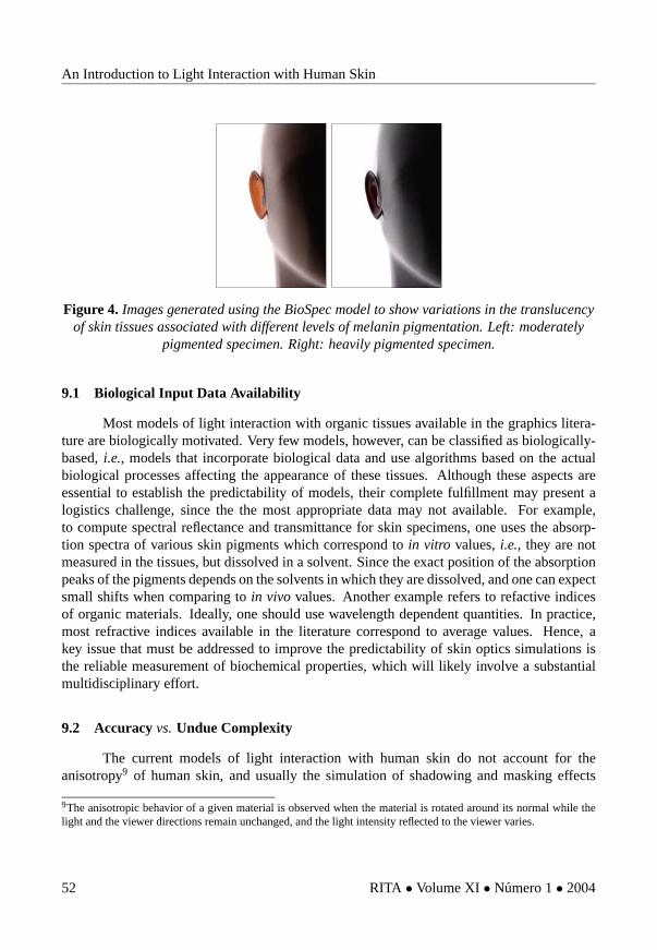
Despite the limitations mentioned above, the scattering and spectral curves generatedby the BioSpec model present a good quantitative and qualitative agreement with measureddata. To date, Biospec is the only computer graphics model of light interaction with humanskin to be evaluated through comparisons with spectral and scattering measured data, makingit one of the few predictive models available in the computer graphics literature. Besides theexperimental evaluation, Krishnaswamy and Baranoski [51] also rendered images to illustratethe applicability of the BioSpec model in the spectral simulation of medical conditions, e.g.,jaundice (yellowish hue) and erythema (redness), associated with changes in the biophysicalparameters, and to highlight an aspect for which measured data is scarce, namely the translu-cency of skin tissues (Figure 4). The transmission of light through the whole skin can beobserved (in vivo) in body parts with a thin or absent hypodermis, such as ears, eye lids andfingers. In these areas the behavior of the transmitted light is near Lambertian, to the pointwhere no internal structure can be noticeable [73].
9 What’s Next?
In this tutorial, we have taken a broad view of the problems related to the simulationsof light interaction with human skin. Many questions, however, remain to be answered. Inthis section, we examine a number of issues related to the improvement and evaluation ofsuch simulations. We also look at more general open problems, their implications and recentdevelopments in applied tissue optics.
RITA • Volume XI • Numero 1 • 2004 51
An Introduction to Light Interaction with Human Skin
Figure 4. Images generated using the BioSpec model to show variations in the translucencyof skin tissues associated with different levels of melanin pigmentation. Left: moderately
pigmented specimen. Right: heavily pigmented specimen.
9.1 Biological Input Data Availability
Most models of light interaction with organic tissues available in the graphics litera-ture are biologically motivated. Very few models, however, can be classified as biologically-based, i.e., models that incorporate biological data and use algorithms based on the actualbiological processes affecting the appearance of these tissues. Although these aspects areessential to establish the predictability of models, their complete fulfillment may present alogistics challenge, since the the most appropriate data may not available. For example,to compute spectral reflectance and transmittance for skin specimens, one uses the absorp-tion spectra of various skin pigments which correspond to in vitro values, i.e., they are notmeasured in the tissues, but dissolved in a solvent. Since the exact position of the absorptionpeaks of the pigments depends on the solvents in which they are dissolved, and one can expectsmall shifts when comparing to in vivo values. Another example refers to refactive indicesof organic materials. Ideally, one should use wavelength dependent quantities. In practice,most refractive indices available in the literature correspond to average values. Hence, akey issue that must be addressed to improve the predictability of skin optics simulations isthe reliable measurement of biochemical properties, which will likely involve a substantialmultidisciplinary effort.
9.2 Accuracy vs. Undue Complexity
The current models of light interaction with human skin do not account for theanisotropy9 of human skin, and usually the simulation of shadowing and masking effects
9The anisotropic behavior of a given material is observed when the material is rotated around its normal while thelight and the viewer directions remain unchanged, and the light intensity reflected to the viewer varies.
52 RITA • Volume XI • Numero 1 • 2004
An Introduction to Light Interaction with Human Skin
is not biologically-based. Although physically-based algorithms for the modeling of thesephenomena with respect to inorganic materials have been proposed by computer graphicsresearchers [30], the generalization of these algorithms to organic materials is not straight-forward since the simulation of the underlying biophysical processes involves the whole hi-erarchy of local illumination, i.e., issues at the microscopic (BRDF) mesoscopic (normal dis-tribution) and macroscopic (geometry) levels need to be addressed. We believe that a carefulmodeling of skin’s geometrical details [85] is essential to achieve a biophysically-based andpredictable solution for these questions.
Accuracy and computational time are often conflicting issues in biophysically-basedrendering. Although the main goal in this area is the design of accurate and efficient models,sometimes it is difficult to obtain this perfect combination. In order to achieve a higher levelof accuracy, it may be necessary to add complexity to a model, which in turn may negativelyaffect its computational performance. However, this is not always the case. For example,light quickly becomes diffuse after the first interactions in the dermal layers (Section 3.2).In this case, a simple cosine distribution would be closer to the actual scattering profile oflight transmitted through these layers than the use of the HGPF. Phase functions, such as theHGPF and its variations, were originally used in tissue subsurface scattering simulations tofit data measured at specific wavelengths [9]. Since then, their application has been extendedto different organic materials despite the lack of supporting measured data and the fact thattheir parameters have no biological meaning. A recent investigation presented by Baranoskiet al. [5] demonstrates that the use of a data oriented approach instead of the HGPF increasesthe accuracy/time ratio of algorithmic subsurface scattering simulations, and contributes totheir predictability since the simulations are not controlled by arcane parameters.
9.3 Evaluation and Comparison Data Availability
Usually models of light interaction with matter are evaluated by visually inspecting theimages generated using such models. Clearly, such an evaluation may be biased by factors notdirectly related to the model. For example, the use of an accurate geometric model of the bodypart being rendered and a careful post-processing spectral tone reproduction [8] may improvethe realistic appearance of rendered skin specimens. These aspects, however, correspond todifferent stages of the rendering pipeline which should be evaluated independently
A current trend is to perform comparisons between model readings and measureddata so that the models can be used in a predictive manner [35]. For example the BioSpecmodel was tested as a separated unit of the rendering pipeline and the results were comparedwith actual measured data [24, 58, 101]. These comparisons were performed using a virtualspectrophotometer and a virtual goniophotometer (Section 2), and reproducing the actualmeasurement conditions as faithfully as possible.
RITA • Volume XI • Numero 1 • 2004 53

An Introduction to Light Interaction with Human Skin
This evaluation approach, however, is bounded by the availability of measured data.As mentioned earlier, skin reflectance and transmittance curves can be found in the biomed-ical literature, but they are usually restricted to a narrow range of measurement conditions.Furthermore, high fidelity comparisons of measured and modeled quantities require bothqualitative and quantitative characterization data for the specimen at hand. Usually only qual-itative characterization data is available for skin specimens used in tissue optics experiments.In terms of goniometric (BRDF and BTDF) curves, despite efforts of computer graphics re-searchers [18, 56, 58, 57], there is still a shortage of data, and the available data sets presentthe same limitations outlined for the reflectance and transmittance data sets. The lack of mea-sured data is even more serious with respect to subsurface scattering since, to the best of ourknowledge, the only relatively reliable data set corresponds to the measurements performedby Bruls and van der Leun [9], which are limited to two wavelengths in the visible range.Therefore, substantial efforts should be also directed towards the reliable measurement ofmultispectral surface and subsurface skin data.
9.4 Biomedical Applications
As mentioned earlier, computer simulations can assist the visual diagnosis of differentskin conditions and diseases [91], including skin tumors [17, 16], and the indirect (non-invasive) measurement of the optical properties of skin tissues. These applications involve thenon-iterative and the iterative use of a theoretical model of light interaction with these tissues[13]. The non-iterative approach consists in inverting the model (Section 4). In an iterativeapproach, the optical properties are implicitly related to measured quantities (e.g., reflectanceand transmittance). The model is then run iteratively until these quantities are matched.Clearly, the predictability of these simulations is tied to the accuracy of the model beingused. This aspect emphasizes the importance of evaluating the models both qualitativelyand quantitatively. This tutorial has shown that no single model is superior in all aspectsinvolved in the simulation of light interaction with human skin, and very few models havebeen properly evaluated. Hence, there is still room for improvement in this area.
Besides tackling the current limitations of the current computer graphics models andimproving their evaluation, it will also be necessary to incorporate time dependent mecha-nisms of photon reemission in their formulation, and to extend their space of parameters tothe non-visible regions of the light spectrum. Important biophysical processes are triggeredby radiation hitting the skin at these wavelengths. For example, ultraviolet light can induceprocesses such as erythema (an abnormal redness of the skin caused by a dilation of theblood vessels followed by an increase in the volume fraction of blood in the dermal layers),melanogenesis (melanin production) and photoaging (discoloration and wrinkle formation)[49, 50, 96, 68]. The ultraviolet electromagnetic radiation (light) can be divided into threeregions: UV-A (ranging from 320nm to 380nm), UV-B (ranging from 280nm to 320nm) and
54 RITA • Volume XI • Numero 1 • 2004

An Introduction to Light Interaction with Human Skin
UV-C (ranging from 100nm to 280nm). The UV-C is mostly absorbed by the ozone layersin the atmosphere. UV-B penetrates deeper than UV-C in skin layers, and it may increasethe melanogenesis after a certain period (6-8 hours) that follows the erythema reaction [50].UV-A penetrates deeper than UV-B, and it can induce epidermal pigmentation immediatelyfollowing exposure [50]. There are only few substances that can absorb UV-A efficiently[80]. Although the lack of UV-B in infants and small children may lead to disruption of bonegrowth and increase the probability of tooth decay [80], overexposure to ultraviolet radiationcan induce the formation of melanomas [26] and carcinomas [68], the first and the secondmost common forms of skin cancer respectively. Melanoma is also the most serious form ofskin cancer since it presents high metastatic potential and low cure rates [26].
10 Conclusion
In this tutorial, we presented an overview of the state of the art in the simulationof light interaction with human skin from a computer graphics perspective. Although thatare several aspects beyond the scope of this work, such as geometrical modeling and tonereproduction, that also affect the realistic appearance of virtual humans, we believe that thepredictability of rendering frameworks can be increased by bring more biological knowledgeand data to bear on the modeling of light interaction with organic materials. Such an approachwill likely extend the range of scientific applications of computer graphics algorithms asappropriately suggested by by Donald Greenberg in his Steve Coons Award speech [34]:
If computer graphics is to have a role in improving the future of our civilization,the real value will be in its application to science, engineering and design.
Acknowledgements
Thanks are due to the anonymous reviewers for their useful insights. The authors arealso grateful for funding through the National Sciences and Engineering Council of Canada(NSERC Grant 213281) and the Canada Foundation for Innovation (CFI Project 6218).
References
[1] S. Alaluf, U. Heinrich, W. Stahl, H. Tronnier, and S. Wiseman. Dietary carotenoids contribute tonormal human skin color and uv photosensitivity. Journal of Nutrition, 132:399–403, 2002.
[2] R.R. Anderson and J.A. Parrish. The optics of human skin. Journal of Investigative Dermatology,77(1):13–19, 1981.
RITA • Volume XI • Numero 1 • 2004 55

An Introduction to Light Interaction with Human Skin
[3] Elli Angelopoulou. Understanding the color of human skin. In Human Vision and ElectronicImaging VI, pages 243–251. SPIE, vol. 4299, 2001.
[4] S. Aydinli and H. Kaase. Measurement of luminous characteristics of daylighting materials.Technical Report IEA SHCP TASK 21 / ECBCS ANNEX 29, Institute of Electronics and Light-ing Technology, Technical University of Berlin, September 1999.
[5] G.V.G. Baranoski, A. Krishnaswamy, and B. Kimmel. Revisiting the foundations of subsurfacescattering. Technical Report CS-2003-45, School of Computer Science, University of Waterloo,December 2003.
[6] G.V.G. Baranoski and J.G. Rokne. An algorithmic reflectance and transmittance model for planttissue. Computer Graphics Forum (EUROGRAPHICS Proceedings), 16(3):141–150, September1997.
[7] G.V.G. Baranoski, J.G. Rokne, and G. Xu. Virtual spectrophotometric measurements for biolog-ically and physically-based rendering. The Visual Computer, 17(8):506–518, 2001.
[8] I.E. Bell and G.V.G. Baranoski. More than RGB: moving toward spectral color reproduction.ACM SIGGRAPH Course Notes, San Diego, CA, July 2002. Course 24.
[9] W.A.G. Bruls and J.C. van der Leun. Forward scattering properties of human epidermal layers.Photochem. Photobiol., 40:231–242, 1984.
[10] R.L. Burden and J.D. Faires. Numerical Analysis. PWS-KENT Publishing Company, Boston,fifth edition, 1993.
[11] S. Chandrasekhar. Radiative Transfer. Dover Publications Inc., New York, 1960.
[12] M.R. Chedekel. Photophysics and photochemistry of melanin. In M.R. Chedekel L. Zeise andT.B. Fitzpatrick, editors, Melanin: Its Role in Human Photoprotection, pages 11–22, OverlandPark, Kansas, USA, 1995. Valdenmar Publishing Company. 2223b.
[13] W. Cheong, S.A. Prahl, and A.J. Welch. A review of the optical properties of biological tissues.IEEE Journal of Quantum Electronics, 26(12):2166–2185, December 1990.
[14] D.Y. Churmakov, I.V. Meglinsky, S.A. Piletsky, and D.A. Greenhalgh. Analysis of skin tissuesspatial fluorescence distribution by the Monte Carlo simulation. Journal of Physics D: AppliedPhysics, 36:1722–1728, July 2003.
[15] R.L. Cook and K.E. Torrance. A reflectance model for computer graphics. ACM Transactionson Graphics, 1(1):7–24, January 1982.
[16] S.D. Cotton. A noninvasive skin imaging system. Technical Report CSR-97-03, School ofComputer Science, The University of Birmingham, 1997.
[17] S.D. Cotton and E. Claridge. Developing a predictive model of skin colouring. In SPIE Vol.2708, Medical Imaging 1996, pages 814–825, 1996.
[18] K.J. Dana, B. van Ginneken, S.K. Nayar, and J.J. Koenderink. Reflectance and texture of realworld surfaces. ACM Transactions on Graphics, 18(1):1–34, 1999.
[19] B.L. Diffey. A mathematical model for ultraviolet optics in skin. Physics in Medicine andBiology, 28(6):647–657, 1983.
56 RITA • Volume XI • Numero 1 • 2004

An Introduction to Light Interaction with Human Skin
[20] M. Doi and S. Tominaga. Spectral estimation of human skin color using the Kubelka-Munktheory. In SPIE/IS&T Electronic Imaging, pages 221–228. SPIE, vol. 5008, 2003.
[21] R.M.P. Doornbos, R. Lang, M.C. Aalders, F.W. Cross, and H.J.C.M. Sterenborg. The deter-mination of in vivo human tissue optical properties and absolute chromophore concentrationsusing spatially resolved steady-state diffuse reflectance spectroscopy. Physics in Medicine andBiology, 44:967–981, 1999.
[22] ASTM Standard E284-91C. Standard terminology of appearance. In L.B. Wolff, S.A. Shafer,and G.E. Healey, editors, Physics-Based Vision Principles and Practice: Radiometry, pages146–161, Boston, 1992. Jones and Bartlett Publishers.
[23] G. Eason, A. Veitch, R. Nisbet, and F. Turnbull. The theory of backscattering of light by blood.Journal of Physics, 11:1463–1479, 1978.
[24] M.A. Everett, E. Yeargers, R.M. Sayre, and R.L. Olsen. Penetration of epidermis by ultravioletrays. Photochemistry and Photobiology, 5:533–542, 1966.
[25] T.J. Farell, M.S. Patterson, and B. Wilson. A diffusion theory model of spatially resolved, steady-state diffuse reflectance for the noninvasive determination of tissue optical properties in vivo.Medical Physics, 19:879–888, 1992.
[26] T.B. Fitzpatrick and J.L. Bolognia. Human melanin pigmentation: role in pathogenesis of cuta-neous melanoma. In M.R. Chedekel L. Zeise and T.B. Fitzpatrick, editors, Melanin: Its Role inHuman Photoprotection, pages 177–182, Overland Park, Kansas, USA, 1995. Valdenmar Pub-lishing Company.
[27] R. Flewelling. Noninvasive optical monitoring. In J.D. Bronzino, editor, The Biomedical Engi-neering Handbook, pages 1–11, Boca Raton, FL, USA, 1981. IEEE Press. Section 86.
[28] Stephen T. Flock, Michael S. Patterson, Brian C. Wilson, and Douglas R. Wyman. Monte Carlomodeling of light propagation in highly scattering tissues - I: Model predictions and compari-son with diffusion theory. IEEE Transactions on Biomedical Engineering, 36(12):1162–1168,December 1989.
[29] S.C. Foo. A gonioreflectometer for measuring the bidirectional reflectance of material for use inillumination computation. Master’s thesis, Cornell University, August 1997.
[30] A. Fournier. From local to global illumination and back. In P. M. Hanrahan and W. Purgathofer,editors, Rendering Techniques’95 (Proceedings of the Sixth Eurographics Rendering Workshop),pages 127–136, Dublin, June 1995. Springer-Verlag.
[31] T. Fourty, F. Baret, S. Jacquemoud, G. Schmuck, and J. Verdebout. Leaf optical propertieswith explicit description of its biochemical composition: Direct and inverse problems. RemoteSensing of Environment, 56:104–117, 1996.
[32] K. Furutso. Diffusion equation derived from space-time transport equation. Journal of theOptical Society of America, 70:360, 1980.
[33] A.S. Glassner. Principles of Digital Image Synthesis. Morgan Kaufmann Publishers, Inc, SanFrancisco, 1995.
[34] D.P. Greenberg. 1987 Steven A. Coons Award Lecture. Computer Graphics, 22(1):7–14, Febru-ary 1988.
RITA • Volume XI • Numero 1 • 2004 57

An Introduction to Light Interaction with Human Skin
[35] D.P. Greenberg, J. Arvo, E. Lafortune, K.E. Torrance, J.A. Ferwerda, B. Walter, B. Trumbore,P. Shirley, S. Pattanaik, and S. Foo. A framework for realistic image synthesis. In SIGGRAPH,Annual Conference Series, pages 477–494, 1997.
[36] P. Hanrahan and W. Krueger. Reflection from layered surfaces due to subsurface scattering. InSIGGRAPH, Annual Conference Series, pages 165–174, August 1993.
[37] A. Haro, B. Guenter, and I. Essa. Real-time, photo-realistic, physically based rendering of finescale human skin structure. In P. M. Hanrahan and W. Purgathofer, editors, Rendering Tech-niques’2001 (Proceedings of the 12th Eurographics Rendering Workshop), pages 53–62, Lon-don, June 2001. Springer-Verlag.
[38] L.G. Henyey and J.L. Greenstein. Diffuse radiation in the galaxy. Astrophysics Journal, 93:70–83, 1941.
[39] R.S. Hunter and R.W. Harold. The Measurement of Appearance. John Wiley & Sons, New York,second edition, 1987.
[40] A. Ishimaru. Wave Propagation and Scattering in Random Media, volume 1. IEEE Press, NewYork, 2nd edition, 1978.
[41] S. Jacquemoud and S.L. Ustin. Leaf optical properties: A state of the art. In 8th InternationalSymposium of Physical Measurements & Signatures in Remote Sensing, pages 223–332, Aussois,France, January 2001. CNES.
[42] S.L. Jacques. Origins of tissue optical properties in the uva visible and nir regions. OSA TOPSon Advances in Optical Imaging and Photon Migration, 2:364–369, 1996.
[43] S.L. Jacques, C.A. Alter, and S.A. Prahl. Angular dependence of HeNe laser light scattering byhuman dermis. Lasers in Life Sciences, 1(4):309–333, 1987.
[44] H.W. Jensen and J. Buhler. A rapid hierarchical rendering technique for translucent materials. InSIGGRAPH, Annual Conference Series, pages 576–581, July 2002.
[45] H.W. Jensen, S.R. Marschner, M. Levoy, and P. Hanrahan. A practical model for subsurface lighttransport. In SIGGRAPH, Annual Conference Series, pages 511–518, August 2001.
[46] Z. Jin and K. Stammes. Radiative transfer in nonuniformly refracting layered media:atmosphere-ocean system. Applied Optics, 33(3):431–442, January 1994.
[47] D.B. Judd and G. Wyszecki. Color in Business, Science and Industry. John Wiley & Sons, NewYork, third edition, 1975.
[48] K.F. Karner, H. Mayer, and M. Gervautz. An image based measurement system for anisotropicreflection. Computer Graphics Forum (EUROGRAPHICS Proceedings), 15(3):119–128, August1996.
[49] G. Kelfkens and J.C van der Leun. Skin temperature changes after irradiation with UVB orUVC: implications for the mechanism underlying ultraviolet erythema. Physics in Medicine andBiology, 34(5):599–608, 1989.
[50] N. Kollias. The spectroscopy of human melanin pigmentation. In M.R. Chedekel L. Zeise andT.B. Fitzpatrick, editors, Melanin: Its Role in Human Photoprotection, pages 31–38, OverlandPark, Kansas, USA, 1995. Valdenmar Publishing Company.
58 RITA • Volume XI • Numero 1 • 2004

An Introduction to Light Interaction with Human Skin
[51] A. Krishnaswamy and G.V.G. Baranoski. A biophysically-based spectral model of light interac-tion with human skin. Computer Graphics Forum (EUROGRAPHICS Proceedings), 2004. Toappear.
[52] A. Krishnaswamy, G.V.G. Baranoski, and Jon G. Rokne. Improving the reliability/cost ratio ofgoniophotometric measurements. Journal of Graphics Tools, 9(3):31–51, 2004.
[53] P. Kubelka and F. Munk. Ein beitrag zur optik der farbanstriche. Zurich Tech. Physik, 12:543,1931.
[54] R. Lee, M.M. Mathews-Roth, M.A. Pathak, and J.A. Parrish. The detection of carotenoid pig-ments in human skin. Journal of Investigative Dermatology, 64:175–177, 1975.
[55] S.R. Marschner, H.W. Jensen, M. Cammarano, S. Worley, and P. Hanrahan. Light scatteringfrom human hair fibers. ACM Transactions on Graphics, 22(3):780–791, 2003.
[56] S.R. Marschner, S. H. Westin, E.P.F. Lafortune, K.E. Torrance, and D.P. Greenberg. Image-based brdf measurement. Technical Report PCG-99-1, Program of Computer Graphics, CornellUniversity, USA, January 1999.
[57] S.R. Marschner, S. H. Westin, E.P.F. Lafortune, K.E. Torrance, and D.P. Greenberg. Image-basedbrdf measurement including human skin. In D. Lischinski and G. W. Larson, editors, RenderingTechniques’1999 (Proceedings of the 10th Eurographics Rendering Workshop), pages 119–130,Granada, June 1999. Springer-Verlag.
[58] S.R. Marschner, S. H. Westin, E.P.F. Lafortune, K.E. Torrance, and D.P. Greenberg. Reflectancemeasurements of human skin. Technical Report PCG-99-2, Program of Computer Graphics,Cornell University, USA, January 1999.
[59] I.V. Meglinsky and S.J. Matcher. Modelling the sampling volume for skin blood oxygenation.Medical & Biological Engineering & Computing, 39:44–49, 2001.
[60] I.V. Meglinsky and S.J. Matcher. Computer simulation of the skin reflectance spectra. ComputerMethods and Programs in Biomedicine, 70:179–186, 2003.
[61] N. Metropolis and S. Ulam. The Monte Carlo method. Journal of the American StatisticalAssociation, 44(247):335–341, September 1949.
[62] J.R. Mourant, J.P. Freyer, A.H. Hielscher, A.A. Eick, D. Shen, and T.M. Johnson. Mechanisms oflight scattering from biological cells relevant to noninvasive optical-tissue diagnostics. AppliedOptics, 37(16):3586–3593, June 1998.
[63] H. Nakai, Y. Manabe, and S. Inokuchi. Simulation analysis of spectral distributions of humanskin. In 14th International Conference on Pattern Recognition, pages 1065–1067, 1998.
[64] C.S. Ng and L. Li. A multi-layered reflection model of natural human skin. In Computer Graph-ics International 2001, pages 249–256, Hong Kong, July 2001.
[65] F.E. Nicodemus, J.C. Richmond, J.J. Hsia, I.W. Ginsberg, and T. Limperis. Geometrical consid-erations and nomenclature for reflectance. In L.B. Wolff, S.A. Shafer, and G.E. Healey, editors,Physics-Based Vision Principles and Practice: Radiometry, pages 94–145, Boston, 1992. Jonesand Bartlett Publishers.
RITA • Volume XI • Numero 1 • 2004 59

An Introduction to Light Interaction with Human Skin
[66] M. Nischik and C. Forster. Analysis of skin erythema using true-color images. IEEE Transac-tions on Medical Imaging, 16(6):711–716, December 1997.
[67] D. Parsad, K. Wakamatsu, A.J. Kanwar, B. Kumar, and S. Ito. Eumelanin and phaeomelanin con-tents of depigmented and repigmented skin in vitiligo patients. British Journal of Dermatology,149:624–626, 2003.
[68] M.A. Pathak. Functions of melanin and protection by melanin. In M.R. Chedekel L. Zeise andT.B. Fitzpatrick, editors, Melanin: Its Role in Human Photoprotection, pages 125–134, OverlandPark, Kansas, USA, 1995. Valdenmar Publishing Company.
[69] S.A. Prahl. Light Transport in Tissue. PhD thesis, The University of Texas at Austin, TX, USA,December 1988.
[70] S.A. Prahl, M. Keijzer, S.L. Jacques, and A.J. Welch. A Monte Carlo model of light propagationin tissue. SPIE Institute Series, IS 5:102–111, 1989.
[71] S.A. Prahl, M.J.C. van Gemert, and A.J. Welch. Determining the optical properties of turbidmedia using the adding-doubling method. Applied Optics, 32(4):559–568, 1993.
[72] S. Premoze. Analytic light transport approximations for volumetric materials. In Proceedingsof the 10th Pacific Conference on Computer Graphics and Applications - Pacific Graphics’2002,pages 48–57, Beijing, October 2002.
[73] J. Rodriguez, I.V. Yaroslavsky, A.N. Yaroslavsky, H. Battarbee, and V.V. Tuchin. Time-resolvedimaging in diffusive media. In V.V. Tuchin, editor, Handbook of Optical Biomedical Diagnostics,pages 357–404, Bellingham, WA, USA, 2002. SPIE Press.
[74] D.K. Sardar and L.B. Levy. Optical properties of whole blood. Lasers in Medical Science,13:106–111, 1998.
[75] M. Shimada, Y. Yamada, M. Itoh, and T. Yatagai. Melanin and blood concentration in humanskin studied by multiple regression analysis: assessment by Monte Carlo simulation. Physics inMedicine and Biology, 46:2397–2406, 2001.
[76] A.N. Shiryaev. Probability. Springer-Verlag, New York, second edition, 1996.
[77] C.R. Simpson, M. Kohl, M. Essenpreis, and M. Cope. Near infrared optical properties of ex-vivo human skin and subcutaneous tissues measured using the Monte Carlo inversion technique.Physics in Medicine and Biology, 43:2465–2478, 1998.
[78] J. Stam. An illumination model for a skin layer bounded by rough surfaces. In P. M. Hanrahanand W. Purgathofer, editors, Rendering Techniques’2001 (Proceedings of the 12th EurographicsRendering Workshop), pages 39–52, London, June 2001. Springer-Verlag.
[79] K. Stamnes and P. Conklin. A new multi-layer discrete ordinate approach to radiative trans-fer in vertically inhomogeneous atmospheres. Journal of Quantum Spectroscopy and RadiativeTransfer, 31(3):273–282, 1984.
[80] K. Stanzl and L. Zastrow. Melanin: an effective photoprotectant against UV-A rays. InM.R. Chedekel L. Zeise and T.B. Fitzpatrick, editors, Melanin: Its Role in Human Photopro-tection, pages 59–63, Overland Park, Kansas, USA, 1995. Valdenmar Publishing Company.
60 RITA • Volume XI • Numero 1 • 2004

An Introduction to Light Interaction with Human Skin
[81] J.M. Steinke and A.P. Shepherd. Diffusion model of the optical absorbance of whole blood.Journal of the Optical Society of America, 5(6):813–822, 1988.
[82] Y. Su, W. Wang, K. Xu, and C. Jiang. The optical properties of skin. In Optics in Health Careand Biomedical Optics: Diagnostics and Treatment, pages 299–304. SPIE, vol. 4916, 2002.
[83] J.L. Swerdlow. Unmasking skin. National Geographic, pages 36–63, November 2002.
[84] P.S. Talreja, G.B. Kasting, N.K. Kleene, W.L. Pickens, and T. Wang. Visualization of the lipidbarrier and measurement of lipid pathlength in human stratum corneum. AAPS PharmSCi,3(2):1–9, 2001.
[85] N. Thalmann, P. Kalra, J.L. Leveque, R. Bazin, D. Batisse, and B. Querleux. A computa-tional skin model: fold and wrinkle formation. IEEE Transactions on Information Technology inBiomedicine, 6(4):317–323, 2002.
[86] A.J. Thody, E.M. Higgins, K. Wakamatsu, S. Ito, S.A. Burchill, and J.M. Marks. Pheomelaninas well as eumelanin is present in human dermis. Journal of Investigative Dermatology, 97:340–344, 1991.
[87] K.E. Torrance and E.M. Sparrow. Theory for off-specular reflection from roughned surfaces.Journal of the Optical Society of America, 57(9):1105–1114, 1967.
[88] T.S. Trowbridge and K.P. Reitz. Average irregularity representation of a rough surface for rayreflection. Journal of the Optical Society of America, 65(5):531–536, May 1975.
[89] N. Tsumura, M. Kawabuchi, H. Haneishi, and Y. Miyabe. Mapping pigmentation in human skinby multi- visible-spectral imaging by inverse optical scattering technique. In IS&T/SID EighthColor Imaging Conference, pages 81–84, 2000.
[90] N. Tsumura, N. Ojima, K. Sato, M. Shiraishi, H. Shimizu, H. Nabeshima, S. Akazaki, K. Hori,and Y. Miyake. Image-based skin color and texture analysis/synthesis by extracting hemoglobinand melanin information in the skin. In SIGGRAPH, Annual Conference Series, 2003.
[91] V.V. Tuchin. Tissue Optics Light Scattering Methods and Instruments for Medical Diagnosis.The International Society for Optical Engineering, Bellingham, WA, USA, 2000.
[92] V.V. Tuchin, S.R. Utz, and I.V. Yaroslavsky. Tissue optics, light distribution, and spectroscopy.Optical Engineering, 33:3178–3188, 1994.
[93] J.V. Uspensky. Introduction to Mathematical Probability. McGraw-Hill, New York, 1937.
[94] H.C. van de Hulst. Multiple Light Scattering: Tables, Formulas, and Applications, volume 1.Academic Press, New York, 1980.
[95] H.C. van de Hulst. Light Scattering by Small Particles. Dover Publications Inc., New York, 2ndedition, 1981.
[96] J.C. van der Leun. Ultraviolet Erythema. PhD thesis, University of Utrecht, The Netherlands,1966.
[97] M.J.C. van Gemert, S.L. Jacques, H.J.C.M. Sterenborg, and W.M. Star. Skin optics. IEEETransactions on Biomedical Engineering, 36(12):1146–1154, 1989.
[98] M.J.C. van Gemert and W.M. Star. Relations between the Kubelka-Munk and the transportequation models for anisotropic scattering. Laser in the Life Sciences, 1(4):287–298, 1987.
RITA • Volume XI • Numero 1 • 2004 61

An Introduction to Light Interaction with Human Skin
[99] M.J.C. van Gemert, A.J. Welch, and W.M. Star. Tissue optics for a slab geometry in diffusionapproximation. Laser in the Life Sciences, 2:295–302, 1987.
[100] B. van Ginneken, M. Stavridi, and J.J. Koenderink. Diffuse and specular reflectance from roughsurfaces. Applied Optics, 37(1):130–139, 1998.
[101] M.J. Vrhel, R. Gershon, and L.S. Iwan. Measurement and analysis of object reflectance spectra.Color Research and Application, 19(1):4–9, 1994.
[102] S. Wan, R. Anderson, and J.A. Parrish. Analytical modeling for the optical properties of the skinwith in vitro and in vivo appplications. Photochemistry and Photobiology, 34:493–499, 1981.
[103] L. Wang and S.L. Jacques. Hybrid method of Monte Carlo simulation and diffusion theory forlight reflectance by turbid media. Optical Society of America, 10(8):1746–1752, 1995.
[104] G.J. Ward. Measuring and modeling anisotropic reflection. Computer Graphics (SIGGRAPHProceedings), pages 265–272, July 1992.
[105] B.C. Wilson and G. Adam. A Monte Carlo model for the absorption and flux distributions oflight in tissue. Medical Physics, 10:824–830, 1983.
[106] A.N. Yaroslavsky, A.V. Priezzhev, J. Rodriguez, I.V. Yaroslavsky, and H. Battarbee. Optics ofblood. In V.V. Tuchin, editor, Handbook of Optical Biomedical Diagnostics, pages 169–216,Bellingham, WA, USA, 2002. SPIE Press.
[107] A.N. Yaroslavsky, S.R. Utz, S.N. Tatarintsev, and V.V. Tuchin. Angular scattering properties ofhuman epidermal layers. In Human Vision and Electronic Imaging VI, pages 38–41. SPIE, vol.2100, 1994.
[108] G. Yoon. Absorption and Scattering of Laser Light in Biological Media - Mathematical Modelingand Methods for Determining Optical Properties. PhD thesis, University of Texas at Austin,USA, 1988.
[109] G. Yoon, S.A. Prahl, and A.J. Welch. Accuracies of the diffusion approximation and its similarityrelations for laser irradiated biological media. Applied Optics, 28(12):2250–2255, 1989.
[110] G. Yoon, A.J. Welch, M. Motamedi, and M.C.J. van Gemert. Development and application ofthree-dimensional light distribution model for laser irradiated tissue. IEEE Journal of QuantumElectronics, QE-23:1721–1733, 1987.
[111] G. Zonios, J. Bykowsky, and N. Kollias. Skin melanin, hemoglobin, and light scattering prop-erties can be quantitatively assessed in vivo using diffuse reflectance spectroscopy. Journal ofInvestigative Dermatology, 117(6):1452–1457, 2001.
62 RITA • Volume XI • Numero 1 • 2004
Related Documents