Article An Intrinsically Disordered Peptide from Ebola Virus VP35 Controls Viral RNA Synthesis by Modulating Nucleoprotein-RNA Interactions Graphical Abstract Highlights d The minimum Ebola virus VP35 peptide that binds nucleoprotein is defined d The structure of the VP35 peptide/N-terminal nucleoprotein complex is determined d A role for VP35 peptide in viral RNA synthesis is defined d A possible framework to target the VP35/nucleoprotein interface is defined Authors Daisy W. Leung, Dominika Borek, ..., Christopher F. Basler, Gaya K. Amarasinghe Correspondence [email protected] In Brief Ebola virus RNA synthesis is a tightly controlled process. Leung et al. reveal how a peptide derived from Ebola VP35 protein impacts viral RNA synthesis by modulating interactions between Ebola virus nucleoprotein and RNA. Accession Numbers 4YPI Leung et al., 2015, Cell Reports 11, 376–389 April 21, 2015 ª2015 The Authors http://dx.doi.org/10.1016/j.celrep.2015.03.034

Welcome message from author
This document is posted to help you gain knowledge. Please leave a comment to let me know what you think about it! Share it to your friends and learn new things together.
Transcript
Article
An Intrinsically Disordered
Peptide from Ebola VirusVP35 Controls Viral RNA Synthesis by ModulatingNucleoprotein-RNA InteractionsGraphical Abstract
Highlights
d The minimum Ebola virus VP35 peptide that binds
nucleoprotein is defined
d The structure of the VP35 peptide/N-terminal nucleoprotein
complex is determined
d A role for VP35 peptide in viral RNA synthesis is defined
d A possible framework to target the VP35/nucleoprotein
interface is defined
Leung et al., 2015, Cell Reports 11, 376–389April 21, 2015 ª2015 The Authorshttp://dx.doi.org/10.1016/j.celrep.2015.03.034
Authors
Daisy W. Leung, Dominika Borek, ...,
Christopher F. Basler,
Gaya K. Amarasinghe
In Brief
Ebola virus RNA synthesis is a tightly
controlled process. Leung et al. reveal
how a peptide derived from Ebola VP35
protein impacts viral RNA synthesis by
modulating interactions between Ebola
virus nucleoprotein and RNA.
Accession Numbers
4YPI

Cell Reports
Article
An Intrinsically Disordered Peptidefrom Ebola Virus VP35 Controls Viral RNA Synthesisby Modulating Nucleoprotein-RNA InteractionsDaisy W. Leung,1,7 Dominika Borek,2,7 Priya Luthra,3 Jennifer M. Binning,1 Manu Anantpadma,4 Gai Liu,1 Ian B. Harvey,1
Zhaoming Su,5 Ariel Endlich-Frazier,3 Juanli Pan,1 Reed S. Shabman,3,6 Wah Chiu,5 Robert A. Davey,4
Zbyszek Otwinowski,2 Christopher F. Basler,3 and Gaya K. Amarasinghe1,*1Department of Pathology and Immunology, Washington University School of Medicine, St Louis, MO 63110, USA2Departments of Biophysics and Biochemistry and Center for Structural Genomics of Infectious Diseases, University of Texas Southwestern
Medical Center at Dallas, Dallas, TX 75390, USA3Department of Microbiology, Icahn School of Medicine at Mount Sinai, New York, NY 10029, USA4Department of Virology and Immunology, Texas Biomedical Research Institute, San Antonio, TX 78227, USA5National Center for Macromolecular Imaging, Verna and Marrs McLean Department of Biochemistry and Molecular Biology, Baylor Collegeof Medicine, Houston, TX 77030, USA6Present address: Virology Group, J. Craig Venter Institute, Rockville, MD 20850, USA7Co-first author
*Correspondence: [email protected]://dx.doi.org/10.1016/j.celrep.2015.03.034
This is an open access article under the CC BY license (http://creativecommons.org/licenses/by/4.0/).
SUMMARY
During viral RNA synthesis, Ebola virus (EBOV)nucleoprotein (NP) alternates between an RNA-tem-plate-bound form and a template-free form toprovide the viral polymerase access to the RNAtemplate. In addition, newly synthesized NP mustbe prevented from indiscriminately binding tononcognate RNAs. Here, we investigate the molec-ular bases for these critical processes. We identifyan intrinsically disordered peptide derived fromEBOV VP35 (NPBP, residues 20–48) that binds NPwith high affinity and specificity, inhibits NP oligo-merization, and releases RNA from NP-RNA com-plexes in vitro. The structure of the NPBP/DNPNTD
complex, solved to 3.7 A resolution, reveals howNPBP peptide occludes a large surface area thatis important for NP-NP and NP-RNA interactionsand for viral RNA synthesis. Together, our resultsidentify a highly conserved viral interface that isimportant for EBOV replication and can be targetedfor therapeutic development.
INTRODUCTION
Ebolaviruses and marburgviruses are nonsegmented negative-
sense RNA viruses (NNSVs) that cause severe hemorrhagic fever
(Sanchez et al., 2006). Because of the severity of filovirus dis-
ease, which is associated with case fatality rates approaching
90% during some outbreaks, ebolaviruses and marburgviruses
remain significant threats to global human health. The recent
epidemic in Western Africa caused by Ebola virus (EBOV)
Makona variant is the largest filovirus outbreak on record, and
376 Cell Reports 11, 376–389, April 21, 2015 ª2015 The Authors
the subsequent import of EBOV to non-African countries high-
lights the public health impact of these zoonotic pathogens.
Like other non-segmented negative-strand virus (NNSV) fam-
ily members, EBOV has a single-stranded RNA (ssRNA) genome
that undergoes transcription upon entry into the host cytosol
prior to the generation of viral proteins (Muhlberger, 2007; San-
chez et al., 2006). The roughly 19-kb ebolavirus genome has
seven genes that encode for at least eight distinct translation
products (Sanchez and Kiley, 1987; Sanchez et al., 1993,
2007; Shabman et al., 2014). Viral genome replication and
transcription of individual genes into distinct 50-capped, 30-poly-adenylated monocistronic mRNAs are carried out by the viral
RNA-dependent RNA polymerase complex (RdRp) (Muhlberger,
2007). This complex consists of the enzymatic component of the
RDRP, the large protein (L), along with the viral nucleoprotein
(NP), viral protein (VP)35 and VP30, and acts upon the nucleo-
capsid, which consists of single-stranded viral genomic or anti-
genomic RNAs (Muhlberger, 2007) that are encapsidated by NP.
The EBOV core nucleocapsid protects ssRNAs from degrada-
tion, similar to that observed for paramyxoviruses and rhabdovi-
ruses (Masters and Banerjee, 1988).
Based on sequence homology, the filovirus NP has a para-
myxovirus/filovirus conserved region spanning residues 1 to
450 and a region unique to filoviruses from 451 to 739 (Shi
et al., 2008; Watanabe et al., 2006) (Figures 1A and S1A). The
N-terminal region mediates NP self-association and forms heli-
cal tube-like structures in vitro (Watanabe et al., 2006) and in
cells (Huang et al., 2002). Mapping studies indicate that the NP
C terminus contributes to NP-VP40 interaction and NP incorpo-
ration into ebolavirus-like particles (Noda et al., 2007). Recently,
a crystal structure of the EBOV NP C-terminal residues encom-
passing 641–739 was described (Dziuba�nska et al., 2014), but
how this structure contributes to EBOV infection is unknown.
Viral polymerase cofactor VP35 is a suppressor of innate im-
mune signaling and is also critical for viral RNA synthesis (Feng
et al., 2007; Luthra et al., 2013; Prins et al., 2009; Schumann
et al., 2009). Its role in RNA synthesis is thought to be functionally
analogous to the P protein in other NNSVs (Sanchez et al., 2006).
VP35 has anN-terminal oligomerization domain and aC-terminal
interferon inhibitory domain (IID), which binds double-stranded
RNA (dsRNA) and is critical for innate immune inhibition (Basler
et al., 2003; Hartman et al., 2004; Leung et al., 2009, 2010b).
The N-terminal oligomerization domain is thought to participate
in protein-protein interactions, including VP35 self-association
(Moller et al., 2005; Reid et al., 2005) as well as association
with the viral polymerase L (Trunschke et al., 2013). VP35 oligo-
merization is also important for RNA synthesis (Moller et al.,
2005), but it appears to be dispensable for VP35/NP co-localiza-
tion (Moller et al., 2005).
Despite previous efforts, it is unclear how VP35-NP interac-
tionsmodulate EBOVRNA synthesis. Here, we describe an inter-
action between the EBOV VP35 and NP proteins that critically
impacts NP function and EBOV RNA synthesis.
RESULTS
The N Terminus of NP Is Important for OligomerizationPrevious deletion analysis of EBOV NP revealed that three
truncations (residues 2–150, 151–300, and 301–450) resulted in
loss of NP function in the viral RDRP complex (Watanabe et al.,
2006). In order to identify specific residues involved in NP-NP as-
sociation, we generated a series of truncation constructs (Fig-
ure S1A), which were evaluated in both low- (150 mM NaCl)
and high-salt (500 mM NaCl) buffers. All constructs tested,
including NP 1–457 and 25–457, formed large homo-oligomeric
complexes as NP elutes in the void volume in buffer containing
150mMNaCl (Figure 1B). In buffer with 500mMNaCl, many con-
structs displayed behavior similar to those in 150 mM NaCl
except for NP 25–457 (DNPNTD), where some protein eluted
in the included volume and some near the void volume (V0),
representing a multimeric population that is likely an aggregate
(Figure 1C). Moreover, we observed >60% monodispersed
(well-behaved) DNPNTD protein in solution at concentrations
>10 mg/ml based on multiangle light scattering (MALS) studies.
Results from finer truncations (i.e., two- to three-residue dele-
tions) within the first 24 residues suggested a critical role for res-
idues 21–22 as proteins starting at residue 23 eluted in the
included volume. In order to further evaluate the impact of Y21
andH22, we generated singlemutants (Y21AorH22A) and a dou-
ble mutant (Y21A/H22A) of NP 1–457. Of these NP mutants, only
the Y21A/H22A double mutant eluted in the included volume and
the peak displayed low levels of polydispersity (Figure 1D). These
results further support an important role for the NP paramyxo-
virus/filovirus conserved region (NP residues 1–457) in NP-NP
self-association.
The N and C Termini of NP Are Required for Viral RNASynthesisIn order to further test the role of NP-NP self-association in viral
RNA synthesis, we tested N- and C-terminal truncations of NP
using a minigenome (MG) assay that uses a model Ebola virus
genomic RNA encoding Renilla luciferase as a template for tran-
scription and replication by a viral polymerase complex reconsti-
tuted by expression of NP, VP35, VP30, and L (Muhlberger,
2007; Muhlberger et al., 1999). In these assays, mutation of
Y21A/H22A in the context of the full-length NP and the 20–739
NP construct resulted in a complete loss of function (Figures
1E and S1B). Deletion of the first 19 residues of NP (NP 20–
739) results in a partial loss in activity (�60%), suggesting that
these residues are important, but not essential, for activity. In
contrast, C-terminal deletion mutants, such as NP 1–550 or NP
1–601, were severely impaired (<10% of wild-type [WT] activity)
in the MG assay (Figure 1E).
We tested three additional EBOV NP constructs, NP 25–739,
NP 25–550, and NP 1–550, for their ability to support MG activity
in the presence and absence of WT NP (Figures 1F and S1C).
Expression of N-terminal NP truncations, such as NP 25–739
and NP 25–550, did not affect the MG function of NP. However,
a C-terminal truncation construct that retained the entire N termi-
nus (NP 1–550) and the ability to oligomerize displayed domi-
nant-negative activity, suggesting that interactions through the
N terminus limit the MG activity of the WT NP (1–739). Collec-
tively these results support a role of both the NP N and C termini
during viral RNA synthesis.
NPBP Peptide from VP35 Is Necessary and Sufficient toBind NPEBOV NP associates with EBOV VP35 in the context of the repli-
cation complex, and this interaction is thought to play a major
role during viral RNA synthesis (Elliott et al., 1993; Huang et al.,
2002). Previous analyses of EBOV VP35 identified several re-
gions that are highly conserved among filoviruses, including an
N-terminal oligomerization domain and a C-terminal IID (Fig-
ure 2A) (Hartman et al., 2004; Leung et al., 2010a; Reid et al.,
2005). However, sequence analysis of ebolavirus VP35s re-
vealed an additional, highly conserved region N-terminal to the
previously predicted oligomerization domain (Figure S1D) (Reid
et al., 2005). To determine whether this region in VP35 is required
to support viral RNA synthesis, we generated a series of VP35
truncated constructs, where we systematically deleted ten-
residue segments from the N terminus and evaluated the role
of these residues in viral RNA synthesis using the MG assay.
The resulting data revealed that the first 19 residues are largely
dispensable for MG activity (Figure 2B). In contrast, residues
20–52 are critical for EBOV MG activity, as N-terminal trunca-
tions that extend into this conserved region, including trunca-
tions such as in the 30–340 construct, resulted in >60% loss of
activity relative to WT VP35 (residues 1-340). Deletion of the
entire NPBP region plus the first 19 amino acids results in
near-complete loss of MG activity, suggesting that the entire
NPBP is required for activity.
In order to determine the molecular basis for this observa-
tion, we next tested interactions between VP35 and NP using
in vitro binding assays and various VP35 and NP truncations.
These results revealed that EBOV DNPNTD was sufficient for
high-affinity binding to the EBOV VP35 N terminus. These
studies also revealed that VP35-NP association is coordinated,
at least in part, through the VP35 N terminus, as VP35 1–168
binds NP whereas VP35 58–340 does not (Figures 2C and
S2A). In order to further characterize this interaction, we gener-
ated a series of peptides spanning VP35 residues 20–52.
Cell Reports 11, 376–389, April 21, 2015 ª2015 The Authors 377

A1 25 739
NP
oligomerization
641CTDNTD
E
F
C
% m
inig
enom
e ac
tivity
NP
1-73
9
NP
25-7
39
NP
25-5
50
NP
1-55
0
NP
1-73
9
NP
25-7
39
NP
25-5
50
NP
1-55
0
+ NP 1-739
- - -
NP
1-73
9
NP
1-73
9 Y
HA
A
NP
20-7
39
NP
20-7
39 Y
HA
A
NP
1-60
1
NP
1-60
1 Y
HA
A
NP
25-6
01
NP
1-55
0
NP
25-5
50
100
120
40
80
60
20
0
% m
inig
enom
e ac
tivity
-
D
elution volume (ml)
0 5 10 15 20 250.0
0.2
0.4
0.6
0.8
1.0
Nor
mal
ized
abs
orba
nce
(mA
U)
B
0 5 10 15 20 25elution volume (mL)
ΔNPNTD
0.0
0.2
0.4
0.6
0.8
1.0
Norm
alize
dab
sorb
ance
(mA
U)
0.0
0.2
0.4
0.6
0.8
1.0
Norm
alize
dab
sorb
ance
(mA
U)
0 5 10 15 20 25elution volume (mL)
100
120
40
80
60
20
0
Figure 1. The First 24 Residues of the EBOV NP Are Important for Oligomerization and Replication
(A) Domain organization of EBOV NP protein, including regions identified in this study.
(B andC) Size-exclusion chromatography results for NPNTD (residues 1–457, blue) andDNPNTD (residues 25–457, green) in (B) 150mMNaCl and (C) 500mMNaCl
buffer.
(legend continued on next page)
378 Cell Reports 11, 376–389, April 21, 2015 ª2015 The Authors
6 nm
20 nm
D
A
B
58
20
45
168
30
20
60
20 48
41
27 48
1
340
KD (nM)
n.d.
18.0 ± 6.0
28.1 ± 4.6
n.d.
n.d.
19.4 ± 0.40
132 ± 25
VP35 constructs
90°
E
1-34
0
10-3
40
20-3
40
30-3
40
52-3
40
C200
150
100
50
0
% m
inig
enom
e ac
tivity
(ren
/luc*
100)
VP35
β-tubulin
40
3060
1 20 48 220 340
VP35 IIDoligomerizationNPBP
Figure 2. A Peptide Derived from EBOV
Polymerase Co-factor VP35, Termed
NPBP, Binds DNPNTD with High Affinity and
Specificity
(A) Domain organization of EBOV VP35, including
an oligomerization and IID domains. There is
a highly conserved region N-terminal to the
oligomerization domain, residues 20–48, termed
NPBP.
(B) Impact of the VP35 N terminus was evaluated
by the Ebola minigenome assay. Ability of either
WT or mutant VP35 proteins lacking various
N-terminal sequences was tested and plotted as
normalized minigenome activity, where WT VP35
activity was set to 100%. Below, representative
western blots show similar levels of VP35 protein
expression. b-tubulin was used as loading control.
Error bars represent the SD from independent
replicates.
(C) Summary of binding measurements by ITC
between DNPNTD and VP35 N-terminal truncation
mutants reveal that residues 20–48 are necessary
and sufficient for high-affinity binding. n.d., not
determined.
(D) The structure of DNPNTD in complex with VP35
NPBP peptide in a ribbon representation (left) and
in a 90� rotated configuration (right). Each NPBP/
DNPNTD complex in the asymmetric unit is colored
yellow/orange (mol A/mol E), magenta/blue (mol
B/mol F), green/purple (mol C/mol G), and cyan/
pink (mol D/mol H).
See also Figures S1D and S2–S4 and Table S1.
Peptides were subjected to quantitative analysis by isothermal
titration calorimetry (ITC), and a summary of the results is
shown in Figure 2C. These results revealed that VP35 residues
20–48 (i.e., NPBP) comprise the most critical region for high-
affinity DNPNTD binding (KD = 18.0 ± 6.0 nM at 500 mM NaCl;
Figures 2C and S2B; and KD = 28.9 ± 6.4 nM at 150 mM
NaCl; Figure S2C). VP35 peptides 1–45 and 27–48 resulted in
1.5- and 6.5-fold reduction in binding affinity relative to that
of NPBP for DNPNTD protein, which support the importance
of the core NPBP peptide. In contrast to these observations,
VP35 peptides 30–60 and 20–41 displayed significantly weaker
binding to DNPNTD (Figures 2C and S2C). Further analysis of
the ITC data and MALS results indicates that NPBP binds to
(D) Representative chromatograms for NP 20–550 WT (cyan) and Tyr21Ala/His22Ala double mutant (red). NP
NaCl). The arrow marks the void volume in (B)–(D).
(E and F) Minigenome activity assays using NP truncation and point mutations as indicated in the figure. (E) N
important for oligomerization, andmutation of these residues results in loss of MG activity. (F) Mutants lacking
NP. Error bars represent the SD from independent replicates.
See also Figure S1.
Cell Reports 11, 376–3
DNPNTD with a 1:1 stoichiometry (Fig-
ure S2B) with a complex molecular
weight corresponding to one molecule
of DNPNTD and one molecule of NPBP
(Figure S2D). We also assessed the sec-
ondary structure of the NPBP peptide by
circular dichroism (CD) spectroscopy,
which revealed that NPBP and truncations of NPBP are intrin-
sically disordered in solution. However, the characteristic
random coil sequence of NPBP can also form helical structure
as the addition of trifluoroethanol (TFE) resulted in CD spectra
with higher helical characteristics (Figure S2E). However, helical
propensity alone is not sufficient to bind NP protein, as some
peptides (for example, VP35 20–43) do not bind despite being
helical in TFE-containing buffers (Figures S2C and S2E).
Together, these results reveal that the residues encompassed
by VP35 NPBP peptide are intrinsically disordered in solution
but have a tendency to form helical structure, which may be
critical for high-affinity binding to NP in the context of the full-
length VP35 protein.
25–550 (black) is shown as a comparison (500 mM
-terminal residues, including Tyr21 and His22, are
only the C terminus inhibit MG activity of full-length
89, April 21, 2015 ª2015 The Authors 379

Structure of the VP35 NPBP/DNPNTD ComplexWe solved the crystal structure of the VP35 NPBP/DNPNTD
complex to 3.7 A resolution by selenomethionine single-wave-
length anomalous dispersion (Se-SAD) and observed four
NPBP/DNPNTD heterodimers in the asymmetric unit (termed
mol A, mol B, mol C, and mol D for DNPNTD and mol E, mol F,
mol G, and mol H for NPBP) (Figures 2D and S3A; Table S1).
Analysis of crystal packing revealed that 16 copies of the
NPBP/DNPNTD complex form a back-to-back double ring with
eight DNPNTD molecules arranged in each ring and �6-nm inner
and 20-nm outer diameters (Figures 2D and S3B). The overall
structure of DNPNTD contains two identifiable lobes, a head
lobe spanning NP residues 38 to 240 and a foot lobe from 244
to 383 that is connected together by a flexible hinge located
within the head lobe (Figure S4). The head lobe contains 12 a
helices, with two parallel b strands (b1 and b2), including the
first b strand located at the beginning of the structured region
of NP. The first 37 residues at the N terminus are unstructured.
The foot lobe contains ten a helices with two short antiparallel
b strands (b3 and b4). The last two helices (a21 and a22) of the
foot lobe make structural contacts that are unique to EBOV
NP (Figures S3B and S3C) when compared to other NNSV
nucleoproteins (N). Specifically, these helices make extensive
non-bonded contacts with helices a21 and a22 from n-1 and
n+1 NP molecules, where n is one of the eight molecules that
make up the ring (Figures S3B and S3C). The a21 and a22 a he-
lices extend into the middle of the ring structure formed by
symmetry related molecules. From the structure, the function
of the ring configuration is unclear. However, previous data for
other nucleoprotein molecules from NNSVs and previous elec-
tron microscopy studies of EBOV suggest that the ring configu-
ration we observe here, where NP-NP interactions stabilize ring
formation, may provide the intermolecular interactions to main-
tain NP-NP contacts in the Ebola virus nucleocapsid (Bharat
et al., 2012).
NPBP Binds the Foot Lobe of DNPNTD
All fourDNPNTDmolecules are present in the structure. However,
the head lobes of molecules B and D show a high degree of dis-
order with corresponding high average and individual B-factors
(Table S1; Figure S3D). The B-factors indicate that the conforma-
tions of head lobes in symmetrically equivalent copies of these
molecules across the crystal lattice differ in their orientation by
1–2 A. In contrast, molecules A and C are well ordered (Figures
3A and S3D). Thus, in the analysis of intermolecular interactions
between NPBP and DNPNTD, we use the coordinates of mol G
(NPBP) and mol C (DNPNTD), respectively (Figure 3). In the struc-
ture of mol G/mol C, NPBP interacts exclusively with the foot
lobe of DNPNTD. NPBP forms two orthogonal helices (VP35 res-
idues 26–36 form an a helix and residues 40–42 form a turn of a
310 helix) and makes multiple hydrogen bonds and non-bonded
contacts with NP (Figures 3B–3E). In an effort to validate the
presence of NPBP peptide in our structure, we obtained addi-
tional NPBP/DNPNTD complex crystals where the peptide was
labeled at positions Met20 and Met34 with selenomethionine
(SeMet) and the DNPNTD was also SeMet labeled. Analysis of
the diffraction data for one of these crystals revealed anomalous
signal at the side chains of Met20 and Met34, which confirms
380 Cell Reports 11, 376–389, April 21, 2015 ª2015 The Authors
that the peptide observed in the structure results from the
VP35 NPBP (Figure 3C).
Next, we evaluated the interactions between NPBP and
DNPNTD at the molecular level by assessing surface electro-
statics, hydrogen bonding, and non-bonded contacts. Evalua-
tion of the NPBP and DNPNTD in the complex suggest that there
is limited charge complementarity (Figure 3D). Closer examina-
tion of the residue-specific binding interactions shows that
nearly all the residues in the peptide (from residues 20 to 47)
are within 4.5 A of NP residues, which is consistent with the for-
mation of a large binding interface with many important contacts
that drive specificity and affinity (Figures 3E and 4A). The com-
plementary binding interface from Ebola virus NP consists of
helices a13, a14, and a15 and b3 and b4 strands, where NPBP
wraps around the b3 and b4 beta strands (Figure 3E). All residues
of NP that contribute to the binding interface are conserved
across different Ebola viruses (Figure S1A). This interface
contains �2,600 A2 of shared buried surface area, of which
�1,200 A2 is contributed by NPBP. We also observe several
important hydrogen bonds. For example NPBP residues
Pro21, Gly22, Glu24, Ser26, Ser30, Glu31, and Phe44 are
engaged in intermolecular H bonds with NP residues (Figure 4A).
Interestingly, Arg37 from NPBP also makes an H bond with
Glu292 from NP and the molecular interface formed by these
bonds also further occlude the NPBP a helix from the solvent
(Figure 4B). Collectively, these observations further support the
specificity of the NPBP/NP interactions.
To further define the NPBP/DNPNTD interface, NP residues
were mutated to alanine and binding to VP35 NPBP in vitro
and cell-based MG activity were assessed. Arg240 and
Asp252, were important for VP35 NPBP interactions as the
observed ITC-based binding affinities for the mutants are sub-
stantially lower than WT NP binding to NPBP (Figure 4C). Spe-
cific interactions between NPBP and NP also significantly
depend on Lys248 as mutation of this residue to alanine results
in near-complete loss of binding. We also tested the importance
of these residues in the MG assay in order to confirm that
Arg240, Lys248, and Asp252 are critical for viral RNA synthesis.
Resulting data shown in Figure 4D reveals that mutations altering
NPBP and NP binding impact Ebola virus RNA synthesis in the
MG assay. Since these mutations are at the interface, a potential
explanation is that these residues enhance the interaction be-
tween WT VP35 and NP.
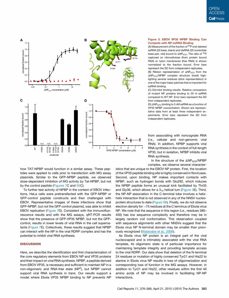
DNPNTD Binds ssRNA, and the ssRNABinding Is Inhibitedby NPBPThe ability of NP to bind RNA is well established (Bharat et al.,
2012; Huang et al., 2002; Muhlberger et al., 1999; Noda et al.,
2010, 2011; Watanabe et al., 2006). Therefore, we assessed if
DNPNTD can bind ssRNA and dsRNA. Using a dot blot assay,
we find that NP selectively binds ssRNA (KD = 620 ±
230 nM), whereas NP does not bind dsRNA (Figure 5A). Anal-
ysis of the NPBP/DNPNTD structure identified several highly
basic regions (Figure 3D). Using surface charge as a guide,
we identified K160, K171, and K174 as potential ssRNA binding
sites (Figure 5B). Mutation of these residues to alanine demon-
strates their role in ssRNA binding (Figure 5C). In contrast,
NP residues that are critical for NPBP binding and for EBOV
A
D
B C
180°
N
CN
C
SeMet34
SeMet20
Ile47
180°
E R240
E31
K281
F44K257
S30
D252
K248
α15
β3
β4
α13
G22
P21Q355
R269 C45
R37
E292
α20
α21
180°
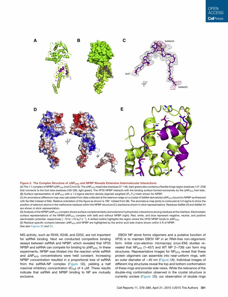
Figure 3. The Complex Structure of DNPNTD and NPBP Reveals Extensive Intermolecular Interactions
(A) The 1:1 complex of NPBP/DNPNTD (mol C/mol G). TheDNPNTD head lobe (residues 37–146, dark green) also contains a flexible hinge region (residues 147–239)
that connects to the foot lobe (residues 240–285, light green). The VP35 NPBP interacts with the binding surface formed exclusively by the DNPNTD foot lobe.
(B) Surface representation of DNPNTD with a 1.0 sigma electron density (sigmaA weighted 2Fo-Fc) mesh shown for NPBP.
(C) An anomalous difference map was calculated from data collected at the selenium edge on a crystal of SeMet derivatized DNPNTD bound to NPBP synthesized
with Se-Met instead of Met. Relative orientation of the figure as shown is 180� rotated from (B). The anomalous map (pink) is contoured at 3.0 sigma to show the
position of selenium atoms in the methionine residues within the NPBP structure (Ca backbone shown in stick representation). Residues SeMet 20 and SeMet 34
are shown in stick representation.
(D) Analysis of theNPBP/DNPNTD complex shows surface complementarity and extensive hydrophobic interactions among residues at the interface. Electrostatic
surface representations of the NPBP/DNPNTD complex with (left) and without NPBP (right). Red, white, and blue represent negative, neutral, and positive
electrostatic potential, respectively (�10 to +10 kBT e�1). A dotted outline highlights the region where the VP35 NPBP binds to DNPNTD.
(E) Residue-specific contacts between DNPNTD and NPBP are highlighted by the amino acid side chains shown within 5 A of NPBP.
See also Figures S2 and S4.
MG activity, such as R240, K248, and D252, are not important
for ssRNA binding. Next we conducted competitive binding
assays between ssRNA and NPBP, which revealed that VP35
NPBP and ssRNA can compete for binding to DNPNTD. In these
experiments, NPBP was titrated into the reaction while ssRNA
and DNPNTD concentrations were held constant. Increasing
NPBP concentration resulted in a proportional loss of ssRNA
from the ssRNA-NP complex (Figure 5D), yielding a half
maximal inhibitory concentration (IC50) of 4 mM. These results
indicate that ssRNA and NPBP binding to NP are mutually
exclusive.
EBOV NP alone forms oligomers and a putative function of
VP35 is to maintain EBOV NP in an RNA-free non-oligomeric
form. Initial cryo-electron microscopy (cryo-EM) studies re-
vealed that NPNTD (1–457) and WT NP (1–739) can form ring
structures. Representative images for NPNTD reveal that these
protein oligomers can assemble into near-uniform rings, with
an outer diameter of �40 nm (Figure 6A). Individual images of
different ring structures reveal the top and bottom conformation
of these rings and provide side views. While the relevance of the
double-ring conformation observed in the crystal structure is
currently unclear (Figure 2D), our observation of double rings
Cell Reports 11, 376–389, April 21, 2015 ª2015 The Authors 381

C D
E
A
B
Figure 4. Mutational Analysis Validates the NPBP Binding Site on NP
(A) Non-bonded contacts drive NPBP/DNPNTD binding. LigPlot+ diagram showing extensive hydrophobic and hydrogen bond interactions between DNPNTD and
NPBP. Protein side chains are shown as ball and sticks. Hydrogen bonds are shown as orange dotted lines. Non-bonded contacts are shown as spoked arcs.
(B) Surface and ribbon representation highlighting the interaction between NPBP (purple) and the foot lobe of DNPNTD (light green). VP35 NPBP residues Glu24
and Arg37 and NP residues Lys257 and Glu292 are shown in stick representation.
(C) Summary of ITC binding measurements between MBP fusion DNPNTD mutants and NPBP reveal that residues R240, K248, and D252 are involved in NPBP/
DNPNTD complex interactions. n.d., not determined.
(D) Impact of the NP mutants was evaluated by the MG assay. Ability of either WT or mutant NP proteins to promote MG activity were tested and plotted as MG
activity relative to no NP control. The p values were determined by Student’s t test. **p < 0.001. Representative western blots to show similar levels of NP protein
expression. Error bars represent the SD from independent replicates.
(E) Representative western blots for NP proteins used in (D) at 250 and 500 ng plasmid transfections.
under distinct sample conditions by cryo-EM suggests that this
conformation may be functionally important. However, in our
crystal structure, crystal packing may impact the ring formation,
since DNPNTD/NPBP is a monomer in solution. Addition of NPBP
to the cryo-EM sample results in loss of the ring conformation,
presumably due to the formation of 1:1 heterodimers between
NPBP and NPNTD (Figure 6B). Taken together, our cryo-EM
and X-ray results show that NPBP can cause morphological dif-
ferences between the oligomeric ring structures formed by
NPNTD and NPNTD/NPBP complexes. Next, we evaluated a vari-
ety of NP constructs, including DNPNTD construct and NPNTD,
using size exclusion chromatography (SEC) as a measure of
the hydrodynamic behavior of NP upon binding to NPBP under
low- (150 mM) and high-NaCl (500 mM) buffer conditions. In
contrast to previous results for the peptide-free NP proteins
(Figures 1B and 1C), which eluted at the V0, all NPBP-bound
NP proteins eluted at a volume consistent with a well-behaved
heterodimer (one molecule of NP and one NPBP molecule)
(Figure 6C), indicating that NPBP interaction also prevents
oligomerization.
382 Cell Reports 11, 376–389, April 21, 2015 ª2015 The Authors
VP35 NPBP Inhibits Ebola RNA SynthesisIn order to determine whether VP35 NPBP can function in trans
to complement a VP35 protein lacking the NPBP region and sup-
port RNA synthesis, we tested various VP35 N-terminal trunca-
tion mutants together with plasmids expressing GFP-NPBP or
using a cell-penetrating peptide fused to the NPBP sequence
in the MG assay. Specifically, we fused the NPBP with an N-ter-
minal TAT peptide (sequence YGRKKRRQRRR). In this assay,
we used VP35 lacking the N-terminal 51 residues and assessed
how addition of different regions of EBOV VP35 impact MG
activity. Resulting data show that GFP-NPBP or other VP35 trun-
cations (52–340) were nonfunctional in this assay, suggesting
that the function of NPBP is required in cis with the rest of the
VP35 polypeptide sequence (Figures 7A and S5A).
Given that NPBP occupies a functionally critical site on NP and
is required for RNA synthesis, we next tested the ability of NPBP
to inhibit Ebola virus RDRP activity. We used plasmid expressing
GFP-NPBP and observed dose-dependent inhibition of MG ac-
tivity by NPBP (GFP-NPBP) with an IC50 = 33 mM relative to a
GFP peptide control (Figures 7B and S5B). We then tested
A
D
B
C
norm
aliz
ed fr
actio
n bo
und
VP35 NPBP concentration (μM)1 2 3 4 5 6 7 8
0.0
0.2
0.4
0.6
0.8
1.0
1.2
K160
K171
R174
0.01 0.1 1 10
0.0
0.2
0.4
0.6
0.8
1.0
1.2
norm
alize
dfra
ctio
nbo
und
ΔNPNTD concentration (μM)
N.D. N.D. N.D.
WT
K160
A
K171
A/R1
74A
K160
A/K1
71A/
R174
A
R 240
A
K 248
A
D252
A
0.0
0.2
0.4
0.6
0.8
1.0
1.2
norm
alize
dfra
ctio
nbo
und
constructs
Figure 5. EBOV VP35 NPBP Binding Can
Compete with NP-ssRNA Binding
(A) Measurement of the fraction of 32P end-labeled
ssRNA (20 base, black) and dsRNA (20 nucleotide
base pair, red) bound to DNPNTD. The ratio of 32P
captured on nitrocellulose (from protein bound
RNA) or nylon membranes (free RNA) is shown
normalized to the fraction bound. Error bars
represent the SD from independent replicates.
(B) Ribbon representation of DNPNTD from the
DNPNTD/NPBP complex structure (inset) high-
lighting several residues (stick representation) in
one of themajor basic patches that is important for
ssRNA binding.
(C) Dot-blot binding results. Relative comparison
of mutant NP proteins binding to 20 nt ssRNA
compared to WT NP. Error bars represent the SD
from independent replicates.
(D)DNPNTD binding to 5 nM ssRNA as a function of
VP35 NPBP concentration. Shown are represen-
tative data from at least three independent ex-
periments. Error bars represent the SD from
independent replicates.
how TAT-NPBP would function in a similar assay. These pep-
tides were applied to cells prior to transfection with MG assay
plasmids. Similar to the GFP-NPBP peptide, we observed
dose-dependent inhibition of MG activity by Tat-NPBP, but not
by the control peptide (Figures 7C and S5C).
To further test activity of NPBP in the context of EBOV infec-
tions, HeLa cells were pretransfected with the GFP-NPBP or
GFP-control peptide constructs and then challenged with
EBOV. Representative images of these infections show that
GFP-NPBP, but not the GFP control plasmid, was able to inhibit
EBOV replication (Figure 7D). Consistent with the immunofluo-
rescence results and with the MG assays, qRT-PCR results
show that the presence of GFP-VP35 NPBP, but not the GFP-
control, results in lower levels of viral RNA in the cell superna-
tants (Figure 7E). Collectively, these results suggest that NPBP
can interact with the NP in the viral RDRP complex and has the
potential to inhibit viral RNA synthesis.
DISCUSSION
Here, we describe the identification and first characterization of
the core regulatory elements from EBOV NP and VP35 proteins
and their impact on viral RNA synthesis. NPBP, a peptide derived
from EBOV VP35, is necessary and sufficient to maintain NP in a
non-oligomeric and RNA-free state (NP0), but NPBP cannot
support viral RNA synthesis in trans. Our results support a
model where Ebola VP35 NPBP binding to NP prevents NP
Cell Reports 11, 376–3
from associating with noncognate RNA
(i.e., cellular and non-genomic viral
RNA). In addition, NPBP supports viral
RNA synthesis in the context of full-length
VP35, but in isolation, NPBP inhibits viral
RNA synthesis.
In the structure of the DNPNTD/NPBP
complex, we observe several character-
istics that are unique to the EBOV NP protein. First, the location
of the VP35 peptide binding site is highly conserved in filoviruses.
Second, upon binding, NP makes important contacts with
NPBP, such as hydrogen bonds with Glu292, which induces
the NPBP peptide forms an unusual kink facilitated by Thr35
and Gly36, which allows for a 310 helical turn (Figure 3E). Third,
the NP-NP association in the C-terminal lobe involves a helix-
helix interaction that is not observed in any of the NNSV nucleo-
protein structures to date (Figure S6). Finally, we do not observe
electron density for�75 residues at the C terminus of Ebola virus
NP. We note that the sequence in this region (i.e., residues 380–
450) has low sequence complexity and therefore may be in
largely random coil conformation. This observation coupled
with sequence alignments with other NNSVs suggest that the
Ebola virus NP N-terminal domain may be smaller than previ-
ously recognized (Watanabe et al., 2006).
As Ebola virus NP protein is an integral part of the viral
nucleocapsid and is intimately associated with the viral RNA
template, its oligomeric state is of particular importance for
maintaining template integrity and providing template access
to the viral RDRP. Our data show that deletion of the N-terminal
24 residues or mutation of highly conserved Tyr21 and His22 to
alanine in Ebola virus NP results in loss of oligomerization and
corresponding loss of function in the MG assays. However, in
addition to Tyr21 and His22, other residues within the first 40
amino acids of NP may be involved in facilitating NP-NP
interactions.
89, April 21, 2015 ª2015 The Authors 383

C
0 5 10 15 20 25
0.0
0.2
0.4
0.6
0.8
1.0
0
Norm
alize
dab
s orb
ance
elution volume (mL)
NPBPNPBP/NP complex
20 nm
100 nm100 nm
A B
Figure 6. EBOV VP35 NPBP Binding Can Dismantle Oligomeric Ring Structures Formed by NPNTD
(A) Representative cryo-EM images of NPNTD forming double ring structures, observed from top and side views.
(B) Ring-like structures formed by NPNTD disappear upon addition of NPBP peptide just prior to cryo-EM grid preparation.
(C) Representative chromatograms of NPBP complex of NPNTD in 150 mM NaCl (blue dotted line) and 500 mM NaCl buffers (blue solid line), DNPNTD in 150 mM
NaCl (green dotted line), and 500 mM NaCl (green solid line) from size exclusion chromatography. NPBP peptide alone is shown in 150 mM NaCl (purple dotted
line) and 500 mM NaCl (purple solid line) buffer conditions. Location of the void volume for the Superdex 200 column is indicated with an arrow.
The NP C terminus is dispensable for oligomerization, but not
for viral RNA synthesis, as deletions C-terminal to residue 551
result in loss of Ebola RNA binding. Our observations are also
consistent with previous studies, where a 150-residue deletion
at the N terminus resulted in loss of NP-NP association (Wata-
nabe et al., 2006). However, because such large deletions can
result in the loss of structural integrity, it is difficult to attribute
these results to a role for oligomerization. Our results also sug-
gest that the NP-NP self-association is regulated by the region
that binds NPBP (Figure 2A). Importantly, this binding and result-
ing change in the NP oligomeric state does not depend on the
oligomeric state of VP35 (Moller et al., 2005; Reid et al., 2005)
as we observe similar affinities between NP-VP35 and NP-
NPBP (see Figures 2C and S2).
VP35 prevents high-affinity NP-RNA interactions until nascent
NP reaches the site of viral RNA synthesis, where VP35 facilitates
transfer of a monomeric and template-free NP (NP0) to the viral
template RNA, as our data show that VP35 NPBP binding re-
leases RNA from NP. Upon binding to template RNA to form
the NP-RNA complex, NP is released from interactions with
VP35 NPBP through a yet-unknown mechanism. Release from
NPBP and subsequent binding to viral RNA likely induces NP
oligomerization, which can further stabilize the RNA-bound NP
proteins. Our results, particularly data that show how NPBP
can outcompete ssRNA binding, are consistent with a model
where NPBP overrides NP-NP and NP-RNA interactions in order
to maintain Ebola NP in the NP0 state. Not surprisingly, deletion
of the NPBP sequence from Ebola VP35 results in loss of viral
RNA synthesis likely due to the lack of NP0 to support viral
RNA synthesis. However, when a truncated VP35 lacking
NPBP (residues 52-340) was co-expressed with the VP35
NPBP peptide and other components in the MG assay, NPBP
384 Cell Reports 11, 376–389, April 21, 2015 ª2015 The Authors
failed to support viral RNA synthesis in trans. In addition, proper
viral RNA synthesis may require NPBP in the context of the full-
length VP35 to bind NP to generate NP0. Among the possible
reasons for this outcome is a need for NPBP, which is located
in the VP35 N terminus, to function together with VP35 C-termi-
nal IID region. Our previous studies revealed that VP35 IID is also
important for virus RNA synthesis. Specifically, we were able to
show that conserved basic residues within the a-helical subdo-
main of VP35 IID were critical for VP35-NP interactions (Leung
et al., 2009; Prins et al., 2010). Another possibility is that the
NPBP binding to NP also provides a means to correctly localize
the NP0 proteins to the viral RDRP at a site near the viral nucle-
ocapsid (NP-RNA). In such an arrangement, full-length VP35
can also provide template access to the polymerase L.
NNSV P and VP35 proteins are essential for viral RNA synthe-
sis despite the lack of any sequence similarity among these pro-
teins. The P protein, which is functionally equivalent to Ebola
virus VP35, is thought to maintain newly synthesized nucleopro-
tein molecules in a non-oligomeric and RNA-free state (N0).
Studies on vesicular stomatitis virus (VSV) and Nipah virus
(NiV) have shown that an N-terminal region of each respective
P protein is sufficient to bind N0. In addition, P peptide binding
(N0-P) presumably blocks RNA access to a groove formed be-
tween the N-terminal and C-terminal lobes of nucleoprotein
based upon structural alignments of the N0-P and N-RNA com-
plex structures. Comparison of the NP structure from our Ebola
virus DNPNTD/NPBP complex with corresponding structures of
VSV (Leyrat et al., 2011a, 2011b) and NiV N (Yabukarski et al.,
2014) in complex with the P-derived peptide (N0-P), analogous
to the Ebola VP35 NPBP, reveals several differences. The P pep-
tide binding site and the secondary structure of the P peptide are
different from Ebola virus VP35 NPBP bound to NP (Figure S6). In

ED
VP
35 N
PB
PG
FP-c
ontro
lA B C
100
120
40
20
0
80
60
VP35
120
40
0
100
% m
inig
enom
e ac
tivity
VP35- + + + + + + +
GFP-control GFP-VP35 NPBP
- + + + + + + +
Tat-control Tat-VP35 NPBP
*
*
**
**
*
**
**
*
25
60
80
% m
inig
enom
e ac
tivity
GFPcontrol
GFPVP35NPBP
Tatcontrol
TatVP35NPBP
125
50
25
75
0
100
VP35 - + + + + + + ++ + + + + + +++
% m
inig
enom
e ac
tivity
(ren
/luc*
100)
Hoechst GFP anti-NP
52-340
20
10
0
30
Rel
ativ
e co
pies
vR
NA
NP
GFP-control GFP-NPBP15h 24h 15h 24h
***
*
**
Figure 7. VP35 NPBP Inhibits EBOV Replication
(A) NPBP in the context of VP35, but not as an isolated peptide, can support RNA synthesis. MG assay performed with VP35 52–340 with additional VP35
truncations, as indicated in the figure, failed to support RNA synthesis. 500 ng of GFP-NPBP and 100 mM of TAT-NPBP were used. Error bars represent the SD
from independent replicates.
(B and C) Representative results from MG assay with either (B) GFP control plasmid or GFP-NPBP peptide expressing plasmid with three different doses (125,
250, and 500 ng; *p = 0.04, **p = 0.001). (C) Control TAT-peptide or TAT-NPBP (1, 10, and 100 mM). Activity in (A)–(C) is reported as a percent of the average activity
recorded for WT VP35 (*p = 0.01, *p = 0.002). The p values were determined by Student’s t test. Error bars represent the SD from independent replicates.
(D) VP35 NPBP inhibits EBOV infection in HeLa cells. Cells were transfected with either plasmid encoding GFP-NPBP peptide fusion (top) or GFP expressing
plasmid (bottom) and then challenged with Zaire Ebola virus at an MOI of 0.05 48 hr after plasmid transfection. Representative images show cell nuclei stained
with Hoechst 33342 dye (blue), plasmid expression (green), and virus replication (red, stained with anti-Ebola NP mAb).
(E) qRT-PCR analysis for GFP or GFP-NPBP (VP35 residues 20–48) expressing cells that were infected with Ebola virus. At 15 hr and 24 hr post-infection,
supernatant was collected, TRIzol treated, and subjected to reverse transcription using a primer a complementary to the negative-sense viral RNA (vRNA)
followed by qRT-PCR using primers directed to EBOV NP (*p = 0.06, **p = 0.006). The p values were determined by Student’s t test. Error bars represent the SD
from independent replicates.
See also Figures S5 and S6.
addition, there are intra-peptide interactions that stabilize the
NPBP-bound conformation and are absent in the corresponding
VSV- and NiV N-bound peptides. Moreover, in VSV and NiV, the
N-N contacts are different from those observed for Ebola virus
NP-NP. Specifically, we observe inter-NP interactions (see Fig-
ures S3B and S3C) that occur via helices in the foot lobe whereas
the inter-N interactions in the VSV and NiV occur mainly via loop-
loop interactions. Despite these structural differences, we
observe several similarities. VSV and NiV N proteins and Ebola
virus NP have similar folds and display similar domain arrange-
ments in the first�350–400 residues of each protein. In addition,
our DNPNTD/NPBP structure along with ssRNA binding studies
show that NPBPbinding is incompatible with RNA binding. Com-
parison of the DNPNTD/NPBP structure from the current study
with ssRNA-bound structures of VSV and RSV nucleoproteins
suggest that the highly basic region highlighted in Figure 3D
(left) may recognize and bind ssRNA in Ebola NP. Our results
also show that the VP35 NPBP binding relieves NP oligomeriza-
tion and NP-RNA binding by interacting with two distinct regions
within NP that are important for viral RNA synthesis. These sites
Cell Reports 11, 376–389, April 21, 2015 ª2015 The Authors 385
on EBOV NP do not appear to overlap, since mutations of indi-
vidual residues important for ssRNA binding and for NPBP bind-
ing appear to function independently. In contrast, structural
alignments of the N0-P and N-RNA structures suggest that
P peptide binding to N protein in VSV and NiV may also limit
N-RNA interactions by steric hindrance (Leyrat et al., 2011a;
Yabukarski et al., 2014). Collectively, these results suggest that
NNSVs may use variations of a common mechanism to control
viral RNA synthesis.
Our results here show for the first time how EBOV VP35 uses
the NPBP region to control viral RNA synthesis and highlight
NNSV-common and filoviral-specific mechanisms by which
EBOV VP35 regulates NP-NP and NP-RNA template interac-
tions. These results also provide the framework to develop
anti-viral therapeutics that target filoviruses.
EXPERIMENTAL PROCEDURES
Cloning and Purification
Ebola virus NP and VP35 constructs were subcloned into a modified pET15b
(Novagen) vector containing a maltose binding protein fusion tag and TEV pro-
tease site and expressed in BL21(DE3) Escherichia coli cells grown at 37�C.Cells were induced for 12–14 hr at 18�C with 0.5 mM isopropyl b-D-1-thioga-
lactopyranoside and harvested at 8,0003 g for 10 min, resuspended in buffer
A (20 mM Tris [pH 7.5], 1 M or 150 mM NaCl for NP and VP35, respectively),
20 mM imidazole (pH 7), 5 mM b-mercaptoethanol, and a protease inhibitor.
Cells were lysed using an Avestin C3 homogenizer, clarified by centrifugation
at 47,000 3 g for 30 min, and supernatant was purified by affinity and ion-
exchange chromatography. Fusion tagswere removed by TEV protease cleav-
age and purified by ion exchange columns prior to final size-exclusion column.
VP35 peptides were purchased from GenScript.
Selenomethionine-Labeled Protein
Proteins were expressed in M9-minimal media, supplemented with appropri-
ately labeled metabolites, and purified in a manner similar to native protein
described above.
Isothermal Titration Calorimetry
NP and VP35 binding was measured by VP-ITC microcalorimeter (MicroCal/
GE Healthcare). Samples were dialyzed overnight at 25�C against 1 l of buffer
B (20mMTris [pH 7.5], 500mMNaCl, and 2mM tris(2-carboxyethyl)phosphine
[TCEP]). Raw ITC data were processed using Origin software and data fit by
non-linear least-squares analysis to yield KD (equilibrium binding constant)
and n (number of binding sites). A representative of two to four independent
experiments is shown.
SEC-MALS
SEC-MALS experiments were performed using a DAWN-HELEOS II detector
(Wyatt Technologies) coupled to a Superdex SD200 column (GE Healthcare)
in buffer C (20 mM Tris [pH 7.5], 250 mM NaCl, and 2 mM TCEP). 2 mg/ml
sample was injected and raw data were analyzed using ASTRA 6 soft-
ware (Wyatt Technologies) to determine the weight averaged molecular
mass (MW). Protein concentrations were determined using the refractive
index measured by an Optilab T-rEX (Wyatt Technologies) and a dn/dc =
0.185 ml 3 g�1.
Minigenome Assays
BSRT7 cells were co-transfected by using Lipofectamine 2000 (Invitrogen)
with T7-driven expression plasmids encoding the EBOV NP, L, VP30, and
VP35 proteins along with plasmid expressing a T7 promoter-driven EBOV
minigenome RNA, which encodes a Renilla luciferase reporter gene and
constitutively expressing firefly luciferase expression plasmid that served as
a transfection control. At 24 hr posttransfection, cells were lysed with passive
lysis buffer (Promega) and reporter activities were determined using the Dual
386 Cell Reports 11, 376–389, April 21, 2015 ª2015 The Authors
Luciferase assay kit (Promega). Renilla luciferase activity was normalized to
firefly luciferase activity. Minigenome reporter activation was expressed as
percent minigenome activity setting activity of WT VP35 as 100%. Error bars
represent the SD from three independent replicates.
Crystallization, Data Collection, and Structure Solution
DNPNTD and VP35 NPBP were incubated together in 1:1 molar ratio prior to
loading onto a Superdex SD200 column (GE Healthcare) with buffer containing
20 mM Tris (pH 7), 250 mM NaCl, and 2 mM TCEP. NPBP/DNPNTD complex
crystals were generated using the hanging-drop vapor diffusion method and
streak seeding in well solution containing 100 mM Tris (pH 7.2), 50 mMHEPES
(pH 7), and 23% PEG400. Crystals were cryoprotected in stabilization solution
containing 100 mM Tris (pH 7.2), 50 mM HEPES (pH 7), 50 mM NaCl, 2 mM
TCEP, 15% PEG400, and 15% PEG3350 followed by vitrification in liquid
nitrogen.
X-ray data were collected at the Structural Biology Center 19ID at the
Advanced Photon Source (Argonne, IL), Datasets from two crystals of SeMet
derivatized NP protein bound to native VP35 NPBP were collected at low
remote energy to minimize radiation damage with an oscillation angle of 0.8
and a crystal-to-detector distance of 500 mm. Dataset from one crystal of
SeMet derivatized NP bound to SeMet derivatized VP35 NPBP was collected
at the Se absorption peak tomaximize the anomalous signal with an oscillation
angle of 0.8 and a crystal-to-detector distance of 450 mm.
The structure of Ebola virus VP35 NPBP/DNPNTD complex was solved using
the diffraction data obtained from three crystals. The partial model was built
using phases obtained from the averaging datasets of these crystals. The
data averaging took the differences in the levels of anomalous signal into
account. HKL3000 was used to process diffraction data sets for all crystals
(Minor et al., 2006; Otwinowski and Minor, 1997). Computational corrections
for absorption in a crystal and imprecise calculations of the Lorentz factor re-
sulting from a minor misalignment of the goniostat were applied (Borek et al.,
2003; Otwinowski et al., 2003). Anisotropic diffraction was corrected to adjust
the error model and to compensate for the phasing signal for radiation-
induced increase of non-isomorphism within the crystal (Borek et al., 2007,
2010, 2013). The data statistics are presented in Table S1. The crystals dif-
fracted anisotropically to a resolution of 3.7 A in the best direction and 4.0
and 4.2 A in the other two directions. The estimated level of anomalous signal
was 3.6% of the native intensity. We performed a search for heavy atom
positions to a resolution of 7.0 A with ShelxD (Sheldrick, 2008), which identi-
fied 28 Se positions with correlation coefficients: CCAll = 54.79% and
CCWeak = 28.18%. The handedness of the solution was determined with
ShelxE by analyzing the connectivity and contrast of the electron density
maps. 26 Se positions were refined anisotropically to 4.4 A with MLPHARE
(Otwinowski, 1991), with the final FOM reaching 0.205. 4-fold NCS was iden-
tified by Resolve (Terwilliger, 2003, 2004). NCS averaging and solvent flat-
tening was performed by DM (Cowtan and Main, 1998). The resulting electron
density map was used for model building, which consisted of several cycles of
BUCCANEER (Cowtan, 2006). Intermediate models from different cycles of
BUCCANEER were combined manually into a more complete model for one
of the four chains, which had the lowest thermal motions. This model was
subsequently propagated by applying the NCS operators, and then iterative
model building was repeated. The resulting oligomer was then manually
rebuilt and corrected by iterative application of Coot, Refmac (Emsley and
Cowtan, 2004; Murshudov et al., 1997, 1999) along with the local NCS re-
straints, jelly body refinement, and in the later cycles with TLS refinement
(Winn et al., 2003).
TheDNPNTD chains in the asymmetric unit have different levels of order, with
average B-factors for chains A, B, C, and D of 123, 190, 90, and 234 A2,
respectively. Although the N-terminal domains for chains B and D could not
be accurately modeled, the order of chains A and C was sufficient to build
the entire peptide chain that consists of N-terminal head lobe and C-terminal
foot lobe. SeMet positions for the DNPNTD protein together with the SeMet-
derivatized NPBP dataset analyzed separately provided unambiguous tracing
for the DNPNTD chains and NPBP chains. This model was propagated through
an NCS symmetry operator and provides a credible indication as to where the
N-terminal domains of chains B and D are located. However, due to their large
thermal motions (equivalent to 1–2 A shift of subdomains), the validation

statistics, which analyze how this region fits to the electron density, are not
meaningful. The model was refined with these domains, because the electron
density and packing interactions indicate where the domains are located,
albeit the individual secondary structure elements can barely be fitted. All
atomic interactions are analyzed based on the chains C and G.
Cryo-EM Specimen Preparation and Data Acquisition
The 400-mesh R1.2/1.3 holey carbon Quantifoil grid (Quantifoil Micro Tools
GmbH) was cleaned with acetone (Sigma-Aldrich) for 12 hr and glow
discharged before use. A 3.0-ml aliquot of purified Ebola NP oligomer at
�2 mg/ml in the presence or absence of 0.5 ml disruption peptide was applied
to the grid, blotted for 8 s, and immediately frozen in liquid ethane using a Leica
EM GP (Leica Microsystems) with temperature held constantly at 10�C and
99% humidity during the process. The grid was stored in liquid nitrogen until
imaging. All grids were examined on a JEM 2100 (LaB6 gun) cryo-electron mi-
croscope (JEOL) operated at 200 kV, spot size of 2, condenser aperture of
70 mm, and objective aperture of 60 mm. Images were recorded under low-
dose condition on a Gatan 4k 3 4k CCD camera (Gatan) at 350,000 micro-
scope magnification (corresponding to a calibrated sampling of 2.16 A/pixel)
and a dose of 50–60 electrons/A2 with defocus ranging from 3 to 5 mm. Particle
images were manually boxed and extracted using EMAN2 (Tang et al., 2007)
program with a box size of 288 3 288 pixels.
Structural Figure Generation and Analysis
Surface area and surface complementarity were calculated using AREAIMOL
and Sc, respectively, as implemented in CCP4 program suite (CCP4,
1994). Structure figures were prepared using PyMOL (DeLano, 2002). Pro-
tein-protein interactions were analyzed using LigPlot+ (Laskowski and Swin-
dells, 2011). Topology diagrams were generated by PDBSum (Laskowski,
2007) and TopDraw (Bond, 2003). Sequence alignment was performed using
ClustalW (Thompson et al., 2002) and prepared using ESPript 3.0 (Gouet
et al., 2003).
RNA Binding and Competition Assays
20-nt ssRNAwas labeled at the RNA 50 using 32P-g-ATP. 25 mMofDNPNTDwas
serially diluted and incubated with 32P-20 nt ssRNA at 5 nM for 15 min prior to
loading onto a dot blot apparatus (Bio-Rad). For peptide-competition experi-
ments, 20 mM of VP35 NPBP was serially diluted and incubated with DNPNTD
at 4 mM for 30 min prior to the addition of 32P-20 nt ssRNA (at 5 nM). The pro-
tein-RNA complex was then passed through nitrocellulose (NC) and nylon (NY)
membranes. The membranes were scanned using a Typhoon 9410 Variable
Model Imager, and the amount of 32P-20 nt ssRNA bound to NC and NYmem-
branes was quantified from the radioactivity detected. Data analyzed by Origin
and the fraction ssRNA bound to DNPNTD proteins were calculated using the
equation shown below:
fraction bound=RNA bound to NC
RNA bound to NC+RNA bound to NY:
Virus Infection
HeLa cells were transfected with either a plasmid encoding GFP-NPBP
peptide fusion or a GFP control expressing plasmid. 24 hr after plasmid
transfection, cells were transferred to the BSL-4 laboratory and challenged
with Zaire EBOV at an MOI of 0.05 and were fixed by immersing in neutral
buffered formalin overnight. Fixed cells were washed three times with PBS
and then stained with anti-Ebola-GP antibody followed by an anti-mouse-
546 (Alexa) secondary antibody, nuclei were stained with Hoechst-33342
(Life-Technologies; 1:50,000 dilution). Cells were imaged on a Nikon Ti Eclipse
microscope. Total cells (blue), transfected cells (green; GFP), and Ebola-
infected cells (red; GP-specific mAb) were counted using CellProfiler software
(Broad Institute) using customized pipelines, which are available from R.A.D.
upon request.
qRT-PCR
293T cells were transfected with GFP or VP35 NPBP and infected with Zaire
EBOV. At 15 and 24 hr postinfection, supernatant and cells were collected
for RNA isolation using TRIZOL. For reverse transcription, either an oligo(dT)
primer (for detection of mRNA) or a primer complementary to the negative-
sense viral RNA (vRNA) was used, followed by quantitative real-time PCR
using primers directed against NP. In parallel, qRT-PCR directed against
b-actin mRNA was performed, to control for differences in the amount of total
input RNA. The relative copy number of target RNA was calculated from the
change in threshold cycle (CT) value between b-actin mRNA and target
RNA. mRNA was reverse transcribed using a vRNA specific primer (NP), fol-
lowed by quantitative real-time PCR using NP-specific primers. Shown are
the means and SE from two independent experiments.
ACCESSION NUMBERS
Coordinates and structure factors have been deposited in to the PDB and are
available under accession number 4YPI.
SUPPLEMENTAL INFORMATION
Supplemental Information includes Supplemental Experimental Procedures,
six figures, and one table and can be found with this article online at http://
dx.doi.org/10.1016/j.celrep.2015.03.034.
AUTHOR CONTRIBUTIONS
D.W.L., C.F.B., and G.K.A. conceived and designed the overall study. D.W.L.
defined VP35 NPBP. D.W.L., J.M.B., and G.K.A. developed NP purification
methods. J.P. purified proteins for crystallization. D.W.L. identified and opti-
mized crystallization conditions and acquired crystallographic data, which
was used by D.B. and Z.O. to solve the structure and build and refined the
model. D.W.L., D.B., Z.O., C.F.B., and G.K.A. conducted analysis of the struc-
ture and designed validation assays. D.W.L., G.L., and G.K.A. designed and
conducted RNA binding studies. I.H. conducted peptide analysis. P.L.,
R.S.S., and C.F.B. designed functional validation assays. P.L. and A.E.-F. con-
ducted cell-based studies and analyzed results. Z.S. and W.C. conducted
cryo-EM studies. M.A. and R.A.D. designed and executed BSL4 studies.
D.W.L., D.B., C.F.B., and G.K.A. wrote the manuscript with input from all co-
authors. All authors analyzed results and approved the submission.
ACKNOWLEDGMENTS
We thank Drs. T. Ellenberger, D. Fremont, T. Brett, N. Tolia, W. Li, and J. Pay-
ton for discussions; members of the G.K.A./G.L. W.C., C.F.B., and R.A.D. lab-
oratories for experimental support; and Drs. S. Ginell, N. Duke, and J. Lazarz at
Advanced Photon Source (APS) Sector 19 for beamline access and assistance
during data collection. Use of Structural Biology Center beamlines at the APS
is supported by the US DOE under contract DE-AC02-06CH11357. Use of the
National Magnetic Resonance Facility at Madison is supported by NIH grant
P41GM103399. This work was supported in part by a contract to the Center
for Structural Genomics of Infectious Diseases (CSGID) from the NIAID/NIH/
DHS (contract number HHSN272201200026C, Anderson-PI) to Z.O., by
DOD grants DTRA 1-21-1-0002 to R.A.D., DTRA-HDTRA1-12-1-0051 and
DTRA HDTRA1-14-1-0013 to C.F.B. and G.K.A., and by NIH grants
(R01AI107056 to D.W.L.; R01GM053163 to Z.O.; R01AI077519 to R.A.D.;
R01AI059536 to C.F.B.; U19AI109945 [Basler-PI] to C.F.B., D.W.L., and
G.K.A.; and U19AI109664 [Basler-PI] to D.W.L., D.B., C.F.B. and G.K.A.;
U19 AI070489 [Holtzman-PI], P41GM103832, and P50 GM1003297 to W.C.;
and R01AI081914 to G.K.A.).
Received: December 8, 2014
Revised: February 18, 2015
Accepted: March 12, 2015
Published: April 9, 2015
REFERENCES
Basler, C.F., Mikulasova, A., Martinez-Sobrido, L., Paragas, J., Muhlberger, E.,
Bray, M., Klenk, H.D., Palese, P., and Garcıa-Sastre, A. (2003). The Ebola virus
Cell Reports 11, 376–389, April 21, 2015 ª2015 The Authors 387

VP35 protein inhibits activation of interferon regulatory factor 3. J. Virol. 77,
7945–7956.
Bharat, T.A., Noda, T., Riches, J.D., Kraehling, V., Kolesnikova, L., Becker, S.,
Kawaoka, Y., and Briggs, J.A. (2012). Structural dissection of Ebola virus and
its assembly determinants using cryo-electron tomography. Proc. Natl. Acad.
Sci. USA 109, 4275–4280.
Bond, C.S. (2003). TopDraw: a sketchpad for protein structure topology car-
toons. Bioinformatics 19, 311–312.
Borek, D., Minor, W., and Otwinowski, Z. (2003). Measurement errors and their
consequences in protein crystallography. Acta Crystallogr. D Biol. Crystallogr.
59, 2031–2038.
Borek, D., Ginell, S.L., Cymborowski, M., Minor, W., and Otwinowski, Z.
(2007). The many faces of radiation-induced changes. J. Synchrotron Radiat.
14, 24–33.
Borek, D., Cymborowski, M., Machius, M., Minor, W., and Otwinowski, Z.
(2010). Diffraction data analysis in the presence of radiation damage. Acta
Crystallogr. D Biol. Crystallogr. 66, 426–436.
Borek, D., Dauter, Z., and Otwinowski, Z. (2013). Identification of patterns in
diffraction intensities affected by radiation exposure. J. Synchrotron Radiat.
20, 37–48.
Collaborative Computational Project, Number 4 (1994). The CCP4 suite: pro-
grams for protein crystallography. Acta Crystallogr. D Biol. Crystallogr. 50,
760–763.
Cowtan, K. (2006). The Buccaneer software for automated model building. 1.
Tracing protein chains. Acta Crystallogr. D Biol. Crystallogr. 62, 1002–1011.
Cowtan, K., and Main, P. (1998). Miscellaneous algorithms for density modifi-
cation. Acta Crystallogr. D Biol. Crystallogr. 54, 487–493.
DeLano, W.L. (2002). The PyMOL Molecular Graphics System (DeLano Scien-
tific).
Dziuba�nska, P.J., Derewenda, U., Ellena, J.F., Engel, D.A., and Derewenda,
Z.S. (2014). The structure of the C-terminal domain of the Zaire ebolavirus
nucleoprotein. Acta Crystallogr. D Biol. Crystallogr. 70, 2420–2429.
Elliott, L.H., Sanchez, A., Holloway, B.P., Kiley, M.P., and McCormick, J.B.
(1993). Ebola protein analyses for the determination of genetic organization.
Arch. Virol. 133, 423–436.
Emsley, P., and Cowtan, K. (2004). Coot: model-building tools for molecular
graphics. Acta Crystallogr. D Biol. Crystallogr. 60, 2126–2132.
Feng, Z., Cerveny, M., Yan, Z., and He, B. (2007). The VP35 protein of Ebola
virus inhibits the antiviral effect mediated by double-stranded RNA-dependent
protein kinase PKR. J. Virol. 81, 182–192.
Gouet, P., Robert, X., and Courcelle, E. (2003). ESPript/ENDscript: Extracting
and rendering sequence and 3D information from atomic structures of pro-
teins. Nucleic Acids Res. 31, 3320–3323.
Hartman, A.L., Towner, J.S., and Nichol, S.T. (2004). A C-terminal basic amino
acid motif of Zaire ebolavirus VP35 is essential for type I interferon antagonism
and displays high identity with the RNA-binding domain of another interferon
antagonist, the NS1 protein of influenza A virus. Virology 328, 177–184.
Huang, Y., Xu, L., Sun, Y., and Nabel, G.J. (2002). The assembly of Ebola virus
nucleocapsid requires virion-associated proteins 35 and 24 and posttransla-
tional modification of nucleoprotein. Mol. Cell 10, 307–316.
Laskowski, R.A. (2007). Enhancing the functional annotation of PDB structures
in PDBsum using key figures extracted from the literature. Bioinformatics 23,
1824–1827.
Laskowski, R.A., and Swindells, M.B. (2011). LigPlot+: multiple ligand-
protein interaction diagrams for drug discovery. J. Chem. Inf. Model. 51,
2778–2786.
Leung, D.W., Ginder, N.D., Fulton, D.B., Nix, J., Basler, C.F., Honzatko, R.B.,
and Amarasinghe, G.K. (2009). Structure of the Ebola VP35 interferon inhibi-
tory domain. Proc. Natl. Acad. Sci. USA 106, 411–416.
Leung, D.W., Prins, K.C., Basler, C.F., and Amarasinghe, G.K. (2010a). Ebola-
virus VP35 is a multifunctional virulence factor. Virulence 1, 526–531.
388 Cell Reports 11, 376–389, April 21, 2015 ª2015 The Authors
Leung, D.W., Prins, K.C., Borek, D.M., Farahbakhsh, M., Tufariello, J.M., Ram-
anan, P., Nix, J.C., Helgeson, L.A., Otwinowski, Z., Honzatko, R.B., et al.
(2010b). Structural basis for dsRNA recognition and interferon antagonism
by Ebola VP35. Nat. Struct. Mol. Biol. 17, 165–172.
Leyrat, C., Jensen, M.R., Ribeiro, E.A., Jr., Gerard, F.C., Ruigrok, R.W., Black-
ledge, M., and Jamin, M. (2011a). The N(0)-binding region of the vesicular sto-
matitis virus phosphoprotein is globally disordered but contains transient
a-helices. Protein Sci. 20, 542–556.
Leyrat, C., Yabukarski, F., Tarbouriech, N., Ribeiro, E.A., Jr., Jensen, M.R.,
Blackledge, M., Ruigrok, R.W., and Jamin, M. (2011b). Structure of the vesic-
ular stomatitis virus N0-P complex. PLoS Pathog. 7, e1002248.
Luthra, P., Ramanan, P., Mire, C.E., Weisend, C., Tsuda, Y., Yen, B., Liu, G.,
Leung, D.W., Geisbert, T.W., Ebihara, H., et al. (2013). Mutual antagonism be-
tween the Ebola virus VP35 protein and the RIG-I activator PACT determines
infection outcome. Cell Host Microbe 14, 74–84.
Masters, P.S., and Banerjee, A.K. (1988). Complex formation with vesicular
stomatitis virus phosphoprotein NS prevents binding of nucleocapsid protein
N to nonspecific RNA. J. Virol. 62, 2658–2664.
Minor, W., Cymborowski, M., Otwinowski, Z., and Chruszcz, M. (2006). HKL-
3000: the integration of data reduction and structure solution—from diffraction
images to an initial model in minutes. Acta Crystallogr. D Biol. Crystallogr. 62,
859–866.
Moller, P., Pariente, N., Klenk, H.D., and Becker, S. (2005). Homo-oligomeriza-
tion of Marburgvirus VP35 is essential for its function in replication and tran-
scription. J. Virol. 79, 14876–14886.
Muhlberger, E. (2007). Filovirus replication and transcription. Future Virol 2,
205–215.
Muhlberger, E., Weik, M., Volchkov, V.E., Klenk, H.D., and Becker, S. (1999).
Comparison of the transcription and replication strategies of marburg
virus and Ebola virus by using artificial replication systems. J. Virol. 73,
2333–2342.
Murshudov, G.N., Vagin, A.A., and Dodson, E.J. (1997). Refinement of macro-
molecular structures by the maximum-likelihood method. Acta Crystallogr. D
Biol. Crystallogr. 53, 240–255.
Murshudov, G.N., Vagin, A.A., Lebedev, A., Wilson, K.S., and Dodson, E.J.
(1999). Efficient anisotropic refinement of macromolecular structures using
FFT. Acta Crystallogr. D Biol. Crystallogr. 55, 247–255.
Noda, T., Halfmann, P., Sagara, H., and Kawaoka, Y. (2007). Regions in Ebola
virus VP24 that are important for nucleocapsid formation. J. Infect. Dis. 196 (2),
S247–S250.
Noda, T., Hagiwara, K., Sagara, H., and Kawaoka, Y. (2010). Characterization
of the Ebola virus nucleoprotein-RNA complex. J. Gen. Virol. 91, 1478–1483.
Noda, T., Kolesnikova, L., Becker, S., and Kawaoka, Y. (2011). The importance
of the NP: VP35 ratio in Ebola virus nucleocapsid formation. J. Infect. Dis. 204
(3), S878–S883.
Otwinowski, Z. (1991). Maximum likelihood refinement of heavy atom
parameters. In: Isomorphous Replacement and Anomalous Scattering. Pro-
ceedings of the CCP4 Study Weekend, 25–26 January 1991, W. Wolf, P.R.
Evans, and A.G.W. Leslie, eds. (Science and Engineering Research Council),
pp. 80–85.
Otwinowski, Z., and Minor, W. (1997). Processing of X-ray diffraction data
collected in oscillation mode. Methods Enzymol. 276, 307–326.
Otwinowski, Z., Borek, D., Majewski, W., and Minor, W. (2003). Multiparamet-
ric scaling of diffraction intensities. Acta Crystallogr. A 59, 228–234.
Prins, K.C., Cardenas, W.B., and Basler, C.F. (2009). Ebola virus protein VP35
impairs the function of interferon regulatory factor-activating kinases
IKKepsilon and TBK-1. J. Virol. 83, 3069–3077.
Prins, K.C., Binning, J.M., Shabman, R.S., Leung, D.W., Amarasinghe, G.K.,
and Basler, C.F. (2010). Basic residues within the ebolavirus VP35 protein
are required for its viral polymerase cofactor function. J. Virol. 84, 10581–
10591.

Reid, S.P., Cardenas, W.B., and Basler, C.F. (2005). Homo-oligomerization
facilitates the interferon-antagonist activity of the ebolavirus VP35 protein.
Virology 341, 179–189.
Sanchez, A., and Kiley, M.P. (1987). Identification and analysis of Ebola virus
messenger RNA. Virology 157, 414–420.
Sanchez, A., Kiley, M.P., Holloway, B.P., and Auperin, D.D. (1993). Sequence
analysis of the Ebola virus genome: organization, genetic elements, and com-
parison with the genome of Marburg virus. Virus Res. 29, 215–240.
Sanchez, A., Geisbert, T.W., and Feldmann, H. (2006). Filoviridae:Marburg and
Ebola Viruses. In Fields Virology, D.M. Knipe, P.M. Howley, R.A. Griffin, M.A.
Martin, B. Roizman, and S.E. Straus, eds. (Lippincott Williams & Wilkins),
pp. 1409–1448.
Sanchez, A., Wagoner, K.E., and Rollin, P.E. (2007). Sequence-based human
leukocyte antigen-B typing of patients infected with Ebola virus in Uganda in
2000: identification of alleles associated with fatal and nonfatal disease out-
comes. J. Infect. Dis. 196 (2), S329–S336.
Schumann, M., Gantke, T., andMuhlberger, E. (2009). Ebola virus VP35 antag-
onizes PKR activity through its C-terminal interferon inhibitory domain. J. Virol.
83, 8993–8997.
Shabman, R.S., Jabado, O.J., Mire, C.E., Stockwell, T.B., Edwards, M., Maha-
jan, M., Geisbert, T.W., and Basler, C.F. (2014). Deep sequencing identifies
noncanonical editing of Ebola and Marburg virus RNAs in infected cells.
MBio 5, e02011.
Sheldrick, G.M. (2008). A short history of SHELX. Acta Crystallogr. A 64,
112–122.
Shi, W., Huang, Y., Sutton-Smith, M., Tissot, B., Panico, M., Morris, H.R., Dell,
A., Haslam, S.M., Boyington, J., Graham, B.S., et al. (2008). A filovirus-unique
region of Ebola virus nucleoprotein confers aberrant migration and mediates
its incorporation into virions. J. Virol. 82, 6190–6199.
Tang, G., Peng, L., Baldwin, P.R., Mann, D.S., Jiang, W., Rees, I., and Ludtke,
S.J. (2007). EMAN2: an extensible image processing suite for electron micro-
scopy. J. Struct. Biol. 157, 38–46.
Terwilliger, T.C. (2003). SOLVE and RESOLVE: automated structure solution
and density modification. Methods Enzymol. 374, 22–37.
Terwilliger, T. (2004). SOLVE and RESOLVE: automated structure solution,
density modification and model building. J. Synchrotron Radiat. 11, 49–52.
Thompson, J.D., Gibson, T.J., and Higgins, D.G. (2002). Multiple sequence
alignment using ClustalW and ClustalX. Curr. Protoc. Bioinformatics Chap-
ter 2, 3.
Trunschke, M., Conrad, D., Enterlein, S., Olejnik, J., Brauburger, K., and Muhl-
berger, E. (2013). The L-VP35 and L-L interaction domains reside in the amino
terminus of the Ebola virus L protein and are potential targets for antivirals.
Virology 441, 135–145.
Watanabe, S., Noda, T., and Kawaoka, Y. (2006). Functional mapping of the
nucleoprotein of Ebola virus. J. Virol. 80, 3743–3751.
Winn, M.D., Murshudov, G.N., and Papiz, M.Z. (2003). Macromolecular TLS
refinement in REFMAC at moderate resolutions. Methods Enzymol. 374,
300–321.
Yabukarski, F., Lawrence, P., Tarbouriech, N., Bourhis, J.M., Delaforge, E.,
Jensen, M.R., Ruigrok, R.W., Blackledge, M., Volchkov, V., and Jamin, M.
(2014). Structure of Nipah virus unassembled nucleoprotein in complex with
its viral chaperone. Nat. Struct. Mol. Biol. 21, 754–759.
Cell Reports 11, 376–389, April 21, 2015 ª2015 The Authors 389
Related Documents


![Expression of Nucleoprotein Gene of CTN Strain Rabies Virus ...Nucleoprotein is a nucleocapsid protein in the rabies vi-rus and the major internal component of the rabies virion [3].](https://static.cupdf.com/doc/110x72/6101c66f10fd343854552adf/expression-of-nucleoprotein-gene-of-ctn-strain-rabies-virus-nucleoprotein-is.jpg)