RESEARCH ARTICLE An integrated overview of the midgut bacterial flora composition of Phlebotomus perniciosus, a vector of zoonotic visceral leishmaniasis in the Western Mediterranean Basin Wael Fraihi 1,2☯ , Wasfi Fares 1☯ , Pascale Perrin 3☯ , Franck Dorkeld 4 , Denis Sereno 3,5 *, Walid Barhoumi 1 , Imed Sbissi 2 , Saifedine Cherni 1 , Ifhem Chelbi 1 , Ravi Durvasula 6 , Marcelo Ramalho-Ortigao 7 , Maher Gtari 2 , Elyes Zhioua 1 * 1 Laboratory of Vector Ecology, Pasteur Institute of Tunis, Tunis, Tunisia, 2 Laboratory of Microorganisms and Active Biomolecules, University of Tunis-El Manar, Faculty of Sciences, Tunis, Tunisia, 3 MIVEGEC/ Universite ´ de Montpellier CNRS/UMR 5244/IRD 224 - Centre IRD, Montpellier, France, 4 INRA - UMR 1062 CBGP (INRA, IRD, CIRAD), Montpellier SupAgro, Montferrier-Sur-Lez, France, 5 UMR177, Centre IRD de Montpellier, Montpellier, France, 6 Division of Infectious Diseases, Center for Global Health, Department of Internal Medicine, UNM School of Medicine Albuquerque, New Mexico, United States of America, 7 Department of Preventive Medicine and Biostatistics, Uniformed Services University of the Health Sciences (USUHS), Bethesda, Maryland, United States of America ☯ These authors contributed equally to this work. * [email protected] (EZ); [email protected] (DS) Abstract Background The Leishmania developmental life cycle within its sand fly vector occurs exclusively in the lumen of the insect’s digestive tract in the presence of symbiotic bacteria. The composition of the gut microbiota and the factors that influence its composition are currently poorly understood. A set of factors, including the host and its environment, may influence this com- position. It has been demonstrated that the insect gut microbiota influences the develop- ment of several human pathogens, such as Plasmodium falciparum. For sand flies and Leishmania, understanding the interactions between the parasite and the microbial environ- ment of the vector midgut can provide new tools to control Leishmania transmission. Methodology/Principal findings The midguts of female Phlebotomus perniciosus from laboratory colonies or from the field were collected during the months of July, September and October 2011 and dissected. The midguts were analyzed by culture-dependent and culture-independent methods. A total of 441 and 115 cultivable isolates were assigned to 30 and 11 phylotypes from field-collected and colonized P. perniciosus, respectively. Analysis of monthly variations in microbiota com- position shows a species diversity decline in October, which is to the end of the Leishmania infantum transmission period. In parallel, a compilation and a meta-analysis of all available data concerning the microbiota of two Psychodidae genera, namely Phlebotomus and PLOS Neglected Tropical Diseases | https://doi.org/10.1371/journal.pntd.0005484 March 29, 2017 1 / 22 a1111111111 a1111111111 a1111111111 a1111111111 a1111111111 OPEN ACCESS Citation: Fraihi W, Fares W, Perrin P, Dorkeld F, Sereno D, Barhoumi W, et al. (2017) An integrated overview of the midgut bacterial flora composition of Phlebotomus perniciosus, a vector of zoonotic visceral leishmaniasis in the Western Mediterranean Basin. PLoS Negl Trop Dis 11(3): e0005484. https://doi.org/10.1371/journal. pntd.0005484 Editor: Paulo Filemon Pimenta, Fundac ¸ao Oswaldo Cruz, BRAZIL Received: September 17, 2016 Accepted: March 11, 2017 Published: March 29, 2017 Copyright: © 2017 Fraihi et al. This is an open access article distributed under the terms of the Creative Commons Attribution License, which permits unrestricted use, distribution, and reproduction in any medium, provided the original author and source are credited. Data Availability Statement: All relevant data are within the paper and its Supporting Information files. Funding: The authors received no specific funding for this work. Competing interests: The authors have declared that no competing interests exist.

Welcome message from author
This document is posted to help you gain knowledge. Please leave a comment to let me know what you think about it! Share it to your friends and learn new things together.
Transcript

RESEARCH ARTICLE
An integrated overview of the midgut
bacterial flora composition of Phlebotomus
perniciosus, a vector of zoonotic visceral
leishmaniasis in the Western Mediterranean
Basin
Wael Fraihi1,2☯, Wasfi Fares1☯, Pascale Perrin3☯, Franck Dorkeld4, Denis Sereno3,5*,
Walid Barhoumi1, Imed Sbissi2, Saifedine Cherni1, Ifhem Chelbi1, Ravi Durvasula6,
Marcelo Ramalho-Ortigao7, Maher Gtari2, Elyes Zhioua1*
1 Laboratory of Vector Ecology, Pasteur Institute of Tunis, Tunis, Tunisia, 2 Laboratory of Microorganisms
and Active Biomolecules, University of Tunis-El Manar, Faculty of Sciences, Tunis, Tunisia, 3 MIVEGEC/
Universite de Montpellier CNRS/UMR 5244/IRD 224 - Centre IRD, Montpellier, France, 4 INRA - UMR 1062
CBGP (INRA, IRD, CIRAD), Montpellier SupAgro, Montferrier-Sur-Lez, France, 5 UMR177, Centre IRD de
Montpellier, Montpellier, France, 6 Division of Infectious Diseases, Center for Global Health, Department of
Internal Medicine, UNM School of Medicine Albuquerque, New Mexico, United States of America,
7 Department of Preventive Medicine and Biostatistics, Uniformed Services University of the Health Sciences
(USUHS), Bethesda, Maryland, United States of America
☯ These authors contributed equally to this work.
* [email protected] (EZ); [email protected] (DS)
Abstract
Background
The Leishmania developmental life cycle within its sand fly vector occurs exclusively in the
lumen of the insect’s digestive tract in the presence of symbiotic bacteria. The composition
of the gut microbiota and the factors that influence its composition are currently poorly
understood. A set of factors, including the host and its environment, may influence this com-
position. It has been demonstrated that the insect gut microbiota influences the develop-
ment of several human pathogens, such as Plasmodium falciparum. For sand flies and
Leishmania, understanding the interactions between the parasite and the microbial environ-
ment of the vector midgut can provide new tools to control Leishmania transmission.
Methodology/Principal findings
The midguts of female Phlebotomus perniciosus from laboratory colonies or from the field
were collected during the months of July, September and October 2011 and dissected. The
midguts were analyzed by culture-dependent and culture-independent methods. A total of
441 and 115 cultivable isolates were assigned to 30 and 11 phylotypes from field-collected
and colonized P. perniciosus, respectively. Analysis of monthly variations in microbiota com-
position shows a species diversity decline in October, which is to the end of the Leishmania
infantum transmission period. In parallel, a compilation and a meta-analysis of all available
data concerning the microbiota of two Psychodidae genera, namely Phlebotomus and
PLOS Neglected Tropical Diseases | https://doi.org/10.1371/journal.pntd.0005484 March 29, 2017 1 / 22
a1111111111
a1111111111
a1111111111
a1111111111
a1111111111
OPENACCESS
Citation: Fraihi W, Fares W, Perrin P, Dorkeld F,
Sereno D, Barhoumi W, et al. (2017) An integrated
overview of the midgut bacterial flora composition
of Phlebotomus perniciosus, a vector of zoonotic
visceral leishmaniasis in the Western
Mediterranean Basin. PLoS Negl Trop Dis 11(3):
e0005484. https://doi.org/10.1371/journal.
pntd.0005484
Editor: Paulo Filemon Pimenta, Fundacao Oswaldo
Cruz, BRAZIL
Received: September 17, 2016
Accepted: March 11, 2017
Published: March 29, 2017
Copyright:© 2017 Fraihi et al. This is an open
access article distributed under the terms of the
Creative Commons Attribution License, which
permits unrestricted use, distribution, and
reproduction in any medium, provided the original
author and source are credited.
Data Availability Statement: All relevant data are
within the paper and its Supporting Information
files.
Funding: The authors received no specific funding
for this work.
Competing interests: The authors have declared
that no competing interests exist.

Lutzomyia, was performed and compared to P. perniciosus, data obtained herein. This inte-
grated analysis did not reveal any substantial divergences between Old and New world
sand flies with regards to the midgut bacterial phyla and genera diversity. But clearly, most
bacterial species (>76%) are sparsely distributed between Phlebotominae species.
Conclusion/Significance
Our results pinpoint the need for a more exhaustive understanding of the bacterial richness
and abundance at the species level in Phlebotominae sand flies in order to capture the role
of midgut bacteria during Leishmania development and transmission. The occurrence of
Bacillus subtilis in P. perniciosus and at least two other sand fly species studied so far sug-
gests that this bacterial species is a potential candidate for paratransgenic or biolological
approaches for the control of sand fly populations in order to prevent Leishmania
transmission.
Author summary
The use of conventional microbiological methods gave us the opportunity to investigate
the richness of symbiotic bacteria that inhabit the gut of P. perniciosus during its main
period of activity. Our results were subsequently analyzed in the framework of what has
been done on sand flies microbiota in order to validate our results and to address the
question of the definition of the core bacterial microbiota of sand flies. A meta-analysis on
the respective gut microbiota of Old and New World sand flies shows that the majority of
bacterial species is observed only in one host whereas less than 8% are shared by more
than two hosts. Our results pinpoint the need for a more exhaustive understanding of the
microbiota composition and dynamic in phlebotominae, with the aim to implement new
biological approaches for the control of sand fly populations in order to prevent Leish-mania transmission.
Introduction
Sand flies are vectors of various pathogens, including arboviruses and bacteria, but are best
known as the principal vectors of Leishmania, the etiological agent of leishmaniasis, a
neglected tropical disease with clinical symptoms varying in form from cutaneous to visceral
[1,2]. According to the most recent reports, leishmaniasis affects nearly 12 million people
located in tropical, subtropical, and Mediterranean regions [3,4] with an estimated 350 million
people at risk [5]. Among all vector-borne diseases, visceral leishmaniasis (VL) is the second
leading cause of death after malaria, with an annual incidence of 500,000 cases and 60,000
deaths each year [3,4]. To date, no effective vaccine is available against leishmaniasis, and treat-
ments mainly rely on chemotherapy using pentavalent drugs. Currently, the effectiveness of
the treatment varies because of adverse side effects on patients and the emergence of parasite
drug resistance [6,7].
Phlebotomus and Lutzomyia are the main sand fly genera involved in the transmission of
Leishmania sp. in both the Old and the New World [2, 8–10]. Sand flies become infected when
they blood feed on an infected host. Ingested amastigote parasites undergo a complex develop-
mental cycle within the sand fly and are limited to the midgut of the insect [11]. Thus, the
Microbiota of P. perniciosus
PLOS Neglected Tropical Diseases | https://doi.org/10.1371/journal.pntd.0005484 March 29, 2017 2 / 22

midgut of the vector is the first point of contact between ingested parasites and the apical sur-
face of the intestinal epithelial cells of the vector. Bacteria have been isolated from the midgut
of P. papatasi, a vector of Leishmania major, the etiologic agent of zoonotic cutaneous leish-
maniasis (ZCL) [12], and studies have suggested a role for these bacteria in the immune
response and homeostasis [12–15]. Female sand flies feed on blood for egg laying. In addition
to blood, they take sugar meals derived from a number of different sources, including leaves,
fruit, and aphid honeydew. Such food sources offer many opportunities to ingest microorgan-
isms [16–18]. The microbiota found in sand fly guts could mirror their diets.
In low- and middle-income countries, such as Tunisia, large vector eradication programs
are challenging owing to limited resources. New approaches to control vector transmission of
Leishmania infantum are of major interest. These programs are needed to control the trans-
mission of L. infantum in Tunisia. Paratransgenesis has been suggested as a feasible strategy
for controlling the transmission of pathogens by arthropod vectors. This approach consists of
the use of genetically altered symbiotic bacteria that secrete effector molecules that kill the
infectious agents. Since these bacteria should co-localize with the pathogen and be transmitted
vertically to the next generation, they are introduced into vectors to block pathogen transmis-
sion [19–20]. This "Trojan-Horse" approach was initially developed to interfere with the trans-
mission of Trypanosoma cruzi by its triatomine vector [19]. Among possible bacterial species
that could be considered as candidates for the development of a paratransgenic approach,
Bacillus pumilus and Bacillus flexuswere identified as the most frequent cultivable bacteria
identified in the midgut of P. papatasi field-collected from Tunisia, Turkey, and India [21]. In
addition, Bacillus subtilis isolated from Phlebotomus argentipes is currently being considered as
a possible candidate for paratransgenesis aimed at preventing Leishmania donovani transmis-
sion [22,23].
In North Africa, Phlebotomus perniciosus is the main vector of L. infantum, the etiologic
agent of zoonotic visceral leishmaniasis (ZVL) [24]. We sought to develop a paratransgenic
platform to control the transmission of L. infantum by P. perniciosus. Here, we assessed the
richness of bacterial species of laboratory-reared and field-collected sand flies. We investigated
the monthly variations of the bacterial diversity carried by sand flies in an endemic area of
ZVL in Tunisia, during the period of Leishmania infantum transmission. We analyzed these
new data within the context of previously published studies on the microbiota of sand flies.
Materials and methods
Sand fly collection, identification and gut dissection
Sand flies collection: Laboratory-reared P. perniciosus (Tunisian strain) was obtained from a
colony maintained at the Vector Ecology Laboratory of Pasteur Institute of Tunis [25]. Phlebo-tomus perniciosus individuals were also collected in a sheep shelter in the village of Utique
located in Northern Tunisia (37˚08’N, 7˚74’E), with the owner consent, by using CDC traps.
Sand fly trapping was performed from dusk to dawn one night per month, from July to Octo-
ber 2011. This period corresponds to the period of main activity of P. perniciosus in Tunisia
[26]. Field-collected sand flies were brought alive to the laboratory. However, as it is difficult
to determine the age of field-collected sand flies, we arbitrarily attribute the day of their sam-
pling as the day one. All field-collected sand flies were dissected within three days after collec-
tion. Laboratory-reared sand flies were dissected three-to-seven days after their emergence.
Prior to dissection, each sand fly was rinsed in 70% ethanol for 3 minutes, followed by three
successive rinsings in sterile PBS. Sand flies were then dissected on ice under stereo-
microscope, in order to remove the midgut for bacterial identification and the genitalia for
morphological identification to species level [26,27]. Only P. perniciosus females were used.
Microbiota of P. perniciosus
PLOS Neglected Tropical Diseases | https://doi.org/10.1371/journal.pntd.0005484 March 29, 2017 3 / 22

Gut dissection: Each sand fly gut was individually placed in 1.5 ml microcentrifuge tubes
containing 200 μl of sterile PBS (pH 7.3), homogenized with a disposable pestle, and diluted
from 10−1 to 10−10 in 200 μl PBS. Each homogenate was plated onto individual 1.5% agar plates
with TSA (Trypticase Soy Agar), PCA (Plate Count Agar), YMA (Yeast Mannitol Agar) or
Luedemann medium and incubated at 30˚C for 2 to 4 days in aerobic conditions. Individual
colonies were selected and used for further identification.
DNA extraction
Chromosomal DNA extraction was performed as previously described [28]. After overnight
incubation at 30˚C in TSA, PCA, Luedemann or YMA medium, colonies were suspended in
500 μl of TE buffer (10 mM Tris-HCl, 0.1 mM EDTA, pH 8) to which 20 μl of lysozyme
(35 mg/ml) was added and incubated at 37˚C for 30 min. Then, 40 μl of sodium dodecyl sulfate
(SDS 10%) and 5 μl of freshly prepared proteinase K (10 mg/ml) were added, and the solution
was incubated at 30˚C for 30 min. The solution was homogenized after the addition of 100 μl
of 5 M NaCl and 80 μl of CTAB/NaCl (10%/0.7 M) and incubated at 65˚C for 10 min. DNA
was purified by the addition of phenol-chloroform-isoamyl alcohol (25:24:1, pH 8.0), followed
by chloroform-isoamyl alcohol (24:1) and then precipitated by the addition of 0.6 volumes of
isopropanol. DNA pellets were washed with 200 μl of 70% ethanol and dried at 37˚C before
being resuspended in TE buffer (10 mM Tris-HCl, 0.1 mM EDTA, pH 8) and stored at -20˚C.
Total DNA extraction for the Denaturing Gradient Gel Electrophoresis (DGGE) analysis was
conducted on whole midguts dissected from sand flies using the same total DNA extraction
protocol described above [28].
Bacterial colony screening and identification
Fig 1 summarizes the procedure used for the isolation and identification of bacterial species. A
total of 180 field-collected and 35 colonized P. perniciosus females were processed. From field-
collected sand flies, 135 guts were used for culture-dependent identification and 45 guts were
analyzed by DGGE, a culture-independent method. The 35 samples from colonized P. perni-ciosus were processed only for culture-dependent identification.
Screening of colonies with ITS-PCR
The length and sequences polymorphisms of the Intergenic Transcribed Spacers (ITS), located
between the 16S and 23S rRNA, is quite often due to the presence of tRNA genes. PCR amplifi-
cation of the 16S-23S intergenic transcribed spacer regions between the rRNA genes (ITS) was
performed for screening the bacterial phylotype diversity [29–31]. The universal primers, ITSF
(5’-GTCGTAACAAGGTAGCCGTA-3’) and ITSR (5’-CAAGGCATCCACCGT-3’), are com-
plementary to nucleotide (nt) positions 1423–1443 of the 16S rDNA and nt positions 38–23 of
the 23S rDNA of Escherichia coli, respectively [30]. Each reaction tube contains 1X PCR buffer
(Invitrogen), 2 mM MgCl2, 0.2 mM deoxynucleoside triphosphate mix, 0.1 μM of each primer,
0.5 U of Taq polymerase (Invitrogen) and 400 ng of DNA extracted from single colonies. The
total volume was adjusted to 25 μl. Amplification parameters were as follows: initial denatur-
ation at 94˚C for 5 min, followed by 35 cycles at 94˚C for 30 s, 50˚C for 30 s, 72˚C for 45 s,
with a final extension step of 10 min at 72˚C, using an ABS2720 thermocycler.
Amplification of the 16S rDNA
Amplification of the 16S rDNA gene was carried out with universal primers SD-Bact-0008-a-
S-20 and S-D-Bact-1495-a-S-20 [32]. Each reaction tube contained 1x PCR buffer (Invitrogen),
Microbiota of P. perniciosus
PLOS Neglected Tropical Diseases | https://doi.org/10.1371/journal.pntd.0005484 March 29, 2017 4 / 22

0.5 μM of each primer, 2.5 mM MgCl2, 200 ng of purified DNA, 0.2 mM dNTPs and 0.3 units
of Taq polymerase (Invitrogen) and the total volume was adjusted to 25 μl. Samples were
amplified according to the following cycle: an initial denaturation step at 94˚C for 10 min, fol-
lowed by 35 cycles at 94˚C for 1 min, 55˚C for 1 min, 72˚C for 1 min and a final extension step
of 10 min at 72˚C, using an ABS2720 thermocycler. PCR amplicons were then purified using
the QIAquick PCR Purification Kit (Qiagen) and sequenced.
Denaturing-Gradient Gel Electrophoresis (DGGE) analysis
Amplification of the V3-V5 region of the 16S rDNA: PCR amplification targeting the 16S
rDNA genes was performed using the universal primers specific to the bacterial domain: 907r
(5’-CCGTCAATTCCTTTGATGTTT-3’) and 357f (5’-TACGGGAGGCAGCAG-3’) [33]. A
40-bp GC-clamp was added to primer 357f to avoid complete denaturation of the DNA and
allow the separation of DNA strands during migration in denaturing conditions [34–36]. Each
reaction tube contained 1x PCR buffer (Invitrogen), 2.5 mM MgCl2, 0.12 mM dNTPs, 0.3 mM
of each primer, 1 U of Taq DNA polymerase (Invitrogen) and 50 ng of DNA in a final volume
of 50 μl. Amplification parameters were as follows: an initial denaturation step at 94˚C for
4 min, 10 cycles at 94˚C for 30 s, 61˚C for 1 min and 72˚C for 1 min, followed by 20 cycles at
94˚C for 30 s, 56˚C for 1 min and 72˚C for 1 min. At the end of these cycles, a final extension
step was performed at 72˚C for 10 min.
DGGE analysis: PCR products were run on a 7% polyacrylamide gel in a 40%–60% denatur-
ing gradient of urea and formamide for 16S rDNA analysis. DGGE was performed using a
Fig 1. Schematic representation of the bacterial colonies isolation and identification procedure.
https://doi.org/10.1371/journal.pntd.0005484.g001
Microbiota of P. perniciosus
PLOS Neglected Tropical Diseases | https://doi.org/10.1371/journal.pntd.0005484 March 29, 2017 5 / 22

BioRad DCode Universal Mutation Detection System at 100 V at 59˚C for 17 hr, in 1.0 × TAE
buffer (20 mmol/L Tris, 10 mmol/L acetate, 1 mmol/L EDTA pH 7.4). After electrophoresis,
gels were stained for 30 min with ethidium bromide.
Identification of the DGGE Bands: Excised bands of DGGE gels were washed twice with
1 mL sterilized distilled water in a 1.5-mL tube. A portion of the gel piece (< 1 mm3) was used
as the direct template for PCR to recover DNA fragments. Amplification conditions for the
V3-V5 region were as follows: an initial denaturation step at 94˚C for 4 min followed by 35
cycles at 94˚C for 30 s, 56˚C for 1 min and 72˚C for 1 min and a final extension step at 72˚C
for 10 min. Primers were identical to those described above except that the forward primer
had no GC-clamp attached. The amplified products were purified with the QIAquick PCR
Purification Kit (Qiagen) and then sequenced.
Sequencing of 16S rDNA
The 16S rDNA sequencing was carried out using the BigDye Terminator v3.1 Cycle sequenc-
ing Kit and the ABI 3130 sequence analyzer. The partial 16S rRNA gene sequences were com-
pared with sequences available in the ribosomal database, release 11.4. Isolates were assigned
at the species level on the basis of the 16S rRNA gene sequence similarity of the available
sequences in the ribosomal database, measured by using the Seqmatch tool of RDP [37]
(https://rdp.cme.msu.edu/). In addition, the partial 16S rDNA sequences were submitted to
the BLASTn server of NCBI, using the 16S ribosomal RNA database (Bacteria and Archea)
(http://blast.ncbi.nlm.nih.gov/Blast.cgi). The nucleotide similarity thresholds of the 16S rDNA
sequences with the nearest neighbor were:� 95% and 97.5% [38] applied at the genus and spe-
cies levels, respectively.
Diversity analysis
All the analyses were conducted with the R-vegan package, v. 2.0–10 [39]. α-diversity was cal-
culated using Shannon’s and Simpson’s diversity indices. Correspondance analysis (CA analy-
sis) on the monthly data was carried out with the FactomineR package (https://cran.r-project.
org/web/packages/FactoMineR/) using the R language (http://www.R-project.org).
Meta-analysis of Phlebotominae microbiota
All the published data concerning bacterial species identification associated with Phlebotomusand Lutzomyia species (the only two genera for which we have data) were compiled and ana-
lyzed. Studies describing the identification of the midgut bacteria at the family, class or phylum
level were not considered. To assess bacterial richness associated with the adult sand fly, data
were collected without taking into account the method of bacterial isolation (culture-
dependent vs culture-independent) and identification (DNA sequencing of 16S rDNA, bacte-
riology). The overall dataset used in our analyses included ten Phlebotominae (L. cruzi [40],
L. longipalpis [41,42], L. evansi [43], P. argentipes [22], P. duboscqi [44], P. halepensis [45],
P. papatasi [21, 45–48], P. sergenti [45], P. perfiliewi [45], P. chinensis [49] and P. perniciosus)and their associated microbiota for the present study. Bacterial richness is visualized through
network analysis using Cytoscape (http://www.cytoscape.org/) [50]. To achieve this goal, data
were extracted from our own database (focused on Phlebotominae) as CSV files, containing
vertices or nodes (representing hosts and bacteria) and edges (representing links). These files
were loaded into Cytoscape v 3.4.0, a tool specializing in graphical representation. This graph
was modified to keep only one edge between host and bacteria. Bacterial nodes were colored
to show their degrees of interaction with hosts.
Microbiota of P. perniciosus
PLOS Neglected Tropical Diseases | https://doi.org/10.1371/journal.pntd.0005484 March 29, 2017 6 / 22

Results
Overall bacteria composition as revealed by culture-dependent and
culture-independent analysis
First, we determined the number of Colony Forming Units (CFU) of each individual midgut
using PCA medium; they ranged from 5 to 121 per individual sand fly midgut. A total of 441
and 115 independent colonies were obtained from field-collected and colonized sand flies,
respectively (Fig 1). Examples of the different types of bacterial colonies are shown in Fig 2A.
ITS-PCR analysis was used to dereplicate strain diversity among the 556 colonies. Each ITS
profile is composed of one to five reproducible bands that display apparent molecular weights
ranging from 50 to approximately 1,500 bp; these bands represent a phylotype (Fig 2B).
Among the 441 colonies isolated from field-collected P. perniciosus, 25 distinct ITS-PCR pro-
files were identified (Fig 2B see �). Of a total of 115 independent colonies from colonized P.
perniciosus, only 6 independent phylotypes were identified (Fig 2B). When possible, the 16S
rDNA locus of a sample representative of each ITS-PCR profile was further amplified to
attempt identification at the genus and species levels. All the bacterial colonies isolated from
sand fly midguts belong to three phyla: Firmicutes, Actinobacteria, and Proteobacteria. For the
field-collected P. perniciosus, the calculated midgut bacterial composition was: Firmicutes
(53.5%), Actinobacteria (15.2%) and Proteobacteria (31.3%). For laboratory-reared sand flies,
the midgut bacterial composition was Firmicutes (66.7%) and Proteobacteria (33.3%). We did
not isolate bacteria belonging to the Actinobacteria phylum from the midgut of laboratory-
reared sand flies. Nevertheless, only 35 females were processed and bacterial colonies were iso-
lated solely using the PCA medium, which might have influenced the output of our analysis.
Fig 2. (A) Aspect of some gut isolated bacterial colonies grown on Luedemann medium. (B) Example of PCR-ITS analysis
performed on 49 out of 556 bacterial colonies isolated from the guts of colony-reared and field-collected sandflies. * indicates the 25
individual ITS-PCR profiles whose colonies were further analyzed by 16S rDNA sequencing.
https://doi.org/10.1371/journal.pntd.0005484.g002
Microbiota of P. perniciosus
PLOS Neglected Tropical Diseases | https://doi.org/10.1371/journal.pntd.0005484 March 29, 2017 7 / 22

The results of isolating bacterial species from the midguts of field-collected and lab-reared
P. perniciosus, performed in a culture dependent manner, are shown in Table 1. Of the six bac-
terial species identified in laboratory-reared sand flies (Table 1), three are also found in the
midgut of field-collected sand flies (Stenotrophomonas maltophilia, Bacillus sp., Lysinibacillussp.) (Table 1). We isolated Veillonella sp. and Burkholderia fungorum only from the laboratory-
reared sand flies (Table 1). Overall, the bacterial richness recorded in field-collected sand flies,
at the species level, seems to be more important than in laboratory-reared flies, even if the total
number of lab-reared flies studied is small.
To further characterize the bacterial richness in field-collected sand flies, a culture-
independent method (DGGE) was performed on the 45 dissected midguts (Fig 1). Despite var-
iation in the number and intensity of the bands detected, the observed DGGE profile is com-
posed of at least 12 distinguishable bands. Among these bands, six were successfully
sequenced. In addition to bacteria already identified using culture-dependent methods, like
Enterococcus sp. (Accession N˚ KY303721 and KY303722), we also identified Wolbachia sp.
and Ehrlichia sp. (Fig 3). BLASTing the sequence from the DG5 band (459 bp Accession N˚
KY303723) indicated an overall similarity of 99% with the Pel strain of Wolbachia, isolated
from Culex quinquefasciatus (NR-074127.1). The same query on the RDP database disclosed
98% similarity with Wolbachia inokumae DQ402518, which was already found in field col-
lected P. perniciosus from Marseille, France [51]. A search in the RDP database with the
sequence obtained from the DG1 band (718 bp, Accession N˚ KY322518) produced hits with
various species of Ehrlichia, including 96% similarity with Ehrlichia canis-M73226. A similarity
Table 1. Bacterial species assignation.
P. perniciosus origin Medium Species assignation Similarity % Accession N˚ Length (bp) Phylum
Wild Luedemann Ochrobactrum intermedium 99 KY303725 879 α-proteobacteria
Wild Luedemann Sporosarcina koreensis 98 KY303698 830 Firmicutes
Wild Luedemann Ochrobactrum sp. 98 KY303699 879 α-proteobacteria
Wild Luedemann Rhizobium pusense 97 KY303727 902 α-proteobacteria
Wild Luedemann/PCA Enterococcus faecalis 99 KY303701 908 Firmicutes
Wild Luedemann Roseomonas ludipueritiae 99 KY303702 843 α-proteobacteria
Wild Luedemann Kocuria polaris 98 KY303703 786 Actinobacteria
Wild Luedemann Nocardia ignorata 99 KY303705 683 Actinobacteria
Wild/Colony Luedemann/PCA Lysinibacillus sp. 98 KY303706 751 Firmicutes
Wild PCA Bacillus oleronius 99 KY303707 838 Firmicutes
Wild/Colony PCA Bacillus sp. 99 KY303708 836 Firmicutes
Wild PCA Bacillus subtilis 98 KY303709 836 Firmicutes
Wild PCA Bordetella avium 98 KY303726 527 β-proteobacteria
Wild PCA Brevundimonas terrae 98 KY303710 835 α-proteobacteria
Wild PCA Staphyloccocus epidermidis 98 KY303711 958 Firmicutes
Wild PCA Bacillus galactosidilyticus 98 KY303712 918 Firmicutes
Wild/Colony PCA Stenotrophomonas maltophilia 98 KY303713 910 γ-proteobacteria
Wild PCA Serratia sp. 99 KY303714 800 Firmicutes
Colony PCA Bacillus casmanesis 97 KY303715 981 Firmicutes
Colony PCA Burkholderia fungotum 98 KY303716 912 β-proteobacteria
Colony PCA Veillonella sp. 97 KY303717 599 Firmicutes
Wild YMA Microbacterium sp. 98 KY303718 1002 Actinobacteria
Wild YMA Saccharomonospora sp. 98 KY303719 912 Actinobacteria
Wild YMA Micrococcus sp. 99 KY303720 948 Actinobacteria
https://doi.org/10.1371/journal.pntd.0005484.t001
Microbiota of P. perniciosus
PLOS Neglected Tropical Diseases | https://doi.org/10.1371/journal.pntd.0005484 March 29, 2017 8 / 22

of 96% with Ehrlichia ewingii (NR-044747) was found when BLAST analysis was performed on
the 718-bp DNA fragment (Fig 3). To our knowledge, this is the first report of the presence of
Ehrlichia sp. DNA in sand fly midguts.
Sand fly-associated bacteria, as revealed via meta-analysis of the
literature data
A meta-analysis was conducted to assess the bacterial species diversity of Phlebotomus and Lut-zomyia microbiota. This analysis included previously published studies concerning adults of
seven phlebotomine sand fly species (P. argentipes, P. chinensis, P. duboscqi, P. halepensis, P.
sergenti, P. papatasi, P. perfiliewi) our study reported on P. perniciosus and previously pub-
lished data reported on three Lutzomyia species (L. cruzi, L. evansi, L. longipalpis) [22,40–49].
Owing to the small number of studies conducted on the microbiota of Phlebotominae and the
lack of information about sex in several cases, we chose to not take into account the genera of
the specimen in order to highlight trends. This analysis shows that most bacteria identified
from Old World sand fly species belong to the Firmicutes phylum, 39,8% (Fig 4A left panel)
(41–42% for our study on P. perniciosus) and the Proteobacteria phylum, 46,8% (Fig 4A right
panel) (37% for our study on P. perniciosus). Bacteria of the Bacteroides genus are not recorded
Fig 3. DGGE profile of amplified gene fragments of bacterial 16S rDNA from midguts of P. perniciosus on polyacrylamide gel
(7%) and band identification for subsequent sequence analysis.
https://doi.org/10.1371/journal.pntd.0005484.g003
Microbiota of P. perniciosus
PLOS Neglected Tropical Diseases | https://doi.org/10.1371/journal.pntd.0005484 March 29, 2017 9 / 22

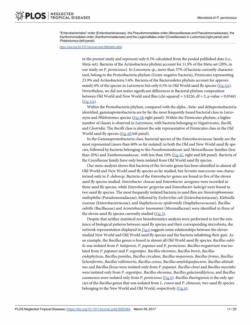
Fig 4. (A) Composition of the bacterial phyla found in Lutzomyia genus and Phlebotomus genus sand flies. Pie charts show
the proportion of each of the five phyla detected namely Proteobacteria, Firmicutes, Actinobacteria, Bacteroidetes and
Chloroflexi. (B) Relative proportion of Proteobacterial classes observed in Lutzomyia and Phlebotomus genera sand flies,
respectively on the left and relative proportion of Firmicutes bacterial classes found in Lutzomyia and Phlebotomus genera
respectively on the right. (C) Relative proportion of the major families of bacteria in the gammaproteobacterial class:
Microbiota of P. perniciosus
PLOS Neglected Tropical Diseases | https://doi.org/10.1371/journal.pntd.0005484 March 29, 2017 10 / 22
in the present study and represent only 0.5% calculated from the pooled published data (i.e.,
Meta-set). Bacteria of the Actinobacteria phylum account for 11.9% of the Meta-set (20%, in
our study on P. perniciosus). In Lutzomyia sp., more than 57% of bacteria currently character-
ized, belong to the Proteobacteria phylum (Gram-negative bacteria), Firmicutes representing
23.9% and Actinobacteria 5.6%. Bacteria of the Bacteroidetes phylum account for approxi-
mately 6% of the species in Lutzomyia but only 0.5% in Old World sand fly species (Fig 4A).
Nevertheless, we did not notice significant differences in Bacterial phylum composition
between Old World and New World sand flies (chi-squared = 5.8226, df = 2, p-value = 0.0544)
(Fig 4A).
Within the Proteobacteria phylum, compared with the alpha-, beta- and deltaproteobacteria
identified, gammaproteobacteria are by far the most frequently found bacterial class in Lutzo-myia and Phlebotomus species (Fig 4B right panel). Within the Firmicutes phylum, a higher
number of classes is observed in Lutzomyia, with bacteria belonging to Negativicutes, Bacilli,and Clostridia. The Bacilli class is almost the sole representative of Firmicutes class in the Old
World sand fly species (Fig 4B left panel).
In the Gammaproteobacteria class, bacterial species of the Enterobacteriaceae family are the
most represented (more than 60% so far isolated) in both the Old and New World sand fly spe-
cies, followed by bacteria belonging to the Pseudomonadaceae and Moraxellaceae families (less
than 20%) and Xanthomonadaceae, with less than 10% (Fig 4C right and left panel). Bacteria of
the Coxiellaceae family have only been isolated from Old World sand fly species.
Our meta-analysis shows that bacteria of the Serratia genus has been identified in almost all
Old World and New World sand fly species so far studied, but Serratia marcescens was charac-
terized only in P. duboscqi. Bacteria of the Enterobacter genus are found in five of the eleven
sand fly species studied. Enterobacter cloacae and Enterobacter aerogenes were recorded in
three sand fly species, while Enterobacter gergoviae and Enterobacter ludwigii were found in
two sand fly species. The most frequently isolated bacteria in sand flies are Stenotrophomonasmaltophilia (Pseudomonadaceae), followed by Escherichia coli (Enterobacteriaceae), Klebsiellaozaenae (Enterobacteriaceae), and Staphylococcus epidermidis (Staphylococcaceae). Bacillussubtilis (Bacillaceae) and Acinetobacter baumannii (Moraxellaceae) were identified in three of
the eleven sand fly species currently studied (Fig 5).
Despite that neither statistical nor bioinformatics analysis were performed to test the exis-
tence of biological patterns between sand fly species and their corresponding microbiota, the
network representation displayed in Fig 6 suggests some relationships between the eleven
studied New World and Old World sand fly species and the bacteria inhabiting their guts. As
an example, the Bacillus genus is found in almost all Old World sand fly species. Bacillus subti-lis was isolated from P. halepensis, P. papatasi and P. perniciosus. Bacillus megaterium was iso-
lated from P. papatasi and P. argentipes. Bacillus oleronius, Bacillus brevis, Bacillusendophyticus, Bacillus pumilus, Bacillus circulans, Bacillus mojavensis, Bacillus firmus, Bacilluslicheniformis, Bacillus vallismortis, Bacillus cereus, Bacillus amyloliquefasciens, Bacillus altitudi-nus and Bacillus flexuswere isolated only from P. papatasi. Bacillus closei and Bacillus mycoïdeswere isolated only from P. argentipes. Bacillus oleronius, Bacillus galactosidilyticus, and Bacilluscasamensis were isolated only from P. perniciosus (Fig 6). Bacillus thuringiensis is the only spe-
cies of the Bacillus genus that was isolated from L. evansi and P. chinensis, two sand fly species
belonging to the New World and Old World, respectively (Fig 6).
“Enterobacteriales” order (Enterobacteriaceae), the Pseudomonadales order (Moraxellaceae and Pseudomonadaceae), the
Xanthomonadales order (Xanthomonadaceae) and the Legionellales order (Coxiellaceae) in Lutzomyia (right panel) and
Phlebotomus (left panel).
https://doi.org/10.1371/journal.pntd.0005484.g004
Microbiota of P. perniciosus
PLOS Neglected Tropical Diseases | https://doi.org/10.1371/journal.pntd.0005484 March 29, 2017 11 / 22

Nevertheless, in the Meta-Set no significant differences in the microbiota composition at
the genus level was observed as demonstrated in the Fig 7 that depicts the Shannon (left) and
Simpson (right) indices of diversity for Old World (i.e Phlebotomus) and New World sand flies
(Lutzomyia).
Monthly dynamics of the gut bacterial consortium during the main period
of P. perniciosus activity
Fig 8A depicts the monthly proportions of each previously characterized bacterial species. To
perform this analysis, bacterial species identification was linked to each individual phylotype
recorded by PCR-ITS analysis. Then, the number of colonies harboring the same ITS-PCR
profile was determined and their monthly proportion calculated.
In July, among the nine bacterial species identified in midguts of wild P. perniciosus, only
bacteria belonging to two phyla, Proteobacteria and Firmicutes, were found. In September, the
bacteria belonged to the Actinobacteria, Proteobacteria and Firmicutes phyla. In October, only
bacteria belonging to Firmicutes were isolated (Fig 8A). Shannon and Simpson indices of
diversity confirmed a lower diversity in October (Fig 8B). Correspondence analysis performed
on data from monthly dynamics highlights the contrasting situation between October and Sep-
tember/July, depicted on the first axis. October is characterized by B. oleronus and unidentified
Fig 5. Bacterial genera (part I) and species (part II) shared by two or more of the eleven Phlebotominae sand flies species. Color chart indicate
the occurrence of the bacterial genera or species.
https://doi.org/10.1371/journal.pntd.0005484.g005
Microbiota of P. perniciosus
PLOS Neglected Tropical Diseases | https://doi.org/10.1371/journal.pntd.0005484 March 29, 2017 12 / 22

Bacillus and Lysinibacillus species. The differences between July and September/October is
depicted by the second axis of the correspondance analysis (Fig 8C). This analysis further rein-
forces our observation of a monthly evolution of the microbiota within the sand fly gut.
Discussion
Currently, there are considerable efforts to study arthropod gut microbiota, especially those of
medically important vectors. The microbiota is considered in the context of possible extended
phenotypes conferred on the insect hosts that allow niche diversification and rapid evolution
[52]. As early as 1929, Adler and Theodor [53] suggested that the presence of microorganisms
in the guts of sand flies might impact the development of the parasite Leishmania. In the mid-
80s, the team of Shlein and collaborators [12] observed a large number of “germ (bacteria)
contaminations” in guts of wild-caught female P. papatasi. However, the composition of the
Fig 6. Network analysis showing the shared bacteria species found in Phlebotomus sand flies including P. argentipes, P. duboscqii, P.
halepensis, P. papatasi, P. perlifiliewi, P. perniciosus, P. sergenti, and P. chinensis) identified by squares surrounded by green and bacteria
found in Lutzomyia sand flies including L. evansi, L. cruzi and L. longipalpis identified with squares surrounded by blue. Shared bacteria are
identified by the same colours used in Fig 5.
https://doi.org/10.1371/journal.pntd.0005484.g006
Microbiota of P. perniciosus
PLOS Neglected Tropical Diseases | https://doi.org/10.1371/journal.pntd.0005484 March 29, 2017 13 / 22

sand fly’s gut microbiota was studied much later by Dillon et al. [54]. Ochrobactrum sp. was
the first bacterium to be isolated from the midguts of P. duboscqi, a proven vector of L. majorin Sub-Saharan Africa [44], and from other sand fly species [40], including laboratory-reared
Lutzomyia longipalpis [55] and New World L. intermedia [56]. This bacterium, probably
ingested by larva, passes to nymphs and up to the adults through transstadial transmission
[44]. Recently, several publications were dedicated to the study of the microbial composition
associated with the digestive tract of sand flies. Only a few studies concerning biotic and abi-
otic factors influencing the composition of the bacterial community of the midgut of sand flies
were performed.
This study brings additional evidence on the microbiota composition in the midgut of P.
perniciosus. Our results suggest that lab-reared P. perniciosus display a lower bacterial richness
in their midgut than in field-collected sand flies. This difference is likely due in part to the type
of food diet ingested by larvae and adults during rearing. In the laboratory, P. perniciosus larvae
are fed sterile chaw (50% rabbit food plus 50% rabbit feces). After emergence, glucose is the
main source of carbohydrates for adults [25]. Under natural conditions, larvae, as well as adult
P. perniciosus, have a wide variety of diet including various sources of blood meals [18,57].
Fig 7. Shannon’s diversity (left side) and Simpson’s diversity values (right side) for Lutzomyia (Lutz) and Phlebotomus
(Phle). Error bars are 95% confidence interval.
https://doi.org/10.1371/journal.pntd.0005484.g007
Microbiota of P. perniciosus
PLOS Neglected Tropical Diseases | https://doi.org/10.1371/journal.pntd.0005484 March 29, 2017 14 / 22

Fig 8. (A) Dynamic of the midgut bacterial richness and abundance during July, September, and October
2011. An ITS-PCR profile was assigned to a bacterial species and identified by the sequencing of the 16S
rDNA locus. The percentage of each bacterial species was obtained by counting the number of colonies
having the same ITS-PCR profile and expressed as a percentage of the total colonies isolated during the
considered month. (B) Shannon’s diversity value (left side) and Simpson’s diversity value (right side) for the
Microbiota of P. perniciosus
PLOS Neglected Tropical Diseases | https://doi.org/10.1371/journal.pntd.0005484 March 29, 2017 15 / 22

Therefore, the nature of the feeding regimen leads to a striking contrast between field-collected
and laboratory-reared sand flies, which might explain the lower bacterial richness observed in
colonized sand flies.
Among the bacterial genera found associated with P. perniciosus midgut, we identified iso-
lates belonging to the Burkholderia genus and Stenotrophomonas maltophilia, an aerobic non-
fermentative and a Gram-negative bacterium. We also identified bacterial species commonly
found in the digestive tract of humans or other mammals, but which have not yet been
described in the midguts of sand flies, like Veillonella sp. In addition Sporosarcina koreensis,Rhizobium pusense and Nocardia (a rare endophyte bacterium) have never been found in asso-
ciation with the sand fly gut. The richness of sand fly-associated bacteria, illustrated by the
meta-analysis, point to some interesting outcomes. In Lutzomyia sp., more than 57% of identi-
fied bacteria belong to the Proteobacteria phylum (Gram-negative bacteria), whereas for Old
World sand fly species, including P. perniciosus, Proteobacteria (47%) and Firmicutes (40%)
are preponderant. Such a difference in the gut microbiota composition might be due to a num-
ber of factors, including the long divergence of evolution between the two subgenera [2]; some
new studies are required to assess this observation. Another surprising finding is the high rich-
ness of Bacillus species found in Old World sand flies, in which the majority of these bacteria
are host specific (Fig 6). Stenotrophomonas maltophilia, that has emerged as an important
opportunistic pathogen [58] was found to inhabit the gut of most of the sand fly species so far
studied. This bacterial species is a common microorganism found in aqueous habitats, plant
rhizosphere, animal food and water sources. Thus, delineating the origin of the colonization of
midguts by S. maltophilia and evaluating its role, if any, in the sand fly biology and physiology
are of major importance.
Our results have, for the first time, disclosed monthly variation in the diversity of the sand
fly’s gut microbiota, during the period of transmission of L. infantum. In fact, it appears that
the richness of the gut microbiota is related to sand fly seasonal activity. This diversity could
reflect the environmental conditions, such as temperature and humidity, but it may also be
linked to variations in plant cover, such as flower blooming. At the beginning of the sand fly
season (July), Ochrobactrum sp. and Serratia sp., both affiliated with the Proteobacterium phy-
lum, were the principal bacterial genera isolated. The peak of activity of P. perniciosus occurs
in September and October, a period that also corresponds to the L. infantum transmission sea-
son [59]. The analysis of the gut bacterial flora of sand flies collected in September reveals a
higher diversity (Fig 8). In particular, we recorded the presence of Microbacterium, Micrococ-cus, Kocuria, Stenotrophomonas, and Bacillus sp. (Actinobacteria, Proteobacteria and Firmi-
cutes). In July, O. intermedium and Serratia sp. are the dominant bacteria genera in the midgut
of P. perniciosus and these bacteria became undetectable towards the main peak of sand fly
activity identified in Tunisia, i.e., during the months of September and October [59]. The prev-
alence of L. infantum infection in the P. perniciosus population increases over the summer
months and reaches a peak of 9% during September-October [60,61]. Ochrobactrum interme-dium has been found previously to negatively affect Leishmania mexicana infection in L. longi-palpis [55]. Certain strains of S. marcescens are capable of producing a pigment called
prodigiosin, which ranges in color from dark red to pale pink depending on the age of the
months of July, September and October respectively. Error bars are 95% confidence interval. (C)
Correspondance analysis (CA) based on bacterial species frequencies (in black) according the different
months (July, September and October in blue). Bacteria are identified by an abbreviation: Ba, Bacillus; Br,
Brevundimonas; En, Enterococcus, Ko, Kocuria; Ly, Lysinibacillus; Mib, Microbacterium; Mic, Micrococcus;
Oc, Ochrobactrum; No, Nocardia; Rh, Rhizobium; Ro, Roseomonas; Sa, Sacharomonospora; Se, Serratia;
Sp, Sporosarcina; Ste, Stenotrophomonas; St, Staphylococcus.
https://doi.org/10.1371/journal.pntd.0005484.g008
Microbiota of P. perniciosus
PLOS Neglected Tropical Diseases | https://doi.org/10.1371/journal.pntd.0005484 March 29, 2017 16 / 22

colonies. Derivatives of prodigiosin have recently been found to have anti-T. cruzi and anti-
Leishmania (Leishmania mexicana) activity by promoting mitochondrial dysfunction leading
to parasite programmed cell death [62,63]. To what extent such interplay between the bacterial
colonies that exert toxic effects might interfere with the dynamic of L. infantum transmission
awaits further investigation.
Sand flies are vectors of medical and veterinary importance. Understanding the establish-
ment of the sand fly microbiota is critical towards clarifying underlying details of sand fly
Leishmania-microbiota interactions [64]. Bacteria such as O. intermedium, which has been
previously characterized in the guts of larvae, pupae, and adults of P. duboscqi [44], is an
opportunistic pathogen to humans [65]. Serratia sp., an entomopathogenic bacteria found in
this study, has been previously isolated from L. longipalpis [40] and L. intermedia [56]. Borde-tella avium, isolated only once from a specimen caught during July, has never been previously
isolated from sand fly midgut microflora. Bordetella avium is a highly pathogenic bacterium,
causing the avian bordetellosis [66]. Klebsiella ozaenae, known also as a human pathogenic
bacterium, has been found in four out of the ten studied sand fly species (not isolated in this
study). K. ozaenae was isolated from the midgut of gravid and freshly fed females of P. papatasiand P. halepensis [45] and from some Lutzomyia species (Fig 5). Klebsiella species are ubiqui-
tous in nature [67,68] and are recorded in all habitats where sand flies proliferate. Moreover,
the presence of K. ozaenae in the midgut of gravid females [45] will highlight their capacity to
survive in the gut of this insect. Nevertheless, as for all bacterial species known to be etiological
agents of human diseases, the sole observation of their presence in sand fly gut is not sufficient
to incriminate sand flies as a potential vector but gives information on the bacterial dissemina-
tion via blood-feeding insects. The data collected are not sufficient to incriminate sand flies as
a biological vector of K. ozaenae but are enough to raise suspicion regarding their role in the
dissemination of K. ozaenae. Furthermore, whether certain clinical outcomes from leishmania-
sis may be linked to bacteria potentially deposited during the Leishmania-infected sand fly bite
still remains to be fully investigated [68]. These studies will not only shed light on the effect of
the gut bacterial community on the sand fly fitness but also on the establishment and the trans-
mission of Leishmania parasites in endemic areas.
This meta-analysis aimed to identify the best bacterial candidate for a paratransgenic
approach. Our study is based on data aggregated from various publications that use culture-
dependent and culture-independent methodologies and various set of technical approaches
used to study the sand fly microbiota. For these reasons, conclusions raised with this study
should be taken with caution and analyzed in the light of the limitations and pitfalls inherently
associated with the compilation of heterogeneous data. Among limitations, some are linked to
the physiological state of the sample. The gut microbiome is highly dynamic [69] and therefore
influences the outcome of the analysis. When using a culture-dependent approach, we have to
keep in mind that only 20% of environmental bacteria can be grown on a growth medium
[70]. Therefore, the composition of the microbiota is not a direct reflection of the bacterial
community structure (abundance and richness) inside the insect, but an altered version of the
ecosystem from where they came. Nucleic acid-based analysis, involving historically used
methods (such as construction and Sanger sequencing of metagenomic clone libraries, auto-
mated ribosomal internal transcribed spacer analysis (ARISA), terminal restriction fragment
length polymorphism (T-RFLP), denaturing gradient gel electrophoresis (DGGE)) and next
generation sequencing technology require a critical step that must combine an efficient cell
disruption without DNA degradation and uniform nucleic acid extraction. Unfortunately, no
consensus protocol for microbial DNA extraction of insect-associated microbiota is currently
available [70]. Although 16S rRNA gene sequencing is highly useful with regards to bacterial
classification, it has a low phylogenetic power at the species level for some genera [71,72].
Microbiota of P. perniciosus
PLOS Neglected Tropical Diseases | https://doi.org/10.1371/journal.pntd.0005484 March 29, 2017 17 / 22

Depending on the 16S rRNA variable region targeted and the database used to perform the
taxonomic profiling, misassignation of bacterial OTU at the species level could be frequent
[73]. Nevertheless, taking into account all the above mentioned limits and pitfalls, we think
that an exhaustive approach aimed at collecting a maximum of data on the microbiota of sand
flies will give key information on the most commonly identified bacteria in sand fly species
and those that are more specific.
Our groups are interested in the development of a paratransgenic platform to control the
transmission of leishmaniasis. To that end, a strain of the non-pathogenic Bacillus species
(Bacillus subtilis), isolated from P. papatasi, is proposed as a possible candidate for paratrans-
genic approach. In this study, we isolated B. subtilis from P. perniciosus midgut, in addition to
other Bacillus species (Bacillus oleronius, Bacillus casamensis, Bacillus galactosidilyticus and
Bacillus sp.). Bacteria belonging to the Bacillus genus seem to display a host-specific distribu-
tion, with only B. subtilis being isolated in more than one sand fly species (P. halepensis, P.
papatasi, and P. perniciosus). In addition, we observed that no bacteria belonging to the Bacil-lus genus have been characterized to date in adult New World sand fly species. Therefore, even
if this bacterium possesses the main advantages of being non-pathogenic, easy to cultivate and
to perform genetic manipulation, its use for paratransgenic control of Leishmania can be chal-
lenged by its capacity to establish long-term colonies in the gut of various sand fly species. In
particular, if a paratransgenic approach is developed using B. subtilis as a host, it will be essen-
tial to probe its capacity to efficiently colonize the gut of Lutzomyia species and of other Old
World sand fly species in which this bacterium has yet not been found in the gut. Thus, it will
be of major epidemiological importance to develop a regional strategy for each endemic area
with different bacterial isolates.
Conclusion
The knowledge of interactions between sand flies, Leishmania and nonpathogenic microor-
ganisms that inhabit the gut will help to delineate an appropriate bacterial host recipient that
can be used for paratransgenesis designed to prevent Leishmania transmission. The identifica-
tion at the species level of the midgut’s cultured flora of P. perniciosus, linked to its seasonal
variation, is likely to provide new perspectives towards a better understanding of the role of
the gut bacterial community on sand fly-pathogen interactions. This knowledge is crucial in
order to implement control strategies for sand fly zoonotic visceral leishmaniasis.
Acknowledgments
We thank the anonymous referees for their helpful scientific comments that have greatly
improve the quality of the manuscript.
Author Contributions
Conceptualization: EZ MG RD DS PP FD MRO.
Data curation: DS PP FD.
Formal analysis: PP FD DS.
Investigation: WFr WFa WB.
Methodology: WFr IS WFa MG.
Resources: EZ MG IC SC.
Supervision: DS EZ RD MG.
Microbiota of P. perniciosus
PLOS Neglected Tropical Diseases | https://doi.org/10.1371/journal.pntd.0005484 March 29, 2017 18 / 22

Validation: EZ IC.
Writing – original draft: EZ PP DS.
Writing – review & editing: EZ PP DS.
References1. Maroli M, Feliciangeli MD, Bichau L, Charrel RN, Gradoni L: Phlebotomine sandflies and the spreading
of leishmaniases and other diseases of public health concern. Med Vet Entomol. 2013; 27:123–47.
https://doi.org/10.1111/j.1365-2915.2012.01034.x PMID: 22924419
2. Akhoundi M, Kuhls K, Cannet A, Votypka J, Marty P, Delaunay P, Sereno D. A historical overview of the
classification, evolution, and dispersion of Leishmania Parasites and Sandflies. PLoS Negl Trop Dis.
2016; 10:e0004349. https://doi.org/10.1371/journal.pntd.0004349 PMID: 26937644
3. Desjeux P. Leishmaniasis: current situation and new perspectives. Comp Immunol Microbiol Infect Dis.
2004; 27:305–18. https://doi.org/10.1016/j.cimid.2004.03.004 PMID: 15225981
4. Alvar J, Velez ID, Bern C, Herrero M, Desjeux P, Cano J, Janin J, WHO Leishmaniasis control team.
Leishmaniasis worldwide and global estimates of its incidence. PLoS ONE. 2012; 7:e35671. https://doi.
org/10.1371/journal.pone.0035671 PMID: 22693548
5. World Health Organization (WHO). Urbanization: An increasing risk factor for Leishmaniasis. Wkly Epi-
demiol Record. 2002; 77:365–72.
6. Seblova V, Oury B, Eddaikra N, Aït-Oudhia K, Pratlong F, Gazanion E, Maïa C, Volf P, Sereno D. Trans-
mission potential of antimony-resistant Leishmania field isolates. Antimicrob Agents Chemother. 2014;
58:6273–76. https://doi.org/10.1128/AAC.02406-13 PMID: 25049256
7. Sereno D, Maia C, Aït-Oudhia K. Antimony resistance and environment: Elusive links to explore during
Leishmania life cycle. Int J Parasitol Drugs Drug Resist. 2012; 2:200–03. https://doi.org/10.1016/j.
ijpddr.2012.07.003 PMID: 24533281
8. Depaquit J, Ferte H, Leger N. The subgenus Paraphlebotomus (Phlebotomus, Phlebotominae, Psycho-
didae, Diptera): a review. Morphological and Molecular Studies. Ann Pharm Fr. 2002; 58:333–40.
9. Dedet JP. Les leishmanioses en Afrique du Nord. Bull Inst Pasteur. 1979; 77:85–94.
10. Ready PD. Biology of phlebotomine sand flies as vectors of disease agents. Annu Rev Entomol. 2013;
58:227–50. https://doi.org/10.1146/annurev-ento-120811-153557 PMID: 23317043
11. Rogers ME, Hajmova M, Joshi MB, Sadlova J, Dwyer DM, Volf P, Bates PA. Leishmania chitinase facili-
tates colonization of sand fly vectors and enhances transmission to mice. Cell Microbiol. 2008;
10:1363–72. https://doi.org/10.1111/j.1462-5822.2008.01132.x PMID: 18284631
12. Schlein Y, Polacheck I, Yuval B. Mycoses, bacterial infection an antibacterial activity in sandflies (Psy-
chodidae) and their possible role in the transmission of leishmaniasis. Parasitol. 1985, 90:57–66.
13. Dillon RJ, El Kordy E, Lanee RP. The prevalence of a microbiota in the digestive tract of Phlebotomus
papatasi. Ann Trop Med Parasitol. 1996; 90:669–73. PMID: 9039284
14. Tanada Y, Kaya HK. Associations between insects and nonpathogenics microorganisms. In Tanada
Y. & Kaya H.K. (eds.), Insect pathology. Academic Press, New York; 1993. pp.12–51.
15. Dillon RJ, Dillon VM. The gut bacteria of insects: nonpathogenic interactions. Annu Rev Entomol. 2004;
49:71–92. https://doi.org/10.1146/annurev.ento.49.061802.123416 PMID: 14651457
16. Cameron MM, Pessoa FA, Vasconcelos AW, Ward RD. Sugar meal sources for the phlebotomine sand-
flies Lutzomyia longipalpis in Ceara State, Brazil. Med Vet Entomol. 1995; 9:263–72. PMID: 7548943
17. Schlein Y, Yuval B. Leishmaniasis in the Jordan Valley. Attraction of Phlebotomus papatasi (Diptera:
Psychodidae) to plants in the field. J Med Entomol. 1987; 50:20–27.
18. Schlein Y. Sandfly diet and Leishmania. Parasitol. 1986; 2:175–77.
19. Durvasula R, Gumbs A, Panackal A, Kruglov O, Aksoy S, Merrifield RB, Richards FF, Beard CB. Pre-
vention of insect-borne disease: an approach using transgenic symbiotic bacteria. Proc Natl Acad Sci
USA. 1997; 94:3274–78. PMID: 9096383
20. Berasategui A, Shukla S, Salem H, Kaltenpoth M. Potential applications of insect symbionts in biotech-
nology. Appl Microbiol Biotechnol. 2016; 100:1567–77. https://doi.org/10.1007/s00253-015-7186-9
PMID: 26659224
21. Mukhopadhyay J, Braig HR, Rowton ED, Ghosh K. Naturally occurring culturable aerobic gut flora of
adult Phlebotomus papatasi, vector of Leishmania major in the Old World. PLoS ONE. 2012; 7:e35748.
https://doi.org/10.1371/journal.pone.0035748 PMID: 22629302
Microbiota of P. perniciosus
PLOS Neglected Tropical Diseases | https://doi.org/10.1371/journal.pntd.0005484 March 29, 2017 19 / 22

22. Hillesland H, Read A, Subhadra B, Hurwitz I, McKelvey R, Ghosh K, Das P, Durvasula R. Identification
of aerobic gut bacteria from the Kala Azar vector, Phlebotomus argentipes: A platform for potential para-
transgenic manipulation of sand flies. Am J Trop Med Hyg. 2008; 79:881–86. PMID: 19052297
23. Hurwitz I, Hillesland H, Fieck A, Das P, Duvarsala R. The paratransgenic sand fly: a platform for control
of Leishmania transmission. Parasites & Vectors. 2011; 4: 82.
24. Ben Ismail R. Incrimination de Phlebotomus perniciosus comme vecteur de Leishmania infantum. Arch
Inst Pasteur de Tunis. 1993; 70:91–110.
25. Chelbi I, Zhioua E. Biologiy of Phlebotomus papatasi (Diptera: Psychodidae) in the Laboratory. J Med
Entomol. 2007; 44:597–00. PMID: 17695013
26. Croset H, Rioux JA, Master M, Bayar N. Les phlebotomes de la Tunisie (Diptera, Phlebotominae). Mise
au point systematique, chronologique et ethologique. Ann Parasitol Hum Comp. 1978; 53:711–49.
PMID: 754625
27. Leger N, Pesson B, Madulo-Leblond G, Abonnenc E. Sur la differenciation des femelles du sous genre
Larroussisus Nitzulescu, 1931(Diptera, Phlebotominae) de la region mediterraneenne. Ann Parasitol
Hum Comp. 1983; 85:611–23.
28. De Bruijn FJ. Handbook of molecular microbial ecology II: metagenomics in different habitats. Ed Wiley
Blackwell 2011, pp: 96.
29. Fisher MM, Triplett EW. Automated approach for ribosomal intergenic spacer analysis of microbial
diversity and its application to freshwater bacterial communities. Appl Environ Microbiol. 1999;
65:4630–36 PMID: 10508099
30. Garcıa-Martınez J, Acinas SG, Anton AI, Rodrıguez-Valera F. Use of the 16S—23S ribosomal genes
spacer region in studies of prokaryotic diversity. J Microbiol Meth. 1999; 36:55–64.
31. Daffonchio D, Cherif A, Brusetti L, Rizzi A, Mora D, Boudabous A, Borin S. Nature of polymorphisms in
16S-23S rRNA gene Intergenic Transcribed Spacer fingerprinting of Bacillus and related Genera. Appl
Env Microbiol. 2000: 69:5128–37.
32. Wheeler A, Oerther DB, Larsen N, Stahl DA, Raskin L. The oligonucleotide probe database. Appl Envi-
ron Microbiol. 1996; 62:3557–59. PMID: 8837410
33. Yu Z, Morrison M. Comparisons of different hypervariable regions of rrs genes for use in fingerprinting
of microbial communities by PCR-denaturing gradient gelelectrophoresis. Appl Environ Microbiol. 2004;
70:4800–06. https://doi.org/10.1128/AEM.70.8.4800-4806.2004 PMID: 15294817
34. Sass AM, Sass H, Coolen MJ, Cypionka H, Overmann J. Microbial communities in the chemocline of a
hypersaline deep-sea basin (Urania basin, Mediterranean Sea). Appl Environ Microbiol. 2001;
67:5392–402. https://doi.org/10.1128/AEM.67.12.5392-5402.2001 PMID: 11722884
35. Muyzer J, Smalla K. Application of denaturing gradient gel electrophoresis (DGGE) and temperature
gradient gel electrophoresis (TGGE) in microbial ecology. Antonie van Leeuwenhoek. 1998; 73:
127–41. PMID: 9602286
36. Ettoumi B, Bouhajja E, Borin S, Daffonchio D, Boudabous A, Cherif A. Gammaproteobacteria occur-
rence and microdiversity in Tyrrhenian Sea sediments as revealed by cultivation-dependent and
-independent approaches. Syst Appl Microbiol. 2010; 33:222–31. https://doi.org/10.1016/j.syapm.
2010.02.005 PMID: 20413241
37. Cole JR, Wang Q, Cardenas E, Fish J, Chai B, Farris RJ, Kulam-Syed-Mohideen AS, McGarrell DM,
Marsh T, Garrity GM, Tiedje JM. The Ribosomal Database Project: improved alignments and new tools
for rRNA analysis. Nucleic Acids Res. 2009; 37 (Database issue): D141–5. https://doi.org/10.1093/nar/
gkn879 PMID: 19004872
38. Stackerbrandt E, Goebel BM. Taxonomic Note: A Place for DNA-DNA Reassociation and 16S rRNA
Sequence Analysis in the Present Species Definition in Bacteriology. Int J Syst Evol Microbiol. 1994;
44:846–49.
39. Oksanen J, Blanchet FG, Kindt R, Legendre P, Minchin PR, O’Hara RB (2015). Vegan: Community
Ecology. http://cran.r-project.org/package=vegan
40. Sant’anna MRV, Darby AV, Brazil RP, Montoya-Lerma J, Dillon VM, Bates PA, Dillon RJ. Investigation
of the bacterial communities associated with females of Lutzomyia sand fly species from South Amer-
ica. Plos one. 2012; 7:e42531. https://doi.org/10.1371/journal.pone.0042531 PMID: 22880020
41. Gouveia C, Asensi MD, Zahner V, Rangel EF, De Oliveira SMP. Study on the bacterial midgut micro-
biota associated to different Brazilian populations of Lutzomyia longipalpis (Lutz & Neiva) (Diptera: Psy-
chodidae). Neotrop Entomol. 2008; 37:597–60. PMID: 19061048
42. McCarthy CB, Diambra LA, Pomar RVR. Metagenomic analysis of taxa associated with Lutzomyia long-
ipalpis, vector of visceral leishmaniasis, using an unbiased high-throughput approach. PLoS Negl Trop
Dis. 2011; 5:9.
Microbiota of P. perniciosus
PLOS Neglected Tropical Diseases | https://doi.org/10.1371/journal.pntd.0005484 March 29, 2017 20 / 22

43. Vivero RJ, Jaramillo NG, Cadavid-Restrepo G, Soto SI, Herrera CX. Structural differences in gut bacte-
ria communities in developmental stages of natural populations of Lutzomyia evansi from Colombia’s
Caribbean coast. Parasit Vectors. 2016; 9:496. https://doi.org/10.1186/s13071-016-1766-0 PMID:
27618991
44. Volf P, Kiewegova A, Nemec A. Bacterial colonisation in the gut of Phlebotomus duboscqi (Diptera: Psy-
chodidae): Transtadial passage and the role of female diet. Folia Parasitol. 2002; 49:73–7. PMID:
11993554
45. Akhoundi A, Bakhtiari R, Guillard T, Baghaei A, Tolouei R, Sereno D, Toubas D, Depaquit J, Abyaneh
MR. Diversity of the bacterial and fungal microflora from the midgut and cuticle of Phlebotomine sand
flies collected in North-Western Iran. PloS ONE. 2012; 7:11.
46. Guernaoui S, Garcia D, Gazanion E, Ouhdouch Y, Boumezzough A, Pesson B, Fontenille D, Sereno D:
Bacterial flora as indicated by PCR-temperature gradient gel electrophoresis (TGGE) of 16S rDNA
gene fragments from isolated guts of phlebotomine sand flies (Diptera: Psychodidae). J Vec Ecol. 2011;
36:S144–7.
47. Maleki-Ravasan N, Oshaghi MA, Afshar D, Arandian MH, Hajibani S., Akhvan AA, Yakhckali B, Shirazi
MH, Rassi Y, Jafari R, Aminian K, Fazeli-Varzaneh RA, Duvarsala R. Aerobic bacterial flora of biotic
and abiotic compartments of a hyperendemic zoonotic cutaneous Leishmaniasis (ZCL) focus. Parasite
& Vect. 8:63.
48. Mukhopadhyay J, Braig HR, Rowton ED, Ghosh K. Naturally occurring culturable aerobic gut flora of
adult Phlebotomus papatasi, vector of Leishmania major in the Old World. PLoS ONE. 2012; 7: 5.
49. Li K, Chen H, Jiang J, Li X, Xu J, Ma Y. Diversity of bacteriome associated with Phlebotomus chinensis
(Diptera: Psychodidae) sand flies in two wild populations from China. Sci Rep. 2016; 6:36406. https://
doi.org/10.1038/srep36406 PMID: 27819272
50. Shannon P, Markiel A, Ozier O, Baliga NS, Wang JT, Ramage D, Amin N, Schwikowski B, Ideker T.
Cytoscape: a software environment for integrated models of biomolecular interaction networks.
Genome Res. 2003; 13:2498–2504. https://doi.org/10.1101/gr.1239303 PMID: 14597658
51. Matsumoto K, Izri A, Dumon H, Raoult D, Parola P. First detection of Wolbachia spp., including a new
genotype, in sand flies collected in Marseille, France. J Med Entomol. 2008; 45:466–9. PMID:
18533441
52. Minard G, Mavingui P, Moro CV. Diversity and function of bacterial microbiota in the mosquito holobiont.
Parasit Vectors. 2013; 6:146. https://doi.org/10.1186/1756-3305-6-146 PMID: 23688194
53. Adler S, Theodor O. Attempts to transmit Leishmania tropica by bite: the transmission of L. tropica by
Phlebotomus sergenti. Ann Trop Med Parasitol. 1929; 23:1–18.
54. Dillon RJ, El Kordy E, Shehata M, Lane RP. The prevalence of a microbiota in the digestive tract of Phle-
botomus papatasi. Ann Trop Med Parasitol. 1996; 90:669–73. PMID: 9039284
55. Sant’Anna MRV, Diaz-Albiter H, Aguiar-Martins K, Al Salem WS, Cavalcante RR, Dillon VM, Bates PA,
Genta FA, Dillon RJ: Colonization resistance in the sand fly gut: Leishmania protects Lutzomyia longi-
palpis from bacterial infection. Parasit & Vect. 2014; 7:329.
56. Monteiro CC, Villegas LE, Campolina TB, Pires AC, Miranda JC, Pimenta PF, Secundino NF. Bacterial
diversity of the American sand fly Lutzomyia intermedia using high-throughput metagenomic sequenc-
ing. Parasit Vectors. 2016; 9:480. https://doi.org/10.1186/s13071-016-1767-z PMID: 27581188
57. Samie M, Wallbanks KR, Moore JS, Molineux DH. Glycosidase activity in the sandfly Phlebotomus
papatasi. Comp Biochem Physiol. 1990; 96:577–79.
58. Brook JS. Stenotrophomonas maltophilia: an emerging global opportunisitic pathogen. Clin Microbiol
Rev. 2012; 25:2–41. https://doi.org/10.1128/CMR.00019-11 PMID: 22232370
59. Bichaud L, Dachraoui K, Piorkowski G, Chelbi I, Moureau G, Cherni S, De Lamballerie X, Sakhria S,
Charrel RN, Zhioua E. Isolation of Toscana virus from sand flies, Tunisia. Emerg Infect Dis. 2013; 19:
322–324.
60. Zoghlami Z, Chouihi E, Barhoumi W, Dachraoui K, Massoudi N, Ben Helel K, Habboul Z, Hadhri MH,
Limam S, Mhadhbi M, Gharbi M, Zhioua E. Interaction between canine and human visceral leishmania-
ses in a holoendemic focus of Central Tunisia. Acta Trop. 2014; 139:32–8. https://doi.org/10.1016/j.
actatropica.2014.06.012 PMID: 25004438
61. Barhoumi W, Fares W, Cherni S, Derbali M, Dachraoui K, Chelbi I, Ramalho-Ortigao M, Beier JC,
Zhioua E. Changes of sand fly populations and Leishmania infantum infection rates in an irrigated vil-
lage located in arid Central Tunisia. Inter J Envir Res Public Health. 2016; 13: 329.
62. Moraes CS, Seabra SH, Castro DP, Brazil RP, de Souza W, Garcia ES, Azambuja P: Leishmania
(Leishmania) chagasi interactions with Serratia marcescens: ultrastructural studies, lysis and carbohy-
drate effects. Exp Parasitol. 2008; 118:561–568. https://doi.org/10.1016/j.exppara.2007.11.015 PMID:
18206142
Microbiota of P. perniciosus
PLOS Neglected Tropical Diseases | https://doi.org/10.1371/journal.pntd.0005484 March 29, 2017 21 / 22

63. Genes C, Baquero E, Echeverri F, Maya JD, Triana O. Mitochondrial dysfunction in Trypanosoma cruzi:
the role of Serratia marcescens prodigiosin in the alternative treatment of Chagas disease. Parasites
&Vect. 2011; 4:66.
64. Kelly PH, Bahr SM, Serafim TD, Ajami NJ, Petrosino JF, Meneses C, Kirby JR, Valenzuela JG, Kam-
hawi S, Wilson ME. The Gut Microbiome of the Vector Lutzomyia longipalpis is Essential for Survival of
Leishmania infantum. MBio. 2017; 8(1).
65. Teyssier C, Marchandin H, Jean-Pierre H., Diego I, Darbas H, Jeannot JL, Gouby A, Jumas-Bilak E.
Molecular and phenotypic features for identification of the opportunistic pathogens Ochrobactrum spp.
J Med Microbiol. 2005; 54:945. https://doi.org/10.1099/jmm.0.46116-0 PMID: 16157548
66. Raffel TR, Register KB, Marks SA, Temple L. Prevalence of Bordetella avium infection in selected wild
and domesticated birds in the Eastern USA. J Wild Dis. 2002; 38:40–6.
67. Botelho-Nevers E, Gouriet F, Lepidi H, Couvret A, Amphoux B, Dessi P, Raoult D. Chronic nasal infec-
tion caused by Klebsiella rhinoscleromatis or Klebsiella ozaenae: two forgotten infectious diseases.
Inter J Infect Dis. 2007; 11:423–29.
68. Bagley S. Habitat association of Klebsiella species. Infect Control. 1985; 6:52–8. PMID: 3882590
69. Finney CA, Kamhawi S, Wasmuth JD. Does the arthropod microbiota impact the establishment of vec-
tor-borne diseases in mammalian hosts? PLoS Pathog. 2015; 11:e1004646. https://doi.org/10.1371/
journal.ppat.1004646 PMID: 25856431
70. Hiergeist A, Glasner J, Reischl U, Gessner A. Analyses of Intestinal Microbiota: Culture versus
Sequencing. ILAR J. 2015; 56:228–40. https://doi.org/10.1093/ilar/ilv017 PMID: 26323632
71. Janda JM, Abbott SL. 16S rRNA gene sequencing for bacterial identification in the diagnostic labora-
tory: pluses, perils, and pitfalls. J Clin Microbiol. 2007; 45:2761–2764. https://doi.org/10.1128/JCM.
01228-07 PMID: 17626177
72. Drancourt M, Bollet C, Carlioz A, Martelin R, Gayral JP, Raoult D. 16S ribosomal DNA sequence analy-
sis of a large collection of environmental and clinical unidentifiable bacterial isolates. J Clin Microbiol.
2000; 3:3623–30.
73. Yang B, Wang Y, Qian PY. Sensitivity and correlation of hypervariable regions in 16S rRNA genes in
phylogenetic analysis. BMC Bioinformatics. 2016; 17:13.
Microbiota of P. perniciosus
PLOS Neglected Tropical Diseases | https://doi.org/10.1371/journal.pntd.0005484 March 29, 2017 22 / 22
Related Documents