An Insight into the Transcriptome of the Digestive Tract of the Bloodsucking Bug, Rhodnius prolixus Jose ´ M. C. Ribeiro 1 *, Fernando A. Genta 2,3 , Marcos H. F. Sorgine 2,4 , Raquel Logullo 5 , Rafael D. Mesquita 2,5 , Gabriela O. Paiva-Silva 2,4 , David Majerowicz 4 , Marcelo Medeiros 6 , Leonardo Koerich 2,7 , Walter R. Terra 2,8 , Cle ´ lia Ferreira 2,8 , Andre ´ C. Pimentel 8 , Paulo M. Bisch 9 , Daniel C. Leite 9 , Michelle M. P. Diniz 9 , Joa ˜o Lı ´dio da S. G. V. Junior 9,10 , Manuela L. Da Silva 6,9 , Ricardo N. Araujo 2,11 , Ana Caroline P. Gandara 4 , Se ´ bastien Brosson 12 , Didier Salmon 4 , Sabrina Bousbata 12 , Natalia Gonza ´ lez-Caballero 3 , Ariel Mariano Silber 13 , Michele Alves-Bezerra 4 , Katia C. Gondim 2,4 , Ma ´ rio Alberto C. Silva-Neto 2,4 , Georgia C. Atella 2,4 , Helena Araujo 2,14 , Felipe A. Dias 4 , Carla Polycarpo 2,4 , Raquel J. Vionette-Amaral 2,4 , Patrı ´cia Fampa 15 , Ana Claudia A. Melo 2,5 , Aparecida S. Tanaka 2,16 , Carsten Balczun 17 , Jose ´ Henrique M. Oliveira 4 , Renata L. S. Gonc ¸ alves 4 , Cristiano Lazoski 2,7 , Rolando Rivera-Pomar 18,19 , Luis Diambra 18 , Gu ¨ nter A. Schaub 17 , Elo ´ i S. Garcia 2,3 , Patrı´cia Azambuja 2,3 , Glo ´ ria R. C. Braz 2,5 *, Pedro L. Oliveira 2,4 * 1 Section of Vector Biology, Laboratory of Malaria and Vector Research, National Institute of Allergy and Infectious Diseases, National Institutes of Health, Rockville, Maryland, United States of America, 2 Instituto Nacional de Cie ˆ ncia e Tecnologia em Entomologia Molecular, Federal University of Rio de Janeiro, Rio de Janeiro, Brazil, 3 Instituto Oswaldo Cruz, Fundac ¸a ˜ o Oswaldo Cruz, Rio de Janeiro, Rio de Janeiro, Brazil, 4 Instituto de Bioquı ´mica Me ´ dica, Programa de Biotecnologia e Biologia Molecular, Universidade Federal do Rio de Janeiro, Rio de Janeiro, Brazil, 5 Department of Biochemistry, Institute of Chemistry, Federal University of Rio de Janeiro, Rio de Janeiro, Brazil, 6 Instituto Nacional de Metrologia Qualidade e Tecnologia, Diretoria de Metrologia Aplicada a `s Cie ˆ ncias da Vida, Programa de Biotecnologia, Pre ´dio 27, CEP 25250- 020, Duque de Caxias, Rio de Janeiro, Brazil, 7 Departamento de Gene ´ tica, Instituto de Biologia, Universidade Federal do Rio de Janeiro, CEP 21944-970, Rio de Janeiro, Brazil, 8 Departamento de Bioquı ´mica, Instituto de Quı ´mica, Universidade de Sa ˜o Paulo, Sa ˜o Paulo, Brazil, 9 Instituto de Biofı ´sica Carlos Chagas Filho, Universidade Federal do Rio de Janeiro, Rio de Janeiro, Brazil, 10 Center for Technological Innovation, Evandro Chagas Institute, Ananindeua, Para ´, Brazil, 11 Departamento de Parasitologia do Instituto de Cie ˆ ncias Biolo ´ gicas da Universidade Federal de Minas Gerais, Belo Horizonte, Minas Gerais, Brazil, 12 Institute for Molecular Biology and Medicine (IBMM), Universite ´ Libre de Bruxelles, Gosselies, Belgium, 13 Departamento de Parasitologia, Instituto de Cie ˆ ncias Biome ´ dicas, Universidade de Sa ˜o Paulo, Sa ˜o Paulo, Brazil, 14 Institute for Biomedical Sciences, Federal University of Rio de Janeiro, Rio de Janeiro, Brazil, 15 Instituto de Biologia, DBA, UFRRJ, Serope ´ dica, Rio de Janeiro, Brazil, 16 Departamento de Bioquı ´mica, Escola Paulista de Medicina, Universidade Federal de Sa ˜o Paulo, Sa ˜o Paulo, Brazil, 17 Zoology/Parasitology Group, Ruhr-Universita ¨t, Bochum, Germany, 18 Centro Regional de Estudios Genomicos, Universidad Nacional de La Plata, Florencio Varela, Argentina, 19 Centro de Bioinvestigaciones, Universidad Nacional del Noroeste de Buenos Aires, Pergamino, Argentina Abstract The bloodsucking hemipteran Rhodnius prolixus is a vector of Chagas’ disease, which affects 7–8 million people today in Latin America. In contrast to other hematophagous insects, the triatomine gut is compartmentalized into three segments that perform different functions during blood digestion. Here we report analysis of transcriptomes for each of the segments using pyrosequencing technology. Comparison of transcript frequency in digestive libraries with a whole-body library was used to evaluate expression levels. All classes of digestive enzymes were highly expressed, with a predominance of cysteine and aspartic proteinases, the latter showing a significant expansion through gene duplication. Although no protein digestion is known to occur in the anterior midgut (AM), protease transcripts were found, suggesting secretion as pro- enzymes, being possibly activated in the posterior midgut (PM). As expected, genes related to cytoskeleton, protein synthesis apparatus, protein traffic, and secretion were abundantly transcribed. Despite the absence of a chitinous peritrophic membrane in hemipterans - which have instead a lipidic perimicrovillar membrane lining over midgut epithelia - several gut-specific peritrophin transcripts were found, suggesting that these proteins perform functions other than being a structural component of the peritrophic membrane. Among immunity-related transcripts, while lysozymes and lectins were the most highly expressed, several genes belonging to the Toll pathway - found at low levels in the gut of most insects - were identified, contrasting with a low abundance of transcripts from IMD and STAT pathways. Analysis of transcripts related to lipid metabolism indicates that lipids play multiple roles, being a major energy source, a substrate for perimicrovillar membrane formation, and a source for hydrocarbons possibly to produce the wax layer of the hindgut. Transcripts related to amino acid metabolism showed an unanticipated priority for degradation of tyrosine, phenylalanine, and tryptophan. Analysis of transcripts related to signaling pathways suggested a role for MAP kinases, GTPases, and LKBP1/ AMP kinases related to control of cell shape and polarity, possibly in connection with regulation of cell survival, response of pathogens and nutrients. Together, our findings present a new view of the triatomine digestive apparatus and will help us understand trypanosome interaction and allow insights into hemipteran metabolic adaptations to a blood-based diet. Citation: Ribeiro JMC, Genta FA, Sorgine MHF, Logullo R, Mesquita RD, et al. (2014) An Insight into the Transcriptome of the Digestive Tract of the Bloodsucking Bug, Rhodnius prolixus. PLoS Negl Trop Dis 8(1): e2594. doi:10.1371/journal.pntd.0002594 Editor: Christian Tschudi, Yale School of Public Health, United States of America Received August 6, 2013; Accepted November 4, 2013; Published January 9, 2014 PLOS Neglected Tropical Diseases | www.plosntds.org 1 January 2014 | Volume 8 | Issue 1 | e2594

Welcome message from author
This document is posted to help you gain knowledge. Please leave a comment to let me know what you think about it! Share it to your friends and learn new things together.
Transcript
An Insight into the Transcriptome of the Digestive Tractof the Bloodsucking Bug, Rhodnius prolixusJose M. C. Ribeiro1*, Fernando A. Genta2,3, Marcos H. F. Sorgine2,4, Raquel Logullo5, Rafael D. Mesquita2,5,
Gabriela O. Paiva-Silva2,4, David Majerowicz4, Marcelo Medeiros6, Leonardo Koerich2,7, Walter R. Terra2,8,
Clelia Ferreira2,8, Andre C. Pimentel8, Paulo M. Bisch9, Daniel C. Leite9, Michelle M. P. Diniz9, Joao Lıdio
da S. G. V. Junior9,10, Manuela L. Da Silva6,9, Ricardo N. Araujo2,11, Ana Caroline P. Gandara4,
Sebastien Brosson12, Didier Salmon4, Sabrina Bousbata12, Natalia Gonzalez-Caballero3, Ariel
Mariano Silber13, Michele Alves-Bezerra4, Katia C. Gondim2,4, Mario Alberto C. Silva-Neto2,4,
Georgia C. Atella2,4, Helena Araujo2,14, Felipe A. Dias4, Carla Polycarpo2,4, Raquel J. Vionette-Amaral2,4,
Patrıcia Fampa15, Ana Claudia A. Melo2,5, Aparecida S. Tanaka2,16, Carsten Balczun17, Jose
Henrique M. Oliveira4, Renata L. S. Goncalves4, Cristiano Lazoski2,7, Rolando Rivera-Pomar18,19,
Luis Diambra18, Gunter A. Schaub17, Eloi S. Garcia2,3, Patrıcia Azambuja2,3, Gloria R. C. Braz2,5*,
Pedro L. Oliveira2,4*
1 Section of Vector Biology, Laboratory of Malaria and Vector Research, National Institute of Allergy and Infectious Diseases, National Institutes of Health, Rockville,
Maryland, United States of America, 2 Instituto Nacional de Ciencia e Tecnologia em Entomologia Molecular, Federal University of Rio de Janeiro, Rio de Janeiro, Brazil,
3 Instituto Oswaldo Cruz, Fundacao Oswaldo Cruz, Rio de Janeiro, Rio de Janeiro, Brazil, 4 Instituto de Bioquımica Medica, Programa de Biotecnologia e Biologia Molecular,
Universidade Federal do Rio de Janeiro, Rio de Janeiro, Brazil, 5 Department of Biochemistry, Institute of Chemistry, Federal University of Rio de Janeiro, Rio de Janeiro,
Brazil, 6 Instituto Nacional de Metrologia Qualidade e Tecnologia, Diretoria de Metrologia Aplicada as Ciencias da Vida, Programa de Biotecnologia, Predio 27, CEP 25250-
020, Duque de Caxias, Rio de Janeiro, Brazil, 7 Departamento de Genetica, Instituto de Biologia, Universidade Federal do Rio de Janeiro, CEP 21944-970, Rio de Janeiro,
Brazil, 8 Departamento de Bioquımica, Instituto de Quımica, Universidade de Sao Paulo, Sao Paulo, Brazil, 9 Instituto de Biofısica Carlos Chagas Filho, Universidade Federal
do Rio de Janeiro, Rio de Janeiro, Brazil, 10 Center for Technological Innovation, Evandro Chagas Institute, Ananindeua, Para, Brazil, 11 Departamento de Parasitologia do
Instituto de Ciencias Biologicas da Universidade Federal de Minas Gerais, Belo Horizonte, Minas Gerais, Brazil, 12 Institute for Molecular Biology and Medicine (IBMM),
Universite Libre de Bruxelles, Gosselies, Belgium, 13 Departamento de Parasitologia, Instituto de Ciencias Biomedicas, Universidade de Sao Paulo, Sao Paulo, Brazil,
14 Institute for Biomedical Sciences, Federal University of Rio de Janeiro, Rio de Janeiro, Brazil, 15 Instituto de Biologia, DBA, UFRRJ, Seropedica, Rio de Janeiro, Brazil,
16 Departamento de Bioquımica, Escola Paulista de Medicina, Universidade Federal de Sao Paulo, Sao Paulo, Brazil, 17 Zoology/Parasitology Group, Ruhr-Universitat,
Bochum, Germany, 18 Centro Regional de Estudios Genomicos, Universidad Nacional de La Plata, Florencio Varela, Argentina, 19 Centro de Bioinvestigaciones,
Universidad Nacional del Noroeste de Buenos Aires, Pergamino, Argentina
Abstract
The bloodsucking hemipteran Rhodnius prolixus is a vector of Chagas’ disease, which affects 7–8 million people today inLatin America. In contrast to other hematophagous insects, the triatomine gut is compartmentalized into three segmentsthat perform different functions during blood digestion. Here we report analysis of transcriptomes for each of the segmentsusing pyrosequencing technology. Comparison of transcript frequency in digestive libraries with a whole-body library wasused to evaluate expression levels. All classes of digestive enzymes were highly expressed, with a predominance of cysteineand aspartic proteinases, the latter showing a significant expansion through gene duplication. Although no proteindigestion is known to occur in the anterior midgut (AM), protease transcripts were found, suggesting secretion as pro-enzymes, being possibly activated in the posterior midgut (PM). As expected, genes related to cytoskeleton, proteinsynthesis apparatus, protein traffic, and secretion were abundantly transcribed. Despite the absence of a chitinousperitrophic membrane in hemipterans - which have instead a lipidic perimicrovillar membrane lining over midgut epithelia -several gut-specific peritrophin transcripts were found, suggesting that these proteins perform functions other than being astructural component of the peritrophic membrane. Among immunity-related transcripts, while lysozymes and lectins werethe most highly expressed, several genes belonging to the Toll pathway - found at low levels in the gut of most insects -were identified, contrasting with a low abundance of transcripts from IMD and STAT pathways. Analysis of transcriptsrelated to lipid metabolism indicates that lipids play multiple roles, being a major energy source, a substrate forperimicrovillar membrane formation, and a source for hydrocarbons possibly to produce the wax layer of the hindgut.Transcripts related to amino acid metabolism showed an unanticipated priority for degradation of tyrosine, phenylalanine,and tryptophan. Analysis of transcripts related to signaling pathways suggested a role for MAP kinases, GTPases, and LKBP1/AMP kinases related to control of cell shape and polarity, possibly in connection with regulation of cell survival, response ofpathogens and nutrients. Together, our findings present a new view of the triatomine digestive apparatus and will help usunderstand trypanosome interaction and allow insights into hemipteran metabolic adaptations to a blood-based diet.
Citation: Ribeiro JMC, Genta FA, Sorgine MHF, Logullo R, Mesquita RD, et al. (2014) An Insight into the Transcriptome of the Digestive Tract of the BloodsuckingBug, Rhodnius prolixus. PLoS Negl Trop Dis 8(1): e2594. doi:10.1371/journal.pntd.0002594
Editor: Christian Tschudi, Yale School of Public Health, United States of America
Received August 6, 2013; Accepted November 4, 2013; Published January 9, 2014
PLOS Neglected Tropical Diseases | www.plosntds.org 1 January 2014 | Volume 8 | Issue 1 | e2594
This is an open-access article, free of all copyright, and may be freely reproduced, distributed, transmitted, modified, built upon, or otherwise used by anyone for anylawful purpose. The work is made available under the Creative Commons CC0 public domain dedication.
Funding: JMCR was supported by the Intramural Research Program of the NIH, National Institute of Allergy and Infectious Diseases. GRCB, FAG, ACAM, DS, SaB andSeB were supported by CAPES; CP, HMA, MHFS, PLO, GRCB, ACAM, AMS and FAG were supported by CNPq; AMS, ACP, AST, CF and WRT were supported by FAPESP;ACAM, FAG, CP, HMA, MHFS, PLO and GRCB were supported by FAPERJ; RNA was supported by FAPEMIG and PRPq/UFMG; RRP was supported by grants ANPCyTPICT-2010-0135, UNNOBA PFCI-512/12 and by the Max Planck Society Partner Laboratory Program; AMS was supported by a grant from INBEQMeDI; DS, SaB and SeBwere supported by the Wallonie-Bruxelles International (WBI)/Fundacao Coordenacao de Aperfeicoamento de Pessoal de Nıvel Superior (CAPES) bilateral cooperationagreement; CP was also supported by a grant from the WHO. The funders had no role in study design, data collection and analysis, decision to publish, or preparationof the manuscript.
Competing Interests: The authors have declared that no competing interests exist.
* E-mail: [email protected] (JMCR); [email protected] (GRCB); [email protected] (PLO)
Introduction
Triatomine bugs belong to the family Reduviidae within the
order Hemiptera (infra-order: Heteroptera), all instars of which
feed exclusively on blood [1,2]. Several species are vectors of
Chagas’ disease in the Americas, a chronic and debilitating
disease, often fatal, which infects 7–8 million people in Latin
America today [3]. Among the 140 triatomine species in five tribes
[4], Rhodnius prolixus—a vector in Central and South America—
became a model insect for insect physiology and biochemistry
thanks to its use by Dr. Vincent Wigglesworth in the 1930s and
onward [5]. Despite being a bloodfeeder, due to its taxonomic
position, R. prolixus data are useful for researchers working with
heteropteran agricultural pests [1]. Recently, its genome was
targeted for sequencing, and included in this effort was the
sequencing of several organ-specific cDNA libraries using pyrose-
quencing technology, which are described here.
The gut of triatomines differs from other hematophagous insects
for which genomic data are available (mainly Diptera) because it is
divided into three distinct segments (anterior midgut, AM;
posterior midgut, PM and rectum, RE) that perform different
functions during digestion of the blood meal and make this insect
highly adapted for a blood meal. For example, a 30-mg R. prolixus
Vth instar nymph can take 10 times its own weight in blood in
fifteen minutes, the blood being stored in the bug’s AM. Within
seconds of initiating the meal, diuretic hormones and serotonin are
released into the hemolymph triggering salt and water transport
from the meal to the hemolymph, and into the Malpighian tubules
and finally into the RE, thus concentrating the meal and reducing
the bug’s weight [5,6]. Indeed, the bug’s meal is reduced to its half
by this urination within a few hours [5].
R. prolixus evolved from ancestors that on adapting to plant sap
sucking lost their digestive serine proteinases and associated
peritrophic membrane. This is a chitin-protein anatomical
structure that may be synthesized by the whole or part of the
midgut (type I) or by a ring of cells at the entrance of the midgut
(type II). The peritrophic membrane envelops the food bolus in the
midgut of most insects, leading to compartmentalization of the
digestive process [7,8]. Instead, the midgut cell microvilli in
Hemiptera are ensheathed by a phospholipid membrane, the
perimicrovillar membrane (PMM) [7,9], which extends toward the
midgut lumen with dead ends and, when collapsing, forms sheath
packs [10–12]. PMMs were isolated from both R. prolixus [12] and
Dysdercus peruvianus [13] midguts, leading to the identification of a-
glucosidase as their biochemical enzyme marker. The presumed
role of PMM was to absorb nutrients (mainly free amino acids)
from the dilute sap ingested by the hemipteran and thysanopteran
ancestors. On adapting to a diet rich in proteins, the heteropteran
hemipteran (like R. prolixus and D. peruvianus) used lysosome-
derived enzymes for digestion and the PMM as a substitute for the
peritrophic membrane in the compartmentalization of digestion
[7,9,12].
The AM additionally harbors an endosymbiont, Rhodococcus
rhodnii, which is essential for the bugs’ development and fertility
[14–18]. The digestive tract is also where Trypanosoma cruzi, the
protozoan agent of Chagas’ disease, develops [19]. No proteolytic
digestion occurs in the AM, where hemoglobin remains red in
color for over a week after feeding, but where various
endoglycosidases have been described [20]. Digestion of complex
lipids, as triacylglycerol, is negligible in AM and takes place in the
PM [21].
The AM slowly releases its contents into the PM over a
period of ,20 days, when the Vth instar nymph molts to an
adult [5]. While most insects have trypsin-like enzymes, and an
alkaline gut pH, for digesting proteins, Hemiptera have
lysosomal-like cathepsins which are secreted into an acidic
gut [22]. There are a negligible [23] and a major [24] cysteine
proteinase that accounts for 85% of the total proteinase
activity. This activity was initially interpreted as a cathepsin B
but later was shown to include a cathepsin L-like proteinase
[24,25]. A cathepsin D-like proteinase accounts for the
remaining midgut proteinase activity [24]. Amino and
carboxypeptidases produce amino acids from the endopepti-
dase products [24,26]. Toxic amounts of oxygen radical-
producing heme are a byproduct of hemoglobin digestion, but
these are stacked in the gut as a non-oxidizing form similar to
the malarial pigment hemozoin. The stacking process in R.
prolixus is dependent on the presence of PMM [27,28].
The RE, like the mammalian bladder, possesses a transitional
epithelium that can stretch to accommodate the feces and urine
[5,29]. It is from the rectal discharges that T. cruzi is released onto
the mammalian host. The epithelia of the three gut segments are
surrounded by smooth muscle [5].
As part of the R. prolixus genome sequencing effort several tissues
in different post-feeding states and from different developmental
stages were used to construct cDNA libraries which were
submitted to pyrosequencing, including a whole body library
(WB, 862,980 reads) and gut segment libraries from AM (156,780
reads), PM (145,986 reads) and RE (170,565 reads). Other tissues
were also investigated, including fat body (FB, 177,944 reads),
Malpighian tubule (MT, 186,149 reads), ovary (OV, 111,190), and
testes (TE, 140,156 reads). These reads were assembled together
into contigs, allowing identification of transcripts which are
significantly overexpressed in particular tissues, thus allowing an
insight on digestive organs’ specific transcripts in R. prolixus.
Additionally, over 2,900 coding sequences (CDS) were obtained,
most (,2,300) of them full length (Met to stop codon), which
should help train the gene-finder programs for this organism and
help characterize specifically transcribed genes in the R. prolixus
digestive tract.
Digestive Tract Transcriptome of Rhodnius prolixus
PLOS Neglected Tropical Diseases | www.plosntds.org 2 January 2014 | Volume 8 | Issue 1 | e2594
Methods
Ethics statementAll animal care and experimental protocols were conducted
following the guidelines of the institutional care and use committee
(Committee for Evaluation of Animal Use for Research from the
Federal University of Rio de Janeiro, CAUAP-UFRJ) and the
NIH Guide for the Care and Use of Laboratory Animals (ISBN 0-
309-05377-3). The protocols were approved by CAUAP-UFRJ
under registry #IBQM001. Technicians dedicated to the animal
facility at the Institute of Medical Biochemistry (UFRJ) carried out
all aspects related to rabbit husbandry under strict guidelines to
insure careful and consistent handling of the animals.
InsectsInsects used for transcriptome were R. prolixus from a colony
kept at UFRJ (Rio de Janeiro), fed with rabbit blood, and raised at
28uC and 70% relative humidity. Adult females (five from each
condition) receiving their second blood meal after the imaginal
molt were dissected before feeding, twelve hours, twenty-four
hours, two days, and five days after blood meal. A group of males
(blood fed, five days after blood meal) was dissected to obtain
testes. Organs (AM, PM, RE, FB, OV, MT, and TE) were
dissected, homogenized in TriZol reagent (Invitrogen, San Diego,
CA, USA), and processed as described below. To obtain a whole
body (WB) library, nymphs and adults in several stages of feeding
plus eggs were collected and extracted with TriZol, as follows:
Eggs were collected at the day of oviposition and at days 2, 5 and 7
of development. First instars were collected at fasting (2 weeks after
emergence) and at 2, 5 and 7 days after feeding (DAF); second and
third instars were collected at fasting and at 2, 5, 7 and 9 DAF.
Fourth instars were collected at fasting and at 2, 5, 7, 9 and 12
DAF. Fifth instars were collected at fasting and at 2, 5, 7, 9, 12, 14,
17 and 19 DAF. Adult males and females were collected at fasting
and at 2, 5, 7, 9 and 12 DAF. All these 45 RNA preparations were
pooled and used to obtain WB cDNA as described below.
RNA extraction, library preparation, and sequencingOrgans were homogenized in TriZol reagent, and total RNA
was isolated, followed by mRNA purification using the Micro-Fast
track 2.0 kit from Invitrogen (San Diego, CA, USA) according to
manufacturer’s instructions. Libraries were constructed using the
Smart cDNA Library Construction kit from Clontech (Palo Alto,
CA, USA) and normalized using the Trimmer cDNA Normali-
zation kit from Evrogen (Moscow, Russia).
The libraries were sequenced on a 454 genome sequencer FLX
Titanium machine (Roche 454 Life Sciences, Branford, CT, USA).
BioinformaticsA detailed description of our bioinformatic pipeline can be
found in our previous publication [30]. Pyrosequencing reads were
extracted from vector and primer sequences by running
VecScreen. The resulting assemblies plus the clean pyrosequenced
data were joined by an iterative BLAST and cap3 assembler [30].
This assembler tracks all reads used for each contig, allowing
deconvolution of the number of reads used from each library for
tissue expression comparisons using a x2 test. To compare gene
expression between libraries, paired comparisons of their number
of reads hitting each contig were calculated by X2 tests to detect
significant differences between samples when the minimum
expected value was larger than 5 and P,0.05. A 2-fold change
(up or down) was considered of interest when statistically
significant. Normalized fold ratios of the library reads were
computed by adjusting the numerator by a factor based on the
ratio of the total number of reads in each library, and adding one
to the denominator to avoid division by zero. Notice that due to
library normalization, the actually reported ratios are smaller than
in reality. This assembled contigs can be browsed on Supporting
InformationS1 which is a hyperlinked excel file.
Coding sequences were extracted using an automated pipeline
based on similarities to known proteins or by obtaining CDS from
the larger open reading frame of the contigs containing a signal
peptide. A non-redundant set of the coding and their protein
sequences were mapped into a hyperlinked Excel spreadsheet,
which is presented as Supporting Information S2. Signal peptide,
transmembrane domains, furin cleavage sites, and mucin-type
glycosylation were determined with software from the Center for
Biological Sequence Analysis (Technical University of Denmark,
Lyngby, Denmark). To assign coding sequences as being of
bacterial, viral, or invertebrate origins, the top blastp scores of the
deduced proteins against each database were compared. If the ratio
between the top two scores was larger than 1.25 and the e value of
the blastp against pathogen or vertebrate was smaller than 1e-15,
then the CDS was assigned to the top-scoring organism group. This
automatic analysis was followed up by manual verification.
Functional classification of the contigs and proteins was done
using a program written by JMCR that takes in consideration a
vocabulary of 280 words that are scanned against matches to the
KOG, GO, CDD, SwissProt and NR databases, and assigned to
29 functional categories, as explained in [30]. The algorithm also
takes in consideration the position of the word in the match
description.
Sequence alignments were done with the ClustalX software
package [31]. Phylogenetic analysis and statistical neighbor-joining
bootstrap tests of the phylogenies were done with the Mega5
package [32].
Raw sequences were deposited on the Sequence Read Archive
(SRA) from the NCBI under bioproject accession PRJNA191820.
The individual run files received accession numbers SRR206936,
SRR206937, SRR206938, SRR206946, SRR206947, SRR
206948, SRR206952, SRR206983, and SRR206984. A total of
2,475 coding sequences and their translations were submitted to
the Transcriptome Shotgun Assembly (TSA) project deposited at
DDBJ/EMBL/GenBank under the accessions GAHY01000001-
2475.
Author Summary
The bloodsucking bug Rhodnius prolixus is a vector ofChagas’ disease, which affects 7–8 million people in LatinAmerica. In contrast to other insects, the digestive tract ofRhodnius has three segments that perform differentfunctions during blood digestion. Here we report analysisof transcriptomes for each of these segments usingpyrosequencing technology amounting to several millionsequences. Comparison of transcript frequency in diges-tive libraries with a whole-body library was used toevaluate expression levels, leading to the discovery ofseveral families of enzymes associated with the digestionof proteins, carbohydrates, and lipids, as well as proteinsinvolved in immunity, signal transduction, amino-acidmetabolism, and detoxification. Together, our findingspresent a new view of the triatomine digestive apparatusand will help us understand the mechanism of blooddigestion by Rhodnius and its interaction with the agent ofChagas’ disease, Trypanosoma cruzi, a parasite that growswithin the insect’s digestive system.
Digestive Tract Transcriptome of Rhodnius prolixus
PLOS Neglected Tropical Diseases | www.plosntds.org 3 January 2014 | Volume 8 | Issue 1 | e2594
Proteomic analysisSolutions. All solvents and salts were of the highest quality
available (HPLC Grade) from Biosolve LTD, SIGMA and Merck.
Sample preparation for SDS-PAGE. AM, PM and RE
were dissected from five Rhodnius females 4 days after feeding on
rabbit blood, washed two times in PBS (137 mM NaCl, 2.7 mM
KCl, 17 mM NA2HPO4, 1.7 mM KH2PO4, pH 7.4) and lysed in
25 mM Tris-HCl (pH 7.5), 150 mM NaCl, 1% (w/v) CHAPS
supplemented with protease inhibitors (Roche, Vilvoorde, Bel-
gium) at 4uC for 1 h. The extract was centrifuged at 120,000 g at
4uC for 80 min. Proteins present in the resulting supernatant were
called soluble proteins. The pellet was washed 3 times with
100 mM sodium carbonate buffer pH 11 to eliminate ribosomal
proteins and then extracted two times with 25 mM Tris-HCl
(pH 7,5), 150 mM NaCl, 1% (w/v) CHAPS, 1% (w/v) Triton X-
114 supplemented with protease inhibitors at 4uC for 1 h. Triton-
soluble proteins were called membrane proteins. Soluble and
membrane proteins were precipitated with 100% ice-cold acetone
overnight at 220uC. Pellets were centrifuged at 16,000 g for
15 min and washed two times with 80% ice-cold acetone. Proteins
were separated on 4–12% (w/v) NuPAGE gels (Invitrogen,
Merelbeke, Belgium) and revealed by SafeStain Coomassie Blue
(Invitrogen, Merelbeke, Belgium).
Protein identification by LC-MS/MS. The protein bands
from SDS-PAGE were excised, reduced, alkylated, and trypsin
digested with sequencing grade modified trypsin (Promega,
Leiden, Holland) as described previously [33]. The resulting
peptides were fractionated by nano-flow LC using a 10 cm
long675 mm ID63 mm C18 capillary column connected to an
EASY-nLC (Proxeon Biosystems, Odense, Denmark) in tan-
dem to a Waters mass spectrometer model QTOF Ultima
Global (Waters, Zellik, Belgium). The elution was performed
with a flow rate of 300 nl/min in a gradient of 10–50% solvent
B in 35 min followed by 50–100% in 15 min (solvent A: 2%
ACN/0.1% FA; solvent B: 98% ACN/0.1% FA) and directly
analyzed on the Q-TOF. The full MS scan was collected in the
positive ion mode in the mass range from 300–1200 m/z. The
three most intense ions were submitted to CID with 15–40 V
collision energy. Spectra were searched against Rhodnius
annotated ORF sequences using in-house Mascot software
(www.matrixscience.com). Database search parameters were
the following: trypsin as the digestion enzyme (one miscleavage
site allowed); 150 ppm for peptide mass tolerance; carbami-
domethylation of cysteine residues and oxidation of methionine
residues as fixed and variable modifications, respectively.
Mascot individual search algorithms internal estimates using
a 95% confidence cutoff was used. Protein identifications were
then inspected manually for confirmation prior to acceptance.
The mass spectrometry raw data have been deposited to
PeptideAtlas public repository (http://www.peptideatlas.org/)
with the identifier PASS00333.
Ion assignment to protein deduced from trans-
criptome. Results from Mascot search were exported as a
CSV table to a DAT file containing the ions identified in each
band. The peptides identified by MS were converted to Prosite
block format [34] through a custom program. This data-
containing file was used to search matches in the Fasta-formatted
database of deduced proteins, using the Seedtop program, which is
part of the BLAST package. The result of the Seedtop search was
inserted into the hyperlinked spreadsheet (Supporting Information
S3) to produce a hyperlinked text file with details of the match.
This spreadsheet contains only the deduced proteins confirmed by
at least two ions.
Results and Discussion
Library specifications and assemblyThe 1,951,750 reads were assembled into 317,104 contigs and
singletons, 66,010 of which had a length above 250 nt. These
contigs are found in Supporting Information S1. Only this larger
set was used in this work, which included a total of 1,641,334
reads, or 84% of the total. The assembly had 27,751 contigs larger
than 499 nt, 8,324 contigs with lengths above 999 nt, and 972
above 1999 nt. Because the assembly algorithm included tracking
of the reads, the number of reads resulting from each tissue could
be accounted in the final contig, allowing for statistical tests of
significant departure from expected values, namely x2 tests. The
nature of the RNA could be estimated by BLAST [35]
comparisons to different databases, as indicated in the Methods
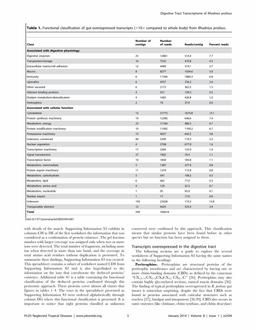
section. We accordingly identified transcripts that were signifi-
cantly more expressed in the whole digestive tract when compared
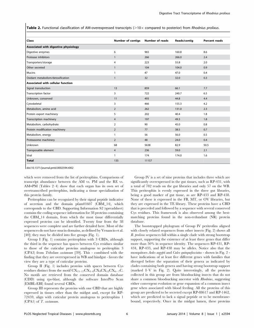
to the WB library (Table 1), those more expressed in the AM when
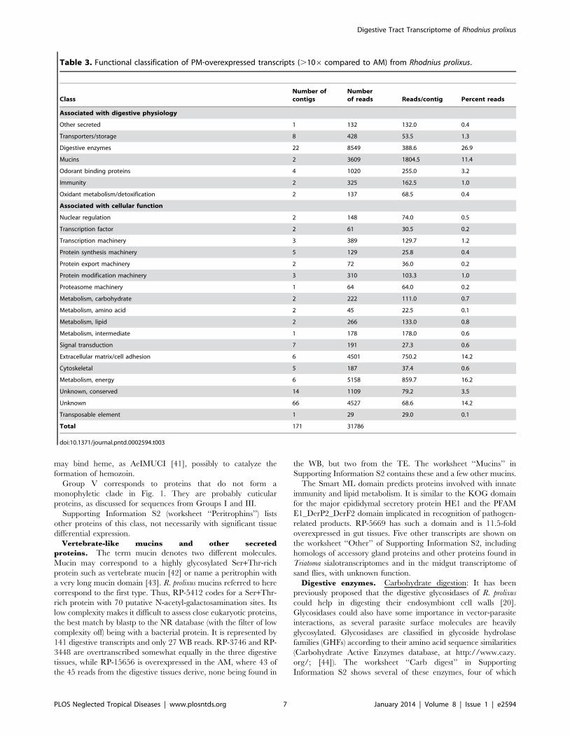
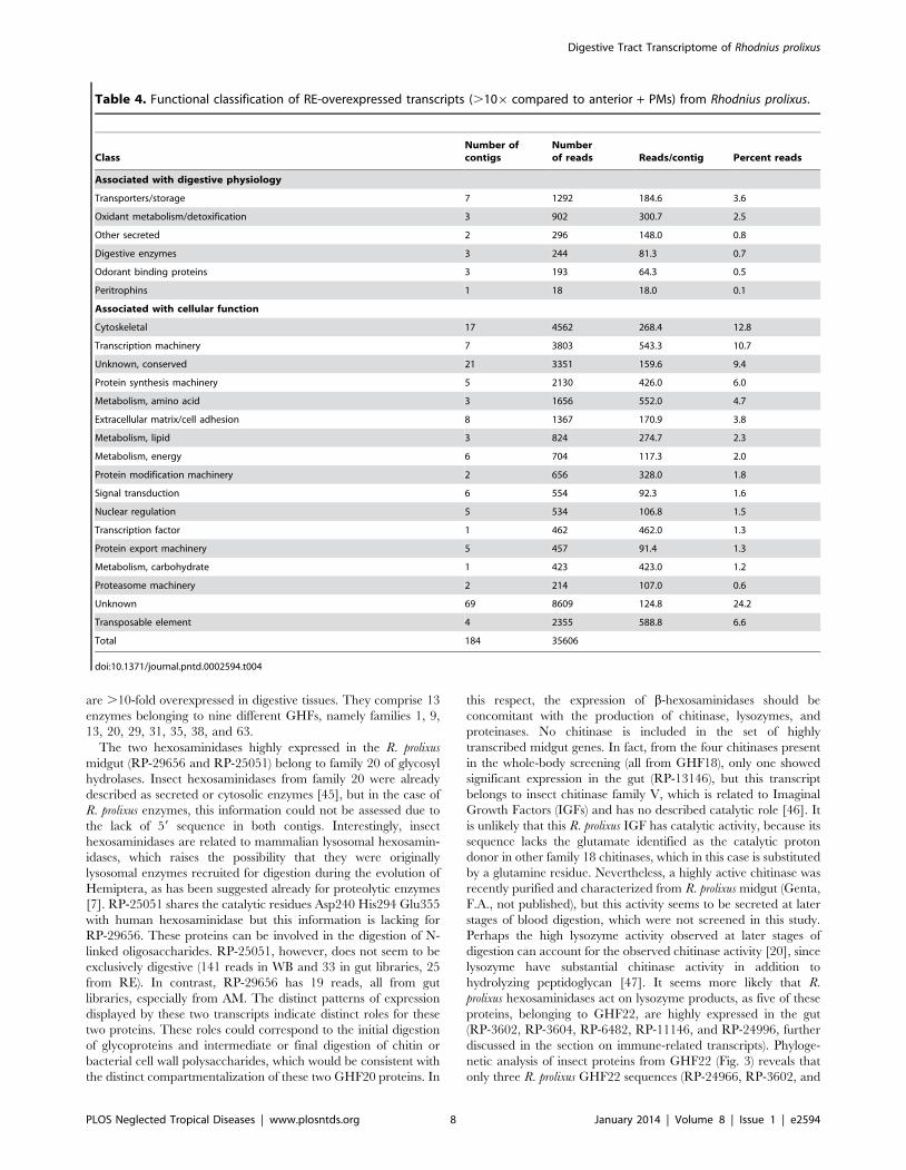
compared to the PM (Table 2), those more expressed in the PM
when compared to the AM (Table 3), and those more expressed in
the RE when compared to the combined AM+PM set (Table 4).
Analysis was concentrated on contigs that were overexpressed in
the digestive system with a P value,0.05; however, contigs related
to selected specific aspects of midgut metabolism were also
included in the analysis even when found at lower gut expression.
We also made an effort to obtain coding sequences for all
contigs that were significantly more expressed in the gut as well as
for transcripts that presented .90% coverage with their best
protein matches from the NR database, provided in Supporting
Information S2, containing 2,570 CDS. The following sections
highlight the gut-overexpressed transcripts but also include other
CDS of related families for comparison. These are located in the
several worksheets of Supporting Information S2 following the
worksheet named RP-CDS. We will make frequent reference to
the number of ‘‘reads’’ from the pyrosequencing runs, each read
being one sequence unit that was used to assemble the contigs that
are the subject of analysis. In the remainder of this paper, when
mentioning a contig represented in Supporting Information S1,
this will be indicated by Asb-### where ### is the contig
number shown in column A. When reference is made to a CDS
from Supporting Information S2, this will be indicated by RP-
### where ### refers to the CDS number shown also in
column A.
Proteomic analysisAn exploratory proteomic analysis of Rhodnius’ gut compart-
ments was performed. The samples analyzed were prepared from
insects fed on blood. The tissues were harvested on the fourth day
after blood feeding. Regardless of this one point harvesting, about
10% of the proteins deduced from conceptual translation of the
assembled 454 reads had their existence confirmed by this
proteomic approach. Additional figure S1 shows the SDS-PAGE
fractionation of membrane and soluble protein extracts obtained
as described in methods from the tree compartments of Rhodnius’
digestive tract. This figure exhibits the numbering of each fraction
that was in gel digested and subsequently analyzed by mass
spectrometry. The assignment of the ions produced by mass
spectrometry to the deduced proteins was first done by the use of
Mascot (www.matrixscience.com) and subsequently converted to
Prosite block format as described in methods. This data-containing
file was used to search matches in a formatted database of the
deduced proteins, using the Seedtop program. The result of the
Seedtop search was inserted into the hyperlinked spreadsheet
(Supporting Information S3) to produce a hyperlinked text file
Digestive Tract Transcriptome of Rhodnius prolixus
PLOS Neglected Tropical Diseases | www.plosntds.org 4 January 2014 | Volume 8 | Issue 1 | e2594
with details of the match. Supporting Information S3 exhibits in
columns CH to DE of the first worksheet the information that was
considered as a confirmation of protein existence. The gel fraction
number with larger coverage was assigned only when two or more
ions were detected. The total number of fragments, including same
ion when detected in more than one band, and the coverage in
total amino acid residues without duplication is presented. To
summarize these findings, Supporting Information S3 was created.
This spreadsheet contains a subset of worksheet named CDS from
Supporting Information S2 and is also hyperlinked to the
information on the ions that corroborate the deduced proteins’
existence. Additional table S1 is a table containing the functional
classification of the deduced proteins confirmed through this
proteomic approach. These proteins cover almost all classes that
figures in tables 1–4. The rows in the spreadsheet presented as
Supporting Information S3 were ordered alphabetically through
column DG where this functional classification is presented. It is
important to notice that eight proteins classified as unknown
conserved were confirmed by this approach. This classification
means that similar proteins have been found before in other
species but no function has been assigned to them.
Transcripts overexpressed in the digestive tractThe following sections are a guide to explore the several
worksheets of Supporting Information S2 having the same names
as the following headings:
Peritrophins. Peritrophins are structural proteins of the
peritrophic membranes and are characterized by having one or
more chitin-binding domains (CBDs) as defined by the consensus
‘‘CX15–17CX5–6CX9CX12 CX6–7C’’ [36]. Peritrophins may also
contain highly glycosylated sections, named mucin domains [36].
The finding of typical peritrophins overexpressed in R. prolixus gut
tissues is somewhat surprising, despite the fact that CBDs were
found in proteins associated with cuticular structures such as
trachea [37], hindgut and integument [38,39]. CBD also occurs in
some enzymes (like chitinase, chitin synthase, and chitin deacylase)
Table 1. Functional classification of gut-overexpressed transcripts (.106 compared to whole body) from Rhodnius prolixus.
ClassNumber ofcontigs
Numberof reads Reads/contig Percent reads
Associated with digestive physiology
Digestive enzymes 25 12861 514.4 7.7
Transporters/storage 16 7532 470.8 4.5
Extracellular matrix/cell adhesion 12 4489 374.1 2.7
Mucins 8 8277 1034.6 5.0
Immunity 6 11306 1884.3 6.8
Lipocalins 6 4357 726.2 2.6
Other secreted 6 2175 362.5 1.3
Odorant binding proteins 4 557 139.3 0.3
Oxidant metabolism/detoxification 4 1683 420.8 1.0
Peritrophins 2 74 37.0 0.0
Associated with cellular function
Cytoskeletal 13 21773 1674.8 13.1
Protein synthesis machinery 19 12286 646.6 7.4
Metabolism, energy 23 11184 486.3 6.7
Protein modification machinery 10 11092 1109.2 6.7
Proteasome machinery 15 9637 642.5 5.8
Unknown, conserved 44 5249 119.3 3.2
Nuclear regulation 4 2708 677.0 1.6
Transcription machinery 17 2260 132.9 1.4
Signal transduction 27 1902 70.4 1.1
Transcription factor 10 1858 185.8 1.1
Metabolism, intermediate 5 1387 277.4 0.8
Protein export machinery 11 1274 115.8 0.8
Metabolism, carbohydrate 5 541 108.2 0.3
Metabolism, lipid 6 462 77.0 0.3
Metabolism, amino acid 4 129 32.3 0.1
Metabolism, nucleotide 1 95 95.0 0.1
Nuclear export 1 17 17.0 0.0
Unknown 193 23028 119.3 13.8
Transposable element 12 6425 535.4 3.9
Total 509 166618
doi:10.1371/journal.pntd.0002594.t001
Digestive Tract Transcriptome of Rhodnius prolixus
PLOS Neglected Tropical Diseases | www.plosntds.org 5 January 2014 | Volume 8 | Issue 1 | e2594
which were removed from the list of peritrophins. Comparisons of
transcript abundance between the AM vs. PM and the RE vs.
AM+PM (Tables 2–4) show that each organ has its own set of
overtranscribed peritrophins, indicating a tissue specialization of
this protein family.
Peritrophins can be recognized by their signal peptide indicative
of secretion and the domain pfam01607 (CBM_14), which
corresponds to the CBD. Supporting Information S2 (spreadsheet)
contains the coding sequence information for 38 proteins containing
the CBM_14 domain, from which the most tissue differentially
expressed proteins can be identified. Twenty four from the 38
sequences were complete and are further detailed here. Most of the
sequences do not have mucin domains, as defined by Venancio et al.
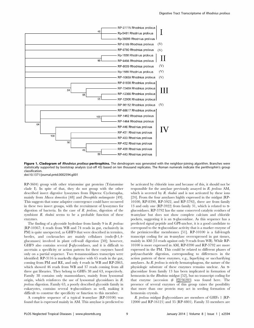
[40]; they may be divided into five groups (Fig. 1).
Group I (Fig. 1) contains peritrophins with 3 CBDs, although
the third in the sequence has spaces between Cys residues similar
to those of the cuticular proteins analogous to peritrophin 3
(CPA3) from Tribolium castaneum [39]. This - combined with the
finding that they are overexpressed in WB and hindgut - favors the
view they are a type of cuticular proteins.
Group II (Fig. 1) includes proteins with spaces between Cys
residues distinct from the motif CX15–17CX5–6CX9CX12CX6-–7C.
No motifs are retrieved from the conserved domain database
(CDD) using rps-blast, although the software InterPro Scan
(EMBL-EBI) found several CBDs.
Group III represents the proteins with one CBD that are highly
expressed in tissues other than the midgut and, except for RP-
72459, align with cuticular protein analogous to peritrophins 1
(CPA1) of T. castaneum.
Group IV is a set of nine proteins that includes three which are
significantly overexpressed in the gut tissues, such as RP-431, with
a total of 782 reads on the gut libraries and only 57 on the WB.
This peritrophin is evenly expressed in the three gut libraries,
being a good marker of gut tissue, as are RP-433 and RP-438.
None of these is expressed in the FB, MT, or OV libraries, but
they are expressed in the TE library. These proteins have a CBD
that is preceded and followed by a sequence with several conserved
Cys residues. This framework is also observed among the best-
matching proteins found in the non-redundant (NR) protein
database.
The bootstrapped phylogram of Group IV peritrofins aligned
with closely related sequences from other insects (Fig. 2) shows all
R. prolixus sequences fall within a single clade with strong bootstrap
support, supporting the existence of at least three genes that differ
more than 50% in sequence identity. The sequences RP-431, RP-
434, RP-433, and RP-438 may be alleles. Notice also that the
mosquitoes Aedes aegypti and Culex quinquefasciatus - shown in Fig. 2 -
have indications of at least five different genes with families that
diverged before the separation of their genera as indicated by
clades containing both genera and having strong bootstrap support
(marked I–V in Fig. 2). Quite interestingly, all the proteins
collected in this group are from bloodsucking insects that do not
share a common bloodsucking ancestor with Rhodnius, suggesting
either convergent evolution or gene expansion of a common insect
gene when associated with blood feeding. All the proteins of this
group are predicted to be secreted except RP-88617 and RP-1462,
which are predicted to lack a signal peptide or to be membrane-
bound, respectively. Once in the midgut lumen, these proteins
Table 2. Functional classification of AM-overexpressed transcripts (.106 compared to posterior) from Rhodnius prolixus.
Class Number of contigs Number of reads Reads/contig Percent reads
Associated with digestive physiology
Digestive enzymes 6 965 160.8 8.6
Protease inhibitors 1 266 266.0 2.4
Transporters/storage 4 223 55.8 2.0
Other secreted 1 104 104.0 0.9
Mucins 1 47 47.0 0.4
Oxidant metabolism/detoxification 1 32 32.0 0.3
Associated with cellular function
Signal transduction 13 859 66.1 7.7
Transcription factor 3 722 240.7 6.5
Unknown, conserved 11 493 44.8 4.4
Cytoskeletal 3 466 155.3 4.2
Metabolism, amino acid 2 262 131.0 2.3
Protein export machinery 5 202 40.4 1.8
Transcription machinery 4 197 49.3 1.8
Metabolism, carbohydrate 2 90 45.0 0.8
Protein modification machinery 2 77 38.5 0.7
Metabolism, energy 1 56 56.0 0.5
Proteasome machinery 2 48 24.0 0.4
Unknown 68 5638 82.9 50.5
Transposable element 4 236 59.0 2.1
Viral 1 174 174.0 1.6
Total 135 11157
doi:10.1371/journal.pntd.0002594.t002
Digestive Tract Transcriptome of Rhodnius prolixus
PLOS Neglected Tropical Diseases | www.plosntds.org 6 January 2014 | Volume 8 | Issue 1 | e2594
may bind heme, as AeIMUCI [41], possibly to catalyze the
formation of hemozoin.
Group V corresponds to proteins that do not form a
monophyletic clade in Fig. 1. They are probably cuticular
proteins, as discussed for sequences from Groups I and III.
Supporting Information S2 (worksheet ‘‘Peritrophins’’) lists
other proteins of this class, not necessarily with significant tissue
differential expression.
Vertebrate-like mucins and other secreted
proteins. The term mucin denotes two different molecules.
Mucin may correspond to a highly glycosylated Ser+Thr-rich
protein such as vertebrate mucin [42] or name a peritrophin with
a very long mucin domain [43]. R. prolixus mucins referred to here
correspond to the first type. Thus, RP-5412 codes for a Ser+Thr-
rich protein with 70 putative N-acetyl-galactosamination sites. Its
low complexity makes it difficult to assess close eukaryotic proteins,
the best match by blastp to the NR database (with the filter of low
complexity off) being with a bacterial protein. It is represented by
141 digestive transcripts and only 27 WB reads. RP-3746 and RP-
3448 are overtranscribed somewhat equally in the three digestive
tissues, while RP-15656 is overexpressed in the AM, where 43 of
the 45 reads from the digestive tissues derive, none being found in
the WB, but two from the TE. The worksheet ‘‘Mucins’’ in
Supporting Information S2 contains these and a few other mucins.
The Smart ML domain predicts proteins involved with innate
immunity and lipid metabolism. It is similar to the KOG domain
for the major epididymal secretory protein HE1 and the PFAM
E1_DerP2_DerF2 domain implicated in recognition of pathogen-
related products. RP-5669 has such a domain and is 11.5-fold
overexpressed in gut tissues. Five other transcripts are shown on
the worksheet ‘‘Other’’ of Supporting Information S2, including
homologs of accessory gland proteins and other proteins found in
Triatoma sialotranscriptomes and in the midgut transcriptome of
sand flies, with unknown function.
Digestive enzymes. Carbohydrate digestion: It has been
previously proposed that the digestive glycosidases of R. prolixus
could help in digesting their endosymbiont cell walls [20].
Glycosidases could also have some importance in vector-parasite
interactions, as several parasite surface molecules are heavily
glycosylated. Glycosidases are classified in glycoside hydrolase
families (GHFs) according to their amino acid sequence similarities
(Carbohydrate Active Enzymes database, at http://www.cazy.
org/; [44]). The worksheet ‘‘Carb digest’’ in Supporting
Information S2 shows several of these enzymes, four of which
Table 3. Functional classification of PM-overexpressed transcripts (.106 compared to AM) from Rhodnius prolixus.
ClassNumber ofcontigs
Numberof reads Reads/contig Percent reads
Associated with digestive physiology
Other secreted 1 132 132.0 0.4
Transporters/storage 8 428 53.5 1.3
Digestive enzymes 22 8549 388.6 26.9
Mucins 2 3609 1804.5 11.4
Odorant binding proteins 4 1020 255.0 3.2
Immunity 2 325 162.5 1.0
Oxidant metabolism/detoxification 2 137 68.5 0.4
Associated with cellular function
Nuclear regulation 2 148 74.0 0.5
Transcription factor 2 61 30.5 0.2
Transcription machinery 3 389 129.7 1.2
Protein synthesis machinery 5 129 25.8 0.4
Protein export machinery 2 72 36.0 0.2
Protein modification machinery 3 310 103.3 1.0
Proteasome machinery 1 64 64.0 0.2
Metabolism, carbohydrate 2 222 111.0 0.7
Metabolism, amino acid 2 45 22.5 0.1
Metabolism, lipid 2 266 133.0 0.8
Metabolism, intermediate 1 178 178.0 0.6
Signal transduction 7 191 27.3 0.6
Extracellular matrix/cell adhesion 6 4501 750.2 14.2
Cytoskeletal 5 187 37.4 0.6
Metabolism, energy 6 5158 859.7 16.2
Unknown, conserved 14 1109 79.2 3.5
Unknown 66 4527 68.6 14.2
Transposable element 1 29 29.0 0.1
Total 171 31786
doi:10.1371/journal.pntd.0002594.t003
Digestive Tract Transcriptome of Rhodnius prolixus
PLOS Neglected Tropical Diseases | www.plosntds.org 7 January 2014 | Volume 8 | Issue 1 | e2594
are .10-fold overexpressed in digestive tissues. They comprise 13
enzymes belonging to nine different GHFs, namely families 1, 9,
13, 20, 29, 31, 35, 38, and 63.
The two hexosaminidases highly expressed in the R. prolixus
midgut (RP-29656 and RP-25051) belong to family 20 of glycosyl
hydrolases. Insect hexosaminidases from family 20 were already
described as secreted or cytosolic enzymes [45], but in the case of
R. prolixus enzymes, this information could not be assessed due to
the lack of 59 sequence in both contigs. Interestingly, insect
hexosaminidases are related to mammalian lysosomal hexosamin-
idases, which raises the possibility that they were originally
lysosomal enzymes recruited for digestion during the evolution of
Hemiptera, as has been suggested already for proteolytic enzymes
[7]. RP-25051 shares the catalytic residues Asp240 His294 Glu355
with human hexosaminidase but this information is lacking for
RP-29656. These proteins can be involved in the digestion of N-
linked oligosaccharides. RP-25051, however, does not seem to be
exclusively digestive (141 reads in WB and 33 in gut libraries, 25
from RE). In contrast, RP-29656 has 19 reads, all from gut
libraries, especially from AM. The distinct patterns of expression
displayed by these two transcripts indicate distinct roles for these
two proteins. These roles could correspond to the initial digestion
of glycoproteins and intermediate or final digestion of chitin or
bacterial cell wall polysaccharides, which would be consistent with
the distinct compartmentalization of these two GHF20 proteins. In
this respect, the expression of b-hexosaminidases should be
concomitant with the production of chitinase, lysozymes, and
proteinases. No chitinase is included in the set of highly
transcribed midgut genes. In fact, from the four chitinases present
in the whole-body screening (all from GHF18), only one showed
significant expression in the gut (RP-13146), but this transcript
belongs to insect chitinase family V, which is related to Imaginal
Growth Factors (IGFs) and has no described catalytic role [46]. It
is unlikely that this R. prolixus IGF has catalytic activity, because its
sequence lacks the glutamate identified as the catalytic proton
donor in other family 18 chitinases, which in this case is substituted
by a glutamine residue. Nevertheless, a highly active chitinase was
recently purified and characterized from R. prolixus midgut (Genta,
F.A., not published), but this activity seems to be secreted at later
stages of blood digestion, which were not screened in this study.
Perhaps the high lysozyme activity observed at later stages of
digestion can account for the observed chitinase activity [20], since
lysozyme have substantial chitinase activity in addition to
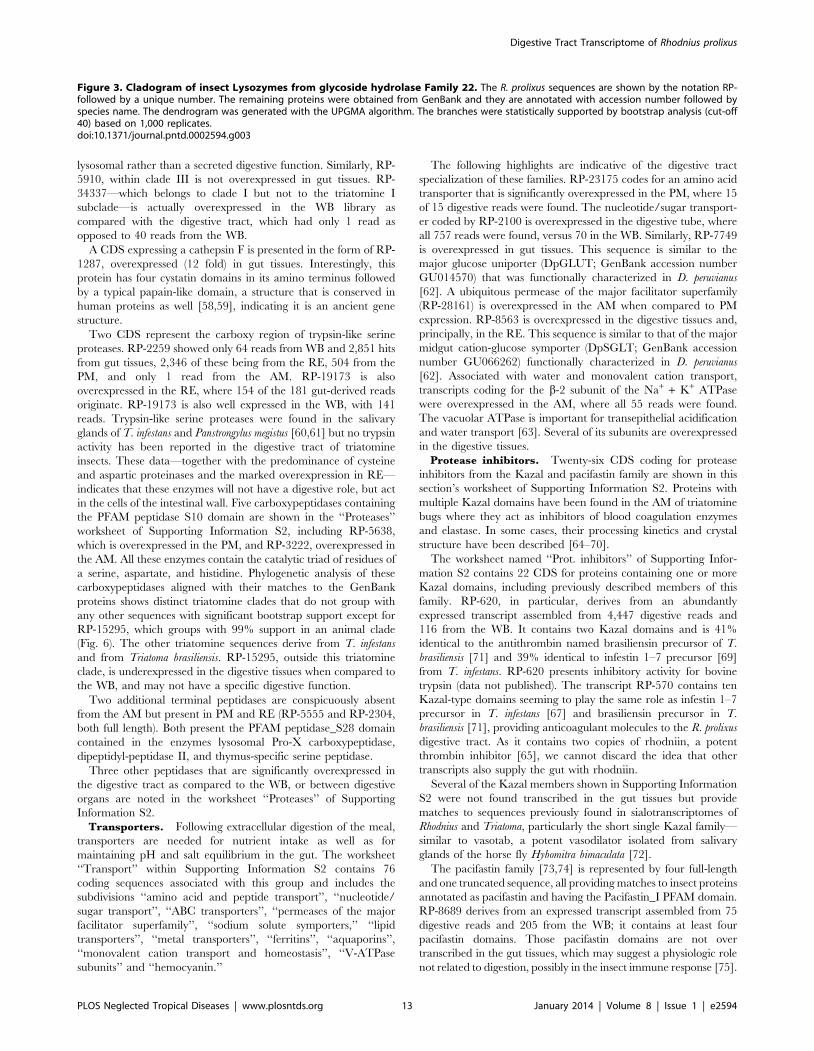
hydrolyzing peptidoglycan [47]. It seems more likely that R.
prolixus hexosaminidases act on lysozyme products, as five of these
proteins, belonging to GHF22, are highly expressed in the gut
(RP-3602, RP-3604, RP-6482, RP-11146, and RP-24996, further
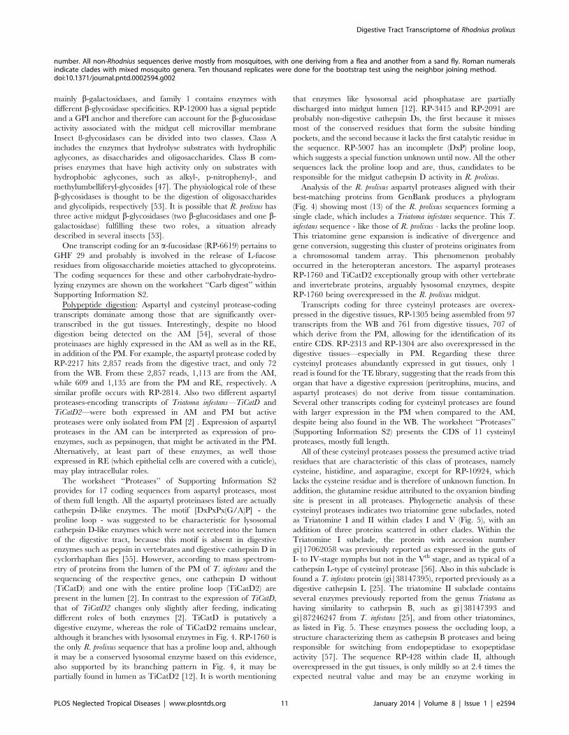
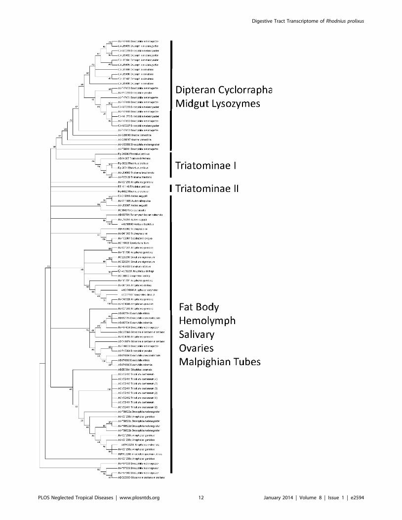
discussed in the section on immune-related transcripts). Phyloge-
netic analysis of insect proteins from GHF22 (Fig. 3) reveals that
only three R. prolixus GHF22 sequences (RP-24966, RP-3602, and
Table 4. Functional classification of RE-overexpressed transcripts (.106 compared to anterior + PMs) from Rhodnius prolixus.
ClassNumber ofcontigs
Numberof reads Reads/contig Percent reads
Associated with digestive physiology
Transporters/storage 7 1292 184.6 3.6
Oxidant metabolism/detoxification 3 902 300.7 2.5
Other secreted 2 296 148.0 0.8
Digestive enzymes 3 244 81.3 0.7
Odorant binding proteins 3 193 64.3 0.5
Peritrophins 1 18 18.0 0.1
Associated with cellular function
Cytoskeletal 17 4562 268.4 12.8
Transcription machinery 7 3803 543.3 10.7
Unknown, conserved 21 3351 159.6 9.4
Protein synthesis machinery 5 2130 426.0 6.0
Metabolism, amino acid 3 1656 552.0 4.7
Extracellular matrix/cell adhesion 8 1367 170.9 3.8
Metabolism, lipid 3 824 274.7 2.3
Metabolism, energy 6 704 117.3 2.0
Protein modification machinery 2 656 328.0 1.8
Signal transduction 6 554 92.3 1.6
Nuclear regulation 5 534 106.8 1.5
Transcription factor 1 462 462.0 1.3
Protein export machinery 5 457 91.4 1.3
Metabolism, carbohydrate 1 423 423.0 1.2
Proteasome machinery 2 214 107.0 0.6
Unknown 69 8609 124.8 24.2
Transposable element 4 2355 588.8 6.6
Total 184 35606
doi:10.1371/journal.pntd.0002594.t004
Digestive Tract Transcriptome of Rhodnius prolixus
PLOS Neglected Tropical Diseases | www.plosntds.org 8 January 2014 | Volume 8 | Issue 1 | e2594
RP-3604) group with other triatomine gut proteins (Triatomine
clade I). In spite of that, they do not group with the other
described insect digestive lysozymes from Diptera: Cyclorrapha,
mainly from Musca domestica [48] and Drosophila melanogaster [49].
This suggests that some adaptive convergence could have occurred
in these two insect groups, with the recruitment of lysozymes for
digestion of bacteria. In the case of R. prolixus, digestion of the
symbiont R. rhodnii seems to be a probable function of these
enzymes.
The finding of a glycoside hydrolase from family 9 in R. prolixus
(RP-10367; 4 reads from WB and 74 reads in gut, exclusively in
PM) is quite unexpected, as GHF9 that were described in termites,
beetles, and cockroaches are mainly cellulases (endo-b-1,4-
glucanases) involved in plant cell-wall digestion [50]; however,
GHF9 also contains several b-glycosidases, and it is difficult to
ascertain a specificity or action pattern for these enzymes based
only on a partial sequence. Two a-mannosidases transcripts were
identified: RP-3116 is markedly digestive with 65 reads in the gut,
coming from PM and RE, and only 4 reads in WB and RP-2863,
which showed 46 reads from WB and 37 reads coming from all
three gut libraries. They belong to GHFs 38 and 63, respectively.
Family 38 contains only mannosidases, mainly from lysosomal
origin, which reinforces the use of lysosomal glycosidases in R.
prolixus digestion. Family 63, a poorly described glycoside family in
eukaryotes, contains several a-glucosidases as well, making it
difficult to construe the specificity or function to this member.
A complete sequence of a typical a-amylase (RP-10100) was
found that is expressed mainly in AM. This amylase is predicted to
be activated by chloride ions and because of this, it should not be
responsible for the amylase previously assayed in R. prolixus AM,
which is secreted by R. rhodnii and is not activated by these ions
[24]. From the four amylases highly expressed in the midgut (RP-
10100, RP-8390, RP-5922, and RP-3792), three are from family
13 and only one (RP-5922) from family 31, which is related to a-
glucosidases. RP-3792 has the same conserved catalytic residues of
a-amylase but does not show complete calcium and chloride
pockets, suggesting it is an a-glucosidase. As this sequence has a
predicted signal peptide and GPI-anchor, it is a good candidate to
correspond to the a-glucosidase activity that is a marker enzyme of
the perimicrovillar membranes [51]. RP-10100 is a full-length
transcript coding for an a-amylase overexpressed in gut tissues,
mainly in AM (53 reads against only 9 reads from WB). While RP-
10100 is more expressed in AM, RP-8390 and RP-3792 are more
expressed in the PM. This could be related to different phases of
polysaccharide digestion, corresponding to differences in the
action pattern of these enzymes, e.g., liquefying or saccharifying
amylases. As R. prolixus is strictly hematophagous, the nature of the
physiologic substrate of these enzymes remains unclear. An a-
glucosidase from family 13 has been implicated in formation of
hemozoin in the Rhodnius midgut [52], but no transcript coding for
that enzyme (accession # FJ236283) was found here. The
presence of several enzymes of this group raises the possibility
that more than one protein may act in seeding formation of
hemozoin crystals.
R. prolixus midgut b-glycosidases are members of GHFs 1 (RP-
12000 and RP-16121) and 35 (RP-4801). Family 35 members are
Figure 1. Cladogram of Rhodnius prolixus peritrophins. The dendrogram was generated with the neighbor-joining algorithm. Branches werestatistically supported by bootstrap analysis (cut-off 45) based on ten thousand replicates. The Roman numerals indicate the perithrophin’s groupclassification.doi:10.1371/journal.pntd.0002594.g001
Digestive Tract Transcriptome of Rhodnius prolixus
PLOS Neglected Tropical Diseases | www.plosntds.org 9 January 2014 | Volume 8 | Issue 1 | e2594
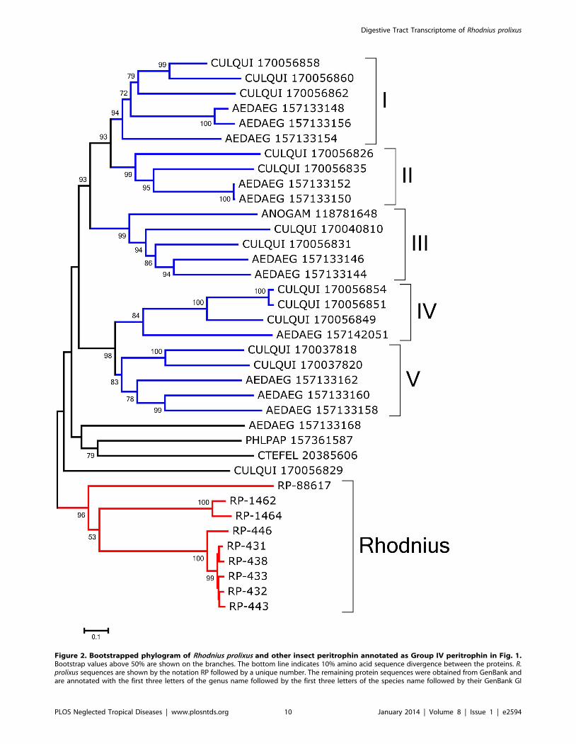
Figure 2. Bootstrapped phylogram of Rhodnius prolixus and other insect peritrophin annotated as Group IV peritrophin in Fig. 1.Bootstrap values above 50% are shown on the branches. The bottom line indicates 10% amino acid sequence divergence between the proteins. R.prolixus sequences are shown by the notation RP followed by a unique number. The remaining protein sequences were obtained from GenBank andare annotated with the first three letters of the genus name followed by the first three letters of the species name followed by their GenBank GI
Digestive Tract Transcriptome of Rhodnius prolixus
PLOS Neglected Tropical Diseases | www.plosntds.org 10 January 2014 | Volume 8 | Issue 1 | e2594
mainly b-galactosidases, and family 1 contains enzymes with
different b-glycosidase specificities. RP-12000 has a signal peptide
and a GPI anchor and therefore can account for the b-glucosidase
activity associated with the midgut cell microvillar membrane
Insect ß-glycosidases can be divided into two classes. Class A
includes the enzymes that hydrolyse substrates with hydrophilic
aglycones, as disaccharides and oligosaccharides. Class B com-
prises enzymes that have high activity only on substrates with
hydrophobic aglycones, such as alkyl-, p-nitrophenyl-, and
methylumbelliferyl-glycosides [47]. The physiological role of these
b-glycosidases is thought to be the digestion of oligosaccharides
and glycolipids, respectively [53]. It is possible that R. prolixus has
three active midgut b-glycosidases (two b-glucosidases and one b-
galactosidase) fulfilling these two roles, a situation already
described in several insects [53].
One transcript coding for an a-fucosidase (RP-6619) pertains to
GHF 29 and probably is involved in the release of L-fucose
residues from oligosaccharide moieties attached to glycoproteins.
The coding sequences for these and other carbohydrate-hydro-
lyzing enzymes are shown on the worksheet ‘‘Carb digest’’ within
Supporting Information S2.
Polypeptide digestion: Aspartyl and cysteinyl protease-coding
transcripts dominate among those that are significantly over-
transcribed in the gut tissues. Interestingly, despite no blood
digestion being detected on the AM [54], several of those
proteinases are highly expressed in the AM as well as in the RE,
in addition of the PM. For example, the aspartyl protease coded by
RP-2217 hits 2,857 reads from the digestive tract, and only 72
from the WB. From these 2,857 reads, 1,113 are from the AM,
while 609 and 1,135 are from the PM and RE, respectively. A
similar profile occurs with RP-2814. Also two different aspartyl
proteases-encoding transcripts of Triatoma infestans—TiCatD and
TiCatD2—were both expressed in AM and PM but active
proteases were only isolated from PM [2] . Expression of aspartyl
proteases in the AM can be interpreted as expression of pro-
enzymes, such as pepsinogen, that might be activated in the PM.
Alternatively, at least part of these enzymes, as well those
expressed in RE (which epithelial cells are covered with a cuticle),
may play intracellular roles.
The worksheet ‘‘Proteases’’ of Supporting Information S2
provides for 17 coding sequences from aspartyl proteases, most
of them full length. All the aspartyl proteinases listed are actually
cathepsin D-like enzymes. The motif [DxPxPx(G/A)P] - the
proline loop - was suggested to be characteristic for lysosomal
cathepsin D-like enzymes which were not secreted into the lumen
of the digestive tract, because this motif is absent in digestive
enzymes such as pepsin in vertebrates and digestive cathepsin D in
cyclorrhaphan flies [55]. However, according to mass spectrom-
etry of proteins from the lumen of the PM of T. infestans and the
sequencing of the respective genes, one cathepsin D without
(TiCatD) and one with the entire proline loop (TiCatD2) are
present in the lumen [2]. In contrast to the expression of TiCatD,
that of TiCatD2 changes only slightly after feeding, indicating
different roles of both enzymes [2]. TiCatD is putatively a
digestive enzyme, whereas the role of TiCatD2 remains unclear,
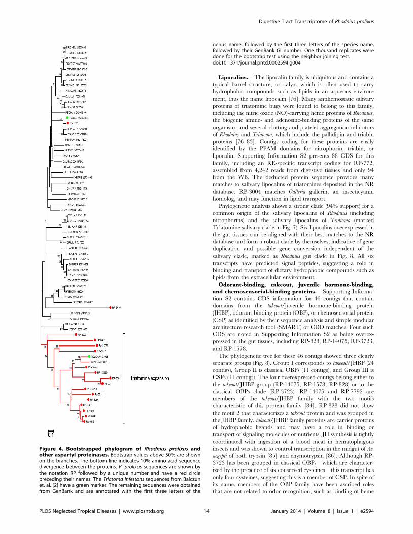
although it branches with lysosomal enzymes in Fig. 4. RP-1760 is
the only R. prolixus sequence that has a proline loop and, although
it may be a conserved lysosomal enzyme based on this evidence,
also supported by its branching pattern in Fig. 4, it may be
partially found in lumen as TiCatD2 [12]. It is worth mentioning
that enzymes like lysosomal acid phosphatase are partially
discharged into midgut lumen [12]. RP-3415 and RP-2091 are
probably non-digestive cathepsin Ds, the first because it misses
most of the conserved residues that form the subsite binding
pockets, and the second because it lacks the first catalytic residue in
the sequence. RP-5007 has an incomplete (DxP) proline loop,
which suggests a special function unknown until now. All the other
sequences lack the proline loop and are, thus, candidates to be
responsible for the midgut cathepsin D activity in R. prolixus.
Analysis of the R. prolixus aspartyl proteases aligned with their
best-matching proteins from GenBank produces a phylogram
(Fig. 4) showing most (13) of the R. prolixus sequences forming a
single clade, which includes a Triatoma infestans sequence. This T.
infestans sequence - like those of R. prolixus - lacks the proline loop.
This triatomine gene expansion is indicative of divergence and
gene conversion, suggesting this cluster of proteins originates from
a chromosomal tandem array. This phenomenon probably
occurred in the heteropteran ancestors. The aspartyl proteases
RP-1760 and TiCatD2 exceptionally group with other vertebrate
and invertebrate proteins, arguably lysosomal enzymes, despite
RP-1760 being overexpressed in the R. prolixus midgut.
Transcripts coding for three cysteinyl proteases are overex-
pressed in the digestive tissues, RP-1305 being assembled from 97
transcripts from the WB and 761 from digestive tissues, 707 of
which derive from the PM, allowing for the identification of its
entire CDS. RP-2313 and RP-1304 are also overexpressed in the
digestive tissues—especially in PM. Regarding these three
cysteinyl proteases abundantly expressed in gut tissues, only 1
read is found for the TE library, suggesting that the reads from this
organ that have a digestive expression (peritrophins, mucins, and
aspartyl proteases) do not derive from tissue contamination.
Several other transcripts coding for cysteinyl proteases are found
with larger expression in the PM when compared to the AM,
despite being also found in the WB. The worksheet ‘‘Proteases’’
(Supporting Information S2) presents the CDS of 11 cysteinyl
proteases, mostly full length.
All of these cysteinyl proteases possess the presumed active triad
residues that are characteristic of this class of proteases, namely
cysteine, histidine, and asparagine, except for RP-10924, which
lacks the cysteine residue and is therefore of unknown function. In
addition, the glutamine residue attributed to the oxyanion binding
site is present in all proteases. Phylogenetic analysis of these
cysteinyl proteases indicates two triatomine gene subclades, noted
as Triatomine I and II within clades I and V (Fig. 5), with an
addition of three proteins scattered in other clades. Within the
Triatomine I subclade, the protein with accession number
gi|17062058 was previously reported as expressed in the guts of
I- to IV-stage nymphs but not in the Vth stage, and as typical of a
cathepsin L-type of cysteinyl protease [56]. Also in this subclade is
found a T. infestans protein (gi|38147395), reported previously as a
digestive cathepsin L [25]. The triatomine II subclade contains
several enzymes previously reported from the genus Triatoma as
having similarity to cathepsin B, such as gi|38147393 and
gi|87246247 from T. infestans [25], and from other triatomines,
as listed in Fig. 5. These enzymes possess the occluding loop, a
structure characterizing them as cathepsin B proteases and being
responsible for switching from endopeptidase to exopeptidase
activity [57]. The sequence RP-428 within clade II, although
overexpressed in the gut tissues, is only mildly so at 2.4 times the
expected neutral value and may be an enzyme working in
number. All non-Rhodnius sequences derive mostly from mosquitoes, with one deriving from a flea and another from a sand fly. Roman numeralsindicate clades with mixed mosquito genera. Ten thousand replicates were done for the bootstrap test using the neighbor joining method.doi:10.1371/journal.pntd.0002594.g002
Digestive Tract Transcriptome of Rhodnius prolixus
PLOS Neglected Tropical Diseases | www.plosntds.org 11 January 2014 | Volume 8 | Issue 1 | e2594
Digestive Tract Transcriptome of Rhodnius prolixus
PLOS Neglected Tropical Diseases | www.plosntds.org 12 January 2014 | Volume 8 | Issue 1 | e2594
lysosomal rather than a secreted digestive function. Similarly, RP-
5910, within clade III is not overexpressed in gut tissues. RP-
34337—which belongs to clade I but not to the triatomine I
subclade—is actually overexpressed in the WB library as
compared with the digestive tract, which had only 1 read as
opposed to 40 reads from the WB.
A CDS expressing a cathepsin F is presented in the form of RP-
1287, overexpressed (12 fold) in gut tissues. Interestingly, this
protein has four cystatin domains in its amino terminus followed
by a typical papain-like domain, a structure that is conserved in
human proteins as well [58,59], indicating it is an ancient gene
structure.
Two CDS represent the carboxy region of trypsin-like serine
proteases. RP-2259 showed only 64 reads from WB and 2,851 hits
from gut tissues, 2,346 of these being from the RE, 504 from the
PM, and only 1 read from the AM. RP-19173 is also
overexpressed in the RE, where 154 of the 181 gut-derived reads
originate. RP-19173 is also well expressed in the WB, with 141
reads. Trypsin-like serine proteases were found in the salivary
glands of T. infestans and Panstrongylus megistus [60,61] but no trypsin
activity has been reported in the digestive tract of triatomine
insects. These data—together with the predominance of cysteine
and aspartic proteinases and the marked overexpression in RE—
indicates that these enzymes will not have a digestive role, but act
in the cells of the intestinal wall. Five carboxypeptidases containing
the PFAM peptidase S10 domain are shown in the ‘‘Proteases’’
worksheet of Supporting Information S2, including RP-5638,
which is overexpressed in the PM, and RP-3222, overexpressed in
the AM. All these enzymes contain the catalytic triad of residues of
a serine, aspartate, and histidine. Phylogenetic analysis of these
carboxypeptidases aligned with their matches to the GenBank
proteins shows distinct triatomine clades that do not group with
any other sequences with significant bootstrap support except for
RP-15295, which groups with 99% support in an animal clade
(Fig. 6). The other triatomine sequences derive from T. infestans
and from Triatoma brasiliensis. RP-15295, outside this triatomine
clade, is underexpressed in the digestive tissues when compared to
the WB, and may not have a specific digestive function.
Two additional terminal peptidases are conspicuously absent
from the AM but present in PM and RE (RP-5555 and RP-2304,
both full length). Both present the PFAM peptidase_S28 domain
contained in the enzymes lysosomal Pro-X carboxypeptidase,
dipeptidyl-peptidase II, and thymus-specific serine peptidase.
Three other peptidases that are significantly overexpressed in
the digestive tract as compared to the WB, or between digestive
organs are noted in the worksheet ‘‘Proteases’’ of Supporting
Information S2.
Transporters. Following extracellular digestion of the meal,
transporters are needed for nutrient intake as well as for
maintaining pH and salt equilibrium in the gut. The worksheet
‘‘Transport’’ within Supporting Information S2 contains 76
coding sequences associated with this group and includes the
subdivisions ‘‘amino acid and peptide transport’’, ‘‘nucleotide/
sugar transport’’, ‘‘ABC transporters’’, ‘‘permeases of the major
facilitator superfamily’’, ‘‘sodium solute symporters,’’ ‘‘lipid
transporters’’, ‘‘metal transporters’’, ‘‘ferritins’’, ‘‘aquaporins’’,
‘‘monovalent cation transport and homeostasis’’, ‘‘V-ATPase
subunits’’ and ‘‘hemocyanin.’’
The following highlights are indicative of the digestive tract
specialization of these families. RP-23175 codes for an amino acid
transporter that is significantly overexpressed in the PM, where 15
of 15 digestive reads were found. The nucleotide/sugar transport-
er coded by RP-2100 is overexpressed in the digestive tube, where
all 757 reads were found, versus 70 in the WB. Similarly, RP-7749
is overexpressed in gut tissues. This sequence is similar to the
major glucose uniporter (DpGLUT; GenBank accession number
GU014570) that was functionally characterized in D. peruvianus
[62]. A ubiquitous permease of the major facilitator superfamily
(RP-28161) is overexpressed in the AM when compared to PM
expression. RP-8563 is overexpressed in the digestive tissues and,
principally, in the RE. This sequence is similar to that of the major
midgut cation-glucose symporter (DpSGLT; GenBank accession
number GU066262) functionally characterized in D. peruvianus
[62]. Associated with water and monovalent cation transport,
transcripts coding for the b-2 subunit of the Na+ + K+ ATPase
were overexpressed in the AM, where all 55 reads were found.
The vacuolar ATPase is important for transepithelial acidification
and water transport [63]. Several of its subunits are overexpressed
in the digestive tissues.
Protease inhibitors. Twenty-six CDS coding for protease
inhibitors from the Kazal and pacifastin family are shown in this
section’s worksheet of Supporting Information S2. Proteins with
multiple Kazal domains have been found in the AM of triatomine
bugs where they act as inhibitors of blood coagulation enzymes
and elastase. In some cases, their processing kinetics and crystal
structure have been described [64–70].
The worksheet named ‘‘Prot. inhibitors’’ of Supporting Infor-
mation S2 contains 22 CDS for proteins containing one or more
Kazal domains, including previously described members of this
family. RP-620, in particular, derives from an abundantly
expressed transcript assembled from 4,447 digestive reads and
116 from the WB. It contains two Kazal domains and is 41%
identical to the antithrombin named brasiliensin precursor of T.
brasiliensis [71] and 39% identical to infestin 1–7 precursor [69]
from T. infestans. RP-620 presents inhibitory activity for bovine
trypsin (data not published). The transcript RP-570 contains ten
Kazal-type domains seeming to play the same role as infestin 1–7
precursor in T. infestans [67] and brasiliensin precursor in T.
brasiliensis [71], providing anticoagulant molecules to the R. prolixus
digestive tract. As it contains two copies of rhodniin, a potent
thrombin inhibitor [65], we cannot discard the idea that other
transcripts also supply the gut with rhodniin.
Several of the Kazal members shown in Supporting Information
S2 were not found transcribed in the gut tissues but provide
matches to sequences previously found in sialotranscriptomes of
Rhodnius and Triatoma, particularly the short single Kazal family—
similar to vasotab, a potent vasodilator isolated from salivary
glands of the horse fly Hybomitra bimaculata [72].
The pacifastin family [73,74] is represented by four full-length
and one truncated sequence, all providing matches to insect proteins
annotated as pacifastin and having the Pacifastin_I PFAM domain.
RP-8689 derives from an expressed transcript assembled from 75
digestive reads and 205 from the WB; it contains at least four
pacifastin domains. Those pacifastin domains are not over
transcribed in the gut tissues, which may suggest a physiologic role
not related to digestion, possibly in the insect immune response [75].
Figure 3. Cladogram of insect Lysozymes from glycoside hydrolase Family 22. The R. prolixus sequences are shown by the notation RP-followed by a unique number. The remaining proteins were obtained from GenBank and they are annotated with accession number followed byspecies name. The dendrogram was generated with the UPGMA algorithm. The branches were statistically supported by bootstrap analysis (cut-off40) based on 1,000 replicates.doi:10.1371/journal.pntd.0002594.g003
Digestive Tract Transcriptome of Rhodnius prolixus
PLOS Neglected Tropical Diseases | www.plosntds.org 13 January 2014 | Volume 8 | Issue 1 | e2594
Lipocalins. The lipocalin family is ubiquitous and contains a
typical barrel structure, or calyx, which is often used to carry
hydrophobic compounds such as lipids in an aqueous environ-
ment, thus the name lipocalin [76]. Many antihemostatic salivary
proteins of triatomine bugs were found to belong to this family,
including the nitric oxide (NO)-carrying heme proteins of Rhodnius,
the biogenic amine- and adenosine-binding proteins of the same
organism, and several clotting and platelet aggregation inhibitors
of Rhodnius and Triatoma, which include the pallidipin and triabin
proteins [76–83]. Contigs coding for these proteins are easily
identified by the PFAM domains for nitrophorin, triabin, or
lipocalin. Supporting Information S2 presents 88 CDS for this
family, including an RE-specific transcript coding for RP-772,
assembled from 4,242 reads from digestive tissues and only 94
from the WB. The deducted protein sequence provides many
matches to salivary lipocalins of triatomines deposited in the NR
database. RP-3004 matches Galleria gallerin, an insecticyanin
homolog, and may function in lipid transport.
Phylogenetic analysis shows a strong clade (94% support) for a
common origin of the salivary lipocalins of Rhodnius (including
nitrophorins) and the salivary lipocalins of Triatoma (marked
Triatomine salivary clade in Fig. 7). Six lipocalins overexpressed in
the gut tissues can be aligned with their best matches to the NR
database and form a robust clade by themselves, indicative of gene
duplication and possible gene conversion independent of the
salivary clade, marked as Rhodnius gut clade in Fig. 8. All six
transcripts have predicted signal peptides, suggesting a role in
binding and transport of dietary hydrophobic compounds such as
lipids from the extracellular environment.
Odorant-binding, takeout, juvenile hormone-binding,
and chemosensorial-binding proteins. Supporting Informa-
tion S2 contains CDS information for 46 contigs that contain
domains from the takeout/juvenile hormone-binding protein
(JHBP), odorant-binding protein (OBP), or chemosensorial protein
(CSP) as identified by their sequence analysis and simple modular
architecture research tool (SMART) or CDD matches. Four such
CDS are noted in Supporting Information S2 as being overex-
pressed in the gut tissues, including RP-828, RP-14075, RP-3723,
and RP-1578.
The phylogenetic tree for these 46 contigs showed three clearly
separate groups (Fig. 8). Group I corresponds to takeout/JHBP (24
contigs), Group II is classical OBPs (11 contigs), and Group III is
CSPs (11 contigs). The four overexpressed contigs belong either to
the takeout/JHBP group (RP-14075, RP-1578, RP-828) or to the
classical OBPs clade (RP-3723). RP-14075 and RP-7792 are
members of the takeout/JHBP family with the two motifs
characteristic of this protein family [84]. RP-828 did not show
the motif 2 that characterizes a takeout protein and was grouped in
the JHBP family. takeout/JHBP family proteins are carrier proteins
of hydrophobic ligands and may have a role in binding or
transport of signaling molecules or nutrients. JH synthesis is tightly
coordinated with ingestion of a blood meal in hematophagous
insects and was shown to control transcription in the midgut of Ae.
aegypti of both trypsin [85] and chymotrypsin [86]. Although RP-
3723 has been grouped in classical OBPs—which are character-
ized by the presence of six conserved cysteines—this transcript has
only four cysteines, suggesting this is a member of CSP. In spite of
its name, members of the OBP family have been ascribed roles
that are not related to odor recognition, such as binding of heme
Figure 4. Bootstrapped phylogram of Rhodnius prolixus andother aspartyl proteinases. Bootstrap values above 50% are shownon the branches. The bottom line indicates 10% amino acid sequencedivergence between the proteins. R. prolixus sequences are shown bythe notation RP followed by a unique number and have a red circlepreceding their names. The Triatoma infestans sequences from Balczunet. al. [2] have a green marker. The remaining sequences were obtainedfrom GenBank and are annotated with the first three letters of the
genus name, followed by the first three letters of the species name,followed by their GenBank GI number. One thousand replicates weredone for the bootstrap test using the neighbor joining test.doi:10.1371/journal.pntd.0002594.g004
Digestive Tract Transcriptome of Rhodnius prolixus
PLOS Neglected Tropical Diseases | www.plosntds.org 14 January 2014 | Volume 8 | Issue 1 | e2594
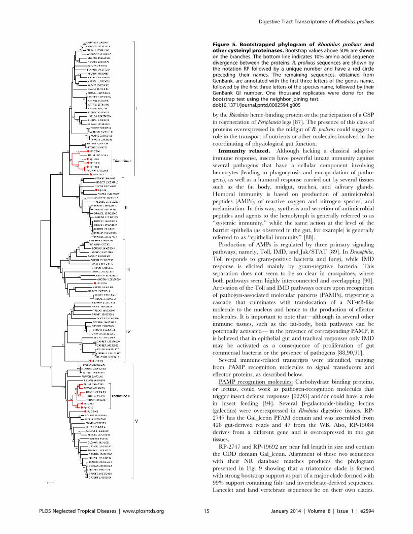
by the Rhodnius heme-binding protein or the participation of a CSP
in regeneration of Periplaneta legs [87]. The presence of this class of
proteins overexpressed in the midgut of R. prolixus could suggest a
role in the transport of nutrients or other molecules involved in the
coordinating of physiological gut function.
Immunity related. Although lacking a classical adaptive
immune response, insects have powerful innate immunity against
several pathogens that have a cellular component involving
hemocytes (leading to phagocytosis and encapsulation of patho-
gens), as well as a humoral response carried out by several tissues
such as the fat body, midgut, trachea, and salivary glands.
Humoral immunity is based on production of antimicrobial
peptides (AMPs), of reactive oxygen and nitrogen species, and
melanization. In this way, synthesis and secretion of antimicrobial
peptides and agents to the hemolymph is generally referred to as
‘‘systemic immunity,’’ while the same action at the level of the
barrier epithelia (as observed in the gut, for example) is generally
referred to as ‘‘epithelial immunity’’ [88].
Production of AMPs is regulated by three primary signaling
pathways, namely, Toll, IMD, and Jak/STAT [89]. In Drosophila,
Toll responds to gram-positive bacteria and fungi, while IMD
response is elicited mainly by gram-negative bacteria. This
separation does not seem to be so clear in mosquitoes, where
both pathways seem highly interconnected and overlapping [90].
Activation of the Toll and IMD pathways occurs upon recognition
of pathogen-associated molecular patterns (PAMPs), triggering a
cascade that culminates with translocation of a NF-kB-like
molecule to the nucleus and hence to the production of effector
molecules. It is important to note that—although in several other
immune tissues, such as the fat-body, both pathways can be
potentially activated— in the presence of corresponding PAMP, it
is believed that in epithelial gut and tracheal responses only IMD
may be activated as a consequence of proliferation of gut
commensal bacteria or the presence of pathogens [88,90,91].
Several immune-related transcripts were identified, ranging
from PAMP recognition molecules to signal transducers and
effector proteins, as described below.
PAMP recognition molecules: Carbohydrate binding proteins,
or lectins, could work as pathogen-recognition molecules that
trigger insect defense responses [92,93] and/or could have a role
in insect feeding [94]. Several b-galactoside-binding lectins
(galectins) were overexpressed in Rhodnius digestive tissues. RP-
2747 has the Gal_lectin PFAM domain and was assembled from
428 gut-derived reads and 47 from the WB. Also, RP-15084
derives from a different gene and is overexpressed in the gut
tissues.
RP-2747 and RP-19692 are near full length in size and contain
the CDD domain Gal_lectin. Alignment of these two sequences
with their NR database matches produces the phylogram
presented in Fig. 9 showing that a triatomine clade is formed
with strong bootstrap support as part of a major clade formed with
99% support containing fish- and invertebrate-derived sequences.
Lancelet and land vertebrate sequences lie on their own clades.
Figure 5. Bootstrapped phylogram of Rhodnius prolixus andother cysteinyl proteinases. Bootstrap values above 50% are shownon the branches. The bottom line indicates 10% amino acid sequencedivergence between the proteins. R. prolixus sequences are shown bythe notation RP followed by a unique number and have a red circlepreceding their names. The remaining sequences, obtained fromGenBank, are annotated with the first three letters of the genus name,followed by the first three letters of the species name, followed by theirGenBank GI number. One thousand replicates were done for thebootstrap test using the neighbor joining test.doi:10.1371/journal.pntd.0002594.g005
Digestive Tract Transcriptome of Rhodnius prolixus
PLOS Neglected Tropical Diseases | www.plosntds.org 15 January 2014 | Volume 8 | Issue 1 | e2594

Figure 6. Bootstrapped phylogram of Rhodnius prolixus and other carboxypeptidases. Bootstrap values above 50% are shown on thebranches. The bottom line indicates 10% amino acid sequence divergence between the proteins. R. prolixus sequences are shown by the notation RPfollowed by a unique number and have a red circle preceding their names. The remaining sequences were obtained from GenBank and are
Digestive Tract Transcriptome of Rhodnius prolixus
PLOS Neglected Tropical Diseases | www.plosntds.org 16 January 2014 | Volume 8 | Issue 1 | e2594
The two Triatoma dimidiata sequences that group with the Rhodnius
sequences have been described in a sialotranscriptome [95].
Sugar-inhibitable hemaglutinins have been described in the gut
of triatomines [96,97]. Galectins are overexpressed in gut and
salivary glands of Anopheles infected with bacteria or Plasmodium
[98–100]. It is speculated that galectins are involved in insect
immune response similarly to how they are in mammals—by
opsonizing bacteria and other pathogens facilitating their recog-
nition, agglutination, and/or phagocytosis for immune-competent
cells. Also, a galectin (PpGalec) has been implicated in Leishmania
major adhesion to the midgut epithelia of Phlebotomus papatasi. In this
case, blockage of this protein with specific antibodies leads to an
important decrease in vector parasite load after six days post
infection [101]. It would be interesting to assess whether any of
these proteins might be involved in T. cruzi binding to the midgut
epithelium.
RP-16133 codes for a 59 truncated transcript producing
matches against the NR database to proteins annotated as
hemolectin. The best match (gi|193601326) has multiple domains,
including von Willebrand, coagulation factor 5/8, TIL, and the
C8 domains. Hemolectin is hemocyte-specific in Drosophila and is
involved in the fly’s clotting system [102–104].
Three contigs containing peptidoglycan recognition protein
(PGRP) domains were also identified in the digestive tissues (Asb-
69756, Asb-23314, Asb-48139). Asb-69756 and Asb-23314 do not
present predicted trans-membrane regions and are likely to be
soluble PGRPs. Interestingly, Asb-69756 probably presents
amidase activity, as all five conserved catalytic amino acid residues
are present in this protein. If that is the case, Asb-69756 could be
involved in destruction of bacteria-released peptidoglycan, down-
regulating the bug’s immune response. Asb-23314, on the other
hand, is unlikely to present amidase activity, because one of the
five conserved catalytic residues is missing. If that is the case, Asb-
23314 could be involved in detecting peptidoglycan and activating
an epithelial IMD response. The last PGRP domain containing
transcript, Asb-23314, also does not present amidase activity but
show a predicted transmembrane domain and is homologous to
the Drosophila PGRP-LC (NP_729468.2). This transcript might
constitute an actual PGRP-LC and may represent a receptor
primarily responsible for activation of the IMD pathway in
Rhodnius.
Immune signaling pathways: Transcripts coding for members of
the immune signaling pathways were not overexpressed in gut
compared to WB, but several of them showed a significant number
of reads, indicating that they were operating in these tissues.
Despite this, these transcripts were included in our analysis,
because the midgut epithelia is the area of most intense contact
between microorganisms and insects and is the only part of the
triatomine body in contact with T. cruzi. Although it is generally
accepted that the Toll pathway is not active in digestive tissues
[88,105], several contigs putatively coding for proteins from this
pathway were identified—namely, a Toll receptor (Asb-44175), its
adaptor protein MyD88 (Asb-69782), the kinase pelle (Asb-15772)
and the pelle-associated protein pellino (Asb-24337) [106]. The
evolutionarily conserved intermediate in the Toll/IL-1 signal
transduction pathway [107], ECSIT (Asb-9158) and a protein
from the Spatzle family (RP-45859) were identified in the
transcriptome. Interestingly, contigs coding for two additional
putative Toll-interacting proteins (Tollips; Asb-22553 and Asb-
45642), for an inhibitor of the Toll pathway transcription factor
rpDorsal Cactus (Asb-31044), the Cactus-binding protein cactin
(Asb-33928), and a contig containing an NF-kB-repressing factor
domain (Asb-17843) were also identified. Although these contigs
were not overexpressed in the gut libraries when compared to WB,
this is the first time that such a high number of Toll-related
proteins were found consistently in a midgut transcriptome,
suggesting that, in spite of the relative low abundance, this
pathway may be of physiologic significance in gut immunity in
Rhodnius.
In contrast to this high number of Toll-related transcripts, only
one contig coding for a member of the IMD pathway was
identified in the digestive tissues. It coded for the IMD negative
regulator Caspar (Asb-145) [108]. This contig was highly
expressed in the gut (80 reads) but also in WB (92 reads). Low
expression levels also were found for the STAT pathway, where a
transcript coding for a STAT (Asb-17321; 4 reads only in AM and
none in WB) was identified. Together, these results suggest that all
three main known immune pathways are active in the Rhodnius
midgut.
A transcript resembling eiger was identified (Asb-21490; 21
reads from gut and 31 reads from WB). Eiger, the insect homolog
of mammalian TNF, has been implicated in the immune response
against extracellular pathogens [109] as well as against bacterial
oral infection [110]. Eiger/TNF was suggested to be part of an
ancient proof-reading pathway directed to suppress tumors in
epithelial tissues based on alterations of polarity that are typical of
malignant cells but that can also be found in cells that are either
physically damaged or exposed to pathogens [111]. As mentioned
below, there are several transcripts expressed in the gut that
belong to signaling pathways related to cell polarity, indicating
that sensing and control of cell polarity is a priority of Rhodnius
intestinal cells. This could provide a link between tissue
morphology and innate immunity related to intestinal pathogens.
Interestingly, three contigs putatively coding for proteins with a
double-strand (ds) RNA binding domain were identified in
Rhodnius digestive tissue libraries. One of these (Asb-16245) codes
for a putative R2D2 protein. R2D2 is known to associate with
Dicer-2 and is essential for channeling the siRNA generated by
this protein to the RISC complex [112]. Tar RNA-binding
proteins (TRBPs; Asb-26443) have a dsRNA binding domain and
are structural components of the RISC complex [113]. Finally,
also identified was a contig coding for a protein homologous to
loquacious (Asb-21490), a protein that contains two dsRNA
binding domains and was originally described as part of the
miRNA generating machinery in Drosophila [114].
Effector proteins: Large amounts of lysozyme activity were
described in the anterior and PM of R. prolixus [20]. Several
lysozyme-coding transcripts were found to be overexpressed in gut
tissues. RP-3602 was assembled from 7966 digestive reads and 619
WB-derived reads, hence being 23.1-fold overexpressed in
digestive tissues. This lysozyme was previously reported as
upregulated in the midgut following bacterial challenge as well
as ingestion of T. cruzi [115]. In addition, RP-6482—although
somewhat more mildly overexpressed in the digestive tissues than
RP-3602—was reported to be upregulated in the FB after
injection of bacteria into the hemocoel [115]. RP-24996 was the
only lysozyme transcript not overexpressed in the digestive tissues.
All lysozyme transcripts possess catalytic aspartate and glutamate
residues except for lysozyme 2 from T. infestans [116]. The function
of this unusual lysozyme remains to be elucidated. In T. brasiliensis,
annotated with the first three letters of the genus name, followed by the first three letters of the species name, followed by their GenBank GI number.One thousand replicates were done for the bootstrap test using the neighbor joining test.doi:10.1371/journal.pntd.0002594.g006
Digestive Tract Transcriptome of Rhodnius prolixus
PLOS Neglected Tropical Diseases | www.plosntds.org 17 January 2014 | Volume 8 | Issue 1 | e2594
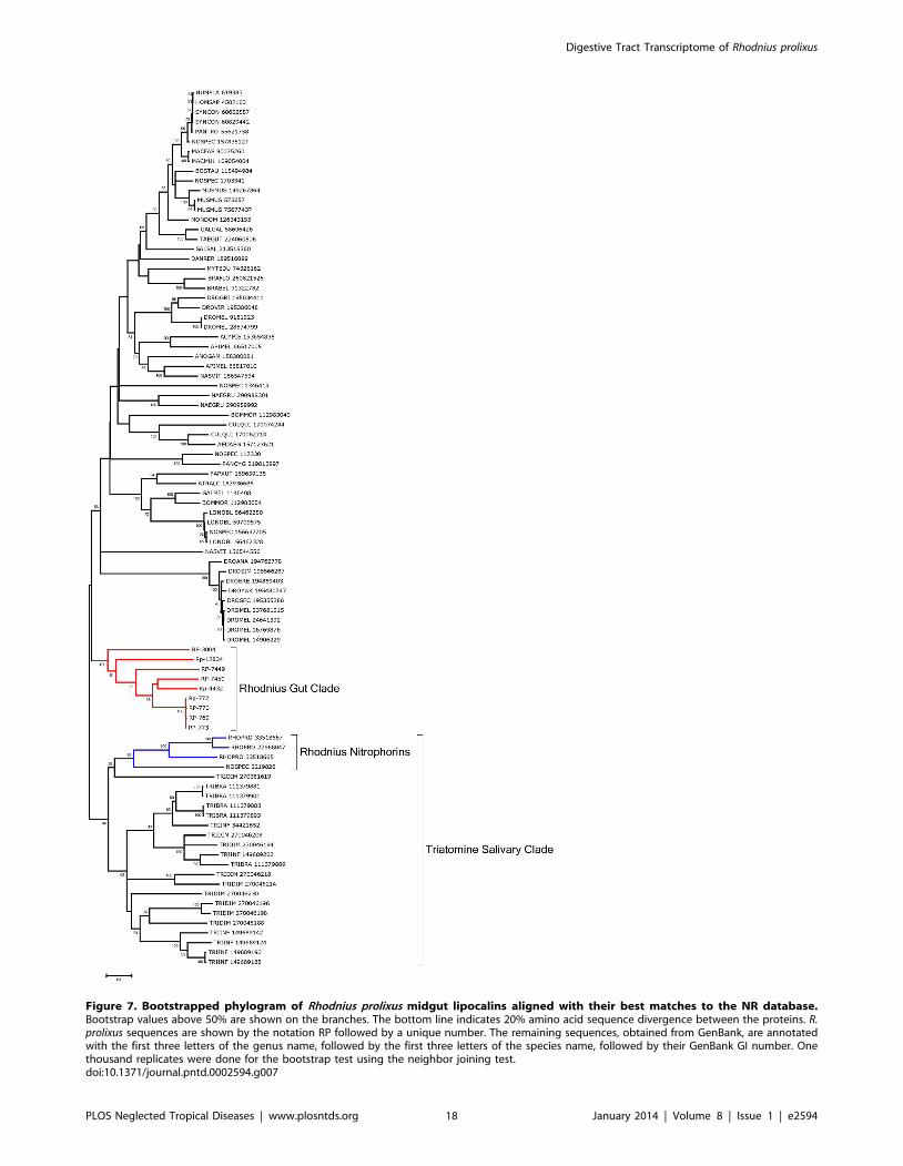
Figure 7. Bootstrapped phylogram of Rhodnius prolixus midgut lipocalins aligned with their best matches to the NR database.Bootstrap values above 50% are shown on the branches. The bottom line indicates 20% amino acid sequence divergence between the proteins. R.prolixus sequences are shown by the notation RP followed by a unique number. The remaining sequences, obtained from GenBank, are annotatedwith the first three letters of the genus name, followed by the first three letters of the species name, followed by their GenBank GI number. Onethousand replicates were done for the bootstrap test using the neighbor joining test.doi:10.1371/journal.pntd.0002594.g007
Digestive Tract Transcriptome of Rhodnius prolixus
PLOS Neglected Tropical Diseases | www.plosntds.org 18 January 2014 | Volume 8 | Issue 1 | e2594
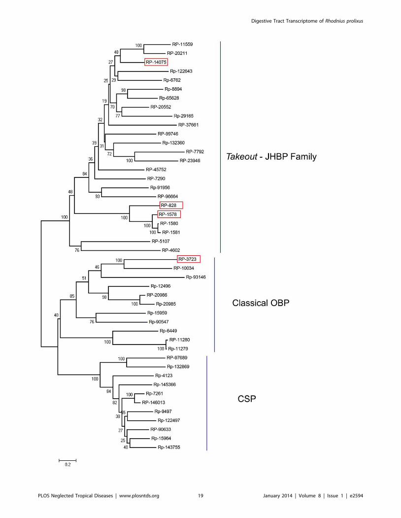
Digestive Tract Transcriptome of Rhodnius prolixus
PLOS Neglected Tropical Diseases | www.plosntds.org 19 January 2014 | Volume 8 | Issue 1 | e2594
expression of lysozyme 1 (lys1) is also upregulated in the AM after
feeding, with a maximum five days after blood uptake, suggesting
activity against developing bacteria [117]. In addition to providing
protection against airborne bacteria, these lysozymes might also
function in digestion of symbiotic bacteria, which develop to high
densities in the AM after blood ingestion (see section ‘‘carbohy-
drate digestion’’). However, the number of symbionts is negatively
correlated to the expression level of lysozyme and defensin genes
[18,118].
Defensins are ubiquitous antimicrobial peptides found in both
invertebrates and vertebrates [119]. Insect defensins are small
cationic peptides with molecular weights of about 4 kDa. They
possess three disulphide bridges and contain three characteristic
domains: an amino terminal flexible loop, followed by an a-helix
and a carboxy-terminal anti-parallel b-sheet [120,121]. Eight
defensin sequences were included in Supporting Information S2.
RP-12696 was 8.6-fold overexpressed in gut tissues; another three
sequences are mildly overexpressed. Defensin A in R. prolixus has
been shown to be upregulated in the intestine after immune
activation by bacterial challenge of the hemocoel, and a much
stronger upregulation was detected in the FB [122]. Similar to the
upregulation of lysozyme, the transcript levels of the defensin 1
gene (def1) in T. brasiliensis is increased following blood ingestion in
the AM [117], indicating activity against developing bacteria;
however, insect defensins are not only active against bacteria but
also interfere with development of eukaryotic parasites in the
vector, e.g., Plasmodium and filarial helminths [122–124].
The SCP superfamily of proteins includes the plant pathogen-
esis-related protein, the secretory cysteine-rich proteins found in
snake venoms, and allergen 5 found in vespid venoms [125]. These
proteins may have diverse functions including as proteases and for
defense. RP-7994 is a member of this family assembled from 215
reads, 184 of which are from digestive tissues.
Signaling pathways. Circuits of protein phosphorylation
and dephosphorylation involve the concerted action of two
enzyme families: protein kinases (PKs)—which add phosphate to
the hydroxyl group of serine, threonine, or tyrosine—and protein
phosphatases (PPs) that eventually remove such phosphate groups.
Together with sequencing of genomes, the full gene complement
of PKs and PPs—the kinome and phosphatome, respectively—
allowed us to obtain a broader picture of signaling networks in
eukaryotic cells from several organisms, but not for human disease
vectors. The presence of some PKs and PPs and their probable
functions in the gut of Rhodnius are discussed below. Additionally,
some signal transduction pathways and transcription factors that
regulate morphogenetic processes during development are dis-
cussed that may be involved in regenerative processes of gut
physiology, as has been suggested for members of the BMP, FGF,
and Wnt families of transcription factors [126].
MAPK signaling cascades: Protein tyrosine phosphatase (PTP)
4A3 (Asb-40892) is a member of dual-specificity phosphatases
(DUSPs) that are able to dephosphorylate both phosphotyrosine
and phosphothreonine residues in target proteins. Such enzymes
are usually deactivators of mitogen-activated PK (MAPK)
cascades. Such gene products recorded 21 reads from the digestive
system (exclusively from PM) and none from the WB library. PTP
4A3 belongs to a subfamily of DUSPs also known as phosphatases
of regenerating liver (PRLs), which play a major role in
oncogenesis and are overexpressed in gastric and colorectal
tumors and modulate kinases of the Erk branch of the MAPK
cascade [127].
Ste20-like kinase is a serine/threonine PK, an upstream
regulator of various MAPK cascades, and has 18 reads (Asb-
39211), also exclusively from the PM. Ste20-like kinases function
as MAPK4 enzymes, which activate the downstream cascade of
MAPK3, MAP2K, and MAPK. These enzymes were originally
discovered as mediators of pheromone signaling in yeast and are
involved in ion transport, cytoskeleton organization, and response
to osmotic stress. A member of this family, Ste20-related proline/
alanine-rich kinase, is activated by hypertonicity, leading to
activation of p38 and JNK MAPK cascades that phosphorylate
ion transporters that regulate cell volume [128]. Activation of
Ste20-like kinases also may occur in response to PAMPs such as
LPS, peptidoglycan and flagelin. Variations of osmotic pressure
and presence of bacteria or protozoan parasites are both major
factors that govern the physiology of the Rhodnius midgut.
A transcript (Asb-18967) similar to MAPK-activated PK or
MAPKAK2, also known as MK2, showed15 reads from the AM.
This is a Ser/Thr PK activated by p38 MAPK and is involved in
cell-shape change and cell adhesion. MK2 activation is also
required for cytokine production during inflammatory responses
[129]. It was recently demonstrated that MK2 activity is essential
to cutaneous wound healing [130] and thus an enzyme of this
group may be a modulator of tissue injury/immune response in
AM epithelia.
Another transcript (Asb-29380) highly similar to PK C lambda/
iota (PKCl/i) has 26 reads in all three gut libraries. This is a Ser/
Thr kinase that belongs to the atypical group of PK C isoforms
that are independent of calcium and diacylglycerol, which are
modulators of other PK C isoforms. PKCl/i signals through the
Rac1/MEK/ERK1,2 pathway that ultimately induces carcino-
genesis in human intestine epithelial cells [131]. Its overexpression
is now a prognostic for human gastric cancer [132]. A role of
PKCl/i has been demonstrated in the establishment of epithelial
cell polarity through binding members of the PAR family of
proteins [133]. There is strong evidence that binding of PKCl/ito PAR3 and PAR6 modulate such events that depend on cell
polarity as endocytosis, phosphoinositide signaling, microtubule
and spindle orientation, and organization of actin cytoskeleton
[134]. Thus, PKCl/i in the R. prolixus gut may be a modulator of
intracellular arrangement of cytoskeleton and organelle distribu-
tion during digestive cell physiology and when it engages in its own
division.
Lkb1/AMPK: An interface between cell morphology and blood
digestion: The digestive system shows 86 reads of stk11 or LKB1
(Asb-6501) compared to only 27 from WB. Although Lkb1 is a PK
that acts as a regulator of cell polarity and tumor suppression, it is
well known as a target of cell growth regulator AMP-activated PK
(AMPk; Asb-15260), a major modulator of cell energy homeostasis
[135] that shows 19 reads distributed among all three gut libraries.
AMPK is activated by cell signals that decrease cellular ATP.
AMPK activation leads to the downregulation of ATP-consuming
pathways. The major AMPK targets are glycogen synthase, acetyl-
CoA carboxylase, non-muscle myosin light chain, and mTOR,
Figure 8. Bootstrapped phylogram of Rhodnius prolixus midgut Takeout-JHBP, Odorant Binding Protein and Chemosensorial Protein.Amino acid sequences of 46 contigs were combined to create an entry file for phylogenetic analysis in MEGA 4.0.2. An unrooted consensus neighborjoining tree was generated based on ten thousand bootstrap replicates with pairwise gap deletions using neighbor joining method. Bootstrap valueslower than 50% are not shown. Red boxes indicate the over expressed proteins. JHBP: Juvenile hormone binding proteins. OBP: Odorant bindingproteins. CSP: Chemosensorial proteins. For more details, see text.doi:10.1371/journal.pntd.0002594.g008
Digestive Tract Transcriptome of Rhodnius prolixus
PLOS Neglected Tropical Diseases | www.plosntds.org 20 January 2014 | Volume 8 | Issue 1 | e2594
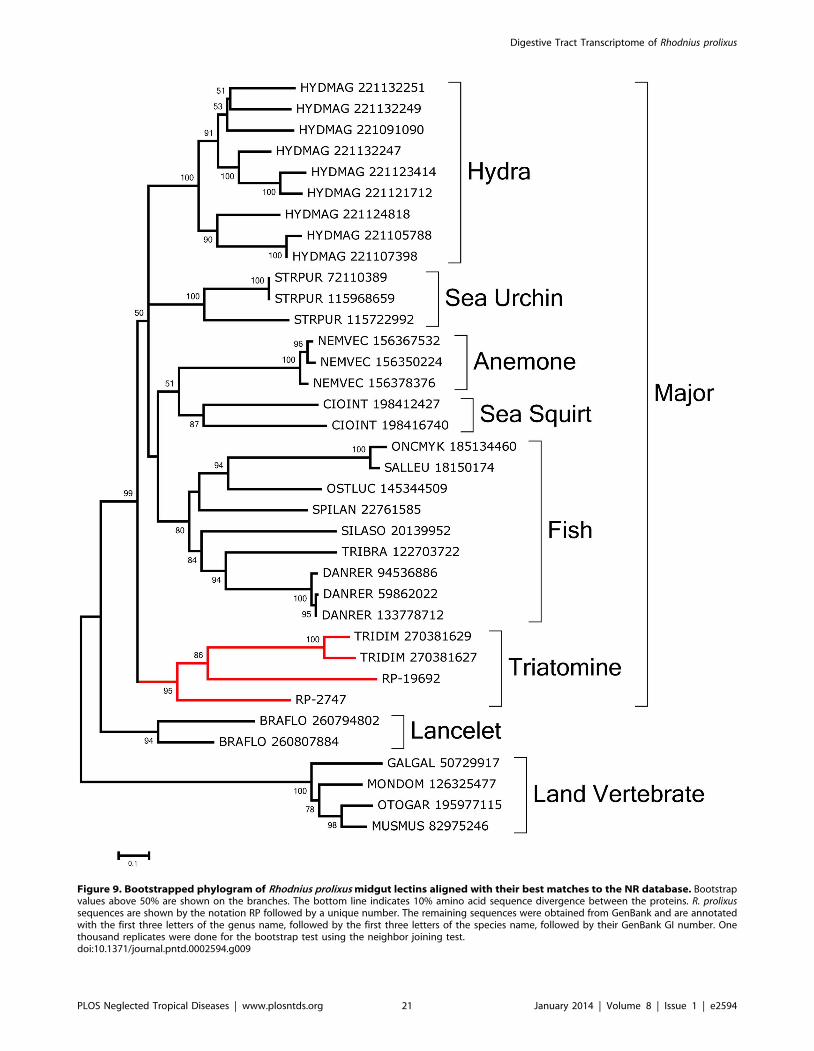
Figure 9. Bootstrapped phylogram of Rhodnius prolixus midgut lectins aligned with their best matches to the NR database. Bootstrapvalues above 50% are shown on the branches. The bottom line indicates 10% amino acid sequence divergence between the proteins. R. prolixussequences are shown by the notation RP followed by a unique number. The remaining sequences were obtained from GenBank and are annotatedwith the first three letters of the genus name, followed by the first three letters of the species name, followed by their GenBank GI number. Onethousand replicates were done for the bootstrap test using the neighbor joining test.doi:10.1371/journal.pntd.0002594.g009
Digestive Tract Transcriptome of Rhodnius prolixus
PLOS Neglected Tropical Diseases | www.plosntds.org 21 January 2014 | Volume 8 | Issue 1 | e2594
which thus leads to the downregulation both of glycogen, lipid,
protein synthesis and of cell polarity [136]. Studies on the Peutz-
Jeghers syndrome, an autosomal gastrointestinal polyposis disorder
caused by germline mutation in the LKB1 gene, have revealed a
conserved link between energy metabolism and cell polarity-
dependent cell functions such as organization of the actin
cytoskeleton and sorting of apical and basolateral membrane
proteins to facilitate directed endosomal transportation. One
molecular mechanism that links LKB1 and control of cell shape is
its ability to phosphorylate the regulatory light chain of nonmuscle
myosin II (MLRC), which thus regulates cytokinesis and—through
myosin II—adjusts the formation of tight and adherens junctions
[137]. AMPK-null mutants of Drosophila present several abnor-
malities in mitosis and cell polarity [138]. In addition, AMPK
activation by energy deprivation leads to large changes in cell
shape. Significant expression of LBK1 and AMPK in the gut
suggests that this pathway may participate in regulation of cell
polarity and energy metabolism of intestinal cells.
Another AMPK target in Rhodnius gut transcriptome, TOR
(target of rapamycin; Asb-43781 and Asb-70063) is a PK that
regulates several cellular process such as cell growth, proliferation,
and survival [139]. In mosquitoes, it was shown that amino acid
ingestion induces early trypsin protein synthesis coincident with
activation of the TOR pathway [140], which also was implicated
in control of expression of vitellogenin gene that takes place after a
blood meal [141]. Although showing a low number of reads (3),
these TOR transcripts suggest the presence of this nutrient and
energy-sensing signaling pathway that connects the ingested meal
with blood digestion and yolk protein synthesis, two different
biologic processes that are separated in a time frame. Such a
hypothesis must be tested at the molecular level.
Developmental regulators in adult gut. Wnt and Notch:
Although the gut libraries used here were from adult females,
transcripts related to two signaling pathways classically related to
control of morphogenesis during development—Wnt and
Notch—were identified. In adults these transcripts may be
important for self-renewal or regeneration of intestinal cells
[142,143]. The transcript coding for an ortholog for defective in
proventriculus (dve; Asb-11146) showed 126 reads from gut libraries
(113 being from AM) and only 3 reads from WB. This gene owes
its name to studies in D. melanogaster showing that mutants for dve
have morphologic defects of the proventriculus [144–146], a
region that develops at the junction of the foregut and the midgut
and functions as a valve regulating the passage of food. During
development, dve has been shown to respond to Wg (Wnt), Dpp
(BMP), EGFR, and Notch signaling in the gut [144,146,147];
however, dve also has an important role in the digestive
physiology of the gut. Expression of dve in midgut copper
cells—cells that resemble absorptive mouse enterocyte cells—is
necessary for acid secretion and for copper absorption [148].
Therefore, considering the high degree of sequence conservation
and enrichment in adult gut tissue, dve is likely to perform a
physiologic role in Rhodnius, as well. As possible regulators of dve,
Wnt pathway elements were identified, although overexpression
of Wnt pathway components in relation to the WB library was
not homogeneous throughout the gut. Reads for b-catenin, a
transducer of Wnt signals, were found enriched in the AM (RP-
41815/Asb-1876). Furthermore, an ortholog of a RAN-binding
protein, a negative regulator of the pathway (Asb-62348, with 77
reads exclusively in the RE), provides additional support for the
notion that the pathway is functional. Four calmodulin transcripts
were detected in the transcriptome (RP-98600, RP-96030, RP-
95216, and RP-1777), but only one (RP-1777) was expressed in
gut tissue. Finally, casein kinase II is likely expressed in gut tissue,
as reads for the a (RP-3340) and b (RP-15495) subunits were also
detected.
Interestingly, several potential Notch substrates or regulators
were detected in Rhodnius, with a number of transcripts enriched in
the gut. Notch is required for several gut-associated functions in
various species. Notch regulates differentiation of endocrine cells
of the mouse gut endoderm [149], regulates the switch between
luminal and glandular fate in the endodermal epithelium of the
chick [150], and is required in the gastrointestinal tract stem-cell
niche [151]. Hairy (Asb-2287; 35 reads from gut, 25 being from
AM, and 12 reads from WB) is a known element of the Notch
pathway in vertebrates and invertebrates, displaying transcription
repressor activity characteristic of Her proteins [152]. Another
potential element of the Notch pathway is a transcript (Asb-24840,
exclusively from digestive libraries; 18 reads, mainly from PM)
similar to BTB/POZ domain-containing proteins bric-a-brac,
Broad, and tramtrack from D. melanogaster that were shown to
interact with the Notch path [153]. Neurofibromin (Asb-10846) is
a protein shown to be regulated by Notch in the nervous system
[154,155], but it also was shown to regulate longevity and
resistance to stress through cAMP regulation of mitochondrial
respiration and reactive oxygen species production [156].
RNA-processing, translation and secretion. Posttran-
scriptional control of gene expression provides a prompt response
to metabolic changes. In mosquitoes, translation of trypsin mRNA
is regulated [157–159] through the TOR signaling pathway
sensing the amino acid pool [140]. Other digestion-related
pathways such as components of iron metabolism are also
regulated at the posttranscriptional level, such as ferritin through
iron regulatory proteins [160,161]. Thus, analysis of gut-specific
genes involved in translation apparatus may provide hints about
posttranscriptional regulation.
There was an overall increment of expression of genes involved
in RNA processing, translation, and protein secretion in the gut
libraries compared to WB, probably as a consequence of the need
for high rates of protein synthesis needed to cope with formation of
secreted polypeptides—such as digestive enzymes and peritrophins
described above—and to support epithelial cell division that must
occur after a blood meal. Posttranscriptional control of gene
expression provides a prompt response to metabolic changes. This
has significance in comparative transcriptomics, as transcripts that
do not change their abundance might still be targets of control.
Regarding protein trafficking and elongation factors, some
transcripts were overexpressed in all three gut libraries when
compared to WB, such as the protein tyrosine phosphatase SHP1/
p47 (Asb-1670), the endosomal membrane protein EMP70 (Asb-
308 and Asb-663), the coatomer protein complex subunit (Asb-
5008), an ADP-ribosylation factor (ARF, Asb-7450), EiF3C (Asb-
610, 839, and 840), an aspartyl-tRNA synthetase (Asb-4568), and
some rRNAs. Changes in ribosomal protein mRNAs have been
described in the fat body of Ae. aegypti [162]. Similarly, we observed
differential transcriptome expression in the gut. Ribosomal
proteins S24 (Asb-39300), L18a (Asb-42186), L8 (Asb-18849),
L21 (Asb-199), AS (Asb-1710), L19 (Asb-1947), L32 (Asb-1715),
S15a (Asb-65370), P1 (Asb-5747), S16 (Asb-4689), and S29 (Asb-
6829) are enriched at least five fold in the gut, while subunits S7
(Asb-1131), S25 (Asb-17734), S23 (Asb-8803), L29 (Asb-18997),
and S21 (Asb-10782) are decreased. We found transcripts (Asb-
1398,1400, and 1402) in the gut of R. prolixus with high similarities
to TRM4, a tRNA 5-methylcytosine (m5C) methyltransferase with
3,163 reads from all gut libraries and 318 reads from WB. tRNA
modifications have been being implicated in tRNA stability [163–
165], translational fidelity [166–168], response to stress [169,170],
and control of cell growth [171]. Just recently it has been shown
Digestive Tract Transcriptome of Rhodnius prolixus
PLOS Neglected Tropical Diseases | www.plosntds.org 22 January 2014 | Volume 8 | Issue 1 | e2594
that one of the yeast responses to oxidative stress is the increase of
m5C at the anticodon wobble position 34 in tRNALeu(CAA), a
tRNA modification inserted by the Trm4 methyltransferase. This
modification leads to selective translation of mRNA species
enriched in the TTG codon, among them a specific paralog of a
ribosomal protein [172]. The high expression of the TRM4-like in
the gut of R. prolixus might be related to this fact, as its PM after a
blood meal becomes a site of high oxidative stress [173], and also,
as methylation of tRNAs make them more stable, it might make
them available for the high turnover of protein expression
following a meal. Further studies are necessary to prove these
hypotheses.
In contrast, other transcripts were specifically more expressed
by one of the segments of the digestive apparatus. Although most
of the basal factors involved in RNA metabolism—namely
splicing, polyadenylation, or translation—are expressed in all cell
types, it has been shown that different isoforms can have roles in a
tissue- or stage-specific manner [174,175]. We did not observe
changes in most of the translation factors, except in the RE, where
there was increased expression of eukaryotic translation initiation
factor 1A domain containing protein (Asb-19360; 1283 reads in
Rec and 55 in WB; EF2 with 203 reads from WB and 1267 reads
in Rec (Asb-1428 = RP-7150) and one isoform of initiation factor
4E (eIF4E; Asb-5727) with 13 reads from Rec and only one from
WB. Two other isoforms of eIF4E are detected in the
transcriptome (RP-92257 and RP-7125). RP-7125 is present in
both WB and gut, while RP-92257 is only detected in WB. Despite
the similarity in sequence, they correspond to different genes
rather than alternative splicing, as they are encoded by different
genomic contigs (Supporting Information S1). eIF4E, the cap
binding protein, is a target of regulation through the TOR
pathway (discussed above in the section on protein phosphoryla-
tion circuits). Interestingly, the main isoform identified in the gut is
similar to the Drosophila eIF4E-HP, a stage-specific translational
repressor [175,176] with a conserved change of the tryptophan
residue that contributes to cap recognition for a tyrosine and the
lack of eIF4G/eIF-4EBP binding domain.
Regarding protein trafficking, we identify the Clathrin assembly
protein AP180 (Asb-63672; 14 reads all in RE), the exocyst
complex component 8 (Asb-11867; 32 reads in AM) responsible
for tethering of secretory vesicles to the plasma membrane after
leaving the Golgi compartment [177]; an emp24 (Asb-41987; 30
reads in PM) which is a transmembrane protein that is involved in
transport of secretory proteins from ER to Golgi [178], and a
guanine nucleotide exchange factor similar to Drosophila schizo
(Asb-4883; 50 reads from AM and 1 from WB) that are known to
act on ARF GTPases, which are known to regulate endocytosis
[179]. While these data are consistent with ultrastructural evidence
of intense protein synthesis and exocytosis in all three segments,
marked tissue-specific expression of some components suggests
that vesicle trafficking and protein secretion may proceed through
distinct routes and be subjected to distinct pathways in each
segment.
Detoxification. Plants produce innumerous toxic compounds
to deter phytophagous insects which react with gut detoxification
enzymes such as cytochrome P450s, glutathione transferases (GSTs)
and other oxidases, these enzymes also participating in insecticide
resistance [180–182]. It is expected that a blood diet would reduce
the requirements for detoxification, such as that for alkaloids. On
the other hand, excess heme in the diet imposes an oxidative
challenge, leading to production of toxic products of lipid
peroxidation, the elimination of which is accomplished by a similar
array of genes [183]. In Ae. aegypti, some P450 genes from CYP6 and
CYP9—classically involved in xenobiotic metabolism—are also
transcribed in response to oxidative stress [184]. A probable
member of the subfamily CYP6 [185] encoded by RP-7174 is highly
expressed in all gut tissues (1519 reads), but poorly in the WB library
(6 reads). Other possible members of CYP6/CYP9 subfamilies (RP-
6932, RP-6776, RP-6043, RP-1459, RP-1608, RP-5848, RP-7653,
RP-4925, RP-6931, RP-1613, RP-6041, and RP-11775) are
significantly more expressed in gut when compared with WB.
Consistent with this plethora of cytochrome P450s is the presence of
transcripts that code for a cytochrome P450 reductase (RP-3922,
with 173 reads from all three gut libraries versus 80 from WB),
which is responsible for providing two electrons needed for
activation of the oxygen molecule by a P450 enzyme during its
catalytic cycle [186]. Alternatively, the same role can be fulfilled by
a cytochrome b5 (RP-10436), a small membrane-bound electron
carrier hemoprotein [187] that, although not differentially ex-
pressed in the gut, showed up with 83 reads mainly in AM and PM.
In addition to detoxification function, several insect P450s are
known to be involved in steroid and lipid metabolism [188]. Final
hydroxylation steps of conversion of steroid precursors into active
insect ecdysteroid, 20-hydroxyecdysone, are accomplished by
cytochrome P450 enzymes encoded by genes in the Halloween
family [189].
GSTs are involved in detoxification by catalyzing the conjuga-
tion of glutathione with xenobiotic and toxic endogenous
compounds, including products of free radical metabolism. Among
the seven GST transcripts found in gut tissues, five were
significantly overexpressed in all three segments of the gut, each
of these being identified as a member of a different class: Zeta (RP-
4940), Delta/Epsilon (RP-10873), Sigma (RP-10298 and RP-
8544), and Theta (RP-3968).
Superoxide dismutase (SOD) catalyzes the dismutation of
superoxide radical to hydrogen peroxide and oxygen, lowering
superoxide levels and preventing formation of other reactive
oxygen species and their derivatives. Its action is complemented by
H2O2-eliminating enzymes such as catalase (Asb-14022 and Asb-
10100), glutathione peroxidases (Asb-2104), and peroxiredoxin
(Asb-10688- and Asb-10473) and its power-reducing pair thior-
edoxin (RP-6757). There are two major types of SOD enzymes
present in animals, Cu/Zn SOD (cytoplasmic/nuclear) and Mn
SOD (mitochondrial). Analysis of the Rhodnius transcriptome
showed the presence of five SOD transcripts (RP-11791, RP-
3874, RP-28439, RP-16118 and RP-1534). RP-11791, a Cu/Zn
SOD, showed slightly higher expression in the gut tissues (171
reads from all three libraries and 154 reads from WB), but RP-
3874, a Mn SOD, although present in the gut did not show high
message levels. In Rhodnius, the level of hydrogen peroxide was
shown to be controlled at least in part by catalase and glutathione-
dependent mechanisms [190]. Also, a glutathione peroxidase
activity has been shown in Rhodnius [191] that could be accounted
for by the transcript RP-10221, 3.5-fold overexpressed in gut
tissues, that seems to code for an authentic selenium-dependent
enzyme. Transcripts for the rate-limiting enzyme of glutathione
synthesis pathway, glutamate-cysteine ligase, were also found in
gut tissues, coding both for its catalytic subunit (Asb-10777; 12
reads in gut tissues and only one in WB) and for the regulatory
subunit (RP-13180; 84 reads in gut and only 47 in WB).
Glutaredoxin, a small antioxidant enzyme whose active disulfide
bond is reduced directly by gluthatione, is highly expressed (Asb-
10150; 305 reads in the gut versus 73 in WB). Sulfate conjugation
mediated by sulfotransferases (SULTs)—a mechanism of detoxi-
fication of xenobiotics as well as endogenous compounds—leads to
inactivation of substrate compounds and/or increase in their
water-solubility, thereby facilitating their removal from the body.
Transcripts coding for these enzymes were found expressed in the
Digestive Tract Transcriptome of Rhodnius prolixus
PLOS Neglected Tropical Diseases | www.plosntds.org 23 January 2014 | Volume 8 | Issue 1 | e2594
gut (RP-22910, RP-11341, RP-16870, RP-97304 and RP-25906),
although none were gut enriched compared to the WB. Nitration
of tyrosine, in both protein-bound and free amino acid form, can
readily occur in cells under oxidative/nitrosative stress, and
elevated levels of nitrotyrosine have been shown to cause DNA
damage or trigger apoptosis. Sulfation of nitrotyrosine occurs in
cells under oxidative/nitrative stress, and it has been demonstrated
that SULTs contribute to the metabolism of nitrotyrosine
[192,193]; however, although listed in the detoxification work-
sheet, sulfotransferases also add sulfate to proteoglycans of the
extracellular matrix and therefore may be implicated in tissue
remodeling as well.
Together, these data suggest that the Rhodnius gut has a complex
network of enzymes involved in regulation of redox balance,
especially involving control of the intracellular pool of reduced
thiols. In spite of not being exposed to allelochemicals in food, the
triatomine gut has retained significant expression of both Phase I
and Phase II detoxification pathways, and the hypothesis that this
may be a mechanism to ameliorate blood-induced oxidative stress
needs further investigation.
The supply of reducing equivalents in the form of nicotinamide
adenine dinucleotide phosphate (NADPH) is one of the most
important factors in cell protection against oxidative damage.
Some dehydrogenases have been shown to play a role in redox
balance [194,195], and at least one is highly overexpressed in the
gut RP-6620 (614 reads in gut and 243 in WB).
The worksheet ‘‘Detox’’ in Supporting Information S2 presents
detailed information on other cytochromes, cytochrome P-450
reductases, glutathione transferases, sulfotransferases, superoxide
dismutases, short-chain dehydrogenases, and other dehydrogenas-
es.
Iron and heme metabolism. Eukaryotic cells strictly control
heme homeostasis by regulating biosynthesis and degradation
pathways of this iron tetrapyrrol, due to its toxicity [196]. The
heme biosynthesis pathway has been previously described in R.
prolixus [197]. In fact, transcripts coding for all the enzymes that
participate in this pathway have been found in the sequenced
libraries. Most of these transcripts are more expressed in the WB
than in the gut libraries. The exception is 5-aminolevulinate
synthase (ALA-synthase, RP-2456), responsible for the rate-
limiting step of heme biosynthesis, which is significantly more
expressed in the digestive tissues.
Although it is already known that part of the heme molecules,
released by host blood digestion, cross the digestive systems and
reach the hemolymph [198], the proteins responsible for heme
transport across cellular membranes remain undescribed in
insects. Interestingly, transcripts coding for a protein similar to
feline leukemia virus Type C receptor (FLVCR), described as a
heme exporter [199], were found in the digestive libraries (Asb-
18956 and Asb-197149).
In most organisms studied, heme is degraded by heme
oxygenase (HO), a microsomal enzyme that catalyzes the oxidative
cleavage of the tetrapyrrol ring producing a-biliverdin (BV),
carbon monoxide, and iron; however, R. prolixus presents a unique
heme-degradation pathway wherein heme is first modified by
addition of two cysteinylglycine residues before cleavage of the
porphyrin ring by HO, followed by trimming of the dipeptides,
producing a dicysteinyl-c-biliverdin [200]. Digestive tissues and
pericardial cells present a high content of c-BV, suggesting high
HO activity [200,201]. In this context, two distinct heme
oxygenase transcripts (Asb-16264 and Asb-16263) were identified
in WB and digestive tissue libraries, mostly in AM and PM, which
were assigned to the same genomic contigs, suggesting that they
may be generated by alternative splicing.
After heme oxidative degradation by heme oxygenase, cells face
the challenge of storage and transport of the released iron without
allowing oxidative damage to cells. Transferrins are extracellular
proteins that bind free iron with high affinity, transferring the
metal to cells by a receptor-mediated process. At least three highly
expressed transcripts of transferrin (RP-6018, Asb-8333 and Asb-
16041) were identified in the sequenced libraries. RP-6018 and
Asb-16041 transcripts are over-represented in the WB library,
whereas a high expression of Asb-8333 is also found in the
digestive tissues, especially in PM and RE. Remarkably, the
transcript coding for the transferrin receptor (RP-960) is more
expressed in the same digestive libraries, suggesting that these
tissues have to deal not only with iron molecules coming from the
lumen but also with those provided from hemolymphatic
transferrins. It is worthwhile to speculate that these tissues may
be responsible for driving the excess circulating iron to excretion.
Another protein that plays a key role in iron metabolism is
ferritin. As in vertebrates, arthropod ferritins are heteromultimers
composed of two types of subunits that, in insects, are named
heavy and light chain homologs (HCH and LCH, respectively).
Three different transcripts of HCH subunits (RP-1172, RP-5775
and RP-7917) and two LCH subunits (RP-8697 and RP-3378)
were found in the sequenced libraries. As is well known for most
insect ferritins, the majority of expressed subunits present signal
peptides for secretion. The exceptions are HCH (RP-7917 and
RP-105633) transcripts that present a putative mitochondrial
target sequence, which were not found in digestive libraries; these,
as described for mammalian and Drosophila mitochondrial ferritins,
are highly expressed in testis [202].
While most HCH and LCH subunits are ubiquitously
expressed, HCH RP-1172 and LCH RP-3378 are more abundant
in digestive libraries, particularly in PM, suggesting that they may
be required during digestion and iron excretion processes.
Ferritin expression is posttranscriptionally regulated by intra-
cellular iron levels due to the presence of a stem-loop structure
found in the 59 untranslated regions of mRNA named iron-
responsive element (IRE). In the absence of iron, the iron
regulatory protein (IRP) binds to the IRE structure, sterically
blocking ferritin mRNA translation. This phenomenon is reversed
when IRP specifically associates with an iron atom. IREs are
present in the secreted HCH subunits RP-1172 and 5775 but not
in the LCH transcripts. In fact, among all insects studied to date,
only in Lepidoptera are IREs also found in LCH mRNAs [203].
Although at low level, transcripts coding for IRP (Asb-50964) were
found in WB and PM libraries. The presence of all components of
the IRP-IRE system suggests that the mechanism for translational
control of mRNAs by iron has been conserved in this insect. Thus,
a survey of other IRE-containing transcripts—especially among
proteins involved in iron and heme metabolism—deserves to be
done.
Lipid metabolism. During the blood meal, R. prolixus ingests
a large amount of lipids such as triacylglycerol, free fatty acids and
cholesterol, and the midgut is the main site of dietary lipid
absorption [204]. Plasma lipids are absorbed by the PM
epithelium and used in synthesis of different lipid classes that are
distributed to the tissues associated with lipophorin (Lp) particles
[205,206]. Triacylglycerol digestion takes place in the PM [21],
and this gut section has high expression levels of genes coding
digestive lipases (e.g., RP-2369, RP-2952, RP-21001). The free
fatty acids generated during this digestive reaction are absorbed by
midgut epithelial cells [21], and fatty acids need to be esterified to
coenzyme A (CoA) to be used by lipid metabolism pathways. This
can be made by an acyl-CoA synthetase, which shows two distinct
transcripts (RP-4249 and RP-24413), both more expressed in AM
Digestive Tract Transcriptome of Rhodnius prolixus
PLOS Neglected Tropical Diseases | www.plosntds.org 24 January 2014 | Volume 8 | Issue 1 | e2594
and PM. Alternatively, fatty acyl-CoA may be produced from de
novo synthesis from acetate using acetyl-CoA synthetase (RP-
29987), a transcript that has 31 reads in midgut and only 2 in WB.
Interestingly, the AM is the major site of acetyl-CoA synthetase
gene expression (and, possibly, de novo fatty acid synthesis), while
the PM appears to be specialized in direct absorption of fatty acid
from the blood meal [21]. Acyl-CoA is used in both catabolic and
anabolic pathways. The midgut transcriptome shows marked
expression of genes involved in b-oxidation, as 3-hydroxyacyl-CoA
dehydrogenases (Asb-3668), enoyl-CoA hydratases (e.g., Asb-3371,
Asb-3615), and carnitine O-acyltransferase (Asb-7656, Asb-
20469), suggesting that the Rhodnius midgut is using fatty acid
oxidation as a major source of energy. One transcript coding for a
fatty acyl-CoA elongase (Asb-44706) showed only 3 reads from
WB and 123 reads from gut, mainly from Rec (119 reads), possibly
related to synthesis of long-chain hydrocarbons that are compo-
nents of the wax layer that covers the wall of the hindgut [207].
Expression of the sterol regulatory element-binding-protein
homolog (Asb-14714; 17 reads in gut versus 4 in WB), especially in
AM and PM, suggest that the Rhodnius midgut is able to make de
novo lipid synthesis, as this transcription factor induces expression
of acetyl-CoA carboxylase and fatty acid synthase in Drosophila
[208]. As this transcriptome was made from organs dissected from
both unfed and blood-fed insects, it is not possible to determine
when fatty acid synthesis would occur. The Rhodnius midgut also
expresses the NPC1b homolog (Asb-2638; 55 reads in gut and 19
in WB), especially in anterior and PM, organs involved in
absorption of cholesterol, which is transferred to lipophorin
similarly to what happens with other lipids (Entringer et al.,
unpublished results). NPC1b protein is essential to absorption of
ingested cholesterol by midgut cells in Drosophila [209]. High
expression of transcripts coding for hydroxysteroid 17-b dehydro-
genase (Asb-5710) and C-4 sterol methyl oxidase (Asb-5381)
indicate that ingested cholesterol may be further metabolized into
other sterols.
The midgut transcriptome also reveals upregulation of genes
involved in complex lipid metabolism, as fatty acid desaturase
(Asb-1771), glycerophosphoryl diester phosphodiesterase (Asb-
14330), and diacylglycerol O-acyltransferase (Asb-1487). There
are also high expression levels of genes that participate specifically
in phospholipid biosynthesis, such as sphingomyelin phosphodies-
terase (Asb-1419 and Asb-1420, with 133 reads in all gut libraries
and 60 in WB), that catalyzes the hydrolysis of sphingomyelin to
ceramide, which may be further metabolized to bioactive lipids, as
sphingosine and sphingosine 1-phosphate. A transcript similar to a
choline kinase (Asb-6000) also showed high expression (65 reads in
gut, mainly AM and PM, and 25 in WB). This enzyme
phosphorylates choline to generate phosphoryl choline, which is
the first step in the so-called Kennedy pathway for phosphatidyl-
choline synthesis [210]. High choline kinase activity has been
implicated in tumor development, possibly by regulating Akt
phosphorylation, thereby promoting cell survival and proliferation
[211], a role that could be critical for tissues that need high cell-
renewal rates, such as digestive epithelia. Phospholipid transfer
proteins (PL-TPs) such as the phosphatidylinositol transfer protein
(Asb-15071; Asb-40276) are expressed in the Rhodnius midgut.
These proteins transport phospholipid inside the cells—transfer-
ring either phosphatidylinositol or phosphatidylcholine between
membranes [212]—and contribute to releasing secretory granules
and secreting of vesicles from the trans-Golgi network [213].
These proteins probably are related to phospholipid synthesis
needed to generate membranes of secretory vesicles to be used in
the formation of the perimicrovillar membranes or to be
transferred to lipophorin and exported to the hemocoel [214].
Another possible function of phospholipids in the gut involves
their signaling role as a source of bioactive lipid molecules through
the action of phospholipases (PLs). PLs work as digestive
hydrolases but also comprise a heterogeneous group of ubiquitous
enzymes involved in such diverse processes as membrane
homeostasis, signal transduction, and generation of bioactive
molecules [215]. One product of PL action (specifically PLA2) is
lysophosphatidylcholine, which is a component of saliva and feces
of R. prolixus [216]. Only one transcript coding for a lysopho-
spholipase like-1 is overexpressed in gut (RP-7099; 48 reads
mainly from AM and PM libraries, and 9 from WB) but several
other candidate PLs show significant expression levels in Rhodnius
gut: RP-1587 (PLC C), RP- 4722 (lysophospholipase), RP-5116
(PLD), RP-6129 (PLB), and RP- 7274 (PL/carboxyhydrolase).
Signaling by lysophosphatidic acid is turned off [217–219] by
means of lysophosphatidic acid acyltransferase (LPAAT, RP-
10018), showing 19 reads in gut, most in PM, and 5 reads in WB.
As already mentioned, PL-TPs transport phospholipids from their
site of synthesis to other cell membranes, but also have been
related to phospholipase C-mediated inositol signaling, PI3 kinase-
mediated phosphorylation of PIP2 to PIP3, and formation of
leukotrienes and lysophospholipids [213,220,221]. Four transcripts
coding for PL-TPs with SEC14 domain (RP-6243, RP-6447, RP-
21186 and RP-12057) were overexpressed in gut tissues,
highlighting the complexity of PL metabolism and trafficking in
these tissues.
In different cell types, lipids are stored in cytoplasmic organelles
termed lipid droplets (LDs). LDs store fatty acids and cholesterol as
neutral lipids, predominantly triglycerides (TG), cholesterol esters,
and diacylglycerol, surrounded by a phospholipid monolayer and
coated with a complex set of proteins [222]. Perilipins (Rp-2667;
635 reads from gut and only 21 from WB, overexpressed in all gut
tissues, but especially in RE) are proteins characteristic of LDs.
Proteins belonging to the PAT family are now collectively referred
to as perilipins, including proteins previously known as adipophilin
and tail-interacting proteins [223]. Perilipins regulate lipase access
to LDs according to cell metabolic needs [224,225]. In the past
few years, it became clear that LDs are not simple lipid storage
depots but rather complex organelles involved in multiple cellular
functions such as lipid biosynthesis and catabolism, signal
transduction, and energy and cholesterol homeostasis. Proper
use of both dietary lipid and lipid synthesized de novo from other
metabolic precursors involves absorption, intracellular trafficking
inside gut epithelia, and transfer to the hemocoel—a chain of
events that almost certainly must involve LDs.
Amino acid metabolism. Proteins are largely the most
abundant component of vertebrate blood, and therefore, its
digestion is a formidable source of amino acids. When transcripts
most abundantly expressed in the midgut were analyzed, a marked
predominance of enzymes related to amino acid degradation/
gluconeogenesis was found. From 28 transcripts related to amino
acid metabolism that were significantly overexpressed in the gut,
21 coded for degradation pathways. The first biochemical reaction
in most of amino acid degradation pathways is catalyzed by
transaminases, which transfer –NH2 to ketoacids (mainly oxalo-
acetate, a-ketoglutarate rendering aspartate or glutamate, respec-
tively, and to pyruvate rendering alanine) or dehydrogenases that
transfer –NH2 to H2O rendering NH4+. Among the transaminas-
es, it is remarkable that broad-spectrum transaminases, mainly
tyrosine aminotransferase (TAT; RP-18771 slightly overexpressed
in gut tissues) and aspartate aminotransferase (ASAT; Asb-40230;
RP-5603) are present in all three sections of the gut, indicating the
presence of a robust transamination network. Typical ASATs
constitute a node linking alanine, aspartate, glutamate, cysteine,
Digestive Tract Transcriptome of Rhodnius prolixus
PLOS Neglected Tropical Diseases | www.plosntds.org 25 January 2014 | Volume 8 | Issue 1 | e2594
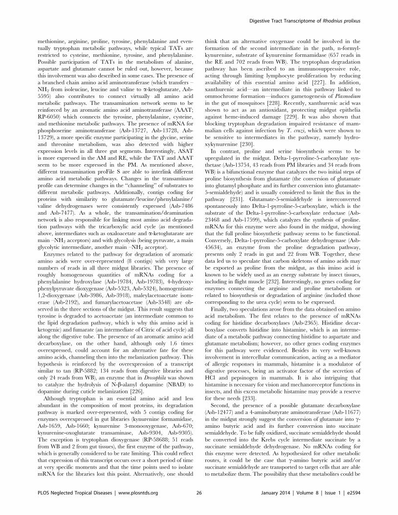
methionine, arginine, proline, tyrosine, phenylalanine and even-
tually tryptophan metabolic pathways, while typical TATs are
restricted to cysteine, methionine, tyrosine, and phenylalanine.
Possible participation of TATs in the metabolism of alanine,
aspartate and glutamate cannot be ruled out, however, because
this involvement was also described in some cases. The presence of
a branched chain amino acid aminotransferase (which transfers –
NH2 from isoleucine, leucine and valine to a-ketoglutarate, Asb-
5595) also contributes to connect virtually all amino acid
metabolic pathways. The transamination network seems to be
reinforced by an aromatic amino acid aminotransferase (AAAT;
RP-6050) which connects the tyrosine, phenylalanine, cysteine,
and methionine metabolic pathways. The presence of mRNA for
phosphoserine aminotransferase (Asb-13727, Asb-13728, Asb-
13729), a more specific enzyme participating in the glycine, serine
and threonine metabolism, was also detected with higher
expression levels in all three gut segments. Interestingly, ASAT
is more expressed in the AM and RE, while the TAT and AAAT
seem to be more expressed in the PM. As mentioned above,
different transamination proFile S are able to interlink different
amino acid metabolic pathways. Changes in the transaminase
profile can determine changes in the ‘‘channeling’’ of substrates to
different metabolic pathways. Additionally, contigs coding for
proteins with similarity to glutamate/leucine/phenylalanine/
valine dehydrogenases were consistently expressed (Asb-7486
and Asb-7477). As a whole, the transamination/deamination
network is also responsible for linking most amino acid degrada-
tion pathways with the tricarboxylic acid cycle (as mentioned
above, intermediates such as oxaloacetate and a-ketoglutarate are
main –NH2 acceptors) and with glycolysis (being pyruvate, a main
glycolytic intermediate, another main –NH2 acceptor).
Enzymes related to the pathway for degradation of aromatic
amino acids were over-represented (8 contigs) with very large
numbers of reads in all three midgut libraries. The presence of
roughly homogeneous quantities of mRNAs coding for a
phenylalanine hydroxylase (Asb-19784, Asb-19783), 4-hydroxy-
phenylpyruvate dioxygenase (Asb-5323, Asb-5324), homogentisate
1,2-dioxygenase (Asb-3986, Asb-3918), maleylacetoacetate isom-
erase (Asb-2192), and fumarylacetoacetase (Asb-3548) are ob-
served in the three sections of the midgut. This result suggests that
tyrosine is degraded to acetoacetate (an intermediate common to
the lipid degradation pathway, which is why this amino acid is
ketogenic) and fumarate (an intermediate of Citric of acid cycle) all
along the digestive tube. The presence of an aromatic amino acid
decarboxylase, on the other hand, although only 1.6 times
overexpressed, could account for an alternative fate for these
amino acids, channeling then into the melanization pathway. This
hypothesis is reinforced by the overexpression of a transcript
similar to tan (RP-5882; 134 reads from digestive libraries and
only 24 reads from WB), an enzyme that in Drosophila was shown
to catalyze the hydrolysis of N-b-alanyl dopamine (NBAD) to
dopamine during cuticle melanization [226].
Although tryptophan is an essential amino acid and less
abundant in the composition of most proteins, its degradation
pathway is marked over-represented, with 5 contigs coding for
enzymes overexpressed in gut libraries (kynurenine formamidase,
Asb-1659, Asb-1660; kynurenine 3-monooxygenase, Asb-670;
kynurenine-oxoglutarate transaminase, Asb-9304, Asb-9305).
The exception is tryptophan dioxygenase (RP-58688; 51 reads
from WB and 2 from gut tissues), the first enzyme of the pathway,
which is generally considered to be rate limiting. This could reflect
that expression of this transcript occurs over a short period of time
at very specific moments and that the time points used to isolate
mRNA for the libraries lost this point. Alternatively, one should
think that an alternative oxygenase could be involved in the
formation of the second intermediate in the path, n-formyl-
kynurenine, substrate of kynurenine formamidase (657 reads in
the RE and 702 reads from WB). The tryptophan degradation
pathway has been ascribed to an immunosuppressive role,
acting through limiting lymphocyte proliferation by reducing
availability of this essential amino acid [227]. In addition,
xanthurenic acid—an intermediate in this pathway linked to
ommochrome formation—induces gametogenesis of Plasmodium
in the gut of mosquitoes [228]. Recently, xanthurenic acid was
shown to act as an antioxidant, protecting midgut epithelia
against heme-induced damage [229]. It was also shown that
blocking tryptophan degradation impaired resistance of mam-
malian cells against infection by T. cruzi, which were shown to
be sensitive to intermediates in the pathway, namely hydro-
xykynurenine [230].
In contrast, proline and serine biosynthesis seems to be
upregulated in the midgut. Delta-1-pyrroline-5-carboxylate syn-
thetase (Asb-13754, 43 reads from PM libraries and 34 reads from
WB) is a bifunctional enzyme that catalyzes the two initial steps of
proline biosynthesis from glutamate (the conversion of glutamate
into glutamyl phosphate and its further conversion into glutamate-
5-semialdehyde) and is usually considered to limit the flux in the
pathway [231]. Glutamate-5-semialdehyde is interconverted
spontaneously into Delta-1-pyrroline-5-carboxylate, which is the
substrate of the Delta-1-pyrroline-5-carboxylate reductase (Asb-
23468 and Asb-17599), which catalyzes the synthesis of proline.
mRNAs for this enzyme were also found in the midgut, showing
that the full proline biosynthetic pathway seems to be functional.
Conversely, Delta-1-pyrroline-5-carboxylate dehydrogenase (Asb-
45634), an enzyme from the proline degradation pathway,
presents only 2 reads in gut and 22 from WB. Together, these
data led us to speculate that carbon skeletons of amino acids may
be exported as proline from the midgut, as this imino acid is
known to be widely used as an energy substrate by insect tissues,
including in flight muscle [232]. Interestingly, no genes coding for
enzymes connecting the arginine and proline metabolism or
related to biosynthesis or degradation of arginine (included those
corresponding to the urea cycle) seem to be expressed.
Finally, two speculations arose from the data obtained on amino
acid metabolism. The first relates to the presence of mRNAs
coding for histidine decarboxylases (Asb-2365). Histidine decar-
boxylase converts histidine into histamine, which is an interme-
diate of a metabolic pathway connecting histidine to aspartate and
glutamate metabolism; however, no other genes coding enzymes
for this pathway were evidenced. Besides its very well-known
involvement in intercellular communication, acting as a mediator
of allergic responses in mammals, histamine is a modulator of
digestive processes, being an activator factor of the secretion of
HCl and pepsinogen in mammals. It is also intriguing that
histamine is necessary for vision and mechanoreceptor functions in
insects, and this excess metabolic histamine may provide a reserve
for these needs [233].
Second, the presence of a possible glutamate decarboxylase
(Asb-12477) and a 4-aminobutyrate aminotransferase (Asb-11677)
in the midgut strongly suggest the conversion of glutamate into c-
amino butyric acid and its further conversion into succinate
semialdehyde. To be fully oxidized, succinate semialdehyde should
be converted into the Krebs cycle intermediate succinate by a
succinate semialdehyde dehydrogenase. No mRNAs coding for
this enzyme were detected. As hypothesized for other metabolic
routes, it could be the case that c-amino butyric acid and/or
succinate semialdehyde are transported to target cells that are able
to metabolize them. The possibility that these metabolites could be
Digestive Tract Transcriptome of Rhodnius prolixus
PLOS Neglected Tropical Diseases | www.plosntds.org 26 January 2014 | Volume 8 | Issue 1 | e2594
acting as messengers for intercellular communication should be
also considered.
Viruses, Wolbachia, and transposable elementsThe polyprotein for a picornavirus similar to the honey bee slow
paralysis virus is found expressed in the WB, AM, and RE (Asb-
4202). This viral sequence was not found in the genome scaffolds,
suggesting it may not be part of the insect genome. The DNA
helicase of a virus similar to Cotesia vestalis bracovirus was also found
in (Asb-64576); other transcripts matching Cotesia virus were also
found. Several phage proteins were also identified, and these could
derive from bacterial transcripts. For example, Asb-15041 is 70%
identical to a phage from a Wolbachia endosymbiont, but this is
mapped to R. prolixus genome in contig 5802 and could represent a
horizontal transfer. Also, 80 transcripts best-matched bacterial
proteins (presented in worksheet ‘‘Bacteria Virus TE’’ in
Supporting Information S1), many of which appear to be mapped
to the genomic contig 17820 (assembly version 3.0) including
several sequences best matching Wolbachia endosymbionts. These
could be interpreted as contaminant microorganisms present in
both the colonies used to make the transcriptome reported here
and the colonies that were used to sequence the genome. As these
colonies have been kept in captivity for decades and were obtained
independently from very distant places, this would make this
Wolbachia a strong symbiont candidate. If these genomic contigs do
not represent artifacts of genome assembly, this could represent an
insertion of Wolbachia genetic material common to both Rhodnius
strains, as has been reported for several insect species, where
segments as large as the entire genome of a bacteria are found
inserted into the genome of the arthropod [234].
Abundant transcripts coding for TEs, on the other hand, are
found incorporated into the genome, as expected. In particular
class I TE sequences of the families Gypsy, Bell, Line, and Copia
are abundant. The class II (cut and paste) transposons are also
particularly abundant, with expressed sequence tags coding for
full-length transposases of a Mariner element (Asb-69103, RP-
85192), suggesting active transposition. PIF/Harbinger elements
are also transcribed (Asb-6109).
OdditiesOne-zinc-dependent metalloprotease was detected (RP-9242),
which may be involved in cleaving growth factors or extracellular
matrix components.
Transcript Asb-10133 codes for a small protein of 80 amino
acids, highly expressed in the Rec and homologous to the bladder
cancer-associated protein (BLCAP/Bc10) downregulated during
invasive cancer growth in bladder [235]. The function of this
protein is unknown, but its expression is characteristic of stratified
epithelia, also found in Rhodnius hindgut.
Transcript Asb-820 codes for a pantheteinase overexpressed in
all three segments (1025 reads from gut libraries and 197 from
WB). Enzymes belonging to this gene family are involved in
vitamin recycling, both hydrolyzing biotinyl-peptides, generating
free biotin, and transferring biotin to acceptor proteins. These
proteins could in this way make biotin from the diet available to
allow the insect to synthesize its own biotin-dependent enzymes,
such as carboxylases.
ConclusionsCurrently, the R. prolixus genome has been sequenced with a 96
coverage. Transcripts reported here helped to obtain the predicted
gene set that is available at vector base homepage (https://www.
vectorbase.org/organisms/rhodnius-prolixus) and were also used
to support the manual annotation effort. The transcriptome
described here represents a significant increase in the amount of
information on Rhodnius genome, with 2,475 near full-length
coding sequences being deposited to GenBank. Several transcripts
corresponding to functions that were expected— such as digestive
enzymes and transporters—appeared in large numbers, and some
findings have added new data that can help to understand aspects
of the digestive physiology of this insect and its interaction with
intestinal microbiota and trypanosomatids, as well as generate new
working hypotheses for future research. The differential expression
data here reported is based in a single sample comparison and
further results using microarray or RNAseq data are required for
their validation.
Supporting Information
Figure S1 Protein extracts fractionated on a 4–12% NuPAGE
gels, revealed by SafeStain Coomassie Blue.
(DOCX)
Supporting Information S1 Hyperlinked spreadsheet with
contig assemblies.
(DOCX)
Supporting Information S2 Hyperlinked spreadsheet with
deducted coding sequences.
(XLS)
Supporting Information S3 Hyperlinked spreadsheet with
deducted coding sequences and details of the proteomic match.
(XLSX)
Table S1 Table exhibiting functional class distribution of the
proteins confirmed by proteomic approach.
(DOCX)
Acknowledgments
We thank Brenda Rae Marshall, DPSS, NIAID, for editing. This
manuscript is dedicated to the memory of Dr. Alexandre Peixoto.
Author Contributions
Conceived and designed the experiments: FAG GOPS GRCB MM MHFS
PLO SaB. Performed the experiments: ACPG FAG GOPS MM MHFS
PLO DM DS LK NGC SaB SeB. Analyzed the data: ACPG FAG GOPS
MM MHFS PLO SaB DM DS LK NGC SeB ACAM ACP AST AMS CP
GRCB CB CF CL DCL ESG FAD GCA GAS HA JLdSGVJ JHMO
JMCR KCG LD MLDS MACSN MAB MMPD PA PF PMB RDM
RJVA RL RLSG RNA RRP WRT. Contributed reagents/materials/
analysis tools: FAG GRCB JMCR PLO RDM SaB. Wrote the paper:
ACPG FAG GOPS MM MHFS PLO SaB DM DS LK NGC SeB ACAM
ACP AST AMS CP GRCB CB CF CL DCL ESG FAD GCA GAS HA
JLdSGVJ JHMO JMCR KCG LD MLDS MACSN MAB MMPD PA PF
PMB RDM RJVA RL RLSG RNA RRP WRT.
References
1. Grimaldi D, Engel M (2005) Evolution of the insects. New York: Cambridge
University Press. 772 p.
2. Balczun C, Siemanowski J, Pausch JK, Helling S, Marcus K, et al. (2012)
Intestinal aspartate proteases TiCatD and TiCatD2 of the haematophagous
bug Triatoma infestans (Reduviidae): sequence characterisation, expression
pattern and characterisation of proteolytic activity. Insect Biochem Mol Biol
42: 240–250.
3. WHO (2013) Chagas disease (American trypanosomiasis) Fact sheet Nu340.
4. Schofield CJ, Galvao C (2009) Classification, evolution, and species groups
within the Triatominae. Acta Trop 110: 88–100.
Digestive Tract Transcriptome of Rhodnius prolixus
PLOS Neglected Tropical Diseases | www.plosntds.org 27 January 2014 | Volume 8 | Issue 1 | e2594
5. Wigglesworth VB (1972) The principles of insect physiology. New York:
Chapman and Hall. 827 p.
6. Te Brugge VA, Schooley DA, Orchard I(2002) The biological activity of
diuretic factors in Rhodnius prolixus. Peptides 23: 671–681.
7. Terra WR (2001) The origin and functions of the insect peritrophic membrane
and peritrophic gel. Arch Biochem Biophys 47: 47–61.
8. Bolognesi R, Terra WR, Ferreira C (2008) Peritrophic membrane role in
enhancing digestive efficiency. Theoretical and experimental models. J Insect
Physiol 54: 1413–1422.
9. Terra WR (1988) Physiology and biochemistry of insect digestion: an
evolutionary perspective. Braz J Med Biol Res 21: 675–734.
10. Lane NJ, Harrison JB (1979) An unusual cell-surface modification - Double
plasma-membrane. J Cell Sci 39: 355–372.
11. Gutierrez LS, Burgos MH (1986) The surface extracellular coat of the midgut
in Triatoma infestans. I. Mechanism of development. J Ultrastruct Mol Struct Res
95: 75–83.
12. Ferreira C, Ribeiro AF, Garcia ES, Terra WR (1988) Digestive enzymes
trapped between and associated with the double plasma-membranes of Rhodnius
prolixus posterior midgut cells. Insect Biochem 18: 521–530.
13. Silva CP, Ribeiro AF, Terra WR (1996) Enzyme markers and isolation of the
microvillar and perimicrovillar membranes of Dysdercus peruvianus (Hemiptera:
Pyrrhocoridae) midgut cells. Insect Biochem Mol Biol 26: 1011–1018.
14. Wigglesworth VB (1936) Symbiotic bacteria in a blood sucking insect, Rhodnius
prolixus Stal. Parasitology 28: 284–289.
15. Lake P, Friend WG (1968) The use of artificial diets to determine some of the
effects of Nocardia rhodnii on the development of Rhodnius prolixus. J Insect Physiol
14: 543–562.
16. Hill P, Campbell JA, Petrie IA (1976) Rhodnius prolixus and its symbiotic
actinomycete: a microbiological, physiological and behavioral study. Proc R Soc
London B 194: 501–525.
17. Baines S (1956) The role of the symbiotic bacteria in the nutrition of Rhodnius
prolixus. J Exp Biol 33: 533–541.
18. Eichler S, Schaub GA (2002) Development of symbionts in triatomine bugs and
the effects of infections with trypanosomatids. Exp Parasitol 100: 17–27.
19. Garcia ES, Genta FA, de Azambuja P, Schaub GA (2010) Interactions between
intestinal compounds of triatomines and Trypanosoma cruzi. Trends Parasitol 26:
499–505.
20. Ribeiro JMC, Pereira MEA (1984) Midgut glycosidases of Rhodnius prolixus.
Insect Biochem 14: 103–108.
21. Grillo LA, Majerowicz D, Gondim KC (2007) Lipid metabolism in Rhodnius
prolixus (Hemiptera: Reduviidae): role of a midgut triacylglycerol-lipase. Insect
Biochem Mol Biol 37: 579–588.
22. Garcia ED, Garcia ML (1977) Control of protease secretion in the intestine of
fifth instar larvae of Rhodnius prolixus. J Insect Physiol 23: 247–251.
23. Garcia ES, Guimaraes JA, Prado JL (1978) Purification and characterization of
a sulfhydryl-dependent protease from Rhodnius prolixus midgut. Arch Biochem
Biophys 188: 315–322.
24. Terra WR, Ferreira C, Garcia ES (1988) Origin, distribution, properties and
functions of the major Rhodnius prolixus midgut hydrolases. Insect Biochemistry
18: 423–434.
25. Kollien AH, Waniek PJ, Nisbet AJ, Billingsley PF, Schaub GA (2004) Activity
and sequence characterization of two cysteine proteases in the digestive tract of
the reduviid bug Triatoma infestans. Insect Mol Biol 13: 569–579.
26. Garcia ES, Guimaraes JA (1979) Proteolytic enzymes in the Rhodnius prolixus
midgut. Experientia 35: 305–306.
27. Oliveira MF, Silva JR, Dansa-Petretski M, de Souza W, Lins U, et al. (1999)
Haem detoxification by an insect. Nature 400: 517–518.
28. Oliveira MF, Timm BL, Machado EA, Miranda K, Attias M, et al. (2002) On
the pro-oxidant effects of haemozoin. FEBS Lett 512: 139–144.
29. Boker CA, Schaub GA (1984) Scanning electron microscopic studies of
Trypanosoma cruzi in the rectum of its vector Triatoma infestans. Z Parasitenkd 70:
459–469.
30. Karim S, Singh P, Ribeiro JM (2011) A deep insight into the sialotranscriptome
of the Gulf Coast tick, Amblyomma maculatum. PLoS ONE 6: e28525.
31. Jeanmougin F, Thompson JD, Gouy M, Higgins DG, Gibson TJ (1998)
Multiple sequence alignment with Clustal X. Trends Biochem Sci 23: 403–405.
32. Tamura K, Peterson D, Peterson N, Stecher G, Nei M, et al. (2011) MEGA5:
molecular evolutionary genetics analysis using maximum likelihood, evolution-
ary distance, and maximum parsimony methods. Mol Biol Evol 28: 2731–2739.
33. Shevchenko A, Wilm M, Vorm O, Mann M (1996) Mass spectrometric
sequencing of proteins silver-stained polyacrylamide gels. Anal Chem 68: 850–
858.
34. Hulo N, Bairoch A, Bulliard V, Cerutti L, De Castro E, et al. (2006) The
PROSITE database. Nucleic Acids Res 34: D227–230.
35. Altschul SF, Madden TL, Schaffer AA, Zhang J, Zhang Z, et al. (1997) Gapped
BLAST and PSI-BLAST: a new generation of protein database search
programs. Nucleic Acids Res 25: 3389–3402.
36. Tellam RL, Wijffels G, Willadsen P (1999) Peritrophic matrix proteins. Insect
Biochem Mol Biol 29: 87–101.
37. Barry MK, Triplett AA, Christensen AC (1999) A peritrophin-like protein
expressed in the embryonic tracheae of Drosophila melanogaster. Insect Biochem
Mol Biol 29: 319–327.
38. Gaines PJ, Walmsley SJ, Wisnewski N (2003) Cloning and characterization offive cDNAs encoding peritrophin-A domains from the cat flea, Ctenocephalides
felis. Insect Biochem Mol Biol 33: 1061–1073.
39. Jasrapuria S, Arakane Y, Osman G, Kramer KJ, Beeman RW, et al. (2010)
Genes encoding proteins with peritrophin A-type chitin-binding domains inTribolium castaneum are grouped into three distinct families based on phylogeny,
expression and function. Insect Biochem Mol Biol 40: 214–227.
40. Venancio TM, Cristofoletti PT, Ferreira C, Verjovski-Almeida S, Terra WR(2009) The Aedes aegypti larval transcriptome: a comparative perspective with
emphasis on trypsins and the domain structure of peritrophins. Insect Mol Biol18: 33–44.
41. Devenport M, Alvarenga PH, Shao L, Fujioka H, Bianconi ML, et al. (2006)
Identification of the Aedes aegypti peritrophic matrix protein AeIMUCI as aheme-binding protein. Biochemistry 45: 9540–9549.
42. Lang T, Hansson GC, Samuelsson T (2007) Gel-forming mucins appeared
early in metazoan evolution. Proc Natl Acad Sci U S A 104: 16209–16214.
43. Hegedus D, Erlandson M, Gillott C, Toprak U (2009) New insights intoperitrophic matrix synthesis, architecture, and function. Annu Rev Entomol 54:
285–302.
44. Cantarel BL, Coutinho PM, Rancurel C, Bernard T, Lombard V, et al. (2009)The carbohydrate-active EnZymes database (CAZy): an expert resource for
glycogenomics. Nucleic Acids Res 37: D233–238.
45. Tomiya N, Narang S, Park J, Abdul-Rahman B, Choi O, et al. (2006)
Purification, characterization, and cloning of a Spodoptera frugiperda Sf9 beta-N-acetylhexosaminidase that hydrolyzes terminal N-acetylglucosamine on the N-
glycan core. J Biol Chem 281: 19545–19560.
46. Zhu Q, Arakane Y, Beeman RW, Kramer KJ, Muthukrishnan S (2008)Functional specialization among insect chitinase family genes revealed by RNA
interference. Proc Natl Acad Sci U S A 105: 6650–6655.
47. Amano K, Ito E (1978) The action of lysozyme on partially deacetylated chitin.Eur J Biochem 85: 97–104.
48. Cancado FC, Chimoy Effio P, Terra WR, Marana SR (2008) Cloning,
purification and comparative characterization of two digestive lysozymes fromMusca domestica larvae. Braz J Med Biol Res 41: 969–977.
49. Regel R, Matioli SR, Terra WR (1998) Molecular adaptation of Drosophila
melanogaster lysozymes to a digestive function. Insect Biochem Mol Biol 28: 309–319.
50. Watanabe H, Tokuda G (2010) Cellulolytic systems in insects. Annu Rev
Entomol 55: 609–632.
51. Silva CP, Silva JR, Vasconcelos FF, Petretski MD, Damatta RA, et al. (2004)Occurrence of midgut perimicrovillar membranes in paraneopteran insect
orders with comments on their function and evolutionary significance.
Arthropod Struct Dev 33: 139–148.
52. Mury FB, da Silva JR, Ferreira LS, dos Santos Ferreira B, de Souza-Filho GA,
et al. (2009) Alpha-glucosidase promotes hemozoin formation in a blood-
sucking bug: an evolutionary history. PLoS One 4: e6966.
53. Terra WR, Ferreira C (2005) Biochemistry of digestion. In: Gilbert LI, Iatrou
K, Gill SS, editors. Comprehensive Insect Molecular Science. Oxford: Elsevier.
pp. 171–224.
54. Billingsley PF (1988) Morphometric analysis of Rhodnius prolixus Stal (Hemi-ptera:Reduviidae) midgut cells during blood digestion. Tissue Cell 20: 291–
301.
55. Padilha MH, Pimentel AC, Ribeiro AF, Terra WR (2009) Sequence andfunction of lysosomal and digestive cathepsin D-like proteinases of Musca
domestica midgut. Insect Biochem Mol Biol 39: 782–791.
56. Lopez-Ordonez T, Rodriguez MH, Hernandez-Hernandez FD (2001)Characterization of a cDNA encoding a cathepsin L-like protein of Rhodnius
prolixus. Insect Mol Biol 10: 505–511.
57. Musil D, Zucic D, Turk D, Engh RA, Mayr I, et al. (1991) The refined 2.15 AX-ray crystal structure of human liver cathepsin B: the structural basis for its
specificity. Embo J 10: 2321–2330.
58. Wex T, Wex H, Bromme D (1999) The human cathepsin F gene–a fusionproduct between an ancestral cathepsin and cystatin gene. Biol Chem 380:
1439–1442.
59. Nagler DK, Sulea T, Menard R (1999) Full-length cDNA of human cathepsinF predicts the presence of a cystatin domain at the N-terminus of the cysteine
protease zymogen. Biochem Biophys Res Commun 257: 313–318.
60. Meiser CK, Piechura H, Meyer HE, Warscheid B, Schaub GA, et al. (2010) A
salivary serine protease of the haematophagous reduviid Panstrongylus megistus:sequence characterization, expression pattern and characterization of proteo-
lytic activity. Insect Mol Biol 19: 409–421.
61. Amino R, Tanaka AS, Schenkman S (2001) Triapsin, an unusual activatableserine protease from the saliva of the hematophagous vector of Chagas’ disease
Triatoma infestans (Hemiptera: Reduviidae). Insect Biochem Mol Biol 31: 465–472.
62. Bifano TD, Alegria TG, Terra WR (2010) Transporters involved in glucose
and water absorption in the Dysdercus peruvianus (Hemiptera: Pyrrhocoridae)anterior midgut. Comp Biochem Physiol B Biochem Mol Biol 157: 1–9.
63. Nelson N, Harvey WR (1999) Vacuolar and plasma membrane proton-
adenosinetriphosphatases. Physiol Rev 79: 361–385.
64. Mende K, Petoukhova O, Koulitchkova V, Schaub GA, Lange U, et al. (1999)Dipetalogastin, a potent thrombin inhibitor from the blood-sucking insect.
Dipetalogaster maximus cDNA cloning, expression and characterization.
Eur J Biochem 266: 583–590.
Digestive Tract Transcriptome of Rhodnius prolixus
PLOS Neglected Tropical Diseases | www.plosntds.org 28 January 2014 | Volume 8 | Issue 1 | e2594
65. Friedrich T, Kroger B, Bialojan S, Lemaire HG, Hoffken HW, et al. (1993) AKazal-type inhibitor with thrombin specificity from Rhodnius prolixus. J Biol
Chem 268: 16216–16222.
66. Meiser CK, Piechura H, Werner T, Dittmeyer-Schafer S, Meyer HE, et al.
(2010) Kazal-type inhibitors in the stomach of Panstrongylus megistus (Triatomi-nae, Reduviidae). Insect Biochem Mol Biol 40: 345–353.
67. Lovato DV, Nicolau de Campos IT, Amino R, Tanaka AS (2006) The full-
length cDNA of anticoagulant protein infestin revealed a novel releasableKazal domain, a neutrophil elastase inhibitor lacking anticoagulant activity.
Biochimie 88: 673–681.
68. Campos IT, Tanaka-Azevedo AM, Tanaka AS (2004) Identification andcharacterization of a novel factor XIIa inhibitor in the hematophagous insect,
Triatoma infestans (Hemiptera: Reduviidae). FEBS Lett 577: 512–516.
69. Campos IT, Amino R, Sampaio CA, Auerswald EA, Friedrich T, et al. (2002)Infestin, a thrombin inhibitor presents in Triatoma infestans midgut, a Chagas’
disease vector: gene cloning, expression and characterization of the inhibitor.
Insect Biochem Mol Biol 32: 991–997.
70. van de Locht ALD, Bauer MHR, Friedrich TKB, Hoffken W, Bode W (1995)
Two heads are better than one: crystal structure of the insect derived double
domain Kazal inhibitor rhodniin in complex with thrombin. EMBO J 14:5149–5157.
71. Araujo RN, Campos IT, Tanaka AS, Santos A, Gontijo NF, et al. (2007)
Brasiliensin: A novel intestinal thrombin inhibitor from Triatoma brasiliensis
(Hemiptera: Reduviidae) with an important role in blood intake. Int J Parasitol
37: 1351–1358.
72. Takac P, Nunn MA, Meszaros J, Pechanova O, Vrbjar N, et al. (2006)Vasotab, a vasoactive peptide from horse fly Hybomitra bimaculata (Diptera,
Tabanidae) salivary glands. J Exp Biol 209: 343–352.
73. Kellenberger C, Roussel A (2005) Structure-activity relationship within theserine protease inhibitors of the pacifastin family. Protein Pept Lett 12: 409–
414.
74. Simonet G, Claeys I, Franssens V, De Loof A, Broeck JV (2003) Genomics,evolution and biological functions of the pacifastin peptide family: a conserved
serine protease inhibitor family in arthropods. Peptides 24: 1633–1644.
75. de Marco R, Lovato DV, Torquato RJ, Clara RO, Buarque DS, et al. (2010)The first pacifastin elastase inhibitor characterized from a blood sucking
animal. Peptides 31: 1280–1286.
76. Flower DR, North AC, Sansom CE (2000) The lipocalin protein family:
structural and sequence overview. Biochim Biophys Acta 1482: 9–24.
77. Andersen JF, Gudderra NP, Francischetti IM, Ribeiro JM (2005) The role of
salivary lipocalins in blood feeding by Rhodnius prolixus. Arch Insect Biochem
Physiol 58: 97–105.
78. Noeske-Jungblut C, Haendler B, Donner P, Alagon A, Possani L, et al. (1995)Triabin, a highly potent exosite inhibitor of thrombin. J Biol Chem 270:
28629–28634.
79. Glusa E, Bretschneider E, Daum J, Noeske-Jungblut C (1997) Inhibition ofthrombin-mediated cellular effects by triabin, a highly potent anion-binding
exosite thrombin inhibitor. Thromb Haemost 77: 1196–1200.
80. Fuentes-Prior P, Noeske-Jungblut C, Donner P, Schleuning WD, Huber R, etal. (1997) Structure of the thrombin complex with triabin, a lipocalin-like
exosite-binding inhibitor derived from a triatomine bug. Proc Natl AcadSci U S A 94: 11845–11850.
81. Sansom CE, North ACT, Sawyer L (1994) Structural analysis and classification
of lipocalins and related proteins using a profile-search method. BiochimBiophys Acta 1208: 247–255.
82. Noeske-Jungblut C, Kratzschmar J, Haendler B, Alagon A, Possani L, et al.
(1994) An inhibitor of collagen-induced platelet aggregation from the saliva of
Triatoma pallidipennis. J Biol Chem 269: 5050–5053.
83. Haendler B, Becker A, C N-J, Kratzschmar J, Donner P, et al. (1995)
Expression of active recombinant pallidipin, a novel platelet aggregation
inhibitor, in the periplasm of Escherichia coli. Biochem J 307: 465–470.
84. So WV, Sarov-Blat L, Kotarski CK, McDonald MJ, Allada R, et al. (2000)
takeout, a novel Drosophila gene under circadian clock transcriptional
regulation. Mol Cell Biol 20: 6935–6944.
85. Noriega FG, Shah DK, Wells MA (1997) Juvenile hormone controls earlytrypsin gene transcription in the midgut of Aedes aegypti. Insect Mol Biol 6: 63–
66.
86. Bian G, Raikhel AS, Zhu J (2008) Characterization of a juvenile hormone-regulated chymotrypsin-like serine protease gene in Aedes aegypti mosquito.
Insect Biochem Mol Biol 38: 190–200.
87. Nomura A, Kawasaki K, Kubo T, Natori S (1992) Purification and localizationof p10, a novel protein that increases in nymphal regenerating legs of Periplaneta
americana (American cockroach). Int J Dev Biol 36: 391–398.
88. Lemaitre B, Hoffmann J (2007) The host defense of Drosophila melanogaster. AnnuRev Immunol 25: 697–743.
89. Buchon N, Broderick NA, Chakrabarti S, Lemaitre B (2009) Invasive and
indigenous microbiota impact intestinal stem cell activity through multiplepathways in Drosophila. Genes Dev 23: 2333–2344.
90. Meister S, Agianian B, Turlure F, Relogio A, Morlais I, et al. (2009) Anopheles
gambiae PGRPLC-mediated defense against bacteria modulates infections withmalaria parasites. Plos Pathogens 5: e1000542.
91. Dong Y, Manfredini F, Dimopoulos G (2009) Implication of the mosquito
midgut microbiota in the defense against malaria parasites. PLoS Pathog 5:
e1000423.
92. Ratcliffe NA, Rowley AF (1983) Recognition factors in insect hemolymph. DevComp Immunol 7: 653.
93. Kanost MR, Jiang H, Yu XQ (2004) Innate immune responses of alepidopteran insect, Manduca sexta. Immunol Rev 198: 97–105.
94. Araujo RN, Pereira MH, Soares AC, Pereira ID, Diotaiuti L, et al. (2009)
Effect of intestinal erythrocyte agglutination on the feeding performance ofTriatoma brasiliensis (Hemiptera: Reduviidae). J Insect Physiol 55: 862–868.
95. Kato H, Jochim RC, Gomez EA, Sakoda R, Iwata H, et al. (2010) A repertoireof the dominant transcripts from the salivary glands of the blood-sucking bug,
Triatoma dimidiata, a vector of Chagas disease. Infect Genet Evol 10: 184–191.
96. Pereira ME, Andrade AF, Ribeiro JM (1981) Lectins of distinct specificity in
Rhodnius prolixus interact selectively with Trypanosoma cruzi. Science 211: 597–
600.
97. Ratcliffe NA, Nigam Y, Mello CB, Garcia ES, Azambuja P (1996) Trypanosoma
cruzi and erythrocyte agglutinins: a comparative study of occurrence andproperties in the gut and hemolymph of Rhodnius prolixus. Exp Parasitol 83: 83–
93.
98. Dimopoulos G, Richman A, dellaTorre A, Kafatos FC, Louis C (1996)
Identification and characterization of differentially expressed cDNAs of the
vector mosquito, Anopheles gambiae. Proc Natl Acad Sci USA 93: 13066–13071.
99. Dimopoulos G, Seeley D, Wolf A, Kafatos FC (1998) Malaria infection of the
mosquito Anopheles gambiae activates immune-responsive genes during criticaltransition stages of the parasite life cycle. EMBO J 17: 6115–6123.
100. Dimopoulos G, Muller HM, Levashina EA, Kafatos FC (2001) Innate immune
defense against malaria infection in the mosquito. Curr Opin Immunol 13: 79–88.
101. Kamhawi S, Ramalho-Ortigao M, Pham VM, Kumar S, Lawyer PG, et al.(2004) A role for insect galectins in parasite survival. Cell 119: 329–341.
102. Goto A, Kumagai T, Kumagai C, Hirose J, Narita H, et al. (2001) A Drosophila
haemocyte-specific protein, hemolectin, similar to human von Willebrand
factor. Biochem J 359: 99–108.
103. Goto A, Kadowaki T, Kitagawa Y (2003) Drosophila hemolectin gene isexpressed in embryonic and larval hemocytes and its knock down causes
bleeding defects. Dev Biol 264: 582–591.
104. Lesch C, Goto A, Lindgren M, Bidla G, Dushay MS, et al. (2007) A role for
Hemolectin in coagulation and immunity in Drosophila melanogaster. Dev CompImmunol 31: 1255–1263.
105. Ryu JH, Ha EM, Lee WJ (2010) Innate immunity and gut-microbe mutualism
in Drosophila. Dev Comp Immunol 34: 369–376.
106. Grosshans J, Schnorrer F, Nusslein-Volhard C (1999) Oligomerisation of Tube
and Pelle leads to nuclear localisation of dorsal. Mech Dev 81: 127–138.
107. Kopp E, Medzhitov R, Carothers J, Xiao C, Douglas I, et al. (1999) ECSIT is
an evolutionarily conserved intermediate in the Toll/IL-1 signal transductionpathway. Genes Dev 13: 2059–2071.
108. Kim M, Lee JH, Lee SY, Kim E, Chung J (2006) Caspar, a suppressor of
antibacterial immunity in Drosophila. Proc Natl Acad Sci U S A 103: 16358–16363.
109. Schneider DS, Ayres JS, Brandt SM, Costa A, Dionne MS, et al. (2007)Drosophila eiger mutants are sensitive to extracellular pathogens. PLoS Pathog 3:
e41.
110. Berkey CD, Blow N, Watnick PI (2009) Genetic analysis of Drosophila
melanogaster susceptibility to intestinal Vibrio cholerae infection. Cell Microbiol
11: 461–474.
111. Vidal M (2010) The dark side of fly TNF: an ancient developmental proof
reading mechanism turned into tumor promoter. Cell Cycle 9: 3851–3856.
112. Liu Q, Rand TA, Kalidas S, Du F, Kim HE, et al. (2003) R2D2, a bridge
between the initiation and effector steps of the Drosophila RNAi pathway.Science 301: 1921–1925.
113. Gregory RI, Chendrimada TP, Cooch N, Shiekhattar R (2005) Human RISC
couples microRNA biogenesis and posttranscriptional gene silencing. Cell 123:631–640.
114. Saito K, Ishizuka A, Siomi H, Siomi MC (2005) Processing of pre-microRNAsby the Dicer-1-Loquacious complex in Drosophila cells. PLoS Biol 3: e235.
115. Ursic-Bedoya RJ, Nazzari H, Cooper D, Triana O, Wolff M, et al. (2008)Identification and characterization of two novel lysozymes from Rhodnius
prolixus, a vector of Chagas disease. J Insect Physiol 54: 593–603.
116. Balczun C, Knorr E, Topal H, Meiser CK, Kollien AH, et al. (2008) Sequencecharacterization of an unusual lysozyme gene expressed in the intestinal tract of
the reduviid bug Triatoma infestans (Insecta). Parasitol Res 102: 229–232.
117. Araujo CA, Waniek PJ, Stock P, Mayer C, Jansen AM, et al. (2006) Sequence
characterization and expression patterns of defensin and lysozyme encoding
genes from the gut of the reduviid bug Triatoma brasiliensis. Insect Biochem MolBiol 36: 547–560.
118. Kollien AH, Fechner S, Waniek PJ, Schaub GA (2003) Isolation andcharacterization of a cDNA encoding for a lysozyme from the gut of the
reduviid bug Triatoma infestans. Arch Insect Biochem Physiol 53: 134–145.
119. Bulet P, Stocklin R, Menin L (2004) Anti-microbial peptides: from
invertebrates to vertebrates. Immunol Rev 198: 169–184.
120. Bonmatin JM, Bonnat JL, Gallet X, Vovelle F, Ptak M, et al. (1992) Two-dimensional 1H NMR study of recombinant insect defensin A in water:
resonance assignments, secondary structure and global folding. J Biomol NMR2: 235–256.
121. Bulet P, Hetru C, Dimarcq JL, Hoffmann D (1999) Antimicrobial peptides ininsects; structure and function. Dev Comp Immunol 23: 329–344.
Digestive Tract Transcriptome of Rhodnius prolixus
PLOS Neglected Tropical Diseases | www.plosntds.org 29 January 2014 | Volume 8 | Issue 1 | e2594
122. Lopez L, Morales G, Ursic R, Wolff M, Lowenberger C (2003) Isolation andcharacterization of a novel insect defensin from Rhodnius prolixus, a vector of
Chagas disease. Insect Biochem Mol Biol 33: 439–447.
123. Lowenberger CA, Ferdig MT, Bulet P, Khalili S, Hoffmann JA, et al. (1996)
Aedes aegypti: induced antibacterial proteins reduce the establishment anddevelopment of Brugia malayi. Exp Parasitol 83: 191–201.
124. Lowenberger CA, Kamal S, Chiles J, Paskewitz S, Bulet P, et al. (1999)
Mosquito-Plasmodium interactions in response to immune activation of thevector. Exp Parasitol 91: 59–69.
125. Gibbs GM, Roelants K, O’Bryan MK (2008) The CAP superfamily: cysteine-rich secretory proteins, antigen 5, and pathogenesis-related 1 proteins–roles in
reproduction, cancer, and immune defense. Endocr Rev 29: 865–897.
126. Rubin DC (2007) Intestinal morphogenesis. Curr Opin Gastroenterol 23: 111–
114.
127. Zhao WB, Wang X (2008) Phosphatases of regenerating liver: a novel target in
human solid tumors. Chin Med J (Engl) 121: 1469–1474.
128. Yan Y, Merlin D (2008) Ste20-related proline/alanine-rich kinase: a novel
regulator of intestinal inflammation. World J Gastroenterol 14: 6115–6121.
129. Duraisamy S, Bajpai M, Bughani U, Dastidar SG, Ray A, et al. (2008) MK2: a
novel molecular target for anti-inflammatory therapy. Expert Opin TherTargets 12: 921–936.
130. Thuraisingam T, Xu YZ, Eadie K, Heravi M, Guiot MC, et al. (2010)
MAPKAPK-2 signaling is critical for cutaneous wound healing. J InvestDermatol 130: 278–286.
131. Zhang J, Anastasiadis PZ, Liu Y, Thompson EA, Fields AP (2004) Proteinkinase C (PKC) betaII induces cell invasion through a Ras/Mek-, PKC iota/
Rac 1-dependent signaling pathway. J Biol Chem 279: 22118–22123.
132. Takagawa R, Akimoto K, Ichikawa Y, Akiyama H, Kojima Y, et al. (2010)
High expression of atypical protein kinase C lambda/iota in gastric cancer as aprognostic factor for recurrence. Ann Surg Oncol 17: 81–88.
133. Suzuki A, Ohno S (2006) The PAR-aPKC system: lessons in polarity. J Cell Sci119: 979–987.
134. McCaffrey LM, Macara IG (2009) Widely conserved signaling pathways in theestablishment of cell polarity. Cold Spring Harb Perspect Biol 1: a001370.
135. Jansen M, Ten Klooster JP, Offerhaus GJ, Clevers H (2009) LKB1 and AMPKfamily signaling: the intimate link between cell polarity and energy metabolism.
Physiol Rev 89: 777–798.
136. Hardie DG, Pan DA (2002) Regulation of fatty acid synthesis and oxidation bythe AMP-activated protein kinase. Biochem Soc Trans 30: 1064–1070.
137. Forcet C, Billaud M (2007) Dialogue between LKB1 and AMPK: a hot topic atthe cellular pole. Sci STKE 2007: pe51.
138. Lee JH, Koh H, Kim M, Kim Y, Lee SY, et al. (2007) Energy-dependentregulation of cell structure by AMP-activated protein kinase. Nature 447:
1017–1020.
139. Guertin DA, Sabatini DM (2005) An expanding role for mTOR in cancer.
Trends Mol Med 11: 353–361.
140. Brandon MC, Pennington JE, Isoe J, Zamora J, Schillinger AS, et al. (2008)
TOR signaling is required for amino acid stimulation of early trypsin proteinsynthesis in the midgut of Aedes aegypti mosquitoes. Insect Biochem Mol Biol 38:
916–922.
141. Roy SG, Hansen IA, Raikhel AS (2007) Effect of insulin and 20-
hydroxyecdysone in the fat body of the yellow fever mosquito, Aedes aegypti.
Insect Biochem Mol Biol 37: 1317–1326.
142. Jiang H, Patel PH, Kohlmaier A, Grenley MO, McEwen DG, et al. (2009)
Cytokine/Jak/Stat signaling mediates regeneration and homeostasis in theDrosophila midgut. Cell 137: 1343–1355.
143. Lin G, Xu N, Xi R (2008) Paracrine Wingless signalling controls self-renewal ofDrosophila intestinal stem cells. Nature 455: 1119–1123.
144. Fuss B, Hoch M (1998) Drosophila endoderm development requires a novelhomeobox gene which is a target of Wingless and Dpp signalling. Mech Dev
79: 83–97.
145. Nakagoshi H, Hoshi M, Nabeshima Y, Matsuzaki F (1998) A novel homeobox
gene mediates the Dpp signal to establish functional specificity within target
cells. Genes Dev 12: 2724–2734.
146. Nakagoshi H, Shirai T, Nabeshima Y, Matsuzaki F (2002) Refinement of
wingless expression by a wingless- and notch-responsive homeodomain protein,defective proventriculus. Dev Biol 249: 44–56.
147. Shirai T, Maehara A, Kiritooshi N, Matsuzaki F, Handa H, et al. (2003)Differential requirement of EGFR signaling for the expression of defective
proventriculus gene in the Drosophila endoderm and ectoderm. BiochemBiophys Res Commun 311: 473–477.
148. Nakagawa Y, Fujiwara-Fukuta S, Yorimitsu T, Tanaka S, Minami R, et al.(2011) Spatial and temporal requirement of defective proventriculus activity
during Drosophila midgut development. Mech Dev 128: 258–267.
149. Jensen J, Pedersen EE, Galante P, Hald J, Heller RS, et al. (2000) Control of
endodermal endocrine development by Hes-1. Nat Genet 24: 36–44.
150. Matsuda Y, Wakamatsu Y, Kohyama J, Okano H, Fukuda K, et al. (2005)Notch signaling functions as a binary switch for the determination of glandular
and luminal fates of endodermal epithelium during chicken stomachdevelopment. Development 132: 2783–2793.
151. Yen TH, Wright NA (2006) The gastrointestinal tract stem cell niche. StemCell Rev 2: 203–212.
152. Ledent V, Vervoort M (2001) The basic helix-loop-helix protein family:comparative genomics and phylogenetic analysis. Genome Res 11: 754–770.
153. Shilo BZ (2005) Regulating the dynamics of EGF receptor signaling in space
and time. Development 132: 4017–4027.
154. Guo HF, Tong J, Hannan F, Luo L, Zhong Y (2000) A neurofibromatosis-1-
regulated pathway is required for learning in Drosophila. Nature 403: 895–898.
155. Mummery-Widmer JL, Yamazaki M, Stoeger T, Novatchkova M, Bhalerao S,
et al. (2009) Genome-wide analysis of Notch signalling in Drosophila bytransgenic RNAi. Nature 458: 987–992.
156. Tong JJ, Schriner SE, McCleary D, Day BJ, Wallace DC (2007) Life extension
through neurofibromin mitochondrial regulation and antioxidant therapy for
neurofibromatosis-1 in Drosophila melanogaster. Nat Genet 39: 476–485.
157. Borovsky D, Janssen I, Vanden Broeck J, Huybrechts R, Verhaert P, et al.
(1996) Molecular sequencing and modeling of Neobellieria bullata trypsin.
Evidence for translational control by Neobellieria trypsin-modulating oostatic
factor. Eur J Biochem 237: 279–287.
158. Noriega FG, Colonna AE, Wells MA (1999) Increase in the size of the amino
acid pool is sufficient to activate translation of early trypsin mRNA in Aedes
aegypti midgut. Insect Biochem Mol Biol 29: 243–247.
159. Noriega FG, Wells MA (1999) A molecular view of trypsin synthesis in the
midgut of Aedes aegypti. J Insect Physiol 45: 613–620.
160. Zhang D, Dimopoulos G, Wolf A, Minana B, Kafatos FC, et al. (2002) Cloning
and molecular characterization of two mosquito iron regulatory proteins. Insect
Biochem Mol Biol 32: 579–589.
161. Hajdusek O, Sojka D, Kopacek P, Buresova V, Franta Z, et al. (2009)Knockdown of proteins involved in iron metabolism limits tick reproduction
and development. Proc Natl Acad Sci U S A 106: 1033–1038.
162. Niu LL, Fallon AM (2000) Differential regulation of ribosomal protein gene
expression in Aedes aegypti mosquitoes before and after the blood meal. Insect
Mol Biol 9: 613–623.
163. Motorin Y, Helm M (2010) tRNA stabilization by modified nucleotides.
Biochemistry 49: 4934–4944.
164. Alexandrov A, Chernyakov I, Gu W, Hiley SL, Hughes TR, et al. (2006) Rapid
tRNA decay can result from lack of nonessential modifications. Mol Cell 21:87–96.
165. Tuorto F, Liebers R, Musch T, Schaefer M, Hofmann S, et al. (2012) RNA
cytosine methylation by Dnmt2 and NSun2 promotes tRNA stability and
protein synthesis. Nat Struct Mol Biol 19: 900–905.
166. Yarian C, Townsend H, Czestkowski W, Sochacka E, Malkiewicz AJ, et al.
(2002) Accurate translation of the genetic code depends on tRNA modified
nucleosides. J Biol Chem 277: 16391–16395.
167. Urbonavicius J, Qian Q, Durand JM, Hagervall TG, Bjork GR (2001)Improvement of reading frame maintenance is a common function for several
tRNA modifications. EMBO J 20: 4863–4873.
168. Bjork GR, Durand JM, Hagervall TG, Leipuviene R, Lundgren HK, et al.
(1999) Transfer RNA modification: influence on translational frameshifting and
metabolism. FEBS Lett 452: 47–51.
169. Thompson DM, Parker R (2009) Stressing out over tRNA cleavage. Cell 138:
215–219.
170. Schaefer M, Pollex T, Hanna K, Tuorto F, Meusburger M, et al. (2010) RNA
methylation by Dnmt2 protects transfer RNAs against stress-induced cleavage.Genes Devel 24: 1590–1595.
171. Emilsson V, Naslund AK, Kurland CG (1992) Thiolation of transfer RNA in
Escherichia coli varies with growth rate. Nucleic Acids Res 20: 4499–4505.
172. Chan CT, Pang YL, Deng W, Babu IR, Dyavaiah M, et al. (2012)
Reprogramming of tRNA modifications controls the oxidative stress response
by codon-biased translation of proteins. Nat Commun 3: 937.
173. Souza AV, Petretski JH, Demasi M, Bechara EJ, Oliveira PL (1997) Urate
protects a blood-sucking insect against hemin-induced oxidative stress. Free
Radic Biol Med 22: 209–214.
174. Hernandez G, Altmann M, Sierra JM, Urlaub H, del Corral RD, et al. (2005)
Functional analysis of seven genes encoding eight translation initiation factor
4E (eIF4E) isoforms in Drosophila. Mech Dev 122: 529–543.
175. Hernandez G, Vazquez-Pianzola P (2005) Functional diversity of the
eukaryotic translation initiation factors belonging to eIF4 families. Mech Dev
122: 865–876.
176. Cho PF, Poulin F, Cho-Park YA, Cho-Park IB, Chicoine JD, et al. (2005) A
new paradigm for translational control: Inhibition via 59-39 mRNA tethering byBicoid and the eIF4E cognate 4EHP. Cell 121: 411–423.
177. He B, Guo W (2009) The exocyst complex in polarized exocytosis. Curr Opin
Cell Biol 21: 537–542.
178. Schimmoller F, Singerkruger B, Schroder S, Kruger U, Barlowe C, et al. (1995)The absence of Emp24p, a component of ER-derived COPPII-coated vesicles,
causes a defect in transport of selected proteins to the Golgi. Embo J 14: 1329–
1339.
179. Onel S, Bolke L, Klambt C (2004) The Drosophila ARF6-GEF schizo controls
commissure formation by regulating slit. Development 131: 2587–2594.
180. Egan TJ (2008) Haemozoin formation. Mol Biochem Parasitol 157: 127–136.
181. Enayati AA, Ranson H, Hemingway J (2005) Insect glutathione transferases
and insecticide resistance. Insect Mol Biol 14: 3–8.
182. Pasteur N, Raymond M (1996) Insecticide resistance genes in mosquitoes: their
mutations, migration, and selection in field populations. J Hered 87: 444–449.
183. Graca-Souza AV, Maya-Monteiro C, Paiva-Silva GO, Braz GR, Paes MC, et
al. (2006) Adaptations against heme toxicity in blood-feeding arthropods. InsectBiochem Mol Biol 36: 322–335.
Digestive Tract Transcriptome of Rhodnius prolixus
PLOS Neglected Tropical Diseases | www.plosntds.org 30 January 2014 | Volume 8 | Issue 1 | e2594
184. Poupardin R, Riaz MA, Vontas J, David JP, Reynaud S (2010) Transcription
profiling of eleven cytochrome P450s potentially involved in xenobioticmetabolism in the mosquito Aedes aegypti. Insect Mol Biol 19: 185–193.
185. Feyereisen R (2006) Evolution of insect P450. Biochem Soc Trans 34: 1252–
1255.186. Murataliev MB, Feyereisen R, Walker FA (2004) Electron transfer by diflavin
reductases. Biochim Biophys Acta 1698: 1–26.187. Murataliev MB, Guzov VM, Walker FA, Feyereisen R (2008) P450 reductase
and cytochrome b5 interactions with cytochrome P450: effects on house fly
CYP6A1 catalysis. Insect Biochem Mol Biol 38: 1008–1015.188. Feyereisen R (1999) Insect P450 enzymes. Annu Rev Entomol 44: 507–533.
189. Rewitz KF, Rybczynski R, Warren JT, Gilbert LI (2006) The Halloween genescode for cytochrome P450 enzymes mediating synthesis of the insect moulting
hormone. Biochem Soc Trans 34: 1256–1260.190. Paes MC, Oliveira MB, Oliveira PL (2001) Hydrogen peroxide detoxification
in the midgut of the blood-sucking insect, Rhodnius prolixus. Arch Insect Biochem
Physiol 48: 63–71.191. Paes MC, Oliveira PL (1999) Extracellular glutathione peroxidase from the
blood-sucking bug, Rhodnius prolixus. Arch Insect Biochem Physiol 41: 171–177.192. Liu MC, Yasuda S, Idell S (2007) Sulfation of nitrotyrosine: biochemistry and
functional implications. IUBMB Life 59: 622–627.
193. Yasuda S, Yasuda T, Liu MY, Shetty S, Idell S, et al. (2011) Sulfation ofchlorotyrosine and nitrotyrosine by human lung endothelial and epithelial cells:
role of the human SULT1A3. Toxicol Appl Pharmacol 251: 104–109.194. Valderrama R, Corpas FJ, Carreras A, Gomez-Rodriguez MV, Chaki M, et al.
(2006) The dehydrogenase-mediated recycling of NADPH is a key antioxidantsystem against salt-induced oxidative stress in olive plants. Plant Cell
Environment 29: 1449–1459.
195. Devireddy LR, Hart DO, Goetz DH, Green MR (2010) A mammaliansiderophore synthesized by an enzyme with a bacterial homolog involved in
enterobactin production. Cell 141: 1006–1017.196. Ponka P (1999) Cell biology of heme. Am J Med Sci 318: 241–256.
197. Braz GR, Abreu L, Masuda H, Oliveira PL (2001) Heme biosynthesis and
oogenesis in the blood-sucking bug, Rhodnius prolixus. Insect Biochem Mol Biol31: 359–364.
198. Dansa-Petretski M, Ribeiro JM, Atella GC, Masuda H, Oliveira PL (1995)Antioxidant role of Rhodnius prolixus heme-binding protein. Protection against
heme-induced lipid peroxidation. J Biol Chem 270: 10893–10896.199. Quigley JG, Yang Z, Worthington MT, Phillips JD, Sabo KM, et al. (2004)
Identification of a human heme exporter that is essential for erythropoiesis. Cell
118: 757–766.200. Paiva-Silva GO, Cruz-Oliveira C, Nakayasu ES, Maya-Monteiro CM, Dunkov
BC, et al. (2006) A heme-degradation pathway in a blood-sucking insect. ProcNatl Acad Sci U S A 103: 8030–8035.
201. Caiaffa CD, Stiebler R, Oliveira MF, Lara FA, Paiva-Silva GO, et al. (2010)
Sn-protoporphyrin inhibits both heme degradation and hemozoin formation inRhodnius prolixus midgut. Insect Biochem Mol Biol 40: 855–860.
202. Missirlis F, Holmberg S, Georgieva T, Dunkov BC, Rouault TA, et al. (2006)Characterization of mitochondrial ferritin in Drosophila. Proc Natl Acad
Sci U S A 103: 5893–5898.203. Dunkov B, Georgieva T (2006) Insect iron binding proteins: insights from the
genomes. Insect Biochem Mol Biol 36: 300–309.
204. Billingsley PF (1990) The midgut ultrastructure of hematophagous insects. AnnRev Entomol 35: 219–248.
205. Atella GC, Gondim C, Masuda H (1995) Loading of lipophorin particles withphospholipids at the midgut of Rhodnius prolixus. Arch Insect Biochem Physiol
30: 337–350.
206. Coelho HS, Atella GC, Moreira MF, Gondim KC, Masuda H (1997)Lipophorin density variation during oogenesis on Rhodnius prolixus. Arch Insect
Biochem Physiol 35: 301–313.207. Schmidt J, Kleffmann T, Schaub GA (1998) Hydrophobic attachment of
Trypanosoma cruzi to a superficial layer of the rectal cuticle in the bug Triatoma
infestans. Parasitol Res 84: 527–536.208. Rawson RB (2003) The SREBP pathway–insights from Insigs and insects. Nat
Rev Mol Cell Biol 4: 631–640.209. Voght SP, Fluegel ML, Andrews LA, Pallanck LJ (2007) Drosophila NPC1b
promotes an early step in sterol absorption from the midgut epithelium. CellMetab 5: 195–205.
210. Wong JT, Chan M, Lee D, Jiang JY, Skrzypczak M, et al. (2000)
Phosphatidylcholine metabolism in human endothelial cells: modulation byphosphocholine. Mol Cell Biochem 207: 95–100.
211. Chua BT, Gallego-Ortega D, Ramirez de Molina A, Ullrich A, Lacal JC, et al.(2009) Regulation of Akt(ser473) phosphorylation by choline kinase in breast
carcinoma cells. Mol Cancer 8: 131.
212. Helmkamp GM, Jr., Harvey MS, Wirtz KW, Van Deenen LL (1974)
Phospholipid exchange between membranes. Purification of bovine brain
proteins that preferentially catalyze the transfer of phosphatidylinositol. J Biol
Chem 249: 6382–6389.
213. Cockcroft S (1998) Phosphatidylinositol transfer proteins: a requirement in
signal transduction and vesicle traffic. Bioessays 20: 423–432.
214. Atella GC, Arruda MA, Masuda H, Gondim KC (2000) Fatty acid
incorporation by Rhodnius prolixus midgut. Arch Insect Biochem Physiol 43:
99–107.
215. Zhao Y, Natarajan V (2009) Lysophosphatidic acid signaling in airway
epithelium: role in airway inflammation and remodeling. Cell Signal 21: 367–
377.
216. Golodne DM, Monteiro RQ, Graca-Souza AV, Silva-Neto MA, Atella GC
(2003) Lysophosphatidylcholine acts as an anti-hemostatic molecule in the
saliva of the blood-sucking bug Rhodnius prolixus. J Biol Chem 278: 27766–
27771.
217. Chen X, Hyatt BA, Mucenski ML, Mason RJ, Shannon JM (2006)
Identification and characterization of a lysophosphatidylcholine acyltransferase
in alveolar type II cells. Proc Natl Acad Sci U S A 103: 11724–11729.
218. Moolenaar WH (1995) Lysophosphatidic acid signalling. Curr Opin Cell Biol
7: 203–210.
219. Choi JW, Herr DR, Noguchi K, Yung YC, Lee CW, et al. (2010) LPA
receptors: subtypes and biological actions. Annu Rev Pharmacol Toxicol 50:
157–186.
220. Geijtenbeek TB, Westerman J, Heerma W, Wirtz KW (1996) Phosphatidyl-
choline transfer protein from bovine liver contains highly unsaturated
phosphatidylcholine species. FEBS Lett 391: 333–335.
221. Kanno K, Wu MK, Scapa EF, Roderick SL, Cohen DE (2007) Structure and
function of phosphatidylcholine transfer protein (PC-TP)/StarD2. Biochim
Biophys Acta 1771: 654–662.
222. Ducharme NA, Bickel PE (2008) Lipid droplets in lipogenesis and lipolysis.
Endocrinology 149: 942–949.
223. Kimmel AR, Brasaemle DL, McAndrews-Hill M, Sztalryd C, Londos C (2010)
Adoption of PERILIPIN as a unifying nomenclature for the mammalian PAT-
family of intracellular lipid storage droplet proteins. J Lipid Res 51: 468–471.
224. Miura S, Gan JW, Brzostowski J, Parisi MJ, Schultz CJ, et al. (2002) Functional
conservation for lipid storage droplet association among perilipin, ADRP, and
TIP47 (PAT)-related proteins in mammals, Drosophila, and Dictyostelium. J Biol
Chem 277: 32253–32257.
225. Patel RT, Soulages JL, Hariharasundaram B, Arrese EL (2005) Activation of
the lipid droplet controls the rate of lipolysis of triglycerides in the insect fat
body. J Biol Chem 280: 22624–22631.
226. True JR, Yeh SD, Hovemann BT, Kemme T, Meinertzhagen IA, et al. (2005)
Drosophila tan encodes a novel hydrolase required in pigmentation and vision.
PLoS Genet 1: e63.
227. Mellor AL, Keskin DB, Johnson T, Chandler P, Munn DH (2002) Cells
expressing indoleamine 2,3-dioxygenase inhibit T cell responses. J Immunol
168: 3771–3776.
228. Billker O, Lindo V, Panico M, Etienne AE, Paxton T, et al. (1998)
Identification of xanthurenic acid as the putative inducer of malaria
development in the mosquito. Nature 392: 289–292.
229. Lima VL, Dias F, Nunes RD, Pereira LO, Santos TS, et al. (2012) The
antioxidant role of xanthurenic acid in the Aedes aegypti midgut during digestion
of a blood meal. PLoS One 7: e38349.
230. Knubel CP, Martinez FF, Fretes RE, Diaz Lujan C, Theumer MG, et al.
(2010) Indoleamine 2,3-dioxigenase (IDO) is critical for host resistance against
Trypanosoma cruzi. FASEB J 24: 2689–2701.
231. Parre E, de Virville J, Cochet F, Leprince AS, Richard L, et al. (2010) A new
method for accurately measuring Delta(1)-pyrroline-5-carboxylate synthetase
activity. Methods Mol Biol 639: 333–340.
232. Scaraffia PY, Wells MA (2003) Proline can be utilized as an energy substrate
during flight of Aedes aegypti females. J Insect Physiol 49: 591–601.
233. Melzig J, Burg M, Gruhn M, Pak WL, Buchner E (1998) Selective histamine
uptake rescues photo- and mechanoreceptor function of histidine decarboxyl-
ase-deficient Drosophila mutant. J Neurosci 18: 7160–7166.
234. Dunning Hotopp JC, Clark ME, Oliveira DC, Foster JM, Fischer P, et al.
(2007) Widespread lateral gene transfer from intracellular bacteria to
multicellular eukaryotes. Science 317: 1753–1756.
235. Gromova I, Gromov P, Celis JE (2002) bc10: A novel human bladder cancer-
associated protein with a conserved genomic structure downregulated in
invasive cancer. Int J Cancer 98: 539–546.
Digestive Tract Transcriptome of Rhodnius prolixus
PLOS Neglected Tropical Diseases | www.plosntds.org 31 January 2014 | Volume 8 | Issue 1 | e2594
Related Documents