Provided by the author(s) and NUI Galway in accordance with publisher policies. Please cite the published version when available. Downloaded 2022-07-19T00:35:21Z Some rights reserved. For more information, please see the item record link above. Title An injectable collagen scaffold delivering exogenous microRNA as a therapy to modulate extracellular matrix remodelling Author(s) Monaghan, Michael Publication Date 2013-06-01 Item record http://hdl.handle.net/10379/4685

Welcome message from author
This document is posted to help you gain knowledge. Please leave a comment to let me know what you think about it! Share it to your friends and learn new things together.
Transcript

Provided by the author(s) and NUI Galway in accordance with publisher policies. Please cite the published
version when available.
Downloaded 2022-07-19T00:35:21Z
Some rights reserved. For more information, please see the item record link above.
TitleAn injectable collagen scaffold delivering exogenousmicroRNA as a therapy to modulate extracellular matrixremodelling
Author(s) Monaghan, Michael
PublicationDate 2013-06-01
Item record http://hdl.handle.net/10379/4685

An Injectable Collagen Scaffold Delivering Exogenous microRNA as a Therapy to Modulate
Extracellular Matrix Remodelling
A thesis submitted to the National University of Ireland for the
Degree of Doctor of Philosophy
By
Michael Monaghan
April 2013
Network of Excellence for Functional Biomaterials
National University of Ireland, Galway
Research Supervisor: Professor Abhay Pandit

ii
iii
Table of Contents
List of Appendices ........................................................................................................................................ vii
List of Figures ................................................................................................................................................. x
List of Tables ............................................................................................................................................... xix
Acknowledgements ...................................................................................................................................... xxi
List of Abbreviations .................................................................................................................................. xxii
Abstract ..................................................................................................................................................... xxvi
Chapter One: Literature Review ................................................................................................................... 1
1.1 Introduction ..................................................................................................................................... 2
1.2 RNA Interference ............................................................................................................................ 3
1.3 Non-viral Delivery ........................................................................................................................... 5
1.4 RNAi as a Therapeutic ..................................................................................................................... 8
1.5 RNAi Delivery through Scaffolds .................................................................................................. 11
1.5.1 Hydrogels .................................................................................................................................. 11
1.5.2 Sponges ..................................................................................................................................... 14
1.5.3 Fibers........................................................................................................................................ 15
1.5.4 Microspheres ............................................................................................................................. 16
1.5.5 Surface Coatings ....................................................................................................................... 17
1.5.6 Topical Delivery ........................................................................................................................ 18
1.6 RNAi for Treatment of Myocardial Infarction ................................................................................ 22
1.6.1 siRNA ........................................................................................................................................ 23
1.6.2 miRs Mimicking Nature – miR Manipulation in the Treatment of Myocardial Infarction ............. 26
1.7 Delivery Strategies......................................................................................................................... 32
1.8 Engineering Ligands for Improved Uptake ..................................................................................... 33
1.8.1 Antibodies ................................................................................................................................. 33
1.8.2 Cell-targeting Peptides .............................................................................................................. 34
1.9 Fibrosis ......................................................................................................................................... 35
1.10 Project Rationale ........................................................................................................................... 40
1.10.1 Rationale for the use of a miR Mimic ..................................................................................... 40
1.10.2 Rationale for the use of miR-29B ........................................................................................... 41
1.10.3 Rationale for the use of pDMAEMA ....................................................................................... 41
1.10.4 Rationale for the Selection of an Antibody Fragment Directed towards CD90 ........................ 41
1.10.5 Rationale for the use of a Crosslinked Atelocollagen Type I Scaffold ...................................... 42
1.10.6 Rationale for the use of Scaffold-Based RNAi Delivery ........................................................... 42

iv
1.11 Objectives and Hypotheses ............................................................................................................ 42
1.12 References ..................................................................................................................................... 47
Chapter Two: Delivering Exogenous miRNA .............................................................................................. 59
2.1 Introduction ................................................................................................................................... 60
2.2 Materials and Methods ................................................................................................................... 62
2.2.1 Materials ................................................................................................................................... 62
2.2.2 ATRP of pDMAEMA .................................................................................................................. 62
2.2.3 De-ATRP of pD-b-P/DA ............................................................................................................. 63
2.2.4 Molecular Weight Determination by Gel Permeation Chromatography ...................................... 65
2.2.5 1H Nuclear Magnetic Resonance ................................................................................................ 65
2.2.6 Agarose Gel Electrophoresis ..................................................................................................... 65
2.2.7 Thiol Binding of Polymer ........................................................................................................... 66
2.2.8 Fab Generation ........................................................................................................................ 66
2.2.9 Fab Conjugation ...................................................................................................................... 67
2.2.10 Characterization of miR-Complexes Decorated with Antibody Fragments ................................... 67
2.2.11 Reporter Plasmid Construction, Transformation, Propagation and Isolation ............................... 67
2.2.12 Cell Extraction ........................................................................................................................... 68
2.2.13 Transfection Studies.................................................................................................................... 68
2.2.14 Statistical Analyses ..................................................................................................................... 72
2.3 Results and Discussion .................................................................................................................. 72
2.4 Conclusion .................................................................................................................................... 89
2.5 References ..................................................................................................................................... 90
Chapter Three: An Injectable Scaffold Delivery System ............................................................................. 88
3.1 Introduction ................................................................................................................................... 93
3.2 Materials and Methods ................................................................................................................... 95
3.2.1 Materials ................................................................................................................................... 95
3.2.2 Collagen Scaffold Preparation ................................................................................................... 95
3.2.3 TNBSA Assay............................................................................................................................. 95
3.2.4 Degradation by Collagenase ...................................................................................................... 96
3.2.5 Rheological Evaluation.............................................................................................................. 96
3.2.6 Complexation ............................................................................................................................ 96
3.2.7 Fab Conjugation ...................................................................................................................... 98
3.2.8 Elution Studies .......................................................................................................................... 98
3.2.9 Evaluating Matrix Binding of RNA complexes ............................................................................ 99
3.2.10 Cell Extraction ........................................................................................................................... 99
3.2.11 Monolayer Silencing Study.......................................................................................................... 99

v
3.2.12 Scaffold Delivery Silencing Studies ........................................................................................... 100
3.2.13 RNA Extraction ........................................................................................................................ 100
3.2.14 Real-time Reverse Transcription Polymerase Chain Reaction .................................................... 100
3.2.15 Western Blot Analysis ............................................................................................................... 101
3.2.16 Statistical Analysis .................................................................................................................... 101
3.3 Results and Discussion ................................................................................................................ 102
3.3.1 Scaffold Characterization ........................................................................................................ 102
3.3.2 Silencing Collagen Type I and Collagen Type III...................................................................... 108
3.3.3 Functional miR-29B Delivery from Scaffold ............................................................................. 115
3.4 Conclusions ................................................................................................................................. 116
3.5 References ................................................................................................................................... 117
Chapter Four: Evaluation of miR Delivery In Vivo ................................................................................... 120
4.1 Introduction ................................................................................................................................. 121
4.2 Materials and Methods ................................................................................................................. 124
4.2.1 Materials ................................................................................................................................. 124
4.2.2 Complexation .......................................................................................................................... 124
4.2.3 Atelocollagen/ Poly (ethylene glycol) Ether Tetrasuccinimidyl Glutarate Scaffold Preparation . 124
4.2.4 In Vivo Rodent Skin Excisional Wound Model .......................................................................... 125
4.2.5 Harvesting and Processing of Tissue ........................................................................................ 126
4.2.6 Normalised Wound Contraction ............................................................................................... 126
4.2.7 Volume Fraction of Granulation Tissue ................................................................................... 126
4.2.8 Ratio of Collagen III/I ............................................................................................................. 127
4.2.9 Collagen Type III Immunohistochemistry ................................................................................. 127
4.2.10 Simultaneous Detection of Rat Protein Expression Using Membrane Array for 90 Proteins ....... 128
4.2.11 TGF-β1, MMP-8 and TIMP-1 Quantification ............................................................................. 128
4.2.12 RNA Extraction ........................................................................................................................ 129
4.2.13 Wound Healing RT-PCR Array ................................................................................................. 129
4.2.14 Statistical Analysis .................................................................................................................... 129
4.3 Results and Discussion ................................................................................................................ 130
4.3.1 Gross Wound Contraction ....................................................................................................... 130
4.3.2 Normalized Wound Contraction ............................................................................................... 130
4.3.2 Proteomic Profiles from Treatments ......................................................................................... 139
4.4 Conclusions ................................................................................................................................. 154
4.5 References ................................................................................................................................... 156
Chapter Five: Summary and Future Directions ........................................................................................ 160
5.1 Introduction ................................................................................................................................. 161
vi
5.2 Summary ..................................................................................................................................... 161
5.2.1 Phase I- Delivering Exogenous miRNA (Chapter Two) ............................................................. 161
5.2.2 Phase II- An Injectable Scaffold Delivery System (Chapter Three) ............................................ 162
5.2.3 Phase III- System Evaluation in a Rat Excisional Wound Model (Chapter Four) ....................... 163
5.3 Limitations .................................................................................................................................. 164
5.3.1 Phase I- Delivering Exogenous miRNA (Chapter Two) ............................................................. 164
5.3.2 Phase II- An Injectable Scaffold Delivery System (Chapter Three) ............................................ 167
5.3.3 Phase III- System Evaluation in a Rat Excisional Wound Model (Chapter Four) ....................... 167
5.4 Future Directions ......................................................................................................................... 168
5.4.1 Delivery of miR-29B using 4S-StarPEG/Collagen Scaffold via Intramyocardial Injection ......... 169
5.4.2 Using Oligonucleotides to Silence miRs ................................................................................... 171
5.4.3 Combination of 4S-StarPEG Collagen Scaffold with Cell Therapy ............................................ 174
5.4.4 Delivery of pre- miR-29B ......................................................................................................... 174
5.4.4.1 Viral Delivery of pre- miR-29B ............................................................................................ 177
5.4.4.2 Non-viral Delivery of pre-miR-29B ...................................................................................... 178
5.4.4.2.1 Plasmid DNA .................................................................................................................. 178
5.4.4.2.2 Minicircle DNA ............................................................................................................... 179
5.4.4.3 Delivery via Avirulent Bacteria ............................................................................................ 180
5.4.5 Application of Platform in Other Pathological Settings ............................................................ 181
5.5 Conclusions ................................................................................................................................. 184
5.6 References ................................................................................................................................... 185
Appendices.................................................................................................................................................. 191
vii
List of Appendices
A. Evaluation of miR-29B in a Myocardial Infarction Model ................................................................... 192
A.1. Introduction ................................................................................................................................. 192
A.2. Materials and Methods ................................................................................................................. 194
A.2.1 Materials… ...................................................................................................................... ... 194
A.2.2 Animals ................................................................................................................................... 194
A.2.3 Myocardial Ischemia and Reperfusion Model ........................................................................... 194
A.2.4 Explant Analysis ...................................................................................................................... 195
A.2.5 Immunohistochemistry ............................................................................................................. 195
A.2.6 Apoptosis Detection Staining ................................................................................................... 196
A.2.7 In Situ Hybridization to Detect Delivered miR-29B .................................................................. 196
A.2.8 Statistical Analysis................................................................................................................... 196
A.4. Conclusions ................................................................................................................................. 216
A.5. References ................................................................................................................................... 218
B. Assessment In Vitro Using a Human Skin Equivalent Model................................................................ 221
B.1. Introduction ................................................................................................................................. 221
B.2. Materials and Methods ................................................................................................................. 221
B.2.1 Materials ................................................................................................................................. 221
B.2.2 Complexation .......................................................................................................................... 221
B.2.3 Atelocollagen/ Poly (ethylene glycol) Ether Tetrasuccinimidyl Glutarate Scaffold Preparation . 221
B.2.4 Generation of In Vitro Skin Equivalents ................................................................................... 222
B.2.5 Harvesting and Processing of Tissue ........................................................................................ 223
B.2.6 Wound Closure ........................................................................................................................ 223
B.2.7 Collagen Type I and Collagen Type III Immunohistochemistry ................................................. 224
B.2.8 Statistical Analysis................................................................................................................... 224
B.3. Results and Discussion ................................................................................................................ 224
B.4. References ................................................................................................................................... 229
C. Materials and Reagents ......................................................................................................................... 230
D. Re-Constitution of Collagen .................................................................................................................. 234
D.1. Sircol Assay to Determine Collagen Purity .................................................................................. 234
D.2. Preparation of Non-crosslinked Hydrogel ..................................................................................... 235
D.3. Crosslinking of Collagen Type I Hydrogel using 4S-Succinimydyl Glutarate Terminated Poly
(ethylene glycol) .......................................................................................................................... 235
D.4. TNBSA Quantitation of Free Amines Following Crosslinking ...................................................... 237
D.5. Enzymatic Degradation of Hydrogels Following Crosslinking ...................................................... 237
E. Synthesis of Linear pDMAEMA co PEGMEA/PEGDA Hyperbranched Polymer .............................. 239
E.1. Synthesis of Linear pDMAEMA .................................................................................................. 239
E.2. Deactivation Enhanced Atom Transfer Radical Polymerisation of Linear pDMAEMA co-branched
PEGDA/PEGMEA ...................................................................................................................... 239
E.3. Gel Permeation Chromatography of Synthesized Polymers ........................................................... 240
F. Characterization of miR-Complexing Agent Polyplexes ....................................................................... 241
F.1. UV Spectroscopy ......................................................................................................................... 241
F.2. Gel Electrophoresis for Characterisation of the Electrophoretic Mobility of Complexes ................ 241
viii
G. Preparation, Propagation, Isolation and Purification of Plasmid......................................................... 242
G.1. LB Agar Plates ............................................................................................................................ 242
G.2. Digestion of Reporter Plasmid ..................................................................................................... 242
G.3. Qiaquick™ PCR Purification Kit Protocol ..................................................................................... 242
G.4. Annealing of Oligonucleotides ..................................................................................................... 243
G.5. Ligation of Oligonucleotides into Digested Vector ...................................................................... 243
G.6. Transformation ............................................................................................................................ 244
G.7. Plasmid Propagation .................................................................................................................... 244
G.8. Gigaprep™ Protocol for Extraction of Plasmid DNA ..................................................................... 245
G.9. Sequencing of Constructed Plasmid DNA
H. In Vitro Transfection ............................................................................................................................. 247
H.1. Gaussia Transfection of Cultured Cells......................................................................................... 247
H.2. Gaussia Luciferase Transfection Analysis .................................................................................... 247
H.3. Dual Luciferase Transfection Analysis ........................................................................................ 248
I. Cell Culture ............................................................................................................................................ 249
I.1. Aseptic Technique ....................................................................................................................... 249
I.2. Feeding Flasks ............................................................................................................................. 249
I.3. Cell Splitting ............................................................................................................................... 249
I.4. Thawing Cells ............................................................................................................................. 250
I.5. Cell Seeding ................................................................................................................................ 250
I.6. Freezing Cells .............................................................................................................................. 251
I.7. Cell Counting .............................................................................................................................. 251
I.8. Isolation of Rat Neonatal Cardiac Fibroblasts ............................................................................... 253
I.9. alamarBlue® Assay ...................................................................................................................... 253
J. Generation of Antibody Fragments from Hybridoma Culture ............................................................. 254
J.1. Extraction of Monoclonal Antibody from Hybridoma Supernatant .............................................. 254
J.2. Antibody Extraction from Protein Fraction ................................................................................... 255
J.3. Pepsin Digestion and Reduction of Monoclonal Antibodies Preparation of F(ab`)2 ................... 255
J.3.1 Fragment Generation .............................................................................................................. 256
J.3.2 F(ab`)2 Purification ................................................................................................................. 258
J.3.3 Regeneration of Column .......................................................................................................... 258
J.4. Reduction of Di-sulfide Bonds (Production of Fab`s from F(ab`)2 ................................................. 259
K. SDS -PAGE Analysis of Extracted Proteins ......................................................................................... 260
K.1. BioRad Protein Assay to Quantify Protein .................................................................................... 260
K.2. Sample Preparation ...................................................................................................................... 260
K.3. Preparation of Gels ...................................................................................................................... 261
L. Western Blot Analysis of Transferred Proteins .................................................................................... 262
L.1. Ponceau Stain 1% (v/v) acetic acid) ............................................................................................. 265
L.2. Immunostaining of Transfer Membrane from Western Blot .......................................................... 265
M. Protein Blot Array ................................................................................................................................ 266
M.1. Protein Extraction ........................................................................................................................ 266
M.2. Protein Blot ................................................................................................................................. 267
M.3. Image Analysis (Using LUTS Protein Membrane Array ............................................................... 268
ix
N. Michael –type Addition of Fab`s to Hyperbranched Polymer ........................................................... 270
N.1. Ellman’s Assay for Quantitation of Reduced Fragments Material Preparation ............................... 270
N.2. Michael-type Addition Conjugation of Antibody Fragments to pDMAEMA co-branched
PEGDA/PEGMEA ...................................................................................................................... 271
O. Rat Excisional Model ............................................................................................................................. 271
P. Generation of In Vitro Human Skin Equivalents .................................................................................. 275
P.1. Isolation of Keratinocytes ............................................................................................................ 275
P.2. Isolation of Fibroblasts................................................................................................................. 276
P.3. Generation of Organotypical Skin Model ..................................................................................... 276
Q. Fixation Methods ................................................................................................................................... 278
Q.1. Formalin Fixation for Tissue ........................................................................................................ 279
Q.2. Fixation with 4 % Paraformaldehyde in PBS: for Cells ................................................................. 279
R. Staining Methods ................................................................................................................................... 278
R.1. Haematoxylin and Eosin Staining ................................................................................................ 278
R.2. Russell Movat Pentachrome Stain ................................................................................................ 279
R.3. Polarized Light Microscopy ......................................................................................................... 281
R.4. Immunohistochemical Staining of Paraffin Embedded Sections.................................................... 283
R.5. TUNEL Staining ......................................................................................................................... 285
S. RNA Extraction ...................................................................................................................................... 286
S.1. Extraction from Cells and Scaffolds ............................................................................................. 286
T. Reverse Transcription of RNA to cDNA ............................................................................................... 287
T.1. Reverse Transcription of RNA to cDNA for Regular PCR ............................................................ 287
T.2. Reverse Transcription of RNA to cDNA for RT2 Profiler PCR Array ........................................... 289
U. Real-time PCR ....................................................................................................................................... 289
U.1. Regular PCR ............................................................................................................................... 289
U.2. Protocol for PCR using RT2 Profiler PCR Array .......................................................................... 290
V. Fluorescent Labelling of siRNA with Cy™
3........................................................................................... 293
W. Induction of Myocardial Infarction ...................................................................................................... 295
X. One-day microRNA ISH Protocol (using Mercury LNA microRNA ISH Optimization Kit
(Formalin Fixed Paraffin Embedded Sections) ..................................................................................... 300
Y. Journal Publications and Conference Proceedings ............................................................................... 303
Y.1. Journal Publications .................................................................................................................... 303
T.2. Conference Podium Presentations ................................................................................................ 303
T.1. Conference Poster Presentations .................................................................................................. 305
T.2. Awards........................................................................................................................................ 306
x
List of Figures
Chapter One: Literature Review
Figure 1.1: Schematic illustrating non-viral RNAi delivery mechanisms. Red arrows show the mechanism of
siRNA and miR mimics entering the cell, uptake and activation by RISC, binding to target mRNA
and inhibiting translation. Blue arrows indicate the delivery of pDNA encoding shRNA, which
must enter the nucleus to transcribe shRNA from the nucleus. shRNA becomes cleaved by
DICER, uploaded by RISC and terminates at the same endpoint as the red arrow pathway. Finally,
black arrows indicate the delivery of antagomiRs, designed to bind to endogenous miRs and
inhibit them from binding to their target mRNAs. ........................................................................ 6
Figure 1.2: Schematic of scaffold-mediated delivery of RNAi. A: scaffold implanted at a target site, B:
scaffold loaded with non-viral RNAi vectors and C: degradation of scaffold enabling cell
ingrowth and release of therapeutic RNAi. ................................................................................ 12
Figure 1.3: Dysregulation of endogenous microRNAs following myocardial infarction. Colours correspond to
the remote myocardium (red), borderzone of infarct (blue), ischemic tissue (green) and globally
(black), and where appropriate according to each cell type. miRs on the lower side of the scales
are downregulated, whereas those on the upper side of the scales are upregulated....................... 29
Figure 1.4: Schematic mechanisms of hypoxia-induced collagen deposition. Hypoxia stimulates the
production of collagens via oxidative stress or transforming growth factor beta (TGF-β1)
signalling pathway (in blue). Oxidative stress can also activate TGF-β1, which might induce the
expression of pro-fibrogenic genes, including those encoding collagens. Matrix metalloproteinases
(MMPs) and the endogenous tissue inhibitors of metalloproteinases (TIMPs) can be regulated at
transcriptional levels through epigenetic mechanisms (i.e. DNA methylation and histone
modification) in response to hypoxia. In addition, hypoxia activates several transcriptional factors
(e.g. nuclear factor-kappaB (NF-κB), activating protein 1 (AP-1), signal transducers and activators
of transcription-1 (STAT) and TGF-β) that subsequently bind to some of the key transcriptional
binding sites, regulating MMP gene expression (in orange). The MMPs digest collagens and
reduce collagen deposition; as an autoregulation, collagens bind to their discoidin domain receptor
(DDR) to upregulate MMPs levels. In addition to the inhibitory effect on MMPs, TIMPs also have
a key role in cell proliferation and cell death. ............................................................................. 36
Figure 1.5: A model for the role of miR-29 in cardiac fibrosis. In response to cardiac stress, TGF-β1 is
activated and triggers the down-regulation of miR-29 in cardiac fibroblasts and consequent up-
regulation of the expression of collagens and other ECM proteins involved in fibrosis................ 38
Chapter Two: Delivering Exogenous miRNA
Figure 2.1: Schematic diagram of deactivation enhanced – ATRP showing the activated and deactivated
routes, controlling the formation of a branched structure, which will enhance the uptake of these
complexes. Antibody fragment binds to the free vinyls available in a Michael type addition
whereas cationic attraction between pDMAEMA and miR enables complexation. ...................... 64
Figure 2.2: Michael type addition of antibody fragment to pD-b-P/DA using available free vinyls. (a)
Antibody fragment with heavy and light chains highlighted and reduced disulphide bond to create
free thiol termination. (b) Michael type addition of thiol terminated antibody fragment to free
vinyls using base donor solution to donate electrons for covalent linkage of thiol to double carbon
bond of pD-b-P/DA. (c) Final reaction product. .......................................................................... 69
Figure 2.3: Overview of reporter luciferase knockdown. (A) miR-29B sequence for silencing protein
expression. (B) pmiRGlo vector construction. The firefly and Renilla luciferase genes are driven
xi
by human phosphoglycerate kinase (PGK) and simian virus 40 (SV40) promoters, respectively. A
binding site of miR-29B was cloned into the pmiR-Glo vector downstream of the firefly gene
(pmiR-29B) using a multiple cloning site (MCS). (C) Sequence details of pmiR-29B with target
sequence in purple, a NOT-I digestion site in Blue and restriction enzyme sites XbaI and XhoI in
red. ........................................................................................................................................... 71
Figure 2.4: Representative GPC trace showing the controlled growth of pD-b-P/DA over time. A increase in
retention time (indicated by a shift in the curve to the left) demonstrates controlled increase in
molecular weight of pD-b-P/DA and the unimodal distribution of the curves demonstrates an
appropriate poly dispersity index (PDI). .................................................................................... 74
Figure 2.5: 1H NMR spectra of pD-b-P/DA used to confirm the branching structure of pD-b-P/DA synthesized
from a linear pDMAEMA (acting as a macro initiator) by in situ DE-ATRP. From this spectra, the
structure pD-b-P/DA was evaluated by integrating the peaks present in the spectra and applying
these integrals to Equations 2.2. ................................................................................................ 76
Figure 2.6: pD-b-P/DA binding ability of miR mimic at increasing w:w ratios (0.5:1 → 20:1) demonstrated by
representative acrylamide gel electrophoresis; vertical arrow indicates direction of charge. (a)
Negatively charged RNA migrates towards the anode (- negative) whereas binding with pD-b-
P/DA facilitates electrostatic interaction causing a net positive charge of RNA and the RNA
travels towards the cathode (+ positive). Complete shift in electrophoretic mobility begins at a
w:w ratio of 5:1 however there is RNA present towards the cathode at lower w:w ratios but
complete binding does not occur at the lower w:w ratios. Verification of complexation observed in
(a) was performed by reversing the charge terminals in (b) and the same observations occur. ..... 78
Figure 2.7: pD-b-P/DA significantly consumes cysteine compared to PEI (control) after 30 minutes as
measured indirectly using Ellman’s assay. There is a slight reduction in μmoles of cysteine after
30 minutes, however this can be attributed to possible oxidation of the cysteine or formation of di-
sulphide bonds between the cysteine and itself. Consumption occurs through Michael-type
addition of thiols with the double carbon bond present in pD-b-P/DA. Data presented is the mean
± standard deviation, n = 4. ....................................................................................................... 79
Figure 2.8: SDS acrylamide gel of pD-b-P/DA conjugated with CD90 antibody Fab`s at molar ratios of pD-b-
P/DA reacted with moles thiol from antibody Fab’s (determined using Ellmans assay), under non-
reduced conditions and reduced conditions (treatment with β-mercaptoethanol to separate the
heavy (Ch) and light (Cl) of the antibody fragments). Non-reduced conditions reveal an increase
in molecular weight of the Fab`s due to conjugation with pD-b-P/DA due to the slower migration
of the bands which is most evident at a 2:1 molar ratio. Additionally, under reduced conditions,
the heavy chain (Ch) of the antibody fragment also has an increased molecular weight due to the
conjugation of pD-b-P/DA. ........................................................................................................ 80
Figure 2.9: Verification of reporter system with pmiR-29B (plasmid encoding firefly luciferase with miR-29B
target). miR-29B delivery effectively knocks down Firefly luciferase in target pmiR-29B
transfected cardiac fibroblasts compared to pmiR-Control (unmodified plasmid) and delivery of
scrambled miR. Data presented is the mean ± standard deviation, n = 6. * indicates a statistically
significant knockdown compared to all other groups, p < 0.05. .................................................. 81
Figure 2.10: Optimisation of Dharmafect™ to miR-29B mimic w/w ratio by evaluating relative inhibition using
reporter luciferase plasmid (left y-axis) and cellular metabolic activity through alamarBlue®
metabolic oxidative reduction (right y-axis). The most significant knockdown of normalized
expression occurs at a w/w ratio of 2:1 while maintaining appropriate cellular metabolic activity.
A constant weight of 300 ng of miR-29B mimic was used in this experiment. Higher w/w ratios
result reduced metabolic activity and also less relative inhibition. Data presented is the mean ±
xii
standard deviation, n = 4. * indicates a statistically significant knockdown compared to all other
groups, p < 0.05. ....................................................................................................................... 82
Figure 2.11: Optimisation of Dharmafect™ complexed miR-29B dose in terms of μg RNA delivered at a w/w
of 2:1 Dharmafect™: miR mimic. Relative inhibition was evaluated using reporter luciferase
plasmid (pmiR-29B, left y-axis) and cellular metabolic activity through alamarBlue® metabolic
oxidative reduction (right y-axis). The most significant relative inhibition occurs at a dose of 0.3
and 0.35 μg while maintaining appropriate cellular metabolic activity. Higher miR-29B doses
result in a reduction in metabolic activity toxicity and also an increase in relative inhibition which
is due ot the increased Dharmafect™ that the cells are subjected to. Data presented is the mean ±
standard deviation, n = 4. * indicates a statistically significant knockdown compared to all other
groups, p < 0.05. ....................................................................................................................... 83
Figure 2.12: Relative inhibition of reporter luciferase (left y-axis) in response to naked miR-29B, miR-29B
complexed at varying w/w ratios with pD-b-P/DA and controls. Also presented is the effect of
miR-29B complexed with pD-b-P/DA and controls with on cellular metabolic activity (right y-
axis). Data presented is the mean ± standard deviation, n = 4. * represents a statistically significant
difference at p< 0.05. ................................................................................................................ 84
Figure 2.13: Antibody fragment conjugation does not affect the complexation of pD-b-P/DA with RNA.
Representative agarose gel electrophoresis of immunopolyplexes (decorated with antibody
fragments). Each lane is labelled according to the w/w ratio of pD-b-P/DA: miR mimic.
Negatively charged, uncomplexed miR mimic migrates towards the anode (- negative) terminal
whereas miR mimics that are complexed with pD-b-P/DA undergo a change in electrophoretic
mobility as they become positively charged and travel towards the cathode (+ positive). There is a
separation at w/w ratios of 2:1 and 4:1 (i.e. two distinct bands in the same lane) as some, but not
all miR mimics have become complexed with pD-b-P/DA and therefore the free negative RNA
migrates towards that anode and the positive pD-b-P/DA complexed RNA travels towards the
cathode. .................................................................................................................................... 85
Figure 2.14: Relative inhibition of reporter luciferase from cardiac fibroblasts (white bars) and HUVECS
(black bars) pre-transfected with pmiR-29B and subsequently treated with miR-29B in different
formulations. Data presented is the mean ± standard deviation, n = 4. * indicates a statistically
significant difference at 48 hours compared to other groups at that time point (p < 0.05). ........... 86
Figure 2.15: Cellular metabolic activity derived from alamarBlue® reduction of cardiac fibroblasts (white bars)
and HUVECS (black bars) pre-transfected with pmiR-29B and subsequently treated with miR-
29B in different formulations. Data presented is the mean ± standard deviation, n = 4. No
significant difference was detected between any of the groups. .................................................. 87
Chapter Three: An Injectable Scaffold Delivery System
Figure 3.1: Type I atelocollagen and 4S-StarPEG reaction. Succinimidyl glutarate is an NHS-ester which
binds with amine groups and therefore the succinimidyl groups react with the amine groups
present on the molecules of type I atelcollagen at 37 °C. ............................................................ 97
Figure 3.2: Quantification of free amine groups after crosslinking with 4S-StarPEG. * indicates statistical
significance between 1mm crosslinking compared to 0.25 mm and 0.125 mm crosslinking, **
compared to all other groups as in the case of glutaraldehyde (GTA); % remaining amine groups
is statistically decreased when compared with all other groups. There exists an inversely
proportional relationship between the concentration of 4S-StarPEG and the % of free amines
remaining in scaffolds after crosslinking. Analysis performed using one-way ANOVA (n = 4, p <
0.05). ...................................................................................................................................... 103
Figure 3.3: Degradation by collagenase quantified by mass remaining after 48 hours. Non-crosslinked control
(0) has completely degraded after 48 hours and therefore no mass is represented. Glutaraldehyde
xiii
(GTA) was used as a positive control. * indicates a statistically significant difference using one-
way ANOVA compared to all other data sets presented in the graph (n = 3, p < 0.05)............... 104
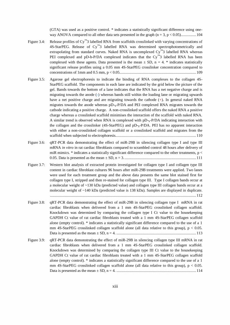
Figure 3.4: Release profiles of Cy™3 labelled RNA from scaffolds crosslinked with varying concentrations of
4S-StarPEG. Release of Cy™3 labelled RNA was determined spectrophotometrically and
extrapolating from standard curves. Naked RNA is uncomplexed Cy™3 labelled RNA whereas
PEI complexed and pD-b-P/DA complexed indicates that the Cy™3 labelled RNA has been
complexed with these agents. Data presented is the mean ± SD, n = 4. * indicates statistically
significant release profiles using a 0.05 mm 4S-StarPEG crosslinker concentration compared to
concentrations of 1mm and 0.5 mm, p < 0.05........................................................................... 109
Figure 3.5: Agarose gel electrophoresis to indicate the binding of RNA complexes to the collagen 4S-
StarPEG scaffold. The components in each lane are indicated by the grid below the picture of the
gel. Bands towards the bottom of a lane indicates that the RNA has a net negative charge and is
migrating towards the anode (-) whereas bands still within the loading lane or migrating upwards
have a net positive charge and are migrating towards the cathode (+). In general naked RNA
migrates towards the anode whereas pD-b-P/DA and PEI complexed RNA migrates towards the
cathode indicating a positive charge. A non-crosslinked scaffold offers the naked RNA a positive
charge whereas a crosslinked scaffold minimises the interaction of the scaffold with naked RNA.
A similar trend is observed when RNA is complexed with pD-b-P/DA indicating interaction with
the collagen and the crosslinker (4S-StarPEG) and pD-b-P/DA. PEI has no apparent interaction
with either a non-crosslinked collagen scaffold or a crosslinked scaffold and migrates from the
scaffold when subjected to electrophoresis............................................................................... 110
Figure 3.6: qRT-PCR data demonstrating the effect of miR-29B in silencing collagen type I and type III
mRNA in vitro in rat cardiac fibroblasts compared to scrambled control 48 hours after delivery of
miR mimics. * indicates a statistically significant difference compared to the other treatments, p <
0.05. Data is presented as the mean ± SD, n = 3. ...................................................................... 111
Figure 3.7: Western blot analysis of extracted protein investigated for collagen type I and collagen type III
content in cardiac fibroblast cultures 96 hours after miR-29B treatments were applied. Two lanes
were used for each treatment group and the above data presents the same blot stained first for
collagen type I, stripped and then re-stained for collagen type III. Type I collagen bands occur at
a molecular weight of ~130 kDa (predicted value) and collagen type III collagen bands occur at a
molecular weight of ~140 kDa (predicted value is 138 kDa). Samples are displayed in duplicate.
............................................................................................................................................... 112
Figure 3.8: qRT-PCR data demonstrating the effect of miR-29B in silencing collagen type I mRNA in rat
cardiac fibroblasts when delivered from a 1 mm 4S-StarPEG crosslinked collagen scaffold.
Knockdown was determined by comparing the collagen type I Ct value to the housekeeping
GAPDH Ct value of rat cardiac fibroblasts treated with a 1 mm 4S-StarPEG collagen scaffold
alone (empty control). * indicates a statistically significant difference compared to the use of a 1
mm 4S-StarPEG crosslinked collagen scaffold alone (all data relative to this group), p < 0.05.
Data is presented as the mean ± SD, n = 4. .............................................................................. 113
Figure 3.9: qRT-PCR data demonstrating the effect of miR-29B in silencing collagen type III mRNA in rat
cardiac fibroblasts when delivered from a 1 mm 4S-StarPEG crosslinked collagen scaffold.
Knockdown was determined by comparing the collagen type III Ct value to the housekeeping
GAPDH Ct value of rat cardiac fibroblasts treated with a 1 mm 4S-StarPEG collagen scaffold
alone (empty control). * indicates a statistically significant difference compared to the use of a 1
mm 4S-StarPEG crosslinked collagen scaffold alone (all data relative to this group), p < 0.05.
Data is presented as the mean ± SD, n = 4. .............................................................................. 114
xiv
Chapter Four: Evaluation of miR Delivery In Vivo
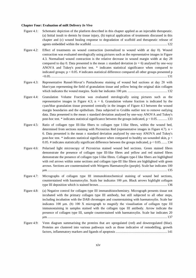
Figure 4.1: Schematic depiction of the platform described in this chapter applied as an injectable therapeutic.
(a) Initial insult to dermis by tissue injury, (b) topical application of treatments discussed in this
chapter and (c) wound healing response to degradation of scaffold and therapeutic release of
agents embedded within the scaffold. ...................................................................................... 122
Figure 4.2: Effect of treatments on wound contraction (normalized to wound width at day 0). Wound
contraction was evaluated sterologically using pictures such as the representative images in Figure
4.3. Normalised wound contraction is the relative decrease in wound margin width at day 28
compared to day 0. Data presented is the mean ± standard deviation (n = 6) analysed by one-way
ANOVA and Tukey's post-hoc test. * indicates statistical significance when compared to
indicated groups; p < 0.05. # indicates statistical difference compared all other groups presented p
<0.05. ..................................................................................................................................... 131
Figure 4.3: Representative Russel-Movat’s Pentachrome staining of wound bed sections at day 28 with
blue/cyan representing the field of granulation tissue and yellow being the original skin collagen
which indicates the wound margins. Scale bar indicates 100 μm. ............................................. 132
Figure 4.4: Granulation Volume Fraction was evaluated sterologically using pictures such as the
representative images in Figure 4.3; n = 6. Granulation volume fraction is indicated by the
cyan/blue granulation tissue presented centrally in the images of Figure 4.3 between the wound
margin boundaries and the epithelium. Data subjected to Grubbs outlier test to remove outlying
data. Data presented is the mean ± standard deviation analysed by one-way ANOVA and Tukey's
post-hoc test. * indicates statistical significance between the groups indicated, p < 0.05. .......... 133
Figure 4.5: Ratio of collagen type III-like fibers to collagen type I-like fibres, within the wound bed,
determined from sections staining with Picrosirius Red (representative images in Figure 4.7). n =
6. Data presented is the mean ± standard deviation analyzed by one-way ANOVA and Tukey's
post-hoc test. * indicates statistical significance when compared to healthy un-wounded skin, p <
0.05. # indicates statistically significant difference between the groups indicated, p < 0.05. ...... 134
Figure 4.6: Polarised light microscopy of Picrosirius stained wound bed sections. Green stained fibres
demonstrate the presence of collagen type III-like fibres and yellow and red stained fibres
demonstrate the presence of collagen type I-like fibres. Collagen type-I like fibers are highlighted
with red arrows within some sections and collagen type-III like fibres are highlighted with green
arrows. Sections are counterstained with Weigerts Haematoxylin (purple). Scale bar indicates 100
μm. ......................................................................................................................................... 135
Figure 4.7: Micrographs of collagen type III immunohistochemical staining of wound bed sections,
counterstained with haematoxylin. Scale bar indicates 100 μm. Black arrows highlight collagen
type III deposition which is stained brown. .............................................................................. 136
Figure 4.8: (a) Negative control for collagen type III immunohistochemistry. Micrograph presents tissue not
incubated with the primary collagen type III antibody, but still subjected to all other steps,
including incubation with the DAB chromagen and counterstaining with haematoxylin. Scale bar
indicates 100 μm. (b) 100 X micrograph to magnify the visualisation of collagen type III
immunostaining in samples stained with the collagen type III antibody. Arrow indicate the
presence of collagen type III, sample counterstained with haematoxylin. Scale bar indicates 20
μm. ......................................................................................................................................... 137
Figure 4.9: Venn diagram summarising the proteins that are upregulated (red) and downregulated (blue).
Proteins are clustered into various pathways such as those indicative of remodelling, growth
factors, inflammatory markers and ligands of apoptosis ........................................................... 141
xv
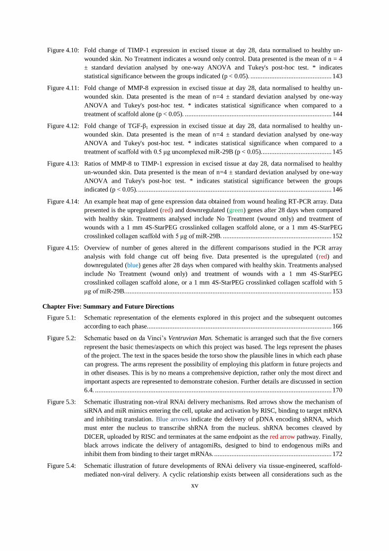
Figure 4.10: Fold change of TIMP-1 expression in excised tissue at day 28, data normalised to healthy un-
wounded skin. No Treatment indicates a wound only control. Data presented is the mean of n = 4
± standard deviation analysed by one-way ANOVA and Tukey's post-hoc test. * indicates
statistical significance between the groups indicated (p < 0.05). ............................................... 143
Figure 4.11: Fold change of MMP-8 expression in excised tissue at day 28, data normalised to healthy un-
wounded skin. Data presented is the mean of n=4 ± standard deviation analysed by one-way
ANOVA and Tukey's post-hoc test. * indicates statistical significance when compared to a
treatment of scaffold alone (p < 0.05). ..................................................................................... 144
Figure 4.12: Fold change of TGF-β1 expression in excised tissue at day 28, data normalised to healthy un-
wounded skin. Data presented is the mean of n=4 ± standard deviation analysed by one-way
ANOVA and Tukey's post-hoc test. * indicates statistical significance when compared to a
treatment of scaffold with 0.5 μg uncomplexed miR-29B (p < 0.05). ........................................ 145
Figure 4.13: Ratios of MMP-8 to TIMP-1 expression in excised tissue at day 28, data normalised to healthy
un-wounded skin. Data presented is the mean of n=4 ± standard deviation analysed by one-way
ANOVA and Tukey's post-hoc test. * indicates statistical significance between the groups
indicated (p < 0.05). ................................................................................................................ 146
Figure 4.14: An example heat map of gene expression data obtained from wound healing RT-PCR array. Data
presented is the upregulated (red) and downregulated (green) genes after 28 days when compared
with healthy skin. Treatments analysed include No Treatment (wound only) and treatment of
wounds with a 1 mm 4S-StarPEG crosslinked collagen scaffold alone, or a 1 mm 4S-StarPEG
crosslinked collagen scaffold with 5 μg of miR-29B. ............................................................... 152
Figure 4.15: Overview of number of genes altered in the different comparisons studied in the PCR array
analysis with fold change cut off being five. Data presented is the upregulated (red) and
downregulated (blue) genes after 28 days when compared with healthy skin. Treatments analysed
include No Treatment (wound only) and treatment of wounds with a 1 mm 4S-StarPEG
crosslinked collagen scaffold alone, or a 1 mm 4S-StarPEG crosslinked collagen scaffold with 5
μg of miR-29B. ....................................................................................................................... 153
Chapter Five: Summary and Future Directions
Figure 5.1: Schematic representation of the elements explored in this project and the subsequent outcomes
according to each phase. .......................................................................................................... 166
Figure 5.2: Schematic based on da Vinci’s Ventruvian Man. Schematic is arranged such that the five corners
represent the basic themes/aspects on which this project was based. The legs represent the phases
of the project. The text in the spaces beside the torso show the plausible lines in which each phase
can progress. The arms represent the possibility of employing this platform in future projects and
in other diseases. This is by no means a comprehensive depiction, rather only the most direct and
important aspects are represented to demonstrate cohesion. Further details are discussed in section
6.4. ......................................................................................................................................... 170
Figure 5.3: Schematic illustrating non-viral RNAi delivery mechanisms. Red arrows show the mechanism of
siRNA and miR mimics entering the cell, uptake and activation by RISC, binding to target mRNA
and inhibiting translation. Blue arrows indicate the delivery of pDNA encoding shRNA, which
must enter the nucleus to transcribe shRNA from the nucleus. shRNA becomes cleaved by
DICER, uploaded by RISC and terminates at the same endpoint as the red arrow pathway. Finally,
black arrows indicate the delivery of antagomiRs, designed to bind to endogenous miRs and
inhibit them from binding to their target mRNAs. .................................................................... 172
Figure 5.4: Schematic illustration of future developments of RNAi delivery via tissue-engineered, scaffold-
mediated non-viral delivery. A cyclic relationship exists between all considerations such as the
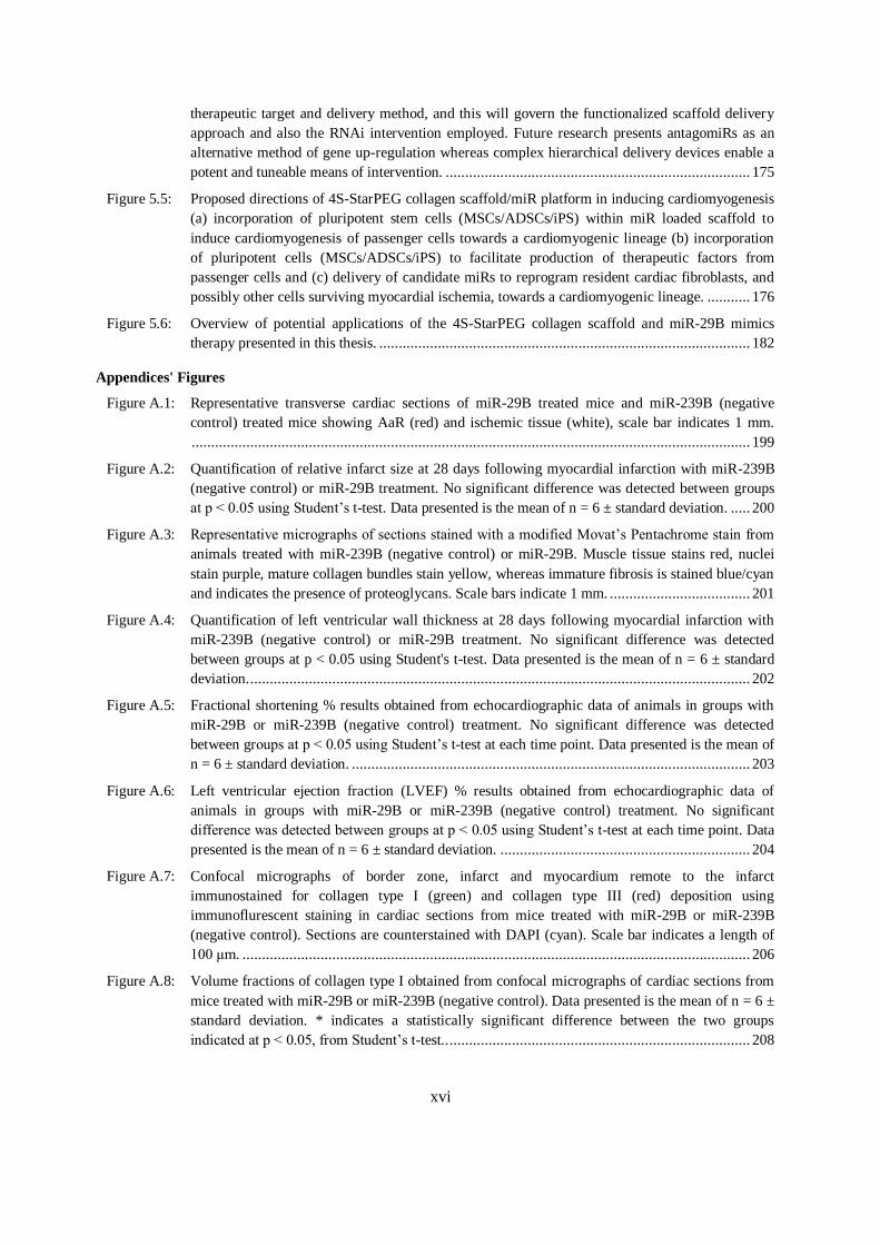
xvi
therapeutic target and delivery method, and this will govern the functionalized scaffold delivery
approach and also the RNAi intervention employed. Future research presents antagomiRs as an
alternative method of gene up-regulation whereas complex hierarchical delivery devices enable a
potent and tuneable means of intervention. .............................................................................. 175
Figure 5.5: Proposed directions of 4S-StarPEG collagen scaffold/miR platform in inducing cardiomyogenesis
(a) incorporation of pluripotent stem cells (MSCs/ADSCs/iPS) within miR loaded scaffold to
induce cardiomyogenesis of passenger cells towards a cardiomyogenic lineage (b) incorporation
of pluripotent cells (MSCs/ADSCs/iPS) to facilitate production of therapeutic factors from
passenger cells and (c) delivery of candidate miRs to reprogram resident cardiac fibroblasts, and
possibly other cells surviving myocardial ischemia, towards a cardiomyogenic lineage. ........... 176
Figure 5.6: Overview of potential applications of the 4S-StarPEG collagen scaffold and miR-29B mimics
therapy presented in this thesis. ............................................................................................... 182
Appendices' Figures
Figure A.1: Representative transverse cardiac sections of miR-29B treated mice and miR-239B (negative
control) treated mice showing AaR (red) and ischemic tissue (white), scale bar indicates 1 mm.
............................................................................................................................................... 199
Figure A.2: Quantification of relative infarct size at 28 days following myocardial infarction with miR-239B
(negative control) or miR-29B treatment. No significant difference was detected between groups
at p < 0.05 using Student’s t-test. Data presented is the mean of n = 6 ± standard deviation. ..... 200
Figure A.3: Representative micrographs of sections stained with a modified Movat’s Pentachrome stain from
animals treated with miR-239B (negative control) or miR-29B. Muscle tissue stains red, nuclei
stain purple, mature collagen bundles stain yellow, whereas immature fibrosis is stained blue/cyan
and indicates the presence of proteoglycans. Scale bars indicate 1 mm. .................................... 201
Figure A.4: Quantification of left ventricular wall thickness at 28 days following myocardial infarction with
miR-239B (negative control) or miR-29B treatment. No significant difference was detected
between groups at p < 0.05 using Student's t-test. Data presented is the mean of n = 6 ± standard
deviation. ................................................................................................................................ 202
Figure A.5: Fractional shortening % results obtained from echocardiographic data of animals in groups with
miR-29B or miR-239B (negative control) treatment. No significant difference was detected
between groups at p < 0.05 using Student’s t-test at each time point. Data presented is the mean of
n = 6 ± standard deviation. ...................................................................................................... 203
Figure A.6: Left ventricular ejection fraction (LVEF) % results obtained from echocardiographic data of
animals in groups with miR-29B or miR-239B (negative control) treatment. No significant
difference was detected between groups at p < 0.05 using Student’s t-test at each time point. Data
presented is the mean of n = 6 ± standard deviation. ................................................................ 204
Figure A.7: Confocal micrographs of border zone, infarct and myocardium remote to the infarct
immunostained for collagen type I (green) and collagen type III (red) deposition using
immunoflurescent staining in cardiac sections from mice treated with miR-29B or miR-239B
(negative control). Sections are counterstained with DAPI (cyan). Scale bar indicates a length of
100 μm. .................................................................................................................................. 206
Figure A.8: Volume fractions of collagen type I obtained from confocal micrographs of cardiac sections from
mice treated with miR-29B or miR-239B (negative control). Data presented is the mean of n = 6 ±
standard deviation. * indicates a statistically significant difference between the two groups
indicated at p < 0.05, from Student’s t-test.. ............................................................................. 208
xvii
Figure A.9: Volume fractions of collagen type III obtained from confocal micrographs of cardiac sections
from mice treated with miR-29B or miR-239B (negative control). Data presented is the mean of n
= 6 ± standard deviation. No statistically significant difference between groups is detected using
Student’s t-test, p < 0.05. ......................................................................................................... 208
Figure A.10: Ratio of volume fractions of collagen type III/I obtained from confocal micrographs of cardiac
sections from mice treated with miR-29B or miR-239B (negative control). Data presented is the
mean of n = 6 ± standard deviation. No statistically significant difference between groups is
detected using Student’s t-test, p < 0.05. .................................................................................. 209
Figure A.11: Micrographs of infarct and remote myocardium following treatment with miR-29B or miR-239B
(negative control). Sections are stained for α-SMA using immunoflurescent staining (red) and
counterstained with DAPI (cyan). Scale bar indicates a length of 200 μm ................................. 210
Figure A.12: α-SMC volume fractions obtained from confocal micrographs of cardiac sections from mice
treated with miR-29B or miR-239B (negative control). Data presented is the mean of n = 6 ±
standard deviation. No statistically significant difference between groups is detected using
Student’s t-test, p < 0.05.......................................................................................................... 211
Figure A.13: Micrographs of infarct and remote myocardium following treatment with miR-29B or miR-239B
(negative control). Sections are stained for TGF-β1 using immunofluorescent staining (green) and
counterstained with DAPI (blue). Scale bar indicates a length of 100 μm. ................................ 212
Figure A.14: Micrographs of infarct and remote myocardium following treatment with miR-29B or miR-239B
(negative control). Sections are stained using an in situ TUNEL kit for apoptosis and
counterstained with Mayer’s Haematoxylin. Arrow indicates an apoptotic cell. Scale bar indicates
a length of 20 μm. ................................................................................................................... 213
Figure A.15: Quantification of number of apoptotic cells per field of view in the infarct and remote myocardium
following treatment with miR-29B or miR-239B (negative control). Data presented is the mean of
n = 6 ± standard deviation. No statistically significant difference between groups is detected using
Student’s t-test, p < 0.05. ......................................................................................................... 214
Figure A.16: In situ hybridisation using LNA oligonucleotides to detect miRs on formalin fixed paraffin
embedded sections. U6 is detection of U6 spliceosomal RNA. Scrambled LNA is employed as a
background control. Blue indicates the detection of RNA using LNA oligonucleotides which is
highlighted with arrows. Sections are counterstained with Nuclear Fast Red. Rat myocardium has
very low detection of miR-1 and U6 whereas human sections have a higher detection. ............. 215
Figure B.1: Schematic representation of human skin equivalent fabrication. ............................................... 225
Figure B.2: (a) Photograph of wounded skin model with biopsy punch wound in the centre, (b) Movat’s
pentachrome staining of wounded skin model after 10 days with 4S-StarPEG scaffold still present
(black arrow) and (c) immunostaining of wounded skin model after 10 days. Green indicates
collagen type I immunostaining, red indicates collagen type III immunostaining and blue indicates
cell nucleus. ............................................................................................................................ 226
Figure B.3: Effect of treatments on skin equivalent model wound area contraction (normalized to wound area
at day 0). Data presented is the mean ± standard deviation (n = 6) analysed by one-way ANOVA
and Tukey's post-hoc test. No statistically significant difference is detected between the samples at
p < 0.05. 29B indicates the use of 0.5 μg of miR-29B, Scram indicates the use of 0.5 μg of
scrambled miR, and C indicates the use of pD-b-P/DA. ............................................................ 227
Figure D.1: Absorbance ranges of titrated isolated collagen and collagen standards stained with Sircol™
Reagent. Data presented is the mean of n = 3 with trendline added to denote pattern. Based on the
equations of the trendlines, the % collagen of the isolated collagen, relative to the 100% collagen
standard is ~80%. .................................................................................................................... 236
xviii
Figure D.2: Reaction of TNBSA with primary amine-containing molecule to produce a chromogenic
derivative. ............................................................................................................................... 238
Figure I.1: Example haemocytometer grid ................................................................................................ 252
Figure J.1: (a) SDS page electrophoresis of protein fractions of washes (W) and eluent fractions (F) during
antibody purification process. (b) Western blot using goat anti-mouse biotinylated antibody
clearly showing antibody present in fractions 1, 2, and 3. (F1, F2 and F3). See Appendices N and
O for detailed protocols. .......................................................................................................... 257
Figure M.1: Protein membrane array intensity profiles of (a) initial intensities and (b) normalised intensities.
............................................................................................................................................... 269
Figure O.1: Excisional wound model with four 1 cm2 wounds created in the back of female Lewis rats at (a)
initially after surgery and (b) day 28 after surgery. The wounds have completely closed at the end
time-point. .............................................................................................................................. 273
Figure O.2: Measurements of gross wound area using tracing paper (normalized to tracings performed at day
0). Data presented is the mean ± standard deviation (n = 6) analysed by one-way ANOVA and
Tukey's post-hoc test, p < 0.05. No statistically significant difference is detected between any of
the groups investigated. ........................................................................................................... 274
Figure P.1: Isolated human keratinocytes in culture ................................................................................... 277
Figure R.1 Polarigraphs of wound edges, where collagen distribution and organization is shown. Thicker,
collagen type I-like fibers, polarise to display as red and yellow fibres whereas thinner, collagen
type III-like fibers, polarise to display as green fibres. ............................................................. 282
Figure V.1: Agarose gel electrophoresis of (a) unlabelled and (b) Cy™3 labelled siRNA. The migration of (b)
is slower due to an increased molecular weight due to labelling with Cy™3. However, labelling
may not be homogenous due to the observation of a very thick, weaker band at (b). ................. 294
Figure W.1: Tracheotomy and intubation. (A) A midline cervical skin incision is made and the salivary glands
as well as the muscles overlying the trachea are gently separated. (B, C) The muscles are fixed
aside with sutures and clamps. Thereby the trachea is visible. (D, E) A cut is made between the
fourth and fifth tracheal cartilage. (E, F) Intubation is facilitated by attaching a suture on the
posterior tracheal cartilage on which a small bulldog clamp is fixed. ........................................ 296
Figure W.2: Thoracotomy. (A) The mouse is relocated on its right side and the skin is incised. (B, C)The
Subcutaneous tissue is dissected free. (D)The ventral serrated muscle of thorax is transected. (E)
The intercostal muscles are incised in the third intercostal space. (F) The opening is widened with
a rip retractor. ......................................................................................................................... 297
Figure W.3: Occlusion of the left descending artery (LAD). (A) By widening the intercostal space the left side
of the heart and the lung become apparent. (B) After opening the pericardium the left auricle is
visible (triangle) at whose tip the LAD (arrow) takes course towards the apex. (C, D) The LAD is
occluded at the level of the tip of the left auricle. (E) A loose snare is formed. (F) Furthermore, a
piece of polyethylene tubing is placed on the artery to later allow the reestablishment of blood
flow. ....................................................................................................................................... 298
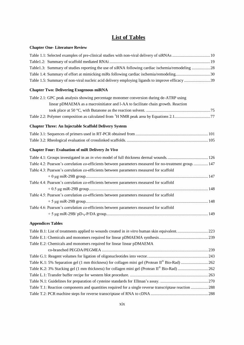
xix
List of Tables
Chapter One- Literature Review
Table 1.1: Selected examples of pre-clinical studies with non-viral delivery of siRNAs ................................... 10
Table1.2: Summary of scaffold mediated RNAi ............................................................................................. 19
Table1.3: Summary of studies reporting the use of siRNA following cardiac ischemia/remodeling ................. 28
Table 1.4: Summary of effort at mimicking miRs following cardiac ischemia/remodeling................................ 30
Table 1.5: Summary of non-viral nucleic acid delivery employing ligands to improve efficacy ........................ 39
Chapter Two: Delivering Exogenous miRNA
Table 2.1: GPC peak analysis showing percentage monomer conversion during de-ATRP using
linear pDMAEMA as a macroinitiator and l-AA to facilitate chain growth. Reaction
took place at 50 °C, with Butanone as the reaction solvent. ........................................................... 75
Table 2.2: Polymer composition as calculated from 1H NMR peak area by Equations 2.1................................. 77
Chapter Three: An Injectable Scaffold Delivery System
Table 3.1: Sequences of primers used in RT-PCR obtained from ................................................................... 101
Table 3.2: Rheological evaluation of crosslinked scaffolds. ........................................................................... 105
Chapter Four: Evaluation of miR Delivery In Vivo
Table 4.1: Groups investigated in an in vivo model of full thickness dermal wounds. ..................................... 126
Table 4.2: Pearson’s correlation co-efficients between parameters measured for no-treatment group. ............. 147
Table 4.3: Pearson’s correlation co-efficients between parameters measured for scaffold
+ 0 μg miR-29B group. ................................................................................................................ 147
Table 4.4: Pearson’s correlation co-efficients between parameters measured for scaffold
+ 0.5 μg miR-29B group. ............................................................................................................. 148
Table 4.5: Pearson’s correlation co-efficients between parameters measured for scaffold
+ 5 μg miR-29B group. ................................................................................................................ 148
Table 4.6: Pearson’s correlation co-efficients between parameters measured for scaffold
+ 5 μg miR-29B/ pD-b-P/DA group. ............................................................................................. 149
Appendices Tables
Table B.1: List of treatments applied to wounds created in in vitro human skin equivalent. ............................ 223
Table E.1: Chemicals and monomers required for linear pDMAEMA synthesis ............................................. 239
Table E.2: Chemicals and monomers required for linear linear pDMAEMA
co-branched PEGDA/PEGMEA .................................................................................................. 239
Table G.1: Reagent volumes for ligation of oligonucleotides into vector. ....................................................... 243
Table K.1: 5% Separation gel (1 mm thickness) for collagen mini gel (Protean II® Bio-Rad) ......................... 262
Table K.2: 3% Stacking gel (1 mm thickness) for collagen mini gel (Protean II® Bio-Rad) ............................ 262
Table L.1: Transfer buffer recipe for western blot procedure. ........................................................................ 263
Table N.1: Guidelines for preparation of cysteine standards for Ellman’s assay. ............................................ 270
Table T.1: Reaction components and quantities required for a single reverse transcriptase reaction ................ 288
Table T.2: PCR machine steps for reverse transcriptase of RNA to cDNA ..................................................... 288

xx
Table T.3: Genomic DNA elimination mix ................................................................................................... 288
Table T.4: Reverse-transcription mix ............................................................................................................ 288
Table U.1: Polymerase chain reaction components ........................................................................................ 290
Table U.2: Typical real time PCR program. .................................................................................................. 290
Table U.3: PCR components mix .................................................................................................................. 291
Table U.4: Cycling conditions for the Roche LightCycler 480® ..................................................................... 291
Table U.5: List of genes investigated in rat wound healing PCR array ........................................................... 292
Table V.1: Reaction setup for labelling 5 μg 21-mer duplex siRNA. .............................................................. 293

xxi
Acknowledgements
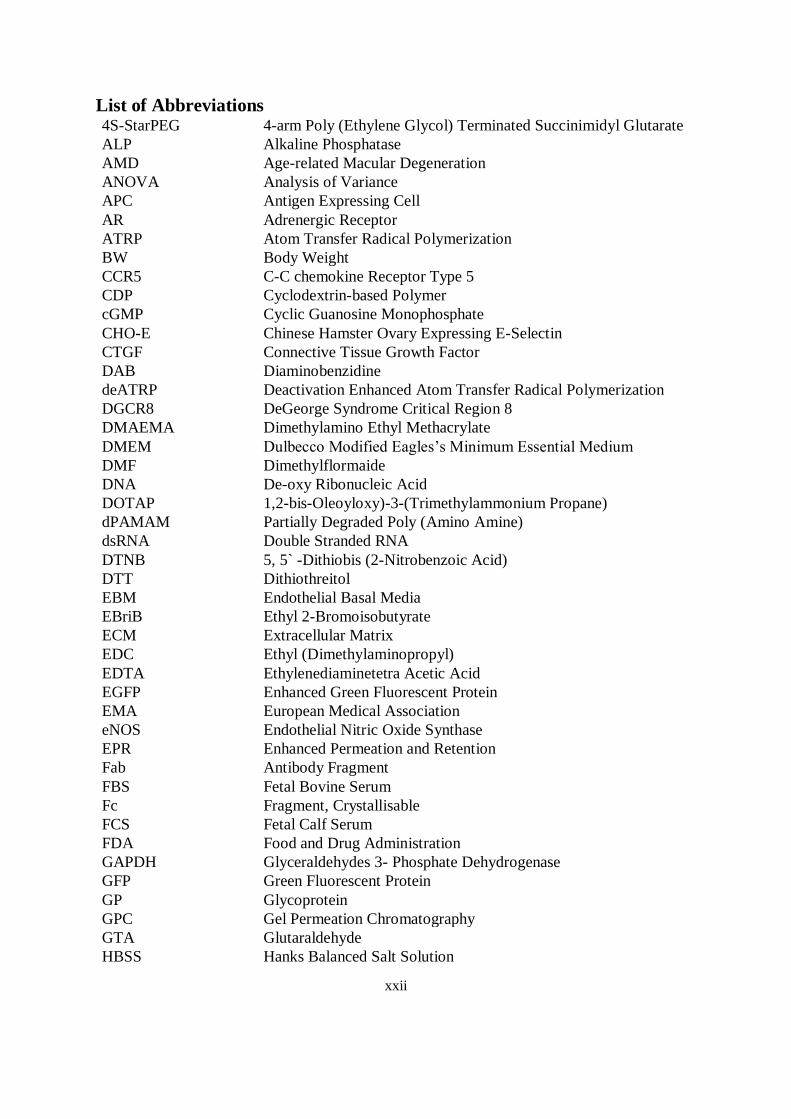
xxii
List of Abbreviations 4S-StarPEG 4-arm Poly (Ethylene Glycol) Terminated Succinimidyl Glutarate
ALP Alkaline Phosphatase
AMD Age-related Macular Degeneration
ANOVA Analysis of Variance
APC Antigen Expressing Cell
AR Adrenergic Receptor
ATRP Atom Transfer Radical Polymerization
BW Body Weight
CCR5 C-C chemokine Receptor Type 5
CDP Cyclodextrin-based Polymer
cGMP Cyclic Guanosine Monophosphate
CHO-E Chinese Hamster Ovary Expressing E-Selectin
CTGF Connective Tissue Growth Factor
DAB Diaminobenzidine
deATRP Deactivation Enhanced Atom Transfer Radical Polymerization
DGCR8 DeGeorge Syndrome Critical Region 8
DMAEMA Dimethylamino Ethyl Methacrylate
DMEM Dulbecco Modified Eagles’s Minimum Essential Medium
DMF Dimethylflormaide
DNA De-oxy Ribonucleic Acid
DOTAP 1,2-bis-Oleoyloxy)-3-(Trimethylammonium Propane)
dPAMAM Partially Degraded Poly (Amino Amine)
dsRNA Double Stranded RNA
DTNB 5, 5` -Dithiobis (2-Nitrobenzoic Acid)
DTT Dithiothreitol
EBM Endothelial Basal Media
EBriB Ethyl 2-Bromoisobutyrate
ECM Extracellular Matrix
EDC Ethyl (Dimethylaminopropyl)
EDTA Ethylenediaminetetra Acetic Acid
EGFP Enhanced Green Fluorescent Protein
EMA European Medical Association
eNOS Endothelial Nitric Oxide Synthase
EPR Enhanced Permeation and Retention
Fab Antibody Fragment
FBS Fetal Bovine Serum
Fc Fragment, Crystallisable
FCS Fetal Calf Serum
FDA Food and Drug Administration
GAPDH Glyceraldehydes 3- Phosphate Dehydrogenase
GFP Green Fluorescent Protein
GP Glycoprotein
GPC Gel Permeation Chromatography
GTA Glutaraldehyde
HBSS Hanks Balanced Salt Solution
xxiii
HCl Hydrochloric Acid
HDMEC Human Dermal Microvascular Endothelial Cells
H&E Haematoxylin &Eosin
HEK Hamster Embryonic Kidney
HEPES 4-(2-Hydrozyethyl)-1-Piperazine Ethane Sulfonic Acid
HIF-1 Hypoxia-Inducible Factor-1
HIV Human Immunodeficiency Virus Type 1
hMGFP Humanized Montrastrea Green Fluorescent Protein
hMSC Human Mesenchymal Stem Cell
HPLC High Performance Liquid Chromatograpy
HRP Horseradish Peroxidase
HSP Heat Shock Protein
HSVEC Human Saphenous Vein Endothelial Cells
HuR Human Stabilizing Protein
HUVEC Human Umbilical Vein Endothelial Cell
ICAM Intercellular Adhesion Molecule
IgG Immunoglobulin G
IL Interleukin
iNOS Inducible Nitric Oxide Synthase
IPC Ischemic Preconditioning
L-AA L-Ascorbic Acid
LB Lysogeny Broth
LNA Locked Nucleic Acid
LV Left Ventricle
mAb Monoclonal Antibody
Mapk-1 Mitogen-Activated Protein Kinase 1
Mhy6: Alpha Heavy Myosin Chain
MI Myocardial Infarction
miR MicroRNA
MMP Matrix-Metalloproteinase
mRNA Messenger RNA
mTOR Mammalian Target of Rapamycin
MVM Multi-Vinyl Monomers
MWCO Molecular Weight Cut Off
MyD88 Myeloid Differentiation Factor 88
Myh7 β- Myosin Heavy Chain
N/P Amine to Phospate Ratio
NaOH Sodium Hydroxide
NF-ҡB Transcription Factor kappa B
NHE1 Cardiac Na+/H
+ Exchanger
NMDA-R2B N-Methyl-D-Aspartate Receptor Type 2B
NOD Non Obese Diabetic
NP Nucleocapsid
nt Nucleotide
P2X7 Purinergic P2X7 Receptor
P4H Prolyl-4 Hydroxylase
xxiv
PACT Protein Kinase RNA Activator
PAGE Poly (Acrylamide) Gel Electrophoresis
PBS Phosphate Buffered Solution
PCL Poly (ε-Caprolactone)
PCM Primary Cardiomyocyte
PCR Polymerase Chain Reaction
PDCD4 Programmed Cell Death-4
PDE5a Phosphodiesterase 5a
pDMAEMA Poly (Dimethylamino Ethyl Methacrylate)
pDNA Plasmid DNA
PECAM Platelet Endothelial Cell Adhesion Molecule
PEG Poly (Ethylene Glycol)
PEGDA Poly (Ethylene Glycol) Diacrylate
PEGM Poly (Ethylene Glycol) Methyl Ether
PEGMEA Poly (Ethylene Glycol) Methyl Ether Acrylate
PEI Poly (Ethylenimine)
PGE2 Prostaglandin E2
PLGA Poly (Lactic-co-Glycolic Acid)
PLK-1 Polo-like Kinase 1
PLL Poly (L-Lactide)
PMDETA Pentamethyldiethylenetriamine
Pol II Polymerase II
Pre-miRAs Precursor miRs
PVA Poly (Vinyl Alcohol)
PVDF Poly Vinylidene Fluoride
q-PCR Quantitative PCR
RI Refractive Index
RISC RNA Induced Silencing Complex
RNA Ribonucleic Acid
RNAi RNA Interference
ROS Reactive Oxygen Species
RSV Respiratory Syncytial Virus
RT Real-time
RT Reverse Transcription
scFv Single Chain Variable Fragment
SCID Severe Combined Immunodefficiency
SDS Sodium Dodecyl Sulphate
SG Succinimydl Glutarate
SH Thiol
SH2 Src Homology Domain 2
shRNA Short Hairpin RNA
siRNA Short Interfering RNA
STAT3 Signal Transducer and Activator of Transcription 3
TAT Tyrosine Aminotransferase
TBE Tris-Borate
Tfr Transferrin Receptor
xxv
TGF-β Transforming Growth Factor beta
TIPA Radio Immunoprecipitation Assay
TLR Toll-Like Receptors
TNBSA 2,2,6-Trinitrobenzenesulfonic Acid
TNF-α Tissue Necrosis Factor alpha
TR Transferrin
TRBP TAR RNA Binding Protein
UTR Untranslated Region
UV Ultraviolet
v/v Volume per Volume
VCAM Vascular Cell Adhesion Molecule
VEGF Vascular Endothelial Growth Factor
VEGFR Vascular Endothelial Growth Factor Receptor
Vif Virion Infectivity Factor
VS Vinyl-Sufone
w/v Weight per Volume
w/w Weight to Weight Ratio
xxvi
Abstract
Cardiovascular disease is the leading cause of death in the developed world and is responsible for
approximately 36% of Irish mortality. Myocardial infarction (MI), which is literally the death of
cardiac tissue due to lack of oxygenation, accounts for the majority of deaths associated with
cardiovascular disease. This death of cardiac tissue leads to a loss of cardiac function as the damaged
area becomes a non-contractile scar. Amelioration of this process is a main aim of regenerative
cardiac strategies such as anti-fibrotic therapies. Thus, anti-fibrotic interfering RNA (RNAi) therapy
with exogenous microRNA (miR)-29B was proposed as a method to modulate extracellular matrix
(ECM) remodelling following MI. It was hypothesized that miR-29B scaffold delivery will efficiently
modulate the ECM remodelling response and reduce maladaptive remodelling, such as aggressive
deposition of collagen type I, after injury. The primary objective of this doctoral project was to
develop a scaffold-based, controlled release gene therapy system. A co-polymer of a linear poly
(dimethylamino) ethyl methacrylate (pDMAEMA) block and a hyperbranched poly (ethylene glycol)
methyl ether acrylate (PEGMEA) based unit with poly (ethylene glycol) diacrylate (PEGDA) as the
branching agent (pD-b-/PDA) was synthesised, with the purpose of complexing miRs, using
deactivation enhanced atom transfer radical (De-ATRP) synthesis. Non-viral complexes of miR-29B
and pD-b-/PDA were optimized for both monolayer and three-dimensional delivery from a crosslinked
collagen-based scaffold in vitro. The release of these complexes from the scaffolds was assessed and
their ability to silence collagen type I and collagen type III expression was evaluated. When cardiac
fibroblasts were cultured with complex loaded scaffolds, relatively low levels of collagen type I and
collagen type III mRNA expression were observed for up to two weeks of culture. When scaffolds
loaded with miR-29B or miR-29B complexes were applied to a rat excisional wound model, reduced
wound contraction, improved collagen type III/I ratios and a significantly higher MMP-8: TIMP-1
ratio were detected. From these investigations it was concluded miR-29B functionalization of the
scaffold significantly increased the ratio of collagen type III/I and MMP-8/TIMP-1 ratios.
Furthermore, these effects were not improved through the use of pD-b-/PDA, and were significantly
influenced by the dose of miR-29B in the collagen scaffold (0.5 μg versus 5 μg). This is the first study
to describe a biomaterial scaffold combined exogenous miRs capable of improving ECM remodelling
following injury. There is significant potential for further development of this platform which could
ultimately represent a step towards the realization of true cardiac regeneration.

Chapter One
Literature Review
The majority of this chapter has been previously published in
Monaghan M, Pandit A. RNA interference therapy via functionalized scaffolds. Adv. Drug Del. Rev. 2011, 63: 197-208
Monaghan M, Greiser U, Wall J.G, O'Brien T, Pandit A. Interference: An alteRNAtive therapy following acute myocardial infarction. Trends Pharmacol. Sci. 2012, 33: 635-645
Literature Review
2
1.1 Introduction
Tissue engineering is the use of a combination of biomaterial scaffolds, cells, and engineering, with
suitable biochemical and physio-chemical factors to improve or replace biological functions 1.
Scaffolds employed in tissue engineering act as platforms that can recapitulate the in vivo milieu,
allowing cells to influence their own microenvironments and/or retain cells and biochemical factors.
These scaffolds can provide structural support, porosity to enable cellular infiltration, and possible
release of embedded biomolecules and cues for the surrounding tissues to regenerate. Scaffolds can be
further ameliorated by the incorporation of cells to replace those that have been lost or to offer
paracrine support to those that are compromised at the site of delivery 2. Biomaterial-based scaffolds
have played a central role in regenerative medicine and tissue engineering and several key
requirements for scaffolds have been identified. Scaffolds fabricated from a range of natural and
synthetic materials are desired to be biodegradable to obviate the need for a removal procedure. In
tandem, predictable biodegradation of the scaffold can facilitate controlled release of biomolecules
embedded within the scaffold. The degradation time of the scaffold should ideally mirror the time
necessary for tissue regeneration. Degradation of the biomaterial can create a path for tissue ingrowth
and this can be further facilitated by a highly porous scaffold which can also allow for the initial
transport of oxygen and nutrients, as well as for the removal of metabolic waste and degradation
products.
The current paradigm of tissue engineering incorporates the use of biomolecules, which can be growth
factors, pharmaceutical agents, or mediators of nucleic acid therapy. Therefore it is intuitive that
scaffolds act as reservoirs in the delivery of nucleic acid such as interfering RNA (RNAi) (see Figure
1.2). RNAi delivery from a scaffold enables localized treatment, as the scaffold, acting as a reservoir
of RNAi, facilitates delivery of this therapeutic molecule in comparison to delivery via a systemic
approach 3, 4
. Targeting a cell population or anatomical location by injection or systemic delivery is
complex and poses many challenges; direct delivery of a therapy from a scaffold, however, can
surmount these barriers. Cells surrounding a scaffold are considered for targeting and become
exposed to the therapeutic agent limiting unwanted exposure in other areas. Additionally, scaffold-
based delivery has the potential to maintain effective levels of payload and nucleotide bioactivity for
extended periods which broadens the opportunity for cellular uptake and increases the likelihood of
nucleic acid transfer. Delivery from the majority of biomaterial systems most likely occurs by means
of a combination of payload interaction with the matrix and subsequent release, with the payload and
material designed to regulate these interactions.
Literature Review
3
Another advantage of a scaffold is its role in the protection of its payload from attack by immune
responses, and limitation of degradation by serum nucleases or proteases. The use of scaffolds in the
delivery of nucleic acid therapy has been extensively reviewed previously 4, 5
; and the basic criteria,
concepts and interactions between scaffolds and their therapeutic loads do not change in the context of
RNAi delivery.
1.2 RNA Interference
RNAi is a fundamental pathway in eukaryotic cells by which sequence-specific double stranded RNA
targets, binds to, and inhibits the translation of a complementary mRNA. Many similar RNAi
mechanisms exist which pursue distinct pathways inside eukaryotic cells. In general, however, most
pathways terminate at a common end; in that they inhibit the translation of messenger RNA (mRNA)
(see Figure 1.1). Synthetic approaches employing antisense oligonucleotides have evolved to double
stranded RNAs (dsRNAs) following seminal discoveries by Fire et al. in 2001 6, illustrating a ten-fold
improvement in silencing target genes when compared to a single stranded RNA alone. These
exogenous molecules manipulate an existing regulatory pathway: that of microRNA (miR).
The miR pathway begins in the nucleus with transcription of long primary miR (pri-miRs) by RNA
polymerase II (Pol II) 7 and subsequent processing into ~70 nucleotide (nt) stem-loop structures
termed ‗precursor miRAs‘ (pre—miRs) by the RNase III enzyme Drosha which functions with the
dsRNA binding protein of DiGeorge syndrome critical region 8 (DGCR8) 8. Exportin-5, another
dsRNA- binding protein, chaperones these pre-miRs to the cytoplasm in a Ran-GTP-dependent
manner. Once in the cytoplasm; Dicer, an endoribonuclease in the RNAse III family, and its
associated dsRNA-binding protein partners, HIV-1 and the TAR RNA binding protein (TRBP) 9,
cleave pre-miR to a mature miR duplex ~22 nt long and load this mature miR into a RNA induced
silencing complex (RISC) 10, 11
. miR interaction with RISC involves cleavage of the miR passenger
strand by Ago2 although imperfect sequence complementarities between the mature miR strand and
its complementary passenger strand may inhibit Ago2 from cleaving the passenger strand 12
.
Alternatively, this interface employs a circumventing mechanism using helicase activity to unwind
and remove the passenger strand. Regardless of the mechanism, the removal of passenger strand
enables the binding of the mature miR to its target mRNA 3‘ UTR. Gene silencing is then catalysed
by RISC through translational repression and consequent mRNA degradation.
The seed sequence of a mature miR encompasses the first 2 - 7 or 2 - 2 nucleotides from its 5‘ end and
needs to have full complementarity with its target to enable RNAi, whereas imperfect matches in the
3‘ end of the miR strand are tolerable 13
. As a result, a single miR can regulate multiple cellular
Literature Review
4
targets and, conversely, each gene can be controlled by many miRs, suggesting an elaborate and
multifaceted mechanism of gene regulation. Numerous hypotheses toward the effects of imperfect
binding of a miR to its target mRNA and its effect on gene silencing have been put forward, including
interruption of translation at the initiation or elongation step, co-translational degradation,
transportation to cytoplasmic processing bodies (P-bodies), and/or de-capping or deadenylation 14-16
.
On the other hand, when complete sequence complementarities occur; silencing is achieved via
cleavage of the mRNA through RISC activity 17
. In both cases, miR targeting and binding results in
post-transcriptional silencing of the target gene.
Appreciation of endogenous RNAi pathways has enabled improvement in synthetic strategies to
silence gene expression. One approach is to, albeit stably or transiently using plasmid DNA (pDNA)
encoding the expression of short hairpin RNA (shRNA) 18-20
or delivery to the cytosol of
shRNA/siRNA and dsRNA. The utilization of either approach needs to account for the stability, the
therapeutic target and the efficacy of delivery. Efficacy of shRNA-expressing pDNA requires entry
into the cell, endosomal escape and subsequent entry to the nucleus via a nuclear pore, as in the case
of non-dividing cells 21, 22
or nuclear uptake during cell division 23
. An alternative approach is to
deliver dsRNAs exogenously which become cleaved in the cytoplasm by Drosha to siRNA and
subsequently loaded into the Argonaut/RISC complex. Therefore, dsRNAs are advantageous in
delivery compared with shRNA-expressing pDNA as they obviate the necessity for nuclear entry and
can therefore enable a more efficient process of RNAi. Silencing gene expression using long dsRNAs
has been demonstrated in an array of organisms including plants and drosophila. However, in
mammalian cells, it has been observed that dsRNAs longer than ~30 nt elicit an antiviral associated
interferon response which ultimately results in non-specific suppression of gene expression through
activation of RNase L and general degradation of RNA molecules 24
.
This response can be evaded however, by the design of the dsRNA. Elbashir et al. observed that
smaller dsRNAs, less than 30 nt are capable of bypassing the mammalian immune response while
inducing RNA interference to achieve specific gene silencing 25
. This realisation progressed to the
hypothesis that synthetic dsRNAs of very short length (siRNAs) with perfect homology to the mRNA
of a target gene can be delivered as an alternative mediator of gene silencing. However, early innate
immuno-stimulatory activity with these synthetic molecules remains a concern. siRNAs containing
certain nucleotide sequence motifs (GU) can stimulate toll-like receptors (TLRs) in the endosomal
pathway. Obviation of this is desirable because if it is not carefully avoided, siRNAs capable of
triggering immuno-stimulatory activity 26
. The antigenic potential of siRNAs can occur via TLR-3,
and has been reported as a possible safety concern 27
. This has brought scrutiny to some studies where
anti-angiogenic specific siRNAs have been reported as being successful as other pre-clinical studies
Literature Review
5
have concluded that non-specific siRNAs also induce an anti-angiogenic effect due to interaction with
TLRs, and engaging the TLR3/interleukin pathway 28
.
The exogenous introduction of short siRNAs with appropriate length and 2-nt 3‘ overhang can be
loaded directly onto RISC for RNAi function, obviating interaction with Dicer, TRBP or protein
kinase RNA activator (PACT); however, the loading process is ten times less efficient than that of
shRNA or longer dsRNAs. Increasing the length of the siRNA duplex to 29-30 nt with a 2-nt 3‘
overhang only at the antisense end of the duplex appears to improve efficacy 29
. Dicer processing
before RISC loading can increase the potency of post-transcriptional gene silencing and can be
achieved by using longer chemically synthesized siRNAs (27 nt) and shRNAs (28 nt) 29, 30
. It is likely
that Dicer and TRBP-PACT present an effective basis for RISC formation and shuttle their cleaved
products directly to RISC 12, 31
. Employing this loading stem in exogenous RNAi therapies can elicit a
more potent gene-silencing effect through the use of these Dicer substrates. Twenty seven nt dsRNAs
are intended to be asymmetrical, with one 2-nt 3‘ overhang and one blunt end. A single stranded
siRNA results from Dicer processing due to Dicer recognition of the 2-nt 3‘ overhang for processing.
However, the blunt end, which includes DNA bases, might trigger low levels of interferon induction;
but the potency of this longer dsRNA means that lower concentrations of 27 mers are required to
silence gene expression which may avoid or minimise such an interferon response 32
. Furthermore, it
is possible that increasing the length of a siRNA duplex with an unprocessed end dictates
directionality as a result of imposed thermodynamic instability determining the guide strand shape and
thereby enhancing its association with Dicer/TRBP/PACT complex for more efficient loading onto
RISC. shRNA on the other hand, assimilates into the endogenous miR pathway and in doing so is
significantly more efficient 33, 34
.
1.3 Non-viral Delivery
Current clinical applications of RNAi are focused on using previously mentioned 21–nt siRNA
duplexes with 2-nt 3‘ overhangs for Dicer recognition which are suitable for large-scale synthesis that
enables uniform production of these chemically synthesized molecules. The clinical relevance of
siRNA is supported by its ability to transfect relatively non-proliferating cells in which the nuclear
entry of shRNA-expressing pDNA is limited. For example, successful target gene knockdown in cells
such as bone marrow-derived dendritic cells 35
and primary T lymphocytes 36
, for which transfection
with pDNA is difficult due to low permeability in their nuclear envelope has been reported with
siRNA. For a quantitative comparison of the duration of effect of siRNA and shRNA-expressing
pDNA, McAnuff et al. compared the potency of siRNA and shRNA-expressing pDNA mediated
gene-silencing in vivo by co-administration of siRNA or shRNA-expressing pDNA with the pDNA
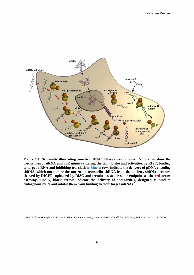
Literature Review
6
Figure 1.1: Schematic illustrating non-viral RNAi delivery mechanisms. Red arrows show the
mechanism of siRNA and miR mimics entering the cell, uptake and activation by RISC, binding
to target mRNA and inhibiting translation. Blue arrows indicate the delivery of pDNA encoding
shRNA, which must enter the nucleus to transcribe shRNA from the nucleus. shRNA becomes
cleaved by DICER, uploaded by RISC and terminates at the same endpoint as the red arrow
pathway. Finally, black arrows indicate the delivery of antagomiRs, designed to bind to
endogenous miRs and inhibit them from binding to their target mRNAs *.
* Adapted from Monaghan M, Pandit A. RNA interference therapy via functionalized scaffolds. Adv. Drug Del. Rev. 2011, 63: 197-208
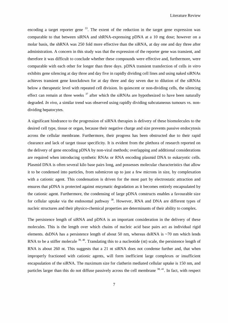
Literature Review
7
encoding a target reporter gene 33
. The extent of the reduction in the target gene expression was
comparable to that between siRNA and shRNA-expressing pDNA at a 10 mg dose; however on a
molar basis, the shRNA was 250 fold more effective than the siRNA, at day one and day three after
administration. A concern in this study was that the expression of the reporter gene was transient, and
therefore it was difficult to conclude whether these compounds were effective and, furthermore, were
comparable with each other for longer than three days. pDNA transient transfection of cells in vitro
exhibits gene silencing at day three and day five in rapidly dividing cell lines and using naked siRNAs
achieves transient gene knockdown for at day three and day seven due to dilution of the siRNAs
below a therapeutic level with repeated cell division. In quiescent or non-dividing cells, the silencing
effect can remain at three weeks 37
after which the siRNAs are hypothesized to have been naturally
degraded. In vivo, a similar trend was observed using rapidly dividing subcutaneous tumours vs. non-
dividing hepatocytes.
A significant hindrance to the progression of siRNA therapies is delivery of these biomolecules to the
desired cell type, tissue or organ, because their negative charge and size prevents passive endocytosis
across the cellular membrane. Furthermore, their progress has been obstructed due to their rapid
clearance and lack of target tissue specificity. It is evident from the plethora of research reported on
the delivery of gene encoding pDNA by non-viral methods; overlapping and additional considerations
are required when introducing synthetic RNAs or RNA encoding plasmid DNA to eukaryotic cells.
Plasmid DNA is often several kilo base pairs long, and possesses molecular characteristics that allow
it to be condensed into particles, from submicron up to just a few microns in size, by complexation
with a cationic agent. This condensation is driven for the most part by electrostatic attraction and
ensures that pDNA is protected against enzymatic degradation as it becomes entirely encapsulated by
the cationic agent. Furthermore, the condensing of large pDNA constructs enables a favourable size
for cellular uptake via the endosomal pathway 38
. However, RNA and DNA are different types of
nucleic structures and their physico-chemical properties are determinants of their ability to complex.
The persistence length of siRNA and pDNA is an important consideration in the delivery of these
molecules. This is the length over which chains of nucleic acid base pairs act as individual rigid
elements. dsDNA has a persistence length of about 50 nm, whereas dsRNA is ~70 nm which lends
RNA to be a stiffer molecule 39, 40
. Translating this to a nucleotide (nt) scale, the persistence length of
RNA is about 260 nt. This suggests that a 21 nt siRNA does not condense further and, that when
improperly fractioned with cationic agents, will form inefficient large complexes or insufficient
encapsulation of the siRNA. The maximum size for clatherin mediated cellular uptake is 150 nm, and
particles larger than this do not diffuse passively across the cell membrane 38, 41
. In fact, with respect

Literature Review
8
to siRNA complexes, it has been shown 42
that particles of siRNA complexed with poly(ethylenimine)
(PEI) greater than 150 nm in size are unable to cause gene silencing in vitro.
RNAi can be therapeutically viable with the use of a non-viral vector by prolonging the serum half-
life and intracellular buffering of siRNA by improving pharmacokinetics and nuclease resistance. For
example, it has been reported that chemically modified siRNAs encapsulated within a lipid core have
extended serum half-life (6.5h) compared to those without protection (0.8h). Non-viral vectors
available for siRNA/pDNA delivery, such as cationic polymers 43
, lipid based approaches 44
,
nanoparticle approaches or biomolecular chaperoning as these areas have been covered in exhaustive
reviews 45, 46
; and key developments in pre-clinical and clinical studies will be elaborated in the next
section.
1.4 RNAi as a Therapeutic
The use of RNAi has undergone a rapid transition to pre-clinical studies due to advances made
previously in non-viral nucleic acid delivery and the current understanding of cellular pathways
during states of pathogenesis, and due to the availability of a wide range of in vivo models. Although
clinical trials are in progress, the number of these trials is relatively few and almost all of these trials
employ naked or slightly modified siRNAs to deliver RNAi.
The most advanced clinical trials are in the treatment of age-related macular degeneration (AMD)
which is a leading cause of blindness. These trials involve the injection of naked siRNA into the
macula of the eye, targeted to genes for vascular endothelial growth factor (VEGF-A) and the VEGF
receptor (VEGFR), and have shown some therapeutic potential in their inhibition of excessive
vascularization of the eye that leads to AMD 47
. The first clinical trial involving siRNA was carried
out by Acuity Pharmaceuticals Inc. for the treatment of AMD, for which phase I results have been
completed. The completed phase II trials reported tolerated doses with a lack of adverse systemic
effects. Testing had advanced into phase III trials, when Acuity Pharmaceuticals Inc. was taken over
by OPKO Health Inc.; however, the company decided to terminate its phase III clinical study of
bevasiranib (a naked siRNA) for the treatment of AMD. No systemic safety issues were identified and
local ocular risks were generally considered ‗unremarkable‘ by the company. However, a careful
review of the data by the Independent Data Monitoring Committee (IDMC) concluded that the trial,
as structured, was unlikely to meet a significant beneficial primary end point 48
.
Allergen Inc. is conducting a phase II clinical trial on a siRNA therapy for AMD, with completed
phase I results indicating minimal side effects and improved vision in some of the patients 49
. In the
phase I clinical trial targeting VEGFR-1 in patients with choroidal neovascularisation resulting from
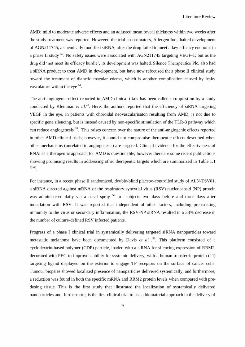
Literature Review
9
AMD; mild to moderate adverse effects and an adjusted mean foveal thickness within two weeks after
the study treatment was reported. However, the trial co-ordinators, Allergen Inc., halted development
of AGN211745, a chemically modified siRNA, after the drug failed to meet a key efficacy endpoint in
a phase II study 50
. No safety issues were associated with AGN211745 targeting VEGF-1; but as the
drug did ‗not meet its efficacy hurdle‘, its development was halted. Silence Therapeutics Plc. also had
a siRNA product to treat AMD in development, but have now refocused their phase II clinical study
toward the treatment of diabetic macular edema, which is another complication caused by leaky
vasculature within the eye 51
.
The anti-angiogenic effect reported in AMD clinical trials has been called into question by a study
conducted by Kleinman et al.28
. Here, the authors reported that the efficiency of siRNA targeting
VEGF in the eye, in patients with choroidal neovascularisaton resulting from AMD, is not due to
specific gene silencing, but is instead caused by non-specific stimulation of the TLR-3 pathway which
can reduce angiogenesis 28
. This raises concern over the nature of the anti-angiogenic effects reported
in other AMD clinical trials; however, it should not compromise therapeutic effects described when
other mechanisms (unrelated to angiogenesis) are targeted. Clinical evidence for the effectiveness of
RNAi as a therapeutic approach for AMD is questionable; however there are some recent publications
showing promising results in addressing other therapeutic targets which are summarized in Table 1.1
52-66.
For instance, in a recent phase II randomized, double-blind placebo-controlled study of ALN-TSV01,
a siRNA directed against mRNA of the respiratory syncytial virus (RSV) nucleocapsid (NP) protein
was administered daily via a nasal spray 52
to subjects two days before and three days after
inoculation with RSV. It was reported that independent of other factors, including pre-existing
immunity to the virus or secondary inflammation, the RSV-NP siRNA resulted in a 38% decrease in
the number of culture-defined RSV infected patients.
Progress of a phase I clinical trial in systemically delivering targeted siRNA nanoparticles toward
metastatic melanoma have been documented by Davis et al .53
. This platform consisted of a
cyclodextrin-based polymer (CDP) particle, loaded with a siRNA for silencing expression of RRM2,
decorated with PEG to improve stability for systemic delivery, with a human transferrin protein (Tf)
targeting ligand displayed on the exterior to engage TF receptors on the surface of cancer cells.
Tumour biopsies showed localized presence of nanoparticles delivered systemically, and furthermore,
a reduction was found in both the specific mRNA and RRM2 protein levels when compared with pre-
dosing tissue. This is the first study that illustrated the localization of systemically delivered
nanoparticles and, furthermore, is the first clinical trial to use a biomaterial approach in the delivery of
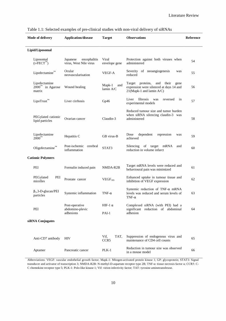
Literature Review
10
Table 1.1: Selected examples of pre-clinical studies with non-viral delivery of siRNAs
Mode of delivery Application/disease Target Observations Reference
Lipid/Liposomal
Liposomal (i-FECT™)
Japanese encephalitis virus, West Nile virus
Viral envelope gene
Protection against both viruses when administered
54
Lipofectamine™ Ocular neovascularisation
VEGF-A Severity of neoangiogenesis was reduced
55
Lipofectamine 2000™ in Agarose matrix
Wound healing Mapk-1 and lamin A/C
Target proteins, and their gene expression were silenced at days 14 and 21(Mapk-1 and lamin A/C)
56
LipoTrust™ Liver cirrhosis Gp46 Liver fibrosis was reversed in experimental models
57
PEGylated cationic lipid particles
Ovarian cancer Claudin-3
Reduced tumour size and tumor burden when siRNA silencing claudin-3 was administered
58
Lipofectamine 2000™
Hepatitis C GB virus-B Dose dependent repression was achieved
59
Oligofectamine™ Post-ischemic cerebral inflammation
STAT3 Silencing of target mRNA and reduction in volume infarct
60
Cationic Polymers
PEI Formalin induced pain NMDA-R2B Target mRNA levels were reduced and behavioural pain was minimized
61
PEGylated PEI micelles
Prostate cancer VEGF165 Enhanced uptake in tumour tissue and inhibition of VEGF expression
62
β1,3-D-glucan/PEI
particles Systemic inflammation TNF-α
Systemic reduction of TNF-α mRNA
levels was reduced and serum levels of
TNF-α
63
PEI Post-operative abdomino-plevic adhesions
HIF-1 α
PAI-1
Complexed siRNA (with PEI) had a significant reduction of abdominal adhesion
64
siRNA Conjugates
Anti-CD7 antibody HIV Vif, TAT, CCR5
Suppression of endogenous virus and maintenance of CD4 cell counts
65
Aptamer Pancreatic cancer PLK-1 Reduction in tumour size was observed in a mouse model
66
Abbreviations: VEGF: vascular endothelial growth factor; Mapk-1: Mitogen-activated protein kinase 1; GP: glycoprotein; STAT3: Signal
transducer and activator of transcription 3; NMDA-R2B: N-methyl-D-aspartate receptor type 2B; TNF-α: tissue necrosis factor α; CCR5: C-
C chemokine receptor type 5; PLK-1: Polo-like kinase 1; Vif: virion infectivity factor; TAT: tyrosine aminotransferase.
Literature Review
11
RNAi. Currently, the use of non-viral vector systems is being employed to improve the efficacy of
siRNA delivery and to improve targeting to tissues of interest. Table 1.1 summarizes selected
examples in pre-clinical trials to date. Overall, while clinical trials have been conducted, none of these
trials have demonstrated the full efficacy of this therapy. As previously discussed, most trials are in
their formative stages, while others have been terminated or discontinued. It becomes imperative;
therefore, that optimisation of delivery, together with careful selection of target gene, is addressed
before translation to the clinic.
1.5 RNAi Delivery through Scaffolds
Many biomaterials used for controlled drug delivery can also be used to fabricate scaffolds. Release
from the scaffold occurs by combinatorial processes of polymer degradation and vector diffusion. A
key requirement associated with the encapsulation of RNAi and non-viral vectors (if any) is that the
methods employed to fabricate the scaffold is compatible with the integrity of the biomolecules and
their vectors. Methods can involve high temperatures, organic solvents and the generation of free
radicals or shear stresses that may damage the payload. Even if the nucleic acid (pDNA or RNA) is
stably encapsulated, the nucleic acid can still be damaged by the degradation products of a vector. The
release kinetics of encapsulated RNAi from tissue engineering scaffolds is also reliant upon a number
of factors such as the concentration of the scaffold, its degree of cross-linking, and the scaffold
material which can make its degradation responsive to pH, temperature, cellular enzymatic products
and/or hydrolysis. This degradation rate can influence the time-course release of the embedded RNAi
as the scaffold loses volume and its products become metabolised by surrounding tissue. Furthermore,
charge interaction between the scaffold material and the RNAi which can be further encapsulated
using a delivery vector can affect the release 71
.
Although the advent of RNAi based-therapy is relatively recent, there have; nevertheless, been
numerous efforts to deliver this therapy via scaffolds and hierarchical structures. Several polymeric
materials are utilized in this field; and for convenience, both natural and synthetic polymer based
scaffold will be discussed, which are also summarized in Table 1.2. Furthermore, these materials can
be manipulated to obtain a wide range of biomaterials with different structural properties and
function-based morphologies; and therefore this discussion will explore the various types of scaffold
attainable.
1.5.1 Hydrogels
Hydrogels are formed by crosslinking or self-assembly of a variety of natural or synthetic hydrophilic
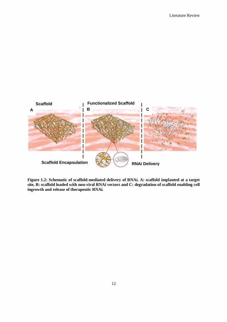
Literature Review
12
Figure 1.2: Schematic of scaffold-mediated delivery of RNAi. A: scaffold implanted at a target
site, B: scaffold loaded with non-viral RNAi vectors and C: degradation of scaffold enabling cell
ingrowth and release of therapeutic RNAi.
Scaffold Functionalized Scaffold
Scaffold Encapsulation RNAi Delivery
A B C
Literature Review
13
polymers to produce structures that contain over 90% water. Many natural materials, such as collagen
or fibrin, have been widely used as matrix components for the formulation of hydrogels. These
hydrogels have been used in tissue engineering applications due to their innate cellular interactions
and cell-mediated degradation. Synthetic materials, such as poly (ethylene glycol) (PEG), lack
intrinsic cellular signals yet allow for greater manipulation of the hydrogel properties relative to
natural materials 67, 68
. Hydrogels can be fabricated ex vivo for subsequent implantation, or can be
formed in situ allowing for a less-invasive approach via catheter delivery. In addition, they can act as
a delivery vehicle for implanted cells and many studies have encapsulated cells within hydrogels prior
to gelation with minimal effects on cell viability 69, 70
. Degradation of hydrogels can be tailored to
occur through a variety of mechanisms including digestion by cell secreted enzymes and hydrolysis,
and can be manipulated by the size of the interconnecting pore network within the hydrogel, ion
exchange, and/or the strength of crosslinking interactions. Therapeutic release from a hydrogel is
dependent upon the physical structure of the hydrogel, its degradation, and interactions between the
matrix of the hydrogel, nucleic acids and/or non-viral vector. In general, hydrogels produce high
encapsulation efficiencies with release occurring through either diffusion alone (non-biodegradable
hydrogels), or through a combination of hydrogel degradation and payload diffusion.
The use of hydrogels as a reservoir of RNAi has been illustrated in many studies due to their potential
as an injectable therapeutic. Their use in this respect has been demonstrated by Krebs et al.71
who
employed collagen hydrogels, photo-crosslinked alginate hydrogels, and calcium crosslinked alginate
hydrogels as reservoirs of siRNA to silence GFP expression. The silencing efficacy of siRNA released
into supernatant and applied to cells stably expressing GFP and the silencing effect on these cells
stably expressing GFP embedded within the hydrogels was investigated. A point to note in this
particular study is that serum deprivation conditions were employed which does limit the translation
of this technology to a pre-clinical setting if the objective is to deliver RNAi in vivo. Regardless, when
the hydrogels were loaded with naked siRNA, they offered maintenance of its bioactivity even after
six days. The authors reported that silencing of the reporter gene was even more pronounced at six
days than at three days, with greater than 80% silencing. An additional investigation of this work was
complexing the siRNA with PEI and chitosan to offer better protection in vivo and also to delay its
release from the hydrogel due to charge interaction of the positively charged PEI/chitosan with
negatively charged alginate. However, the remaining bioactivity of this siRNA was not determined.
Cells embedded within the hydrogels also had prolonged silencing of GFP expression at six days,
particularly when embedded in a collagen hydrogel. This study demonstrated the efficacy of loading
siRNA into hydrogel reservoirs to enable a prolonged silencing and also elucidated the electrostatic
effect of using a vector and siRNA and embedding within a hydrogel. The influence of the
Literature Review
14
electrostatic charge between PEI and chitosan charged siRNA complexes mirrored that of hydrogel
mediated delivery of pDNA complexes 72
, showing that through this parameter there is further control
over the release rate of a therapeutic payload.
Combinatorial approaches in delivering both pDNA to transduce the expression of a gene of interest
while silencing that of another using RNAi is an approach that is gaining popularity as knowledge of
cellular events accumulates. A combinatorial scaffold approach has been reported by Singh et al.
albeit where the pDNA was also delivered as a reporter of siRNA gene silencing 73
. In their study,
hydrogels were constructed of Dextran vinyl-sufone (Dextran-VS) crosslinked with tetra-functional
PEG thiol (PEG4SH), and PEG diacrylate, also crosslinked with PEG4SH. These hydrogel scaffolds
were impregnated with a chemoattractant biomolecule; MIP3α, and within the hydrogel; poly (lactic-
co-glycolic acid) (PLGA) microparticles encapsulating siRNA against interleukin10 (IL-10) and
surface-coated with pDNA-PEI complexes, were embedded. Although this study also focused on the
chemoattractant potential of the scaffold, examination of the gene silencing ability of this platform
using RT-PCR to analyse gene expression of IL-10 found that primary antigen expressing cells
(APCs) had up to 80% silencing with 10% (w/v) crosslinked dextran hydrogels five days post
transfection which was comparable to microparticles alone. Furthermore, a trend was observed in that
silencing of IL-10 was reduced when the concentration of the dextran hydrogel was increased (20 and
30%) indicating that the release and permeability kinetics of the hydrogel affects the uptake of
siRNA.
1.5.2 Sponges
The use of a sponge-based construct to deliver siRNA has been reported. Crosslinked collagen
sponges as reservoirs of siRNA complexes to silence SNAIL1 expression have been studied 74
and
these platforms have previously been used as reservoirs of pDNA 75
. A crosslinked collagen sponge
served as a reservoir of complexed siRNA directed toward the silencing of SNAIL1 with a cumulative
release of 65% at day seven. Although no significant effect was observed at day one and two when
seeded with 3T3 murine fibroblasts, at seven days a significant reduction in SNAIL1 expression was
observed in cells incubated with release supernatants from anti-SNAIL1 siRNA loaded scaffolds
when compared to negative control siRNA loaded scaffolds. Again, similar to effects observed with
hydrogels, sponge-based scaffolds were shown to prolong the silencing effect of siRNA whilst acting
as a support for cellular attachment and growth.
Although most of the efforts discussed have reported down-regulating reporter genes or a certain gene
of interest, recent reports in the field have illustrated the use of siRNA loaded matrices in directing
stem cell differentiation. Andersen et al. reported that poly (ε-caprolactone) (PCL) scaffolds
Literature Review
15
functionalized with a siRNA nano-particulate can deliver a range of target siRNAs to enhance
differentiation of hMSCs in vitro and in vivo and create a spatio-temporal pattern to generate areas of
defined differentiation 76
. Previously, the authors had coated surfaces with lyophilized siRNA
complexed with TransIT-TKO™
, a commercial, lipid based delivery vector 77
. In vitro, delivering a
lyophilized polymer/lipid-based nano-particulate with EGFP silencing siRNA, embedded within a
structured PCL scaffold, to cells stably expressing EGFP, achieved up to 60% silencing when
compared to non-functionalized scaffolds 76
. Furthermore, delivery of either BCL212 or TRIB2
silencing siRNAs from the structured PCL scaffolds, reduced expression of these genes by 41% and
51% respectively, after 72 hours, which was quantified using q-PCR. The silencing of BCL212 and
TRIB2 was postulated to direct osteogenic and adipogenic differentiation of hMSCs on scaffolds,
which had been reported in previous studies using monolayer cultures 78, 79
. The researchers found,
that after seven days in adipogenic differentiation media, hMSCs on scaffolds with TRIB2 siRNA had
significantly increased expression of adipogenic markers when compared with controls. Similarly,
when hMSCs were subjected to osteogenic differentiation, scaffolds coated with BCL2L2 siRNA
particles had the greatest expression of osteogenic markers at day seven. The authors then translated
this platform of a PCL scaffold loaded with target siRNAs (TRIB2 and BCL2L2) and non-targeting
siRNA nanoparticles, seeded with hMSCs, into non-obese diabetic/severe combined immunodeficient
(NOD/SCID) mice. The in vivo results demonstrated that TRIB2 and BCL2L2 targeted siRNA
initiated adipogenic and osteogenic differentiated respectively but could not drive terminal
differentiation as a sole differentiation cue.
Similar results were observed in an in vitro experiment in which the authors reported that the TRIB2
siRNA- coated scaffold stained positive for markers of adipogenic differentiation and BCL2L2
siRNA scaffolds strongly exhibited markers for osteogenic differentiation compared to controls 76
.
Gradient controlled differentiation was also controlled using this scaffold both in vitro and in vivo. In
vitro hMSCs were seeded on a scaffold containing TRIB2 siRNA on one side and BCL2L2 siRNA on
the other half. Measurement of mRNA levels elucidated that the expression of one adipogenic marker
(AP2) was four-fold upregulated in the TRIB2 siRNA-coated part, and ALP was 45% upregulated in
the BCL2L2 siRNA coated part. In vivo, staining showed that the cells could still be induced to
commit to alternative differentiation (osteogenic and adipogenic) in specific locations within the same
implant by placing the different siRNAs in distinct locations.
1.5.3 Fibers
Choice of scaffold morphology and architecture for a specific clinical target is dictated by the end
application, desired release, and mimicry of the native tissue in order to guide neighbouring cell
Literature Review
16
infiltration and differentiation 80, 81
. Hence researchers in the field have developed various constructs
with varying architecture and form.
One such study employed PCL nano-fibers as reservoirs of both naked and complexed siRNA against
glyceraldehydes 3 – phosphate dehydrogenase (GADPH) 82
some of which were PEGylated. Up to
80% silencing of GADPH was observed when release supernatants were added to HEK cells up to 30
days post incubation. When cells were directly seeded on scaffolds impregnated with naked siRNA
and siRNA complexed with a commercial transfection reagent (TransIT-TKO™
), GAPDH silencing
was achieved with naked siRNA but to an average of ~20% efficiency; whereas the impregnation of
complexed siRNA impregnated scaffolds attained a silencing efficiency of ~75% and comparable
with a bolus delivery of complexed siRNA after 96 hours. Although this study realises the concept
that siRNA delivery via a reservoir matrix can be improved using a complexing agent, the full extent
to its use as a therapeutic has not been demonstrated due to the lack of time points and duration of the
study during which the cells were seeded on the nano-fiber scaffolds.
1.5.4 Microspheres
Nucleic acids (such as RNAi) can be introduced into microspheres during the fabrication process in
which a water-in-oil-in-water multiple emulsion/solvent evaporation or extraction technique is
employed. Preliminary studies have shown that the molecule size and structure (bases etc.), the
dosage of loading and the size of microspheres, greatly affects the encapsulation efficiency and
release properties 83
. Higher loading is generally associated with a larger size, but is independent of
oligonucleotide length 83
.
The in vitro release profile of antisense oligonucleotides from microspheres has been shown to be
affected by the size of the spheres, drug loading, oligonucleotide length and molecular weight of the
polymer. Small microspheres (1-2 μm) fabricated from PLGA have been reported to release most
entrapped oligonucleotide within four days, compared with 40 days for larger spheres (10- 20 μm) 84
.
In the same report a triphasic release was observed with larger PLGA spheres: a burst release in the
first 48 hours, attributed to the rapid release of the oligonucleotides present on or close to the
microsphere surface; a sustained release phase (about 25 days) in which oligonucleotide diffusion
through pores present in the microsphere‘s shell material occurred; and a final more rapid release
phase (up to a complete release at about 60 days) due to degradation of the polymer that accelerated
oligonucleotide release. In the case of smaller spheres, increased surface area to volume ratio results
in a greater concentration of oligonucleotides at or near to the surface with subsequent rapid release.
Another factor to consider is the mass of oligonucleotides loaded into microspheres as there have been
reports that an increase in nucleotide loading results in a higher burst effect and a more rapid release
Literature Review
17
rate in PEG/PLGA blend microspheres 85
. The release profile is only slightly dependent on nucleic
acid length, with a release rate that increases as the nucleic acid molecular weight decreases. Finally, a
high burst effect and a rapid in vitro release rate have been attributed to microsphere porosity. Pores
on the microsphere surface occur when an osmotically active agent is added to the internal aqueous
phase. The use of an osmotic agent, such as NaCl, into the external aqueous phase allows one to
reduce microsphere surface porosity, thus reducing the initial burst effect and decelerating in vitro
release rate 86
.
One study has reported the encapsulation of double stranded oligonucleotides within PLGA
microspheres 87
. In this study; microspheres encapsulating a decoy dsRNA against the transcription
factor-κB (NF-κB) showed a negligible burst effect and had exhibited a gradual release profile, with
complete release after about 40 days. Khan et al. have encapsulated double-stranded siRNA into
PLGA microspheres and found that in vitro the release of siRNA was much slower and had a very
limited burst effect compared with that obtained with a single-stranded RNA 88
. The difference
between the release profiles was ascribed to the different chemical nature of the two nucleic acids,
while only a modest effect was attributed to variation in molecular weight. Increase in oligonucleotide
encapsulation within PLGA or poly (L-lactide) (PLL) microspheres, as well as optimization of their in
vitro release properties, has been achieved by using cationic additives which were able to complex the
nucleic acid within the matrix. Rather than releasing siRNA all at once, the microspheres protect
against serum nucleases and release a slow continual supply of bioactive siRNA to cells, sustaining
RNAi.
1.5.5 Surface Coatings
Surface functionalization is another approach in reservoir delivery for RNAi and offers much
versatility in translation to current implants and scaffolds being used clinically. Zhang et al. have
delivered shRNA encoding pDNA via a multilayer reservoir of pDNA/calcium phosphate scaffolds 89
.
Briefly, polyelectrolyte multilayered films were prepared on glass cover slips, and alternating layers
of poly (L-lysine) and nanoparticles were built up by alternated immersions. The use of one and six
alternate layers was investigated with shRNA for the silencing of osteopontin expression and another
for the silencing of osteocalcin expression in vitro. Employing both RT-PCR and
immunocytochemistry the authors reported that qualitatively; the expression of osteopontin and
osteocalcin had greater silencing with six alternate layers in the platform than it had with one layer.
Furthermore, the overall use of a multilayer reservoir, silenced osteopontin and osteocalcin compared
to negative controls and free dissolved shRNA pDNA.
Literature Review
18
Many studies have employed biomaterial coatings harnessing RNAi to improve their performance or
to elicit a therapeutic response. Takahashi et al. have reported a PEG methyl ether (PEGM) hydrogel
coating for loading siRNA targeted toward a mammalian target of rapamycin (mTOR) 90
. The authors
observed promising results in vitro wherein mTOR expression was silenced up to seven days. The
authors also investigated the effect of varying the concentration of the hydrogels and found that
prolonged and sustained release could be achieved with a higher concentration of hydrogel. However,
when investigated as a surface coating for subcutaneous implants in a mouse model, a significant
reduction on the formation of a fibrous capsule around the implant was not achieved. This is due to
the choice of siRNA, although TGF-β1 silencing siRNA was also investigated, or, alternatively, may
be due to a response to the hydrogel material. A limitation in this study is that a control implant
without any hydrogel coating was not included hence the results cannot be interpreted.
Anderson et al. have also employed lyophilized TransIT-TKO™
-siRNA complexes as coatings for
silencing of reporter EGFP and TNF-α 77
. This coating, which additionally retains bioactivity ~two
months under room temperature storage, was effective at silencing EGFP expression (~85 %) but,
more importantly, was effective at silencing TNF-α expression in a macrophage cell line. This
approach could be harnessed to effectively mediate a macrophage response to biomedical devices and
other tissue engineering approaches.
One of the most common types of device currently in clinical use are stents and their emergence as
reservoirs of anti-mitotic drug delivery is well established. Recent investigations have not only
harnessed their capability as reservoirs of nucleic acid delivery agents 91
but their use as a reservoir of
RNAi has also been reported. In one such study 92
, bare metal stents were coated with a cationized
pullalan hydrogel loaded with siRNA targeted at silencing matrix-metalloproteinase-2 (MMP-2)
expression in vascular cells. The cationized matrix retained siRNA for an extended period. This
retention was due to the charge interaction of the matrix and siRNA. When implanted into a rabbit
model of neointimal hyperplasia, although MMP-2 expression was not significantly silenced when
compared to a scrambled control, proMMP-2 expression was silenced up to 28 ± 13%.
1.5.6 Topical Delivery
From a tissue engineering perspective, most scaffolds aim to occupy an anatomical location to add
structural support and/or deliver a therapeutic agent. However, some areas of tissue engineering aim
to employ biomaterials as active dressings for the delivery of therapeutic agents both topically and
transdermally. Although not traditionally tissue engineering applications the therapeutic delivery of
siRNA across the skin epithelium has been reported by Takanashi et al. 93
where siRNA was delivered
using a commercially available lipid/alcohol-based formulation (‗GeneCream™
‘). After dermal
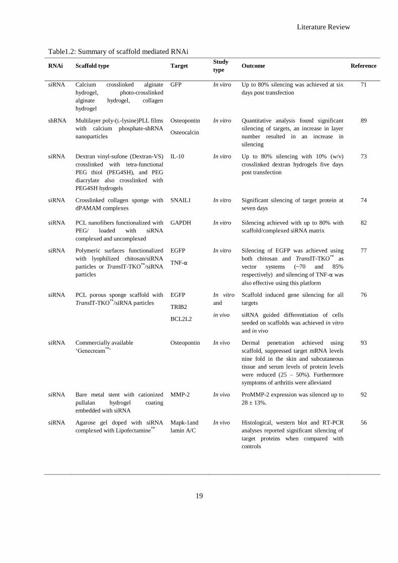
Literature Review
19
Table1.2: Summary of scaffold mediated RNAi
RNAi Scaffold type Target Study
type Outcome Reference
siRNA Calcium crosslinked alginate
hydrogel, photo-crosslinked
alginate hydrogel, collagen
hydrogel
GFP In vitro Up to 80% silencing was achieved at six
days post transfection
71
shRNA Multilayer poly-(L-lysine)PLL films
with calcium phosphate-shRNA
nanoparticles
Osteopontin
Osteocalcin
In vitro Quantitative analysis found significant
silencing of targets, an increase in layer
number resulted in an increase in
silencing
89
siRNA Dextran vinyl-sufone (Dextran-VS)
crosslinked with tetra-functional
PEG thiol (PEG4SH), and PEG
diacrylate also crosslinked with
PEG4SH hydrogels
IL-10 In vitro Up to 80% silencing with 10% (w/v)
crosslinked dextran hydrogels five days
post transfection
73
siRNA Crosslinked collagen sponge with
dPAMAM complexes
SNAIL1 In vitro Significant silencing of target protein at
seven days
74
siRNA PCL nanofibers functionalized with
PEG/ loaded with siRNA
complexed and uncomplexed
GAPDH In vitro Silencing achieved with up to 80% with
scaffold/complexed siRNA matrix
82
siRNA Polymeric surfaces functionalized
with lyophilized chitosan/siRNA
particles or TransIT-TKO™/siRNA
particles
EGFP
TNF-α
In vitro Silencing of EGFP was achieved using
both chitosan and TransIT-TKO™ as
vector systems (~70 and 85%
respectively) and silencing of TNF-α was
also effective using this platform
77
siRNA PCL porous sponge scaffold with
TransIT-TKO™/siRNA particles
EGFP
TRIB2
BCL2L2
In vitro
and
in vivo
Scaffold induced gene silencing for all
targets
siRNA guided differentiation of cells
seeded on scaffolds was achieved in vitro
and in vivo
76
siRNA Commercially available
‗Genecream™‘
Osteopontin In vivo Dermal penetration achieved using
scaffold, suppressed target mRNA levels
nine fold in the skin and subcutaneous
tissue and serum levels of protein levels
were reduced (25 – 50%). Furthermore
symptoms of arthritis were alleviated
93
siRNA Bare metal stent with cationized
pullalan hydrogel coating
embedded with siRNA
MMP-2 In vivo ProMMP-2 expression was silenced up to
28 ± 13%.
92
siRNA Agarose gel doped with siRNA
complexed with Lipofectamine™
Mapk-1and
lamin A/C
In vivo Histological, western blot and RT-PCR
analyses reported significant silencing of
target proteins when compared with
controls
56
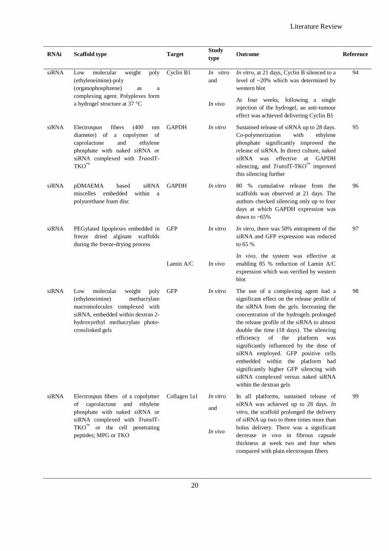
Literature Review
20
RNAi Scaffold type Target Study
type Outcome Reference
siRNA Low molecular weight poly
(ethyleneimine)-poly
(organophosphzene) as a
complexing agent. Polyplexes form
a hydrogel structure at 37 °C
Cyclin B1 In vitro
and
In vivo
In vitro, at 21 days, Cyclin B silenced to a
level of ~20% which was determined by
western blot
At four weeks, following a single
injection of the hydrogel, an anti-tumour
effect was achieved delivering Cyclin B1
94
siRNA Electrospun fibers (400 nm
diameter) of a copolymer of
caprolactone and ethylene
phosphate with naked siRNA or
siRNA complexed with TransIT-
TKO™
GAPDH In vitro Sustained release of siRNA up to 28 days.
Co-polymerization with ethylene
phosphate significantly improved the
release of siRNA. In direct culture, naked
siRNA was effective at GAPDH
silencing, and TransIT-TKO™ improved
this silencing further
95
siRNA pDMAEMA based siRNA
miscelles embedded within a
polyurethane foam disc
GAPDH In vitro 80 % cumulative release from the
scaffolds was observed at 21 days. The
authors checked silencing only up to four
days at which GAPDH expression was
down to ~65%
96
siRNA PEGylated lipoplexes embedded in
freeze dried alginate scaffolds
during the freeze-drying process
GFP
Lamin A/C
In vitro
In vivo
In vitro, there was 50% entrapment of the
siRNA and GFP expression was reduced
to 65 %
In vivo, the system was effective at
enabling 85 % reduction of Lamin A/C
expression which was verified by western
blot
97
siRNA Low molecular weight poly
(ethyleneimine) methacrylate
macromolecules complexed with
siRNA, embedded within dextran 2-
hydroxyethyl methacrylate photo-
crosslinked gels
GFP In vitro The use of a complexing agent had a
significant effect on the release profile of
the siRNA from the gels. Increasing the
concentration of the hydrogels prolonged
the release profile of the siRNA to almost
double the time (18 days). The silencing
efficiency of the platform was
significantly influenced by the dose of
siRNA employed. GFP positive cells
embedded within the platform had
significantly higher GFP silencing with
siRNA complexed versus naked siRNA
within the dextran gels
98
siRNA Electrospun fibers of a copolymer
of caprolactone and ethylene
phosphate with naked siRNA or
siRNA complexed with TransIT-
TKO™ or the cell penetrating
peptides; MPG or TKO
Collagen 1a1 In vitro
and
In vivo
In all platforms, sustained release of
siRNA was achieved up to 28 days. In
vitro, the scaffold prolonged the delivery
of siRNA up two to three times more than
bolus delivery. There was a significant
decrease in vivo in fibrous capsule
thickness at week two and four when
compared with plain electrospun fibers
99

Literature Review
21
RNAi Scaffold type Target Study
type Outcome Reference
siRNA Chitosan nanoparticles
encapsulating siRNA embedded
within electrospun PLGA fibers
(diameter ~ 900 nm)
EGFP In vitro The release profile of siRNA from the
platform was significantly dependent of
the pH of the release buffer. 50% EGFP
silencing occurred after 48 hours
100
Abbreviations: GFP: green fluorescent protein; Dextran-VS: Dextran vinyl-sufone: Dextran-VS; PEG: poly (ethylene
glycol); IL-10: interleukin 10; dPAMAM: partially degraded poly (amino amine); PCL: poly (ε-caprolactone); GAPDH:
glyceraldehydes 3 – phosphate dehydrogenase; EGFP: enhanced green fluorescent protein; TNF-α: tissue necrosis factor α;
MMP-2: matrix-metalloproteinase.
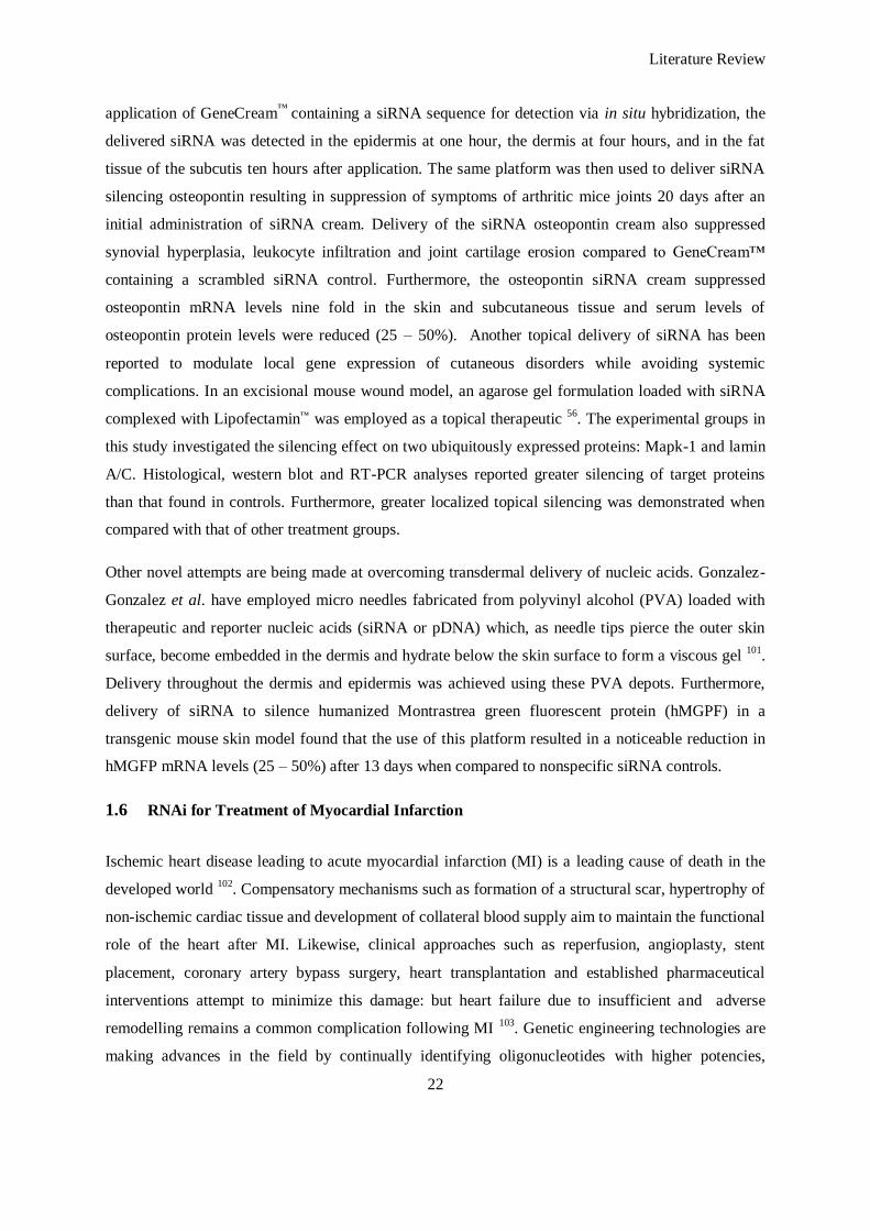
Literature Review
22
application of GeneCream™
containing a siRNA sequence for detection via in situ hybridization, the
delivered siRNA was detected in the epidermis at one hour, the dermis at four hours, and in the fat
tissue of the subcutis ten hours after application. The same platform was then used to deliver siRNA
silencing osteopontin resulting in suppression of symptoms of arthritic mice joints 20 days after an
initial administration of siRNA cream. Delivery of the siRNA osteopontin cream also suppressed
synovial hyperplasia, leukocyte infiltration and joint cartilage erosion compared to GeneCream™
containing a scrambled siRNA control. Furthermore, the osteopontin siRNA cream suppressed
osteopontin mRNA levels nine fold in the skin and subcutaneous tissue and serum levels of
osteopontin protein levels were reduced (25 – 50%). Another topical delivery of siRNA has been
reported to modulate local gene expression of cutaneous disorders while avoiding systemic
complications. In an excisional mouse wound model, an agarose gel formulation loaded with siRNA
complexed with Lipofectamin™ was employed as a topical therapeutic 56
. The experimental groups in
this study investigated the silencing effect on two ubiquitously expressed proteins: Mapk-1 and lamin
A/C. Histological, western blot and RT-PCR analyses reported greater silencing of target proteins
than that found in controls. Furthermore, greater localized topical silencing was demonstrated when
compared with that of other treatment groups.
Other novel attempts are being made at overcoming transdermal delivery of nucleic acids. Gonzalez-
Gonzalez et al. have employed micro needles fabricated from polyvinyl alcohol (PVA) loaded with
therapeutic and reporter nucleic acids (siRNA or pDNA) which, as needle tips pierce the outer skin
surface, become embedded in the dermis and hydrate below the skin surface to form a viscous gel 101
.
Delivery throughout the dermis and epidermis was achieved using these PVA depots. Furthermore,
delivery of siRNA to silence humanized Montrastrea green fluorescent protein (hMGPF) in a
transgenic mouse skin model found that the use of this platform resulted in a noticeable reduction in
hMGFP mRNA levels (25 – 50%) after 13 days when compared to nonspecific siRNA controls.
1.6 RNAi for Treatment of Myocardial Infarction
Ischemic heart disease leading to acute myocardial infarction (MI) is a leading cause of death in the
developed world 102
. Compensatory mechanisms such as formation of a structural scar, hypertrophy of
non-ischemic cardiac tissue and development of collateral blood supply aim to maintain the functional
role of the heart after MI. Likewise, clinical approaches such as reperfusion, angioplasty, stent
placement, coronary artery bypass surgery, heart transplantation and established pharmaceutical
interventions attempt to minimize this damage: but heart failure due to insufficient and adverse
remodelling remains a common complication following MI 103
. Genetic engineering technologies are
making advances in the field by continually identifying oligonucleotides with higher potencies,
Literature Review
23
particularly RNAi; more effective cellular targets, typically in signalling pathways; and delivery
approaches with the next generation of drug platforms. These advances have enabled detailed
genomic investigations of endogenous miR expression profiles in impaired myocardium yielding new
aberrantly expressed gene targets for silencing using RNAi technology. In addition, an increased
understanding of surface-expressed markers offers the opportunity to develop more specific, cell-
directed delivery technologies for genetic payloads 104
.
1.6.1 siRNA
Circulating inflammatory cells, including neutrophils and monocytes/macrophages, arrive to the
infarct site in the sub-acute phase following MI, initiated by complement components from ischemic
tissue, and release of cytokines 105
that are triggered by mechanical deformation, ischemic stimuli,
reactive oxygen species (ROS) and amplification of cytokine pathways. This inflammatory response
is initiated significantly within an hour of MI, peaks at two weeks post-MI, and this response tapers
off as inflammatory cells disappear from the infarct site; modulating fibroblast phenotype transition
and activation of proteinases that remodel the myocardium during the remodeling response within
four weeks following MI. These fibroblasts play a significant role in this remodeling phase, laying
down a fibrous collagenous scar to compensate for the loss of myocardium due to MI 106
.
The clinical focus of MI nucleic acid therapy research has been on upregulation or overexpression of
therapeutic genes, whereas siRNA delivery studies have reached the clinic only in the areas of cancer
5 and AMD
47, among other targets
4. Currently RNAi research in the pathogenesis of MI is at the
investigatory stage in which tissue is being profiled for overexpressed and underexpressed miRs.
Considering the infancy of this research field, the large majority of the upcoming studies related to
RNAi addressing the different phases following MI discussed are in pre-clinical animal models.
Manipulating inflammation is an approach that has gained particular interest post-MI. Using lentiviral
particles, Krishnamurthy et al. knocked down human stabilizing protein (HuR – known to stabilize
mRNA of proinflammatory cytokines) by intramyocardial delivery of HuR-specific shRNA into the
infarct borderzone at five different locations immediately following MI 107
. This was followed by
daily intravenous injections of the same shRNA for one week, and resulted in a significant reduction
in fibrotic area and improved left ventricular function at day 28. HuR can be suppressed by anti-
inflammatory interleukin 10 (IL-10); therefore, a parallel strategy with IL-10 knockout mice was
studied with similar results which the authors ascribed to the HuR knockdown mimicking the anti-
inflammatory effects of IL-10.

Literature Review
24
Another anti-inflammatory approach by Van Tassell et al. 108
aimed at inhibiting the myeloid
differentiation factor 88 (MyD88), an innate adaptor protein that binds to Toll/IL-1 receptor
complexes and activates the IL-1 associated kinase complex. Pharmacologic inhibition of MyD88 was
achieved by intraperitoneal administration of ST2825 (which mimics the TIR domain of the MyD88
protein, thereby inhibiting homodimerization), IMG2005 (a synthetic oligopeptide with similar
structure to ST2825) and a siRNA designed to silence MyD88 prior to induction of MI in mice and
seven days following surgery. The authors reported protection of left ventricular dilation but no
measureable reduction in infarct size with any of the silencing approaches (ST2825, IMG2005 and
siRNA). This result can be attributed to the premature endpoint (seven days) as remodeling is still at
an early stage.
A final RNAi intervention of inflammation has also been reported by Mezzaroma et al. This report
describes the formation of a multiprotein complex, termed an inflammasome, necessary for caspase-1
activation and IL-1β release (players in the initial inflammatory response) 106
. The inflammasome is
formed by recruitment of an apoptosis speck-like protein containing a caspase-recruitment domain, by
cryopyrin (an intracellular Nod-like receptor activated in response to tissue injury), which the study
sought to silence using siRNA. Additionally, the authors investigated the silencing of the purinergic
P2X7 receptor which is activated by extracellular ATP and leads to K+ efflux and cryopyrin
activation. P2X7 and cryopyrin inhibition using siRNA prevented the formation of the inflammasome
and limited the infarct size and cardiac enlargement after acute MI. However, the effect of this
treatment on the ejection fraction of the infarcted myocardium following MI has not been reported to
date.
β-blockers are routinely used in the prevention of MI in patients with coronary heart disease and in
treating patients following MI 109
. Although extremely beneficial, they also pose negative side effects
such as aggravation of asthma and peripheral vascular diseases due to antagonism of the β2 adrenergic
receptor (β2AR). Therefore, a highly specific β1 antagonist is desirable for clinical testing, something
which is achievable using siRNA technology. Arnold et al. 110
have reported successful delivery and
functional knockdown of the β1AR using a siRNA/liposomal formulation (1,2-bis-oleoyloxy)-3-
(trimethylammonium) propane, (DOTAP), both in vitro and in treating spontaneously hypertensive
rats and rats subjected to MI. Briefly, one month after hypertensive rats received a single siRNA
dose, a significant decrease (10%) in LV/BW ratio (which signified a decrease in cardiac
hypertrophy) was observed compared with that of saline and scrambled siRNA control animals,
suggesting a capacity of β1 siRNA to inhibit hypertension-related LV-hypertrophy. In the MI model,
siRNA injected intravenously three days prior to induction of MI considerably improved LV function.
Furthermore, the number of apoptotic cells per field reduced significantly with siRNA treatment.
Literature Review
25
These findings alone suggest a potential of RNAi as a next generation pharmaceutical in the case of
specific β-blocking.
Li et al. have employed an adenoviral shRNA as a more direct pharmacological inhibitor of
phosphodiesterase 5a (PDE5a) than tadafil 111
. PDE5a degrades cyclic guanosine monophosphate
(cGMP) by hydrolysis and therefore its inhibition enhances the advantageous effects of cGMP such as
regulation of apoptosis, vasodilation and regulation of ion channel conductance. It has been found that
mice treated with shRNA silencing PDE5a showed significant preservation of ejection fraction and
fractional shortening, and reduction in infarction size and fibrosis. The authors obviated any concerns
with regard the effect of the therapeutic in off target organs (as it would have similar effects as
tadafil), by delivering the shRNA intramyocardially.
Another anti-apoptotic approach looked at inhibiting the Src homology domain 2 (SH2) - containing
tyrosine phopatase-1 (SHP-1) which has a key role in apoptosis and decreases the phosphorylation of
Akt (which exerts a powerful cardioprotective effect after ischemia) 112
. The authors delivered a
siRNA vector targeting SHP-1 directly into the left ventricular wall immediately following MI.
Silencing was confirmed in vivo and, more significantly, a reduction in infarct size and a significantly
higher fractional shortening one and two days after MI with the group treated with siRNA targeting
SHP-1 was evident. The authors concluded that as SHP-1 is required for Fas-mediated cytotoxic
signaling; SHP-1 silencing had an anti-apoptotic effect.
Hypoxia-inducible factor-1 (HIF-1) - a transcription factor consisting of two basic-helix-loop-helix-
PAS transcription factors, HIF-1α and HIF-1β - mediates tissue responses to ischemia and has
garnered interest in RNAi approaches. It promotes the transcription of in excess of 100 genes, notably
vascular endothelial growth factor (VEGF-A), which is beneficial for angiogenesis, and inducible
nitric oxide synthase (iNOS), whose role is being debated as it is likely to have a beneficial effect
(attributed to a decrease in neutrophil and platelet adhesion or vasodilation) or a detrimental effect
(related to the production of free radicals and inactivation of mitochondrial enzymes) 113
. Considering
its pivotal role in these processes, HIF-1α has become a focus of numerous RNAi approaches to
improve recovery following MI; it has been shown to induce therapeutic angiogenesis, limit infarct
size and improve myocardial function after acute coronary occlusion in a transgenic mouse model 113
.
Therefore, increasing HIF-1 expression can be an approach in the treatment of MI and also in the case
of RNAi silencing factors that in turn would normally be inhibiting HIF-1α. One such approach
investigated whether cardiac preconditioning via prolyl-4 hydroxylase-2 silencing (to activate HIF-
1α) would improve cardiac function following MI 114
. Administering a siRNA silencing prolyl-4
hydroxylase-2 intraperitoneally 24 hours prior, to 30 minutes ischemia and 120 minutes reperfusion,
Literature Review
26
the authors reported reduced infarct sizes and a decrease in polymorphonuclear leukocyte infiltration
(associated with diminished chemokine and adhesion molecule expression). Unfortunately, further
analysis revealed the drawback that the murine knockdown target was pro-collagen prolyl-4
hydroxylase-2 rather than HIF prolyl-4 hydroxylase-2, and the authors were not able to conclusively
explain a link between the knockdown of pro-collagen prolyl-4 hydroxylase-2 and HIF-1α
upregulation and the reduced infarct sizes. A more robust strategy was attempted by others using a
shRNA plasmid to silence HIF prolyl-4 hydroxylase-2 whose selection was confirmed by GenBank
database 115
which had not been conducted in the previous similar study 114
. Activity of the shRNA
plasmid, via a reporter luciferase gene was monitored and plasmid-mediated transgene expression was
observed for up to five weeks. Fractional shortening and increased angiogenesis in the infarcted heart
compared with a scrambled sequence was improved at four weeks and was associated with higher
levels of HIF1-α, verified by western blot analysis. The study concluded that RNAi of HIF prolyl-4
hydroxylase-2 increases HIF1-α expression, leading to improved recovery following MI.
1.6.2 miRs Mimicking Nature – miR Manipulation in the Treatment of Myocardial Infarction
Recently, the focus of RNAi approaches has encompassed the use of synthetic sequences to mimic or
silence endogenous miRs that are abruptly deregulated following MI. Van Rooij et al. first described
regulation of miR expression in heart disease by the use of microarray profiling in murine cardiac
hypertrophy/failure 116
. In the same study, selected miR patterns in human cardiomyopathy were
described. miRs are attractive therapeutic targets because of the significant control exercised by
individual miRs or miR clusters over pathways that regulate cardiac hypertrophy, cardiomyocyte
apoptosis, myocardial vascularization, and cardiac fibrosis.
The first report of delivering miRs towards a therapeutic end is the delivery of miR-1, miR-12 and
miR-24, which were revealed by the authors to have been upregulated during ischemic
preconditioning in a mouse model 117
. The authors isolated this cohort of miRs from preconditioned
myocardium and injected them intramyocardially into mice 48 hours prior to ischemia/reperfusion
injury. This miR preconditioning led to an upregulation of eNOS, HSP70 and the HSP70 transcription
factor HSF-1. Furthermore, the treatment group had a significantly reduced infarct size when
compared to the negative controls. The beneficial miRs in this study conflict with papers that have
since been published. For instance, miR-24 is shown to be slightly upregulated in response to
ischemic preconditioning and the authors postulate that miR-24 could be a therapeutic miR although
this is not conclusively discussed in the results. In a more recent study, miR-24 has been shown
upregulated in endothelial cells following cardiac ischemia. Forced overexpression of miR-24 by
delivering pre-miR-24 mimics in vitro induced apoptosis 118
. Furthermore, the administration of
antagomiRs targeting miR-24, zero and two days following MI resulted in an improved capillary
Literature Review
27
density and smaller significant infarct size 14 days after MI and improved fractional shortening
compared to the scrambled control. Although the previous study does demonstrate a reduced infarct
size with an ischemic preconditioning cocktail of miRs, one must fully elucidate the role of each
individual miR to achieve a therapeutic goal.
There exists a family of miRs, coined myomiRs, which are the three highly expressed muscle-specific
miRs: miR-208a, -208b, and -499, shown to control myosin heavy chain isoform expression 119
.
Specifically, miR-208 is a miR encoded within an intron of the Myh6 (α-myosin heavy chain), which
is downregulated in response to cardiac injury and is essential for the expression of Myh7 (β-myosin
heavy chain) and Myh7b (a closely related isoform) 120
. AntagomiRs, in a locked nucleic acid (LNA)
format to target miR-208a, delivered subcutaneously during hypertension-induced heart failure,
hindered myosin switching and cardiac remodeling while improving cardiac function, overall health
and survival 121
. Although the study monitored the survival of a hypertension-induced heart failure
model, application of this treatment in the context of MI appears promising.
miR-126 has been demonstrated to be downregulated within the infarcted myocardium but
upregulated at the periphery of the infarct (borderzone). A crucial function of miR-126 in the
development of the fetal vascular system was documented in a study showing that miR-126 knockout
in mice impaired vascularization after experimental MI 122
, suggesting that miR-126 directs both
initial vascular development and angiogenesis. miR-126 expression occurs exclusively in endothelial
cells, whereas miR-210 is expressed in cardiomyocytes, where it is induced by HIF1-α, and is
associated with increased survival/decreased apoptosis. Delivery of a minicircle DNA vector
encoding the miR-210 precursor decreased ventricular remodeling, improved ventricular ejection
performance, diminished cardiomyocyte apoptosis and enhanced myocardial neovascularization
following MI in mice 123
. Conversely, Bonauer et al. described an increase in expression of miR-92a
after experimental MI and observed that it decreased neovascularization. Consequently,
administration of a miR-92a antagomiR showed increased border zone vascularization, diminished
infarct size and enhanced post-infarction ventricular function following MI in a mouse model 124
.
Typically, myocardial fibrosis comes after significant cardiomyocyte death due to MI, and several
miRs (fibromiRs) modulate cardiac fibrosis. Expression of miR-29 is higher in cardiac fibroblasts
than in cardiomyocytes, but the expression of miR-29B in cardiac fibroblasts is downregulated in
infarct border zone of infarcted hearts 125
. By contrast, miR-21 expression is increased in the border
zone of infarcted myocardium and in the fibroblasts of failing human hearts, where miR-21 enhances
fibroblast survival by oppressing programmed cell death-4 (PDCD4) and sprouty homolog-1, and
promotes myocardial fibrosis by activating the extracellular signal-regulated and mitogen-activated
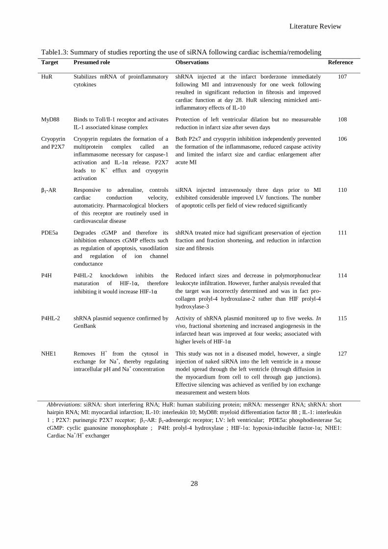
Literature Review
28
Table1.3: Summary of studies reporting the use of siRNA following cardiac ischemia/remodeling
Target Presumed role Observations Reference
HuR Stabilizes mRNA of proinflammatory
cytokines
shRNA injected at the infarct borderzone immediately
following MI and intravenously for one week following
resulted in significant reduction in fibrosis and improved
cardiac function at day 28. HuR silencing mimicked anti-
inflammatory effects of IL-10
107
MyD88 Binds to Toll/Il-1 receptor and activates
IL-1 associated kinase complex
Protection of left ventricular dilation but no measureable
reduction in infarct size after seven days
108
Cryopyrin
and P2X7
Cryopyrin regulates the formation of a
multiprotein complex called an
inflammasome necessary for caspase-1
activation and IL-1α release. P2X7
leads to K+ efflux and cryopyrin
activation
Both P2x7 and cryopyrin inhibition independently prevented
the formation of the inflammasome, reduced caspase activity
and limited the infarct size and cardiac enlargement after
acute MI
106
β1-AR Responsive to adrenaline, controls
cardiac conduction velocity,
automaticity. Pharmacological blockers
of this receptor are routinely used in
cardiovascular disease
siRNA injected intravenously three days prior to MI
exhibited considerable improved LV functions. The number
of apoptotic cells per field of view reduced significantly
110
PDE5a Degrades cGMP and therefore its
inhibition enhances cGMP effects such
as regulation of apoptosis, vasodilation
and regulation of ion channel
conductance
shRNA treated mice had significant preservation of ejection
fraction and fraction shortening, and reduction in infarction
size and fibrosis
111
P4H P4HL-2 knockdown inhibits the
maturation of HIF-1α, therefore
inhibiting it would increase HIF-1α
Reduced infarct sizes and decrease in polymorphonuclear
leukocyte infiltration. However, further analysis revealed that
the target was incorrectly determined and was in fact pro-
collagen prolyl-4 hydroxulase-2 rather than HIF prolyl-4
hydroxylase-3
114
P4HL-2 shRNA plasmid sequence confirmed by
GenBank
Activity of shRNA plasmid monitored up to five weeks. In
vivo, fractional shortening and increased angiogenesis in the
infarcted heart was improved at four weeks; associated with
higher levels of HIF-1α
115
NHE1 Removes H+ from the cytosol in
exchange for Na+, thereby regulating
intracellular pH and Na+ concentration
This study was not in a diseased model, however, a single
injection of naked siRNA into the left ventricle in a mouse
model spread through the left ventricle (through diffusion in
the myocardium from cell to cell through gap junctions).
Effective silencing was achieved as verified by ion exchange
measurement and western blots
127
Abbreviations: siRNA: short interfering RNA; HuR: human stabilizing protein; mRNA: messenger RNA; shRNA: short
hairpin RNA; MI: myocardial infarction; IL-10: interleukin 10; MyD88: myeloid differentiation factor 88 ; IL-1: interleukin
1 ; P2X7: purinergic P2X7 receptor; β1-AR: β1-adrenergic receptor; LV: left ventricular; PDE5a: phosphodiesterase 5a;
cGMP: cyclic guanosine monophosphate ; P4H: prolyl-4 hydroxylase ; HIF-1α: hypoxia-inducible factor-1α; NHE1:
Cardiac Na+/H+ exchanger
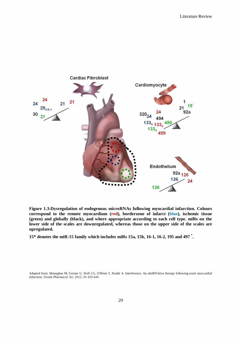
Literature Review
29
Figure 1.3:Dysregulation of endogenous microRNAs following myocardial infarction. Colours
correspond to the remote myocardium (red), borderzone of infarct (blue), ischemic tissue
(green) and globally (black), and where appropriate according to each cell type. miRs on the
lower side of the scales are downregulated, whereas those on the upper side of the scales are
upregulated.
15* denotes the miR-15 family which includes miRs 15a, 15b, 16-1, 16-2, 195 and 497 *.
Adapted from: Monaghan M, Greiser U, Wall J.G, O'Brien T, Pandit A. Interference: An alteRNAtive therapy following acute myocardial
infarction. Trends Pharmacol. Sci. 2012, 33: 635-645
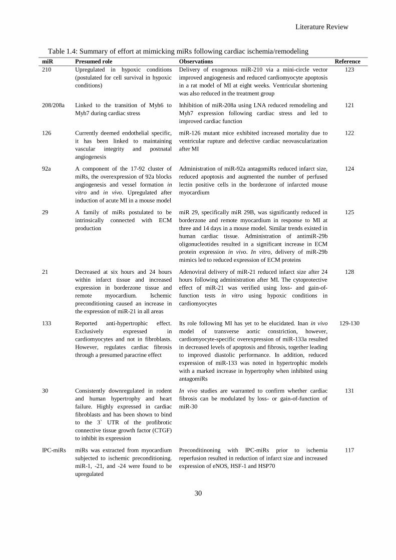
Literature Review
30
Table 1.4: Summary of effort at mimicking miRs following cardiac ischemia/remodeling
miR Presumed role Observations Reference
210 Upregulated in hypoxic conditions
(postulated for cell survival in hypoxic
conditions)
Delivery of exogenous miR-210 via a mini-circle vector
improved angiogenesis and reduced cardiomyocyte apoptosis
in a rat model of MI at eight weeks. Ventricular shortening
was also reduced in the treatment group
123
208/208a Linked to the transition of Myh6 to
Myh7 during cardiac stress
Inhibition of miR-208a using LNA reduced remodeling and
Myh7 expression following cardiac stress and led to
improved cardiac function
121
126 Currently deemed endothelial specific,
it has been linked to maintaining
vascular integrity and postnatal
angiogenesis
miR-126 mutant mice exhibited increased mortality due to
ventricular rupture and defective cardiac neovascularization
after MI
122
92a A component of the 17-92 cluster of
miRs, the overexpression of 92a blocks
angiogenesis and vessel formation in
vitro and in vivo. Upregulated after
induction of acute MI in a mouse model
Administration of miR-92a antagomiRs reduced infarct size,
reduced apoptosis and augmented the number of perfused
lectin positive cells in the borderzone of infarcted mouse
myocardium
124
29 A family of miRs postulated to be
intrinsically connected with ECM
production
miR 29, specifically miR 29B, was significantly reduced in
borderzone and remote myocardium in response to MI at
three and 14 days in a mouse model. Similar trends existed in
human cardiac tissue. Administration of antimiR-29b
oligonucleotides resulted in a significant increase in ECM
protein expression in vivo. In vitro, delivery of miR-29b
mimics led to reduced expression of ECM proteins
125
21 Decreased at six hours and 24 hours
within infarct tissue and increased
expression in borderzone tissue and
remote myocardium. Ischemic
preconditioning caused an increase in
the expression of miR-21 in all areas
Adenoviral delivery of miR-21 reduced infarct size after 24
hours following administration after MI. The cytoprotective
effect of miR-21 was verified using loss- and gain-of-
function tests in vitro using hypoxic conditions in
cardiomyocytes
128
133 Reported anti-hypertrophic effect.
Exclusively expressed in
cardiomyocytes and not in fibroblasts.
However, regulates cardiac fibrosis
through a presumed paracrine effect
Its role following MI has yet to be elucidated. Inan in vivo
model of transverse aortic constriction, however,
cardiomyocyte-specific overexpression of miR-133a resulted
in decreased levels of apoptosis and fibrosis, together leading
to improved diastolic performance. In addition, reduced
expression of miR-133 was noted in hypertrophic models
with a marked increase in hypertrophy when inhibited using
antagomiRs
129-130
30 Consistently downregulated in rodent
and human hypertrophy and heart
failure. Highly expressed in cardiac
fibroblasts and has been shown to bind
to the 3` UTR of the profibrotic
connective tissue growth factor (CTGF)
to inhibit its expression
In vivo studies are warranted to confirm whether cardiac
fibrosis can be modulated by loss- or gain-of-function of
miR-30
131
IPC-miRs miRs was extracted from myocardium
subjected to ischemic preconditioning.
miR-1, -21, and -24 were found to be
upregulated
Preconditinoning with IPC-miRs prior to ischemia
reperfusion resulted in reduction of infarct size and increased
expression of eNOS, HSF-1 and HSP70
117
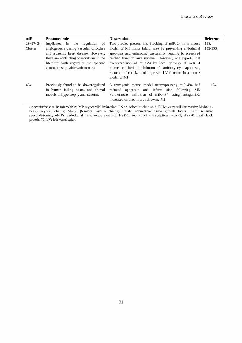
Literature Review
31
miR Presumed role Observations Reference
23~27~24
Cluster
Implicated in the regulation of
angiogenesis during vascular disorders
and ischemic heart disease. However,
there are conflicting observations in the
literature with regard to the specific
action, most notable with miR-24
Two studies present that blocking of miR-24 in a mouse
model of MI limits infarct size by preventing endothelial
apoptosis and enhancing vascularity, leading to preserved
cardiac function and survival. However, one reports that
overexpression of miR-24 by local delivery of miR-24
mimics resulted in inhibition of cardiomyocyte apoptosis,
reduced infarct size and improved LV function in a mouse
model of MI
118,
132-133
494 Previously found to be downregulated
in human failing hearts and animal
models of hypertrophy and ischemia
A transgenic mouse model overexpressing miR-494 had
reduced apoptosis and infarct size following MI.
Furthermore, inhibition of miR-494 using antagomiRs
increased cardiac injury following MI
134
Abbreviations: miR: microRNA; MI: myocardial infarction; LNA: locked nucleic acid; ECM: extracellular matrix; Myh6: α-heavy myosin chains; Myh7: β-heavy myosin chains; CTGF: connective tissue growth factor; IPC: ischemic preconditioning; eNOS: endothelial nitric oxide synthase; HSF-1: heat shock transcription factor-1; HSP70: heat shock protein 70; LV: left ventricular.
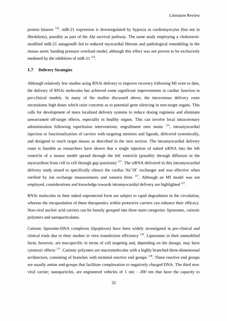
Literature Review
32
protein kinases 126
. miR-21 expression is downregulated by hypoxia in cardiomyocytes (but not in
fibroblasts), possibly as part of the Akt survival pathway. The same study employing a cholesterol-
modified miR-21 antagomiR led to reduced myocardial fibrosis and pathological remodeling in the
mouse aortic banding pressure overload model, although this effect was not proven to be exclusively
mediated by the inhibition of miR-21 126
.
1.7 Delivery Strategies
Although relatively few studies using RNAi delivery to improve recovery following MI exist to date,
the delivery of RNAi molecules has achieved some significant improvements in cardiac function in
pre-clinical models. In many of the studies discussed above, the intravenous delivery route
necessitates high doses which raise concerns as to potential gene silencing in non-target organs. This
calls for development of more localized delivery systems to reduce dosing regimens and eliminate
unwarranted off-target effects, especially in healthy organs. This can involve local intracoronary
administration following reperfusion interventions; engraftment onto stents 135
, intramyocardial
injection or functionalization of carriers with targeting moieties and ligands, delivered systemically,
and designed to reach target tissues as described in the next section. The intramyocardial delivery
route is feasible as researchers have shown that a single injection of naked siRNA into the left
ventricle of a mouse model spread through the left ventricle (possibly through diffusion in the
myocardium from cell to cell through gap junctions) 127
. The siRNA delivered in this intramyocardial
delivery study aimed to specifically silence the cardiac Na+/H
+ exchanger and was effective when
verified by ion exchange measurements and western blots 127
. Although an MI model was not
employed, considerations and knowledge towards intramyocardial delivery are highlighted 127
.
RNAi molecules in their naked unprotected form are subject to rapid degradation in the circulation,
whereas the encapsulation of these therapeutics within protective carriers can enhance their efficacy.
Non-viral nucleic acid carriers can be loosely grouped into three main categories: liposomes, cationic
polymers and nanoparticulates.
Cationic liposome-DNA complexes (lipoplexes) have been widely investigated in pre-clinical and
clinical trials due to their modest in vitro transfection efficiency 136
. Liposomes in their unmodified
form; however, are non-specific in terms of cell targeting and, depending on the dosage, may have
cytotoxic effects 137
. Cationic polymers are macromolecules with a highly branched three-dimensional
architecture, consisting of branches with terminal reactive end groups 138
. These reactive end groups
are usually amine end-groups that facilitate complexation to negatively charged DNA. The third non-
viral carrier; nanoparticles, are engineered vehicles of 1 nm – 200 nm that have the capacity to
Literature Review
33
transport pharmaceuticals and genetic material. They can be produced in the form of rods, tubes and
spheres/shells and can be fabricated from synthetic or natural polymers and metals 139
, depending on
considerations such as large scale production requirements and their intended application. These
structures can be used in a variety of clinical applications, including that of improved in vivo
localization using magnetic manipulation.
The functionality of these synthetic carriers such as these is increasingly improved through processes
such as PEGylation, charge manipulation and ligand conjugation. An important aspect of this
functionality is the addition of receptor engaging ligands to offer improved targeting and binding to
specific cell types. This gives rise to the need to identify cell-specific surface molecules, and this
aspect is discussed in the next section.
1.8 Engineering Ligands for Improved Uptake
Attaching cell-targeting ligands to nucleic acid carriers as discussed above can offer selectivity to a
particular cell type/accumulation at target tissue, and control the route of internalization into the target
cell. Endocytosis is the main route of cellular internalization of non-viral vectors and multiple
mechanisms have been described to date 38
. Internalized molecules typically traffic in intracellular
vesicles and eventually fuse with lysosomes to be degraded, presenting a further challenge in reaching
intracellular target sites.
Cell-targeting ligands include natural ligands based on transferrin, peptide formulations (RGD) to
engage integrins and antibodies, and functional fragments thereof. In MI, many markers of
inflammation and endocytic receptors receptive to specific ligands can allow for improved and
controlled uptake of non-viral vectors carrying pDNA for transcription of desired shRNA and miR
molecules, or direct siRNA payloads 140-142
.Synthetic vectors have shown potential for improved
delivery using ligand-based nucleic acid therapy (Table 1.5). It is worth noting here that the misnomer
‗targeted‘ is avoided, especially in the context of intravenous administration as this would suggest
delivery specifically to, and unique to, one particular target; in fact, less than 5% of delivered drug
particles engage with their intended cell or tissue and instead accumulate in the liver, lungs, kidney
and spleen 143
. Passive targeting is achieved in tumour tissue via the enhanced permeation and
retention (EPR) effect, but in the context of MI, this effect is less pronounced, and therefore
engagement with cell-specific markers and receptors is desirable for specific delivery.
1.8.1 Antibodies
Hybridoma technology and the development of chimeric antibodies (mouse antigen-binding domains
fused to human constant regions) 144
have enabled large-scale production of antibodies, specific for a
Literature Review
34
diverse range of targets. The use of monoclonal antibodies (mAbs) is advantageous over most
fragments due to the higher binding avidity that results from their bivalency, and the presence of the
Fc domain appropriate to tether the mAb to a delivery vehicle in an appropriate orientation.
Conversely, the presence of exposed Fc domains that can bind Fc receptors in non-target tissues,
particularly on macrophages, leads to high uptake in the liver and spleen has the potential to increase
the immunogenicity of such an antibody functionalized delivery vehicle. Extensive engineering of Fab
and single-chain Fv (scFv) fragments has led to the development of bivalent 145
and even bispecific 146
variants for improved target binding while obviating immunogenic responses. The lack of availability
of an Fc domain for orientation of attachment to carriers can be overcome by chemical and/or genetic
modification of the fragments to create active groups distant from the target-binding pocket 147
.
Advantages of such fragments are their smaller size (which allows denser packing on vectors and
greater penetration of tissues), their ease of production and modification in recombinant – particularly
bacterial – expression systems 148
and, increasingly, the development of large combinatorial libraries
of fragments that can be screened relatively easily and rapidly to isolate binders against any molecule
of interest 149
. In addition, antibody fragments can be engineered to improve their linking by covalent
or ionic interactions to reservoirs of genetic material.
Béduneau et al. have engineered liposomes conjugated with monoclonal antibodies and Fab fragments
with specificity for the transferrin receptor (Tfr) 150
. Both liposome populations demonstrated
enhanced accumulation at Tfr sites 24 h post-injection in a healthy rat model, most notably in the
brain, and liposomes functionalized with whole antibodies proved to be more efficient. Lau et al.
reported improved extravasation of siRNA-albumin conjugates into organs with fenestrated sinusoidal
endothelia (including the myocardium) to silence an insulin-like growth factor type I receptor (IGF-
IR) 151
. These findings provide an important insight into targeting cardiac tissue using siRNA,
although it would be interesting to see the results of similar experiments carried out with a cell-
specific targeting ligand (as opposed to a more general protein such as albumin) or in a pathological
model. In principle, while past research efforts have focussed primarily on monoclonal antibodies, in
theory any monoclonal antibody, Fab fragment or scFv can be linked to a synthetic vector to engage
with specific cells.
1.8.2 Cell-targeting Peptides
Phage display technology generates and screens large libraries of peptides or proteins for binders to a
specific, immobilized ligand and is used to identify peptides rather than antibodies or fragments that
can bind specifically to many molecular targets. Liu and co-workers have identified peptides specific
for atherosclerotic lesions induced in apoE knockout mice 152
, with GRP78 identified in ex vivo
Literature Review
35
studies as the endothelial cell surface target of one such peptide. Meanwhile, Tepe et al. 153
have
described a radiolabeled endothelin derivative which selectively accumulates on atherosclerotic
plaques after intravenous administration. PEI has been conjugated to the cyclic RGD-4C to effect
delivery to endothelial cells expressing αυβ3 while the use of RGD-containing peptides and a short
polylysine sequence for increased electrostatic binding of DNA has been demonstrated to enhance
DNA delivery by up to five-fold 154
. Other researchers have also documented the use of cationic
nanoparticles conjugated to the αυβ3 integrin-ligand to selectively deliver a mutant Raf protein to
inhibit endothelial cell survival by blocking angiogenesis 155
.
In terms of the myocardium, cardiomyocyte prostaglandin receptors using prostaglandin E2 (PGE2)
as a ligand 156
have been used as potential targets. A primary cardiomyocyte (PCM)-specific peptide
generated by phage display 157
and investigated in vitro. Both delivery strategies (PGE2 or PCM
specific peptide as ligands) resulted in significant silencing of Fas when utilized with cationic
complexes to deliver siRNA against the Fas gene (a transmembrane protein which belongs to the
TNF-α family that when ligand/receptor engagement occurs apoptosis is induced). Fas silencing using
these systems resulted in a significant reduction in cardiomyocyte apoptosis compared to controls in
vitro. Although this system was also an in vitro demonstration of the effect, it indicates a therapeutic
promise of the approach that warrants further investigation.
The lack of successful clinical strategies employing ligand-based drug delivery is a concern for
researchers aiming to develop cancer-based therapeutics and for those in the field of MI. However,
there are many options available to the cardiovascular researcher that can enhance delivery to the
infarcted myocardium. The coronary vasculature is an anatomically defined network enabling local
catheter-based delivery which obviates pre-clearance of therapeutics by other organs and provides
direct interaction with the infarcted myocardium. Systemic delivery with the aim of a successful
targeted tissue is still beyond realization until optimization of the more fundamental aspects of
carriers such as physico-chemical properties of the bulk material, size and charge is performed to fully
understand the interaction in vivo after systemic delivery.
1.9 Fibrosis
Fibrosis is the final, common pathological outcome of tissue insult and/or inflammation and a central
target in the development of the platform developed in this thesis. Although collagen deposition is an
indispensable and, typically, a reversible element of wound healing, normal tissue repair can evolve
into a progressively irreversible fibrotic response if the tissue injury is severe, repetitive, or if the
wound-healing response itself becomes dysregulated. Fibrosis is defined as the excessive
accumulation of fibrous connective tissue (components of the ECM such as collagen and fibronectin)
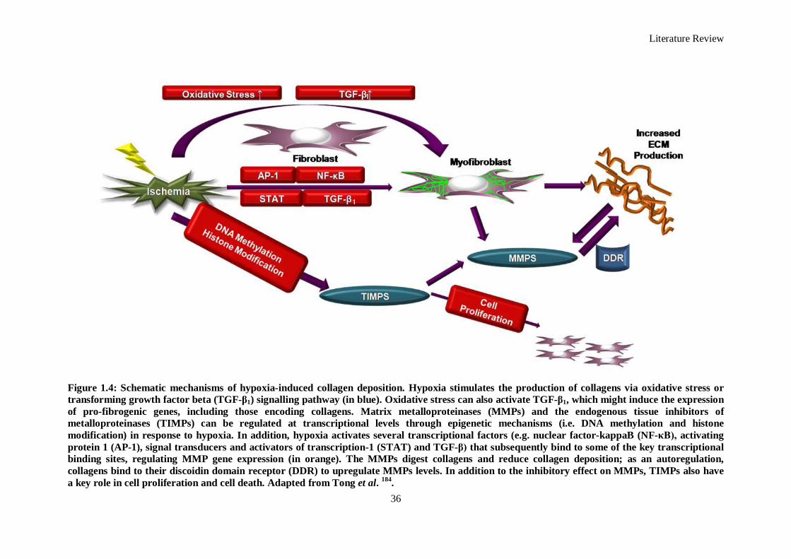
Literature Review
36
Figure 1.4: Schematic mechanisms of hypoxia-induced collagen deposition. Hypoxia stimulates the production of collagens via oxidative stress or
transforming growth factor beta (TGF-β1) signalling pathway (in blue). Oxidative stress can also activate TGF-β1, which might induce the expression
of pro-fibrogenic genes, including those encoding collagens. Matrix metalloproteinases (MMPs) and the endogenous tissue inhibitors of
metalloproteinases (TIMPs) can be regulated at transcriptional levels through epigenetic mechanisms (i.e. DNA methylation and histone
modification) in response to hypoxia. In addition, hypoxia activates several transcriptional factors (e.g. nuclear factor-kappaB (NF-κB), activating
protein 1 (AP-1), signal transducers and activators of transcription-1 (STAT) and TGF-β) that subsequently bind to some of the key transcriptional
binding sites, regulating MMP gene expression (in orange). The MMPs digest collagens and reduce collagen deposition; as an autoregulation,
collagens bind to their discoidin domain receptor (DDR) to upregulate MMPs levels. In addition to the inhibitory effect on MMPs, TIMPs also have
a key role in cell proliferation and cell death. Adapted from Tong et al. 184
.
1
1
Literature Review
37
in and around inflamed or damaged tissue, which can lead to permanent scarring, organ malfunction
and, ultimately death, as seen in heart failure 158
. In cardiac tissue, fibroblasts are recognised as key
regulators of ECM components and also are a major source of MMPs and TIMPs (Figure 1.4). In the
inflammatory environment following MI, cytokines, grown factors, and other environmental stimuli
modulate fibroblast function by changing the production patterns of ECM components, MMPs and
TIMPs, by stimulating their migration and proliferation, and by mediating the interaction between
cardiac fibroblasts and other cell types.
Cardiac remodelling is the most common underlying cause of end-stage heart failure morbidity and
mortality worldwide after MI 159-161
. Significant fibrosis, occurring in both the infarcted and non-
infarcted myocardium, represents a characteristic pathological alteration of post infarct remodelling
and is recognised to be a major determinant of the progressive deterioration of ventricular function
after MI. This fibrosis is modulated by a cascade of biochemical intracellular signalling processes that
are triggered by necrotic myocardium in the context of MI in combination with increased preload and
afterload. It also reduces the electrical coupling between cardiomyocytes as fibroblasts produce
smaller or larger collagenous septa, which electrically insulate cardiac cells or muscle bundles. Due to
this, the normal myocardial architecture becomes disrupted and is transformed into a pathological
substrate characterized by the presence of multiple insulating barriers, which force the depolarization
wave to spread non-uniformly162, 163
.
Without performing an exhaustive review of the many factors that play a key role in the pathological
post infarct remodelling process, key events and signalling molecules emerge that play key roles. For
instance, TGF–β1 is a key signalling molecule that induces cardiac fibrosis by activating the
proliferation and collagen production of cardiac fibroblasts 164
. Intracellular mitogen-activated protein
kinase (MPAK) signalling cascades have also been proven to play an important role in the
pathogenesis of cardiac fibrosis 165
.
The importance of fibrosis as a determinant of myocardial performance and disease outcome is
increasingly appreciated. Nevertheless, efforts to develop novel therapies that specifically target the
cardiac fibroblast are at a relatively early stage in common with approaches to fibrotic disease in other
organ systems. Current pharmaceutical attempts at modulating fibrosis include inhibitors of rennin-
angiotensin-aldosterone 166-169
, endothelin receptor antagonists 170
, statins 171, 172
, cytokine therapy 173-
175, and inhibitors of MMPs
176, 177, all of which are extensively reviewed elsewhere
178.
miRs are emerging as key players in the pathogenesis of fibrosis. For instance the miR-29 cluster in
humans includes hsa-miR-29A, hsa-miR-29B-1, hsa-miR-29B-2 and hsa-miR-29C. miR-29B-1 and
mir-29B-2 have identical mature sequences, which are together called miR-29B. Mature miR-29s
share identical sequences at nucleotide positions 2-7, the seed region that plays a key role in
determining which protein-coding-genes a microRNA would target 179
. Therefore, predicted target
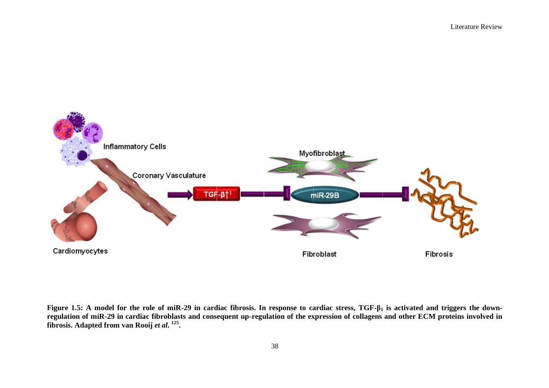
Literature Review
38
Figure 1.5: A model for the role of miR-29 in cardiac fibrosis. In response to cardiac stress, TGF-β1 is activated and triggers the down-
regulation of miR-29 in cardiac fibroblasts and consequent up-regulation of the expression of collagens and other ECM proteins involved in
fibrosis. Adapted from van Rooij et al. 125.
1
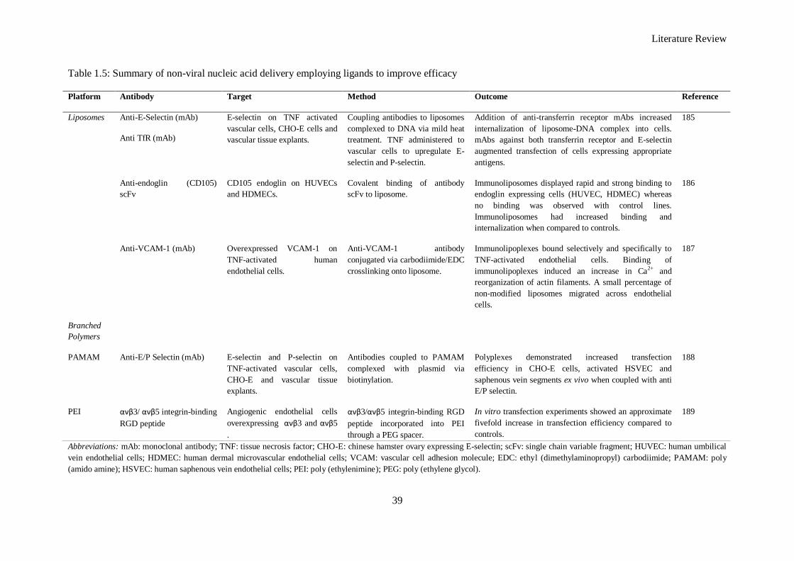
Literature Review
39
Table 1.5: Summary of non-viral nucleic acid delivery employing ligands to improve efficacy
Abbreviations: mAb: monoclonal antibody; TNF: tissue necrosis factor; CHO-E: chinese hamster ovary expressing E-selectin; scFv: single chain variable fragment; HUVEC: human umbilical
vein endothelial cells; HDMEC: human dermal microvascular endothelial cells; VCAM: vascular cell adhesion molecule; EDC: ethyl (dimethylaminopropyl) carbodiimide; PAMAM: poly
(amido amine); HSVEC: human saphenous vein endothelial cells; PEI: poly (ethylenimine); PEG: poly (ethylene glycol).
Platform Antibody Target Method Outcome Reference
Liposomes Anti-E-Selectin (mAb)
Anti TfR (mAb)
E-selectin on TNF activated
vascular cells, CHO-E cells and
vascular tissue explants.
Coupling antibodies to liposomes
complexed to DNA via mild heat
treatment. TNF administered to
vascular cells to upregulate E-
selectin and P-selectin.
Addition of anti-transferrin receptor mAbs increased
internalization of liposome-DNA complex into cells.
mAbs against both transferrin receptor and E-selectin
augmented transfection of cells expressing appropriate
antigens.
185
Anti-endoglin (CD105)
scFv
CD105 endoglin on HUVECs
and HDMECs.
Covalent binding of antibody
scFv to liposome.
Immunoliposomes displayed rapid and strong binding to
endoglin expressing cells (HUVEC, HDMEC) whereas
no binding was observed with control lines.
Immunoliposomes had increased binding and
internalization when compared to controls.
186
Anti-VCAM-1 (mAb) Overexpressed VCAM-1 on
TNF-activated human
endothelial cells.
Anti-VCAM-1 antibody
conjugated via carbodiimide/EDC
crosslinking onto liposome.
Immunolipoplexes bound selectively and specifically to
TNF-activated endothelial cells. Binding of
immunolipoplexes induced an increase in Ca2+ and
reorganization of actin filaments. A small percentage of
non-modified liposomes migrated across endothelial
cells.
187
Branched
Polymers
PAMAM Anti-E/P Selectin (mAb) E-selectin and P-selectin on
TNF-activated vascular cells,
CHO-E and vascular tissue
explants.
Antibodies coupled to PAMAM
complexed with plasmid via
biotinylation.
Polyplexes demonstrated increased transfection
efficiency in CHO-E cells, activated HSVEC and
saphenous vein segments ex vivo when coupled with anti
E/P selectin.
188
PEI ανβ3/ ανβ5 integrin-binding
RGD peptide
Angiogenic endothelial cells
overexpressing ανβ3 and ανβ5
.
ανβ3/ανβ5 integrin-binding RGD
peptide incorporated into PEI
through a PEG spacer.
In vitro transfection experiments showed an approximate
fivefold increase in transfection efficiency compared to
controls.
189
Literature Review
40
genes for the miR-29 family members largely overlap. It has been confirmed that miR-29B-1 and
miR-29A are transcribed together as a polycistronic pri-miR and likewise miR-29B-2 and miR-29C
are transcribed together 180
. It has collectively been shown that the miR-29 family members target at
least 16 genes related to extracellular matrix (ECM) which encode for several key proteins involved in
the physiological or pathological formation of ECM including; a large number of collagen isoforms,
laminin γ, fibrilin 1, elastin, matrix metalloproteinase 2, and integrin β1 125, 181-183
. This is unique to the
miR-29 family as no other miR is predicted to target more than 11 of the 20 collagen genes (Figure
1.5).
1.10 Project Rationale
The preceding sections have illustrated the use of RNAi as an intervention in the treatment of the
infarcted myocardium and the delivery of RNA from biomaterial scaffolds in various applications.
From a biomaterials approach, non-viral vectors are discussed, with focus on the various methods of
delivering and functionalizing these vehicles. Furthermore, the use of biomaterial platforms on the
macro-scale as scaffold reservoirs of delivery is presented. The project described herein combines
many elements of this introduction. A type I atelocollagen scaffold is proposed as a delivery platform
for RNAi that is injectable and crosslinks in situ. The RNAi investigated in this study is in the form of
an exogenous miR mimic of the 29B sequence (miR-29B) which is downregulated following ECM
remodelling and has a pivotal role in ECM components synthesis. Finally, a poly(2-
(dimethylamino)ethyl methacrylate) (pDMAEMA) based transfection agent (pD-b-P/DA) which can
conjugate antibody fragments receptive to the CD90 antigen is synthesized as a protective vehicle of
the exogenous miR. As anti-fibrotic therapy can be effective in promoting beneficial remodelling of
the infarcted myocardium, promotion of recovery of cardiac function is possible and was investigated
via systemic delivery in the appendix chapter.
1.10.1 Rationale for the use of a miR Mimic
Various non-viral delivery methods of miRs exist such as pDNA encoding shRNA, pre-miRs and
mature double stranded miR mimics. It was decided to employ mature miR mimics in order to obviate
the necessity for nuclear entry and transcription of pre-miRs. Furthermore, as miR-29B is transcribed
along with other members of the miR-29 family, therefore the direct delivery of a mature miR
sequence allows the determination of the effect of miR-29B alone.
Literature Review
41
1.10.2 Rationale for the use of miR-29B
Fibrosis caused by a dysregulation in the wound healing response is most likely due to traumatic
injury, such that is present following MI. This fibrosis is mediated by the excessive deposition of
collagen type I fibrils and re-organization of the ECM. The focus in this thesis is on reducing fibrosis
by delivering miR-29B which is directly related (and also downregulated) during increased ECM
remodelling 190-194
, by delivering exogenous miR mimics of miR-29B.
1.10.3 Rationale for the use of pDMAEMA
A non-viral delivery approach composed of miR mimics and a pDMAEMA based transfection agent
was developed. While viral systems can be among the most effective agents available for nucleic acid
transfer, they have associated health risks, high production costs, and a variety of other drawbacks.
Thus, a non-viral delivery system was selected. pDMAEMA based transfection agents have shown
much efficacy as non-viral transfection agents posing minimal toxicity while also maintaining
efficient delivery of nucleic acid 195-196
. pDMAEMA has been shown to efficiently transfect a variety
of cells 197-198
; however there is a drawback of toxicity due to the cationic nature of this agent. The
incorporation of poly (ethylene glycol) into such a structure can offer a steric shield to non-viral
complexes from interaction with blood components and also contributes to reduced toxicity levels 199
.
Deactivation enhanced atom transfer radical polymerization enables the controlled synthesis and
propagation of multi-vinyl monomers 200-201
allowing for the creation of highly ordered structures with
vinyl functionality. Chapter Two of this thesis provides further details in how this was envisaged for
this thesis; briefly, vinyl functionality can allow for the incorporation and decoration of targeting
moieties (in this case antibody fragments) which increases transfection efficacy by mediating receptor
mediated endocytosis.
1.10.4 Rationale for the Selection of an Antibody Fragment Directed towards CD90
Considering that fibrosis is the therapeutic target of the platform developed in this thesis, it was
sought to enhance the delivery of miR-29B to cardiac fibroblasts which are predominant cell type in
the myocardium and play an active role in extracellular matrix remodelling. Cardiac fibroblasts, as
with all fibroblasts, express CD90, a cell surface antigen which is not present in cardiomyocytes or
the endothelium of the coronary vasculature. As previously discussed, at the end of the literature
review; the functionalisation of gene delivery vehicles with cell receptive moieties (such as
monoclonal antibodies, antibody fragments, peptides and scFvs) can improve their efficiency and
uptake into cells preferentially. CD90 was selected due to its association with cardiac fibroblasts and
Literature Review
42
also as it was available through a hybridoma cell line which facilitates a continuing source of parent
antibody through which antibody fragments can be derived in large amounts.
1.10.5 Rationale for the use of a Crosslinked Atelocollagen Type I Scaffold
Type I collagen was selected as the optimal scaffold because it is a well-characterised natural
biomaterial that is commonly used in the field. Furthermore, many collagen-based biomaterial
products are approved for medical use in humans by the US Food and Drug Administration (FDA)
and European Medical Association (EMA). Atelocollagen is a less immunogenic form that is prepared
by removing the telopeptides from the end of the collagen fibrils 202
. Type I collagen is also the
principal constituent of native extracellular matrix (ECM) which promotes growth, attachment, and
proliferation of a variety of cell types. Finally, there are multiple reports of collagen being used as an
effective nucleic acid delivery system, further recommending its use in this application 203-205
.
1.10.6 Rationale for the use of Scaffold-Based RNAi Delivery
Individually, each of the elements (scaffold, miR mimics, non-viral delivery agent) of the proposed
combinatorial RNAi delivery system have shown significant potential for use in tissue engineering
applications. However, each component has limitations when delivered in isolation. It has been
hypothesized that implanted collagen may play a minor role in modulating the production of tissue
MMPs as part of the foreign body response 206
. This role is exceptionally minor however, and
collagen scaffolds have relatively minor effects on ischemic tissue and are ideal as a delivery system
as they induces minimal foreign body response, are biodegradable, and can be prepared, stored and
sterilized with relative ease 207, 208
. Hence, the use of a collagen scaffold reservoir system will act as a
tuneable reservoir of miR-29B complexes and will also add functional support to the treated areas.
The combination of biomaterial scaffold and pDMAEMA based delivery agent should furthermore
improve the efficacy of the RNAi, thereby allowing the miRs to act more efficiently. This thesis
reports the development of each individual component and the combination thereof to improve
delivery of miRs to effect a therapeutic treatment in the inhibition of fibrosis.
1.11 Objectives and Hypotheses
The overall hypothesis of this thesis is that an effective and efficient delivery of miR-29B complexes
can be achieved to silence the production of collagen type I through an injectable non-toxic type I
atelocollagen hydrogel scaffold with/without the use of a pDMAEMA based complexing agent. The
specific objectives of the in vitro studies were to develop and characterize a pDMAEMA based
delivery agent for complexation of miR mimics and conjugation of antibody fragments. The in vitro
Literature Review
43
studies were designed to develop and characterize an atelocollagen type I scaffold crosslinked using
poly (ethylene glycol) ether tetrasuccinimidyl glutarate (4S-StarPEG) and were evaluated for their
ability and effectiveness as a delivery reservoir of RNAi in various forms towards a goal of silencing
miR-29B. The in vivo studies investigated the effects of miR-29B on ECM remodelling in a rat
syngeneic dermal excisional wound model.
Objectives: Phase I - Delivering Exogenous miRNA (Chapter Two)
Synthesize a block co-polymer composed of a linear poly (dimethylamino) ethyl methacrylate
(pDMAEMA) block and a hyperbranched poly (ethylene glycol) methyl ether acrylate
(PEGMEA) based unit with poly (ethylene glycol) diacrylate (PEGDA) as the multi-vinyl
monomer (MVM) branching agent (pD-b-/PDA) using De-ATRP synthesis. This resultant pD-b-
/PDA will be characterised during polymer synthesis using gel permeation chromatography and
its structure evaluated using proton nucleic magnetic resonance (1H NMR).
Demonstrate effective complexation of miR mimics with pD-b-/PDA by characterising its
electrophoretic mobility, prove and demonstrate Michael-type addition of thiols to pD-b-/PDA
using Ellman‘s Assay, and antibody fragments to pD-b-/PDA by SDS-PAGE.
Optimise and deliver miRs with pD-b-/PDA to effect silencing in a dual reporter system of Renilla
Luciferase and Firefly Luciferase using cardiac fibroblasts as an experimental cell type.
Hypotheses: Phase I - Delivering Exogenous miRNA (Chapter Two)
A co-polymer composed of a linear poly (dimethylamino) ethyl methacrylate (pDMAEMA)
block and a hyperbranched poly (ethylene glycol) methyl ether acrylate (PEGMEA) based unit
with poly (ethylene glycol) diacrylate (PEGDA) as the MVM branching agent (pD-b-/PDA) can
complex and delivery miR mimics with reduced cytotoxicity and also offer the possibility of
facile conjugation of additional functional groups by Michael type addition.
Objectives: Phase II- An Injectable Hydrogel Delivery System (Chapter Three)
Develop an atelocollagen type I hydrogel and characterise the effect of crosslinking that can be
achieved using 4S-StarPEG by determining the amine content of the scaffolds using a
trinitrobenzenesulfonic acid (TNBSA) assay, the effect on the degradation profile of the
hydrogels using collagenase degradation and the effect on mechanical properties using rheology.
Literature Review
44
Investigate the effect of crosslinker density on the release profile of interfering RNA from
hydrogels and the effect of complexing the RNA with a complexing agent on the release of the
RNA from the hydrogels, spectrophotometrically, using Cy™
3 labelled siRNA.
Investigate the ability of this hydrogel acting as a reservoir in vitro of miR-29B, characterising its
release profile and the subsequent effect of specifically determining the effect on production of
collagen type I and type III expression in vitro.
Hypotheses: Phase II - An Injectable Hydrogel Delivery System (Chapter Three)
An atelocollagen type I hydrogel crosslinked with 4S-StarPEG will act as a reservoir of miR-29B
in naked form, miR-29B complexed with a poly (2-(dimethylamino) ethyl methacrylate)
(pDMAEMA) based polymer (pD-b-P/DA- developed in Phase I) and miR-29B complexed with
PEI.
This scaffold/miR platform will effectively silence ECM genes; namely collagen type I and
collagen type III.
Objectives: Phase III – System Evaluation in a Rat Excisional Wound Model (Chapter Four)
To investigate the effect of the components in this platform (miR-29B, 4S-StarPEG-collagen scaffold,
pD-b-P/DA) on a number of key parameters involved in wound healing, using a number of techniques.
Specifically:
Evaluate, using histological analysis, wound contraction and granulation tissue formation
following full thickness excisional skin wounding in a rat model.
Quantify collagen type I and III deposition by polarised light microscopy and qualitative
immunohistochemistry for collagen type III within the wound bed.
Employ an antibody labelled membrane protein array to elucidate the up/down regulation of
factors associated with apoptosis, inflammation and ECM remodelling.
Use enzyme linked immunosorbent assays (ELISA) to quantify the expression of TGF-β1,
TIMP-1, MMP-8 and determine the ratio of MMP-8: TIMP-1.
Determine the effect of the components of this platform on genes associated with wound
healing using a rat wound healing PCR Array.

Literature Review
45
Hypotheses: Phase III- System Evaluation in a Rat Excisional Wound Model (Chapter Four)
Incorporation of miR-29B complexes (with or without the use of pD-b-P/DA) into a crosslinked
collagen hydrogel will have significant benefit on wound closure, collagen type I and collagen
type III deposition and key ECM remodelling mediators (TGF-β1, TIMP-1, and MMP-8) in a rat
excisional wound model.
These parameters will all be responsive to the dose of miR-29B incorporated within the scaffold
and therefore a dose response will be observed through the parameters investigated.
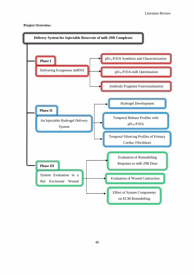
Literature Review
46
Project Overview:
pD-b-P/DA Synthesis and Characterization
Delivery System for Injectable Reservoir of miR-29B Complexes
pD-b-P/DA-miR Optimisation
Antibody Fragment Functionalisation
Hydrogel Development
Temporal Release Profiles with
pD-b-P/DA
Evaluation of Remodelling
Response to miR-29B Dose
Temporal Silencing Profiles of Primary
Cardiac Fibroblasts
Evaluation of Wound Contraction
Effect of System Components
on ECM Remodelling
Phase I
Delivering Exogenous miRNA
System Evaluation in a
Rat Excisional Wound
Model
Phase III
Phase II
An Injectable Hydrogel Delivery
System
Literature Review
47
1.12 References
1. Langer R, Vacanti JP. Tissue engineering. Science. 1993;260:920-926
2. Langer R. Tissue engineering. Mol. Ther. 2000;1:12-15
3. Kulkarni M, Greiser U, O'Brien T, Pandit A. Liposomal gene delivery mediated by tissue-engineered scaffolds. Trends Biotechnol. 2010;28:28-36
4. De Laporte L, Shea LD. Matrices and scaffolds for DNA delivery in tissue engineering. Adv.
Drug Delivery Rev. 2007;59:292-307 5. O'Rorke S, Keeney M, Pandit A. Non-viral polyplexes: Scaffold mediated delivery for gene
therapy. Prog. Polym. Sci. 2010;35:441-458
6. Fire A, Xu S, Montgomery MK, Kostas SA, Driver SE, Mello CC. Potent and specific genetic
interference by double-stranded RNA in caenorhabditis elegans. Nature. 1998;391:806-811 7. Lee Y, Kim M, Han J, Yeom K-H, Lee S, Baek SH, Kim VN. MicroRNA genes are
transcribed by RNA polymerase II. EMBO J. 2004;23:4051-4060
8. Mueller G, Miller M, DeRose E, Ghosh M, London R, Hall T. Solution structure of the drosha double-stranded RNA-binding domain. Silence. 2010;1:2
9. Sohn SY, Bae WJ, Kim JJ, Yeom K-H, Kim VN, Cho Y. Crystal structure of human DGCR8
core. Nat. Struct. Mol. Biol. 2007;14:847-853
10. Provost P, Dishart D, Doucet J, Frendewey D, Samuelsson B, Radmark O. Ribonuclease activity and RNA binding of recombinant human dicer. EMBO J. 2002;21:5864-5874
11. MacRae IJ, Zhou K, Li F, Repic A, Brooks AN, Cande WZ, Adams PD, Doudna JA.
Structural basis for double-stranded RNA processing by DICER. Science. 2006;311:195-198 12. Preall JB, Sontheimer EJ. RNAi: RISC gets loaded. Cell. 2005;123:543-545
13. Lewis BP, Burge CB, Bartel DP. Conserved seed pairing, often flanked by adenosines,
indicates that thousands of human genes are microRNA targets. Cell. 2005;120:15-20 14. Brodersen P, Voinnet O. Revisiting the principles of microRNA target recognition and mode
of action. Nat. Rev. Mol. Cell. Biol. 2009;10:141-148
15. Filipowicz W, Bhattacharyya SN, Sonenberg N. Mechanisms of post-transcriptional
regulation by microRNAs: Are the answers in sight? Nat. Rev. Genet. 2008;9:102-114 16. He L, Hannon GJ. MicroRNAs: Small RNAs with a big role in gene regulation. Nat. Rev.
Genet. 2004;5:522-531
17. Yekta S, Shih I-h, Bartel DP. MicroRNA-directed cleavage of HoxB8 mRNA. Science. 2004;304:594-596
18. Azkur AK, Kim B, Suvas S, Lee Y, Kumaraguru U, Rouse BT. Blocking mouse MMP-9
production in tumor cells and mouse cornea by short hairpin (sh) RNA encoding plasmids. Oligonucleotides. 2005;15:72-84
19. Yang Y, Bai Y, Xie G, Zhang N, Ma Y-P, Chen L-J, Jiang Y, Zhao X, Wei Y-Q, Deng H-X.
Efficient inhibition of non-small-cell lung cancer xenograft by systemic delivery of plasmid-
encoding short-hairpin RNA targeting VEGF. Cancer Biother. Radio. 2010;25:65-73 20. Takahashi Y, Nishikawa M, Takakura Y. Suppression of tumor growth by intratumoral
injection of short hairpin RNA-expressing plasmid DNA targeting β-catenin or hypoxia-
inducible factor 1α. J. Controlled Release. 2006;116:90-95 21. Lam AP, Dean DA. Progress and prospects: Nuclear import of nonviral vectors. Gene Ther.
2010;17:439-447
22. Wagstaff KM, Jans DA. Nucleocytoplasmic transport of DNA: Enhancing non-viral gene
transfer. Biochem. J. 2007;406:185-202 23. Lechardeur D, Verkman AS, Lukacs GL. Intracellular routing of plasmid DNA during non-
viral gene transfer. Adv. Drug Delivery Rev. 2005;57:755-767
24. Stark GR, Kerr IM, Williams BRG, Silverman RH, Schreiber RD. How cells respond to interferons. Annu. Rev. Biochem. 1998;67:227
Literature Review
48
25. Elbashir SM, Harborth J, Lendeckel W, Yalcin A, Weber K, Tuschl T. Duplexes of 21-
nucleotide RNAs mediate RNA interference in cultured mammalian cells. Nature. 2001;411:494-498
26. Judge AD, Sood V, Shaw JR, Fang D, McClintock K, MacLachlan I. Sequence-dependent
stimulation of the mammalian innate immune response by synthetic siRNA. Nat. Biotech.
2005;23:457-462 27. Karikó K, Bhuyan P, Capodici J, Weissman D. Small interfering RNAs mediate sequence-
independent gene suppression and induce immune activation by signaling through toll-like
receptor 3. J. Immunol. 2004;172:6545-6549 28. Kleinman ME, Yamada K, Takeda A, Chandrasekaran V, Nozaki M, Baffi JZ, Albuquerque
RJC, Yamasaki S, Itaya M, Pan Y, Appukuttan B, Gibbs D, Yang Z, Kariko K, Ambati BK,
Wilgus TA, DiPietro LA, Sakurai E, Zhang K, Smith JR, Taylor EW, Ambati J. Sequence- and target-independent angiogenesis suppression by siRNA via TLR3. Nature. 2008;452:591-
597
29. Kim D-H, Behlke MA, Rose SD, Chang M-S, Choi S, Rossi JJ. Synthetic dsRNADICER
substrates enhance RNAi potency and efficacy. Nat. Biotech. 2005;23:222-226 30. Siolas D, Lerner C, Burchard J, Ge W, Linsley PS, Paddison PJ, Hannon GJ, Cleary MA.
Synthetic shRNAs as potent RNAi triggers. Nat. Biotech. 2005;23:227-231
31. Chendrimada TP, Gregory RI, Kumaraswamy E, Norman J, Cooch N, Nishikura K, Shiekhattar R. TRBP recruits the DICER complex to Ago2 for microRNA processing and
gene silencing. Nature. 2005;436:740-744
32. Marques JT, Devosse T, Wang D, Zamanian-Daryoush M, Serbinowski P, Hartmann R, Fujita T, Behlke MA, Williams BRG. A structural basis for discriminating between self and
nonself double-stranded RNAs in mammalian cells. Nat. Biotech. 2006;24:559-565
33. McAnuff MA, Rettig GR, Rice KG. Potency of siRNA versus shRNA mediated knockdown
in vivo. J. Pharmacol. Sci. 2007;96:2922-2930 34. Vlassov AV, Korba B, Farrar K, Mukerjee S, Seyhan AA, Ilves H, Kaspar RL, Leake D,
Kazakov SA, Johnston BH. ShRNAs targeting hepatitis c: Effects of sequence and structural
features, and comparision with siRNA. Oligonucleotides. 2007;17:223-236 35. Shen L, Evel-Kabler K, Strube R, Chen S-Y. Silencing of socs1 enhances antigen
presentation by dendritic cells and antigen-specific anti-tumor immunity. Nat. Biotech.
2004;22:1546-1553
36. Mantei A, Rutz S, Janke M, Kirchhoff D, Jung U, Patzel V, Vogel U, Rudel T, Andreou I, Weber M, Scheffold A. SiRNA stabilization prolongs gene knockdown in primary T
lymphocytes. Eur. J. Immunol. 2008;38:2616-2625
37. Bartlett DW, Davis ME. Insights into the kinetics of siRNA-mediated gene silencing from live-cell and live-animal bioluminescent imaging. Nucleic Acids Res. 2006;34:322-333
38. Khalil IA, Kogure K, Akita H, Harashima H. Uptake pathways and subsequent intracellular
trafficking in nonviral gene delivery. Pharmacol. Rev. 2006;58:32-45 39. Hagerman PJ. Investigation of the flexibility of DNA using transient electric birefringence.
Biopolymers. 1981;20:1503-1535
40. Shah SA, Brunger AT. The 1.8 å crystal structure of a statically disordered 17 base-pair RNA
duplex: Principles of RNA crystal packing and its effect on nucleic acid structure. J. Mol. Biol. 1999;285:1577-1588
41. Bishop NE. An update on non-clathrin-coated endocytosis. Rev. Med. Virol. 1997;7:199-209
42. Richards Grayson A, Doody A, Putnam D. Biophysical and structural characterization of polyethylenimine-mediated siRNA delivery in vitro. Pharm. Res. 2006;23:1868-1876
43. Gary DJ, Puri N, Won Y-Y. Polymer-based siRNA delivery: Perspectives on the fundamental
and phenomenological distinctions from polymer-based DNA delivery. J. Controlled Release. 2007;121:64-73
44. Schroeder A, Levins CG, Cortez C, Langer R, Anderson DG. Lipid-based nanotherapeutics
for siRNA delivery. J. Internal Med. 2010;267:9-21
Literature Review
49
45. Kim S-S, Garg H, Joshi A, Manjunath N. Strategies for targeted nonviral delivery of siRNAs
in vivo. Trends Mol. Med. 2009;15:491-500 46. Zhang S, Zhao B, Jiang H, Wang B, Ma B. Cationic lipids and polymers mediated vectors for
delivery of siRNA. J. Controlled Release. 2007;123:1-10
47. Fattal E, Bochot A. Ocular delivery of nucleic acids: Antisense oligonucleotides, aptamers
and siRNA. Adv. Drug Del. Rev. 2006;58:1203-1223 48. Perkel J. RNAi therapeutics: A two-year update. Science. 2009;326:454-
49. Kaiser PK, Symons RCA, Shah SM, Quinlan EJ, Tabandeh H, Do DV, Reisen G, Lockridge
JA, Short B, Guerciolini R, Nguyen QD. RNAi-based treatment for neovascular age-related macular degeneration by siRNA-027. Am. J. Ophthal. 2010;150:33-39.e32
50. Allergen. A study using intravitreal injections of a small interfering RNA in patients with age-
related macular degeneration. Clinical Trials.gov [Internet]. 2011;Cited 2013 Jan 31:NLM Identifier: NCT00395057
51. Whitehead KA, Langer R, Anderson DG. Knocking down barriers: Advances in siRNA
delivery. Nat. Rev. Drug Discov. 2009;8:129-138
52. DeVincenzo J, Lambkin-Williams R, Wilkinson T, Cehelsky J, Nochur S, Walsh E, Meyers R, Gollob J, Vaishnaw A. A randomized, double-blind, placebo-controlled study of an RNAi-
based therapy directed against respiratory syncytial virus. Proc. Natl. Acad. Sci. USA.
2010;107:8800-8805 53. Davis ME, Zuckerman JE, Choi CHJ, Seligson D, Tolcher A, Alabi CA, Yen Y, Heidel JD,
Ribas A. Evidence of RNAi in humans from systemically administered siRNA via targeted
nanoparticles. Nature. 2010;464:1067-1070 54. Kumar P, Lee SK, Shankar P, Manjunath N. A single siRNA suppresses fatal encephalitis
induced by two different flaviviruses. PLoS Med. 2006;3:e96
55. Murata M, Takanami T, Shimizu S, Kubota Y, Horiuchi S, Habano W, Ma J-x, Sato S.
Inhibition of ocular angiogenesis by diced small interfering RNAs (siRNAs) specific to vascular endothelial growth factor (VEGF). Curr. Eye Res. 2006;31:171-180
56. Thanik VD, Greives MR, Lerman OZ, Seiser N, Dec W, Chang CC, Warren SM, Levine JP,
Saadeh PB. Topical matrix-based siRNA silences local gene expression in a murine wound model. Gene Ther. 2007;14:1305-1308
57. Sato Y, Murase K, Kato J, Kobune M, Sato T, Kawano Y, Takimoto R, Takada K, Miyanishi
K, Matsunaga T, Takayama T, Niitsu Y. Resolution of liver cirrhosis using vitamin a-coupled
liposomes to deliver siRNA against a collagen-specific chaperone. Nat. Biotech. 2008;26:431-442
58. Huang Y-H, Bao Y, Peng W, Goldberg M, Love K, Bumcrot DA, Cole G, Langer R,
Anderson DG, Sawicki JA. Claudin-3 gene silencing with siRNA suppresses ovarian tumor growth and metastasis. Proc. Natl. Acad. Sci. USA. 2009;106:3426-3430
59. Yokota T, Iijima S, Kubodera T, Ishii K, Katakai Y, Ageyama N, Chen Y, Lee Y-J, Unno T,
Nishina K, Iwasaki Y, Maki N, Mizusawa H, Akari H. Efficient regulation of viral replication by siRNA in a non-human primate surrogate model for hepatitis c. Biochem. Biophys. Res.
Commun. 2007;361:294-300
60. Satriotomo I, Bowen KK, Vemuganti R. Jak2 and Stat3 activation contributes to neuronal
damage following transient focal cerebral ischemia. J. Neurochem. 2006;98:1353-1368 61. Tan PH, Yang LC, Shih HC, Lan KC, Cheng JT. Gene knockdown with intrathecal siRNA of
nmda receptor NR2b subunit reduces formalin-induced nociception in the rat. Gene Ther.
2004;12:59-66 62. Kim SH, Jeong JH, Lee SH, Kim SW, Park TG. Local and systemic delivery of VEGF siRNA
using polyelectrolyte complex micelles for effective treatment of cancer. J. Controlled
Release. 2008;129:107-116 63. Aouadi M, Tesz GJ, Nicoloro SM, Wang M, Chouinard M, Soto E, Ostroff GR, Czech MP.
Orally delivered siRNA targeting macrophage map4k4 suppresses systemic inflammation.
Nature. 2009;458:1180-1184
Literature Review
50
64. Segura T, Schmokel H, Hubbell JA. RNA interference targeting hypoxia inducible factor 1α
reduces post-operative adhesions in rats. J. Surg. Res. 2007;141:162-170 65. Kumar P, Ban H-S, Kim S-S, Wu H, Pearson T, Greiner DL, Laouar A, Yao J, Haridas V,
Habiro K, Yang Y-G, Jeong J-H, Lee K-Y, Kim Y-H, Kim SW, Peipp M, Fey GH,
Manjunath N, Shultz LD, Lee S-K, Shankar P. T cell-specific siRNA delivery suppresses
HIV-1 infection in humanized mice. Cell. 2008;134:577-586 66. McNamara JO, Andrechek ER, Wang Y, Viles KD, Rempel RE, Gilboa E, Sullenger BA,
Giangrande PH. Cell type-specific delivery of siRNAs with aptamer-siRNA chimeras. Nat.
Biotech. 2006;24:1005-1015 67. Kloxin AM, Kloxin CJ, Bowman CN, Anseth KS. Mechanical properties of cellularly
responsive hydrogels and their experimental determination. Adv. Mater. 2010;22:3484-3494
68. Ashley GW, Henise J, Reid R, Santi DV. Hydrogel drug delivery system with predictable and tunable drug release and degradation rates. Proc. Natl. Acad. Sci. U S A. 2013
69. Wang D-a, Williams CG, Li Q, Sharma B, Elisseeff JH. Synthesis and characterization of a
novel degradable phosphate-containing hydrogel. Biomaterials. 2003;24:3969-3980
70. Garcia Y, Collighan R, Griffin M, Pandit A. Assessment of cell viability in a three-dimensional enzymatically cross-linked collagen scaffold. J. Mater. Sci.: Mater. Med.
2007;18:1991-2001
71. Krebs MD, Jeon O, Alsberg E. Localized and sustained delivery of silencing RNA from macroscopic biopolymer hydrogels. J. Am. Chem. Soc. 2009;131:9204-9206
72. Wieland JA, Houchin-Ray TL, Shea LD. Non-viral vector delivery from PEG-hyaluronic acid
hydrogels. J. Controlled Release. 2007;120:233-241 73. Singh A, Suri S, Roy K. In-situ crosslinking hydrogels for combinatorial delivery of
chemokines and siRNA-DNA carrying microparticles to dendritic cells. Biomaterials.
2009;30:5187-5200
74. Vinas-Castells R, Holladay C, di Luca A, Diaz VM, Pandit A. Snail-1 down-regulation using small interfering RNA complexes delivered through collagen scaffolds. Bioconjug. Chem.
2009;20:2262-2269
75. Holladay C, Keeney M, Greiser U, Murphy M, O'Brien T, Pandit A. A matrix reservoir for improved control of non-viral gene delivery. J. Controlled Release. 2009;136:220-225
76. Andersen MO, Nygaard JV, Burns JS, Raarup MK, Nyengaard JR, Bunger C, Besenbacher F,
Howard KA, Kassem M, Kjems J. SiRNA nanoparticle functionalization of nanostructured
scaffolds enables controlled multilineage differentiation of stem cells. Mol. Ther. 2010 77. Andersen MØ, Howard KA, Paludan SR, Besenbacher F, Kjems J. Delivery of siRNA from
lyophilized polymeric surfaces. Biomaterials. 2008;29:506-512
78. Zhao Y, Ding S. A high-throughput siRNA library screen identifies osteogenic suppressors in human mesenchymal stem cells. Proc. Natl. Acad. Sci. USA. 2007;104:9673-9678
79. Naiki T, Saijou E, Miyaoka Y, Sekine K, Miyajima A. TRB2, a mouse tribbles ortholog,
suppresses adipocyte differentiation by inhibiting Akt and C/Ebpβ. J. of Biol. Chem.. 2007;282:24075-24082
80. Zhang Y, Venugopal JR, El-Turki A, Ramakrishna S, Su B, Lim CT. Electrospun biomimetic
nanocomposite nanofibers of hydroxyapatite/chitosan for bone tissue engineering.
Biomaterials. 2008;29:4314-4322 81. Hubbell JA. Materials as morphogenetic guides in tissue engineering. Curr. Opin. Biotechnol.
2003;14:551-558
82. Cao H, Jiang X, Chai C, Chew SY. RNA interference by nanofiber-based siRNA delivery system. J. Controlled Release. 2010;144:203-212
83. Lewis KJ, Irwin WJ, Akhtar S. Development of a sustained-release biodegradable polymer
delivery system for site-specific delivery of oligonucleotides: Characterization of P(LA-GA) copolymer microspheres in vitro. J. Drug Targeting. 1998;5:291-302
Literature Review
51
84. Akhtar S, Lewis KJ. Antisense oligonucleotide delivery to cultured macrophages is improved
by incorporation into sustained-release biodegradable polymer microspheres. Int. J. Pharm. 1997;151:57-67
85. Zhu X, Lu L, Currier BL, Windebank AJ, Yaszemski MJ. Controlled release of NFκB decoy
oligonucleotides from biodegradable polymer microparticles. Biomaterials. 2002;23:2683-
2692 86. Fattal E, Barratt G. Nanotechnologies and controlled release systems for the delivery of
antisense oligonucleotides and small interfering RNA. Br. J. Pharmacol. 2009;157:179-194
87. De Rosa G, Maiuri MC, Ungaro F, De Stefano D, Quaglia F, La Rotonda MI, Carnuccio R. Enhanced intracellular uptake and inhibition of NF-κB activation by decoy oligonucleotide
released from PLGA microspheres. J. Gene Med. 2005;7:771-781
88. Khan A, Benboubetra M, Sayyed PZ, Wooi Ng K, Fox S, Beck G, Benter IF, Akhtar S. Sustained polymeric delivery of gene silencing antisense odns, siRNA, dnazymes and
ribozymes: In vitro and in vivo studies. J. Drug Targeting. 2004;12:393-404
89. Zhang X, Kovtun A, Mendoza-Palomares C, Oulad-Abdelghani M, Fioretti F, Rinckenbach S,
Mainard D, Epple M, Benkirane-Jessel N. SiRNA-loaded multi-shell nanoparticles incorporated into a multilayered film as a reservoir for gene silencing. Biomaterials.
2010;31:6013-6018
90. Takahashi H, Wang Y, Grainger DW. Device-based local delivery of siRNA against mammalian target of rapamycin (MTor) in a murine subcutaneous implant model to inhibit
fibrous encapsulation. J. Controlled Release. 2010; 147; 400-407
91. Sharif F, Daly K, Crowley J, O'Brien T. Current status of catheter- and stent-based gene therapy. Cardiovasc. Res. 2004;64:208-216
92. San Juan Al, Bala M, Hlawaty H, Portes P, Vranckx R, Feldman LJ, Letourneur D.
Development of a functionalized polymer for stent coating in the arterial delivery of small
interfering RNA. Biomacromolecules. 2009;10:3074-3080 93. Takanashi M, Oikawa K, Sudo K, Tanaka M, Fujita K, Ishikawa A, Nakae S, Kaspar RL,
Matsuzaki M, Kudo M, Kuroda M. Therapeutic silencing of an endogenous gene by siRNA
cream in an arthritis model mouse. Gene Ther. 2009;16:982-989 94. Kim YM, Park MR, Song SC. Injectable polyplex hydrogel for localized and long-term
delivery of siRNA. ACS Nano. 2012;6:5757-5766
95. Rujitanaroj PO, Wang YC, Wang J, Chew SY. Nanofiber-mediated controlled release of
siRNA complexes for long term gene-silencing applications. Biomaterials. 2011;32:5915-5923
96. Nelson CE, Gupta MK, Adolph EJ, Shannon JM, Guelcher SA, Duvall CL. Sustained local
delivery of siRNA from an injectable scaffold. Biomaterials. 2012;33:1154-1161 97. Wu SY, Chang HI, Burgess M, McMillan NA. Vaginal delivery of siRNA using a novel
pegylated lipoplex-entrapped alginate scaffold system. J. Control Release. 2011;155:418-426
98. Nguyen K, Dang PN, Alsberg E. Functionalized, biodegradable hydrogels for control over sustained and localized siRNA delivery to incorporated and surrounding cells. Acta Biomater.
2013;9:4487-4495
99. Rujitanaroj PO, Jao B, Yang J, Wang F, Anderson JM, Wang J, Chew SY. Controlling
fibrous capsule formation through long-term down-regulation of collagen type I (col1a1) expression by nanofiber-mediated siRNA gene silencing. Acta Biomater. 2013;9:4513-4524
100. Chen M, Gao S, Dong M, Song J, Yang C, Howard KA, Kjems J, Besenbacher F.
Chitosan/siRNA nanoparticles encapsulated in PLGA nanofibers for siRNA delivery. ACS Nano. 2012;6:4835-4844
101. Gonzalez-Gonzalez E, Speaker TJ, Hickerson RP, Spitler R, Flores MA, Leake D, Contag
CH, Kaspar RL. Silencing of reporter gene expression in skin using siRNAs and expression of plasmid DNA delivered by a soluble protrusion array device (PAD). Mol. Ther.
2010;18:1667-1674
Literature Review
52
102. Members WG, Roger VL, Go AS, Lloyd-Jones DM, Benjamin EJ, Berry JD, Borden WB,
Bravata DM, Dai S, Ford ES, Fox CS, Fullerton HJ, Gillespie C, Hailpern SM, Heit JA, Howard VJ, Kissela BM, Kittner SJ, Lackland DT, Lichtman JH, Lisabeth LD, Makuc DM,
Marcus GM, Marelli A, Matchar DB, Moy CS, Mozaffarian D, Mussolino ME, Nichol G,
Paynter NP, Soliman EZ, Sorlie PD, Sotoodehnia N, Turan TN, Virani SS, Wong ND, Woo
D, Turner MB. Executive summary: Heart disease and stroke statistics—2012 update. Circulation. 2012;125:188-197
103. Sutton MGSJ, Sharpe N. Left ventricular remodeling after myocardial infarction :
Pathophysiology and therapy. Circulation. 2000;101:2981-2988 104. Emdin M, Vittorini S, Passino C, Clerico A. Old and new biomarkers of heart failure. New
Engl. J. Med. 2009;11:331-335
105. Frangogiannis NG. Regulation of the inflammatory response in cardiac repair. Circ. Res. 2012;110:159-173
106. Mezzaroma E, Toldo S, Farkas D, Seropian IM, Van Tassell BW, Salloum FN, Kannan HR,
Menna AC, Voelkel NF, Abbate A. The inflammasome promotes adverse cardiac remodeling
following acute myocardial infarction in the mouse. Proc. Natl. Acad. Sci. USA. 2011;108:19725-19730
107. Krishnamurthy P, Lambers E, Verma S, Thorne T, Qin G, Losordo DW, Kishore R.
Myocardial knockdown of mRNA-stabilizing protein Hur attenuates post-MI inflammatory response and left ventricular dysfunction in IL-10-null mice. FASEB J. 2010;24:2484-2494
108. Van Tassell BW, Seropian IM, Toldo S, Salloum FN, Smithson L, Varma A, Hoke NN,
Gelwix C, Chau V, Abbate A. Pharmacologic inhibition of myeloid differentiation factor 88 (Myd88) prevents left ventricular dilation and hypertrophy after experimental acute
myocardial infarction in the mouse. J. Cardiovasc. Pharmacol. 2010;55:385-390
109. Landmesser U, Drexler H. Chronic heart failure: An overview of conventional treatment
versus novel approaches. Nat. Clin. Pract. Cardiovasc. Med. 2005;2:628-638 110. Arnold A-S, Tang YL, Qian K, Shen L, Valencia V, Phillips MI, Zhang YC. Specific β1-
adrenergic receptor silencing with small interfering RNA lowers high blood pressure and
improves cardiac function in myocardial ischemia. J. Hypertens. 2007;25:197-205 111. Li L, Haider HK, Wang L, Lu G, Ashraf M. Adenoviral short hairpin RNA therapy targeting
phosphodiesterase 5a relieves cardiac remodeling and dysfunction following myocardial
infarction. Am. J. Physiol - Heart Circ Physiol. 2012;302:H2112-H2121
112. Sugano M, Tsuchida K, Hata T, Makino N. RNA interference targeting SHP-1 attenuates myocardial infarction in rats. FASEB J. 2005;19:2054-2056
113. Kido M, Du L, Sullivan CC, Li X, Deutsch R, Jamieson SW, Thistlethwaite PA. Hypoxia-
inducible factor 1-α reduces infarction and attenuates progression of cardiac dysfunction after myocardial infarction in the mouse. J. Am. Coll. Cardiol. 2005;46:2116-2124
114. Natarajan R, Salloum FN, Fisher BJ, Ownby ED, Kukreja RC, Fowler AA. Activation of
hypoxia-inducible factor-1 via prolyl-4 hydoxylase-2 gene silencing attenuates acute inflammatory responses in postischemic myocardium. Am. J. Physiol.-Heart C. .
2007;293:H1571-H1580
115. Huang M, Chan DA, Jia F, Xie X, Li Z, Hoyt G, Robbins RC, Chen X, Giaccia AJ, Wu JC.
Short hairpin RNA interference therapy for ischemic heart disease. Circulation. 2008;118:S226-S233
116. van Rooij E, Sutherland LB, Liu N, Williams AH, McAnally J, Gerard RD, Richardson JA,
Olson EN. A signature pattern of stress-responsive microRNAs that can evoke cardiac hypertrophy and heart failure. Proc. Natl. Acad. Sci. U. S. A. 2006;103:18255-18260
117. Yin C, Salloum FN, Kukreja RC. A novel role of microRNA in late preconditioning. Circ.
Res. 2009;104:572-575 118. Fiedler J, Jazbutyte V, Kirchmaier BC, Gupta SK, Lorenzen J, Hartmann D, Galuppo P,
Kneitz S, Pena JTG, Sohn-Lee C, Loyer X, Soutschek J, Brand T, Tuschl T, Heineke J,
Martin U, Schulte-Merker S, Ertl G, Engelhardt S, Bauersachs J, Thum T. MicroRNA-24
Literature Review
53
regulates vascularity after myocardial infarction / clinical perspective. Circulation.
2011;124:720-730 119. van Rooij E, Quiat D, Johnson BA, Sutherland LB, Qi X, Richardson JA, Kelm Jr RJ, Olson
EN. A family of microRNAs encoded by myosin genes governs myosin expression and
muscle performance. Dev. Cell. 2009;17:662-673
120. Callis TE, Pandya K, Seok HY, Tang R-H, Tatsuguchi M, Huang Z-P, Chen J-F, Deng Z, Gunn B, Shumate J, Willis MS, Selzman CH, Wang D-Z. MicroRNA-208a is a regulator of
cardiac hypertrophy and conduction in mice. J. Clin. Inves. 2009;119:2772-2786
121. Montgomery RL, Hullinger TG, Semus HM, Dickinson BA, Seto AG, Lynch JM, Stack C, Latimer PA, Olson EN, van Rooij E. Therapeutic inhibition of miR-208a improves cardiac
function and survival during heart failure / clinical perspective. Circulation. 2011;124:1537-
1547 122. Wang S, Aurora AB, Johnson BA, Qi X, McAnally J, Hill JA, Richardson JA, Bassel-Duby
R, Olson EN. The endothelial-specific microRNA, miR-126, governs vascular integrity and
angiogenesis. Dev. Cell. 2008;15:261-271
123. Hu S, Huang M, Li Z, Jia F, Ghosh Z, Lijkwan MA, Fasanaro P, Sun N, Wang X, Martelli F, Robbins RC, Wu JC. MicroRNA-210 as a novel therapy for treatment of ischemic heart
disease. Circulation. 2010;122:S124-S131
124. Bonauer A, Carmona G, Iwasaki M, Mione M, Koyanagi M, Fischer A, Burchfield J, Fox H, Doebele C, Ohtani K, Chavakis E, Potente M, Tjwa M, Urbich C, Zeiher AM, Dimmeler S.
MicroRNA-92a controls angiogenesis and functional recovery of ischemic tissues in mice.
Science. 2009;324:1710-1713 125. van Rooij E, Sutherland LB, Thatcher JE, DiMaio JM, Naseem RH, Marshall WS, Hill JA,
Olson EN. Dysregulation of microRNAs after myocardial infarction reveals a role of miR-29
in cardiac fibrosis. Proc. Nat Acad Sci. 2008;105:13027-13032
126. Thum T, Gross C, Fiedler J, Fischer T, Kissler S, Bussen M, Galuppo P, Just S, Rottbauer W, Frantz S, Castoldi M, Soutschek J, Koteliansky V, Rosenwald A, Basson MA, Licht JD, Pena
JTR, Rouhanifard SH, Muckenthaler MU, Tuschl T, Martin GR, Bauersachs J, Engelhardt S.
MicroRNA-21 contributes to myocardial disease by stimulating map kinase signalling in fibroblasts. Nature. 2008;456:980-984
127. Morgan PE, Correa MV, Ennis IL, Diez AA, Pérez NG, Cingolani HE. Silencing of
sodium/hydrogen exchanger in the heart by direct injection of naked siRNA. J. Appl. Physiol.
2011;111:566-572 128. Dong S, Cheng Y, Yang J, Li J, Liu X, Wang X, Wang D, Krall TJ, Delphin ES, Zhang C.
MicroRNA expression signature and the role of microRNA-21 in the early phase of acute
myocardial infarction. J. Biol. Chem. 2009;284:29514-29525 129. Care A, Catalucci D, Felicetti F, Bonci D, Addario A, Gallo P, Bang M-L, Segnalini P, Gu Y,
Dalton ND, Elia L, Latronico MVG, Hoydal M, Autore C, Russo MA, Dorn GW, Ellingsen
O, Ruiz-Lozano P, Peterson KL, Croce CM, Peschle C, Condorelli G. MicroRNA-133 controls cardiac hypertrophy. Nat. Med. 2007;13:613-618
130. Matkovich SJ, Wang W, Tu Y, Eschenbacher WH, Dorn LE, Condorelli G, Diwan A,
Nerbonne JM, Dorn GW. MicroRNA-133a protects against myocardial fibrosis and
modulates electrical repolarization without affecting hypertrophy in pressure-overloaded adult hearts. Circ. Res. 2010;106:166-175
131. Duisters RF, Tijsen AJ, Schroen B, Leenders JJ, Lentink V, van der Made I, Herias V, van
Leeuwen RE, Schellings MW, Barenbrug P, Maessen JG, Heymans S, Pinto YM, Creemers EE. miR-133 and mir-30 regulate connective tissue growth factor. Circ. Res. 2009;104:170-
178
132. Qian L, Van Laake LW, Huang Y, Liu S, Wendland MF, Srivastava D. miR-24 inhibits apoptosis and represses bim in mouse cardiomyocytes. J. Exp. Med. 2011;208:549-560
Literature Review
54
133. Wang J, Huang W, Xu R, Nie Y, Cao X, Meng J, Xu X, Hu S, Zheng Z. MicroRNA-24
regulates cardiac fibrosis after myocardial infarction. J. Cell. Mol. Med. 2012:Epub ahead of print
134. Wang X, Zhang X, Ren X-P, Chen J, Liu H, Yang J, Medvedovic M, Hu Z, Fan G-C.
MicroRNA-494 targeting both proapoptotic and antiapoptotic proteins protects against
ischemia/reperfusion-induced cardiac injury / clinical perspective. Circulation. 2010;122:1308-1318
135. A. Nolte TW, M. Schneider, O. Kray, M. Avci-Adali, G. Ziemer, and H. P. Wendel. Small-
interfering RNA–eluting surfaces as a novel concept for intravascular local gene silencing. Mol. Med. 2011;17:1213-1222
136. Clark PR, Hersh EM. Cationic lipid-mediated gene transfer: Current concepts. Curr. Opin.
Mol. Ther. 1999;1:158-176 137. Audouy S dLL, Hoekstra D, Molema G, . In vivo characteristics of cationic liposomes as
delivery vectors for gene therapy. Pharm. Res. 2002. 2002;19:1599-1605
138. Bhavsar D, Subramanian K, Sethuraman S, Krishnan UM. Translational siRNA therapeutics
using liposomal carriers: Prospects and challenges. Curr. Gene Ther. 2012; 12: 315-32. 139. Ravi Kumar M, Hellermann G, Lockey RF, Mohapatra SS. Nanoparticle-mediated gene
delivery: State of the art. Expert Opin. Biol. Ther. 2004;4:1213-1224
140. Porter TR. Cardiovascular imaging of remote myocardial ischemia: Detecting a molecular trace of evidence left behind. Circulation. 2007;115:292-293
141. Dvir T, Bauer M, Schroeder A, Tsui JH, Anderson DG, Langer R, Liao R, Kohane DS.
Nanoparticles targeting the infarcted heart. Nano Lett. 2011;11:4411-4414 142. Kean TJ, Duesler L, Young RG, Dadabayev A, Olenyik A, Penn M, Wagner J, Fink DJ,
Caplan AI, Dennis JE. Development of a peptide-targeted, myocardial ischemia-homing,
mesenchymal stem cell. J. Drug Target. 2012;20:23-32
143. Bae YH, Park K. Targeted drug delivery to tumors: Myths, reality and possibility. J. Controlled Release. 2011;153:198-205
144. Jain RK, Baxter LT. Mechanisms of heterogeneous distribution of monoclonal antibodies and
other macromolecules in tumors: Significance of elevated interstitial pressure. Cancer Res. 1988;48:7022-7032
145. Perez L, Ayala M, Pimentel G, Bell H, Canaan-Haden L, Bequet M, Gonzalez LJ, Miranda
M, Ravelo R, Roque L, Acevedo B, Oliva JP, Gavilondo JV. A multivalent recombinant
antibody fragment specific for carcinoembryonic antigen. Biotechnol. Appl. Biochem. 2006;43:39-48
146. Buhler P, Wolf P, Gierschner D, Schaber I, Katzenwadel A, Schultze-Seemann W,
Wetterauer U, Tacke M, Swamy M, Schamel WW, Elsasser-Beile U. A bispecific diabody directed against prostate-specific membrane antigen and CD3 induces t-cell mediated lysis of
prostate cancer cells. Cancer Immunol. Immunother. 2008;57:43-52
147. Wang D, Li Q, Hudson W, Berven E, Uckun F, Kersey JH. Generation and characterization of an anti-CD19 single-chain fv immunotoxin composed of c-terminal disulfide-linked
DGRTA. Bioconjug Chem. 1997;8:878-884
148. Hu X, O'Hara L, White S, Magner E, Kane M, Gerard Wall J. Optimisation of production of a
domoic acid-binding scfv antibody fragment in escherichia coli using molecular chaperones and functional immobilisation on a mesoporous silicate support. Protein Expr. Purif.
2007;52:194-201
149. Pansri P, Jaruseranee N, Rangnoi K, Kristensen P, Yamabhai M. A compact phage display human scfv library for selection of antibodies to a wide variety of antigens. BMC Biotechnol.
2009;9:6
150. Béduneau A, Hindré F, Clavreul A, Leroux J-C, Saulnier P, Benoit J-P. Brain targeting using novel lipid nanovectors. J. Control Release. 2008;126:44-49
Literature Review
55
151. Lau S, Graham B, Cao N, Boyd BJ, Pouton CW, White PJ. Enhanced extravasation, stability
and in vivo cardiac gene silencing via in situ siRNA–albumin conjugation. Mol. Pharm. 2011;9:71-80
152. Liu C, Bhattacharjee G, Boisvert W, Dilley R, Edgington T. In vivo interrogation of the
molecular display of atherosclerotic lesion surfaces. Am. J. Pathol. 2003;163:1859-1871
153. Tepe G, Duda SH, Meding J, Brehme U, Ritter J, Hanke H, Hilger CS, Claussen CD, Dinkelborg LM. Tc-99m-labeled endothelin derivative for imaging of experimentally induced
atherosclerosis. Atherosclerosis. 2001;157:383-392
154. Suh W, Han S-O, Yu L, Kim SW. An angiogenic, endothelial-cell-targeted polymeric gene carrier. Mol. Ther. 2002;6:664-672
155. Hajitou A, Pasqualini R, Arap W. Vascular targeting: Recent advances and therapeutic
perspectives. Trends Cardiovasc. Med. 2006;16:80-88 156. Kim SH, Jeong JH, Ou M, Yockman JW, Kim SW, Bull DA. Cardiomyocyte-targeted siRNA
delivery by prostaglandin E2-FassiRNA polyplexes formulated with reducible poly(amido
amine) for preventing cardiomyocyte apoptosis. Biomaterials. 2008;29:4439-4446
157. Nam HY, McGinn A, Kim P-H, Kim SW, Bull DA. Primary cardiomyocyte-targeted bioreducible polymer for efficient gene delivery to the myocardium. Biomaterials.
2010;31:8081-8087
158. Conrad CH, Brooks WW, Hayes JA, Sen S, Robinson KG, Bing OHL. Myocardial fibrosis and stiffness with hypertrophy and heart failure in the spontaneously hypertensive rat.
Circulation. 1995;91:161-170
159. Pfeffer MA. Left ventricular remodeling after acute myocardial infarction. Annu. Rev. Med. 1995;46:455-466
160. Sutton MG, Sharpe N. Left ventricular remodeling after myocardial infarction:
Pathophysiology and therapy. Circulation. 2000;101:2981-2988
161. Yousef ZR, Redwood SR, Marber MS. Postinfarction left ventricular remodelling: Where are the theories and trials leading us? Heart. 2000;83:76-80
162. Shaw RM, Rudy Y. Ionic mechanisms of propagation in cardiac tissue. Roles of the sodium
and l-type calcium currents during reduced excitability and decreased gap junction coupling. Circ. Res. 1997;81:727-741
163. Kucera JP, Kleber AG, Rohr S. Slow conduction in cardiac tissue: Insights from optical
mapping at the cellular level. J. Electrocardiol. 2001;34 Suppl:57-64
164. Bujak M, Frangogiannis NG. The role of TGF-β signaling in myocardial infarction and cardiac remodeling. Cardiovasc. Res. 2007;74:184-195
165. Muslin AJ. Mapk signalling in cardiovascular health and disease: Molecular mechanisms and
therapeutic targets. Clin. Sci. (Lond). 2008;115:203-218 166. Sleight P. Angiotensin II and trials of cardiovascular outcomes. Am. J. Cardiol. 2002;89:11A-
16A; discussion 16A-17A
167. Fox KM. Efficacy of perindopril in reduction of cardiovascular events among patients with stable coronary artery disease: Randomised, double-blind, placebo-controlled, multicentre
trial (the EUROPA study). Lancet. 2003;362:782-788
168. Schwartzkopff B, Brehm M, Mundhenke M, Strauer BE. Repair of coronary arterioles after
treatment with perindopril in hypertensive heart disease. Hypertension. 2000;36:220-225 169. Yusuf S, Pfeffer MA, Swedberg K, Granger CB, Held P, McMurray JJ, Michelson EL,
Olofsson B, Ostergren J. Effects of candesartan in patients with chronic heart failure and
preserved left-ventricular ejection fraction: The charm-preserved trial. Lancet. 2003;362:777-781
170. Rich S, McLaughlin VV. Endothelin receptor blockers in cardiovascular disease. Circulation.
2003;108:2184-2190 171. Ridker PM, Rifai N, Pfeffer MA, Sacks FM, Moye LA, Goldman S, Flaker GC, Braunwald E.
Inflammation, pravastatin, and the risk of coronary events after myocardial infarction in
Literature Review
56
patients with average cholesterol levels. Cholesterol and recurrent events (care) investigators.
Circulation. 1998;98:839-844 172. Blenkinsopp J. The stain wars. Lancet. 2003;362:1854; discussion 1856
173. Ziesche R, Hofbauer E, Wittmann K, Petkov V, Block LH. A preliminary study of long-term
treatment with interferon gamma-1b and low-dose prednisolone in patients with idiopathic
pulmonary fibrosis. N. Engl. J. Med. 1999;341:1264-1269 174. Hori Y, Katoh T, Hirakata M, Joki N, Kaname S, Fukagawa M, Okuda T, Ohashi H, Fujita T,
Miyazono K, Kurokawa K. Anti-latent TGF-β binding protein-1 antibody or synthetic
oligopeptides inhibit extracellular matrix expression induced by stretch in cultured rat mesangial cells. Kidney Int. 1998;53:1616-1625
175. Feldman AM, Kadokami T, Higuichi Y, Ramani R, McTiernan CF. The role of anticytokine
therapy in heart failure: Recent lessons from preclinical and clinical trials? Med. Clin. North Am. 2003;87:419-440
176. Brown PD. Ongoing trials with matrix metalloproteinase inhibitors. Expert Opin. Investig.
Drugs. 2000;9:2167-2177
177. Coussens LM, Fingleton B, Matrisian LM. Matrix metalloproteinase inhibitors and cancer: Trials and tribulations. Science. 2002;295:2387-2392
178. Brown RD, Ambler SK, Mitchell MD, Long CS. The cardiac fibroblast: Therapeutic target in
myocardial remodeling and failure. Annu. Rev. Pharmacol. Toxicol. 2005;45:657-687 179. Kriegel AJ, Liu Y, Fang Y, Ding X, Liang M. The miR-29 family: Genomics, cell biology,
and relevance to renal and cardiovascular injury. Physiol. Genomics. 2012;44:237-244
180. Chang TC, Yu D, Lee YS, Wentzel EA, Arking DE, West KM, Dang CV, Thomas-Tikhonenko A, Mendell JT. Widespread microRNA repression by myc contributes to
tumorigenesis. Nat. Genet. 2008;40:43-50
181. Li Z, Hassan MQ, Jafferji M, Aqeilan RI, Garzon R, Croce CM, van Wijnen AJ, Stein JL,
Stein GS, Lian JB. Biological functions of miR-29b contribute to positive regulation of osteoblast differentiation. J. Biol. Chem. 2009;284:15676-15684
182. Liu Y, Taylor NE, Lu L, Usa K, Cowley AW, Jr., Ferreri NR, Yeo NC, Liang M. Renal
medullary microRNAs in dahl salt-sensitive rats: Mir-29b regulates several collagens and related genes. Hypertension. 2010;55:974-982
183. Sengupta S, den Boon JA, Chen IH, Newton MA, Stanhope SA, Cheng YJ, Chen CJ,
Hildesheim A, Sugden B, Ahlquist P. MicroRNA 29c is down-regulated in nasopharyngeal
carcinomas, up-regulating mRNAs encoding extracellular matrix proteins. Proc. Natl. Acad. Sci. U S A. 2008;105:5874-5878
184. Tong W, Zhang L. Fetal hypoxia and programming of matrix metalloproteinases. Drug
Discovery Today. 2012;17:124-134 185. Tan PH, Manunta M, Ardjomand N, Xue SA, Larkin DF, Haskard DO, Taylor KM, George
AJ. Antibody targeted gene transfer to endothelium. J Gene Medicine. 2003;5:311-323
186. Volkel T, Holig P, Merdan T, Muller R, Kontermann RE. Targeting of immunoliposomes to endothelial cells using a single-chain fv fragment directed against human endoglin (CD105).
Biochim. Biophys. Acta. 2004;1663:158-166
187. Voinea M, Manduteanu I, Dragomir E, Capraru M, Simionescu M. Immunoliposomes
directed toward vcam-1 interact specifically with activated endothelial cells--a potential tool for specific drug delivery. Pharm. Res. 2005;22:1906-1917
188. Theoharis S, Krueger U, Tan PH, Haskard DO, Weber M, George AJT. Targeting gene
delivery to activated vascular endothelium using anti e/p-selectin antibody linked to pamam dendrimers. J. Immunol. Methods. 2009;343:79-90
189. Kircheis R, Kichler A, Wallner G, Kursa M, Ogris M, Felzmann T, Buchberger M, Wagner E.
Coupling of cell-binding ligands to polyethylenimine for targeted gene delivery. Gene Ther. 1997;4:409-418
Literature Review
57
190. Li N, Cui J, Duan X, Chen H, Fan F. Suppression of type i collagen expression by miR-29b
via Pi3k, Akt, and Sp1 pathway in human tenon's fibroblasts. Invest. Ophthalmol. Vis. Sci. 2012;53:1670-1678
191. Roderburg C, Urban G-W, Bettermann K, Vucur M, Zimmermann H, Schmidt S, Janssen J,
Koppe C, Knolle P, Castoldi M, Tacke F, Trautwein C, Luedde T. Micro-RNA profiling
reveals a role for miR-29 in human and murine liver fibrosis. Hepatology. 2011;53:209-218 192. Qin W, Chung ACK, Huang XR, Meng X-M, Hui DSC, Yu C-M, Sung JJY, Lan HY. TGF-
β/smad3 signaling promotes renal fibrosis by inhibiting miR-29. J. Am. Soc. Nephrol.
2011;22:1462-1474 193. Kwiecinski M, Noetel A, Elfimova N, Trebicka J, Schievenbusch S, Strack I, Molnar L, von
Brandenstein M, Töx U, Nischt R, Coutelle O, Dienes HP, Odenthal M. Hepatocyte growth
factor (HGF) inhibits collagen I and IV synthesis in hepatic stellate cells by miRNA-29 induction. PLoS ONE. 2011;6:e24568
194. Liu Y, Taylor NE, Lu L, Usa K, Cowley AW, Ferreri NR, Yeo NC, Liang M. Renal
medullary microRNAs in dahl salt-sensitive rats. Hypertension. 2010;55:974-982
195. Lin S, Du F, Wang Y, Ji S, Liang D, Yu L, Li Z. An acid-labile block copolymer of pdmaema and peg as potential carrier for intelligent gene delivery systems. Biomacromolecules.
2007;9:109-115
196. You Y-Z, Manickam DS, Zhou Q-H, Oupický D. Reducible poly(2-dimethylaminoethyl methacrylate): Synthesis, cytotoxicity, and gene delivery activity. J. Controlled Release.
2007;122:217-225
197. Lin S, Du F, Wang Y, Ji S, Liang D, Yu L, Li Z. An acid-labile block copolymer of pdmaema and peg as potential carrier for intelligent gene delivery systems. Biomacromolecules.
2008;9:109-115
198. Wu D, Liu Y, Jiang X, Chen L, He C, Goh SH, Leong KW. Evaluation of hyperbranched
poly(amino ester)s of amine constitutions similar to polyethylenimine for DNA delivery. Biomacromolecules. 2005;6:3166-3173
199. Funhoff AM, Monge S, Teeuwen R, Koning GA, Schuurmans-Nieuwenbroek NME,
Crommelin DJA, Haddleton DM, Hennink WE, van Nostrum CF. PEG shielded polymeric double-layered micelles for gene delivery. J. Controlled Release. 2005;102:711-724
200. Newland B, Zheng Y, Jin Y, Abu-Rub M, Cao H, Wang W, Pandit A. Single cyclized
molecule versus single branched molecule: A simple and efficient 3D ―knot‖ polymer
structure for nonviral gene delivery. J. Am. Chem. Soc. 2012; 134; 4782-4789 201. Zheng Y, Cao H, Newland B, Dong Y, Pandit A, Wang W. 3D single cyclized polymer chain
structure from controlled polymerization of multi-vinyl monomers: Beyond flory–stockmayer
theory. J. Am. Chem. Soc. 2011;133:13130-13137 202. Lynn AK, Yannas IV, Bonfield W. Antigenicity and immunogenicity of collagen. J. Biomed.
Mater. Res. Part B: Appl. Biomat. 2004;71B:343-354
203. Sano A, Maeda M, Nagahara S, Ochiya T, Honma K, Itoh H, Miyata T, Fujioka K. Atelocollagen for protein and gene delivery. Adv. Drug Del. Rev. 2003;55:1651-1677
204. Hanai K, Takeshita F, Honma K, Nagahara S, Maeda M, Minakuchi Y, Sano A, Ochiya T.
Atelocollagen-mediated systemic DDS for nucleic acid medicines. Ann. N. Y. Acad. Sci.
2006;1082:9-17 205. Minakuchi Y, Takeshita F, Kosaka N, Sasaki H, Yamamoto Y, Kouno M, Honma K,
Nagahara S, Hanai K, Sano A, Kato T, Terada M, Ochiya T. Atelocollagen-mediated
synthetic small interfering RNA delivery for effective gene silencing in vitro and in vivo. Nucleic Acids Res. 2004;32:e109
206. van Putten SM, Wubben M, Hennink WE, van Luyn MJ, Harmsen MC. The downmodulation
of the foreign body reaction by cytomegalovirus encoded interleukin-10. Biomaterials. 2009;30:730-735

Literature Review
58
207. Dai W, Hale SL, Kay GL, Jyrala AJ, Kloner RA. Delivering stem cells to the heart in a
collagen matrix reduces relocation of cells to other organs as assessed by nanoparticle technology. Regen. Med. 2009;4:387-395
208. Dai W, Wold LE, Dow JS, Kloner RA. Thickening of the infarcted wall by collagen injection
improves left ventricular function in rats: A novel approach to preserve cardiac function after
myocardial infarction. J. Am. Coll. Cardiol. 2005;46:714-719

Chapter Two
Delivering Exogenous miRNA
The majority of this chapter has been previously published in:
Monaghan M, Greiser U, Cao H, Wang W, Pandit A. An antibody fragment functionalized dendritic pegylated poly(2-(dimethylamino)ethyl diacrylate) as a vehicle of exogenous microRNA. Drug Deliv. Trans. Res. 2012;2:406-414
Delivering Exogenous miRNA
60
2.1 Introduction
RNA interference (RNAi) is recognized as another gene therapy in manipulating the proteomic
behaviour of cells and tissues at the post-transcriptional level. There is significant interest in the
manipulation of endogenous small interfering microRNAs (miRs) and their subsequent increase
and/or decrease in levels in states of development, regeneration, remodelling and pathogenesis 1-4
.
These small, non-coding, naturally occurring RNAs negatively regulate the stability and
translation of target protein-coding mRNAs at the 3` untranslated region. They typically affect a
cluster of genes rather than one specific gene, and this allows them to play critical roles in a
variety of biological processes.
Intuitively, delivery of such a therapeutic is a criterion that needs to be addressed as miRs are
highly prone to degradation by ubiquitous RNases, have a short half-life in serum, must be
internalized into eukaryotic cells and require obviation of lysosomal compartmentalisation.
Additionally, they must reach their intended target effectively and persist for an extended of time
as in the case of systemic delivery 5. The delivery of exogenous miRs, similar to plasmid based
gene therapy, is achievable through both a viral and non-viral approach. Viral delivery systems
exist have been tested in clinical studies to date, to effect long-term expression of a therapeutic
gene with excellent efficiency, but sometimes transient regulation of aberrant genes during acute
illnesses may be desired. Viral delivery also has inherent safety concerns such as immunogenicity
and possible mutation; and despite advances in the field to overcome these concerns 6, these
factors still remain contentious. A non-viral vector approach, although less efficient than viral
vectors, offers transient, and sometimes stable expression, is better suited to the mass production
and the scalability necessary for clinical translation 7. Non-viral vectors can prolong the serum
half-life and intracellular buffering of the nucleic acid cargo, thereby improving pharmacokinetics
and nuclease resistance. Previously, exogenous miRs have been complexed (encapsulated via
electrostatic interactions between a cationic polymer and the negatively charged nucleic acid) and
delivered using copolypeptides 8. More recently, studies have reported on the therapeutic effect of
delivering non-viral miRs in the form of duplex mature miR sequences using liposomal based
carriers 9 and plasmid DNA (pDNA) transcribing candidate miRs delivered systemically using
liposomes 10
. As with the delivery of exogenous DNA to cells, efforts are being made to deliver
interfering RNA by a number of non-viral methods which include liposomes 11
, nanoparticles 12
and cationic polymers 13
. Specifically, the use of cationic polymer structures offers a facile
approach that can enable tuneable, efficient and potentially large-scale production to be achieved.
Delivering Exogenous miRNA
61
To complex miR with a cationic polymer, a charge interaction transpires with that of the
negatively charged phosphate groups of nucleic acid, usually facilitated by tertiary or secondary
amines of the complexing agent. Efforts at reducing the toxicity of these polymers include
PEGylation to enable systemic delivery and adequate interaction with blood components (thereby
increasing circulation times) and the decoration of these structures with peptide and/or protein
sequences. However, both these approaches present limitations in both processing and in the
efficacy of the carrier. PEGylation of carriers does improve serum interaction and reduces toxicity
with cells. Their functionalisation with peptides or monoclonal antibodies can introduce issues of
immunogenicity and requires secondary linking which can be cumbersome and often introduces
hazardous compounds 14
.
The use of Michael-type addition reaction offers a facile „click chemistry‟ approach in which an
alkaline reaction solution behaves as a base donor and can enable covalent linkage, most
commonly between vinyl groups and thiol (SH) moieties. Previous approaches have employed
this technique in decorating surfaces with SH functionalized peptides with effective outcomes 15
.
This reaction enables a facile and orientated conjugation of SH terminated antibody fragments
(Fab`s), derived from a monoclonal parent. It additionally would eradicate any complement-
activated response by the Fc region of the antibody and orientate the antigen-binding region
effectively on the surface of the structure. Attaching cell-targeting ligands (such as antibody
fragments) to gene carriers can offer selectivity to a particular cell type/accumulation at target
tissue, and control the route of internalisation into the target cell.
Previous efforts have reported a deactivation enhanced atom transfer polymerization (De-ATRP)
approach, which can suppress the gelation and produce hyperbranched polymers from the
homopolymerization of multivinyl monomers (MVMs) 16
. Furthermore, the employment of De-
ATRP in can achieve hyperbranched transfection agent that have competitive efficiency when
compared with commercial transfection agents 17
. Based on these observations, it is hypothesized
that a co-polymer composed of a linear poly (dimethylamino) ethyl methacrylate (pDMAEMA)
block and a hyperbranched poly (ethylene glycol) methyl ether acrylate (PEGMEA) based unit
with poly (ethylene glycol) diacrylate (PEGDA) as the MVM branching agent (pD-b-/PDA) can
reduce cytotoxicity and also offer the possibility of facile conjugation of additional functional
groups by Michael-type addition.
Therefore, the objectives of the work presented in this chapter are:
i. Synthesize a block co-polymer composed of a linear poly (dimethylamino) ethyl methacrylate
(pDMAEMA) block and a hyperbranched poly (ethylene glycol) methyl ether acrylate
Delivering Exogenous miRNA
62
(PEGMEA) based unit with poly (ethylene glycol) diacrylate (PEGDA) as the MVM
branching agent (pD-b-/PDA) using De-ATRP synthesis. This resultant pD-b-/PDA will be
characterized during polymer synthesis using gel permeation chromatography and its structure
evaluated using proton nucleic magnetic resonance (1H NMR).
ii. Demonstrate effective complexation of miR mimics with pD-b-/PDA by characterising its
electrophoretic mobility, prove and demonstrate Michael-type addition of thiols to pD-b-/PDA
using Ellman‟s Assay, and antibody fragments to pD-b-/PDA and by SDS PAGE.
iii. Optimise and deliver miRs with pD-b-/PDA to effect silencing in a dual reporter system of
Renilla Luciferase and Firefly Luciferase using cardiac fibroblasts as an experimental cell
type.
2.2 Materials and Methods
2.2.1 Materials
All laboratory consumables were obtained from Sigma Aldrich (Dublin, Ireland), unless
otherwise stated. Monomers 2-(dimethylamino) ethyl methacrylate (DMAEMA MW = 157), poly
(ethylene glycol) diacrylate (PEGDAMW = 258), poly (ethylene glycol) methyl ether acrylate
(PEGMEA MW = 454) were purchased from Sigma Aldrich Corporation. Ethyl 2-bromoisobutyrate
(EBriB, 98%, Sigma Aldrich), Pentamethyldiethylenetriamine (PMDETA, 99%, Sigma Aldrich),
copper (II) chloride (CuCl2, 97%, Sigma Aldrich), L-ascorbic acid (L-AA, 99%, Sigma Aldrich),
d-Chloroform (99.8%, Sigma Aldrich), 2-Butanone (HPLC grade, LabScan), Dimethylflormaide
(DMF, HPLC grade, Thermo Fisher Inc., Dublin, Ireland) were used as received. SYBR® Safe
Gel stain (Invitrogen, Dun Laoghaire, Ireland) and alamarBlue® (Invitrogen) were used as
received. pmirGlo™
Vector, restriction enzymes and DualGlo® luciferase assay kit were purchased
from Promega Corporation (Southhampton, United Kingdom). Oligonucleotides were purchased
from Eurofins MWG GmbH (Ebersberg, Germany).
2.2.2 ATRP of pDMAEMA
EBriB (0.2732 g, 1 equiv) was used as the radical initiator and the reaction was catalysed
PMDETA (0.061 g, 0.5 equiv)/ CuCl2 (0.0347 g, 0.25 equiv). DMAEMA was added at a
corresponding molar equivalent of 127 (27.96 g) with an equal volume of butanone acting as the
reaction solvent, and mixed in a double-necked round bottomed flask. To this the EBriB and
PMDTA were added and, the mixture was purged with argon for 20 minutes to remove any
oxygen in the mixture. CuCl2 was added to the inert mixture and the reaction initiated by
immersion in an oil bath at 50 °C stirring at 700 rpm. The reaction was halted when monomer
Delivering Exogenous miRNA
63
conversion of ~50% was achieved by purging oxygen into the system. The resultant polymer was
precipitated drop-wise in a five-fold excess volume of hexane. After it had stood for 20 minutes,
the excess hexane was decanted and residual hexane removed by vacuum drying. (Appendix E.1).
2.2.3 De-ATRP of pD-b-P/DA
pD-b-P/DA was synthesized via a modified deactivation enhanced ATRP reaction as previously
described 18
.Briefly, this modification allows the formation of a hyperbranched structure in a
simple „one-pot‟ reaction, giving the advantages of a branched structure while avoiding the
complications of dendrimer synthesis and purification. Unlike conventional ATRP that uses a
halogen –CuI/Ligand catalyst, deactivation enhanced ARTP (De-ATRP) uses a halogen –
CuI/halogen-Cu
II mixture that enhances the deactivation of polymerization. The approach in this
study utilizes CuCl2 as the sole catalytic reagent which is reduced to the CuI state by the use of a
reducing agent: L-ascorbic acid (L-AA). Thus, the ATRP deactivation/activation equilibrium can
be easily adjusted by the L-AA ratio, which facilitates controlled chain grown. (Appendix E.2)
Linear pDMAEMA, synthesized and as described in the previous paragraph, was employed as a
macro-initiator in a De-ATRP reaction. pDMAEMA was dissolved in a four-fold excess of
butanone as the reaction solvent and added to a double-necked round bottomed flask. To this,
poly (ethylene glycol) methyl ether acrylate (PEGMEA) and poly (ethylene glycol) diacrylate
(PEGDA) were added at ratios of 90 and 10 respectively corresponding to the original EBriB
quantity added in the previous ATRP reaction (57.204 g and 3.612 g respectively). The mixture
was purged with argon for 20 minutes to remove any oxygen in the mixture and dynamic
equilibrium was set by adding L-AA (100 μl of 48.6 mg/ml L-AA/ deionized water solution,
0.025 equiv) under positive pressure of argon to reduce CuII (deactivated state) to Cu
I
(propagation state). The reaction (see Figure 2.1) was carried out in a two-neck round bottomed
flask under argon following addition of L-AA, and immersed in an oil bath at 50 °C stirring at 700
rpm. Samples were withdrawn at the start and after every hour for gel permeation chromatography
(GPC,Varian™
920-LC, Agilent Technologies, USA), with an additional final sample being taken
at the end for proton nuclear magnetic resonance analysis (1H NMR , 300 MHz NMR
Spectrometer, Bruker, UK). Copper was removed from the GPC samples by running them
through a silica gel column, followed by dilution in Dimethyl Formaldehyde (DMF) for analysis.
The reaction was halted by purging oxygen into the system. The polymer was precipitated
dropwise into five-fold excess of cooled 1:1 hexane: di-ethyl ether. Re-dissolved polymer (in
acetone) was then dialyzed with distilled water for several days using a 6000-8000 MW cut-off
dialysis membrane (Spectra/Por®, Spectrum Laboratories
®, Breda, The Netherlands) before being
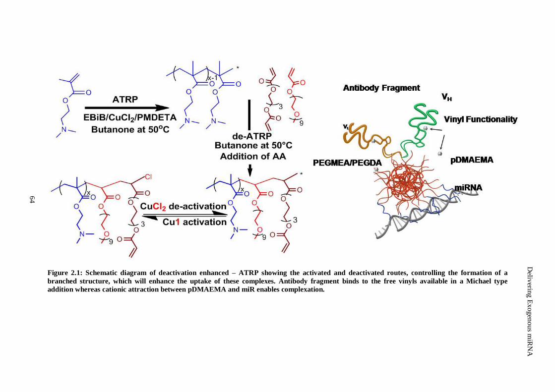
Figure 2.1: Schematic diagram of deactivation enhanced – ATRP showing the activated and deactivated routes, controlling the formation of a
branched structure, which will enhance the uptake of these complexes. Antibody fragment binds to the free vinyls available in a Michael type
addition whereas cationic attraction between pDMAEMA and miR enables complexation.
Deliv
ering E
xogen
ou
s miR
NA
64
Delivering Exogenous miRNA
65
freeze dried for subsequent studies. The final polymer product was stored at -20 °C and protected
from light until use.
2.2.4 Molecular Weight Determination by Gel Permeation Chromatography
Small samples were withdrawn from the reaction at specific intervals using a glass syringe with luer
needle under positive pressure of argon. These were then diluted in DMF and filtered through alumina
for chromatography followed by a 0.2 μm filter before analysis. The molecular weight and molecular
weight distribution of each sample was determined using a Varian™
920 LC instrument with a
refractive index S2 detector (RI). Chromatograms were run at 50 °C using DMF as eluent with a flow
rate of 1 ml/min. The machine was calibrated with linear polystyrene standards. A detailed description
of GPC sample preparation is available in Appendix E.3.
2.2.5 1H Nuclear Magnetic Resonance
1H NMR was performed using a 300 MHz Bruker NMR with MestRec™ processing software. The
polymer was dissolved in Deuterated chloroform (CDCl3, Sigma Aldrich) for 1H NMR analysis and all
chemical shifts are reported in ppm relative to dilute tetramethylsilane (TMS). The 1H NMR spectrum
confirmed the presence of each monomer in the polymer structure and the presence of free vinyl
groups. The NMR spectrum was used to determine the degree of branching within the polymer
structure via Equations 2.1.
2.2.6 Agarose Gel Electrophoresis
Complex formation of miR/ pD-b-P/DA was observed by polyacrylamide gel retardation. This
procedure enables the user to see the effect of complexation on electrophoretic mobility of
oligonucleotides. Various weight ratios of pD-b-P/DA, from 0 to 20, were added to 200 ng of miR-
29B (Syn-rno-miR-29B; miScriptmiRNA Mimic™
(MSY0000801), Qiagen, Hilden, Germany) in
phosphate buffered solution (PBS), vortexed briefly and incubated for 30 minutes at room
temperature. After incubation, each sample was mixed proportionately with a loading dye and
electrophoresed on a 13% polyacrylamide gel (w/v) for one hour at 100 V. 89 mM Tris-borate (TBE),
2 mM ethylenediaminetetraacetic acid (EDTA) buffer was used as the electrophoresis buffer.
Following SYBR®Safe (1:10000) staining for ten minutes, the gel was illuminated with a UV
illuminator to show the location of migrated miR complexes. This experiment was repeated under the
same conditions except that the direction of charge was reversed in order to verify complex formation.
A detailed description of this protocol is available in Appendix F.2.
Delivering Exogenous miRNA
66
2.2.7 Thiol Binding of Polymer
The predicted presence of vinyl groups on this structure enables the covalent binding of thiols via
Michael type addition. In a model system, cysteine hydrochloride monohydrate was employed as a
model of cysteine, having a free thiol group present in its structure. The binding efficacy of pD-b-
P/DA was evaluated by monitoring the consumption of cysteine over time and measured indirectly
using Ellman‟s assay (Appendix N.1). This assay is a colorimetric reaction between Ellman‟s reagent
(5, 5′-dithiobis (2-nitrobenzoic acid), DTNB) and free thiol (SH) groups. 0.4 mg of DTNB dissolved
in 1 mL of the corresponding Ellman‟s buffer (0.2 M NaH2PO4 with 1 mM EDTA at pH 8.0, purged
with argon for 30 minutes) was used as stock solution. For baseline measurement, Ellman‟s stock
diluted 1:10 in Ellman‟s buffer was taken as blank. Standards were created using cysteine
hydrochloride monohydrate (Sigma Aldrich) dissolved in Ellman‟s buffer (0 – 1.5 mM final
concentration). Known concentrations of cysteine were incubated with optimal ratios of pD-b-P/DA,
and corresponding weights of poly (ethylenimine) (PEI), using Ellman‟s buffer as the reaction buffer.
Samples were serially diluted in Ellman‟s buffer and 10% (v/v) of the stock solution and incubated at
37 °C. At each time point (0, 0.5, 1, 2 and 3 hours), 50 μl of Ellman‟s reagent was added to the
reactant. Solutions were measured at 412 nm and the concentration of thiol groups was extrapolated
from the standard curve.
2.2.8 Fab` Generation
Hybridoma cultures expressing antibodies against CD90 (Health Protection Agency, Salisbury, UK)
were expanded in RPMI containing 20% serum. The CD90 antigen was chosen as it is expressed on
the cell surface of fibroblasts which would enable more cell surface attachment of non-viral vectors
with a CD90 antibody fragment attached. These cultures were weaned in graduations of 10, 5, 2.5 and
gradually to 0.5 % serum concentrations. Cells were maintained at 0.5% fetal bovine serum (FBS)
concentration for six weeks at a concentration of 5 x 10⁵ cells/ml to produce antibodies. At the end of
incubation, remaining cells and debris were removed from the media through centrifugation and the
protein fraction of the media separated on Centricon®
(MWCO = 100 kDa, Millipore Corp., Billenica,
MA, USA) filter devices. Antibodies within this fraction were isolated using an antibody purification
kit according to the manufacturer‟s specifications (Nab™
Protein A/G Spin Kit, Thermo Scientific,
Dublin, Ireland). Generation of F(ab')2 was performed using the Pierce F(ab')2 Preparation Kit™
(Thermo Scientific) according to the protocol provided. Fab' generation was performed using Reduce-
Imm Immobilized Reductant Columns™
(Thermo Scientific) according to the protocol provided by the
manufacturer. Molar quantification of antibody fragments was based on free thiol determination based
on Ellman‟s assay. Detailed descriptions of these protocols are available in Appendices J.1, J.2, and
J.3.
Delivering Exogenous miRNA
67
2.2.9 Fab` Conjugation
Known molar concentrations (deduced from Ellman‟s assay) of Fab's were reacted with pD-b-P/DA in
a reaction buffer of 0.2 mM NaH2PO4 with 1 mM EDTA at pH 8.0 (purged prior to reaction with argon
for 30 minutes). Briefly, reactions were performed in which molar ratios of Fab' thiol concentration
(from Ellman‟s assay) and moles of free vinyls (calculated from 1H NMR data) were reacted at ratios
of 1:1 and 1:2. Fab`s alone were used as a control. Samples were reacted for one hour under gentle
rocking (Figure 2.2).
2.2.10 Characterization of miR-Complexes Decorated with Antibody Fragments
Sodium dodecyl sulfate poly (acrylamide) gel electrophoresis (SDS-PAGE) under reducing and non-
reducing conditions was conducted to confirm conjugation of Fab's to pD-b-P/DA. Briefly, 1 μg of
each sample was removed (confirmed by absorbance at 280 nm) denatured via heat treatment and, in
the case of reducing conditions, reduced using β-mercaptoethanol. Samples were loaded onto 10 %
acrylamide gels (Bio-Rad Laboratories Inc., Hertfordshire, UK) and electrophoresis was performed at
a constant voltage (150 V) for approximately one hour for both reducing and non-reducing conditions.
The proteins were revealed by silver staining (SilverQuest™
Silver Staining Kit, Invitrogen) and
subsequently micrographed. Refer to Appendix K for detailed protocol.
Agarose gel electrophoresis was performed to see the effect of antibody fragment conjugation on
complex formation. Briefly, 1 μg of miR was complexed at varying w/w ratios with pD-b-P/DA
conjugated with antibody fragments at a w/w ratio of 1:1 (moles antibody fragments free thiols: moles
vinyl groups available in PEGDA portion of pD-b-P/DA). An agarose gel (1% agarose in TBE buffer,
with SYBR®Safe DNA stain) was made to analyze the weight/weight ratio for polyplex formation.
5 ml of each polymer/miR solution were added along with 5 μl loading dye to each well and subjected
simultaneously to 80 mV for up 30 minutes.
2.2.11 Reporter Plasmid Construction, Transformation, Propagation and Isolation
For construction of a vector containing miRNA target sequence fused downstream from a luciferase
expression cassette, the pmiRGlo™
Dual-Luciferase miRNA Target Expression Vector (Promega
Corporation) was employed (Figure 2.3). This plasmid encodes transcription for both Renilla and
Firefly luciferase with a miR binding site downstream of the Firefly mRNA. The pmiRGlo™
vector
was linearized using XhoI and XbaI restriction enzymes (Promega Corporation) for three hours at 37
°C. This reaction was inactivated by heat at 65 °C for 25 minutes and purified using Qiaquick® PCR
purification kit (Qiagen).
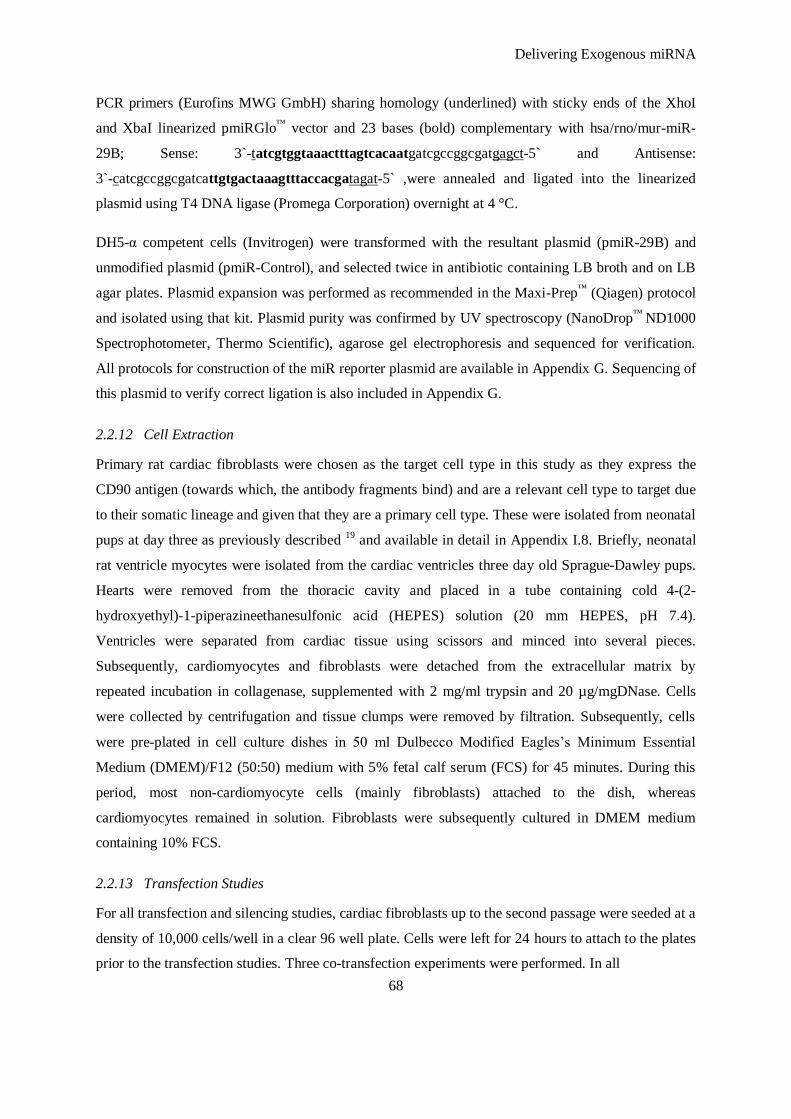
Delivering Exogenous miRNA
68
PCR primers (Eurofins MWG GmbH) sharing homology (underlined) with sticky ends of the XhoI
and XbaI linearized pmiRGlo™
vector and 23 bases (bold) complementary with hsa/rno/mur-miR-
29B; Sense: 3`-tatcgtggtaaactttagtcacaatgatcgccggcgatgagct-5` and Antisense:
3`-catcgccggcgatcattgtgactaaagtttaccacgatagat-5` ,were annealed and ligated into the linearized
plasmid using T4 DNA ligase (Promega Corporation) overnight at 4 °C.
DH5-α competent cells (Invitrogen) were transformed with the resultant plasmid (pmiR-29B) and
unmodified plasmid (pmiR-Control), and selected twice in antibiotic containing LB broth and on LB
agar plates. Plasmid expansion was performed as recommended in the Maxi-Prep™
(Qiagen) protocol
and isolated using that kit. Plasmid purity was confirmed by UV spectroscopy (NanoDrop™
ND1000
Spectrophotometer, Thermo Scientific), agarose gel electrophoresis and sequenced for verification.
All protocols for construction of the miR reporter plasmid are available in Appendix G. Sequencing of
this plasmid to verify correct ligation is also included in Appendix G.
2.2.12 Cell Extraction
Primary rat cardiac fibroblasts were chosen as the target cell type in this study as they express the
CD90 antigen (towards which, the antibody fragments bind) and are a relevant cell type to target due
to their somatic lineage and given that they are a primary cell type. These were isolated from neonatal
pups at day three as previously described 19
and available in detail in Appendix I.8. Briefly, neonatal
rat ventricle myocytes were isolated from the cardiac ventricles three day old Sprague-Dawley pups.
Hearts were removed from the thoracic cavity and placed in a tube containing cold 4-(2-
hydroxyethyl)-1-piperazineethanesulfonic acid (HEPES) solution (20 mm HEPES, pH 7.4).
Ventricles were separated from cardiac tissue using scissors and minced into several pieces.
Subsequently, cardiomyocytes and fibroblasts were detached from the extracellular matrix by
repeated incubation in collagenase, supplemented with 2 mg/ml trypsin and 20 µg/mgDNase. Cells
were collected by centrifugation and tissue clumps were removed by filtration. Subsequently, cells
were pre-plated in cell culture dishes in 50 ml Dulbecco Modified Eagles‟s Minimum Essential
Medium (DMEM)/F12 (50:50) medium with 5% fetal calf serum (FCS) for 45 minutes. During this
period, most non-cardiomyocyte cells (mainly fibroblasts) attached to the dish, whereas
cardiomyocytes remained in solution. Fibroblasts were subsequently cultured in DMEM medium
containing 10% FCS.
2.2.13 Transfection Studies
For all transfection and silencing studies, cardiac fibroblasts up to the second passage were seeded at a
density of 10,000 cells/well in a clear 96 well plate. Cells were left for 24 hours to attach to the plates
prior to the transfection studies. Three co-transfection experiments were performed. In all
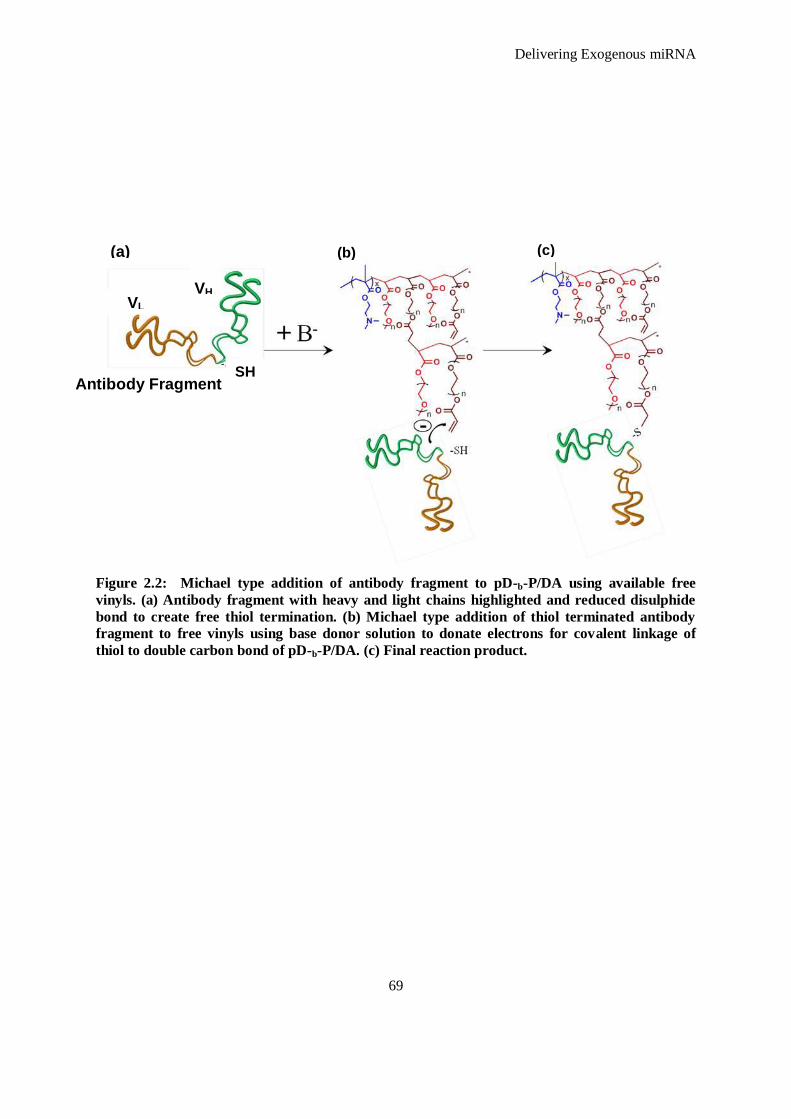
Delivering Exogenous miRNA
69
Figure 2.2: Michael type addition of antibody fragment to pD-b-P/DA using available free
vinyls. (a) Antibody fragment with heavy and light chains highlighted and reduced disulphide
bond to create free thiol termination. (b) Michael type addition of thiol terminated antibody
fragment to free vinyls using base donor solution to donate electrons for covalent linkage of
thiol to double carbon bond of pD-b-P/DA. (c) Final reaction product.
Antibody Fragment SH
VL VH
(b) (c) (a)
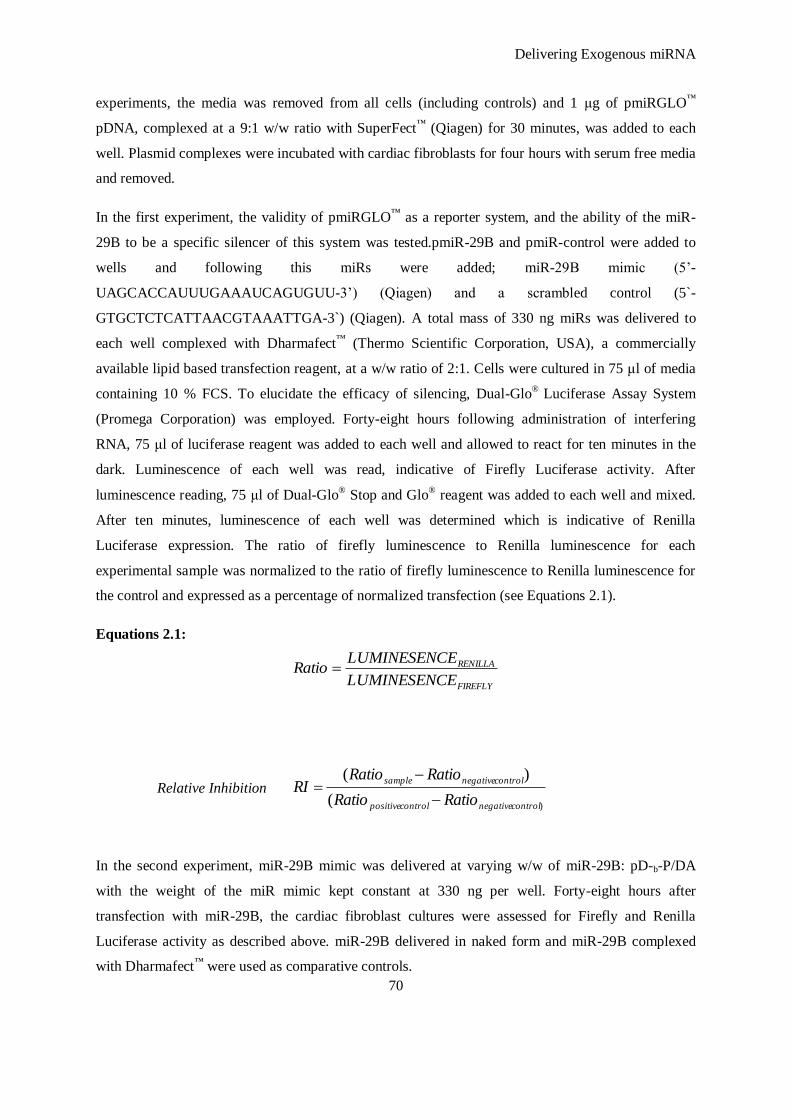
Delivering Exogenous miRNA
70
experiments, the media was removed from all cells (including controls) and 1 μg of pmiRGLO™
pDNA, complexed at a 9:1 w/w ratio with SuperFect™
(Qiagen) for 30 minutes, was added to each
well. Plasmid complexes were incubated with cardiac fibroblasts for four hours with serum free media
and removed.
In the first experiment, the validity of pmiRGLO™
as a reporter system, and the ability of the miR-
29B to be a specific silencer of this system was tested.pmiR-29B and pmiR-control were added to
wells and following this miRs were added; miR-29B mimic (5‟-
UAGCACCAUUUGAAAUCAGUGUU-3‟) (Qiagen) and a scrambled control (5`-
GTGCTCTCATTAACGTAAATTGA-3`) (Qiagen). A total mass of 330 ng miRs was delivered to
each well complexed with Dharmafect™
(Thermo Scientific Corporation, USA), a commercially
available lipid based transfection reagent, at a w/w ratio of 2:1. Cells were cultured in 75 μl of media
containing 10 % FCS. To elucidate the efficacy of silencing, Dual-Glo®
Luciferase Assay System
(Promega Corporation) was employed. Forty-eight hours following administration of interfering
RNA, 75 μl of luciferase reagent was added to each well and allowed to react for ten minutes in the
dark. Luminescence of each well was read, indicative of Firefly Luciferase activity. After
luminescence reading, 75 μl of Dual-Glo® Stop and Glo
® reagent was added to each well and mixed.
After ten minutes, luminescence of each well was determined which is indicative of Renilla
Luciferase expression. The ratio of firefly luminescence to Renilla luminescence for each
experimental sample was normalized to the ratio of firefly luminescence to Renilla luminescence for
the control and expressed as a percentage of normalized transfection (see Equations 2.1).
Equations 2.1:
Relative Inhibition
In the second experiment, miR-29B mimic was delivered at varying w/w of miR-29B: pD-b-P/DA
with the weight of the miR mimic kept constant at 330 ng per well. Forty-eight hours after
transfection with miR-29B, the cardiac fibroblast cultures were assessed for Firefly and Renilla
Luciferase activity as described above. miR-29B delivered in naked form and miR-29B complexed
with Dharmafect™
were used as comparative controls.
FIREFLY
RENILLA
ELUMINESENC
ELUMINESENCRatio
)(
)(
controlnegativecontrolpositive
controlnegativesample
RatioRatio
RatioRatioRI
Delivering Exogenous miRNA
71
Figure 2.3: Overview of reporter luciferase knockdown. (A) miR-29B sequence for silencing
protein expression. (B) pmiRGlo vector construction. The firefly and Renilla luciferase genes
are driven by human phosphoglycerate kinase (PGK) and simian virus 40 (SV40) promoters,
respectively. A binding site of miR-29B was cloned into the pmiR-Glo vector downstream of the
firefly gene (pmiR-29B) using a multiple cloning site (MCS). (C) Sequence details of pmiR-29B
with target sequence in purple, a NOT-I digestion site in Blue and restriction enzyme sites XbaI
and XhoI in red.
pmiR -29B: 3`-AGTCGAACGTACGGACGTCCAGTCGAGATCTATCGTGGTAAACTTTAGTCACAAatgatCGCCGGCGATGAGCTCCGATCGC-5`
pmiR-Control: 3`-AGTCGAACGTACGGACGTCCAGTCGAGATCTGAGCTCCGATCGC-5`
PGKFirefly
LuciferasemiR-29B Target SV 40
Renilla Luciferase
PGKFirefly
LuciferaseUnmodified SV 40
Renilla Luciferase
5`
5`
3`
3`
Promoter PromoterMCS
miR-29B: 5’-UAGCACCAUUUGAAAUCAGUGUU-3’A
B
C
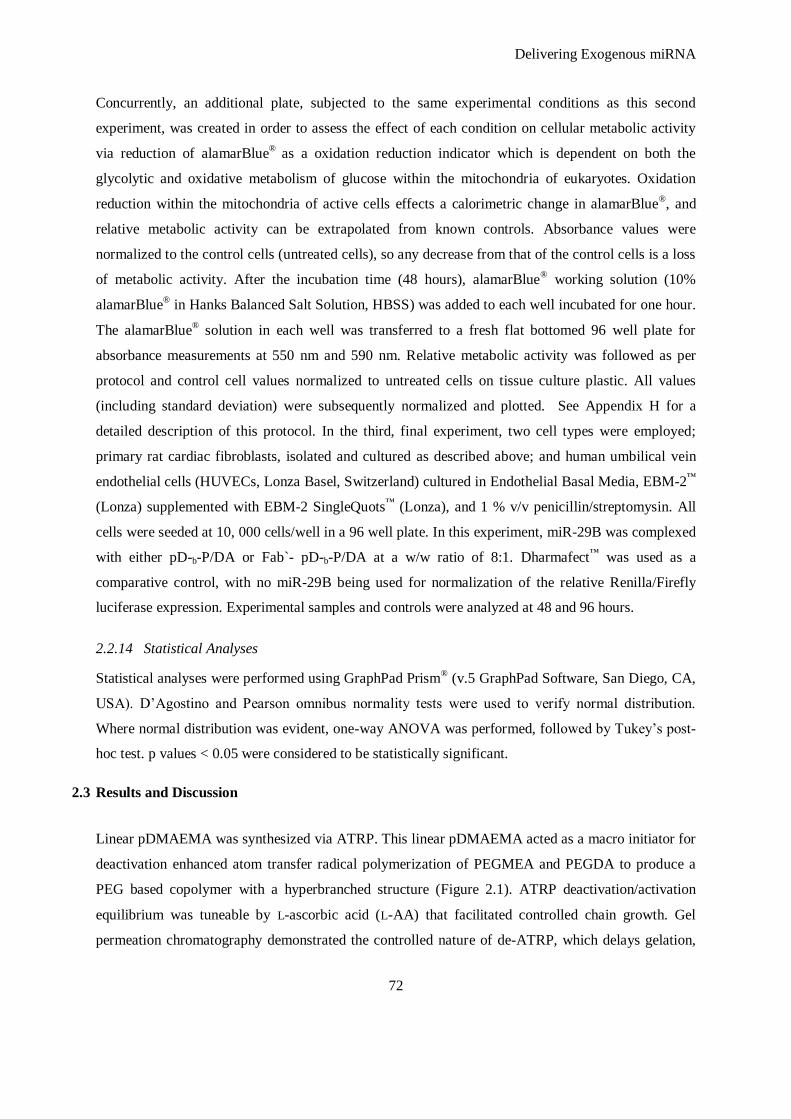
Delivering Exogenous miRNA
72
Concurrently, an additional plate, subjected to the same experimental conditions as this second
experiment, was created in order to assess the effect of each condition on cellular metabolic activity
via reduction of alamarBlue®
as a oxidation reduction indicator which is dependent on both the
glycolytic and oxidative metabolism of glucose within the mitochondria of eukaryotes. Oxidation
reduction within the mitochondria of active cells effects a calorimetric change in alamarBlue®, and
relative metabolic activity can be extrapolated from known controls. Absorbance values were
normalized to the control cells (untreated cells), so any decrease from that of the control cells is a loss
of metabolic activity. After the incubation time (48 hours), alamarBlue® working solution (10%
alamarBlue®
in Hanks Balanced Salt Solution, HBSS) was added to each well incubated for one hour.
The alamarBlue® solution in each well was transferred to a fresh flat bottomed 96 well plate for
absorbance measurements at 550 nm and 590 nm. Relative metabolic activity was followed as per
protocol and control cell values normalized to untreated cells on tissue culture plastic. All values
(including standard deviation) were subsequently normalized and plotted. See Appendix H for a
detailed description of this protocol. In the third, final experiment, two cell types were employed;
primary rat cardiac fibroblasts, isolated and cultured as described above; and human umbilical vein
endothelial cells (HUVECs, Lonza Basel, Switzerland) cultured in Endothelial Basal Media, EBM-2™
(Lonza) supplemented with EBM-2 SingleQuots™
(Lonza), and 1 % v/v penicillin/streptomysin. All
cells were seeded at 10, 000 cells/well in a 96 well plate. In this experiment, miR-29B was complexed
with either pD-b-P/DA or Fab`- pD-b-P/DA at a w/w ratio of 8:1. Dharmafect™
was used as a
comparative control, with no miR-29B being used for normalization of the relative Renilla/Firefly
luciferase expression. Experimental samples and controls were analyzed at 48 and 96 hours.
2.2.14 Statistical Analyses
Statistical analyses were performed using GraphPad Prism® (v.5 GraphPad Software, San Diego, CA,
USA). D‟Agostino and Pearson omnibus normality tests were used to verify normal distribution.
Where normal distribution was evident, one-way ANOVA was performed, followed by Tukey‟s post-
hoc test. p values < 0.05 were considered to be statistically significant.
2.3 Results and Discussion
Linear pDMAEMA was synthesized via ATRP. This linear pDMAEMA acted as a macro initiator for
deactivation enhanced atom transfer radical polymerization of PEGMEA and PEGDA to produce a
PEG based copolymer with a hyperbranched structure (Figure 2.1). ATRP deactivation/activation
equilibrium was tuneable by L-ascorbic acid (L-AA) that facilitated controlled chain growth. Gel
permeation chromatography demonstrated the controlled nature of de-ATRP, which delays gelation,
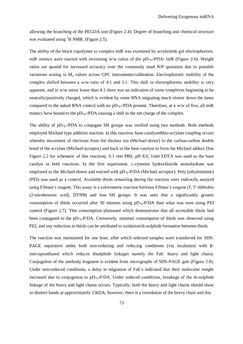
Delivering Exogenous miRNA
73
allowing the branching of the PEGDA unit (Figure 2.4). Degree of branching and chemical structure
was evaluated using 1H NMR, (Figure 2.5).
The ability of the block copolymer to complex miR was examined by acrylamide gel electrophoresis.
miR mimics were reacted with increasing w/w ratios of the pD-b-/PDA/ miR (Figure 2.6). Weight
ratios are quoted for increased accuracy over the commonly used N/P quotation due to possible
variations arising in Mn values across GPC instruments/calibration. Electrophoretic mobility of the
complex shifted between a w/w ratio of 4:1 and 5:1. This shift in electrophoretic mobility is very
apparent, and in w/w ratios lower than 4:1 there was an indication of some complexes beginning to be
neutrally/positively charged, which is evident by some RNA migrating much slower down the lanes
compared to the naked RNA control with no pD-b-/PDA present. Therefore, at a w/w of five, all miR
mimics have bound to the pD-b-/PDA causing a shift in the net charge of the complex.
The ability of pD-b-/PDA to conjugate SH groups was verified using two methods. Both methods
employed Michael type addition reaction. In this reaction, base-catalyzedthio-acrylate coupling occurs
whereby movement of electrons from the thiolate ion (Michael-donor) to the carbon-carbon double
bond of the acrylate (Michael-acceptor) and back to the base catalyst to form the Michael adduct (See
Figure 2.2 for schematic of this reaction). 0.1 mM PBS, pH 8.0, 1mM EDTA was used as the base
catalyst in both reactions. In the first experiment, L-cysteine hydrochloride monohydrate was
employed as the Michael-donor and reacted with pD-b-P/DA (Michael acceptor). Poly (ethylenimine)
(PEI) was used as a control. Available thiols remaining during the reaction were indirectly assayed
using Ellman‟s reagent. This assay is a colorimetric reaction between Ellman‟s reagent (5, 5′-dithiobis
(2-nitrobenzoic acid), DTNB) and free SH groups. It was seen that a significantly greater
consumption of thiols occurred after 30 minutes using pD-b-P/DA than what was seen using PEI
control (Figure 2.7). This consumption plateaued which demonstrates that all accessible thiols had
been conjugated to the pD-b-P/DA. Conversely, minimal consumption of thiols was observed using
PEI, and any reduction in thiols can be attributed to oxidation/di-sulphide formation between thiols.
The reaction was maintained for one hour, after which selected samples were transferred for SDS-
PAGE separation under both non-reducing and reducing conditions (via incubation with β-
mercaptoethanol which reduces disulphide linkages namely the Fab` heavy and light chain).
Conjugation of the antibody fragment is evident from micrographs of SDS-PAGE gels (Figure 2.8).
Under non-reduced conditions, a delay in migration of Fab`s indicated that their molecular weight
increased due to conjugation to pD-b-P/DA. Under reduced conditions, breakage of the di-sulphide
linkage of the heavy and light chains occurs. Typically, both the heavy and light chains should show
as distinct bands at approximately 25kDA; however, there is a retardation of the heavy chain and this
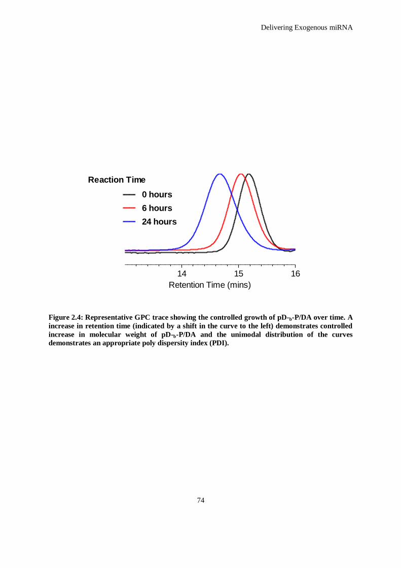
Delivering Exogenous miRNA
74
14 15 16
0 hours
6 hours
24 hours
Reaction Time
Retention Time (mins)
Figure 2.4: Representative GPC trace showing the controlled growth of pD-b-P/DA over time. A
increase in retention time (indicated by a shift in the curve to the left) demonstrates controlled
increase in molecular weight of pD-b-P/DA and the unimodal distribution of the curves
demonstrates an appropriate poly dispersity index (PDI).
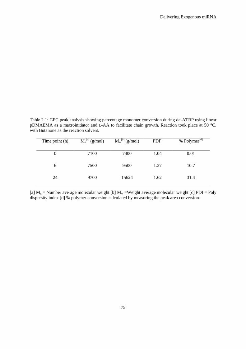
Delivering Exogenous miRNA
75
Table 2.1: GPC peak analysis showing percentage monomer conversion during de-ATRP using linear
pDMAEMA as a macroinitiator and L-AA to facilitate chain growth. Reaction took place at 50 °C,
with Butanone as the reaction solvent.
[a] Mn = Number average molecular weight [b] Mw =Weight average molecular weight [c] PDI = Poly
dispersity index [d] % polymer conversion calculated by measuring the peak area conversion.
Time point (h) Mn[a]
(g/mol) Mw[b]
(g/mol) PDIc] % Polymer
[d]
0 7100 7400 1.04 0.01
6 7500 9500 1.27 10.7
24 9700 15624 1.62 31.4
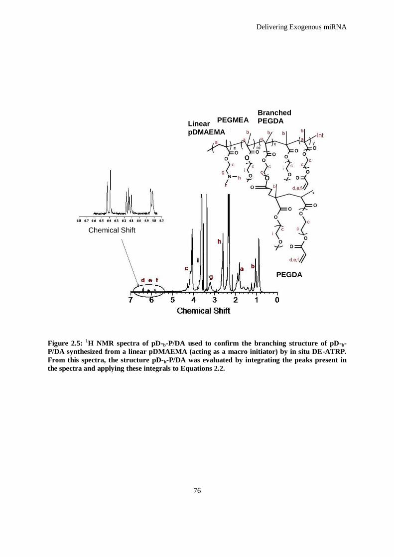
Delivering Exogenous miRNA
76
Figure 2.5: 1H NMR spectra of pD-b-P/DA used to confirm the branching structure of pD-b-
P/DA synthesized from a linear pDMAEMA (acting as a macro initiator) by in situ DE-ATRP.
From this spectra, the structure pD-b-P/DA was evaluated by integrating the peaks present in
the spectra and applying these integrals to Equations 2.2.
Chemical Shift
Linear pDMAEMA
PEGMEA Branched PEGDA
PEGDA

Delivering Exogenous miRNA
77
Equations 2.2
DMAEMA Component (n) =
PEGMEA Component (m) =
PEGDA (free vinyl) Component (x) =
Branching PEGDA Component (y) =
Total Content
Therefore, the percentage of each component within the polymer structure is a percentage of
the Total Content, for instance:
% DMAEMA
Table 2.2: Polymer composition as calculated from 1H NMR peak area by Equations 2.1.
Polymer Composition by 1H NMR (%)
DMAEMA PEGMEA Branching PEGDA Free Vinyl
68 19.5 12.45 3.1
2
g
3
i
3
fed
)242( mxnc
yxmn
yxmn
n
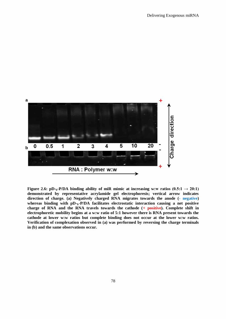
Delivering Exogenous miRNA
78
Figure 2.6: pD-b-P/DA binding ability of miR mimic at increasing w:w ratios (0.5:1 → 20:1)
demonstrated by representative acrylamide gel electrophoresis; vertical arrow indicates
direction of charge. (a) Negatively charged RNA migrates towards the anode (- negative)
whereas binding with pD-b-P/DA facilitates electrostatic interaction causing a net positive
charge of RNA and the RNA travels towards the cathode (+ positive). Complete shift in
electrophoretic mobility begins at a w:w ratio of 5:1 however there is RNA present towards the
cathode at lower w:w ratios but complete binding does not occur at the lower w:w ratios.
Verification of complexation observed in (a) was performed by reversing the charge terminals
in (b) and the same observations occur.
a
b
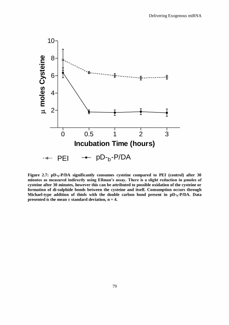
Delivering Exogenous miRNA
79
Figure 2.7: pD-b-P/DA significantly consumes cysteine compared to PEI (control) after 30
minutes as measured indirectly using Ellman’s assay. There is a slight reduction in μmoles of
cysteine after 30 minutes, however this can be attributed to possible oxidation of the cysteine or
formation of di-sulphide bonds between the cysteine and itself. Consumption occurs through
Michael-type addition of thiols with the double carbon bond present in pD-b-P/DA. Data
presented is the mean ± standard deviation, n = 4.
0 0.5 1 2 3
2
4
6
8
10
pD-b-P/DAPEI
Incubation Time (hours)
m
ole
s C
yste
ine
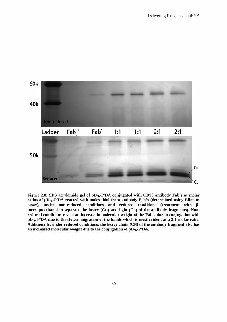
Delivering Exogenous miRNA
80
Figure 2.8: SDS acrylamide gel of pD-b-P/DA conjugated with CD90 antibody Fab`s at molar
ratios of pD-b-P/DA reacted with moles thiol from antibody Fab’s (determined using Ellmans
assay), under non-reduced conditions and reduced conditions (treatment with β-
mercaptoethanol to separate the heavy (CH) and light (CL) of the antibody fragments). Non-
reduced conditions reveal an increase in molecular weight of the Fab`s due to conjugation with
pD-b-P/DA due to the slower migration of the bands which is most evident at a 2:1 molar ratio.
Additionally, under reduced conditions, the heavy chain (CH) of the antibody fragment also has
an increased molecular weight due to the conjugation of pD-b-P/DA.
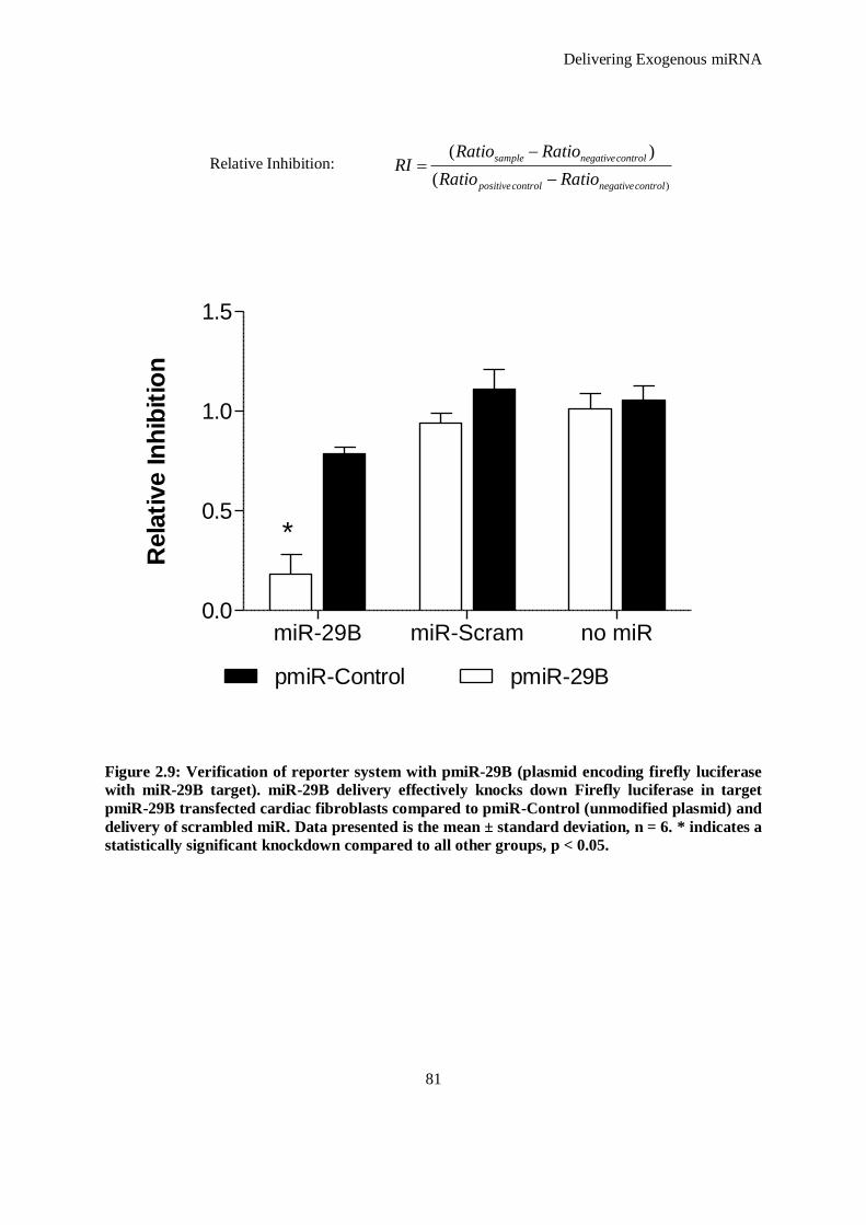
Delivering Exogenous miRNA
81
)(
)(
controlnegativecontrolpositive
controlnegativesample
RatioRatio
RatioRatioRI
Relative Inhibition:
Figure 2.9: Verification of reporter system with pmiR-29B (plasmid encoding firefly luciferase
with miR-29B target). miR-29B delivery effectively knocks down Firefly luciferase in target
pmiR-29B transfected cardiac fibroblasts compared to pmiR-Control (unmodified plasmid) and
delivery of scrambled miR. Data presented is the mean ± standard deviation, n = 6. * indicates a
statistically significant knockdown compared to all other groups, p < 0.05.
miR-29B miR-Scram no miR0.0
0.5
1.0
1.5
pmiR-29BpmiR-Control
*
Rela
tive In
hib
itio
n
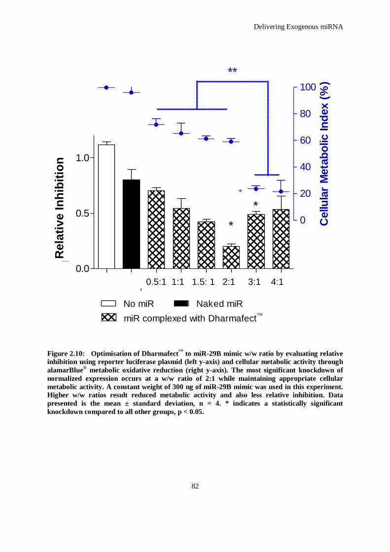
Delivering Exogenous miRNA
82
Figure 2.10: Optimisation of Dharmafect™
to miR-29B mimic w/w ratio by evaluating relative
inhibition using reporter luciferase plasmid (left y-axis) and cellular metabolic activity through
alamarBlue® metabolic oxidative reduction (right y-axis). The most significant knockdown of
normalized expression occurs at a w/w ratio of 2:1 while maintaining appropriate cellular
metabolic activity. A constant weight of 300 ng of miR-29B mimic was used in this experiment.
Higher w/w ratios result reduced metabolic activity and also less relative inhibition. Data
presented is the mean ± standard deviation, n = 4. * indicates a statistically significant
knockdown compared to all other groups, p < 0.05.
0.5:
11:
11.
5:1
2:1
3:1
4:1
0.0
0.5
1.0
*
*
No miR Naked miR
miR complexed with Dharmafect
No
rmalized
Exp
ressio
n
0
20
40
60
80
100
**
Cellu
lar
Meta
bo
lic In
dex (
%)
*
0.5:1 1:1 1.5: 1 2:1 3:1 4:1
Rela
tiv
e I
nh
ibit
ion
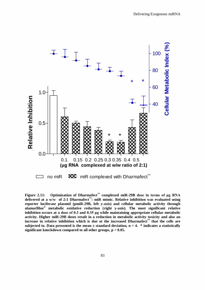
Delivering Exogenous miRNA
83
Figure 2.11: Optimisation of Dharmafect™
complexed miR-29B dose in terms of μg RNA
delivered at a w/w of 2:1 Dharmafect™
: miR mimic. Relative inhibition was evaluated using
reporter luciferase plasmid (pmiR-29B, left y-axis) and cellular metabolic activity through
alamarBlue® metabolic oxidative reduction (right y-axis). The most significant relative
inhibition occurs at a dose of 0.3 and 0.35 μg while maintaining appropriate cellular metabolic
activity. Higher miR-29B doses result in a reduction in metabolic activity toxicity and also an
increase in relative inhibition which is due ot the increased Dharmafect™
that the cells are
subjected to. Data presented is the mean ± standard deviation, n = 4. * indicates a statistically
significant knockdown compared to all other groups, p < 0.05.
100
ng
150
ng
200
ng
250
ng
300
ng
350
ng
400
ng
500
ng 0.0
0.5
1.0
no miR miR complexed with Dharmafect
* *
No
rmalized
Exp
ressio
n
40
60
80
100
* *
Cellu
lar
Meta
bo
lic In
dex (
%)
0.1 0.15 0.2 0.25 0.3 0.35 0.4 0.5 (μg RNA complexed at w/w ratio of 2:1)
Rela
tiv
e I
nh
ibit
ion
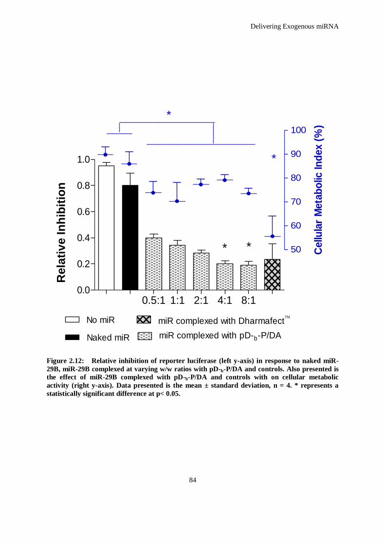
Delivering Exogenous miRNA
84
Figure 2.12: Relative inhibition of reporter luciferase (left y-axis) in response to naked miR-
29B, miR-29B complexed at varying w/w ratios with pD-b-P/DA and controls. Also presented is
the effect of miR-29B complexed with pD-b-P/DA and controls with on cellular metabolic
activity (right y-axis). Data presented is the mean ± standard deviation, n = 4. * represents a
statistically significant difference at p< 0.05.
0.5:1 1:1 2:1 4:1 8:10.0
0.2
0.4
0.6
0.8
1.0
* *
No miR
Naked miR miR complexed with pD-b-P/DA
miR complexed with Dharmafect
No
rmalized
Exp
ressio
n
50
60
70
80
90
100
*
*
Cellu
lar
Meta
bo
lic In
dex (
%)
Rela
tiv
e I
nh
ibit
ion
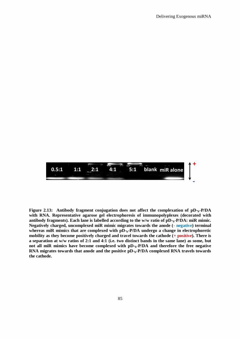
Delivering Exogenous miRNA
85
Figure 2.13: Antibody fragment conjugation does not affect the complexation of pD-b-P/DA
with RNA. Representative agarose gel electrophoresis of immunopolyplexes (decorated with
antibody fragments). Each lane is labelled according to the w/w ratio of pD-b-P/DA: miR mimic.
Negatively charged, uncomplexed miR mimic migrates towards the anode (- negative) terminal
whereas miR mimics that are complexed with pD-b-P/DA undergo a change in electrophoretic
mobility as they become positively charged and travel towards the cathode (+ positive). There is
a separation at w/w ratios of 2:1 and 4:1 (i.e. two distinct bands in the same lane) as some, but
not all miR mimics have become complexed with pD-b-P/DA and therefore the free negative
RNA migrates towards that anode and the positive pD-b-P/DA complexed RNA travels towards
the cathode.
0.5:1 1 :1 2:1 4 :1 5:1 blank
+
-
miR alone
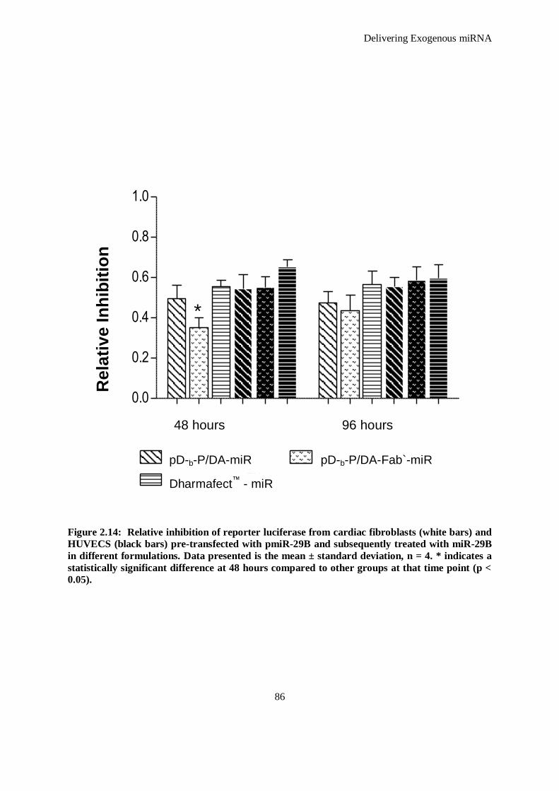
Delivering Exogenous miRNA
86
Figure 2.14: Relative inhibition of reporter luciferase from cardiac fibroblasts (white bars) and
HUVECS (black bars) pre-transfected with pmiR-29B and subsequently treated with miR-29B
in different formulations. Data presented is the mean ± standard deviation, n = 4. * indicates a
statistically significant difference at 48 hours compared to other groups at that time point (p <
0.05).
0.0
0.2
0.4
0.6
0.8
1.0
pD-b-P/DA - miR pD-b-P/DA - Fab` - miR
DharmafectTM -
miR
*
48 hours 96 hours
No
rmalized
Exp
ressio
n
48 hours 96 hours
pD-b-P/DA-miR pD-b-P/DA-Fab`-miR
Dharmafect™ - miR
Rela
tiv
e I
nh
ibit
ion
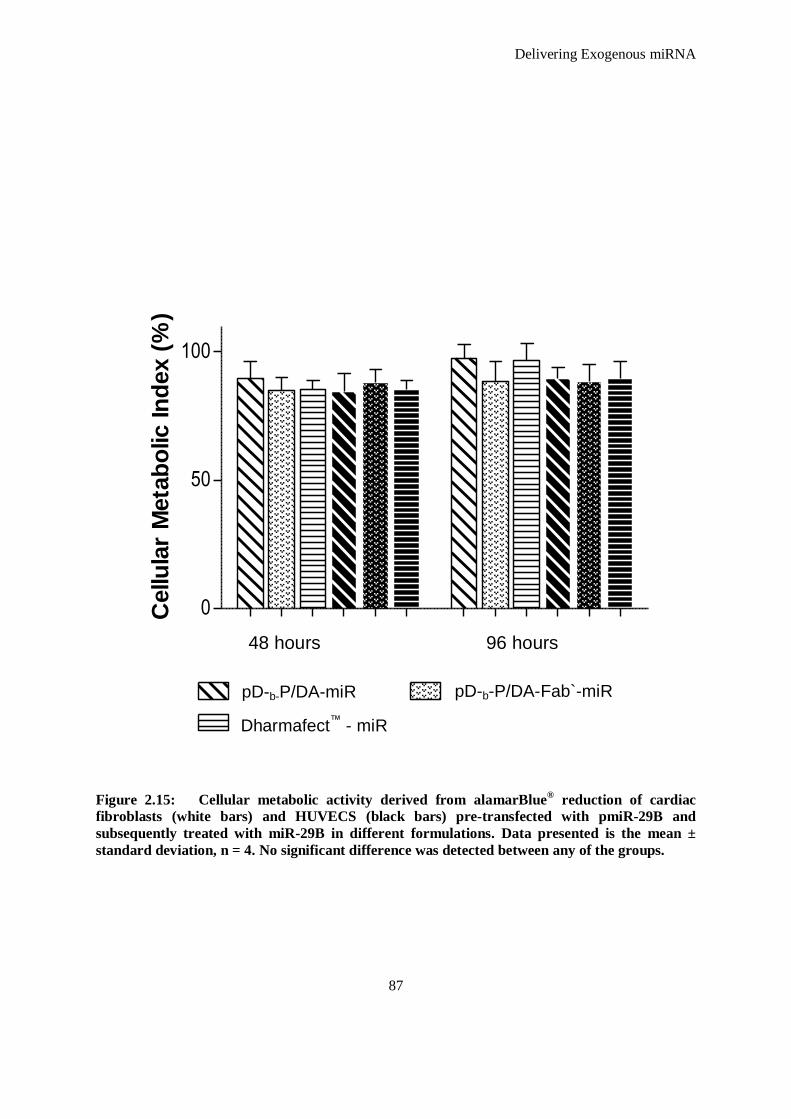
Delivering Exogenous miRNA
87
Figure 2.15: Cellular metabolic activity derived from alamarBlue® reduction of cardiac
fibroblasts (white bars) and HUVECS (black bars) pre-transfected with pmiR-29B and
subsequently treated with miR-29B in different formulations. Data presented is the mean ±
standard deviation, n = 4. No significant difference was detected between any of the groups.
0
50
100
pD-b-P/DA - miR pD-b-P/DA - Fab` - miR
DharmafectTM -
miR
48 hours 96 hours
Cellu
lar
Meta
bo
lic In
dex (
%)
48 hours 96 hours
pD-b-P/DA-miR pD-b-P/DA-Fab`-miR
Dharmafect™ - miR
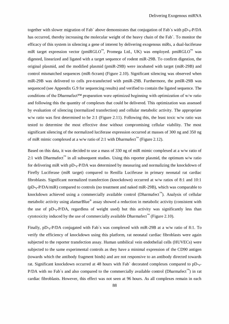
Delivering Exogenous miRNA
88
together with slower migration of Fab` above demonstrates that conjugation of Fab`s with pD-b-P/DA
has occurred, thereby increasing the molecular weight of the heavy chain of the Fab`. To monitor the
efficacy of this system in silencing a gene of interest by delivering exogenous miRs, a dual-luciferase
miR target expression vector (pmiRGLO™
, Promega Ltd., UK) was employed. pmiRGLO™
was
digested, linearized and ligated with a target sequence of rodent miR-29B. To confirm digestion, the
original plasmid, and the modified plasmid (pmiR-29B) were incubated with target (miR-29B) and
control mismatched sequences (miR-Scram) (Figure 2.10). Significant silencing was observed when
miR-29B was delivered to cells pre-transfected with pmiR-29B. Furthermore, the pmIR-29B was
sequenced (see Appendix G.9 for sequencing results) and verified to contain the ligated sequence. The
conditions of the Dharmafect™ preparation were optimized beginning with optimization of w/w ratio
and following this the quantity of complexes that could be delivered. This optimization was assessed
by evaluation of silencing (normalized transfection) and cellular metabolic activity. The appropriate
w/w ratio was first determined to be 2:1 (Figure 2.11). Following this, the least toxic w/w ratio was
tested to determine the most effective dose without compromising cellular viability. The most
significant silencing of the normalized luciferase expression occurred at masses of 300 ng and 350 ng
of miR mimic complexed at a w/w ratio of 2:1 with Dharmafect™
(Figure 2.12).
Based on this data, it was decided to use a mass of 330 ng of miR mimic complexed at a w/w ratio of
2:1 with Dharmafect™
in all subsequent studies. Using this reporter plasmid, the optimum w/w ratio
for delivering miR with pD-b-P/DA was determined by measuring and normalizing the knockdown of
Firefly Luciferase (miR target) compared to Renilla Luciferase in primary neonatal rat cardiac
fibroblasts. Significant normalized transfection (knockdown) occurred at w/w ratios of 8:1 and 10:1
(pD-b-P/DA/miR) compared to controls (no treatment and naked miR-29B), which was comparable to
knockdown achieved using a commercially available control (Dharmafect™
). Analysis of cellular
metabolic activity using alamarBlue® assay showed a reduction in metabolic activity (consistent with
the use of pD-b-P/DA, regardless of weight used) but this activity was significantly less than
cytotoxicity induced by the use of commercially available Dharmafect™
(Figure 2.10).
Finally, pD-b-P/DA conjugated with Fab`s was complexed with miR-29B at a w/w ratio of 8:1. To
verify the efficiency of knockdown using this platform, rat neonatal cardiac fibroblasts were again
subjected to the reporter transfection assay. Human umbilical vein endothelial cells (HUVECs) were
subjected to the same experimental controls as they have a minimal expression of the CD90 antigen
(towards which the antibody fragment binds) and are not responsive to an antibody directed towards
rat. Significant knockdown occurred at 48 hours with Fab` decorated complexes compared to pD-b-
P/DA with no Fab`s and also compared to the commercially available control (Dharmafect™
) in rat
cardiac fibroblasts. However, this effect was not seen at 96 hours. As all complexes remain in each
Delivering Exogenous miRNA
89
well during the duration of a study, it is hypothesized that in the first time point (48 hours) the Fab`
decorated complexes have adhered quicker to their target cell and have been internalized before those
with no Fab`. Therefore silencing occurs quicker. However, at the 96 hour time point it is probable
that all complexes have adhered to the cells due to the charge interaction of the complexes with the
cell membrane and therefore no statistically significant difference can be detected between Fab`
decorated complexes and non-Fab` decorated complexes. No effect of Fab` decorated complexes was
observed in HUVECs (Figure 2.15). Additionally, no significant difference was observed between any
of the groups at 48 hours or between any of the groups at 96 hours with regard to cellular metabolic
activity with addition of the complexes (Figure 2.16).
2.4 Conclusion
The development of non-toxic, efficient, and clinically relevant delivery systems remains an
important challenge for the clinical applications of interfering RNA-based therapeutics. In this study
synthesis of a hyperbranched multi-vinyl polymer was achieved by controlling ATRP in deactivation
enhanced mode by employing L-AA as a reducing agent and using a macro-initiator (in this case
pDMAEMA) achieving a co-polymer system using DE-ATRP. This reaction was monitored using
GPC and the structure of the synthesized pD-b-/PDA was evaluated using 1H NMR. In this study, the
efficacy of a miR mimic delivery platform that is based on a cationic interaction of pDMAEMA
tertiary amines and phosphates present in the backbone of the miR was demonstrated. This interaction
was monitored and verified using agarose gel electrophoresis. The presence of double carbon bond
sites as potential acceptor units in a relatively mild but highly efficient Michael type addition (as
described in Figure 2.2) enabled the conjugation of antibody fragments to this system, which was
demonstrated using SDS-PAGE and enabled improved delivery and efficacy of miRs (as verified by
knockdown in normalized transfection in cardiac fibroblasts at 48 hours). This method opens up a
plethora of options where this platform can be employed and tuned toward functionalization
strategies. Realistically, any SH-terminated moiety can act as a Michael donor for conjugation to the
pD-b-/PDA synthesized and characterized in this chapter. This encompasses any monoclonal antibody
reduced to form Fab`s, peptides engineered to have cysteine terminations, and also scFvs selected and
amplified using phage display technology 20
to produce effective targeting molecules. Presented here
in this chapter, is demonstrative data proving the efficacy of this system which will be tested towards
a functional application in the proceeding chapters.
Delivering Exogenous miRNA
90
2.5 References
1. Shan X, Lin X, Fu H, Deng Y, Zhou L, Zhu N, Liu Y, Zhang Y, Li Y, Lin S-G, Yu Y. Upregulated expression of miR-1/miR-206 in a rat model of myocardial infarction. Biochem.
Biophys.l Res. Comm. 2009;381:597-601
2. Trang P, Wiggins JF, Daige CL, Cho C, Omotola M, Brown D, Weidhaas JB, Bader AG,
Slack FJ. Systemic delivery of tumor suppressor microRNA mimics using a neutral lipid emulsion inhibits lung tumors in mice. Mol. Ther. 2011;19:1116-1122
3. van Rooij E, Sutherland LB, Thatcher JE, DiMaio JM, Naseem RH, Marshall WS, Hill JA,
Olson EN. Dysregulation of microRNAs after myocardial infarction reveals a role of miR-29 in cardiac fibrosis. Proc. Nat.Acad. Sci. 2008;105:13027-13032
4. Yekta S, Shih I-h, Bartel DP. MicroRNA-directed cleavage of HoxB8 mRNA. Science.
2004;304:594-596 5. Monaghan M, Pandit A. RNA interference therapy via functionalized scaffolds. Adv. Drug
Del. Rev. 2011;63:197-208
6. Manjunath N, Wu H, Subramanya S, Shankar P. Lentiviral delivery of short hairpin RNAs.
Adv. Drug Del. Rev. 2009;61:732-745 7. Tomlinson E, Rolland AP. Controllable gene therapy pharmaceutics of non-viral gene
delivery systems. J. Controlled Release. 1996;39:357-372
8. Rahbek UL, Howard KA, Oupicky D, Manickam DS, Dong M, Nielsen AF, Hansen TB, Besenbacher F, Kjems J. Intracellular siRNA and precursor miRNA trafficking using
bioresponsive copolypeptides. J. Gene Med. 2008;10:81-93
9. Xu D, Takeshita F, Hino Y, Fukunaga S, Kudo Y, Tamaki A, Matsunaga J, Takahashi R-u,
Takata T, Shimamoto A, Ochiya T, Tahara H. Mir-22 represses cancer progression by inducing cellular senescence. J. Cell Biol. 2011;193:409-424
10. Pramanik D, Campbell NR, Karikari C, Chivukula R, Kent OA, Mendell JT, Maitra A.
Restitution of tumor suppressor microRNAs using a systemic nanovector inhibits pancreatic cancer growth in mice. Mol. Cancer Ther.. 2011;10:1470-1480
11. Huang H, Bao Y, Peng W, Goldberg M, Love K, Bumcrot DA, Cole G, Langer R, Anderson
DG, Sawicki JA. Claudin-3 gene silencing with siRNA suppresses ovarian tumor growth and metastasis. Proc. Nat. Acad. Sci. 2009;106:3426-3430
12. Davis ME, Zuckerman JE, Choi CHJ, Seligson D, Tolcher A, Alabi CA, Yen Y, Heidel JD,
Ribas A. Evidence of RNAi in humans from systemically administered siRNA via targeted
nanoparticles. Nature. 2010;464:1067-1070 13. Kim SH, Jeong JH, Lee SH, Kim SW, Park TG. Local and systemic delivery of VEGF siRNA
using polyelectrolyte complex micelles for effective treatment of cancer. J. Controlled
Release. 2008;129:107-116 14. Nelson AL, Dhimolea E, Reichert JM. Development trends for human monoclonal antibody
therapeutics. Nat. Rev. Drug Discov. 2010;9:767-774
15. Heggli M, Tirelli N, Zisch A, Hubbell JA. Michael-type addition as a tool for surface functionalization. Bioconjugate Chem.. 2003;14:967-973
16. Zheng Y, Cao H, Newland B, Dong Y, Pandit A, Wang W. 3D single cyclized polymer chain
structure from controlled polymerization of multi-vinyl monomers: Beyond flory–stockmayer
theory. J. Amer. Chem. Soc. 2011;133:13130-13137 17. Newland B, Zheng Y, Jin Y, Abu-Rub M, Cao H, Wang W, Pandit A. Single cyclized
molecule versus single branched molecule: A simple and efficient 3D “knot” polymer
structure for nonviral gene delivery. J. Amer. Chem. Soc. 2012;134:4782-4789 18. Newland B, Tai H, Zheng Y, Velasco D, Di Luca A, Howdle SM, Alexander C, Wang W,
Pandit A. A highly effective gene delivery vector - hyperbranched poly(2-
(dimethylamino)ethyl methacrylate) from in situ deactivation enhanced ATRP. Chem.
Commun. 2010;46:4698-4700

Delivering Exogenous miRNA
91
19. Lu B, Mahmud H, Maass AH, Yu B, van Gilst WH, de Boer RA, Silljé HHW. The PLK1
inhibitor BI 2536 temporarily arrests primary cardiac fibroblasts in mitosis and generates aneuploidy PLoS ONE. 2010;5:e12963
20. O‟Dwyer R, Razzaque R, Hu X, Hollingshead S, Wall J. Engineering of cysteine residues
leads to improved production of a human dipeptidase enzyme in E. Coli. App. Biochem.
Biotech. 2009;159:178-190

Delivering Exogenous miRNA
92

Chapter Three
An Injectable Scaffold Delivery System
The majority of this chapter is due to be submitted for publication in:
Monaghan M, Browne S, Schenke-Layland K, Pandit A. A biodegradable crosslinked scaffold as a delivery platform of
exogenous microRNAs- in vitro and in vivo evaluation. Submitted to Mol. Ther.
An Injectable Scaffold Delivery System
93
3.1 Introduction
Biomaterial based scaffolds have played a central role in regenerative medicine and tissue
engineering and several key requirements for scaffolds have been identified. It is desirable that
scaffolds fabricated from a range of natural and synthetic materials be biodegradable to obviate
the need for a removal procedure. In tandem, predictable biodegradation of the scaffold can
facilitate controlled release of biomolecules embedded within the scaffold. Degradation of the
scaffold can create a path for tissue ingrowth and this can be further facilitated by a highly porous
scaffold which can also allow for the initial transport of oxygen and nutrients as well as for the
removal of metabolic waste and degradation products.
The current paradigm of tissue engineering incorporates the use of biomolecules, which can
include growth factors, pharmaceutical agents, or mediators of gene therapy. Scaffolds can act as
reservoirs in the delivery of RNAi1. RNAi delivery from a scaffold enables localized treatment, as
the scaffold, acting as a reservoir of RNAi facilitates enhanced delivery of this therapeutic
molecule than would be the case with intravenous delivery strategies which include systemic
delivery in unprotected formulations 2, 3
. Targeting a cell population or anatomical location by
injection or systemic delivery is complex and poses many challenges; direct delivery of a therapy
from a scaffold, however, can surmount these barriers. Cells in a target tissue, surrounding a
scaffold acting as a reservoir, become exposed to the therapeutic load within the scaffold limiting
unwanted exposure in other areas. In addition, scaffold-based delivery has the potential to
maintain effective levels of payload and nucleotide bioactivity for extended periods which
broadens the opportunity for cellular internalization and increases the likelihood of transfection.
Delivery from most scaffolds occurs by means of a combination of therapeutic payload (in this
case RNA) interaction with the scaffold and subsequent release through degradation of the
scaffold, with the payload and material properties having a significant influence on these
interactions.
The focus of this chapter is with miR-29B of the miR-29 family. This class of miRNAs has been
specifically associated with the regulation of fibrosis in a number of tissues including renal 4,
bone 5, pulmonary
6, hepatic
7 and cardiac tissue
8. miR-29B has also been elucidated to have a
significant role in remodeling extracellular matrix tissue, possessing a significant relationship
with collagen production 8.
A key requirement associated with the encapsulation of RNAi and non-viral vectors is that the
scaffold fabrication method must be compatible with the biomolecules and vector integrity.
An Injectable Scaffold Delivery System
94
Methods to achieve this fabrication can involve high temperatures, organic solvents and the
generation of free radicals or shear stresses that may damage the payload. Even if the nucleic acid
(pDNA or RNA) is stably encapsulated, it can still be damaged by the degradation products 9. The
release kinetics of encapsulated RNAi from scaffolds is also reliant upon a number of factors such
as the concentration of the scaffold, its degree of crosslinking, and the scaffold material which can
make its degradation responsive to pH, temperature, cellular enzymatic products and/or
hydrolysis 10
. This degradation rate can influence the time-course release of the embedded RNAi
as the scaffold loses volume and becomes assimilated by surrounding tissue 11
. Although the
advent of RNAi-based therapy is relatively recent, there have, nevertheless, been numerous efforts
to deliver this therapy via scaffolds and hierarchical structures. The use of scaffolds as a reservoir
of RNAi has been illustrated in many studies because of their potential as an injectable system for
therapeutics which assume the shape of irregular spaces and defects 12-15
.
In order to improve stability and mechanical properties of scaffolds, cross-linkers such as
glutaraldehyde and carbodiimide have been used to modify the chemico-physcial properties of
these scaffolds. However, these molecules have been shown to be highly toxic, limiting their use
in implantable scaffolds 16
. Consequently, different approaches using non-toxic chemical cross-
linkers have been developed and in particular the use of poly (ethylene glycol) ether tetra-
succinimidyl glutarate (4S-StarPEG) has been advocated 17, 18
.
Herein, a number of concepts within one platform for the effective silencing of protein using non-
viral double stranded miRs are presented in this chapter. A method to formulate an atelocollagen
type I scaffold crosslinked using a 4S-StarPEG which is non-toxic and gels in situ is described.
The use of scaffolds for the viral and non-viral delivery of therapeutic genes is well established in
the literature 19-21
. However, using a scaffold as a reservoir of exogenous miRs has not, been
previously reported. Therefore, it is hypothesized that an atelocollagen type I scaffold crosslinked
with 4S-StarPEG can act as a reservoir of miR-29B in naked form, miR-29B complexed with a
poly (2-(dimethylamino) ethyl methacrylate) (pDMAEMA) based polymer (pD-b-P/DA-
developed in Chapter Two) and miR-29B complexed with PEI. This scaffold/miR platform will
effectively silence ECM proteins; namely collagen type I and collagen type III.
Therefore, the objectives of the work presented in this chapter are to:
i. Develop an atelocollagen type I scaffold and characterise the effect of crosslinking that
can be achieved using 4S-StarPEG by determining the amine content of the scaffolds
using a trinitrobenzenesulfonic acid (TNBSA) assay, the effect on the degradation profile
An Injectable Scaffold Delivery System
95
of the scaffolds using collagenase degradation and the effect on mechanical properties
using rheology.
ii. Investigate the effect of crosslinker density on the release profile of interfering RNA from
scaffolds and the effect of complexing the RNA with a complexing agent on the release of
the RNA from the scaffolds, spectrophotometrically, using Cy™
3 labeled siRNA.
iii. Investigate the ability of this scaffold acting as a reservoir in vitro of miR-29B,
characterising its release profile and the subsequent effect of silencing collagen type I and
type III expression in vitro.
3.2 Materials and Methods
3.2.1 Materials
All solvents were of analytical or HPLC grade and were obtained from Sigma Aldrich Chemical
Company (Dublin, Ireland) unless otherwise stated. All oligonucleotides and primers were
purchased from Eurofins MWG GmbH (Ebersberg,Germany). 4S-StarPEG (Mw = 10,000) was
purchased from JenKem Technology Co. Ltd.(Texas, USA).
3.2.2 Collagen Scaffold Preparation
Atelocollagen was isolated as described elsewhere 22
(Appendix D). Nine parts of collagen
solution (3.5 mg/ml w/v) was gently and thoroughly mixed with one part of 10 X phosphate
buffered saline (PBS). The solution was neutralised by the drop-wise addition of 2 M sodium
hydroxide (NaOH) until a final pH of 7–7.5 was reached and kept in an ice bath to delay hydrogel
formation. 4S-StarPEG was then added to final concentrations of 0.125 mM, 0.25 mM, 0.5 mM, 1
mM and 2 mM but always in a volume of 50 μl. 0.625% glutaraldehyde (GTA) was used as a
positive control. The solutions were incubated for one hour at 37 °C in a humidified atmosphere
to induce gelation. See Appendix D.3 for detailed protocol.
3.2.3 TNBSA Assay
The primary amine groups of type I atelocollagen scaffolds were determined using 2, 4, 6-
Trinitrobenzenesulfonic acid (TNBSA) detection assay as previously described23-24
. Briefly, after
crosslinking and hydrogel formation, the scaffolds were incubated in 0.1 M sodium bicarbonate
pH 8.5. 0.01 % of TNBSA was added to the samples and incubated for two hours at 37 °C. The
reaction was stopped using 10 % sodium dodecyl sulphate (SDS) and 1 M hydrochloric acid
(HCl). The scaffolds were then incubated at 120 °C for 15 minutes. Absorbance of each sample
An Injectable Scaffold Delivery System
96
was read at 335 nm and the free amine groups quantified by interpolating values from a linear
standard curve of known concentrations of glycine. See Appendix D.4 for detailed protocol.
3.2.4 Degradation by Collagenase
Resistance of the scaffolds to enzymatic digestion was evaluated using collagenase assay 25
.
Briefly, scaffolds were incubated for one hour in 0.1 M Tris–HCl (pH 7.4), containing 50 mM
calcium chloride (CaCl2) at 37 °C. Subsequently, bacterial collagenase type IV (770 units/mg,
extracted from Clostridium histolyticum), reconstituted in 0.1 M Tris–HCl at a concentration of 10
units/mg collagen type I, was added. After incubation for 48 hours at 37 °C, the enzymatic
reaction was stopped by the addition of 0.25 M EDTA. After vacuum dehydration, the remaining
mass of the scaffolds was weighed and normalised to the remaining mass of GTA cross-linked
scaffolds. See Appendix D.5 for detailed protocol.
3.2.5 Rheological Evaluation
In order to identify the gel time of the scaffold as a function of cross-linking, rheological
measurements were performed at 37 °C using a Haake Modular Advanced Rheometer System™
(MARS) rheometer (Thermo Haakes, Germany) previously described 26
. Briefly, type I
atelocollagen, 10 X PBS and 1 M NaOH alone, with 0.625% GTA, or with different concentrations
of 4S-StarPEG (0.5 mM, 1 mM and 2 mM), were added to the plate at 37 °C. The rheometer was
equipped with a circulating water bath to accurately control the temperature. To minimise the
influence of water loss on mechanical behaviour, samples were coated with paraffin oil. Dynamic
frequency sweep experiments were carried out to determine the storage (G′) and loss (G″) moduli
as a function of time at 37 °C. The measurements of the storage (G′) and loss (G″) moduli during
the gelation were recorded as a function of time for five different frequencies (a, b, c, d and e
rad/s) using multi-wave facilities. The gel point was defined as the time that G′ equaled G″.
3.2.6 Complexation
miR-29B mimic and negative control scrambled miR mimic (miR-scram) were obtained from
Qiagen with the sequences for rno-miR-29B: 5`-uagcaccauuugaaaucaguguu-3`; and a control
scrambled mimic: 5`-gtgctctcattaacgtaaattga-3`. miR mimics were mixed individually with pD-b-
P/DAand poly (ethylenimine) (PEI; 25 kDa) at w/w ratios of 8:1 and 2:1 respectively. The
components were mixed and complexes allowed to form at room temperature for 60 minutes in
serum free media. Complexation was analysed by acrylamide gel electrophoresis (see Appendix K
for detailed protocol). For monolayer culture experiments, 333 ng of miRs were complexed and
added to each well of a 96 well plate in a volume of 50 μl. For scaffold delivery of miRs from a
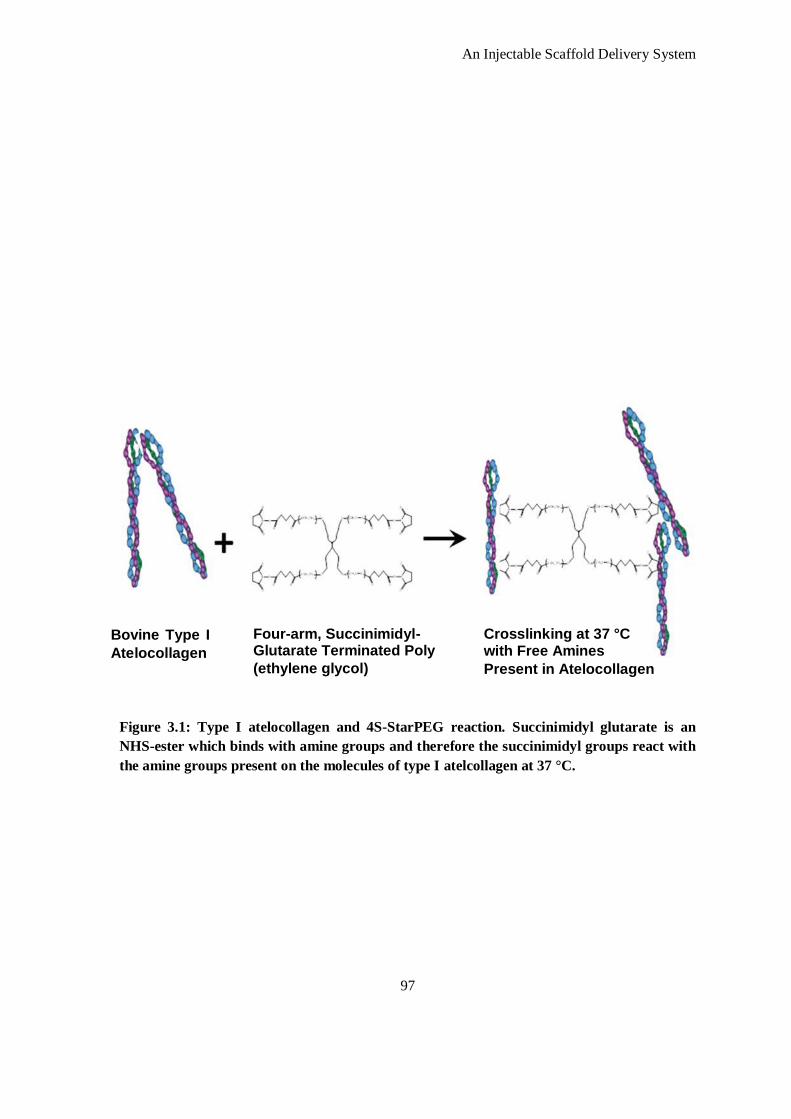
An Injectable Scaffold Delivery System
97
Figure 3.1: Type I atelocollagen and 4S-StarPEG reaction. Succinimidyl glutarate is an
NHS-ester which binds with amine groups and therefore the succinimidyl groups react with
the amine groups present on the molecules of type I atelcollagen at 37 °C.
Bovine Type I
Atelocollagen
Four-arm, Succinimidyl-Glutarate Terminated Poly
(ethylene glycol)
Crosslinking at 37 °C with Free Amines
Present in Atelocollagen
An Injectable Scaffold Delivery System
98
collagen type I scaffold crosslinked with 4S-StarPEG at a molar ratio of 1:1, 2 μg of miRs were
complexed and mixed evenly to the crosslinked scaffold solution on ice before gelation occurred.
3.2.7 Fab` Conjugation
Antibody fragments (Fab`s) were derived from monoclonal antibodies as described in Chapter
Two. Known molar concentrations (deduced from Ellman’s assay) of these Fab`s were reacted
with pD-b-P/DA in a reaction buffer of 0.2 mM NaH2PO4 with 1 mM EDTA at pH 8.0 (purged
prior to reaction with argon for 30 minutes). Briefly, reactions were performed in which molar
ratios of Fab` thiol concentration (from Ellman’s assay) and moles of free vinyls (calculated from
1H NMR data presented in Chapter Two) were reacted at a ratio of 1:1.
3.2.8 Elution Studies
The characterization of the 4S-StarPEG collagen type I scaffold’s release profile of interfering
RNA was performed using siRNA as a model oligonucleotide labeled with Cy™
3. Previous
experiments using intercalating staining of oligonucleotides with PicoGreen® proved unreliable as
the use of the complexing agents hindered the binding of this agent to the nucleic acid, an
observation which is noted in pDNA release studies 27
. siRNA was labeled using Silencer siRNA
Labeling Kit Cy™
3 (Ambion®/Life Technologies, Dublin, Ireland) according to the
manufacturer’s instructions (Appendix V for detailed protocol). Release of the miRNA complexes
was evaluated using the fluorescence from the control siRNA, which was labeled with Cy™
3.
Briefly, the scaffolds were prepared as described above, in a 48-well plate with the additional step
of adding 2 μg of Cy™
3 labeled siRNA. Three groups were investigated in this study: naked
siRNA, siRNA complexed with pD-b-P/DA and siRNA complexed with PEI. In addition the
effect of crosslinking density on the release profiles of siRNA from the scaffolds was investigated
using three conditions: a crosslinking density of 1 mM, 0.5 mM and 0.05 mM. The loaded
scaffolds were incubated for two hours at room temperature to allow the complexes to associate
with the scaffold and for complete gelation to occur. Thereafter, the scaffolds were individually
removed and transferred to the bottom of a 24-well well plate. To this, an equal volume of tris
(hydroxymethyl) aminomethane-n/ethylenediaminetetraacetic acid (Tris–EDTA) buffer (10 mM
Tris–HCl and 1 mM EDTA, pH = 7.5) was added. At each time point, this process was repeated.
At the end of the experiment, the siRNA content of the solutions was quantified by measuring the
fluorescence with a Varioskan Flash® spectral scanning multimode reader (Thermo Scientific,
Vantaa, Finland) and the cumulative release of siRNA complexes from the scaffold was
calculated following comparison with a standard curve.
An Injectable Scaffold Delivery System
99
3.2.9 Evaluating Matrix Binding of RNA complexes
To further understand the mechanism of how RNA complexes are held within the scaffold the
electrophoretic kinetics of RNA complexes within the scaffold were evaluated using agarose gel
electrophoresis. 1 % (w/v) agarose was heated in tris-acetic acid-Ethylenediaminetetraacetic acid
(EDTA) buffer (40 mM Tris, 20 mM acetic acid, and 1 mM EDTA respectively) and a 10,000 fold
dilution of Syber®Safe dye was added prior to gel casting. Non-crosslinked atelocollagen
scaffolds or scaffolds crosslinked with 4S-StarPEG, to a volume of 10 μl, were deposited into the
running wells of this gel with 330 ng of naked RNA, RNA complexed with pD-b-P/DA, or
complexed with PEI. Naked RNA and scaffolds containing no RNA were applied as controls.
Additionally, naked RNA mixed with an indicative loading buffer was run to monitor the
migration of the RNA on the gel. However, this was not applied to the other samples to avoid any
interference with the kinetics of the scaffolds. A voltage of 50 V was applied to the agarose gel
and the current was set to ‘auto’. After 20 minutes the loading buffer indicated sufficient
migration to observe the electrophoretic mobility of the complexes under UV light.
3.2.10 Cell Extraction
Rat cardiac fibroblasts were isolated from neonatal pups as previously described 28
(Appendix
I.8). Briefly, neonatal rat ventricle myocytes were isolated from the cardiac ventricles of three to
five days old Sprague-Dawley pups. Hearts were removed from the thoracic cavity and placed in
a tube containing cold (4-(2-hydroxyethyl)-1-piperazineethanesulfonic acid) HEPES solution (20
mM HEPES, pH 7.4). Ventricles were separated from surrounding tissue using scissors and
minced into several pieces. Subsequently cardiomyocytes and fibroblasts were detached from the
extracellular matrix by repeated incubation in collagenase, supplemented with 2 mg/ml trypsin
and 20 µg/ml DNase. Cells were collected by centrifugation and tissue clumps were removed by
filtration. The cells were then pre-plated in cell culture dishes in 50 ml Dulbecco's Modified Eagle
Medium (DMEM) /F12 (50:50) medium with 5% fetal bovine serum (FBS) for 45 minutes.
During this period, most non-cardiomyocyte cells (mainly fibroblasts) attached to the dish,
whereas cardiomyocytes remained in solution. Fibroblasts were subsequently cultured in
DMEM/F12 medium containing 10% FBS.
3.2.11 Monolayer Silencing Study
Primary rat cardiac fibroblasts were seeded on a six-well plate at a density of 1 x 10⁶
cells per
well. After one day incubation at 37 °C, 5% CO2, to ensure adherence and acclimatization, miR-
29B or miR-scram; uncomplexed, complexed with pD-b-P/DA or complexed with PEI were added
to each well in a total volume of 250 μl with a concentration of 0.5 μg of miR. After ten minutes,
An Injectable Scaffold Delivery System
100
the total volume in the wells was brought to 1 ml with the addition of DMEM/F12 containing
FBS at a concentration of 5%. The experiment was maintained for 48 hours after which samples
were processed for gene or protein analysis via RT-PCR and Western blot respectively.
3.2.12 Scaffold Delivery Silencing Studies
Silencing studies were performed on six-well well plates seeded with 1 x 10⁶ cells per well. After
one day incubation to ensure adherence and acclimatization, scaffolds were placed onto the cells.
In total 250 μl of scaffold solution was applied to each well. The scaffolds were applied directly
to ensure direct contact with cells and also to bring about a direct interaction between the cells and
the scaffold. The use of a 250 μl volume applied in a six-well enabled a very thin scaffold to be
produced which maintained diffusion of nutrients to maintain the viability of the cells.
3.2.13 RNA Extraction
RNA extraction was performed at 7, 14, and 21 days. One mL of TRI Reagent® (Applera Ireland,
Dublin, Ireland) was added to each construct and incubated for five minutes at room temperature.
Scaffolds were mechanically disrupted using a sterile pipette tip. Phase separation was performed
by adding chloroform (Sigma-Aldrich), and total RNA was purified using an RNeasy™
kit
(Qiagen), according to the supplier’s recommended procedure (Appendix S).
3.2.14 Real-time Reverse Transcription Polymerase Chain Reaction
Total RNA quantity and purity were determined using spectrophotometry at 260 and 280 nm
using an ultraviolet spectrophotometer (NanoDrop™
ND-1000 Spectrophotometer, NanoDrop
Technologies, Wilmington, DE, USA). RNA integrity was checked electrophoretically using the
RNA 6000 Nano LabChip™
kit with an Agilent Bioanalyser 2100 (Agilent Technologies, Cork,
Ireland). Reverse transcription (RT) was performed using the ImProm-II™
RT system according
to the manufacturer’s protocol (Promega, Southampton, United Kingdom, Appendix T). Gene
transcription was examined using real-time RT polymerase chain reaction (PCR). Reactions were
performed and monitored using an ABI 7000® sequence detection system (Applied Biosystems,
Foster City, CA) using TaqMan® Real-time Gene Expression Mastermix (TaqMan, Applied
Biosystems) and specific primers which are detailed in Table 3.1. The primers were designed and
their specificity checked using primer-BLAST (available freely from www.ncbi.com) and their
efficiency determined by RT-PCR on ten-fold serial dilutions of template cDNA. Gene
transcription was inferred from calibration samples and normalized in relation to transcription of
the housekeeping gene; glyceraldehydes-3-phosphate dehydrogenase (GAPDH). The 2∆∆-Ct
method was used to calculate relative gene expression for each target gene (Appendices T and U).
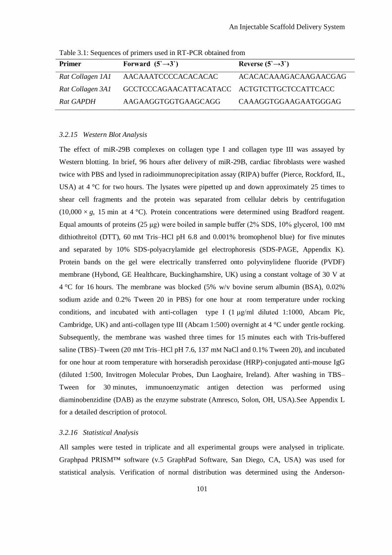
An Injectable Scaffold Delivery System
101
Table 3.1: Sequences of primers used in RT-PCR obtained from
Primer Forward (5`→3`) Reverse (5`→3`)
Rat Collagen 1A1 AACAAATCCCCACACACAC ACACACAAAGACAAGAACGAG
Rat Collagen 3A1 GCCTCCCAGAACATTACATACC ACTGTCTTGCTCCATTCACC
Rat GAPDH AAGAAGGTGGTGAAGCAGG CAAAGGTGGAAGAATGGGAG
3.2.15 Western Blot Analysis
The effect of miR-29B complexes on collagen type I and collagen type III was assayed by
Western blotting. In brief, 96 hours after delivery of miR-29B, cardiac fibroblasts were washed
twice with PBS and lysed in radioimmunoprecipitation assay (RIPA) buffer (Pierce, Rockford, IL,
USA) at 4 °C for two hours. The lysates were pipetted up and down approximately 25 times to
shear cell fragments and the protein was separated from cellular debris by centrifugation
(10,000 × g, 15 min at 4 °C). Protein concentrations were determined using Bradford reagent.
Equal amounts of proteins (25 μg) were boiled in sample buffer (2% SDS, 10% glycerol, 100 mM
dithiothreitol (DTT), 60 mM Tris–HCl pH 6.8 and 0.001% bromophenol blue) for five minutes
and separated by 10% SDS-polyacrylamide gel electrophoresis (SDS-PAGE, Appendix K).
Protein bands on the gel were electrically transferred onto polyvinylidene fluoride (PVDF)
membrane (Hybond, GE Healthcare, Buckinghamshire, UK) using a constant voltage of 30 V at
4 °C for 16 hours. The membrane was blocked (5% w/v bovine serum albumin (BSA), 0.02%
sodium azide and 0.2% Tween 20 in PBS) for one hour at room temperature under rocking
conditions, and incubated with anti-collagen type I (1 μg/ml diluted 1:1000, Abcam Plc,
Cambridge, UK) and anti-collagen type III (Abcam 1:500) overnight at 4 °C under gentle rocking.
Subsequently, the membrane was washed three times for 15 minutes each with Tris-buffered
saline (TBS)–Tween (20 mM Tris–HCl pH 7.6, 137 mM NaCl and 0.1% Tween 20), and incubated
for one hour at room temperature with horseradish peroxidase (HRP)-conjugated anti-mouse IgG
(diluted 1:500, Invitrogen Molecular Probes, Dun Laoghaire, Ireland). After washing in TBS–
Tween for 30 minutes, immunoenzymatic antigen detection was performed using
diaminobenzidine (DAB) as the enzyme substrate (Amresco, Solon, OH, USA).See Appendix L
for a detailed description of protocol.
3.2.16 Statistical Analysis
All samples were tested in triplicate and all experimental groups were analysed in triplicate.
Graphpad PRISM™ software (v.5 GraphPad Software, San Diego, CA, USA) was used for
statistical analysis. Verification of normal distribution was determined using the Anderson-
An Injectable Scaffold Delivery System
102
Darling test. Analysis of variance (ANOVA) was used to determine statistical significance
between groups. All graphical data is presented as mean ± standard deviation. p values of < 0.05
were considered statistically significant. The existence of outliers was determined using Grubb’s
test, however no outliers were detected.
3.3 Results and Discussion
3.3.1 Scaffold Characterization
The potential of scaffolds as delivery platforms of non-viral therapeutics has been previously
documented both in vitro and in vivo, with tangible beneficial outcomes 20, 29
. Specifically the in
vitro effects of 4S-StarPEG crosslinked collagen scaffolds have been investigated as a delivery
platform for delivering adipose derived mesenchymal stem cells 30
. In the study described in this
chapter, collagen type I was crosslinked using 4S-StarPEG via free amine groups available in
collagen type I. Due to the four-arm structure of 4S-StarPEG, theoretically one mole of 4S-
StarPEG crosslinks with four moles of free amines available in collagen type I (essentially a 1:4
molar ratio).With this in mind, the range of crosslinking densities was based around this
theoretical value. Determining the extent of crosslinking via TNBSA quantification of free amine
groups indicated a decrease in free amine groups which was inversely proportional to crosslinker
density (Figure 3.2) which reached a maximum decrease to 9.5% free amines available after 1 mM
crosslinking density. Although there is no significant difference between the most effective
crosslinking concentration (1 mM and 2 mM), there is a trend towards an increase in free amines at
As previously discussed, efforts at using intercalating dyes in this project proved inefficient due to
generation of standard curves that have unacceptable correlation co-efficients and therefore
readings could not be extrapolated as a percent release of nucleic acid. This is due to the strong
binding of cationic complexing agents to the nucleic acid which hinders intercalating dyes from
binding with complexed nucleic acid appropriately. This observation has been observed in many
pDNA release studies 27, 31, 32
and similarly applies to other nucleic acids such as the RNA
employed in this study. Based on this, the siRNAs were labelled using Cy™
3 and produced
standard curves with reliable correlation co-efficients which can be extrapolated.
The concentration of 4S-StarPEG had a significant effect on the release profiles of siRNA (Figure
3.4). This could be due to both an interaction of the siRNA with the crosslinker and the altered,
crosslinker-dependent degradation profile of the collagen scaffold. As RNA has amine groups
present in its backbone it is possible that the crosslinker interacts with these free amines. Such an
interaction, however, will be minimal as Cy™
3 labelling of the RNA occurs via interaction of the
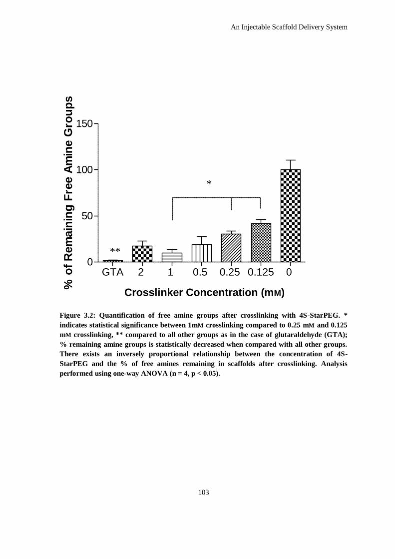
An Injectable Scaffold Delivery System
103
Figure 3.2: Quantification of free amine groups after crosslinking with 4S-StarPEG. *
indicates statistical significance between 1mM crosslinking compared to 0.25 mM and 0.125
mM crosslinking, ** compared to all other groups as in the case of glutaraldehyde (GTA);
% remaining amine groups is statistically decreased when compared with all other groups.
There exists an inversely proportional relationship between the concentration of 4S-
StarPEG and the % of free amines remaining in scaffolds after crosslinking. Analysis
performed using one-way ANOVA (n = 4, p < 0.05).
GTA 2 1 0.5 0.25 0.125 0 0
50
100
150
*
Crosslinking Variable (mM)
% o
f R
em
ain
ing
Fre
e A
min
e G
rou
ps
**
*
Crosslinker Concentration (mM)
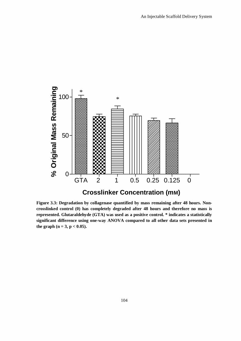
An Injectable Scaffold Delivery System
104
Figure 3.3: Degradation by collagenase quantified by mass remaining after 48 hours. Non-
crosslinked control (0) has completely degraded after 48 hours and therefore no mass is
represented. Glutaraldehyde (GTA) was used as a positive control. * indicates a statistically
significant difference using one-way ANOVA compared to all other data sets presented in
the graph (n = 3, p < 0.05).
GTA 2 1 0.5 0.25 0.125 0 0
50
100 *
Crosslinking Variable (mM)
% O
rig
inal M
ass R
em
ain
ing
* *
Crosslinker Concentration (mM)
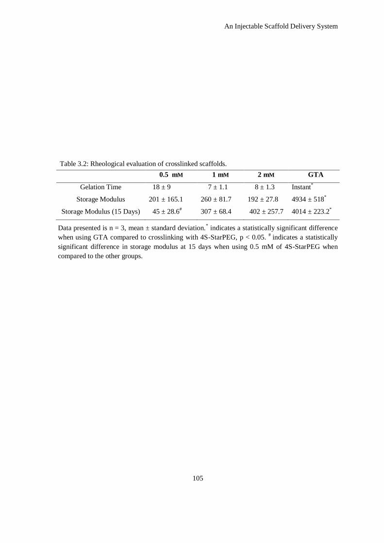
An Injectable Scaffold Delivery System
105
Table 3.2: Rheological evaluation of crosslinked scaffolds.
0.5 mM 1 mM 2 mM GTA
Gelation Time 18 ± 9 7 ± 1.1 8 ± 1.3 Instant*
Storage Modulus 201 ± 165.1 260 ± 81.7 192 ± 27.8 4934 ± 518*
Storage Modulus (15 Days) 45 ± 28.6# 307 ± 68.4 402 ± 257.7 4014 ± 223.2
*
Data presented is n = 3, mean ± standard deviation.* indicates a statistically significant difference
when using GTA compared to crosslinking with 4S-StarPEG, p < 0.05. #
indicates a statistically
significant difference in storage modulus at 15 days when using 0.5 mM of 4S-StarPEG when
compared to the other groups.
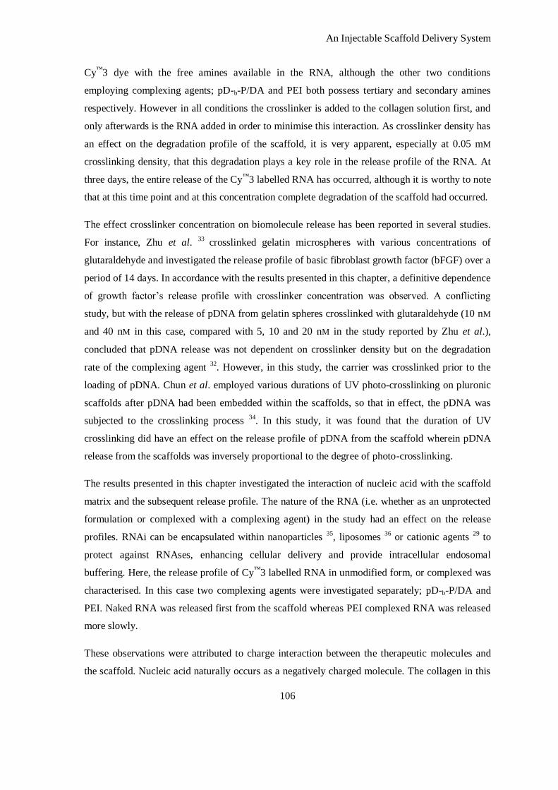
An Injectable Scaffold Delivery System
106
Cy™
3 dye with the free amines available in the RNA, although the other two conditions
employing complexing agents; pD-b-P/DA and PEI both possess tertiary and secondary amines
respectively. However in all conditions the crosslinker is added to the collagen solution first, and
only afterwards is the RNA added in order to minimise this interaction. As crosslinker density has
an effect on the degradation profile of the scaffold, it is very apparent, especially at 0.05 mM
crosslinking density, that this degradation plays a key role in the release profile of the RNA. At
three days, the entire release of the Cy™
3 labelled RNA has occurred, although it is worthy to note
that at this time point and at this concentration complete degradation of the scaffold had occurred.
The effect crosslinker concentration on biomolecule release has been reported in several studies.
For instance, Zhu et al. 33
crosslinked gelatin microspheres with various concentrations of
glutaraldehyde and investigated the release profile of basic fibroblast growth factor (bFGF) over a
period of 14 days. In accordance with the results presented in this chapter, a definitive dependence
of growth factor’s release profile with crosslinker concentration was observed. A conflicting
study, but with the release of pDNA from gelatin spheres crosslinked with glutaraldehyde (10 nM
and 40 nM in this case, compared with 5, 10 and 20 nM in the study reported by Zhu et al.),
concluded that pDNA release was not dependent on crosslinker density but on the degradation
rate of the complexing agent 32
. However, in this study, the carrier was crosslinked prior to the
loading of pDNA. Chun et al. employed various durations of UV photo-crosslinking on pluronic
scaffolds after pDNA had been embedded within the scaffolds, so that in effect, the pDNA was
subjected to the crosslinking process 34
. In this study, it was found that the duration of UV
crosslinking did have an effect on the release profile of pDNA from the scaffold wherein pDNA
release from the scaffolds was inversely proportional to the degree of photo-crosslinking.
The results presented in this chapter investigated the interaction of nucleic acid with the scaffold
matrix and the subsequent release profile. The nature of the RNA (i.e. whether as an unprotected
formulation or complexed with a complexing agent) in the study had an effect on the release
profiles. RNAi can be encapsulated within nanoparticles 35
, liposomes 36
or cationic agents 29
to
protect against RNAses, enhancing cellular delivery and provide intracellular endosomal
buffering. Here, the release profile of Cy™
3 labelled RNA in unmodified form, or complexed was
characterised. In this case two complexing agents were investigated separately; pD-b-P/DA and
PEI. Naked RNA was released first from the scaffold whereas PEI complexed RNA was released
more slowly.
These observations were attributed to charge interaction between the therapeutic molecules and
the scaffold. Nucleic acid naturally occurs as a negatively charged molecule. The collagen in this
An Injectable Scaffold Delivery System
107
study has been crosslinked using the free amine groups (NH2) present in its backbone and not the
negatively charged carboxyl (COOH) groups which offers the collagen a net negative charge.
Therefore nucleic acid, in this case double stranded RNA, cannot interact electrostatically with the
collagen scaffold and therefore is released much quicker from the scaffold. pD-b-P/DA possesses
a net positive charge (albeit a weak one) attributable to the tertiary amines present on DMAEMA.
Based on this, there exists a slightly slower release of pD-b-P/DA complexed RNA from the
scaffold. Finally PEI possesses a strong net positive charge when complexed with RNA and
therefore has a significant electrostatic interaction with the negatively charged collagen scaffold.
This strong electrostatic interaction causes a significantly slower release profile of RNA from the
scaffold. This is also reflected in the electrophoresis data present in Figure 3.5. Non-crosslinked
scaffolds containing RNA (naked or complexed with pD-b-P/D) present a net positive charge.
However, when the scaffolds are crosslinked with 4S-StarPEG, they lose their ability to bind with
RNA in its naked form or complexed with pD-b-P/DA. This is extremely interesting and
strengthens the findings from the release studies in Figure 3.4. Additionally, RNA complexed
with PEI has an electrophoretic shift towards the cathode indicating a net positive charge. This is
unaffected even if the PEI/RNA complexes are placed in a non-crosslinked scaffold or a scaffold
crosslinked with 4S-StarPEG. Based on this, it can be postulated that the PEI/RNA complexes
leave the scaffold as intact PEI/RNA complexes, however complexation with pD-b-P/DA within a
scaffold suggests the RNA leaves the scaffold and in turn leaves the pD-b-P/DA behind as it is
not as tightly bound as PEI/RNA complexes. Similar observations have been observed in other
studies reported in the literature. For instance, Chew et al. 32
monitored the release profile of
naked pDNA loaded gelatin microspheres which were incorporated into a porous poly (propylene
fumarate (PPF) scaffold. Additional groups investigated in this study include pDNA incorporated
into the same scaffold and complexed with hydrolytically degradable branched triacrylate/amine
polycationic polymers (TAPPs); P-AEPZ and P-DED synthesized with amine monomer 1-(-2-
aminoethyl)piperazine (AEPZ) or N,N-dimethylethylenediamine (DED). Complexation of the
pDNA caused a significantly lower initial burst release (within the first 24 hours). The authors of
this study also attributed the release profiles of the pDNA to the nature in which the pDNA was
embedded within microspheres and the charge interaction between them. siRNA release profiles
from scaffolds have also been studied. Krebs et al. 12
found that siRNA embedded within a photo-
crosslinked alginate hydrogel had a much quicker release and increased burst release compared to
a collagen scaffold, which can be explained due to the negatively charged polysaccharide,
alginate. Modulation of alginate scaffolds by combining with chitosan or PEI resulted in
decreased burst release from the hydrogels which is due to an electrostatic interaction between the
nucleotides and the positively charged polymers mixed with the alginate.
An Injectable Scaffold Delivery System
108
3.3.2 Silencing Collagen Type I and Collagen Type III
To remove any ambiguity regarding the specificity of miR-29B to silence collagen type I and
collagen type III, RT-PCR was performed on rat cardiac fibroblasts treated with miR-29B and
non-targeting, scrambled controls in vitro (Figure 3.6). As expected the scrambled control did not
alter any significant silencing of collagen type I and collagen type III mRNA in vitro. This non-
alteration eliminates the possibility of global, non-specific silencing due to the use of double
stranded RNA. While it is true that there are targets of miR-29B that are not validated in this
thesis and have yet to be discovered; there are no noticeable effects on cellular viability or
proliferation detected in response to miR-29B specifically in the studies presented here. The use
of miR-29B in a naked unprotected form did not exhibit effective silencing of collagen type I
(1.11 relative expression, 2∆∆Ct
) or collagen type III mRNA (1.14 relative expression) which
suggests the requirement for a complexing agent when performing miR delivery to cells in 2D
culture (Figure 3.6). Complexation of miR-29B with PEI did show effective silencing of collagen
type I and type III mRNA expression (0.51 and 0.55 relative expression, 2∆∆Ct
respectively).
Although PEI is an effective carrier of nucleic acid to cells, it poses drawbacks as it is toxic; an
important consideration if silencing is to be maintained over an extended period of time. The
effect of miR-29B on collagen type I and collagen type III protein production was also
investigated using western blot analysis (Figure 3.7). The bands support the observations of the
RT-PCR data in Figure 3.6, such that the use of a complexing agent significantly increases the
efficacy of miR-29B and thereby decreases production of collagen type I and collagen type III.
However, for full appreciation of this repetition of western blot is needed with the inclusion of
appropriate loading control immunoblotting for a housekeeping protein such as GAPDH or β-
actin.
Exogenous miR-29B mimics, identical to those reported in this chapter, have been previously
reported to silence collagen type I expression in vitro and all such studies report the employment
of a complexing agent (in the majority of cases commercial agents such as Lipofectamine™
2000)
to deliver such mimics 37-39
. Such agents are warranted as the administration of naked miRs in cell
media are inefficient on cell monolayers in vitro at such small doses (72 ng 37
and 361 ng 38, 39
)
which is reflected in the results presented in this chapter with a dose of 333 ng of miR-29B
mimic. However, commercial agents such as those employed in previous studies, are marketed as
diagnostic and research tools; as their compromised toxicity and concerns of regulatory agencies
limits their therapeutic potential.
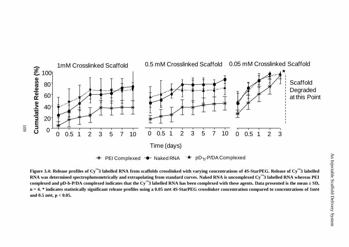
Figure 3.4: Release profiles of Cy™
3 labelled RNA from scaffolds crosslinked with varying concentrations of 4S-StarPEG. Release of Cy™
3 labelled
RNA was determined spectrophotometrically and extrapolating from standard curves. Naked RNA is uncomplexed Cy™
3 labelled RNA whereas PEI
complexed and pD-b-P/DA complexed indicates that the Cy™
3 labelled RNA has been complexed with these agents. Data presented is the mean ± SD,
n = 4. * indicates statistically significant release profiles using a 0.05 mM 4S-StarPEG crosslinker concentration compared to concentrations of 1mM
and 0.5 mM, p < 0.05.
Naked RNA pD-b-P/DA ComplexedPEI Complexed
0.5 mM Crosslinked Scaffold
0 0.5 1 2 3 5 7 10
Time (days)
0.05 mM Crosslinked Scaffold
0 0.5 1 2 3
1mM Crosslinked Scaffold
0 0.5 1 2 3 5 7 100
20
40
60
80
100
Cu
mu
lati
ve R
ele
ase (
%)
Scaffold
Degraded at this Point
*
109
An In
jectable S
caffold
Deliv
ery S
ystem
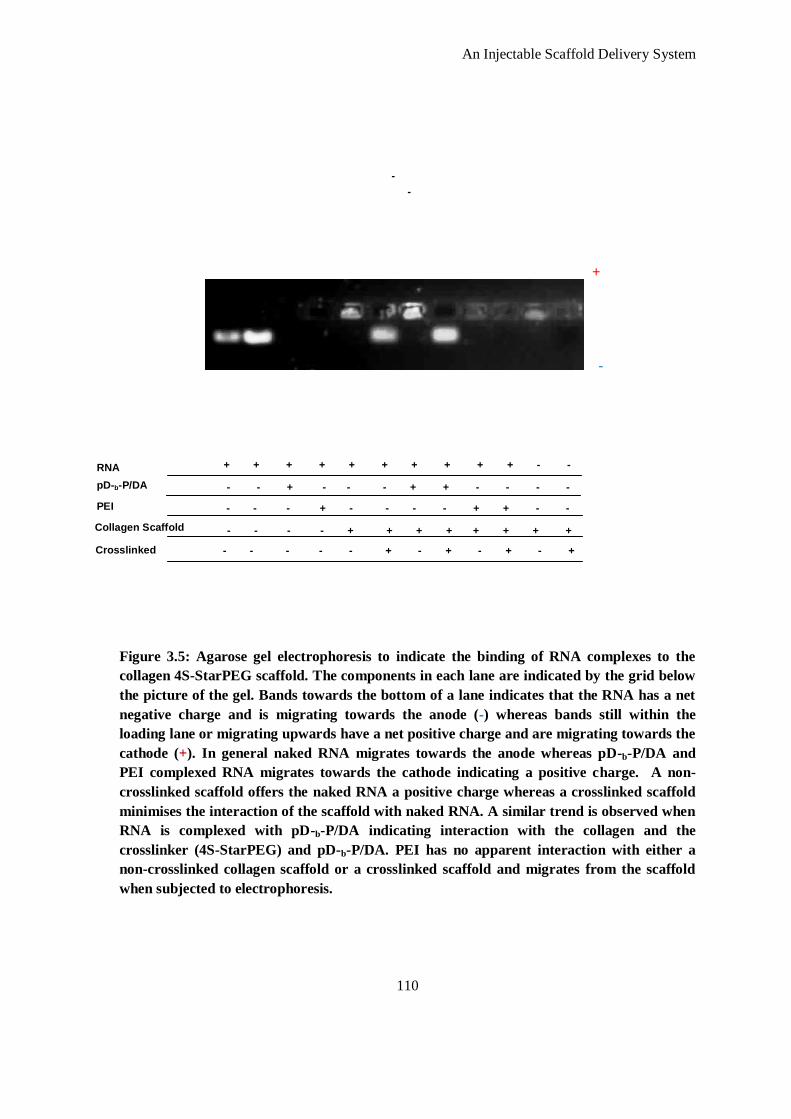
An Injectable Scaffold Delivery System
110
+
-
RNA
pD-b-P/DA
PEI
Collagen Scaffold
Crosslinked
+ + + + + + + + + + - -
- - - - - + - + - + - +
-
-
- - - - + + + + + + + +
- - - + - - - - + + - -
- - + - - - + + - - - -
Figure 3.5: Agarose gel electrophoresis to indicate the binding of RNA complexes to the
collagen 4S-StarPEG scaffold. The components in each lane are indicated by the grid below
the picture of the gel. Bands towards the bottom of a lane indicates that the RNA has a net
negative charge and is migrating towards the anode (-) whereas bands still within the
loading lane or migrating upwards have a net positive charge and are migrating towards the
cathode (+). In general naked RNA migrates towards the anode whereas pD-b-P/DA and
PEI complexed RNA migrates towards the cathode indicating a positive charge. A non-
crosslinked scaffold offers the naked RNA a positive charge whereas a crosslinked scaffold
minimises the interaction of the scaffold with naked RNA. A similar trend is observed when
RNA is complexed with pD-b-P/DA indicating interaction with the collagen and the
crosslinker (4S-StarPEG) and pD-b-P/DA. PEI has no apparent interaction with either a
non-crosslinked collagen scaffold or a crosslinked scaffold and migrates from the scaffold
when subjected to electrophoresis.
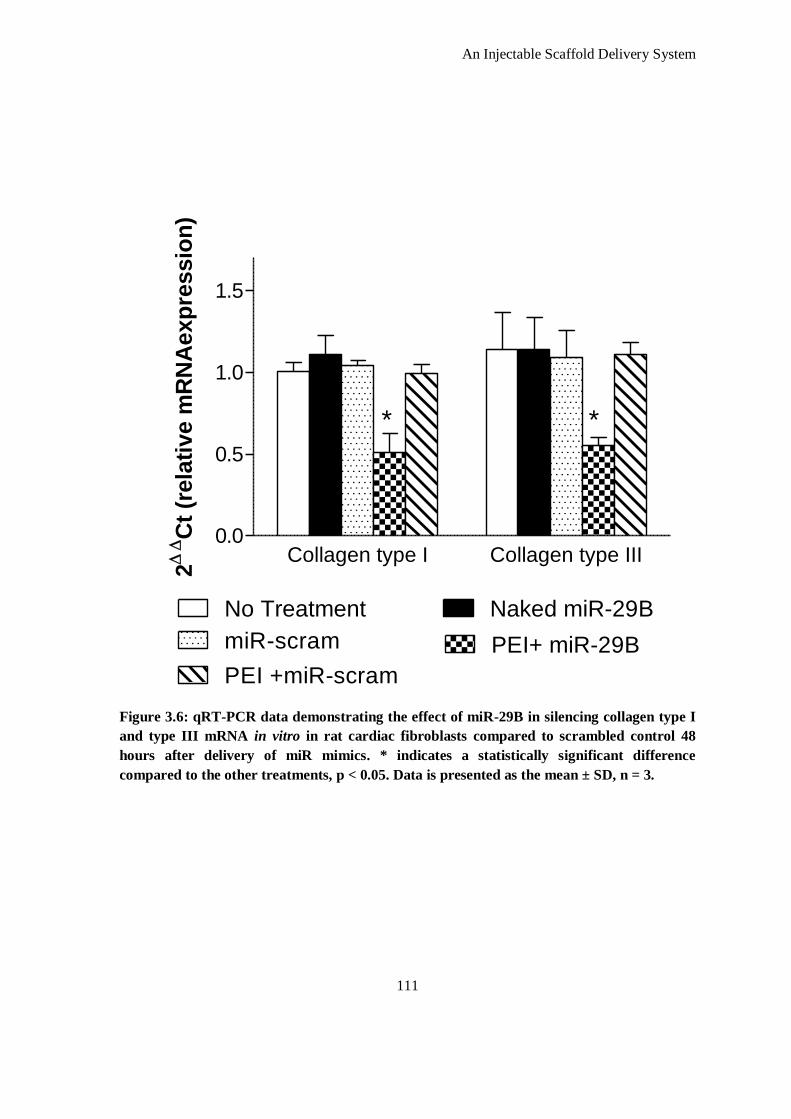
An Injectable Scaffold Delivery System
111
Figure 3.6: qRT-PCR data demonstrating the effect of miR-29B in silencing collagen type I
and type III mRNA in vitro in rat cardiac fibroblasts compared to scrambled control 48
hours after delivery of miR mimics. * indicates a statistically significant difference
compared to the other treatments, p < 0.05. Data is presented as the mean ± SD, n = 3.
Collagen type I Collagen type III0.0
0.5
1.0
1.5
No Treatment Naked miR-29B
miR-scram PEI+ miR-29B
PEI +miR-scram
* *
2C
t (r
ela
tive m
RN
Aexp
ressio
n)
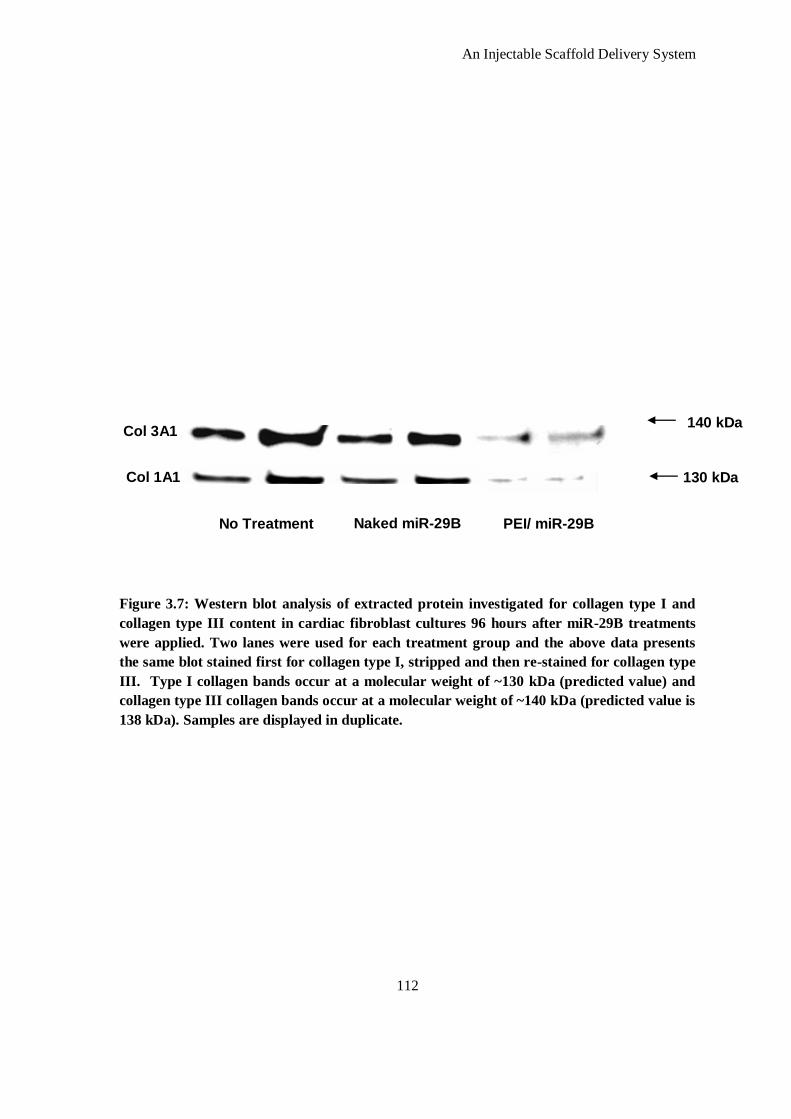
An Injectable Scaffold Delivery System
112
Figure 3.7: Western blot analysis of extracted protein investigated for collagen type I and
collagen type III content in cardiac fibroblast cultures 96 hours after miR-29B treatments
were applied. Two lanes were used for each treatment group and the above data presents
the same blot stained first for collagen type I, stripped and then re-stained for collagen type
III. Type I collagen bands occur at a molecular weight of ~130 kDa (predicted value) and
collagen type III collagen bands occur at a molecular weight of ~140 kDa (predicted value is
138 kDa). Samples are displayed in duplicate.
130 kDa
140 kDa Col 3A1
Col 1A1
No Treatment Naked miR-29B PEI/ miR-29B
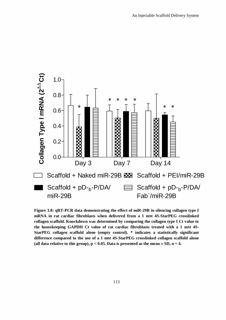
An Injectable Scaffold Delivery System
113
Figure 3.8: qRT-PCR data demonstrating the effect of miR-29B in silencing collagen type I
mRNA in rat cardiac fibroblasts when delivered from a 1 mM 4S-StarPEG crosslinked
collagen scaffold. Knockdown was determined by comparing the collagen type I Ct value to
the housekeeping GAPDH Ct value of rat cardiac fibroblasts treated with a 1 mM 4S-
StarPEG collagen scaffold alone (empty control). * indicates a statistically significant
difference compared to the use of a 1 mM 4S-StarPEG crosslinked collagen scaffold alone
(all data relative to this group), p < 0.05. Data is presented as the mean ± SD, n = 4.
Day 3 Day 7 Day 140.0
0.2
0.4
0.6
0.8
1.0
Scaffold + Naked miR-29B Scaffold + PEI/miR-29B
Scaffold + pD-b-P/DA/
miR-29B
Scaffold + pD-b-P/DA/
Fab`/miR-29B
**
* ** **
Co
llag
en
Typ
e I m
RN
A (
2C
t)
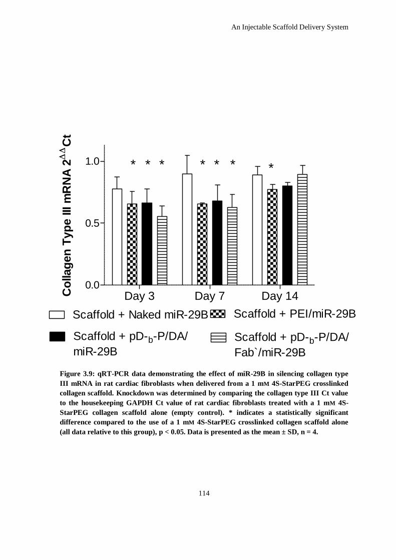
An Injectable Scaffold Delivery System
114
Figure 3.9: qRT-PCR data demonstrating the effect of miR-29B in silencing collagen type
III mRNA in rat cardiac fibroblasts when delivered from a 1 mM 4S-StarPEG crosslinked
collagen scaffold. Knockdown was determined by comparing the collagen type III Ct value
to the housekeeping GAPDH Ct value of rat cardiac fibroblasts treated with a 1 mM 4S-
StarPEG collagen scaffold alone (empty control). * indicates a statistically significant
difference compared to the use of a 1 mM 4S-StarPEG crosslinked collagen scaffold alone
(all data relative to this group), p < 0.05. Data is presented as the mean ± SD, n = 4.
Day 3 Day 7 Day 140.0
0.5
1.0
Scaffold + pD-b-P/DA/
miR-29B
Scaffold + pD-b-P/DA/
Fab`/miR-29B
Scaffold + Naked miR-29B Scaffold + PEI/miR-29B
* * ** * * *
Co
llag
en
Typ
e III m
RN
A 2
C
t
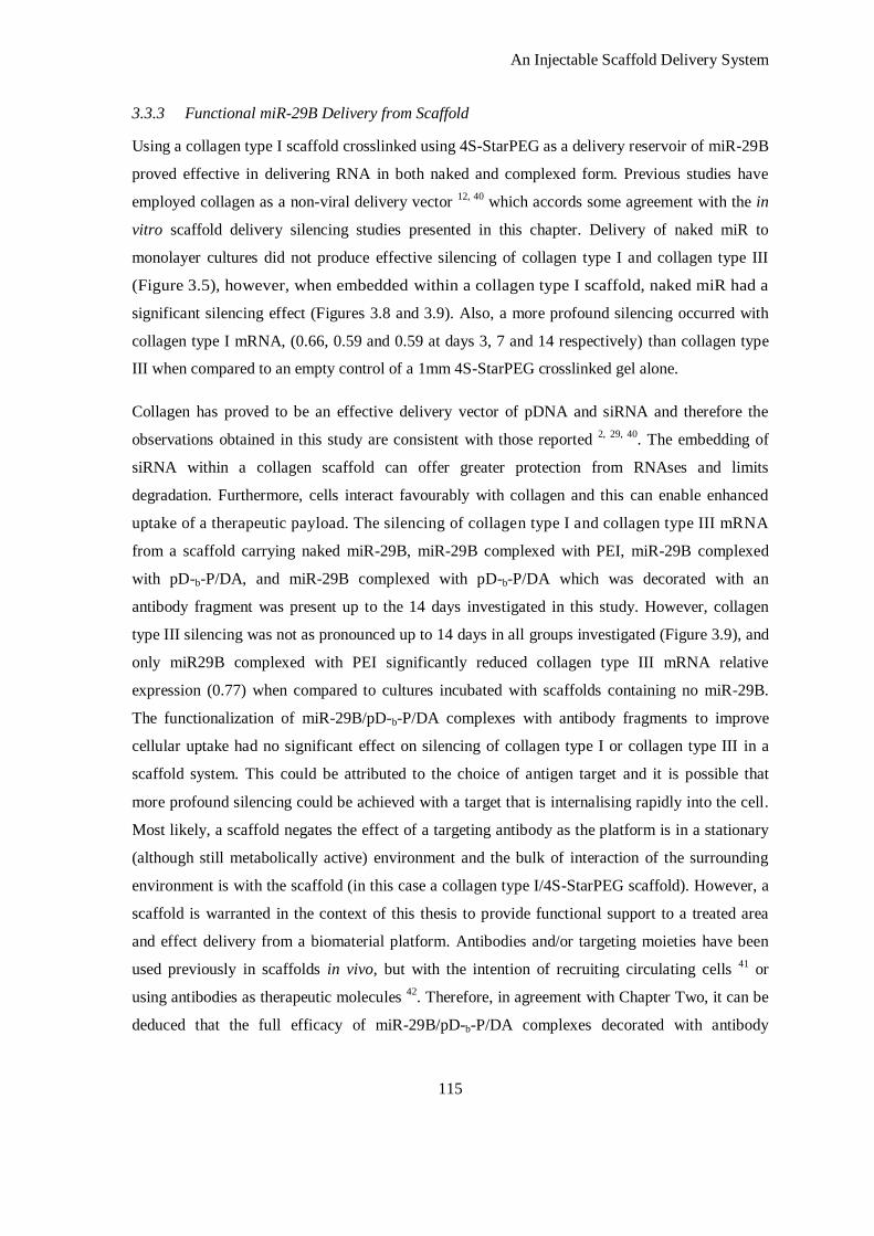
An Injectable Scaffold Delivery System
115
3.3.3 Functional miR-29B Delivery from Scaffold
Using a collagen type I scaffold crosslinked using 4S-StarPEG as a delivery reservoir of miR-29B
proved effective in delivering RNA in both naked and complexed form. Previous studies have
employed collagen as a non-viral delivery vector 12, 40
which accords some agreement with the in
vitro scaffold delivery silencing studies presented in this chapter. Delivery of naked miR to
monolayer cultures did not produce effective silencing of collagen type I and collagen type III
(Figure 3.5), however, when embedded within a collagen type I scaffold, naked miR had a
significant silencing effect (Figures 3.8 and 3.9). Also, a more profound silencing occurred with
collagen type I mRNA, (0.66, 0.59 and 0.59 at days 3, 7 and 14 respectively) than collagen type
III when compared to an empty control of a 1mm 4S-StarPEG crosslinked gel alone.
Collagen has proved to be an effective delivery vector of pDNA and siRNA and therefore the
observations obtained in this study are consistent with those reported 2, 29, 40
. The embedding of
siRNA within a collagen scaffold can offer greater protection from RNAses and limits
degradation. Furthermore, cells interact favourably with collagen and this can enable enhanced
uptake of a therapeutic payload. The silencing of collagen type I and collagen type III mRNA
from a scaffold carrying naked miR-29B, miR-29B complexed with PEI, miR-29B complexed
with pD-b-P/DA, and miR-29B complexed with pD-b-P/DA which was decorated with an
antibody fragment was present up to the 14 days investigated in this study. However, collagen
type III silencing was not as pronounced up to 14 days in all groups investigated (Figure 3.9), and
only miR29B complexed with PEI significantly reduced collagen type III mRNA relative
expression (0.77) when compared to cultures incubated with scaffolds containing no miR-29B.
The functionalization of miR-29B/pD-b-P/DA complexes with antibody fragments to improve
cellular uptake had no significant effect on silencing of collagen type I or collagen type III in a
scaffold system. This could be attributed to the choice of antigen target and it is possible that
more profound silencing could be achieved with a target that is internalising rapidly into the cell.
Most likely, a scaffold negates the effect of a targeting antibody as the platform is in a stationary
(although still metabolically active) environment and the bulk of interaction of the surrounding
environment is with the scaffold (in this case a collagen type I/4S-StarPEG scaffold). However, a
scaffold is warranted in the context of this thesis to provide functional support to a treated area
and effect delivery from a biomaterial platform. Antibodies and/or targeting moieties have been
used previously in scaffolds in vivo, but with the intention of recruiting circulating cells 41
or
using antibodies as therapeutic molecules 42
. Therefore, in agreement with Chapter Two, it can be
deduced that the full efficacy of miR-29B/pD-b-P/DA complexes decorated with antibody
An Injectable Scaffold Delivery System
116
fragments can only be realised in a bioreactor system in vitro or via systemic delivery in vivo
which would be appropriate miR-29B was envisaged to be delivered systemically.
3.4 Conclusions
4S-StarPEG proved to be an effective crosslinker of type I atelocollagen and allowed the
formation of stable scaffolds. The physico-chemical properties of these scaffolds can be varied
according to crosslinker density. This amount of crosslinking was verified by quantification of
free amine groups, resistance to enzymatic degradation and by assessment of mechanical
behaviour. It was demonstrated that the release of a therapeutic agent from the scaffold was
dependent on the crosslinker density (through scaffold integrity and degradation profile) and also
the electrostatic interaction of the therapeutic within the scaffold. miR-29B was shown to
specifically silence collagen type I and collagen type III in monolayer culture, but only when
delivered using a complexing agent (down to 51 and 55% respectively). Using a 4S-StarPEG
scaffold as a delivery platform of these miR-29B complexes, efficient silencing, of both collagen
type I and collagen type III mRNA, up to a period of up to 14 days was achieved and knockdown
of collagen type I miRNA was higher than that of collagen type III. Furthermore, naked miR-29B
within the scaffold was effective at silencing collagen type I and collagen type III mRNA, but the
functionalization of miR-29B/pD-b-P/DA complexes with antibody fragments did not result in a
significant improvement in efficiency when delivered in a scaffold platform.
An Injectable Scaffold Delivery System
117
3.5 References
1. Monaghan M, Pandit A. RNA interference therapy via functionalized scaffolds. Adv. Drug Del. Rev. 2011;63:197-208
2. Kulkarni M, Greiser U, O'Brien T, Pandit A. Liposomal gene delivery mediated by tissue-
engineered scaffolds. Trends Biotechnol. 2010;28:28-36
3. De Laporte L, Shea LD. Matrices and scaffolds for DNA delivery in tissue engineering. Adv. Drug Delivery Rev. 2007;59:292-307
4. Liu Y, Taylor NE, Lu L, Usa K, Cowley AW, Jr, Ferreri NR, Yeo NC, Liang M. Renal
medullary microRNAs in dahl salt-sensitive rats: Mir-29b regulates several collagens and related genes. Hypertension. 2010;55:974-982
5. Li Z, Hassan MQ, Jafferji M, Aqeilan RI, Garzon R, Croce CM, van Wijnen AJ, Stein JL,
Stein GS, Lian JB. Biological functions of miR-29b contribute to positive regulation of osteoblast differentiation. J. Biol. Chem.. 2009;284:15676-15684
6. Cushing L, Kuang PP, Qian J, Shao F, Wu J, Little F, Thannickal VJ, Cardoso WV, Lu J.
Mir-29 is a major regulator of genes associated with pulmonary fibrosis. Am. J. Respir.
Cell Mol. Biol. 2010:2010-0323OC 7. Roderburg C, Urban G-W, Bettermann K, Vucur M, Zimmermann H, Schmidt S, Janssen
J, Koppe C, Knolle P, Castoldi M, Tacke F, Trautwein C, Luedde T. Micro-RNAprofiling
reveals a role for miR-29 in human and murine liver fibrosis. Hepatology. 2011;53:209-218
8. van Rooij E, Sutherland LB, Thatcher JE, DiMaio JM, Naseem RH, Marshall WS, Hill
JA, Olson EN. Dysregulation of microRNAs after myocardial infarction reveals a role of
mir-29 in cardiac fibrosis. Proc. Nat. Acad. Sci. USA. 2008;105:13027-13032 9. Walter E, Moelling K, Pavlovic J, Merkle HP. Microencapsulation of DNA using
poly(DL-lactide-co-glycolide): Stability issues and release characteristics. J. Controlled
Release. 1999;61:361-374 10. Guan J, Stankus JJ, Wagner WR. Biodegradable elastomeric scaffolds with basic
fibroblast growth factor release. J. Controlled Release. 2007;120:70-78
11. Hutmacher DW. Scaffolds in tissue engineering bone and cartilage. Biomaterials. 2000;21:2529-2543
12. Krebs MD, Jeon O, Alsberg E. Localized and sustained delivery of silencing RNA from
macroscopic biopolymer hydrogels. J. Amer. Chem. Soc. 2009;131:9204-9206
13. Singh AK, Reyrat J-M. Laboratory maintenance of mycobacterium smegmatis. Curr. Protoc. Microbiol. 2009; 14:10C.1.1 – 10C.1.12.
14. Kim Y-M, Park M-R, Song S-C. Injectable polyplex hydrogel for localized and long-term
delivery of siRNA. ACS Nano. 2012;6:5757-5766 15. Manaka T, Suzuki A, Takayama K, Imai Y, Nakamura H, Takaoka K. Local delivery of
siRNA using a biodegradable polymer application to enhance BMP-induced bone
formation. Biomaterials. 2011;32:9642-9648 16. Saito H, Murabayashi S, Mitamura Y, Taguchi T. Characterization of alkali-treated
collagen gels prepared by different crosslinkers. J. Mater. Sci. Mater. Med.
2008;19:1297-1305
17. Taguchi T, Xu L, Kobayashi H, Taniguchi A, Kataoka K, Tanaka J. Encapsulation of chondrocytes in injectable alkali-treated collagen gels prepared using poly(ethylene
glycol)-based 4-armed star polymer. Biomaterials. 2005;26:1247-1252
18. Sargeant TD, Desai AP, Banerjee S, Agawu A, Stopek JB. An in situ forming collagen–PEG hydrogel for tissue regeneration. Acta Biomater. 2012;8:124-132
19. Breen A, Dockery P, O'Brien T, Pandit A. Fibrin scaffold promotes adenoviral gene
transfer and controlled vector delivery. J. Biomed. Mat. Res. A. 2009;89A:876-884
An Injectable Scaffold Delivery System
118
20. Holladay C, Power K, Sefton M, O'Brien T, Gallagher WM, Pandit A. Functionalized
scaffold-mediated interleukin 10 gene delivery significantly improves survival rates of stem cells in vivo. Mol Ther. 2011;19:969-978
21. O'Rorke S, Keeney M, Pandit A. Non-viral polyplexes: Scaffold mediated delivery for
gene therapy. Prog. Pol. Sci.. 2010;35:441-458
22. Zeugolis DI, Paul RG, Attenburrow G. Factors influencing the properties of reconstituted collagen fibers prior to self-assembly: Animal species and collagen extraction method. J.
of Biomed. Mat. Res. Part A. 2008;86A:892-904
23. Ofner IIICM, Bubnis WA. Chemical and swelling evaluations of amino group crosslinking in gelatin and modified gelatin matrices. Pharm. Res.. 1996;13:1821-1827
24. Garcia Y, Collighan R, Griffin M, Pandit A. Assessment of cell viability in a three-
dimensional enzymatically cross-linked collagen scaffold. J. Mat. Sci. Mat. Med. 2007;18:1991-2001
25. Chan J, Burugapalli K, Naik H, Kelly J, Pandit A. Amine functionalization of cholecyst-
derived extracellular matrix with generation 1 PAMAM dendrimer. Biomacromolecules.
2008;9:528-536 26. Fatimi A, François Tassin J, Quillard S, Axelos V, Weiss P. The rheological properties of
silated hydroxypropylmethylcellulose tissue engineering matrices. Biomaterials.
2008;29:533-543 27. Holladay C, Keeney M, Newland B, Mathew A, Wang W, Pandit A. A reliable method
for detecting complexed DNA in vitro. Nanoscale. 2010;2:2718-2723
28. Lu B, Mahmud H, Maass AH, Yu B, van Gilst WH, de Boer RA, Silljé HHW. The PLK1 inhibitor BI 2536 temporarily arrests primary cardiac fibroblasts in mitosis and generates
aneuploidy in vitro. PLoS ONE. 2010;5:e12963
29. Vinas-Castells R , Holladay C , di Luca A , Diaz VM , Pandit A. Snail1 down-regulation
using small interfering RNA complexes delivered through collagen scaffolds. Bioconj. Chem. 2009;20:2262-2269
30. Collin EC, Grad S, Zeugolis DI, Vinatier CS, Clouet JR, Guicheux JJ, Weiss P, Alini M,
Pandit AS. An injectable vehicle for nucleus pulposus cell-based therapy. Biomaterials. 2011;32:2862-2870
31. Kim HJ, Kwon MS, Choi JS, Yang S-M, Yoon JK, Kim K, Park J. Highly effective and
slow-biodegradable network-type cationic gene delivery polymer: Small library-like
approach synthesis and characterization. Biomaterials. 2006;27:2292-2301 32. Chew SA, Kretlow JD, Spicer PP, Edwards AW, Baggett LS, Tabata Y, Kasper FK,
Mikos AG. Delivery of plasmid DNA encoding BMP-2 with a biodegradable branched
polycationic polymer in a critical-size rat cranial defect model. Tissue Eng. Part A. 2011;17:751-763
33. Zhu XH, Tabata Y, Wang CH, Tong YW. Delivery of basic fibroblast growth factor from
gelatin microsphere scaffold for the growth of human umbilical vein endothelial cells. Tissue Eng. Part A. 2008;14:1939-1947
34. Chun KW, Lee JB, Kim SH, Park TG. Controlled release of plasmid DNA from photo-
cross-linked pluronic hydrogels. Biomaterials. 2005;26:3319-3326
35. Andersen MO, Nygaard JV, Burns JS, Raarup MK, Nyengaard JR, Bunger C, Besenbacher F, Howard KA, Kassem M, Kjems J. siRNA nanoparticle functionalization
of nanostructured scaffolds enables controlled multilineage differentiation of stem cells.
Mol. Ther. 2010;18:2018-2027 36. Hirsch M, Ziroli V, Helm M, Massing U. Preparation of small amounts of sterile siRNA-
liposomes with high entrapping efficiency by dual asymmetric centrifugation. J.
Controlled Release. 2009;135:80-88 37. Sekiya Y, Ogawa T, Yoshizato K, Ikeda K, Kawada N. Suppression of hepatic stellate
cell activation by microRNA-29b. Biochem. Biophys. Res. Commun. 2011;412:74-79
An Injectable Scaffold Delivery System
119
38. Ogawa T, Iizuka M, Sekiya Y, Yoshizato K, Ikeda K, Kawada N. Suppression of type I
collagen production by microRNA-29b in cultured human stellate cells. Biochem. Biophys. Res. Commun. 2010;391:316-321
39. Li N, Cui J, Duan X, Chen H, Fan F. Suppression of type I collagen expression by miR-
29b via Pi3K, AKT, and SP1 pathway in human tenon's fibroblasts. Invest. Ophthalmol.
Vis. Sci. 2012;53:1670-1678 40. Inaba S, Nagahara S, Makita N, Tarumi Y, Ishimoto T, Matsuo S, Kadomatsu K, Takei
Y. Atelocollagen-mediated systemic delivery prevents immunostimulatory adverse effects
of siRNA in mammals. Mol Ther. 2012;20:356-366 41. Li Q-L, Huang N, Chen C, Chen J-L, Xiong K-Q, Chen J-Y, You T-X, Jin J, Liang X.
Oriented immobilization of anti-CD34 antibody on titanium surface for self-
endothelialization induction. J. Biomed. Mat. Res. Part A. 2010;94A:1283-1293 42. Koutsopoulos S, Zhang S. Two-layered injectable self-assembling peptide scaffold
hydrogels for long-term sustained release of human antibodies. J. Controlled Release.
2012;160:451-458
Chapter Four
Evaluation of miR Delivery In Vivo
The majority of this chapter is submitted for publication in
Monaghan M, Browne S, Schenke-Layland K, Pandit A. A biodegradable crosslinked scaffold as a delivery platform of exogenous microRNAs- in vitro and in vivo evaluation. Submitted toMol. Ther.
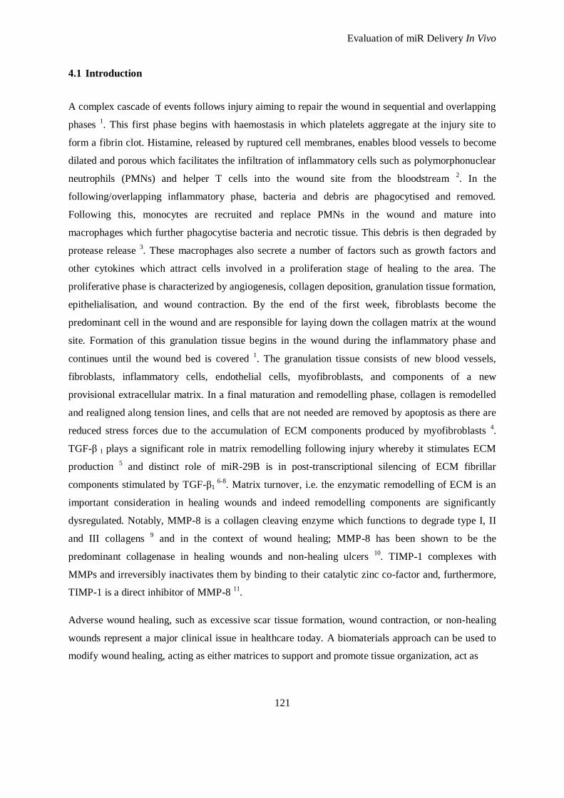
Evaluation of miR Delivery In Vivo
121
4.1 Introduction
A complex cascade of events follows injury aiming to repair the wound in sequential and overlapping
phases 1. This first phase begins with haemostasis in which platelets aggregate at the injury site to
form a fibrin clot. Histamine, released by ruptured cell membranes, enables blood vessels to become
dilated and porous which facilitates the infiltration of inflammatory cells such as polymorphonuclear
neutrophils (PMNs) and helper T cells into the wound site from the bloodstream 2. In the
following/overlapping inflammatory phase, bacteria and debris are phagocytised and removed.
Following this, monocytes are recruited and replace PMNs in the wound and mature into
macrophages which further phagocytise bacteria and necrotic tissue. This debris is then degraded by
protease release 3. These macrophages also secrete a number of factors such as growth factors and
other cytokines which attract cells involved in a proliferation stage of healing to the area. The
proliferative phase is characterized by angiogenesis, collagen deposition, granulation tissue formation,
epithelialisation, and wound contraction. By the end of the first week, fibroblasts become the
predominant cell in the wound and are responsible for laying down the collagen matrix at the wound
site. Formation of this granulation tissue begins in the wound during the inflammatory phase and
continues until the wound bed is covered 1. The granulation tissue consists of new blood vessels,
fibroblasts, inflammatory cells, endothelial cells, myofibroblasts, and components of a new
provisional extracellular matrix. In a final maturation and remodelling phase, collagen is remodelled
and realigned along tension lines, and cells that are not needed are removed by apoptosis as there are
reduced stress forces due to the accumulation of ECM components produced by myofibroblasts 4.
TGF-β 1 plays a significant role in matrix remodelling following injury whereby it stimulates ECM
production 5 and distinct role of miR-29B is in post-transcriptional silencing of ECM fibrillar
components stimulated by TGF-β1 6-8
. Matrix turnover, i.e. the enzymatic remodelling of ECM is an
important consideration in healing wounds and indeed remodelling components are significantly
dysregulated. Notably, MMP-8 is a collagen cleaving enzyme which functions to degrade type I, II
and III collagens 9 and in the context of wound healing; MMP-8 has been shown to be the
predominant collagenase in healing wounds and non-healing ulcers 10
. TIMP-1 complexes with
MMPs and irreversibly inactivates them by binding to their catalytic zinc co-factor and, furthermore,
TIMP-1 is a direct inhibitor of MMP-8 11
.
Adverse wound healing, such as excessive scar tissue formation, wound contraction, or non-healing
wounds represent a major clinical issue in healthcare today. A biomaterials approach can be used to
modify wound healing, acting as either matrices to support and promote tissue organization, act as
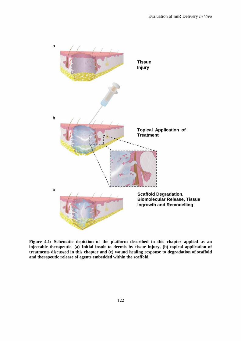
Evaluation of miR Delivery In Vivo
122
Figure 4.1: Schematic depiction of the platform described in this chapter applied as an
injectable therapeutic. (a) Initial insult to dermis by tissue injury, (b) topical application of
treatments discussed in this chapter and (c) wound healing response to degradation of scaffold
and therapeutic release of agents embedded within the scaffold.
Tissue
Injury
Topical Application of Treatment
Scaffold Degradation, Biomolecular Release, Tissue
Ingrowth and Remodelling
a
b
c
Evaluation of miR Delivery In Vivo
123
barriers to limit wound contraction 12
and scar tissue formation 13-19
and/or incorporate the delivery of
bioactive substances such as cytokines 20
, growth factors 21
, living cells 22
, and with nucleic acids-
specifically anti-sense oligonucleotides 23-25
(see schematic; Figure 4.1). Collagen is the most
abundant structural protein in the dermal extracellular matrix (ECM) 26
and maintains a highly
conserved amino acid sequence. Appropriately, most commercially available skin substitutes are
collagen-based, ranging from acellular dressings such as BioBrane®
(Smith & Nephew) or Integra® to
autologous keratinocyte-loaded constructs such as Apligraf® (Novartis) and OrCell
® (Ortec
International Inc.) 27, 28
. Current limitations of such implants include poor vascularisation, poor
healing times, compromised mechanical strength and the introduction of a relatively inert skin
replacement 29
.
In the previous chapter, the in vitro evaluation of an atelocollagen type I scaffold crosslinked using
poly (ethylene glycol) ether tetrasuccinimidyl glutarate (4S-StarPEG) was described. Scaffold
characterisation was performed to verify the effect of crosslinking using 4S-StarPEG on the
physiochemical and rheological properties of the scaffold. It was concluded that the most effective
crosslinker concentration for the atelocollagen type I using 4S-StarPEG occurred at 1 mM. This
crosslinking significantly improved enzymatic resistance of the scaffold and increased storage
modulus when compared to a non-crosslinked scaffold. The complexing agent, pD-b-P/DA,
synthesized and characterised in Chapter Two, was also tested in Chapter Three in combination with
the scaffold to deliver miR-29B as an exogenous regulator of collagen type I and collagen type III
mRNA expression. Notably, it was observed that the scaffold with miR-29B was efficient in silencing
collagen type I and collagen type III mRNA in cardiac fibroblasts for a period of up to 14 days in
vitro. However, from the results of Chapter Three, it was concluded that the inclusion of an antibody
fragment (Fab`) on the surface of the pD-b-P/DA/ miR-29B complex did not significantly increase the
silencing effect compared to scaffold with pD-b-P/DAmiR-29B complexes that had no Fab`s.
Therefore this additional functionalization was not included in subsequent studies.
In this chapter, in vivo evaluation of the scaffold functionalised with miR-29B and its effect on wound
healing, specifically the effect on ECM remodelling following injury was studied. The hypothesis
tested was that the scaffold functionalised with miR-29B, when applied to a rat excisional wound
model will modulate the wound healing response by reducing collagen type I expression and
beneficially increase the collagen type III/I ratio (indicating a less stiff, more compliant tissue) in the
remodelling dermis. The second hypothesis tested was that miR-29B in combination with the scaffold
will work synergistically towards an improvement in wound healing through modulation of MMP-8
and TIMP-1 expression.
Evaluation of miR Delivery In Vivo
124
Therefore, the objective was to investigate the effect of the components in this platform (miR-29B,
4S-StarPEG-collagen scaffold, pD-b-P/DA) on a number of key parameters involved in wound
healing, using a number of techniques. Specifically;
i. Evaluate, using histological analysis, wound contraction and granulation tissue formation
following full thickness excisional skin wounding in a rat model .
ii. Quantify collagen type I and III deposition by polarised light microscopy and qualitative
immunohistochemistry for collagen type III within the wound bed.
iii. Employ an antibody labelled membrane protein array to elucidate the up/down regulation of
factors associated with apoptosis, inflammation and ECM remodelling.
iv. Use enzyme linked immunosorbent assays (ELISA) to quantify the expression of TGF-β1,
TIMP-1, MMP-8 and determine the ratio of MMP-8: TIMP-1.
v. Determine the effect of the components of this platform on genes associated with wound
healing using a rat wound healing PCR Array.
4.2 Materials and Methods
4.2.1 Materials
All solvents were of analytical or HPLC grade and were obtained from Sigma Aldrich Chemical Co.
(Dublin, Ireland) unless otherwise stated. All oligonucleotides and primers were purchased from
Eurofins MWG GmbH (Ebersberg, Germany). 4S-StarPEG was purchased from JenKem Technology
USA (Allen, TX, USA).
4.2.2 Complexation
miR-29B mimic was obtained from Qiagen (Hilden, Germany) with the sequences for rno-miR-29B:
5`-uagcaccauuugaaaucaguguu-3`; miRNA mimics were mixed individually with pD-b-P/DAat a w/w
ratio of 8:1 in 1 X phosphate buffered solution (PBS) as previously optimised in Chapter Two. The
components were mixed and complexes allowed to form at room temperature for 60 minutes.
4.2.3 Atelocollagen/ Poly (ethylene glycol) Ether Tetrasuccinimidyl Glutarate Scaffold Preparation
Atelocollagen was isolated as described elsewhere 30
and the procedure is outlined in detail in
Appendix D. Nine parts of collagen solution (3.5 mg/ml w/v) was gently and thoroughly mixed with
one part 10 X PBS. The solution was neutralised by the drop-wise addition of 2 M sodium hydroxide
(NaOH) until a final pH of 7–7.5 was reached and kept in an ice bath to delay gel formation. 4S-
StarPEG was then added at a final concentration of 1 mM in a volume of 200 μl. The solutions were
incubated for one hour at 37 °C in a humidified atmosphere to induce gelation (Appendix D.3).
Evaluation of miR Delivery In Vivo
125
4.2.4 In Vivo Rodent Skin Excisional Wound Model
All procedures performed were conducted under animal license no. B100/4342 granted by the Irish
Department of Health and approved by the Animals Ethics Committee of the National University of
Ireland, Galway. In addition, animal care and management followed the Standard Operating
Procedures of the Animal Facility at the National Centre for Biomedical Engineering Science
(NCBES). Fourteen Lewis female rats obtained from Harlan Laboratories (Bicester, UK) were
allowed to acclimatize to housing conditions for at least seven days prior to use. The animals ranged
between 200 and 250 g of body weight. The agents used to anesthetise the animals were intra-
peritoneal xylazine and ketamine (Xylapan and Narketan, Vetoquinol Ltd., Buckingham, UK) at a
dose rate of 100 and 10 mg/kg, respectively. The experimental design included one time period of 28
days, which is an appropriate timepoint to evaluate the effect of treatments on extracellular matrix
remodelling and composition of the maturing wound 31
. The animals were anaesthetized, shaved and
the dimensions of the wounds marked with a permanent ink marker. The surgical field was disinfected
prior to surgery using iodine. Four full thickness one cm2 wounds were placed at least one cm apart on
the back of each rat. All procedures were performed under standard general practice principles of
asepsis. Analgesia was administered subcutaneously up to four days post surgery and antibiotic
therapy if necessary. The positioning of the four treatments was randomised and recorded. The treated
wounds were covered with transparent polyurethane dressing (Opsite®, Smith & Nephew, Hull, UK).
Numbered jackets were used on all the animals with the intention of preventing wound disturbance
and facilitating the identification of experimental groups. Extra measures were taken to minimise
wound disruption by housing all the animals individually for the duration of the study which was 28
days following surgery (See Appendix O for detailed description of surgery). Animals were
monitored throughout the study and scored according to a behaviour scoring sheet (Appendix O)
A number of parameters were investigated in this in vivo study. The 4S-StarPEG collagen scaffold
and miR-29B were independently and jointly investigated. Furthermore, the efficacy of pD-b-P/DA as
a vehicle of miRs was investigated when embedded into a scaffold. To investigate the optimal dose
delivery regimen through a scaffold, two dosing regimens were employed: a low dose (0.5 μg) and a
ten-fold higher dose (5 μg). The higher dose of 5 μg was chosen as there exist reports of topical
siRNA delivery in the ranges of 1 μg 32
, 7 μg 33
and 8.7 μg 34
. Furthermore, 0.5 μg was chosen as a low
dose as it is a ten-fold decrease and is also within the range of siRNA doses employed in vitro
(Chapter Two). Specifically; the groups investigated are detailed in Table 4.1 as follows:
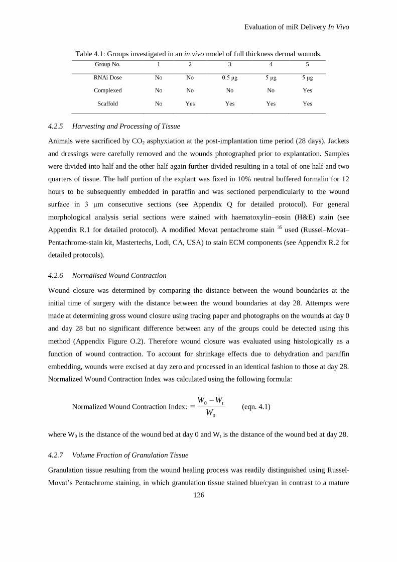
Evaluation of miR Delivery In Vivo
126
Table 4.1: Groups investigated in an in vivo model of full thickness dermal wounds.
Group No. 1 2 3 4 5
RNAi Dose No No 0.5 μg 5 μg 5 μg
Complexed No No No No Yes
Scaffold No Yes Yes Yes Yes
4.2.5 Harvesting and Processing of Tissue
Animals were sacrificed by CO2 asphyxiation at the post-implantation time period (28 days). Jackets
and dressings were carefully removed and the wounds photographed prior to explantation. Samples
were divided into half and the other half again further divided resulting in a total of one half and two
quarters of tissue. The half portion of the explant was fixed in 10% neutral buffered formalin for 12
hours to be subsequently embedded in paraffin and was sectioned perpendicularly to the wound
surface in 3 μm consecutive sections (see Appendix Q for detailed protocol). For general
morphological analysis serial sections were stained with haematoxylin–eosin (H&E) stain (see
Appendix R.1 for detailed protocol). A modified Movat pentachrome stain 35
used (Russel–Movat–
Pentachrome-stain kit, Mastertechs, Lodi, CA, USA) to stain ECM components (see Appendix R.2 for
detailed protocols).
4.2.6 Normalised Wound Contraction
Wound closure was determined by comparing the distance between the wound boundaries at the
initial time of surgery with the distance between the wound boundaries at day 28. Attempts were
made at determining gross wound closure using tracing paper and photographs on the wounds at day 0
and day 28 but no significant difference between any of the groups could be detected using this
method (Appendix Figure O.2). Therefore wound closure was evaluated using histologically as a
function of wound contraction. To account for shrinkage effects due to dehydration and paraffin
embedding, wounds were excised at day zero and processed in an identical fashion to those at day 28.
Normalized Wound Contraction Index was calculated using the following formula:
Normalized Wound Contraction Index: 0
0
W
WW t (eqn. 4.1)
where W0 is the distance of the wound bed at day 0 and Wt is the distance of the wound bed at day 28.
4.2.7 Volume Fraction of Granulation Tissue
Granulation tissue resulting from the wound healing process was readily distinguished using Russel-
Movat’s Pentachrome staining, in which granulation tissue stained blue/cyan in contrast to a mature
Evaluation of miR Delivery In Vivo
127
yellow dermis. Micrographs were obtained at a magnification of 5 X; in each case the epithelium was
horizontal to the image plane. The area of the granulation area was determined using ImageJ imaging
software (National Institute of Health, USA) and the volume fraction of granulation tissue calculated
using the following formula:
Volume Fraction of Granulation Tissue (%) = 100 tA
A
t
Gt (eqn. 4.2)
where AGt is the area of the granulation tissue on the slide, At is the area of total tissue present on the
slide and t is the thickness.
4.2.8 Ratio of Collagen III/I
Quantitative polarised microscopy was performed on paraffin embedded sections stained with sirius
red dye (Biocolor Ltd., Carrickfergus N. Ireland), a bifringence enhancer of collagens 36
. The slides
were counterstained using Weigerts Haematoxylin for eight minutes. The sirius red dye enhancement
of bifringence was used to differentiate between collagen type I and type III. Collagen type I-like
fibres were identified as yellow and red and collagen type III-like fibres in green 37
(see Appendix R.3
for detailed description of protocol). In the visualisation of the images, a polarising attachment BX-
POL (Olympus, South End-on-Sea, UK) was used in a bright field microscope. This device consists
of a polariser (U-POT) inserted in the illumination path and an analyser (U-ANT) oriented orthogonal
to the polarised beam. Images were taken at 200 X magnification in the newly formed tissue within
the wound bed and analysed (Appendix R.3). This analysis was performed using ImageJ threshold
functions and the ratios calculated by dividing the output of collagen type III by collagen type I fibre
analysis.
4.2.9 Collagen Type III Immunohistochemistry
Collagen type III production by the infiltrated fibroblasts in the wound area was elucidated using
immunohistochemistry of representative sections. Hydrated sections were subjected to heat-induced
antigen retrieval in pH 6.0 citrate buffer in a pressure cooker The antibody used in the identification
was a mouse monoclonal antibody to collagen type III (Abcam Plc, Cambridge, UK; dilution: 1:50);
applied overnight at 4°C following blocking with goat-block. Endogenous peroxidase was blocked
using hydrogen peroxide (DakoCytomation, Stockport, UK) for five minutes. An anti-goat horse
radish peroxidase (HRP) labeled secondary antibody (DakoCytomation) was applied for 30 min
followed by addition of 3,3' Diaminobenzidine (DAB) chromagen (DakoCytomation) and the samples
were counterstained with Mayer's Haematoxylin. Three images were taken per slide and six slides per
Evaluation of miR Delivery In Vivo
128
treatment. The location of analysis was medial and the magnification used was × 400. Volume
fractions of collagen type III were analysed using a grid size of 2.5 μm2 (Appendix R.4).
4.2.10 Simultaneous Detection of Rat Protein Expression Using Membrane Array for 90 Proteins
Explanted tissue was frozen at -80 °C until ready. The tissue was thawed slowly on ice, chopped into
small pieces and approximately 5 mg was incubated in a lysis buffer containing a cocktail of protease
inhibitors for five minutes. The tissue was mechanically disrupted using a bead mill homogeniser
which oscillates a stainless steel bead through the tissue (TissueLyserLT, Qiagen, Hilden Germany)
for five minutes at least twice until tissue was completely homogenised and centrifuged at 15,000 g
for 15 minutes. The protein fraction of the centrifuged sample was extracted, aliquoted and stored at -
80 °C until further use. The protein content of the samples was determined using a protein
quantification assay kit (BioRad, Hercules, CA. Appendix M.1) and probed for 90 proteins
simultaneously using a biotin label-based rat antibody array (RayBio®, Norcross, GA, USA)
according to the manufacturer’s instructions. Briefly, four samples were pooled according to the
treatment group to a total of 1 mg/ml and dialysed overnight to remove remaining lysis buffer and the
protein membranes were blocked using a provided blocking buffer. The proteins were labelled with
biotin using amines present in proteins and incubated with the antibody labelled membranes
overnight. Following this, HRP-conjugated streptavidin was reacted with the membranes and
afterwards treated with the incubation buffer provided. Membranes were exposed using a Kodak™
Image Station 4000MM Pro (Kodak, Japan). Micrographs of the exposed membranes were digitally
evaluated using ImageJ software plug-ins. The images obtained were imported into ImageJ and
analysed using a protein array analyser plug-in by normalising to the given positive control signals on
the membranes. For relative comparison, all samples were normalised to healthy native skin which
was not wounded (see Appendix M for details of all procedures).
4.2.11 TGF-β1, MMP-8 and TIMP-1 Quantification
Three enzyme linked immunosorbent assays (ELISAs) were performed using protein isolates which
had been aliquoted from the treatment groups. The ELISA analytes included one for transforming
growth factor (TGF)-β1 (Abcam Plc), matrix metalloproteinase (MMP)-8 (Abcam Plc), and tissue
inhibitor of metalloproteinase (TIMP)-1 (R&D Systems, Minneapolis, MN, USA). As previously
mentioned; TGF-β 1 plays a significant role in matrix remodelling following injury whereby it
stimulates ECM production 5. A distinct role of miR-29B is in post-transcriptional silencing of ECM
fibrillar components stimulated by TGF-β1 6- 8
, therefore quantification of TGF-β1 expression was
essential in this study. Matrix turnover, i.e. the enzymatic remodelling of ECM is an important
consideration in healing wounds and indeed remodelling components were revealed to be
Evaluation of miR Delivery In Vivo
129
significantly dysregulated in the protein membrane array. Notably, MMP-8 is a collagen cleaving
enzyme which functions to degrade type I, II and III collagens 9 and in the context of wound healing;
MMP-8 has been shown to be the predominant collagenase in healing wounds and non-healing ulcers
10. TIMP-1 complexes with MMPs and irreversibly inactivates them by binding to their catalytic zinc
co-factor and, furthermore, TIMP-1 is a direct inhibitor of MMP-8 11
.
An equal amount and concentration of each protein sample was added to each well for each ELISA
plate which was determined using a protein assay kit (BioRad, Hercules, CA). ELISAs were
performed according to the manufacturer’s instructions (Appendices Q.4, Q.5 and Q.6).
4.2.12 RNA Extraction
One mL of TRI Reagent® (Applera Ireland, Dublin, Ireland) was added to each tissue sample and
incubated for five minutes at room temperature. The tissue was mechanically disrupted using a bead
mill homogeniser (TissueLyserLT, Qiagen) at least twice for five minutes until tissue was completely
homogenised. Phase separation was performed by adding chloroform, and total RNA was purified
using an RNeasy kit (Qiagen), according to the supplier’s recommended procedure (Appendix S).
4.2.13 Wound Healing RT-PCR Array
Contaminant DNA was eliminated from RNA preparations using DNase I. The yield and quality of
total RNA was determined according to the ratio of spectrophotometric absorbance values at
wavelengths of 260 and 280 nm. cDNA synthesis was performed using DNase-treated RNA and
random decamer primers using an RT2 First Strand Kit (Qiagen). The cDNA generated was used as a
template for quantitative real-time PCR.A mastermix was prepared using a RT2 SYBR Green
Mastermix. This mixture was added to 384 wells in an RT2 Profiler™ PCR Array, Rat Wound
Healing Array (PARB-0121Z; Qiagen). This PCR array contained RT2 qPCR Primer Assays for a set
of 84 cytokines which are detailed in Appendix U, Table U.5. The standard cycling conditions were as
recommended by the PCR array supplier. Data were collected at the end of the annealing step. Fold
changes in gene expression between the affected and control groups were calculated using the ΔΔCt
method in the PCR array data analysis template. An examination of Ct value consistency for the
housekeeping genes indicated that normalization was performed adequately. A similar evaluation of
the built-in RNA controls verified an absence of genomic DNA contamination and inhibitors of either
the reverse transcription or PCR (Appendix U.2).
4.2.14 Statistical Analysis
All samples were tested in triplicate and all experimental groups were analysed in triplicate. GraphPad
Prism® (v.5 GraphPad Software, San Diego, CA, USA) was used for statistical analysis. Analysis of
Evaluation of miR Delivery In Vivo
130
variance (ANOVA) was used followed by Tukey’s post-hoc test to determine statistical significance
between groups. ANOVA was performed assuming normal distribution of the data which was tested
and verified using the Anderson-Darling test. All graphical data is presented as mean ± standard
deviation of mean. p values of < 0.05 were considered statistically significant. Outliers were
calculated using Grubb’s test and eliminated from the results to be analysed. Where appropriate,
Pearson’s test for correlation was performed where significance was set at p < 0.05.
4.3 Results and Discussion
4.3.1 Gross Wound Contraction
In the evaluation of gross wound contraction, the use of sterile tracing papers was attempted as an
objective method for measuring wound area fractions. However, this method proved inaccurate and
did not provide a statistically significant output (see Appendix O, Figure O.2 for these results).
Furthermore, post-excisional wound photographs were used as visual aids in the assessment of wound
healing but were not considered an option for quantification purposes. Measurements cannot be
performed accurately on photographs of non-flat surfaces and, furthermore, another major difficulty
in the evaluation of wound closure by this method was to determine an objective visual boundary
between wounded and healed areas. Assessment of epithelialisation showed that all wounds were
fully healed and therefore no difference between treatment groups can be detected using this
parameter. The rationale for using a rat excisional model in this study was due to the established data
of miR profiling in rodents 38
and also the commercial availability of antibody and protein tools that
can be applied to this species. Additionally, this rodent model is an established model of cutaneous
wound healing 39-42
. Other models such as a rabbit ear ulcer model could not be used due to the
limited availability of primary antibodies, ELISAs and miR profiling.
4.3.2 Normalized Wound Contraction
Through histological analysis of the wounds at day 28 compared to day 0, application of scaffolds to
the wounds significantly reduced the contraction of the wounds which was evaluated by the distance
of original wound edges at day 28 (Figure 4.2). Such a response with regard to wound contraction
after treatment with biomaterial scaffolds has been conclusively demonstrated by others 39-42
. The
percentage of wound contraction, as measured histologically when comparing the wound width at day
28 against initial wound width at day 0, was reduced significantly by 10 % in the scaffold only
treatment group (Figure 4.2). Contraction is also significantly reduced when miR-29B is incorporated
into the scaffold. Incorporation of 0.5 μg or 5 μg of miR-29B significantly reduced contraction by 15
% when compared to no treatment control. However, incorporating 5 μg of miR-29B complexed with
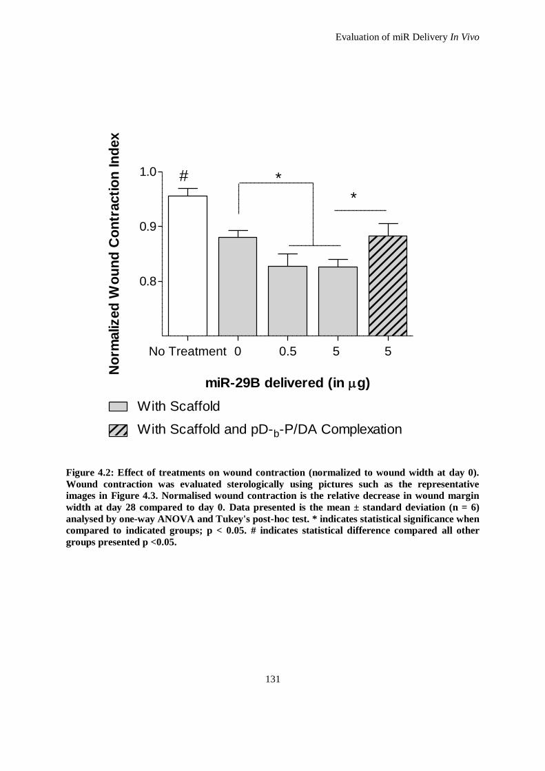
Evaluation of miR Delivery In Vivo
131
No Treatment 0 0.5 5 5
0.8
0.9
1.0
With Scaffold
With Scaffold and pD-b-P/DA Complexation
#
*
*
miR-29B delivered (in g)
No
rmalized
Wo
un
d C
on
tracti
on
In
dex
Figure 4.2: Effect of treatments on wound contraction (normalized to wound width at day 0).
Wound contraction was evaluated sterologically using pictures such as the representative
images in Figure 4.3. Normalised wound contraction is the relative decrease in wound margin
width at day 28 compared to day 0. Data presented is the mean ± standard deviation (n = 6)
analysed by one-way ANOVA and Tukey's post-hoc test. * indicates statistical significance when
compared to indicated groups; p < 0.05. # indicates statistical difference compared all other
groups presented p <0.05.
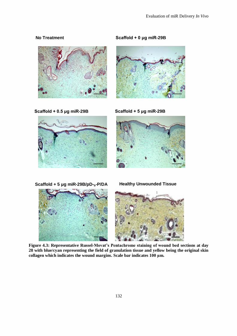
Evaluation of miR Delivery In Vivo
132
Figure 4.3: Representative Russel-Movat’s Pentachrome staining of wound bed sections at day
28 with blue/cyan representing the field of granulation tissue and yellow being the original skin
collagen which indicates the wound margins. Scale bar indicates 100 μm.
No Treatment Scaffold + 0 μg miR-29B
Scaffold + 5 μg miR-29B
Scaffold + 5 μg miR-29B/pD-b-P/DA
Scaffold + 0.5 μg miR-29B
Healthy Unwounded Tissue
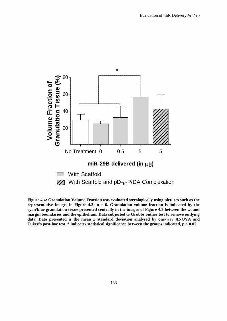
Evaluation of miR Delivery In Vivo
133
No Treatment 0 0.5 5 5
20
40
60
80
With Scaffold
With Scaffold and pD-b-P/DA Complexation
*
miR-29B delivered (in g)
Vo
lum
e F
rac
tio
n o
f
Gra
nu
lati
on
Tis
su
e (
%)
Figure 4.4: Granulation Volume Fraction was evaluated sterologically using pictures such as the
representative images in Figure 4.3; n = 6. Granulation volume fraction is indicated by the
cyan/blue granulation tissue presented centrally in the images of Figure 4.3 between the wound
margin boundaries and the epithelium. Data subjected to Grubbs outlier test to remove outlying
data. Data presented is the mean ± standard deviation analysed by one-way ANOVA and
Tukey's post-hoc test. * indicates statistical significance between the groups indicated, p < 0.05.
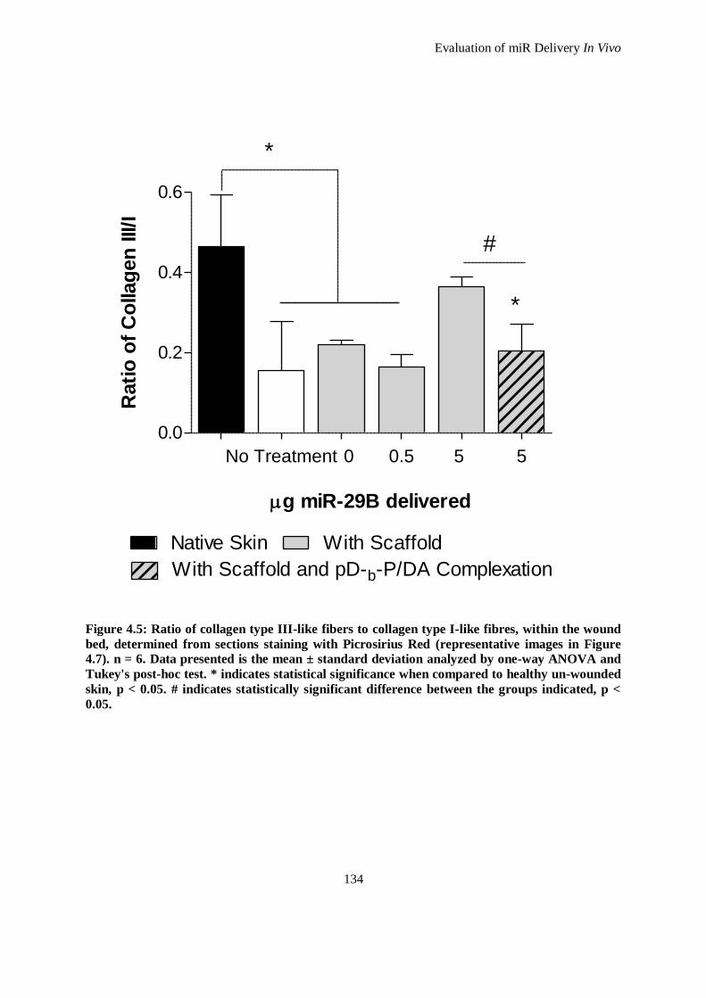
Evaluation of miR Delivery In Vivo
134
No Treatment 0 0.5 5 5
0.0
0.2
0.4
0.6
With Scaffold
With Scaffold and pD-b-P/DA Complexation
*
*
#
Native Skin
g miR-29B delivered
Rati
o o
f C
ollag
en
III/I
Figure 4.5: Ratio of collagen type III-like fibers to collagen type I-like fibres, within the wound
bed, determined from sections staining with Picrosirius Red (representative images in Figure
4.7). n = 6. Data presented is the mean ± standard deviation analyzed by one-way ANOVA and
Tukey's post-hoc test. * indicates statistical significance when compared to healthy un-wounded
skin, p < 0.05. # indicates statistically significant difference between the groups indicated, p <
0.05.
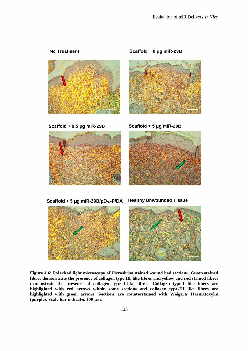
Evaluation of miR Delivery In Vivo
135
Figure 4.6: Polarised light microscopy of Picrosirius stained wound bed sections. Green stained
fibres demonstrate the presence of collagen type III-like fibres and yellow and red stained fibres
demonstrate the presence of collagen type I-like fibres. Collagen type-I like fibers are
highlighted with red arrows within some sections and collagen type-III like fibres are
highlighted with green arrows. Sections are counterstained with Weigerts Haematoxylin
(purple). Scale bar indicates 100 μm.
No Treatment Scaffold + 0 μg miR-29B
Scaffold + 5 μg miR-29B
Scaffold + 5 μg miR-29B/pD-b-P/DA
Scaffold + 0.5 μg miR-29B
Healthy Unwounded Tissue
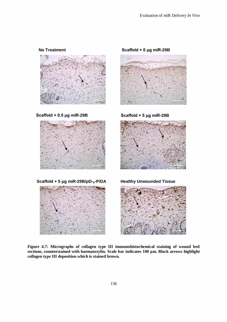
Evaluation of miR Delivery In Vivo
136
Figure 4.7: Micrographs of collagen type III immunohistochemical staining of wound bed
sections, counterstained with haematoxylin. Scale bar indicates 100 μm. Black arrows highlight
collagen type III deposition which is stained brown.
No Treatment Scaffold + 0 μg miR-29B
Scaffold + 5 μg miR-29B
Scaffold + 5 μg miR-29B/pD-b-P/DA
Scaffold + 0.5 μg miR-29B
Healthy Unwounded Tissue
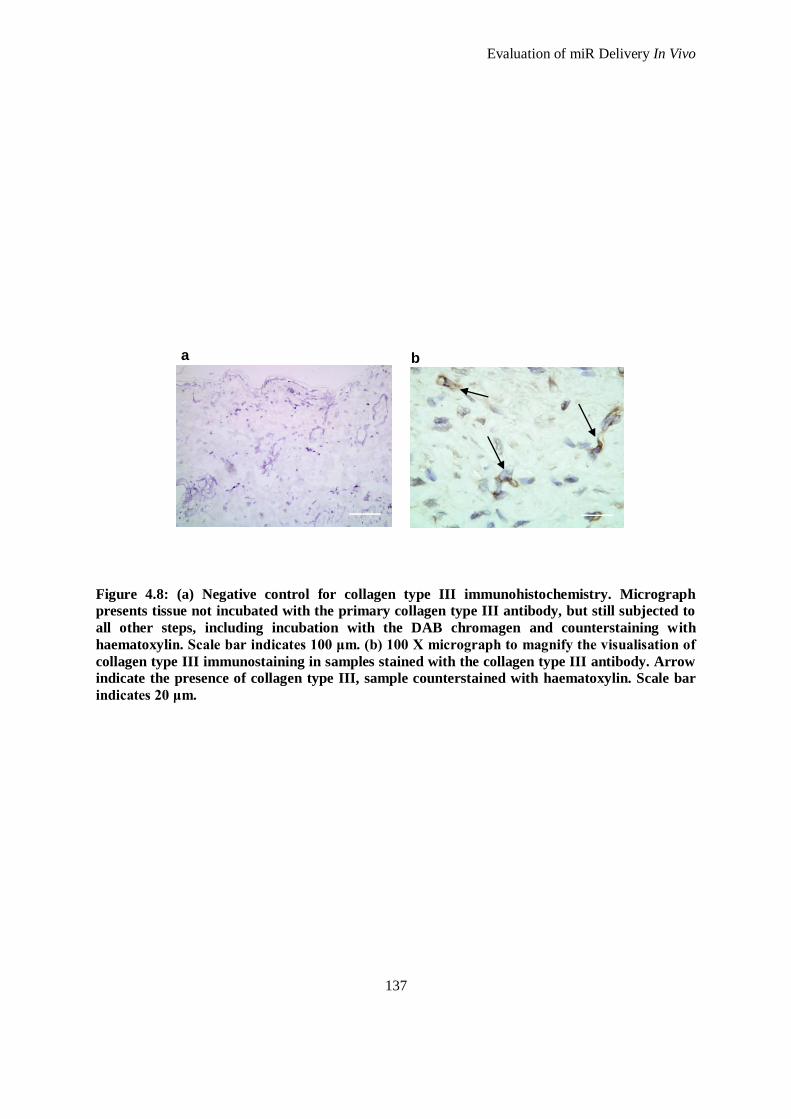
Evaluation of miR Delivery In Vivo
137
Figure 4.8: (a) Negative control for collagen type III immunohistochemistry. Micrograph
presents tissue not incubated with the primary collagen type III antibody, but still subjected to
all other steps, including incubation with the DAB chromagen and counterstaining with
haematoxylin. Scale bar indicates 100 μm. (b) 100 X micrograph to magnify the visualisation of
collagen type III immunostaining in samples stained with the collagen type III antibody. Arrow
indicate the presence of collagen type III, sample counterstained with haematoxylin. Scale bar
indicates 20 μm.
a b
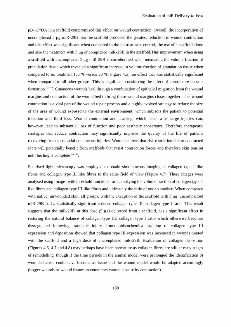
Evaluation of miR Delivery In Vivo
138
pD-b-P/DA in a scaffold compromised this effect on wound contraction. Overall, the incorporation of
uncomplexed 5 μg miR-29B into the scaffold produced the greatest reduction in wound contraction
and this effect was significant when compared to the no treatment control, the use of a scaffold alone
and also the treatment with 5 μg of complexed miR-29B in the scaffold.This improvement when using
a scaffold with uncomplexed 5 μg miR-29B is corroborated when measuring the volume fraction of
granulation tissue which revealed a significant increase in volume fraction of granulation tissue when
compared to no treatment (55 % versus 30 %, Figure 4.5), an effect that was statistically significant
when compared to all other groups. This is significant considering the effect of contracture on scar
formation 43, 44
. Cutaneous wounds heal through a combination of epithelial migration from the wound
margins and contraction of the wound bed to bring these wound margins closer together. This wound
contraction is a vital part of the wound repair process and a highly evolved strategy to reduce the size
of the area of wound exposed to the external environment, which subjects the patient to potential
infection and fluid loss. Wound contraction and scarring, which occur after large injuries can,
however, lead to substantial loss of function and poor aesthetic appearance. Therefore therapeutic
strategies that reduce contraction may significantly improve the quality of the life of patients
recovering from substantial cuntaneous injuries. Wounded areas that risk restriction due to contracted
scars will potentially benefit from scaffolds that resist contraction forces and therefore skin tension
until healing is complete 45, 46
.
Polarised light microscopy was employed to obtain simultaneous imaging of collagen type I like
fibres and collagen type III like fibres in the same field of view (Figure 4.7). These images were
analysed using ImageJ with threshold functions for quantifying the volume fraction of collagen type I-
like fibres and collagen type III-like fibres and ultimately the ratio of one to another. When compared
with native, unwounded skin, all groups, with the exception of the scaffold with 5 μg uncomplexed
miR-29B had a statistically significant reduced collagen type III: collagen type I ratio. This result
suggests that the miR-29B, at this dose (5 μg) delivered from a scaffold, has a significant effect in
restoring the natural balance of collagen type III: collagen type I ratio which otherwise becomes
dysregulated following traumatic injury. Immunohistochemical staining of collagen type III
expression and deposition showed that collagen type III expression was increased in wounds treated
with the scaffold and a high dose of uncomplexed miR-29B. Evaluation of collagen deposition
(Figures 4.6, 4.7 and 4.8) may perhaps have been premature as collagen fibres are still at early stages
of remodelling, though if the time periods in the animal model were prolonged the identification of
wounded areas could have become an issue and the wound model would be adapted accordingly
(bigger wounds or wound frames to counteract wound closure by contraction).
Evaluation of miR Delivery In Vivo
139
4.3.2 Proteomic Profiles from Treatments
In order to obtain an overview of the proteins dysregulated following cutaneous injury in this model,
and following application of the platform developed in this study, a biotin label based rat antibody
array, capable of detecting the expression levels of 90 rat proteins was employed. This array provided
the most extensive range of proteins that could be assessed in this model and encompassed many key
proteins pivotal in ECM remodelling. The array, which consisted of a membrane blot array, was
analysed using a protein array plug-in on Image J (see Appendix M.2 for details of how this analysis
was performed). The membranes were normalised according to internal controls specified in the array,
and all membranes were analysed in comparison with tissue obtained from the dermis of an untreated
(and unwounded) animal. Following this, the array of proteins dysregulated in the control group
which received no treatment to an incurred wound was considered the standard from which all the
treatments were compared. For simplicity and ease of understanding the main findings of this array
(i.e. proteins that are upregulated and those that are downregulated) are represented in Figure 4.9 in a
Venn diagram where each treatment parameter is represented by a circle. The findings illustrate a
global effect as they are representative of pooled samples and cannot be subjected to discrete
quantification. The proteins affected have been clustered for transparency; for instance; growth factors
such as VEGF, EGFR; apoptotic factors such as Fas Ligand, FADD; ECM remodelling proteins
Activin, TGF-βs, TIMPs, MMPs; and inflammatory cytokines; IL-1α, IL-1β, MCP-1.
Apoptosis, is a morphologically and biochemically distinct process of cell death which is a crucial
control mechanism for the development of organs during embryogenesis, and also for the
maintenance of tissue homeostasis in mature organisms 47
. Apoptosis has been identified as a key
player in the transition between granulation tissue and the formation of definitive scar after soft tissue
injury 48
. The apoptosis of fibroblasts, endothelial cells, and pericytes appears to affect cell types in
successive waves after wound closure. When a wound closes and evolves into a scar, there is a
dramatic decrease in cellularity and, in particular myofibroblasts 49
.
In Figure 4.9, the proteins associated with apoptosis, namely Fas TNFSNF6 and Fas Ligand
TNFSNF6 are shown to be downregulated when a scaffold is applied. This suggests that the transition
between granulation tissue and formation of a definitive scar has not evolved. Indeed, this is in
agreement with the previous observations in which wound contraction was reduced when using a 4S-
StarPEG collagen scaffold (Figure 4.2). However, when incorporating miR-29B into the 4S-StarPEG
collagen scaffold at the high dose of 5 μg of miR-29B, uncomplexed or complexed with pD-b-P/DA,
apoptotic factors are upregulated. This is extremely significant for a number of reasons. It
demonstrates that the miR-29B is having an effect on the upregulation of apoptotic proteins and this
effect is also dose dependent. The role of miR-29B on apoptosis has only recently been elucidated.
Evaluation of miR Delivery In Vivo
140
For instance, miR-29B is known to have many targets, one of which is the anti-apoptotic protein Mcl-
1 50-52
. Therefore, increased presence of miR-29B can elicit increased apoptosis. This is somewhat in
agreement with the data presented from the protein array in which an upregulation of apoptotic
proteins is present when delivering 5 μg of miR-29B (uncomplexed or complexed with pD-b-P/DA).
However, this contradicts the association of apoptosis with the transition of granulation tissue to the
formation of a definitive scar. While the underlying mechanisms may remain unclear, one needs to
take into consideration the other targets and effects of miR-29B. Additionally, the in vivo environment
present here has increased expression of TGF-β1, and TGF-β1 has often been reported to offer
apoptotic resistance to cells 53, 54
. The inflammatory response is regarded as the first of a number of
overlapping processes that constitute wound healing. In skin repair, the infiltrating leukocytes,
monocytes (which later become macrophages) and neutrophils are the principal components of the
inflammatory response 55, 56
. They not only act as effector cells combating pathogens but are also
involved in tissue degradation and tissue formation. Ultimately, the inflammatory response dictates
the quality of the healing response. There are several situations that provide evidence that the
inflammatory phase during repair is intimately linked to the extent of scar formation 57- 59
. In contrast
to post-natal repair, wound healing in early foetal skin exhibits scarless regeneration of dermal
architecture which is intrinsically linked with the lack of inflammatory response 60-62
. In support of
this, scar formation has been demonstrated to be accelerated when inflammation is initiated in foetal
wounds 63
. From the results of the protein membrane array, a number of observations can be made on
the up/down regulation of proteins involved in the inflammatory response. For instance, application of
the 4S-StarPEG collagen scaffold resulted in a knockdown of the inflammatory cytokines. This
suggest that the 4S-StarPEG collagen scaffold elicits a minimal host response as it is partially
composed of atelocollagen, a form of collagen without the terminal peptides, which has extremely
low antigenicity due to the elimination of antigenic regions by proteolytic processing 64
. The
incorporation of miR-29B into the scaffold elicits the upregulation of some inflammatory cytokines
(IL-1α, IL-1β) and this pattern exists for both 0.5 μg and 5 μg of uncomplexed miR-29B. However,
when 5 μg of miR-29B complexed with pD-b-P/DA is delivered with the 4S-StarPEG collagen
scaffold there is an upregulation of multiple inflammatory cytokines. This is due, in part, to the
presence of pD-b-P/DA in the scaffold. The higher dose of miR-29B chosen in this study; 5 μg,
equates to 40 μg of pD-b-P/DA when delivered in complexed form. pD-b-P/DA is relatively non-toxic
in vitro due to its highly PEGylated structure; however this characteristic is dose-dependent. The
degradation of pD-b-P/DA in vivo most likely occurs through hydrolysis of the ester links in all of its
components; pDMAEMA, PEGDA and PEGMEA. The PEG component of both PEGDA and
PEGMEA are linear esters which are susceptible to hydrolytic degradation 65
. This degrades the
complexing agent into smaller components which can be gradually cleared by the tissue and excreted
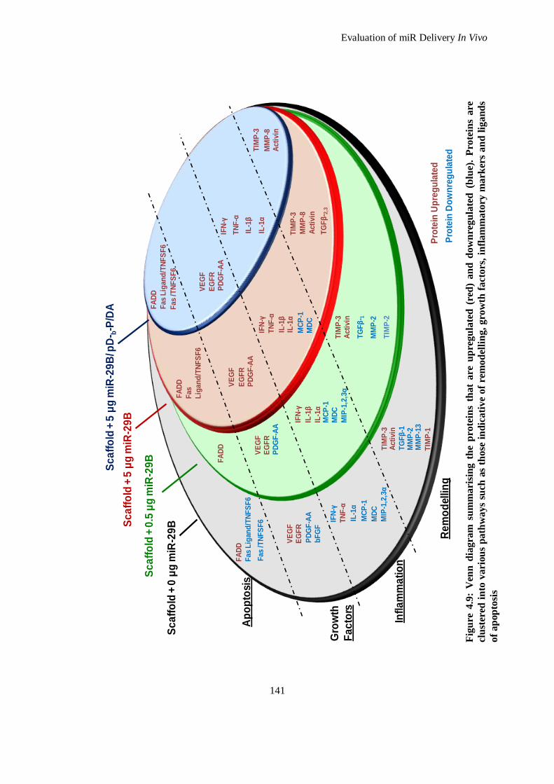
Evaluation of miR Delivery In Vivo
141
Fig
ure
4.9
: V
enn
dia
gra
m s
um
mari
sin
g t
he
pro
tein
s th
at
are
up
regu
late
d (
red
) a
nd
do
wn
reg
ula
ted
(b
lue)
. P
rote
ins
are
clu
ster
ed i
nto
va
riou
s p
ath
ways
such
as
those
in
dic
ati
ve
of
rem
od
elli
ng,
gro
wth
fa
cto
rs,
infl
am
ma
tory
ma
rker
s a
nd
lig
an
ds
of
ap
op
tosi
s
FA
DD
FA
DD
Fas
Lig
an
d/T
NF
SF
6
FA
DD
Fas L
igan
d/T
NF
SF
6
Fas
/TN
FS
F6
FA
DD
Fas L
igan
d/T
NF
SF
6
Fas
/TN
FS
F6
IFN
-γ
IL-1
α
IL-1
β
MC
P-1
MD
C
MIP
-1,2
,3α
Acti
vin
MM
P-8
TIM
P-3
Acti
vin
MM
P-8
TG
Fβ
- 2,3
TIM
P-3
Acti
vin
MM
P-2
TG
Fβ
- 1
TIM
P-2
TIM
P-3
Acti
vin
MM
P-2
MM
P-1
3
TG
Fβ
-1
TIM
P-1
TIM
P-3
IFN
-γ
IL-1
α
MC
P-1
MD
C
MIP
-1,2
,3α
TN
F-α
IFN
-γ
IL-1
β
IL-1
α
TN
F-α
IFN
-γ
IL-1
αIL
-1β
MC
P-1
MD
C
TN
F-α
EG
FR
PD
GF
-AA
VE
GF
EG
FR
PD
GF
-AA
VE
GF
EG
FR
PD
GF
-AA
VE
GF
bF
GF
EG
FR
PD
GF
-AA
VE
GF
Pro
tein
Up
reg
ula
ted
Pro
tein
Do
wn
reg
ula
ted
Ap
op
tosis
Infl
am
ma
tio
n
Sc
aff
old
+ 0
μg
miR
-29
B
Sc
aff
old
+ 0
.5 μ
g m
iR-2
9B
Sc
aff
old
+ 5
μg
miR
-29
B
Sc
aff
old
+ 5
μg
miR
-29
B/ p
D- b
-P/D
A
Gro
wth
Fa
cto
rs
Re
mo
dellin
g
Evaluation of miR Delivery In Vivo
142
via renal filtration. Acrylates and methacrylates (which are components of PEGDA and PEGMEA
respectively) are widely used in the formulation of polymeric materials for medical and dental
applications, but there are reports on the local toxicity of these monomers 66
. The degradation
products of pD-b-P/DA will contain some acrylates and methacrylates as PEGDA and PEGMEA were
used as branching and backbone structures in the complexing agent. However, considering that 3 % of
vinyl groups are free in this structure (as determined in Chapter Two), local toxicity due to acrylate
products should be negligible. There also exists a concern regarding the presence of copper chloride
which was used in the ATRP reaction to synthesise pD-b-P/DA. Although filtration and dialysis were
performed, it is possible that trace amounts of copper may still be present. This could account for the
upregulation of inflammatory cytokines present in the group which includes pD-b-P/DA due to its
degradation products in vivo.
Based on the observations from the biotin label based rat antibody array, three ELISAs were
performed to quantify and determine a statistically significant relationship between the treatments and
the affected protein. These ELISAs measured TIMP-1 (Figure 4.10), MMP-8 (Figure 4.11) and TGF-
β1 (Figure 4.12). The observations detected in the membrane protein array with regard to TGF- β1,
TIMP-1 and MMP-8 expression are, for the most part, in agreement with the ELISAs. Any
inconsistency between the two representations can be attributed to samples which may have high
levels of a particular protein causing deviation in the ELISA results and possible saturation during the
pooling of sample for the membrane protein array. TGF-β1 is dysregulated (in this case
downregulated) in the 4S-StarPEG collagen scaffold group (based on protein array data and ELISA
quantification). This is reflected in the same group by the downregulation of MMP2 and MMP13 (in
the membrane protein array) which are known to be stimulated by TGF-β1 67, 68
. However, the
membrane protein array was performed on pooled samples and it could be possible that one sample
could be masking/saturating the true levels of TGF-β1. The TGF-β family is a key mediator of ECM
production and this has been the basis upon which TGF-β1 has been delivered with biomaterials in
order to enhance ECM production. For instance, Pandit et al. delivered TGF-β using a collagen
scaffold in a rabbit ear ulcer model and found an increased contraction rate when compared to a
collagen scaffold alone. However, this treatment also resulted in a significantly increased ultimate
tensile strength of the wounds when compared to an untreated wound suggesting a stronger healed
wound. Regardless, TGF-β1 is beneficial to increase the healing rates of wounds that do not need to be
aesthetically appealing, yet the ideal scenario of tissue regeneration needs to allow for controlled
healing through which there is not exaggerated deposition of fibrillar ECM proteins that contribute to
unsightly, compromised scar tissue. It is established that miR-29B acts post-transcriptionally on many
of the ECM proteins that are stimulated by TGF-β1 69, 70
, however there are many conflicting reports
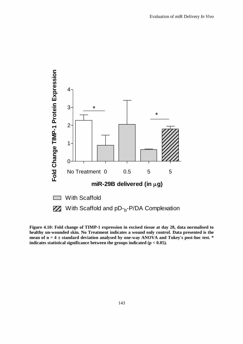
Evaluation of miR Delivery In Vivo
143
No Treatment 0 0.5 5 5
0
1
2
3
4
With Scaffold
With Scaffold and pD-b-P/DA Complexation
**
miR-29B delivered (in g)
Fo
ld C
han
ge T
IMP
-1 P
rote
in E
xp
ressio
n
Figure 4.10: Fold change of TIMP-1 expression in excised tissue at day 28, data normalised to
healthy un-wounded skin. No Treatment indicates a wound only control. Data presented is the
mean of n = 4 ± standard deviation analysed by one-way ANOVA and Tukey's post-hoc test. *
indicates statistical significance between the groups indicated (p < 0.05).
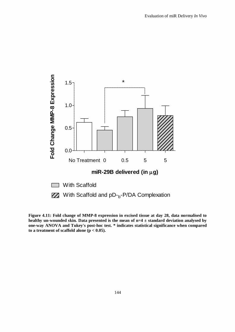
Evaluation of miR Delivery In Vivo
144
No Treatment 0 0.5 5 5
0.0
0.5
1.0
1.5
With Scaffold
With Scaffold and pD-b-P/DA Complexation
*
miR-29B delivered (in g)
Fo
ld C
han
ge M
MP
-8 E
xp
ressio
n
Figure 4.11: Fold change of MMP-8 expression in excised tissue at day 28, data normalised to
healthy un-wounded skin. Data presented is the mean of n=4 ± standard deviation analysed by
one-way ANOVA and Tukey's post-hoc test. * indicates statistical significance when compared
to a treatment of scaffold alone (p < 0.05).
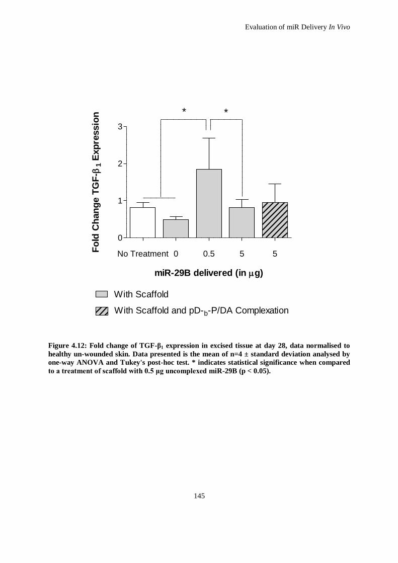
Evaluation of miR Delivery In Vivo
145
No Treatment 0 0.5 5 5
0
1
2
3
With Scaffold
With Scaffold and pD-b-P/DA Complexation
* *
miR-29B delivered (in g)
Fo
ld C
han
ge T
GF
- 1
Exp
ressio
n
Figure 4.12: Fold change of TGF-β1 expression in excised tissue at day 28, data normalised to
healthy un-wounded skin. Data presented is the mean of n=4 ± standard deviation analysed by
one-way ANOVA and Tukey's post-hoc test. * indicates statistical significance when compared
to a treatment of scaffold with 0.5 μg uncomplexed miR-29B (p < 0.05).
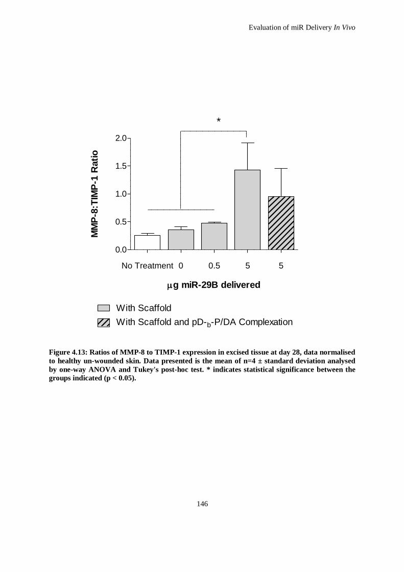
Evaluation of miR Delivery In Vivo
146
No Treatment 0 0.5 5 5
0.0
0.5
1.0
1.5
2.0
With Scaffold
With Scaffold and pD-b-P/DA Complexation
*
g miR-29B delivered
MM
P-8
:TIM
P-1
Rati
o
Figure 4.13: Ratios of MMP-8 to TIMP-1 expression in excised tissue at day 28, data normalised
to healthy un-wounded skin. Data presented is the mean of n=4 ± standard deviation analysed
by one-way ANOVA and Tukey's post-hoc test. * indicates statistical significance between the
groups indicated (p < 0.05).
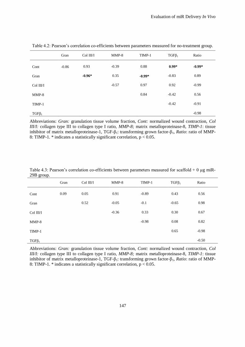
Evaluation of miR Delivery In Vivo
147
Table 4.2: Pearson’s correlation co-efficients between parameters measured for no-treatment group.
Gran Col III/I MMP-8 TIMP-1 TGFβ1 Ratio
Cont
-0.86
0.93
-0.39
0.88
0.99*
-0.99*
Gran
-0.96*
0.35
-0.99*
-0.83
0.89
Col III/I
-0.57
0.97
0.92
-0.99
MMP-8
0.84
-0.42
0.56
TIMP-1
-0.42
-0.91
TGFβ1
-0.98
Abbreviations: Gran: granulation tissue volume fraction, Cont: normalized wound contraction, Col
III/I: collagen type III to collagen type I ratio, MMP-8; matrix metalloproteinase-8, TIMP-1: tissue
inhibitor of matrix metalloproteinase-1, TGF-β1: transforming grown factor-β1, Ratio: ratio of MMP-
8: TIMP-1. * indicates a statistically significant correlation, p < 0.05.
Table 4.3: Pearson’s correlation co-efficients between parameters measured for scaffold + 0 μg miR-29B group.
Gran Col III/I MMP-8 TIMP-1 TGFβ1 Ratio
Cont
0.09
0.05
0.91
-0.89
0.43
0.56
Gran
0.52
-0.05
-0.1
-0.65
0.98
Col III/I
-0.36
0.33
0.30
0.67
MMP-8
-0.98
0.08
0.82
TIMP-1
0.65
-0.98
TGFβ1
-0.50
Abbreviations: Gran: granulation tissue volume fraction, Cont: normalized wound contraction, Col
III/I: collagen type III to collagen type I ratio, MMP-8; matrix metalloproteinase-8, TIMP-1: tissue inhibitor of matrix metalloproteinase-1, TGF-β1: transforming grown factor-β1, Ratio: ratio of MMP-
8: TIMP-1. * indicates a statistically significant correlation, p < 0.05.
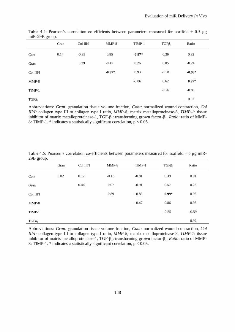
Evaluation of miR Delivery In Vivo
148
Table 4.4: Pearson’s correlation co-efficients between parameters measured for scaffold + 0.5 μg miR-29B group.
Gran Col III/I MMP-8 TIMP-1 TGFβ1 Ratio
Cont
0.14
-0.95
0.85
-0.97*
0.39
0.92
Gran
0.29
-0.47
0.26
0.05
-0.24
Col III/I
-0.97*
0.93
-0.58
-0.99*
MMP-8
-0.86
0.62
0.97*
TIMP-1
-0.26
-0.89
TGFâ1
0.67
Abbreviations: Gran: granulation tissue volume fraction, Cont: normalized wound contraction, Col
III/I: collagen type III to collagen type I ratio, MMP-8; matrix metalloproteinase-8, TIMP-1: tissue
inhibitor of matrix metalloproteinase-1, TGF-β1: transforming grown factor-β1, Ratio: ratio of MMP-8: TIMP-1. * indicates a statistically significant correlation, p < 0.05.
Table 4.5: Pearson’s correlation co-efficients between parameters measured for scaffold + 5 μg miR-29B group.
Gran Col III/I MMP-8 TIMP-1 TGFβ1 Ratio
Cont
0.02
0.12
-0.13
-0.81
0.39
0.01
Gran
0.44
0.07
-0.91
0.57
0.23
Col III/I
0.89
-0.83
0.99*
0.95
MMP-8
-0.47
0.86
0.98
TIMP-1
-0.85
-0.59
TGFâ1
0.92
Abbreviations: Gran: granulation tissue volume fraction, Cont: normalized wound contraction, Col
III/I: collagen type III to collagen type I ratio, MMP-8; matrix metalloproteinase-8, TIMP-1: tissue inhibitor of matrix metalloproteinase-1, TGF-β1: transforming grown factor-β1, Ratio: ratio of MMP-
8: TIMP-1. * indicates a statistically significant correlation, p < 0.05.
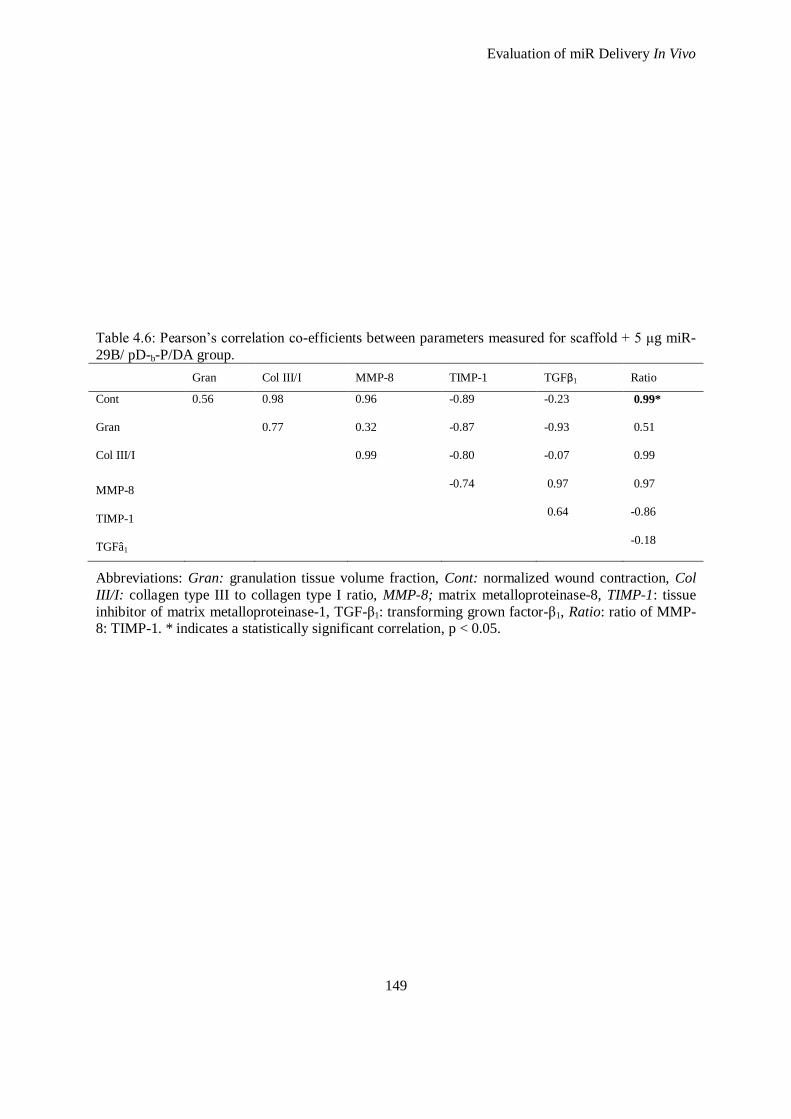
Evaluation of miR Delivery In Vivo
149
Table 4.6: Pearson’s correlation co-efficients between parameters measured for scaffold + 5 μg miR-
29B/ pD-b-P/DA group.
Gran Col III/I MMP-8 TIMP-1 TGFβ1 Ratio
Cont
0.56
0.98
0.96
-0.89
-0.23
0.99*
Gran
0.77
0.32
-0.87
-0.93
0.51
Col III/I
0.99
-0.80
-0.07
0.99
MMP-8 -0.74
0.97
0.97
TIMP-1 0.64
-0.86
TGFâ1 -0.18
Abbreviations: Gran: granulation tissue volume fraction, Cont: normalized wound contraction, Col
III/I: collagen type III to collagen type I ratio, MMP-8; matrix metalloproteinase-8, TIMP-1: tissue
inhibitor of matrix metalloproteinase-1, TGF-β1: transforming grown factor-β1, Ratio: ratio of MMP-8: TIMP-1. * indicates a statistically significant correlation, p < 0.05.
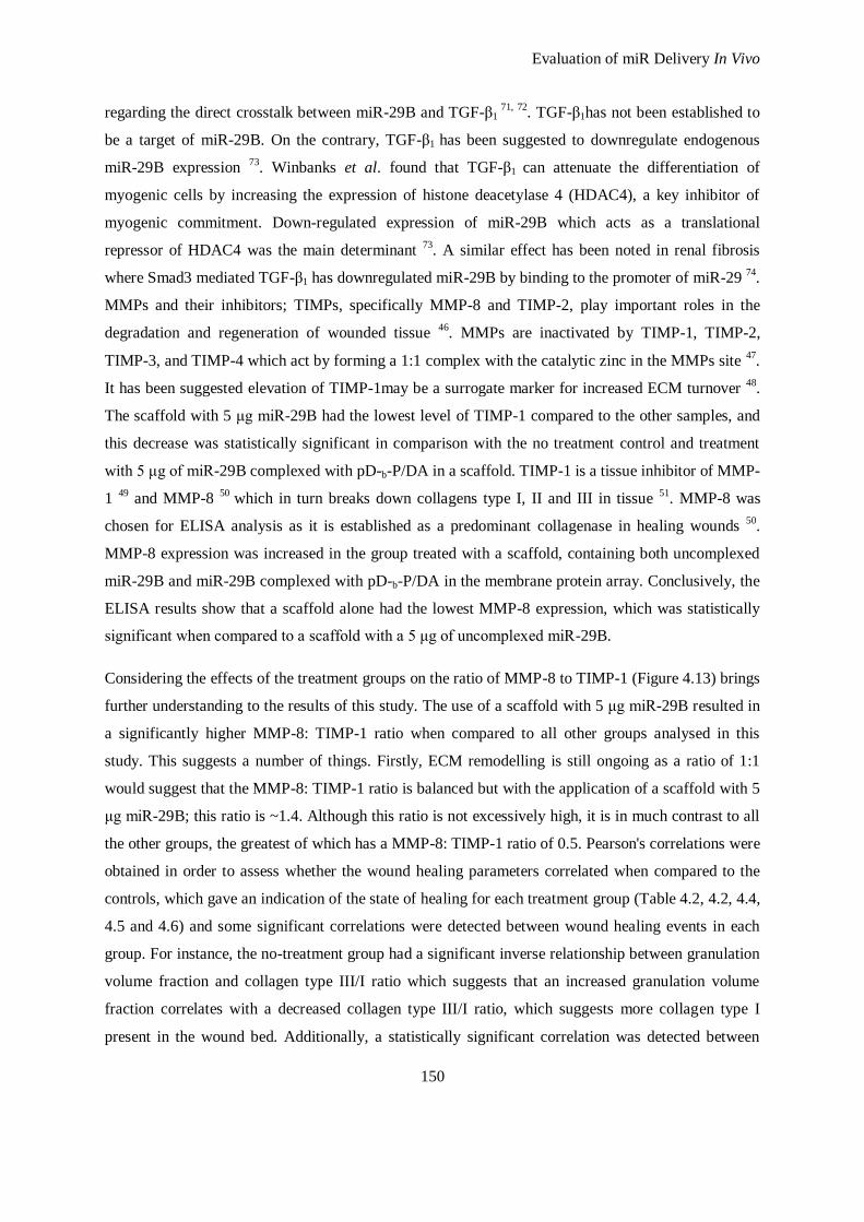
Evaluation of miR Delivery In Vivo
150
regarding the direct crosstalk between miR-29B and TGF-β1 71, 72
. TGF-β1has not been established to
be a target of miR-29B. On the contrary, TGF-β1 has been suggested to downregulate endogenous
miR-29B expression 73
. Winbanks et al. found that TGF-β1 can attenuate the differentiation of
myogenic cells by increasing the expression of histone deacetylase 4 (HDAC4), a key inhibitor of
myogenic commitment. Down-regulated expression of miR-29B which acts as a translational
repressor of HDAC4 was the main determinant 73
. A similar effect has been noted in renal fibrosis
where Smad3 mediated TGF-β1 has downregulated miR-29B by binding to the promoter of miR-29 74
.
MMPs and their inhibitors; TIMPs, specifically MMP-8 and TIMP-2, play important roles in the
degradation and regeneration of wounded tissue 46
. MMPs are inactivated by TIMP-1, TIMP-2,
TIMP-3, and TIMP-4 which act by forming a 1:1 complex with the catalytic zinc in the MMPs site 47
.
It has been suggested elevation of TIMP-1may be a surrogate marker for increased ECM turnover 48
.
The scaffold with 5 μg miR-29B had the lowest level of TIMP-1 compared to the other samples, and
this decrease was statistically significant in comparison with the no treatment control and treatment
with 5 μg of miR-29B complexed with pD-b-P/DA in a scaffold. TIMP-1 is a tissue inhibitor of MMP-
1 49
and MMP-8 50
which in turn breaks down collagens type I, II and III in tissue 51
. MMP-8 was
chosen for ELISA analysis as it is established as a predominant collagenase in healing wounds 50
.
MMP-8 expression was increased in the group treated with a scaffold, containing both uncomplexed
miR-29B and miR-29B complexed with pD-b-P/DA in the membrane protein array. Conclusively, the
ELISA results show that a scaffold alone had the lowest MMP-8 expression, which was statistically
significant when compared to a scaffold with a 5 μg of uncomplexed miR-29B.
Considering the effects of the treatment groups on the ratio of MMP-8 to TIMP-1 (Figure 4.13) brings
further understanding to the results of this study. The use of a scaffold with 5 μg miR-29B resulted in
a significantly higher MMP-8: TIMP-1 ratio when compared to all other groups analysed in this
study. This suggests a number of things. Firstly, ECM remodelling is still ongoing as a ratio of 1:1
would suggest that the MMP-8: TIMP-1 ratio is balanced but with the application of a scaffold with 5
μg miR-29B; this ratio is ~1.4. Although this ratio is not excessively high, it is in much contrast to all
the other groups, the greatest of which has a MMP-8: TIMP-1 ratio of 0.5. Pearson's correlations were
obtained in order to assess whether the wound healing parameters correlated when compared to the
controls, which gave an indication of the state of healing for each treatment group (Table 4.2, 4.2, 4.4,
4.5 and 4.6) and some significant correlations were detected between wound healing events in each
group. For instance, the no-treatment group had a significant inverse relationship between granulation
volume fraction and collagen type III/I ratio which suggests that an increased granulation volume
fraction correlates with a decreased collagen type III/I ratio, which suggests more collagen type I
present in the wound bed. Additionally, a statistically significant correlation was detected between
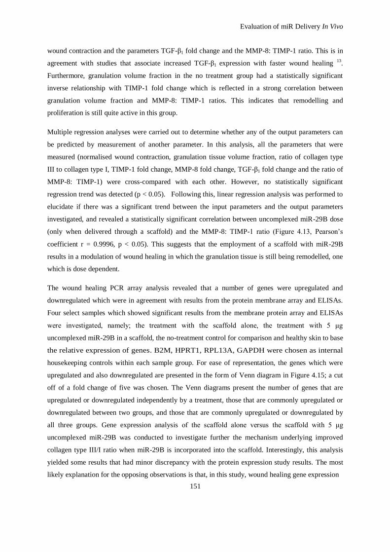
Evaluation of miR Delivery In Vivo
151
wound contraction and the parameters TGF-β1 fold change and the MMP-8: TIMP-1 ratio. This is in
agreement with studies that associate increased TGF-β1 expression with faster wound healing 13
.
Furthermore, granulation volume fraction in the no treatment group had a statistically significant
inverse relationship with TIMP-1 fold change which is reflected in a strong correlation between
granulation volume fraction and MMP-8: TIMP-1 ratios. This indicates that remodelling and
proliferation is still quite active in this group.
Multiple regression analyses were carried out to determine whether any of the output parameters can
be predicted by measurement of another parameter. In this analysis, all the parameters that were
measured (normalised wound contraction, granulation tissue volume fraction, ratio of collagen type
III to collagen type I, TIMP-1 fold change, MMP-8 fold change, TGF-β1 fold change and the ratio of
MMP-8: TIMP-1) were cross-compared with each other. However, no statistically significant
regression trend was detected (p < 0.05). Following this, linear regression analysis was performed to
elucidate if there was a significant trend between the input parameters and the output parameters
investigated, and revealed a statistically significant correlation between uncomplexed miR-29B dose
(only when delivered through a scaffold) and the MMP-8: TIMP-1 ratio (Figure 4.13, Pearson’s
coefficient r = 0.9996, p < 0.05). This suggests that the employment of a scaffold with miR-29B
results in a modulation of wound healing in which the granulation tissue is still being remodelled, one
which is dose dependent.
The wound healing PCR array analysis revealed that a number of genes were upregulated and
downregulated which were in agreement with results from the protein membrane array and ELISAs.
Four select samples which showed significant results from the membrane protein array and ELISAs
were investigated, namely; the treatment with the scaffold alone, the treatment with 5 μg
uncomplexed miR-29B in a scaffold, the no-treatment control for comparison and healthy skin to base
the relative expression of genes. B2M, HPRT1, RPL13A, GAPDH were chosen as internal
housekeeping controls within each sample group. For ease of representation, the genes which were
upregulated and also downregulated are presented in the form of Venn diagram in Figure 4.15; a cut
off of a fold change of five was chosen. The Venn diagrams present the number of genes that are
upregulated or downregulated independently by a treatment, those that are commonly upregulated or
downregulated between two groups, and those that are commonly upregulated or downregulated by
all three groups. Gene expression analysis of the scaffold alone versus the scaffold with 5 μg
uncomplexed miR-29B was conducted to investigate further the mechanism underlying improved
collagen type III/I ratio when miR-29B is incorporated into the scaffold. Interestingly, this analysis
yielded some results that had minor discrepancy with the protein expression study results. The most
likely explanation for the opposing observations is that, in this study, wound healing gene expression
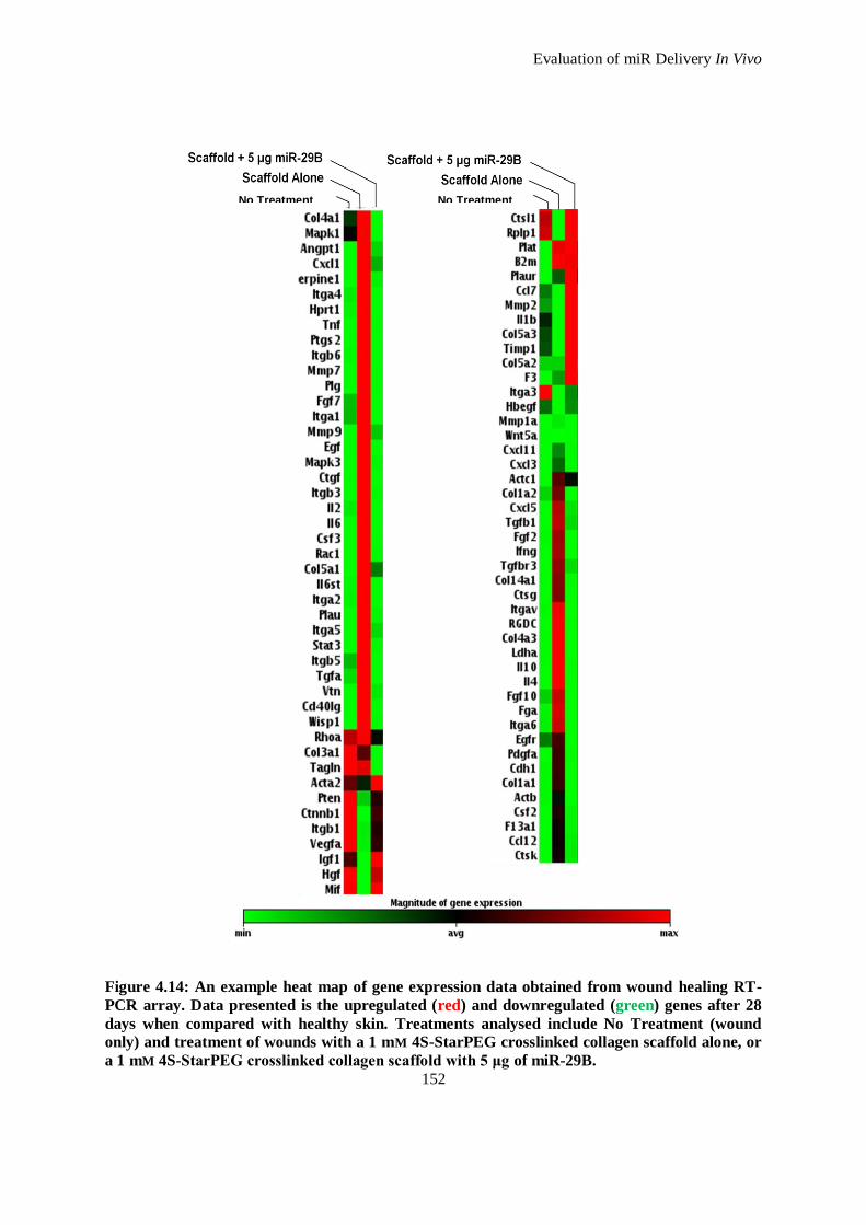
Evaluation of miR Delivery In Vivo
152
Figure 4.14: An example heat map of gene expression data obtained from wound healing RT-
PCR array. Data presented is the upregulated (red) and downregulated (green) genes after 28
days when compared with healthy skin. Treatments analysed include No Treatment (wound
only) and treatment of wounds with a 1 mM 4S-StarPEG crosslinked collagen scaffold alone, or
a 1 mM 4S-StarPEG crosslinked collagen scaffold with 5 μg of miR-29B.
No Treatment No Treatment
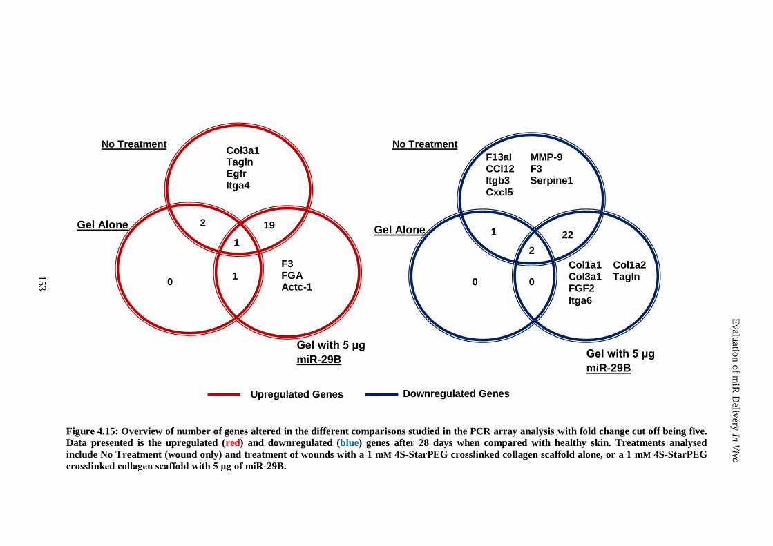
Figure 4.15: Overview of number of genes altered in the different comparisons studied in the PCR array analysis with fold change cut off being five.
Data presented is the upregulated (red) and downregulated (blue) genes after 28 days when compared with healthy skin. Treatments analysed
include No Treatment (wound only) and treatment of wounds with a 1 mM 4S-StarPEG crosslinked collagen scaffold alone, or a 1 mM 4S-StarPEG
crosslinked collagen scaffold with 5 μg of miR-29B.
Col1a1 Col1a2 Col3a1 Tagln FGF2
Itga6
F3 FGA Actc-1
F13aI MMP-9 CCl12 F3 Itgb3 Serpine1 Cxcl5
Col3a1 Tagln Egfr Itga4
1 0
2 19
0
1
Gel Alone
No Treatment
Evalu
ation
of m
iR D
elivery
In V
ivo
No Treatment
Gel with 5 μg
miR-29B
2
22 1
Upregulated Genes
Downregulated Genes
Gel Alone
Gel with 5 μg
miR-29B
153
0
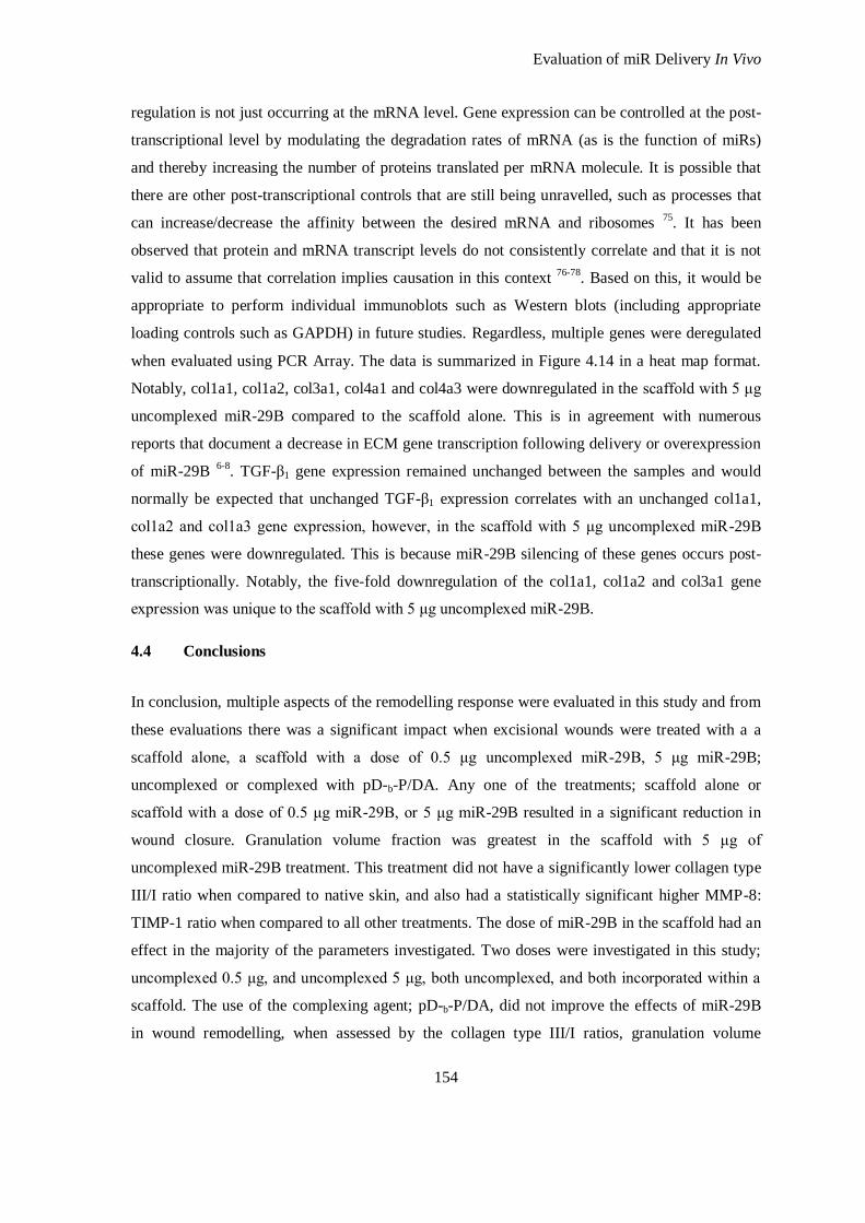
Evaluation of miR Delivery In Vivo
154
regulation is not just occurring at the mRNA level. Gene expression can be controlled at the post-
transcriptional level by modulating the degradation rates of mRNA (as is the function of miRs)
and thereby increasing the number of proteins translated per mRNA molecule. It is possible that
there are other post-transcriptional controls that are still being unravelled, such as processes that
can increase/decrease the affinity between the desired mRNA and ribosomes 75
. It has been
observed that protein and mRNA transcript levels do not consistently correlate and that it is not
valid to assume that correlation implies causation in this context 76-78
. Based on this, it would be
appropriate to perform individual immunoblots such as Western blots (including appropriate
loading controls such as GAPDH) in future studies. Regardless, multiple genes were deregulated
when evaluated using PCR Array. The data is summarized in Figure 4.14 in a heat map format.
Notably, col1a1, col1a2, col3a1, col4a1 and col4a3 were downregulated in the scaffold with 5 μg
uncomplexed miR-29B compared to the scaffold alone. This is in agreement with numerous
reports that document a decrease in ECM gene transcription following delivery or overexpression
of miR-29B 6-8
. TGF-β1 gene expression remained unchanged between the samples and would
normally be expected that unchanged TGF-β1 expression correlates with an unchanged col1a1,
col1a2 and col1a3 gene expression, however, in the scaffold with 5 μg uncomplexed miR-29B
these genes were downregulated. This is because miR-29B silencing of these genes occurs post-
transcriptionally. Notably, the five-fold downregulation of the col1a1, col1a2 and col3a1 gene
expression was unique to the scaffold with 5 μg uncomplexed miR-29B.
4.4 Conclusions
In conclusion, multiple aspects of the remodelling response were evaluated in this study and from
these evaluations there was a significant impact when excisional wounds were treated with a a
scaffold alone, a scaffold with a dose of 0.5 μg uncomplexed miR-29B, 5 μg miR-29B;
uncomplexed or complexed with pD-b-P/DA. Any one of the treatments; scaffold alone or
scaffold with a dose of 0.5 μg miR-29B, or 5 μg miR-29B resulted in a significant reduction in
wound closure. Granulation volume fraction was greatest in the scaffold with 5 μg of
uncomplexed miR-29B treatment. This treatment did not have a significantly lower collagen type
III/I ratio when compared to native skin, and also had a statistically significant higher MMP-8:
TIMP-1 ratio when compared to all other treatments. The dose of miR-29B in the scaffold had an
effect in the majority of the parameters investigated. Two doses were investigated in this study;
uncomplexed 0.5 μg, and uncomplexed 5 μg, both uncomplexed, and both incorporated within a
scaffold. The use of the complexing agent; pD-b-P/DA, did not improve the effects of miR-29B
in wound remodelling, when assessed by the collagen type III/I ratios, granulation volume
Evaluation of miR Delivery In Vivo
155
fraction and wound closure. Indeed the use of pD-b-P/DA complexed with 5 μg miR-29B in a
scaffold caused an upregulation of inflammatory cytokines when compared to 5 μg of
uncomplexed miR-29B in a scaffold. Notably, there was a significant correlation between the
dose of uncomplexed miR-29B when delivered through the scaffold and the ratio of MMP-8:
TIMP-1 which indicates that not only is the combination of these parameters important, but the
dose of miR-29B also has a significant effect on the parameters investigated. Through all the
investigations of this chapter it can be concluded that although a scaffold alone is beneficial
towards reduced wound contraction, incorporation of miR-29B in a dose dependent manner (in
this case 5 μg) ameliorates the wound healing process through modulation of MMP-8 and TIMP-
1, collagen type I degradation, and the post-transcriptional inhibition of ECM proteins which are
being stimulated by TGF-β1.
Evaluation of miR Delivery In Vivo
156
4.5 References
1. Stadelmann WK, Digenis AG, Tobin GR. Physiology and healing dynamics of chronic cutaneous wounds. Am. J. Surg. 1998;176:26S-38S
2. Steed DL. The role of growth factors in wound healing. Surg. Clin. North Am.
1997;77:575-586
3. Singer AJ, Clark RA. Cutaneous wound healing. N. Engl. J. Med. 1999;341:738-746 4. Lucas T, Waisman A, Ranjan R, Roes J, Krieg T, Müller W, Roers A, Eming SA.
Differential roles of macrophages in diverse phases of skin repair. J.Immunol.
2010;184:3964-3977 5. Roberts AB, Heine UI, Flanders KC, Sporn MB. TGF-β. Major role in regulation of
extracellular matrix. Ann. N. Y. Acad. Sci. 1990;580:225-232
6. Li Z, Hassan MQ, Jafferji M, Aqeilan RI, Garzon R, Croce CM, van Wijnen AJ, Stein JL, Stein GS, Lian JB. Biological functions of miR-29b contribute to positive regulation of
osteoblast differentiation. J. Biol. Chem. 2009;284:15676-15684
7. Ogawa T, Iizuka M, Sekiya Y, Yoshizato K, Ikeda K, Kawada N. Suppression of type I
collagen production by microRNA-29b in cultured human stellate cells. Biochem. Biophys. Res. Commun. 2010;391:316-321
8. Li N, Cui J, Duan X, Chen H, Fan F. Suppression of type I collagen expression bymiR-
29b via Pi3k, AKT, and Sp1 pathway in human tenon's fibroblasts. Invest. Ophthalmol. Vis. Sci. 2012;53:1670-1678
9. Nagase H, Woessner JF. Matrix metalloproteinases. J. Biol. Chem. 1999;274:21491-
21494
10. Nwomeh BC, Liang H-X, Cohen IK, Yager DR. MMP-8 is the predominant collagenase in healing wounds and nonhealing ulcers. J. Surg. Res. 1999;81:189-195
11. Pradhan-Palikhe P, Vesterinen T, Tarkkanen J, Leivo I, Sorsa T, Salo T, Mattila PS.
Plasma level of tissue inhibitor of MMP-1 but not that of MMP-8 predicts survival in head and neck squamous cell cancer. Oral Oncology. 2010;46:514-518
12. Yannas IV, Burke JF, Orgill DP, Skrabut EM. Wound tissue can utilize a polymeric template to synthesize a functional extension of skin. Science. 1982;215:174-176
13. Pandit A, Ashar R, Feldman D. The effect of TGF-β delivered through a collagen scaffold
on wound healing. J. Invest. Surg. 1999;12:89-100
14. Doukas J, Blease K, Craig D, Ma C, Chandler LA, Sosnowski BA, Pierce GF. Delivery of FGF genes to wound repair cells enhances arteriogenesis and myogenesis in skeletal
muscle. Mol. Ther. 2002;5:517-527
15. Gu DL, Nguyen T, Gonzalez AM, Printz MA, Pierce GF, Sosnowski BA, Phillips ML, Chandler LA. Adenovirus encoding human PDGF-b delivered in collagen exhibits safety,
biodistribution, and immunogenicity profiles favorable for clinical use. Mol. Ther.
2004;9:699-711 16. Supp DM, Boyce ST. Engineered skin substitutes: Practices and potentials. Clin.
Dermatol. 2005;23:403-412
17. Hudon V, Berthod F, Black AF, Damour O, Germain L, Auger FA. A tissue-engineered
endothelialized dermis to study the modulation of angiogenic and angiostatic molecules on capillary-like tube formation in vitro. Br. J. Dermatol. 2003;148:1094-1104
18. Tremblay PL, Hudon V, Berthod F, Germain L, Auger FA. Inosculation of tissue-
engineered capillaries with the host's vasculature in a reconstructed skin transplanted on mice. Am. J. Transplant. 2005;5:1002-1010
19. Butler CE, Orgill DP. Simultaneous in vivo regeneration of neodermis, epidermis, and
basement membrane. Adv. Biochem. Eng. Biotechnol. 2005;94:23-41
Evaluation of miR Delivery In Vivo
157
20. Ghahary A, Tredget E, Shen Q, Kilani R, Scott P, Takeuchi M. Liposome associated
interferon-α-2b functions as an anti-fibrogenic factor in dermal wounds in the guinea pig. Mol. Cell. Biochem. 2000;208:129-137
21. Formiga FR, Pelacho B, Garbayo E, Abizanda G, Gavira JJ, Simon-Yarza T, Mazo M,
Tamayo E, Jauquicoa C, Ortiz-de-Solorzano C, Prósper F, Blanco-Prieto MJ. Sustained
release of VEGF through PLGA microparticles improves vasculogenesis and tissue remodeling in an acute myocardial ischemia–reperfusion model. J. Controlled Release.
2010;147:30-37
22. Holladay C, Power K, Sefton M, O'Brien T, Gallagher WM, Pandit A. Functionalized scaffold-mediated interleukin 10 gene delivery significantly improves survival rates of
stem cells in vivo. Mol. Ther. 2011;19:969-978
23. Li B, Davidson JM, Guelcher SA. The effect of the local delivery of platelet-derived growth factor from reactive two-component polyurethane scaffolds on the healing in rat
skin excisional wounds. Biomaterials. 2009;30:3486-3494
24. Hong H-J, Jin S-E, Park J-S, Ahn WS, Kim C-K. Accelerated wound healing by Smad3
antisense oligonucleotides-impregnated chitosan/alginate polyelectrolyte complex. Biomaterials. 2008;29:4831-4837
25. Liu X, Ma L, Liang J, Zhang B, Teng J, Gao C. RNAi functionalized collagen-
chitosan/silicone membrane bilayer dermal equivalent for full-thickness skin regeneration with inhibited scarring. Biomaterials. 2013;34:2038-2048
26. Yannas IV, Burke JF, Gordon PL, Huang C, Rubenstein RH. Design of an artificial skin.
Control of chemical composition. J. Biomed. Mater. Res. 1980;14:107-132 27. Chvapil M, Kronenthal L, Van Winkle W, Jr. Medical and surgical applications of
collagen. Int. Rev. Connect. Tissue Res. 1973;6:1-61
28. MacNeil S. Biomaterials for tissue engineering of skin. Materials Today. 2008;11:26-35
29. Shevchenko RV, James SL, James SE. A review of tissue-engineered skin bioconstructs available for skin reconstruction. J. R. Soc. Interface. 2010;7:229-258
30. Zeugolis DI, Paul RG, Attenburrow G. Factors influencing the properties of reconstituted
collagen fibers prior to self-assembly: Animal species and collagen extraction method. J. Biomed. Mater. Res. 2008;86A:892-904
31. Braiman-Wiksman L, Solomonik I, Spira R, Tennenbaum T. Novel insights into wound
healing sequence of events. Toxicol. Pathol. 2007;35:767-779
32. Zheng D, Giljohann DA, Chen DL, Massich MD, Wang X-Q, Iordanov H, Mirkin CA, Paller AS. Topical delivery of siRNA-based spherical nucleic acid nanoparticle
conjugates for gene regulation. Proc. Nat. Acad. Sci. USA 2012;109:11975-11980
33. Woodrow KA, Cu Y, Booth CJ, Saucier-Sawyer JK, Wood MJ, Mark Saltzman W. Intravaginal gene silencing using biodegradable polymer nanoparticles densely loaded
with small-interfering RNA. Nat. Mater. 2009;8:526-533
34. Lee WR, Shen SC, Zhuo RZ, Wang KC, Fang JY. Enhancement of topical small interfering RNA delivery and expression by low-fluence erbium:Yag laser pretreatment
of skin. Hum. Gene Ther. 2009;20:580-588
35. Russell HK. A modification of Movat's pentachrome stain. Arch. Pathol. 1972;94:187-
191 36. Rowe AJ, Finlay HM, Canham PB. Collagen biomechanics in cerebral arteries and
bifurcations assessed by polarizing microscopy. J. Vasc. Res. 2003;40:406-415
37. Cuttle L, Nataatmadja M, Fraser JF, Kempf M, Kimble RM, Hayes MT. Collagen in the scarless fetal skin wound: Detection with picrosirius-polarization. Wound Repair Regen.
2005;13:198-204
38. Griffiths‐Jones S. The microRNA registry. Nucleic Acids Res. 2004;32:D109-D111
39. Yannas IV, Tzeranis DS, Harley BA, So PTC. Biologically active collagen-based scaffolds: Advances in processing and characterization. Philos. Trans.R. Soc. A.
2010;368:2123-2139
Evaluation of miR Delivery In Vivo
158
40. Garcia Y, Wilkins B, Collighan RJ, Griffin M, Pandit A. Towards development of a
dermal rudiment for enhanced wound healing response. Biomaterials. 2008;29:857-868 41. Powell HM, Supp DM, Boyce ST. Influence of electrospun collagen on wound
contraction of engineered skin substitutes. Biomaterials. 2008;29:834-843
42. Lee SY, Oh JH, Kim JC, Kim YH, Kim SH, Choi JW. In vivo conjunctival reconstruction
using modified PLGA grafts for decreased scar formation and contraction. Biomaterials. 2003;24:5049-5059
43. Witte MB, Barbul A. General principles of wound healing. Surg Clinics N. Amer.
1997;77:509-528 44. Nedelec B, Ghahary A, Scott PG, Tredget EE. Control of wound contraction. Basic and
clinical features. Hand Clin. 2000;16:289-302
45. Kwan P, Hori K, Ding J, Tredget EE. Scar and contracture: Biological principles. Hand Clin. 2009;25:511-528
46. Zurada JM, Kriegel D, Davis IC. Topical treatments for hypertrophic scars. J. Am. Acad.
Dermatol. 2006;55:1024-1031
47. Kerr JF, Wyllie AH, Currie AR. Apoptosis: A basic biological phenomenon with wide-ranging implications in tissue kinetics. Br. J. Cancer. 1972;26:239-257
48. Desmouliere A, Badid C, Bochaton-Piallat ML, Gabbiani G. Apoptosis during wound
healing, fibrocontractive diseases and vascular wall injury. Int. J. Biochem. Cell Biol. 1997;29:19-30
49. Desmouliere A, Redard M, Darby I, Gabbiani G. Apoptosis mediates the decrease in
cellularity during the transition between granulation tissue and scar. Am. J. Pathol. 1995;146:56-66
50. Amodio N, Di Martino MT, Foresta U, Leone E, Lionetti M, Leotta M, Gulla AM, Pitari
MR, Conforti F, Rossi M, Agosti V, Fulciniti M, Misso G, Morabito F, Ferrarini M, Neri
A, Caraglia M, Munshi NC, Anderson KC, Tagliaferri P, Tassone P. MiR-29b sensitizes multiple myeloma cells to bortezomib-induced apoptosis through the activation of a
feedback loop with the transcription factor Sp1. Cell Death Dis. 2012;3:e436
51. Mott JL, Kobayashi S, Bronk SF, Gores GJ. MiR-29 regulates MCL-1 protein expression and apoptosis. Oncogene. 2007;26:6133-6140
52. Zhang YK, Wang H, Leng Y, Li ZL, Yang YF, Xiao FJ, Li QF, Chen XQ, Wang LS.
Overexpression of microRNA-29b induces apoptosis of multiple myeloma cells through
down regulating MCL-1. Biochem. Biophys. Res. Commun. 2011;414:233-239 53. Herrera B, Alvarez AM, Beltran J, Valdes F, Fabregat I, Fernandez M. Resistance to
TGF-β-induced apoptosis in regenerating hepatocytes. J. Cell Physiol. 2004;201:385-392
54. Saitoh M, Miyazawa K. Transcriptional and post-transcriptional regulation in TGF-β-mediated epithelial-mesenchymal transition. J. Biochem. 2012;151:563-571
55. Simpson DM, Ross R. The neutrophilic leukocyte in wound repair a study with
antineutrophil serum. J. Clin. Invest. 1972;51:2009-2023 56. Leibovich SJ, Ross R. The role of the macrophage in wound repair. A study with
hydrocortisone and antimacrophage serum. Am. J. Pathol. 1975;78:71-100
57. Werner S, Grose R. Regulation of wound healing by growth factors and cytokines.
Physiol. Rev. 2003;83:835-870 58. Wynn TA. Common and unique mechanisms regulate fibrosis in various
fibroproliferative diseases. J. Clinical Invest. 2007;117:524-529
59. Wilgus TA, Vodovotz Y, Vittadini E, Clubbs EA, Oberyszyn TM. Reduction of scar formation in full-thickness wounds with topical celecoxib treatment. Wound Repair
Regen. 2003;11:25-34
60. Bullard KM, Longaker MT, Lorenz HP. Fetal wound healing: Current biology. World J. Surg. 2003;27:54-61
61. Adolph VR, DiSanto SK, Bleacher JC, Dillon PW, Krummel TM. The potential role of
the lymphocyte in fetal wound healing. J. Pediatr. Surg. 1993;28:1316-1320
Evaluation of miR Delivery In Vivo
159
62. Dillon PW, Keefer K, Blackburn JH, Houghton PE, Krummel TM. The extracellular
matrix of the fetal wound: Hyaluronic acid controls lymphocyte adhesion. J. Surg. Res. 1994;57:170-173
63. Whitby DJ, Ferguson MW. Immunohistochemical localization of growth factors in fetal
wound healing. Dev. Biol. 1991;147:207-215
64. Ogawa S, Onodera J, Honda R, Fujimoto I. Influence of systemic administration of atelocollagen on mouse livers: An ideal biomaterial for systemic drug delivery. J.
Toxicol. Sci. 2011;36:751-762
65. Mann BK, Gobin AS, Tsai AT, Schmedlen RH, West JL. Smooth muscle cell growth in photopolymerized hydrogels with cell adhesive and proteolytically degradable domains:
Synthetic ECM analogs for tissue engineering. Biomaterials. 2001;22:3045-3051
66. Dillingham EO, Lawrence WH, Autian J, Schmalz G. Acrylate and methacrylate esters: Relationship of hemolytic activity and in vivo toxicity. J. Biomed. Mater. Res.
1983;17:945-957
67. Kim ES, Sohn YW, Moon A. TGF-β-induced transcriptional activation of MP-2 is
mediated by activating transcription factor (ATF)2 in human breast epithelial cells. Cancer Lett. 2007;252:147-156
68. Leivonen SK, Chantry A, Hakkinen L, Han J, Kahari VM. Smad3 mediatesTGF-β-
induced collagenase-3 (MMP-13) expression in human gingival fibroblasts. Evidence for cross-talk between Smad3 and P38 signaling pathways. J. Biol. Chem. 2002;277:46338-
46346
69. van Rooij E, Sutherland LB, Thatcher JE, DiMaio JM, Naseem RH, Marshall WS, Hill JA, Olson EN. Dysregulation of microRNAs after myocardial infarction reveals a role of
miR-29 in cardiac fibrosis. Proc. Nat. Acad. Sci.USA. 2008;105:13027-13032
70. Maurer B, Stanczyk J, Jüngel A, Akhmetshina A, Trenkmann M, Brock M, Kowal-
Bielecka O, Gay RE, Michel BA, Distler JHW, Gay S, Distler O. MicroRNA-29, a key regulator of collagen expression in systemic sclerosis. Arthritis & Rheumatism.
2010;62:1733-1743
71. Luna C, Li G, Qiu J, Epstein DL, Gonzalez P. Cross-talk between miR-29 andTGF-βs in trabecular meshwork cells. Invest. Ophthalmol. Vis. Sci. 2011;52:3567-3572
72. Du B, Ma L-M, Huang M-B, Zhou H, Huang H-L, Shao P, Chen Y-Q, Qu L-H. High
glucose down-regulatesmiR-29a to increase collagen IVproduction in HK-2 cells. FEBS
Letters. 2010;584:811-816 73. Winbanks CE, Wang B, Beyer C, Koh P, White L, Kantharidis P, Gregorevic P. TGF-β
regulates miR-206 and miR-29 to control myogenic differentiation through regulation of
HDAC4. J. Biol. Chem. 2011;286:13805-13814 74. Qin W, Chung AC, Huang XR, Meng XM, Hui DS, Yu CM, Sung JJ, Lan HY. TGF-
β/Smad3 signaling promotes renal fibrosis by inhibiting miR-29. J. Am. Soc. Nephrol.
2011;22:1462-1474 75. Gry M, Rimini R, Stromberg S, Asplund A, Ponten F, Uhlen M, Nilsson P. Correlations
between RNA and protein expression profiles in 23 human cell lines. BMC Genomics.
2009;10:365
76. Gygi SP, Rochon Y, Franza BR, Aebersold R. Correlation between protein and mRNA abundance in yeast. Mol. Cell Biol. 1999;19:1720-1730
77. Greenbaum D, Jansen R, Gerstein M. Analysis of mRNA expression and protein
abundance data: An approach for the comparison of the enrichment of features in the cellular population of proteins and transcripts. Bioinformatics. 2002;18:585-596
78. Greenbaum D, Colangelo C, Williams K, Gerstein M. Comparing protein abundance and
mRNA expression levels on a genomic scale. Genome Biol. 2003;4:117
|

Chapter Five
Summary and Future Directions
A portion of this chapter has been previously published in:
Monaghan M, Pandit A, RNA interference therapy via functionalized scaffolds. Adv. Drug Del. Rev. 2011, 63: 197-208.
MonaghanM, Greiser U, Wall J.G, O'Brien T, PanditA. Interference: An alteRNAtive therapy following acute myocardial infarction. Trends Pharmacol. Sci. 2012, 33: 635-645
Summary and Future Directions
161
5.1 Introduction
Regenerative medicine aims to restore damaged tissue to its native structural and functional state.
While this goal may be overly optimistic, progress towards this goal will likely improve the outlook
for patients in a variety of disease and injury states. Simply reducing the extent of fibrotic scarring
will reduce the pathology in a number of conditions. This is unquestionably true in the case of
remodelling of cardiac tissue after myocardial infarction (MI). Gene therapy and biomaterials have
each been proposed for the treatment of damaged heart tissue in a variety of applications, ranging
from reducing inflammation, inducing angiogenesis, for functional reprogramming of cells, and to
mechanically augment the damaged tissue. The overall goal of this research was to develop a
biomaterial RNAi delivery system which inhibits fibrosis in a region of interest and thereby
improving the functional outcome in a pathological setting with the future objective being towards a
therapeutic option following MI.
5.2 Summary
5.2.1 Phase I- Delivering Exogenous miRNA (Chapter Two)
The objective of the first phase of this work was to develop a non-viral delivery vector that enabled
effective complexation of microRNAs (miRs) and also conjugate antibody fragments to enable
improvement in vector delivery. The proposed system was a linear poly (dimethylamino) ethyl
methacrylate (pDMAEMA) structure synthesized using atom transfer radical polymerization (ATRP)
and used as a macro-initiator to grow a hyperbranched poly (ethylene glycol) methyl ether acrylate
(PEGMEA) based unit with a poly (ethylene glycol) diacrylate (PEGDA) block in a deactivation
enhanced ATRP reaction to PEGylate and attribute functionalisation. This system was optimized and
characterized in vitro as described in Chapter Two and given the nomenclature pD-b-P/DA.
The development of pD-b-P/DA included growth and characterization of this polymer to a molecular
weight of 14,000 kDa, the controlled growth of which was verified using gel permeation
chromatography (GPC). Proton nuclear magnetic resonance (1H NMR) determined this structure to be
highly branched and PEGylated and to have vinyl groups (3 % content) present for conjugation of
thiol terminated moieties. Optimization of the miR- pD-b-P/DA complexes was then conducted. This
involved characterization using agarose gel electrophoresis, transmission electron microscopy (TEM),
and UV NanoDrop™
spectroscopy to verify and optimise the complexation of pD-b-P/DA with miR
mimics. A miR: pD-b-P/DA ratio of 1:8 was found to be optimal for the reporter gene; a dual plasmid
of Firefly and Renilla Luciferase which was designed, constructed and verified as part of this thesis. A
lipid based transfection agent; DharmaFect™
was optimized in order to serve as a comparative control
Summary and Future Directions
162
when silencing reporter genes encoded by a luciferase-based plasmid transcribing in primary rat
cardiac fibroblasts.
Antibody fragments (Fab`s), receptive to the CD90 antigen, were generated using hybridoma
technology to generate monoclonal antibodies. Thiol terminated fragments were generated using
pepsin digestion followed by reduction of the di-sulphide bond present in antibodies using
dithiothreitol. pD-b-P/DA was tested for its ability to bind thiols using two methods. The first test
entailed the use of cysteine mono-hydrochloride as a model of thiol terminated fragments. pD-b-P/DA
was able to effectively consume thiols within 30 minutes which was determined via Ellmans assay.
Thiol conjugativity was also demonstrated using SDS-PAGE of Fab`s with pD-b-P/DA. Using a
custom luciferase reporter of knockdown; improved silencing was observed when delivering miR
mimics complexed pD-b-P/DA decorated with Fab`s at 48 hours after delivery compared to miR/pD-b-
P/DA complexes with no Fab`s; however this effect disappeared at 96 hours.
5.2.2 Phase II- An Injectable Scaffold Delivery System (Chapter Three)
Phase II aimed to develop a scaffold using collagen type I and obtain non-toxic in situ crosslinking
using a PEG-based crosslinker, which in this case was poly (ethylene glycol) ether tetrasuccinimidyl
glutarate (4S-StarPEG). It was hypothesized that this scaffold will be non-toxic to cells, have
improved mechanical and chemical stability due to its crosslinking, and will enable the delivery and
release of miR-29B complexes. Phase II of this project, which is described in Chapter Three realises
all of these objectives. Crosslinking was achieved using 4S-StarPEG, and this was verified using
quantitative chemical absorbance of the free amine groups remaining after crosslinking. The optimal
crosslinking concentration was determined to be a 1 mM concentration of 4S-StarPEG which had only
9.5 % of free amines remaining after crosslinking (relative to a non-crosslinked collagen scaffold.
Crosslinking using 4S-StarPEG also improved resistance to enzymatic degradation of the scaffolds
and hydrogels crosslinked with concentration of 1 mM 4S-StarPEG had a remaining mass of 85 %
whereas a non-crosslinked hydrogel had completely degraded at 48 hours. Furthermore, a plateau in
crosslinking occurred in which a concentration of 1 mM crosslinker offered the maximum crosslinking
density. Investigation of the release profiles of RNA from the scaffolds in vitro showed a crosslinker
concentration dependent release, which was attributed to two reasons. Firstly, that the more
crosslinked the scaffold, the slower degradation occurs which delays the release of RNA.
Furthermore, due to the possible interaction of the crosslinker with amine groups present in the RNA,
the crosslinker is likely to have crosslinked the RNA into the scaffold. In essence, the higher the
crosslinker density (in this case the highest used was 1 mM) the more retarded the release from the
scaffold. The lowest crosslinker density (0.05 mM) had the quickest release, in fact, 100 % release had
Summary and Future Directions
163
occurred after three days, but this was also due to the fact that the scaffold had completely degraded at
this point. An additional parameter investigated, that of RNA carrier, was deemed to also to have a
significant effect on the release profile for RNA from the scaffold. Poly (ethylenimine) (PEI) which is
highly cationic due to an abundance of primary amine groups had the slowest release from the
scaffold. This is attributed to a charge interaction of the positive amine groups of PEI interacting with
the negatively charged collagen in the scaffold and also possible crosslinking with the 4S-StarPEG
which reacts with amine groups. pD-b-P/DA has only a slightly positive charge due to the presence of
tertiary amines and therefore had less charge interaction with the scaffold. Due to this, its release
profile was much quicker than that of PEI and was similar to that seen with naked RNA.
The next component of in vitro testing in Phase II required the investigation of miR-29B as an
inhibitor of collagen type I and collagen type III. Previously, in Chapter Two, delivery of exogenous
miRs resulted in a demonstrated knockdown in a reporter Luciferase protein. It was sought to evaluate
this in both a monolayer culture model and scaffold delivery model of knockdown. miR-29B was
shown to specifically silence collagen type I and collagen type III in monolayer culture, but only
when delivered using a complexing agent (down to 51 and 55% respectively). Then, using a 4S-
StarPEG scaffold as a delivery platform of these miR-29B complexes, efficient silencing, of both
collagen type I and collagen type III mRNA, up to a period of up to 14 days was achieved and
knockdown of collagen type I miRNA was more profound than that of collagen type III.
Furthermore, naked miR-29B within the scaffold was effective at silencing collagen type I and
collagen type III mRNA, but the functionalization of miR-29B/pD-b-P/DA complexes with antibody
fragments did not result in a significant improvement in efficiency when delivered in a scaffold.
5.2.3 Phase III- System Evaluation in a Rat Excisional Wound Model (Chapter Four)
The penultimate phase of testing of this system occurred in vivo, as described in Chapter Four. The
goal in Phase III was to evaluate the scaffold and miR-29B and its effect on wound healing in vivo,
specifically the effect on ECM remodelling following injury in a rat dermal excisional wound model.
It was hypothesized that the scaffold functionalized with miR-29B, when applied to a rat excisional
wound model will modulate the wound healing response by reducing collagen type I production and
ameliorate the ratio of collagen type III/I in the remodelling dermis. It was also the hypothesis of
Chapter Four that functionalization of the scaffold with miR-29B will work synergistically towards an
improvement in the ratio of collagen type III/I and ameliorate the wound healing process through
reduction in wound contraction and modulation of matrix metalloproteinase-8 (MMP-8) and its tissue
inhibitor; TIMP-1. There was a significant impact when excisional wounds were treated with a
scaffold alone, a scaffold with a dose of 0.5 μg uncomplexed miR-29B, 5 μg miR-29B; uncomplexed
Summary and Future Directions
164
or complexed with pD-b-P/DA. Any one of the treatments; scaffold alone or scaffold with a dose of
0.5 μg miR-29B, or 5 μg miR-29B resulted in a significant reduction in wound closure. Granulation
volume fraction was greatest in the scaffold with 5 μg of uncomplexed miR-29B. This same group
was also the only group which did not have a significantly lower collagen type III/I ratio when
compared to native skin, and this group also had a statistically significant higher MMP-8: TIMP-1
ratio when compared to all other groups. The dose of miR-29B in the scaffold had an effect in the
majority of the parameters investigated. Two doses were investigated in this study; uncomplexed 0.5
μg, and uncomplexed 5 μg, both uncomplexed, and both incorporated within a scaffold. The use of the
complexing agent; pD-b-P/DA, did not improve the effects of miR-29B in wound remodelling, when
evaluated through the collagen type III/I ratios, granulation volume fraction and wound closure.
Indeed the use of pD-b-P/DA complexed with 5 μg miR-29B in a scaffold caused an upregulation of
inflammatory cytokines when compared to 5 μg of uncomplexed miR-29B in a scaffold. Notably,
there was a significant correlation between the dose of uncomplexed miR-29B when delivered
through the scaffold and the ratio of MMP-8: TIMP-1 which signifies that not only the combination of
these parameters, but the dose of miR-29B also has a significant effect on the parameters investigated.
Further analysis of phase III warrants quantitative analysis of collagen type I and collagen type III
expression within the wound bed through employment of ELISAs, additional immunohistochemistry
and immunoblots (including appropriate loading controls). Additionally, labelling the miR-29B with
radio-labelling agents or the Cy™
3 labelling agent, employed in phase II for evaluation of the release
profiles, would facilitate an analysis of the penetration of the miR-29B therapeutic into the
surrounding tissue. Live imaging could also be employed if suitable experimental constraints were
applied (such as minimising light exposure to the wound).
5.3 Limitations
5.3.1 Phase I- Delivering Exogenous miRNA (Chapter Two)
The choice of transfection reagent is critical, as every available agent has both advantages and
disadvantages. Choosing to use a non-viral approach reduces the potential concerns associated with
viruses but dramatically reduces the efficiency of transfection. Within the non-viral set of transfection
reagents, other trade-offs must be made. The use of pD-b-P/DA offered adequate complexation of
miRs and its PEGylation enabled the delivery of a higher dose, without a significant impact on
cellular viability. More importantly, it was envisaged that the resultant transfection reagent would
facilitate apt conjugation of antibody fragments to improve delivery.
Summary and Future Directions
165
The complexing polymer described; pD-b-P/DA, efficiently complexed miR mimics and conjugated
antibody fragments. However, it is not without its drawbacks. pD-b-P/DA could only compete with
the commercially available controls on a ‘dose basis’, which essentially means that although it poses
minimal toxicity and therefore can be delivered in higher doses than the commercially available
controls, it is less effective in transfection. That being said, it does provide good transfection, and
effective silencing in vitro. pD-b-P/DA also facilitated the conjugation of antibody fragments via a
Michael-type addition chemical reaction. Conjugation of a CD90 antibody fragment effected a
significant knockdown in the reporter luciferase system when compared to the use of pD-b-P/DA with
no antibody fragment at 48 hours. Additionally, the effect was shown to be cell specific when
comparing to HUVECs which have minimal expression of CD90 and are cells from another species.
Knockdown was maintained at 96 hours, however the use of pD-b-P/DA conjugated with the CD90
antibody fragment had no statistically significant difference between any of the other groups,
including pD-b-P/DA with no antibody fragment attached. This is hypothesized to be due to
saturation at this point (Figure 2.15). Many non-viral delivery agent are tested in systems where they
are in contact with cells for a limited amount of time (four hours) and then removed, also sometimes
in serum-free conditions (which can reduce the exposure of exogenous RNA/DNA to serum proteases
and enzymes). As a potential therapeutic delivery agent, pD-b-P/DA was not removed from cells in
culture. The use of ‘targeting ligands’ is a controversial area which, following the results in Phase I
and II, can only be demonstrated in flow/systemic delivery environments. In this study, all conditions
were in static environments. Furthermore, the CD90 antigen targeted by the antibody fragment in this
platform; in hindsight, may have not been the most appropriate one. Monoclonal antibody engineering
is an arduous and perhaps archaic process and the identification of appropriate, rapidly internalising
antigens with which to target is warranted. Furthermore, a more specific, targeted ligand is needed
which could be achieved using phage display technology for more accuracy 1.
The in vitro transfection system was optimal for the initial testing of pD-b-P/DA based miR delivery.
The reporter Luciferase transfection analysis of silencing was robust and enabled reliable testing. In
the experiments described in Chapter Two, all testing was conducted on monocultures that received
nutrients via cell culture media. This was an ideal environment for comparing an array of
formulations as there were minimal ethical considerations and the conditions in which transfection
occurred could be tightly regulated. However, in vitro systems, especially two-dimensional cell
monolayers, do not represent in vivo conditions.
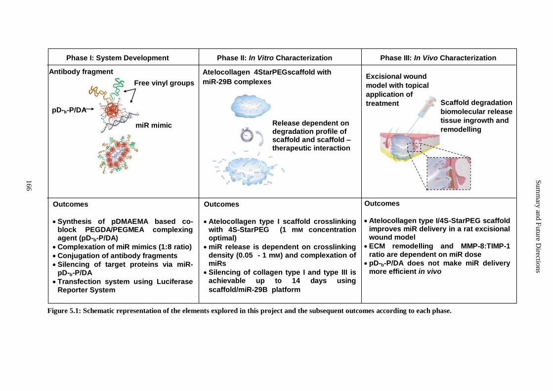
Sum
mary
and F
utu
re Directio
ns
166
Figure 5.1: Schematic representation of the elements explored in this project and the subsequent outcomes according to each phase.
Phase I: System Development Phase II: In Vitro Characterization Phase III: In Vivo Characterization
miR mimic
Antibody fragment
Free vinyl groups
pD-b-P/DA
Excisional wound
model with topical
application of
treatment
Scaffold degradation
biomolecular release
tissue ingrowth and
remodelling
Atelocollagen 4StarPEGscaffold with
miR-29B complexes
Release dependent on degradation profile of scaffold and scaffold – therapeutic interaction
Outcomes
Synthesis of pDMAEMA based co-block PEGDA/PEGMEA complexing agent (pD-b-P/DA)
Complexation of miR mimics (1:8 ratio)
Conjugation of antibody fragments
Silencing of target proteins via miR-pD-b-P/DA
Transfection system using Luciferase Reporter System
Outcomes
Atelocollagen type I scaffold crosslinking with 4S-StarPEG (1 mM concentration optimal)
miR release is dependent on crosslinking density (0.05 - 1 mM) and complexation of miRs
Silencing of collagen type I and type III is achievable up to 14 days using
scaffold/miR-29B platform
Outcomes
Atelocollagen type I/4S-StarPEG scaffold improves miR delivery in a rat excisional wound model
ECM remodelling and MMP-8:TIMP-1 ratio are dependent on miR dose
pD-b-P/DA does not make miR delivery more efficient in vivo
Summary and Future Directions
167
5.3.2 Phase II- An Injectable Scaffold Delivery System (Chapter Three)
Even as the lack of a perfect transfection reagent represented as a limitation in the first phase of
this work, the nature of the miR- pD-b-P/DA complexes, or complexes represented a major
limitation to the second phase of the work. The scaffold employed has huge potential as an
injectable system in situ. However, in the scope of this PhD, each gel was prepared individually
using standard pipetting techniques in the laboratory. There exists a variation in collagen sources
as to the volume of NaOH required to neutralise the solution, however, within all of the
experiments of this thesis only one source was used. Additionally, gels could not be made in large
batches from which aliquots could be used to make smaller gels as larger volumes had a negative
impact on the self-assembly kinetics of the 4S-StarPEG crosslinked hydrogels. A robust,
automated dual syringe system, such as is the case in fibrin platforms 2, is appropriate for the
mixing and injection which would minimise any possible batch to batch variation in scaffold, and
allow for efficient injection of scaffolds, perhaps even incorporating a simultaneous delivery of
therapeutic agents such as DNA, RNA, cells etc.
The scaffold’s physico-chemical properties were investigated in vitro; however these tests cannot
fully imitate the complex interactions that are presented in vivo. For instance, although
crosslinking of the scaffold offered resistance to degradation against collagenase produced in
clostridium histolyticum, the true degradation of this atelocollagen scaffold in vivo can never be
completely elucidated in vitro. The same limitations apply to the release profiles of RNA in its
various complexed/uncomplexed forms. As mentioned in the limitations of Phase I, Phase II cell
experiments were performed on monocultures that received nutrients via cell culture media.
Although an ideal environment for comparing the array of formulations, two dimensional sheets
do not truly represent in vivo conditions.
A final limitation in phase II is that of collagen type I and collagen type III knockdown. Granted,
knockdown of collagen type I and collagen type III expression was demonstrated. However, in the
evaluation of collagen type I and collagen type III production by cardiac fibroblasts, the western
blot presented in Figure 3.6 can only be used as a guide and future studies require repetition of
this with the inclusion of appropriate loading controls such as GAPDH or β-actin. Such a study
would require the analysis of protein at additional time points to monitor silencing over time.
5.3.3 Phase III- System Evaluation in a Rat Excisional Wound Model (Chapter Four)
The ideal model for all of the in vivo work is a model of myocardial infarction. However, as only
a single treatment can be tested per ischemic heart, and very little information existed in the
Summary and Future Directions
168
literature regarding optimal doses, scaffold mediated miR delivery or miR persistence in vivo, the
excisional wound model presented in Phase III was selected as a method to narrow the focus of a
possible future myocardial infarction study, develop analysis techniques and to determine answers
to key questions present from the outset of this study. However, the clinical significance of this
model in the context of myocardial infarction is limited. True, it presents a wounded environment,
with aspects of inflammation and extracellular matrix remodelling; however, it does not represent
an ischemic environment and the unique cascade of inflammatory events that follows it. The
excisional wound model did; however, have one primary similarity to the cardiac model; in that
the scaffold would be in direct contact with a region of ECM after acute insult. While there may
be diseases where such a model could have clinical relevance, they were not investigated in this
study. Furthermore, as there was minimal ischemia associated with the delivery of these scaffolds,
the miR-29B as an inhibitor of fibrosis, may have been less significant in this wound model.
A further limitation is the model itself. A rabbit ear model of hypertrophic wounding was
considered; however, antibodies and complete miR sequencing of this species is incomplete and
could render the study limited if further mechanistic and investigatory probes were to be
employed. The rodent excisional wound healing model has been extensively used to study wound
healing, cutaneous regeneration, stem cell and tissue transplantation and immune rejection 3.
Despite its convenience and frequent use, the model has marked limitations due to the differing
wound healing characteristics of rodents when compared with humans 4. In rodents, whose skin is
mobile, contraction accounts for a large part of wound closure; in humans, where the skin is
tethered to subcutaneous tissues, wounds are healed by the generation of new tissue. Moreover,
contraction of the rodent skin around the wound is affected by factors such as animal posture and
motion, as well as wound dressing, and it thus causes considerable variations in wound closure 5.
Efforts were made in order to normalise the epithelialisation rate to the degree of contraction
which has been performed previous studies 6, 7
, yet at a 28 day time point this consideration
revealed no significant impact on the results as complete epithelialisation and wound closure had
occurred at this time point, which was in agreement with previous studies 6, 7
. The rat excisional
wound model has been previously reported 6 and its limitations stressed from the onset, however,
it does provide a model to apply a number of treatments in a resilient animal. Furthermore, it
allows four separate conditions to be created on the dorsum of each rat.
5.4 Future Directions
Each aspect of the phases described in this thesis can be further investigated by addressing their
limitations and discussing the various avenues of exploration which have been highlighted
Summary and Future Directions
169
through this research. This section discusses five potential future projects that follow. Briefly, the
first discusses the immediate future direction of this work; that being intramyocardial injection of
miR-29B mimics embedded within a collagen scaffold. The second describes alternative methods
of harnessing the endogenous miRs but through the approach of miR inhibition via antagomiRs.
The third potential future project describes the incorporation of therapeutic stem cells and
reprogramming these stem cells with the platform described in this thesis towards a myocardial
application. The fourth proposes the option of delivering pre-miR through various strategies, and
finally the fifth discusses additional therapeutic applications of the described platform developed
in this thesis. The future directions that can be explored are summarized schematically in Figure
5.2.
5.4.1 Delivery of miR-29B using 4S-StarPEG/Collagen Scaffold via Intramyocardial Injection
An important characteristic of using injectable biomaterials for cardiac repair is their potential
ability to provide a minimally invasive approach, which will decrease the damage incurred to the
targeted and surrounding tissues during delivery. Furthermore, exposure of non-target tissue to the
embedded therapeutic will be substantially minimized. Therapeutic delivery of injectable
scaffolds includes epicardial, intracoronary, and endocardial injection. In the instance of using the
atelocollagen type I/4S-StarPEG system described in this thesis, a double-barrel syringe is ideal to
support delivery if this system were to ever reach the clinic, yet no such system for double-barrel
driven cardiac catheter technology exists presently. Direct epicardial and catheter-based
endocardial delivery can be applied via thoracic puncture and injection into the wall of the heart.
An alternative approach is to utilize the coronary artery, via its leaky vasculature following MI, to
deliver the scaffold, in liquid form before gelation has occurred, into the tissue avoiding direct
puncturing of the tissue. Single bolus injections are common in small animal models 2, 8-12
;
however, multiple injections are typically necessary to adequately distribute the material in large
animal models where the size of the heart is closer to that of human patients 13-15
. Furthermore,
each animal model has a range of injected volumes that is scaled to the relative size of the animal.
A further important consideration is the time point of injection which can range from
immediate, to one week, to two months following MI. This time point is a crucial parameter
for designing new treatment strategies due to the dynamic processes post-MI. The stages
following ischemic injury involve cardiomyocyte necrosis, a cascade of inflammatory cells,
fibrotic scar formation, and cardiomyocyte slippage followed by LV dilation. A different
population of cells is impacted depending on the time of administration, and some processes such
as LV dilation cannot currently be reversed. Thus, the optimal time post MI needs to be carefully
considered and whether the therapy is intended to prevent or reverse negative LV remodelling.

Summary and Future Directions
170
Figure 5.2: Schematic based on da Vinci’s Ventruvian Man. Schematic is arranged such that
the five corners represent the basic themes/aspects on which this project was based. The legs
represent the phases of the project. The text in the spaces beside the torso show the plausible
lines in which each phase can progress. The arms represent the possibility of employing this
platform in future projects and in other diseases. This is by no means a comprehensive
depiction, rather only the most direct and important aspects are represented to demonstrate
cohesion. Further details are discussed in section 6.4.
Summary and Future Directions
171
Several in vivo studies have implemented immediate injections of their material post-MI, but this
procedure does not properly mimic clinical restrictions as patients would not normally receive a
therapy for several hours post-MI. Scaffolds have previously been injected independently into the
infarcted myocardium and have resulted in improvement in ventricular function due to ventricular
thickening 12
and therefore the platform developed here can be applied. Biopolymers that have
already been used as injectable biomaterials into the infarcted myocardium include fibrin 2, 11, 16, 17
,
collagen 10, 11
, alginate 8, 9, 16
, hyaluronic acid 13, 18, 19
, chitosan 20, 21
, decellularized extracellular
matrix 12, 22
and, more recently, keratin 23
. Although the functional measurements and histological
investigations vary between the studies, a clear consensus is emerging that the injection of
scaffolds into the myocardium post-MI results in functional improvement following MI and
presents as a therapeutic option following MI. Clinical trials with alginate as an injectable
material treating MI have now been reported. This alginate scaffold was originally developed and
tested by BioLinRX as BL-1040, and the study yielded positive results in the first-in-man trial
according to unpublished reports 24
. The pilot study, which ended in January 2012, exhibited
preliminary efficacy and safety of the material after implantation in 27 patients 25
.
The progression of the injectable scaffold developed in this thesis could include evaluation in
large animal models with extended time points. Appropriate controls such as systemic
administration of current clinical pharmacological interventions generally prescribed follow MI,
empty treatment control groups and an open-chest intervention procedure are warranted in such a
study. A consistent parametrical standard to identify a successful outcome for injectable
biomaterials is lacking in the field. The parameters of wall thickness, LV volumes, fractional
shortening, and ejection fraction have been relied as important indicators of recovery; yet in the
clinic, ejection stroke volume is the best predictor of survival and re-hospitalization events post–
MI 26, 27
.
5.4.2 Using Oligonucleotides to Silence miRs
As previously discussed in this thesis, several approaches have been adopted to hijack the
endogenous regulation of genes via the miRNA pathway. Another approach is using antisense
oligonucleotides targeted toward miRNAs, referred to as antagomiRs (Figure 5.3, black pathway
and Figure 5.4). These antagomiRs target the mature miRNA sequence and inhibit the silencing of
oligonucleotides and can be conjugated to cholesterol to improve cellular uptake facilitating
discrimination between single nucleotide mismatches of the targeted miRNA. Reduction of target
miRNA levels has been achieved in multiple tissues for an extended period when antagomiRs are
designed to bind to pathogenic or abnormally expressed miRNAs 28
. These molecules can be
embedded within the scaffold developed in this project. There are several key requirements for
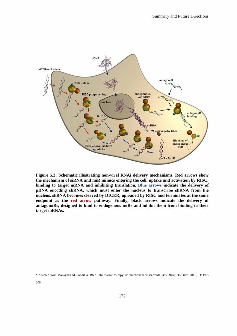
Summary and Future Directions
172
Figure 5.3: Schematic illustrating non-viral RNAi delivery mechanisms. Red arrows show
the mechanism of siRNA and miR mimics entering the cell, uptake and activation by RISC,
binding to target mRNA and inhibiting translation. Blue arrows indicate the delivery of
pDNA encoding shRNA, which must enter the nucleus to transcribe shRNA from the
nucleus. shRNA becomes cleaved by DICER, uploaded by RISC and terminates at the same
endpoint as the red arrow pathway. Finally, black arrows indicate the delivery of
antagomiRs, designed to bind to endogenous miRs and inhibit them from binding to their
target mRNAs.
* Adapted from Monaghan M, Pandit A. RNA interference therapy via functionalized scaffolds. Adv. Drug Del. Rev. 2011, 63: 197-
208
Summary and Future Directions
173
therapeutic oligonucleotides to achieve effective down-regulation of a targeted miRNA; namely:
in vivo stability, specificity and high binding affinity to the miRNA of interest. Previous research
in antisense oligonucleotides has been based on these criteria to improve the efficacy of the
oligonucleotides. Only a handful of modifications have been studied to date, one of which
involves the linkage of chemical groups to the 2’ hydroxyl group to modify the stability and
affinity of the oligonucleotides 29-30
. These chemical modifications include 2’-O-methoxyethyl
(MOE)-modified oligonucleotides which have better affinity and specificity to RNA than their
OMe-counterparts, and locked nucleic acid (LNA)-modified oligonucleotides (LNA-antagomiR),
in which the 2’-O-oxygen is joined to the 4’-position via a methylene spacer to form a rigid cyclic
structure, fixed into a C3’- endo (RNA) sugar conformation. An additional chemical modification
applied to regulate oligonucleotide stability is the balance between phosphodiester and
phosphorothioate linkages between the nucleotides, with phosphorothioate being more resistant to
nucleases than phophodiester, thereby providing greater stability to the oligonucleotides 31
.
The 2’ –O-methylo-group (OMe) is a frequently used modification to oligonucleotides and has a
limited amount of nuclease resistance but improved binding affinity to RNA compared to
unmodified sequences. In 2004, Hutbagner and colleagues pioneered the knockdown of let-7
function in Dropshilia using OME modified antisense oligonucleotides 32
. Following this,
Krutzfeldt et al. used antagomiRs to inhibit miR-122, a liver specific miRNA in the first
mammalian in vivo study using antagomiRs to inhibit endogenous miRNA28
. These chemically
modified oligonucleotides were conjugated to cholesterol to improve cellular uptake facilitating
discrimination between single nucleotide mismatches of the targeted miRNA. Reduction of target
miRNA levels has been achieved in multiple tissues for an extended period when antagomiRs
have been systemically administered via intravenous injection33
. However, off target results were
observed; as non-predicted up-regulation of mRNAs which were not predicted targets of miR-122
were upregulated indicating that a secondary mechanism can be involved in the modulation effect
of miR-122.Another oligonucleotide modification strategy, the 2’-O’methoxyethyl
phosphorothioate modification (MOE) antisense oligonucleotide, has demonstrated successful
miRNA inhibition in the liver. As previously discussed, 2’MOE antisense oligonucleotides are
also effective in vivo at silencing miR-122, up-regulating target-gene expression and reducing
cholesterol levels in normal mice 34
. However, the onset of action of the 2’ MOE ASO appears to
be slower than 2’Ome as a similar acute dosing schedule as that applied to the 2’Ome antisense
oligonucleotides did not produce cholesterol lowering and produced only ~50% reduction of miR-
122 for the 2’MOE oligos.
Summary and Future Directions
174
In theory, oligonucleotides may antagonize miRNA function at all levels from transcription of the
miRNA gene through processing and maturation of the miRNA to binding of the miRNA to its
target mRNA. Because miRNAs are relatively small it is imperative to use high-affinity antisense
chemistries to target miRNAs. So far, greater than 50 publications on the use of antagomiRs in
cell culture and in vivo exist and one can conclude that 2’-O-ME, 2’-O’MOE, morpholino and
LNA are probably the most efficient antagomiR strategies 30
.
5.4.3 Combination of 4S-StarPEG Collagen Scaffold with Cell Therapy
Considering the crucial role that miRs play in development and differentiation, it seems intuitive
that miRs could be used to program stem cells toward a beneficial lineage. Many transplanted
mesenchymal stem cells (MSCs) are lost following delivery to the infarcted myocardium and
efforts at using gene therapy to improve their survival have proven moderately successful 35
.
RNAi re-programming of these cells to improve their survival and/or modulate their paracrine
effects in vivo has not been reported to date. The scaffold/miR platform presented in this thesis
can be harnessed to improve MSC survival in vivo, thereby enhancing their therapeutic effect (see
Figure 5.5). A possible derivative of stem cell incorporation into this platform is the
reprogramming of stem cells or progenitor cells embedded within a functionalized hydrogel
towards a cardiomyogenic lineage to facilitate improved ventricular recovery following
myocardial infarction. For instance, cardiac progenitor cells transduced to overexpress miR-21, -
24 and -221and injected via the intra myocardial route following MI without reperfusion have led
to greater functional recovery 36
. Other researchers have demonstrated the direct reprogramming
of cardiac fibroblasts to cardiomyocyte cells in vitro and in vivo using a cocktail of miR-1, -3, -
208 and -499, most notably in ischemic mouse myocardium 37
. Although this proof-of-concept
study does not, as of yet, report a functional improvement with this therapy37
, it has huge potential
considering the cardiac fibroblast is the most populous cell in the myocardium, both pre- and
post-MI. Additionally, RNAi can be used to induce pluripotent stem (iPS) cells, an approach that
has only, very recently, been achieved by one group using only miRs to successfully induce
somatic cells to pluripotency 38
. This approach will have a potential in directing cells that are easy
to harvest into pluripotent cells ex vivo and then directing them towards a cardiomyogenic lineage
in vivo again using a combination of miRs.
5.4.4 Delivery of pre- miR-29B
The miR-29B discussed in this thesis is transcribed endogenously as a cluster. The miR-29 family
in humans includes hsa-miR-29A, hsa-miR-29B-1, hsa-miR-29B-2 and hsa-miR-29C. miR-29B-1
and mir-29B-2 have identical mature sequences, which are together called miR-29B. Mature 29s
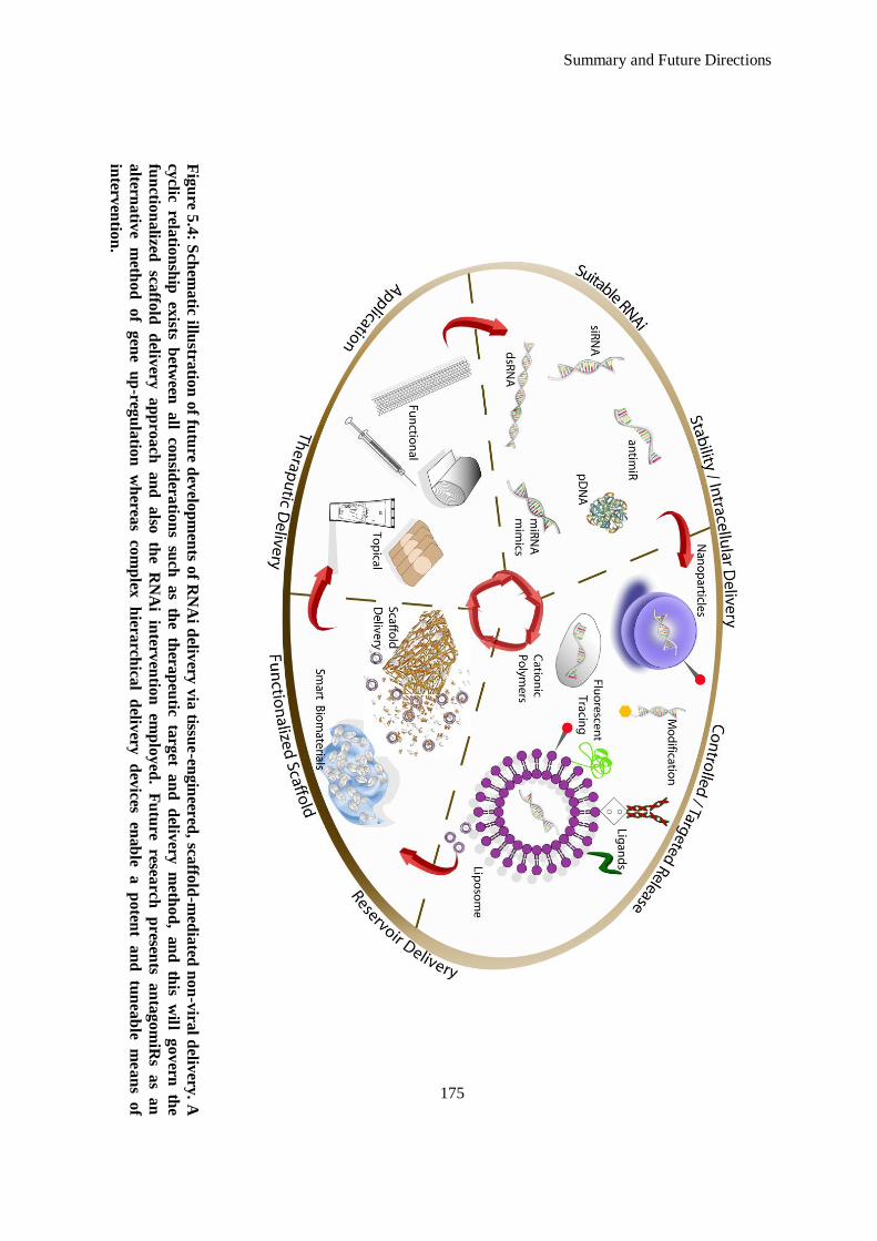
Summary and Future Directions
175
Fig
ure 5
.4: S
chem
atic illu
stratio
n o
f futu
re dev
elop
men
ts of R
NA
i deliv
ery v
ia tissu
e-en
gin
eered, sca
ffold
-med
iated
no
n-v
iral d
elivery
. A
cyclic
relatio
nsh
ip ex
ists b
etween
a
ll co
nsid
eratio
ns
such
as
the
thera
peu
tic ta
rget
an
d d
elivery
m
etho
d,
an
d th
is w
ill g
ov
ern th
e
fun
ction
alized
sca
ffold
d
elivery
a
pp
roa
ch
an
d
also
th
e R
NA
i in
terven
tion
em
plo
yed
. F
utu
re resea
rch
presen
ts a
nta
go
miR
s a
s a
n
altern
ativ
e m
etho
d
of
gen
e u
p-reg
ula
tion
w
herea
s co
mp
lex
hiera
rchica
l d
elivery
d
evices
enab
le a
po
tent
an
d
tun
eab
le m
ea
ns
of
interv
entio
n.
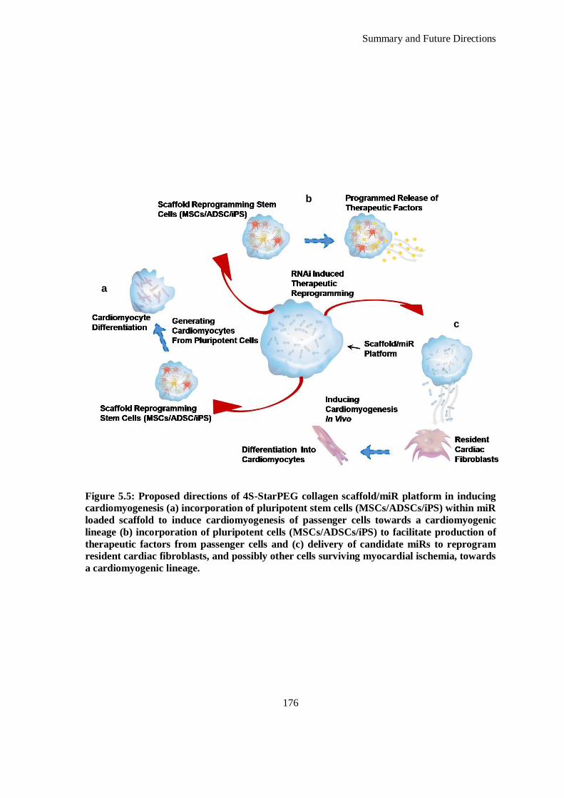
Summary and Future Directions
176
Figure 5.5: Proposed directions of 4S-StarPEG collagen scaffold/miR platform in inducing
cardiomyogenesis (a) incorporation of pluripotent stem cells (MSCs/ADSCs/iPS) within miR
loaded scaffold to induce cardiomyogenesis of passenger cells towards a cardiomyogenic
lineage (b) incorporation of pluripotent cells (MSCs/ADSCs/iPS) to facilitate production of
therapeutic factors from passenger cells and (c) delivery of candidate miRs to reprogram
resident cardiac fibroblasts, and possibly other cells surviving myocardial ischemia, towards
a cardiomyogenic lineage.
a
b
c
Summary and Future Directions
177
share identical sequences at nucleotide positions 2-7, the seed region that plays a key role in
determining which protein-coding-genes a miR would target 39
. Therefore, predicted target genes
for the miR-29 family members largely overlap. It has been confimed that miR-29B-1 and miR-
29A are transcribed together as a polycistronic pri-miR and likewise miR-29B-2 and miR-29C are
transcribed together 40
. Studies have identified several critical cis elements in the proximal region
of miR-29 gene promoters which indicates that they have binding sites to transcriptional factors
including a Gli binding site at -424 and three NF–κB binding sites at -561, -110, and +134 in the
human miR-29B-1/A promoter 41
, a Smad3 binding site in a highly conserved region ~22 kb
upstream of miR-29B-2/C promoter 42
, at least one Yin-Yang-1 binding site in the miR-29B-2/c
promoter 43
, a CEBP binding site located at +15 to +29 bp immediately downstream of the miR-
29B-1/A transcription start site 44
, and two TCF/LEF binding sites within the proximal promoter
of miR-29B-1/A 45
. In addition, miR-29 expression has been shown to be regulated by various
transcriptional regulator and signalling pathways, including Wnt signalling 45
. Post-transcriptional
processing or stability of mature miR-29s may contribute importantly to the observed differential
regulation documented in many studies.
In the experiments presented in this thesis, miR-29B was delivered as a synthetic, double stranded
siRNA which mimics mature endogenous miR before being uptaken into the RNA induced
silencing complex (RISC), however there are a number of other steps and formats at which miR-
29B can be delivered at, and in, respectively. True, siRNAs can be readily synthesized and can
quickly silence the expression of a vast number of genes both in vitro and in vivo and have shown
moderate success in clinical trials; their remains however many drawbacks. In vivo delivery of
siRNAs or even lipid-complexed siRNAs is inefficient and these oligonucleotides have short half-
lives in vivo. One approach to overcome this is to express interfering RNA (miRs and siRNAs)
intracellularly from plasmid DNA, or so called ‘short hairpin RNAs’ (shRNAs). These are
generally expressed from Pol III promoters and contain perfect stems of 21-25 bp with small
terminal loops. Similar to pre-miRNAs, they are exported from the nucleus by Exportin-5 where
they are cleaved by Dicer to form structures analogous to siRNAs 46
. Three approaches discussed
below allow for more stable delivery of miR/siRNA sequences via the delivery of miRs as pre-
miRs being transcribed from the nucleus and the preceeding pre-miR clusters that transcribe them.
5.4.4.1 Viral Delivery of pre- miR-29B
Using viral vectors, it is possible to alleviate some of the drawbacks of non-viral approaches,
some of which are; inefficient in vivo delivery and transient gene silencing. A large number of in
vivo studies, using mostly adeno-associated viruses (AAV) and lentivirus (LV) vectors to express

Summary and Future Directions
178
shRNAs have been performed to evaluate the effectiveness of RNAi in the treatment of cancer,
cardiac disease, retinal disease, neurodegenerative disease, viral infections, and other diseases
which are reviewed in the literature 47-49
. To date, these categories of viral vectors have the best
safety records and pseudotyping of the vectors with different capsids (AAV) or envelope (LV)
proteins permits tissue-tropic targeting. The use of AAVs for the delivery of sh/miRNA
expression cassettes is interesting as these cassettes are restricted in size making them amenable
for packaging into AAV vectors, which are the smallest known viral vectors. On the other hand,
lentiviruses stably integrate into the host cell genome and thus allow persistent transgene
expression (although this can become silenced over time). LVs are capable of transducing both
dividing and non-dividing cells.
Viral delivery has been used to mimic endogenous miRs in anti-cancer therapeutics. Specific
miRNAs are often overexpressed in tumor cells, but in general most miRs are down-regulated in
tumor tissue 50
. In a mouse model of hepatocellular carcinoma, miR-29A is markedly reduced in
human hepatocellular carcinoma (HCC) cells. Intravenous administration of a self-complementary
adeno-associated (scAAV8) vector expressing miR-26A from a ubiquitous Pol II promoter, to
transgenic mice that conditionally express Myc in the liver, protected the mice from tumor
formation 51
. Specifically, eighty percent of vector-injected mice were either completely free of
tumor or had significantly smaller tumours. Viral delivery of pre-miR-29B enables more stable
expression of miR-29B or its entire linked cluster, inducing more stable expression of this anti-
fibrotic molecule.
5.4.4.2 Non-viral Delivery of pre-miR-29B
5.4.4.2.1 Plasmid DNA
pDNA can be designed to transcribe pre-miRs and indeed pDNA delivery of miRs has been
documented in many studies 52-55
. McAnuff et al. compared the potency of siRNA and shRNA-
expressing pDNA mediated gene-silencing in vivo by co-administration of siRNA or shRNA-
expressing pDNA with pDNA encoding a target reporter gene 56
. The extent of the reduction in
the target gene expression was comparable to that between siRNA and shRNA-expressing pDNA
at a 10 mg dose; however on a molar basis, the shRNA was 250 fold more effective than the
siRNA one-three days after administration. A concern in this study was that the expression of the
reporter gene was transient, and therefore it was difficult to conclude whether these compounds
were effective and, furthermore, were comparable with each other for longer than three days56
.
pDNA transient transfection of cells in vitro exhibits gene silencing at three and five days in
Summary and Future Directions
179
rapidly dividing cell lines and, with naked siRNAs in general, achieves transient gene knockdown
at three and seven days due to dilution of the siRNAs below a therapeutic level with repeated cell
division. In quiescent or non-dividing cells, the silencing effect can remain at three weeks 57
after
which the siRNAs are naturally degraded 56
. In vivo, a similar trend was observed using rapidly
dividing subcutaneous tumours vs. non-dividing hepatocytes 56
.
Although pDNA is considered safe and easy to produce in large-scale quantities, one
disadvantage is the low gene transfer efficiency when compared with viral vector systems. Major
focus has been placed on improving their delivery and optimizing the pDNA itself. Conventional
plasmid vectors can be subdivided into a bacterial backbone and a transcription unit 58
. This
transcription unit usually carries the target gene or sequence along with necessary elements. The
bacterial backbone includes elements including an antibiotic resistance gene, an origin of
replication, unmethylated CpG motifs which activate the innate immune system of the host by
binding to the Toll-like receptor 9 (TLR9) of antigen-presenting cells 58
. Some of these sequences
are required for the production of plasmid DNA, but independently, each of them raise biological
safety concerns. Additionally, regulatory agencies recommend completely avoiding the use of
antibiotic resistant markers 59
. Therefore, the bacterial backbone of conventional pDNA
constitutes a significant portion of pDNA without a therapeutic effect leading to a decreased
bioavailability because of the increased size of the plasmid DNA.
5.4.4.2.2 Minicircle DNA
To address the issue just described, minicircle DNA can be employed. These are small (~ 4kb)
circular plasmid derivatives that have been freed from all prokaryotic vector parts. They are
derived from full length parental plasmids from which this parent plasmid is excised into two
supercoiled molecules: a replicative miniplasmid carrying the undesired backbone sequence, and
a minicircle carrying the therapeutic expression unit 60
. Conventional minicircles lack an origin of
replication so they do not replicate within target cells and the encoded genes will disappear as the
cell divides (which can be either an advantage or a disadvantage depending on whether the
application demands persistent or transient expression). This property can be overcome however
using self-replication minicircles which possess S/MAR elements 60
. Darquet et al. were the first
to show that minicircle DNA leads to much higher transgene expression levels in cell culture
experiments than a parental plasmid or other conventional control plasmids encoding the same
transgene 61
. The same group investigated this transgene expression in a pre-clinical model in
1999 and demonstrated that minicircle DNA in a mouse cranial tibial muscle was more effective
in reporter gene expression than with parental plasmids or larger plasmids 62
.
Summary and Future Directions
180
Delivery of shRNA via minicircle plasmids has been achieved in the literature 63-65
and has shown
much promise in the knockdown of target proteins. For instance, Huang et al. created a minicircle
vector transcribing shRNA for silencing both prolyl hydroxylase and factor-inhibiting hypoxia-
inducible factor (HIF) –1α (both of which degrade HIF-1α which is cardioprotective to ischemic
injury) 65
. The authors reported higher expression of angiogenic factors in the double knock-down
group compared with single knockdown and a shRNA scramble control groups in vitro. In vivo,
the authors demonstrated improved cardiac function in the minicircle group compared to the
scrambled control and a greater recruitment of bone marrow cells to the ischemic myocardium 65
when delivered intramyocardially in a syngeneic mouse model of MI. Minicircle DNA has yet to
fully demonstrate its potential as an effective transcriber of pre-miRs as very few examples exist
in the literature. Only recently, Hu et al. have shown that miR-210 delivered via a minicircle
vector intramyocardially following MI in a syngeneic mouse model caused decreased apoptosis,
increased neovascularisation and significant improvement in left ventricular fractional shortening
compared with scrambled minicircle vectors and sham controls 66
. Plasmid mediated miR delivery
is still in its infancy, however, plasmid mediated RNA interference will be more likely to achieve
clinical acceptance if vectors, such as minicircle vectors are used to eliminate elements in the
nucleotide backbone that cause regulatory concern.
5.4.4.3 Delivery via Avirulent Bacteria
Avirulent bacteria with specificity for infarcted myocardium can present as an alternative delivery
vehicles for RNAi molecules. The use of live, attenuated Salmonella for in vivo delivery of
therapeutic molecules has proven effective in pre-clinical models in specifically targeting tumors
and effecting expression of delivered genes after intravenous delivery 67
. There has been little
consideration of the delivery of functional genes/RNAi in this manner to date; yet functional
studies have reported the effectiveness of using Salmonella, which specifically accumulated in
tumours in a mouse model, to deliver STAT3-targeting shRNA to rescue the effects of a
vaccination. The combined approach of specific accumulation and silencing of STAT3 resulted in
reduced STAT3 expression and, furthermore, significantly suppressed tumour growth in an
advanced melanoma tumour model. The effectiveness of this approach in silencing STAT3
indicates the potential of delivering other interfering RNAs through a shRNA approach.
Additionally, Le et al. have described accumulation and selective proliferation in the infarcted
myocardium of one strain of an avirulent bacterium that can be cleared using antibiotics within
two days. Furthermore, protein excretion could be controlled by the administration of L-arabinose
by virtue of design of the plasmid-encoded gene expression cassette 68
. This approach has many
favourable characteristics pertaining to therapeutic delivery following MI: specific localisation to
Summary and Future Directions
181
infarcted myocardium, activation of expression through a secondary activator, and optional
elimination of this ‘live’ therapy through antibiotic treatment. However, there are concerns
surrounding immunogenicity and possible mutagenesis of the bacteria, concerns that already exist
in the field of viral gene delivery and plasmid mediate non-viral delivery.
5.4.5 Application of Platform in Other Pathological Settings
A collagen type I scaffold can be used as a reservoir of miR-29B in other pathological settings
(see Figure 5.6). For instance, in Chapter 4, this system was applied in a dermal wound healing
model where the scaffold was found to improve the effectiveness of miR-29B delivered. The
concept was relatively simple and had a number of advantages. For example, as non-viral
techniques were used, there was minimal risk of viral proteins being expressed by the subjected
cells. Moreover, the use of mature miR sequences negated the requirement of nuclear entry and
presented the delivery of one single miR.
It has collectively been shown that the miR-29 family members (which includes miR-29B which
has been tested in this thesis) target at least 16 genes related to extracellular matrix which encode
for several key proteins involved in the physiological or pathological formation of extracellular
matrix including; a large number of collagen isoforms, laminin γ, fibrilin 1, elastin, matrix
metalloproteinase 2, and integrin β1 69-72
. This is unique to the miR-29 family as no other miR is
predicted to target more than 11 of the 20 collagen genes. With regard to hind limb ischemia
(HLI), Greco et al. investigated the cohort of miRs that become dysregulated in the recovering
ischemic limb in a mouse model of HLI and found that miRs- 29A and 29B were decreased in the
ischemic tissue, whereas the expression of miR-29C was increased 73
. miR-29B is also a key
regulator in pulmonary fibrosis. Cushing et al. performed a large-scale screening for miRNAs
potentially involved in bleomycin-induced fibrosis and found the expression of miR-29 family
members was significantly reduced in fibrotic lungs 74
. Additionally, Xiao et al. found that miR-
29 gene transfer into diseased lung tissues in mice was capable of preventing and treating
pulmonary fibrosis including inflammatory macrophage (M1) infiltration induced by
bleomycin in mice 75
. It has also been suggested that miR-29B can also prevent liver fibrosis by
blocking activation of hepatic stellate cells through cell arresting mechanisms 76
which resulted in
an indirect blocking of TGF-β upregulation, which is significant as TGF-β is an important
transcriptional stimulator of many extracellular matrix genes and is an important factor that
downregulates miR-29B 77-80
.
An important role of miR-29B in renal injury has been identified in studies using Dahl salt
sensitive rat (a consomic variation of this rat; SS.13BN, which has substantially attenuated
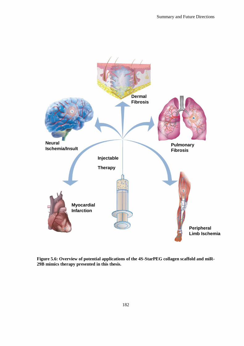
Summary and Future Directions
182
Figure 5.6: Overview of potential applications of the 4S-StarPEG collagen scaffold and miR-
29B mimics therapy presented in this thesis.
Peripheral
Limb Ischemia
Myocardial
Infarction
Pulmonary
Fibrosis
Dermal
Fibrosis
Neural
Ischemia/Insult
Injectable
Therapy
Summary and Future Directions
183
hypertension and renal injury 81
) where miR-29B expression in the renal medulla, was upregulated
by three days of a high-salt diet. In vivo knockdown experiments using locked nucleic acid-
modified antagomiRs concluded that miR-29B contributed to protection against interstitial
fibrosis in the SS.13BN rat 70
. Further studies linking miR-29B in renal fibrosis was reported in a
mouse model of obstructive nephropathy 42
. Severe tubulointerstitial fibrosis was associated with
reduced expression of miR-29. Forced overexpression of miR-29B attenuated renal fibrosis in this
model and a study of cultured renal tubular cells showed that miR-29B was downregulated by
TGF-β1 via Smad3 42
. Long et al. found miR-29C upregulated in glomeruli of diabetic (db/db)
mice and in kidney microvascular endothelial cells and podocytes cultured in high ambient
glucose 82
. Systemic delivery of antagomiRs directed towards miR-29C reduced albuminuria and
mesangial matrix accumulation in db/db mice. The injurious effect of miR-29C was associated
with proapoptotic effects of miR-29C on cultured podocytes and targeting of Sprouty homolog 1
82.
miR-29B also plays an important role in the regulation of cell proliferation, differentiation, and
apoptosis, a role which has been illustrated by the role of miR-29s in cancer. Downregulation of
miR-29s has been correlated with many types of cancer including leukemia 83-84
, melanoma 85
,
liver 86
, colon 87
, cervical 88
, and lung cancer 89
. In many cases, downregulation of miR-29B
correlated with more aggressive forms of cancer or relapse, suggesting therapeutic restoration of
miR-29B could improve disease prognosis 83, 90-91
. This has been explored where induced
expression of miR-29A and miR-29B slowed cell growth and induced apoptosis of leukaemia
cells in vitro and exogenous delivery of miR-29B to xenografted K562 cell tumours was effective
in reducing their size 92
. One of the mechanisms by which all members of the miR-29 family
suppress tumor growth is by relieving the suppression of p53. The p53 transcription factor is
important in controlling expression of genes that regulate cell growth, senescence, apoptosis, and
genome integrity in response to stress and suppression or inactivation of p53 is a common
characteristic in many types of cancer 93-94
. All three members of the miR-29 family, including
miR-29B can target p85α and CDC42, which are genes that normally suppress p53 expression 95
.
The application of miR-29B therapeutics, in tandem with the delivery platform presented in this
thesis has applicability in the treatment of ischemic brain injury. Shi et al. have found that miR-
29B levels are significantly increased in rodent brains after transient middle cerebral artery
occlusion and neurons after oxygen-glucose deprivation 96
. Furthermore, ectopic expression of
miR-29B promoted neuronal cell deaths, whereas its repression decreased cell death. The authors
linked this to the miR-29B targeting Bcl2L2 and thereby inhibiting Bcl2L2 gene expression. More
importantly, Bcl2L2 overexpression rescued neuronal cell death induced by miR-29B 96
. Given
Summary and Future Directions
184
the plethora of evidence described it is feasible that the levels of miR-29B can be modulated
towards therapeutic benefit in other pathological settings, which is summarized in Figure 6.4.
5.5 Conclusions
In conclusion, a scaffold functionalized with miR therapy has been developed which can be used
as a delivery system capable of effectively and silencing the production of collagen type I and
modulating the turnover of ECM components over time. In vitro, it was demonstrated that this
system can effectively mediate the protein silencing of cells with both reporter genes and
therapeutic genes. In vivo, a statistically significant effect was observed on the collagen type III/I
ratio, MMP-8/TIMP-1 ratio, wound contraction and volume fraction of granulation tissue 28 days
after administration of a scaffold with miR-29B to a dermal excisional wound in a rat model.
These effects were most significant when a dose of 5 μg of uncomplexed miR-29B was
incorporated into the scaffold. However the use of the complexing agent developed in phase I;
pD-b-P/DA, did not have the same significant effect when used with 5 μg of miR-29B
incorporated into the scaffold.
Summary and Future Directions
185
5.6 References
1. Nam HY, McGinn A, Kim P-H, Kim SW, Bull DA. Primary cardiomyocyte-targeted
bioreducible polymer for efficient gene delivery to the myocardium. Biomaterials.
2010;31:8081-8087 2. Christman KL, Vardanian AJ, Fang Q, Sievers RE, Fok HH, Lee RJ. Injectable fibrin
scaffold improves cell transplant survival, reduces infarct expansion, and induces
neovasculature formation in ischemic myocardium. J. Am. Coll. Cardiol. 2004;44:654-
660 3. Wu Y, Chen L, Scott PG, Tredget EE. Mesenchymal stem cells enhance wound healing
through differentiation and angiogenesis. Stem Cells. 2007;25:2648-2659
4. Sullivan TP, Eaglstein WH, Davis SC, Mertz P. The pig as a model for human wound healing. Wound Repair Regen. 2001;9:66-76
5. Orgill D, Blanco DC. Wounded skin contraction.Biomaterials for treating skin loss. CRC
Press; 2009; 21-22.
6. Garcia Y, Wilkins B, Collighan RJ, Griffin M, Pandit A. Towards development of a dermal rudiment for enhanced wound healing response. Biomaterials. 2008;29:857-868
7. Damodaran G, Tiong WH, Collighan R, Griffin M, Navsaria H, Pandit A. In vivo effects
of tailored laminin-332 α3 conjugated scaffolds enhances wound healing: A histomorphometric analysis. J. Biomed. Mater. Res. A. 2013
8. Leor J, Tuvia S, Guetta V, Manczur F, Castel D, Willenz U, Petneházy Ö, Landa N,
Feinberg MS, Konen E, Goitein O, Tsur-Gang O, Shaul M, Klapper L, Cohen S. Intracoronary injection of in situ forming alginate hydrogel reverses left ventricular
remodeling after myocardial infarction in swine. J. Amer. Coll. Cardiol. 2009;54:1014-
1023
9. Landa N, Miller L, Feinberg MS, Holbova R, Shachar M, Freeman I, Cohen S, Leor J. Effect of injectable alginate implant on cardiac remodeling and function after recent and
old infarcts in rat. Circulation. 2008;117:1388-1396
10. Dai W, Wold LE, Dow JS, Kloner RA. Thickening of the infarcted wall by collagen injection improves left ventricular function in rats: A novel approach to preserve cardiac
function after myocardial infarction. J. Am. Coll. Cardiol. 2005;46:714-719
11. Huang NF, Yu J, Sievers R, Li S, Lee RJ. Injectable biopolymers enhance angiogenesis after myocardial infarction. Tissue Eng. 2005;11:1860-1866
12. Singelyn JM, Sundaramurthy P, Johnson TD, Schup-Magoffin PJ, Hu DP, Faulk DM,
Wang J, Mayle KM, Bartels K, Salvatore M, Kinsey AM, DeMaria AN, Dib N,
Christman KL. Catheter-deliverable hydrogel derived from decellularized ventricular extracellular matrix increases endogenous cardiomyocytes and preserves cardiac function
post-myocardial infarction. J. Amer. Coll. Cardiol. 2012;59:751-763
13. Ifkovits JL, Tous E, Minakawa M, Morita M, Robb JD, Koomalsingh KJ, Gorman JH, 3rd, Gorman RC, Burdick JA. Injectable hydrogel properties influence infarct expansion
and extent of postinfarction left ventricular remodeling in an ovine model. Proc. Natl.
Acad. Sci. USA. 2010;107:11507-11512
14. Ou L, Li W, Zhang Y, Wang W, Liu J, Sorg H, Furlani D, Gabel R, Mark P, Klopsch C, Wang L, Lutzow K, Lendlein A, Wagner K, Klee D, Liebold A, Li RK, Kong D,
Steinhoff G, Ma N. Intracardiac injection of matrigel induces stem cell recruitment and
improves cardiac functions in a rat myocardial infarction model. J. Cell. Mol. Med. 2011;15:1310-1318
15. Mukherjee R, Zavadzkas JA, Saunders SM, McLean JE, Jeffords LB, Beck C, Stroud RE,
Leone AM, Koval CN, Rivers WT, Basu S, Sheehy A, Michal G, Spinale FG. Targeted myocardial microinjections of a biocomposite material reduces infarct expansion in pigs.
Ann. Thorac. Surg. 2008;86:1268-1276
Summary and Future Directions
186
16. Christman KL, Fok HH, Sievers RE, Fang Q, Lee RJ. Fibrin glue alone and skeletal
myoblasts in a fibrin scaffold preserve cardiac function after myocardial infarction. Tissue Eng. 2004;10:403-409
17. Yu J, Christman KL, Chin E, Sievers RE, Saeed M, Lee RJ. Restoration of left ventricular
geometry and improvement of left ventricular function in a rodent model of chronic
ischemic cardiomyopathy. J. Thorac. Cardiovasc. Surg. 2009;137:180-187 18. Yoon SJ, Fang YH, Lim CH, Kim BS, Son HS, Park Y, Sun K. Regeneration of ischemic
heart using hyaluronic acid-based injectable hydrogel. J. Biomed. Mater. Res.
2009;91:163-171 19. Tous E, Ifkovits JL, Koomalsingh KJ, Shuto T, Soeda T, Kondo N, Gorman JH, 3rd,
Gorman RC, Burdick JA. Influence of injectable hyaluronic acid hydrogel degradation
behavior on infarction-induced ventricular remodeling. Biomacromolecules. 2011;12:4127-4135
20. Lu WN, Lu SH, Wang HB, Li DX, Duan CM, Liu ZQ, Hao T, He WJ, Xu B, Fu Q, Song
YC, Xie XH, Wang CY. Functional improvement of infarcted heart by co-injection of
embryonic stem cells with temperature-responsive chitosan hydrogel. Tissue Eng. Part A. 2009;15:1437-1447
21. Liu Z, Wang H, Wang Y, Lin Q, Yao A, Cao F, Li D, Zhou J, Duan C, Du Z, Wang C.
The influence of chitosan hydrogel on stem cell engraftment, survival and homing in the ischemic myocardial microenvironment. Biomaterials. 2012;33:3093-3106
22. Okada M, Payne TR, Oshima H, Momoi N, Tobita K, Huard J. Differential efficacy of
gels derived from small intestinal submucosa as an injectable biomaterial for myocardial infarct repair. Biomaterials. 2010;31:7678-7683
23. Shen D, Wang X, Zhang L, Zhao X, Li J, Cheng K, Zhang J. The amelioration of cardiac
dysfunction after myocardial infarction by the injection of keratin biomaterials derived
from human hair. Biomaterials. 2011;32:9290-9299 24. BiolineRX Ltd. Safety and feasibility of the injectable BL-1040 implant. NCT00557531.
2007
25. Ikara Holdings Inc. IK-5001 for the prevention and remodling of the ventricle and congestive heart failure after acute myocardial infarction. NCT01226563. 2010
26. White HD, Norris RM, Brown MA, Brandt PW, Whitlock RM, Wild CJ. Left ventricular
end-systolic volume as the major determinant of survival after recovery from myocardial
infarction. Circulation. 1987;76:44-51 27. McManus DD, Shah SJ, Fabi MR, Rosen A, Whooley MA, Schiller NB. Prognostic value
of left ventricular end-systolic volume index as a predictor of heart failure hospitalization
in stable coronary artery disease: Data from the heart and soul study. J. Am. Soc. Echocardiogr. 2009;22:190-197
28. Krutzfeldt J, Rajewsky N, Braich R, Rajeev KG, Tuschl T, Manoharan M, Stoffel M.
Silencing of microRNAs in vivo with `antagomiRs'. Nature. 2005;438:685-689 29. Manoharan M. 2'-carbohydrate modifications in antisense oligonucleotide therapy:
Importance of conformation, configuration and conjugation. Biochim. Biophys. Acta,
Gene Struct. Expr. 1999;1489:117-130
30. Stenvang J, Kauppinen S. microRNAs as targets for antisense-based therapeutics. Expert Opin. Biol. Ther. 2008;8:59-81
31. Esau CC. Inhibition of microRNA with antisense oligonucleotides. Methods. 2008;44:55-
60 32. Hutvágner G, Simard MJ, Mello CC, Zamore PD. Sequence-specific inhibition of small
RNA function. PLoS Biol. 2004;2:e98
33. Krützfeldt J, Kuwajima S, Braich R, Rajeev KG, Pena J, Tuschl T, Manoharan M, Stoffel M. Specificity, duplex degradation and subcellular localization of antagomiRs. Nucleic
Acids Res. 2007;35:2885-2892
Summary and Future Directions
187
34. Esau C, Davis S, Murray SF, Yu XX, Pandey SK, Pear M, Watts L, Booten SL, Graham
M, McKay R, Subramaniam A, Propp S, Lollo BA, Freier S, Bennett CF, Bhanot S, Monia BP. MiR-122 regulation of lipid metabolism revealed by in vivo antisense
targeting. Cell. Metab. 2006;3:87-98
35. Holladay CA, Duffy AM, Chen X, Sefton MV, O’Brien TD, Pandit AS. Recovery of
cardiac function mediated by msc and interleukin-10 plasmid functionalized scaffold. Biomaterials. 2012;33:1303-1314
36. Hu S, Huang M, Nguyen PK, Gong Y, Li Z, Jia F, Lan F, Liu J, Nag D, Robbins RC, Wu
JC. Novel microRNA prosurvival cocktail for improving engraftment and function of cardiac progenitor cell transplantation. Circulation. 2011;124:S27-S34
37. Jayawardena TM, Egemnazarov B, Finch EA, Zhang L, Payne JA, Pandya K, Zhang Z,
Rosenberg P, Mirotsou M, Dzau VJ. MicroRNA-mediated in vitro and in vivodirect reprogramming of cardiac fibroblasts to cardiomyocytes. Circ. Res. 2012
38. Anokye-Danso F, Trivedi Chinmay M, Juhr D, Gupta M, Cui Z, Tian Y, Zhang Y, Yang
W, Gruber Peter J, Epstein Jonathan A, Morrisey Edward E. Highly efficient miRNA-
mediated reprogramming of mouse and human somatic cells to pluripotency. Cell Stem Cell. 2011;8:376-388
39. Kriegel AJ, Liu Y, Fang Y, Ding X, Liang M. The miR-29 family: Genomics, cell
biology, and relevance to renal and cardiovascular injury. Physiol. Genomics. 2012;44:237-244
40. Chang TC, Yu D, Lee YS, Wentzel EA, Arking DE, West KM, Dang CV, Thomas-
Tikhonenko A, Mendell JT. Widespread microRNA repression by Myc contributes to tumorigenesis. Nat. Genet. 2008;40:43-50
41. Mott JL, Kurita S, Cazanave SC, Bronk SF, Werneburg NW, Fernandez-Zapico ME.
Transcriptional suppression of miR-29b-1/miR-29a promoter by c-Myc, hedgehog, and
NF-κb. J. Cell Biochem. 2010;110:1155-1164 42. Qin W, Chung AC, Huang XR, Meng XM, Hui DS, Yu CM, Sung JJ, Lan HY. TGF-
β/Smad3 signaling promotes renal fibrosis by inhibiting miR-29. J. Am. Soc. Nephrol.
2011;22:1462-1474 43. Wang H, Garzon R, Sun H, Ladner KJ, Singh R, Dahlman J, Cheng A, Hall BM,
Qualman SJ, Chandler DS, Croce CM, Guttridge DC. Nf-κb-yy1-miR-29 regulatory
circuitry in skeletal myogenesis and rhabdomyosarcoma. Cancer Cell. 2008;14:369-381
44. Eyholzer M, Schmid S, Wilkens L, Mueller BU, Pabst T. The tumour-suppressive miR-29a/b1 cluster is regulated by Cebpa and blocked in human AML. Br. J. Cancer.
2010;103:275-284
45. Kapinas K, Kessler C, Ricks T, Gronowicz G, Delany AM. MiR-29 modulates Wnt signaling in human osteoblasts through a positive feedback loop. J. Biol. Chem.
2010;285:25221-25231
46. Monaghan M, Pandit A. RNA interference therapy via functionalized scaffolds. Adv. Drug Deliv. Rev. 2011;63:197-208
47. Couto LB, High KA. Viral vector-mediated RNA interference. Curr. Opin. Pharmacol.
2010;10:534-542
48. de Fougerolles A, Vornlocher H-P, Maraganore J, Lieberman J. Interfering with disease: A progress report on siRNA-based therapeutics. Nat. Rev. Drug Discov. 2007;6:443-453
49. Monaghan M, Greiser U, Wall JG, O'Brien T, Pandit A. Interference: An alteRNAtive
therapy following acute myocardial infarction. Trends Pharmacol. Sci. 2012;33:635-645 50. Gaur A, Jewell DA, Liang Y, Ridzon D, Moore JH, Chen C, Ambros VR, Israel MA.
Characterization of microRNA expression levels and their biological correlates in human
cancer cell lines. Cancer Res. 2007;67:2456-2468 51. Kota J, Chivukula RR, O'Donnell KA, Wentzel EA, Montgomery CL, Hwang HW,
Chang TC, Vivekanandan P, Torbenson M, Clark KR, Mendell JR, Mendell JT.
Summary and Future Directions
188
Therapeutic microRNA delivery suppresses tumorigenesis in a murine liver cancer model.
Cell. 2009;137:1005-1017 52. Chung K-H, Hart CC, Al-Bassam S, Avery A, Taylor J, Patel PD, Vojtek AB, Turner DL.
Polycistronic RNA polymerase II expression vectors for RNA interference based on
Bic/miR-155. Nucleic Acids Res. 2006;34:e53
53. Zeng Y, Wagner EJ, Cullen BR. Both natural and designed microRNAs can inhibit the expression of cognate mRNAs when expressed in human cells. Mol. Cell. 2002;9:1327-
1333
54. Boden D, Pusch O, Silbermann R, Lee F, Tucker L, Ramratnam B. Enhanced gene silencing of HIV-1 specific siRNA using microRNA designed hairpins. Nucleic Acids
Res. 2004;32:1154-1158
55. Silva JM, Li MZ, Chang K, Ge W, Golding MC, Rickles RJ, Siolas D, Hu G, Paddison PJ, Schlabach MR, Sheth N, Bradshaw J, Burchard J, Kulkarni A, Cavet G,
Sachidanandam R, McCombie WR, Cleary MA, Elledge SJ, Hannon GJ. Second-
generation shRNA libraries covering the mouse and human genomes. Nat. Genet.
2005;37:1281-1288 56. McAnuff MA, Rettig GR, Rice KG. Potency of siRNA versus shRNA mediated
knockdown in vivo. J. Pharmacol. Sci. 2007;96:2922-2930
57. Bartlett DW, Davis ME. Insights into the kinetics of siRNA-mediated gene silencing from live-cell and live-animal bioluminescent imaging. Nucleic Acids Res. 2006;34:322-333
58. Jechlinger W. Optimization and delivery of plasmid DNA for vaccination. Expert Rev.
Vaccines. 2006;5:803-825 59. Guidance for human somatic cell therapy and gene therapy. March 1998. Center for
biologics evaluation and research, food and drug administration. Hum. Gene Ther.
1998;9:1513-1524
60. Argyros O, Wong SP, Fedonidis C, Tolmachov O, Waddington SN, Howe SJ, Niceta M, Coutelle C, Harbottle RP. Development of S/MAR minicircles for enhanced and
persistent transgene expression in the mouse liver. J. Mol. Med.. 2011;89:515-529
61. Darquet AM, Cameron B, Wils P, Scherman D, Crouzet J. A new DNA vehicle for nonviral gene delivery: Supercoiled minicircle. Gene Ther. 1997;4:1341-1349
62. Darquet AM, Rangara R, Kreiss P, Schwartz B, Naimi S, Delaere P, Crouzet J, Scherman
D. Minicircle: An improved DNA molecule for in vitro and in vivo gene transfer. Gene
Ther. 1999;6:209-218 63. Yang Z, Li G, Zhang Y, Liu X, Tien P. A novel minicircle vector based system for
inhibting the replication and gene expression of enterovirus 71 and coxsackievirus a16.
Antiviral Res. 2012;96:234-244 64. Levi B, Hyun JS, Nelson ER, Li S, Montoro DT, Wan DC, Jia FJ, Glotzbach JC, James
AW, Lee M, Huang M, Quarto N, Gurtner GC, Wu JC, Longaker MT. Nonintegrating
knockdown and customized scaffold design enhances human adipose-derived stem cells in skeletal repair. Stem Cells. 2011;29:2018-2029
65. Huang M, Nguyen P, Jia F, Hu S, Gong Y, de Almeida PE, Wang L, Nag D, Kay MA,
Giaccia AJ, Robbins RC, Wu JC. Double knockdown of prolyl hydroxylase and factor-
inhibiting hypoxia-inducible factor with nonviral minicircle gene therapy enhances stem cell mobilization and angiogenesis after myocardial infarction. Circulation.
2011;124:S46-54
66. Hu S, Huang M, Li Z, Jia F, Ghosh Z, Lijkwan MA, Fasanaro P, Sun N, Wang X, Martelli F, Robbins RC, Wu JC. MicroRNA-210 as a novel therapy for treatment of
ischemic heart disease. Circulation. 2010;122:S124-131
67. Arrach N, Zhao M, Porwollik S, Hoffman RM, McClelland M. Salmonella promoters preferentially activated inside tumors. Cancer Res. 2008;68:4827-4832
Summary and Future Directions
189
68. Le UN, Kim H-S, Kwon J-S, Kim MY, Nguyen VH, Jiang SN, Lee B-I, Hong Y, Shin
MG, Rhee JH, Bom H-S, Ahn Y, Gambhir SS, Choy HE, Min J-J. Engineering and visualization of bacteria for targeting infarcted myocardium. Mol. Ther. 2011;19:951-959
69. Li Z, Hassan MQ, Jafferji M, Aqeilan RI, Garzon R, Croce CM, van Wijnen AJ, Stein JL,
Stein GS, Lian JB. Biological functions of miR-29b contribute to positive regulation of
osteoblast differentiation. J. Biol. Chem. 2009;284:15676-15684 70. Liu Y, Taylor NE, Lu L, Usa K, Cowley AW, Jr., Ferreri NR, Yeo NC, Liang M. Renal
medullary microRNAs in dahl salt-sensitive rats: MiR-29b regulates several collagens
and related genes. Hypertension. 2010;55:974-982 71. Sengupta S, den Boon JA, Chen IH, Newton MA, Stanhope SA, Cheng YJ, Chen CJ,
Hildesheim A, Sugden B, Ahlquist P. MicroRNA 29c is down-regulated in
nasopharyngeal carcinomas, up-regulating mRNAs encoding extracellular matrix proteins. Proc. Natl. Acad. Sci. USA. 2008;105:5874-5878
72. van Rooij E, Sutherland LB, Thatcher JE, DiMaio JM, Naseem RH, Marshall WS, Hill
JA, Olson EN. Dysregulation of microRNAs after myocardial infarction reveals a role of
miR-29 in cardiac fibrosis. Proc. Natl. Acad. Sci. USA. 2008;105:13027-13032 73. Greco S, De Simone M, Colussi C, Zaccagnini G, Fasanaro P, Pescatori M, Cardani R,
Perbellini R, Isaia E, Sale P, Meola G, Capogrossi MC, Gaetano C, Martelli F. Common
micro-RNA signature in skeletal muscle damage and regeneration induced by duchenne muscular dystrophy and acute ischemia. FASEB J. 2009;23:3335-3346
74. Cushing L, Kuang PP, Qian J, Shao F, Wu J, Little F, Thannickal VJ, Cardoso WV, Lü J.
MiR-29 is a major regulator of genes associated with pulmonary fibrosis. Amer. J. Respir. Cell Mol. Biol. 2011;45:287-294
75. Xiao J, Meng X-M, Huang XR, Chung ACK, Feng Y-L, Hui DSC, Yu C-M, Sung JJY,
Lan HY. MiR-29 inhibits bleomycin-induced pulmonary fibrosis in mice. Mol. Ther.
2012;20:1251-1260 76. Sekiya Y, Ogawa T, Yoshizato K, Ikeda K, Kawada N. Suppression of hepatic stellate
cell activation by microRNA-29b. Biochem. Biophys. Res. Commun. 2011;412:74-79
77. Kuang PP, Zhang XH, Rich CB, Foster JA, Subramanian M, Goldstein RH. Activation of elastin transcription by TGF-β in human lung fibroblasts. Am. J. Physiol. Lung Cell Mol.
Physiol. 2007;292:L944-952
78. Lei J, Silbiger S, Ziyadeh FN, Neugarten J. Serum-stimulated α1 type IV collagen gene
transcription is mediated by TGF-β and inhibited by estradiol. Am. J. Physiol. 1998;274:F252-258
79. Neumann C, Yu A, Welge-Lussen U, Lutjen-Drecoll E, Birke M. The effect of TGF-β2 on
elastin, type VI collagen, and components of the proteolytic degradation system in human optic nerve astrocytes. Invest. Ophthalmol. Vis. Sci. 2008;49:1464-1472
80. Sysa P, Potter JJ, Liu X, Mezey E. Transforming growth factor-beta1 up-regulation of
human α1I collagen is mediated by Sp1 and Smad2 transacting factors. DNA Cell Biol. 2009;28:425-434
81. Cowley AW, Jr., Roman RJ, Kaldunski ML, Dumas P, Dickhout JG, Greene AS, Jacob
HJ. Brown norway chromosome 13 confers protection from high salt to consomic dahl
sensitive rat. Hypertension. 2001;37:456-461 82. Long J, Wang Y, Wang W, Chang BH, Danesh FR. MicroRNA-29c is a signature
microRNA under high glucose conditions that targets sprouty homolog 1, and its in vivo
knockdown prevents progression of diabetic nephropathy. J. Biol. Chem. 2011;286:11837-11848
83. Calin GA, Ferracin M, Cimmino A, Di Leva G, Shimizu M, Wojcik SE, Iorio MV,
Visone R, Sever NI, Fabbri M, Iuliano R, Palumbo T, Pichiorri F, Roldo C, Garzon R, Sevignani C, Rassenti L, Alder H, Volinia S, Liu CG, Kipps TJ, Negrini M, Croce CM. A
microRNA signature associated with prognosis and progression in chronic lymphocytic
leukemia. N. Engl. J. Med. 2005;353:1793-1801
Summary and Future Directions
190
84. Garzon R, Garofalo M, Martelli MP, Briesewitz R, Wang L, Fernandez-Cymering C,
Volinia S, Liu CG, Schnittger S, Haferlach T, Liso A, Diverio D, Mancini M, Meloni G, Foa R, Martelli MF, Mecucci C, Croce CM, Falini B. Distinctive microRNA signature of
acute myeloid leukemia bearing cytoplasmic mutated nucleophosmin. Proc. Natl. Acad.
Sci. USA. 2008;105:3945-3950
85. Nguyen T, Kuo C, Nicholl MB, Sim MS, Turner RR, Morton DL, Hoon DS. Downregulation of microRNA-29c is associated with hypermethylation of tumor-related
genes and disease outcome in cutaneous melanoma. Epigenetics. 2011;6:388-394
86. Xiong Y, Fang JH, Yun JP, Yang J, Zhang Y, Jia WH, Zhuang SM. Effects of microRNA-29 on apoptosis, tumorigenicity, and prognosis of hepatocellular carcinoma.
Hepatology. 2010;51:836-845
87. Li Y, Wang F, Xu J, Ye F, Shen Y, Zhou J, Lu W, Wan X, Ma D, Xie X. Progressive miRNA expression profiles in cervical carcinogenesis and identification of HPV-related
target genes for miR-29. J. Pathol. 2011;224:484-495
88. Cummins JM, He Y, Leary RJ, Pagliarini R, Diaz LA, Jr., Sjoblom T, Barad O, Bentwich
Z, Szafranska AE, Labourier E, Raymond CK, Roberts BS, Juhl H, Kinzler KW, Vogelstein B, Velculescu VE. The colorectal microRNAome. Proc. Natl. Acad. Sci. USA.
2006;103:3687-3692
89. Yanaihara N, Caplen N, Bowman E, Seike M, Kumamoto K, Yi M, Stephens RM, Okamoto A, Yokota J, Tanaka T, Calin GA, Liu CG, Croce CM, Harris CC. Unique
microRNA molecular profiles in lung cancer diagnosis and prognosis. Cancer Cell.
2006;9:189-198 90. Pekarsky Y, Santanam U, Cimmino A, Palamarchuk A, Efanov A, Maximov V, Volinia
S, Alder H, Liu CG, Rassenti L, Calin GA, Hagan JP, Kipps T, Croce CM. Tcl1
expression in chronic lymphocytic leukemia is regulated by miR-29 and miR-181. Cancer
Res. 2006;66:11590-11593 91. Zhao JJ, Lin J, Lwin T, Yang H, Guo J, Kong W, Dessureault S, Moscinski LC, Rezania
D, Dalton WS, Sotomayor E, Tao J, Cheng JQ. MicroRNA expression profile and
identification of miR-29 as a prognostic marker and pathogenetic factor by targeting CDK6 in mantle cell lymphoma. Blood. 2010;115:2630-2639
92. Garzon R, Heaphy CE, Havelange V, Fabbri M, Volinia S, Tsao T, Zanesi N, Kornblau
SM, Marcucci G, Calin GA, Andreeff M, Croce CM. MicroRNA 29b functions in acute
myeloid leukemia. Blood. 2009;114:5331-5341 93. Aylon Y, Oren M. P53: Guardian of ploidy. Mol. Oncol. 2011;5:315-323
94. Vogelstein B, Lane D, Levine AJ. Surfing the p53 network. Nature. 2000;408:307-310
95. Park SY, Lee JH, Ha M, Nam JW, Kim VN. MiR-29 miRNAs activate p53 by targeting p85α and CDC42. Nat. Struct. Mol. Biol. 2009;16:23-29
96. Shi G, Liu Y, Liu T, Yan W, Liu X, Wang Y, Shi J, Jia L. Upregulated miR-29b
promotes neuronal cell death by inhibiting Bcl2l2 after ischemic brain injury. Exp. Brain Res. 2012;216:225-230

Summary and Future Directions
191

Appendices
Appendices
Appendices
192
A. Evaluation of miR-29B in a Myocardial Infarction Model
A.1. Introduction
Immediately following myocardial infarction (MI) a highly regulated process of cardiac
repair/remodelling follows the necrotic loss of cardiomyocytes beginning with the activation of latent
matrix metalloproteinases (MMPs) which degrade the existing extracellular matrix (ECM) and
coronary vasculature 1. This proteolytic activity declines by the end of week one post-MI and is
coincident with the increased expression of tissue inhibitors of MMPs (TIMPs) 2. Circulating
inflammatory cells (which include neutrophils and monocytes/macrophages) arrive at the infarct site
drawn by adhesion molecules and chemoattractant cytokines expressed by the endothelium of the
coronary vasculature bordering the infarct site. The penultimate fibrotic phase following MI
substitutes for lost parachymal cells following the initial phase of collagen degradation and begins
with the activation of transforming growth factor-β1 (TGF-β1), a key mediator of fibrosis. Increased
synthesis of fibrillar type III and type I collagens is present at week one post-MI and their organized
assembly in the form of scar tissue becomes evident at week two which continues to accumulate over
eight weeks 3. This excessive accumulation of ECM proteins in the interstitium and perivascular
regions of the myocardium during cardiac fibrosis is a hallmark of maladaptive hypertrophy and heart
failure 4. It causes the disruption of normal myocardial structures and increased mechanical stiffness,
which together contribute to contractile dysfunction of the heart. This fibrosis can also disturb the
electrical continuity between cardiomyocytes, leading to the impairment of conduction and facilitating
the occurrence of arrhythmias.
Fibroblasts are responsible for the turnover of ECM components, both in the healthy heart, as well as
in pathological fibrosis 5-6
. In the stressed myocardium, fibroblasts differentiate and become active
(termed myofibroblasts) in response to cytokines and growth factors such as TGF-β1 6. These
activated cells proliferate, migrate, and remodel the cardiac interstitium by modulating the secretion
of ECM components and MMPs. Signalling cascades that control ECM synthesis, ECM degradation,
and fibroblast proliferation and apoptosis involve SMADs, Rho/Rock, Elk-related tyrosine kinase-
mitogen activated protein (ERK-MAP), and P13K/Akt signalling pathways 6. Thus far, four
microRNAs (miRs) have been implicated in the regulation of cardiac fibrosis: miR-21, miR-29, miR-
30, and miR-133.
The Chapters Two, Three and Four have detailed the development and understanding of a tissue
engineering platform consisting of an atelocollagen type I hydrogel crosslinked using poly (ethylene
glycol) ether tetrasuccinimidyl glutarate (4S-StarPEG), a complexing agent; pD-b-P/DA and miR-
29B.
Appendices
193
As previously discussed, miR-29B has been specifically associated with the regulation of fibrosis in a
number of tissues including renal 7, bone
8, pulmonary
9, hepatic
10 and cardiac tissue
11. miR-29B
has also been elucidated to have a significant role in remodelling ECM tissue, possessing a significant
relationship with collagen production 11
. It has collectively been shown that the miR-29 family
members target at least 16 genes related to ECM which encode for several key proteins involved in
the physiological or pathological formation of extracellular matrix including; a large number of
collagen isoforms, laminin γ, fibrilin 1, elastin, MMP 2, and integrin β1 11-14
. This is unique to the
miR-29 family as no other miR is predicted to target more than 11 of the 20 collagen genes. In
cultured cardiac fibroblasts, miR-29 has been downregulated after TGF- β stimulation, suggesting that
the decrease in miR-29 in the pathological remodelling of the heart is mediated by TGF-β 11
. Van
Rooij et al. were the first to show that the miR-29 family directly targets a multitude of ECM genes
such as col 1a1, col3a1, elastin and fibrillin 11
. The reported downregulation of miR-29 in several
cardiac pathologies suggests that this loss may actually contribute to the development of cardiac
fibrosis, by relieving the repression on ECM gene expression. Knockdown of miR-29 by anti-sense
oligonucleotides designed to block miR-29 (antagomiRs) in the healthy mouse heart resulted in
increased expression of ECM genes at the mRNA level, but it is currently not known whether this is
sufficient to induce excessive fibrosis 11
. miR-29 is also linked to fibroblasts survival. In a screen for
miRs that are able to modulate p53 activity in NIH-3T3 cells, Park et al. found that the miR-29 family
induces apoptosis through targeting of CDC42 (a Rho family GTPase) and P85α (the regulatory
subunit of P13K), both of which are known to negatively regulate p53. In the diabetic kidney, miR-29
was shown to directly target the miR-21 target gene, Spry1 and promote activation of Rho kinases 15
.
Together this data, coupled with conclusions from Chapter Three and Chapter Four, indicate that
miR-29 acts as a regulator of cardiac fibrosis via direct repression of a multitude of ECM genes and
possibly also by inducing apoptosis of fibroblasts. Therapeutically, it will be interesting to test
whether overexpression of miR-29B in the heart, using miRNA mimics would be sufficient to prevent
or repress pathological fibrosis.
To achieve this goal the delivery of miR-29B mimics systemically to the infarcted myocardium; three
and five days following ischemia reperfusion (IR) injury of the myocardium was performed. It is the
hypothesis of this approach, that miR-29B will inhibit maladaptive remodelling of the infarcted
myocardium and border zone areas thereby improving the remodelling response of the injured
myocardium, and the overall functional recovery of the animal. Therefore the objectives of the studies
presented here are to:
i. Determine the effect of miR-29B, delivered systemically, on cardiac function at 14 days and
28 days following MI by measuring left ventricular ejection fraction and fractional shortening
by echocardiography.
Appendices
194
ii. Quantify the deposition of collagen type I, collagen type III and the ratio of collagen type III/I
in the infarcted heard 28 days post-MI within the infarct, at the border zone of the infarct and
at the remote myocardium and determine if systemic administration of miR-29B has an effect
on this deposition.
iii. Investigate the effect of miR-29B on the expression of α-smooth mucle actin (SMA), TGF-β1,
and also the effect on programmed cell death (apoptosis) in the infarct and remote
myocardium using immunohistochemistry.
A.2. Materials and Methods
A.2.1 Materials
miR-29B mimic and miR-239B mimics (negative control) were obtained from Dharmacon (Waltham,
MA, USA) with the sequences for mur-miR-29B: 5`-uagcaccauuugaaaucaguguu-3` and cel-miR-
239B: 5`-uuuguacuucggcuaaggugcug -3`. miR-239B is expressed in C. Elegans, and, having no
mammalian target is a suitable negative control. Mimics were reconstituted in 1 X of an
accompanying siRNA buffer upon arrival, stored at −20 °C and thawed immediately prior to use. All
other materials were obtained from Sigma Aldrich Chemical Co. (Dublin, Ireland) unless otherwise
stated.
A.2.2 Animals
Specific pathogen-free wild type C57BL/6J mice were obtained from Charles River (Sulzfeld,
Germany). All animal experiments were performed in accordance with the Guide for the Care and
Use of Laboratory Animals published by the US National Institute of Health (NIH Publication No.
85-23, revised 1996), guidelines for the use of living animals in scientific studies, and the German
Law for the protection of animals. The animal studies were approved by the District Government of
Tübingen.
A.2.3 Myocardial Ischemia and Reperfusion Model
Anaesthesia and surgery were performed as described previously 16
. To induce MI, left anterior
descending artery (LAD) ligation was performed for 30 minutes on male C57BL/6J mice (Appendix
W). The mice were randomly assigned to receive either miR-29B mimic or miR-239B mimic by
intravenous injection three and five days following reperfusion (n = 6, 50μg for each dose, suspended
in 200 μl of RNA reconstitution buffer). Fourteen and 28 days after IR, left ventricular function was
assessed echocardiographically (Vevo 770; VisualSonics, Amsterdam, the Netherlands) by
determination of the fractional area change (FAC). Reperfusion of the ischemic area (area at risk,
AaR) was estimated by Evans Blue/ triphenyltetrazolium chloride (TTC) staining immediately before
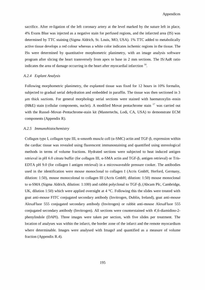
Appendices
195
sacrifice. After re-ligation of the left coronary artery at the level marked by the suture left in place,
4% Evans Blue was injected as a negative stain for perfused regions, and the infarcted area (IS) was
determined by TTC staining (Sigma Aldrich, St. Louis, MO, USA). 1% TTC added to metabolically
active tissue develops a red colour whereas a white color indicates ischemic regions in the tissue. The
ISs were determined by quantitative morphometric planimetry, with an image analysis software
program after slicing the heart transversely from apex to base in 2 mm sections. The IS/AaR ratio
indicates the area of damage occurring in the heart after myocardial infarction 16
.
A.2.4 Explant Analysis
Following morphometric planimetry, the explanted tissue was fixed for 12 hours in 10% formalin,
subjected to gradual serial dehydration and embedded in paraffin. The tissue was then sectioned in 3
μm thick sections. For general morphology serial sections were stained with haematoxylin–eosin
(H&E) stain (cellular components, nuclei). A modified Movat pentachrome stain 17
was carried out
with the Russel–Movat–Pentachrome-stain kit (Mastertechs, Lodi, CA, USA) to demonstrate ECM
components (Appendix R).
A.2.5 Immunohistochemistry
Collagen type I, collagen type III, α-smooth muscle cell (α-SMC) actin and TGF-β1 expression within
the cardiac tissue was revealed using fluorescent immunostaining and quantified using stereological
methods in terms of volume fractions. Hydrated sections were subjected to heat induced antigen
retrieval in pH 6.0 citrate buffer (for collagen III, α-SMA actin and TGF-β1 antigen retrieval) or Tris-
EDTA pH 9.0 (for collagen I antigen retrieval) in a microwaveable pressure cooker. The antibodies
used in the identification were mouse monoclonal to collagen I (Acris GmbH, Herford, Germany,
dilution: 1:50), mouse monocolonal to collagen III (Acris GmbH; dilution: 1:50) mouse monoclonal
to α-SMA (Sigma Aldrich, dilution: 1:100) and rabbit polyclonal to TGF-β1 (Abcam Plc, Cambridge,
UK, dilution 1:50) which were applied overnight at 4 °C. Following this the slides were treated with
goat anti-mouse FITC conjugated secondary antibody (Invitrogen, Dublin, Ireland), goat anti-mouse
AlexaFluor 555 conjugated secondary antibody (Invitrogen) or rabbit anti-mouse AlexaFluor 555
conjugated secondary antibody (Invitrogen). All sections were counterstained with 4',6-diamidino-2-
phenylindole (DAPI). Three images were taken per section, with five slides per treatment. The
location of analyses was within the infarct, the border zone of the infarct and the remote myocardium
where determinable. Images were analysed with ImageJ and quantified as a measure of volume
fraction (Appendix R.4).
Appendices
196
A.2.6 Apoptosis Detection Staining
Terminal deoxynucleotidyl transferase dUTP nick end labelling (TUNEL) in a commercially available
kit format (GenScript Corp, Piscataway, NJ, USA), was used to detect apoptotic cells within freshly
cut paraffin embedded sections as per the manufacturer‘s instructions. All sections were treated with
Proteinase K (20 μg/ml) for 10 minutes prior to blocking and staining steps. The negative control was
stained with the label solution without the TUNEL reaction solution to control for background
fluorescence and non-specific binding. As a positive control, a sample was incubated with DNase I to
degrade DNA in that sample. For staining, the sections were incubated with a TUNEL reaction
mixture, treated with a streptavidin-horse radish peroxidase solution, and finally the samples were
treated with a 3, 3'-Diaminobenzidine (DAB) working solution to develop the colour. All samples
were counterstained with Mayer‘s Haematoxylin and micrographed for stereological analysis using
ImageJ. Apoptosis was quantified as the number of brown (apoptotic) cells per field of view
(Appendix R.5).
A.2.7 In Situ Hybridization to Detect Delivered miR-29B
5'-Digoxigenin (DIG) labelled, LNA-modified oligonucleotide ISH probes were purchased from
Exiqon for miR-1 (positive control), miR-29B, U6 spliceosome (positive control) and a scrambled
LNA-modified oligonucleotide as background control. Formalin-fixed slides were hydrated in serial
alcohol concentrations and treated with Proteinase-K for 10 minutes at 37 °C. Sections were
dehydrated in quick incubations with various concentrations of ethanol. Sections were hybridized with
the LNA probes at various concentrations at the melting temperature (Tm) for one hour. Hybridized
sections were then washed with 50% formamide and 2 × SSC at the hybridization temperature.
Following 1 hour of blocking in 2% sheep serum, 2 mg/ ml bovine serum albumin in phosphate-
buffered saline (PBS) with 0.1% Tween, the slides were incubated with anti-DIG/ alkaline
phosphatase antibody/enzyme conjugate (1:800; Roche Diagnostics Ltd, Burgess Hill, UK) for one
hour at 30 °C. Following successive washes in PBS with 0.1% Tween, the sections were incubated
with nitroblue tetrazolium and 5- bromo-4-chloro-3-indoyl phosphate substrate (NBT-BCIP; Roche)
for two hours. The reaction was stopped by washing with PBS, and nuclei were counterstained with
Nuclear Fast Red.
A.2.8 Statistical Analysis
Results are depicted throughout as means ± standard deviation. Statistical analysis was performed
using Prism™
(GraphPad Software, La Jolla, CA, USA) and Student‘s t-test statistics to determine
statistically significant differences between miR-29B treatment and the control miR-239 treatment.
Statistical significance was set at p < 0.05.
Appendices
197
A.3. Results and Discussion
The results represent the findings from a pilot pre-clinical evaluation in n = 6 (miR-29B treatment
group) and n = 7 (negative control; miR-239B treatment group) animals surviving to 28 days. Given
this number, it is feasible to assume that statistically significant differences could be detected if these
numbers were increased to provide more power in this study. With this in mind, optimistic trends
were observed in many of the parameters investigated in this study. For instance; the left ventricular
ejection fraction (LVEF) % tended to be improved in the group treated with miR-29B at both 14 and
28 days (Figure A.6). No significant changes were observed in infarct size (Figure A.2) or wall
thickness (Figure A.4), nor were any correlations observed between LVEF% (Figure A.6) and the
morphometric or histological parameters. While functional improvements are often associated with
improved histological parameters, many other studies have failed to find statistically significant
effects on the morphometry of the myocardium despite observing changes in overall cardiac function
18-21. This disparity may be due to a number of factors. Firstly, histological infarct measurement
techniques are subject to several limitations. Thinning of the infarcted wall can lead to
underestimations of infarct volume, for example 22
. Furthermore, induction of MI by coronary artery
ligation yields highly variable infarct sizes 23
and because these parameters can only be measured
once the heart has been explanted for analysis, the initial infarct size is unknown. Thus, it is difficult
to assess the change in infarct size. Conversely, LVEF %, can be assessed frequently during the
course of the experiment. This allows continuous monitoring of cardiac function and comparisons
between time points.
The dosing regimen delivered in this study was a tail-vein injection at three days and at five days, and
a delivery of 50 μg of miR (suspended in 200 μl of RNA reconstitution buffer) at each time point. It is
very difficult to ascertain the appropriate dosing amount and regime in such a study as there is a
limited amount of information available in the literature with regard to therapeutic regimens of
siRNA. That being said, doses are reported at between 20 μg to 2 mg per Kg weight which equates to
approximately a 40 μg maximum dose in a mouse model, close to the 50 μg doses described in this
study 24
. One must take into consideration the actual amount of miRs that reach the intended target,
for instance in this case; the infarcted myocardium. Studies report that systemically delivered siRNA
tends to accumulate in the liver, kidneys, lungs, heart and spleen, with negligible traces of siRNA
present in other tissues 25-26
. The persistence of siRNAs delivered in vivo can be improved through
encapsulation within nanoparticles and liposomal formulations 25, 27
. Most in vivo studies with naked
siRNAs attempt to circumvent the problem of limited bio-distribution by administering high doses
directly to the target organ by injection 28
but in this initial study it was sought to determine the effect
(if any) of miR-29B on the infarcted myocardium before embarking on local delivery of the
therapeutic. Efforts were made at performing in situ hybridisation (ISH) of cardiac sections to
Appendices
198
visualise the delivered miR-29B by probing with a digozigenin (DIG) labelled locked nucleic acid
(LNA) against miR-29B (Figure A.16). ISH was performed on freshly cut sections of myocardium for
nuclear U6 spliceosomal RNA (as a method/user control), miR-1 which is highly expressed in the
myocardium (as a tissue control) and miR-29B which was delivered in this study. Additionally, ISH
was performed on human biopsies of cardiac tissue, cervical tissue and breast tissue as additional
controls. U6 spliceosomal RNA was detected in all tissue sections, however, its intensity was lower in
the mouse cardiac tissue explanted in this study when compared to the control tissues. miR-1 was not
detected in the mouse cardiac explants in this study but was detected in the human cardiac biopsies.
miR-29B was not detected in any tissue samples. Based on these observations in can be concluded
that the RNA in the mouse explants had degraded in the processing of the tissue. U6 spliceosomal
RNA was detected, as this is a highly expressed RNA and, being present in the nucleus, had an
additional protection against endogenous RNases but its weaker detection when compared to the
human biopsies indicates some degradation had occurred. Furthermore, detection of miR-1 was
achieved in the human cardiac biopsies whereas it was not detected in the explanted mouse cardiac
sections. No detection of this otherwise positive control lends further evidence that the RNA in the
mouse explants has been subjected to degradation. A review of the processing techniques indicates
that the RNA may have degraded during Evan‘s Blue/TTC staining as the RNAse free status of these
agents was not ensured and the human biopsies used as controls had never been subjected to such
processing. Future studies are now ensuring that all processing steps are RNase free for future miR-
29B detections and that possibly the RNA could be conjugated with a radioisotope or Cy™
3 for ease
of detection.
Infarct size was investigated at the day 28 time point using TTC and Evans Blue staining (Figure A.1)
and there was a trend towards a smaller infarct size (when normalised to the area at risk, Figure A.2).
One limitation of conventional infarct size estimates is that these estimates do not account for the
composition of the infarcted area and this can be a critical parameter in predicting cardiac function.
Employment of a modified Movat‘s Pentachrome stain 17
enabled the visualisation of many ECM
components following myocardial infarction (Figure A.3). For instance, muscle tissue composed
predominantly of cardiomyocytes stains red while mature collagens are stained yellow. There are
copius amounts of ECM structures staining blue, indicative of immature fibrosis both in the infarct
and remote myocardium. This tissue is most likely composed of glycoproteins such as fibronectin,
proteoglycans such as heparan sulfate, immature fibrillar collagens and non-fibrillar collagens (for
example, type IV collagen) 29
. Although 28 days is a standard end-point in pre-clinical models of MI
22, 30-33, it would be appropriate to repeat this study with additional sacrificial time points; one at an
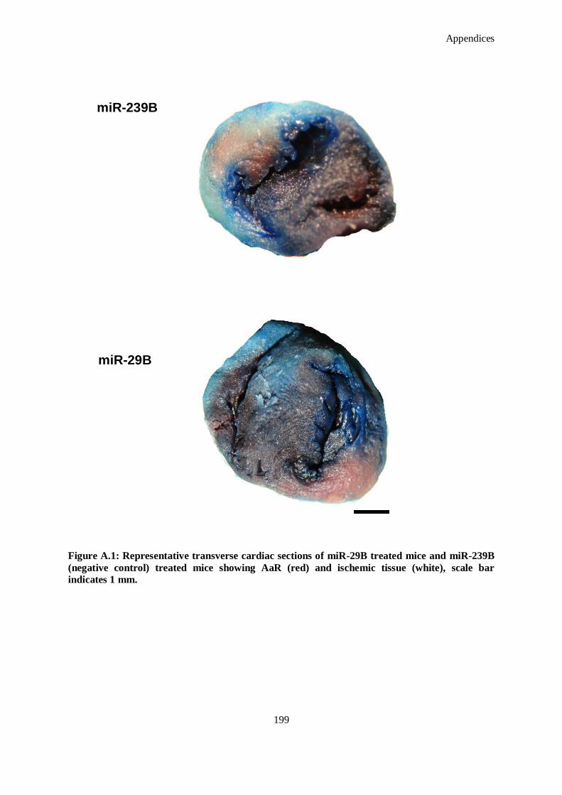
Appendices
199
Figure A.1: Representative transverse cardiac sections of miR-29B treated mice and miR-239B
(negative control) treated mice showing AaR (red) and ischemic tissue (white), scale bar
indicates 1 mm.
miR-239B
miR-29B
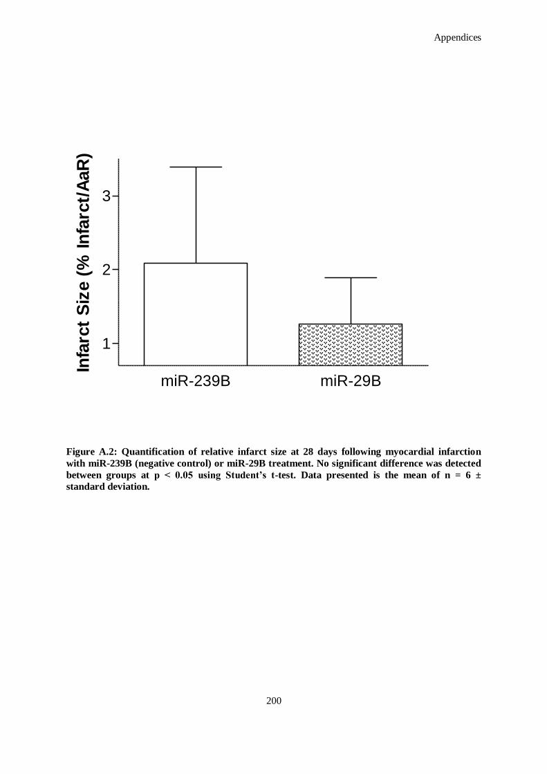
Appendices
200
Figure A.2: Quantification of relative infarct size at 28 days following myocardial infarction
with miR-239B (negative control) or miR-29B treatment. No significant difference was detected
between groups at p < 0.05 using Student’s t-test. Data presented is the mean of n = 6 ±
standard deviation.
miR-239B miR-29B
1
2
3
Infa
rct
Siz
e (
% In
farc
t/A
aR
)
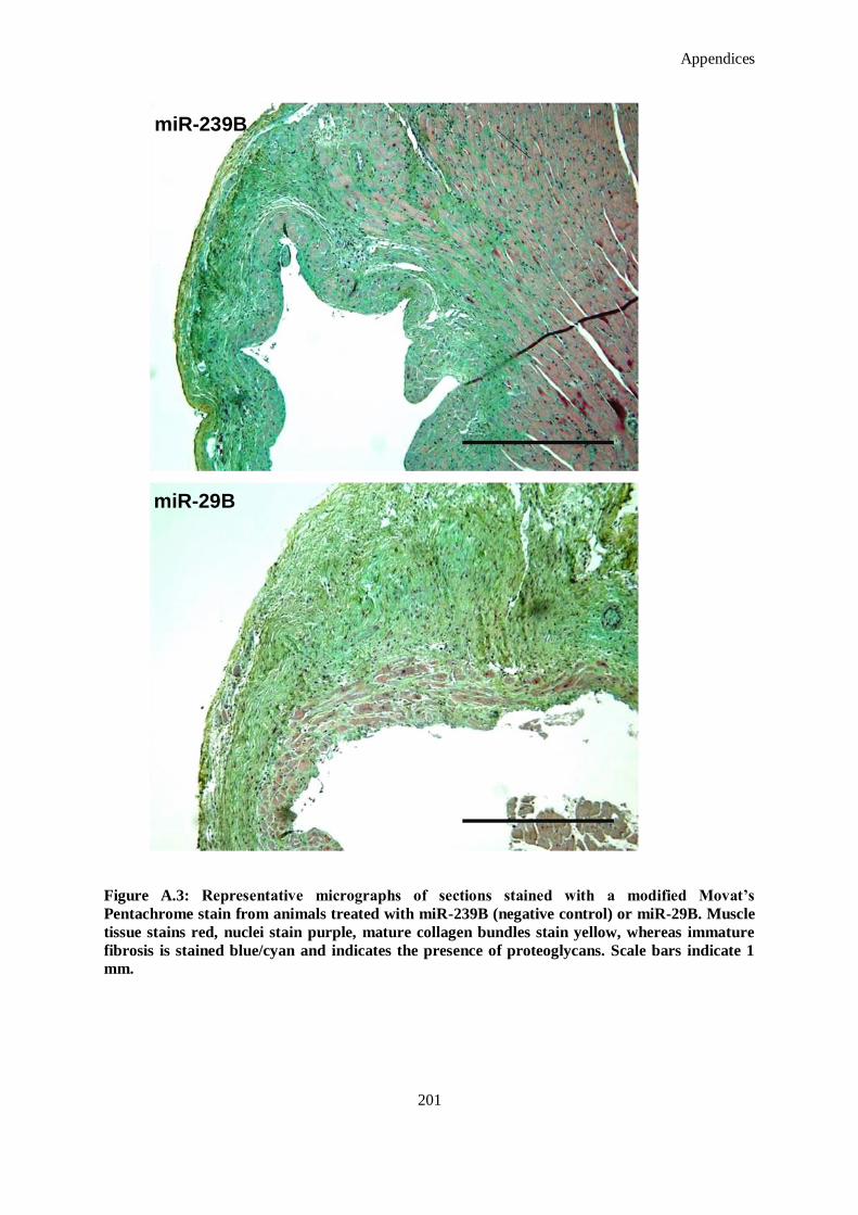
Appendices
201
Figure A.3: Representative micrographs of sections stained with a modified Movat’s
Pentachrome stain from animals treated with miR-239B (negative control) or miR-29B. Muscle
tissue stains red, nuclei stain purple, mature collagen bundles stain yellow, whereas immature
fibrosis is stained blue/cyan and indicates the presence of proteoglycans. Scale bars indicate 1
mm.
miR-239B
miR-29B
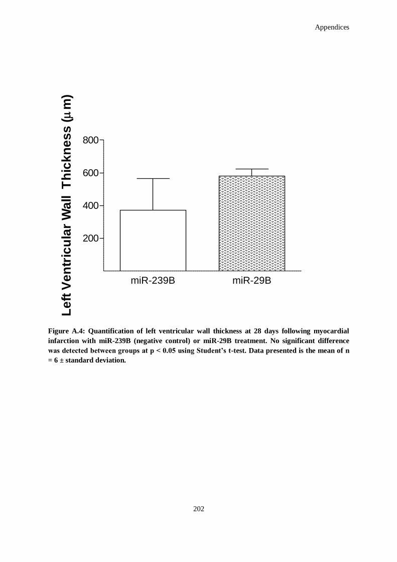
Appendices
202
miR-239B miR-29B
200
400
600
800
Le
ft V
en
tric
ula
r W
all T
hic
kn
es
s (
m)
Figure A.4: Quantification of left ventricular wall thickness at 28 days following myocardial
infarction with miR-239B (negative control) or miR-29B treatment. No significant difference
was detected between groups at p < 0.05 using Student’s t-test. Data presented is the mean of n
= 6 ± standard deviation.
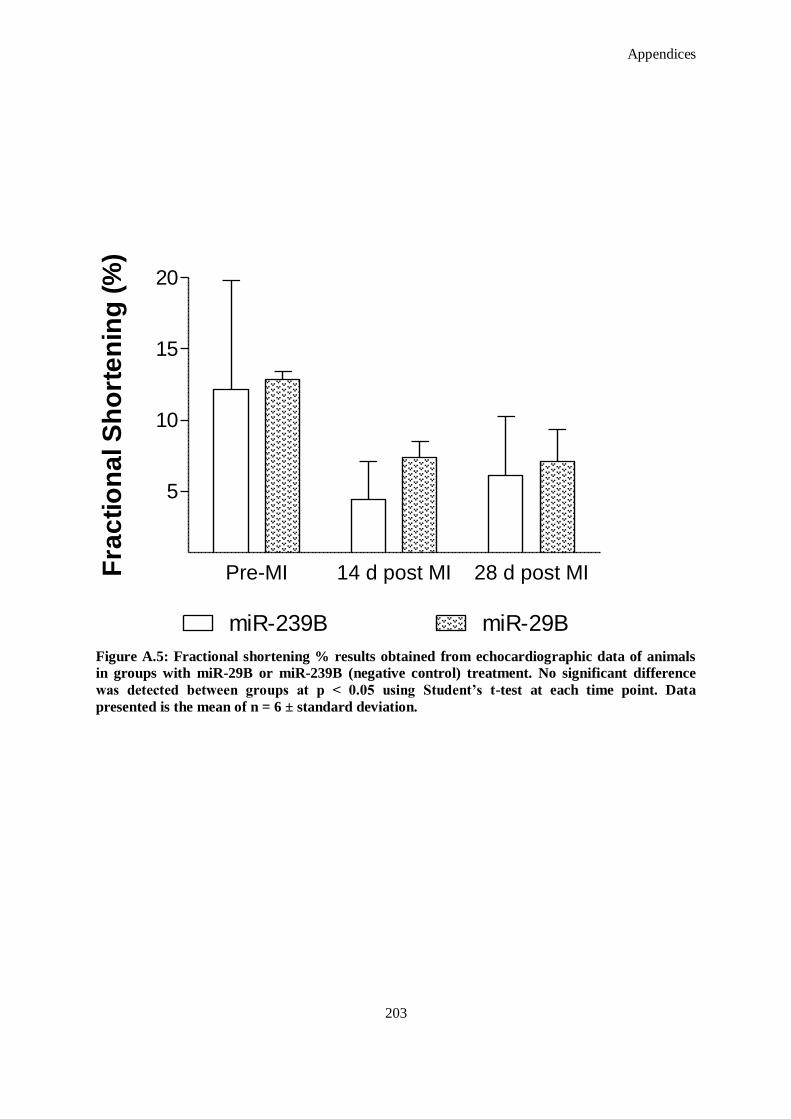
Appendices
203
Figure A.5: Fractional shortening % results obtained from echocardiographic data of animals
in groups with miR-29B or miR-239B (negative control) treatment. No significant difference
was detected between groups at p < 0.05 using Student’s t-test at each time point. Data
presented is the mean of n = 6 ± standard deviation.
Pre-MI 14 d post MI 28 d post MI
5
10
15
20
miR-239B miR-29B
Fra
cti
on
al S
ho
rte
nin
g (
%)
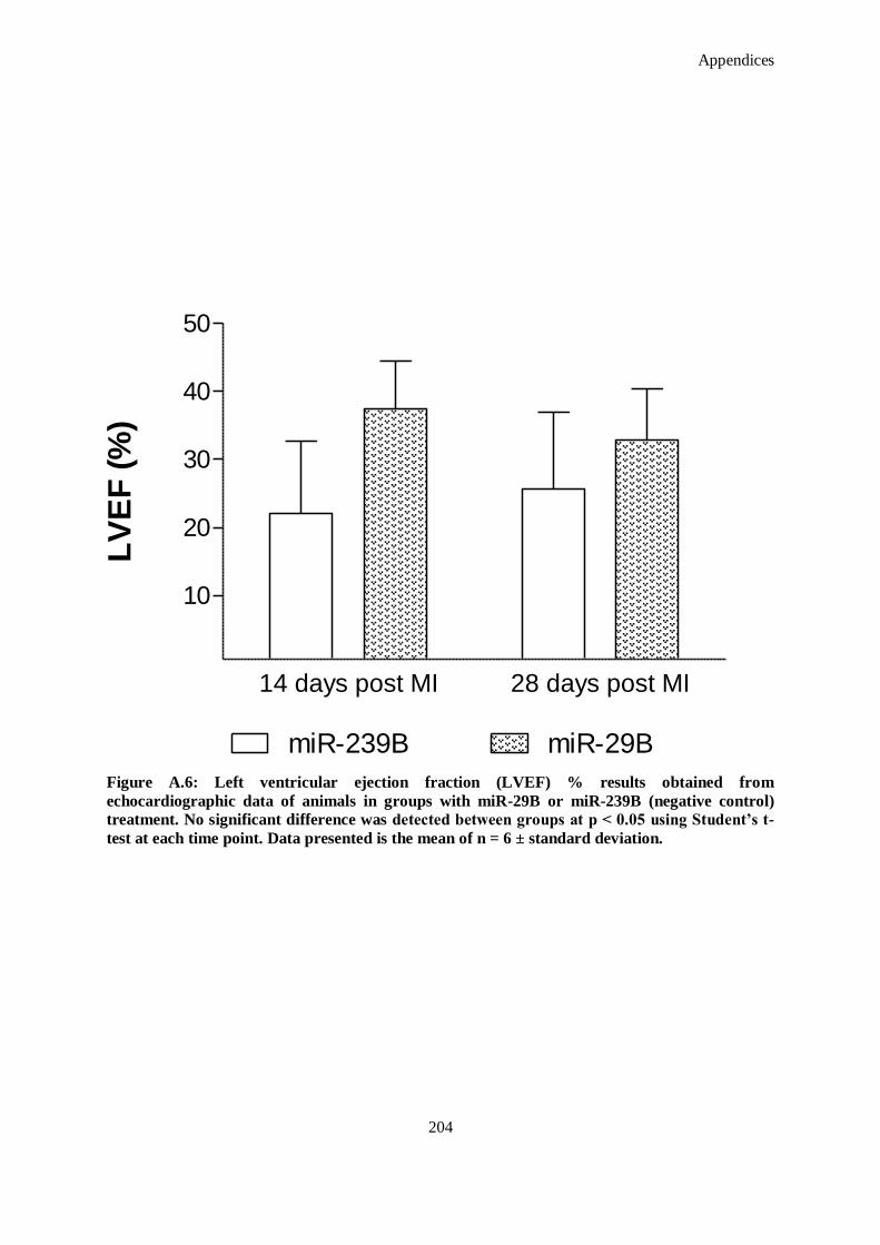
Appendices
204
Figure A.6: Left ventricular ejection fraction (LVEF) % results obtained from
echocardiographic data of animals in groups with miR-29B or miR-239B (negative control)
treatment. No significant difference was detected between groups at p < 0.05 using Student’s t-
test at each time point. Data presented is the mean of n = 6 ± standard deviation.
14 days post MI 28 days post MI
10
20
30
40
50
miR-239B miR-29B
LV
EF
(%
)
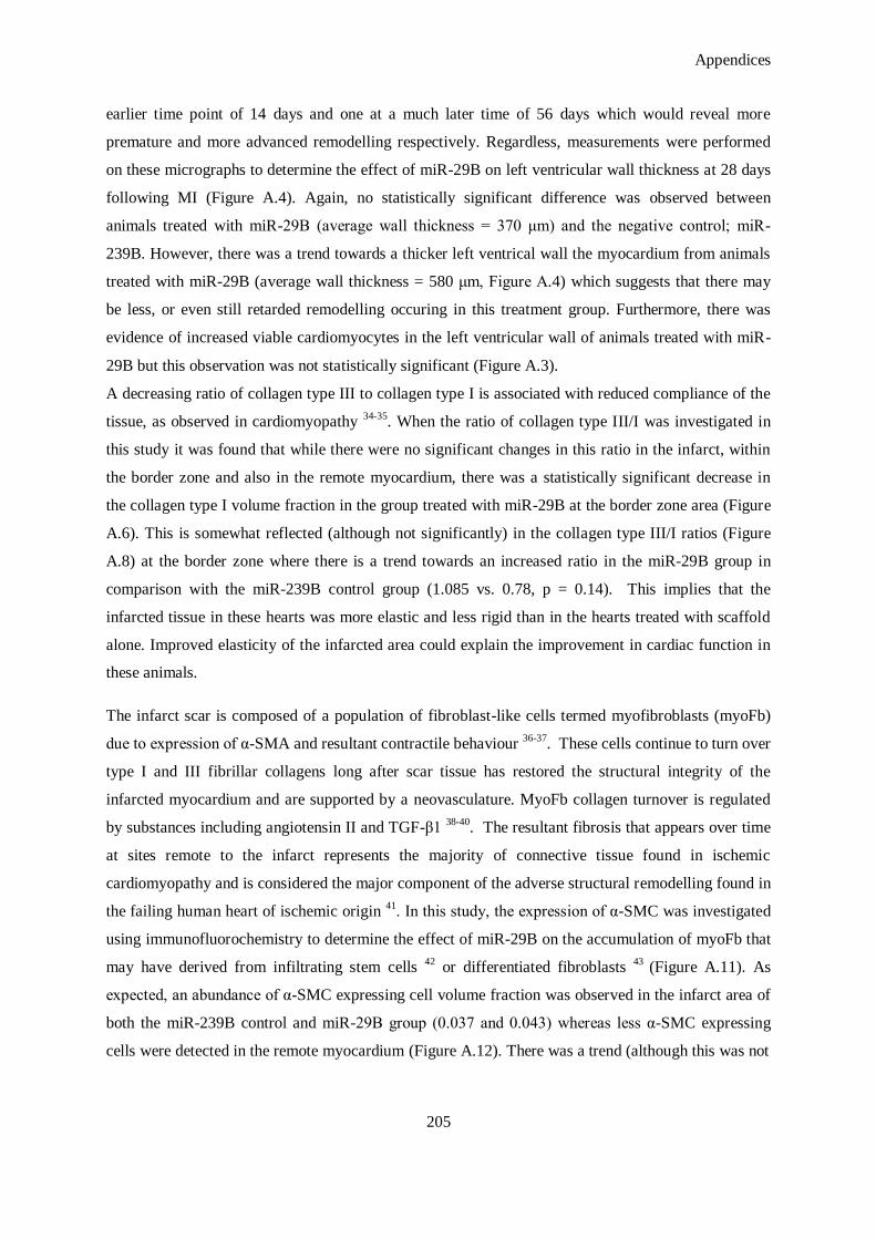
Appendices
205
earlier time point of 14 days and one at a much later time of 56 days which would reveal more
premature and more advanced remodelling respectively. Regardless, measurements were performed
on these micrographs to determine the effect of miR-29B on left ventricular wall thickness at 28 days
following MI (Figure A.4). Again, no statistically significant difference was observed between
animals treated with miR-29B (average wall thickness = 370 μm) and the negative control; miR-
239B. However, there was a trend towards a thicker left ventrical wall the myocardium from animals
treated with miR-29B (average wall thickness = 580 μm, Figure A.4) which suggests that there may
be less, or even still retarded remodelling occuring in this treatment group. Furthermore, there was
evidence of increased viable cardiomyocytes in the left ventricular wall of animals treated with miR-
29B but this observation was not statistically significant (Figure A.3).
A decreasing ratio of collagen type III to collagen type I is associated with reduced compliance of the
tissue, as observed in cardiomyopathy 34-35
. When the ratio of collagen type III/I was investigated in
this study it was found that while there were no significant changes in this ratio in the infarct, within
the border zone and also in the remote myocardium, there was a statistically significant decrease in
the collagen type I volume fraction in the group treated with miR-29B at the border zone area (Figure
A.6). This is somewhat reflected (although not significantly) in the collagen type III/I ratios (Figure
A.8) at the border zone where there is a trend towards an increased ratio in the miR-29B group in
comparison with the miR-239B control group (1.085 vs. 0.78, p = 0.14). This implies that the
infarcted tissue in these hearts was more elastic and less rigid than in the hearts treated with scaffold
alone. Improved elasticity of the infarcted area could explain the improvement in cardiac function in
these animals.
The infarct scar is composed of a population of fibroblast-like cells termed myofibroblasts (myoFb)
due to expression of α-SMA and resultant contractile behaviour 36-37
. These cells continue to turn over
type I and III fibrillar collagens long after scar tissue has restored the structural integrity of the
infarcted myocardium and are supported by a neovasculature. MyoFb collagen turnover is regulated
by substances including angiotensin II and TGF-β1 38-40
. The resultant fibrosis that appears over time
at sites remote to the infarct represents the majority of connective tissue found in ischemic
cardiomyopathy and is considered the major component of the adverse structural remodelling found in
the failing human heart of ischemic origin 41
. In this study, the expression of α-SMC was investigated
using immunofluorochemistry to determine the effect of miR-29B on the accumulation of myoFb that
may have derived from infiltrating stem cells 42
or differentiated fibroblasts 43
(Figure A.11). As
expected, an abundance of α-SMC expressing cell volume fraction was observed in the infarct area of
both the miR-239B control and miR-29B group (0.037 and 0.043) whereas less α-SMC expressing
cells were detected in the remote myocardium (Figure A.12). There was a trend (although this was not
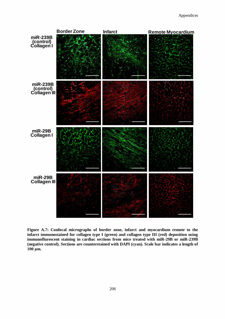
Appendices
206
Figure A.7: Confocal micrographs of border zone, infarct and myocardium remote to the
infarct immunostained for collagen type I (green) and collagen type III (red) deposition using
immunoflurescent staining in cardiac sections from mice treated with miR-29B or miR-239B
(negative control). Sections are counterstained with DAPI (cyan). Scale bar indicates a length of
100 μm.
Border Zone Infarct Remote MyocardiummiR-239B(control)
Collagen I
miR-29BCollagen I
miR-239B(control)
Collagen III
miR-29BCollagen III
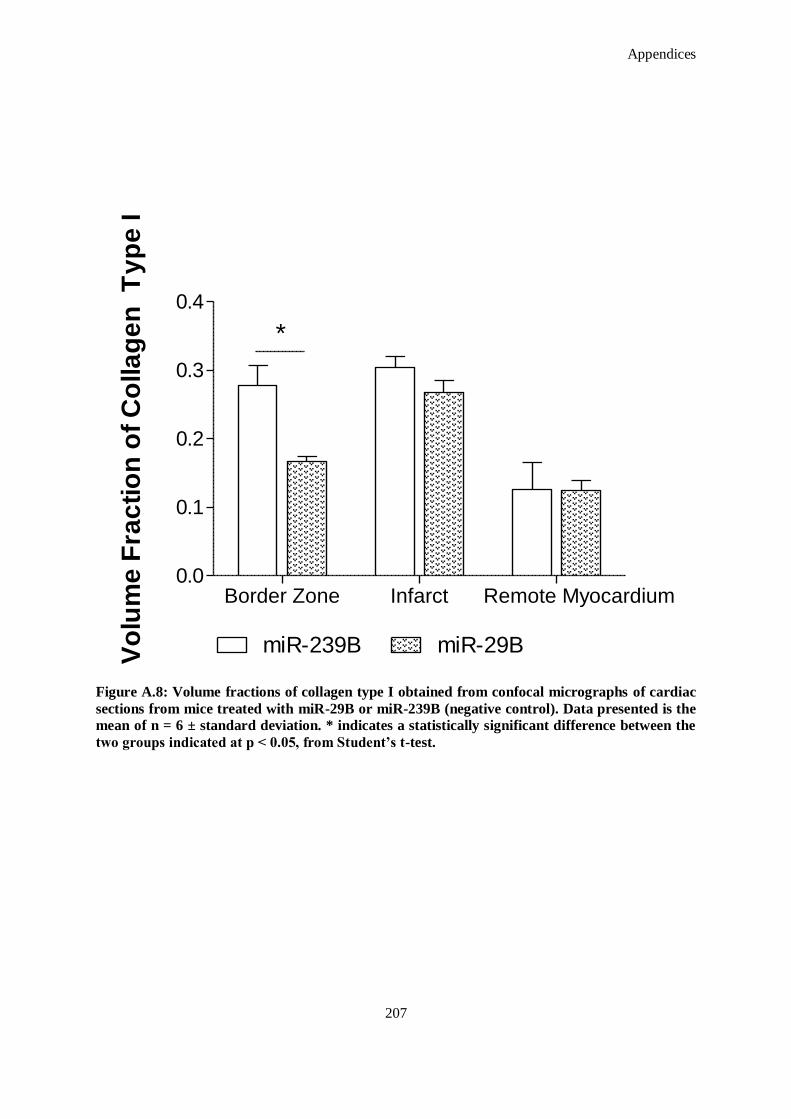
Appendices
207
Border Zone Infarct Remote Myocardium0.0
0.1
0.2
0.3
0.4
miR-239B miR-29B
*
Vo
lum
e F
rac
tio
n o
f C
olla
ge
n T
yp
e I
Figure A.8: Volume fractions of collagen type I obtained from confocal micrographs of cardiac
sections from mice treated with miR-29B or miR-239B (negative control). Data presented is the
mean of n = 6 ± standard deviation. * indicates a statistically significant difference between the
two groups indicated at p < 0.05, from Student’s t-test.
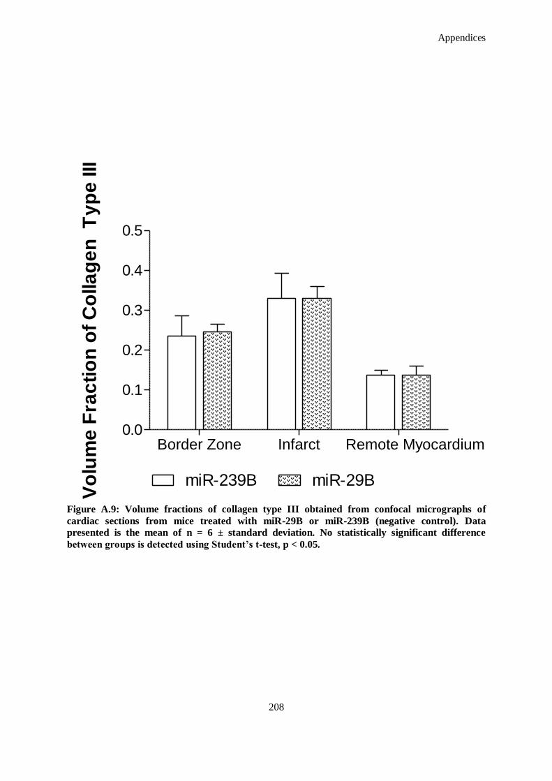
Appendices
208
Figure A.9: Volume fractions of collagen type III obtained from confocal micrographs of
cardiac sections from mice treated with miR-29B or miR-239B (negative control). Data
presented is the mean of n = 6 ± standard deviation. No statistically significant difference
between groups is detected using Student’s t-test, p < 0.05.
Border Zone Infarct Remote Myocardium0.0
0.1
0.2
0.3
0.4
0.5
miR-239B miR-29B
Vo
lum
e F
rac
tio
n o
f C
olla
ge
n T
yp
e III
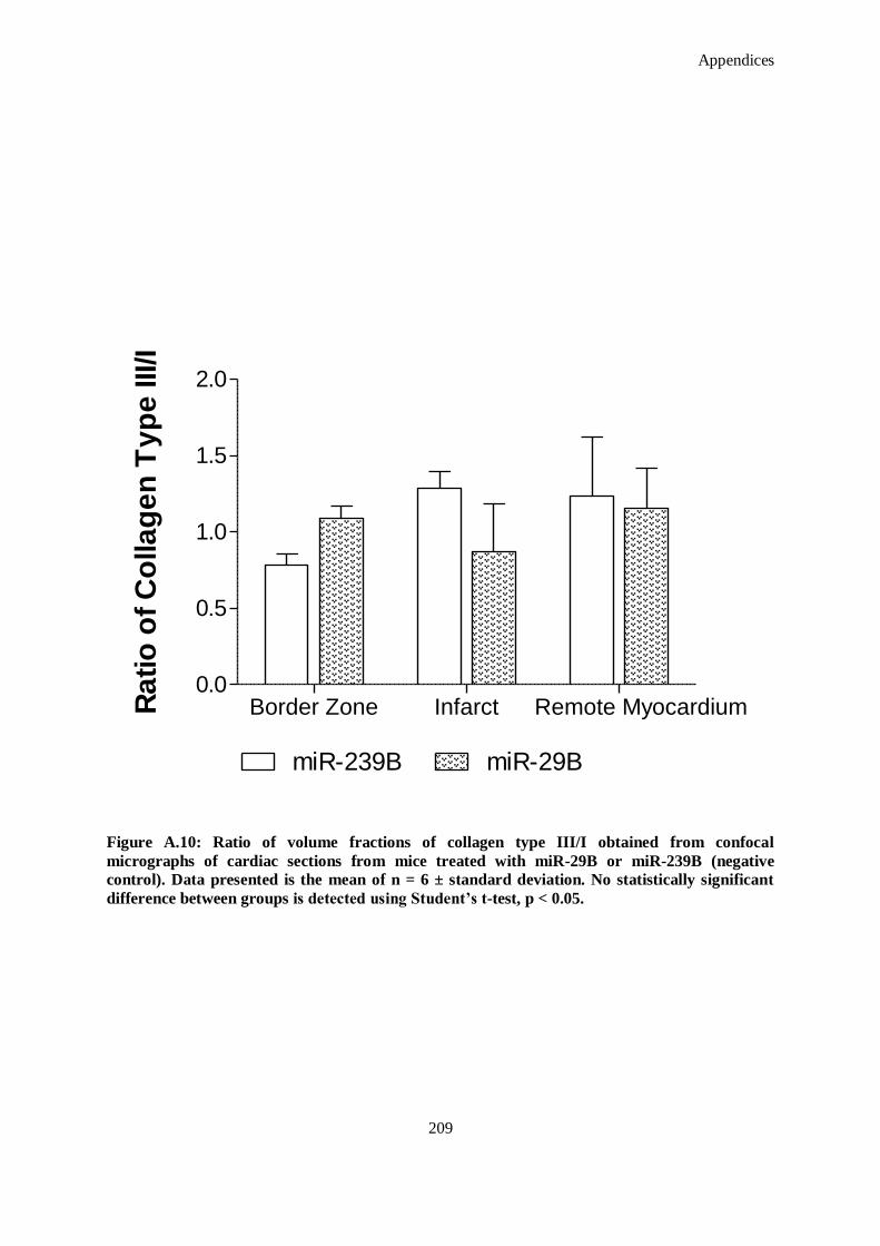
Appendices
209
Border Zone Infarct Remote Myocardium0.0
0.5
1.0
1.5
2.0
miR-239B miR-29B
Ra
tio
of
Co
lla
ge
n T
yp
e III/I
Figure A.10: Ratio of volume fractions of collagen type III/I obtained from confocal
micrographs of cardiac sections from mice treated with miR-29B or miR-239B (negative
control). Data presented is the mean of n = 6 ± standard deviation. No statistically significant
difference between groups is detected using Student’s t-test, p < 0.05.

Appendices
210
Figure A.11: Micrographs of infarct and remote myocardium following treatment with miR-
29B or miR-239B (negative control). Sections are stained for α-SMA using immunoflurescent
staining (red) and counterstained with DAPI (cyan). Scale bar indicates a length of 200 μm
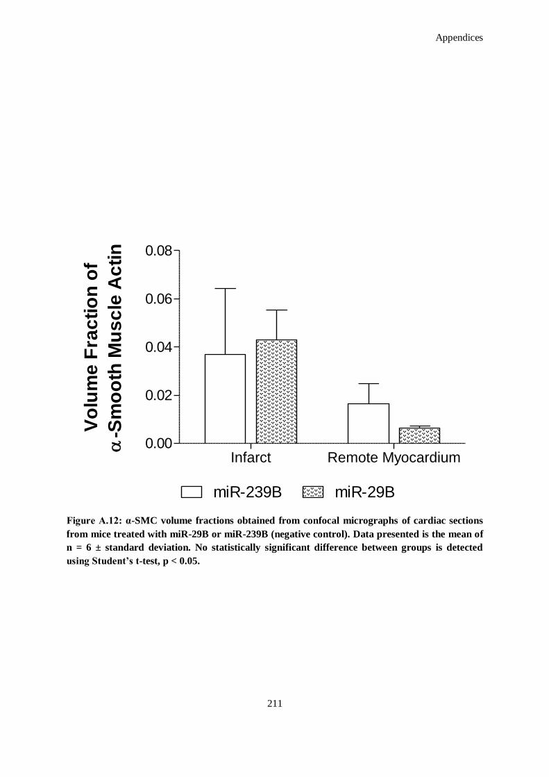
Appendices
211
Infarct Remote Myocardium0.00
0.02
0.04
0.06
0.08
miR-239B miR-29B
Vo
lum
e F
rac
tio
n o
f
-S
mo
oth
Mu
sc
le A
cti
n
Figure A.12: α-SMC volume fractions obtained from confocal micrographs of cardiac sections
from mice treated with miR-29B or miR-239B (negative control). Data presented is the mean of
n = 6 ± standard deviation. No statistically significant difference between groups is detected
using Student’s t-test, p < 0.05.
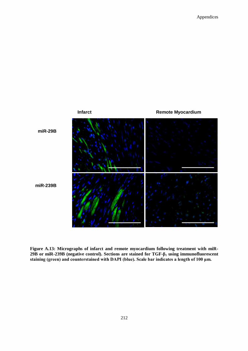
Appendices
212
Figure A.13: Micrographs of infarct and remote myocardium following treatment with miR-
29B or miR-239B (negative control). Sections are stained for TGF-β1 using immunofluorescent
staining (green) and counterstained with DAPI (blue). Scale bar indicates a length of 100 μm.
miR-29B
miR-239B
Infarct Remote Myocardium
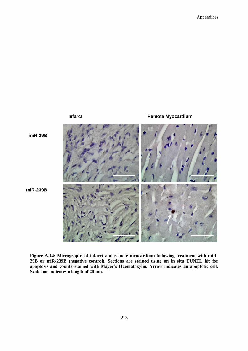
Appendices
213
Figure A.14: Micrographs of infarct and remote myocardium following treatment with miR-
29B or miR-239B (negative control). Sections are stained using an in situ TUNEL kit for
apoptosis and counterstained with Mayer’s Haematoxylin. Arrow indicates an apoptotic cell.
Scale bar indicates a length of 20 μm.
miR-29B
miR-239B
Infarct Remote Myocardium
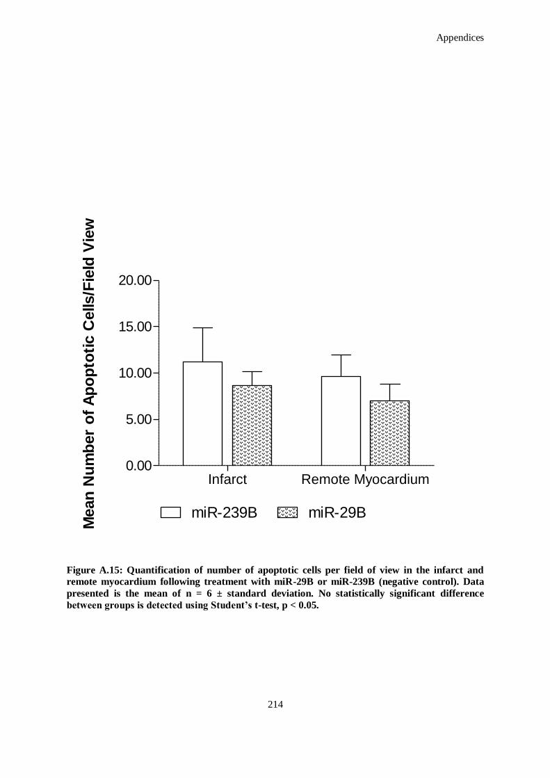
Appendices
214
Infarct Remote Myocardium0.00
5.00
10.00
15.00
20.00
miR-239B miR-29B
Mean
Nu
mb
er
of
Ap
op
toti
c C
ells/F
ield
Vie
w
Figure A.15: Quantification of number of apoptotic cells per field of view in the infarct and
remote myocardium following treatment with miR-29B or miR-239B (negative control). Data
presented is the mean of n = 6 ± standard deviation. No statistically significant difference
between groups is detected using Student’s t-test, p < 0.05.
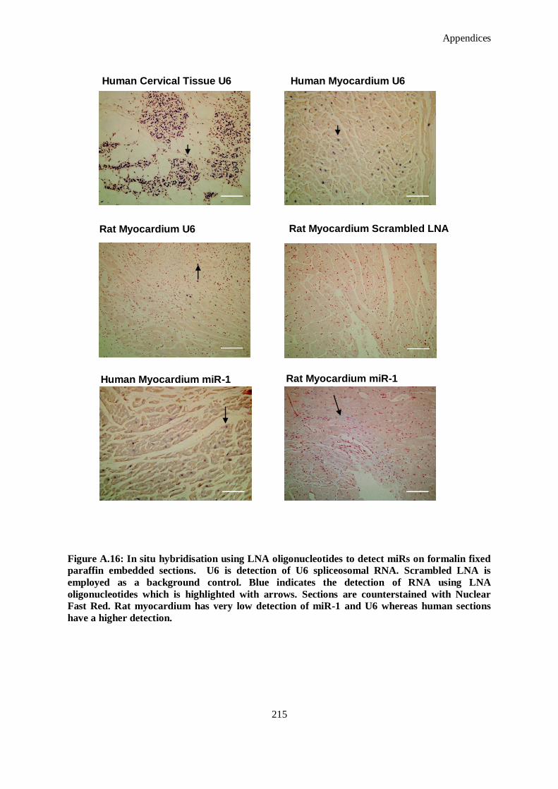
Appendices
215
Figure A.16: In situ hybridisation using LNA oligonucleotides to detect miRs on formalin fixed
paraffin embedded sections. U6 is detection of U6 spliceosomal RNA. Scrambled LNA is
employed as a background control. Blue indicates the detection of RNA using LNA
oligonucleotides which is highlighted with arrows. Sections are counterstained with Nuclear
Fast Red. Rat myocardium has very low detection of miR-1 and U6 whereas human sections
have a higher detection.
Human Cervical Tissue U6 Human Myocardium U6
Rat Myocardium Scrambled LNA
Human Myocardium miR-1
Rat Myocardium U6
Rat Myocardium miR-1
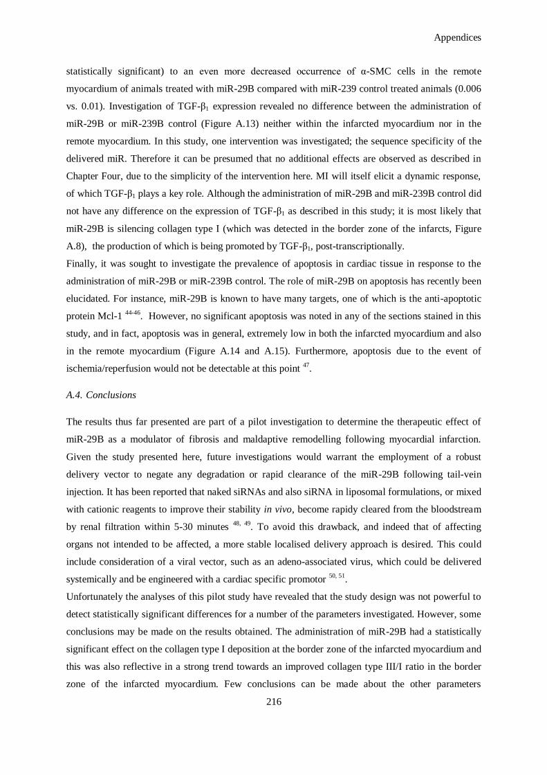
Appendices
216
statistically significant) to an even more decreased occurrence of α-SMC cells in the remote
myocardium of animals treated with miR-29B compared with miR-239 control treated animals (0.006
vs. 0.01). Investigation of TGF-β1 expression revealed no difference between the administration of
miR-29B or miR-239B control (Figure A.13) neither within the infarcted myocardium nor in the
remote myocardium. In this study, one intervention was investigated; the sequence specificity of the
delivered miR. Therefore it can be presumed that no additional effects are observed as described in
Chapter Four, due to the simplicity of the intervention here. MI will itself elicit a dynamic response,
of which TGF-β1 plays a key role. Although the administration of miR-29B and miR-239B control did
not have any difference on the expression of TGF-β1 as described in this study; it is most likely that
miR-29B is silencing collagen type I (which was detected in the border zone of the infarcts, Figure
A.8), the production of which is being promoted by TGF-β1, post-transcriptionally.
Finally, it was sought to investigate the prevalence of apoptosis in cardiac tissue in response to the
administration of miR-29B or miR-239B control. The role of miR-29B on apoptosis has recently been
elucidated. For instance, miR-29B is known to have many targets, one of which is the anti-apoptotic
protein Mcl-1 44-46
. However, no significant apoptosis was noted in any of the sections stained in this
study, and in fact, apoptosis was in general, extremely low in both the infarcted myocardium and also
in the remote myocardium (Figure A.14 and A.15). Furthermore, apoptosis due to the event of
ischemia/reperfusion would not be detectable at this point 47
.
A.4. Conclusions
The results thus far presented are part of a pilot investigation to determine the therapeutic effect of
miR-29B as a modulator of fibrosis and maldaptive remodelling following myocardial infarction.
Given the study presented here, future investigations would warrant the employment of a robust
delivery vector to negate any degradation or rapid clearance of the miR-29B following tail-vein
injection. It has been reported that naked siRNAs and also siRNA in liposomal formulations, or mixed
with cationic reagents to improve their stability in vivo, become rapidy cleared from the bloodstream
by renal filtration within 5-30 minutes 48, 49
. To avoid this drawback, and indeed that of affecting
organs not intended to be affected, a more stable localised delivery approach is desired. This could
include consideration of a viral vector, such as an adeno-associated virus, which could be delivered
systemically and be engineered with a cardiac specific promotor 50, 51
.
Unfortunately the analyses of this pilot study have revealed that the study design was not powerful to
detect statistically significant differences for a number of the parameters investigated. However, some
conclusions may be made on the results obtained. The administration of miR-29B had a statistically
significant effect on the collagen type I deposition at the border zone of the infarcted myocardium and
this was also reflective in a strong trend towards an improved collagen type III/I ratio in the border
zone of the infarcted myocardium. Few conclusions can be made about the other parameters
Appendices
217
investigated as there exist no statistically significant differences. However, miR-29B had an apparent
trend towards an improvement on LVEF % which can be attributed to improved mechanical
compliance at the border zone of the infarct due to an increased collagen type III/I ratio. Finally, these
results provide an insight into further parameters to be investigated in future studies and highlight the
requirement of a localised delivery system to circumnavigate possible off-target effects and clearance
of a systemically delivered therapeutic.
The cardiac study was somewhat limited in its applicability. For example, a mouse heart weighs less
than a gram, while a human heart generally weighs more than 200 g. The difference in mass is
essentially correlated in the difference in the wall thickness (~ 1 mm vs. ~1 cm). The cellular
composition of the myocardium; however, do not differ, and thus the host response is somewhat
representative of the response a human heart would have to the treatment. Additionally, the MI model
in this study was created by ligating the LAD of an otherwise healthy animal. In humans, occlusion of
the coronary artery is a slow process, onset by underlying atherosclerosis, and present (in the majority
of cases) in individuals with sedentary lifestyles and unhealthy diets. Where possible, MI is treated by
reperfusing the affected tissue, an event which was simulated in this model.
The study was powered to detect changes in left ventricular ejection fraction (LVEF %) but not in
wall thickness or other histological parameters. The number of animals in each treatment group
should be at least ten to provide such power. Thus, there may have been changes in the histological
parameters that were not statistically significant due to the small sample size in this pilot study. It was
decided to investigate first the systemic delivery of miR-29B which is reported here. Intramyocardial
delivery is envisaged and is discussed in the Future Directions Section of this Thesis.
Appendices
218
A.5. References
1. Cleutjens JP, Kandala JC, Guarda E, Guntaka RV, Weber KT. Regulation of collagen degradation in the rat myocardium after infarction. J. Mol. Cell Cardiol. 1995;27:1281-1292
2. Sun Y, Zhang JQ, Zhang J, Lamparter S. Cardiac remodeling by fibrous tissue after infarction
in rats. J. Lab Clin. Med. 2000;135:316-323
3. Sun Y, Cleutjens JP, Diaz-Arias AA, Weber KT. Cardiac angiotensin converting enzyme and myocardial fibrosis in the rat. Cardiovasc. Res. 1994;28:1423-1432
4. Berk BC, Fujiwara K, Lehoux S. ECM remodeling in hypertensive heart disease. J. Clin.
Invest. 2007;117:568-575 5. Souders CA, Bowers SL, Baudino TA. Cardiac fibroblast: The renaissance cell. Circ. Res.
2009;105:1164-1176
6. Creemers EE, Pinto YM. Molecular mechanisms that control interstitial fibrosis in the pressure-overloaded heart. Cardiovasc. Res. 2011;89:265-272
7. Liu Y, Taylor NE, Lu L, Usa K, Cowley AW, Jr, Ferreri NR, Yeo NC, Liang M. Renal
medullary microRNAs in DAHL salt-sensitive rats: MiR-29b regulates several collagens and
related genes. Hypertension. 2010;55:974-982 8. Li Z, Hassan MQ, Jafferji M, Aqeilan RI, Garzon R, Croce CM, van Wijnen AJ, Stein JL, Stein
GS, Lian JB. Biological functions of miR-29b contribute to positive regulation of osteoblast
differentiation. J. Biol. Chem. 2009;284:15676-15684 9. Cushing L, Kuang PP, Qian J, Shao F, Wu J, Little F, Thannickal VJ, Cardoso WV, Lu J. MiR-
29 is a major regulator of genes associated with pulmonary fibrosis. Am. J. Respir. Cell Mol.
Biol. 2010:2010-0323OC
10. Roderburg C, Urban G-W, Bettermann K, Vucur M, Zimmermann H, Schmidt S, Janssen J, Koppe C, Knolle P, Castoldi M, Tacke F, Trautwein C, Luedde T. Micro-RNA profiling reveals
a role for miR-29 in human and murine liver fibrosis. Hepatology. 2011;53:209-218
11. van Rooij E, Sutherland LB, Thatcher JE, DiMaio JM, Naseem RH, Marshall WS, Hill JA, Olson EN. Dysregulation of microRNAs after myocardial infarction reveals a role of miR-29 in
cardiac fibrosis. Proc. Nat. Acad. Sci.U S A. 2008;105:13027-13032
12. Li Z, Hassan MQ, Jafferji M, Aqeilan RI, Garzon R, Croce CM, van Wijnen AJ, Stein JL, Stein GS, Lian JB. Biological functions of miR-29b contribute to positive regulation of osteoblast
differentiation. J. Biol. Chem. 2009;284:15676-15684
13. Liu Y, Taylor NE, Lu L, Usa K, Cowley AW, Jr., Ferreri NR, Yeo NC, Liang M. Renal
medullary microRNAs in dahl salt-sensitive rats: MiR-29b regulates several collagens and related genes. Hypertension. 2010;55:974-982
14. Sengupta S, den Boon JA, Chen IH, Newton MA, Stanhope SA, Cheng YJ, Chen CJ,
Hildesheim A, Sugden B, Ahlquist P. MicroRNA 29c is down-regulated in nasopharyngeal carcinomas, up-regulating mRNAs encoding extracellular matrix proteins. Proc. Natl. Acad.
Sci. USA. 2008;105:5874-5878
15. Park SY, Lee JH, Ha M, Nam JW, Kim VN. MiR-29 miRNAs activate p53 by targeting p85α and CDC42. Nat. Struct. Mol. Biol. 2009;16:23-29
16. Borst O, Ochmann C, Schönberger T, Jacoby C, Stellos K, Seizer P, Flögel U, Lang F, Gawaz
M. Methods employed for induction and analysis of experimental myocardial infarction in
mice. Cell. Physiol. Biochem. 2011;28:1-12 17. Russell HK. A modification of movat's pentachrome stain. Arch. Pathol. 1972;94:187-191
18. Mizuno T, Yau TM, Weisel RD, Kiani CG, Li RK. Elastin stabilizes an infarct and preserves
ventricular function. Circulation. 2005;112:I81-88 19. Dai W, Hale SL, Kay GL, Jyrala AJ, Kloner RA. Delivering stem cells to the heart in a
collagen matrix reduces relocation of cells to other organs as assessed by nanoparticle
technology. Regen. Med. 2009;4:387-395
20. Derval N, Barandon L, Dufourcq P, Leroux L, Lamaziere JM, Daret D, Couffinhal T, Duplaa C. Epicardial deposition of endothelial progenitor and mesenchymal stem cells in a coated
Appendices
219
muscle patch after myocardial infarction in a murine model. Eur. J. Cardiothorac. Surg.
2008;34:248-254 21. Giraud MN, Ayuni E, Cook S, Siepe M, Carrel TP, Tevaearai HT. Hydrogel-based engineered
skeletal muscle grafts normalize heart function early after myocardial infarction. Artif. Organs.
2008;32:692-700
22. Takagawa J, Zhang Y, Wong ML, Sievers RE, Kapasi NK, Wang Y, Yeghiazarians Y, Lee RJ, Grossman W, Springer ML. Myocardial infarct size measurement in the mouse chronic
infarction model: Comparison of area- and length-based approaches. J. Appl. Physiol.
2007;102:2104-2111 23. van den Bos EJ, Mees BM, de Waard MC, de Crom R, Duncker DJ. A novel model of
cryoinjury-induced myocardial infarction in the mouse: A comparison with coronary artery
ligation. Am. J. Physiol. Heart Circ. Physiol. 2005;289:H1291-1300 24. Kim SS, Garg H, Joshi A, Manjunath N. Strategies for targeted nonviral delivery of siRNAs in
vivo. Trends Mol. Med. 2009;15:491-500
25. Shi B, Keough E, Matter A, Leander K, Young S, Carlini E, Sachs AB, Tao W, Abrams M,
Howell B, Sepp-Lorenzino L. Biodistribution of small interfering RNA at the organ and cellular levels after lipid nanoparticle-mediated delivery. J. Histochem. Cytochem.
2011;59:727-740
26. Soutschek J, Akinc A, Bramlage B, Charisse K, Constien R, Donoghue M, Elbashir S, Geick A, Hadwiger P, Harborth J, John M, Kesavan V, Lavine G, Pandey RK, Racie T, Rajeev KG, Rohl
I, Toudjarska I, Wang G, Wuschko S, Bumcrot D, Koteliansky V, Limmer S, Manoharan M,
Vornlocher HP. Therapeutic silencing of an endogenous gene by systemic administration of modified siRNAs. Nature. 2004;432:173-178
27. Alvarez-Erviti L, Seow Y, Yin H, Betts C, Lakhal S, Wood MJA. Delivery of siRNA to the
mouse brain by systemic injection of targeted exosomes. Nat. Biotech. 2011;29:341-345
28. Hamar P, Song E, Kokeny G, Chen A, Ouyang N, Lieberman J. Small interfering RNA targeting Fas protects mice against renal ischemia-reperfusion injury. Proc. Natl. Acad. Sci. U
S A. 2004;101:14883-14888
29. Czubryt MP. Common threads in cardiac fibrosis, infarct scar formation, and wound healing. Fibrogen. Tissue Rep.. 2012;5:19
30. Trueblood NA, Xie Z, Communal C, Sam F, Ngoy S, Liaw L, Jenkins AW, Wang J, Sawyer
DB, Bing OHL, Apstein CS, Colucci WS, Singh K. Exaggerated left ventricular dilation and
reduced collagen deposition after myocardial infarction in mice lacking osteopontin. Circ. Res. 2001;88:1080-1087
31. Wu JC, Nasseri BA, Bloch KD, Picard MH, Scherrer-Crosbie M. Influence of sex on
ventricular remodeling after myocardial infarction in mice. J. Am. Soc. Echocardiogr. 2003;16:1158-1162
32. van den Borne SWM, van de Schans VAM, Strzelecka AE, Vervoort-Peters HTM, Lijnen PM,
Cleutjens JPM, Smits JFM, Daemen MJAP, Janssen BJA, Blankesteijn WM. Mouse strain determines the outcome of wound healing after myocardial infarction. Cardiovasc. Res.
2009;84:273-282
33. Gao E, Lei YH, Shang X, Huang ZM, Zuo L, Boucher M, Fan Q, Chuprun JK, Ma XL, Koch
WJ. A novel and efficient model of coronary artery ligation and myocardial infarction in the mouse. Circ. Res. 2010;107:1445-1453
34. Kakkar R, Lee RT. Intramyocardial fibroblast myocyte communication. Circ. Res.
2010;106:47-57 35. Marijianowski MM, Teeling P, Mann J, Becker AE. Dilated cardiomyopathy is associated with
an increase in the type I/type III collagen ratio: A quantitative assessment. J. Am. Coll. Cardiol.
1995;25:1263-1272 36. Willems IE, Havenith MG, De Mey JG, Daemen MJ. The α-smooth muscle actin-positive cells
in healing human myocardial scars. Am. J. Pathol. 1994;145:868-875
37. Sun Y, Weber KT. Infarct scar: A dynamic tissue. Cardiovasc. Res. 2000;46:250-256
Appendices
220
38. Desmouliere A, Geinoz A, Gabbiani F, Gabbiani G. TGF-β1 induces α-smooth muscle actin
expression in granulation tissue myofibroblasts and in quiescent and growing cultured fibroblasts. J. Cell. Biol. 1993;122:103-111
39. Campbell SE, Katwa LC. Angiotensin II stimulated expression of TGF-β1 in cardiac fibroblasts
and myofibroblasts. J. Mol. Cell Cardiol. 1997;29:1947-1958
40. Katwa LC, Campbell SE, Tyagi SC, Lee SJ, Cicila GT, Weber KT. Cultured myofibroblasts generate angiotensin peptides de novo. J. Mol. Cell. Cardiol. 1997;29:1375-1386
41. Beltrami CA, Finato N, Rocco M, Feruglio GA, Puricelli C, Cigola E, Quaini F, Sonnenblick
EH, Olivetti G, Anversa P. Structural basis of end-stage failure in ischemic cardiomyopathy in humans. Circulation. 1994;89:151-163
42. van Amerongen MJ, Bou-Gharios G, Popa E, van Ark J, Petersen AH, van Dam GM, van Luyn
MJ, Harmsen MC. Bone marrow-derived myofibroblasts contribute functionally to scar formation after myocardial infarction. J. Pathol. 2008;214:377-386
43. Santiago JJ, Dangerfield AL, Rattan SG, Bathe KL, Cunnington RH, Raizman JE, Bedosky
KM, Freed DH, Kardami E, Dixon IM. Cardiac fibroblast to myofibroblast differentiation in
vivo and in vitro: Expression of focal adhesion components in neonatal and adult rat ventricular myofibroblasts. Dev. Dyn. 2010;239:1573-1584
44. Amodio N, Di Martino MT, Foresta U, Leone E, Lionetti M, Leotta M, Gulla AM, Pitari MR,
Conforti F, Rossi M, Agosti V, Fulciniti M, Misso G, Morabito F, Ferrarini M, Neri A, Caraglia M, Munshi NC, Anderson KC, Tagliaferri P, Tassone P. MiR-29b sensitizes multiple myeloma
cells to bortezomib-induced apoptosis through the activation of a feedback loop with the
transcription factor Sp1. Cell Death Dis. 2012;3:e436 45. Mott JL, Kobayashi S, Bronk SF, Gores GJ. MiR-29 regulates MCL-1 protein expression and
apoptosis. Oncogene. 2007;26:6133-6140
46. Zhang YK, Wang H, Leng Y, Li ZL, Yang YF, Xiao FJ, Li QF, Chen XQ, Wang LS.
Overexpression of microRNA-29b induces apoptosis of multiple myeloma cells through down regulating MCL-1. Biochem. Biophys. Res. Commun. 2011;414:233-239
47. Rodriguez M, Lucchesi BR, Schaper J. Apoptosis in myocardial infarction. Ann. Med.
2002;34:470-479 48. Huang Y, Hong J, Zheng S, Ding Y, Guo S, Zhang H, Zhang X, Du Q, Liang Z. Elimination
pathways of systemically delivered siRNA. Mol. Ther. 2011;19:381-385
49. Gao S, Dagnaes-Hansen F, Nielsen EJB, Wengel J, Besenbacher F, Howard KA, Kjems J. The
effect of chemical modification and nanoparticle formulation on stability and biodistribution of siRNA in mice. Mol. Ther. 2009;17:1225-1233
50. Philips MI, Tang Y, Schmidt-Ott K, Qian K, Kagiyama S. Vigilant vector: Heart-specific
promoter in an adeno-associated virus vector for cardioprotection. Hypertension. 2002; 39: 651-655
51. Geisler A, Jungmann A, Kurreck J, Poller W, Katus HA, Vetter R, Fechner H, Muller OJ.
MicroRNA 122-regulated transgene expression increases specificity of cardiac gene transfer upon intravenous delivery of AAV9 vectors. Gene Ther. 2011; 18:199-209

Appendices
221
B. Assessment In Vitro Using a Human Skin Equivalent Model
B.1. Introduction
In this Appendix in vitro evaluation of the scaffold functionalised with miR-29B and its effect on
wound closure in an in vitro human skin equivalent model, and also its influence effect on collagen
type I and collagen type III was investigated. The hypothesis tested was that the scaffold
functionalised with miR-29B, when applied to a human skin equivalent model will modulate the
healing response by reducing contraction of the wound and reduce collagen type I production.
Therefore, the objective was to investigate the effect of the components in this platform (miR-29B,
4S-StarPEG-collagen scaffold, pD-b-P/DA) on a number of key parameters involved in wound
healing, using a number of techniques. Specifically,
i. Evaluate, using histological analysis, wound closure following injury with a biopsy punch in
an in vitro human skin equivalent model .
ii. Quantify collagen type I and III deposition using immunohistochemistry.
B.2. Materials and Methods
B.2.1 Materials
All solvents were of analytical or HPLC grade and were obtained from Sigma Aldrich Chemical Co.
(Ireland) unless otherwise stated. All oligonucleotides and primers were purchased from Eurofins
MWG GmbH (Ebersberg, Germany). 4S-StarPEG was purchased from JenKem Technology USA
(Allen, TX, USA).
B.2.2 Complexation
miR-29B mimic was obtained from Qiagen (Hilden, Germany) with the sequences for rno-miR-29B:
5`-uagcaccauuugaaaucaguguu-3`; miRNA mimics were mixed individually with pD-b-P/DA at a w/w
ratio of 8:1 in 1 X phosphate buffered solution (PBS) as previously optimised in Chapter Two. The
components were mixed and complexes allowed to form at room temperature for 60 minutes.
B.2.3 Atelocollagen/ Poly (ethylene glycol) Ether Tetrasuccinimidyl Glutarate Scaffold Preparation
Atelocollagen was isolated as described in detail in Appendix D. Nine parts of collagen solution (3.5
mg/ml w/v) was gently and thoroughly mixed with one part 10 X PBS. The solution was neutralised
by the drop-wise addition of 2 M sodium hydroxide (NaOH) until a final pH of 7–7.5 was reached and
kept in an ice bath to delay gel formation. 4S-StarPEG was then added at a final concentration of 1
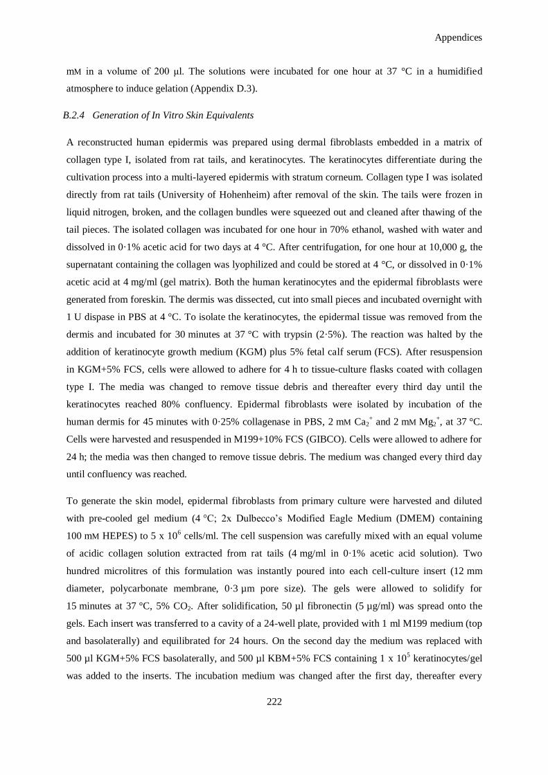
Appendices
222
mM in a volume of 200 μl. The solutions were incubated for one hour at 37 °C in a humidified
atmosphere to induce gelation (Appendix D.3).
B.2.4 Generation of In Vitro Skin Equivalents
A reconstructed human epidermis was prepared using dermal fibroblasts embedded in a matrix of
collagen type I, isolated from rat tails, and keratinocytes. The keratinocytes differentiate during the
cultivation process into a multi-layered epidermis with stratum corneum. Collagen type I was isolated
directly from rat tails (University of Hohenheim) after removal of the skin. The tails were frozen in
liquid nitrogen, broken, and the collagen bundles were squeezed out and cleaned after thawing of the
tail pieces. The isolated collagen was incubated for one hour in 70% ethanol, washed with water and
dissolved in 0·1% acetic acid for two days at 4 °C. After centrifugation, for one hour at 10,000 g, the
supernatant containing the collagen was lyophilized and could be stored at 4 °C, or dissolved in 0·1%
acetic acid at 4 mg/ml (gel matrix). Both the human keratinocytes and the epidermal fibroblasts were
generated from foreskin. The dermis was dissected, cut into small pieces and incubated overnight with
1 U dispase in PBS at 4 °C. To isolate the keratinocytes, the epidermal tissue was removed from the
dermis and incubated for 30 minutes at 37 °C with trypsin (2·5%). The reaction was halted by the
addition of keratinocyte growth medium (KGM) plus 5% fetal calf serum (FCS). After resuspension
in KGM+5% FCS, cells were allowed to adhere for 4 h to tissue-culture flasks coated with collagen
type I. The media was changed to remove tissue debris and thereafter every third day until the
keratinocytes reached 80% confluency. Epidermal fibroblasts were isolated by incubation of the
human dermis for 45 minutes with 0·25% collagenase in PBS, 2 mM Ca2+ and 2 mM Mg2
+, at 37 °C.
Cells were harvested and resuspended in M199+10% FCS (GIBCO). Cells were allowed to adhere for
24 h; the media was then changed to remove tissue debris. The medium was changed every third day
until confluency was reached.
To generate the skin model, epidermal fibroblasts from primary culture were harvested and diluted
with pre-cooled gel medium (4 °C; 2x Dulbecco‘s Modified Eagle Medium (DMEM) containing
100 mM HEPES) to 5 x 10⁶ cells/ml. The cell suspension was carefully mixed with an equal volume
of acidic collagen solution extracted from rat tails (4 mg/ml in 0·1% acetic acid solution). Two
hundred microlitres of this formulation was instantly poured into each cell-culture insert (12 mm
diameter, polycarbonate membrane, 0·3 µm pore size). The gels were allowed to solidify for
15 minutes at 37 °C, 5% CO2. After solidification, 50 µl fibronectin (5 µg/ml) was spread onto the
gels. Each insert was transferred to a cavity of a 24-well plate, provided with 1 ml M199 medium (top
and basolaterally) and equilibrated for 24 hours. On the second day the medium was replaced with
500 µl KGM+5% FCS basolaterally, and 500 µl KBM+5% FCS containing 1 x 10⁵ keratinocytes/gel
was added to the inserts. The incubation medium was changed after the first day, thereafter every
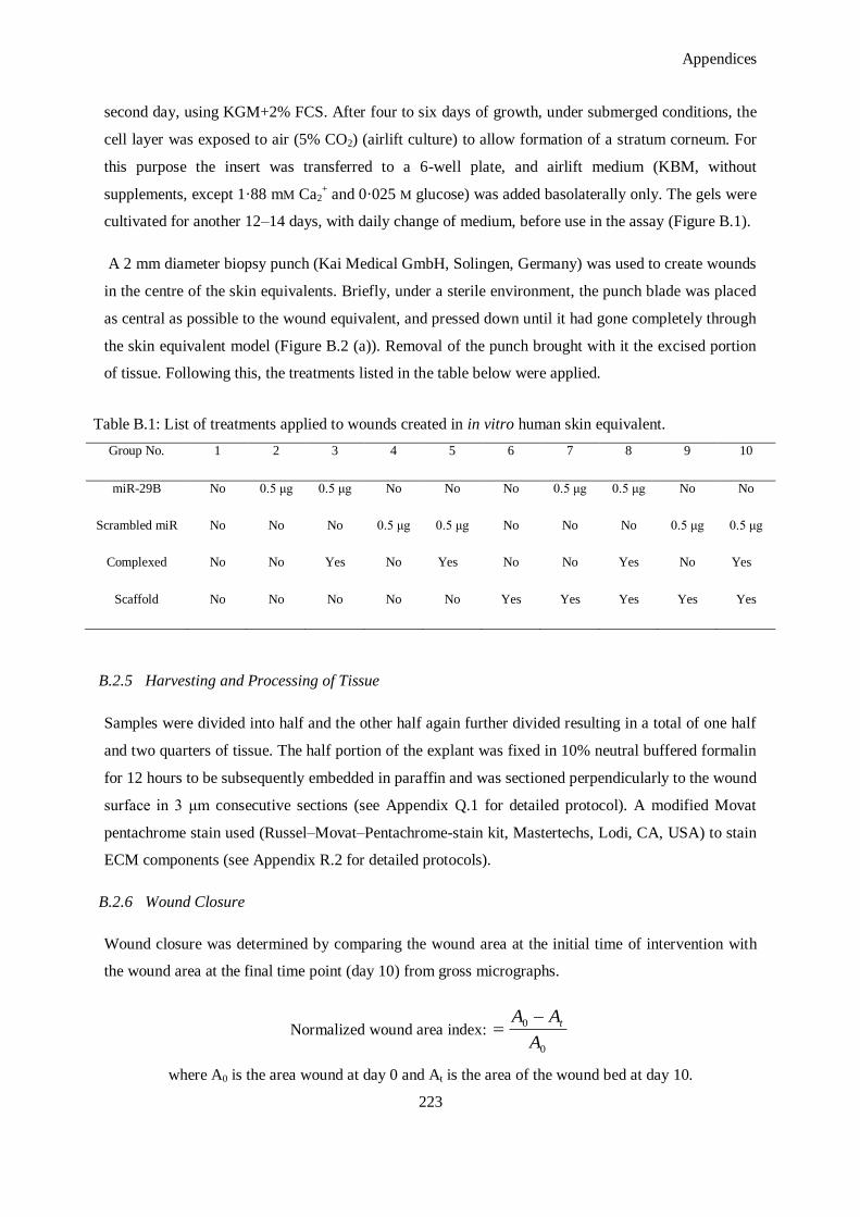
Appendices
223
second day, using KGM+2% FCS. After four to six days of growth, under submerged conditions, the
cell layer was exposed to air (5% CO2) (airlift culture) to allow formation of a stratum corneum. For
this purpose the insert was transferred to a 6-well plate, and airlift medium (KBM, without
supplements, except 1·88 mM Ca2+ and 0·025 M glucose) was added basolaterally only. The gels were
cultivated for another 12–14 days, with daily change of medium, before use in the assay (Figure B.1).
A 2 mm diameter biopsy punch (Kai Medical GmbH, Solingen, Germany) was used to create wounds
in the centre of the skin equivalents. Briefly, under a sterile environment, the punch blade was placed
as central as possible to the wound equivalent, and pressed down until it had gone completely through
the skin equivalent model (Figure B.2 (a)). Removal of the punch brought with it the excised portion
of tissue. Following this, the treatments listed in the table below were applied.
Table B.1: List of treatments applied to wounds created in in vitro human skin equivalent.
Group No. 1 2 3 4 5 6 7 8 9 10
miR-29B No 0.5 μg 0.5 μg No No No 0.5 μg 0.5 μg No No
Scrambled miR No No No 0.5 μg 0.5 μg No No No 0.5 μg 0.5 μg
Complexed No No Yes No Yes No No Yes No Yes
Scaffold No No No No No Yes Yes Yes Yes Yes
B.2.5 Harvesting and Processing of Tissue
Samples were divided into half and the other half again further divided resulting in a total of one half
and two quarters of tissue. The half portion of the explant was fixed in 10% neutral buffered formalin
for 12 hours to be subsequently embedded in paraffin and was sectioned perpendicularly to the wound
surface in 3 μm consecutive sections (see Appendix Q.1 for detailed protocol). A modified Movat
pentachrome stain used (Russel–Movat–Pentachrome-stain kit, Mastertechs, Lodi, CA, USA) to stain
ECM components (see Appendix R.2 for detailed protocols).
B.2.6 Wound Closure
Wound closure was determined by comparing the wound area at the initial time of intervention with
the wound area at the final time point (day 10) from gross micrographs.
Normalized wound area index: 0
0
A
AA t
where A0 is the area wound at day 0 and At is the area of the wound bed at day 10.
Appendices
224
B.2.7 Collagen Type I and Collagen Type III Immunohistochemistry
Collagen type I and collagen type III expression within the skin equivalents was revealed using
fluorescent immunostaining and quantified using stereological methods in terms of volume fractions.
Hydrated sections were subjected to heat induced antigen retrieval in pH 6.0 citrate buffer (for
collagen III, α-SMA actin and TGF-β1 antigen retrieval) or Tris-EDTA pH 9.0 (for collagen I antigen
retrieval) in a pressure cooker. The antibodies used in the identification were mouse monoclonal to
collagen I (Acris Antibodies dilution: 1:50) and rabbit monocolonal to collagen III (Acris, Antibodies;
dilution: 1:50) Following this the slides were treated with goat anti-mouse FITC conjugated secondary
antibody (Invitrogen) and goat anti-rabbit AlexaFluor 555 conjugated secondary antibody
(Invitrogen). All sections were counterstained with 4',6-diamidino-2-phenylindole (DAPI).
B.2.8 Statistical Analysis
All samples were tested in triplicate and all experimental groups were analysed in triplicate. GraphPad
Prism® (v.5 GraphPad Software, San Diego, CA, USA) was used for statistical analysis. Analysis of
variance (ANOVA) was used followed by Tukey‘s post-hoc test to determine statistical significance
between groups. ANOVA was performed assuming normal distribution of the data which was tested
and verified using the Anderson-Darling test. p values of < 0.05 were considered statistically
significant.
B.3. Results and Discussion
This in vitro skin equivalent model proved not suitable to determine the effect of the treatment on
wound closure. There was closure in all of the wounds, but there was no statistically significant
difference between any of the groups investigated. There are a number of reasons to explain this. The
human skin equivalent can act as a suitable model to elucidate in vitro the permeation of drugs and
compounds through the epidermis and into the dermis 1-4
. It has also been proposed as a model of skin
cancer 5-6
, psoriasis 7-8
and infection 2, 9-10
. Its applicability in a wound model however is somewhat
limited as the wound healing process in vivo includes more cell types than only fibroblasts and
keratinocytes. Several approaches of inducing a wound include scratching, abrading and burning 11-12
.
The most commonly used instruments to induce the injury are mashers, scalpels, biopsy punches,
dermatomes, liquid nitrogen and lasers 13
. The most common model is the burn model which employs
a bass wire heated to 150 °C 12
. However, there are concerns over wound-size reproducibility in all of
these methods employed. The biopsy punch employed here had a standard size of 2 mm diameter yet
the application of this wound is influenced by user expertise, angle of entry employed and force
applied. Standardized application of such a wound would require an automated, robotally controlled
system with geospatial positioning.
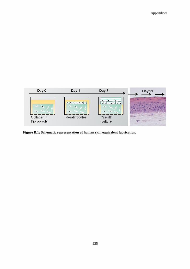
Appendices
225
Figure B.1: Schematic representation of human skin equivalent fabrication.
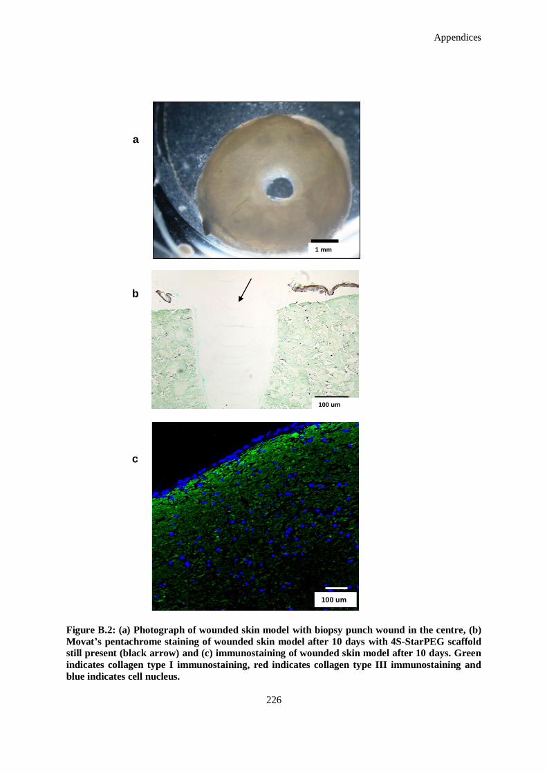
Appendices
226
Figure B.2: (a) Photograph of wounded skin model with biopsy punch wound in the centre, (b)
Movat’s pentachrome staining of wounded skin model after 10 days with 4S-StarPEG scaffold
still present (black arrow) and (c) immunostaining of wounded skin model after 10 days. Green
indicates collagen type I immunostaining, red indicates collagen type III immunostaining and
blue indicates cell nucleus.
a
b
c
1 mm
100 um
100 um
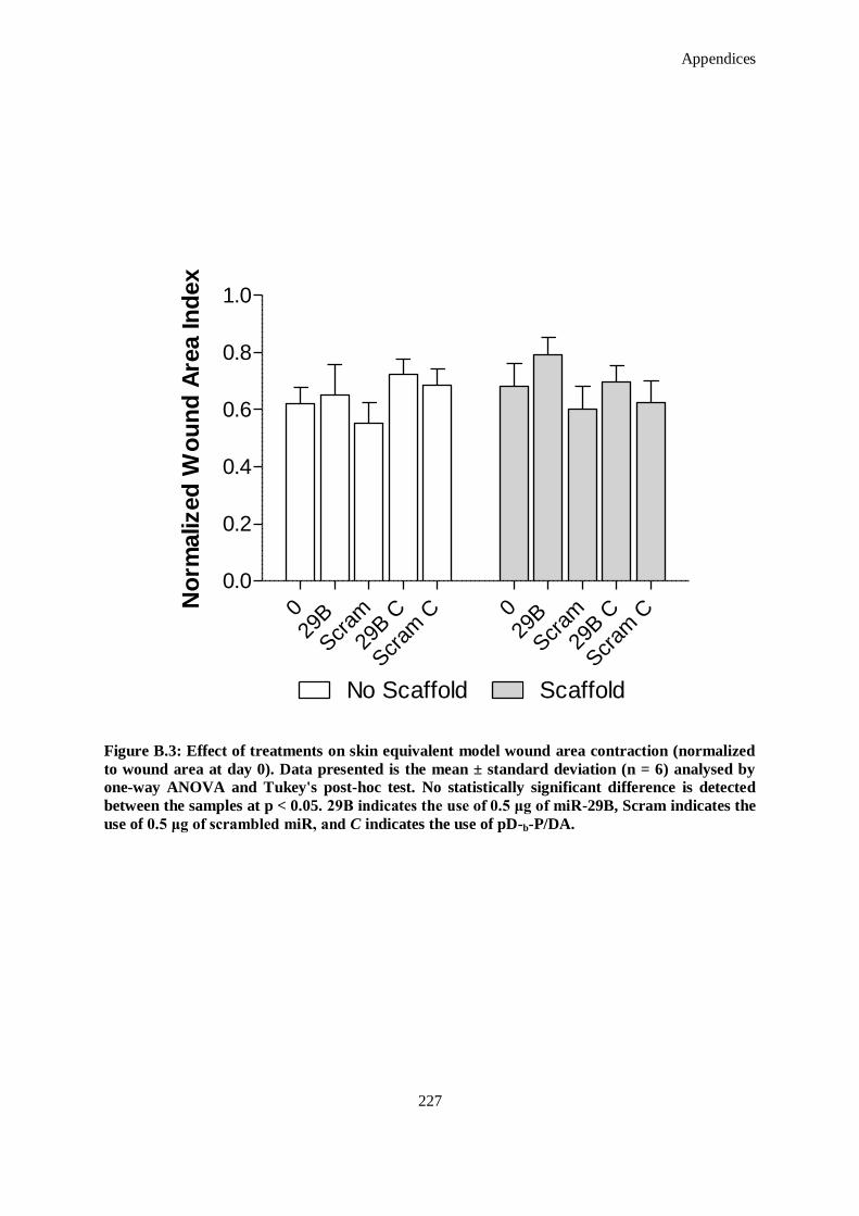
Appendices
227
029
B
Scr
am
29B C
Scr
am C 0
29B
Scr
am
29B C
Scr
am C
0.0
0.2
0.4
0.6
0.8
1.0
No Scaffold Scaffold
No
rmalized
Wo
un
d A
rea In
dex
Figure B.3: Effect of treatments on skin equivalent model wound area contraction (normalized
to wound area at day 0). Data presented is the mean ± standard deviation (n = 6) analysed by
one-way ANOVA and Tukey's post-hoc test. No statistically significant difference is detected
between the samples at p < 0.05. 29B indicates the use of 0.5 μg of miR-29B, Scram indicates the
use of 0.5 μg of scrambled miR, and C indicates the use of pD-b-P/DA.
Appendices
228
Fibroblasts will contract hydrogels in vitro and the system developed here has no boundary to impede
horizontal contraction 14
. Additionally, this model possesses no innate immune system so the cascade
of inflammation is not initiated to the degree to which that in vivo is subjected. This complex cascade
of events follows cutaneous injury in sequential and overlapping phases 15
. It initiates with
haemostasis in which platelets aggregate at the injury site to form a fibrin clot. Histamine, released by
ruptured cell membranes, enables blood vessels to become dilated and porous which facilitates the
infiltration of inflammatory cells such as polymorphonuclear neutrophils (PMNs) and helper T cells
into the wound site from the bloodstream 16
. In the following/overlapping inflammatory phase,
bacteria and debris are phagocytised and removed. Macrophages secrete a number of factors such as
growth factors and other cytokines which attract cells involved in a proliferation stage of healing to
the area. By the end of the first week, fibroblasts become the predominant cell in the wound and are
responsible for laying down the collagen matrix in the wound site. Formation of this granulation
tissue begins in the wound during the inflammatory phase and continues until the wound bed is
covered 15
. The innate immune response and also a systemic inflammatory response is lacking in the
in vitro skin equivalent model presented. Therefore a true representation of wound healing and also
evaluation of the treatments applied in this chapter, which aim to modulate wound healing, cannot be
achieved. This could account for the absence of any statistically significant differences when wound
contraction area and collagen type I and collagen type III deposition were investigated (Figure B.2).
Additionally, there was strong staining of collagen type I which is due to the matrix of the skin
equivalent model matrix being entirely composed of collagen type I. Based on these conclusions and
results, it was decided not to perform further analysis on the models employed in this study.
Appendices
229
B.4. References
1. Obi-Tabot ET, Tian XQ, Chen TC, Holick MF. A human skin equivalent model that mimics the
photoproduction of vitamin D3 in human skin. In Vitro Cell Dev. Biol. Anim. 2000;36:201-204
2. Mertsching H, Weimer M, Kersen S, Brunner H. Human skin equivalent as an alternative to
animal testing. GMS Krankenhaushygiene Interdisziplinar. 2008;3, ISSN 1863-5245.
3. Welss T, Basketter DA, Schroder KR. In vitro skin irritation: Facts and future. State of the art
review of mechanisms and models. Toxicol. In Vitro. 2004;18:231-243
4. Groeber F, Holeiter M, Hampel M, Hinderer S, Schenke-Layland K. Skin tissue engineering —
in vivo and in vitro applications. Adv. Drug Del. Rev.. 2011;63:352-366
5. Meier F, Nesbit M, Hsu MY, Martin B, Van Belle P, Elder DE, Schaumburg-Lever G, Garbe C,
Walz TM, Donatien P, Crombleholme TM, Herlyn M. Human melanoma progression in skin
reconstructs : Biological significance of BFGF. Am. J. Pathol. 2000;156:193-200
6. Van Kilsdonk JW, Bergers M, Van Kempen LC, Schalkwijk J, Swart GW. Keratinocytes drive
melanoma invasion in a reconstructed skin model. Melanoma Res. 2010;20:372-380
7. Saiag P, Coulomb B, Lebreton C, Bell E, Dubertret L. Psoriatic fibroblasts induce
hyperproliferation of normal keratinocytes in a skin equivalent model in vitro. Science.
1985;230:669-672
8. Jean J, Lapointe M, Soucy J, Pouliot R. Development of an in vitro psoriatic skin model by tissue
engineering. J. Dermatol. Sci. 2009;53:19-25
9. Andrei G, van den Oord J, Fiten P, Opdenakker G, De Wolf-Peeters C, De Clercq E, Snoeck R.
Organotypic epithelial raft cultures as a model for evaluating compounds against αherpesviruses.
Antimicrob. Agents Chemother. 2005;49:4671-4680
10. Dieterich C, Schandar M, Noll M, Johannes FJ, Brunner H, Graeve T, Rupp S. In vitro
reconstructed human epithelia reveal contributions of candida albicans EGF1 and CPH1 to
adhesion and invasion. Microbiology. 2002;148:497-506
11. Xie Y, Rizzi SC, Dawson R, Lynam E, Richards S, Leavesley DI, Upton Z. Development of a
three-dimensional human skin equivalent wound model for investigating novel wound healing
therapies. Tissue Eng. Part C Methods. 2010;16:1111-1123
12. Emanuelsson P, Kratz G. Characterization of a new in vitro burn wound model. Burns.
1997;23:32-36
13. Vaughan MB, Ramirez RD, Brown SA, Yang JC, Wright WE, Shay JW. A reproducible laser-
wounded skin equivalent model to study the effects of aging in vitro. Rejuvenation Res.
2004;7:99-110
14. Carlson MA, Longaker MT. The fibroblast-populated collagen matrix as a model of wound
healing: A review of the evidence. Wound Repair. Regen. 2004;12:134-147
15. Stadelmann WK, Digenis AG, Tobin GR. Physiology and healing dynamics of chronic cutaneous
wounds. Am. J. Surg. 1998;176:26S-38S
16. Steed DL. The role of growth factors in wound healing. Surg. Clin. North Am. 1997;77:575-586
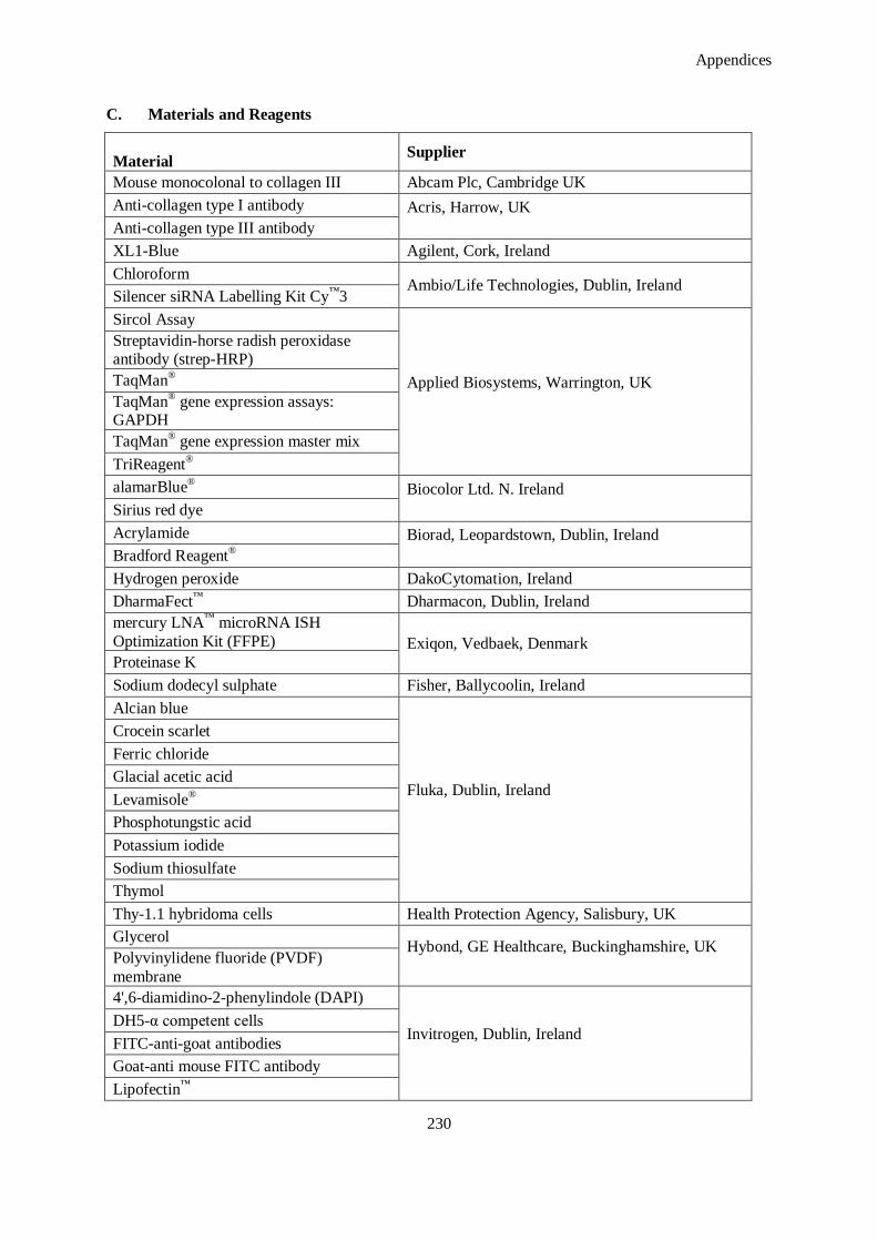
Appendices
230
C. Materials and Reagents
Material Supplier
Mouse monocolonal to collagen III Abcam Plc, Cambridge UK
Anti-collagen type I antibody Acris, Harrow, UK
Anti-collagen type III antibody
XL1-Blue Agilent, Cork, Ireland
Chloroform Ambio/Life Technologies, Dublin, Ireland
Silencer siRNA Labelling Kit Cy™
3
Sircol Assay
Applied Biosystems, Warrington, UK
Streptavidin-horse radish peroxidase antibody (strep-HRP)
TaqMan®
TaqMan® gene expression assays:
GAPDH
TaqMan® gene expression master mix
TriReagent®
alamarBlue® Biocolor Ltd. N. Ireland
Sirius red dye
Acrylamide Biorad, Leopardstown, Dublin, Ireland
Bradford Reagent®
Hydrogen peroxide DakoCytomation, Ireland
DharmaFect™
Dharmacon, Dublin, Ireland
mercury LNA™
microRNA ISH
Optimization Kit (FFPE) Exiqon, Vedbaek, Denmark
Proteinase K
Sodium dodecyl sulphate Fisher, Ballycoolin, Ireland
Alcian blue
Fluka, Dublin, Ireland
Crocein scarlet
Ferric chloride
Glacial acetic acid
Levamisole®
Phosphotungstic acid
Potassium iodide
Sodium thiosulfate
Thymol
Thy-1.1 hybridoma cells Health Protection Agency, Salisbury, UK
Glycerol Hybond, GE Healthcare, Buckinghamshire, UK Polyvinylidene fluoride (PVDF)
membrane
4',6-diamidino-2-phenylindole (DAPI)
Invitrogen, Dublin, Ireland
DH5-α competent cells
FITC-anti-goat antibodies
Goat-anti mouse FITC antibody
Lipofectin™
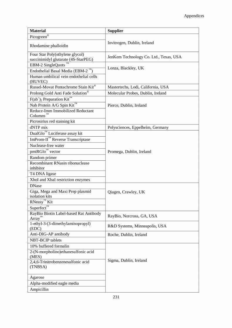
Appendices
231
Material Supplier
Picogreen®
Invitrogen, Dublin, Ireland Rhodamine phalloidin
Four Star Poly(ethylene glycol)
succinimidyl glutarate (4S-StarPEG) JenKem Technology Co. Ltd., Texas, USA
EBM-2 SingleQuots ™
Lonza, Blackley, UK
Endothelial Basal Media (EBM-2 ™
)
Human umbilical vein endothelial cells
(HUVEC)
Russel-Movat Pentachrome Stain Kit®
Mastertechs, Lodi, California, USA
Prolong Gold Anti Fade Solution®
Molecular Probes, Dublin, Ireland
F(ab`)2 Preparation Kit™
Pierce, Dublin, Ireland
Nab Protein A/G Spin Kit™
Reduce-Imm Immobilized Reductant
Columns ™
Picrosirius red staining kit
dNTP mix Polysciences, Eppelheim, Germany
DualGlo™
Luciferase assay kit
Promega, Dublin, Ireland
ImProm-II™
Reverse Transcriptase
Nuclease-free water
pmiRGlo™
vector
Random primer
Recombinant RNasin ribonuclease
inhibitor
T4 DNA ligase
XhoI and XbaI restriction enzymes
DNase
Qiagen, Crawley, UK
Giga, Mega and Maxi Prep plasmid isolation kits
RNeasy™
Kit
Superfect™
RayBio Biotin Label-based Rat Antibody
Array™
RayBio, Norcross, GA, USA
1-ethyl-3-(3-dimethylaminopropyl)
(EDC) R&D Systems, Minneapolis, USA
Anti-DIG-AP antibody Roche, Dublin, Ireland
NBT-BCIP tablets
10% buffered formalin
Sigma, Dublin, Ireland
2-(N-morpholino)ethanesulfonic acid
(MES)
2,4,6-Trinitrobenzenesulfonic acid (TNBSA)
Agarose
Alpha-modified eagle media
Ampicillin
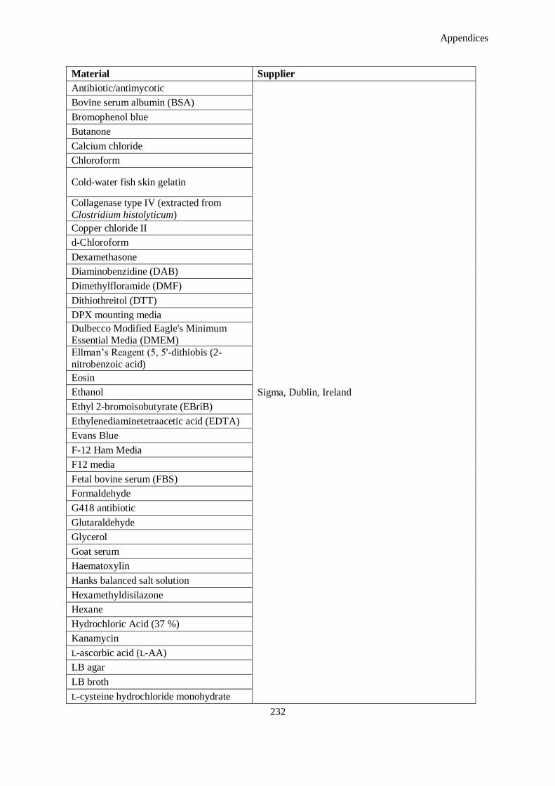
Appendices
232
Material Supplier
Antibiotic/antimycotic
Sigma, Dublin, Ireland
Bovine serum albumin (BSA)
Bromophenol blue
Butanone
Calcium chloride
Chloroform
Cold-water fish skin gelatin
Collagenase type IV (extracted from
Clostridium histolyticum)
Copper chloride II
d-Chloroform
Dexamethasone
Diaminobenzidine (DAB)
Dimethylfloramide (DMF)
Dithiothreitol (DTT)
DPX mounting media
Dulbecco Modified Eagle's Minimum
Essential Media (DMEM)
Ellman‘s Reagent (5, 5'-dithiobis (2-
nitrobenzoic acid)
Eosin
Ethanol
Ethyl 2-bromoisobutyrate (EBriB)
Ethylenediaminetetraacetic acid (EDTA)
Evans Blue
F-12 Ham Media
F12 media
Fetal bovine serum (FBS)
Formaldehyde
G418 antibiotic
Glutaraldehyde
Glycerol
Goat serum
Haematoxylin
Hanks balanced salt solution
Hexamethyldisilazone
Hexane
Hydrochloric Acid (37 %)
Kanamycin
L-ascorbic acid (L-AA)
LB agar
LB broth
L-cysteine hydrochloride monohydrate
Appendices
233
Material Supplier
Mayer's Haematoxylin
Sigma, Dublin, Ireland
Methanol
N-hydroxylsuccinimide (NHS)
Nuclear fast red
Paraformaldehyde
Penicillin/streptomysin
Pentamethyldiethylenetriamine
(PMDETA)
Phosphate buffered saline tablets
Poly (ethylene glycol) diacrylate
(PEGDA)
Poly (ethylene glycol) methyl ether
acrylate (PEGMEA)
Poly (ethylenimine) (PEI)
Poly-L-lysine (PLL)
Ponceau S
Radioimmunoprecipitation assay (RIPA)
RNA away
Sheep serum
Sodium azide
Sodium chloride
Sodium dihydrogen phosphate
Sodium hydroxide
Sterile nuclease and ribonuclease free water
Sucrose
Triphenyltetrazolium chloride (TTC)
Tris-hydrochloride
Triton X-100
Trypsin
Trypsin-EDTA
Tween-20
Weigerts Haematoxylin
Xylene (histological grade)
FITC-anti-mouse antibodies
Horseradish peroxidase (HRP)-conjugated
anti-mouse IgG

Appendices
234
D. Re-Constitution of Collagen
1. Remove collagen (atelocollagen isolated from bovine Achilles tendon) from -20° storage.
2. Pull apart fibrous collagen to enable homogeneous freeze drying.
3. Place collagen in container suitable for freeze drying, e.g. 6 well plate/petri dish.
4. Place container in freeze drier and dry overnight.
5. Once dried, weigh out desired mass of collagen while ensuring maintenance at 4 °C.
6. Note: Consider Sircol™
assay results when weighing collagen, e.g. 1 mg may equal 0.8 mg
collagen given 80 % purity. The Sircol™
assay protocol is described in Appendix D.1.
7. Prepare a solution of 50 mM hydrochloric acid (bring to 4 °C).
8. Place desired amount of collagen in the hydrochloric acid.
9. Place solution, maintained at 4 °C for 8-12 hours (overnight, for convenience) to swell the
collagen.
10. Homogenise the solution with a blender/homogenizer.
Note: Kinetic effects of blending may induce heat, so ensure maintenance at 4 °C.
11. Remove excess bubbles by vacuum through Buckner flask.
12. Store re-constituted collagen at 4 °C until use.
D.1. Sircol Assay to Determine Collagen Purity
1. Adjust concentration of isolated collagen to 1 mg/ml using 5 mM hydrochloric acid. Rationale
for this is that the collagen standard supplied with the Sircol™
kit is also 1 mg/ml and will
thus ensure better relativity.
Note: The isolated sample does not contain 1 mg/ml of pure collagen as there are trace
amounts of fat tissue and non-digested proteins. The Sircol™
assay will determine the actual
collagen content within this isolate.
2. Label 1.5 ml eppendorfs appropriately for 0, 10, 20, 30, 40 and 50 µg of standard and test
samples. Both standards and test samples should be prepared in triplicate.
3. Add 0, 10, 20, 30, 40 and 50 µl of collagen standard into appropriate tubes.
4. Add 0, 10, 20, 30, 40 and 50 µl of isolated collagen into appropriate tubes.
5. There should now be a total of 36 tubes, half of which contain collagen standard and the other
half containing isolated collagen.
6. Adjust the volume of all tubes to 100 µl with distilled water.
7. To each tube add 1 ml of Sircol™
Dye reagent (Sirius Red in picric acid).
8. Place tubes in an eppendorf rack and place rack on mechanical shaker for 30 minutes. The
dye should bind to the collagen and precipitate out of the solution.
9. Transfer the tubes to a micro-centrifuge and centrifuge at 10,000 g for 10 minutes.
10. The un-bound dye can be removed by gently tipping the contents gently onto absorbent paper.

Appendices
235
11. The remaining un-bound dye can be removed with a cotton bud. Be careful not to touch the
pellet during this procedure.
12. Add 1 ml of alkali reagent (0.5 M sodium hydroxide) to each tube.
13. Re-cap the tubes and centrifuge the contents until the precipitate has become dissolved (may
take up to 15 minutes).
14. Remove 200 µl from each tube and place in a separate well of a clear 96 well plate.
15. Place the plate in a plate reader and measure absorbance at 550 nm for 0.1 seconds per well.
16. Remove the average blank reading i.e. 0 µg collagen sample, from all other readings.
17. By plotting collagen reference samples against isolated collagen samples the collagen content
can be determined. E.g. if 50 µg/ml standard gives average reading of 0.963 and 50 µg/ml test
samples gives an average reading of 0.769, then the test sample contains 80% collagen
(0.769/0.963).
D.2. Preparation of Non-crosslinked Hydrogel
1. Perform all actions while ensuring maintenance at 4 °C.
Note: When using scaffolds for biological interaction, perform steps below in cell culture hood
with sterile-filtered buffers.
2. Add collagen type I solubilised in 50 mM hydrochloric acid to an eppendorf to required volume.
3. Mix this at a 1:9 ratio of 10 X PBS to collagen solution to buffer.
4. Mix evenly with 1 ml pipette and ensure complete mixing.
5. Add dropwise 2 M sodium hydroxide (in 1/2 µl quantities) until pH reaches 7.2 – 7.5.
Note: The proportion of NaOH can vary from batch to batch of collagen, depending on purity,
storage conditions and solubility of collagen. Monitor pH using pH strips with a narrow range
(typically 6 – 8).
6. When desired pH has been achieved, maintain at 4 °C until gelation and/or crosslinking is
required.
D.3. Crosslinking of Collagen Type I Hydrogel using 4S-Succinimydyl Glutarate Terminated Poly
(ethylene glycol)
1. After preparing hydrogel solution, leave at 4 °C (on ice is suitable).
2. Prepare four arm poly (ethylene glycol) succinimidyl glutarate (4S-StarPEG) solutions in 1 X
PBS. Note: 4S-StarPEG must be stored under argon, protected from light and at -20 °C.
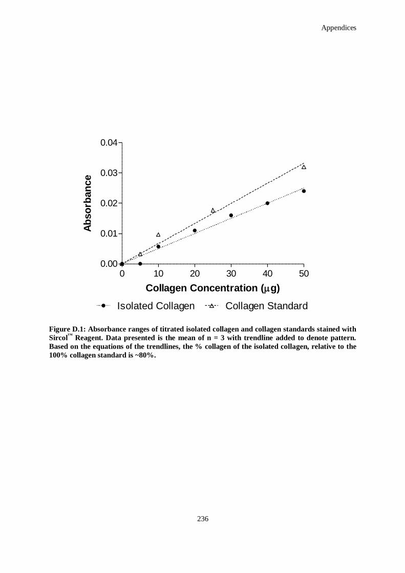
Appendices
236
Figure D.1: Absorbance ranges of titrated isolated collagen and collagen standards stained with
Sircol™
Reagent. Data presented is the mean of n = 3 with trendline added to denote pattern.
Based on the equations of the trendlines, the % collagen of the isolated collagen, relative to the
100% collagen standard is ~80%.
0 10 20 30 40 500.00
0.01
0.02
0.03
0.04
Collagen StandardIsolated Collagen
Collagen Concentration (g)
Ab
so
rban
ce

Appendices
237
3. Mix required mass of 4S-StarPEG to neutralised collagen solution. Theoretically, the 4S-
StarPEG will covalently bind to free amines in the collagen. On a molar basis, calculate the
required number of moles to bind at a 1:4 ratio of 4S-StarPEG to collagen free amines and
determine the required mass of this
4. Mix crosslinking solution containing required mass/moles with collagen solution. Maintain at 4
°C.
Note: Work quickly after adding crosslinker because complete gelation occurs within eight
minutes. Add to well plate/eppendorf/syringe as required.
D.4. TNBSA Quantitation of Free Amines Following Crosslinking
1. Prepare a standard curve with Glycine in sodium bicarbonate pH 8.5 (0.1 M) (H2N-CH2-COOH)
(100 nM, 50 nM, 25 nM, 10 nM, 5 nM and 0 nM).
2. Add 250 µl of 0.01% of TNBSA in 0.5 ml of each sample. Mix well.
3. Incubate at 37 °C for 2 hours.
4. Add 250 µl of 10% SDS and 125 µl of 1 M HCl.
5. Hydrolyse scaffold/hydrogel by incubating the samples at 120 °C for 15 minutes (autoclaving is
also appropriate).
6. Measure the absorbance at 335 nm from 250 µl.
D.5. Enzymatic Degradation of Hydrogels Following Crosslinking
1. Incubate hydrogel for 1 hour in 0.1 M Tris-HCl (pH 7.4), 50 mM CaCl2 at 37°C.
2. Reconstitute bacterial collagenase (extracted from Clostridium histolyticum) in 0.1 M Tris-HCl
at a concentration of 10 units/mg.
3. Incubate at 37 °C. At each time point (12, 24, 48 hours), cease the enzymatic reaction by
adding 0.25 M EDTA. Store and label supernatant at -20°C until further analysis.
4. Centrifuge samples at 4,000 rpm, for 5 minutes at 4°C.
5. Wash samples 3 times with distilled water and freeze-dry.
Mass loss is calculated by comparing the initial mass Wo with the mass at a given time point Wt, as
shown:
Mass loss (%)
100
t
to
W
WW
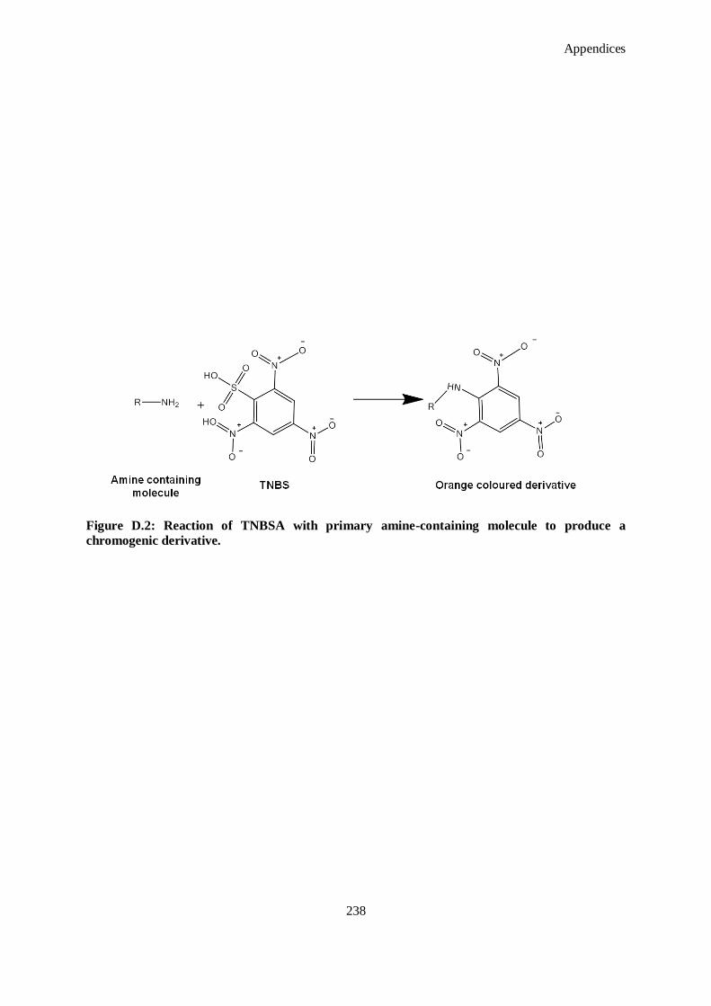
Appendices
238
Figure D.2: Reaction of TNBSA with primary amine-containing molecule to produce a
chromogenic derivative.
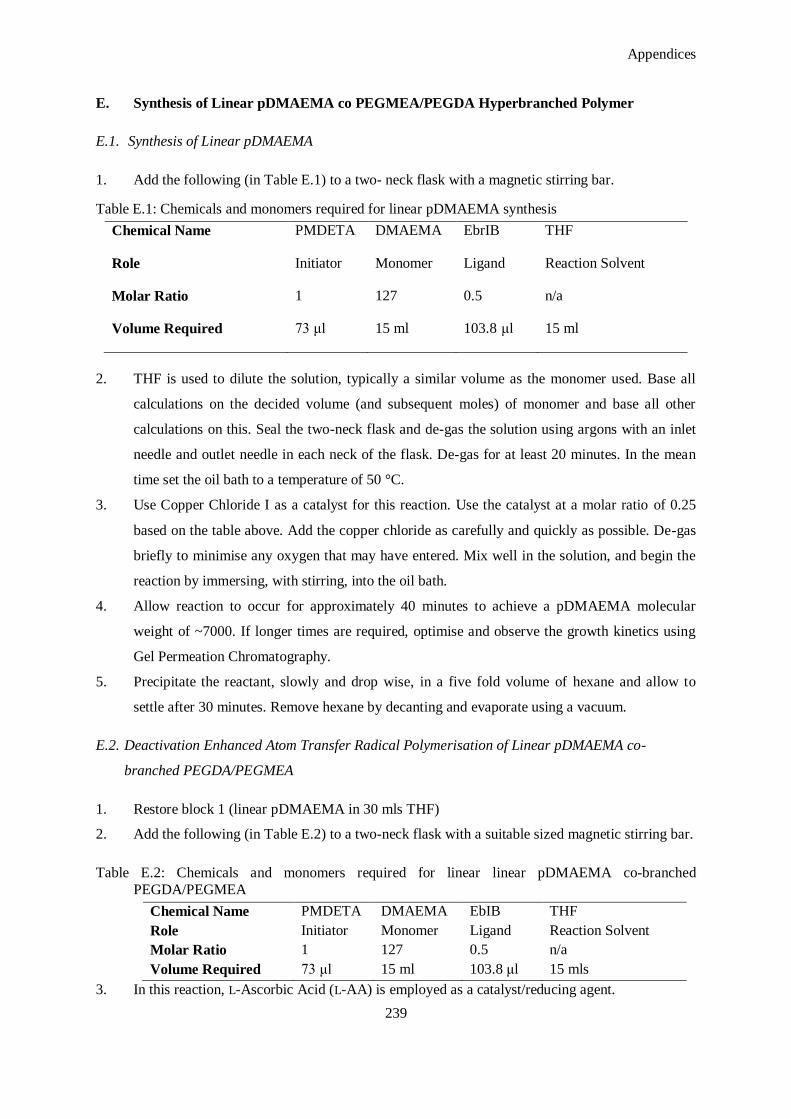
Appendices
239
E. Synthesis of Linear pDMAEMA co PEGMEA/PEGDA Hyperbranched Polymer
E.1. Synthesis of Linear pDMAEMA
1. Add the following (in Table E.1) to a two- neck flask with a magnetic stirring bar.
Table E.1: Chemicals and monomers required for linear pDMAEMA synthesis
Chemical Name PMDETA DMAEMA EbrIB THF
Role Initiator Monomer Ligand Reaction Solvent
Molar Ratio 1 127 0.5 n/a
Volume Required 73 μl 15 ml 103.8 μl 15 ml
2. THF is used to dilute the solution, typically a similar volume as the monomer used. Base all
calculations on the decided volume (and subsequent moles) of monomer and base all other
calculations on this. Seal the two-neck flask and de-gas the solution using argons with an inlet
needle and outlet needle in each neck of the flask. De-gas for at least 20 minutes. In the mean
time set the oil bath to a temperature of 50 °C.
3. Use Copper Chloride I as a catalyst for this reaction. Use the catalyst at a molar ratio of 0.25
based on the table above. Add the copper chloride as carefully and quickly as possible. De-gas
briefly to minimise any oxygen that may have entered. Mix well in the solution, and begin the
reaction by immersing, with stirring, into the oil bath.
4. Allow reaction to occur for approximately 40 minutes to achieve a pDMAEMA molecular
weight of ~7000. If longer times are required, optimise and observe the growth kinetics using
Gel Permeation Chromatography.
5. Precipitate the reactant, slowly and drop wise, in a five fold volume of hexane and allow to
settle after 30 minutes. Remove hexane by decanting and evaporate using a vacuum.
E.2. Deactivation Enhanced Atom Transfer Radical Polymerisation of Linear pDMAEMA co-
branched PEGDA/PEGMEA
1. Restore block 1 (linear pDMAEMA in 30 mls THF)
2. Add the following (in Table E.2) to a two-neck flask with a suitable sized magnetic stirring bar.
Table E.2: Chemicals and monomers required for linear linear pDMAEMA co-branched
PEGDA/PEGMEA
Chemical Name PMDETA DMAEMA EbIB THF
Role Initiator Monomer Ligand Reaction Solvent
Molar Ratio 1 127 0.5 n/a
Volume Required 73 μl 15 ml 103.8 μl 15 mls
3. In this reaction, L-Ascorbic Acid (L-AA) is employed as a catalyst/reducing agent.

Appendices
240
4. Resuspend required mass (moles) of L-AA in H2O. If necessary make a concentrated solution in
order to minimise the volume of H2O added to the reaction.
5. Seal the two-neck flask and de-gas the solution using argons with an inlet needle and outlet
needle in each neck of the flask. De-gas for at least 20 minutes.
6. In the meantime set the oil bath to a temperature of 50 °C.
7. Add suspended L-AA to the reaction with the use of a syringe to avoid opening the seal on the
flask.
8. At this point take a sample for t = 0.
9. Begin reaction by placing in the oil bath at 50 °C.
10. Take samples at regular time points and investigate the growth of the PEGDA/PEGMEA
branching using Gel Permeation Chromatograpy (GPC).
11. When required, halt the reaction by purging the system with oxygen. At this point, protect the
resultant polymer from light at all times.
12. Precipitate the reactant slowly and dropwise in a five fold volume of 1:1 Hexane: Di-ethyl
Ether.
13. Allow to stand for 30 minutes and vacuum as before.
14. Dissolve remainder polymer in acetone, and dialyse in ddH2O with a membrane MWCO of
8,000. Protect from light and dialyse for three days, changing the water at as many intervals as
possible. This removes any remaining monomer, or unreacted copper.
15. Freeze dry after dialysis is complete.
16. After freeze drying, aliquot the resultant polymer and store, protected from light at -20 °C.
E.3. Gel Permeation Chromatography of Synthesized Polymers
1. Under argon pressure, gently remove samples (approximately 1 mL maximum) before/during/
after reaction process.
2. Dilute sample in dimethyl formaldehyde (DMF) ten-fold and then run sample thourgh a silica
gel column to remove any copper present in the sample.
3. Filter the polymer solution. Small solids and particles may clog the GPC resulting in expensive
repairs. Every sample that is run on the GPC must be filtered. Screw needle (with cover) on
glass syringe. Remove cover.
4. Draw up the polymer solution through the needle into the glass syringe. Draw up an additional
1-2 mL air so that the syringe contains both polymer solution and air.
5. Replace cover on needle. Remove the needle carefully. Discard needle in sharps container.
6. Hold the syringe upright so that the air is at the port of the syringe. Screw a PTFE filter on the
end of the glass syringe.

Appendices
241
7. Turn the needle upside down so that the polymer solution is at the port of the syringe. Empty
syringe of contents through the filter into a clean 1 mL GPC vial. Push the ―extra‖air through
the filter to clear all the polymer solution of the filter.
8. Cap the GPC vial immediately and dispose of the PTFE filter. Load glass vials in GPC carousel
tray for analysis.
F. Characterization of miR-Complexing Agent Polyplexes
F.1. UV Spectroscopy
1. Start-up NanoDrop™
.
2. Wipe the pedestal clean with a Kimwipe™
.
3. The software will require a distilled water sample to initialize, then will ask for a blank sample
– this is a sample with no DNA in it, or any other buffer solutions, etc. Only 1-2 μL required.
4. Fill in the Sample ID space, then start reading samples.
5. The output of the NanoDrop™
is a spectral representation of the UV absorbance of the sample.
Normally, the peak absorbance is at 260 nm. If the samples are tightly polyplexed, this peak
may be closer to 300 nm. Once polyplexed, the concentration measurement is no longer
reliable.
F.2. Gel Electrophoresis for Characterisation of the Electrophoretic Mobility of Complexes
1. Add 0.7 g agarose in 100 mL TAE buffer (for large gel) or 0.35 agarose in 50 mL TAE buffer
(small gel).
2. Add about 2 mL extra liquid to account for boiling and mix in an Erlenmeyer flask, and put into
microwave.
3. Leave in microwave until boiling.
4. Remove from microwave with hot mitts or something to protect hands from heat.
5. Allow to cool until can be held comfortably in hand.
6. Add 10 μL Syber®Safe dye (Do not add until the solution is cool enough or the experiment will
be ruined).
7. Pour immediately into plate, add comb, and remove any bubbles. Leave to cool and set
8. Pipette samples into wells.
9. Run gels at 80-100V and leave the current on ‗Auto‘. Check that bubbles are forming along the
wires at the bottom.
10. Check after 15 minutes – dye should have moved slightly out of the wells. Check that the
direction is right and that all seems to be in order. If you leave it much after this, it is too late to
rectify it.

Appendices
242
11. Check every half hour/hour. Depending on the gel, dye, etc. it can take 3-4 hours to run. Make
sure to stop it by the time that the dye reaches about ¾ of the way along the gel.
12. Put under the imager and observe under UV light – save images.
13. Dispose of gel appropriately.
G. Preparation, Propagation, Isolation and Purification of Plasmid
G.1. LB Agar Plates
1. Place 1 tablet of Luria broth (LB) agar into 50 ml of distilled water.
2. Place in autoclave.
3. When removed let cool until it can be handled comfortably.
4. For cultures that require kanamycin use 10 µg/ml, hence 10 mg/L.
5. Prepare Bunsen burner and plates.
6. Remove the lid of the LB agar.
7. Run the top across the Bunsen flame.
8. Pour into the plate in close vicinity to the flame.
9. Let plates set at room temperature.
G.2. Digestion of Reporter Plasmid
1. Prepare the following in a sterile eppendorf:
Sterile deionized water 16.3 μl
RE10X Buffer 2 μl
Acetylated BSA (10 μg/μl) 0.2 μl
Plasmid DNA (1 μg/μl) 1 μl
2. Mix by pipetting and then add
XhoI restriction enzyme (10 units/μl) 0.5 μl
XbaI restriction enzyme (10 units/μl) 0.5 μl
3. Mix gently by pipetting, close tube, centrifuge for a few seconds in a microcentrifuge and
incubate for 3 hours at 37 °C.
4. Halt reaction by heat inactivation at 65 °C for 25 minutes.
5. Purify the product by using Qiaquick™
PCR purification kit (Appendix I.3)
G.3. Qiaquick™
PCR Purification Kit Protocol
1. Add 5 volumes of Buffer PB to1 volume of the PCR sample and mix.
2. Place a Qiaquick™
-spin column in a 2 ml collection tube.
3. To bind DNA, apply the sample to the Qiaquick™
column and centrifuge for 30-60 seconds.

Appendices
243
4. Discard flow –through. Place the Qiaquick™
column back into the same tube.
5. To wash, add 0.75 ml Buffer PE to the Qiaquick™
column and centrifuge for 30-60 seconds.
6. Discard flow-through and place the Qiaquick™
column back in the same tube. Centrifuge the
column for an additional 1 minute.
7. Place the Qiaquick™
column in a clean 1.5 ml microcentrifuge tube.
8. To elute DNA, add 50 μl Buffer EB (10 mM Tris-Cal, pH 8.5) or water (pH 7.0-8.5) to the
centre of the Qiaquick™
membrane and centrifuge for 1 minute. Alternatively for increased
DNA concentration, add 30 μl elution buffers to the centre of the Qiaquick™
membrane, let the
column stand for one minute, and then centrifuge.
G.4. Annealing of Oligonucleotides
1. Dilute both oligonucleotides (supplied by user) to 1μg/μl.
2. Combine 2 μl of each oligonucleotide with 46 μl of Oligo Annealing Buffer.
3. Heat at 90 °C for three minutes, then transfer to a 37 °C water bath for 15 minutes.
4. Use the annealed oligonucleotides immediately, or store at -20 °C.
G.5. Ligation of Oligonucleotides into Digested Vector
1. Dilute annealed oligonucleotides 1:10 in nuclease-free water to a final concentration of 4 ng/μl
per oligonucleotide. Ligate 4 ng of annealed oligonucleotides and 50 ng of linearized vector
using a standard ligation protocol.
2. The 2 X Rapid Ligation Buffer contains ATP, which degrades during temperature fluctuations.
Avoid multiple freeze-thaw cycles and exposure to frequent temperature changes by making
single-use aliquots of the buffer after the buffer is thawed for the first time. Store the aliquots at
–20 °C.
3. Vortex the 2 X Rapid Ligation Buffer before each use.
4. Mix the reactions by pipetting volumes according to Table G.1. Incubate the reactions at room
temperature for 5 minutes. Alternatively, the reaction can be incubated for one hour at room
temperature or overnight at 4 °C.
Table G.1: Reagent volumes for ligation of oligonucleotides into vector.
Negative Control
2X Rapid Ligation Buffer 5μl 5μl
pGeneClip™ Vector (50ng/μl 1μl 1μl
annealed oligonucleotides A and B (4ng/μl each) – 1μl
Nuclease-Free Water 3μl 2μl
T4 DNA Ligase (3 units/μl) 1μl 1μl
Total volume 10μl 10μl
5. The ligation reaction mixture can be used directly for the transformation of competent cells.

Appendices
244
G.6. Transformation
1. Turn on water bath at 42 °C.
2. Take out plates for transformation to warm up.
3. Fill ice box.
4. Put 1 μg plasmid into ‗plasmid +‘ tubes.
5. Label another tube control.
6. Take one tube of XL1Blue (bacteria) from box in -80 °C freezer and thaw on ice.
7. When thawed, add 50 μl bacteria to each tube.
8. Flick to mix.
9. Leave on ice for two minutes.
10. Put tubes in 42 °C waterbath for 90 seconds.
11. Place in ice for two minutes.
Note: Can also repeat four times by 40 seconds on ice, 40 seconds at 42 °C.
12. Add 1 ml room temperature LB broth (no antibiotic).
13. Tape up-right on shaker for 30-45 minutes.
14. Prepare plates, Bunsen burner, spreader, 70% ethanol, etc. Add 100 μl of inoculums to
‗ampicilin +‘ plates (or ‘kanamycin +‘ plates depending on plasmid).
15. Take spreader out of 70% ethanol and burn off quickly in flame. Let cool (touch off edge of LB
agar).
16. Spread cells over plate.
17. Let sit for ~20 minutes then put in 37 °C incubator.
G.7. Plasmid Propagation
1. Prepare LB (2.5 L each for Giga) for growing up cells.
2. Add RNase to P1 (1 vial/bottle).
3. Prepare 70 % ethanol by adding 40 ml 100% ethanol to endotoxin-free H2O.
4. Check P2 for precipitation – can warm to 37 °C.
5. Put buffer P3 at 4 °C.
6. Pick a colony off an old plate and use it to inoculate a 1 ml eppendorf of LB.
7. Incubate for ~eight hours.
8. Streak a plate and allow to grow overnight at 37 °C.
Appendices
245
G.8. Gigaprep™
Protocol for Extraction of Plasmid DNA
P1, P2, P3, Buffer FWB2, Buffer ER, Buffer QN, and Buffer TE are all supplied in Gigaprep™
Kit.
1. Pick a single colony from a freshly streaked selective plate and inoculate a starter culture.
Incubate for eight hours at 37 °C with vigorous shaking (~300 rpm). Use 50 ml tubes.
2. Pour 2.5-5 ml starter into 2.5 L ‗LB + antibiotic‘. For ampicilin: 20 μg/ml. For kanamycin: 10-
30 μg/ml.
3. Incubate at 37 °C for 12-16 hours. Needs to be in a shaker.
4. Switch to large vessels for a total volume of ~8 L. Vessel should be four times larger than
volume. Therefore must split into 3 x 2.5 L flasks. Place in shaker overnight (37 °C).
5. Harvest by centrifugation at 6,000 g for 15 minutes at 4 °C.
6. Remove supernatant.
7. Attach a Qiafilter™ (
Mega/Giga) to a glass bottle that can be vacuum-connected. (1L of larger).
8. Resuspend pellet in 125 ml P1.
9. Add 125 ml P2 (warmed and mixed homogeneously). Invert vigorously 4-6 times and incubate
at room temp for five minutes.
10. Add 125 ml of pre-cooled P3 and invert four to six times. Mix well until fluffy material has
formed and lysate is no longer viscous anymore.
11. Pour lysate into prepared cartridge and incubate at room temperature for ten minutes.
Alternatively, centrifuge at 10,000 rpm for 30 minutes, transfer supernatant and centrifuge
again at 10,000 rpm for 15 minutes.
12. Switch on vacuum and turn off when done.
13. Add 50 ml Buffer FWB2 to cartridge and gently stir precipitate with sterile spatula. Turn on
vacuum again.
14. Add 300 ml Buffer ER to lysate, invert bottle ten times and put on ice for 30 minutes (This step
is only for endo-free kits). Near the end of the 30 minutes, equilibrate Qiagen-tip™
1000 by
adding 75 ml Buffer QBT and empty via gravity flow.
15. Add filtered lysate and allow to enter resin via gravity flow.
16. Wash tip in total of 600 ml Buffer QN (can pre warm).
17. Precipitate DNA by adding 70 ml room temperature isopropanol to elute DNA.
18. Mix and centrifuge immediately at 15,000 g for 30 minutes at 4 °C (or 5,000 g for 60 minutes).
19. Wash pellet in 10 ml of endotoxin free room temperature 70% ethanol and centrifuge at same
conditions.
20. Air dry pellet and re-dissolve in suitable volume of Buffer TE.
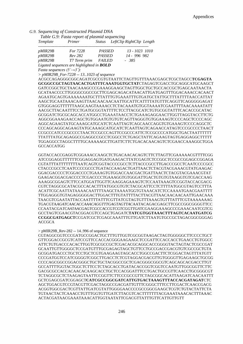
Appendices
246
G.9. Sequencing of Constructed Plasmid DNA
Table G.9: Fasta report of plasmid sequencing Template Primer Status LeftClip RightClip Length
---------------------------------------------------------------------------------
pMIR29B For 7228 PASSED 13 - 1023 1010
pMIR29B Rev 282 PASSED 14 - 996 982
pMIR29B T7 Term prim FAILED - 385
Ligated sequences are highlighted in BOLD
Fasta sequences (5`→3`):
> pMIR29B_For-7228 -- 13..1023 of sequence ACGCCAGAGGGCGGCAGATCGCCGTGTAATTCTAGTTGTTTAAACGAGCTCGCTAGCCTCGAGTA
GCGGCCGCTAGTAACACTGATTTCAAATGGTGCTATCTAGAGTCGACCTGCAGGCATGCAAGCT
GATCCGGCTGCTAACAAAGCCCGAAAGGAAGCTAGTTGGCTGCTGCCACCGCTGAGCAATAACTAGCATAACCCCTTGGGGCGGCCGCTTCGAGCAGACATAACATTGATGAGTTTGGACAAACCACAACT
AGAATGCAGTGAAAAAAATGCTTTATTTGTGAAATTTGTGATGCTATTGCTTTATTTTAACCATTATAAGCTGCAATAAACAAGTTAACAACAACAATTGCATTCATTTTATGTTTCAGGTTCAGGGGGAGAT
GTGGGAGGTTTTTTAAGCAAGTAAAACCTCTACAAATGTGGTAAAATCGAATTTTAACAAAATATTAACGCTTACAATTTCCTGATGCGGTATTTTCTCCTTACGCATCTGTGCGGTATTTCACACCGCATAC
GCGGATCTGCGCAGCACCATGGCCTGAAATAACCTCTGAAAGAGGAACTTGGTTAGGTACCTTCTGAGGCGGAAAGAACCAGCTGTGGAATGTGTGTCAGTTAGGGTGTGGAAAGTCCCCAGCTCCCCAGC
AGGCAGAAGTATGCAAAGCATGCATCTCAATTAGTCAGCAACCAGGTGTGAAAGTCCCCAGGCTCCCCAGCAGGCAGAAGTATGCAAAGCATGCATCTCAATTAGTCAGAACCATAGTCCCGCCCCTAACT
CCGCCCATCCCGCCCCTAACTCCGCCCAGTTCCGCCCATTCTCCGCCCCATGGCTGACTAATTTTTTTTATTTATGCAGAGGCCGAGGCCGCCTCGGCCTCTGAGCTATTCAGAAGTAGTGAGGAGGCTTTTT
TGGAGGCCTAGGCTTTTGCAAAAAGCTTGATTCTTCTGACACAACAGTCTCGAACCAAAGGCTGGAGCCACCATGG
GGTACCACCGAGGTCGGAAACCAAGCTCTGACAACACAGTCTTCTTAGTTCGAAAAACGTTTTCGG
ATCCGGAGGTTTTTTCGGAGGAGTGATGAAGACTTATCGAGTCTCCGGCTCCGCCGGAGCCGGAGA
CGTATTTATTTTTTTTAATCAGTCGGTACCCCGCCTCTTACCCGCCTTGACCCGCCTCAATCCCCGCCCTACCCGCCTCAATCCCCGCCCTGATACCAAGACTGATTAACTCTACGTACGAAACGTATGAAGAC
GGACGACCCCTCGGACCCCTGAAAGTGTGGACCAACGACTGATTAACTCTACGTACGAAACGTATGAAGACGGACGACCCCTCGACCCCTGAAAGGTGTGGGATTGACTGTGTGTAAGGTGTCGACCAAG
AAAGGCGGAGTCTTCCATGGATTGGTTCAAGGAGAAAGTCTCCAATAAAGTCCGGTACCACGACGCGTCTAGGCGCATACGCCACACTTTATGGCGTGTCTACGCATTCCTCTTTTATGGCGTAGTCCTTTA
ACATTCGCAATTATAAAACAATTTTAAGCTAAAATGGTGTAAACATCTCCAAAATGAACGAATTTTTTGGAGGGTGTAGAGGGGGACTTGGACTTTGTATTTTACTTACGTTAACAACAACAATTGAACAAA
TAACGTCGAATATTACCAATTTTATTTCGTTATCGTAGTGTTTAAAGTGTTTATTTCGTAAAAAAAGTGACGTAAGATCAACACCAAACAGGTTTGAGTAGTTACAATACAGACGAGCTTCGCCGGCGGGGTTCC
CCAATACGATCAATAACGAGTCGCCACCGTCGTCGGTTGATCGAAGGAAAGCCCGAAACAATCGTCGGCCTAGTCGAACGTACGGACGTCCAGCTGAGATCTATCGTGGTAAACTTTAGTCACAATGATCG
CCGGCGATGAGCTCCGATCGCTCGAGCAAATTTGTTGATCTTAATGTGCCGCTAGACGGCGGGAGACCGCA
> pMIR29B_Rev-282 -- 14..996 of sequence
CGTAGGCGGTCCCGATGCCGGACTGCTTTGTTGGTCGCGGTAAGACTAGTGGGGGCTTCCCCTGCTGTTCGGACCGCGTCATCCGTTCCACCACGGGAAGAAGCTCCGATTCCACCACCTGAACCTGTGGCC
ATTCTGTGACCCACACTTGGTCGCGCCGCTCGACACGCAGGCACCGGGGTACTAGTACTCGCCGATGCAATTGTTGGGGCTCCGATGTTTGCGAGAGTAGCTGTTCCTGCCGACCGACGTGTCGCCGCTGTA
GCGGATGACCCTGCTCCTGCTCGTGAAGAAGTAGCACCTGGCCGACTTCTCGGACTAGTTTATGTTCCCGATGGTCCATCGGGGTCGGCTTGACCTCTCGTAGGACGACGTTGTGGGGTTGAGAAGCTGCGG
CCCCAGCGGCCGGACGGGCTGCTGCTACGGCCGCTCGACGGGCGGCGTCAGCAGCACGACCTTGTGCCATTTTGGTACTGGCTCTTCCTCTAGCACCTGATACACCGGTCGGTCCAATGTTGGCGGTTCTTC
GACGCGCCACCACAACACAAGCACCTGCTCCACGGATTTCCTGACTGCCGTTCAACCTGCGGGCGTTCTAGGCGCTCTAAGAGTAATTCCGGTTCTTCCCGCCGTTCTAGCGGCACATTAAGATCAACAATTT
GCTCGAGCGATCGGAGCTCATCGCCGGCGATCATTGTGACTAAAGTTTACCACGATAGATCTCAGCTGGACGTCCGTACGTTCGACTAGGCCGACGATTGTTTCGGGCTTTCCTTCGACTCAACCGACG
ACGGTGGCGACTCGTTATTGATCGTATTGGGGAACCCCGCCGGCGAAGCTCGTCTGTACTATTCTA
TGTAACTACTCAAACCTGTTTGGTGTTGATCTTACGTCACTTTTTTTACGAAATAAACACTTTAAACACTACGATAACGAAATAAACATTGGTAATATTCGACGTTATTTGTTCATTGTTGTT

Appendices
247
H. In Vitro Transfection
H.1. Gaussia Transfection of Cultured Cells
1. A standard transfection assay of 10,000 cells uses 1 µg of G. Luciferase encoding plasmid
DNA at optimum w/w ratios of DNA: transfection agent.
2. Twenty-four hours prior to transfection, seed 10,000 cells in the well of a 24 well plate. The use
of cell culture inserts are convenient if confocal microscopy studies are to be performed.
3. On the day of transfection, prepare plasmid complexes under a cell culture laminar hood.
4. For a single transfection, add 1 µg of plasmid DNA to suitable amount of transfection reagent
in a sterile eppendorf.
5. Adjust volume to 50 µl with serum free media.
6. Gently vortex sample for five seconds and let stand at room temperature for a further ten
minutes.
7. Remove media from cells and add the complexed DNA.
8. Adjust volume to standard culture volume with standard cell media and place in cell culture
media.
9. At selected time periods (usually every two days), remove media from the cells and store at -20
°C for analysis.
10. Replace media with standard cell culture media and return to cell culture incubator.
11. At each time point, repeat steps 9 and 10.
H.2. Gaussia Luciferase Transfection Analysis
1. Thaw the samples that were collected according to Appendix O. Also thaw the luciferase buffer
supplied with the kit. For analysis of a single sample, the following protocol should be
followed:
2. Prepare luciferase dye (1 X) from stock solution supplied in kit (100 X). Use the supplied buffer
(1 X) for diluting. Note: This dye is sensitive to light so protect with tin foil.
3. Prepare a fresh opaque (white is best but be consistent with all measurements) 96 well plate.
4. Place 100 µl of PBS into a single well.
5. Add 10 μl of cell culture media which is removed from transfected cells.
6. Prepare plate reader for sample loading (ideally a plate reader with injection needles should be
used but if not available use a multi channel pipette and test one column per test run). Plate
reader settings should be set to luminescence (1.0 seconds).
7. Add luciferase dye which is prepared according to step 3.
8. Close plate reader lid and read sample.

Appendices
248
)(
)(
controlnegativecontrolpositive
controlnegativesample
RatioRatio
RatioRatioRI
9. A luciferase reading will appear on screen which is an indication of the level of transfection.
H.3. Dual Luciferase Transfection Analysis
1. Remove multiwell plates containing mammalian cells from the incubator. Make certain that the
plates are compatible with the type of luminometer being used.
2. Measuring firefly luciferase activity: Add a volume of Dual-Glo® Luciferase Reagent equal to
the culture medium volume to each well and mix. For 96-well plates, typically 75 μl of reagent
is added to cells grown in 75 μl of medium. For 384-well plates, typically 20 μl of reagent is
added to cells grown in 2 μl of medium.
3. Wait at least ten minutes, then measure the firefly luminescence (consult the luminometer
manual for proper use of the instrument). Optimal results will be generated if the luminescence
is measured within two hours of addition of Dual-Glo® Luciferase Reagent.
4. Measuring Renilla luciferase activity: Add a volume of Dual-Glo® Stop & Glo
® Reagent equal
to the original culture medium volume to each well and mix. As noted in Step 2, this volume is
typically 75 μl for 96-well plates and 20 μl for 384-well plates. Note: Dual-Glo® Stop & Glo
®
Reagent should be added to plate wells within 4 hours of addition of Dual-Glo® Luciferase
Reagent.
5. Wait at least 10 minutes, and then measure luminescence. Renilla luminescence should be
measured in the same plate as the firefly luminescence was measured (Step 3). Optimal results
will be generated if the luminescence is measured within 2 hours of addition of Dual-Glo® Stop
& Glo® Reagent.
6. Calculate the ratio of luminescence from the experimental reporter to luminescence from the
control reporter. Normalize this ratio to the ratio of a control well or series of control wells that
are treated consistently on all plates. This normalization provides optimal and consistent results
from the Dual-Glo® Luciferase Assay System.
7. Ratios can then be calculated from the Normalized Ratios. This is expressed on a scale of 0-1
where 0 is complete inhibition of a reporter protein (Renilla) and 1 is no inhibition whatsoever.
Positive control: Renilla/Luciferase Transfection only
Negative control: Cells only
Relative Inhibition
nilla
Firefly CE LUMINESCEN
CE LUMINESCEN Ratio
Re

Appendices
249
I. Cell Culture
I.1. Aseptic Technique
1. Ensure inside of hood is as clean as possible (spray with Virkon then 70% Ethanol) Spray
everything entering the hood with 70% Ethanol.
2. Assume every exposed surface is contaminated – if anything touches the hood or your hands or
lab coat, it most likely will have contamination.
3. The insides of sterile containers are the only clean areas (ie. sterile media, sterile plates, flasks,
tubes, etc.).
4. Keep sterile containers sealed.
5. Contamination often starts near the lids of containers – never touch lids or necks of flasks or
bottles with the tip of a pipette.
6. If something is contaminated, do not open it in the hood.
I.2. Feeding Flasks
1. Use aseptic technique (as always).
2. Clean hood, spray all equipment, take flask from incubator, spray with ethanol and wipe clean
with tissue.
3. Remove media from flask. Note: when pipetting, never let the liquid get up to the cotton at the
top of the pipette as this will break pipette boy will be broken.
4. Put media in waste container, or use aspirator to remove.
5. Pipette in new media (three sizes of flask commonly used: T25, T75, and T175)
T25: 3-5 mL media, T75: 8-10 mL media, T175: 25-30 mL media
6. Return flask to incubator.
I.3. Cell Splitting
1. Remove media from flask.
2. Wash flask with Hanks balanced salt solution or Dulbecco‘s PBS by pipetting in 5-10 mL,
tilting the flask to get the liquid covering the full surface, then removing it.
3. Add enough 0.25% Trypsin-EDTA to cover the bottom of the flask when it‘s lying in the
proper configuration (T25: 1 mL, T75: 3 mL, T175: 7-10 mL).
4. Put in incubator for five minutes (enzyme is active at 37 °C).
5. Take flask out and look at under microscope. Cells should be rounded and moving around if the
flask is examined. If the cells are still adhered to the bottom, try tapping the side of the flask
gently. If they don‘t come off, leave it for another minute or two.
6. Once all (or almost all) cells are free, spray the flask and return it to the hood.

Appendices
250
7. Add an equal volume of 10% serum media to the flask (ie. if you added 5 mL T/E, add 5 mL
media) (deactivates the trypsin).
8. Remove all of the liquid and transfer this to a sterile centrifuge tube (15 or 50 mL).
9. Spin for five minutes at 400 g.
10. Remove the supernatant and resuspend the pellet (the cells) in new media.
11. Count and seed into new flasks.
I.4. Thawing Cells
1. Wearing protective gloves and face shield, remove tube containing cells from liquid nitrogen
cylinder.
2. Warm tube in palm of hands or in waterbath.
3. When tube contents have thawed transfer to a 15 ml tube.
4. Add 10 ml or appropriate pre-warmed media with gentle aspiration.
5. Centrifuge the tube at 1500 rpm for five minutes.
6. Remove the supernatant and discard.
7. Add 10 ml or appropriate pre-warmed media with gentle aspiration to homogeneously
distribute cells.
8. Transfer the contents of the tube to a new tissue culture flask.
9. Label the flask with name, date and cell type.
10. Place the flask in an incubator set at 37 °C and 5% CO2.
11. Refresh media every two to three days.
I.5. Cell Seeding
1. When cells have become ~80% confluent in tissue culture flask remove from incubator.
2. Remove cell media and discard.
3. Add 5 ml of pre-warmed Hanks‘ balanced salt solution.
4. Gently agitate the cell culture flask.
5. Remove Hanks balanced salt solution and discard.
6. Repeat steps 3-5.
7. Add 3 ml of pre-warmed trypsin-EDTA.
Note: 3 ml is sufficient for T-75 flasks. Use 5 ml for T-175 flasks.
8. Place in incubator for five minutes.
9. Upon removal from incubator view the cells under light microscope. If cells have not detached,
gently tap the base of the flask to dislodge the remaining cells.
10. Return to laminar flow hood and add 7 ml of cell media.
11. Gently agitate flask.

Appendices
251
12. Remove contents of flask and deposit in a 15 ml tube.
13. Centrifuge the tube at 1500 rpm for five minutes.
14. Remove supernatant and discard taking care not to disturb the cell pellet at the base of the tube.
15. Add media (5-10 ml) to resuspend the pellet. Aspirate the media to homogenise the cells.
16. Remove a sample for use with the haemocytometer. 50 μl is sufficient. Place the sample in a
1.5 ml eppendorf.
17. Add 50 μl Trypan blue.
18. Assemble hemocytometer and inject a sample into either side with the 200 μl pipette.
19. Place the haemocytometer under the light microscope and count the number of cells in the
centre square on opposing sides of the hemocytometer.
20. Calculate the average cell number on opposing sides and multiply by 2 to account for Trypan
blue dilution.
21. Multiply by 10,000 to calculate the number of cells/ml in 15 ml tube. Depending on the
volume, the total cell number can be calculated.
22. Dilute the cells as per number required per well; i.e. if 10,000 cells are required per well then
dilute cells to 10,000 cells/ml.
23. Place sterile scaffolds and glass coverslips (controls) at the base of the well.
24. Add 1 ml of cell media containing appropriate number of cells.
25. Label the well plate with name, date and cell type.
26. Place well plate in incubator.
I.6. Freezing Cells
1. When freezing down cells, aspirate the media off cell pellet after it has been centrifuged.
2. Resuspend the pellet in freezing media (freezing media: 45 ml FBS, 5 ml DMSO. Make up
freezing media and store at 4 °C). Cell number determines amount of freezing media. Generally
~1 ml per vial, 500,000 cells to 5 million cells per vial.
3. Put cells in Mr. Frosty container and put into -80 °C freezer immediately. Cells can be then put
into liquid nitrogen after 24 hours.
I.7. Cell Counting
1. Take 50 μl of the cell suspension and add to 50 μl Trypan blue.
2. Add 10 μl of this cell/trypan blue suspension to each side of a haemocytometer (Trypan blue is
excluded by live cells – blue cells are dead, clear cells are alive, see Figure I.1 for schematic).
3. Count cells on both sides and get the average
4. To calculate the total cell number: average cell no./box x 104 X dilution factor (in this case 2) x
original volume cells were suspended in (in this case 2 ml).
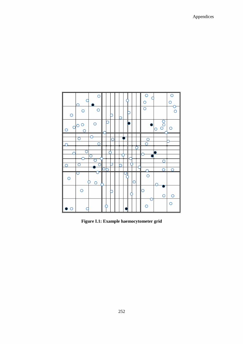
Appendices
252
Figure I.1: Example haemocytometer grid

Appendices
253
I.8. Isolation of Rat Neonatal Cardiac Fibroblasts
1. Every medium, HEPES buffer and PBS should be pre-warmed to 37 °C. Treating
cardiomyocytes with cold solutions will impair their functionality. Also prepare an ice-cold
aliquot of PBS.
2. Sacrifice two to three day old rats and excise hearts from all pups. Store the excised hearts in
calcium and magnesium free PBS on ice.
3. Squeeze hearts gently with forceps to expel the blood from the lumen. Transfer hearts into fresh
ice-cold PBS. Move ventricles to a dry six-cm Petri dish and mince tissue as small as possible
with a scalpel blade.
4. Transfer minced neonatal heart tissue into 20 ml of warm digestion buffer in a falcon tube and
incubate for 5 minutes in a 37 °C water bath. Mix either with a micro stirrer or by gentle
shaking.
5. Let the cells settle for five minutes, remove supernatant, add new pre-warmed digestion buffer
and repeat enzyme treatment six to seven times.
6. Let cells settle down and wash once with pre-warmed HEPES buffer containing 5 % horse
serum. Eventually, spin cells down for one minute at 340 g (if cardiomyocytes are required)
7. Resuspend cells in adhesion medium (20 ml per 10 hearts) and plate suspension on uncoated 10
cm dishes. Incubate for 1 - 1.5 hours at 37 °C/5 % CO2. Repeat this step. During this time,
fibroblasts will stick and spread on the plate whereas cardiomyocytes remain suspended.
8. Collect and count the cells to determine cell density. Expect a yield of 0.8 - 1 x 10⁶
cardiomyocytes per heart. Store cells in warm medium or buffer.
I.9. alamarBlue® Assay
1. Remove media from cells in culture.
2. Add 1000 µl/200 µl of Hanks‘ balanced salt solution into the required number of wells in a
sterile 24/96 well plate.
3. Prepare alamarBlue® working solution in Hanks‘ balanced salt solution (ratio 1:9 respectively).
Add 500 µl/200 µl depending on conditions.
4. Incubate for two hours at 37 °C.
5. After incubating for two hours, transfer 200 µl of the dye into a clear 96 well, flat bottomed
plate.
6. Measure the absorbance at 550 nm and 595 nm (0.5 seconds per well).
7. Subtract the absorbance values of media only from the absorbance values of the absorbance
values of alamarBlue® in media. This gives the absorbance of alamarBlue
® in media absorbance

Appendices
254
100
0
0
positivecontrolHWLW
sampleHWLw
RAA
RAA
of media only. Denote these values AOLW; absorbance of oxidized form at lower wavelength,
and AOHW ; absorbance of oxidized form at higher wavelength.
Calculate correlation factor:
HW
LW
AO
AOR 0
8. To calculate the percent of reduced alamarBlue®
1000 RAAAR HWLWLW
9. To calculate the relative % difference in metabolic reduction:
% Reduction:
J. Generation of Antibody Fragments from Hybridoma Culture
J.1. Extraction of Monoclonal Antibody from Hybridoma Supernatant
1. After the required period, remove cells from hybridoma supernatant by centrifugation at 1500
rpm, for five minutes. Sterile filter supernatant through 0.2 μm pore filter to remove any
remaining cell debris.
2. Add solution to sample filter cup (to a maximum of 70 mL); seal with supplied cap. Place
sample filter cup into filtrate collection cup.
3. Place Centricon Plus-70™
assembly in centrifuge bucket.
Caution: Before proceeding, check centrifuge clearance by manually moving bucket to its full-
swing position. Counterbalance centrifuge with a second bucket containing a second Centricon
Plus-70 unit and an equal volume of sample or water.
4. Spin at up to 3,500 × g until desired concentration is achieved. A typical spin time is 15–20
minutes, depending on solute type and concentration.
Note: If the centrifuge vibrates excessively, stop and rebalance it.
Remove Centricon Plus-70™
unit from centrifuge and separate the sample filter cup from the
filtrate collection unit.
Turn the concentrate/retentate cup upside down and place on top of the sample filter cup.
5. Carefully invert unit, place in centrifuge, and counterbalance with a similar device. Spin at no
more than 1,000 × g for up to two minutes.
6. Remove the retentate cup containing the concentrated sample from the sample filter cup. Keep
the filter cup inverted during this process.

Appendices
255
7. Remove the sample with a pipette or cap the retentate cup and store sample for later use.
J.2. Antibody Extraction from Protein Fraction
1. Equilibrate column and buffers to room temperature. Set table top centrifuge to 1,000 g.
2. Prepare sample for purification by diluting in Binding Buffer to a minimum of 5 ml, or a
maximum volume of 10 ml.
3. Loosen top cap on spin column and snap off bottom closure. Place column in a 50 ml collection
tube, centrifuge for one minute and discard the flow-through.
4. Equilibrate column by adding 10 ml of Binding Buffer. Centrifuge for one minute and discard
the flow-through. Repeat this step once.
5. Cap bottom of column with the included rubber cap. Apply sample to column and tightly cap
top. Incubate at room temperature with end-over-end mixing for ten minutes.
6. Loosen top cap and remove bottom cap. Place column in a new 50 ml collection tube and
centrifuge for one minute.
Note: This first collection tube contains the non-bound sample components and may be
analyzed to assess binding efficiency and capacity.
7. Place column in a new 50 ml collection tube. Wash column by adding 10 ml Binding Buffer.
Centrifuge for one minute and collect wash fraction. Repeat wash two additional times for a
total of three washes.
8. Add 500 μl of Neutralization Buffer to three 50 ml collection tubes and place the spin column
into one of the tubes.
9. Add 5 ml of Elution Buffer to the column and centrifuge for one minute. Transfer the spin
column to another tube that contains Neutralization Buffer, saving the collected solution as the
first elution fraction. Repeat this step two times to obtain three fractions.
10. Determine which fraction(s) contain the purified antibody by measuring the relative absorbance
of each fraction at 280 nm. If required for downstream applications, exchange the buffer using
Zeba™
Desalt Spin Columns, or by using Slide-A-Lyzer® Dialysis Cassettes (see related
Thermo Scientific Products section).
11. To regenerate the column for storage or re-use, add 10 ml of Elution Buffer and centrifuge for
one minute. Repeat once. Wash column with 10 ml of Storage Solution. Add 10 ml of Storage
Solution and store column at 4 °C. Typically, the immobilized protein column may be used up
to ten times without significant loss in binding capacity, although the actual number of effective
usages may vary.
J.3. Pepsin Digestion and Reduction of Monoclonal Antibodies Preparation of F(ab`)2

Appendices
256
1. Gently swirl the immobilized pepsin vial to obtain an even suspension. Seat the spin column frit
with an inverted 200 μl pipette tip.
2. Using a wide-bore or cut pipette tip, place 0.25 ml of the 50% slurry (i.e. 0.125 ml of settled
resin) into a 0.8 ml spin column. Twist off the bottom tab of the column and place into 2.0 ml
microcentrifuge tube. Centrifuge column at 5,000 x g for one minute and discard buffer.
3. Wash resin with 0.5 ml digestion buffer. Centrifuge column at 2,000 x g for one minute and
discard buffer. Cap bottom of spin column with included rubber cap.
4. Twist off the bottom closure of a Zeba™
Desalt Spin Column and loosen cap. Place column in a
15 ml collection tube.
5. Centrifuge column at 1,000 x g for two minutes to remove storage solution. Place a mark on the
side of the column where the compacted resin is slanted upward. Place column in centrifuge
with the mark facing outward in all subsequent centrifugation steps. Note: resin will appear
compacted after centrifugation.
6. Add 1 ml of digestion buffer to column. Centrifuge at 1000 g for two minutes to remove buffer.
Repeat this step three additional times, discarding buffer from the collection tube.
7. Place column in a new collection tube, remove cap and slowly apply 0.5 ml sample to the
centre of the compacted resin bed.
8. Replace cap and centrifuge at 1, 000 g for two minutes to collect the sample. Discard the
column after use.
9. If IgG sample is 0.5-8 mg/ml (i.e. 250 μg to 4 mg, no further preparation is necessary. If sample
volume is less than 0.5 ml, add digestion buffer to a final volume of 0.5 ml.
J.3.1 Fragment Generation
1. Add 0.5 ml of the prepared IgG sample to the spin column containing the equilibrated
immobilized pepsin. Place top cap and bottom plug on the spin column.
2. Incubate digestion reaction for the appropriate time, with an end-over mixer or a tabletop
rocker at 37 °C. Maintain constant mixing of resin during incubation.
3. Remove bottom cap and place column into a 2.0 ml microcentrifuge tube. Centrifuge column at
5,000 g for one minute to separate digest from the immobilized pepsin.
4. Wash resin with 0.5 ml of PBS. Place column into a tube and centrifuge at 5000 g for one
minute. Repeat this step once.
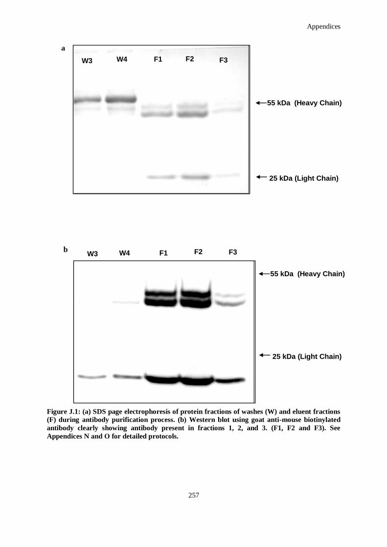
Appendices
257
Figure J.1: (a) SDS page electrophoresis of protein fractions of washes (W) and eluent fractions
(F) during antibody purification process. (b) Western blot using goat anti-mouse biotinylated
antibody clearly showing antibody present in fractions 1, 2, and 3. (F1, F2 and F3). See
Appendices N and O for detailed protocols.
a
W3 W4 F1 F2 F3
W3 W4 F1 F2 F3
55 kDa (Heavy Chain)
25 kDa (Light Chain)
b
55 kDa (Heavy Chain)
25 kDa (Light Chain)

Appendices
258
5. Add both wash fractions to the digested antibody. Total sample volume should be 1.5 ml.
Discard the immobilized pepsin.
Note: for best results, evaluate the digest and wash fraction via SDS-PAGE to assess digestion
completion. Protein purification is only required to remove undigested IgG and degraded Fc do
not bind to Protein A. The resulting F(ab)2 is non reducing. SDS derived from human and
mouse IgG will migrate with an apparent molecular weight of ~110 kDa.
J.3.2 F(ab`)2 Purification
1. Equilibrate the Nab™
Protein A column, PBS and IgG elution buffer to room temperature. Set
centrifuge to 1000 g.
2. Loosen top cap on the Protein A column and snap off bottom closure. Place column in a 15 ml
collection tube and centrifuge for one minute to remove storage solution. Discard the flow-
through.
3. Equilibrate column by adding 2 ml of PBS. Centrifuge for one minute and discard the flow-
through. Repeat this step once.
4. Cap bottom of column with the included rubber cap. Apply sample to column and cap the top
tightly. Resuspend the resin and sample by inversion. Incubate at room temperature with end-
over-end mixing for ten minutes.
5. Loosen top cap and remove bottom cap. Place column in a new 15 ml collection tube and
centrifuge for one minute. Save the flow-through as this fraction contains F(ab`)2 and Fc
fragments.
6. For optimal recovery, wash column with 1 ml of PBS. Centrifuge for one minute and collect
flow-through. Repeat and combine wash fractions with the F(ab`)2 fraction from step 5.
7. Measure protein concentration using the BCA assay or by measuring the absorbance at 280 nm.
Use an estimated extinction coefficient of 1.4. Assuming complete IgG digestion, F(ab)2 yields
may vary from 50 to 70 % depending on the amount of starting antibody and the protein assays
used.
8. If desired, perform dialysis (50K MWCO) to remove the Fc fragments.
J.3.3 Regeneration of Column
1. Apply 1ml of IgG elution buffer to the Protein A Column and centrifuge for one minute. Repeat
this step two times to obtain three fractions which will contain undigested IgG. To save the
undigested IgG, add 100 μl of a neutralization buffer to each of the elution fractions.
2. Add 3 ml of IgG elution buffer to the column and centrifuge for one minute. Discard flow-
through and repeat.
3. Add 3 ml of PBS to the column and centrifuge for one minute.

Appendices
259
4. For storage, add 3 ml of 0.02% sodium azide in PBS to column. Replace top and bottom camps.
Store column upright at 4 oC. Columns can be regenerated at least ten times without significant
loss of binding capacity.
J.4. Reduction of Di-sulfide Bonds (Production of Fab`s from F(ab`)2
1. Equilibrate immobilized reductant column and prepared equilibration buffers to room
temperature before use. Perform all steps of procedure at room temperature.
2. Remove top cap from the immobilized reductant column and pour off storage solution which
contains 0.02% sodium azide.
Note: When uncapping a column, always remove the top cap first to avoid drawing air into the
resin bed. If the top porous disc in the column is dislodged, push it down to the resin bed
surface (but avoid compressing the resin bed itself).
3. Twist off bottom column tab and stand column upright.
4. Equilibrate/wash the immobilized reductant by adding 5 ml of buffer #1 to the column and
allowing it to drain through.
5. Prepare DTT activation solution: Dissolve 15 mg of DTT in 10 ml buffer #1 (results in 10 mM
DTT).
6. Activate the column by applying 10 ml of DTT activation solution to the column and allowing
it to drain through.
7. To remove the activating DTT, wash the column with 10 ml of buffer #1. (If buffer #2 is
chosen and used for the sample, wash column with only 5 ml of buffer #1, followed by 5 ml of
buffer #2).
8. Apply 1 ml of peptide or protein solution to the column. Allow the sample to completely enter
the resin bed. (Flow will stop automatically when the liquid drains down to the top porous
disc).
9. Cap top and bottom of column (white bottom tips supplied) and incubate for 60 minutes for
protein samples; no incubation is needed for peptide samples.
10. Recover reduced peptide or protein sample from the column by applying 9 ml of the
appropriate buffer and collecting separate 1 ml fractions as they emerge from the column tip.
To ensure that the volume of each fraction is exactly 1 ml, apply only 1 ml of buffer at a time
and collect the entire volume that emerges until column flow stops; then change collection
tubes and apply the next 1 ml of buffer. This method utilizes the stop-flow feature of the top
porous disc.
11. Identify the fractions that contain the peptide or protein (now reduced) by determining which
ones have peak absorbance at 280 nm (usually the first two fractions). Be aware that some
peptides do not absorb significantly at 280 nm and cannot be assayed by this method.

Appendices
260
Alternatively, identify fractions that contain the reduced peptide or protein by specifically
measuring for sulfhydryl groups using the Ellman‘s Reagent.
12. Reuse the immobilized reductant column by simply repeating the procedure from the
beginning; the DTT activation step will regenerate the column. Four cycles of regeneration (5
total uses) are possible with each immobilized reductant column. For prolonged storage of used
columns, first wash them with phosphate buffer or water containing 0.02% sodium azide, and
then replace top and bottom caps and store the columns upright at 4 °C.
K. SDS -PAGE Analysis of Extracted Proteins
K.1. BioRad Protein Assay to Quantify Protein
1. Prepare dye reagent by diluting one part Dye Reagent Concentrate with four parts DDI water.
Filter through a Whatman #1 filter (or equivalent) to remove particulates. This diluted reagent
may be used for about two weeks when kept at room temperature.
2. Prepare three to five dilutions of a protein standard, which is representative of the protein
solution to be tested. The linear range of this microtiter plate assay is 0.05 mg/ml to
approximately 0.5 mg/ml. Protein solutions are normally assayed in duplicate or triplicate.
3. Pipet 10 μl of each standard and sample solution into separate microtiter plate wells.
4. Add 200 μl of diluted dye reagent to each well. Mix the sample and reagent thoroughly using a
microplate mixer. Alternatively, use a multi-channel pipet to dispense the reagent. Depress the
plunger repeatedly to mix the sample and reagent in the well. Replace with clean tips and add
reagent to the next set of wells.
5. Incubate at room temperature for at least five minutes. Absorbance will increase over time;
samples should incubate at room temperature for no more than one hour.
6. Measure absorbance at 595 nm.
K.2. Sample Preparation
1. Standard: 1 μg/μl.
2. Samples: 1 μg/μl.
3. Markers: 1 μl of Precision Plus Protein Dual Colour Standards™
(Bio-Rad, 161-0374) in 20 μl
of 5X sample buffer.
4. Empty well: 10 μl of 1X sample buffer (diluted from 5 X sample buffer with water).
5. For mini-gel; 10-well prepare a volume of 15 μl per well with at least 1μg of sample/control per
well.
6. Vortex samples and centrifuge briefly.

Appendices
261
7. Store at 4 °C. Prior to use, heat samples at 95 °C for five minutes, vortex and briefly centrifuge.
Load carefully in each well.
K.3. Preparation of Gels
1. See Table K.1 and K.2 for list of materials and quantities to make gels.
2. Thaw aliquot of ammonium persulphate (APS).
3. Clean glass plates with 70% ethanol and wipe dry with microscopic tissues.
4. Set the gel making apparatus ensuring that the glass plates fit snugly to the platform.
5. Check for leaks by pouring water prior to making the gels.
6. Add gel ingredients to make a 5% resolving gel. This can be done in 15/50 ml conical tube.
7. Add APS and TEMED last, right before gels are to be poured as this initiates polymerisation.
8. Carefully pour prepared mixture into space between the glasses plates until it reaches about 1
cm from the bottom of the wells etched out by the comb. Keep excess to monitor
polymerisation.
9. Overlay gel with 10% ethanol to seal gels.
10. Leave for approximately 30 minutes. A line at the ethanol-gel interface will appear which
indicates polymerisation.
11. Prepare stacking gel according to table. Again, add APS/TEMED immediately before use.
12. Aspirate ethanol out of glass plates using a syringe and imbibe any traces using filter paper.
13. Add APS and TEMED to stacking gel and carefully add to separation gel. Immediately insert
the comb, taking care to avoid trapping any air bubbles. Keep excess to monitor
polymerisation.
14. Allow to set for 10-15 minutes and, in the meantime, denature samples and standards as
described above.
15. After gels have been set, slowly remove combs.
16. Assemble electrophoresis apparatus.
17. Fill the upper/inner chambers with 1 X Running buffer.
18. Wash well with running buffer with a hypodermal needle syringe.
19. Load all samples, standards and markers.
20. Put upper chamber on the main chamber and close the lid. Run the gels
21. For a mini gel: run at 50 V until the front reaches the end of the stacking gel (±35 minutes),
then adjust to 120 V until the front reaches the end of the separating gel (±1hour).
22. Separate the glass plates and release the gel slowly into ddH2O.
23. Proceed with coomassie or silver staining, or western blotting of separated protein.
Appendices
262
Table K.1: 5% Separation gel (1 mm thickness) for collagen mini gel (Protean II® Bio-Rad)
Component Volume
30% Acrylamide/Bis (37.5:1) 830 μl
1.875 M Tris-HCl pH 8.8 1000 μl
10% SDS 50 μl
ddH2O 3070 μl
Ammonium Persulphate (APS, 100 mg/ml) 42 μl
TEMED 5 μl
Total Volume 5000 μl
Table K.2: 3% Stacking gel (1 mm thickness) for collagen mini gel (Protean II® Bio-Rad)
Component Volume
30% Acrylamide/Bis (37.5:1) 200 μl
1.25 M Tris-HCl pH 6.8 200 μl
10% SDS 33 μl
ddH2O 1550 μl
Ammonium Persulphate (APS, 100 mg/ml) 17 μl
TEMED 2 μl
Total Volume 2000 μl
L. Western Blot Analysis of Transferred Proteins
Note: Prepare the transfer buffer, blotting pads, and blotting membranes before performing the
transfer. Preparation of the transfer buffer and materials for transfer may be made while
electrophoresis of the gel is in progress.
Wear gloves at all times during the entire blotting procedure to prevent contamination of gels
and membranes, and to avoid exposure to irritants commonly used in electrophoresis and
blotting procedures.
Do not touch the membrane or gel with bare hands. This may contaminate the gel or membrane
and interfere with further analysis.
Prepare the appropriate buffer for gel type from the recipe according to Table L.1.

Appendices
263
Table L.1: Transfer buffer recipe for western blot procedure.
Component Volume
Novex® Tris-Glycine Transfer Buffer (25 x) 40 mL
Methanol 200 mL
Deionized Water 760 mL
Total Volume 1,000 mL
Use ~ 700 mL of transfer buffer to soak the blotting pads until saturated. Remove air bubbles
by squeezing the blotting pads while they are submerged in buffer. Removing air bubbles is
essential as they can block the transfer of biomolecules.
PVDF membrane: Pre-wet the PVDF membrane for 30 seconds in methanol, ethanol, or
isopropanol. Briefly rinse in deionized water and then place the membrane in a shallow dish
containing 50–100 mL transfer buffer for several minutes.
Nitrocellulose/Nylon membrane: Place the membrane directly in a tray containing the transfer
buffer for several minutes.
Filter paper: Soak briefly in transfer buffer immediately before using.
Gel: Use the gel immediately following the SDS PAGE described in Appendix K. Do not soak
the gel in transfer buffer.
Remove the gel from the cassette for transfer after completion of electrophoresis as described
below/ One may continue electrophoresis of the gel at a low voltage of 5 V. The gel can be left
in the unit for a few hours until it is ready to transfer the gel.
1. After electrophoresis, separate each of the three bonded sides of the gel cassette by inserting the
gel knife into the gap between the cassette‘s two plates. The notched (―well‖) side of the
cassette should face up.
2. Push up and down on the knife handle to separate the plates. Repeat on each side of the
cassette until the plates are completely separated.
Caution: Use caution while inserting the gel knife between the two plates to avoid excessive
pressure towards the gel.
3. The gel may adhere to either side of plates upon opening the cassette. Carefully remove and
discard the plate without the gel. The gel remains on the other plate.
4. Remove wells on the gel with the gel knife.

Appendices
264
5. Place a piece of pre-soaked filter paper on top of the gel, and lay just above the ―foot‖ at the
bottom of the gel (leaving the ―foot‖ of the gel uncovered). Keep the filter paper saturated with
transfer buffer and remove all trapped air bubbles by gently rolling over the surface using a
glass pipette.
6. Turn the plate over so the gel and filter paper are facing downwards over a gloved hand or
clean flat surface covered with a piece of Parafilm®.
7. Remove gel from the plate using the following methods:
8. If the gel rests on the longer (slotted) plate, use the gel knife to push the foot out of the slot in
the plate and the gel will fall off easily.
9. If the gel rests on the shorter (notched) plate, use the gel knife to carefully loosen the bottom of
the gel and allow the gel to peel away from the plate.
10. When the gel is on a flat surface, cut the ―foot‖ off the gel with the gel knife.
Note: Once you have removed the gel from the unit and the cassette, perform the transfer
immediately.
11. Wet the surface of the gel with the transfer buffer and place pre-soaked transfer membrane on
the gel. Remove air bubbles by rolling a glass pipette over the membrane surface.
12. Place the pre-soaked filter paper on top of the transfer membrane. Remove any trapped air
bubbles.
13. Place two soaked blotting pads into the cathode (–) core of the blot module. The cathode core is
the deeper of the two cores and the corresponding electrode plate is a darker shade of gray.
Carefully pick up the gel membrane assembly with a gloved hand and place on the pad in the
same sequence, such that the gel is closest to the cathode plate.
14. Add enough pre-soaked blotting pads to rise 0.5 cm over the rim of the cathode core. Place the
anode (+) core on top of the pads. The gel/membrane sandwich should be held securely
between the two halves of the blot module ensuring complete contact of all components.
Note: To ensure a snug fit, use an additional pad since pads lose their resiliency after many
uses. Replace pads when they begin to lose resiliency and are discolored.
15. Position the gel membrane sandwich and blotting pads in the cathode core of the XCell II™
Blot
Module to fit horizontally across the bottom of the unit. There should be a gap of ~ 1 cm at the
top of the electrodes when the pads and assembly are in place.
Hold the blot module together firmly and slide it into the guide rails on the lower buffer
chamber. The blot module fits into the unit in only one way, such that the (+) sign is seen in the
upper left hand corner of the blot module. The inverted gold post on the right hand side of the
blot module fits into the hole next to the upright gold post on the right side of the lower buffer
chamber.

Appendices
265
16. Depending on the mini-cell that that is being used, follow the appropriate instructions for
positioning the wedge:
Note: When properly placed, the rear wedge will not be flush with the top of the lower buffer
chamber. There will be a gap between the rear wedge and lower chamber.
17. Fill the blot module with transfer buffer until the gel/membrane sandwich is covered in transfer
buffer.
18. Do not fill all the way to the top as this will generate extra conductivity and heat.
19. Fill the outer buffer chamber with ~ 650 mL deionized water by pouring in the gap between the
front of the blot module and front of the lower buffer chamber. The water level should reach
approximately 2 cm from the top of the lower buffer chamber. This serves to dissipate heat
produced during the run.
20. Place the lid on top of the unit.
21. With the power turned off, plug the red and black leads into the power supply.
22. For overnight blotting, perform transfer in the cold room with low power to prevent
overheating. Transfer at constant voltage of 10–15 V overnight. Depending on the transfer
efficiency, adjust the transfer conditions accordingly.
23. Remove blot from the transfer apparatus.
L.1. Ponceau Stain 1% (v/v) acetic acid)
1. Add in the order listed:
10 ml Milli-Q Water
0.3 ml glacial acetic acid (use glassware to measure)
0.033 g Ponceau S
Fill to 30 ml with MiliQ water and store at room temperature.
L.2. Immunostaining of Transfer Membrane from Western Blot
1. Block nonspecific sites with blocking reagent for 20-60 minutes at room temperature (RT) with
Tris Buffered Saline with 0.1% Tween-20 (TBST) containing 5 % (w/v) of bovine serum
albumin (BSA). For best results, block for 1 hour at RT. Blots also may be blocked overnight at
2-8 °C.
2. Remove the blocking reagent and add the appropriate primary antibody dilution. Dilute
antibody to the appropriate dilution factor with TBST containing 5 % (w/v) BSA. Incubate blot
for 1 hour with shaking.
3. If desired, blots may be incubated with primary antibody overnight at 2-8 °C.
4. Wash membrane by suspending it in TBST and agitating for ≥5 minutes. Replace TBST at least
four to six times.

Appendices
266
5. Increasing the TBST volume and/or the number of washes may help reduce background.
Note: Briefly rinsing membrane in TBST before secondary incubation will increase wash
efficiency.
6. Incubate blot with the appropriate HRP-conjugate dilution (dilute in TBST containing 5 %
(w/v) BSA for one hour at RT with shaking. Repeat Step 5 to remove unbound HRP-conjugate.
Note: Membrane must be thoroughly washed after incubation with the HRP-conjugate.
7. Prepare working solution by mixing equal parts of the stable peroxide solution and the
luminol/enhancer solution.
8. Use 0.1 ml working solution per cm2 of membrane. The working solution is stable for 24 hours
at room temperature.
Note: Exposure to the sun or any other intense light can harm the working solution. For best
results keep the working solution in an amber bottle and avoid prolonged exposure to any
intense light. Short-term exposure to typical laboratory lighting will not harm the working
solution.
9. Incubate blot with working solution for five minutes.
Note: Perform previous steps to stain for GAPDH, β-actin or another appropriate loading
control. This is essential to the evaluation of the previous proteins detected.
10. Remove blot from working solution and place it in a plastic membrane protector. (A plastic
sheet protector works very well, although cling film may also be used.) Use an absorbent tissue
to remove excess liquid and to carefully press out any bubbles from between the blot and the
membrane protector.
M. Protein Blot Array
M.1. Protein Extraction
1. Retrieve protein from storage and keep on icebox.
2. Prepare lysis buffer. Avoid using SDS or other strongly denaturing detergents. In general, non-
ionic detergents, such as Triton X-100 or NP-40 are best, although zwitterionic detergents, such
as CHAPS, or mild ionic detergents, such as sodium deoxycholate will work. Use no more
than 2% v/v total detergent Avoid the use of sodium azide Avoid reducing agents, such as
dithiothreitol (DTT) or mercaptoethanols. In this instance, commercially available Lysis Buffer
from Sigma Aldrich was used.
3. Place approximately 1 gram of tissue into eppendorf suitable for TissueLyserLT bead mill
(Qiagen) with 1 ml lysis buffer and one stainless steel bead. The TissueLyserLT functions by
vertically oscillating the rupture chamber at a high speed to move the stainless steel bead up
and down rapidly within the eppendorf to rupture and homogenise the associated tissue.

Appendices
267
4. Place eppendorfs into the pre-cooled bead mill chamber and begin rupture for one minute.
Check samples to see if complete homogenisation has taken place. Otherwise repeat rupture
until complete homogenisation occurs, taking care to keep the ependorfs cool.
M.2. Protein Blot
1. The cell culture supernates should be dialyzed with a Dialysis tube (Item A) before the biotin
labeling procedure. Recommended loading is 2.5~3.0 ml of cell culture supernates into a
dialyzer and dialyzing with at least 2,000 ml 1X PBS buffer (pH = 8) at 4 °C. Change the 1 x
PBS buffer and dialyze again. Allow at least three hours for each dialysis step, stirring gently.
The sample volume may be altered after dialysis.
2. Briefly spin down Internal Control tube (Item C) before use. Add 100 ml 1X PBS, pH=8.0 into
the Internal Control tube, pipette up and down to dissolve the powder. Transfer 2 ml dialyzed
sample into a new tube. Add 40 ml prepared Internal Control into the tube. Mix well.
Immediately before use, briefly spin down the Labeling Reagent tube (Item B). Add 100 ml 1X
PBS into the tube, pipette up and down or vortex to dissolve the powder to prepare 1X Labeling
Reagent solution. Add an appropriate amount of prepared Labeling Reagent into above tube
with sample in step 2, mix well immediately. Incubate the reaction solution at room
temperature for 30 min with gentle shaking. Gently tap the tube to mix the reaction solution
every 5 min. 7.2 ml of 1X Labeling Reagent for labeling 1 mg total protein in supernates.
3. Add 5 ml Stop Solution into the above reaction solution and then use the spin column to
remove free biotin. Twist off the spin column‘s bottom closure and loosen the cap. Place the
column into a 50 ml collection tube. Centrifuge column at 1,000 g for three minutes to remove
storage solution. Add 5 ml 1X PBS into column, centrifuge at 1,000 g for three minutes to 1X
PBS. Repeat additional two times to wash the column. Place the column in a new collection
tube, slowly load the sample to the center of the compact resin bed. Centrifuge the column at
1,000 g for three minutes to collect sample. Stored at –80 °C until testing. Discard column after
use.
4. Place each membrane into the provided tray (―-‖ mark is on the antibody printed side). Note:
The printed side should be facing upward. Add 2.5 ml Blocking Buffer and incubate at room
temperature for one hour to block membranes. Decant Blocking Buffer from each container.
Add 2.5 ml of sample into each array membrane, and cover with the lid. Incubate at room
temperature with gentle shaking for 2 hours. Dilute sample using Blocking Buffer.
5. Decant the samples from each container, and wash three times with 3 ml of 1X Wash Buffer I
at room temperature with shaking and five minutes per wash. Dilute 20X Wash Buffer I with
deionized or distilled water. Decant the 1X Wash Buffer I from each container. Wash three
times with 3 ml of 1X Wash Buffer II at room temperature with gentle shaking. Add 2.5 ml of

Appendices
268
500 fold diluted HRP-conjugated streptavidin (e.g. add 10 ml of HRP-conjugated streptavidin
to 5 ml of Blocking Buffer) to each membrane.
Note: Mix tube containing 500X HRP-Conjugated Streptavidin well before use since
precipitation may form during storage. Incubate at room temperature with gentle shaking for
two hours.
6. Add 2.5 ml of Detection Buffer C and 2.5 ml of Detection Buffer D into a tube (for detecting 2
membranes); Mix both solutions; Drain off excess wash buffer. Place membrane protein side up
(―- ‖ mark is on the protein side top left corner) on a clean plastic plate or its cover (provided in
the kit). Pipette 2.2 ml of the mixed Detection Buffer on to each membrane and incubate at
room temperature with shaking for 2 minutes. Ensure that the detection mixture is evenly
covering the membrane without any air bubbles. Gently place the membrane with forceps,
protein side up, on a piece of plastic sheet (―-‖ mark is on the protein side top left corner).
Cover the array with another piece of plastic sheet. Gently smooth out any air bubbles. Avoid
using pressure on the membrane. Work as quickly as possible. The signal can be detected
directly from the membrane using a chemiluminescence imaging system or by exposing the
array to x-ray film (we recommend using Kodak X-Omat™ AR film) with subsequent
development. Expose the membranes for 40 Seconds. Then re-expose the film according to the
intensity of signals. If the signals are too strong (background too high), reduce exposure time
(eg, 5–30 seconds). If the signals are too weak, increase exposure time (eg, 5–20 min or
overnight). Or re-incubate membranes overnight with 1X HRP-conjugated streptavidin, and
repeat detection on the second day. Save membranes at –20 °C to –80 °C for future reference.
M.3. Image Analysis (Using LUTS Protein Membrane Array
1. Open images in ImageJ. Begin LUTS Protein Membrane Array.
2. Determine and select the array settings for detection on the images obtained from protein array
chemiluminesence.
3. Perform intensity profiles on all array images.
4. Masterise all images using the options on the LUTS Protein Membrane Array (see Figure M.1).
5. Normalise all blots using positive controls and blanks.
6. Export all data images and numerical values of intensity for further analysis.
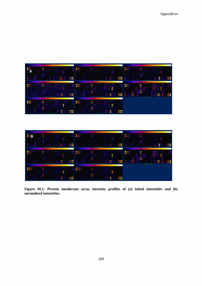
Appendices
269
Figure M.1: Protein membrane array intensity profiles of (a) initial intensities and (b)
normalised intensities.
A
B
Appendices
270
N. Michael –type Addition of Fab`s to Hyperbranched Polymer
N.1. Ellman’s Assay for Quantitation of Reduced Fragments Material Preparation
Reaction buffer: 0.1 M sodium phosphate, pH 8.0, containing 1 mM EDTA
Cysteine hydrochloride monohydrate: M.W. = 175.6
Ellman‘s reagent solution: Dissolve 4 mg Elman‘s Reagent in 1 ml of reaction
buffer.
1. Prepare a set of cysteine standards by dissolving cysteine hydrochloride
monohydrate at the following concentrations in reaction buffer according to
Table N.1.
Table N.1: Guidelines for preparation of cysteine standards for Ellman‘s assay.
0.25 mM Volume of
Reaction
Buffer
25 ml 5 ml of Standard A
Standard Amount of Cysteine
(M.W. = 175.6)
Final Concentration
A 100 ml 26.34 mg 1.5 mM
B 5 ml 25 ml of Standard A 1.25 mM
C 10 ml 20 ml of Standard A 1.0 mM
D 15 ml 15 ml of Standard A 0.75 mM
E 20 ml 10 ml of Standard A 0.5 mM
G 30 ml 0 ml 0.0 mM (Blank)
2. Prepare a set of test tubes, each containing 50 μl of Elman‘s reagent solution and 2.5 ml of
reaction buffer.
3. Add 250 μl of each standard or unknown to the separate test tubes prepared in step 2.
Note: For the unknown(s), make dilutions so that the 250 ul sample applied to the assay
reaction has a sulfhydryl concentration in the working range of the standard curve (0.1-1.0 mM
is ideal).
4. Mix and incubate at room temperature for 15 minutes.
5. Measure absorbance at 412 nm.
6. Plot the values obtained for the standards to generate a standard curve. Determine the
experimental sample concentrations from this curve.
Note: The most accurate results are obtained from the linear portion of the standard curve; i.e.,
the portion yielding an r2
value equal to 1.0. One or more of the high standards may exceed the
linear range.
Appendices
271
N.2. Michael-type Addition Conjugation of Antibody Fragments to pDMAEMA co-branched
PEGDA/PEGMEA
1. Note molar concentration of thiols calculated using Ellmans Assay in Appendix O.1.
2. From 1H NMR data calculate the moles of free vinyl present per mole of pDMAEMA co-
branched PEGDA/PEGMEA
3. Degas 0.1 M sodium phosphate, pH 8.0, containing 1 mM EDTA using argon for one hour prior
to experiment.
4. Using 0.1 M sodium phosphate, pH 8.0 containing 1 mM EDTA as the reaction buffer (10 x),
add antibody fragment containing solution and polymer containing solution together. If
possible, resuspend polymer in 0.1 M sodium phosphate, pH 8.0, containing 1 mM EDTA to
maintain equilibrium of reaction buffer.
5. React at room temperature under gentle rocking for at least 30 minutes. Keep samples for
analysis using SDS-PAGE and for use as complexing reagent in further experiments.
O. Rat Excisional Model
1. Anaesthetize animal using isoflurane and oxygen. In the induction phase 5% of isoflurane will
be used until surgical levels of anaesthesia are reached.
2. The percentage of isoflourane is then reduced to maintain these levels for the duration of the
procedure (to about 2%).
3. Shave the hair off the dorsum areas of the rats.
4. Swab the surgical area with 4% chlorohexidine or povidone iodine to control bacterial
contamination of the surgical field.
5. Record wound size in the animal using tracing paper.
6. Cover the wounds with a standard bio-occlusive transparent dressing (e.g. Opsite®
Smith &
Nephew).
7. Place additional wound dressing/ jackets on the animals with the intention of preventing wound
disturbance.
8. Identify animals using tail marking with a permanent marker.
9. Closely monitor the animals until full recovery from anaesthesia is evident.
10. House the animals individually housed to avoid interference with the dressing. House the
animals in transparent cages and provide standard environmental enrichment.
11. Sacrifice the animals at the required time periods using an overdose of sodium pentobarbital
solution or CO2 asphyxiation.
12. Trace the healed areas on sterile tracing papers and photograph prior to excision and future
histomorphometry.
13. House animals appropriately and monitor regularly using the following distress scoring sheet.

Appendices
272
Distress Scoring Sheet Animals reaching scores of 12 or more will be euthanised.
Parameter Symptom Score Date/Time Date/Time Date/Time Date/Time
Appearance
Normal 0
Lack of grooming 1
Piloerection, ocular and
nasal discharge 2
Piloerection, hunched up 3
Above and eyes half closed 4
Natural
behaviour
Normal 0
Minor changes 1
Less mobile, but alert 2
Restless or still, not alert 3
Hydration Normal 0
Abnormal pinch test 5
Respiratory
signs
No dressing disturbance 0
Mild oozing through
dressing 1
Overt discharge from
wounds 2
Dressings severely damaged 3
Dressing removed and
wounds severely damaged 4
Provoked
behaviour
Normal 0
Minor depression or
exaggerated response 1
Moderate change in
expected behaviour 2
Very weak and precomatose 3
Total 0-19
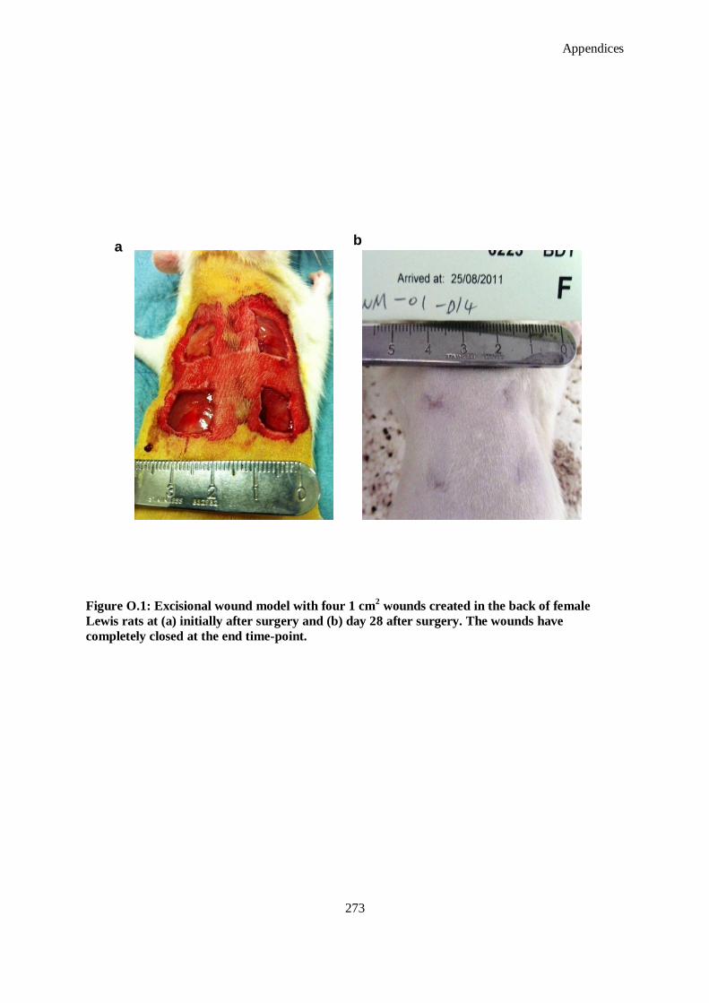
Appendices
273
Figure O.1: Excisional wound model with four 1 cm2 wounds created in the back of female
Lewis rats at (a) initially after surgery and (b) day 28 after surgery. The wounds have
completely closed at the end time-point.
a b
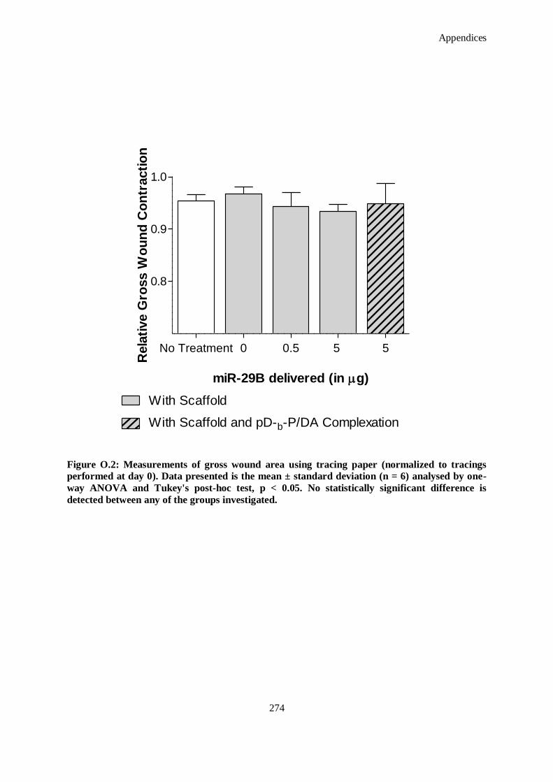
Appendices
274
No Treatment 0 0.5 5 5
0.8
0.9
1.0
With Scaffold
With Scaffold and pD-b-P/DA Complexation
miR-29B delivered (in g)
Rela
tive G
ross W
ou
nd
Co
ntr
acti
on
Figure O.2: Measurements of gross wound area using tracing paper (normalized to tracings
performed at day 0). Data presented is the mean ± standard deviation (n = 6) analysed by one-
way ANOVA and Tukey's post-hoc test, p < 0.05. No statistically significant difference is
detected between any of the groups investigated.

Appendices
275
14. Import images into ImageJ and trace the wound area. Calculate normalized wound contraction
area index using the following formula:
Normalized Wound Contraction Index: 0
0
Aw
AAw wt
where Aw0 is wound area at time of surgery (day 0) and Awt is the wound area at the end time point
(28 days). Data is presented in Figure O.2.
P. Generation of In Vitro Human Skin Equivalents
P.1. Isolation of Keratinocytes
1. Collect data from the biopsy (procedure, date, patient age, gender, region extraction,
dimensions, weight, location of procedure).
2. Place biopsy in 94 mm Petri dish .
3. Remove gross adipose tissue with scalpel.
4. Cut the biopsy into small 0.5 cm2 portions.
5. Rinse twice with PBS plus and a third time with PBS minus.
6. Place tissue into small petri dish.
7. Add 20 mls of dispase (or more precisely 1.25 ml/cm2).
8. Incubate for 18 hours maximum at 4 °C (under static condition).
9. After incubation period, carefully remove epidermis which has dissociated from the
underlying dermis.
10. Remove dispase solution and rinse epidermal tissue with PBS plus (retain dermis portion for
fibroblast isolation.
11. Remove PBS plus, rinse with PBS minus.
12. Place epidermis in a tube, and add 5 mls of trypsin.
13. Rinse petri dish with a further 5 mls of trypsin and add to the tube.
14. Incubate for five minutes in a 37°C waterbath and vortex briefly every two minutes.
15. Stop tyrpsin activity with 1 ml of FCS.
16. Disrupt the solution by pipetting continuously for five minutes until all the tissue has been
disrupted.
17. Strain solution with a 100 μm cell strainer.
18. Rinse the strainer three times with a 5 ml PBS rinse.
19. Centrifuge the keratinocyte suspension that has gone through the strainer for five minutes at
1000 rpm.
20. Resuspend the pellet in KGM-2 Ready media.

Appendices
276
21. Seed 600,000 cells/ T75 flask.
P.2. Isolation of Fibroblasts
1. Place dermal pieces previously acquired in a fresh petri dish.
2. Wash twice with PBS minus and add to a centrifuge tube.
3. Add 40 ml of collagenase to the tissue (more precisely 0.625 ml/cm2).
4. Incubate for four hours at 37 °C.
5. Separate the remaining undigested pieces using a cell strainer and rinsing three times using
PBS plus.
6. Centrifuge the cell suspension for five minutes at 1500 rpm.
7. Wash once with serum containing DMEM.
8. Centrifuge again for five minutes, this time at 1000 rpm.
P.3. Generation of Organotypical Skin Model
1. To generate a volume for a 500 ml Nunc cell culture insert, the collagen and the
Neutralisation Solution need to be added at a ratio of 2:1. At time of experiment, place both
solutions separately on ice.
2. Trypsinise fibrobasts, centrifuge, count and adjust cell suspension concentration to 1 x 10⁶
cells/ml. 50,000 cells are used per 500 ml of collagen.
3. Remove supernatant and resuspend the cells in a required volume of cooled Gel neutralisation
solution, carefully without making bubbles.
4. Transfer the suspension into the collagen solution, resuspend gently but quickly.
5. Using a multi-step pipette, pipette the collagen-cell mixture using a 500 ml pipette rapidly
into each cell culture insert.
6. Allow to stand for 10-20 minutes at 37 °C, to allow gelation to occur.
7. Submerge the gels with DMEM containing 10 % FCS and place in incubator.
8. One day after creating the gels remove the media and coat gels with fibronectin. Allow to coat
for 30 minutes in an incubator.
9. During this time, trypsinise keratinocyes, centrifuge and resuspend in KGM-2 (5% FCS) at 1
x 106 cells/ml.
10. Seed cells on the gels at 1 x 10⁵ cells/gel (100 μl).
11. After 45 minutes incubation in an incubator, submerge gels with KGM-2 (5% FCS).
12. Over the days, decrease the concentration the KGM-2 media contains (5 – 2 %) and resulting
at 0 % FCS by day 6/7. Perform each media change every two days.
13. Aspirate the medium above the gel completely, while not touching the gel with the pipette tip.
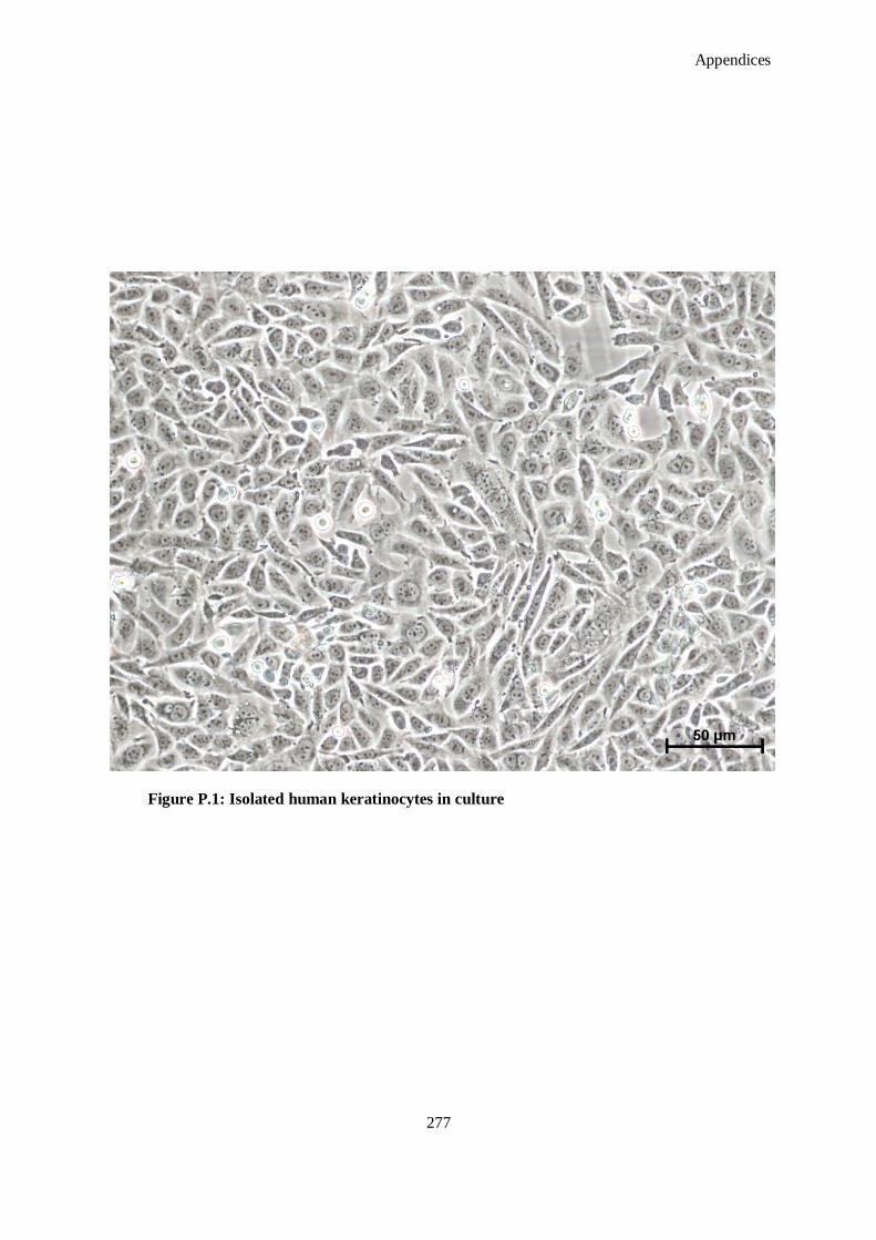
Appendices
277
Figure P.1: Isolated human keratinocytes in culture

Appendices
278
14. Add airlift medium (KGM-2 kit ready mixed with 3.1 ml of 300 mM CaCl2 solution) in the
surrounding outer space (of the wells), filling the media to the meniscus of the gel medium.
15. Pay attention to the media consumption of the cells, and carry out media change every two to
three days until day 12. After 12 days, these are the skin models for testing.
Q. Fixation Methods
Q.1. Formalin Fixation for Tissue
Note: Formalin crosslinks everything, so all endogenous RNases are destroyed.
Important for further RNA extraction of FFPE-samples. Not suitable for fixing cells.
1. Wash the tissue sample in RNase-free water or PBS.
2. Fix the sample in an adequate volume of 10% Formalin in PBS for exactly 12 hours. Wash in
RNase-free water.
3. Put the sample then in 70% ethanol (prepared with RNase-free water and ethanol with
molecular biology grade) and store the sample at 4 °C until starting the embedding procedure.
4. Start embedding machine (starts directly in 70% ethanol), so that no additional water steps are
performed.
Q.2. Fixation with 4% Paraformaldehyde in PBS: for Cells
Note: Paraformaldehyde crosslinking is not so strong and therefore not all endogenous RNases
may be destroyed. Should be used to fix cells, with formalin they will be excessively fixed.
1. Wash the sample with PBS.
2. Fix the sample in an adequate volume of 4% paraformaldehyde in PBS.
3. Tissues can also be left in this solution for up to 48 hours.
R. Staining Methods
R.1. Haematoxylin and Eosin Staining
1. Place microtomed slides in an oven at 60 °C overnight.
2. Hydrate:
Place in Xylene for 10 minutes
Place in new Xylene for 5 minutes
100% ethanol for 3 minutes
95 % ethanol for 3 minutes
70% ethanol for 3 minutes
Rinse in ddH2O and leave for 5 minutes

Appendices
279
3. Stain in Hematoxylin for eight minutes.
4. Wash in running tap water for eight minutes.
5. Differentiate in 1% acid alcohol for 30 seconds.
Note: prepare the 1% acid alcohol fresh and be strict with the 30 seconds time).
6. Wash slides in running H2O for five minutes.
7. Blue in 0.2 % ammonia H2O or saturated lithium carbonate for 30 seconds to one minute.
8. Wash in running tap water for five minutes.
9. Rinse in 95% EtOH – dip ten times.
10. Counterstain in Eosin/Phloxine solution for 30 seconds – 1 minute.
11. Dehydrate:
95% ethanol for 5 minutes
100% ethanol for 5 minutes twice
Xylene for 5 minutes twice.
12. Mount immediately in DPX solution with coverslips. Leave to hard-set.
R.2. Russell Movat Pentachrome Stain
10% alcoholic Haematoxylin:
20 g Haematoxylin
+ 200 ml absolute ethanol
Note: long stirring is needed to dissolve it
Universal iodine solution (Weigert’s Iodine solution):
4 g potassium iodide
+ 2 g iodine
200 ml ddH2O
1% Alcian blue solution:
2 g Alcian blue
+ 200 ml 3% glacial acetic acid
Crocein scarlet-acid fuchsin:
Croceins scarlet (Stock solution)
0.2 g Crocein scarlet
+ 200 ml 0.5% glacial acetic acid
Acid fuchsin (Stock Solution)
0.2 g Acid fuchsin
+ 200 ml 0.5% glacial acetic acid
Crocein scarlet – acid fuchsin (Working Solution)
Mix 8 parts Crocein Scarlet ―stock‖ with 2 parts Acid Fuchsin ―stock‖

Appendices
280
Note: for substances, which are hard dissolve use a pinch of thymol.
1. Deparaffinize and hydrate (ten minutes each)
Xylene x 2
100% ethanol x 2
70% ethanol x 2
50% ethanol x 2
ddH2O x 2
Mix components in the order shown below just prior to use to make Verhoeff‘s elastic
stain:
50 ml 10% alcoholic haematoxylin
50 ml absolute ethanol
50 ml 10% ferric chloride
50 ml universal iodine solution
3. Place slides in Verhoeff‘s elastic stain for 15 minutes.
4. Rinse slides in luke-warm running tap water for five minutes, followed by distilled water.
5. Differentiate sections in 2% ferric chloride until elastic fibers are sharply defined (check using
microscope) ~ one to two minutes.
6. Rinse slides in distilled water and place in 5% sodium thiosulfate for 1 minute.
7. Rinse slides in running tap water for five minutes.
8. Place slides in 3% glacial acetic acid for three minutes.
9. Place slides directly in 1% alcian blue solution for 30 minutes.
10. Rinse slides thoroughly in warm running Tap water for one minute, then rinse in distilled water.
11. Place slide in crocein scarlet-acid fuchsin for two minutes.
12. Rinse slide through three changes of distilled water.
13. Dip slides five times in 1% glacial acetic acid.
14. Place slides in two changes of 5% phosphotungstic acid for two minutes each. Check sections
under microscope; stop differentiation when connective tissue is clear and before the elastic
fibers are de-stained.
15. Dip slides five times in 1% glacial acetic acid.
16. Dehydrate slide through three changes of fresh absolute ethanol.
17. Place slides in alcoholic saffron solution for 30 minutes.
18. Dehydrate slides through three changes of fresh absolute ethanol.
19. Clear slides through three changes of fresh xylene or xylene Substitute.
20. Coverslip using a permanent mounting media (Entellan®).
21. View under microscope and observe the following staining:
Appendices
281
Elastic fibers, nuclei:black
Collagen: yyyeeellllllooowww
Mucins: blue to green
Muscle:red
Fibrinoid :intense red
R.3. Polarized Light Microscopy
1. Deparaffinize and hydrate (ten min each)
Xylene for two minutes-in the hood (toxic fumes)
100% ethanol for 2 minutes
70% ethanol for 2 minutes
50% ethanol for 2 minutes
ddH2O for 2 minutes
2. Incubate in 0.1% Picrosirius for one hour at room temperature.
3. Rinse with deionised water.
4. Place in 0.1N HCl for two minutes.
5. Dehydrate by reversing the hydration steps.
6. Place in xylene prior mounting with DPX Mounting medium and leave to set.
A BX-Pol polarizing attachment can be applied to a Fluorescent Microscope (Olympus BX51). The
attachment consists of an Analyzer (U-ANT mounted in U-TAD, U-TP530 is a tin piece of 530nm of
γ) and polarizer (U-POT). The analyzer is placed in the slot of the dummy slider of U-D6RE
revolving nosepiece and polarizer in the filter receptacle on the microscope frame. The images
obtained are polarigraphs of the specimen rich in collagen. Under polarized light, collagen fibres glow
with bright colours against black background. In general collagen type I forms thick fibres with
tightly packed fibrils, on the contrary type III forms thin fibres with slackly disposed and fine fibrils.
Therefore the colour displayed varies from yellow to red of collagen type I to green of collagen type
III.
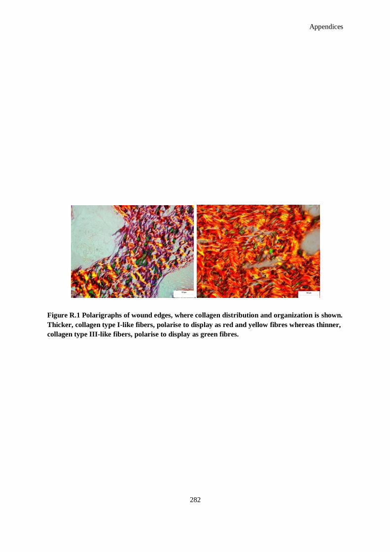
Appendices
282
.
Figure R.1 Polarigraphs of wound edges, where collagen distribution and organization is shown.
Thicker, collagen type I-like fibers, polarise to display as red and yellow fibres whereas thinner,
collagen type III-like fibers, polarise to display as green fibres.

Appendices
283
Procedure
1. Rotate the revolving nosepiece to swing in appropriate objective lens (x 40 objective).
2. While looking through the eyepieces, rotate the polarizer to the position where the field of view
is the darkest.
3. Place a specimen on the stage and start observation.
4. Adjust the field iris diaphragm until circumscribes the field-of-view.
5. Capture images.
R.4. Immunohistochemical Staining of Paraffin Embedded Sections
1. Deparaffinize and hydrate (10 min each)
Xylene for 2 minutes in the hood (toxic fumes)
100% ethanol for 2 minutes
70% ethanol for 2 minutes
50% ethanol for 2 minutes
ddH2O for 2 minutes Pour ddH2O in pressure cooker (less than ½).
2. Immerse slides in staining dish filled up completely with the buffer (if both buffers need to be
used, do the Tris-EDTA first, cool down, wash with PBS once, and proceed with the Citrate).
Antigen Retrieval Buffers
Tris-EDTA buffer (10 mM Tris-Base, 1 mM EDTA ,0 .05% Tween20 pH 9.0)
Tris Base 1.21 g
EDTA 0.37 g
ddH2O 1000 ml
Tween- 20 0.5 ml
Citrate Buffer (10mM Citric acid, .05% Tween-20, pH 6.0)
Citric Acid (anhydrous) 1.92 g
ddH2O 1000 ml
Tween-20 0.5 ml
3. Cook for ~eight minutes in microwave until yellow knob pops up, then 30 more seconds.
4. Let sit for five to ten minutes outside of the cooker.
6. Put on ice for 10-15 min until cooled down.
7. Wash w/PBS 3x (1 x 5drops and aspirate).
8. Incubate with 1% TritonX-100 solution in PBS-30 min (skip the Triton step if the antibodies
are against the membrane antigens, as Triton lyses the membranes).
Appendices
284
9. Wash with PBS.
10. PapPen™
the circles around tissue (hold the slides horizontal and test the PapPen™
before using
on the slide).
11. Incubate for 30 min in goat block at room T (use the serum of the species in which the
secondary antibody was raised in, i.e. normally goat. However, when using goat primary
antibodies, use the donkey serum for blocking).
Goat Block (keep on ice before use)
1% BSA (stabilizer) 0.5 g or 1.67 ml
0.1% Triton x100 50 µl
0.1% cold-water fish skin gelatine 50 µl
0.05% Tween-20 25 µl
Add PBS up to 50 ml
12. Take 25 ml out and add 2% goat serum (0.5 ml). Use the remaining 25 ml as primary antibody
dilution buffer for step 8. Flip filter both solutions.
13. Wash with PBS.
14. Wash in 0.05% Tween-PBS for five minutes (x3) (for 150 ml PBS add 75µl Tween-20).
15. Incubate overnight at 4 ºC with primary antibodies solution. Can use both antibodies on same
section if they are from different animals.
16. Dilute the primary antibodies in dilution buffer (prepared in step 11) to make a total of 300 µl
for each section.
17. Use large culture dishes with wet tissue paper to place the slides in, cover, and place in fridge
over night.
18. Wash in PBS or in 0.05% Tween-PBS on shaker five minutes (x3) or by suction three times.
Perform all following steps in the dark.
19. Dilute secondary antibodies 1:250 (example: AlexaFluor® 488 (green) anti-rabbit and
AlexaFluor® 594 (red) anti-mouse)) in dilution buffer to make 300 µl for each section (one
solution for all).
20. Centrifuge the antibody solutions.
21. Incubate the tissue for 30 min with secondary antibodies at room temperature.
22. Wash with PBS for five minutes (x3) and take out the mounting medium to thaw.
23. Incubate in DAPI solution for 10-15 min at room temperature in dark.
24. DAPI: 1ml stock + 4ml PBS, flip filter.
25. Wash with PBS for five minutes (x3).

Appendices
285
26. Mount the sections using Prolong Gold Anti Fade solution (Molecular Probes; stored at –20
ºC, thaw for ~15 min upside down before use), put the slides flat at 4 ºC overnight and seal the
glass cover slip edges next day with nail polish (wait 24 hours till imaging).
R.5. TUNEL Staining
1. Deparaffinise, rehydrate and wash cryosections in PBS for 30 minutes solution.
2. Incubate tissue sections for 15-30 minutes at 21-37 °C with Proteinase K solution. (The
Proteinase K solution contains 2 ml 50X Proteinase K in 98 ml PBS buffer)
3. Rinse slides with PBS two times for five minutes each time.
4. Incubate with Blocking solution for ten minutes at 15-25°C.The Blocking solution contains 3%
H2O2 in methanol.)
5. Rinse slides with PBS two times for five minutes each time.
6. Positive control: Before beginning the labeling procedure, incubate the fixed and permeabilized
cells or sections with 100 ml DNase I Solution for 10 -30 minutes at 21-37°C to induce DNA
strand degradation.
(DNase I Solution contains 10000 U/ml-50000 U/ml DNase I (grade I) depending on the
sample to be stained in DNase I buffer (the concentration of DNase I is 10000 U/ml– 20000
U/ml for cell sample, 20000U/ml – 30000U/ml for cryopreserved section, and 30000U –
50000U/ml for paraffin-embeded sections). One example of DNase I buffer is 10 mM CaCl2, 6
mM MgCl2, and 10 mM NaCl in 40 mM Tris-HCl, pH 7.9)
7. Rinse slides with PBS two times for five minutes ea ch time. Then keep the area around the
samples dry.
8. Add 50 ml TUNEL Reaction Mixture on samples. Add a coverslip and incubate for 60 minutes
at 37°C under wet conditions, protected from light.(The TUNEL Reaction Mixture contains
45 ml Equilibration Buffer, 1 ml Biotin-11-dUTP, and 4 ml TdT, freshly prepared)Note: Add
50 mL TUNEL Reaction Mixture into the negative control. To ensure a homogeneous dispersal
of TUNEL Reaction Mixture across the cell monolayer and to avoid loss to evaporation,
samples should have to be covered with parafilm or coverslip during incubation.
9. Rinse slide with PBS three times for five minutes each time. Then keep the area around the
samples dry.
10. Add 50 ml Streptavidin-HRP solution on samples. Incubate the slide under wet conditions,
protected from light for 30 minutes at 37°C.
11. (The Streptavidin-HRP solution contains 0.5 ml Streptavidin-HRP in 99.5 ml PBS.
Note: To ensure a homogeneous dispersal of Streptavidin-HRP solution across the cell
monolayer and to avoid loss to evaporation, the samples should be covered with parafilm or a
coverslip during incubation.
12. Rinse slide with PBS three times for five minutes each time.

Appendices
286
13. Add 50 ml DAB - working solution and incubate slide for 30 second-5 minutes at 15-25°C.
(The DAB - working solution: 2.5 ml DAB-A(brown cap)Solution in 50 ml H2O, then add
2.5 ml DAB-B(gray cap)Solution and 2.5 ml DAB-C(yellow cap)Solution, freshly
prepared)
14. Rinse slide three times with PBS and counterstain using Mayer‘s Haematoxylin for eight
minutes. Wash in running water to remove unbound Haematoxylin. Mount under glass
coverslip (such as with PBS/glycerol) and analyze with light microscope (Alternative: Samples
can be counterstained prior to analysis by light microscope).
S. RNA Extraction
S.1.Extraction from Cells and Scaffolds
1. RW1 and RPE buffers are supplied in RNeasy™
Kit.
2. At the predefined time to analyse cells for RNA content, wash scaffold/cells with HBSS.
3. Add 1 ml of TRI Reagent® to wells containing scaffolds/cells.
4. Homogenize samples using a tissue rupture been careful not to contaminate adjacent wells.
Make sure scaffolds have been completely homogenized. Using a 1 ml pipette tip, aspirate the
solution.
5. Store homogenate for five minutes at room temperature to dissociate nucleoprotein complexes.
Remove the TRI Reagent® solution to a sterile 1.5 ml eppendorf.
6. Add 200 μl of chloroform per 1 ml of TRI Reagent®. Shake vigorously for 15 seconds by
inversion. Incubate for 15 minutes at room temperature.
7. Centrifuge at 12,000 g for 15 minutes at 4 °C. Following the centrifugation, three phases will
appear; -a lower red phenol-chloroform phase, an inter-phase, and an aqueous phase
(translucent). mRNA is located within the aqueous phase.
8. Remove the clear upper aqueous phase (~ 650 μl) to a sterile eppendorf. Be careful not to touch
the interface. Leave a little of the upper phase to avoid contact with the interface.
9. Slowly add one volume of 70% ethanol and mix by inversion.
10. Add 700 μl sample from step 8 to RNeasy™
column.
11. Centrifuge at 8,000 g for 15 seconds and discard the collected solution.
12. Repeat step 10 and 11 for remaining sample.
13. Add 350 μl of RW1 buffer to centre of column, centrifuge at 8,000 g for 15 seconds. Discard
the collected solution.
14. Transfer column to new 1.5 ml eppendorf. Add 500 μl RPE to centre of column, centrifuge at
8,000 g for 15 seconds. Discard the collected solution.
Appendices
287
15. Add 500 μl of RPE buffer to centre of column, centrifuge at 8,000 g for 15 seconds. Discard the
collected solution and centrifuge for a further two minutes at 8,000 g.
16. Transfer column to a new 1.5 ml eppendorf. Add 30 μl RNase-free water onto the column,
incubate at room temperature for one minute and centrifuge for one minute at 8,000 g for 15
seconds.
17. Add a further 30 μl RNase-free water onto the column, incubate at room temperature for one
minute and centrifuge at 8,000 g for one minute. Split the collected sample into three for
storage purposes.
18. Determine the concentration using the NanoDrop™
and freeze at -80 °C.
19. Dilute RNA 1:50 or 1:100 in water RNase Free. Measure the absorbance at 260 nm. (Calibrate
the spectrometer with water).The purity is determined from the ratio between A260 and A280.
The ratio A260/A280 should be above 1.8 to indicate a pure form of RNA.
T. Reverse Transcription of RNA to cDNA
T.1. Reverse Transcription of RNA to cDNA for Regular PCR
Preparation before Reverse Transcription
Clean the work surface and spray RNAse away. Wipe dry all the pipettes and gloves with
RNAse away. Use sterile nuclease free tubes which are pre-chilled on ice. Use 1μg of RNA
template and 0.5 μg of oligo dT primers and random primers. Denature the target RNA and
primers by incubation at 70 °C for 5 min. Quick-chill on ice for 5 min.
Recipe for Reverse Transcription Reaction Mix
1. Begin with highest volume. Add the reverse transcriptase enzyme at the last. Prepare the
reaction mix on ice and keep on ice until incubation. Follow Table T.1 for volumes.
2. After combining all components, vortex gently to mix. After mixing, place the tube with
reaction mix into the reverse transcription machine and run the program as detailed in Table
T.2.
3. After reaction is complete proceed with polymerase chain reaction (PCR) or store cDNA at -20
°C for future use.
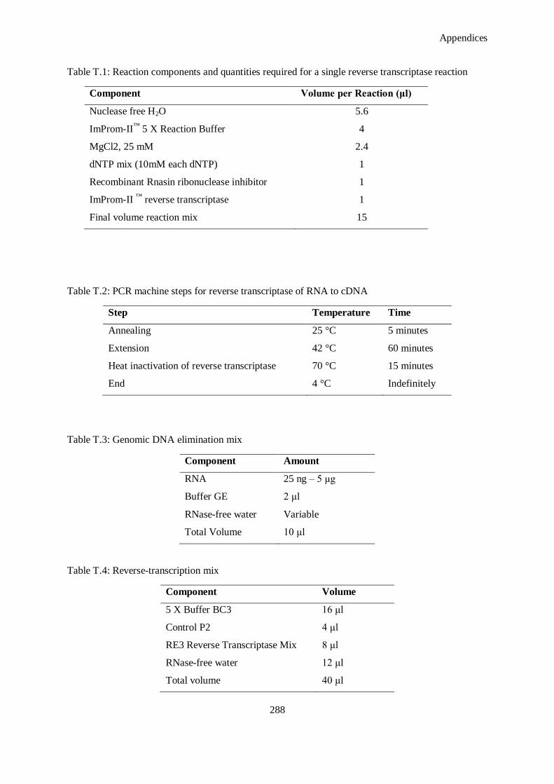
Appendices
288
Table T.1: Reaction components and quantities required for a single reverse transcriptase reaction
Component Volume per Reaction (μl)
Nuclease free H2O 5.6
ImProm-II™
5 X Reaction Buffer 4
MgCl2, 25 mM 2.4
dNTP mix (10mM each dNTP) 1
Recombinant Rnasin ribonuclease inhibitor 1
ImProm-II ™
reverse transcriptase 1
Final volume reaction mix 15
Table T.2: PCR machine steps for reverse transcriptase of RNA to cDNA
Step Temperature Time
Annealing 25 °C 5 minutes
Extension 42 °C 60 minutes
Heat inactivation of reverse transcriptase 70 °C 15 minutes
End 4 °C Indefinitely
Table T.3: Genomic DNA elimination mix
Component Amount
RNA 25 ng – 5 μg
Buffer GE 2 μl
RNase-free water Variable
Total Volume 10 μl
Table T.4: Reverse-transcription mix
Component Volume
5 X Buffer BC3 16 μl
Control P2 4 μl
RE3 Reverse Transcriptase Mix 8 μl
RNase-free water 12 μl
Total volume 40 μl

Appendices
289
T.2. Reverse Transcription of RNA to cDNA for RT2 Profiler PCR Array
1. Thaw reagents of the kit and briefly centrifuge for 10 – 15 seconds to bring the contents to the
bottom of the tubes.
2. Prepare the genomic DNA elimiation mix for each RNA sample according to Table T.3. Mix
gently by pipetting up and down and then briefly centrifuge.
3. Incubate the genomic DNA elimination mix for fibe minutes at 42 °C, then place immediately
on ice for at least one minute.
4. Prepare the reverse-transciption mix according to Table T.4.
5. Add 10 μl reverse transcription mix to each tube containing 10 μl genomic DNA elimination
mix. Mix gently by pipetting up and down.
6. Incubate at 42 °C for exactly 15 minutes. Then immediately stop the reaction by incubating at
95 °C for five minutes.
7. Add 91 μl RNase-free water to each reaction. Mix by pipetting up and down several times.
8. Place the reactions on ice and proceed with the real-time PCR protocol, or alternatively store
the reactions at -20 °C.
U. Real-time PCR
U.1. Regular PCR
1. Dilute cDNA template so as to obtain a final concentration of 20ng per well.
2. Make sure that the cDNA concentration must not exceed 100ng/reaction and 10% of the final
volume.
3. Add the components listed in Table U.1 to prepare master mix.
4. For a triplicate reaction, add cDNA template in triplicate and also add a negative control by
replacing cDNA with nuclease free water.
5. Mix well the master mix by pipetting and add to each well to obtain final volume of 25ml.
6. Add the plastic cover provided by supplier on the PCR plate.
7. Centrifuge 1 minute at 1400 rpm. Place the plate in the machine.
8. Open the step one software and enter the details to map the plate on the software.
9. Choose Sybr Green filter for each well.
10. Choose the endogenous control from the plate and enter in the software.
11. Set up the steps in accordance to the gene and melting temperature (Tm) of the primers. A
general program is detailed in Table U.2.
Appendices
290
Table U.1: Polymerase chain reaction components
Component Volume per Reaction (μl)
2 X Quantifast SYBR Green PCR Master mix 12.5
Forward primer, pmol 0.25
Reverse primer, pmol 0.25
Template cDNA 1.49
Nuclease free water 10.51
Final volume 25
Table U.2: Typical real time PCR program.
Step Temperature Time Ramp Rate No. Cycles
Initial step 50 °C 2 minutes Maximal/fast
mode
1
PCR initial activation step 95 °C 5 minutes 1
Two step cycling
Denaturation 95 °C 10 seconds Maximal/fast
mode 35
Annealing/Extension 60 °C 30 seconds
Final Step
Denaturation 95 °C 15 seconds Maximal/fast
mode 1 Annealing/Extension 60 °C 20 seconds
Final denaturation 95 °C 15 seconds
U.2. Protocol for PCR using RT2 Profiler PCR Array
1. Briefly centrifuge the RT2 SYBR Green Mastermix to bring the contents to the bottom of the
tube.
2. Prepare the PCR components mix in a 5 ml tube or a loading reservoir depending on the RT2
Profiler PCR Array format as described in Table U.4.
3. Dispense the PCR components mix into the RT2 Profiler PCR Array depending on the RT
2
Profiler format.
4. Carefully remove the RT2 Profiler PCR Array from its sealed bag.
5. Add PCR components mix to each well of using an 8-channel pipettor and the 384EZLOAD
covers provided.
6. Place 384Load cover 1 (white) on the plate. Add 10 μl PCR components mix for sample 1 to
the open wells. Remove and discard cover. Repead with cover 2 (yellow, 3 (black) and 4 (red)
on the plate.
7. Carefully, tightly seal the RT2 profiler PCR Array with optical adhesive film.
Appendices
291
8. Centrifuge for one minute at 1000 g at room temperature to remove any bubbles. Visually
inspect the plate from underneath to ensure no bubbles are present in the wells.
9. Place the RT2 Profiler PCR Array on ice while setting up the PCR cycling program.
10. Proceed in real-time cycler as normal.
Table U.3: PCR components mix
Component Volume
2 x RT2 SYBR Green Mastermix 650 μl
cDNA synthesis reaction 102 μl
RNase-free water 548 μl
Total volume 1300 μl
Table U.4: Cycling conditions for the Roche LightCycler 480®
Cycles Duration Temperature Comments
1 10 minutes 95 °C HotStart DNA Taq Polymerase is activated by
this heating step
45 15 s 95 °C
1 minute 60 °C Perform fluorescence data collection
Appendices
292
Table U.5: List of genes investigated in rat wound healing PCR array
Extracellular Matrix & Cell Adhesion:
ECM Components Col14a1, Col1a1, Col1a2, Col3a1, Col4a1, Col4a3, Col5a1, Col5a2, Col5a3,
Vtn.
Remodeling
Enzymes:
CTGS, CTSK, Ctsl1, F13a1, F3 (Tissue Factor), Fga, (Fibrinogen), MMP-1a,
MMP-2, MMP-7, MMP-9, Plat (tPA), Plau (uPA), Plaur (uPAR), Plg, Serpine1
(PAI-1), TIMP-1.
Cellular
Adhesion:
Cdh1 (E-cadherin), Itga1, Itga2, Itga3, Itga4, Itga5, Itga6, Itgav, Itgb1, Itgb3,
Itgb5, Itgb6.
Cytoskeleton: Acta2 (a-SMA), Actc1, Rac1, Rhoa, Tagln.
Inflammatory Cytokines & Chemokines:
CCL-12, CCL-7 (MCP-3), CD-40LG (TNFSF5), CXCL-1, CXCL-11 (I-TAC/IP-9), CXCL-3, CXCL
5, IFNG, IL-10, IL-1B, IL-2, IL-4, IL-6.
Growth Factors
ANGPT1, CSF2 (GM-CSF), CSF3 (GCSF), CTGF, EGF, FGF10, FGF2, FGF7, HBEGF (DTR),
HGF, IGF-1, MIF, PDGFA, TGFA, TGF-B1, TNF, VEGFA.
Signal Transduction:
TGFß: TGF-B1, TGF-BR3, STAT-3
WNT: CTNNB-1, WISP-1, WNT-5A.
Phosphorylation: MAPK-1 (ERK2), MAPK-3 (ERK1), PTEN.
Receptors: EGFR, IL-6ST (GP130).
Other: PTGS-2.
Housekeeping
B2M, HPRT-1, RPL13A, GAPDH, ACTB

Appendices
293
V. Fluorescent Labelling of siRNA with Cy™
3
1. Resuspend experimental RNA (maximum weight of 80 μg) to 20 μM in Nuclease-free Water.
2. Add 100 μl of Reconstitution Solution to the dry Cy™
3 Labelling Regent. Perform all
manipulations in the dark as the Cy™
3 dye is light sensitive.
3. In a sterile, nuclease-free tube, assemble the reagents in the order shown in Table V.1, making
sure to add the labelling Reagent last. Mix well by vortexing.
4. Incubate the reaction mix at a constant temperature of 37 °C for one hour in the dark.
5. Remove from incubation and to the tube add 5 μl of 5 M NaCl and 125 μL of ice cold 100 %
ethanol.
6. Mix well and place the mixture at -20 °C (or colder) for 30-60 minutes. The labelling RNA
precipitates during this incubation.
7. Pellet the labelled RNA by centrifugation at ≥8,000 g for 15-20 minutes. Carefully remove the
supernatant; avoid disrupting the pellet. A red pellet siRNA pellet should be visible.
8. Gently add 175 μL of 70 % ethanol making sure not to disrupt the pellet, and centrifuge at
≥8,000 g for five minutes (use the highest speed compatible with your tubes). Carefully remove
supernatant with a pipette. To remove the last traces of solution, respin the tube briefly and
discard the supernatant.
Table V.1: Reaction setup for labelling 5 μg 21-mer duplex siRNA.
Component Amount (μl)
Nuclease-free Water 18.3
10 X Labelling Buffer 5.0
21-mer duplex siRNA at 20 μM (~5μg/μl) 19.2
Cy™ 3 Labelling Reagent 7.5
Total Volume 50
9. Dry the RNA for five to ten minutes at room temperature. Do not dry the pellet for longer than
five to ten minutes or it will be difficult to solubilise.Resuspend the RNA pellet in Nuclease-
free Water or in the buffer of your choice. If desired, resuspend the labelled RNA in the same
volume it was in before the labelling reaction. Remove an aliquot (1 μg) for analysis via
electrophoresis (Figure V.1).
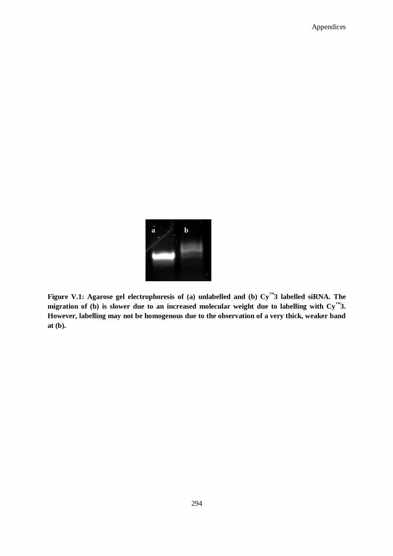
Appendices
294
Figure V.1: Agarose gel electrophoresis of (a) unlabelled and (b) Cy™
3 labelled siRNA. The
migration of (b) is slower due to an increased molecular weight due to labelling with Cy™
3.
However, labelling may not be homogenous due to the observation of a very thick, weaker band
at (b).
a b
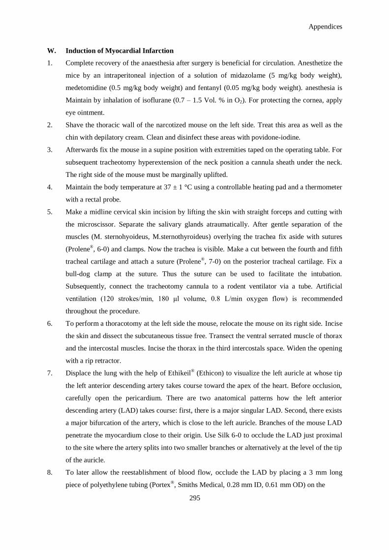
Appendices
295
W. Induction of Myocardial Infarction
1. Complete recovery of the anaesthesia after surgery is beneficial for circulation. Anesthetize the
mice by an intraperitoneal injection of a solution of midazolame (5 mg/kg body weight),
medetomidine (0.5 mg/kg body weight) and fentanyl (0.05 mg/kg body weight). anesthesia is
Maintain by inhalation of isoflurane (0.7 – 1.5 Vol. % in O2). For protecting the cornea, apply
eye ointment.
2. Shave the thoracic wall of the narcotized mouse on the left side. Treat this area as well as the
chin with depilatory cream. Clean and disinfect these areas with povidone-iodine.
3. Afterwards fix the mouse in a supine position with extremities taped on the operating table. For
subsequent tracheotomy hyperextension of the neck position a cannula sheath under the neck.
The right side of the mouse must be marginally uplifted.
4. Maintain the body temperature at 37 ± 1 °C using a controllable heating pad and a thermometer
with a rectal probe.
5. Make a midline cervical skin incision by lifting the skin with straight forceps and cutting with
the microscissor. Separate the salivary glands atraumatically. After gentle separation of the
muscles (M. sternohyoideus, M.sternothyroideus) overlying the trachea fix aside with sutures
(Prolene®, 6-0) and clamps. Now the trachea is visible. Make a cut between the fourth and fifth
tracheal cartilage and attach a suture (Prolene®, 7-0) on the posterior tracheal cartilage. Fix a
bull-dog clamp at the suture. Thus the suture can be used to facilitate the intubation.
Subsequently, connect the tracheotomy cannula to a rodent ventilator via a tube. Artificial
ventilation (120 strokes/min, 180 μl volume, 0.8 L/min oxygen flow) is recommended
throughout the procedure.
6. To perform a thoracotomy at the left side the mouse, relocate the mouse on its right side. Incise
the skin and dissect the subcutaneous tissue free. Transect the ventral serrated muscle of thorax
and the intercostal muscles. Incise the thorax in the third intercostals space. Widen the opening
with a rip retractor.
7. Displace the lung with the help of Ethikeil® (Ethicon) to visualize the left auricle at whose tip
the left anterior descending artery takes course toward the apex of the heart. Before occlusion,
carefully open the pericardium. There are two anatomical patterns how the left anterior
descending artery (LAD) takes course: first, there is a major singular LAD. Second, there exists
a major bifurcation of the artery, which is close to the left auricle. Branches of the mouse LAD
penetrate the myocardium close to their origin. Use Silk 6-0 to occlude the LAD just proximal
to the site where the artery splits into two smaller branches or alternatively at the level of the tip
of the auricle.
8. To later allow the reestablishment of blood flow, occlude the LAD by placing a 3 mm long
piece of polyethylene tubing (Portex®, Smiths Medical, 0.28 mm ID, 0.61 mm OD) on the
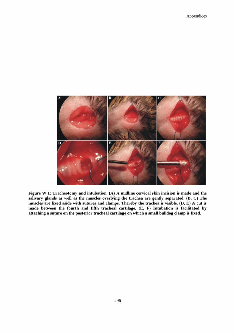
Appendices
296
Figure W.1: Tracheotomy and intubation. (A) A midline cervical skin incision is made and the
salivary glands as well as the muscles overlying the trachea are gently separated. (B, C) The
muscles are fixed aside with sutures and clamps. Thereby the trachea is visible. (D, E) A cut is
made between the fourth and fifth tracheal cartilage. (E, F) Intubation is facilitated by
attaching a suture on the posterior tracheal cartilage on which a small bulldog clamp is fixed.
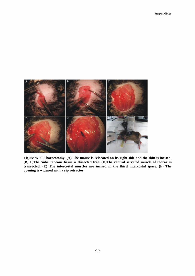
Appendices
297
Figure W.2: Thoracotomy. (A) The mouse is relocated on its right side and the skin is incised.
(B, C)The Subcutaneous tissue is dissected free. (D)The ventral serrated muscle of thorax is
transected. (E) The intercostal muscles are incised in the third intercostal space. (F) The
opening is widened with a rip retractor.
Appendices
298
Figure W.3: Occlusion of the left descending artery (LAD). (A) By widening the intercostal
space the left side of the heart and the lung become apparent. (B) After opening the pericardium
the left auricle is visible (triangle) at whose tip the LAD (arrow) takes course towards the apex.
(C, D) The LAD is occluded at the level of the tip of the left auricle. (E) A loose snare is formed.
(F) Furthermore, a piece of polyethylene tubing is placed on the artery to later allow the
reestablishment of blood flow.

Appendices
299
artery and fix it in place with a ligature. Contract the knot by forming a loose snare.
Discoloration of the left ventricle wall indicates interruption of the coronary flow and verifies
the successful myocardial infarction.
9. Retain occlusion of the LAD must for a well defined period to achieve presentable infarct sizes.
10. Maintain ischemia 30 minutes. After this period, terminate the ischemia by removing the
polyethylene tubing and opening the knot. Leave the suture in the myocardial tissue for the
Evans Blue staining, which is performed before removing the heart. Confirm reperfusion visual
inspection. Remove the rip retractor and deposit the silk 6-0 suture thread outside the thorax.
Close the intercostal space and the pleura with interrupted suture by using Vicryl® 6-0.
11. For extubation, relocate the mouse in the supine position. As soon as the mouse breaths
autonomously take the tracheotomy cannula off from the trachea. Close the latter with a single
Vicryl® 8-0 knot. Remove the holding sutures and close the skin is closed with Prolene
® 6-0 in
interrupted sutures. Support the circulation of the mouse with Ringer‘s solution and 5 %
glucose solution by a subcutaneous injection. To antagonize the anaesthesia, a mixture
consisting of atipamezole (2.5 mg/kg body weight) and flumazenil (0.5 mg/kg body weight) is
administered subcutaneously. Allow the mouse to recover from the surgery under a heating
lamp. Repeat the injection of buprenorphine in an interval of eight hours for three days. At the
end of the reperfusion period, anesthetize the mouse again. Carry out the intubation as already
described. Raise the isoflurane supply to 2 % volume..
12. Cut open the abdomen at the place where the xiphoid process of sternum can be found.
Following this cut the diaphragm. Open up the thorax by cutting the rips on the right side of
the sternum, shifting it to the left and fixating it with a clamp. Locate the silk suture and retie
the left anterior descending artery ligature. Inject 0.5 ml of 5 % Evans Blue dye into the left
ventricular cavity. The circulating dye is uniformly distributed throughout with the exception of
the heart region supplied by the occlude coronary artery. This domain illustrates the area at risk,
which is affected by the ischemia. Sacrifice the mouse by an intracoronary injection of 0.3 ml
potassium chloride solution (1 M). Quickly remove the heart and clean with a sodium chloride
solution. Embedding in Tissue Tec®
it and freeze at -20 °C. Section the frozen heart transversely
into five slides of approximately 1 – 2 mm breadth. Clean each section with sodium chloride
solution and incubate in 1 % TTC for ten minutes.
13. After TTC-staining, viable myocardium stains red and the infarct region appears pale white.
Further fix the slides in 4 % formaldehyde for two hours at room temperature. Thereafter,
photograph the tissue sections on both sides using a digital camera mounted atop the stereo
microscope. The infarct sizes can be determined by quantitative morphometric planimetry using
an image analysis software program.
Appendices
300
X. One-day microRNA ISH Protocol (using Mercury LNA microRNA ISH Optimization Kit
(Formalin Fixed Paraffin Embedded Sections)
Antibody Blocking Solution: PBS, 0.1% Tween, 2 % Sheep serum, 1 % BSA
Antibody Diluent: PBS, 0.05 % Tween, 1% Sheep serum, 1% BSA,
Levamisole (for blocking endogenous AP activity): Prepare a 100 mM stock
Proteinase-K Buffer:
900 ml RNase-free water
5 ml 1M Tris-HCL (pH 7.4)
2 ml 0.5 M EDTA
0.2 mL 5M NaCl
Adjust volume to 1000 ml and autoclave
PBS-T (0.1%), pH 7.4: Add 1 mL of Tween-20 to 1 L of PBS and autoclave.
KTBT (AP stop solution): To 900 ml RNase-free water add:
7.9g Tris- HCl (50 mM)
8.7 g NaCl (150mM)
0.75 g KCl (10mM)
Adjust volume to 1000 ml. Do not adjust pH and autoclave.
Proteinase-K Reagent
Prepare immediately before use. For a Proteinase-K concentration of 15 μg/ml: Add 7.5 μl
Proteinase K stock to 10 ml Proteinase K buffer.
Hybridization Mix (for microRNA ISH buffer and LNA™ Detection probes)
Dilute the 2x microRNA ISH buffer 1:1 with RNase-free water, e.g. mix 1 ml 2 x microRNA
ISH buffer with 1 ml RNase-free water to give 2 ml 1 x buffer.
For each probe to be used in the experiment, place the appropriate amount of LNA™
probe in a
2 ml non-stick RNase-free tube.
Denature the probes at 90 °C for four minutes.
Place the tubes in a table-top microcentrifuge and spin down shortly.
Immediately add the 2 mL 1 x microRNA ISH buffer to each of the tubes with the different
LNA™
probes.
Appendices
301
Antibody Blocking Solution
To make 10 mL blocking and 10 ml diluting solution start with 15 mL PBS-T and add 300 μl
sheep serum. Label the tube as ‗blocking solution‘.
Remove 5 ml from the tube in step 1, place in a new tube and label diluting solution.
To the tube labelled blocking solution, add 330 μl 30 % BSA to give a final concentration of 1
%. The ‗blocking solution‘ is now ready to use.
To the tube labelled diluting solution, add 5 mL PBS (to give 0.05 % Tween and 1 % sheep
serum final concentration) and 330 μl 30 % BSA to give 1 % final concentration. The diluent
solution is now ready for use.
Anti- Digoxigenin (DIG) reagent: Dilute the sheep- anti-DIP-AP antibody 1:800 in antibody diluting
solution.
Alkaline Phosphatase (AP) Substrate: Immediately prior to use, dissolve a nitro-blue tetrazolium
and 5-bromo-4-chloro-3'-indolyphosphate (NBT-BCIP) tablet in Milli-Q™
water according to
the manufacturer‘s instructions. Add levamisol to a final concentration of 0.2 mM.
1. Prior to deparaffinization place slides in 60 °C oven overnight to remove paraffin.
2. Deparaffinize slides in xylene and ethanol solutions at room temperature (RT) by placing slides
with sections in slide rack.
3. Deparaffinization:
Xylene 10 minutes
Xylene 10 minutes
EtOH (99.9%) 5 minutes
EtOH (99.9%) 5 minutes
EtOH (95%) 5 minutes
EtOH (95%) 5 minutes
EtOH (70%) 5 minutes
EtOH (70%) 5 minutes
PBS 5 minutes
4. Incubate slides with Proteinase-K at 37 °C for ten minutes.
5. Wash with PBS
6. Dehydrate slides:
PBS 5 minutes
EtOH (70%) 2 minutes
EtOH (70%) 2 minutes
EtOH (95%) 2 minutes
Appendices
302
EtOH (95%) 2 minutes
EtOH (99.9%) 2 minutes
EtOH (99.9%) 2 minutes
7. Air-dry the slides on clean paper towels afterwards for 15 minutes.
8. Apply slides on a flat surface and apply 25 μl of the hybridization mix. For initial protocol
optimization, probe concentrations could be a) 1nM LNA™ U6 snRNA probe b) 40 nM double
–DIG LNA™ microRNA probe. The probe concentration will need to be optimized for optimal
microRNA ISH signal. Avoid touching the tissue sections with the pipette tip. Then apply a
sterile cover glass onto each section, carefully avoiding air bubbles, and seal along all four
edges with Fixogum™
(rubber cement). Place the slides in the hybridizer and start a program
hybridizing for one hour. Hybridization temperature must be optimized for individual probes.
9. Remove Fixogum™
using tweezers. Avoid sliding the coverslip, which may damage the tissue.
Then, carefully detach cover glass and place the slides into a slide rack placed within a glass jar
containing 5 x sodium saline citrate (SSC) at RT.
10. Wash slides in glass jars according to the following list. To ensure sufficient stringency
perform the washes in glass jars placed in a water bath set to the hybridization temperature:
5 x SSC 5 minutes at Hybridization Temperature
1 x SSC 5 minutes at Hybridization Temperature
1 x SSC 5 minutes at Hybridization Temperature
0.2 x SSC 5 minutes at Hybridization Temperature
0.2 x SSC 5 minutes at Hybridization Temperature
0.2 x SSC 5 minutes at Room Temperature
11. Transfer the slides to glass jars with PBS. Apply a hydrophobic barrier around the tissue
sections using a PapPen® following the manufacturer‘s instructions. Tissue sections are not
allowed to dry out during this and the subsequent immunohistochemistry steps.
12. Place the slides in a humidifying chamber and incubate with blocking solution for 15 minutes at
room temperature. NB. Perform all steps after this in the humidifying chamber.
13. Remove the blocking solution and apply anti-DIG reagent (sheep anti-DIG-AP at 1:800 in
antibody diluting) and incubate for 60 minutes at room temperature.
14. Wash the slides 3 x 3 minutes with PBS-T.
15. Apply freshly prepared AP substrate to the sections and incubate the slides for 2 hours at 30 °C
in the humidifying chamber. Protect from light during development.
16. Incubate the slides in KTBT buffer (2 x 5 minutes) to stop the reaction.
17. Wash with water (2 x 1 minute).
18. Apply 200 μl Nuclear Fast Red™
(nuclear counter stain) for one minute for nuclear counter
staining.

Appendices
303
19. Remove slides from the humidifying chamber to a slide rack placed within a glass jar
containing tap water. Carefully rinse the slides with running tap water for approximately ten
minutes.
20. Dehydrate the slides in ethanol according to the following:
EtOH (70%) 2 minutes
EtOH (70%) 2 minutes
EtOH (95%) 2 minutes
EtOH (95%) 2 minutes
EtOH (99.9%) 2 minutes
EtOH (99.9%) 2 minutes
21. Mount the slides directly with one to two drops of mounting media. Avoid air-drying sections
at this step.
22. Allow to precipitate to settle overnight and analyze by light microscopy the subsequent day.
Y. Journal Publications and Conference Proceedings
Y.1. Journal Publications
1. Monaghan M, Browne S, Schenke-Layland K, Pandit A. Scaffold mediated delivery of
exogenous microRNA 29B to modulate dermal fibrosis, In Submission to Mol. Ther.
2. Monaghan M, Greiser U, Wall J.G, O'Brien T, Pandit A. Interference: An alteRNAtive therapy
following acute myocardial infarction. Trends Pharmacol. Sci. 2012, 33: 635-645
3. Monaghan M, Greiser U, Cao H, Wang W, Pandit A. A ligand enhanced dendritic PEGylated
poly (2-(dimethylamino) ethyl diacrylate) as a vehicle of microRNA, Drug Del. Trans. Res., 2
(5) 406-414
4. Monaghan M, Pandit A. RNA interference therapy via functionalized scaffolds, Adv. Drug
Del. Rev., 63 (2011) 197-208
5. Dash B.C, Réthoré G, Monaghan M, Fitzgerald K, Gallagher W, Pandit A. The influence of
size and charge of chitosan/polyglutamic acid hollow spheres on cellular internalization,
viability and blood compatibility, Biomaterials, 31 (2010) 8188-8197
Y.2. Conference Podium Presentations
1. Monaghan M, Browne S, Wang W, Pandit A. Delivery and efficacy of interfering RNA via a
crosslinked collagen hydrogel in an excisional in vivo model and a human pre‐clinical model to
reduce fibrosis, Podium Presentation at World Congress of Tissue Engineering and
Regenerative Medicine International Society, Vienna, Austria, 5th – 8
th September 2012
Appendices
304
2. Monaghan M, Browne S, Wang W, Pandit A. Delivery and efficacy of interfering RNA via a
crosslinked collagen hydrogel in an excisional in vivo model and a human pre‐clinical model to
reduce fibrosis, Podium Presentation at the 3rd International Conference "Strategies in Tissue
Engineering, Würzburg, Germany, 23rd
-25th May 2012
3. Monaghan M, Browne S, Wang W, Pandit A. Scaffold-mediated non-viral inhibition of
collagen type I and type III in cardiac fibroblasts, Podium Presentation at the World
Conference in Regenerative Medicine, Leipzig, Germany, 2nd
- 4th November 2011
4. Monaghan M, Browne S, Wang W, Pandit A. Scaffold-mediated non-viral inhibition of
collagen type I and type III in cardiac fibroblasts, Podium Presentation at the European Society
of Biomaterials Conference, Dublin, Ireland. 4th – 9
th September 2011
5. Kelly J.L, Doody J, Zeugolis D, Monaghan M, Kelly G, Devocelle M, Kilcoyne M, Joshi L,
Pandit A. Collagen fibers modified with polysialic mimetic peptide are a suitable material for
use as a synthetic peripheral nerve graft. Podium Presentation at the Irish Association of
Plastic Surgery Meeting, Dublin, Ireland, 26th November 2010
6. Dash B.C, Mahor S, Monaghan M, Woodhouse K.A, Pandit A. Elastin like protein based
hollow spheres for gene delivery, Podium Presentation at the European Society of Biomaterials
Conference, Tampere, Finland, 11th -15
th September 2010
7. Monaghan M, Mathew A, Greiser U, Wang W, Pandit A. Scaffold mediated delivery of
microRNA immunopolyplexes, Podium Presentation at the European Society of Biomaterials
Conference, Tampere, Finland, 11th -15
th September 2010
8. Monaghan M, Mathew A, Greiser U, Wang W, Pandit A. Scaffold mediated delivery of
microRNA immunopolyplexes, Podium Presentation at the Tissue Engineering and
Regenerative Medicine Conference, Galway, Ireland, 14th – 17
th June 2010
9. Dash B C, Mahor S, Monaghan M, Woodhouse K A, Pandit A. Elastin like protein based
hollow spheres for gene delivery, Podium Presentation at the Tissue Engineering and
Regenerative Medicine Conference, Galway, Ireland, 14th – 17
th June 2010
10. Mathew A, Monaghan M, Wang W, Pandit A. A linear-dendritic hybrid poly (2-
(dimethylamino) ethyl methacrylate) as a highly effective gene delivery vector, Podium
Presentation at the European Society of Biomaterials Conference, Tampere, Finland, 11th -15
th
September 2010
11. Réthoré G, Monaghan M, Dash B C, Pandit A. Chitosan-polyglutamic acid biodegradable
nanospheres show a blood compatibility response that is dictated by size and surface charge,
Podium presentation at the 2nd World Congress of the Tissue Engineering and Regenerative
Medicine International Society, Seoul, Republic of Korea, 2009
Appendices
305
12. Matthew A, Réthoré G, Dash B C, Monaghan M, Pandit, A. Chitosan/polyglutamic acid
nanospheres: DNA encapsulation and release, cellular uptake and blood interaction studies,
Podium presentation at the UK Society for Biomaterials Conference, Antrim, UK, 2009
Y.3. Conference Poster Presentations
1. Monaghan M, Browne S, Jürgens T, Schönberger T, Gawaz M, Pandit A, Schenke-Layland,
K. Inhibiting fibrosis using RNA interference in a mouse myocardial infarction model, Poster
Presentation at the Keystone Symposium on Cardiac Remodeling, Signaling, Matrix and Heart
Function, Utah, USA. 7th – 12
th April 2013
2. Browne S, Monaghan M, Pandit A. A spatiotemporal biomaterial gene delivery system to
modulate inflammation and Angiogenesis, Poster Presentation at the Keystone Symposium on
Cardiac Remodeling, Signaling, Matrix and Heart Function, Utah, USA. 7th – 12
th April 2013
3. Monaghan M, Browne S, Wang W, Pandit A. Delivery and efficacy of interfering RNA via a
crosslinked collagen hydrogel in an excisional in vivo model and a human pre‐clinical model to
reduce fibrosis, Poster Presentation at the Gordon Research Conference on Extracellular
Matrix Signal Transduction, Maine, USA. 8th- 13
th July 2012
4. Monaghan M, Browne S, Wang W, Pandit A. Delivery and efficacy of interfering RNA via a
crosslinked collagen hydrogel in an excisional in vivo model and a human pre‐clinical model to
reduce fibrosis, Poster Presentation at the Gordon Research Seminar on Extracellular Matrix
Signal Transduction, Maine, USA. 7th- 8
th July 2012
5. Monaghan M, Holladay C, Wang W, Pandit A. A crosslinked collagen type I biomaterial
reservoir for non-viral gene delivery in vivo, Poster Presentation at the European Society of
Biomaterials Conference, Dublin, Ireland. 4th – 9
th September 2011
6. Monaghan M, Mathew A, Greiser U, Wang W, Pandit A. Scaffold mediated delivery of
microRNA immunopolyplexes, Poster Presentation at the 8th International Symposia on
Polymer Therapeutics, Valencia, Spain, 24th – 26
th May 2010
7. Monaghan M, Pandit A. A 3D co-culture vascular model to quantify remodeling after injury
with copolymers of poly (DL-Lactide-co-glycolic-acid) and poly (Lactic-co-glycolic- acid,
Poster presentation at the European Society for Biomaterials Conference, Lusanne,
Switzerland, 8th-11
th September 2009
8. Monaghan M, Pandit A. A 3D co-culture vascular model to quantify remodeling after injury
with copolymers of poly (DL-Lactide-co-glycolic-acid) and poly (Lactic-co-glycolic- acid,
Poster presentation at the World Tissue Engineering and Regenerative Medicine International
Society Conference, Seoul, Korea. August 31st – September 3
rd 2009
9. Monaghan M, O‘Rorke S, Réthoré G, Curtin C, Greiser U, Pandit A. Targeting and tethering
of mesenchymal stem cells using anti CD-105 functionalized nanoscale and dendrimeric
Appendices
306
macroscale platforms, Poster presentation at the World Tissue Engineering and Regenerative
Medicine International Society Conference, Seoul, Korea. August 31st – September 3
rd 2009
10. Monaghan M, Pandit A. Development of a 3D co-culture model to interpret the biological
response of vascular biomaterials in vitro, 4th International Conference on Bioengineering and
Nanotechnology, Dublin, Ireland, July 22nd
-24th
July 2008
11. Monaghan M, Pandit A. Development of a 3D co-culture model to interpret the biological
response of vascular biomaterials in vitro, National Centre for Biomedical Engineering Science
Research Day, Galway, Ireland, March 2008
Y.4. Awards
1. Deutscher Akademischer Austausch Dienst, Short Term Travel Award to conduct research in
University Hospital Tübingen, 2012
2. European Molecular Biology Organisation (EMBO) Short Term Travel Award to conduct
research in Fraunhofer IGB, Stuttgart. 2011
3. Roche Best Poster Award, National Centre for Biomedical Engineering Science (NCBES)
Research day, 2008
4. NUI Galway College of Engineering and Informatics Research Fellow Award, 2008s
Related Documents