An In Vitro Study on the Effects of Nisin on the Antibacterial Activities of 18 Antibiotics against Enterococcus faecalis Zhongchun Tong 1,2. , Yuejiao Zhang 1. , Junqi Ling 1,2 *, Jinglei Ma 1 , Lijia Huang 1 , Luodan Zhang 1 1 Department of Operative Dentistry and Endodontics, Guanghua School of Stomatology, Sun Yat-sen University, Guangzhou, Guangdong, China, 2 Guangdong Provincial Key Laboratory of Stomatology, Sun Yat-sen University, Guangzhou, Guangdong, China Abstract Enterococcus faecalis rank among the leading causes of nosocomial infections worldwide and possesses both intrinsic and acquired resistance to a variety of antibiotics. Development of new antibiotics is limited, and pathogens continually generate new antibiotic resistance. Many researchers aim to identify strategies to effectively kill this drug-resistant pathogen. Here, we evaluated the effect of the antimicrobial peptide nisin on the antibacterial activities of 18 antibiotics against E. faecalis. The MIC and MBC results showed that the antibacterial activities of 18 antibiotics against E. faecalis OG1RF, ATCC 29212, and strain E were significantly improved in the presence of 200 U/ml nisin. Statistically significant differences were observed between the results with and without 200 U/ml nisin at the same concentrations of penicillin or chloramphenicol (p,0.05). The checkerboard assay showed that the combination of nisin and penicillin or chloramphenicol had a synergetic effect against the three tested E. faecalis strains. The transmission electron microscope images showed that E. faecalis was not obviously destroyed by penicillin or chloramphenicol alone but was severely disrupted by either antibiotic in combination with nisin. Furthermore, assessing biofilms by a confocal laser scanning microscope showed that penicillin, ciprofloxacin, and chloramphenicol all showed stronger antibiofilm actions in combination with nisin than when these antibiotics were administered alone. Therefore, nisin can significantly improve the antibacterial and antibiofilm activities of many antibiotics, and certain antibiotics in combination with nisin have considerable potential for use as inhibitors of this drug-resistant pathogen. Citation: Tong Z, Zhang Y, Ling J, Ma J, Huang L, et al. (2014) An In Vitro Study on the Effects of Nisin on the Antibacterial Activities of 18 Antibiotics against Enterococcus faecalis. PLoS ONE 9(2): e89209. doi:10.1371/journal.pone.0089209 Editor: Lynn E. Hancock, University of Kansas, United States of America Received September 12, 2013; Accepted January 16, 2014; Published February 20, 2014 Copyright: ß 2014 Tong et al. This is an open-access article distributed under the terms of the Creative Commons Attribution License, which permits unrestricted use, distribution, and reproduction in any medium, provided the original author and source are credited. Funding: This research is supported by the Fundamental Research Funds for the Central Universities, China (number 12ykpy64), and China Postdoctoral Science Foundation Funded Project (project number 2013M542229). The funders had no role in study design, data collection and analysis, decision to publish, or preparation of the manuscript. Competing Interests: The authors have declared that no competing interests exist. * E-mail: [email protected] . These authors contributed equally to this work. Introduction Antibiotics have saved the lives of millions of people, greatly improving human and animal health in the twentieth century. However, bacterial pathogens commonly develop resistance to many antibiotics due to the extensive use of these antibiotics for human and animal health. Hundreds of thousands of deaths occur annually due to antibiotic treatment failures [1]. At present, the routine approach to addressing this crisis is to develop novel antibiotics. However, novel antibiotics are limited, and pathogens will gradually evolve resistance to these novel antibiotics [2]. Based on the inevitable trend towards bacterial resistance, it is necessary to explore new treatment strategies for effectively killing and eliminating bacterial pathogens. Limiting the evolution of bacterial resistance and using new and existing antibiotics may constitute a new strategy for antibacterial therapy. Antimicrobial peptides (AMP) have been studied for the development of new antibacterial drugs due to their high antibacterial activity and low drug resistance [3,4]. Although AMPs represent a potentially new source of antimicrobials for the treatment of various bacterial infections, conventional antibiotics remain a primary resource for antibacterial therapy and cannot be fully replaced at present. Therefore, combining conventional antibiotics and AMPs can prolong the life spans of many antibiotics. Nisin, an AMP from Lactococcus lactis, consists of 34 amino acid residues and is minimally toxic, odorless, colorless, and tasteless [5]. Nisin possesses high antimicrobial activity against a wide range of Gram-positive bacteria, even against some antibiotic-resistant pathogens [5,6]. Some papers have reported the anti-pathogen activity of antibiotics in combination with nisin [7,8,9,10,11]. Antibiotic-resistant enterococci are one of major causes of hospital-acquired infections, as enterococci are common residents in the gastrointestinal tracts of a wide range of humans and animals. In Enterococcus spp, Enterococcus faecalis ranks among the leading causes of nosocomial infections worldwide [12]. E. faecalis has both an intrinsic and acquired resistance to a variety of antibiotics, including vancomycin and linezolid, and it is difficult to kill with antibiotic therapy alone [13]. The present study evaluates the antibacterial activities of 18 conventional antibiotics in combination with nisin against three E. faecalis strains grown PLOS ONE | www.plosone.org 1 February 2014 | Volume 9 | Issue 2 | e89209

Welcome message from author
This document is posted to help you gain knowledge. Please leave a comment to let me know what you think about it! Share it to your friends and learn new things together.
Transcript

An In Vitro Study on the Effects of Nisin on theAntibacterial Activities of 18 Antibiotics againstEnterococcus faecalisZhongchun Tong1,2., Yuejiao Zhang1., Junqi Ling1,2*, Jinglei Ma1, Lijia Huang1, Luodan Zhang1
1Department of Operative Dentistry and Endodontics, Guanghua School of Stomatology, Sun Yat-sen University, Guangzhou, Guangdong, China, 2Guangdong
Provincial Key Laboratory of Stomatology, Sun Yat-sen University, Guangzhou, Guangdong, China
Abstract
Enterococcus faecalis rank among the leading causes of nosocomial infections worldwide and possesses both intrinsic andacquired resistance to a variety of antibiotics. Development of new antibiotics is limited, and pathogens continuallygenerate new antibiotic resistance. Many researchers aim to identify strategies to effectively kill this drug-resistantpathogen. Here, we evaluated the effect of the antimicrobial peptide nisin on the antibacterial activities of 18 antibioticsagainst E. faecalis. The MIC and MBC results showed that the antibacterial activities of 18 antibiotics against E. faecalisOG1RF, ATCC 29212, and strain E were significantly improved in the presence of 200 U/ml nisin. Statistically significantdifferences were observed between the results with and without 200 U/ml nisin at the same concentrations of penicillin orchloramphenicol (p,0.05). The checkerboard assay showed that the combination of nisin and penicillin or chloramphenicolhad a synergetic effect against the three tested E. faecalis strains. The transmission electron microscope images showed thatE. faecalis was not obviously destroyed by penicillin or chloramphenicol alone but was severely disrupted by eitherantibiotic in combination with nisin. Furthermore, assessing biofilms by a confocal laser scanning microscope showed thatpenicillin, ciprofloxacin, and chloramphenicol all showed stronger antibiofilm actions in combination with nisin than whenthese antibiotics were administered alone. Therefore, nisin can significantly improve the antibacterial and antibiofilmactivities of many antibiotics, and certain antibiotics in combination with nisin have considerable potential for use asinhibitors of this drug-resistant pathogen.
Citation: Tong Z, Zhang Y, Ling J, Ma J, Huang L, et al. (2014) An In Vitro Study on the Effects of Nisin on the Antibacterial Activities of 18 Antibiotics againstEnterococcus faecalis. PLoS ONE 9(2): e89209. doi:10.1371/journal.pone.0089209
Editor: Lynn E. Hancock, University of Kansas, United States of America
Received September 12, 2013; Accepted January 16, 2014; Published February 20, 2014
Copyright: � 2014 Tong et al. This is an open-access article distributed under the terms of the Creative Commons Attribution License, which permitsunrestricted use, distribution, and reproduction in any medium, provided the original author and source are credited.
Funding: This research is supported by the Fundamental Research Funds for the Central Universities, China (number 12ykpy64), and China Postdoctoral ScienceFoundation Funded Project (project number 2013M542229). The funders had no role in study design, data collection and analysis, decision to publish, orpreparation of the manuscript.
Competing Interests: The authors have declared that no competing interests exist.
* E-mail: [email protected]
. These authors contributed equally to this work.
Introduction
Antibiotics have saved the lives of millions of people, greatly
improving human and animal health in the twentieth century.
However, bacterial pathogens commonly develop resistance to
many antibiotics due to the extensive use of these antibiotics for
human and animal health. Hundreds of thousands of deaths occur
annually due to antibiotic treatment failures [1]. At present, the
routine approach to addressing this crisis is to develop novel
antibiotics. However, novel antibiotics are limited, and pathogens
will gradually evolve resistance to these novel antibiotics [2]. Based
on the inevitable trend towards bacterial resistance, it is necessary
to explore new treatment strategies for effectively killing and
eliminating bacterial pathogens. Limiting the evolution of bacterial
resistance and using new and existing antibiotics may constitute a
new strategy for antibacterial therapy.
Antimicrobial peptides (AMP) have been studied for the
development of new antibacterial drugs due to their high
antibacterial activity and low drug resistance [3,4]. Although
AMPs represent a potentially new source of antimicrobials for the
treatment of various bacterial infections, conventional antibiotics
remain a primary resource for antibacterial therapy and cannot be
fully replaced at present. Therefore, combining conventional
antibiotics and AMPs can prolong the life spans of many
antibiotics. Nisin, an AMP from Lactococcus lactis, consists of 34
amino acid residues and is minimally toxic, odorless, colorless, and
tasteless [5]. Nisin possesses high antimicrobial activity against a
wide range of Gram-positive bacteria, even against some
antibiotic-resistant pathogens [5,6]. Some papers have reported
the anti-pathogen activity of antibiotics in combination with nisin
[7,8,9,10,11].
Antibiotic-resistant enterococci are one of major causes of
hospital-acquired infections, as enterococci are common residents
in the gastrointestinal tracts of a wide range of humans and
animals. In Enterococcus spp, Enterococcus faecalis ranks among the
leading causes of nosocomial infections worldwide [12]. E. faecalis
has both an intrinsic and acquired resistance to a variety of
antibiotics, including vancomycin and linezolid, and it is difficult
to kill with antibiotic therapy alone [13]. The present study
evaluates the antibacterial activities of 18 conventional antibiotics
in combination with nisin against three E. faecalis strains grown
PLOS ONE | www.plosone.org 1 February 2014 | Volume 9 | Issue 2 | e89209
under routine culture conditions, as well as biofilms, thus exploring
the feasibility of combinations of nisin and antibiotics against drug-
resistant pathogens.
Materials and Methods
BacteriaThis study used E. faecalis ATCC 29212, E. faecalis OG1RF, and
E. faecalis strain E (A strain isolated from a root canal of a tooth
with persistent apical periodontitis, exhibiting relatively high
resistance, Tong et al. [14]). The three E. faecalis strains were
routinely streaked on brain heart infusion agar (BHI; Difco
Laboratories, Detroit, MI, USA) and cultured aerobically at 37uCfor 24 h. A single bacterial colony was inoculated into 5 ml of BHI
medium and grown to the exponential phase at an OD600 of 0.5.
Preparation of the AntibioticsPenicillin, ampicillin, gentamicin, kanamycin, roxithromycin,
sulfapyridine, streptomycin, vancomycin, chloramphenicol, cefur-
oxime, cephazolin, ceftriaxone, cefepime, metronidazole, cipro-
floxacin, polymyxin, imipenem, and linezolid were prepared at the
concentration of 4,096 mg/L (all antibiotics were obtained from
the First Affiliated Hospital, Sun Yat-sen University, Guangzhou,
China). Nisin stock solution was obtained by dissolving 1 g of nisin
powder (2.5% purity, 1000 U/mg, Sigma-Aldrich, St. Louis, MO)
in 25 ml of dilute HCl (pH 2). All of the antimicrobials were
filtered using a Millipore filter with a 0.22 mm pore size.
The Determination of Minimum Inhibitory Concentration(MIC) and Minimum Bactericidal Concentration (MBC)The MIC and MBC were determined by the microplate
dilution method in Mueller-Hinton broth (MH, Becton, Dickinson
& Co., Sparks, MD) with the addition of 5% lysed horse blood
following the recommendations of the Clinical and Laboratory
Standards Institute [15]. Briefly, the test antibiotics and nisin were
diluted in 2-fold increments from 1:1 to 1:2,096 in MH broth. E.
faecalis culture (OD600 of 0.5) was adjusted to 106 CFU/ml with
MH broth. The antibiotics and bacterial solutions were added in a
1:1 ratio to 96-well microplates and then incubated at 37uC for
24 h. The MIC is defined as the lowest concentration of an
antimicrobial agent at which the bacterial growth is completely
inhibited [16]. A plate count of viable cells was performed to
evaluate the MBC of the antimicrobials. Briefly, 10 ml of bacterialsolution from each well that was considered the MIC was spread
on the MH agar plates. The bacterial colonies were counted after
the plates were incubated at 37uC for 24 h. The MBC was defined
as the lowest concentration of an antibiotic that killed .99.9% of
the total bacteria [17]. Furthermore, to evaluate the effect of nisin
on the antibacterial activity of the test antibiotics, nisin solution
was added to each well at 200 U/ml and the same determination
of MIC and MBC was carried out. The assays were performed
three times on different days.
CheckerboardA checkerboard assay was used to investigate the synergetic
effects between nisin and penicillin or chloramphenicol. The
procedure was referred to in a previous study [18]. Briefly, for the
synergetic assay of nisin and penicillin, the rows of a 96-well
microplate contained the same concentrations of nisin, diluted
two-fold from 2000 to 31.25 U/ml along the y-axis. The column
contained the same concentration of penicillin, diluted two-fold
from 32 to 0.0625 mg/L along the x-axis. The combined effects of
nisin and penicillin were captured by the fractional inhibitory
concentration (FIC) index. The FIC index was calculated
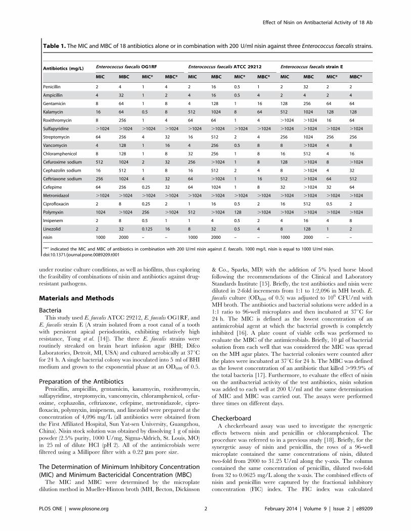
Table 1. The MIC and MBC of 18 antibiotics alone or in combination with 200 U/ml nisin against three Enterococcus faecalis strains.
Antibiotics (mg/L) Enterococcus faecalis OG1RF Enterococcus faecalis ATCC 29212 Enterococcus faecalis strain E
MIC MBC MIC* MBC* MIC MBC MIC* MBC* MIC MBC MIC* MBC*
Penicillin 2 4 1 4 2 16 0.5 1 2 32 2 2
Ampicillin 4 32 1 2 4 16 0.5 4 2 4 2 4
Gentamicin 8 64 1 8 4 128 1 16 128 256 64 64
Kalamycin 16 64 0.5 8 512 1024 8 64 512 1024 128 128
Roxithromycin 8 256 1 4 64 64 1 4 .1024 .1024 16 64
Sulfapyridine .1024 .1024 .1024 .1024 .1024 .1024 .1024 .1024 .1024 .1024 .1024 .1024
Streptomycin 64 256 4 32 16 512 2 4 256 1024 256 256
Vancomycin 4 128 1 16 4 256 0.5 8 8 .1024 4 8
Chloramphenicol 8 128 1 8 32 256 1 8 16 512 4 16
Cefuroxime sodium 512 1024 2 32 256 .1024 1 8 128 .1024 8 .1024
Cephazolin sodium 16 512 1 8 16 512 2 4 8 .1024 4 32
Ceftriaxone sodium 256 1024 4 32 64 .1024 1 16 512 .1024 64 512
Cefepime 64 256 0.25 32 64 1024 1 8 32 .1024 32 64
Metronidazol .1024 .1024 .1024 .1024 .1024 .1024 .1024 .1024 .1024 .1024 .1024 .1024
Ciprofloxacin 2 8 0.25 2 1 16 0.5 2 16 512 0.5 2
Polymyxin 1024 .1024 256 .1024 512 .1024 128 .1024 .1024 .1024 .1024 .1024
Imipenem 2 8 0.5 1 1 4 0.5 2 4 16 4 8
Linezolid 2 32 0.125 16 8 32 0.5 4 8 128 1 2
nisin 1000 2000 – – 1000 2000 – – 1000 2000 – –
‘‘*’’ indicated the MIC and MBC of antibiotics in combination with 200 U/ml nisin against E. faecalis. 1000 mg/L nisin is equal to 1000 U/ml nisin.doi:10.1371/journal.pone.0089209.t001
Effect of Nisin on Antibacterial Activity of 18 Ab
PLOS ONE | www.plosone.org 2 February 2014 | Volume 9 | Issue 2 | e89209
according to the equation: FIC index= FIC A+FIC B= (MIC of
antimicrobial A in combination/MIC of A alone)+(MIC of
antimicrobial B in combination/MIC of B alone). Synergism
was defined as an FIC index #0.5; the additive effect as an FIC
index of 0.5–4.0 and antagonism as an FIC index $4.0 [19]. The
plates were subsequently incubated at 37uC for 24 h. The
procedure was performed three times on different days. Similarly,
the synergetic effect between nisin and chloramphenicol was
evaluated by the same procedure.
Bacterial Survival After Treatment with Four AntibioticsAlone or in Combination with NisinE. faecalis survival rates were evaluated after treatment with
penicillin, vancomycin, chloramphenicol, and linezolid alone or in
combination with nisin. In this assay, E. faecalis ATCC 29212
culture, nisin solution, and different concentrations of antibiotics
were added into 96-well microplates. The resulting concentrations
of cells and nisin were 56107 CFU/ml and 200 U/ml,
respectively, and the four antibiotics were added to the wells
according to the following concentration gradients: penicillin from
1028 to 2 mg/L; chloramphenicol from 1028 to 32 mg/L;
vancomycin, from 1028 to 8 mg/L; linezolid, from 512 to
8 mg/L. Meanwhile, the microplates with the test antibiotics
alone and E. faecalis solution, or with 200 U/ml nisin alone and E.
faecalis solution, were referred to as controls. After the experimen-
tal and control microplates were incubated at 37uC for 24 h, the
surviving bacteria in each well were counted on through a plate
count of viable cells. The assays were carried out three times on
different days.
Transmission Electron MicroscopeThe morphological changes of E. faecalis were observed by
transmission electron microscopy after treatment with the
antibiotics in combination with nisin. Six groups of exponential
phase E. faecalis ATCC 29212 were challenged by the following
drugs for 12 h: 1024 mg/L penicillin (A); 1024 mg/L chloram-
phenicol (B); 2000 U/ml nisin (C); 1024 mg/L penicillin and
2000 U/ml nisin (D); 1024 mg/L chloramphenicol and 2000 U/
ml nisin (E); and phosphate buffered saline (PBS) (Control). After
treatment, sample E. faecalis cells were prepared according to the
TEM analysis of the Dufour, et al. study [20]. In brief, the cell
depositions were fixed with 3% glutaraldehyde, frozen, dehydrated
with increasing concentrations of ethanol, and embedded in resin.
The specimens were examined using a transmission electron
microscope (TEM, TecnaiTM G2, FEI Company, American).
Confocal Laser Scanning MicroscopeThe antibiofilm activities of penicillin, ciprofloxacin, and
chloramphenicol in combination with nisin against E. faecalis were
evaluated by a Live/Dead BacLight Bacterial Viability Kit
(L13152; Molecular Probes, Invitrogen, Inc.,Eugene, OR, USA).
For the E. faecalis biofilm, 1.98 ml of tryptic soy broth (TSB, BD
DIFCO, Sparks MD, USA) supplemented with 1% glucose and
20 ml of an overnight culture of E. faecalis ATCC 29212 were
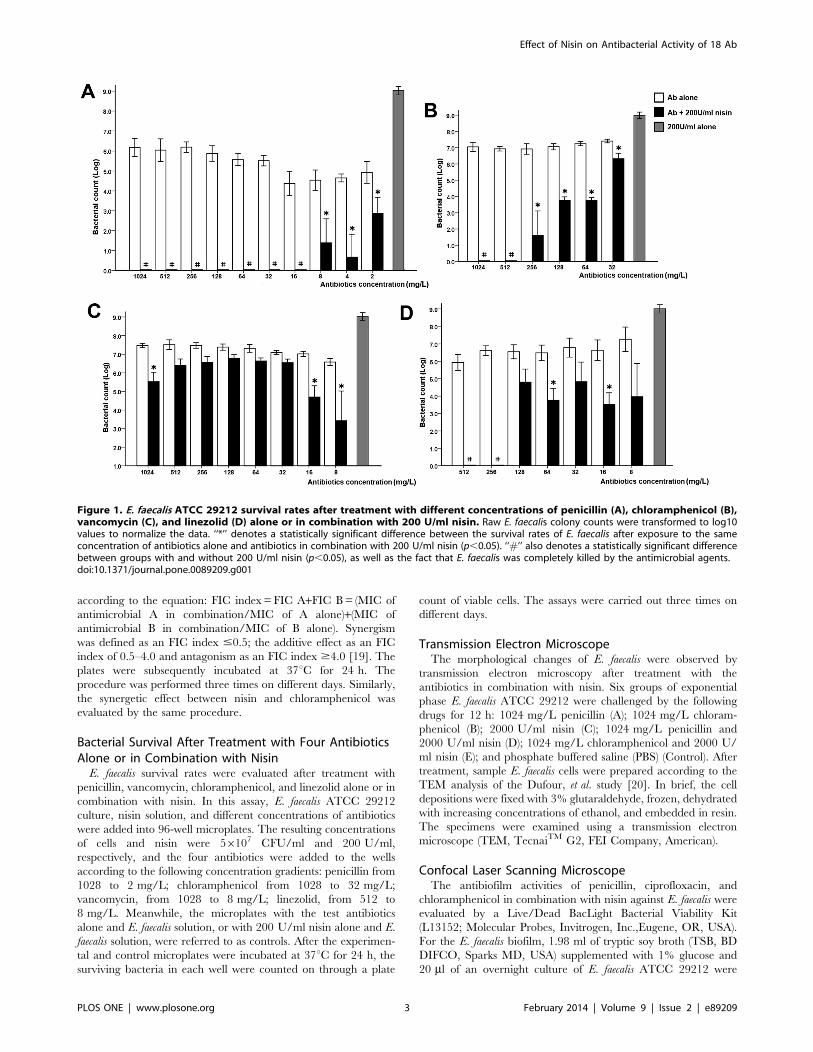
Figure 1. E. faecalis ATCC 29212 survival rates after treatment with different concentrations of penicillin (A), chloramphenicol (B),vancomycin (C), and linezolid (D) alone or in combination with 200 U/ml nisin. Raw E. faecalis colony counts were transformed to log10values to normalize the data. ‘‘*’’ denotes a statistically significant difference between the survival rates of E. faecalis after exposure to the sameconcentration of antibiotics alone and antibiotics in combination with 200 U/ml nisin (p,0.05). ‘‘#’’ also denotes a statistically significant differencebetween groups with and without 200 U/ml nisin (p,0.05), as well as the fact that E. faecalis was completely killed by the antimicrobial agents.doi:10.1371/journal.pone.0089209.g001
Effect of Nisin on Antibacterial Activity of 18 Ab
PLOS ONE | www.plosone.org 3 February 2014 | Volume 9 | Issue 2 | e89209

added into the seven flat bottom plastic wells. Petri dishes with
glass bottoms (D: 35 mm, Hangzhou Shengyou Biotechnology,
China) were then incubated at 37uC for 24 h, and E. faecalis
biofilms were generated on the surfaces of their glass bottoms.
Subsequently, the seven biofilm groups were slightly washed twice
with sterile PBS and then challenged with the following
antimicrobials for 12 h: 512 mg/L penicillin (A); 512 mg/L
ciprofloxacin (B); 512 mg/L chloramphenicol (C); 400 U/ml nisin
(D); 512 mg/L penicillin and 400 U/ml nisin (E); 512 mg/L
ciprofloxacin and 400 U/ml nisin (F); and 512 mg/L chloram-
phenicol and 400 U/ml nisin (G). The seven biofilm groups were
stained with a mixture of 6 mM SYTO 9 stain and 30 mM PI at
room temperature in the dark for 15 min, according to the
specifications of the L13152 Kit. The stained specimens were then
clamped and scanned using a Carl Zeiss confocal laser scanning
microscope (CLSM) and ZEN software (ZEN 2010 light edition,
Carl Zeiss MicroImaging, Inc., Thornwood, NY). SYTO 9 and PI
were excited at 488 nm and 543 nm, respectively. Three-
dimensional E. faecalis biofilms that formed after 12 h of treatment
with 512 mg/L penicillin, 400 U/ml nisin, a combination of the
two antimicrobials, and the controls without treatment were
scanned along the Z axis.
Figure 2. Morphological changes of E. faecalis ATCC 29212 observed by TEM after 12 hours of treatment with antibiotics incombination with nisin. (A) Control; (B) 1024 mg/L penicillin; (C) 2000 U/ml nisin; (D) 1024 mg/L penicillin and 2000 U/ml nisin; (E) 1024 mg/Lchloramphenicol; and (F) 1024 mg/L chloramphenicol and 2000 U/ml nisin.doi:10.1371/journal.pone.0089209.g002
Effect of Nisin on Antibacterial Activity of 18 Ab
PLOS ONE | www.plosone.org 4 February 2014 | Volume 9 | Issue 2 | e89209

Statistical AnalysisStatistical analysis was performed using SPSS 18.0 software.
Raw E. faecalis colony counts were transformed to log10 values to
normalize the data. The student’s t test was used to compare the
survival of E. faecalis after 12 h of treatment with each
concentration of penicillin, vancomycin, chloramphenicol, or
linezolid alone and in conjunction with nisin. A P value ,0.05
was considered statistically significant.
Results
MIC, MBC and CheckerboardThe MICs of nisin against E. faecalis strains ATCC 29212,
OG1RF, and strain E were 1,000 U/ml (1 mg/ml), and their
MBCs were 2,000 U/ml (2 mg/ml). The resistances of the three
strains of E. faecalis to 18 antibiotics alone or in combination with
200 U/ml nisin are shown in Table 1. Penicillin, ampicillin,
vancomycin, ciprofloxacin, imipenem, and linezolid all exhibited
higher antibacterial activity against the three E. faecalis strains than
any of the other antibiotics used, and most of the MICs of the six
antibiotics were less than or equal to 8 mg/L.
Of the 18 tested antibiotics, 3, 5, and 9 antibiotics did not result
in detectable MBCs against OG1RF, ATCC 29212, and strain E,
respectively, and E. faecalis strains exhibited a high level of
resistance to the antibiotics. Furthermore, for some antibiotics
such as kanamycin and streptomycin, the MBC could only be
detected at higher concentrations. However, in the assay of the
antibiotics in combination with the antimicrobial peptide nisin,
200 U/ml nisin significantly improved the antibacterial and
bactericidal activities of all of the tested antibiotics. The exceptions
were sulfapyridine, metronidazol, and polymyxin, none of which
had much of an antibacterial effect on E. faecalis. At the test
concentrations, several antibiotics such as roxithromycin, strepto-
mycin, cefuroxime, cephazolin, ceftriaxone, and cefepime did not
exhibit good antibacterial activity against the test strain E. faecalis,
but the MIC and MBC of the antibiotics were significantly
decreased when combined with 200 U/ml nisin.
For the synergetic effect between nisin and penicillin, the FIC of
nisin against the OG1RF, ATCC 29212, and strain E was 0.06,
0.125, and 0.03, respectively, and the FIC of penicillin against the
three strains was 0.25. Thus, the total FICs of nisin and penicillin
were 0.31, 0.375, and 0.28, respectively. Furthermore, in the
synergetic effect between nisin and chloramphenicol, the FIC of
nisin against the strain E, OR1RF, and ATCC 29212 was 0.03,
0.03, and 0.12, and the FIC of chloramphenicol was 0.125, 0.125,
and 0.03. The total FICs of nisin and chloramphenicol were 0.155,
0.155, and 0.15, respectively. Therefore, the combination between
nisin and penicillin or chloramphenicol was considered to have a
synergetic effect against the three test E. faecalis strains.
Bacterial Survival After Combined Treatment withAntibiotics and NisinThe survival of E. faecalis ATCC 29212 was evaluated after 24 h
of treatment with penicillin, vancomycin, chloramphenicol, or
linezolid in combination with nisin (Fig. 1). In the control
condition, 200 U/ml nisin alone did not inhibit E. faecalis growth,
and E. faecalis grew to approximately 109 CFU/ml. E. faecalis were
not completely killed at 1024 mg/L penicillin, although its MBC
was 16 mg/L. The E. faecalis survival rate did not decrease as the
concentration of the four antibiotics increased. However, in the
presence of 200 U/ml nisin, the bacterial survival rate significantly
decreased, and statistically significant differences were observed
between bacteria treated with or without nisin at the same
concentrations of penicillin or chloramphenicol (p,0.05). In the
presence of nisin, E. faecalis survival rates were shown to be
Figure 3. CLSM images showed the effects of antibiotics in combination with nisin on E. faecalis ATCC 29212 biofilms. (A) 512 mg/Lpenicillin; (B) 512 mg/L ciprofloxacin; (C) 512 mg/L chloramphenicol; (D) 400 U/ml nisin; (E) 512 mg/L penicillin and 400 U/ml nisin; (F) 512 mg/Lciprofloxacin and 400 U/ml nisin; (G) 512 mg/L chloramphenicol and 400 U/ml nisin. Bacteria with intact cell membranes are stained fluorescentgreen, whereas bacteria with damaged cell membranes are stained fluorescent red. All images were shown at 2006magnification and were collectedusing a Carl Zeiss CLSM.doi:10.1371/journal.pone.0089209.g003
Effect of Nisin on Antibacterial Activity of 18 Ab
PLOS ONE | www.plosone.org 5 February 2014 | Volume 9 | Issue 2 | e89209

significantly reduced; as the concentration of the two antibiotics
increased, E. faecalis was completely killed at higher concentra-
tions. Furthermore, 200 U/ml nisin was still able to improve the
bactericidal effects of vancomycin and linezolid, although the
benefits of the two antibiotics from nisin were not as substantial as
those gained from the combination of nisin with penicillin or
chloramphenicol.
Morphological Changes of E. faecalisThe TEM images of E. faecalis illustrated the morphological
changes in the cells after antibiotic treatment. In this study,
2000 U/ml nisin was a suitable concentration that alone did not
cause much of a morphological change in E. faecalis, but could
enhance the damage induced by other antibiotics. The control E.
faecalis, which was not treated with any drugs, exhibited normal
sphericity, as a sign of morphological integrity (Fig. 2A). After 12 h
of treatment with penicillin alone, the majority of E. faecalis still
maintained their original shapes (Fig. 2B). The shapes of a few E.
faecalis cells were destroyed after treatment with nisin alone
(Fig. 2C). Under the combination of penicillin and nisin, many E.
faecalis cells in the observation area lost their original morphology,
instead showing distinct cellular disruption(Fig. 2D). Furthermore,
upon treatment with chloramphenicol alone, E. faecalis cells did
not show overt signs of cellular disruption, but a few cells were
subjected to severe damage by the combination of chloramphen-
icol and nisin (Fig. 2E and 2F).
Antibiofilm AssayCLSM was used to examine the effects of penicillin, ciproflox-
acin, or chloramphenicol in combination with nisin on E. faecalis
biofilms (Fig. 3). In this study, 400 U/ml nisin was an ideal
concentration that did not obviously destroy the E. faecalis biofilm,
but could improve the testing of the antibiotics’ antibiofilm activity
for E. faecalis. Viable cells were stained green and dead cells were
Figure 4. Twenty-four hours old E. faecalis ATCC 29212 biofilms were treated with (A) control biofilm with no treatment, (B) 512 mg/L penicillin alone, (C) 400 U/ml nisin alone, and (D) the combination of 512 mg/L penicillin and 400 U/ml nisin for 12 h. Their 3-dimensional images were scanned along the Z axis at different positions from bottom to top.doi:10.1371/journal.pone.0089209.g004
Effect of Nisin on Antibacterial Activity of 18 Ab
PLOS ONE | www.plosone.org 6 February 2014 | Volume 9 | Issue 2 | e89209
stained red. E. faecalis biofilms exhibited extensive green areas, and
a great deal of cells still survived after treatment with 512 mg/L
penicillin, ciprofloxacin, chloramphenicol, or 400 U/ml nisin
alone (Fig. 3A–3D). In particular, treatment with ciprofloxacin
and chloramphenicol had little effect on the E. faecalis biofilm.
However, treatment with a combination of penicillin, ciproflox-
acin, or chloramphenicol and nisin led to a significant increase in
the red areas of the E. faecalis biofilms, and the particular
combination of penicillin and nisin was able to effectively kill the
majority of cells in the biofilm (Fig. 3E–3G). Three-dimensional
scanning of the control E. faecalis biofilm showed a green 3-
dimensional space (Fig. 4A). After the E. faecalis biofilm was treated
with nisin or penicillin alone, the whole layers almost appeared
green, and small red areas were also found (Fig. 4B and 4C).
However, E. faecalis from top to bottom in the biofilm were
effectively killed by the combination of penicillin and nisin, and
red dominated the 3-dimensional space (Fig. 4D).
Discussion
E. faecalis is among the most antibiotic-resistant bacteria known
at present. E. faecalis has the ability to quickly acquire and
disseminate antibiotic resistance genes by pheromone signals
produced within the genus and species as well as by other bacterial
genera [21]. E. faecalis ATCC 29212 and OG1RF are generally
used for survival and biofilm studies because they have been
extensively used as a representative control strains for clinical and
laboratory experiments [22,23]. As E. faecalis has caused multiple
antibiotic resistant infections, methods of effectively killing this
drug-resistant pathogen have become key goals of microbiologists
and drug development researchers. At present, vancomycin is
considered a drug of last resort, and linezolid has also been
introduced to treat severe infections with antibiotic-resistance
Gram-positive bacteria [24,25]. However, in the in vitro test for E.
faecalis, not even these two potent antibiotics could effectively kill
the three E. faecalis strains in this study. In contrast, the two
conventional antibiotics penicillin and ampicillin exhibited better
antibacterial activity and lower MIC and MBC values for
penicillin and ampicillin than for vancomycin and linezolid.
Similar results were found in a study by Weinstein et al [26].
Therefore, the results of the in vitro evaluation showed that
penicillin and ampicillin may have better antibacterial effects on E.
faecalis than vancomycin and linezolid.
The MBC has generally been defined as the lowest concentra-
tion of an antibiotic that kills .99.9% of the total bacteria
[17,27,28,29]. The MBC of penicillin against ATCC 29212 was
16 mg/L, and viable cells showed more than a 3-log10 reduction.
However, in our determination, bacterial survival did not
decrease, and even may have increased as the concentration of
penicillin increased. Bacterial survival showed less than a 3-log10
reduction at .16 mg/L, so was 16 mg/L still considered the
MBC? In an evaluation of the MBC of 18 test drugs, we found that
no antibiotic completely killed E. faecalis, even at the high
concentration of 1024 mg/L. These in vitro results indicated that
E. faecalis is an antibiotic-resistant pathogen that is difficult to kill.
The phenomenon that pathogens are relatively resistant to higher
concentrations of some antibiotics while remaining susceptible to
lower concentrations of antibiotics was first discovered by Eagle
and Musselman in 1948 [30]. Nowadays, the phenomenon is often
referred to as the ‘‘Eagle effect’’ and has been supported by
additional studies [31,32,33,34]. However, in our study the ‘‘Eagle
effect’’ on E. faecalis did not take place with the addition of nisin,
and E. faecalis was killed by the combination of the antimicrobial
peptide nisin and many test antibiotics. A sub-MIC 200 U/ml
concentration of nisin was used to clarify the antibacterial role of
nisin in drug combinations. This concentration was significantly
less than the MIC of nisin against the three E. faecalis strains and
alone could not inhibit the bacterial growth, as the MICs of
ATCC 29212, OG1RF, and strain E were found to be 1,000 U/
ml. In the evaluation of bacterial survival rates, penicillin,
chloramphenicol, and linezolid in combination with nisin could
completely kill E. faecalis. This bactericidal effect was not due to the
action of 200 U/ml nisin alone, but evidently nisin improves the
bactericidal activities of these antibiotics. Especially with the
addition of nisin, the low concentration of 16 mg/L penicillin
resulted in complete bactericidal activity.
Many studies have indicated that nisin exerts its bactericidal
activity by forming pores and inhibiting cell wall synthesis with a
specific molecule, Lipid II, a principal component of the
membranes of gram-positive bacteria [35,36,37,38]. Nisin uses
Lipid II as a ‘‘docking molecule’’ to form pores on the cell
membrane surface in a targeted manner; at a nanomolar level,
then, nisin is able to effectively kill bacteria [35,39]. Therefore,
200 U/ml nisin is sufficient to form pores on the surface of
bacteria and to facilitate the penetration of other antibiotic
molecules into the microorganisms. In this way, antibiotics will
better capture the antibacterial effects when their antibacterial
action is occurring intracellularly. For example, macrolide
antibiotics (Roxithromycin) binds irreversibly to a site on the
50S subunit of the bacterial ribosome and inhibits the transloca-
tion steps of protein synthesis [40]. Quinolone (ciprofloxacin)
prevents bacterial DNA from unwinding and duplicating [41].
Aminoglycoside antibiotics (gentamicin, kanamycin, and strepto-
mycin) work by binding to the bacterial 30S ribosomal subunit and
inhibiting protein synthesis, and thereby compromise the structure
of the bacterial cell wall [42], etc. The antibacterial activities of
these antibiotics were obviously improved in the presence of a low
concentration of nisin, 200 U/ml. This synergetic antibacterial
mechanism involving the intracellular and cell membranes has
been demonstrated in previous studies. The pores made by nisin
allow more fluoride ions to enter Streptococcus mutans and for more
doxycycline molecules to penetrate into E. faecalis; these actions
result in the synergetic antibacterial activities of nisin and sodium
fluoride, as well as of nisin and doxycycline [43,44]. Furthermore,
the study by Cottagnoud et al. showed that the cell wall disruption
induced by vancomycin acts synergistically with gentamicin
against penicillin-resistant pneumococci by increasing the intra-
cellular penetration of gentamicin [45].
Nevertheless, 200 U/ml nisin was not sufficient to facilitate E.
faecalis inhibition by sulfapyridine, metronidazol, or polymyxin.
This may be due to the intrinsic resistance of E. faecalis to the three
antibiotics; in our study, the MICs of the three antibiotics could
not be detected. As a result, even if nisin helped a greater number
of antibiotic molecules to access the bacteria, the antibiotics were
not able to generate sufficient antibacterial activity.
In the evaluation of the antibacterial activity of the combination
of penicillin and nisin, the results of the combination did not
correspond with the mechanism of intracellular delivery by cell
membrane disruption; however, the antibacterial activity of
penicillin was still significantly strengthened in the presence of
nisin. Penicillin is considered to bind to DD-transpeptidase, a
penicillin-binding protein (PBP) that catalyzes the last step of
peptidoglycan biosynthesis and thus prevents complete cell wall
synthesis [46]. The mechanism of the cell wall disruption differs
from that of nisin by Lipid II, an intermediate in the cell wall
synthesis pathway. Therefore, the two antimicrobials attack the
pathway differently, and are able to generate a greater disruption
within the cell wall. This was also demonstrated by the TEM
Effect of Nisin on Antibacterial Activity of 18 Ab
PLOS ONE | www.plosone.org 7 February 2014 | Volume 9 | Issue 2 | e89209
images. A majority of the cells were severely damaged by the dual
attacks of penicillin and nisin and lost their original cell wall
integrity. Similarly, this combined mechanism is seen in combi-
nations of nisin and the cephalosporins (cefuroxime, cephazolin,
ceftriaxone, and cefepime). However, E. faecalis cells appear more
resistant to the dual action of vancomycin and nisin, and E. faecalis
will still survive the challenge of the two antimicrobials in
combination. Vancomycin decreases the accessibility of Lipid II
by blocking the cell wall biosynthesis, and inhibites the membrane
leakage activity of nisin against intact cells [35,37]. Superior
antibacterial activity is achieved by combining antimicrobials with
different antibacterial mechanisms compared with a combination
of antimicrobials with the same or similar mechanisms [47].
Bacterial biofilms generally become 10–1,000 times more
resistant to the effects of antimicrobial agents than planktonic
cells [48]. A majority of E. faecalis in the biofilm survived the
challenges of penicillin, ciprofloxacin, and chloramphenicol, but
nisin significantly improved the antibiofilm activities of the three
antibiotics, with action taking place throughout many layers of the
biofilm. Compared with some other bacteria, for example,
Streptococcus mutans, E. faecalis form a biofilm that includes a
substantial amount of eDNA but a low level of extracellular
polysaccharides, leading to a low resistance to penetration by
antimicrobial agents [49]. Antimicrobial molecules can easily enter
this biofilm, and the high antibacterial activity of the antibiotics
may play a key role in the inhibition of E. faecalis biofilms.
Therefore, the potent antibacterial activity resulting from the
combination of penicillin and nisin resulted in superior antibiofilm
characteristics against E. faecalis. However, a CLSM image
collected after treatment with penicillin and nisin includes some
minor green areas, indicating that the potent antibacterial activity
of the combination of these two antimicrobial agents had not yet
completely killed all of the bacteria in the biofilm. Alternative
strategies need to be explored in the future to improve the
antibiofilm characteristics of these antibiotics.
These in vitro findings suggest that AMP nisin may significantly
improve the antibacterial and antibiofilm activities of many
antibiotics, and further, antibiotics in combination with nisin have
considerable potential for use in the inhibition of drug-resistant
pathogens.
Author Contributions
Conceived and designed the experiments: ZT YZ JL. Performed the
experiments: ZT YZ. Analyzed the data: ZT YZ JM. Contributed
reagents/materials/analysis tools: YZ LH LZ. Wrote the paper: ZT.
References
1. Sengupta S, Chattopadhyay MK, Grossart HP (2013) The multifaceted roles of
antibiotics and antibiotic resistance in nature. Front Microbiol 4: 47.
2. Palmer AC, Kishony R (2013) Understanding, predicting and manipulating the
genotypic evolution of antibiotic resistance. Nat Rev Genet 14: 243–248.
3. Jenssen H, Hamill P, Hancock RE (2006) Peptide antimicrobial agents. Clin
Microbiol Rev 19: 491–511.
4. Hadley EB, Hancock RE (2010) Strategies for the discovery and advancement of
novel cationic antimicrobial peptides. Curr Top Med Chem 10: 1872–1881.
5. Severina E, Severin A, Tomasz A (1998) Antibacterial efficacy of nisin against
multidrug-resistant Gram-positive pathogens. J Antimicrob Chemother 41: 341–
347.
6. FDA (1988) Food and Drug Administration. Nisin preparation: Affirmation of
GRAS status as a direct human food ingredient. 11251 ed.
7. Naghmouchi K, Le Lay C, Baah J, Drider D (2012) Antibiotic and antimicrobial
peptide combinations: synergistic inhibition of Pseudomonas fluorescens and
antibiotic-resistant variants. Res Microbiol 163: 101–108.
8. Mataraci E, Dosler S (2012) In vitro activities of antibiotics and antimicrobial
cationic peptides alone and in combination against methicillin-resistant
Staphylococcus aureus biofilms. Antimicrob Agents Chemother 56: 6366–6371.
9. Dosler S, Gerceker AA (2012) In vitro activities of antimicrobial cationic
peptides; melittin and nisin, alone or in combination with antibiotics against
Gram-positive bacteria. J Chemother 24: 137–143.
10. Dosler S, Gerceker AA (2011) In vitro activities of nisin alone or in combination
with vancomycin and ciprofloxacin against methicillin-resistant and methicillin-
susceptible Staphylococcus aureus strains. Chemotherapy 57: 511–516.
11. Naghmouchi K, Baah J, Hober D, Jouy E, Rubrecht C, et al. (2013) Synergistic
effect between colistin and bacteriocins in controlling Gram-negative pathogens
and their potential to reduce antibiotic toxicity in mammalian epithelial cells.
Antimicrob Agents Chemother 57: 2719–2725.
12. Richards MJ, Edwards JR, Culver DH, Gaynes RP (2000) Nosocomial infections
in combined medical-surgical intensive care units in the United States. Infect
Control Hosp Epidemiol 21: 510–515.
13. Hollenbeck BL, Rice LB (2012) Intrinsic and acquired resistance mechanisms in
enterococcus. Virulence 3: 421–433.
14. Tong Z, Ling J, Lin Z, Li X, Mu Y (2013) The Effect of MTADN on 10
Enterococcus faecalis Isolates and Biofilm: An In Vitro Study. J Endod 39: 674–
678.
15. Clinical and Laboratory Standards Institute (2006) Methods for dilution
antimicrobial susceptibility tests for bacteria that grow aerobically. Approved
standard M7–A7; 7th ed. Clinical Laboratory Standards Institute, Wayne, PA.
16. Andrews JM (2001) Determination of minimum inhibitory concentrations.
J Antimicrob Chemother 48 Suppl 1: 5–16.
17. Ouhara K, Komatsuzawa H, Kawai T, Nishi H, Fujiwara T, et al. (2008)
Increased resistance to cationic antimicrobial peptide LL-37 in methicillin-
resistant strains of Staphylococcus aureus. J Antimicrob Chemother 61: 1266–
1269.
18. Tong Z, Zhou L, Jiang W, Kuang R, Li J, et al. (2011) An in vitro synergetic
evaluation of the use of nisin and sodium fluoride or chlorhexidine against
Streptococcus mutans. Peptides 32: 2021–2016.
19. Odds FC (2003) Synergy, antagonism, and what the chequerboard puts between
them. J Antimicrob Chemother; 52: 1.
20. Dufour M, Manson JM, Bremer PJ, Dufour JP, Cook JM, et al. (2007)
Characterization of monolaurin resistance in Enterococcus faecalis. ApplEnviron Microbiol 73: 5507–5515.
21. Vickerman MM, Flannagan SE, Jesionowski AM, Brossard KA, Clewell DB, et
al. (2010) A genetic determinant in Streptococcus gordonii Challis encodes a
peptide with activity similar to that of enterococcal sex pheromone cAM373,which facilitates intergeneric DNA transfer. J Bacteriol 192: 2535–2545.
22. Kim EB, Kopit LM, Harris LJ, Marco ML (2012) Draft genome sequence of the
quality control strain Enterococcus faecalis ATCC 29212. J Bacteriol 194: 6006–
6007.
23. Bourgogne A, Garsin DA, Qin X, Singh KV, Sillanpaa J, et al. (2008) Large
scale variation in Enterococcus faecalis illustrated by the genome analysis of
strain OG1RF. Genome Biol 9: R110.
24. Boneca IG, Chiosis G (2003) Vancomycin resistance: occurrence, mechanisms
and strategies to combat it. Expert Opin Ther Targets 7: 311–328.
25. Wilson AP, Cepeda JA, Hayman S, Whitehouse T, Singer M, et al. (2006) In
vitro susceptibility of Gram-positive pathogens to linezolid and teicoplanin andeffect on outcome in critically ill patients. J Antimicrob Chemother 58: 470–473.
26. Weinstein MP (2001) Comparative evaluation of penicillin, ampicillin, and
imipenem MICs and susceptibility breakpoints for vancomycin-susceptible and
vancomycin-resistant Enterococcus faecalis and Enterococcus faecium. J ClinMicrobiol 39: 2729–2731.
27. Fuchs PC, Barry AL, Brown SD (2002) In vitro bactericidal activity of
daptomycin against staphylococci. J Antimicrob Chemother 49: 467–470.
28. Lemaitre N, Ricard I, Pradel E, Foligne B, Courcol R, et al. (2012) Efficacy of
ciprofloxacin-gentamicin combination therapy in murine bubonic plague. PLoS
One 7: e52503.
29. Furustrand Tafin U, Majic I, Zalila Belkhodja C, Betrisey B, Corvec S, et al.(2011) Gentamicin improves the activities of daptomycin and vancomycin
against Enterococcus faecalis in vitro and in an experimental foreign-body
infection model. Antimicrob Agents Chemother 55: 4821–4827.
30. Eagle H, Musselman AD (1948) The rate of bactericidal action of penicillinin vitro as a function of its concentration, and its paradoxically reduced activity
at high concentrations against certain organisms. J Exp Med 88: 99–131.
31. Kondo N, Kuwahara-Arai K, Kuroda-Murakami H, Tateda-Suzuki E,
Hiramatsu K (2001) Eagle-type methicillin resistance: new phenotype of highmethicillin resistance under mec regulator gene control. Antimicrob Agents
Chemother 45: 815–824.
32. Grandiere-Perez L, Jacqueline C, Lemabecque V, Patey O, Potel G, et al. (2005)
Eagle effect in Corynebacterium diphtheriae. J Infect Dis 191: 2118–2120.
33. Shah PM (1982) Paradoxical effect of antibiotics. I. The ‘Eagle effect’.
J Antimicrob Chemother 10: 259–260.
34. Yourassowsky E, Van der Linden MP, Lismont MJ, Crokaert F, Glupczynski Y
(1986) Rate of bactericidal activity for Streptococcus faecalis of a new quinolone,CI-934, compared with that of amoxicillin. Antimicrob Agents Chemother 30:
258–259.
35. Breukink E, de Kruijff B (2006) Lipid II as a target for antibiotics. Nat Rev Drug
Discov 5: 321–332.
Effect of Nisin on Antibacterial Activity of 18 Ab
PLOS ONE | www.plosone.org 8 February 2014 | Volume 9 | Issue 2 | e89209

36. Breukink E, van Heusden HE, Vollmerhaus PJ, Swiezewska E, Brunner L, et al.
(2003) Lipid II is an intrinsic component of the pore induced by nisin in bacterialmembranes. J Biol Chem 278: 19898–19903.
37. Breukink E, Wiedemann I, van Kraaij C, Kuipers OP, Sahl H, et al. (1999) Use
of the cell wall precursor lipid II by a pore-forming peptide antibiotic. Science286: 2361–2364.
38. Christ K, Wiedemann I, Bakowsky U, Sahl HG, Bendas G (2007) The role oflipid II in membrane binding of and pore formation by nisin analyzed by two
combined biosensor techniques. Biochim Biophys Acta 1768: 694–704.
39. Martin NI, Breukink E (2007) Expanding role of lipid II as a target forlantibiotics. Future Microbiol 2: 513–525.
40. Pai MP, Graci DM, Amsden GW (2000) Macrolide drug interactions: an update.Ann Pharmacother 34: 495–513.
41. Hooper DC (2001) Emerging mechanisms of fluoroquinolone resistance. EmergInfect Dis 7: 337–341.
42. Davis BD (1987) Mechanism of bactericidal action of aminoglycosides.
Microbiol Rev 51: 341–350.43. Tong Z, Zhou L, Jiang W, Kuang R, Li J, et al. (2011) An in vitro synergetic
evaluation of the use of nisin and sodium fluoride or chlorhexidine againstStreptococcus mutans. Peptides 32: 2021–2026.
44. Tong Z, Zhou L, Li J, Jiang W, Ma L, et al. (2011) In Vitro Evaluation of the
Antibacterial Activities of MTAD in Combination with Nisin against
Enterococcus faecalis. J Endod 37: 1116–1120.
45. Cottagnoud P, Cottagnoud M, Tauber MG (2003) Vancomycin acts
synergistically with gentamicin against penicillin-resistant pneumococci by
increasing the intracellular penetration of gentamicin. Antimicrob Agents
Chemother 47: 144–147.
46. Scheffers DJ, Pinho MG (2005) Bacterial cell wall synthesis: new insights from
localization studies. Microbiol Mol Biol Rev 69: 585–607.
47. Jia J, Zhu F, Ma X, Cao Z, Li Y, et al. (2009) Mechanisms of drug combinations:
interaction and network perspectives. Nat Rev Drug Discov 8: 111–128.
48. Smith AW (2005) Biofilms and antibiotic therapy: is there a role for combating
bacterial resistance by the use of novel drug delivery systems? Adv Drug Deliv
Rev 57: 1539–1550.
49. Barnes AM, Ballering KS, Leibman RS, Wells CL, Dunny GM (2012)
Enterococcus faecalis produces abundant extracellular structures containing
DNA in the absence of cell lysis during early biofilm formation. MBio 3:
e00193–00112.
Effect of Nisin on Antibacterial Activity of 18 Ab
PLOS ONE | www.plosone.org 9 February 2014 | Volume 9 | Issue 2 | e89209
Related Documents