Acta Biomaterialia 107 (2020) 65–77 Contents lists available at ScienceDirect Acta Biomaterialia journal homepage: www.elsevier.com/locate/actbio Full length article An in vitro hyaluronic acid hydrogel based platform to model dormancy in brain metastatic breast cancer cells Akshay A. Narkhede a,1 , James H. Crenshaw a,1 , David K. Crossman b , Lalita A. Shevde c , Shreyas S. Rao a,∗ a Department of Chemical and Biological Engineering, The University of Alabama, Tuscaloosa, AL 35487-0203, USA b Department of Genetics, University of Alabama at Birmingham, Birmingham, AL, USA c Department of Pathology, O’Neal Comprehensive Cancer Center, University of Alabama at Birmingham, Birmingham, AL, USA a r t i c l e i n f o Article history: Received 20 November 2019 Revised 19 February 2020 Accepted 25 February 2020 Available online 29 February 2020 Keywords: Cancer dormancy Breast cancer brain metastasis In vitro model Hyaluronic acid (HA) hydrogel a b s t r a c t Breast cancer cells (BCCs) can remain dormant at the metastatic site, which when revoked leads to forma- tion of metastasis several years after the treatment of primary tumor. Particularly, awakening of dormant BCCs in the brain results in breast cancer brain metastasis (BCBrM) which marks the most advanced stage of the disease with a median survival period of ~4–16 months. However, our understanding of dor- mancy associated with BCBrM remains obscure, in part, due to the lack of relevant in vitro platforms to model dormancy associated with BCBrM. To address this need, we developed an in vitro hyaluronic acid (HA) hydrogel platform to model dormancy in brain metastatic BCCs via exploiting the bio-physical cues provided by HA hydrogels while bracketing the normal brain and metastatic brain malignancy relevant stiffness range. In this system, we observed that MDA-MB-231Br and BT474Br3 brain metastatic BCCs ex- hibited a dormant phenotype when cultured on soft (0.4 kPa) HA hydrogel compared to stiff (4.5 kPa) HA hydrogel as characterized by significantly lower EdU and Ki67 positivity. Further, we demonstrated the nuclear localization of p21 and p27 (markers associated with dormancy) in dormant MDA-MB-231Br cells contrary to their cytoplasmic localization in the proliferative population. We also demonstrated that the stiffness-based dormancy in MDA-MB-231Br cells was reversible and was, in part, mediated by fo- cal adhesion kinases and the initial cell seeding density. Finally, RNA sequencing confirmed the dormant phenotype in MDA-MB-231Br cells. This platform could further our understanding of dormancy in BCBrM and could be adapted for anti-metastatic drug screening. Statement of Significance Our understanding of dormancy associated with BCBrM remains obscure, in part, due to the lack of rel- evant in vitro platforms to model dormancy associated with BCBrM. Herein, we present a HA hydrogel- based platform to model dormancy in brain metastatic BCCs while recapitulating key aspects of brain microenvironment. We demonstrated that the biophysical cues provided the HA hydrogel mediates dor- mancy in brain metastatic BCCs by assessing both proliferation and cell cycle arrest markers. We also es- tablished the role of focal adhesion kinases and initial cell seeding density in the stiffness-mediated dor- mancy in brain metastatic BCCs. Further, RNA-seq. confirmed the dormant phenotype in brain metastatic BCCs. This platform could be utilized to further our understanding of microenvironmental regulation of dormancy in BCBrM. © 2020 Acta Materialia Inc. Published by Elsevier Ltd. All rights reserved. ∗ Corresponding author. E-mail address: [email protected] (S.S. Rao). 1 These author contributed equally to the work. 1. Introduction Breast cancer can progress to form metastasis at secondary or- gan sites such as bones, lungs, liver and the brain several years af- ter surgical resection of the primary tumor [1]. Approximately, 67% of deaths in case of breast cancer occur past the 5-year survival pe- riod, largely owing to disease relapse at the metastatic site [2–4]. https://doi.org/10.1016/j.actbio.2020.02.039 1742-7061/© 2020 Acta Materialia Inc. Published by Elsevier Ltd. All rights reserved.

Welcome message from author
This document is posted to help you gain knowledge. Please leave a comment to let me know what you think about it! Share it to your friends and learn new things together.
Transcript

Acta Biomaterialia 107 (2020) 65–77
Contents lists available at ScienceDirect
Acta Biomaterialia
journal homepage: www.elsevier.com/locate/actbio
Full length article
An in vitro hyaluronic acid hydrogel based platform to model
dormancy in brain metastatic breast cancer cells
Akshay A. Narkhede
a , 1 , James H. Crenshaw
a , 1 , David K. Crossman
b , Lalita A. Shevde
c , Shreyas S. Rao
a , ∗
a Department of Chemical and Biological Engineering, The University of Alabama, Tuscaloosa, AL 35487-0203, USA b Department of Genetics, University of Alabama at Birmingham, Birmingham, AL, USA c Department of Pathology, O’Neal Comprehensive Cancer Center, University of Alabama at Birmingham, Birmingham, AL, USA
a r t i c l e i n f o
Article history:
Received 20 November 2019
Revised 19 February 2020
Accepted 25 February 2020
Available online 29 February 2020
Keywords:
Cancer dormancy
Breast cancer brain metastasis
In vitro model
Hyaluronic acid (HA) hydrogel
a b s t r a c t
Breast cancer cells (BCCs) can remain dormant at the metastatic site, which when revoked leads to forma-
tion of metastasis several years after the treatment of primary tumor. Particularly, awakening of dormant
BCCs in the brain results in breast cancer brain metastasis (BCBrM) which marks the most advanced
stage of the disease with a median survival period of ~4–16 months. However, our understanding of dor-
mancy associated with BCBrM remains obscure, in part, due to the lack of relevant in vitro platforms to
model dormancy associated with BCBrM. To address this need, we developed an in vitro hyaluronic acid
(HA) hydrogel platform to model dormancy in brain metastatic BCCs via exploiting the bio-physical cues
provided by HA hydrogels while bracketing the normal brain and metastatic brain malignancy relevant
stiffness range. In this system, we observed that MDA-MB-231Br and BT474Br3 brain metastatic BCCs ex-
hibited a dormant phenotype when cultured on soft (0.4 kPa) HA hydrogel compared to stiff (4.5 kPa)
HA hydrogel as characterized by significantly lower EdU and Ki67 positivity. Further, we demonstrated
the nuclear localization of p21 and p27 (markers associated with dormancy) in dormant MDA-MB-231Br
cells contrary to their cytoplasmic localization in the proliferative population. We also demonstrated that
the stiffness-based dormancy in MDA-MB-231Br cells was reversible and was, in part, mediated by fo-
cal adhesion kinases and the initial cell seeding density. Finally, RNA sequencing confirmed the dormant
phenotype in MDA-MB-231Br cells. This platform could further our understanding of dormancy in BCBrM
and could be adapted for anti-metastatic drug screening.
Statement of Significance
Our understanding of dormancy associated with BCBrM remains obscure, in part, due to the lack of rel-
evant in vitro platforms to model dormancy associated with BCBrM. Herein, we present a HA hydrogel-
based platform to model dormancy in brain metastatic BCCs while recapitulating key aspects of brain
microenvironment. We demonstrated that the biophysical cues provided the HA hydrogel mediates dor-
mancy in brain metastatic BCCs by assessing both proliferation and cell cycle arrest markers. We also es-
tablished the role of focal adhesion kinases and initial cell seeding density in the stiffness-mediated dor-
mancy in brain metastatic BCCs. Further, RNA-seq. confirmed the dormant phenotype in brain metastatic
BCCs. This platform could be utilized to further our understanding of microenvironmental regulation of
dormancy in BCBrM.
© 2020 Acta Materialia Inc. Published by Elsevier Ltd. All rights reserved.
h
1
∗ Corresponding author.
E-mail address: [email protected] (S.S. Rao). 1 These author contributed equally to the work.
1
g
t
o
r
ttps://doi.org/10.1016/j.actbio.2020.02.039
742-7061/© 2020 Acta Materialia Inc. Published by Elsevier Ltd. All rights reserved.
. Introduction
Breast cancer can progress to form metastasis at secondary or-
an sites such as bones, lungs, liver and the brain several years af-
er surgical resection of the primary tumor [1] . Approximately, 67%
f deaths in case of breast cancer occur past the 5-year survival pe-
iod, largely owing to disease relapse at the metastatic site [2–4] .
66 A .A . Narkhede, J.H. Crenshaw and D.K. Crossman et al. / Acta Biomaterialia 107 (2020) 65–77
2
2
[
o
e
i
w
t
a
5
a
h
M
l
o
n
t
a
s
f
t
B
a
b
2
2
c
(
t
(
3
m
c
h
c
n
a
2
a
5
c
2
h
t
w
m
H
3
c
μ
c
P
~
i
f
f
fi
This latency period between the treatment of primary tumor and
relapse has been attributed to dormancy [2 , 3 , 5 , 6] . Disseminated
breast cancer cells may remain in a dormant state at the metastatic
site for extended periods of time, which when revoked leads to
metastasis [2 , 3 , 5 , 6] . Specifically, reawakening of dormant breast
cancer cells in the brain leads to brain metastasis, which marks
the most advanced stage of the disease with a median survival pe-
riod of ~4–16 months [7 , 8] . Even with substantial progress in de-
veloping therapeutic regimens, these metastatic breast cancer cells
remain refractory to current therapeutics [8] . Therefore, an im-
proved understanding of the biology of these dormant metastatic
breast cancer cells is urgently needed. However, this has been hin-
dered, in part, due to the lack of relevant in vitro experimen-
tal models to study dormancy in brain metastatic breast cancer
cells [1] .
It is well appreciated that the tumor microenvironment plays
a significant role in cancer dormancy [2 , 5] . Therefore, it is impor-
tant for an in vitro dormancy model to capture the bidirectional
crosstalk between the microenvironment and cancer cells. Specif-
ically, the extracellular matrix (ECM) is one of the key compo-
nents of tumor microenvironment which provides cancer cells with
bio-physical/-chemical cues, hence, mediating the dormant pheno-
type [2 , 5 , 9 , 10] . To understand the mechanisms of microenviron-
mental regulation of dormancy in breast cancer, experimental in
vitro models have been developed. For example, 3D Cultrex® base-
ment membrane extract was employed as an in vitro model to ex-
amine dormancy in mouse mammary cancer cells [11–14] . Simi-
larly, models using poly( ε-caprolactone) electrospun scaffolds [15] ,
laminin-rich ECM [16] , Transglutaminase-crosslinked collagen gels
(Col-Tgel) [17] , Fibrin hydrogels [18 , 19] , poly (ethylene glycol) (PEG)
hydrogels [10] and amikacin hydrate-based hydrogels (Amikagel)
[20] have been devised to study dormancy in primary cancer cells.
In the context of breast cancer metastasis, models of dormancy in-
clude biomaterial-based models (Gelfoam) [21 , 22] and bioreactor-
based models for bone metastasis [23] ; co-culture-based models
[24 , 25] and a combination of co-culture and polyethylene glycol
diacrylate hydrogel biomaterial-based model [26] for liver metas-
tasis.
Mouse models, including experimental metastasis mouse mod-
els, have also been used for dormancy studies [27 , 28] ; however,
controlled investigations using such models is challenging as they
provide very limited control of the organ environment [2] . Further,
high costs, animal-animal variations, as well as the difficulties in-
volved in imaging dormant cancer cells in tissues makes their use
relatively challenging [2] . Whereas some scientific advances have
been made in studying dormancy in primary and metastatic breast
cancer cells in vitro , to the best of our knowledge, experimental in
vitro models to study dormancy in brain metastatic breast cancer
cells in a controlled setting have not been reported.
To address this unmet need, herein, we report a hyaluronic
acid (HA) hydrogel platform, as an in vitro model to study dor-
mancy in brain metastatic breast cancer cells. We chose HA hy-
drogel platform as HA is a major component of the brain ECM
and is highly expressed in brain metastatic tissue [7 , 29 , 30] . In
addition, HA is known to interact with metastatic breast cancer
cells via CD44 receptors [7 , 29] . Herein, we engineered mechani-
cally soft and stiff HA hydrogels while bracketing the normal brain
and metastatic brain malignancy relevant stiffness range. We cul-
tured brain metastatic breast cancer cells on top of HA hydrogels
to study regulation of the dormant phenotype via exploiting the
bio-physical cues provided by soft and stiff HA hydrogels. We also
investigated the role of focal adhesion kinases (FAK) and the ini-
tial cell seeding density in stiffness-mediated dormancy in brain
metastatic breast cancer cells. Finally, we investigated the differen-
tial gene expression in cells cultured on soft vs. stiff HA hydrogels
through RNA sequencing.
. Materials and methods
.1. HA hydrogel preparation
HA hydrogels were prepared as described in our earlier work
7] . Briefly, HA (66–90 kDa; Lifecore Biomedical) (in 1 wt% aque-
us solution) was methacrylated by reacting it with ~18-fold molar
xcess of methacrylic anhydride (Sigma Aldrich) while maintain-
ng pH > 8 at 4 °C, to yield hyaluronic acid methacrylate (HAMA)
ith ~85% degree of methacrylation, as determined through pro-
on nuclear magnetic resonance ( 1 H NMR) [7 , 31] . To prepare soft
nd stiff HA hydrogels, a hydrogel precursor solution containing
wt% HAMA and crosslinker Dithiothreitol (DTT) (Sigma Aldrich)
t a final concentration of 10 mM for soft and 40 mM for stiff HA
ydrogels was prepared in serum-free Dulbecco’s Modified Eagle’s
edium (DMEM) (Sigma Aldrich). 75 μL of hydrogel precursor so-
ution was added to each well of a 96-well plate and incubated
vernight to form hydrogels. This resulted in hydrogels with stiff-
esses of ~0.4 kPa (soft) and ~4.5 kPa (stiff), respectively, as de-
ermined through compression testing performed on RSA-G2 solid
nalyzer instrument (TA Instruments) [7] . To provide cell adhesion
ites, surfaces of both soft and stiff HA hydrogels were consistently
unctionalized with the integrin binding peptide (RGD) (Anaspec)
hrough Michael type addition reaction as described previously [7] .
riefly, 25 μL of 1 mg/mL RGD solution in serum-free DMEM was
dded to each well containing soft or stiff HA hydrogels and incu-
ated for 3 hr at room temperature prior to cell seeding.
.2. Cell culture
td-Tomato (red fluorescent protein; RFP)-expressing MDA-MB-
31Br, a brain metastasizing variant of triple negative breast cancer
ell line MDA-MB-231, was generously provided by Dr. Lonnie Shea
University of Michigan). MDA-MB-231Br cells were routinely cul-
ured in DMEM supplemented with 10% (v/v) fetal bovine serum
FBS) (VWR Life Science) and 1% (v/v) penicillin-streptomycin at
7 °C and 5% CO 2 . BT474Br3, a brain metastasizing variant of hu-
an epidermal growth factor receptor 2 (HER2) positive breast
ancer cell line BT474 [32] , was generously provided by Dr. Di-
ua Yu (University of Texas MD Anderson Cancer Center). BT474Br3
ells were routinely cultured in DMEM/F12 (1:1) (Gibco; Life Tech-
ologies) supplemented with 10% (v/v) fetal bovine serum (FBS)
nd 1% (v/v) penicillin-streptomycin at 37 °C and 5% CO 2 .
.3. EdU cell proliferation assay
Proliferation of MDA-MB-231Br and BT474Br3 cells on soft
nd stiff HA hydrogels was evaluated through incorporation of
-ethynyl-2 ′ -deoxyuridine (EdU) in DNA using Click-iT® EdU mi-
roplate assay kit (C10214; Invitrogen). Briefly, 50 0 0 MDA-MB-
31Br or BT474Br3 cells were seeded on top of soft and stiff HA
ydrogels respectively. At day 2, 10 μM EdU in respective cell cul-
ure media was added to each well and the cells were incubated
ith EdU overnight. At day 3, spent media containing EdU was re-
oved and cells were trypsinized for ~5 min and retrieved from
A hydrogels. The cells were then pelleted and resuspended in
00 μL of 1X PBS (Gibco; Life Technologies). In case of BT474Br3
ells (sphere forming cells), the cell pellet was treated with 200
L Accutase (Corning) to disintegrate the spheres and yield single
ells prior to resuspension in 300 μL of 1X PBS. The cells in 1X
BS were added to a 96-well plate, followed by centrifugation at
10 0 0 x g for ~15 s for the cells to settle at the bottom follow-
ng which the spent PBS was removed and the cells were stained
or EdU as per the manufacturer’s protocol. Briefly, cells were fixed
or 5 min at room temperature by adding 50 μL of Click-iT® EdU
xative to each well followed by the addition of 50 μL reaction

A .A . Narkhede, J.H. Crenshaw and D.K. Crossman et al. / Acta Biomaterialia 107 (2020) 65–77 67
c
i
t
~
w
2
t
d
w
c
t
M
t
d
2
e
a
m
c
t
s
c
b
m
B
m
a
c
g
s
p
T
w
a
c
u
w
t
v
s
2
o
p
B
w
(
B
i
m
c
i
t
w
I
2
o
c
1
c
a
s
a
b
t
p
2
M
1
s
E
w
H
a
2
M
t
M
r
h
R
c
a
B
i
7
i
t
c
d
f
s
m
d
r
i
l
4
(
t
o
s
t
r
f
0
g
p
d
g
i
t
c
m
l
o
m
w
ocktail containing Oregon green® 488 azide which binds to the
ncorporated EdU. The cells were incubated with the reaction cock-
ail for ~25 min. The plate was then centrifuged at ~10 0 0 x g for
15 s and the spent reaction cocktail was removed. The cells were
ashed twice with 1X PBS and counterstained with 4,6-diamidino-
-phenylindole (DAPI) (Invitrogen) nuclear stain. The cells were
hen imaged using an Olympus IX83 microscope with a spinning
isk confocal attachment. Similar exposure time and gain settings
ere maintained throughout all conditions. Percent EdU positive
ells were evaluated through manual counting using multi-point
ool in ImageJ software (NIH). A similar approach was used by
arlow et al., wherein the cells were retrieved from the 3D ma-
rix prior to the detection of EdU labeled cells in order to establish
ormant vs. proliferative phenotypes [21] .
.4. Immunofluorescence staining
Immunofluorescence staining was performed to investigate the
xpression of Ki67, marker for cellular proliferation [33] and p21
nd p27, markers associated with cell cycle arrest and cancer dor-
ancy [6 , 34] . For Ki67 staining, 50 0 0 MDA-MB-231Br or BT474Br3
ells were seeded on top of soft and stiff HA hydrogels respec-
ively. At day 3, cells were retrieved from HA hydrogels as de-
cribed above and added to a 96-well plate. The plate was then
entrifuged at ~10 0 0 x g for ~15 s for the cells to settle at the
ottom. The cells were then fixed with 4% paraformaldehyde, per-
eabilized using 0.25% Triton-X in 1X PBS and blocked with 5%
SA in 1X PBS. The cells were then incubated with 1 μg/ml of pri-
ary antibody (Anti-Ki67 antibody) (ab15580; abcam) overnight
t 4 °C followed by incubation with 2 μg/ml of Alexa Fluor 488-
onjugated goat anti-rabbit secondary antibody (A-11034; Invitro-
en) for 45 min. The cells were counterstained with DAPI nuclear
tain. The plate was centrifuged at ~10 0 0 x g for ~15 s every time
rior to the removal of liquids from wells to avoid the loss of cells.
he cells were then imaged using an Olympus IX83 microscope
ith a spinning disk confocal attachment. Similar exposure time
nd gain settings were maintained throughout all conditions. Per-
ent Ki67 positive cells were evaluated through manual counting
sing multi-point tool in ImageJ software (NIH). A similar approach
as used by Marlow et al., wherein the cells were retrieved from
he 3D matrix prior to Ki67 staining in order to establish dormant
s. proliferative phenotypes [21] .
For p21 and p27 staining, 50 0 0 MDA-MB-231Br cells were
eeded on top of soft and stiff HA hydrogels respectively. At day
, the spent media was removed and the cells were gently washed
nce with 1X PBS. The cells were fixed with 4% paraformaldehyde,
ermeabilized using 0.25% Triton-X in 1X PBS and blocked with 5%
SA in 1X PBS on the HA hydrogels. The cells were then incubated
ith primary antibody (1:50 dilution of stock in 1X PBS) for p21
sc-817; Santa Cruz Biotechnology) or p27 (sc-56338; Santa Cruz
iotechnology) overnight at 4 °C on the HA hydrogels, followed by
ncubation with 2 μg/ml of Alexa Fluor 488-conjugated goat anti-
ouse secondary antibody (A-11001; Invitrogen) for 45 min. The
ells were counterstained with DAPI nuclear stain and imaged us-
ng an Olympus IX83 microscope with a spinning disk confocal at-
achment. Percent cells with nuclear localization of p21 and p27
as evaluated through manual counting using multi-point tool in
mageJ software (NIH).
.5. Focal adhesion kinase (FAK) blocking study
For FAK blocking study, 50 0 0 MDA-MB-231Br cells were seeded
n top of stiff HA hydrogels and were treated with varying con-
entrations of FAK inhibitor 14 (Sigma Aldrich) viz. 0, 1, 2.5, 5, 7.5,
0 and 12.5 μM respectively throughout the experiment. At day 3,
ells were retrieved from stiff HA hydrogels and stained for Ki67
s described above. In a separate setup, MDA-MB-231Br cells on
tiff HA hydrogels were treated with FAK inhibitor 14 as described
bove and were retrieved at day 3 to determine the cellular via-
ility in the presence of varying FAK inhibitor 14 concentrations
hrough trypan blue staining, which was further used to compute
ercent viable Ki67 negative cells.
.6. Cell seeding density study
In order to evaluate the impact of cell seeding density on the
DA-MB-231Br phenotype on soft HA hydrogels, initially, 50 0 0,
0,0 0 0, 20,0 0 0, 35,0 0 0 and 50,0 0 0 MDA-MB-231Br cells were
eeded onto soft HA hydrogels respectively. At day 2, 10 μM
dU was added to each condition and the cells were incubated
ith EdU overnight. At day 3, the cells were retrieved from soft
A hydrogels and stained for the incorporated EdU as described
bove.
.7. Whole transcriptome RNA sequencing
In order to investigate the differential gene expression in MDA-
B-231Br cells cultured on soft versus stiff HA hydrogels, whole
ranscriptome RNA sequencing was performed. Briefly, 50 0 0 MDA-
B-231Br cells were seeded onto the soft and stiff HA hydrogels
espectively. At day 3, cells were retrieved from soft and stiff HA
ydrogels ( n ≥ 5 hydrogels) and lysed to extract total RNA using
Neasy® mini kit (74,104; Qiagen) as per the manufacturer’s proto-
ol. mRNA-sequencing was performed on the Illumina NextSeq500
s described by the manufacturer (Illumina Inc., San Diego, CA).
riefly, the quality of the total RNA was assessed using the Ag-
lent 2100 Bioanalyzer. RNA with RNA Integrity Number (RIN) of
.0 or above was used for sequencing library preparation. The Ag-
lent SureSelect Strand Specific mRNA library kit was used as per
he manufacturer’s instructions (Agilent, Santa Clara, CA). Library
onstruction began with two rounds of polyA selection using oligo
T containing magnetic beads. The resulting mRNA was randomly
ragmented with cations and heat, which was followed by first
trand synthesis using random primers with inclusion of Actino-
ycin D (2.4 ng/μL final concentration). Second strand cDNA pro-
uction was done with standard techniques and the ends of the
esulting cDNA were made blunt, A-tailed and adaptors ligated for
ndexing to allow for multiplexing during sequencing. The cDNA
ibraries were quantitated using qPCR in a Roche LightCycler®
80 with the Kapa Biosystems kit for Illumina library quantitation
Kapa Biosystems, Woburn, MA) prior to cluster generation. Clus-
er generation was performed according to the manufacturer’s rec-
mmendations for onboard clustering (Illumina). Paired end 75 bp
equencing runs were completed to allow for better alignment of
he sequences to the reference genome.
STAR (version 2.7.0b) was used to align the raw RNA-Seq fastq
eads to the human reference genome (GRCh38 p7 Release 25)
rom Gencode [35] . Following alignment, HTSeq-count (version
.9.1) was used to count the number of reads mapping to each
ene [36] . Normalization and differential expression were then ap-
lied to the count files using DESeq2 while controlling for batch
ifferences [37] . For generating networks, a data set containing
ene identifiers and corresponding expression values was uploaded
nto Ingenuity Pathway Analysis (IPA). Each identifier was mapped
o its corresponding object in Ingenuity’s Knowledge Base. A fold
hange (FC) cut-off of ±2 and q-value < 0.05 was set to identify
olecules whose expression was significantly differentially regu-
ated. These molecules, called Network Eligible molecules, were
verlaid onto a global molecular network developed from infor-
ation contained in Ingenuity’s Knowledge Base. Networks of Net-
ork Eligible Molecules were then algorithmically generated based

68 A .A . Narkhede, J.H. Crenshaw and D.K. Crossman et al. / Acta Biomaterialia 107 (2020) 65–77
p
s
i
2
(
m
s
h
F
c
s
l
g
g
F
l
a
s
t
t
o
o
h
(
o
m
E
m
M
p
g
s
2
a
c
s
s
e
d
H
r
3
l
s
i
p
d
t
a
s
s
p
t
o
M
h
S
t
H
n
m
a
on their connectivity. The Functional Analysis identified the bio-
logical functions and/or diseases that were most significant to the
entire data set. Molecules from the dataset that met the FC cut-
off of ±2 and q-value < 0.05 and were associated with biological
functions and/or diseases in Ingenuity’s Knowledge Base were con-
sidered for the analysis. Right-tailed Fisher’s exact test was used to
calculate a p-value determining the probability that each biological
function and/or disease assigned to that data set is due to chance
alone. For heatmaps, the genes that were differentially regulated
(FC cut-off of ±2 and p < 0.05) between soft vs. stiff HA hydrogels
were categorized through DAVID gene ontology tool (using the in-
built functional annotation tool) and IPA. The FPKM values of genes
in the chosen enriched gene sets were inputted into an online tool
named ClustVis to generate the heat maps [38] .
2.8. Statistical analysis
All experiments were repeated at least twice with at least 3
replicates per condition in each experiment. The results are pre-
sented as mean ± standard deviation unless otherwise stated. A
t -test was performed (using JMP Pro-software) for comparison of 2
samples belonging to normal datasets. For multiple comparisons in
normal datasets, the data was subjected to ANOVA followed by the
post-hoc Tukey HSD test (using JMP Pro-software). For non-normal
dataset, Wilcoxon each pair test was performed for multiple com-
parisons (using JMP Pro-software). A p-value less than 0.05 was
considered to be statistically significant.
3. Results
3.1. Brain metastatic breast cancer cells exhibit a dormant phenotype
when cultured on soft HA hydrogel whereas they proliferate on stiff
HA hydrogel
We initially assessed the proliferation of MDA-MB-231Br and
BT474Br3 brain metastatic breast cancer cells when cultured on
soft (0.4 kPa) and stiff (4.5 kPa) HA hydrogels through incorpora-
tion of EdU in the newly synthesized DNA. Incorporation of EdU
has been routinely used to assess cell cycle progression (G0/G1
transition) and cancer dormancy [10 , 21 , 24 , 39] . EdU staining at day
3 post cell seeding revealed that the number of EdU positive cells
was significantly lower when brain metastatic breast cancer cells
were cultured on soft HA hydrogels compared to stiff HA hydro-
gels ( p < 0.05) ( Fig. 1 ). In particular, only ~2 ± 6% MDA-MB-231Br
cells were EdU positive when cultured on soft HA hydrogels com-
pared to ~70 ± 14% EdU positive cells on stiff HA hydrogels. Sim-
ilar observation was made in case of BT474Br3 cells wherein only
~5 ± 7% cells were EdU positive when cultured on soft HA hy-
drogels compared to ~22 ± 11% EdU positive cells on stiff HA
hydrogel.
Further, we analyzed Ki67 positivity of MDA-MB-231Br and
BT474Br3 cells cultured on soft and stiff HA hydrogels. Ki67 is a
proliferation marker which is highly expressed during G2 and mi-
totic (M) phase of cell cycle [33] . In addition, Ki67 has been used
as a marker to study dormancy in several studies [16 , 21 , 24] . Ki67
staining at day 3 revealed that the number of Ki67 positive cells
was significantly lower when brain metastatic breast cancer cells
were cultured on soft HA hydrogels compared to stiff HA hydro-
gels ( p < 0.05) ( Fig. 2 ). In particular, only ~6 ± 5% MDA-MB-231Br
cells were Ki67 positive when cultured on soft HA hydrogels com-
pared to ~92 ± 5% Ki67 positive cells when cultured on stiff HA
hydrogels. Similar observation was made in case of BT474Br3 cells
wherein only ~12 ± 6% cells were Ki67 positive on soft HA hy-
drogels compared to ~32 ± 4% Ki67 positive cells on stiff HA hy-
drogels. Collectively, these observations indicate a non-proliferative
henotype in brain metastatic breast cancer cells when cultured on
oft HA hydrogels due to arrest in early phase of cell cycle result-
ng in a dormant phenotype.
We noted differences in the cellular morphologies of MDA-MB-
31Br and BT474Br3 cells cultured on soft and stiff HA hydrogels
Suppl. Fig. 1). Specifically, MDA-MB-231Br cells exhibited rounded
orphology when cultured on soft HA hydrogel whereas they
pread and extended cellular processes when cultured on stiff HA
ydrogels and 2D tissue culture polystyrene (TCPS) (Suppl. Fig. 1).
urther, cells cultured on stiff HA hydrogel also typically reached
onfluency by day 3 in stark contrast to the cells cultured on the
oft HA hydrogel (Suppl. Fig. 1). BT474Br3 cells formed multicel-
ular aggregates when cultured on both soft and stiff HA hydro-
els however, qualitatively, there were more multicellular aggre-
ates on stiff HA hydrogels compared to soft HA hydrogels (Suppl.
ig. 1). Interestingly, BT474Br3 grown on 2D TCPS form multicel-
ular colonies (Suppl. Fig. 1). Early apoptosis assay revealed low
poptotic index in MDA-MB-231Br and BT474Br3 cells on soft and
tiff HA hydrogels at day 1 with no significant difference between
he groups ( p > 0.05) (Suppl. Fig. 2, Suppl. Fig. 3). Further, the to-
al cell count in the case of MDA-MB-231Br cells at day 3 (based
n automated trypan blue assay) was ~ 4259 ± 658 cells/hydrogel
n soft HA hydrogel and ~16,600 ± 2645 cells/hydrogel on stiff HA
ydrogel (initial cell seeding density being ~50 0 0 cells/hydrogel)
Suppl. Fig. 4). This indicates that culturing MDA-MB-231Br cells
n soft HA hydrogel halts their proliferation (making them dor-
ant), and further corroborates the observations made through
dU and Ki67 staining. In addition, we observed that the dor-
ant phenotype on soft HA hydrogels was reversible as dormant
DA-MB-231Br cells on soft HA hydrogels exhibited a spread mor-
hology and proliferated when transferred onto stiff HA hydro-
els (Suppl. Figs.5 and 6). Further, we observed an enhanced re-
istance to chemotherapeutic drug Paclitaxel (Taxol) in MDA-MB-
31Br cells on soft HA hydrogel compared to stiff HA hydrogel
nd 2D TCPS as indicated by higher half maximal inhibitory con-
entration (IC 50 ) however, the difference did not reach statistical
ignificance ( p = 0.08) (Suppl. Fig. 7). Taken together, these ob-
ervations indicate that the brain metastatic breast cancer cells
xhibit a dormant (non-proliferative) phenotype on soft HA hy-
rogels, whereas they exhibit a proliferative phenotype on stiff
A hydrogels, and that the substrate stiffness-driven dormancy is
eversible.
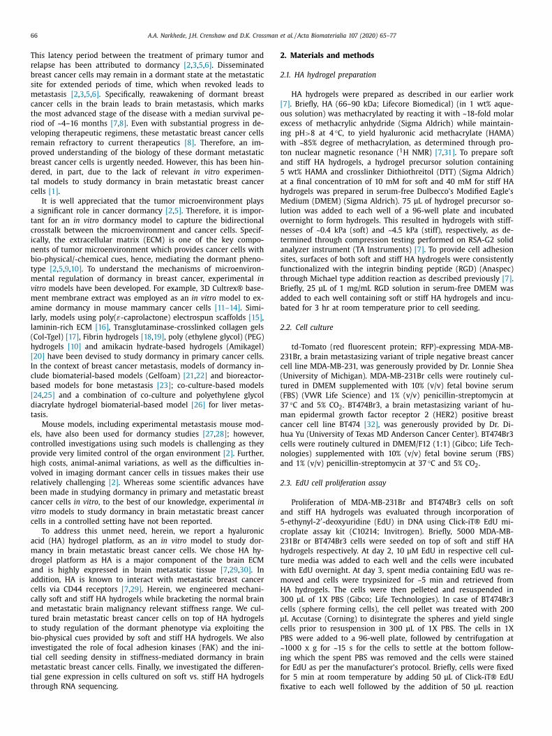
.2. Cyclin-dependent kinase inhibitors p21 and p27 exhibit nuclear
ocalization in MDA-MB-231Br cells cultured on soft HA hydrogels
Cyclin dependent kinase inhibitors p21 and p27 (markers as-
ociated with cell cycle arrest) have been previously implicated
n cancer dormancy [6 , 34] . Herein, we examined their expression
attern in MDA-MB-231Br cells cultured on soft and stiff HA hy-
rogels. We chose MDA-MB-231Br cells for subsequent studies as
hey exhibited drastic differences in the magnitude of both EdU
nd Ki67 positivity when cultured on soft vs. stiff HA hydrogels re-
pectively ( Fig. 1 A,B, Fig. 2 A,B). Interestingly, immunofluorescence
taining at day 2 revealed nuclear localization of both p21 and
27, in MDA-MB-231Br cells cultured on soft HA hydrogel contrary
o their cytoplasmic localization in MDA-MB-231Br cells cultured
n stiff HA hydrogel ( Fig. 3 , Suppl. Fig. 8). In particular, ~89 ± 8%
DA-MB-231Br cells showed nuclear localization of p21 on soft HA
ydrogel compared to only ~9 ± 8% on stiff HA hydrogel ( p < 0.05).
imilarly, ~91 ± 5% MDA-MB-231Br cells showed nuclear localiza-
ion of p27 on soft HA hydrogel compared to only ~3 ± 4% on stiff
A hydrogel ( p < 0.05). Thus, the dormant vs. proliferative phe-
otype observed in these cells was confirmed by assessing both
arkers associated with proliferation (i.e., Ki67 and EdU), as well
s cell cycle arrest (i.e., p21 and p27).

A .A . Narkhede, J.H. Crenshaw and D.K. Crossman et al. / Acta Biomaterialia 107 (2020) 65–77 69
Fig. 1. Brain metastatic breast cancer cells cultured on soft (0.4 kPa) HA hydrogel were largely EdU negative and exhibited a dormant phenotype. A. Representative fluo-
rescence microscopy images of EdU staining at day 3 of MDA-MB-231Br cells (upper panel) and BT474Br3 (lower panel) cultured on soft (0.4 kPa) and stiff (4.5 kPa) HA
hydrogel respectively. Green: EdU; Blue: DAPI (nuclei). Scale bar = 100 μm. B. Quantification of EdU positive MDA-MB-231Br cells cultured on soft and stiff HA hydrogel
respectively. C. Quantification of EdU positive BT474Br3 cells cultured on soft and stiff HA hydrogel respectively. N = 6 replicates per condition. ∗ indicates statistical signifi-
cance ( p < 0.05). Error bar represents standard deviation (For interpretation of the references to colour in this figure legend, the reader is referred to the web version of this
article.).
3
s
n
M
s
g
i
H
r
M
M
h
t
i
d
h
i
t
d
t
t
h
p
(
i
a
(
c
t
b
b
T
b
c
3
M
s
h
5
E
i
i
i
E
p
(
c
t
i
i
(
.3. Role of focal adhesion kinases (FAK) in mediating the
tiffness-based dormancy in MDA-MB-231Br cells
In our previous work, we demonstrated that focal adhesion ki-
ases (FAK) partly mediate the stiffness-based response of MDA-
B-231Br cells [7] . Particularly, we observed that blocking FAK re-
ulted in a significant reduction in proliferation on stiff HA hydro-
els [7] . Further, FAK signaling has been implicated in dormancy, as
nhibition of FAK signaling has been shown to induce dormancy in
Ep3 human carcinoma cells [40] . Therefore, we investigated the
ole of FAK in mediating the stiffness-based dormancy in MDA-
B-231Br cells. We hypothesized that blocking FAK in proliferative
DA-MB-231Br cells on stiff HA hydrogels would result in cells ex-
ibiting a more dormant phenotype. To test our hypothesis, we
reated MDA-MB-231Br cells with varying concentrations of FAK
nhibitor 14 on stiff HA hydrogel and assessed their Ki67 status at
ay 3. Indeed, we observed that with an increase in the FAK in-
ibitor concentration, the Ki67 positivity was significantly reduced
n MDA-MB-231Br cells on stiff HA hydrogels ( Fig. 4 A,B) compared
o the control group ( p < 0.05). Particularly, the Ki67 positivity re-
uced from ~76 ± 8% in the control group to ~32 ± 12% in cells
reated with 12.5 μM of FAK inhibitor 14. Recognizing the fact
hat treatment with FAK inhibitor may impact cellular viability and
ence Ki67 positivity; we assessed the cellular viabilities using try-
an blue assay and used it to calculate viable Ki67 negative cells
Suppl. Table 1). Interestingly, we observed that with an increase
n FAK inhibitor concentration, the number of viable Ki67 neg-
tive cells increased significantly compared to the control group
p < 0.05) ( Fig. 4 C). Specifically, the percent viable Ki67 negative
ells increased from ~7 ± 5% in control group to ~38 ± 2% in cells
reated with 12.5 μM of FAK inhibitor 14. The increase in the num-
er of viable Ki67 negative cells indicates an increase in the num-
er of dormant cells on stiff HA hydrogels following FAK inhibition.
hese results indicated that FAK, in part, mediates the stiffness-
ased dormancy in MDA-MB-231Br brain metastatic breast cancer
ells.
.4. Initial cell seeding density impacts the dormant phenotype in
DA-MB-231Br cells cultured on soft HA hydrogel
We further evaluated the impact of initial cell seeding den-
ity on the dormant phenotype of MDA-MB-231Br cells on soft HA
ydrogels. We initially seeded 50 0 0, 10,0 0 0, 20,0 0 0, 35,0 0 0 and
0,0 0 0 MDA-MB-231Br cells on soft HA hydrogel and performed
dU staining at day 3. Interestingly, we observed that increasing
nitial cell seeding density partly revoked the dormant phenotype
n MDA-MB-231Br cells on soft HA hydrogel as characterized by an
ncrease in percentage of EdU positive cells ( Fig. 5 ). Specifically, the
dU positivity significantly increased from ~2 ± 4% in 50 0 0 cells
er hydrogel condition to ~38 ± 10% at 50,0 0 0 cells per hydrogel
p < 0.05). Increased EdU positivity was also accompanied by some
ell spreading on soft HA hydrogels in conditions with higher ini-
ial cell seeding densities (Suppl. Fig. 9). No significant difference
n the cellular morphologies (in terms of spreading) with vary-
ng cell seeding densities were observed in case of BT474Br3 cells
Suppl. Fig. 9).

70 A .A . Narkhede, J.H. Crenshaw and D.K. Crossman et al. / Acta Biomaterialia 107 (2020) 65–77
Fig. 2. Brain metastatic breast cancer cells cultured on soft (0.4 kPa) HA hydrogel were largely Ki67 negative and exhibited a dormant phenotype. A. Representative fluo-
rescence microscopy images of Ki67 staining at day 3 of MDA-MB-231Br cells (upper panel) and BT474Br3 (lower panel) cultured on soft (0.4 kPa) and stiff (4.5 kPa) HA
hydrogel, respectively. Green: Ki67; Blue: DAPI (nuclei). Scale bar = 100 μm. B. Quantification of Ki67 positive MDA-MB-231Br cells cultured on soft and stiff HA hydrogel
respectively. C. Quantification of Ki67 positive BT474Br3 cells cultured on soft and stiff HA hydrogel respectively. N = 6 replicates per condition. ∗ indicates statistical signifi-
cance ( p < 0.05). Error bar represents standard deviation. (For interpretation of the references to colour in this figure legend, the reader is referred to the web version of this
article.)
p
(
i
o
q
h
a
w
e
M
s
n
4
d
i
w
i
f
E
[
l
d
b
m
p
3.5. Differential gene expression in MDA-MB-231Br cells cultured on
soft versus stiff HA hydrogels through whole transcriptome RNA
sequencing
To associate phenotype with genotype in our model system, we
performed whole transcriptome RNA sequencing. Whole transcrip-
tome RNA sequencing revealed that ~1394 genes were differen-
tially regulated between MDA-MB-231Br cells cultured on soft and
stiff HA hydrogel respectively (FC cut-off ± 2, p < 0.05) (Support-
ing file 1). Consistent with our expectations, major gene groups
downregulated in MDA-MB-231Br cells cultured on soft HA hydro-
gel (dormant) compared to MDA-MB-231Br cells on stiff HA hy-
drogel (proliferative) included genes associated with G1/S transi-
tion of mitotic cell cycle and cell division ( Fig. 6 A, Supporting file
2) (as enriched in DAVID gene ontology tool; p < 0.05). Further,
major gene groups upregulated in MDA-MB-231Br cells cultured
on soft HA hydrogel (dormant) included genes associated with
cell cycle arrest and inflammatory response ( Fig. 6 B-C, Supporting
file 2) (as enriched in DAVID gene ontology tool and IPA respec-
tively; p < 0.05). In addition, Ingenuity Pathway Analysis (IPA)® re-
vealed top pathways as well as the associated molecules that may
be activated in dormant MDA-MB-231Br cells on soft HA hydro-
gels (Suppl. Fig 10, Suppl. Table 2), including glucocorticoid recep-
tor signaling, which is known to be associated with cancer dor-
mancy [41] . We also found that the expression pattern of specific
genes/biomarker associated with cancer dormancy in our model
system was largely in agreement with that reported in the liter-
ature ( Table 1 ). Further, we observed significant upregulation of
N-myc downstream regulated gene 2 (NDRG2) gene (FC = 7.59;
b= 0.02; q = NA) in MDA-MB-231Br cells on soft HA hydrogel
dormant) which is known to act as tumor suppressor [42] , specif-
cally through upregulation of E-Cadherin [43] . Subsequently, we
bserved upregulation of E-Cadherin ( CDH1; FC = 7.74; p = 0.54;
= NA) in MDA-MB-231Br cells on soft HA hydrogel (dormant)
owever this did not reach statistical significance. Interestingly,
long with upregulation of E-cadherin (an epithelial marker [44] ),
e also observed a significant downregulation of vimentin (a mes-
nchymal marker [44] ) ( VIM ; FC = −2.47; p = 0.001; q = 0.06) in
DA-MB-231Br cells on soft HA hydrogel (dormant). Overall, RNA-
eq analysis further confirmed the dormant vs. proliferative phe-
otype observed in our model system.
. Discussion
In this study, we utilized a HA hydrogel platform to model
ormancy in brain metastatic breast cancer cells in vitro . As ECM
s one of the key components of the tumor microenvironment
hich mediates the dormant phenotype in cancer cells, stud-
es have recently reported on biomaterial-based in vitro plat-
orms which recapitulate the key bio-physical/-chemical aspects of
CM to model cancer dormancy in the context of primary tumor
10–18 , 20] . However, very few studies have reported on using uti-
izing biomaterials-based in vitro platform to study breast cancer
ormancy in a metastatic setting [21 , 23–26 ]. Mouse models have
een utilized to study cancer dormancy at metastatic site, however,
ouse models do not offer control or tunability of specific ECM
roperties leading to inadequate decoupling of signals provided
y tumor microenvironment [2] . Amidst limited scientific advances
A .A . Narkhede, J.H. Crenshaw and D.K. Crossman et al. / Acta Biomaterialia 107 (2020) 65–77 71
Fig. 3. Immunofluorescence staining revealed nuclear localization of p21 and p27 in MDA-MB-231Br cells cultured on soft (0.4 kPa) HA hydrogels and cytoplasmic localization
on stiff (4.5 kPa) HA hydrogels. A. Representative fluorescence microscopy images of p21 (upper panel) and p27 (lower panel) staining at day 2 of MDA-MB-231Br cells
cultured on soft (0.4 kPa) and stiff (4.5 kPa) HA hydrogel respectively. Green: p21 or p27; Blue: DAPI (nuclei). Scale bar = 100 μm. B. Quantification of MDA-MB-231Br cells
with nuclear localization of p21 on soft (0.4 kPa) and stiff (4.5 kPa) HA hydrogel respectively. C. Quantification of MDA-MB-231Br cells with nuclear localization of p27 on
soft (0.4 kPa) and stiff (4.5 kPa) HA hydrogel respectively. N = 3 replicates per condition. ∗ indicates statistical significance ( p < 0.05). Error bar represents standard deviation.
(For interpretation of the references to colour in this figure legend, the reader is referred to the web version of this article.)
i
i
e
b
H
m
r
d
s
b
o
s
p
M
o
2
t
d
o
h
h
t
m
c
b
l
t
h
c
s
H
d
m
o
p
o
u
i
o
s
c
B
d
e
p
H
p
p
n developing models for studying cancer dormancy, specifically
n the context of metastasis, we report for the first time on an
xperimental in vitro model to study dormancy in brain metastatic
reast cancer cells in a controlled setting.
We engineered mechanically soft (0.4 kPa) and stiff (4.5 kPa)
A hydrogels bracketing the normal brain (0.2–1 kPa [45] ) and
etastatic brain malignancy (~3.74 kPa [46] ) relevant stiffness
ange, while maintaining similar HA composition, which allowed
ecoupling of the biophysical and biochemical cues. We further re-
orted to utilizing a 3D ‘on-top’ [47] approach wherein we cultured
rain metastatic breast cancer cells MDA-MB-231Br and BT474Br3
n top of soft and stiff HA hydrogels. Previous studies have exten-
ively employed EdU/Ki67-negativity as characteristic of a dormant
henotype [10 , 16 , 21 , 24 , 39] . In our system, we observed that MDA-
B-231Br cells were largely EdU and Ki67 negative when cultured
n soft HA hydrogel compared to stiff HA hydrogel ( Figs. 1 A,B,
A,B). A large proportion of EdU- and Ki67-negative cells indicated
hat the MDA-MB-231Br cells were growth arrested, indicating a
ormant phenotype when cultured on soft HA hydrogels. Similar
bservation was made in case of BT474Br3 cells, wherein cells ex-
ibited lowered EdU and Ki67 positivity when cultured on soft HA
ydrogels compared to stiff HA hydrogel ( Figs. 1 C,D and 2 C,D). In-
erestingly, previous work by Chen et al., demonstrated that high
olecular weight HA promotes quiescence in bone seeking breast
ancer cells MDA-MB-231BO when cultured on plates coated with
asement membrane extract containing HA as characterized by
ow Ki67 and high p21 expression levels [48] . Herein, for the first
ime, we demonstrate that the biophysical cues provided by a HA
ydrogel substrate drives dormancy in brain metastatic breast can-
er cells, wherein culturing brain metastatic breast cancer cells on
oft HA hydrogels induced a dormant phenotype whereas a stiff
A hydrogel promoted a proliferative phenotype. Further, we also
emonstrated that the substrate stiffness-driven dormancy in brain
etastatic breast cancer cells was reversible (Suppl. Fig. 5). One
f the major differentiating factors of the presented HA hydrogel
latform to model dormancy is that it induces dormancy with-
ut physical immobilization/confinement of cells (most commonly
sed approach to model dormancy wherein cells are encapsulated
n stiff matrices [5] ) while recapitulating some of the key aspects
f metastatic site-specific microenvironment.
Interestingly, the magnitude of difference in the EdU and Ki67
tatus was more dramatic in case of MDA-MB-231Br cells when
ultured on soft and stiff HA hydrogel respectively, compared to
T474Br3 cells ( Figs. 1 and 2 ). This may be attributed to the
ifference in the extent of dependence of proliferation of differ-
nt cell types on adhesion to the substrate. Based on the mor-
hology of MDA-MB-231Br cells and BT474Br3 cells cultured on
A hydrogel (Suppl. Fig. 1), it is possible that the extent of de-
endence of proliferation on adhesion to the substrate is more
ronounced in MDA-MB-231Br (sheet forming cells) compared

72 A .A . Narkhede, J.H. Crenshaw and D.K. Crossman et al. / Acta Biomaterialia 107 (2020) 65–77
Fig. 4. FAK mediated the stiffness-based dormancy in brain metastatic breast cancer cells. Blocking FAK in proliferative MDA-MB-231Br cells cultured on stiff (4.5 kPa) HA
hydrogel resulted in a partial dormant phenotype as characterized by a decrease in Ki67 positive cells and an increase in viable Ki67 negative cells with increasing FAK
inhibitor concentration. A. Representative fluorescence microscopy images of Ki67 staining at day 3 of MDA-MB-231Br cells cultured on stiff HA hydrogel in the presence of
varying concentrations of FAK inhibitor. Green: Ki67; Blue: DAPI (nuclei). Scale bar = 100 μm. B. Quantification of Ki67-positive MDA-MB-231Br cells cultured on stiff HA
hydrogel in presence of varying concentrations of FAK inhibitor. C. Quantification of viable Ki67-negative MDA-MB-231Br cells on stiff HA hydrogel in presence of varying
concentrations of FAK inhibitor. N = 6 replicates per condition. ∗ indicates statistical significance ( p < 0.05). Error bar represents standard deviation. (For interpretation of the
references to colour in this figure legend, the reader is referred to the web version of this article.)
H
u
d
e
p
c
e
r
t
s
(
c
M
P
t
H
M
l
i
s
2
p
l
g
w
t
(
m
c
M
t
s
b
to BT474Br3 (sphere forming cells), making MDA-MB-231Br cell
proliferation more sensitive to substrate stiffness compared to
BT474Br3 cells. Further, we performed immunofluorescence stain-
ing to detect phospho-FAK in MDA-MB-231Br and BT474Br3 cells
on 2D TCPS, which revealed qualitatively higher expression of
phospho-FAK in MDA-MB-231Br cells compared to BT474Br3 cells
(Suppl. Fig. 11). These differences in inherent phospho-FAK expres-
sion levels (i.e., on 2D TCPS) may result into higher substrate (HA
hydrogel) adhesion in case of MDA-MB-231Br cells resulting in a
sheet like morphology compared to lower substrate adhesion in
case of BT474Br3 cells resulting in sphere formation. However, fur-
ther studies are needed to establish if the differences in phospho-
FAK expression levels and the extent of dependence of prolifera-
tion on substrate adhesion differentially drive the morphology and
dormancy in the MDA-MB-231Br and BT474Br3 cells in this model
system. Nonetheless, similar observations were made by Marlow
et al., wherein the magnitude of differences in the Ki67 status be-
tween dormant and proliferative breast cancer cells varied between
cell lines [21] . This points to the fact that cancer cell lines differ in
their genetic and biological makeup which demands for investiga-
tion into cell line specific behavior while adapting this platform to
study dormancy [7] .
Further, for the first time, we demonstrated the correlation
between intracellular localization of cyclin dependent kinase in-
hibitors p21 and p27 and the dormant vs. proliferative pheno-
type in MDA-MB-231Br brain metastatic breast cancer cells. We
observed nuclear localization of p21 and p27 in MDA-MB-231Br
cells on soft HA hydrogel (dormant) contrary to the cytoplasmic
localization in MDA-MB-231Br cells on stiff HA hydrogel (prolif-
erative) ( Fig. 3 ). p21 and p27 are known to drive dormancy in
cancer cells [6 , 49] . Previously, studies have reported on changing
intracellular localization of p21 and p27 in cancer cells [11 , 13 , 50]
with nuclear localization associated with growth inhibition and cy-
toplasmic localization associated with tumor progression [50–55] .
erein, for the first time, we show that this process is tightly reg-
lated by the HA hydrogel stiffness. A recent study by Liu et al.
emonstrated that a stiff fibrin hydrogel restricted the growth of
ncapsulated melanoma cells through epigenetic upregulation of
21 and p27 leading to dormancy [18] . Taubenberger et al. also re-
ently demonstrated the growth attenuation of breast cancer cells
ncapsulated within stiff PEG-heparin hydrogels along with up-
egulation of p21 [56] . Further, Nam et al. recently demonstrated
hat the fast-relaxing alginate hydrogels promote growth of encap-
ulated MDA-MB-231 cells through phosphatidylinositol 3-kinase
PI3K)/Akt pathway which drives cytoplasmic localization of p27,
ontrary to nuclear localization of p27 in growth arrested MDA-
B-231 cells encapsulated in slow-relaxing alginate hydrogels [57] .
reviously, we demonstrated that blocking PI3K pathway reduced
he cellular proliferation in MDA-MB-231Br cells cultured on stiff
A hydrogel whereas it did not impact the proliferation in MDA-
B-231Br cells on soft HA hydrogel, which suggests that the pro-
iferative phenotype of MDA-MB-231Br cells on stiff HA hydrogel
s partly mediated by PI3K activity [7] . Therefore, in the present
tudy, it is plausible that activation of PI3K pathway in MDA-MB-
31Br cells on stiff HA hydrogel (proliferative) drives the cyto-
lasmic localization of p21 and p27, whereas they exhibit nuclear
ocalization in MDA-MB-231Br cells cultured on soft HA hydro-
el (dormant) due to inadequate PI3K signaling. Taken together,
e confirmed the dormant vs. proliferative phenotype observed in
hese cells by assessing both markers associated with proliferation
i.e., Ki67 and EdU) as well as cell cycle arrest (i.e., p21 and p27).
Previously, we demonstrated the role of FAK signaling in partly
ediating the stiffness dependent proliferation of MDA-MB-231Br
ells [7] . Specifically, we reported a reduction in proliferation of
DA-MB-231Br cells on stiff HA hydrogels following FAK inhibi-
ion [7] . In the present study, we explored the role of FAK in the
tiffness mediated dormancy in MDA-MB-231Br brain metastatic
reast cancer cells. We observed that blocking FAK signaling in

A .A . Narkhede, J.H. Crenshaw and D.K. Crossman et al. / Acta Biomaterialia 107 (2020) 65–77 73
Fig. 5. Increasing MDA-MB-231Br cell seeding density partially revoked their dormant phenotype on soft (0.4 kPa) HA hydrogel. A. Representative fluorescence microscopy
images of EdU staining (white arrowheads) at day 3 of MDA-MB-231Br cells cultured on soft HA hydrogel at varying cell seeding densities of 50 0 0, 10,0 0 0, 20,0 0 0, 35,0 0 0
and 50,0 0 0 cells per hydrogel respectively. Green: EdU; Blue: DAPI (nuclei). Scale bar = 100 μm. B. Quantification of EdU positive MDA-MB-231Br cells cultured on soft HA
hydrogel at varying cell seeding densities of 50 0 0, 10,0 0 0, 20,0 0 0, 35,0 0 0 and 50,0 0 0 cells per hydrogel respectively. N = 6 replicates per condition. ∗ indicates statistical
significance ( p < 0.05). Error bar represents standard deviation. (For interpretation of the references to colour in this figure legend, the reader is referred to the web version
of this article.)
Fig. 6. RNA-seq. analysis revealed differentially regulated genes in dormant MDA-MB-231Br cells cultured on soft (0.4 kPa) HA hydrogel compared to proliferative MDA-MB-
231Br cells on stiff (4.5 kPa) HA hydrogel. Gene classification was performed using DAVID gene ontology tool and Ingenuity Pathway Analysis, and heatmaps were generated
using ClustVis. A. Genes associated with G1/S transition of mitotic cell cycle and cell division were largely downregulated in dormant MDA-MB-231Br cells on soft HA
hydrogel. B. Genes associated with cell cycle arrest were largely upregulated in dormant MDA-MB-231Br cells on soft HA hydrogel. C. Genes associated with inflammatory
response were largely upregulated in dormant MDA-MB-231Br cells on soft HA hydrogel. Out of 1394 differentially regulated genes, 121 genes are depicted in the presented
categories. Genes having a fold change of at least ±2 and showing a significant statistical difference (compared to MDA-MB-231Br cells on stiff HA hydrogel) ( p < 0.05) were
considered. N = 2 RNA seq. runs; n ≥ 5 hydrogels per run.

74 A .A . Narkhede, J.H. Crenshaw and D.K. Crossman et al. / Acta Biomaterialia 107 (2020) 65–77
Table 1
Expression pattern of specific genes/biomarker associated with cancer dormancy as reported in the literature and in
dormant MDA-MB-231Br cells on soft HA hydrogel. Genes with FC cut-off of ±2 and/or p < 0.05 are included in the table.
Positive FC value indicates upregulation and negative FC value indicates downregulation.
Sr. No. Gene/Biomarker Ref. Fold Change (compared to
MDA-MB-231Br on stiff HA hydrogel)
p-value q-value
Genes/biomarkers largely reported to be upregulated during cancer dormancy in the literature
1 TGFBR3 [6] 2.42 0.0016 0.0833
2 TNFRSF1B [6] 2.43 0.0534 0.3988
3 TNFRSF9 [6] 3.96 0.0457 0.3798
4 TNFRSF10C [6] 2.02 0.0803 0.4599
5 TNFRSF10D [6] 2.70 0.0004 0.0367
6 HIST1H2BK (or H2BC12) [71] 5.99 9.05E-09 7.97E-06
7 IGFBP5 [71] 6.13 0.5902 -NA-
8 MMP2 [71] 11.64 0.3976 -NA-
9 CFH [72] 2.23 0.0058 0.1506
10 GADD45B [72] 4.93 2.72E-05 0.0072
11 ASL [72] −2.09 0.0554 0.4029
12 LOXL1 [72] −4.10 0.0004 0.0368
13 DEC1 [73] 7.74 0.5422 -NA-
14 HBP1 [22] 3.79 1.21E-05 0.0038
15 CDH1 [65 −67] 7.74 0.5422 -NA-
16 LIFR [74] 2.04 0.0251 0.2929
17 EGR1 [68] 3.44 3.78E-05 0.0092
18 TNF [68] 11.38 0.0325 -NA-
19 TNFAIP3 [68] 3.80 0.0005 0.0423
20 PTGS2 [68] 2.24 0.0515 0.3924
21 HIST2H2BE (or H2BC21) [68] 6.81 3.51E-05 0.0086
22 SOX2 [19 , 75] 22.83 0.1453 -NA-
23 NFKBIZ [68] 3.31 6.27E-05 0.0119
24 CDKN1A (p21) [6] 1.69 0.0558 0.4036
Genes/biomarker largely reported to be downregulated during cancer dormancy in the literature
1 MKI67 [24 , 76] −2.01 0.0206 0.2635
2 CDKN3 [71] −2.60 0.0006 0.0492
3 DTYMK [71] −2.61 0.0004 0.0422
4 ESM1 [71] −3.92 0.5813 -NA-
5 FOXD1 [71] −4.57 0.6566 -NA-
6 TK1 [71] −2.49 0.0018 0.0877
7 PLAU (uPA) [40] −3.50 2.25E-05 0.0063
8 ASNS [71] 2.20 0.0052 0.1416
9 ATF3 [71] 9.40 2.85E-10 4.46E-07
d
m
d
f
a
s
m
s
b
[
c
f
i
m
d
i
i
a
g
c
s
d
c
m
m
M
e
c
(
MDA-MB-231Br cells on stiff HA hydrogel (proliferative) led to an
induction of dormant phenotype as indicated by FAK inhibitor 14
dose-dependent increase in the proportion of viable Ki67-negative
cells ( Fig. 4 ). Our observations are in line with a previous study by
Aguirre Ghiso, wherein inhibition of FAK signaling led to dormancy
in HEp3 human carcinoma cells [40] . Further, Schrader et al. also
demonstrated lowered FAK activity in dormant Huh7 and HepG2
cells cultured on top of soft collagen-I coated polyacrylamide hy-
drogels compared to proliferative cells on stiff hydrogels [58] . In
addition, they reported a significant reduction in proliferation in
Huh7 and HepG2 hepatocellular carcinoma cells cultured on top of
stiff collagen-I coated polyacrylamide hydrogels following FAK in-
hibition [58] . Herein, we established for the first time, the role of
FAK in partly mediating the stiffness-based dormancy in MDA-MB-
231Br brain metastatic breast cancer cells utilizing a HA hydrogel
platform.
Interestingly, we also observed that increasing the initial cell
seeding density of MDA-MB-231Br cells on soft HA hydrogel partly
revokes the dormant phenotype as indicated by increased propor-
tion of EdU positive cells ( Fig. 5 ). This suggests that, in this model,
cell seeding density indeed overrides the biophysical cues provided
by soft HA hydrogel, which drive dormancy in the first place. Our
observations are in line with those recently reported by Venugopal
et al., wherein they observed that increasing the cell density on
soft poly-acrylamide gels led to cell spreading and revoked cell
cycle arrest induced by soft substrate thereby promoting cell cy-
cle progression in human mesenchymal stem cells [59] . They at-
tributed this result to the local strain-stiffening of soft substrate
due to high cell seeding density which promotes cellular processes
[59] . In the context of cell-biomaterial interactions underlying this
ormancy model; through the varying cell seeding density experi-
ent, we demonstrated that altering the cell seeding density in-
eed impacts the phenotype conferred by the mechanical cues
rom the biomaterial. This aspect should be taken into consider-
tion when adapting this dormancy model. Further, in an in vivo
etting, studies have indicated that the presence of circulating tu-
or cells (CTC) clusters (also known as ‘ Tumor Microemboli ’) is as-
ociated with a more aggressive metastatic potential particularly in
reast, prostate and small-cell lung cancer compared to single CTCs
60–64] . It is plausible that enhanced cell-cell interactions in CTC
lusters may mask metastatic cells from the adversities posed by
oreign microenvironment (or foreign ECM in this case), resulting
n early dormant to emergent switch and early establishment of
etastasis. Therefore, our observation that increasing cell seeding
ensity overrides the impact of soft HA hydrogel (matrix) resulting
n a proliferative phenotype, supports the previously established
n vivo observation pertaining to the presence of CTC clusters
nd their increased metastatic potential. This observation also sug-
ests that the interplay between tumor cell-tumor cell and tumor
ell-matrix interactions may govern dormancy at the metastatic
ite.
Whole transcriptome RNA sequencing further confirmed the
ormant phenotype of MDA-MB-231Br brain metastatic breast can-
er cells on soft HA hydrogels ( Fig. 6 A,B, Table 1 ). We identified
ultiple genes/biomarkers positively associated with cancer dor-
ancy as reported in the literature to be upregulated in MDA-
B-231Br cells on soft HA hydrogel (dormant) ( Table 1 ). How-
ver, there were some genes whose expression pattern was dis-
ordant with that reported in the literature. For example, ASL
FC = −2.09; p = 0.05; q = 0.40) and LOXL1 (FC = −4.105;

A .A . Narkhede, J.H. Crenshaw and D.K. Crossman et al. / Acta Biomaterialia 107 (2020) 65–77 75
p
a
p
2
l
v
a
F
i
t
t
i
t
d
t
F
T
t
t
w
g
c
f
N
i
p
u
s
p
h
t
s
p
p
i
b
[
c
t
c
s
i
H
i
t
i
[
p
w
b
[
i
w
t
s
m
t
c
c
O
h
c
s
v
5
v
m
b
b
w
a
n
s
e
m
t
o
g
d
p
p
c
t
c
M
w
m
b
w
i
c
s
D
A
R
s
B
T
a
U
a
a
M
A
s
S
f
R
= 0.0 0 04; q = 0.03) genes were significantly downregulated
nd ASNS (FC = 2.20; p = 0.005; q = 0.14) and ATF3 (FC = 9.4;
= 2.85E-10; q = 4.46E-07) significantly upregulated in MDA-MB-
31Br cells on soft HA hydrogel (dormant) contrary to the estab-
ished notion ( Table 1 ). These discrepancies necessitate further in-
estigation into metastatic site-specific expression pattern of genes
ssociated with dormancy in metastatic breast cancer cells.
Through RNA sequencing, we also found that Ki67 ( MKI67 ;
C = −2.01; p = 0.02; q = 0.26) was significantly downregulated
n MDA-MB-231Br cells on soft HA hydrogel (dormant) compared
o the proliferative cells on stiff HA hydrogel ( Table 1 ), consis-
ent with our results obtained through immunofluorescence stain-
ng ( Fig. 2 A, B). Further, we also observed significant upregula-
ion of p21 ( CDKN1A ; FC = 1.69; p = 0.05; q = 0.40) ( Table 1 ) in
ormant MDA-MB-231Br cells on soft HA hydrogels compared to
he proliferative cells on stiff HA hydrogel, however, p27 ( CDKN1B ;
C = 1.26; p = 0.41; q = 0.77) was not differentially regulated.
his suggests that, in the case of p27, it might be solely the in-
racellular localization at the protein level and not the respec-
ive gene expression level that is involved in dormancy. Further,
e noted significant upregulation of N-myc downstream regulated
ene 2 (NDRG2) (FC = 7.59; p = 0.02; q = NA) in MDA-MB-231Br
ells on soft HA hydrogel (dormant), which belongs to the same
amily as NDRG1, which is implicated in cancer dormancy [6 , 49] .
DRG2 has been known to act as a tumor suppressor gene and
ts expression levels have been positively correlated with improved
rognosis [42] , however, its role in cancer dormancy still remains
nknown. Kim et al. demonstrated that NDRG2 acts as tumor
uppressor through upregulation of E-cadherin [43] . Interestingly,
revious literature suggests that the increase in E-cadherin ad-
esions as a result of mesenchymal to epithelial reverse transi-
ion (MErT) may result in a dormant phenotype at the metastatic
ite [65–67] , however, it should be also noted that the specific
henotype of dormant cancer cells remains unknown [58] . In the
resent study, we did note an upregulation of E-Cadherin (an ep-
thelial marker [44] ) ( CDH1 ; FC = 7.74; p = 0.54; q = NA) followed
y significant downregulation of vimentin (a mesenchymal marker
44] ) ( VIM ; FC = −2.47; p = 0.001; q = 0.06) in MDA-MB-231Br
ells on soft HA hydrogel (dormant), however, further investiga-
ions are needed to ascertain if the dormancy in MDA-MB-231Br
ells on soft HA hydrogels is a result of MErT. In addition, we ob-
erved an increased expression of multiple genes associated with
nflammatory response in dormant MDA-MB-231Br cells on soft
A hydrogel ( Fig. 6 C). Previously, Bartosh et al. have reported
ncreased expression levels of factors associated with inflamma-
ory response in dormant MDA-MB-231 cells following cannibal-
zation of mesenchymal stem cells (MSCs) by MDA-MB-231 cells
68] . This may be attributed to senescence associated secretory
henotype which is plethora of pro-inflammatory factors through
hich the dormant cancer cells engage in a crosstalk with neigh-
oring cells in the process of remodeling the microenvironment
68–70] .
Some limitations of this dormancy model that should be taken
nto consideration are as follows: (i) As the HA hydrogel platform
as engineered to contain only one particular RGD density, fu-
ure investigations should consider the impact of varying RGD den-
ity on the dormancy-related responses in this system. (ii) As this
odel utilizes a ‘ 3D on top ’ approach; the complexity of this sys-
em could be further increased via incorporation of additional mi-
roenvironmental components or a 3D encapsulation approach, to
losely model dormancy with additional physiological relevance.
verall, in the present study, we demonstrated a biomimetic HA
ydrogel platform to model dormancy in brain metastatic breast
ancer cells via exploiting the bio-physical cues provided by the
ubstrate while recapitulating the metastatic site-specific microen-
ironment.
. Conclusions
Here, we successfully utilized a HA hydrogel platform to de-
elop an in vitro metastatic site-specific model of cancer dor-
ancy in brain metastatic breast cancer cells via exploiting the
io-physical cues provided by the substrate. We observed that the
rain metastatic breast cancer cells exhibit a dormant phenotype
hen cultured on top of soft HA hydrogel whereas they exhibit
proliferative phenotype when cultured on stiff HA hydrogel as
oted by their EdU and Ki67 status. For the first time, we demon-
trated that the cyclin dependent kinase inhibitors p21 and p27
xhibit a nuclear localization in dormant MDA-MB-231Br brain
etastatic breast cancer cells on soft HA hydrogels contrary to
heir cytoplasmic localization in proliferative MDA-MB-231Br cells
n stiff HA hydrogels. Further, we established that the HA hydro-
el stiffness driven dormancy was reversible and was, in part, me-
iated by FAK signaling. We also demonstrated that the dormant
henotype of MDA-MB-231Br cells on soft HA hydrogels is im-
acted by the initial cell seeding density wherein higher initial
ell seeding densities partly revoke dormancy. Whole transcrip-
ome RNA sequencing revealed that the expression pattern of spe-
ific genes/biomarker associated with cancer dormancy in MDA-
B-231Br cells on soft HA hydrogels was largely in agreement
ith that reported in the literature, further confirming their dor-
ant phenotype. Such a biomimetic HA hydrogel platform could
e utilized to further our understanding of dormancy associated
ith breast cancer brain metastasis through further investigations
nto cell-material interactions. In future, such an in vitro platform
an be adapted to serve as a high throughput anti-metastatic drug
creen to specifically target dormant cancer cells.
eclaration of Competing Interest
The authors declare no conflict of interest.
cknowledgements
This work was supported by a National Science Foundation CA-
EER award (CBET 1749837, to S.R.), University of Alabama Re-
earch Grants Committee (RG14751, to S.R.), and, in part, by the
reast Cancer Research Foundation of Alabama (to S.R. and L.S.).
he authors also acknowledge financial support through the Al-
bama EPSCoR Graduate Research Fellowship (to A.N.) and The
niversity of Alabama Graduate Council Fellowship (to A.N.). The
uthors would like to thank Pinaki Nakod (The University of Al-
bama) for experimental assistance with RNA extraction and Dr.
ichael Crowley (Genomic Science Core Laboratories, University of
labama at Birmingham) for experimental assistance with RNA-
eq.
upplementary materials
Supplementary material associated with this article can be
ound, in the online version, at doi: 10.1016/j.actbio.2020.02.039 .
eferences
[1] A .A . Narkhede , L.A . Shevde , S.S. Rao , Biomimetic strategies to recapitulate or-
gan specific microenvironments for studying breast cancer metastasis, Int. J.Cancer 141 (2017) 1091–1109 .
[2] S.S. Rao , R.V. Kondapaneni , A .A . Narkhede , Bioengineered models to study tu-mor dormancy, J. Biol. Eng. 13 (2019) 3 .
[3] J.A. Aguirre-Ghiso , P. Bragado , M.S. Sosa , Metastasis awakening: targeting dor-
mant cancer, Nat. Med. 19 (2013) 276–277 . [4] C.A. Klein , Framework models of tumor dormancy from patient-derived obser-
vations, Curr. Opin. Genet. Dev. 21 (2011) 42–49 . [5] S. Pradhan , J.L. Sperduto , C.J. Farino , J.H. Slater , Engineered in vitro models of
tumor dormancy and reactivation, J. Biol. Eng. 12 (2018) 37 .

76 A .A . Narkhede, J.H. Crenshaw and D.K. Crossman et al. / Acta Biomaterialia 107 (2020) 65–77
[
[6] M.S. Sosa , P. Bragado , J.A. Aguirre-Ghiso , Mechanisms of disseminated cancercell dormancy: an awakening field, Nat. Rev. Cancer 14 (2014) 611–622 .
[7] A .A . Narkhede , J.H. Crenshaw , R.M. Manning , S.S. Rao , The influence of ma-trix stiffness on the behavior of brain metastatic breast cancer cells in a
biomimetic hyaluronic acid hydrogel platform, J. Biomed. Mater. Res. A 106(2018) 1832–1841 .
[8] I. Witzel , L. Oliveira-Ferrer , K. Pantel , V. Müller , H. Wikman , Breast cancerbrain metastases: biology and new clinical perspectives, Breast Cancer Res. 18
(2016) 8 .
[9] H. Endo , M. Inoue , Dormancy in cancer, Cancer Sci. 110 (2019) 474–480 . [10] S. Pradhan , J.H. Slater , Tunable hydrogels for controlling phenotypic cancer
cell states to model breast cancer dormancy and reactivation, Biomaterials 215(2019) 119177 .
[11] D. Barkan , H. Kleinman , J.L. Simmons , H. Asmussen , A.K. Kamaraju ,M.J. Hoenorhoff, Z.-Y. Liu , S.V. Costes , E.H. Cho , S. Lockett , C. Khanna ,
A.F. Chambers , J.E. Green , Inhibition of metastatic outgrowth from single
dormant tumor cells by targeting the cytoskeleton, Cancer Res 68 (2008)6241–6250 .
[12] D. Barkan , J.E. Green , An in vitro system to study tumor dormancy and theswitch to metastatic growth, J. Vis. Exp. (2011) e2914 .
[13] L.H. El Touny , A. Vieira , A. Mendoza , C. Khanna , M.J. Hoenerhoff, J.E. Green ,Combined SFK/MEK inhibition prevents metastatic outgrowth of dormant tu-
mor cells, J. Clin. Invest. 124 (2014) 156–168 .
[14] D. Barkan , L.H. El Touny , A .M. Michalowski , J.A . Smith , I. Chu , A .S. Davis ,J.D. Webster , S. Hoover , R.M. Simpson , J. Gauldie , J.E. Green , Metastatic growth
from dormant cells induced by a col-I-enriched fibrotic environment, CancerRes 70 (2010) 5706–5716 .
[15] K. Guiro , S.A. Patel , S.J. Greco , P. Rameshwar , T.L. Arinzeh , Investigating breastcancer cell behavior using tissue engineering scaffolds, PLoS ONE 10 (2015)
e0118724 .
[16] C.M. Ghajar , H. Peinado , H. Mori , I.R. Matei , K.J. Evason , H. Brazier , D. Almeida ,A . Koller , K.A . Hajjar , D.Y.R. Stainier , E.I. Chen , D. Lyden , M.J. Bissell , The
perivascular niche regulates breast Tumour dormancy, Nat. Cell Biol. 15 (2013)807–817 .
[17] J.Y. Fang , S.-.J. Tan , Y.-.C. Wu , Z. Yang , B.X. Hoang , B. Han , From competency todormancy: a 3D model to study cancer cells and drug responsiveness, J. Transl.
Med. 14 (2016) 38 .
[18] Y. Liu , J. Lv , X. Liang , X. Yin , L. Zhang , D. Chen , X. Jin , R. Fiskesund , K. Tang ,J. Ma , H. Zhang , W. Dong , S. Mo , T. Zhang , F. Cheng , Y. Zhou , J. Xie , N. Wang ,
B. Huang , Fibrin stiffness mediates dormancy of tumor-repopulating cellsvia a CDC42-driven TET2 epigenetic program, Cancer Res 78 (2018) 3926–
3937 . [19] Q. Jia , F. Yang , W. Huang , Y. Zhang , B. Bao , K. Li , F. Wei , C. Zhang , H. Jia , Low
levels of sox2 are required for melanoma tumor-repopulating cell dormancy,
Theranostics 9 (2019) 424–435 . [20] T.S. Pavan Grandhi , T. Potta , R. Nitiyanandan , I. Deshpande , K. Rege , Chemome-
chanically engineered 3D organotypic platforms of bladder cancer dormancyand reactivation, Biomaterials 142 (2017) 171–185 .
[21] R. Marlow , G. Honeth , S. Lombardi , M. Cariati , S.M. Hessey , A. Pipili , V. Mari-otti , B. Buchupalli , K. Foster , D. Bonnet , A. Grigoriadis , A. Purushotham , A. Tutt ,
G. Dontu , A novel model of dormancy for bone metastatic breast cancer cells,Cancer Res. 73 (2013) 6 886–6 899 .
[22] J. McGrath , L. Panzica , R. Ransom , H.G. Withers , I.H. Gelman , Identification of
genes regulating breast cancer dormancy in 3D bone endosteal niche cultures,Mol. Cancer Res. 17 (2019) 860–869 .
[23] D.M. Sosnoski , R.J. Norgard , C.D. Grove , S.J. Foster , A.M. Mastro , Dormancy andgrowth of metastatic breast cancer cells in a bone-like microenvironment, Clin.
Exp. Metastasis 32 (2015) 335–344 . [24] S.E. Wheeler , A.M. Clark , D.P. Taylor , C.L. Young , V.C. Pillai , D.B. Stolz ,
R. Venkataramanan , D. Lauffenbur ger , L. Griffith , A. Wells , Spontaneous dor-
mancy of metastatic breast cancer cells in an all human liver microphysiologicsystem, Br. J. Cancer 111 (2014) 2342–2350 .
[25] D.P. Taylor , A. Clark , S. Wheeler , A. Wells , Hepatic nonparenchymal cells drivemetastatic breast cancer outgrowth and partial epithelial to mesenchymal
transition, Breast Cancer Res. Treat. 144 (2014) 551–560 . [26] A.M. Clark , S.E. Wheeler , C.L. Young , L. Stockdale , J. Shepard Neiman , W. Zhao ,
D.B. Stolz , R. Venkataramanan , D. Lauffenbur ger , L. Griffith , A. Wells , A liver
microphysiological system of tumor cell dormancy and inflammatory respon-siveness is affected by scaffold properties, Lab Chip 17 (2017) 156–168 .
[27] G.N. Naumov , I.C. MacDonald , P.M. Weinmeister , N. Kerkvliet , K.V. Nadkarni ,S.M. Wilson , V.L. Morris , A .C. Groom , A .F. Chambers , Persistence of solitary
mammary carcinoma cells in a secondary site: a possible contributor to dor-mancy, Cancer Res 62 (2002) 2162–2168 .
[28] C. Heyn , J.A. Ronald , S.S. Ramadan , J.A. Snir , A.M. Barry , L.T. MacKenzie ,
D.J. Mikulis , D. Palmieri , J.L. Bronder , P.S. Steeg , T. Yoneda , I.C. MacDonald ,A.F. Chambers , B.K. Rutt , P.J. Foster , In vivo MRI of cancer cell fate at the sin-
gle-cell level in a mouse model of breast cancer metastasis to the brain, Magn.Reson. Med. 56 (2006) 1001–1010 .
[29] L. Jadin , S. Pastorino , R. Symons , N. Nomura , P. Jiang , T. Juarez , M. Makale ,S. Kesari , Hyaluronan expression in primary and secondary brain tumors, Ann.
Transl. Med. 3 (2015) 80 .
[30] K.J. Wolf , S. Kumar , Hyaluronic acid: incorporating the bio into the material,ACS Biomater. Sci. Eng. 5 (2019) 3753–3765 .
[31] B. Ananthanarayanan , Y. Kim , S. Kumar , Elucidating the mechanobiology of ma-lignant brain tumors using a brain matrix-mimetic hyaluronic acid hydrogel
platform, Biomaterials 32 (2011) 7913–7923 .
[32] S.M. Saldana , H.-.H. Lee , F.J. Lowery , Y.B. Khotskaya , W. Xia , C. Zhang ,S.-.S. Chang , C.-.K. Chou , P.S. Steeg , D. Yu , M.-.C. Hung , Inhibition of type i
insulin-like growth factor receptor signaling attenuates the development ofbreast cancer brain metastasis, PLoS ONE 8 (2013) e73406-e73406 .
[33] M. Sobecki , K. Mrouj , J. Colinge , F. Gerbe , P. Jay , L. Krasinska , V. Dulic , D. Fisher ,Cell-Cycle regulation accounts for variability in ki-67 expression levels, Cancer
Res 77 (2017) 2722–2734 . [34] J.L. Townson , A.F. Chambers , Dormancy of solitary metastatic cells, Cell Cycle 5
(2006) 1744–1750 .
[35] A . Dobin , C.A . Davis , F. Schlesinger , J. Drenkow , C. Zaleski , S. Jha , P. Batut ,M. Chaisson , T.R. Gingeras , STAR: ultrafast universal RNA-seq aligner, Bioinfor-
matics 29 (2012) 15–21 . [36] S. Anders , P.T. Pyl , W. Huber , HTSeq-a python framework to work with high-
-throughput sequencing data, Bioinformatics 31 (2015) 166–169 . [37] M.I. Love , W. Huber , S. Anders , Moderated estimation of fold change and dis-
persion for RNA-seq data with DESeq2, Genome Biol. 15 (2014) 550 .
[38] T. Metsalu , J. Vilo , ClustVis: a web tool for visualizing clustering of multivariatedata using principal component analysis and heatmap, Nucleic Acids Res. 43
(2015) W566–W570 . [39] A.M. Clark , M.P. Kumar , S.E. Wheeler , C.L. Young , R. Venkataramanan , D.B. Stolz ,
L.G. Griffith , D.A. Lauffenbur ger , A. Wells , A model of dormant-emergentmetastatic breast cancer progression enabling exploration of biomarker signa-
tures, Mol. Cell Proteomics 17 (2018) 619–630 .
[40] J.A. Aguirre Ghiso , Inhibition of fak signaling activated by urokinase recep-tor induces dormancy in human carcinoma cells in vivo , Oncogene 21 (2002)
2513–2524 . [41] S. Prekovic, K. Schuurman, A.G. Manjón, M. Buijs, I.M. Peralta, M.D. Wellen-
stein, S. Yavuz, A. Barrera, K. Monkhorst, A. Huber, B. Morris, C. Lieftink, J. Silva,B. Gy ̋orffy, L. Hoekman, B. van den Broek, H. Teunissen, T. Reddy, W. Faller, R.
Beijersbergen, J. Jonkers, M. Altelaar, K.E. de Visser, E. de Wit, R. Medema, W.
Zwart, Glucocorticoids regulate cancer cell dormancy, bioRxiv (2019) 750406,doi: https://doi.org/10.1101/750406 .
[42] A. Lorentzen , R.H. Lewinsky , J. Bornholdt , L.K. Vogel , C. Mitchelmore , Expres-sion profile of the N-myc downstream regulated gene 2 (NDRG2) in human
cancers with focus on breast cancer, BMC Cancer 11 (2011) 14 . [43] Y.J. Kim , H.B. Kang , H.S. Yim , J.H. Kim , J.W. Kim , NDRG2 positively regulates
E-cadherin expression and prolongs overall survival in colon cancer patients,
Oncol. Rep. 30 (2013) 1890–1898 . 44] M. Zeisberg , E.G. Neilson , Biomarkers for epithelial-mesenchymal transitions, J.
Clin. Invest. 119 (2009) 1429–1437 . [45] S.S. Rao , J. DeJesus , A.R. Short , J.J. Otero , A. Sarkar , J.O. Winter , Glioblastoma
behaviors in three-dimensional collagen-hyaluronan composite hydrogels, ACSAppl. Mater. Interfaces 5 (2013) 9276–9284 .
[46] Y. Jamin , J.K. Boult , J. Li , S. Popov , P. Garteiser , J.L. Ulloa , C. Cummings , G. Box ,
S.A. Eccles , C. Jones , Exploring the biomechanical properties of brain malig-nancies and their pathologic determinants in vivo with magnetic resonance
elastography, Cancer Res 75 (2015) 1216–1224 . [47] G.Y. Lee , P.A. Kenny , E.H. Lee , M.J. Bissell , Three-dimensional culture models of
normal and malignant breast epithelial cells, Nat. Methods 4 (2007) 359–365 . [48] X. Chen , Y. Du , Y. Liu , Y. He , G. Zhang , C. Yang , F. Gao , Hyaluronan arrests hu-
man breast cancer cell growth by prolonging the G0/G1 phase of the cell cycle,Acta Biochim. Biophys. Sin. 50 (2018) 1181–1189 .
[49] X.-.L. Gao , M. Zhang , Y.-.L. Tang , X.-.H. Liang , Cancer cell dormancy: mech-
anisms and implications of cancer recurrence and metastasis, Onco. TargetsTher. 10 (2017) 5219–5228 .
[50] A.J. Vincent , S. Ren , L.G. Harris , D.J. Devine , R.S. Samant , O. Fodstad ,L.A. Shevde , Cytoplasmic translocation of p21 mediates NUPR1-induced
chemoresistance: NUPR1 and p21 in chemoresistance, FEBS Lett. 586 (2012)3429–3434 .
[51] M. Asada , T. Yamada , H. Ichijo , D. Delia , K. Miyazono , K. Fukumuro , S. Mizu-
tani , Apoptosis inhibitory activity of cytoplasmic p21Cip1/WAF1 in monocyticdifferentiation, EMBO J. 18 (1999) 1223–1234 .
[52] M.V. Blagosklonny , Are p27 and p21 cytoplasmic oncoproteins? Cell Cycle 1(2002) 391–393 .
[53] J.W. Harper , S.J. Elledge , K. Keyomarsi , B. Dynlacht , L.H. Tsai , P. Zhang , S. Do-browolski , C. Bai , L. Connell-Crowley , E. Swindell , Inhibition of cyclin-depen-
dent kinases by p21, Mol. Biol. Cell 6 (1995) 387–400 .
[54] H. Flores-Rozas , Z. Kelman , F.B. Dean , Z.Q. Pan , J.W. Harper , S.J. Elledge ,M. Donnell , J. Hurwitz , Cdk-interacting protein 1 directly binds with prolifer-
ating cell nuclear antigen and inhibits DNA replication catalyzed by the DNApolymerase delta holoenzyme, Proc. Natl. Acad. Sci. 91 (1994) 8655–8659 .
[55] J. Zhan , J.B. Easton , S. Huang , A. Mishra , L. Xiao , E.R. Lacy , R.W. Kriwacki ,P.J. Houghton , Negative regulation of ASK1 by p21Cip1 involves a small do-
main that includes serine 98 that is phosphorylated by ASK1 in vivo , Mol. Cell
Biol. 27 (2007) 3530–3541 . [56] A.V. Taubenberger , S. Girardo , N. Träber , E. Fischer-Friedrich , M. Kräter , K. Wag-
ner , T. Kurth , I. Richter , B. Haller , M. Binner , D. Hahn , U. Freudenberg ,C. Werner , J. Guck , 3D Microenvironment stiffness regulates tumor spheroid
growth and mechanics via p21 and Rock, Adv. Biosys. 3 (2019) 1900128 . [57] S. Nam , V.K. Gupta , H.-p. Lee , J.Y. Lee , K.M. Wisdom , S. Varma , E.M. Flaum ,
C. Davis , R.B. West , O. Chaudhuri , Cell cycle progression in confining microen-
vironments is regulated by a growth-responsive TRPV4-PI3K/Akt-p27Kip1 sig-naling axis, Sci. Adv. 5 (2019) eaaw6171 .
[58] J. Schrader , T.T. Gordon-Walker , R.L. Aucott , M. van Deemter , A. Quaas ,S. Walsh , D. Benten , S.J. Forbes , R.G. Wells , J.P Iredale , Matrix stiffness mod-
A .A . Narkhede, J.H. Crenshaw and D.K. Crossman et al. / Acta Biomaterialia 107 (2020) 65–77 77
[
[
[
[
[
[
[
[
[
[
ulates proliferation, chemotherapeutic response, and dormancy in hepatocel-lular carcinoma cells, Hepatology 53 (2011) 1192–1205 .
59] B. Venugopal , P. Mogha , J. Dhawan , A. Majumder , Cell density overrides theeffect of substrate stiffness on human mesenchymal stem cells’ morphology
and proliferation, Biomater. Sci. 6 (2018) 1109–1119 . 60] N. Aceto , A. Bardia , D.T. Miyamoto , M.C. Donaldson , B.S. Wittner , J.A. Spencer ,
M. Yu , A. Pely , A. Engstrom , H. Zhu , B.W. Brannigan , R. Kapur , S.L. Stott ,T. Shioda , S. Ramaswamy , D.T. Ting , C.P. Lin , M. Toner , D.A. Haber , S. Mah-
eswaran , Circulating tumor cell clusters are oligoclonal precursors of breast
cancer metastasis, Cell 158 (2014) 1110–1122 . [61] J.M. Hou , M.G. Krebs , L. Lancashire , R. Sloane , A. Backen , R.K. Swain , L.J. Priest ,
A. Greystoke , C. Zhou , K. Morris , T. Ward , F.H. Blackhall , C. Dive , Clinical signif-icance and molecular characteristics of circulating tumor cells and circulating
tumor microemboli in patients with small-cell lung cancer, J. Clin. Oncol. 30(2012) 525–532 .
62] Z. Ahmed , S. Gravel , Intratumor heterogeneity and circulating tumor cell clus-
ters, Mol. Biol. Evol. 35 (2017) 2135–2144 . 63] V.C. Ramani , C.A. Lemaire , M. Triboulet , K.M. Casey , K. Heirich , C. Renier ,
J.G. Vilches-Moure , R. Gupta , A.M. Razmara , H. Zhang , G.W. Sledge , E. Sol-lier , S.S. Jeffrey , Investigating circulating tumor cells and distant metastases in
patient-derived orthotopic xenograft models of triple-negative breast cancer,Breast Cancer Res 21 (2019) 98 .
64] A.N. May , B.D. Crawford , A.M. Nedelcu , In vitro model-systems to understand
the biology and clinical significance of circulating tumor cell clusters, Front.Oncol. 8 (2018) 63 .
65] A. Wells , C. Yates , C.R. Shepard , E-cadherin as an indicator of mesenchymal toepithelial reverting transitions during the metastatic seeding of disseminated
carcinomas, Clin. Exp. Metastasis 25 (2008) 621–628 . 66] M.K. Wendt , M.A. Taylor , B.J. Schiemann , W.P. Schiemann , Down-regulation of
epithelial cadherin is required to initiate metastatic outgrowth of breast can-
cer, Mol. Biol. Cell 22 (2011) 2423–2435 . [67] B. Ma , A. Wells , A.M. Clark , The pan-therapeutic resistance of disseminated tu-
mor cells: role of phenotypic plasticity and the metastatic microenvironment,Semin. Cancer Biol. (2019) .
68] T.J. Bartosh , M. Ullah , S. Zeitouni , J. Beaver , D.J. Prockop , Cancer cells enter dor-mancy after cannibalizing mesenchymal stem/stromal cells (MSCs), Proc. Natl.
Acad. Sci. 113 (2016) E6447–E6456 . 69] J.-.P. Coppé, P.-.Y. Desprez , A. Krtolica , J. Campisi , The senescence-associated
secretory phenotype: the dark side of tumor suppression, Annu. Rev. Pathol. 5(2010) 99–118 .
[70] P.A . Perez-Mancera , A .R. Young , M. Narita , Inside and out: the activities ofsenescence in cancer, Nat. Rev. Cancer 14 (2014) 547–558 .
[71] R.S. Kim , A. Avivar-Valderas , Y. Estrada , P. Bragado , M.S. Sosa , J.A. Aguir-
re-Ghiso , J.E. Segall , Dormancy signatures and metastasis in estrogen receptorpositive and negative breast cancer, PLoS ONE 7 (2012) e35569 .
[72] M.S. Sosa , F. Parikh , A.G. Maia , Y. Estrada , A. Bosch , P. Bragado , E. Ekpin ,A. George , Y. Zheng , H.-.M. Lam , C. Morrissey , C.-.Y. Chung , E.F. Farias , E. Bern-
stein , J.A. Aguirre-Ghiso , NR2F1 controls tumour cell dormancy via SOX9- andRAR β-driven quiescence programmes, Nat. Commun. 6 (2015) 6170 .
[73] M. Collado , J. Gil , A. Efeyan , C. Guerra , A.J. Schuhmacher , M. Barradas , A. Ben-
guría , A. Zaballos , J.M. Flores , M. Barbacid , D. Beach , M. Serrano , Senescence inpremalignant tumours, Nature 436 (2005) 642 .
[74] C. Neophytou , P. Boutsikos , P. Papageorgis , Molecular mechanisms and emerg-ing therapeutic targets of triple-negative breast cancer metastasis, Front. On-
col. 8 (2018) 31 . 75] N. Linde , G. Fluegen , J.A. Aguirre-Ghiso , The relationship between dormant
cancer cells and their microenvironment, Adv. Cancer Res. 132 (2016) 45–71 .
[76] M. Spiliotaki , D. Mavroudis , K. Kapranou , H. Markomanolaki , G. Kallergi , F. Koi-nis , K. Kalbakis , V. Georgoulias , S. Agelaki , Evaluation of proliferation and
apoptosis markers in circulating tumor cells of women with early breast can-cer who are candidates for tumor dormancy, Breast Cancer Res 16 (2014) 485
-485 .
Related Documents