An image-dependent representation of familiar and unfamiliar faces in the human ventral stream Jodie Davies-Thompson, André Gouws, and Timothy J. Andrews * Department of Psychology and York Neuroimaging Centre, University of York, UK Abstract People are extremely proficient at recognizing faces that are familiar to them, but are much worse at matching unfamiliar faces. We used fMR-adaptation to ask whether this difference in recognition might be reflected by an image-invariant representation for familiar faces in face- selective regions of the human ventral visual processing stream. Consistent with models of face processing, we found adaptation to repeated images of the same face image in the fusiform face area (FFA), but not in the superior-temporal face region (STS). To establish if the neural representation in the FFA was invariant to changes in view, we presented different images of the same face. Contrary to our hypothesis, we found that the response in the FFA to different images of the same person was the same as the response to images of different people. A group analysis showed a distributed pattern of adaptation to the same image of a face, which extended beyond the face-selective areas, including other regions of the ventral visual stream. However, this analysis failed to reveal any regions showing significant image-invariant adaptation. These results suggest that information about faces is represented in a distributed network using an image-dependent neural code. Keywords Visual cortex; FFA; OFA; STS; Adaptation 1. Introduction The ability to easily recognize familiar faces across a variety of changes in illumination, expression, viewing angle and appearance contrasts with the inherent difficulty found in the perception and matching of unfamiliar faces across similar image manipulations (Hancock, Bruce, & Burton, 2000). This difference in perception has been integrated into cognitive models of face processing, which propose that familiar and unfamiliar faces are represented differently in the human visual system (Bruce & Young, 1986; Burton, Bruce, & Hancock, 1999). These models propose that the initial processing of all faces involves computation of a view-dependent representation. The information from this early processing stage is compared with image-invariant representations that are specific to familiar faces. Visual areas involved in processing faces form a ventral processing stream that projects toward the temporal lobe (Kanwisher, McDermott, & Chun, 1997; Milner & Goodale, 1995). These findings have been incorporated into a model of face processing which proposes that processing of facial identity is associated with an image-invariant representation in the inferior temporal lobe (FFA), whereas changeable aspects of faces important for social © 2009 Elsevier Ltd. All rights reserved. * Corresponding author at: Department of Psychology, University of York, York, YO10 5DD, UK. Tel.: +44 1904 434356; fax: +44 1904 433181. [email protected] (T.J. Andrews).. Europe PMC Funders Group Author Manuscript Neuropsychologia. Author manuscript; available in PMC 2010 March 16. Published in final edited form as: Neuropsychologia. 2009 May ; 47(6): 1627–1635. doi:10.1016/j.neuropsychologia.2009.01.017. Europe PMC Funders Author Manuscripts Europe PMC Funders Author Manuscripts

Welcome message from author
This document is posted to help you gain knowledge. Please leave a comment to let me know what you think about it! Share it to your friends and learn new things together.
Transcript
An image-dependent representation of familiar and unfamiliarfaces in the human ventral stream
Jodie Davies-Thompson, André Gouws, and Timothy J. Andrews*
Department of Psychology and York Neuroimaging Centre, University of York, UK
AbstractPeople are extremely proficient at recognizing faces that are familiar to them, but are much worseat matching unfamiliar faces. We used fMR-adaptation to ask whether this difference inrecognition might be reflected by an image-invariant representation for familiar faces in face-selective regions of the human ventral visual processing stream. Consistent with models of faceprocessing, we found adaptation to repeated images of the same face image in the fusiform facearea (FFA), but not in the superior-temporal face region (STS). To establish if the neuralrepresentation in the FFA was invariant to changes in view, we presented different images of thesame face. Contrary to our hypothesis, we found that the response in the FFA to different imagesof the same person was the same as the response to images of different people. A group analysisshowed a distributed pattern of adaptation to the same image of a face, which extended beyond theface-selective areas, including other regions of the ventral visual stream. However, this analysisfailed to reveal any regions showing significant image-invariant adaptation. These results suggestthat information about faces is represented in a distributed network using an image-dependentneural code.
KeywordsVisual cortex; FFA; OFA; STS; Adaptation
1. IntroductionThe ability to easily recognize familiar faces across a variety of changes in illumination,expression, viewing angle and appearance contrasts with the inherent difficulty found in theperception and matching of unfamiliar faces across similar image manipulations (Hancock,Bruce, & Burton, 2000). This difference in perception has been integrated into cognitivemodels of face processing, which propose that familiar and unfamiliar faces are representeddifferently in the human visual system (Bruce & Young, 1986; Burton, Bruce, & Hancock,1999). These models propose that the initial processing of all faces involves computation ofa view-dependent representation. The information from this early processing stage iscompared with image-invariant representations that are specific to familiar faces. Visualareas involved in processing faces form a ventral processing stream that projects toward thetemporal lobe (Kanwisher, McDermott, & Chun, 1997; Milner & Goodale, 1995). Thesefindings have been incorporated into a model of face processing which proposes thatprocessing of facial identity is associated with an image-invariant representation in theinferior temporal lobe (FFA), whereas changeable aspects of faces important for social
© 2009 Elsevier Ltd. All rights reserved.*Corresponding author at: Department of Psychology, University of York, York, YO10 5DD, UK. Tel.: +44 1904 434356; fax: +441904 433181. [email protected] (T.J. Andrews)..
Europe PMC Funders GroupAuthor ManuscriptNeuropsychologia. Author manuscript; available in PMC 2010 March 16.
Published in final edited form as:Neuropsychologia. 2009 May ; 47(6): 1627–1635. doi:10.1016/j.neuropsychologia.2009.01.017.
Europe PM
C Funders A
uthor Manuscripts
Europe PM
C Funders A
uthor Manuscripts

communication are processed by the superior-temporal lobe (Haxby, Hoffman, & Gobbini,2000).
The aim of this study is to determine whether face-selective regions in the human ventralstream use an image-invariant neural code to represent familiar faces. fMR-adaptation (thereduction in response to repeated presentations of a stimulus) has been used by a number ofstudies to probe how faces are represented in the human visual system (Grill-Spector,Henson, & Martin, 2006; Krekelberg, Boynton, & van Wezel, 2006). These studies havefound that the neural representation underlying face perception is invariant to changes in thesize (Andrews & Ewbank, 2004; Grill-Spector et al., 1999), position (Grill-Spector et al.,1999), emotional expression (Winston, Henson, Fine-Goulden, & Dolan, 2004) and spatialfrequency composition (Eger, Schyns, & Kleinschmidt, 2004) of the image. In contrast, arelease from adaptation occurs with changes in illumination (Grill-Spector et al., 1999) andviewing angle (Andrews & Ewbank, 2004; Eger, Schweinberger, Dolan, & Henson, 2005;Fang, Murray, & He, 2006; Grill-Spector et al., 1999; Pourtois, Schwartz, Seghier, Lazeyras,& Vuilleumier, 2005a; Pourtois, Schwartz, Seghier, Lazeyras, & Vuilleumier, 2005b).
It is not clear why changes in viewing angle and illumination should result in a release fromadaptation in regions such as the FFA that have been implicated in face recognition (Haxbyet al., 2000). One possibility is that most neuroimaging studies have used unfamiliar facesand an invariant representation is only predicted for familiar faces (Bruce & Young, 1986;Burton et al., 1999; Hancock et al., 2000; Haxby et al., 2000). However, even when familiarfaces were used, a release from adaptation has been reported (Eger et al., 2005; Pourtois etal., 2005b). One explanation for these findings is that changes in viewpoint were oftenconfounded with changes in illumination and appearance. It is also conceivable that theevent-related designs used in these studies may have lacked the sensitivity to detect view-invariant responses in face-selective regions. More recently, we used a blocked-design fMR-adaptation paradigm in which we parametrically varied the viewpoint of familiar andunfamiliar faces. In contrast to previous studies (Eger et al., 2005; Pourtois et al., 2005b), wefound that adaptation to the same identity was still evident in the FFA when we varied theviewing angle of a familiar face (Ewbank & Andrews, 2008). In contrast, a release fromadaptation was found with increasing viewing angles with unfamiliar faces.
Here, we extend these findings by asking whether adaptation occurs to images of the sameunfamiliar or familiar face that vary in appearance, but are taken from the same viewpoint.We compared the response to images from the following conditions: (1) same identity, sameimage; (2) same identity, different images; (3) different identities, different images. Ourprediction was that the response to familiar faces with the same identity would besignificantly lower than the response to images from different identities. In contrast, theresponse to unfamiliar faces would only be lower when the same image is presented. Thiswill provide a direct test of the view-invariant face recognition units proposed in cognitiveand neurological models of face processing. The clear prediction from these models is thatimage-independent adaptation should occur for familiar, but not unfamiliar faces.
2. Methods2.1. Subjects
Nineteen subjects took part in the fMRI study (10 females; mean age, 26). All observerswere right-handed, and had normal to corrected-to-normal vision. Written consent wasobtained for all subjects and the study was approved by the York Neuroimaging CentreEthics Committee. Visual stimuli (approximately 8° × 8°) were back-projected onto a screenlocated inside the magnetic bore, approximately 57 cm from subjects’ eyes.
Davies-Thompson et al. Page 2
Neuropsychologia. Author manuscript; available in PMC 2010 March 16.
Europe PM
C Funders A
uthor Manuscripts
Europe PM
C Funders A
uthor Manuscripts
2.2. Localizer scanTo identify regions responding selectively to faces in the visual cortex, a localizer scan wascarried out for each subject. Subjects viewed 20 blocks of 10 images. Each block containedimages from one of the five different categories: faces, bodies, objects, places, or Fourierscrambled images of the former categories. Face images were taken from the PsychologicalImage Collection at Stirling (PICS; http://www.pics.psych.stir.ac.uk) and body images weretaken from a body image collection at Bangor (Downing et al., 2001; http://www.bangor.ac.uk/~pss811/page7/page7.html). Images of other categories were taken froma variety of web-based sources. Each image was presented for 700 ms followed by a 200 msfixation cross. Stimulus blocks were separated by a 9 s fixation grey screen. Each conditionwas repeated four times, and arranged in a counterbalanced block design.
2.3. Adaptation scanThere were three image conditions: (i) same identity, same image; (ii) same identity,different images; (iii) different identities, different images. The faces were either familiar orunfamiliar. Images of familiar and unfamiliar faces were obtained from a variety of internetsources using Google Images. Only faces with a frontal pose were used. Emotionalexpressions did not differ within blocks. Familiar identities were chosen on the basis offamiliarity ratings obtained from a separate group of subjects. The familiarity of the faceswas confirmed by a post-scan test in which subject were asked whether the faces werefamiliar and whether they could report the name and occupation. Unfamiliar faces werefamous in other countries, but were unknown to the subjects, and were chosen to matchfamiliar faces for their variation in age and appearance. Images were presented in grey scaleand were adjusted to an average brightness level. The mean change in image intensity acrossimages was calculated by taking the average of the absolute differences in grey value at eachpixel for successive pairs of images within a block. Table 1 shows that there was a similarmean intensity change in the corresponding familiar and unfamiliar conditions.
A blocked design was used to present the stimuli. Each stimulus block consisted of 10images from one of the conditions. In each block, images were shown for 700 ms followedby a 200 ms fixation cross. Stimulus blocks were separated by a 9 s fixation grey screen.Each condition was repeated eight times giving a total of 48 (24 unfamiliar, 24 familiar)blocks. Different images were used in each block. To monitor attentional load acrossstimulus conditions, a red dot was superimposed on one or two images in each block.Subjects were required to respond, with a button press, as soon as they saw the imagecontaining the target. The target could appear in any location between the eyes and themouth, and was counterbalanced across conditions.
2.4. Imaging parametersAll experiments were carried out using a GE 3 Tesla HD Excite MRI scanner at the YorkNeuroimaging centre (YNiC) at the University of York. A Magnex head-dedicated gradientinsert coil was used in conjunction with a birdcage, radiofrequency coil tuned to 127.4 MHz.A gradient-echo EPI sequence was used to collect data from 38 contiguous axial slices. (TR3 s, TE = 25 ms, FOV 28 cm × 28 cm, matrix size = 128 × 128, slice thickness 3 mm).These were coregistered onto a T1-weighted anatomical image (1 mm × 1 mm × 1 mm)from each subject. To help with registration, a T1-weighted image was taken in the sameplane as the EPI slices.
2.5. fMRI analysisStatistical analysis of the fMRI data was carried out using FEAT (http://www.fmrib.ox.ac.uk/fsl; Smith et al., 2004). The initial 9 s of data from each scan were
Davies-Thompson et al. Page 3
Neuropsychologia. Author manuscript; available in PMC 2010 March 16.
Europe PM
C Funders A
uthor Manuscripts
Europe PM
C Funders A
uthor Manuscripts
removed to minimize the effects of magnetic saturation. Motion correction was followed byspatial smoothing (Gaussian, FWHM 6 mm) and temporal high-pass filtering (cut off, 0.01Hz). For the localizer scan, face-selective regions of interest (ROI) were determined by thecontrast face > place or face > object thresholded at P < 0.001 (uncorrected). The time seriesof the resulting filtered MR data at each voxel was converted from units of image intensityto percentage signal change by subtracting and then normalizing the mean response of eachscan ([x – mean]/mean × 100). All voxels in a given ROI were then averaged to give a singletime series in each ROI for each subject. Individual stimulus blocks were normalized bysubtracting every time point by the zero point for that stimulus block. The normalized datawere then averaged to obtain the mean time course for each stimulus condition. The peakresponse was calculated as the mean of the response at 9 and 12 s. Repeated-measuresANOVA were used to determine significant differences in the peak response to eachstimulus condition.
To determine whether there were adaptation effects occurring outside the regions of interest,the individual subject data was entered into a higher level group analysis using a mixedeffects design (FLAME, http://www.fmrib.ox.ac.uk/fsl). First, the functional data wastransformed onto a high-resolution T1-anatomical image before being coregistered onto thestandard brain MNI brain (ICBM152). We then compared the relative response to differentconditions in the adaptation and localizer scans. Specifically, we asked whether the spatialextent of face adaptation was restricted to regions showing face-selectivity (face > place) inthe localizer scan. Statistical images were thresholded at P < 0.001 uncorrected or reselcorrected for multiple comparisons at P < 0.05 corrected (http://www.fmrib.ox.ac.uk/fsl).
2.6. Independent behavioural taskTo determine how subjects perceived the identity of the familiar and unfamiliar faces, weperformed an independent behavioural study outside the scanner. 18 subjects (11 from thefMRI study) viewed images of faces from the adaptation study on a computer monitor at adistance of 57 cm. Pairs of images were presented in succession and subjects were asked toindicate by a button press whether the two faces were the same person. Each face waspresented for 700 ms and separated by an interval of 200 ms. There were three possibleconditions: same (identical face images), different (different images of the same person) andunprimed (different images of different people). Each subject viewed a total of 192 trials.
3. Results3.1. Localizer scan
Fig. 1A shows three different regions in the occipital and temporal lobe that showed face-selective activity (face > places): fusiform face area (FFA, Kanwisher et al., 1997); occipitalface area (OFA, Gauthier et al., 2000); and superior temporal sulcus (STS, Hoffman &Haxby, 2000) (Table 2). Each region was defined separately for each individual and allfurther analyses were performed on the mean time courses of voxels in these ROI. Theaverage time-courses of activation in the face-selective regions are shown in Fig. 1B. AnANOVA revealed a main effect of stimulus condition in the FFA (F(4,68) = 55.16, P <0.001), OFA (F(4,64) = 16.99, P < 0.001), and STS (F(4,56) = 9.45, P < 0.001). The FFAwas significantly more activated by images of faces than objects (F(1,17) = 24.36, P <0.001), places (F(1,17) = 109.06, P < 0.001) and scrambled images (F(1,17) = 126.89, P <0.001), but not to bodies (F(1,17) = 4.22, P = 0.06). The OFA also showed a significantlygreater response to faces than to objects (F(1,16) = 5.59, P < 0.05), places (F(1,16) = 46.47,P < 0.001), and scrambled images (F(1,16) = 73.90, P < 0.001), but not to bodies (F(1,16) =1.04, P = 0.32). Finally, STS showed significantly greater activation to faces than objects(F(1,14) = 13.63, P < 0.01), places (F(1,14) = 30.28, P < 0.001), and scrambled images
Davies-Thompson et al. Page 4
Neuropsychologia. Author manuscript; available in PMC 2010 March 16.
Europe PM
C Funders A
uthor Manuscripts
Europe PM
C Funders A
uthor Manuscripts
(F(1,14) = 12.43, P < 0.01), but not to bodies (F(1,14) = 2.66, P = 0.13). The roughlyequivalent response to faces and bodies in face-selective regions such as the FFA isconsistent with previous studies that reported an overlapping response to faces and bodieswith a voxel size similar to that used in this study (Peelen and Downing, 2005; Schwarzloseet al., 2005). However, it is possible to differentiate face- and body-selective regions in thefusiform gyrus using high-resolution fMRI (Schwarzlose et al., 2005).
3.2. Adaptation scanFigs. 2 and 3 show the response to unfamiliar and familiar faces across all image conditions.The analyses showed no difference in the pattern of response between the right and lefthemispheres (FFA: F = 0.0001, P = 0.99; OFA: F = 0.006, P = 0.81; STS: F = 0.09, P =0.78). Accordingly, all subsequent analyses were based on a pooled analysis in which theright and left hemisphere voxels are combined in each ROI. A 2 × 3 × 3 ANOVA (imagecondition, familiarity, region) showed a significant main effect for image condition (F(2,18)= 16.02, P < 0.001), familiarity (F(1,9) = 5.44, P < 0.05) and region (F(2,18) = 9.05, P <0.005). We also found a significant interaction between image condition × region (F(4,36) =11.43, P < 0.001) suggesting the different regions responded differently to the conditions.However there also no significant interaction between familiarity × region (F(2,18) = 0.61, P= 0.55), or familiarity × viewpoint (F(2,18) = 0.74, P = 0.49), suggesting that the pattern ofresponses to familiar and unfamiliar faces was similar across regions.
3.2.1. FFA—The responses in the FFA to images of the same and different faces atdifferent viewpoints in the unfamiliar and familiar face conditions was analyzed using a 2-way ANOVA. This revealed a main effect of image condition (F(2,34) = 69.80, P < 0.001).This was due to a reduced response (adaptation) to the same identity, same image comparedto the different identities, different images conditions in both unfamiliar (t(17) = −6.53, P <0.001) and familiar (t(17) = −5.67, P < 0.001) faces. However, we found no difference inresponse between the same identity, different image, and different identities, differentimage, conditions for unfamiliar (t(17) = 0.66, P = 0.52) or familiar (t(17) = 0.48, P = 0.64)faces. We also found an effect of familiarity (F(1,17) = 7.84, P < 0.05), which was due to alarger response to familiar compared to unfamiliar faces. However, there was no interactionbetween familiarity × image condition (F(2,34) = 0.03, P = 0.97), suggesting that patterns ofadaptation did not differ across familiarity.
3.2.2. OFA—A 2-way ANOVA revealed a main effect of image condition (F(2,32) =23.45, P < 0.001), but no effect of familiarity (F(1,16) = 1.64, P = 0.22). The main effect ofimage condition was due to a reduction in response amplitude to the same identity, sameimage condition compared to the different identities, different images condition for bothunfamiliar (t(16) = −3.58, P < 0.005) and familiar (t(16) = −3.16, P < 0.01) faces. Similar tothe FFA, there was no difference between the same identity, different image, and thedifferent identities, different image, conditions for either unfamiliar (t(16) = −0.46, P = 0.66)or familiar (t(16) = 1.74, P = 0.10) faces. There was also no interaction between familiarity× viewpoint (F(2,32) = 0.79, P = 0.46).
3.2.3. STS—There was no main effect of image condition (F(2,28) = 5.38, P < 0.05). Theresponse to the same identity, same image condition was not significantly different from theresponse to different identities, different images condition (unfamiliar: t(14) = −1.21, P =0.25; familiar: t(14) = −1.16, P = 0.27). Interestingly, there was a trend toward a differencebetween the same identity, different images and different identities, different imagesconditions with familiar faces (t(14) = 2.01, P = 0.06). However, this was not in thedirection predicted by fMR-adaptation (see also, Andrews & Ewbank, 2004—Fig. 5). Asimilar effect was not apparent for unfamiliar faces (t(14) = 0.57, P = 0.58). Finally, there
Davies-Thompson et al. Page 5
Neuropsychologia. Author manuscript; available in PMC 2010 March 16.
Europe PM
C Funders A
uthor Manuscripts
Europe PM
C Funders A
uthor Manuscripts
was a main effect of familiarity (F(1,14) = 19.66, P < 0.001), which reflected a greaterresponse to familiar compared to unfamiliar faces.
3.2.4. Whole-brain analysis—A whole-brain group analysis was performed to determinewhether brain regions outside the regions of interest might show adaptation to faces. Table 3and Fig. 4 show bilateral regions of the occipital and temporal lobe that showed a smallerresponse to the same identity, same image condition compared to the different identity,different images condition. A similar pattern of adaptation was found for familiar andunfamiliar faces. Interestingly, the adaptation extended beyond the core face-selectiveregions shown in Fig. 1A. Time series were obtained from these regions by transforming thegroup statistical maps back to each participants’ brain. Although voxels from the face-selective regions were excluded from this new analysis, a similar pattern of response wasfound with a lower response to the same identity, same image condition.
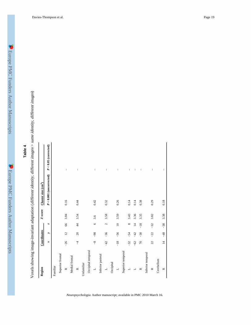
Several regions showed a reduced response to the same identity, different images conditioncompared to the different identity, different images condition at the uncorrected level (P <0.001). However, none of these regions were significant when corrected for multiplecomparisons (Table 4). It is also important to note the markedly smaller cluster sizes inTable 4 (image-invariant adaptation) compared to Table 3 (image-dependent adaptation).Supplementary Table 1 shows the contrast same identity, different images > differentidentity, different images. Although this shows a more extensive pattern of response forfamiliar compared to unfamiliar faces at an uncorrected threshold, no voxels weresignificant when corrected for multiple comparisons.
3.2.5. Behavioural task during scan—During the adaptation scans, subjects wereinstructed to perform a target detection task. Table 5 shows the average response timesacross all conditions. There was no significant effect of familiarity (F(1,18) = 0.11, P = 0.75)or image condition F(2,36) = 3.85, P = 0.06) on the response time. There was also nointeraction between familiarity and condition for response times (F(2,36) = 1.25, P = 0.33).
3.2.6. Post-scan test of familiarity—After the scan, each subject was tested todetermine their familiarity with the faces presented in the adaptation scan. Subjects reportedthat on average 88% (±2.3) of the faces were familiar. In addition, they were able to reportthe occupation (87% ± 2.3) and name (83% ± 2.7) of the majority of the familiar faces. If werestricted our analysis to the same identity images, subjects were able to report that 95%(±1.7) were familiar.
3.3. Independent behavioural taskThe data from the behavioural study (Fig. 5) shows a significant effect of condition onreaction time (RT) and error rate (F(2,34) = 37.14, P < 0.001; F(2,34) = 24.31, P < 0.001).There was also an interaction between condition and familiarity for reaction times (F(2,34) =31.53, P < 0.001). For familiar faces, the shortest RT occurred in the same condition whenthe same face was repeated. There was a significant increase in RT in the different conditionwhen different images of the same person were shown compared to the same condition(t(17) = 3.48, P < 0.01).
For unfamiliar faces, the fastest RT also occurred when the same image was repeated. Therewas a significant increase in RT to the unprimed condition (t(17) = 6.40, P < 0.001).However, the slowest RT occurred in the different condition, which also had the highesterror rate. Indeed, the only difference in RT between the responses to familiar andunfamiliar conditions, was in the different condition (t(17) = 6.87, P < 0.001), whichaccounts for the interaction between condition and familiarity.
Davies-Thompson et al. Page 6
Neuropsychologia. Author manuscript; available in PMC 2010 March 16.
Europe PM
C Funders A
uthor Manuscripts
Europe PM
C Funders A
uthor Manuscripts
4. DiscussionThe aim of this experiment was to probe the neural representations underlying familiar andunfamiliar faces in the human ventral stream. We found a reduced response (adaptation) torepeated images of the same unfamiliar and familiar faces in the FFA and OFA, but not inthe STS. To establish if the neural representation of faces in the FFA was invariant tochanges in the image, we presented different images of the same person. Contrary to ourprediction, we found a release from adaptation with both familiar and unfamiliar faces in theFFA and OFA. This suggests that an image-dependent representation is used to processfamiliar and unfamiliar faces in the ventral visual stream.
Models of face processing propose that information about faces is initially encoded in apictorial or view-dependent representation. This initial encoding leads onto a view-invariantrepresentation that is necessary for recognition (Bruce & Young, 1986; Burton et al., 1999).The difference in the ability to recognize familiar and unfamiliar faces across differentimage manipulations has led to the suggestion that unfamiliar faces are represented by apictorial, view-dependent code, whereas familiar faces are represented by a view-invariantrepresentation. We found support for this notion in a previous study, where we varied theviewing angle of successive face images in a fMR-adaptation paradigm (Ewbank &Andrews, 2008). Adaptation in the FFA to familiar faces was found across all changes inviewing angle, but a release from adaptation was found with increasing viewing angles forunfamiliar faces. Although this previous report demonstrates a more view-invariantrepresentation exists for familiar compared to unfamiliar faces, the current study shows thatthere are limits to the invariance of the neural representation in the core face-selectiveregions. Indeed, a key difference between these studies is that Ewbank and Andrews (2008)changed the view of the same face image, whereas the images in the current study were fromthe same view, but varied in appearance.
To monitor attentional load across all conditions, subjects performed a reaction time task inresponse to a red dot that appeared on some of the images. It is possible that this non-facetask may not have engaged the face-selective mechanisms in the brain. However, using asimilar design, Andrews and Ewbank (2004) reported that face-selective adaptation wasevident in face-selective regions such as the FFA, but not in object- or place-selectiveregions of the ventral stream. Again using a similar design, Ewbank and Andrews (2008)reported view-invariant fMR-adaptation in the FFA for familiar, but not unfamiliar faces.These results are also supported by event-related studies that did use a face task, but failed tofind view-invariant responses in face-selective regions (Eger et al., 2005; Pourtois et al.,2005a, 2005b). Consistent with these findings, Xu, Turk-Browne, and Chun (2007) showedthat adaptation in the PPA depends on visual characteristics of the images rather than taskdifficulty. Together, these results suggest that the absence of a face task may not explain thepattern of response to familiar and unfamiliar faces in this study. Nevertheless, these resultscannot exclude the possibility that a view-invariant response may have been apparent, ifsubjects had to perform a specific face task.
The inability to reveal image-invariant responses in the core face-selective regions need notimply that this pattern of response does not exist in other brain regions. Indeed, previousfMRI studies have reported, viewpoint-invariant adaptation in regions that are not part of thecore face-selective network (Eger et al., 2005; Pourtois et al., 2005a, 2005b). Although ourgroup analysis did reveal some regions that showed a reduced response to different imagesof the same identity (see Table 4), these were not significant when corrected for multiplecomparisons and did not overlap with the patterns reported in previous fMRI studies. Theseresults suggest that the behavioural advantage for the recognition of familiar faces may notdepend on an image-invariant representation in the brain. Further support for this conclusion
Davies-Thompson et al. Page 7
Neuropsychologia. Author manuscript; available in PMC 2010 March 16.
Europe PM
C Funders A
uthor Manuscripts
Europe PM
C Funders A
uthor Manuscripts
is provided by behavioural studies using repetition priming, which report a reduced primingeffect when different images of the same person are used (Bruce, Burton, Carson, Hanna, &Mason, 1994; Ellis, Young, Flude, & Hay, 1987). Using the same stimuli that wereemployed in the fMRI study, we also found a reduced priming effect when a different imageof a previously viewed familiar person was presented compared to when the same imagewas repeated. However, the behavioural difference in response to different images of thesame person compared to the same image of the same person was more apparent forunfamiliar faces with subjects making significantly more errors. Nevertheless, the fMRIresponses in the core face-selective regions to familiar and unfamiliar faces were verysimilar.
In the whole-brain analysis, we found that (image-dependent) adaptation to faces was notrestricted to the core face-selective regions, but was evident across a large part of the ventralstream (see Fig. 4). This finding is significant in the dispute about whether information inthe ventral stream is represented by a modular or distributed neural code (Andrews, 2005;Cohen & Tong, 2001; Haxby et al., 2001; Reddy & Kanwisher, 2006). Previously, wereported that adaptation to faces was restricted to face-selective regions of the ventral streamand was not found in object- and place-selective regions (Andrews & Ewbank, 2004).However, this study restricted its analysis to functionally defined regions of interest. Thecurrent findings would appear to support the idea that information about faces is notrestricted to the face-selective regions, but can be found in other regions of the humanventral stream. Indeed, this is consistent with neuropsychological studies that have shownthe perception and recognition of faces is not restricted to a single location, but can beimpaired by lesions to different parts of the ventral stream (McNeil & Warrington, 1993;Rossion et al., 2003; Steeves et al., 2006). This shows that fMR-adaptation can provide auseful measure of stimulus selectivity in neuroimaging studies of the visual system. Asselectivity is typically defined by comparing the relative response to different types of visualstimulus, the choice of control condition is important in determining which brain areasappear ‘face-selective’. The key advantage of the adaptation paradigm is that it does notinvolve the use of a category contrast and is therefore not restricted to circumscribed regionsof interest.Adaptation to facial identity was not apparent in all face-selective regions.Consistent with previous studies, we failed to find a reduced response to repeated images offamiliar or unfamiliar faces in the superior temporal face regions (Andrews & Ewbank,2004; Ewbank & Andrews, 2008). This fits with models of face processing that emphasizethe difference between inferior temporal processes involved in facial recognition andsuperior temporal processes involved in understanding dynamic aspects of faces (Andrews& Ewbank, 2004; Haxby et al., 2000; Hoffman & Haxby, 2000). In contrast, we found atrend for a larger response to different images of the same identity compared to differentimages of different identities with familiar, but not unfamiliar faces in the STS.
In conclusion, we found no evidence of image-invariant adaptation in face-selective regionsto either familiar faces or unfamiliar faces. We also report that the pattern of image-dependent adaptation extends beyond the face-selective regions and implies that adistributed neural code is used to represent images of faces. The similarity in response tofamiliar and unfamiliar faces contrasts with the marked differences in the way these visualstimuli are perceived.
AcknowledgmentsWe would like to thank Spyroula Spyrou for her help during the initial stages of this project. We would also like tothank Professor Gary Green, Chris Alderson and the other members of the YNiC for their help during the course ofthis project. JD-T is supported by an ESRC studentship. We are also grateful to the reviewers of this manuscript fortheir helpful comments.
Davies-Thompson et al. Page 8
Neuropsychologia. Author manuscript; available in PMC 2010 March 16.
Europe PM
C Funders A
uthor Manuscripts
Europe PM
C Funders A
uthor Manuscripts
Appendix A. Supplementary dataSupplementary data associated with this article can be found, in the online version, at doi:10.1016/j.neuropsychologia.2009.01.017.
ReferencesAndrews TJ. Visual cortex: How are objects and faces represented? Current Biology. 2005; 15:451–
453.
Andrews TJ, Ewbank MP. Distinct representations for facial identity and changeable aspects of facesin the human temporal lobe. NeuroImage. 2004; 23:905–913. [PubMed: 15528090]
Bruce V, Young AW. Understanding face recognition. British Journal of Psychology. 1986; 77:305–327. [PubMed: 3756376]
Bruce, V.; Burton, AM.; Carson, D.; Hanna, E.; Mason, O. Repetition priming of face recognition. In:Umilta, C.; Moskovitch, M., editors. Attention and performance XV. MIT Press; Cambridge: 1994.p. 179-210.
Burton AM, Bruce V, Hancock PJB. From pixels to people: A model of familiar face recognition.Cognitive Science. 1999; 23:1–31.
Cohen JD, Tong F. The face of controversy. Science. 2001; 293:2405–2407. [PubMed: 11577224]
Downing PE, Jiang Y, Shuman M, Kanwisher N. A cortical areas selective for processing the humanbody. Science. 2001; 293:2470–2473. [PubMed: 11577239]
Eger E, Schyns PG, Kleinschmidt A. Scale invariant adaptation in fusiform face-responsive regions.NeuroImage. 2004; 22(1):232–242. [PubMed: 15110013]
Eger E, Schweinberger SR, Dolan RJ, Henson RN. Familiarity enhances invariance of facerepresentations in human ventral visual cortex: fMRI evidence. NeuroImage. 2005; 26:1128–1139.[PubMed: 15961049]
Ellis AW, Young AW, Flude BM, Hay DC. Repetition priming of face recognition. Quarterly Journalof Experimental Psychology. 1987; 39a:193–210. [PubMed: 3615943]
Ewbank MP, Andrews TJ. Differential sensitivity for viewpoint between familiar and unfamiliar facesin human visual cortex. NeuroImage. 2008; 40:1857–1870. [PubMed: 18343161]
Fang F, Murray SO, He S. Duration-dependent fMRI adaptation and distributed viewer-centred facerepresentation in human visual cortex. Cerebral Cortex. 2006; 17(6):1402–1411. [PubMed:16905593]
Gauthier I, Tarr MJ, Moylan J, Skudlarski P, Gore JC, Anderson JW. The fusiform face area is part ofa network that processes faces at the individual level. Journal of Cognitive Neuroscience. 2000;12:495–504. [PubMed: 10931774]
Grill-Spector K, Kushnir T, Hendler T, Edelman S, Itzchak Y, Malach R. Differential processing ofobjects under various viewing conditions in human lateral occipital complex. Neuron. 1999;24:187–203. [PubMed: 10677037]
Grill-Spector K, Henson R, Martin A. Repetition and the brain: Neural models of stimulus-specificeffects. Trends in Cognitive Science. 2006; 10:14–23.
Hancock PJB, Bruce V, Burton AM. Recognition of unfamiliar faces. Trends in Cognitive Sciences.2000; 4:330–337. [PubMed: 10962614]
Haxby JV, Hoffman EA, Gobbini MI. The distributed human neural system for face perception.Trends in Cognitive Science. 2000; 4:223–233.
Haxby JV, Gobbini MI, Furey ML, Ishai A, Schouten JL, Pietrini P. Distributed and overlappingrepresentations of faces and objects in ventral temporal cortex. Science. 2001; 293(5539):2425–2430. [PubMed: 11577229]
Hoffman EA, Haxby JV. Distinct representations of eye gaze and identity in the distributed humanneural system for face perception. Nature Neuroscience. 2000; 3:80–84.
Kanwisher N, McDermott J, Chun MM. The fusiform face area: A module in extrastriate cortexspecialised for face perception. Journal of Neuroscience. 1997; 17:4302–4311. [PubMed:9151747]
Davies-Thompson et al. Page 9
Neuropsychologia. Author manuscript; available in PMC 2010 March 16.
Europe PM
C Funders A
uthor Manuscripts
Europe PM
C Funders A
uthor Manuscripts

Krekelberg B, Boynton GM, van Wezel RJA. Adaptation: From single cells to BOLD signals. Trendsin Neurosciences. 2006; 29(5):250–256. [PubMed: 16529826]
McNeil JE, Warrington EK. Prosopagnosia—a face-specific disorder. Quarterly Journal ofExperimental Psychology. 1993; 46A:1–10. [PubMed: 8446761]
Milner, AD.; Goodale, MA. The visual brain in action. Oxford University Press; 1995.
Peelen M, Downing P. Selectivity for the human body in the fusiform gyrus. Journal ofNeurophysiology. 2005; 93(1):603–608. [PubMed: 15295012]
Pourtois G, Schwartz S, Seghier ML, Lazeyras F, Vuilleumier P. Portraits or people? Distinctrepresentations of face identity in the human visual cortex. Journal of Cognitive Neuroscience.2005a; 17:1043–1057. [PubMed: 16102236]
Pourtois G, Schwartz S, Seghier ML, Lazeyras F, Vuilleumier P. View-independent coding of faceidentity in frontal and temporal cortices is modulated by familiarity: An event-related fMRI study.Neuroimage. 2005b; 24:1214–1224. [PubMed: 15670699]
Reddy L, Kanwisher N. Coding of visual objects in the ventral stream. Current Opinion inNeurobiology. 2006; 16(4):408–414. [PubMed: 16828279]
Rossion B, Caldara R, Seghier M, Schuller A-M, Lazeyras F, Mayers E. A network of occipito-temporal face-sensitive areas besides the right middle fusiform gyrus is necessary for normal faceprocessing. Brain. 2003; 126(11):2381–2395. [PubMed: 12876150]
Schwarzlose R, Baker C, Kanwisher N. Separate Face and Body Selectivity on the Fusiform Gyrus.Journal of Neuroscience. 2005; 25(47):11055–11059. [PubMed: 16306418]
Smith SM, Jenkinson M, Woolrich MW, Beckmann CF, Behrens TEJ, Johansen-Berg H, et al.Advances in functional and structural MR image analysis and implementation as FSL.NeuroImage. 2004; 23(S1):208–219.
Steeves JKE, Culham JC, Duchaine BC, Pratesi CC, Valyear KF, Schindler I, et al. The fusiform facearea is not sufficient for face recognition: Evidence from a patient with dense prosopagnosia andno occipital face area. Neuropsychologia. 2006; 44(4):594–609. [PubMed: 16125741]
Winston JS, Henson RNA, Fine-Goulden MR, Dolan RJ. fMRI-Adaptation reveals dissociable neuralrepresentations of identity and expression in face perception. Journal of Neurophysiology. 2004;92:1830–1839. [PubMed: 15115795]
Xu T, Turk-Browne NB, Chun MM. Dissociating task performance from fMRI repetition attenuationin ventral visual cortex. Journal of Neuroscience. 2007; 27:5981–5985. [PubMed: 17537969]
Davies-Thompson et al. Page 10
Neuropsychologia. Author manuscript; available in PMC 2010 March 16.
Europe PM
C Funders A
uthor Manuscripts
Europe PM
C Funders A
uthor Manuscripts
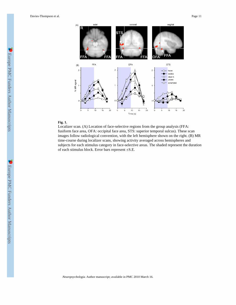
Fig. 1.Localizer scan. (A) Location of face-selective regions from the group analysis (FFA:fusiform face area, OFA: occipital face area, STS: superior temporal sulcus). These scanimages follow radiological convention, with the left hemisphere shown on the right. (B) MRtime-course during localizer scans, showing activity averaged across hemispheres andsubjects for each stimulus category in face-selective areas. The shaded represent the durationof each stimulus block. Error bars represent ±S.E.
Davies-Thompson et al. Page 11
Neuropsychologia. Author manuscript; available in PMC 2010 March 16.
Europe PM
C Funders A
uthor Manuscripts
Europe PM
C Funders A
uthor Manuscripts
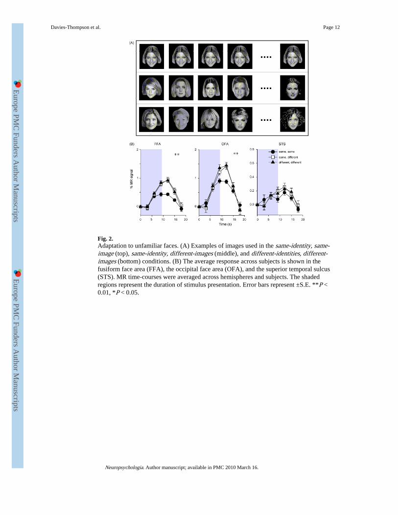
Fig. 2.Adaptation to unfamiliar faces. (A) Examples of images used in the same-identity, same-image (top), same-identity, different-images (middle), and different-identities, different-images (bottom) conditions. (B) The average response across subjects is shown in thefusiform face area (FFA), the occipital face area (OFA), and the superior temporal sulcus(STS). MR time-courses were averaged across hemispheres and subjects. The shadedregions represent the duration of stimulus presentation. Error bars represent ±S.E. **P <0.01, *P < 0.05.
Davies-Thompson et al. Page 12
Neuropsychologia. Author manuscript; available in PMC 2010 March 16.
Europe PM
C Funders A
uthor Manuscripts
Europe PM
C Funders A
uthor Manuscripts
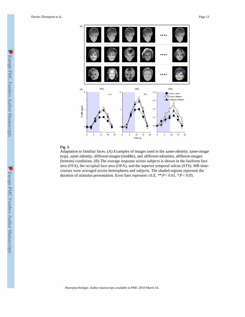
Fig. 3.Adaptation to familiar faces. (A) Examples of images used in the same-identity, same-image(top), same-identity, different-images (middle), and different-identities, different-images(bottom) conditions. (B) The average response across subjects is shown in the fusiform facearea (FFA), the occipital face area (OFA), and the superior temporal sulcus (STS). MR time-courses were averaged across hemispheres and subjects. The shaded regions represent theduration of stimulus presentation. Error bars represent ±S.E. **P < 0.01, *P < 0.05.
Davies-Thompson et al. Page 13
Neuropsychologia. Author manuscript; available in PMC 2010 March 16.
Europe PM
C Funders A
uthor Manuscripts
Europe PM
C Funders A
uthor Manuscripts
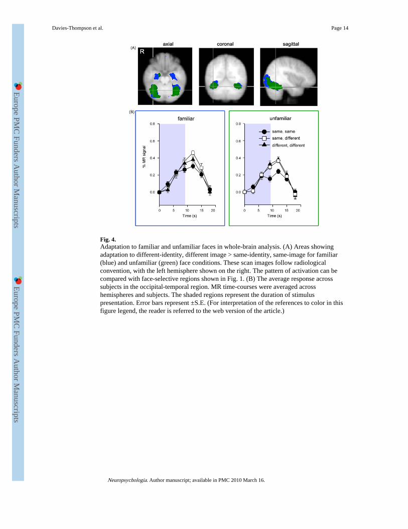
Fig. 4.Adaptation to familiar and unfamiliar faces in whole-brain analysis. (A) Areas showingadaptation to different-identity, different image > same-identity, same-image for familiar(blue) and unfamiliar (green) face conditions. These scan images follow radiologicalconvention, with the left hemisphere shown on the right. The pattern of activation can becompared with face-selective regions shown in Fig. 1. (B) The average response acrosssubjects in the occipital-temporal region. MR time-courses were averaged acrosshemispheres and subjects. The shaded regions represent the duration of stimuluspresentation. Error bars represent ±S.E. (For interpretation of the references to color in thisfigure legend, the reader is referred to the web version of the article.)
Davies-Thompson et al. Page 14
Neuropsychologia. Author manuscript; available in PMC 2010 March 16.
Europe PM
C Funders A
uthor Manuscripts
Europe PM
C Funders A
uthor Manuscripts
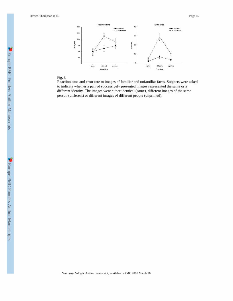
Fig. 5.Reaction time and error rate to images of familiar and unfamiliar faces. Subjects were askedto indicate whether a pair of successively presented images represented the same or adifferent identity. The images were either identical (same), different images of the sameperson (different) or different images of different people (unprimed).
Davies-Thompson et al. Page 15
Neuropsychologia. Author manuscript; available in PMC 2010 March 16.
Europe PM
C Funders A
uthor Manuscripts
Europe PM
C Funders A
uthor Manuscripts

Europe PM
C Funders A
uthor Manuscripts
Europe PM
C Funders A
uthor Manuscripts
Davies-Thompson et al. Page 16
Table 1
Mean change in intensity between successive images in each condition of the adaptation scan (S.E.M.)
Same, same Same, different Different, different
Familiar 0 (0) 16.7 (1.3) 19.6 (1.4)
Unfamiliar 0 (0) 18.1 (1.4) 18.3 (1.3)
Neuropsychologia. Author manuscript; available in PMC 2010 March 16.

Europe PM
C Funders A
uthor Manuscripts
Europe PM
C Funders A
uthor Manuscripts
Davies-Thompson et al. Page 17
Table 2
Mean MNI coordinates of face-selective regions-of-interest
Region n Co-ordinates
x y z
FFA
L 17 −41 −53 −22
R 18 37 −55 −22
OFA
L 15 −41 −83 −15
R 16 42 −80 −11
STS
R 15 50 −58 2
Neuropsychologia. Author manuscript; available in PMC 2010 March 16.

Europe PM
C Funders A
uthor Manuscripts
Europe PM
C Funders A
uthor Manuscripts
Davies-Thompson et al. Page 18
Tabl
e 3
Vox
els
show
ing
imag
e-de
pend
ent a
dapt
atio
n to
fac
es (
diff
eren
t ide
ntity
, dif
fere
nt im
ages
> s
ame
iden
tity,
sam
e im
ages
)
Reg
ion
Coo
rdin
ates
Z-s
core
Clu
ster
siz
e (c
m3 )
xy
zP
< 0
.001
(un
corr
ecte
d)P
< 0
.05
(cor
rect
ed)
Fam
iliar
A
nter
ior
tem
pora
l
L−
22−
6−
224.
492.
66–
R20
−10
−20
4.49
2.91
–
O
ccip
ital t
empo
ral
L−
40−
52−
225.
929
.78
3.96
R40
−52
−24
6.08
35.8
86.
58
Unf
amili
ar
A
nter
ior
tem
pora
l
R20
−12
−20
3.9
0.70
–
O
ccip
ital t
empo
ral
L−
34−
72−
205.
0329
.76
0.71
R34
−64
−22
5.3
25.6
21.
70
Neuropsychologia. Author manuscript; available in PMC 2010 March 16.
Europe PM
C Funders A
uthor Manuscripts
Europe PM
C Funders A
uthor Manuscripts
Davies-Thompson et al. Page 19
Tabl
e 4
Vox
els
show
ing
imag
e-in
vari
ant a
dapt
atio
n (d
iffe
rent
iden
tity,
dif
fere
nt im
ages
> s
ame
iden
tity,
dif
fere
nt im
ages
)
Reg
ion
Coo
rdin
ates
Z-s
core
Clu
ster
siz
e (c
m3 )
xy
zP
< 0
.001
(un
corr
ecte
d)P
< 0
.05
(cor
rect
ed)
Fam
iliar
Su
peri
or f
ront
al
R−
2612
663.
840.
16–
M
edia
l fro
ntal
R−
420
443.
540.
44–
Unf
amili
ar
O
ccip
ital t
empo
ral
L−
8−
986
3.6
0.42
–
In
feri
or p
arie
tal
L−
42−
362
3.58
0.32
–
O
ccip
ital
L−
18−
5610
3.59
0.26
–
Su
peri
or te
mpo
ral
L−
32−
548
3.43
0.14
–
L−
62−
4214
3.36
0.14
–
R70
−30
−18
3.35
0.38
–
In
feri
or te
mpo
ral
R22
−22
−32
3.82
0.29
–
C
ereb
ellu
m
R14
−48
−38
3.38
0.18
–
Neuropsychologia. Author manuscript; available in PMC 2010 March 16.

Europe PM
C Funders A
uthor Manuscripts
Europe PM
C Funders A
uthor Manuscripts
Davies-Thompson et al. Page 20
Table 5
Mean response times in milliseconds (S.E.M.) across subjects in target detection task performed duringadaptation scans
Same, same Same, different Different, different
Familiar 510 (12.8) 468 (11.9) 491 (12.6)
Unfamiliar 499 (14.1) 482 (23.9) 469 (25.9)
Neuropsychologia. Author manuscript; available in PMC 2010 March 16.
Related Documents