ANEW PROCEDURE FOR THE QUANTITATIVE ASSESSMENT OF P-GLYCOPROTEIN EFFLUX PUMP ASSOCIATED WITH HUMAN TLYMPHOCYTES Rosa Ve ´lez, BS; Marı ´a del C. Sa ´nchez, MPH; Pablo Lopez, BS; Yasuhiro Yamamura, PhD Introduction: P-glycoprotein (P-gp), a multi- drug transporter located in plasma mem- branes, reduces intracellular availability of some drugs. Upregulation of P-gp has been observed in some clinical situations, including chronic inflammatory disease and viral infec- tion. However, P-gp is expressed in only a small subset of peripheral blood mononuclear cells (PBMC) and at much lower quantities than it is on P-gp-positive cell lines used in other studies. Methods: P-gp expression was assessed by flow cytometry by using a commercially available, anti-P-gp, allophycocyanin-conjugat- ed monoclonal antibody. Flow cytometry was also used to determine the efflux activity associated with P-gp; with this process, re- fluxed fluorescent P-gp substrate, rhodamine 123 (Rho123), was determined by the subse- quently identified P-gp-positive PBMC subset. Use of verapamil during the dye-loading procedure maximized the amount of dye retained by the cells. Results: The use of allophycocyanin-conjugat- ed monoclonal antibody allowed for the identification of P-gp-positive PBMC subsets, even when the cells were fully loaded with Rho123. We used a logical gating strategy to identify a P-gp-positive PBMC subset, after which P-gp efflux activity of the PBMC subset could be quantitatively assessed. This new procedure enabled us to assess the P-gp efflux function of T lymphocytes in some clinical situations, which induced P-gp upregulation in vivo. Conclusion: This new procedure enables us to quantitatively assess the P-gp efflux activity associated with PBMC. (Ethn Dis. 2008;18[Suppl 2]:S2-75–S2-80) Key Words: New Procedure, P-gp Efflux, Quantitative Assessment, Peripheral Blood Mononuclear Cells INTRODUCTION P-glycoprotein (P-gp) is a plasma membrane-bound multidrug transport- er that actively pumps out a wide variety of chemicals from the interior of the cell, critically determining the intracel- lular bioavailability of numerous drugs. It is found in different cells, including T lymphocytes and B lymphocytes, which determine the function of host immune responses. P-gp is expressed in only a small subset of peripheral blood mono- nuclear cells (PBMC) and at much lower quantities than it is on P-gp- positive cell lines used in other stud- ies. 1,2 Previous studies have shown that P-gp is upregulated under some stress conditions and also in inflammatory diseases such as lupus. 3 Several studies have demonstrated that P-gp is upregu- lated on CD4-positive T cells infected by human HIV-1; such upregulation could severely minimize the intracellular bioavailability of HIV-1 protease inhib- itors, which are excellent P-gp sub- strates. 4,5 Both the quantity of P-gp expressed in certain cells and its efflux pump activity are usually assessed by flow cytometry, after loading P-gp-positive cell lines with the green-fluorescent P-gp substrate, rhodamine 123 (Rho123). 6–8 How- ever, P-gp is detectable only at a much lower density and only in a small subpopulation of PBMCs. Currently available procedures could not, be- cause of a number of technical prob- lems, be used to assess the efflux pump activity of the P-gp-expressing PBMC subset. The present study described the modification of the procedures that enabled us to quantitatively assess the efflux pump activity associated with P-gp-expressing lymphocytes from normal individuals or from patients with chronic inflammatory diseases. METHODS Participants Venous blood samples were asepti- cally collected by using BD Vacutainer Blood Collection Tubes containing sodium heparin (Becton, Dickinson and Co., Franklin Lakes, NJ). EDTA- containing Vacutainer tubes were avoid- ed because of their potential for com- promising P-gp pump activity. The present study protocol and the informed consent form were fully reviewed and approved by the Ponce School of Medicine Institutional Review Board on the Protection of Human Subjects (FWA00000345). Preparation of PBMC Approximately 10 mL blood was loaded on top of a Ficoll-Hypaque gradient in an ACCUSPIN System- HISTOPAQUE tube (Sigma-Aldrich, Inc., Saint Louis, Mo) and centrifuged at room temperature at 2500 rpm for 30 minutes. The plasma layer on the top was gently aspirated off, and the mononuclear cells banding at the inter- phase were collected by aspiration with Pasteur pipettes. Mononuclear cells were collected by centrifugation and washed twice in sterile RPMI-1640 medium (Sigma-Aldrich, Inc.) supple- mented with 10% fetal bovine serum. The cell concentration was adjusted, whenever feasible, to <1310 6 cells/mL. PBMC subsets were identified by flow cytometry after treatment with murine monoclonal antibodies against their differentiation markers, which were conjugated with their respective fluoro- chromes. From the AIDS Research Program, Ponce School of Medicine, Ponce, Puerto Rico (RV, MCS, PL, YY). Address correspondence and reprint requests to: Rosa Ve ´lez Cintro ´n; AIDS Research Program; Ponce School of Medi- cine; PO Box 7004; Ponce, PR 00732; 787- 841-5150; 787-841-5159 (fax); rosaamely@ yahoo.com Ethnicity & Disease, Volume 18, Spring 2008 S2-75

Welcome message from author
This document is posted to help you gain knowledge. Please leave a comment to let me know what you think about it! Share it to your friends and learn new things together.
Transcript
A NEW PROCEDURE FOR THE QUANTITATIVE ASSESSMENT OF P-GLYCOPROTEIN EFFLUX
PUMP ASSOCIATED WITH HUMAN T LYMPHOCYTES
Rosa Velez, BS; Marıa del C. Sanchez, MPH; Pablo Lopez, BS;Yasuhiro Yamamura, PhD
Introduction: P-glycoprotein (P-gp), a multi-
drug transporter located in plasma mem-
branes, reduces intracellular availability of
some drugs. Upregulation of P-gp has been
observed in some clinical situations, including
chronic inflammatory disease and viral infec-
tion. However, P-gp is expressed in only a
small subset of peripheral blood mononuclear
cells (PBMC) and at much lower quantities
than it is on P-gp-positive cell lines used in
other studies.
Methods: P-gp expression was assessed by
flow cytometry by using a commercially
available, anti-P-gp, allophycocyanin-conjugat-
ed monoclonal antibody. Flow cytometry was
also used to determine the efflux activity
associated with P-gp; with this process, re-
fluxed fluorescent P-gp substrate, rhodamine
123 (Rho123), was determined by the subse-
quently identified P-gp-positive PBMC subset.
Use of verapamil during the dye-loading
procedure maximized the amount of dye
retained by the cells.
Results: The use of allophycocyanin-conjugat-
ed monoclonal antibody allowed for the
identification of P-gp-positive PBMC subsets,
even when the cells were fully loaded with
Rho123. We used a logical gating strategy to
identify a P-gp-positive PBMC subset, after
which P-gp efflux activity of the PBMC subset
could be quantitatively assessed. This new
procedure enabled us to assess the P-gp efflux
function of T lymphocytes in some clinical
situations, which induced P-gp upregulation in
vivo.
Conclusion: This new procedure enables
us to quantitatively assess the P-gp efflux
activity associated with PBMC. (Ethn Dis.
2008;18[Suppl 2]:S2-75–S2-80)
Key Words: New Procedure, P-gp Efflux,
Quantitative Assessment, Peripheral Blood
Mononuclear Cells
INTRODUCTION
P-glycoprotein (P-gp) is a plasmamembrane-bound multidrug transport-er that actively pumps out a wide varietyof chemicals from the interior of thecell, critically determining the intracel-lular bioavailability of numerous drugs.It is found in different cells, including Tlymphocytes and B lymphocytes, whichdetermine the function of host immuneresponses. P-gp is expressed in only asmall subset of peripheral blood mono-nuclear cells (PBMC) and at muchlower quantities than it is on P-gp-positive cell lines used in other stud-ies.1,2 Previous studies have shown thatP-gp is upregulated under some stressconditions and also in inflammatorydiseases such as lupus.3 Several studieshave demonstrated that P-gp is upregu-lated on CD4-positive T cells infectedby human HIV-1; such upregulationcould severely minimize the intracellularbioavailability of HIV-1 protease inhib-itors, which are excellent P-gp sub-strates.4,5
Both the quantity of P-gp expressedin certain cells and its efflux pump activityare usually assessed by flow cytometry,after loading P-gp-positive cell lines withthe green-fluorescent P-gp substrate,rhodamine 123 (Rho123).6–8 How-ever, P-gp is detectable only at a muchlower density and only in a smallsubpopulation of PBMCs. Currentlyavailable procedures could not, be-cause of a number of technical prob-lems, be used to assess the efflux pumpactivity of the P-gp-expressing PBMCsubset.
The present study described themodification of the procedures thatenabled us to quantitatively assess theefflux pump activity associated withP-gp-expressing lymphocytes from
normal individuals or from patientswith chronic inflammatory diseases.
METHODS
ParticipantsVenous blood samples were asepti-
cally collected by using BD VacutainerBlood Collection Tubes containingsodium heparin (Becton, Dickinsonand Co., Franklin Lakes, NJ). EDTA-containing Vacutainer tubes were avoid-ed because of their potential for com-promising P-gp pump activity. Thepresent study protocol and the informedconsent form were fully reviewed andapproved by the Ponce School ofMedicine Institutional Review Boardon the Protection of Human Subjects(FWA00000345).
Preparation of PBMCApproximately 10 mL blood was
loaded on top of a Ficoll-Hypaquegradient in an ACCUSPIN System-HISTOPAQUE tube (Sigma-Aldrich,Inc., Saint Louis, Mo) and centrifugedat room temperature at 2500 rpm for30 minutes. The plasma layer on thetop was gently aspirated off, and themononuclear cells banding at the inter-phase were collected by aspiration withPasteur pipettes. Mononuclear cellswere collected by centrifugation andwashed twice in sterile RPMI-1640medium (Sigma-Aldrich, Inc.) supple-mented with 10% fetal bovine serum.The cell concentration was adjusted,whenever feasible, to <13106 cells/mL.PBMC subsets were identified by flowcytometry after treatment with murinemonoclonal antibodies against theirdifferentiation markers, which wereconjugated with their respective fluoro-chromes.
From the AIDS Research Program,Ponce School of Medicine, Ponce, PuertoRico (RV, MCS, PL, YY).
Address correspondence and reprintrequests to: Rosa Velez Cintron; AIDSResearch Program; Ponce School of Medi-cine; PO Box 7004; Ponce, PR 00732; 787-841-5150; 787-841-5159 (fax); [email protected]
Ethnicity & Disease, Volume 18, Spring 2008 S2-75
Assessment of P-gp Expressionby Lymphocyte Subsets
In this study, lymphocytes wereidentified by flow cytometry accordingto their forward and side scatter char-acteristics (lymphocyte gate). Lympho-cytes were only classified in this study asCD3-positive T lymphocytes and CD3-negative non-T lymphocytes. One ofthe existing technical problems to assessefflux pump activity associated withP-gp-positive T cells (or non-T cells)was the inability of the phycoerythrin-conjugated anti-human P-gp monoclonalantibody (clone 17F9; BD Biosciences,San Jose, Calif) to detect P-gp expressedon human PBMC. While the samemonoclonal antibody conjugated withfluorescein-isothiocyanate (FITC) coulddistinguish P-gp-positive—albeit of lowdensity—from P-gp-negative PBMC,the emission spectrum of FITC signifi-cantly overlapped with that of Rho123,the previously mentioned fluorescentP-gp substrate that was used in mostreports.6–8 Allophycocyanin (APC) con-jugate of the same antibody was,
therefore, custom-ordered and found tobe satisfactory for P-gp detection inPBMC, with negligible overlapping ofthe emission curve with that of Rho123.
Activation of T LymphocytesSince normal PBMC expressed very
little P-gp, PBMC were first activatedby the addition of 1 mg/mL of phorbol12-myristate 1,3-acetate (PMA) (Sigma)and 40 ng/mL of IM (IM) (Sigma) for24 hours at 37uC (98uF). ActivatedPBMC were washed twice with theRPMI medium and then treated with10 mL of APC-conjugated murine anti-human P-gp monoclonal antibody(clone 17F9) for 20 minutes at roomtemperature.
Assessment of P-gp EffluxPump Activity
Rho123 was loaded by the methoddescribed by Calado et al,6 with minormodifications. Briefly, PBMC weretreated with 1 mM verapamil (Sigma)for one hour at 37uC (98uF) before theloading of .5 mg/mL Rho123 for
30 minutes at 37uC (98uF). Cells werewashed twice with phosphate bufferedsaline (Sigma), and the FL1 (Rho123)profile was acquired by the flow cytom-eter, BD FACSAria Cell Sorting Sys-tem. The Rho123 profile of loaded Tlymphocytes was obtained as the FL1histogram of events logically gated forthe lymphocyte gate events that werealso CD3 (FL3) positive and P-gp (FL4)positive, according to BD CellQuestsoftware. The post-efflux Rho123 (FL1)profile was similarly obtained afterincubating the cells for 150 minutes at37uC (98uF). The culture was incubatedsimilarly but in the presence of 1 mMof verapamil, which was used to adjustfor possible leakage or non-P-gp-medi-ated efflux of the fluorescent dye.
RESULTS
P-gp Expression on NormalPBMC Activated in vitro
As shown in Figures 1a and 1b,normal PBMC did not consistently
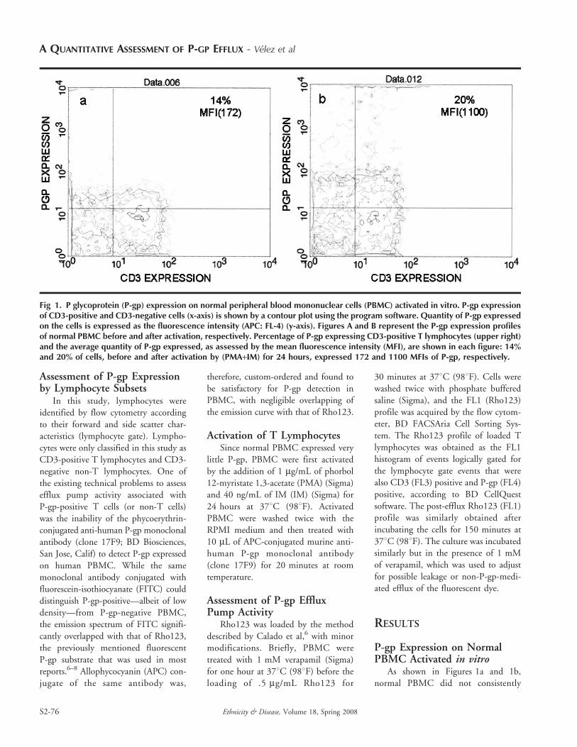
Fig 1. P glycoprotein (P-gp) expression on normal peripheral blood mononuclear cells (PBMC) activated in vitro. P-gp expressionof CD3-positive and CD3-negative cells (x-axis) is shown by a contour plot using the program software. Quantity of P-gp expressedon the cells is expressed as the fluorescence intensity (APC: FL-4) (y-axis). Figures A and B represent the P-gp expression profilesof normal PBMC before and after activation, respectively. Percentage of P-gp expressing CD3-positive T lymphocytes (upper right)and the average quantity of P-gp expressed, as assessed by the mean fluorescence intensity (MFI), are shown in each figure: 14%and 20% of cells, before and after activation by (PMA+IM) for 24 hours, expressed 172 and 1100 MFIs of P-gp, respectively.
A QUANTITATIVE ASSESSMENT OF P-GP EFFLUX - Velez et al
S2-76 Ethnicity & Disease, Volume 18, Spring 2008
express enough P-gp to be clearly
distinguishable from P-gp-negative cells
(Figure 1a). Activation by a combina-
tion of PMA and IM for 24 hours
markedly increased the P-gp expression
on PBMCs, and <20% of activated
CD3-positive T lymphocytes could be
classified as P-gp-positive (Figure 1b),
as opposed to only <14% of non-
activated cells. Activation of T lympho-
cytes increased not only the number of
cells expressing P-gp but also the density
of P-gp expressed in each cell, eg, 172
mean fluorescence intensity (MFI) of
P-gp on T lymphocytes before activa-
tion vs 1100 MFI after activation.
Treatment with PMA and IM similarly
activated both T cells and non-T (CD3-negative) lymphocytes (Figure 1b). Inthis study, however, we only describedthe assessment of the efflux pumpassociated with T lymphocytes.
Use of Verapamil to Enhance theRetention of Rho123
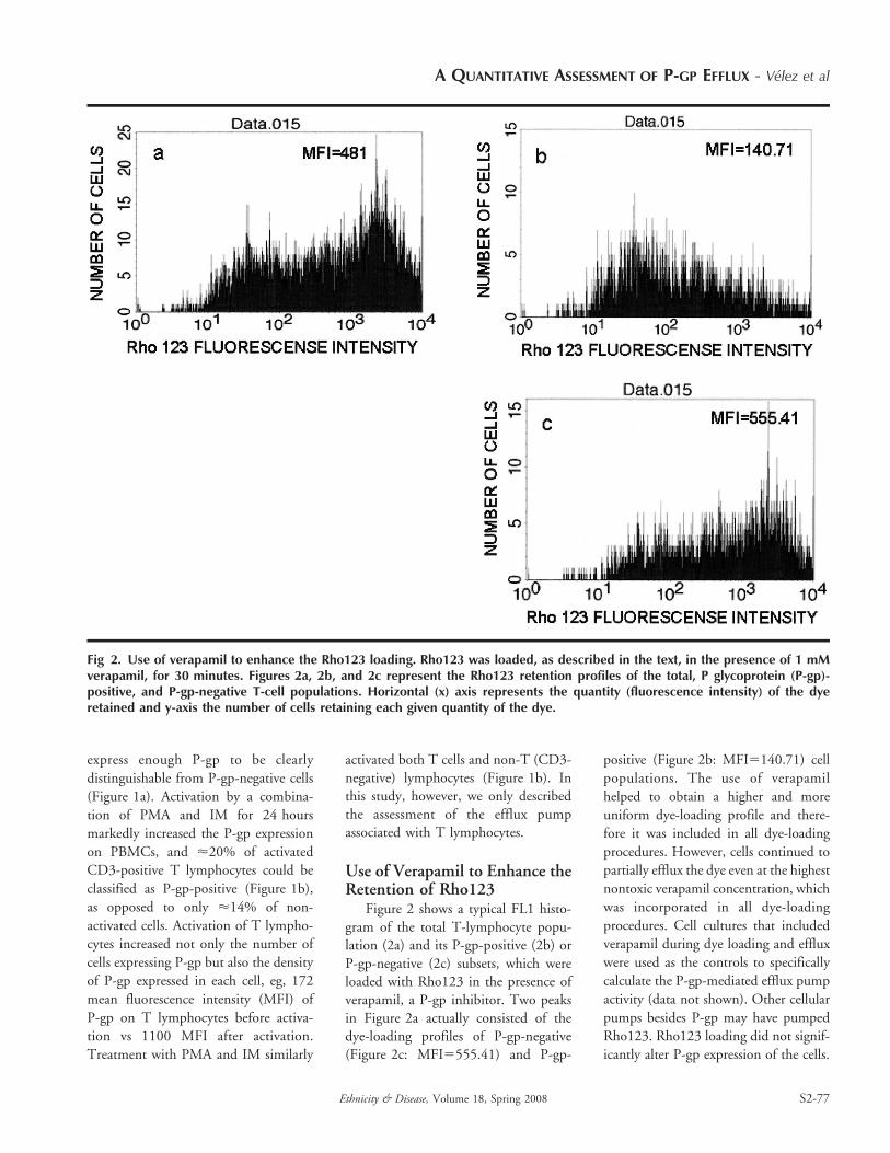
Figure 2 shows a typical FL1 histo-gram of the total T-lymphocyte popu-lation (2a) and its P-gp-positive (2b) orP-gp-negative (2c) subsets, which wereloaded with Rho123 in the presence ofverapamil, a P-gp inhibitor. Two peaksin Figure 2a actually consisted of thedye-loading profiles of P-gp-negative(Figure 2c: MFI5555.41) and P-gp-
positive (Figure 2b: MFI5140.71) cell
populations. The use of verapamil
helped to obtain a higher and more
uniform dye-loading profile and there-
fore it was included in all dye-loading
procedures. However, cells continued to
partially efflux the dye even at the highest
nontoxic verapamil concentration, which
was incorporated in all dye-loading
procedures. Cell cultures that included
verapamil during dye loading and efflux
were used as the controls to specifically
calculate the P-gp-mediated efflux pump
activity (data not shown). Other cellular
pumps besides P-gp may have pumped
Rho123. Rho123 loading did not signif-
icantly alter P-gp expression of the cells.
Fig 2. Use of verapamil to enhance the Rho123 loading. Rho123 was loaded, as described in the text, in the presence of 1 mMverapamil, for 30 minutes. Figures 2a, 2b, and 2c represent the Rho123 retention profiles of the total, P glycoprotein (P-gp)-positive, and P-gp-negative T-cell populations. Horizontal (x) axis represents the quantity (fluorescence intensity) of the dyeretained and y-axis the number of cells retaining each given quantity of the dye.
A QUANTITATIVE ASSESSMENT OF P-GP EFFLUX - Velez et al
Ethnicity & Disease, Volume 18, Spring 2008 S2-77
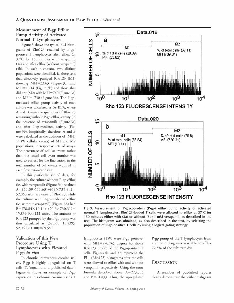
Measurement of P-gp EffluxPump Activity of ActivatedNormal T Lymphocytes
Figure 3 shows the typical FL1 histo-
grams of Rho123 retained by P-gp-
positive T lymphocytes after efflux (at
37uC for 150 minutes with verapamil)
(3a) and after efflux (without verapamil)
(3b). In each histogram, two distinct
populations were identified, ie, those cells
that effectively pumped Rho123 (M1)
showing MFI533.63 (Figure 3a) and
MFI510.14 (Figure 3b) and those that
did not (M2) with MFI5740 (Figure 3a)
and MFI5 730 (Figure 3b). The P-gp-
mediated efflux pump activity of each
culture was calculated as (A–B)/A, where
A and B were the quantities of Rho123
remaining without P-gp efflux activity (in
the presence of verapamil) (Figure 3a)
and after P-gp-mediated activity (Fig-
ure 3b). Empirically, therefore, A and B
were calculated as the addition of (MFI)
3 (% cellular events) of M1 and M2
populations, in respective sets of assays.
The percentage of cellular events rather
than the actual cell event number was
used to correct for the fluctuation in the
total number of cell events acquired in
each flow cytometric run.
In this particular set of data, for
example, the culture without P-gp efflux
(ie, with verapamil) (Figure 3a) retained
A5(30.09333.63)+(693739.84)5
52,060 arbitrary units of Rho123; while
the culture with P-gp-mediated efflux
(ie, without verapamil) (Figure 3b) had
B5(78.84310.14)+(20.63730.31)5
15,839 Rho123 units. The amount of
Rho123 pumped by the P-gp pump was
thus calculated as [(52,060215,839)/
52,060]3(100)569.5%.
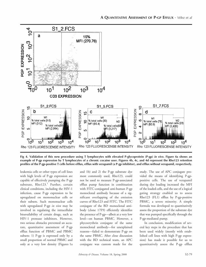
Validation of this NewProcedure Using TLymphocytes with ElevatedP-gp in vivo
In chronic intravenous cocaine us-
ers, P-gp is highly upregulated on T
cells (Y. Yamamura, unpublished data).
Figure 4a shows an example of P-gp
expression in a chronic cocaine user’s T
lymphocytes (15% were P-gp positive,
with MFI5270.76). Figura 4b shows
Rho123 profile of the P-gp-positive T
cells. Figures 4c and 4d represent the
FL1 (Rho123) histograms after the cells
were allowed to efflux with and without
verapamil, respectively. Using the same
formula described above, A5223,303
and B561,833. Thus, the upregulated
P-gp pump of the T lymphocytes froma chronic drug user was able to efflux72.3% of the substrate dye.
DISCUSSION
A number of published reportsclearly demonstrate that either malignant
Fig 3. Measurement of P-glycoprotein (P-gp) efflux pump activity of activatednormal T lymphocytes. Rho123-loaded T cells were allowed to efflux at 37uC for150 minutes either with (3a) or without (3b) 1 mM verapamil, as described in thetext. The histogram was obtained, as also described in the text, by selecting thepopulation of P-gp-positive T cells by using a logical gating strategy.
A QUANTITATIVE ASSESSMENT OF P-GP EFFLUX - Velez et al
S2-78 Ethnicity & Disease, Volume 18, Spring 2008
leukemia cells or other types of cell lines
with high levels of P-gp expression are
capable of effectively pumping the P-gp
substrate, Rho123.1 Further, certain
clinical conditions, including the HIV-1
infection, cause P-gp expression to be
upregulated on mononuclear cells or
their subsets. Such mononuclear cells
with upregulated P-gp in vivo may be
involved in regulating the intracellular
bioavailability of certain drugs, such as
HIV-1 protease inhibitors. However,
two serious obstacles prevented an accu-
rate, quantitative assessment of P-gp
efflux function of PBMC and PBMC
subsets: 1) P-gp is expressed only by a
small proportion of normal PBMC and
only at a very low density (Figures 1a
and 1b) and 2) the P-gp substrate dye
most commonly used, Rho123, could
not be used to measure P-gp-associated
efflux pump function in combination
with FITC-conjugated anti-human P-gp
monoclonal antibody because of a sig-
nificant overlapping of the emission
curves of Rho123 and FITC. The FITC
conjugate of the BD monoclonal anti-
body (clone 17F9) efficiently identifies
the presence of P-gp—albeit at a very low
level—on human PBMC. However, a
phycoerythrin conjugate of the same
monoclonal antibody—for unexplained
reasons—failed to demonstrate P-gp on
the same PBMC. After close discussion
with the BD technical team, an APC
conjugate was custom made for the
study. The use of APC conjugate pro-
vided the means of identifying P-gp-
positive cells. The use of verapamil
during dye loading increased the MFI
of the loaded cells, and the use of a logical
gating strategy enabled us to assess
Rho123 (FL1) efflux by P-gp-positive
PBMC, a severe minority. A simple
formula was developed to quantitatively
assess the proportion of the substrate dye
that was pumped specifically through the
P-gp-mediated pump.
In conclusion, modification of sev-
eral key steps in the procedure that has
been used widely (mostly with estab-
lished cell lines with high P-gp expres-
sion) has made it possible for us to
quantitatively assess the P-gp efflux
Fig 4. Validation of this new procedure using T lymphocytes with elevated P-glycoprotein (P-gp) in vivo. Figure 4a shows anexample of P-gp expression by T lymphocytes of a chronic cocaine user. Figures 4b, 4c, and 4d represent the Rho123 retentionprofiles of the P-gp-positive T cells before efflux, efflux with verapamil (a P-gp inhibitor), and efflux without verapamil, respectively.
A QUANTITATIVE ASSESSMENT OF P-GP EFFLUX - Velez et al
Ethnicity & Disease, Volume 18, Spring 2008 S2-79
pump activity of PBMC and T lym-
phocytes. The procedure was used to
assess the P-gp efflux pump function of
T lymphocytes from chronic drug
addicts, who have highly upregulated
P-gp expression in vivo. It was thus
possible to correlate the phenotypic
expression of P-gp molecules and its
functional efflux pump activity in
normal T lymphocytes as well as in
cells taken from patients suffering from
certain clinical abnormalities.
Implications for the Reductionof Health Disparities
P-gp affects some drugs and a
number of pharmaceutical compounds;
however, no reliable assay was available
for use with PBMCs. This new assay
allows for the correlation of P-gp
expression and functionality in PBMCs,
which—by taking into consideration
the impact of the protein’s function on
the drug’s bioavailability—provides a
new tool to improve patient therapy.
ACKNOWLEDGMENTS
This work was supported by RCMI Programgrant G12 RR003050. We thank the AIDSResearch Program Data Core for assisting inthe data analysis and Ruth Cruz, WandaRivera, Alex Maldonado, and YamarieFigueroa for assisting in the developmentof the experiments. Thanks also go to theRCMI Program Publications Office forediting this manuscript.
REFERENCES
1. Wang E, Casiano CN, Clement RP, et al. In
vitro flow cytometry method to quantitatively
asses inhibitors of P-glycoprotein. J Pharmacol
Exp Ther. 2000;28(5):522–528.
2. Loethchutinat CH, Saengkhae CH, Marbeuf-
Gueye C, et al. New insights into the P-
glycoprotein-mediated effluxes of rhodamines.
Eur J Biochem. 2003;270:476–485.
3. Tsujimura S, Saito K, Nakayamada S, et al.
Clinical relevance of the expression of P-
glycoprotein on peripheral blood lymphocytes
to steroid resistance in patients with systemic
lupus erythematosus. Arthritis Rheum. 2005;
52(6):1676–1683.
4. Kim A, Dintaman J, Waddel D, et al.
Saquinavir, an HIV protease inhibitor, is
transported by P-glycoprotein. J Pharmacol
Exp Ther. 1998;286(3):1439–1445.
5. Kim R, Fromm M, Wandel Ch, et al. The drug
transporter P-glycoprotein limits oral absorp-
tion and brain entry of HIV-1 protease
inhibitors. J Clin Invest. 1998;101(2):289–
294.
6. Calado RT, Garcia AB, Gallo DA, et al.
Reduced function of the multidrug resistance
P-glycoprotein in CD34+ cells of patients with
aplastic anemia. Br J Haematol. 2002;118:
320–326.
7. Heuvel-Eibrink M, Wiemer E, Boevere M,
et al. MDR1 gene-related clonal selection and
P-glycoprotein function and expression in
relapsed or refractory acute myeloid leukemia.
Blood. 2001;97(11):3605–3611.
8. Linda M, Belch P, Belch A. Circulating
monoclonal B cells expressing P glycoprotein
may be a reservoir of multidrug-resistant disease
in multiple myeloma. Blood. 1994;83(3):
724–736.
A QUANTITATIVE ASSESSMENT OF P-GP EFFLUX - Velez et al
S2-80 Ethnicity & Disease, Volume 18, Spring 2008
Related Documents