© 2020 International Journal of Mycobacteriology | Published by Wolters Kluwer - Medknow 107 Abstract Review Article INTRODUCTION Infection caused by Mycobacterium tuberculosis (MTB) complex organisms is one of the top ten causes of death worldwide in recent years. [1] Every year, millions of people develop a new tuberculosis (TB) infection and in 2018 alone, an estimated ten million people developed TB worldwide. [2] TB can be broadly divided into two types: pulmonary TB and extrapulmonary TB (EPTB). Most efforts are expended for managing pulmonary TB, as it is a major public health concern. Nevertheless, the other, silent TB that contributes to the overall prevalence of TB should not be overlooked. EPTB is an infection also caused by MTB complex organisms, but to the parts of the body other than the pulmonary system. EPTB can affect many organs, for example, the lymph nodes, nervous system, joints, genitourinary tract, bones, gastrointestinal system, and others. The infection can present on its own or concomitantly with pulmonary TB infection. In our local setting, the trend of EPTB is on the rise, with an increase of around 70% from 2005 to 2011 for newly diagnosed cases. [3] The most common type of EPTB is tuberculous lymphadenitis. [4] A study in Spain has also shown a slow increase in EPTB cases over time (30.6% to 37.6% over 10 years). [5] In Korea, EPTB comprises about 20% of the diagnosed TB cases. The more alarming feature is that it also shows an increasing trend over time. The most common type of EPTB in Korea is pleural EPTB, followed by that of the lymph nodes and gastrointestinal organs. [6] Tuberculous lymphadenitis is one of the more common types of EPTB in many countries and is often diagnosed very late, and diagnosis requires a high index of suspicion. Lymphadenitis To achieve the World Health Organization end TB Strategy, early detection, and prompt treatment of not only pulmonary but also extrapulmonary tuberculosis (EPTB) should be achieved. The most common EPTB is tuberculous lymphadenitis, and the diagnosis is typically time-consuming. This review aimed to identify the best diagnostic pathway for preventing treatment delay and thus further complications. A systematic keyword search was done using four databases and other relevant publications and using the Preferred Reporting Items for Systematic Reviews and Meta‑Analyses flowchart to search for relevant articles that met the inclusion criteria. The quality of the articles was assessed using Newcastle– Ottawa Scale, and the articles were summarized based on the test for diagnosing tuberculous lymphadenitis. A total of ten articles were included for the synthesis of results, which compared the sensitivity and specificity of each diagnostic test for tuberculous lymphadenitis. The most promising test is the Xpert Mycobacterium tuberculosis/RIF, which has high sensitivity and specificity, but costs much more in comparison to the other tests. An ideal diagnostic method should include the combination of relevant patient history, clinical examination, and laboratory and radiological testing to avoid delays in treatment, misdiagnosis, and further complications. Keywords: Clinical pathway, diagnostic pathway, extrapulmonary tuberculosis, tuberculous lymphadenitis Address for correspondence: Dr. Azmawati Mohammed Nawi, Department of Community Health, Faculty of Medicine, Universiti Kebangsaan Malaysia Medical Centre, Jalan Yaacob Latif, Bandar Tun Razak, Cheras, 56000 Kuala Lumpur, Malaysia. E‑mail: [email protected] ORCID: http://orcid.org/0000‑0002‑0009‑5244 Access this article online Quick Response Code: Website: www.ijmyco.org DOI: 10.4103/ijmy.ijmy_207_19 This is an open access journal, and arcles are distributed under the terms of the Creave Commons Aribuon‑NonCommercial‑ShareAlike 4.0 License, which allows others to remix, tweak, and build upon the work non‑commercially, as long as appropriate credit is given and the new creaons are licensed under the idencal terms. For reprints contact: [email protected] How to cite this article: Sivaratnam L, Nawi AM, Manaf MR. An evidence-based clinical pathway for the diagnosis of tuberculous lymphadenitis: A systematic review. Int J Mycobacteriol 2020;9:107-15. An Evidence‑Based Clinical Pathway for the Diagnosis of Tuberculous Lymphadenitis: A Systematic Review Lavanyah Sivaratnam, Azmawati Mohammed Nawi, Mohd Rizal Abdul Manaf Department of Community Health, Faculty of Medicine, Universiti Kebangsaan Malaysia Medical Centre, Kuala Lumpur, Malaysia Submitted: 23-Dec-2019 Revised: 26-Jan-2020 Accepted: 25-Feb-2020 Published: 29-May-2020 [Downloaded free from http://www.ijmyco.org on Friday, January 29, 2021, IP: 197.46.112.30]

Welcome message from author
This document is posted to help you gain knowledge. Please leave a comment to let me know what you think about it! Share it to your friends and learn new things together.
Transcript

© 2020 International Journal of Mycobacteriology | Published by Wolters Kluwer - Medknow 107
Abstract
Review Article
IntroductIon
Infection caused by Mycobacterium tuberculosis (MTB) complex organisms is one of the top ten causes of death worldwide in recent years.[1] Every year, millions of people develop a new tuberculosis (TB) infection and in 2018 alone, an estimated ten million people developed TB worldwide.[2] TB can be broadly divided into two types: pulmonary TB and extrapulmonary TB (EPTB). Most efforts are expended for managing pulmonary TB, as it is a major public health concern. Nevertheless, the other, silent TB that contributes to the overall prevalence of TB should not be overlooked. EPTB is an infection also caused by MTB complex organisms, but to the parts of the body other than the pulmonary system.
EPTB can affect many organs, for example, the lymph nodes, nervous system, joints, genitourinary tract, bones, gastrointestinal system, and others. The infection can present on its own or concomitantly with pulmonary TB infection. In our local setting, the trend of EPTB is on the rise, with an increase of around 70% from 2005 to 2011 for newly
diagnosed cases.[3] The most common type of EPTB is tuberculous lymphadenitis.[4] A study in Spain has also shown a slow increase in EPTB cases over time (30.6% to 37.6% over 10 years).[5] In Korea, EPTB comprises about 20% of the diagnosed TB cases. The more alarming feature is that it also shows an increasing trend over time. The most common type of EPTB in Korea is pleural EPTB, followed by that of the lymph nodes and gastrointestinal organs.[6]
Tuberculous lymphadenitis is one of the more common types of EPTB in many countries and is often diagnosed very late, and diagnosis requires a high index of suspicion. Lymphadenitis
To achieve the World Health Organization end TB Strategy, early detection, and prompt treatment of not only pulmonary but also extrapulmonary tuberculosis (EPTB) should be achieved. The most common EPTB is tuberculous lymphadenitis, and the diagnosis is typically time-consuming. This review aimed to identify the best diagnostic pathway for preventing treatment delay and thus further complications. A systematic keyword search was done using four databases and other relevant publications and using the Preferred Reporting Items for Systematic Reviews and Meta‑Analyses flowchart to search for relevant articles that met the inclusion criteria. The quality of the articles was assessed using Newcastle–Ottawa Scale, and the articles were summarized based on the test for diagnosing tuberculous lymphadenitis. A total of ten articles were included for the synthesis of results, which compared the sensitivity and specificity of each diagnostic test for tuberculous lymphadenitis. The most promising test is the Xpert Mycobacterium tuberculosis/RIF, which has high sensitivity and specificity, but costs much more in comparison to the other tests. An ideal diagnostic method should include the combination of relevant patient history, clinical examination, and laboratory and radiological testing to avoid delays in treatment, misdiagnosis, and further complications.
Keywords: Clinical pathway, diagnostic pathway, extrapulmonary tuberculosis, tuberculous lymphadenitis
Address for correspondence: Dr. Azmawati Mohammed Nawi, Department of Community Health, Faculty of Medicine, Universiti
Kebangsaan Malaysia Medical Centre, Jalan Yaacob Latif, Bandar Tun Razak, Cheras, 56000 Kuala Lumpur, Malaysia. E‑mail: [email protected]
ORCID: http://orcid.org/0000‑0002‑0009‑5244
Access this article online
Quick Response Code:Website: www.ijmyco.org
DOI: 10.4103/ijmy.ijmy_207_19
This is an open access journal, and articles are distributed under the terms of the Creative Commons Attribution‑NonCommercial‑ShareAlike 4.0 License, which allows others to remix, tweak, and build upon the work non‑commercially, as long as appropriate credit is given and the new creations are licensed under the identical terms.
For reprints contact: [email protected]
How to cite this article: Sivaratnam L, Nawi AM, Manaf MR. An evidence-based clinical pathway for the diagnosis of tuberculous lymphadenitis: A systematic review. Int J Mycobacteriol 2020;9:107-15.
An Evidence‑Based Clinical Pathway for the Diagnosis of Tuberculous Lymphadenitis: A Systematic Review
Lavanyah Sivaratnam, Azmawati Mohammed Nawi, Mohd Rizal Abdul Manaf
Department of Community Health, Faculty of Medicine, Universiti Kebangsaan Malaysia Medical Centre, Kuala Lumpur, Malaysia
Submitted: 23-Dec-2019 Revised: 26-Jan-2020 Accepted: 25-Feb-2020 Published: 29-May-2020
[Downloaded free from http://www.ijmyco.org on Friday, January 29, 2021, IP: 197.46.112.30]

Sivaratnam, et al.: Review on clinical pathway for extrapulmonary tuberculosis
International Journal of Mycobacteriology ¦ Volume 9 ¦ Issue 2 ¦ April-June 2020108
can be caused by both the nontuberculous mycobacteria (NTM) and the MTB complex organisms. NTM causing lymphadenitis is commonly seen among children and should be ruled out at the soonest as the treatment differs for both. Most of the time, NTM infection would resolve spontaneously without treatment, whereas tuberculous lymphadenitis would require a prolonged duration of treatment. Tuberculous lymphadenitis is typically found in the cervical region, and involves unilateral and painless swelling.[7] Apart from that, the clinician would also have to rule out other differential diagnoses before diagnosing tuberculous lymphadenitis. This could lead to delayed initiation of treatment.
The issue with most EPTB cases is late presentation and late diagnosis, which often leads to many complications.[8] Physicians with a poor index of suspicion, coupled with the long duration of culture, add to the delay in treatment.[8,9] Even though EPTB patients without concomitant lung infection are rarely contagious and a source of TB infection, we still need an efficient and systematic clinical care for these patients. From the natural progression of the disease, EPTB can either be a complication of PTB or the EPTB can progress and be disseminated to the lungs. As both ways can become as an infectious disease, urgent care is needed in treating these patients. The patient factor is difficult to control, but awareness can be improved. Regarding the health-care delay, the time from when the patient first seeks treatment to the actual diagnosis and treatment can be reduced by educating staff and having a proper reference guideline or pathway to minimize misdiagnosis and avoid subjecting patients to unnecessary investigation and treatment.
The clinical pathway is currently used to help clinicians standardize case management; it involves an evidence-based pathway that has been well structured to help provide optimum treatment quality while keeping the cost to a minimum.[10] The clinical pathway is typically a comprehensive process developed by a multidisciplinary team that is usually required in for managing that case. The aim of the present article is to review all evidence of the diagnostic pathway used and to identify the best pathway for diagnosing tuberculous lymphadenitis to prevent delayed treatment and further complications.
Methods
A systematic review of the literature was done to identify studies that reported on any clinical pathway in the management of
tuberculous lymphadenitis. Here, we describe computerized methods for searching relevant information on the best means of diagnosing and treating tuberculous lymphadenitis. Four databases (PubMed, SCOPUS, MEDLINE and Ovid, and the Cochrane Library), with a guideline (the Malaysian clinical practice guidelines on EPTB), and a program were used for assessing the quality of the information. Further relevant articles were searched using the snowballing method. The search comprised a set of keywords that included terms related to tuberculous lymphadenitis and the clinical pathway together with their MeSH terms from 2014 until 2019. We used the following keywords: “tuberculosis” OR “tuberculous” AND “lymphadenitis” OR “lymphadenopathy” OR “lymph node enlargement” AND “clinical pathway” OR “care pathway.”
The Preferred Reporting Items for Systematic Reviews and Meta-Analyses (PRISMA) checklist was used as the guideline to search for eligible articles. Table 1 shows the search summary according to each database; the PRISMA checklist is shown in Figure 1. The articles were screened by their title, followed by the abstract and full text. Only relevant articles were considered for analysis.
The inclusion criteria were: (a) reported on any clinical pathway/flow for treating tuberculous lymphadenitis, (b) published in 2014–2019, and (c) full article. The exclusion criteria were: (a) qualitative studies and commentary articles, (b) study in languages other than English, and (c) animal studies. In view of the rapid advancement of diagnosing techniques, where older methods may be obsolete, only articles from the last 5 years were selected.
Data synthesis and quality assessmentFrom the search summary using the PRISMA flowchart, an initial 3280 studies were retrieved using the keyword search; after much filtering, we identified an eventual ten articles for the final results synthesis. The findings from the ten selected articles were used in the pathway process and development. The quality of evidence of the observational studies was assessed using the Newcastle–Ottawa scale[11] [Tables 2 and 3]. Studies that met at least five of the nine criteria were deemed of high quality.
results
For result analysis, we selected ten articles that described the testing requirements and comparison of testing to aid the
Table 1: Database Search Summary
Database Number of articles before filter
Number of articles after filter
Number of articles accepted by title
Number of articles accepted by abstract
Number of articles accepted by full‑text analysis
Medline and Ovid 3105 879 18 8 1PubMed 8 6 3 0 0SCOPUS 32 19 3 1 1Cochrane library 125 75 1 1 0Other sources 10 10 10 10 8Total 3280 989 35 20 10
[Downloaded free from http://www.ijmyco.org on Friday, January 29, 2021, IP: 197.46.112.30]

Sivaratnam, et al.: Review on clinical pathway for extrapulmonary tuberculosis
International Journal of Mycobacteriology ¦ Volume 9 ¦ Issue 2 ¦ April-June 2020 109
diagnosis of tuberculous lymphadenitis. Two articles were case–control studies, four were cohort studies, three were report articles, and one was a clinical practice guideline. Most of the studies were from the UK (40%)[12-15] and India (30%).[16-18] The other studies were from Malaysia,[3] Korea,[6] and Turkey.[19] All of the studies described similar investigation methods for diagnosing a suspected tuberculous lymphadenitis case. We compared the methods used for identification based on the time until results were obtained, and compared the sensitivity and specificity of each test used. The frequencies of the content of the articles are detailed in Table 4; the detailed findings of each article are summarized in Table 5 and the proposed diagnostic pathway for tuberculous lymphadenitis is detailed in Figure 2.
dIscussIon
EPTB is an increasing problem worldwide today. As evidenced by a recent World Health Organization (WHO) report, as much as 15% of all TB cases are due to EPTB.[1] Due to the limitation of late presentation, clinicians typically wind up treating a more complicated case, and thus morbidity increases. One method
for reducing the time to treatment is to ensure that clinicians have a high index of suspicion and order the appropriate treatment and start treatment promptly. With the diagnostic pathway, not only can the time to start treatment be shortened, but also as evidenced by using a clinical pathway, management among clinicians can be synchronized, and at the same time, costs can be minimized by avoiding unnecessary procedures.
As EPTB is not a main concern when dealing with TB, less effort has been expended on managing the disease. This is also the reason for the limited number of research articles specifically on EPTB as compared to pulmonary TB. Based on the available references, the first step in creating a clinical pathway can be achieved. Gathering all recent evidence that supports the pathway is valuable to the aim of reducing the delay in providing early treatment.
The evidence is then broken down to a few focused reviews in creating the diagnostic flow. First, once a patient is suspected to have tuberculous lymphadenitis, identifying the sample to be taken must be considered. Depending on the site of lymph node enlargement, there are many methods for obtaining the sample:
Figure 1: Preferred Reporting Items for Systematic Reviews and meta‑analyses flow chart
[Downloaded free from http://www.ijmyco.org on Friday, January 29, 2021, IP: 197.46.112.30]

Sivaratnam, et al.: Review on clinical pathway for extrapulmonary tuberculosis
International Journal of Mycobacteriology ¦ Volume 9 ¦ Issue 2 ¦ April-June 2020110
Tabl
e 2:
New
cast
le‑O
ttaw
a cr
oss‑
sect
iona
l st
udy
Num
ber
Stud
ySe
lect
ion
Com
para
bilit
yOu
tcom
eQu
ality
sc
ore
Repr
esen
tativ
enes
s of
the
sam
ple
Sam
ple
size
Nonr
espo
nden
tsAs
cert
ainm
ent o
f the
ex
posu
re (r
isk
fact
or)
The
stud
y co
ntro
ls fo
r the
m
ost i
mpo
rtan
t fac
tor
The
stud
y co
ntro
ls fo
r an
y ad
ditio
nal f
acto
rAs
sess
men
t of
the
outc
ome
Stat
istic
al
test
1A
sim
acop
oulo
s et
al.[1
2]*
***
***
7
2.M
cAlli
ster
and
M
acG
rego
r[13]
**
***
**7
Tabl
e 3:
New
cast
le‑O
ttaw
a co
hort
stu
dy
Stud
ySe
lect
ion
Com
para
bilit
yOu
tcom
eQu
ality
sc
ore
Repr
esen
tativ
enes
s of
the
expo
sed
coho
rt
Sele
ctio
n of
the
none
xpos
ed
coho
rt
Asce
rtai
nmen
t of
exp
osur
eDe
mon
stra
tion
that
out
com
e of
inte
rest
was
not
pre
sent
at
the
star
t of s
tudy
Com
para
bilit
y of
co
hort
s on
the
basi
s of
th
e de
sign
or a
naly
sis
Asse
ssm
ent
of o
utco
me
Was
follo
w‑u
p lo
ng e
noug
h fo
r ou
tcom
es to
occ
ur
Adeq
uacy
of
follo
w‑u
p of
co
hort
s1
Gau
tam
et a
l.[14]
**
***
**
*8
2M
ittal
et a
l.[15]
**
**
**
*7
3M
eghj
i and
G
iddi
ngs[1
6]*
**
**
**
7
4Su
zana
et a
l.[17]
**
**
**
*7
*: 1
poi
nt, *
*: 2
poi
nts
[Downloaded free from http://www.ijmyco.org on Friday, January 29, 2021, IP: 197.46.112.30]
Sivaratnam, et al.: Review on clinical pathway for extrapulmonary tuberculosis
International Journal of Mycobacteriology ¦ Volume 9 ¦ Issue 2 ¦ April-June 2020 111
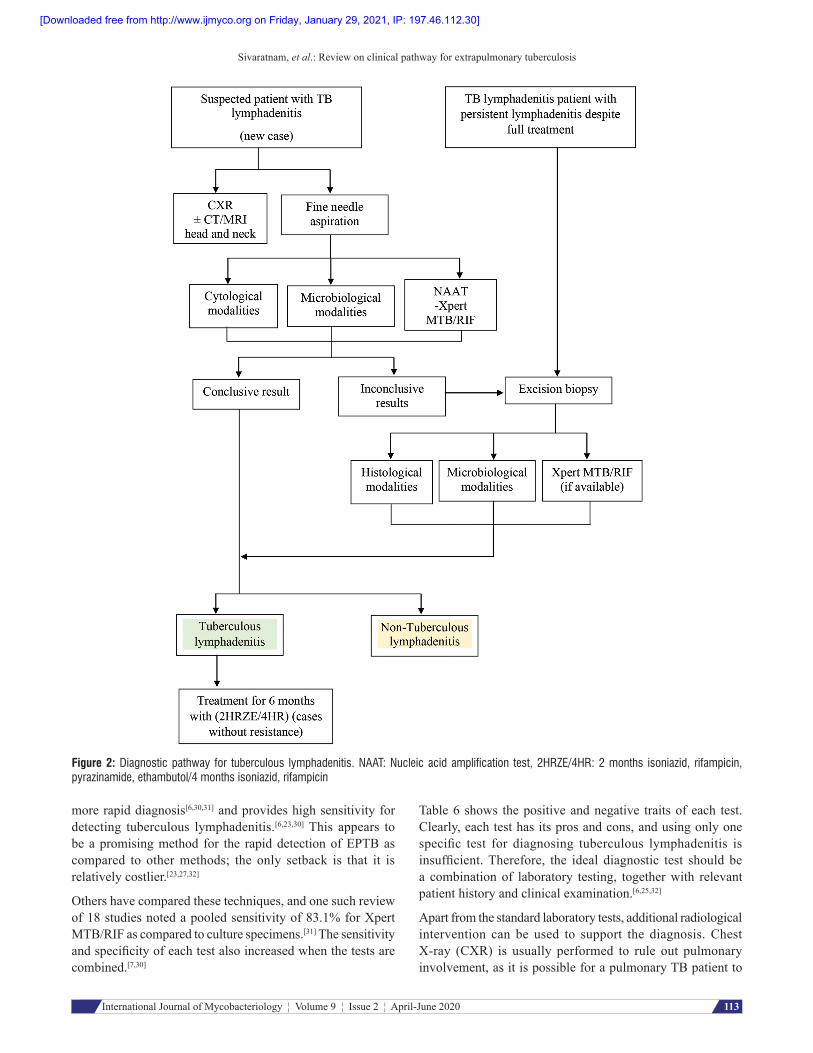
from a needle biopsy, excision, or a more invasive endoscopic or laparoscopic sampling.[7] If the lymph node enlargement is visible, the best sampling technique is fine-needle aspiration (FNA), as it has been proven to be minimally
invasive.[3,7,9,20] While it is claimed to be more sensitive, excision biopsy is a more invasive procedure, therefore it should be reserved if the FNA is inconclusive or if the patient has persistent lymph node enlargement despite completing treatment. FNA has been widely studied, and a 9-year study concluded that FNA is simple, is cost-effective, and can be done as an outpatient procedure, which is particularly useful in a resource-poor setting.[21] There is good agreement for this evidence, and it is accepted by all countries, as advocated by the WHO.
Once clinical samples have been obtained, the best test for diagnosing tuberculous lymphadenitis is dependent on what is available in the current setting, what can be used, laboratory capacity, and operator expertise. The most basic test for diagnosing pulmonary TB is sputum microscopy for detecting the acid-fast bacilli, as both tuberculous and NTM are acid‑fast. This test is fast and has high specificity and sensitivity. However, this is not true for EPTB infection, as the main issue in this case is the paucibacillary nature.[20] Most EPTB patients are not smear positive; therefore, microscopy
Table 4: Frequency of main content in the selected articles
Content Frequency described (number of articles)Mantoux test 2Biopsy
FNA and excision biopsy
7
Nucleic acid amplification test
Xpert MTB/Rif 3Conventional PCR 3
Imaging 2Treatment regimen/duration
3
FNA: Fine-needle aspiration, PCR: Polymerase chain reaction, MTB: Mycobacterium tuberculosis
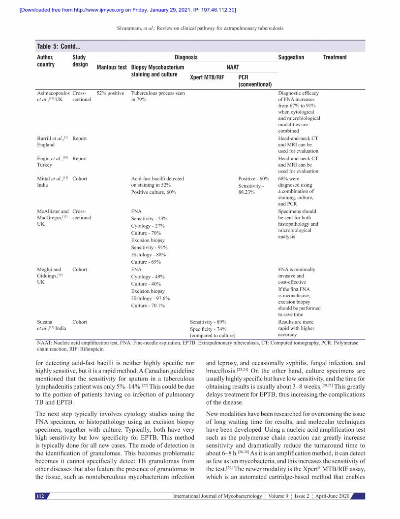
Table 5: Summary result of articles
Author, country
Study design
Diagnosis Suggestion Treatment
Mantoux test Biopsy Mycobacterium staining and culture
NAAT
Xpert MTB/RIF PCR (conventional)
Lee[6], Korea Report The first‑line diagnostic techniqueExcision biopsy has the highest sensitivity, whereas FNA is less invasive and may be usefulIf FNA examination results are inconclusive, excision biopsy may need to be doneIssuesLow sensitivity - 0-40%Delayed results (2-8 weeks)
Assay, a novel automated, cartridge-based NAATXpert was highly sensitive for detecting TB in lymph node samples - 83.1%Pooled specificity of 98.7% for diagnosing EPTB
NAAT is used for rapid molecular detection and diagnosis of EPTB
6-month regimen
Gautam et al.,[14] India
Cohort 31.42% positive
Detection rate: 17.85% Interpretation of data was software based and not user dependentDetection rate - 25.71%Sensitivity - 80%Specificity ‑ 86.89%
Detection rate, 20.71%Sensitivity - 68%Specificity ‑ 89.57%
Combines clinical, radiological, and microbiological methods
6-month regimen
Ministry of Health,[3] Malaysia
Clinical practice guidelines
First-line diagnostic testExcision biopsy only when FNA is inconclusive or persistent lymph node despite full treatment
6-month regimen (2 months isoniazid, RIF, pyrazinamide, ethambutol l/4 months isoniazid, RIF)
Contd...
[Downloaded free from http://www.ijmyco.org on Friday, January 29, 2021, IP: 197.46.112.30]
Sivaratnam, et al.: Review on clinical pathway for extrapulmonary tuberculosis
International Journal of Mycobacteriology ¦ Volume 9 ¦ Issue 2 ¦ April-June 2020112
Table 5: Contd...
Author, country
Study design
Diagnosis Suggestion Treatment
Mantoux test Biopsy Mycobacterium staining and culture
NAAT
Xpert MTB/RIF PCR (conventional)
Asimacopoulos et al.,[12] UK
Cross- sectional
52% positive Tuberculous process seen in 79%
Diagnostic efficacy of FNA increases from 67% to 91% when cytological and microbiological modalities are combined
Burrill et al.,[3] England
Report Head-and-neck CT and MRI can be used for evaluation
Engin et al.,[19] Turkey
Report Head-and-neck CT and MRI can be used for evaluation
Mittal et al.,[15] India
Cohort Acid-fast bacilli detected on staining in 52%Positive culture, 60%
Positive - 60%Sensitivity - 88.23%
68% were diagnosed using a combination of staining, culture, and PCR
McAllister and MacGregor,[13] UK
Cross- sectional
FNASensitivity - 53%Cytology - 27%Culture - 70%Excision biopsySensitivity - 91%Histology - 88%Culture - 69%
Specimens should be sent for both histopathology and microbiological analysis
Meghji and Giddings,[16] UK
Cohort FNACytology - 49%Culture - 40%Excision biopsyHistology - 97.6%Culture - 70.1%
FNA is minimally invasive and cost-effectiveIf the first FNA is inconclusive, excision biopsy should be performed to save time
Suzana et al.,[17] India
Cohort Sensitivity - 89%Specificity ‑ 74% (compared to culture)
Results are more rapid with higher accuracy
NAAT: Nucleic acid amplification test, FNA: Fine‑needle aspiration, EPTB: Extrapulmonary tuberculosis, CT: Computed tomography, PCR: Polymerase chain reaction, RIF: Rifampicin
for detecting acid‑fast bacilli is neither highly specific nor highly sensitive, but it is a rapid method. A Canadian guideline mentioned that the sensitivity for sputum in a tuberculous lymphadenitis patient was only 5%–14%.[22] This could be due to the portion of patients having co-infection of pulmonary TB and EPTB.
The next step typically involves cytology studies using the FNA specimen, or histopathology using an excision biopsy specimen, together with culture. Typically, both have very high sensitivity but low specificity for EPTB. This method is typically done for all new cases. The mode of detection is the identification of granulomas. This becomes problematic becomes it cannot specifically detect TB granulomas from other diseases that also feature the presence of granulomas in the tissue, such as nontuberculous mycobacterium infection
and leprosy, and occasionally syphilis, fungal infection, and brucellosis.[23,24] On the other hand, culture specimens are usually highly specific but have low sensitivity, and the time for obtaining results is usually about 3–8 weeks.[20,25] This greatly delays treatment for EPTB, thus increasing the complications of the disease.
New modalities have been researched for overcoming the issue of long waiting time for results, and molecular techniques have been developed. Using a nucleic acid amplification test such as the polymerase chain reaction can greatly increase sensitivity and dramatically reduce the turnaround time to about 6–8 h.[26-28] As it is an amplification method, it can detect as few as ten mycobacteria, and this increases the sensitivity of the test.[29] The newer modality is the Xpert® MTB/RIF assay, which is an automated cartridge-based method that enables
[Downloaded free from http://www.ijmyco.org on Friday, January 29, 2021, IP: 197.46.112.30]
Sivaratnam, et al.: Review on clinical pathway for extrapulmonary tuberculosis
International Journal of Mycobacteriology ¦ Volume 9 ¦ Issue 2 ¦ April-June 2020 113
Figure 2: Diagnostic pathway for tuberculous lymphadenitis. NAAT: Nucleic acid amplification test, 2HRZE/4HR: 2 months isoniazid, rifampicin, pyrazinamide, ethambutol/4 months isoniazid, rifampicin
more rapid diagnosis[6,30,31] and provides high sensitivity for detecting tuberculous lymphadenitis.[6,23,30] This appears to be a promising method for the rapid detection of EPTB as compared to other methods; the only setback is that it is relatively costlier.[23,27,32]
Others have compared these techniques, and one such review of 18 studies noted a pooled sensitivity of 83.1% for Xpert MTB/RIF as compared to culture specimens.[31] The sensitivity and specificity of each test also increased when the tests are combined.[7,30]
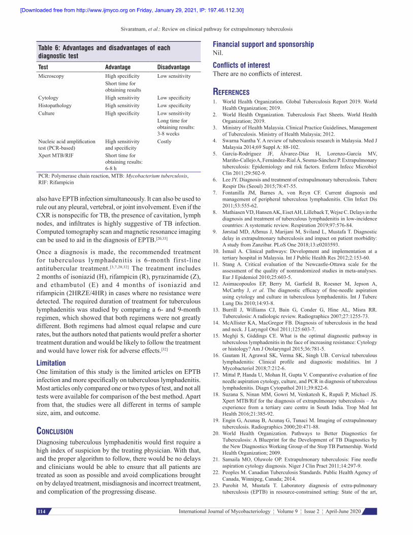
Table 6 shows the positive and negative traits of each test. Clearly, each test has its pros and cons, and using only one specific test for diagnosing tuberculous lymphadenitis is insufficient. Therefore, the ideal diagnostic test should be a combination of laboratory testing, together with relevant patient history and clinical examination.[6,25,32]
Apart from the standard laboratory tests, additional radiological intervention can be used to support the diagnosis. Chest X-ray (CXR) is usually performed to rule out pulmonary involvement, as it is possible for a pulmonary TB patient to
[Downloaded free from http://www.ijmyco.org on Friday, January 29, 2021, IP: 197.46.112.30]
Sivaratnam, et al.: Review on clinical pathway for extrapulmonary tuberculosis
International Journal of Mycobacteriology ¦ Volume 9 ¦ Issue 2 ¦ April-June 2020114
also have EPTB infection simultaneously. It can also be used to rule out any pleural, vertebral, or joint involvement. Even if the CXR is nonspecific for TB, the presence of cavitation, lymph nodes, and infiltrates is highly suggestive of TB infection. Computed tomography scan and magnetic resonance imaging can be used to aid in the diagnosis of EPTB.[20,33]
Once a diagnosis is made, the recommended treatment for tuberculous lymphadenitis is 6-month first-line antitubercular treatment.[3,7,20,32] The treatment includes 2 months of isoniazid (H), rifampicin (R), pyrazinamide (Z), and ethambutol (E) and 4 months of isoniazid and rifampicin (2HRZE/4HR) in cases where no resistance were detected. The required duration of treatment for tuberculous lymphadenitis was studied by comparing a 6- and 9-month regimen, which showed that both regimens were not greatly different. Both regimens had almost equal relapse and cure rates, but the authors noted that patients would prefer a shorter treatment duration and would be likely to follow the treatment and would have lower risk for adverse effects.[32]
LimitationOne limitation of this study is the limited articles on EPTB infection and more specifically on tuberculous lymphadenitis. Most articles only compared one or two types of test, and not all tests were available for comparison of the best method. Apart from that, the studies were all different in terms of sample size, aim, and outcome.
conclusIon
Diagnosing tuberculous lymphadenitis would first require a high index of suspicion by the treating physician. With that, and the proper algorithm to follow, there would be no delays and clinicians would be able to ensure that all patients are treated as soon as possible and avoid complications brought on by delayed treatment, misdiagnosis and incorrect treatment, and complication of the progressing disease.
Financial support and sponsorshipNil.
Conflicts of interestThere are no conflicts of interest.
references1. World Health Organization. Global Tuberculosis Report 2019. World
Health Organization; 2019.2. World Health Organization. Tuberculosis Fact Sheets. World Health
Organization; 2019.3. Ministry of Health Malaysia. Clinical Practice Guidelines, Management
of Tuberculosis. Ministry of Health Malaysia; 2012.4. Swarna Nantha Y. A review of tuberculosis research in Malaysia. Med J
Malaysia 2014;69 Suppl A: 88-102.5. García-Rodríguez JF, Álvarez-Díaz H, Lorenzo-García MV,
Mariño-Callejo A, Fernández-Rial Á, Sesma-Sánchez P. Extrapulmonary tuberculosis: Epidemiology and risk factors. Enferm Infecc Microbiol Clin 2011;29:502-9.
6. Lee JY. Diagnosis and treatment of extrapulmonary tuberculosis. Tuberc Respir Dis (Seoul) 2015;78:47-55.
7. Fontanilla JM, Barnes A, von Reyn CF. Current diagnosis and management of peripheral tuberculous lymphadenitis. Clin Infect Dis 2011;53:555-62.
8. Mathiasen VD, Hansen AK, Eiset AH, Lillebaek T, Wejse C. Delays in the diagnosis and treatment of tuberculous lymphadenitis in low-incidence countries: A systematic review. Respiration 2019;97:576-84.
9. Jørstad MD, Aẞmus J, Marijani M, Sviland L, Mustafa T. Diagnostic delay in extrapulmonary tuberculosis and impact on patient morbidity: A study from Zanzibar. PLoS One 2018;13:e0203593.
10. Ismail A. Clinical pathways: Development and implementation at a tertiary hospital in Malaysia. Int J Public Health Res 2012;2:153-60.
11. Stang A. Critical evaluation of the Newcastle-Ottawa scale for the assessment of the quality of nonrandomized studies in meta-analyses. Eur J Epidemiol 2010;25:603-5.
12. Asimacopoulos EP, Berry M, Garfield B, Roesner M, Jepson A, McCarthy J, et al. The diagnostic efficacy of fine‑needle aspiration using cytology and culture in tuberculous lymphadenitis. Int J Tuberc Lung Dis 2010;14:93-8.
13. Burrill J, Williams CJ, Bain G, Conder G, Hine AL, Misra RR. Tuberculosis: A radiologic review. Radiographics 2007;27:1255-73.
14. McAllister KA, MacGregor FB. Diagnosis of tuberculosis in the head and neck. J Laryngol Otol 2011;125:603-7.
15. Meghji S, Giddings CE. What is the optimal diagnostic pathway in tuberculous lymphadenitis in the face of increasing resistance: Cytology or histology? Am J Otolaryngol 2015;36:781-5.
16. Gautam H, Agrawal SK, Verma SK, Singh UB. Cervical tuberculous lymphadenitis: Clinical profile and diagnostic modalities. Int J Mycobacteriol 2018;7:212-6.
17. Mittal P, Handa U, Mohan H, Gupta V. Comparative evaluation of fine needle aspiration cytology, culture, and PCR in diagnosis of tuberculous lymphadenitis. Diagn Cytopathol 2011;39:822-6.
18. Suzana S, Ninan MM, Gowri M, Venkatesh K, Rupali P, Michael JS. Xpert MTB/Rif for the diagnosis of extrapulmonary tuberculosis – An experience from a tertiary care centre in South India. Trop Med Int Health 2016;21:385-92.
19. Engin G, Acunaş B, Acunaş G, Tunaci M. Imaging of extrapulmonary tuberculosis. Radiographics 2000;20:471-88.
20. World Health Organization. Pathways to Better Diagnostics for Tuberculosis: A Blueprint for the Development of TB Diagnostics by the New Diagnostics Working Group of the Stop TB Partnership. World Health Organization; 2009.
21. Samaila MO, Oluwole OP. Extrapulmonary tuberculosis: Fine needle aspiration cytology diagnosis. Niger J Clin Pract 2011;14:297-9.
22. Peoples M. Canadian Tuberculosis Standards. Public Health Agency of Canada, Winnipeg, Canada; 2014.
23. Purohit M, Mustafa T. Laboratory diagnosis of extra-pulmonary tuberculosis (EPTB) in resource-constrained setting: State of the art,
Table 6: Advantages and disadvantages of each diagnostic test
Test Advantage DisadvantageMicroscopy High specificity
Short time for obtaining results
Low sensitivity
Cytology High sensitivity Low specificityHistopathology High sensitivity Low specificityCulture High specificity Low sensitivity
Long time for obtaining results: 3-8 weeks
Nucleic acid amplification test (PCR-based)Xpert MTB/RIF
High sensitivity and specificityShort time for obtaining results: 6-8 h
Costly
PCR: Polymerase chain reaction, MTB: Mycobacterium tuberculosis, RIF: Rifampicin
[Downloaded free from http://www.ijmyco.org on Friday, January 29, 2021, IP: 197.46.112.30]

Sivaratnam, et al.: Review on clinical pathway for extrapulmonary tuberculosis
International Journal of Mycobacteriology ¦ Volume 9 ¦ Issue 2 ¦ April-June 2020 115
challenges and the need. J Clin Diagn Res 2015;9:EE01-6.24. Zumla A, James DG. Granulomatous infections: Etiology and
classification. Clin Infect Dis 1996;23:146‑58.25. Noussair L, Bert F, Leflon‑Guibout V, Gayet N, Nicolas‑Chanoine MH.
Early diagnosis of extrapulmonary tuberculosis by a new procedure combining broth culture and PCR. J Clin Microbiol 2009;47:1452-7.
26. Lee HS, Kee SJ, Shin JH, Kwon YS, Chun S, Lee JH, et al. Xpert MTB/RIF Assay as a Substitute for Smear Microscopy in an Intermediate-Burden Setting. Am J Respir Crit Care Med 2019;199:784-94.
27. Vadwai V, Boehme C, Nabeta P, Shetty A, Alland D, Rodrigues C. Xpert MTB/RIF: A new pillar in diagnosis of extrapulmonary tuberculosis? J Clin Microbiol 2011;49:2540-5.
28. World Health Organization. Automated Real-time Nucleic Acid Amplification Technology For Rapid and Simultaneous Detection of Tuberculosis and Rifampicin Resistance: Xpert Mtb. World Health Organization; 2013.
29. Manjunath N, Shankar P, Rajan L, Bhargava A, Saluja S, Shriniwas. Evaluation of a polymerase chain reaction for the diagnosis of tuberculosis. Tubercle 1991;72:21-7.
30. Kim YW, Kwak N, Seong MW, Kim EC, Yoo CG, Kim YW, et al. Accuracy of the Xpert® MTB/RIF assay for the diagnosis of extra-pulmonary tuberculosis in South Korea. Int J Tuberc Lung Dis 2015;19:81-6.
31. Denkinger CM, Schumacher SG, Boehme CC, Dendukuri N, Pai M, Steingart KR. Xpert MTB/RIF assay for the diagnosis of extrapulmonary tuberculosis: A systematic review and meta-analysis. Eur Respir J 2014;44:435-46.
32. Sharma SK, Ryan H, Khaparde S, Sachdeva KS, Singh AD, Mohan A, et al. Index-TB guidelines: Guidelines on extrapulmonary tuberculosis for India. Indian J Med Res 2017;145:448-63.
33. CoordinationTD. National Tuberculosis Management Guidelines 2014. Republic of South Africa; 2014.
[Downloaded free from http://www.ijmyco.org on Friday, January 29, 2021, IP: 197.46.112.30]
Related Documents