An Evidence-based Approach to the Diagnosis and Treatment of Neurotrophic Keratopathy Esen K. Akpek, MD Wilmer Eye Institute Johns Hopkins School of Medicine Baltimore, Maryland ACTIVITY DIRECTOR A CME MONOGRAPH FACULTY Natalie Afshari, MD Shiley Eye Institute University of California, San Diego La Jolla, California Sumayya Ahmad, MD Mount Sinai School of Medicine New York, New York Pedram Hamrah, MD, FRCS, FARVO Tufts University School of Medicine Boston, Massachusetts Mina Massaro-Giordano, MD University of Pennsylvania School of Medicine Philadelphia, Pennsylvania Nakul Shekhawat, MD, MPH Wilmer Eye Institute Johns Hopkins School of Medicine Baltimore, Maryland Christopher E. Starr, MD Weill Cornell Medical College New York, New York This monograph was published by Johns Hopkins School of Medicine in partnership with Catalyst Medical Education, LLC. It is not affiliated with JAMA medical research publishing. Visit catalystmeded.com/NK for online testing to earn your CME credit.

Welcome message from author
This document is posted to help you gain knowledge. Please leave a comment to let me know what you think about it! Share it to your friends and learn new things together.
Transcript
An Evidence-based Approach to the
Diagnosis and Treatment of Neurotrophic Keratopathy
Esen K. Akpek, MDWilmer Eye Institute Johns Hopkins School of Medicine Baltimore, Maryland
ACTIVITY DIRECTOR A CME MONOGRAPH
FACULTY
Natalie Afshari, MDShiley Eye Institute University of California, San Diego La Jolla, California
Sumayya Ahmad, MDMount Sinai School of Medicine New York, New York
Pedram Hamrah, MD, FRCS, FARVOTufts University School of Medicine Boston, Massachusetts
Mina Massaro-Giordano, MDUniversity of Pennsylvania School of Medicine Philadelphia, Pennsylvania
Nakul Shekhawat, MD, MPHWilmer Eye Institute Johns Hopkins School of Medicine Baltimore, Maryland
Christopher E. Starr, MDWeill Cornell Medical College New York, New York
This monograph was published by Johns Hopkins School of Medicine in partnership with Catalyst Medical Education, LLC. It is not affiliated with JAMA medical research publishing. Visit catalystmeded.com/NK for online testing to earn your CME credit.
1
DESCRIPTION
Neurotrophic keratopathy (NK) is a serious and rare degenerative corneal disease caused by impairment in the trigeminal innervation of the cornea. Epithelial breakdown can lead to ulceration, melting of the stroma, and ultimately to corneal perforation. Significant damage may be avoided with appropriate therapeutic options when given in a timely manner.
The goal of this CME monograph is to educate eye care providers on the latest scientific understanding of NK, including:
· clinical manifestations and complications;· conditions associated with the development of NK and
populations at risk;· the importance of early detection and early management;· screening and diagnostic strategies; and· optimal management strategies including emerging
treatments and the latest clinical data (safety, efficacy, and mechanism of action [MOA]).
This CME monograph is based on proceedings from a closed roundtable event.
TARGET AUDIENCE
Cornea specialists, comprehensive ophthalmologists, and optometrists.
OBJECTIVES
After participating in this activity, the learner will demonstrate the ability to:
· Discuss the clinical manifestations and complications of NK· Recognize the importance of early diagnosis and
intervention for patients with NK· Identify populations at high risk of having/developing NK· Discuss screening and diagnostic procedures for NK· Evaluate and compare the safety, efficacy, and clinical utility
of current and emerging treatments for NK· Describe the MOA of emerging therapies for the treatment
of NK· Develop management plans that improve outcomes for
patients with NK
ACCREDITATION STATEMENT
The Johns Hopkins University School of Medicine is accredited by the Accreditation Council for Continuing Medical Education (ACCME) to provide continuing medical education for physicians.
CREDIT DESIGNATION STATEMENT
The Johns Hopkins University School of Medicine designates this enduring monograph for a maximum of 2.0 AMA PRA Category 1 Credit(s)™. Physicians should claim only the credit commensurate with the extent of their participation in the activity.
JOHNS HOPKINS STATEMENT OF RESPONSIBILITY
The Johns Hopkins School of Medicine takes responsibility for the content, quality, and scientific integrity of this CME activity.
COPYRIGHT INFORMATION
All rights reserved—The Johns Hopkins School of Medicine. No part of this activity may be used or reproduced in any manner whatsoever without written permission except in the case of brief quotations embodied in articles or reviews.
ACKNOWLEDGEMENT
We wish to acknowledge the following company that has provided an educational grant in support of this activity: Dompé US, Inc.
Mary Gabb, MS, provided medical writing services for this manuscript.
PRESENTER AND PROVIDER DISCLOSURE POLICY
It is the policy of the Johns Hopkins School of Medicine that the presenter and provider globally disclose conflicts of interest. The Johns Hopkins School of Medicine OCME has established policies in place that identify and resolve conflicts of interest during activity planning. Detailed disclosure will be made prior to presentation of the education.
ACTIVITY DIRECTOREsen K. Akpek, MDProfessor of OphthalmologyDirector, Ocular Surface Diseases
and Dry Eye ClinicWilmer Eye InstituteJohns Hopkins School of MedicineBaltimore, Maryland
FACULTYNatalie Afshari, MDProfessor of OphthalmologyChief of Cornea and Refractive SurgeryVice Chair of EducationFellowship Program Director of Cornea
and Refractive SurgeryShiley Eye InstituteUniversity of California, San DiegoLa Jolla, California
Sumayya Ahmad, MD Assistant Professor of OphthalmologyMount Sinai School of MedicineNew York, New York
Pedram Hamrah, MD, FRCS, FARVOProfessorDepartment of OphthalmologyTufts University School of MedicineBoston, Massachusetts
Mina Massaro-Giordano, MDProfessor of Clinical OphthalmologyUniversity of Pennsylvania School
of MedicinePhiladelphia, Pennsylvania
Nakul Shekhawat, MD, MPHCornea and External Disease FellowWilmer Eye InstituteJohns Hopkins School of MedicineBaltimore, Maryland
Christopher E. Starr, MDAssociate Professor of OphthalmologyWeill Cornell Medical CollegeNew York, New York
FULL DISCLOSURE POLICY AFFECTING CME ACTIVITIES
As a provider approved by the Accreditation Council for Continuing Medical Education (ACCME), Johns Hopkins School of Medicine Office of Continuing Medical Education (OCME) requires attested and signed global disclosure of the existence of all financial interests or relationships with commercial interest from any individual in a position to control the content of a CME activity sponsored by OCME.
The following relationships have been reported for this activity:
Name Role Relationship(s)
Esen K. Akpek, MD Activity Director Contracted Research: Allergan, GORE & Associates, Inc. Consulting Fee: Clementia, Novaliq, Novartis Pharma AG, ShireRoyalty: Up-To-DateOther financial relationship: KeraLinkUnpaid Member of Board of Directors: Sjögren’s Syndrome FoundationAd Board: Sanofi, Sun Ophthalmics
Natalie Afshari, MD Presenter Equity Owner: Aescula Tech, Alpine Biotherapeutics, Trefoil
Pedram Hamrah, MD Presenter Consulting Fee: Dompé, Noveome
Mina Massaro-Giordano, MD Presenter Advisory Board: Dompé, LyntheraStock Ownership: Physician Recommended Neutraceuticals
Christopher E. Starr, MD Presenter Consulting Fee: Allergan, BlephEx, Bruder, Dompé, EyePoint, Eyevance, John-son and Johnson, Kala, Novaliq, Quidel, Shire, Sight Sciences, Sun, TearLab
No one else in a position to control the content of this educational activity has disclosed a relevant financial interest or relationship with any commercial interest.
Note: Grants to investigators at the Johns Hopkins University are negotiated and administered by the institution which receives the grants, typically through the Office of Research Administration. Individual investigators who participate in the sponsored project(s) are not directly compensated by the sponsor, but may receive salary or other support from the institution to support their effort on the project(s).
HOW TO OBTAIN CME CREDIT
To register, please visit catalystmeded.com/NK and complete the registration. Once registered, click “Content & Tests” to view the content and complete the post-test and evaluation.
The content is available at: catalystmeded.com/NK
You must participate in the entire activity to receive credit—you must read the objectives, answer the pretest questions (optional via link at catalystmeded.com/NKpre), view the content, and complete the posttest and evaluation. A passing score is defined as answering 75% posttest questions correctly. Upon submission of the completed materials, and if a satisfactory score on the posttest has been achieved, The Johns Hopkins University School of Medicine will issue your AMA PRA Category 1 Credit(s)™ Certificate.
EVALUATION AND OUTCOMES SURVEY
Post activity, an online evaluation form will be available to attendees to evaluate the activity and identify future educational needs. Upon completion of the evaluation, the learner must attest to the number of hours in attendance. A certificate of attendance will be available immediately for download or print. The last day to evaluate the material and attest to your credits is March 1, 2023.
RELEASE DATE
March 1, 2020
FOR FURTHER INFORMATION
General Information: (410) 955-2959E-mail the Office of CME: [email protected]
EXPIRATION DATE
March 1, 2023
2
3
An Evidence-based Approach to the Diagnosis and Treatment of Neurotrophic Keratopathy . . . . . . . . . . . . . . . . . . . . . . . . . . . . . . . . . . . . . . . . . . . . . . . . . . . . . . . . . . . . . . . . 4
Esen K. Akpek, MD
Neurotrophic Keratopathy: Overview . . . . . . . . . . . . . . . . . . . . . . . . . . . . . . . . . . . . . . . . . . . . . . . . . . . . . . . . 5
Sumayya Ahmad, MD
Screening and Diagnosis of Neurotrophic Keratopathy . . . . . . . . . . . . . . . . . . . . . . . . . . . . . . . . . . . . . . . 10
Pedram Hamrah, MD, FRCS, FARVO, and Natalie Afshari, MD
Management of Neurotrophic Keratopathy . . . . . . . . . . . . . . . . . . . . . . . . . . . . . . . . . . . . . . . . . . . . . . . . . . 15
Mina Massaro-Giordano, MD, and Christopher E. Starr, MD
Neurotrophic Keratopathy Presentation and Management: A Case Study Series . . . . . . . . . . . . . . . 23
Persistent Epithelial Defect With Advanced Glaucoma
Postsurgical Neurotrophic Keratopathy
Post Vitrectomy Neurotrophic Keratopathy
Herpes Simplex Virus–Related Keratitis Refractory to Medical Treatment
Stage 1 Neurotrophic Keratopathy
Notes . . . . . . . . . . . . . . . . . . . . . . . . . . . . . . . . . . . . . . . . . . . . . . . . . . . . . . . . . . . . . . . . . . . . . . . . . . . . . . . . . . . . . 32
The contents of this CME monograph are based on proceedings from an expert roundtable held on October 26, 2019, in Baltimore, Maryland.
Table of Contents
4
Neurotrophic keratopathy—also known as neurotrophic keratitis—is gaining increased attention as its long-term sequelae are better understood and as new treatments emerge that may prevent some of the corneal damage. On October 26, 2019, six cornea specialists convened for an expert roundtable in Baltimore, Maryland, to review the current data on neurotrophic keratopathy pathophysiology, screening and diagnosis, and management. To highlight some of the key findings from these presentations, several cases were discussed. We sought to provide a review of this topic as well as practical advice for ophthalmologists to better screen for neurotrophic keratopathy, diagnose it earlier, and improve the management of neurotrophic keratopathy. Faculty were chosen based on their academic and research experience and clinical expertise in the management of neurotrophic keratopathy. This Johns Hopkins School of Medicine CME monograph provides a summary of the findings and discussions from our meeting. We are hopeful that this will provide a useful tool to help eye care providers better recognize and manage neurotrophic keratopathy.
Esen K. Akpek, MDProfessor of OphthalmologyDirector, Ocular Surface Diseases and Dry Eye ClinicWilmer Eye InstituteJohns Hopkins School of MedicineBaltimore, Maryland
An Evidence-based Approach to the Diagnosis and Treatment of Neurotrophic Keratopathy
4
5
Neurotrophic Keratopathy: Overview
Neurotrophic keratopathy (NK) is characterized by decreased or ab-sent corneal sensitivity caused by damage to the trigeminal nerve. The V1 branch of the trigeminal nerve is primarily affected, with
a resultant reduction in reflex tearing and blinking and diminished produc-tion of trophic factors, making the ocular surface more susceptible to injury. Eventually the epithelial barrier breaks down, which can result in damage ranging from punctate epithelial erosions to frank perforation of the cornea.
Neurotrophic keratopathy is not solely a sensory nerve phenomenon. Corneal innervation includes sensory afferent fibers, as well as efferent sym-pathetic and parasympathetic fibers. The corneal nerves enter the stroma and spread evenly, up through the Bowman’s layer and into the epithelium. This extensive corneal nerve plexus provides protective and trophic functions. Crosstalk between the corneal nerves and epithelium regulates integrity, proliferation, migration, and adhesion of the ocular surface cells. There is also synergy with the neurotrophic growth factors released by the epithelial cells. In particular, nerve growth factor has several different functions that include promoting neuronal sprouting after nerve transection, providing tro-phic support to neurons after injury, reversing pathologic changes induced by peripheral nerve damage, improving reflex tearing and epithelial healing, and regulating blood flow to nerves.1
In a patient with NK, changes in the tear film occur due to an absence of stimulation of the lacrimal gland by the corneal nerves. A reduction in trophic factors (normally released by the epithelium) results in diminished migration of limbal stem cells from the limbus toward the center of the cor-nea; these less vital cells are then vulnerable to shearing forces of the eyelid. Simultaneously, there is an insufficient repair response by the epithelium, resulting in the clinical manifestations of NK. The pathognomonic feature of NK is reduced corneal sensitivity, which can be found in even the earliest stages of the disease.
Sumayya Ahmad, MDMOUNT SINAI SCHOOL OF MEDICINE, NEW YORK, NEW YORK
Key Messages• NK is a condition of decreased
corneal sensation, with or without inflammation.
• NK is often not diagnosed until stage 2, when epithelial damage has already started. However, patients with stage 1 NK might report photosensitivity and fluctuating vision upon questioning, particularly when performing sustained visual activities such as reading.
• Early diagnosis of NK before corneal ulceration occurs is important to prevent vision loss.
An Evidence-based Approach to the Diagnosis and Treatment of Neurotrophic Keratopathy
6
• MMP9 activation
• Increase in inflammatory mediators
• Further MMP activation
• Reduced tear production (all tear production is dependent on nerve stimuli)
Tear FilmCorneal Epithelium
• Decreased epithelial turnover, increased amount of epithelial-toxic agents
• Decreased tear film thickness
• Low protection against lid margin (lid wiper) stress
• Tear film instability – evaporative tear loss
• Hyperosmolarity• Increase in
pro-inflammatory cytokines
• Non-healing epithelial defects
• Damage to Bowman’s membrane
• Exposed stroma – vulnerable to enzymatic digestion
• Stromal involvement
• MMP activator/inhibitor imbalance
• Stromal lysis• Risk of perforation
and loss of eye
• Reduced mitosis• Reduced cell
turnover• Reduced cell vitality
in central cornea• Increased epithelial
permeability
• Decreased epithelial thickness in central cornea
• Epithelial damage by lid friction
• Fine or coarse punctate erosions
Clinical Presentation
• Dry eye• Photophobia• Inability to read extensively• Impaired quality of vision• Reduced blink rate
Mild: Stage 1
• Decrease in vision
Moderate: Stage 2
• Possible conjunctival injection
• Possible pain
Severe: Stage 3
Abbreviation: MMP, matrix metallopeptidase.Adapted with permission from Dua HS et al. Prog Retin Eye Res. 2018;66:107-131.3
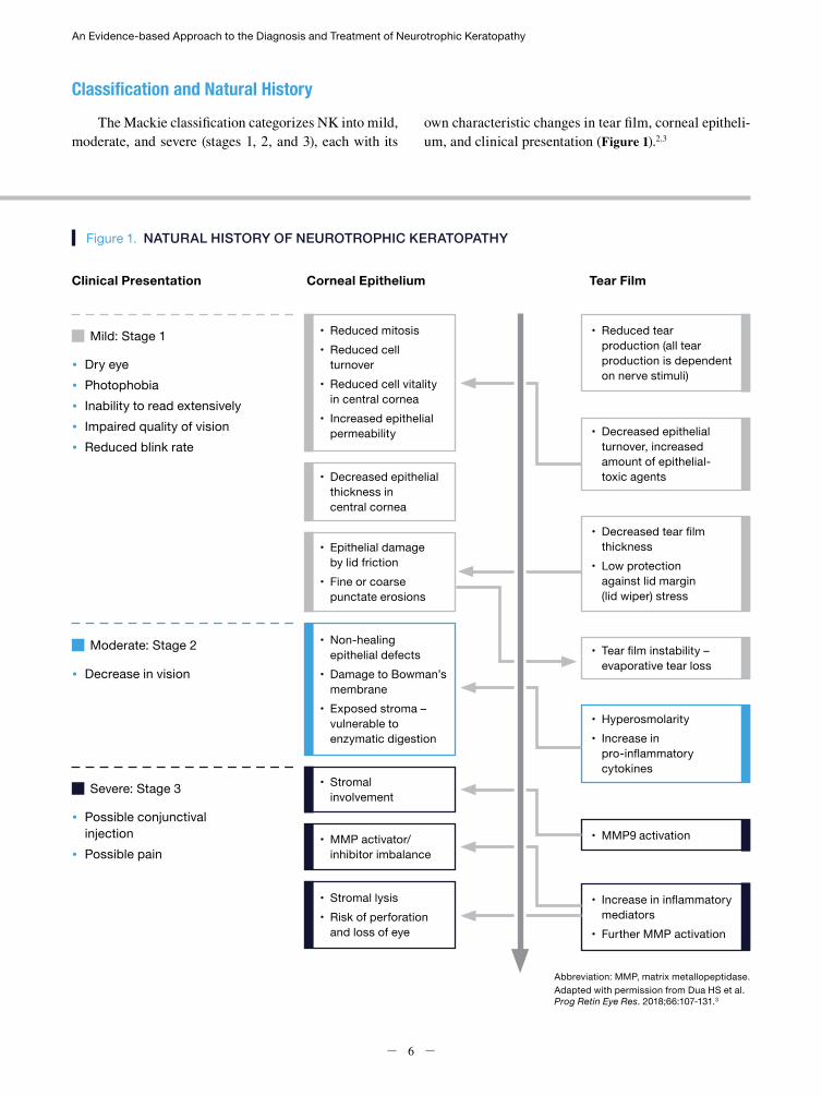
Figure 1. NATURAL HISTORY OF NEUROTROPHIC KERATOPATHY
Classification and Natural HistoryThe Mackie classification categorizes NK into mild,
moderate, and severe (stages 1, 2, and 3), each with its own characteristic changes in tear film, corneal epitheli-um, and clinical presentation (Figure 1).2,3
Neurotrophic Keratopathy: Overview
7
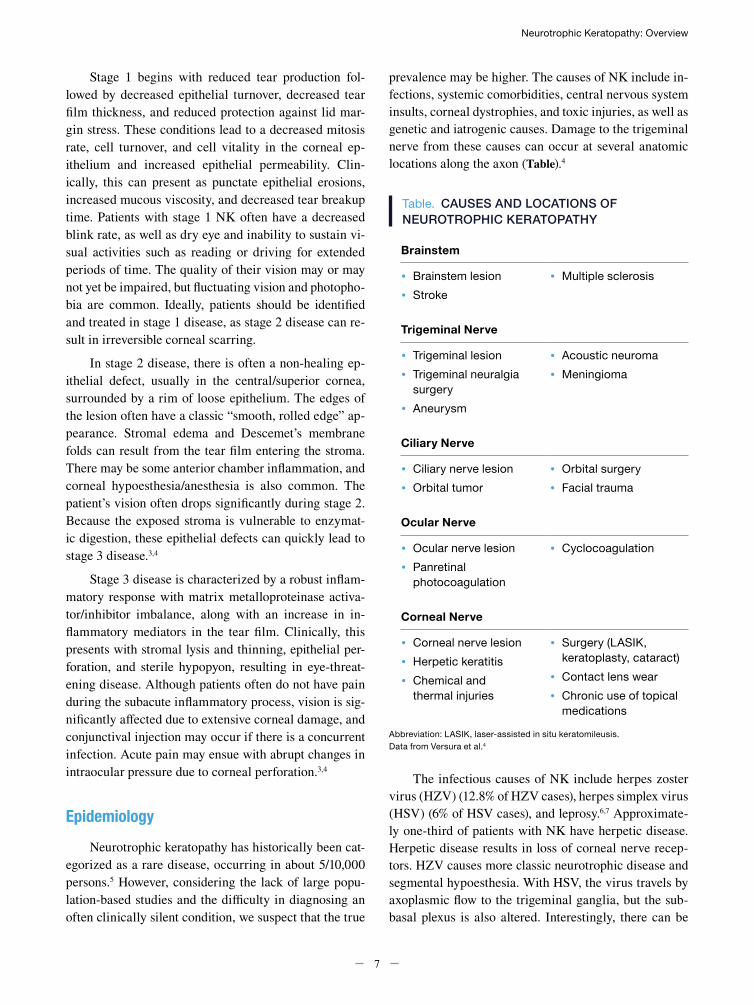
Stage 1 begins with reduced tear production fol-lowed by decreased epithelial turnover, decreased tear film thickness, and reduced protection against lid mar-gin stress. These conditions lead to a decreased mitosis rate, cell turnover, and cell vitality in the corneal ep-ithelium and increased epithelial permeability. Clin-ically, this can present as punctate epithelial erosions, increased mucous viscosity, and decreased tear breakup time. Patients with stage 1 NK often have a decreased blink rate, as well as dry eye and inability to sustain vi-sual activities such as reading or driving for extended periods of time. The quality of their vision may or may not yet be impaired, but fluctuating vision and photopho-bia are common. Ideally, patients should be identified and treated in stage 1 disease, as stage 2 disease can re-sult in irreversible corneal scarring.
In stage 2 disease, there is often a non-healing ep-ithelial defect, usually in the central/superior cornea, surrounded by a rim of loose epithelium. The edges of the lesion often have a classic “smooth, rolled edge” ap-pearance. Stromal edema and Descemet’s membrane folds can result from the tear film entering the stroma. There may be some anterior chamber inflammation, and corneal hypoesthesia/anesthesia is also common. The patient’s vision often drops significantly during stage 2. Because the exposed stroma is vulnerable to enzymat-ic digestion, these epithelial defects can quickly lead to stage 3 disease.3,4
Stage 3 disease is characterized by a robust inflam-matory response with matrix metalloproteinase activa-tor/inhibitor imbalance, along with an increase in in-flammatory mediators in the tear film. Clinically, this presents with stromal lysis and thinning, epithelial per-foration, and sterile hypopyon, resulting in eye-threat-ening disease. Although patients often do not have pain during the subacute inflammatory process, vision is sig-nificantly affected due to extensive corneal damage, and conjunctival injection may occur if there is a concurrent infection. Acute pain may ensue with abrupt changes in intraocular pressure due to corneal perforation.3,4
EpidemiologyNeurotrophic keratopathy has historically been cat-
egorized as a rare disease, occurring in about 5/10,000 persons.5 However, considering the lack of large popu-lation-based studies and the difficulty in diagnosing an often clinically silent condition, we suspect that the true
The infectious causes of NK include herpes zoster virus (HZV) (12.8% of HZV cases), herpes simplex virus (HSV) (6% of HSV cases), and leprosy.6,7 Approximate-ly one-third of patients with NK have herpetic disease. Herpetic disease results in loss of corneal nerve recep-tors. HZV causes more classic neurotrophic disease and segmental hypoesthesia. With HSV, the virus travels by axoplasmic flow to the trigeminal ganglia, but the sub-basal plexus is also altered. Interestingly, there can be
prevalence may be higher. The causes of NK include in-fections, systemic comorbidities, central nervous system insults, corneal dystrophies, and toxic injuries, as well as genetic and iatrogenic causes. Damage to the trigeminal nerve from these causes can occur at several anatomic locations along the axon (Table).4
Table. CAUSES AND LOCATIONS OF NEUROTROPHIC KERATOPATHY
Brainstem
• Brainstem lesion• Stroke
• Multiple sclerosis
Trigeminal Nerve
• Trigeminal lesion• Trigeminal neuralgia
surgery• Aneurysm
• Acoustic neuroma• Meningioma
Ciliary Nerve
• Ciliary nerve lesion• Orbital tumor
• Orbital surgery• Facial trauma
Ocular Nerve
• Ocular nerve lesion• Panretinal
photocoagulation
• Cyclocoagulation
Corneal Nerve
• Corneal nerve lesion• Herpetic keratitis• Chemical and
thermal injuries
• Surgery (LASIK, keratoplasty, cataract)
• Contact lens wear• Chronic use of topical
medications
Abbreviation: LASIK, laser-assisted in situ keratomileusis.Data from Versura et al.4
An Evidence-based Approach to the Diagnosis and Treatment of Neurotrophic Keratopathy
8
bilateral loss of nerve density and bilateral dry eye, per-haps as a result of the virus traveling to the trigeminal ganglia on the contralateral side.8
Central nervous system damage resulting in NK in-cludes surgical damage from trigeminal neuralgia sur-gery, aneurysm repair, or removal of neoplasms (acous-tic neuromas). These cases tend to be severe and very debilitating because destruction of the trigeminal nucle-us results in irreparable damage.
Systemic causes of NK include diabetes, vitamin A deficiency, amyloidosis, and multiple sclerosis (MS). Diabetes in particular is a notable factor because of its high prevalence in the US population (9.4% of the US population and 25% of the US elderly population).9 Dia-betes increases the risk for NK because of microvascular ischemic neuropathy. There is also a greater prevalence of dry eye in people with diabetes compared with the general population, which might be related to an under-lying corneal neuropathy.
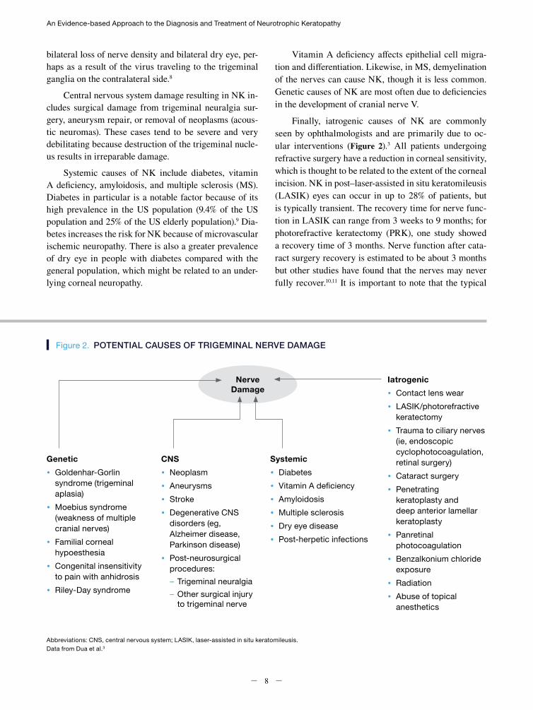
Figure 2. POTENTIAL CAUSES OF TRIGEMINAL NERVE DAMAGE
Nerve Damage
Genetic• Goldenhar-Gorlin
syndrome (trigeminal aplasia)
• Moebius syndrome (weakness of multiple cranial nerves)
• Familial corneal hypoesthesia
• Congenital insensitivity to pain with anhidrosis
• Riley-Day syndrome
Iatrogenic• Contact lens wear• LASIK/photorefractive
keratectomy• Trauma to ciliary nerves
(ie, endoscopic cyclophotocoagulation, retinal surgery)
• Cataract surgery• Penetrating
keratoplasty and deep anterior lamellar keratoplasty
• Panretinal photocoagulation
• Benzalkonium chloride exposure
• Radiation• Abuse of topical
anesthetics
CNS• Neoplasm• Aneurysms• Stroke• Degenerative CNS
disorders (eg, Alzheimer disease, Parkinson disease)
• Post-neurosurgical procedures:– Trigeminal neuralgia– Other surgical injury
to trigeminal nerve
Systemic• Diabetes• Vitamin A deficiency• Amyloidosis• Multiple sclerosis• Dry eye disease• Post-herpetic infections
Abbreviations: CNS, central nervous system; LASIK, laser-assisted in situ keratomileusis.Data from Dua et al.3
Vitamin A deficiency affects epithelial cell migra-tion and differentiation. Likewise, in MS, demyelination of the nerves can cause NK, though it is less common. Genetic causes of NK are most often due to deficiencies in the development of cranial nerve V.
Finally, iatrogenic causes of NK are commonly seen by ophthalmologists and are primarily due to oc-ular interventions (Figure 2).3 All patients undergoing refractive surgery have a reduction in corneal sensitivity, which is thought to be related to the extent of the corneal incision. NK in post–laser-assisted in situ keratomileusis (LASIK) eyes can occur in up to 28% of patients, but is typically transient. The recovery time for nerve func-tion in LASIK can range from 3 weeks to 9 months; for photorefractive keratectomy (PRK), one study showed a recovery time of 3 months. Nerve function after cata-ract surgery recovery is estimated to be about 3 months but other studies have found that the nerves may never fully recover.10,11 It is important to note that the typical
Neurotrophic Keratopathy: Overview
9
REFERENCES
1. Müller LJ, Marfurt CF, Kruse F, Tervo TM. Corneal nerves: structure, contents and function. Exp Eye Res. 2003;76(5):521-542.
2. Mackie IA. Neuroparalytic keratitis. In: Fraunfelder FT, Roy FH, Grove J, eds. Current Ocular Therapy. 4th ed. Philadelphia, PA: W.B. Saunders; 1995:452-454.
3. Dua HS, Said DG, Messmer EM, et al. Neurotrophic keratopathy. Prog Retin Eye Res. 2018;66:107-131.
4. Versura P, Giannaccare G, Pellegrini M, Sebastiani S, Campos EC. Neurotrophic keratitis: current challenges and future prospects. Eye Brain. 2018;10:37-45.
5. National Organization for Rare Disorders. Rare disease database: neurotrophic keratitis. https://rarediseases.org/ rare-diseases/neurotrophic-keratitis/. Accessed January 27, 2020.
6. Labetoulle M, Auquier P, Conrad H, et al. Incidence of herpes simplex virus keratitis in France. Ophthalmology. 2005;112(5):888-895.
7. Dworkin RH, Johnson RW, Breuer J, et al. Recommendations for the management of herpes zoster. Clin Infect Dis. 2007;44(suppl 1):S1-S26.
8. Hamrah P, Cruzat A, Dastjerdi MH, et al. Corneal sensation and subbasal nerve alterations in patients with herpes simplex keratitis: an in vivo confocal microscopy study. Ophthalmology. 2010;117(10):1930-1936.
9. American Diabetes Association. Statistic about diabetes. https://www.diabetes.org/resources/statistics/statistics-about-diabetes?language_content_entity=en. Accessed November 23, 2019.
10. Moon SW, Yeom DJ, Chung SH. Neurotrophic corneal ulcer development following cataract surgery with a limbal relaxing incision. Korean J Ophthalmol. 2011;25(3):210-213.
11. Stachs O, Zhivov A, Kraak R, Hovakimyan M, Wree A, Guthoff R. Structural-functional correlations of corneal innervation after LASIK and penetrating keratoplasty. J Refract Surg. 2010;26(3):159-167.
12. Al-Aqaba MA, Dhillon VK, Mohammed I, Said DG, Dua HS. Corneal nerves in health and disease. Prog Retin Eye Res. 2019;73:100762.
13. Sarkar J, Chaudhary S, Namavari A, et al. Corneal neurotoxicity due to topical benzalkonium chloride. Invest Ophthalmol Vis Sci. 2012;53(4):1792-1802.
patient receiving LASIK/PRK and the typical candidate for cataract surgery represent 2 very different popula-tions. Those undergoing cataract surgery are older and therefore might have greater susceptibility to nerve inju-ry, despite a smaller surgical insult.
Corneal transplantation has been well documented to cause NK. Although tear film function may recov-er more quickly in patients undergoing deep anterior lamellar keratoplasty compared with patients undergo-ing penetrating keratoplasty (PK), there is some degree of corneal desensitization in both patient populations that remains even at 1 year. Studies show that the sub-basal plexus anatomy is altered even 4 decades after PK. While the earliest regeneration of the subbasal plexus is noted at 6 months, patients with keratoconus who are undergoing PK may recover nerve function faster (this may be due to differences in overall health status and age rather than a decreased propensity of PK to cause NK).12
Certain posterior segment procedures (eg, panret-inal photocoagulation or cyclophotocoagulation [CPC] for glaucoma) can cause direct ciliary nerve damage. With CPC, one can attempt to avoid the nasal and temporal quadrants because of the presence of ciliary nerves. In cases of patients with total retinal detach-ment, a retinectomy might be required, which can tran-sect the ciliary nerves. Patients with diabetes undergo-ing these procedures may be more susceptible to ciliary nerve damage and NK because of their age and inherent neuropathy.
Benzalkonium chloride (BAK) may be one of the biggest instigators of NK due to its frequent use in glau-coma medications and as a preservative in other over-the-counter drops. BAK induces inflammation and apoptosis and is known to be neurotoxic, even below standard ophthalmic dosing.13
10
Screening and Diagnosis of Neurotrophic Keratopathy
Key Messages• Early diagnosis of NK is important, especially since
early NK without frank corneal findings may be frequently asymptomatic.
• NK should be actively screened for in patients known to have risk factors.
• A detailed patient history is essential and should include (but not be limited to) ocular medication use, surgical history, and contact lens wear.
• Assessment should include corneal staining using fluorescein, rose bengal or lissamine green staining, corneal sensitivity testing, and may include other cranial nerve testing and corneal nerve imaging using IVCM.
• Corneal sensitivity is an important element of ocular surface evaluation. This should be incorporated into the office protocol, either by a technician or the physician.
• Not all decreases in corneal sensation are clinically relevant. For example, consider the age of the patient.
• It is important to treat all causes of ocular surface disease and dry eye, which are a common comorbidity with NK.
Neurotrophic keratopathy (NK) is an umbrella term to describe a rare epithelial disease state of decreased or absent corneal sensitivity, with
or without inflammation, caused by damage to the oph-thalmic branch of the trigeminal nerve. The pathogno-monic sign of NK is decreased corneal sensation, which sometimes is accompanied by measurable nerve loss us-ing in vivo confocal microscopy (IVCM) and/or ocular pain, as in cases of postherpetic neuralgia. NK should be suspected in any patient presenting with ocular symp-toms disproportionally less than the ocular surface signs and a decreased blink rate. Because of this discrepancy between clinical signs and symptoms, NK tends to not be diagnosed early enough, when the opportunity for re-versal of ocular surface damage and vision restoration with treatment is highest. When NK is diagnosed in the later stages, with the typical corneal findings, vision loss may have become permanent due to corneal scarring or melting.
The causes of NK are myriad, with several of them being quite common in clinical practice, particularly ophthalmology. The more common causes of NK have been summarized in Table 1. Other factors that are high-ly suggestive of stage 1 NK include focal central superfi-cial punctate keratitis (SPK), diffuse SPK throughout the cornea, significantly decreased or absent sensation, and significant or complete loss of corneal nerves by IVCM. Patients with any of these risk factors, based on review of systems, should be screened for NK.
Pedram Hamrah, MD, FRCS, FARVO*; Natalie Afshari, MD†
*TUFTS UNIVERSITY SCHOOL OF MEDICINE, BOSTON, MASSACHUSETTS; †SHILEY EYE INSTITUTE, UNIVERSITY OF CALIFORNIA, SAN DIEGO, LA JOLLA, CALIFORNIA
Screening and Diagnosis of Neurotrophic Keratopathy
11
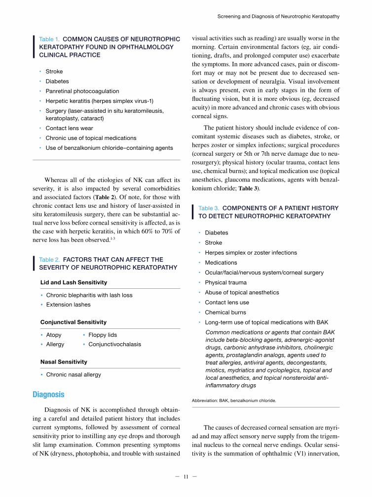
Table 1. COMMON CAUSES OF NEUROTROPHIC KERATOPATHY FOUND IN OPHTHALMOLOGY CLINICAL PRACTICE
• Stroke• Diabetes • Panretinal photocoagulation• Herpetic keratitis (herpes simplex virus-1)• Surgery (laser-assisted in situ keratomileusis,
keratoplasty, cataract)• Contact lens wear• Chronic use of topical medications• Use of benzalkonium chloride–containing agents
Whereas all of the etiologies of NK can affect its severity, it is also impacted by several comorbidities and associated factors (Table 2). Of note, for those with chronic contact lens use and history of laser-assisted in situ keratomileusis surgery, there can be substantial ac-tual nerve loss before corneal sensitivity is affected, as is the case with herpetic keratitis, in which 60% to 70% of nerve loss has been observed.1-3
Table 2. FACTORS THAT CAN AFFECT THE SEVERITY OF NEUROTROPHIC KERATOPATHY
Lid and Lash Sensitivity
• Chronic blepharitis with lash loss• Extension lashes
Conjunctival Sensitivity
• Atopy• Allergy
• Floppy lids• Conjunctivochalasis
Nasal Sensitivity
• Chronic nasal allergy
DiagnosisDiagnosis of NK is accomplished through obtain-
ing a careful and detailed patient history that includes current symptoms, followed by assessment of corneal sensitivity prior to instilling any eye drops and thorough slit lamp examination. Common presenting symptoms of NK (dryness, photophobia, and trouble with sustained
visual activities such as reading) are usually worse in the morning. Certain environmental factors (eg, air condi-tioning, drafts, and prolonged computer use) exacerbate the symptoms. In more advanced cases, pain or discom-fort may or may not be present due to decreased sen-sation or development of neuralgia. Visual involvement is always present, even in early stages in the form of fluctuating vision, but it is more obvious (eg, decreased acuity) in more advanced and chronic cases with obvious corneal signs.
The patient history should include evidence of con-comitant systemic diseases such as diabetes, stroke, or herpes zoster or simplex infections; surgical procedures (corneal surgery or 5th or 7th nerve damage due to neu-rosurgery); physical history (ocular trauma, contact lens use, chemical burns); and topical medication use (topical anesthetics, glaucoma medications, agents with benzal-konium chloride; Table 3).
Table 3. COMPONENTS OF A PATIENT HISTORY TO DETECT NEUROTROPHIC KERATOPATHY
• Diabetes• Stroke• Herpes simplex or zoster infections• Medications• Ocular/facial/nervous system/corneal surgery• Physical trauma• Abuse of topical anesthetics• Contact lens use• Chemical burns• Long-term use of topical medications with BAK
Common medications or agents that contain BAK include beta-blocking agents, adrenergic-agonist drugs, carbonic anhydrase inhibitors, cholinergic agents, prostaglandin analogs, agents used to treat allergies, antiviral agents, decongestants, miotics, mydriatics and cycloplegics, topical and local anesthetics, and topical nonsteroidal anti-inflammatory drugs
The causes of decreased corneal sensation are myri-ad and may affect sensory nerve supply from the trigem-inal nucleus to the corneal nerve endings. Ocular sensi-tivity is the summation of ophthalmic (V1) innervation,
Abbreviation: BAK, benzalkonium chloride.
An Evidence-based Approach to the Diagnosis and Treatment of Neurotrophic Keratopathy
12
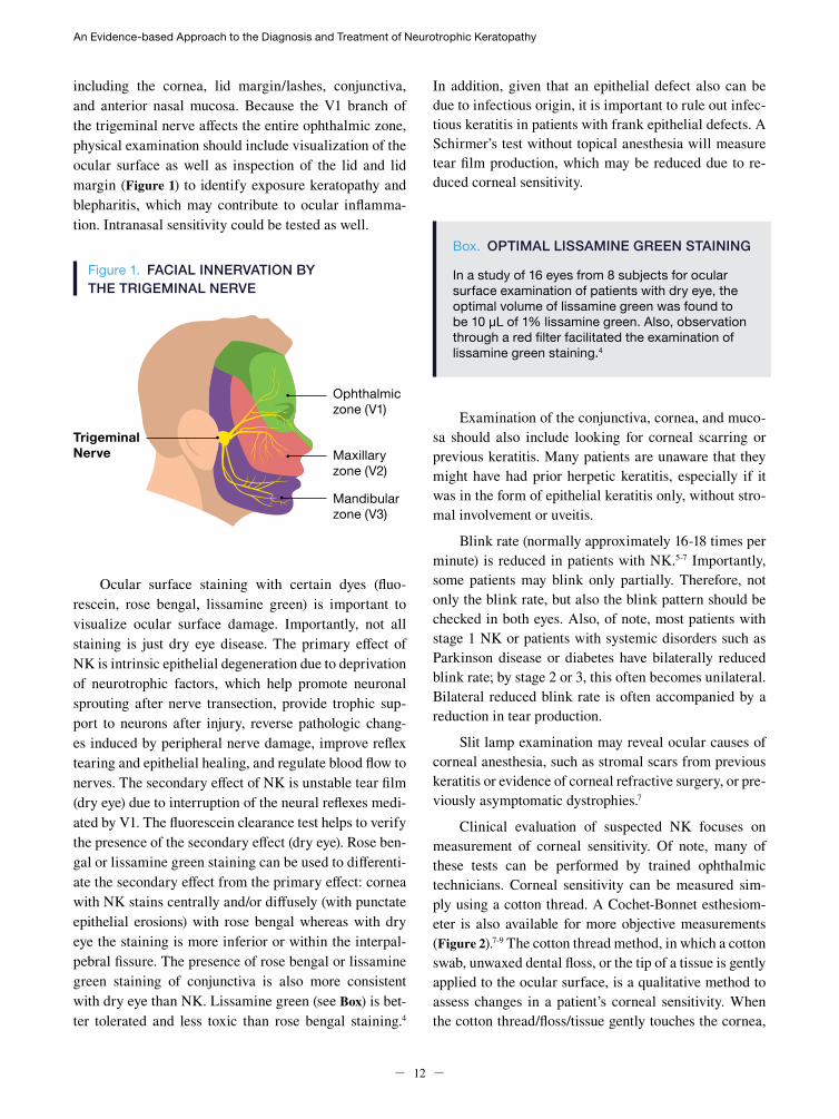
including the cornea, lid margin/lashes, conjunctiva, and anterior nasal mucosa. Because the V1 branch of the trigeminal nerve affects the entire ophthalmic zone, physical examination should include visualization of the ocular surface as well as inspection of the lid and lid margin (Figure 1) to identify exposure keratopathy and blepharitis, which may contribute to ocular inflamma-tion. Intranasal sensitivity could be tested as well.
Figure 1. FACIAL INNERVATION BY THE TRIGEMINAL NERVE
Ocular surface staining with certain dyes (fluo-rescein, rose bengal, lissamine green) is important to visualize ocular surface damage. Importantly, not all staining is just dry eye disease. The primary effect of NK is intrinsic epithelial degeneration due to deprivation of neurotrophic factors, which help promote neuronal sprouting after nerve transection, provide trophic sup-port to neurons after injury, reverse pathologic chang-es induced by peripheral nerve damage, improve reflex tearing and epithelial healing, and regulate blood flow to nerves. The secondary effect of NK is unstable tear film (dry eye) due to interruption of the neural reflexes medi-ated by V1. The fluorescein clearance test helps to verify the presence of the secondary effect (dry eye). Rose ben-gal or lissamine green staining can be used to differenti-ate the secondary effect from the primary effect: cornea with NK stains centrally and/or diffusely (with punctate epithelial erosions) with rose bengal whereas with dry eye the staining is more inferior or within the interpal-pebral fissure. The presence of rose bengal or lissamine green staining of conjunctiva is also more consistent with dry eye than NK. Lissamine green (see Box) is bet-ter tolerated and less toxic than rose bengal staining.4
In addition, given that an epithelial defect also can be due to infectious origin, it is important to rule out infec-tious keratitis in patients with frank epithelial defects. A Schirmer’s test without topical anesthesia will measure tear film production, which may be reduced due to re-duced corneal sensitivity.
Trigeminal Nerve
Mandibular zone (V3)
Maxillary zone (V2)
Ophthalmic zone (V1)
Box. OPTIMAL LISSAMINE GREEN STAINING
In a study of 16 eyes from 8 subjects for ocular surface examination of patients with dry eye, the optimal volume of lissamine green was found to be 10 μL of 1% lissamine green. Also, observation through a red filter facilitated the examination of lissamine green staining.4
Examination of the conjunctiva, cornea, and muco-sa should also include looking for corneal scarring or previous keratitis. Many patients are unaware that they might have had prior herpetic keratitis, especially if it was in the form of epithelial keratitis only, without stro-mal involvement or uveitis.
Blink rate (normally approximately 16-18 times per minute) is reduced in patients with NK.5-7 Importantly, some patients may blink only partially. Therefore, not only the blink rate, but also the blink pattern should be checked in both eyes. Also, of note, most patients with stage 1 NK or patients with systemic disorders such as Parkinson disease or diabetes have bilaterally reduced blink rate; by stage 2 or 3, this often becomes unilateral. Bilateral reduced blink rate is often accompanied by a reduction in tear production.
Slit lamp examination may reveal ocular causes of corneal anesthesia, such as stromal scars from previous keratitis or evidence of corneal refractive surgery, or pre-viously asymptomatic dystrophies.7
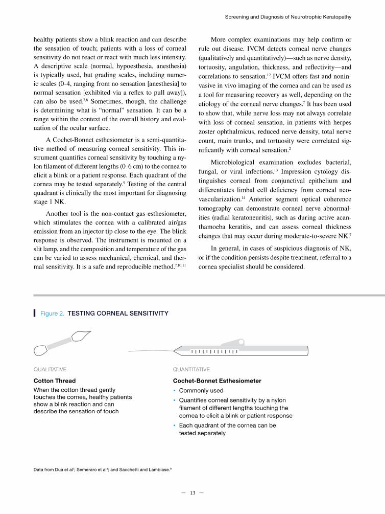
Clinical evaluation of suspected NK focuses on measurement of corneal sensitivity. Of note, many of these tests can be performed by trained ophthalmic technicians. Corneal sensitivity can be measured sim-ply using a cotton thread. A Cochet-Bonnet esthesiom-eter is also available for more objective measurements (Figure 2).7-9 The cotton thread method, in which a cotton swab, unwaxed dental floss, or the tip of a tissue is gently applied to the ocular surface, is a qualitative method to assess changes in a patient’s corneal sensitivity. When the cotton thread/floss/tissue gently touches the cornea,
Screening and Diagnosis of Neurotrophic Keratopathy
13
healthy patients show a blink reaction and can describe the sensation of touch; patients with a loss of corneal sensitivity do not react or react with much less intensity. A descriptive scale (normal, hypoesthesia, anesthesia) is typically used, but grading scales, including numer-ic scales (0-4, ranging from no sensation [anesthesia] to normal sensation [exhibited via a reflex to pull away]), can also be used.7,8 Sometimes, though, the challenge is determining what is “normal” sensation. It can be a range within the context of the overall history and eval-uation of the ocular surface.
A Cochet-Bonnet esthesiometer is a semi-quantita-tive method of measuring corneal sensitivity. This in-strument quantifies corneal sensitivity by touching a ny-lon filament of different lengths (0-6 cm) to the cornea to elicit a blink or a patient response. Each quadrant of the cornea may be tested separately.9 Testing of the central quadrant is clinically the most important for diagnosing stage 1 NK.
Another tool is the non-contact gas esthesiometer, which stimulates the cornea with a calibrated air/gas emission from an injector tip close to the eye. The blink response is observed. The instrument is mounted on a slit lamp, and the composition and temperature of the gas can be varied to assess mechanical, chemical, and ther-mal sensitivity. It is a safe and reproducible method.7,10,11
More complex examinations may help confirm or rule out disease. IVCM detects corneal nerve changes (qualitatively and quantitatively)—such as nerve density, tortuosity, angulation, thickness, and reflectivity—and correlations to sensation.12 IVCM offers fast and nonin-vasive in vivo imaging of the cornea and can be used as a tool for measuring recovery as well, depending on the etiology of the corneal nerve changes.7 It has been used to show that, while nerve loss may not always correlate with loss of corneal sensation, in patients with herpes zoster ophthalmicus, reduced nerve density, total nerve count, main trunks, and tortuosity were correlated sig-nificantly with corneal sensation.2
Microbiological examination excludes bacterial, fungal, or viral infections.13 Impression cytology dis-tinguishes corneal from conjunctival epithelium and differentiates limbal cell deficiency from corneal neo-vascularization.14 Anterior segment optical coherence tomography can demonstrate corneal nerve abnormal-ities (radial keratoneuritis), such as during active acan-thamoeba keratitis, and can assess corneal thickness changes that may occur during moderate-to-severe NK.7
In general, in cases of suspicious diagnosis of NK, or if the condition persists despite treatment, referral to a cornea specialist should be considered.
Data from Dua et al7; Semeraro et al8; and Sacchetti and Lambiase.9
QUALITATIVE QUANTITATIVE
Cotton ThreadWhen the cotton thread gently touches the cornea, healthy patients show a blink reaction and can describe the sensation of touch
Cochet-Bonnet Esthesiometer• Commonly used• Quantifies corneal sensitivity by a nylon
filament of different lengths touching the cornea to elicit a blink or patient response
• Each quadrant of the cornea can be tested separately
Figure 2. TESTING CORNEAL SENSITIVITY
An Evidence-based Approach to the Diagnosis and Treatment of Neurotrophic Keratopathy
14
REFERENCES
Differential DiagnosisSeveral common conditions may worsen the prog-
nosis of NK and should be managed either prior to, or simultaneously with, NK treatment. These include dry eye, blepharitis, contact lens–related disorders, expo-sure keratitis, mild chemical injury, and topical drug toxicity. Likewise, key considerations before beginning NK treatment are listed in Table 4. A key concern is de-termining for how long non-specific treatments should be continued before a significant clinical improvement is seen. Generally speaking, 4 to 8 weeks of treatment should be enough time to determine whether other pos-sible contributing factors should be considered.
Table 4. CONSIDERATIONS BEFORE BEGINNING NEUROTROPHIC KERATOPATHY TREATMENT
• Discontinue and/or modulate frequency of topical medications and consider preservative-free formulations, when possible, to decrease corneal toxicity
• Address blepharitis and meibomian gland dysfunction
• Rule out active infection• Treat ocular surface inflammation when it is
part of the underlying etiology• Treat dry eye, if not previously treated
1. Hamrah P, Cruzat A, Dastjerdi MH, et al. Corneal sensation and subbasal nerve alterations in patients with herpes simplex keratitis: an in vivo confocal microscopy study. Ophthalmology. 2010;117(10):1930-1936.
2. Hamrah P, Cruzat A, Dastjerdi MH, et al. Unilateral herpes zoster ophthalmicus results in bilateral corneal nerve alteration: an in vivo confocal microscopy study. Ophthalmology. 2013;120(1):40-47.
3. Cavalcanti BM, Cruzat A, Sahin A, Pavan-Langston D, Samayoa E, Hamrah P. In vivo confocal microscopy detects bilateral changes of corneal immune cells and nerves in unilateral herpes zoster ophthalmicus. Ocul Surf. 2018;16(1):101-111.
4. Hamrah P, Alipour F, Jiang S, Sohn JH, Foulks GN. Optimizing evaluation of lissamine green parameters for ocular surface staining. Eye. 2011;25(11):1429-1434.
5. Bentivoglio AR, Bressman SB, Cassetta E, Carretta D, Tonali P, Albanese A. Analysis of blink rate patterns in normal subjects. Mov Disord. 1997;12(6):1028-1034.
6. De Haas EBH. Desiccation of cornea and conjunctiva after sensory denervation: significance of desiccation for pathogenesis of neuroparalytic keratitis. JAMA Ophthalmol. 1962;67(4):439-452.
7. Dua HS, Said DG, Messmer EM, et al. Neurotrophic keratopathy. Prog Retin Eye Res. 2018;66:107-131.
8. Semeraro F, Forbice E, Romano V, et al. Neurotrophic keratitis. Ophthalmologica. 2014;231(4):191-197.
9. Sacchetti M, Lambiase A. Diagnosis and management of neurotrophic keratitis. Clin Ophthalmol. 2014;8:571-579.
10. Belmonte C, Acosta MC, Schmelz M, Gallar J. Measurement of corneal sensitivity to mechanical and chemical stimulation with a CO2 esthesiometer. Invest Ophthalmol Vis Sci. 1999;40(2):513-519.
11. Tesón M, Calonge M, Fernández I, Stern ME, González-Garcia MJ. Characterization by Belmonte’s gas esthesiometer of mechanical, chemical, and thermal corneal sensitivity thresholds in a normal population. Invest Ophthalmol Vis Sci. 2012;53(6):3154-3160.
12. Cruzat A, Qazi Y, Hamrah P. In vivo confocal microscopy of corneal nerves in health and disease. Ocul Surf. 2017;15(1):15-47.
13. Bonini S, Rama P, Olzi D, Lambiase A. Neurotrophic keratitis. Eye (Lond). 2003;17(8):989-995.
14. Srinivasan S, Lyall, D. Neurotrophic keratopathy. In: Holland E, Mannis M, Lee B. Ocular Surface Disease: Cornea, Conjunctiva and Tear Film. London, England: W.B. Saunders; 2013:205-211.
15
Management of Neurotrophic Keratopathy
Key Messages• The ocular surface (corneal nerves, corneal and
conjunctival epithelia, and tears) can be thought of as one unit.
• Although the overarching goal of treatment for NK is restoration or improvement of corneal sensation, the specific treatment goals will vary based on the stage of disease. Ideally, NK will be identified and treated early to prevent late-stage sequelae.
• Treatments should be used according to NK stage/severity, but they are not mutually exclusive of one another. Often a combination of approaches needs to be considered.
• Some patients, especially with early stage NK, may need to be made aware of the benefits of NK treatment, especially if they deny any symptoms. However, careful questioning about their ability to perform visual tasks requiring sustained gazing, such as reading, will reveal symptoms. Showing patients objective data (eg, from slit lamp results, corneal staining) can also help to educate them about the benefits of addressing NK early.
In healthy eyes, there is a state of corneal homeostasis among the corneal nerves, the corneal and conjunc-tival epithelia, and the tear film (Figure). The corne-
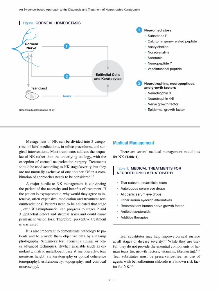
al nerves are a complex mix of sensory and autonomic nerves that provide trophic support to the ocular sur-face. They also stimulate wound healing and maintain anatomic integrity via neuromediators (eg, substance P, calcitonin gene-related peptide), which in turn mediate nerve fiber survival, differentiation, and maturation via neurotrophins, neuropeptides, and growth factors. In response to external irritation, the corneal nerves also stimulate the tear glands to promote tear secretion and the blinking reflex through an interaction with the lac-rimal glands and corneal surface. Tears contain growth factors and nutrients that stimulate epithelial cells.1,2 The ocular surface (nerves, epithelia, and tears) can be thought of as one unit. Blinking, mostly mediated by corneal innervation, also plays a significant role in re-plenishing and redistributing the tear film.
Although the overarching goal of treatment for neu-rotrophic keratopathy (NK) is restoration or improve-ment of corneal sensation, the specific treatment goals will vary based on the stage of disease. In stage 1 NK, treatment goals are focused on improving the corneal epithelium and avoiding epithelial breakdown, mostly by removing offending agents. In stage 2, the goal is to promote ocular surface healing, particularly the corneal epithelium, and to prevent any ulcers. By stage 3, epi-thelial damage has occurred; therefore, the goals are to reduce corneal stromal scarring and prevent perforation.
Mina Massaro-Giordano, MD*; Christopher E. Starr, MD†
*UNIVERSITY OF PENNSYLVANIA SCHOOL OF MEDICINE, PHILADELPHIA, PENNSYLVANIA; †WEILL CORNELL MEDICAL COLLEGE, NEW YORK, NEW YORK
An Evidence-based Approach to the Diagnosis and Treatment of Neurotrophic Keratopathy
16
Management of NK can be divided into 3 catego-ries: off-label medications, in-office procedures, and sur-gical interventions. Most treatments address the sequa-lae of NK rather than the underlying etiology, with the exception of corneal neurotization surgery. Treatments should be used according to NK stage/severity, but they are not mutually exclusive of one another. Often a com-bination of approaches needs to be considered.1,3
A major hurdle to NK management is convincing the patient of the necessity and benefits of treatment. If the patient is asymptomatic, why would they agree to in-tensive, often expensive, medication and treatment rec-ommendations? Patients need to be educated that stage 1, even if asymptomatic, can progress to stages 2 and 3 (epithelial defect and stromal lysis) and could cause permanent vision loss. Therefore, preventive treatment is warranted.
It is also important to demonstrate pathology to pa-tients and to provide them objective data by slit lamp photography, Schirmer’s test, corneal staining, or oth-er advanced techniques, if/when available (such as os-molarity, matrix metallopeptidase 9, meibography, tear meniscus height [via keratography or optical coherence tomography], esthesiometry, topography, and confocal microscopy).
Medical ManagementThere are several medical management modalities
for NK (Table 1).
Table 1. MEDICAL TREATMENTS FOR NEUROTROPHIC KERATOPATHY
• Tear substitutes/artificial tears• Autologous serum eye drops• Allogenic serum eye drops• Other serum eyedrop alternatives• Recombinant human nerve growth factor • Antibiotics/steroids• Additive therapies
Tear substitutes may help improve corneal surface at all stages of disease severity.4-7 While they are use-ful, they do not provide the essential components of hu-man tears (ie, growth factors, vitamins, fibronectin).8-10 Tear substitutes must be preservative-free, as use of agents with benzalkonium chloride is a known risk fac-tor for NK.4,6
Corneal Nerve
Neuromediators• Substance P• Calcitonin gene–related peptide• Acetylcholine• Noradrenaline• Serotonin• Neuropeptide Y• Vasointestinal peptide
Neurotrophins, neuropeptides, and growth factors• Neurotrophin 3• Neurotrophin 4/5• Nerve growth factor• Epidermal growth factor
1
2
Epithelial Cells and Keratocytes
1
2
Tears
Tear gland
Figure. CORNEAL HOMEOSTASIS
Data from Mastropasqua et al.1
Management of Neurotrophic Keratopathy
17
Autologous serum eye drops (ASEDs) are derived from the patient’s own blood. As such, they are inher-ently nonallergenic and biochemically and biomechan-ically similar to tears.9,11 Natural tears have antimicro-bial, nourishing, mechanical, and optical properties and contain many key components for healthy functioning of the eye. While these epitheliotropic factors are not replaced by artificial tears, they are present in ASEDs. In vitro corneal epithelial cells are better maintained by ASEDs than by artificial tears, and ASEDs pro-mote growth and migration of cells, which is import-ant in healing in vitro. The serum percent varies from 20% up to 100%.12-14
There are several disadvantages with ASEDs. They are challenging to produce, the composition of the drops varies widely due to lack of regulations, and there are concerns about quality control, infection risk, and chal-lenges with stability due to special storage requirements. Moreover, it is difficult to compare studies of ASEDs be-cause production protocols vary; not surprisingly, there is a wide range of outcomes. ASEDs also can create medico-legal issues in some countries.9 There are con-flicting views about the increased risk of circulating antibodies or pro-inflammatory mediators with ASEDs in patients with autoimmune diseases. In one paper, patients with secondary Sjögren’s syndrome (SS) had higher serum proinflammatory cytokine levels (tumor necrosis factor α, interleukin [IL]-1β, IL-6, and IL-8) than patients with primary SS. Patients with primary SS had significantly improved ocular symptoms, ocular sur-face staining grades, and tear breakup time with ASEDs. However, patients with secondary SS had no improve-ment. These results may suggest that ASEDs might not be effective for the treatment of secondary SS because of elevated serum proinflammatory cytokine levels.3,15
Under certain circumstances, ASEDs are simply not suitable for some patients (eg, patients who have insufficient venous access, have anemia, are unable to travel to a donor center, are unable to give consent, have uncontrolled diabetes, have immune-mediated diseases who may be taking cytotoxic drugs, have sepsis, or are unable to donate 1 full unit of blood).16 Of note, the 2017 Royal College of Ophthalmologists guideline recom-mends serum eye drops for severe ocular surface dis-ease, including severe dry eye, persistent and recurrent corneal epithelial defects, and NK. However, the guide-line states that patients must have exhausted all available licensed therapies.11
Other options include allogeneic serum (where au-tologous serum is unavailable or unsuitable), platelet-rich plasma (platelets contain growth factors for wound heal-ing), umbilical cord serum, and finger prick blood. Pri-or clinical studies related to these treatment modalities have shown these methods to be safe and effective, with no report of infection.16-20
Human umbilical cord serum has been shown to contain growth factors, including nerve growth factor (NGF), and has been used to treat a variety of ocular surface conditions including NK. The response to cord blood serum appears to be related to the severity of NK, with mild-to-moderate lesions in the form of punctate epithelial defects or confluent staining responding more quickly than frank epithelial defects. Difficulty obtain-ing cord blood serum limits its widespread use.3
The data on finger prick blood are limited but pos-itive.21 The advantages to this source of ASEDs are that numerous factors contained in tears are also found in whole blood and it is inherently nonallergenic. How-ever, there is patient discomfort associated with finger prick and minimal theoretical risk of infection. The risk of ocular infection through transfer of skin patho-gens via repeated close contact between finger and eye exists, though this is minimized by diligent cleaning of the finger with an alcohol pad. The risk of transmission of blood-borne pathogens to the anterior eye is biologi-cally plausible.
The primary safety consideration for autologous serum-based eye drops is the risk of microbial growth during storage because serum-based solutions essential-ly are growth media. Care must be taken in the prepara-tion of these eye drops to ensure that they are prepared in a sterile manner. Compounding pharmacies and eye banks have the equipment necessary to reduce the risk of contamination during preparation. Microbial contam-ination remains a risk in patients who have a compro-mised ocular surface. Although in the dry eye studies included in a report by the American Academy of Oph-thalmology, no patients experienced any reported clini-cal adverse events, bacterial growth was reported in the eye drops of 2 patients with no adverse clinical conse-quence.13 Another disadvantage of ASEDs is the risk of inducing an infection because of, for example, hepatitis of the donor or microbial contamination of the initially sterile dropper bottle during prolonged use. This may occur during preparation as well as application of the
An Evidence-based Approach to the Diagnosis and Treatment of Neurotrophic Keratopathy
18
drops, either to the correct or any accidental recipient. Transmission of HIV by a single serum droplet into an eye has been reported.22
Published evidence for use of NGF in the treatment of NK is strong. The importance of NGF in corneal ho-meostasis has been demonstrated in vitro, ex vivo, and in vivo. Small uncontrolled, open-label studies with mouse NGF produced promising results for the treatment of corneal neurotrophic ulcers.
Manufacturing of human NGF is challenging due to its complex tertiary structure.23 Cenegermin is an Escherichia coli-derived recombinant human NGF for-mulation for topical ophthalmic use in the treatment of moderate and severe NK. Once cleaved to mature NGF, the molecule is identical to human NGF. As a source of NGF, cenegermin targets the nerve pathology associat-ed with NK, not just the sequelae, and therefore has the potential to address the healing deficits seen in NK. It was approved by the US Food and Drug Administra-tion in August 2018 for the treatment of NK and is the only recombinant human NGF available at this time.3 The efficacy and safety of cenegermin has been evalu-ated in patients with moderate or severe NK refractory to nonsurgical treatment in 2 independent, multicenter, randomized, double-masked vehicle-controlled studies (1 in the United States, 1 in the European Union). Both studies compared cenegermin (10 and 20 μg/mL, Euro-pean Union; 20 μg/mL United States) to vehicle for 8 weeks of treatment, followed by 8 weeks of treatment for those originally receiving vehicle, and follow-up was for 48 (European Union) or 24 weeks (United States).
The results showed that the post hoc analysis of the more conservative primary efficacy endpoint (ie, complete corneal healing, defined as 0-mm staining in the lesion area and no other persistent staining in the rest of the cornea after 8 weeks of treatment, compared with <0.5-mm staining in the main primary endpoint) was reached at week 4 by significantly more patients who originally received cenegermin compared with those who had re-ceived vehicle (Table 2). Importantly, 80% of those who achieved corneal healing were still healed 48 weeks af-ter one 8-week cycle of cenegermin treatment.23,24
Nonsurgical Interventional ManagementInterventional options for NK management include
eyelid closure and punctal plugs. Eyelid closure to cover the cornea and protect it against the environment and ef-fects of blinking can be achieved by any of several meth-ods, including taping (eg, nasal strips, porous bandage tape, or paper tape), pressure patching, pad and bandage, and botulinum toxin–induced ptosis. Alternatively, tar-sorrhaphy surgery can be performed in patients who need a permanent closure of the lids. As reviewed by Dua et al, each of these methods has advantages and dis-advantages.3 Botulinum toxin–induced ptosis can be less disturbing to patients who need temporary closure than tarsorrhaphy because there is less scarring to the eyelids. The different toxin products are at different concentra-tions to achieve ptosis; the average duration of ptosis is 6 to 12 weeks. Botulinum toxin is an especially appro-priate option in patients with poor general condition or poor cooperation.25
United States24 European Union23
Cenegermin Vehicle Cenegermin Vehicle
20 μg/mL (n = 24) 20 μg/mL (n = 52)
(n = 24) (n = 52)
Week 4 56.5 20.8 P < .012 58.0 13.7 P < .001a
Week 8 65.2 16.7 P < .001 72.0 33.3 P < .001a
Table 2. RESULTS OF STUDIES OF CENEGERMIN
a 20 μg/mL cenegermin vs vehicle.Data from Bonini et al23 and Pflugfelder et al.24
Management of Neurotrophic Keratopathy
19
Punctal plugs act by occluding the tear ducts to re-tain natural tears. This can be achieved by temporary or permanent plugs or permanent occlusion of the punctum by cauterization. Temporary plugs are generally made of bovine collagen and rest in the punctal opening, making them difficult to remove.26 Silicone plugs may have the risk of rubbing on the ocular surface and irritating the eye but can be easily removed. Again, each has advantages and disadvantages, including discomfort (not common), migration, extrusion (in up to 50% of cases), scarring/punctal stenosis (in up to 33% of cases), infection (ap-proximately 66% of cases), and pyogenic granuloma (up to 35%).3,26 Timing of placement is critical because reten-tion of the tears during inflammation can also expose the ocular surface to more concentrated pro-inflammatory cytokines and other pro-inflammatory mediators, which could make the NK worse.3
Therapeutic corneal or scleral contact lenses can be used for a variety of corneal and anterior segment conditions with the aim of promoting corneal epithelial healing and postponing severe corneal complications.5 Contact lenses act as a physical barrier against the eye-lids to protect against degradation of the corneal epi-thelium and perhaps help with delaying progression to more serious corneal disease. There are 2 types of con-tact lenses (corneal and scleral), and they are available as normal and large diameter silicone hydrogel lenses, rigid gas permeable lenses, hybrid lenses, or prosthetic replacement of the ocular surface ecosystem (PROSE) lenses. Several studies have shown high rates of corneal healing with commercial scleral and PROSE lenses, in-cluding restoration of visual acuity in some studies, but there were some incidences of complications.27-34 Con-tact lenses are palliative and do not treat the underlying problem.35 Moreover, prolonged therapeutic contact lens use may increase the risk of secondary infections and the concomitant use of topical antibiotics is recommend-ed, particularly with soft bandage contact lenses.4,36
Tissue adhesives are a treatment option for cor-neal defects and perforations. For a small perforation (<3 mm) the application of tissue adhesive on the lesion, followed by the application of a soft bandage contact lens or amniotic membrane transplantation, is the pro-cedure of choice.1,3,4,6,7 Two basic types of adhesives are used: synthetic (cyanoacrylate) and biologic (fibrin glue or clot). The cyanoacrylate glue polymerizes rapidly in the presence of water; the formaldehyde released during the polymerization may have some antibacterial activi-
ty. A rigid impermeable plaque is formed on the surface of the cornea; therefore, a bandage lens should be used for protection to avoid trauma to the lid as well as for comfort. This tissue adhesive is not suitable for larger lesions because it can be lost with acute worsening of the condition, but it can be a good standby option and it is nonbiodegradable.3 A fibrin clot or “glue” occurs when purified fibrinogen and thrombin are mixed together. Fibrin polymerizes relatively slowly and is rapidly de-graded; addition of aprotinin or placement of a bandage contact lens can delay degradation. Antifibrinolytic agents (aprotinin and aminocaproic acid) increase the lifespan of the clot by inhibiting fibrinolysis. Fibrin glue has numerous advantages including allowing sufficient working time before inducing firm adhesion; reducing the requirement for sutures; having adequate tensile strength to maintain wound integrity; lowering the risk of infection to the wound site; and being biocompatible, well tolerated, transparent, accessible, affordable, and eventually disappearing. However, there is a potential risk of transmitting blood-borne disease with its use and variable concentrations of fibrin lead to variable performance.37,38
Other procedural interventions particularly for as-sociated dry eye disease/ocular surface disease that are adjunctive in all stages of NK include microblepharo-exfoliation, thermal pulsation procedures (used to treat meibomian gland dysfunction [MGD]), intense pulsed light treatment (used to treat rosacea and MGD), con-junctivochalasis cautery or excision, anti-demodex in-office swabs, neurostimulation (using either an intra-nasal tear neurostimulator [TrueTear, Allergan] device or a selective nicotinic acetylcholine receptor agonist [currently under investigation in NK]), and self-retained amniotic membranes and/or bandage contact lenses (used to treat stage 1 punctate epithelial erosions).
Epithelial debridement is used to address the re-duced epithelial migration due to the rolled, heaped-up edge of the defect in NK. Debridement, despite making the epithelial defect larger, helps facilitate epithelial healing by creating a clean demarcated edge and acti-vating the epithelium to migrate centrally. It is a low-risk procedure easily performed at the slit lamp with a blade or spear.39 While technically being an in-office proce-dure, it may be useful to refer to a corneal specialist for this treatment.
An Evidence-based Approach to the Diagnosis and Treatment of Neurotrophic Keratopathy
20
Nicergoline is an ergot derivative that increases va-sodilation and is used for the treatment of dementia. One study of 27 eyes with NK found complete healing of epi-thelial defects in 85% of eyes after treatment with 10-mg nicergoline twice daily for at least 2 weeks, as well as other positive outcomes.40
Surgical InterventionsSurgical interventions for NK are almost always re-
served for those with stage 2 or 3 NK and can include amniotic membrane transplantation (AMT), tissue adhe-sives (previously discussed), total or partial conjunctival flap, multilayer AMT, corneal transplant, and corneal neurotization. AMT involves placement of an amnion such that the epithelium migrates to the membrane, ei-ther under the amniotic membrane, which will later fall off or is removed (a patch or “onlay”) or when the amnion becomes incorporated into the cornea (a graft or “inlay”). AMT also provides trophic support (multi-ple growth factors, collagens, laminin, fibronectin, and anti-inflammatory and anti-angiogenic factors). AMT not only helps facilitate epithelial healing but also may help increase corneal sensation.41 AMT can be performed as single layer or stacked/multilayer and can be sutured, glued, or self-retained. Of note, the amniotic membrane and fibrin glue carry the potential risk of transmitting blood-borne pathogens from pooled blood, but this can be minimized if the donors are healthy or the patient is using his/her own blood.38 AMT often requires multiple applications (it dissolves before the corneal ulcer heals) in NK but it can be effective for healing NK ulcers when traditional measures have failed.3,42,43
Tarsorrhaphy has been long established for NK treatment. It can be used either as a temporary measure with temporary sutures or as a permanent means of lid closure, which could be either partial or full. A partial tarsorrhaphy (lateral, nasal) allows drops and vision and eye examinations whereas full closure does not. It should be considered in all cases of persistent epithelial defects when medical treatment and/or nonsurgical in-terventions are not effective.3,44
Conjunctival flap procedure is performed mostly for stage 3 NK and with corneal perforations. It is performed to prevent progression of the epithelial defect to perfora-tion by addressing the dryness and exposure and halting the inflammatory process.45 Total conjunctival flaps are best used if there is deep, central, visually significant
stromal involvement and/or perforations with low visual potential. A conjunctival flap is a vascularized pedicle of conjunctiva, which delivers nutrients and growth fac-tors to the cornea. While a conjunctival flap restores the integrity of the globe when all else fails and eliminates the need for frequent topical eyedrop instillation, it can significantly limit vision when covering the central cor-nea. Corneal perforation under the flap, retraction of the flap, and difficulty with reversal can be problematic. A conjunctival flap procedure is generally performed in an operating room.3,46
A corneal transplant can be done as lamellar (par-tial transplant) or penetrating (full corneal transplant). This procedure is often performed as a last resort, when corneal ulceration has failed to heal. The risk for rejec-tion, failure, and melting is high.3,47 Keratoprosthesis on the other hand may lead to successful outcomes with restoration of vision for those with NK who have failed traditional keratoplasty or after a perforation has been stabilized with tarsorrhaphy, glue, or conjunctival flap.48
Finally, corneal neurotization is a complex surgery in which donor nerve branches are inserted at the cor-neal limbus to bypass the unhealthy nerve. It is the only surgical procedure that directly targets the ophthalmic division of the trigeminal nerve to increase corneal in-nervation and restore normal corneal sensation. It can be performed using a direct or indirect technique. Di-rect neurotization transposes healthy contralateral su-praorbital and supratrochlear nerves, which are directed through a large coronal incision, to the limbus of the anesthetic eye. In one study, all 6 patients in whom this technique was used demonstrated improvement of visual acuity and corneal health and sensitivity, and remained free of signs of NK.49 The indirect technique is less in-vasive and uses a sural nerve autograft anastomosed to the supratrochlear nerve and tunneled through an upper eyelid incision instead of a bicoronal incision. It also al-lows for management of bilateral NK. In one study, 3 out of 4 eyes with NK had noticeably improved corneal sen-sitivity (55 mm [standard deviation, 5]) 6 months after surgery and none developed corneal anesthesia-related complications.50
REFERENCES
21
Management of Neurotrophic Keratopathy
1. Mastropasqua L, Massaro-Giordano G, Nubile M, Sacchetti M. Understanding the pathogenesis of neurotrophic keratitis: the role of corneal nerves. J Cell Physiol. 2017;232(4):717-724.
2. Müller LJ, Marfurt CF, Kruse F, Tervo TM. Corneal nerves: structure, contents and function. Exp Eye Res. 2003;76(5):521-542.
3. Dua HS, Said DG, Messmer EM, et al. Neurotrophic keratopathy. Prog Retin Eye Res. 2018;66:107-131.
4. Bonini S, Rama P, Olzi D, Lambiase A. Neurotrophic keratitis. Eye (Lond). 2003;17(8):989-995.
5. Mantelli F, Nardella C, Tiberi E, Sacchetti M, Bruscolini A, Lambiase A. Congenital corneal anesthesia and neurotrophic keratitis: diagnosis and management [published online September 16, 2015]. Biomed Res Int. doi:10.1155/2015/805876.
6. Sacchetti M, Lambiase A. Diagnosis and management of neurotrophic keratitis. Clin Ophthalmol. 2014;8:571-579.
7. Semeraro F, Forbice E, Romano V, et al. Neurotrophic keratitis. Ophthalmologica. 2014;231(14):191-197.
8. Fox RI, Fox CM, eds. Sjögren’s Syndrome: Practical Guidelines to Diagnosis and Therapy. New York, NY: Springer; 2011.
9. Geerling G, Maclennan S, Hartwig D. Autologous serum eye drops for ocular surface disorders. Br J Ophthalmol. 2004;88(11):1467-1474.
10. Mixon W, Angelle PP, Chang RI. Autologous eye drops for the treatment of dry eye and neurotrophic keratitis. Int J Pharm Compd. 2009;11(6):506-515.
11. Rauz S, Koay SY, Foot B, et al. The Royal College of Ophthalmologists guidelines on serum eye drops for the treatment of severe ocular surface disease: full report [published online November 17, 2017]. Eye (Lond). doi:10.1038/eye.2017.209.
12. Pan Q, Angelina A, Marrone SM, Stark WJ, Akpek EK. Autologous serum eye drops for dry eye. Cochrane Database Syst Rev. 2017;2:CD009327.
13. Shtein RM, Shen JF, Kuo AN, Hammersmith KM, Li JY, Weikert MP. Autologous serum-based eye drops for treatment of ocular surface disease: a report by the American Academy of Ophthalmology. Ophthalmology. 2020;127(1):128-133.
14. Tahmaz V, Gehlsen U, Sauerbier L, et al. Treatment of severe chronic ocular graft-versus-host disease using 100% autologous serum eye drops from a sealed manufacturing system: a retrospective cohort study. Br J Opthalmol. 2017;101(3):322-326.
15. Hwang J, Chung SH, Jeon S, Kwok SK, Park SH, Kim MS. Comparison of clinical efficacies of autologous serum eye drops in patients with primary and secondary Sjögren syndrome. Cornea. 2014;33(7):663-667.
16. Chiang CC, Chen WL, Lin JM, Tsai YY. Allogenic serum eye drops for the treatment of persistent corneal epithelial defect. Eye (Lond). 2009;23(2):290-293.
17. Alio JL, Abad M, Artola A, Rodriguez-Prats JL, Pastor S, Ruiz-Colecha J. Use of autologous platelet-rich plasma in the treatment of dormant corneal ulcers. Ophthalmology. 2007;114(7):1286-1293.
18. Lopez-Plandolit S, Morales MC, Freire V, Etxebarria J, Duran JA. Plasma rich in growth factors as therapeutic agent for persistent corneal epithelial defects. Cornea. 2010;29(8):843-848.
19. Kim KM, Shin YT, Kim HK. Effect of autologous platelet-rich plasma on persistent corneal defect after infectious keratitis. Jpn J Ophthalmol. 2012;56(6):544-550.
20. Sanchez-Avila RM, Meraho-Lloves J, Riestra AC, et al. Treatment of patients with neurotrophic keratitis stages 2 and 3 with plasma rich in growth factors (PRGF-Endoret) eye-drops. Int Ophthalmol. 2017;38(3):1193-1204.
21. Than J, Balal S, Wawrzynski J, et al. Fingerprick autologous blood: a novel treatment for dry eye syndrome. Eye (Lond). 2017;31(12):1655-1663.
22. Eberle J, Habermann J, Gürtler LG. HIV-1 infection transmitted by serum droplets into the eye: a case report. AIDS. 2000;14(2):206-207.
23. Bonini S, Lambiase A, Rama P, et al. Phase II randomized, double-masked, vehicle-controlled trial of recombinant human nerve growth factor for neurotrophic keratitis. Ophthalmology. 2018;125(2):1332-1343.
24. Pflugfelder SC, Massaro-Giordano M, Perez VL, et al. Topical recombinant human nerve growth factor (cenegermin) for neurotrophic keratopathy: a multicenter, randomized, vehicle-controlled pivotal trial. Ophthalmology. 2020;127(1):14-26.
25. Vleming EN, Pérez-Rico C, Montes MA, Pareja J. [Persistent corneal defects treated with botulinum toxin-induced ptosis]. Arch Soc Esp Oftalmol. 2007;82(9):547-550.
REFERENCES
22
An Evidence-based Approach to the Diagnosis and Treatment of Neurotrophic Keratopathy
26. Jehangir N, Bever G, Jafar Mahmood SM, Moshirfar M. Comprehensive review of the literature on existing punctal plugs for the management of dry eye disease [published online March 7, 2016]. J Ophthalmol. doi:10.1155/2016/9312340.
27. Cressey A, Jacobs DS, Remington C, Carrasquillo KC. Improvement of chronic corneal opacity in ocular surface disease with prosthetic replacement of the ocular surface ecosystem (PROSE) treatment. Am J Ophthalmol Case Rep. 2018;10:108-113.
28. Harthan JS, Shorter E. Therapeutic uses of scleral contact lenses for ocular surface disease: patient selection and special considerations. Clin Optom (Auckl). 2018;10:65-74.
29. Gupta S, Arora R, Dass Sota L, Kumar M. An alternative approach to bandage contact lenses. CLAO J. 1998;24(2):118-121.
30. Kloek CE, Jeng-Miller KW, Jacobs DS, Dunn IF. Prosthetic replacement of the ocular surface ecosystem treatment of ocular surface disease after skull base tumor resection. World Neurosurg. 2018;110:e124-e128.
31. Remington CD, Jacobs DS. PROSE treatment for pediatric patients with neurotrophic keratitis. Invest Ophthal Vis Sci. 2015;56:6076.
32. Romero-Rangel T, Stavrou P, Cotter J, Rosenthal P, Baltatzis S, Foster CS. Gas-permeable scleral contact lens therapy in ocular surface disease. Am J Ophthal. 2000;130(1):25-32.
33. Sun YZ, Guo L, Zhang FS. Curative effect assessment of bandage contact lens in neurogenic keratitis. Int J Ophthalmol. 2014;7(6):980-983.
34. Arora R, Jain S, Monga S, Narayanan R, Raina UK, Mehta DK. Efficacy of continuous wear PureVision contact lenses for therapeutic use. Contact Lens Ant Eye. 2004;27(1):39-43.
35. Rubinstein MP. Applications of contact lens devices in the management of corneal disease. Eye (Lond). 2003:17(8);872-876.
36. Kent HD, Cohen EJ, Laibson PR, Arentsen JJ. Microbial keratitis and corneal ulceration associated with therapeutic soft contact lenses. CLAO J. 1990;16(1):49-52.
37. Alio JL, Rodriguez AE, Martinez LM, Rio AL. Autologous fibrin membrane combined with solid platelet-rich plasma in the management of perforated corneal ulcers: a pilot study. JAMA Ophthalmol. 2013;131(6):745-751.
38. Panda A, Kumar S, Kumar A, Bansal R, Bhartiya S. Fibrin glue in ophthalmology. Indian J Ophthalmol. 2009;57(5):371-379.
39. Katzman LR, Jeng BH. Management strategies for persistent epithelial defects of the cornea. Saudi J. Ophthalmol. 2014;28(3):168-172.
40. Lee YC, Sim SY. Treatment of neurotrophic keratopathy with nicergoline. Cornea. 2015;34(3):303-307.
41. John T, Tighe SE, Sheha H, et al. Corneal nerve regeneration after self-retained cryopreserved amniotic membrane in dry eye disease [published online August 15, 2017]. J Ophthalmol. doi:10.1155/2017/6404918.
42. Khokhar S, Natung T, Sony P, Sharma N, Agarwal N, Vajpayee RB. Amniotic membrane transplantation in refractory neurotrophic corneal ulcers: a randomized, controlled clinical trial. Cornea. 2005;24(6):654-660.
43. Kruse FE, Rohrschneider K, Völcker HE. Multilayer amniotic membrane transplantation for reconstruction of deep corneal ulcers. Ophthalmology. 1999;106(8):1504-1510.
44. Tuli SS, Schultz GS, Downer DM. Science and strategy for preventing and managing corneal ulceration. Ocul Surf. 2007;5(1):23-39.
45. Pushker N, Dada T, Vajpayee RB, Gupta V, Aggrawal T, Titiyal JS. Neurotrophic keratopathy. CLAO J. 2001;27(2): 100-107.
46. Khodadoust A, Quinter AP. Microsurgical approach to the conjunctival flap. Arch Ophthalmol. 2003;121(8):1189-1193.
47. Jonas JB, Rank RM, Budde WM. Tectonic sclerokeratoplasty and tectonic penetrating keratoplasty as treatment for perforated or predescemetal corneal ulcers. Am J Ophthalmol. 2001;132(1):14-18.
48. Pavan-Langston D, Dohlman CH. Boston keratoprosthesis treatment of herpes zoster neurotrophic keratopathy. Ophthalmology. 2008;115(2 suppl):S21-S23.
49. Terzis JK, Dryer MM, Bodner BI. Corneal neurotization: a novel solution to neurotrophic keratopathy. Plast Reconstr Surg. 2009;123(1):112-120.
50. Elbaz U, Bains R, Zuker RM, Borschel GH, Ali A. Restoration of corneal sensation with regional nerve transfers and nerve grafts: a new approach to a difficult problem. JAMA Ophthalmol. 2014;132(11):1289-1295.

23
Case 1
Persistent Epithelial Defect With Advanced GlaucomaNatalie Afshari, MD SHILEY EYE INSTITUTE, UNIVERSITY OF CALIFORNIA, SAN DIEGO, LA JOLLA, CALIFORNIA
History of Present IllnessA 35-year-old man with advanced glaucoma presented to the cornea clinic with unilateral persistent epithelial defect (PED).
Past Ocular/Surgical HistoryHis past ocular history included open-angle glaucoma, previous glaucoma trabeculectomy and revision surgery, pseudophakia, and corneal edema.
Past Treatment HistoryHe had been treated with lubrication with serum tears, bandage contact lens, amniotic membrane graft, and temporary tarsorrhaphy, without resolution.
Current MedicationsHe was currently taking dorzolamide/timolol twice daily and latanoprost every night at bedtime in each eye.
Initial ExaminationHis initial examination results are in Table 1.
Table 1. INITIAL EXAMINATION RESULTS
Right Eye Left Eye
Visual acuity 20/25 Hand motions
Pupils 4-3 Post surgical
Intraocular pressure
11 6
Extraocular muscles
Full Full
Confrontation visual field
Full Glaucomatous visual field loss
Other ExaminationsSlit lamp examination showed a 3- x 2-mm central corneal epithelial defect with peripheral corneal neovascularization (Table 2). There was mild corneal edema (Figure 1A-C).
The following case study series, based on an expert roundtable held in Baltimore, Maryland, on October 26, 2019, highlights key findings from the discussion and
provides examples of typical neurotrophic keratopathy presentations and management strategies for eye care providers to apply in clinical practice.
Neurotrophic Keratopathy Presentation and Management:
A Case Study Series
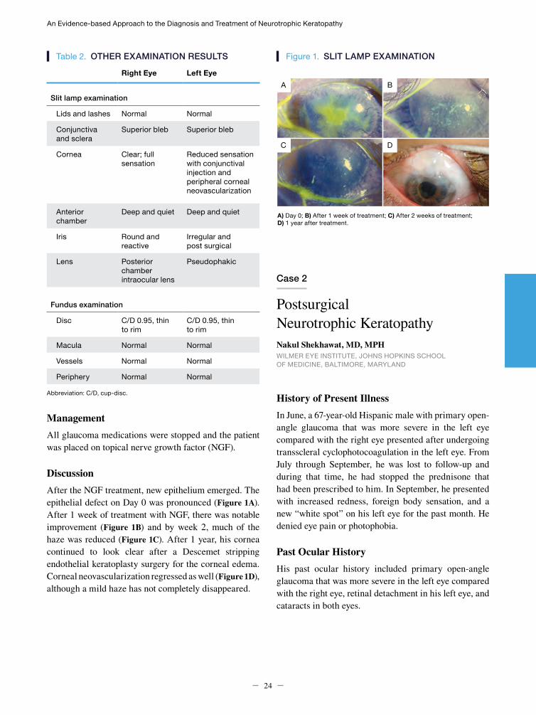
An Evidence-based Approach to the Diagnosis and Treatment of Neurotrophic Keratopathy
24
Table 2. OTHER EXAMINATION RESULTS
Right Eye Left Eye
Slit lamp examination
Lids and lashes Normal Normal
Conjunctiva and sclera
Superior bleb Superior bleb
Cornea Clear; full sensation
Reduced sensation with conjunctival injection and peripheral corneal neovascularization
Anterior chamber
Deep and quiet Deep and quiet
Iris Round and reactive
Irregular and post surgical
Lens Posterior chamber intraocular lens
Pseudophakic
Fundus examination
Disc C/D 0.95, thin to rim
C/D 0.95, thin to rim
Macula Normal Normal
Vessels Normal Normal
Periphery Normal Normal Abbreviation: C/D, cup-disc.
ManagementAll glaucoma medications were stopped and the patient was placed on topical nerve growth factor (NGF).
DiscussionAfter the NGF treatment, new epithelium emerged. The epithelial defect on Day 0 was pronounced (Figure 1A). After 1 week of treatment with NGF, there was notable improvement (Figure 1B) and by week 2, much of the haze was reduced (Figure 1C). After 1 year, his cornea continued to look clear after a Descemet stripping endothelial keratoplasty surgery for the corneal edema. Corneal neovascularization regressed as well (Figure 1D), although a mild haze has not completely disappeared.
A) Day 0; B) After 1 week of treatment; C) After 2 weeks of treatment; D) 1 year after treatment.
Figure 1. SLIT LAMP EXAMINATION
Case 2
Postsurgical Neurotrophic KeratopathyNakul Shekhawat, MD, MPHWILMER EYE INSTITUTE, JOHNS HOPKINS SCHOOL OF MEDICINE, BALTIMORE, MARYLAND
History of Present IllnessIn June, a 67-year-old Hispanic male with primary open-angle glaucoma that was more severe in the left eye compared with the right eye presented after undergoing transscleral cyclophotocoagulation in the left eye. From July through September, he was lost to follow-up and during that time, he had stopped the prednisone that had been prescribed to him. In September, he presented with increased redness, foreign body sensation, and a new “white spot” on his left eye for the past month. He denied eye pain or photophobia.
Past Ocular HistoryHis past ocular history included primary open-angle glaucoma that was more severe in the left eye compared with the right eye, retinal detachment in his left eye, and cataracts in both eyes.
A
C
B
D
Neurotrophic Keratopathy Presentation and Management: A Case Study Series
25
Past Surgical HistoryIn 2017, the patient had a trabeculectomy, cataract extraction, and an intraocular lens implant in his right eye.
In his left eye, he had a detached retina repaired with scleral buckling and a trabectome with explantation of the exposed scleral buckle in 2010. He also underwent transscleral laser cyclophotocoagulation in 2017 and 2019.
Current MedicationsHe was currently taking dorzolamide/timolol twice daily and latanoprost every night at bedtime in each eye.
Initial ExaminationHis initial examination results are in Table 3.
Table 3. INITIAL EXAMINATION RESULTS
Right Eye Left Eye
Visual acuity 20/25 Hand motions
Pupils 4-3 Dilated, nonreactive
Intraocular pressure
14 2
Extraocular muscles
Full Full
Confrontation visual field
Full Constricted in all 4 quadrants
Other ExaminationsSlit lamp examination showed a 1.2- x 1.2-mm central geographic epithelial defect with heaped edges and early infiltrate surrounding (Table 4). There was a surrounding area of mild anterior thinning to 10% remaining. Few radial Descemet’s membrane striae were extending to the central ulcer. There was no frank edema or keratic precipitates on fluorescein staining (Figure 2A-C).
ManagementSerology was not tested for in this patient because the review of systems did not reveal anything suspicious.
The main concern was neurotrophic keratopathy (NK) in the left eye. Moxifloxacin 0.5% (Vigamox; Alcon Laboratories, Inc.) every 4 hours while awake was started in the left eye and a bandage contact lens was
placed with topical antibiotics. By day 4, the epithelial defect was reduced (Figure 2D-F) and by day 13, it was healed with stable mild thinning (Figure 2G and 2H).
Table 4. OTHER EXAMINATION RESULTS
Right Eye Left Eye
Slit lamp examination
Lids and lashes Normal Normal
Conjunctiva and sclera
Superior bleb 2+ injection
Cornea Clear; full sensation
As shown; reduced sensation
Anterior chamber
Deep and quiet Deep and quiet
Iris Round and reactive
Dilated, nonreactive
Lens Posterior chamber intraocular lens
2+ nuclear sclerosis with phacodonesis
Fundus examination
Disc C/D 0.95, thin to rim
C/D 0.95, thin to rim
Macula Normal Normal
Vessels Normal Normal
Periphery Normal Normal Abbreviation: C/D, cup-disc.
Figure 2. SLIT LAMP EXAMINATION
A-C) At presentation; D-F) Day 4; G and H) Day 13.
A
B
C
D
E
F
G
H
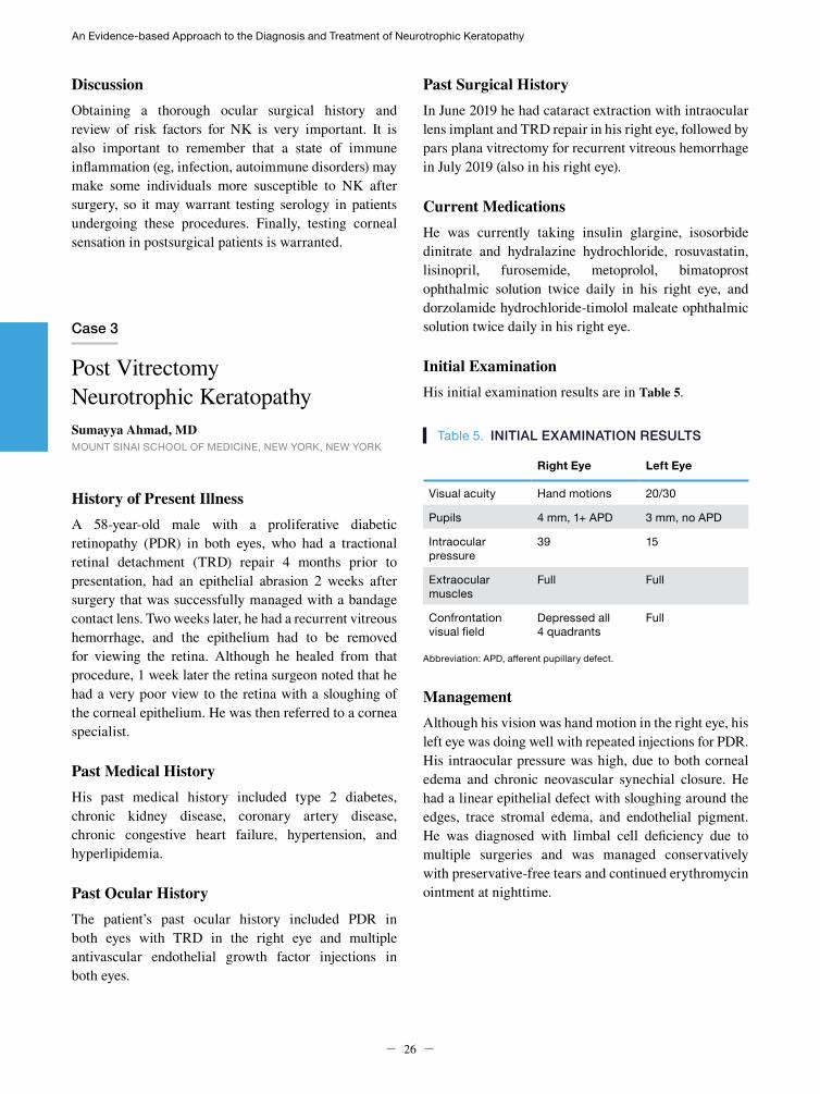
An Evidence-based Approach to the Diagnosis and Treatment of Neurotrophic Keratopathy
26
DiscussionObtaining a thorough ocular surgical history and review of risk factors for NK is very important. It is also important to remember that a state of immune inflammation (eg, infection, autoimmune disorders) may make some individuals more susceptible to NK after surgery, so it may warrant testing serology in patients undergoing these procedures. Finally, testing corneal sensation in postsurgical patients is warranted.
Case 3
Post Vitrectomy Neurotrophic KeratopathySumayya Ahmad, MDMOUNT SINAI SCHOOL OF MEDICINE, NEW YORK, NEW YORK
History of Present IllnessA 58-year-old male with a proliferative diabetic retinopathy (PDR) in both eyes, who had a tractional retinal detachment (TRD) repair 4 months prior to presentation, had an epithelial abrasion 2 weeks after surgery that was successfully managed with a bandage contact lens. Two weeks later, he had a recurrent vitreous hemorrhage, and the epithelium had to be removed for viewing the retina. Although he healed from that procedure, 1 week later the retina surgeon noted that he had a very poor view to the retina with a sloughing of the corneal epithelium. He was then referred to a cornea specialist.
Past Medical HistoryHis past medical history included type 2 diabetes, chronic kidney disease, coronary artery disease, chronic congestive heart failure, hypertension, and hyperlipidemia.
Past Ocular HistoryThe patient’s past ocular history included PDR in both eyes with TRD in the right eye and multiple antivascular endothelial growth factor injections in both eyes.
Past Surgical HistoryIn June 2019 he had cataract extraction with intraocular lens implant and TRD repair in his right eye, followed by pars plana vitrectomy for recurrent vitreous hemorrhage in July 2019 (also in his right eye).
Current MedicationsHe was currently taking insulin glargine, isosorbide dinitrate and hydralazine hydrochloride, rosuvastatin, lisinopril, furosemide, metoprolol, bimatoprost ophthalmic solution twice daily in his right eye, and dorzolamide hydrochloride-timolol maleate ophthalmic solution twice daily in his right eye.
Initial ExaminationHis initial examination results are in Table 5.
Table 5. INITIAL EXAMINATION RESULTS
Right Eye Left Eye
Visual acuity Hand motions 20/30
Pupils 4 mm, 1+ APD 3 mm, no APD
Intraocular pressure
39 15
Extraocular muscles
Full Full
Confrontation visual field
Depressed all 4 quadrants
Full
Abbreviation: APD, afferent pupillary defect.
ManagementAlthough his vision was hand motion in the right eye, his left eye was doing well with repeated injections for PDR. His intraocular pressure was high, due to both corneal edema and chronic neovascular synechial closure. He had a linear epithelial defect with sloughing around the edges, trace stromal edema, and endothelial pigment. He was diagnosed with limbal cell deficiency due to multiple surgeries and was managed conservatively with preservative-free tears and continued erythromycin ointment at nighttime.
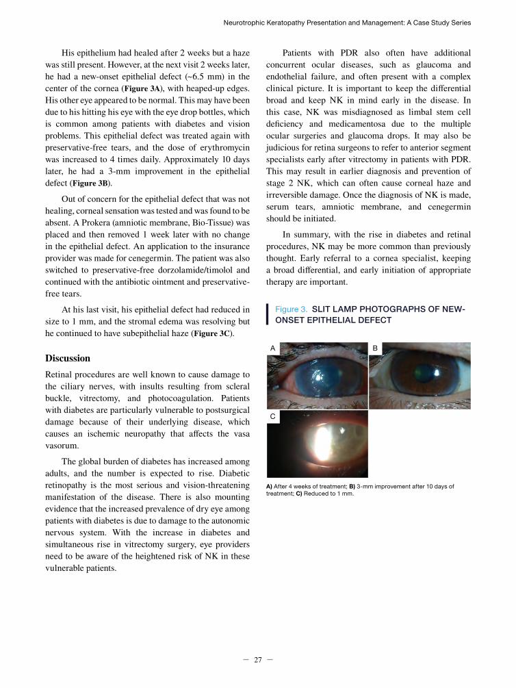
Neurotrophic Keratopathy Presentation and Management: A Case Study Series
27
His epithelium had healed after 2 weeks but a haze was still present. However, at the next visit 2 weeks later, he had a new-onset epithelial defect (~6.5 mm) in the center of the cornea (Figure 3A), with heaped-up edges. His other eye appeared to be normal. This may have been due to his hitting his eye with the eye drop bottles, which is common among patients with diabetes and vision problems. This epithelial defect was treated again with preservative-free tears, and the dose of erythromycin was increased to 4 times daily. Approximately 10 days later, he had a 3-mm improvement in the epithelial defect (Figure 3B).
Out of concern for the epithelial defect that was not healing, corneal sensation was tested and was found to be absent. A Prokera (amniotic membrane, Bio-Tissue) was placed and then removed 1 week later with no change in the epithelial defect. An application to the insurance provider was made for cenegermin. The patient was also switched to preservative-free dorzolamide/timolol and continued with the antibiotic ointment and preservative-free tears.
At his last visit, his epithelial defect had reduced in size to 1 mm, and the stromal edema was resolving but he continued to have subepithelial haze (Figure 3C).
DiscussionRetinal procedures are well known to cause damage to the ciliary nerves, with insults resulting from scleral buckle, vitrectomy, and photocoagulation. Patients with diabetes are particularly vulnerable to postsurgical damage because of their underlying disease, which causes an ischemic neuropathy that affects the vasa vasorum.
The global burden of diabetes has increased among adults, and the number is expected to rise. Diabetic retinopathy is the most serious and vision-threatening manifestation of the disease. There is also mounting evidence that the increased prevalence of dry eye among patients with diabetes is due to damage to the autonomic nervous system. With the increase in diabetes and simultaneous rise in vitrectomy surgery, eye providers need to be aware of the heightened risk of NK in these vulnerable patients.
Patients with PDR also often have additional concurrent ocular diseases, such as glaucoma and endothelial failure, and often present with a complex clinical picture. It is important to keep the differential broad and keep NK in mind early in the disease. In this case, NK was misdiagnosed as limbal stem cell deficiency and medicamentosa due to the multiple ocular surgeries and glaucoma drops. It may also be judicious for retina surgeons to refer to anterior segment specialists early after vitrectomy in patients with PDR. This may result in earlier diagnosis and prevention of stage 2 NK, which can often cause corneal haze and irreversible damage. Once the diagnosis of NK is made, serum tears, amniotic membrane, and cenegermin should be initiated.
In summary, with the rise in diabetes and retinal procedures, NK may be more common than previously thought. Early referral to a cornea specialist, keeping a broad differential, and early initiation of appropriate therapy are important.
Figure 3. SLIT LAMP PHOTOGRAPHS OF NEW-ONSET EPITHELIAL DEFECT
A) After 4 weeks of treatment; B) 3-mm improvement after 10 days of treatment; C) Reduced to 1 mm.
A B
C
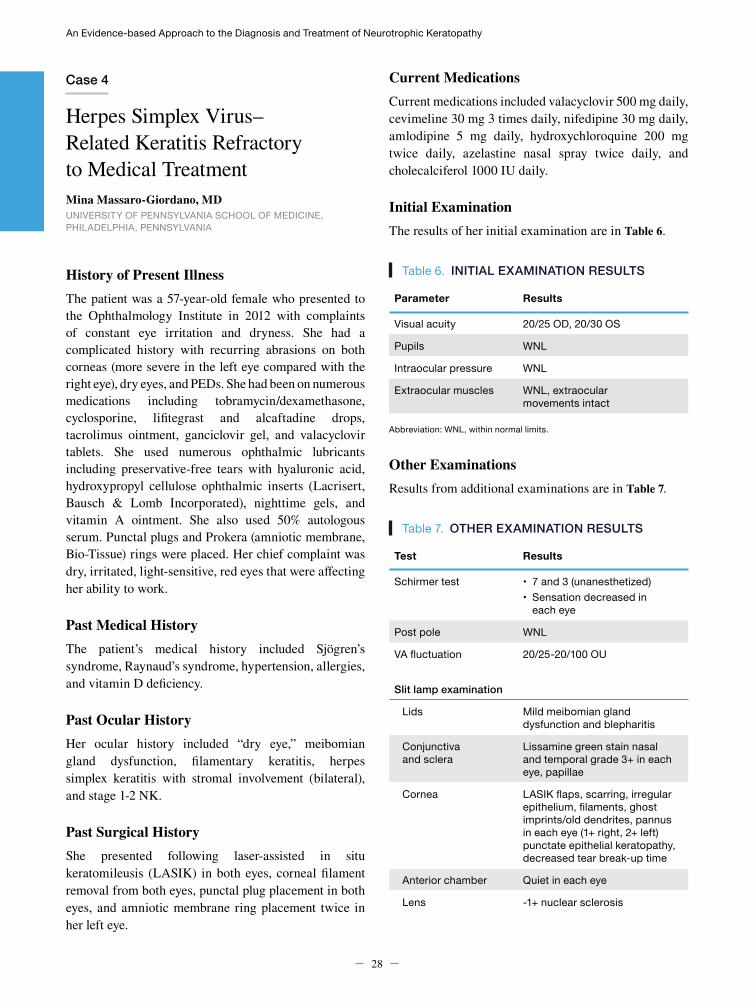
An Evidence-based Approach to the Diagnosis and Treatment of Neurotrophic Keratopathy
28
Case 4
Herpes Simplex Virus–Related Keratitis Refractory to Medical TreatmentMina Massaro-Giordano, MDUNIVERSITY OF PENNSYLVANIA SCHOOL OF MEDICINE, PHILADELPHIA, PENNSYLVANIA
History of Present IllnessThe patient was a 57-year-old female who presented to the Ophthalmology Institute in 2012 with complaints of constant eye irritation and dryness. She had a complicated history with recurring abrasions on both corneas (more severe in the left eye compared with the right eye), dry eyes, and PEDs. She had been on numerous medications including tobramycin/dexamethasone, cyclosporine, lifitegrast and alcaftadine drops, tacrolimus ointment, ganciclovir gel, and valacyclovir tablets. She used numerous ophthalmic lubricants including preservative-free tears with hyaluronic acid, hydroxypropyl cellulose ophthalmic inserts (Lacrisert, Bausch & Lomb Incorporated), nighttime gels, and vitamin A ointment. She also used 50% autologous serum. Punctal plugs and Prokera (amniotic membrane, Bio-Tissue) rings were placed. Her chief complaint was dry, irritated, light-sensitive, red eyes that were affecting her ability to work.
Past Medical HistoryThe patient’s medical history included Sjögren’s syndrome, Raynaud’s syndrome, hypertension, allergies, and vitamin D deficiency.
Past Ocular HistoryHer ocular history included “dry eye,” meibomian gland dysfunction, filamentary keratitis, herpes simplex keratitis with stromal involvement (bilateral), and stage 1-2 NK.
Past Surgical HistoryShe presented following laser-assisted in situ keratomileusis (LASIK) in both eyes, corneal filament removal from both eyes, punctal plug placement in both eyes, and amniotic membrane ring placement twice in her left eye.
Current MedicationsCurrent medications included valacyclovir 500 mg daily, cevimeline 30 mg 3 times daily, nifedipine 30 mg daily, amlodipine 5 mg daily, hydroxychloroquine 200 mg twice daily, azelastine nasal spray twice daily, and cholecalciferol 1000 IU daily.
Initial ExaminationThe results of her initial examination are in Table 6.
Table 6. INITIAL EXAMINATION RESULTS
Parameter Results
Visual acuity 20/25 OD, 20/30 OS
Pupils WNL
Intraocular pressure WNL
Extraocular muscles WNL, extraocular movements intact
Abbreviation: WNL, within normal limits.
Other ExaminationsResults from additional examinations are in Table 7.
Table 7. OTHER EXAMINATION RESULTS
Test Results
Schirmer test • 7 and 3 (unanesthetized)• Sensation decreased in
each eye
Post pole WNL
VA fluctuation 20/25-20/100 OU
Slit lamp examination
Lids Mild meibomian gland dysfunction and blepharitis
Conjunctiva and sclera
Lissamine green stain nasal and temporal grade 3+ in each eye, papillae
Cornea LASIK flaps, scarring, irregular epithelium, filaments, ghost imprints/old dendrites, pannus in each eye (1+ right, 2+ left) punctate epithelial keratopathy, decreased tear break-up time
Anterior chamber Quiet in each eye
Lens -1+ nuclear sclerosis
Neurotrophic Keratopathy Presentation and Management: A Case Study Series
29
Table 7. CONTINUED
Test Results
Ancillary testing
Cochet-Bonnet 3-5 mm
Meibography Grade 1 drop out
Matrix metallopeptidase 9
Positive, few visits
Osmolarity ranges 301-320
Confocal Diminished nerves in subbasal plexus with few dendritic cells
Abbreviations: LASIK, laser-assisted in situ keratomileusis; WNL, within normal limits.
ManagementThis patient was very symptomatic with surface irritation and blurry vision preventing her from working. She had a long and complicated management course. Many treatment options were tried as previously mentioned. She continued to have PEDs (Figure 4A and 4B) and abnormal confocal findings (Figure 4C). She also participated in the NGF 014 trial (she received placebo). In 2016 she was fitted with mini scleral lenses, which she removed each night, and after which her vision improved, as did her ocular surface. There was no lissamine green stain in the area of the conjunctiva and cornea that was protected by the lens (Figure 4D). Many lenses were tried to find the correct fit. She did best with the Zenlens Oblate Z16 5 (Alden Optical). There are numerous papers in the literature to support the use of various types of contact lenses (prosthetic replacement of the ocular surface ecosystem, scleral, mini scleral, gas permeable, and soft) to help with the treatment of NK. Her cause for NK may have been multifactorial (Sjögren’s, dry eye, prior LASIK surgery, and herpes simplex virus). Although many different treatments could have contributed to her improvement, mini scleral lenses are less expensive than the traditional larger scleral lenses and may be easier to fit, and thus can be a valid option in some patients.
A) With irregular epithelium; B) With central fluorescein staining; C) Confocal of subbasal plexus with diminished corneal nerves and few dendritic cells; D) Scleral lens protecting the ocular surface.
DiscussionContact lenses can actually help protect and heal the ocular surface. There are many options for contact lenses and it is important that the fit is correct, which may take numerous efforts. Contact lenses can and should be used with aggressive medical treatments.
Figure 4. 54-YEAR-OLD FEMALE WITH PERSISTENT EPITHELIAL DEFECT
Case 5
Stage 1 Neurotrophic KeratopathyPedram Hamrah, MD, FRCS, FARVOTUFTS UNIVERSITY SCHOOL OF MEDICINE, BOSTON, MASSACHUSETTS
History of Present IllnessA 60-year-old female with decreased vision and pain in her left eye over the past few months was referred to a specialist after having been seen by a community ophthalmologist for 6 months.
A
C
B
D

An Evidence-based Approach to the Diagnosis and Treatment of Neurotrophic Keratopathy
30
Past Ocular HistoryHer ocular history included keratoconjunctivitis sicca in both eyes and herpes simplex keratitis and neurotrophic ulcer in the left eye.
Past Surgical HistoryShe had not undergone any surgical procedures.
Past Medication History Previous medications included artificial tears, lubricating ointment, bandage contact lens, topical steroids, topical cyclosporine, Prokera (amniotic membrane, Bio-Tissue) transplant twice in her left eye, autologous serum tears 20% 4 times daily, and ocular patching.
Initial ExaminationHer initial examination results are listed in Table 8. Her best corrected visual acuity was 20/20 OD and 20/300 OS. She rated her pain on the visual analog scale as 9 out of 10 in her left eye.
Table 8. INITIAL EXAMINATION RESULTS
Right Eye Left Eye
Visual acuity 20/20 20/300
Pupils 4-3 4-3
Intraocular pressure
15 15
Extraocular muscles
Full Full
Confrontation visual field
Full Full
Concomitant MedicationsShe was currently taking gabapentin, low-dose naltrexone, and once-daily loteprednol 0.5%.
Other ExaminationsSlit lamp examination showed moderate superficial punctate keratitis (SPK) in the right eye with 2+ central and interpalpebral SPK and severe 4+ diffuse SPK throughout the cornea in the left eye (Table 9). There was no frank epithelial defect present (Figure 5A).
Table 9. OTHER EXAMINATION RESULTS
Test Right Eye Left Eye
Slit lamp examination
Lids and lashes 2+ MGD 2+ MGD
Conjunctiva and sclera
Trace injection Trace injection
Cornea 2+ SPK; 4/6 sensation
4+ SPK; 0/6 sensation
Anterior chamber
Deep and quiet Deep and quiet
Iris Round and reactive
Dilated, nonreactive
Lens 1+ NS 1+ NS
Fundus examination
Disc C/D 0.3 C/D 0.3
Macula Normal Normal
Vessels Normal Normal
Periphery Normal Normal Abbreviations: C/D, cup-disc; MGD, meibomian gland dysfunction; NS, nuclear sclerosis; SPK, superficial punctate keratitis.
ManagementThe patient was started on low-dose naltrexone and gabapentin for pain management, as well as cenegermin 6 times/day. Her serum concentration was increased from 20% to 50% and steroids were tapered from 4 times daily. After 1 week, there was no improvement in corneal sensitivity or structure. However, the pain was reduced to 2 (from 9). After 8 weeks of treatment, 80% of the corneal staining had disappeared (Figure 5A-D). At that time, the cenegermin was stopped and serum tears were switched back to 20%, 8 times daily. Her vision had improved to 20/30 OS.
DiscussionThis is a case of severe stage 1 NK that was unresponsive to treatments prior to cenegermin. It is interesting to note that this patient had no corneal sensation yet very high pain. In fact, she likely had comorbid neuropathic pain (post-herpetic neuralgia) and NK.
This patient was treated with low-dose naltrexone and gabapentin for the pain, but other options could include nortriptyline, carbamazepine, or oxcarbazepine.
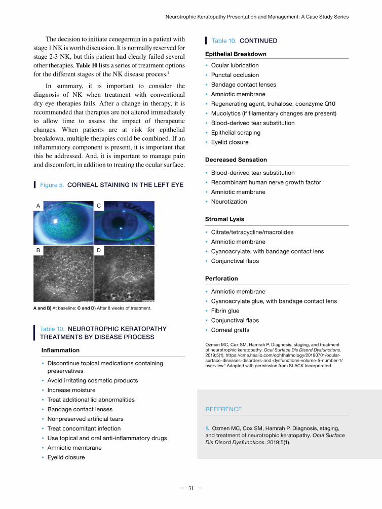
Neurotrophic Keratopathy Presentation and Management: A Case Study Series
31
REFERENCE
Ozmen MC, Cox SM, Hamrah P. Diagnosis, staging, and treatment of neurotrophic keratopathy. Ocul Surface Dis Disord Dysfunctions. 2019;5(1). https://cme.healio.com/ophthalmology/20190701/ocular-surface-diseases-disorders-and-dysfunctions-volume-5-number-1/overview.1 Adapted with permission from SLACK Incorporated.
1. Ozmen MC, Cox SM, Hamrah P. Diagnosis, staging, and treatment of neurotrophic keratopathy. Ocul Surface Dis Disord Dysfunctions. 2019;5(1).
The decision to initiate cenegermin in a patient with stage 1 NK is worth discussion. It is normally reserved for stage 2-3 NK, but this patient had clearly failed several other therapies. Table 10 lists a series of treatment options for the different stages of the NK disease process.1
In summary, it is important to consider the diagnosis of NK when treatment with conventional dry eye therapies fails. After a change in therapy, it is recommended that therapies are not altered immediately to allow time to assess the impact of therapeutic changes. When patients are at risk for epithelial breakdown, multiple therapies could be combined. If an inflammatory component is present, it is important that this be addressed. And, it is important to manage pain and discomfort, in addition to treating the ocular surface.
Figure 5. CORNEAL STAINING IN THE LEFT EYE
Table 10. NEUROTROPHIC KERATOPATHY TREATMENTS BY DISEASE PROCESS
Inflammation
• Discontinue topical medications containing preservatives
• Avoid irritating cosmetic products• Increase moisture• Treat additional lid abnormalities• Bandage contact lenses• Nonpreserved artificial tears• Treat concomitant infection• Use topical and oral anti-inflammatory drugs• Amniotic membrane• Eyelid closure
Table 10. CONTINUED Epithelial Breakdown
• Ocular lubrication• Punctal occlusion• Bandage contact lenses• Amniotic membrane• Regenerating agent, trehalose, coenzyme Q10• Mucolytics (if filamentary changes are present)• Blood-derived tear substitution• Epithelial scraping• Eyelid closure
Decreased Sensation
• Blood-derived tear substitution• Recombinant human nerve growth factor• Amniotic membrane• Neurotization
Stromal Lysis
• Citrate/tetracycline/macrolides• Amniotic membrane• Cyanoacrylate, with bandage contact lens• Conjunctival flaps
Perforation
• Amniotic membrane• Cyanoacrylate glue, with bandage contact lens• Fibrin glue• Conjunctival flaps• Corneal grafts
A and B) At baseline; C and D) After 8 weeks of treatment.
A
B
C
D

32
Notes

Prepared by
Catalyst Medical Education, LLC for Johns Hopkins School of Medicine.
Related Documents