An Electrodiagnostic Approach to the Evaluation of Peripheral Neuropathies Mark B. Bromberg, MD, PhD INTRODUCTION In the clinical setting of a suspected peripheral neuropathy, the history and neurologic examination provide information about general features but cannot define the nature of the pathologic changes and the clinical features may underrepresent the distribu- tion of nerve involvement or time course. Electrodiagnostic tests allow for a more detailed characterization of a neuropathy. 1 Electrodiagnostic tests include nerve conduction and needle electromyographic (EMG) studies. Reliable interpretation of the data assumes recognition of potential technical issues and overall must be within the context of the clinical information. 2,3 A systematic electrodiagnostic approach is based on an understanding of basic nerve anatomy and physiology and how patho- logic changes affect electrodiagnostic data, and this foundation will be reviewed. Electrodiagnostic findings in prototypic axonal and demyelinating neuropathies are presented. These principles are put together as a diagnostic strategy to aid in planning informative tests and interpreting outside studies. Disclosure: The author has no financial interest in any company or product that is related to the content of this article. Department of Neurology, University of Utah, 175 North Medical Drive, Salt Lake City, UT 84132, USA E-mail address: [email protected] KEYWORDS Peripheral neuropathy Diagnosis of peripheral neuropathies Nerve conduction studies EMG studies KEY POINTS A planned approach to electrodiagnostic testing provides the most information. Electrodiagnostic testing is highly sensitive for defining the pattern and degree of nerve involvement. Electrodiagnosis can provide information on the chronicity of a neuropathy. Electrodiagnostic testing can provide insight into the underlying pathophysiology, defining the neuropathy as either primary axonal or primary demyelinating. Phys Med Rehabil Clin N Am 24 (2013) 153–168 http://dx.doi.org/10.1016/j.pmr.2012.08.020 pmr.theclinics.com 1047-9651/13/$ – see front matter Ó 2013 Published by Elsevier Inc.

Welcome message from author
This document is posted to help you gain knowledge. Please leave a comment to let me know what you think about it! Share it to your friends and learn new things together.
Transcript

An Electrodiagnostic Approach tothe Evaluation of PeripheralNeuropathies
Mark B. Bromberg, MD, PhD
KEYWORDS
� Peripheral neuropathy � Diagnosis of peripheral neuropathies� Nerve conduction studies � EMG studies
KEY POINTS
� A planned approach to electrodiagnostic testing provides the most information.
� Electrodiagnostic testing is highly sensitive for defining the pattern and degree of nerveinvolvement.
� Electrodiagnosis can provide information on the chronicity of a neuropathy.
� Electrodiagnostic testing can provide insight into the underlying pathophysiology, definingthe neuropathy as either primary axonal or primary demyelinating.
INTRODUCTION
In the clinical setting of a suspected peripheral neuropathy, the history and neurologicexamination provide information about general features but cannot define the natureof the pathologic changes and the clinical features may underrepresent the distribu-tion of nerve involvement or time course. Electrodiagnostic tests allow for a moredetailed characterization of a neuropathy.1 Electrodiagnostic tests include nerveconduction and needle electromyographic (EMG) studies. Reliable interpretation ofthe data assumes recognition of potential technical issues and overall must be withinthe context of the clinical information.2,3 A systematic electrodiagnostic approach isbased on an understanding of basic nerve anatomy and physiology and how patho-logic changes affect electrodiagnostic data, and this foundation will be reviewed.Electrodiagnostic findings in prototypic axonal and demyelinating neuropathies arepresented. These principles are put together as a diagnostic strategy to aid in planninginformative tests and interpreting outside studies.
Disclosure: The author has no financial interest in any company or product that is related to thecontent of this article.Department of Neurology, University of Utah, 175 North Medical Drive, Salt Lake City, UT84132, USAE-mail address: [email protected]
Phys Med Rehabil Clin N Am 24 (2013) 153–168http://dx.doi.org/10.1016/j.pmr.2012.08.020 pmr.theclinics.com1047-9651/13/$ – see front matter � 2013 Published by Elsevier Inc.
Bromberg154
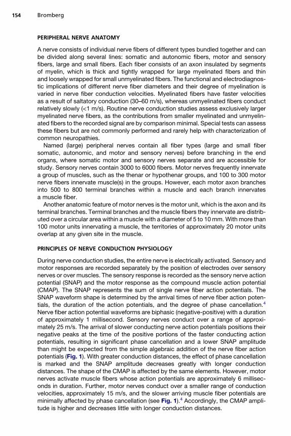
PERIPHERAL NERVE ANATOMY
A nerve consists of individual nerve fibers of different types bundled together and canbe divided along several lines: somatic and autonomic fibers, motor and sensoryfibers, large and small fibers. Each fiber consists of an axon insulated by segmentsof myelin, which is thick and tightly wrapped for large myelinated fibers and thinand loosely wrapped for small unmyelinated fibers. The functional and electrodiagnos-tic implications of different nerve fiber diameters and their degree of myelination isvaried in nerve fiber conduction velocities. Myelinated fibers have faster velocitiesas a result of saltatory conduction (30–60 m/s), whereas unmyelinated fibers conductrelatively slowly (<1 m/s). Routine nerve conduction studies assess exclusively largermyelinated nerve fibers, as the contributions from smaller myelinated and unmyelin-ated fibers to the recorded signal are by comparison minimal. Special tests can assessthese fibers but are not commonly performed and rarely help with characterization ofcommon neuropathies.Named (large) peripheral nerves contain all fiber types (large and small fiber
somatic, autonomic, and motor and sensory nerves) before branching in the endorgans, where somatic motor and sensory nerves separate and are accessible forstudy. Sensory nerves contain 3000 to 6000 fibers. Motor nerves frequently innervatea group of muscles, such as the thenar or hypothenar groups, and 100 to 300 motornerve fibers innervate muscle(s) in the groups. However, each motor axon branchesinto 500 to 800 terminal branches within a muscle and each branch innervatesa muscle fiber.Another anatomic feature of motor nerves is the motor unit, which is the axon and its
terminal branches. Terminal branches and the muscle fibers they innervate are distrib-uted over a circular area within a muscle with a diameter of 5 to 10mm.With more than100 motor units innervating a muscle, the territories of approximately 20 motor unitsoverlap at any given site in the muscle.
PRINCIPLES OF NERVE CONDUCTION PHYSIOLOGY
During nerve conduction studies, the entire nerve is electrically activated. Sensory andmotor responses are recorded separately by the position of electrodes over sensorynerves or over muscles. The sensory response is recorded as the sensory nerve actionpotential (SNAP) and the motor response as the compound muscle action potential(CMAP). The SNAP represents the sum of single nerve fiber action potentials. TheSNAP waveform shape is determined by the arrival times of nerve fiber action poten-tials, the duration of the action potentials, and the degree of phase cancellation.4
Nerve fiber action potential waveforms are biphasic (negative-positive) with a durationof approximately 1 millisecond. Sensory nerves conduct over a range of approxi-mately 25 m/s. The arrival of slower conducting nerve action potentials positions theirnegative peaks at the time of the positive portions of the faster conducting actionpotentials, resulting in significant phase cancellation and a lower SNAP amplitudethan might be expected from the simple algebraic addition of the nerve fiber actionpotentials (Fig. 1). With greater conduction distances, the effect of phase cancellationis marked and the SNAP amplitude decreases greatly with longer conductiondistances. The shape of the CMAP is affected by the same elements. However, motornerves activate muscle fibers whose action potentials are approximately 6 millisec-onds in duration. Further, motor nerves conduct over a smaller range of conductionvelocities, approximately 15 m/s, and the slower arriving muscle fiber potentials areminimally affected by phase cancellation (see Fig. 1).4 Accordingly, the CMAP ampli-tude is higher and decreases little with longer conduction distances.

Fig. 1. Effects of normal temporal dispersion on the SNAP and the CMAP. Left, Actual wave-forms from ulnar nerve study (stimulating at the wrist, A1, and up the arm to Erb point, A5)are shown for sensory (A) and motor (B) nerve conduction. Right, Model of summation ofaction potentials. Sensory nerve action potentials have an approximately 1-millisecond-duration waveform and conduct over an approximately 25 m/s range, leading to markedphase cancellation and relative low amplitude when nerve fiber waveforms are summedin the SNAP. Muscle fiber action potentials have an approximately 6-millisecond-durationwaveform and conduct over an approximately 15 m/s range, leading to much less phasecancellation and a relative high amplitude when muscle fiber waveforms are summed inthe CMAP.
Evaluation of Peripheral Neuropathies 155
Another difference between sensory nerve fibers and muscle fibers is their diame-ters: sensory nerve fibers that contribute to the SNAP have diameters of 8 to 12 mm,whereas muscle fibers that contribute to the CMAP have diameters of 30 to 70 mm.The amplitudes of individual action potentials are proportional to the diameter of the

Bromberg156
fiber; SNAP amplitude (normal range) is 6 to 90 mV, whereas CMAP amplitude (normalrange) is 2000 to 15,000 mV, or 2 to 15 mV.Nerve stimulation is accomplished by passing current between anode and
cathode electrodes (which are 3–4 cm apart) on the skin over the nerve. Currentflows from the anode to the cathode with hyperpolarized beneath the anode anddepolarization under the cathode (the site of impulse initiation). With sufficientcurrent, all axons are activated to produce a maximal SNAP or CMAP response.The cathode must be placed closest to the recording site, for if the anode is placedclosest, 2 situations occur. Because the distance between the electrodes is 3 to4 cm, the intended measured distance between recording and stimulating elec-trodes will be approximately 3 to 4 cm greater, leading to artificially longer distallatencies or slowed conduction velocities. The other is the phenomenon of anodalblock that can cause partial conduction block along the nerve caused by hyperpo-larization of some axons under the anode, leading to artificially lower SNAP andCMAP amplitudes.Nerve conduction studies yield 2 types of metrics: the amplitude of the evoked
response and a set of timing measures (Fig. 2). SNAP and CMAP amplitudes areroughly proportional to the number of axons conducting between the stimulatingand recording electrodes. With progressive axonal loss, both responses lose ampli-tude but the CMAP amplitude drops less than the SNAP amplitude because of theeffects of collateral reinnervation of motor nerves (to be discussed later). Timingmeasures generally reflect activity of the fastest myelinated fibers and include distallatency (conduction over set distances), F-wave latency (conduction over twice thewhole length of motor nerves), and conduction velocity (over segments of nerve).The duration of the CMAP waveform (negative peak duration) provides an estimateof the spectrum of more slowly conducting myelinated fibers.Nerve fiber conduction velocity is slowed at lower temperatures in a linear manner.
However, the effects of temperature are more apparent with sensory than with motornerves. With lower nerve temperatures, sensory nerve action potentials are longer induration, resulting in less phase cancellation and larger SNAP amplitudes. Althoughmuscle fiber action potentials also lengthen in duration, the effect on CMAP amplitudeis negligible. For both, the lower nerve temperature slows conduction, approximately2 m/s/�C.5 Correction factors can be applied when limb temperature is low, but thefactors are approximate and diagnostic interpretation uncertainties can be avoidedby warming limbs to approximately 31�C.
Fig. 2. CMAP waveform metrics, applicable primarily to the CMAP.

Evaluation of Peripheral Neuropathies 157
PRINCIPLES OF NEEDLE EMG
Needle EMG records electrical activity from muscle fibers to assess the integrity andarchitecture or arrangement within the muscle.6 The electrical motor unit recorded bythe EMG needle is called the motor unit action potential (MUAP) and represents onlya portion of the anatomic motor unit, because the electrical uptake area of the elec-trode is less than 1 to 2 mm in diameter. Thus, the MUAP includes only about 7 to15 fibers of an anatomic motor unit, for both concentric and monopolar electrodes.The MUAP waveform varies with slight needle movement based on the intimate prox-imity of the needle tip to 1 to 3 muscle fibers of the anatomic motor unit that contributemost to the waveform amplitude and shape.The EMG study is performed in 2 stages. The first stage is assessment for the pres-
ence of abnormal spontaneous muscle fiber activity when the muscle is at rest, andthe second is assessment of MUAPs during voluntary activation of the muscle. Thepresence of abnormal spontaneous activity is assessed at high EMG screen displaysensitivity or gain, 20 to 50 mV per division. The presence of positive waves and fibril-lation potentials indicates denervation and, in the context of a peripheral neuropathy,denervation from axonal loss (neurogenic denervation). Note that the waveform differ-ences between positive waves and fibrillation potentials depend on the relationshipbetween the electrode tip and the spontaneously active muscle fiber and thus areof equal clinical significance.7 The presence of very low amplitude abnormal sponta-neous activity, less than approximately 50 mV, suggests very long-standing and slowlyprogressive denervation.There are about overlapping 20 motor units in the electrode’s uptake area, and
during voluntary muscle activation the number of active motor units increases fromzero at rest toward the 20 with increasing effort. Of note, most routine EMG studiesare performed at low levels of activation so that individual MUAPs can be observed.Accordingly, during low levels of activation, the electrode records from 3 to 5 MUAPsclose to the needle. At low levels of muscle activation, the recruitment pattern ofMUAPs can be assessed for MUAP discharge frequencies.8 The EMG screen displaysensitivity or gain is set at 200 mV per division and sweep speed at 10 milliseconds perdivision. If the screen has 10 horizontal divisions, the beam sweep time will be 10/s:thus, it is rare for a single MUAP to consistently discharge at 20 Hz (twice/sweep),and observation of several MUAPs discharging at rates greater than 20 Hz supportmotor unit loss. MUAP waveforms can be inspected for increased amplitude by deter-mining if they exceed 2000 to 3000 mV, an approximate upper limit of normal for distalmuscles. MUAP complexity in terms of polyphasia (>4 phases) or polyturn (>5 turns)can be assessed by a trigger and delay line, which permits individual MUAPs to beelectronically captured and spread out.8 It is to be noted that up to 10% of motor unitscan be polyphasic and excessive emphasis should not be placed on occasionalcomplex motor units.The goal of these observations is to estimate pathophysiologic changes. In the
context of a neuropathy, abnormal spontaneous activity supports denervation ofmuscles. The remaining motor nerve fibers sprout collateral branches to reinnervateorphaned muscle fibers and the anatomic motor increases in muscle fiber densitybut not in area. Reinnervated muscle fibers no longer discharge spontaneously, butabnormal spontaneous activity usually persists for 2 reasons. There is subsequentongoing denervation from the neuropathy and the degree of collateral reinnervationis limited, all of which leads to permanently orphaned (denervated) muscle fibers.With reinnervation, the increased density of the anatomic motor unit is reflected inlarger and more complex MUAPs. In neuropathies that are progressing relatively

Bromberg158
rapidly, there will be ongoing denervation, marked by abnormal spontaneous activity,mildly decreased recruitment, and complex MUAPs. In very slowly progressiveneuropathies, there is sufficient time for maximal reinnervation; positive waves andfibrillation potentials from permanently orphaned muscle fibers will be of very lowamplitude; and the motor units will be few in number and of very high amplitude,but not complex, because there has been sufficient time for polyphasia and polyturnsto simplify. Thus, when MUAPs are observed whose amplitudes are 5 to 10 timesnormal (10,000–20,000 mV), consideration should be given for a hereditary neurop-athy, the most slowly developing form of neuropathy. The extent of denervation(distal-proximal gradient) can be assessed by identifying the most proximal musclewith abnormal needle EMG findings.
LIMITS OF NORMAL
As with most biologic data, “normal limits” are derived from distributions of valuesfrom subjects who have no apparent disease. The pool of subjects should includea wide spectrum of ages and body sizes (height). How limits of normal are set iscontroversial, but most laboratories use upper and lower limits (ULN of distal latencyand F-wave latency, LLN for amplitude and conduction velocity) set at 2 to 3 SDs, butdata for the different nerve conduction metrics may not be normally distributed andother limits such as confidence intervals may be more appropriate.9 Although it is pref-erable for each laboratory to gather its own normal data, this is rarely done, and mostlimits are handed down. An obscure origin of a laboratory’s normal values seemsunscientific, but values are generally similar. Some metrics are influenced by bodyhabitus and, although difficult to take into account precisely, should be part of thegeneral considerations. Thin fingers in women can result in very high amplitude digitalSNAP amplitudes, and vice versa for thick fingers. Conduction velocities can bereduced and F-wave latencies in particular can be long in very tall individuals, andvice versa in very short patients. Thus, ULN and LLN should be viewed as referencesand not absolute values and should be interpreted in the overall clinical context. Nerveconduction metrics in the elderly are especially challenging, and although good suralresponses can be obtained in the very elderly, absent responses may not beabnormal.10
ELECTRODIAGNOSTIC MANIFESTATIONS OF NEUROPATHY
Nerve conduction tests help in distinguishing 3 basic conditions of peripheral nerves.The first state is normal conduction, seen whenmost nerve fibers and axons are intact.The second situation is axonal injury, seen when the primary injury occurs to axons.The third case is loss of myelin, seen with demyelination, which generally occurs atmultiple focal sites along a nerve. This causes variation in nerve action potential prop-agation resulting in slowing of conduction velocities or slowing to zero (conductionblock). Slowed conduction velocity causes abnormal dispersion of arrival times ofnerve action potentials at the recording electrode (abnormal temporal dispersion).Of note, there can be mixed patterns with primary demyelination and secondaryaxonal loss. Finally, theremay be slowed conduction from reversible metabolic causeswith no obvious damage to myelin.
Normal Conduction
SNAP and CMAP waveforms are influenced by normal temporal dispersion: SNAPamplitude decreases markedly over greater distances, whereas CMAP amplitudedecreases minimally and the CMAP negative peak duration increases only minimally

Evaluation of Peripheral Neuropathies 159
(see Fig. 1; Fig. 3). Normal conduction is defined as values within the laboratory limitsof normal. Distal latency, F-wave latency and conduction velocity are influenced bylimb length and limb temperature.
Axonal Injury
Loss of axons disconnects fibers from their receptors (sensory nerves) or muscles(motor nerves). Not all fibers may be affected, and the remaining unaffected fibersconduct normally, or all fibers may be affected in severe neuropathies. Generally,axonal loss occurs at the distal ends of fibers, a process called “axonal dying-back.” This results in reductions in both SNAP and CMAP amplitudes. The SNAPamplitude is especially sensitive to axon loss because of the lack of compensatorycollateral reinnervation. Essentially, there is a linear relationship between the numberof axons lost and the amplitude of the SNAP, and with loss of greater than 50% ofaxons, the amplitude may be unrecordable. The CMAP amplitude, in contrast, isless sensitive to early axonal injury because of the supportive effects of collateral rein-nervation. With mild denervation, collateral reinnervation preserves the number ofinnervated muscle fibers and the CMAP amplitude remains high, but with either furtheraxonal loss or a very rapid rate of loss, greater numbers of muscle fibers remain dener-vated and the CMAP amplitude decreases. The effects of collateral sprouting can, insome cases, maintain CMAP amplitude at greater than the LLN until greater than 80%
Fig. 3. Stylized comparisons between motor nerve conduction findings in normal andprimarily axon and primarily demyelinating neuropathies. Differences in CMAP betweenwaveforms are noted when stimulating at distal and proximal sites (P:D amplitude ratio).Normal: a short distal latency and short latency from proximal stimulation site yield a normalconduction velocity. Normal temporal dispersion results in a very small reduction in CMAPamplitude to proximal stimulation (normal ratio). Primary axonal loss: the distal latencyand latency from a proximal stimulation site are only slightly longer than normal becauseof the loss of rapidly conducting fibers with a normal P:D amplitude ratio. Primary demye-lination: the distal latency is prolonged and the conduction velocity is slowed. The P:Damplitude ratio is low because of phase cancellation.
Bromberg160
of axons are lost. When the CMAP is within normal limits, the needle EMG can detectthe effects of motor unit enlargement and verify that axonal loss has occurred.With axonal loss, each remaining nerve fiber conducts at its innate speed. Thus,
measures of timing (distal latency, F-wave latency, and conduction velocity) arereduced only to the extent of loss of large axons (see Fig. 3). The limits of slowingin axonal neuropathy can be empirically assessed by reviewing data from patientswith amyotrophic lateral sclerosis (ALS), a disorder characterized by reduced numbersof axons with no predilection to axon size: distal latency and F-wave latency are rarelylonger than 125% of the ULN, and conduction velocity is rarely slower than 75% of theLLN.11 Temporal dispersion, as measured by negative CMAP peak duration, is largelyunaffected in axonal neuropathies. Overall, with moderate to major axonal loss, theSNAP response will be absent and thus provide no information about conductionvelocity.
Demyelination
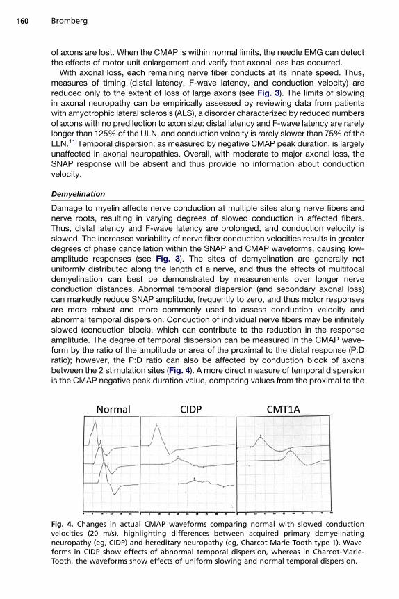
Damage to myelin affects nerve conduction at multiple sites along nerve fibers andnerve roots, resulting in varying degrees of slowed conduction in affected fibers.Thus, distal latency and F-wave latency are prolonged, and conduction velocity isslowed. The increased variability of nerve fiber conduction velocities results in greaterdegrees of phase cancellation within the SNAP and CMAP waveforms, causing low-amplitude responses (see Fig. 3). The sites of demyelination are generally notuniformly distributed along the length of a nerve, and thus the effects of multifocaldemyelination can best be demonstrated by measurements over longer nerveconduction distances. Abnormal temporal dispersion (and secondary axonal loss)can markedly reduce SNAP amplitude, frequently to zero, and thus motor responsesare more robust and more commonly used to assess conduction velocity andabnormal temporal dispersion. Conduction of individual nerve fibers may be infinitelyslowed (conduction block), which can contribute to the reduction in the responseamplitude. The degree of temporal dispersion can be measured in the CMAP wave-form by the ratio of the amplitude or area of the proximal to the distal response (P:Dratio); however, the P:D ratio can also be affected by conduction block of axonsbetween the 2 stimulation sites (Fig. 4). A more direct measure of temporal dispersionis the CMAP negative peak duration value, comparing values from the proximal to the
Fig. 4. Changes in actual CMAP waveforms comparing normal with slowed conductionvelocities (20 m/s), highlighting differences between acquired primary demyelinatingneuropathy (eg, CIDP) and hereditary neuropathy (eg, Charcot-Marie-Tooth type 1). Wave-forms in CIDP show effects of abnormal temporal dispersion, whereas in Charcot-Marie-Tooth, the waveforms show effects of uniform slowing and normal temporal dispersion.
Evaluation of Peripheral Neuropathies 161
distal response; normally, the negative peak duration increases by less than 10% evenover long distances (CMAP from Erb point stimulation compared with wrist stimula-tion; see Figs. 1 and 4).There is one neuropathy that is an exception with slowed conduction velocity
without abnormal temporal dispersion. In Charcot-Marie-Tooth type 1, the CMAPamplitude does not markedly decrease between distal and proximal stimulation sites(see Fig. 4). This is because of uniform changes to myelin along nerve fibers. Conduc-tion can also be mildly slowed by metabolic causes that do not affect myelin structur-ally, and diabetic neuropathies frequently show this pattern.There have been many efforts to design sets of nerve conduction criteria that can
distinguish between primary axonal and primary demyelination in terms of the degreeof slowing. The degree of demyelination in such neuropathies is variable: when mild,there will be only small prolongations in distal and F-wave latency and slowing ofconduction velocity that may be within (or just beyond) normal limits. When demyelin-ation is more marked, there will be slowing greater than expected for the degree ofaxonal loss, and the amount of slowing can be referred back to timing values frompatients with ALS.11 The published criteria are complex to apply, and a more simpleset of guidelines, based on the degree of slowing in ALS, is given in Table 1.12 Theseguidelines are not intended to be strict or exclusive but to assist deciding whetherthere is a demyelinating component in a neuropathy.
Focal Conduction Block
Conduction block refers to blockage of a large number of axons over a short segmentof nerve. Focal conduction block occurs most frequently at sites of entrapment(median nerve at the wrist, carpal tunnel syndrome; ulnar nerve at the elbow, tardyulnar palsy; peroneal nerve at the knee) resulting in mononeuropathies. In the contextof peripheral neuropathies, focal conduction block is sought at sites unaffected byentrapment. The pathologic condition may represent focal demyelination (or alter-ation to myelin at the node of Ranvier) or block by other mechanisms such asalteration or block of membrane ion channels.13,14 Focal conduction block can bedemonstrated by showing loss of CMAP amplitude across the site of block andnormal amplitudes along nerve segments above and below the site. The blockingmechanism can be very specific with block only of motor axons with sensory axons
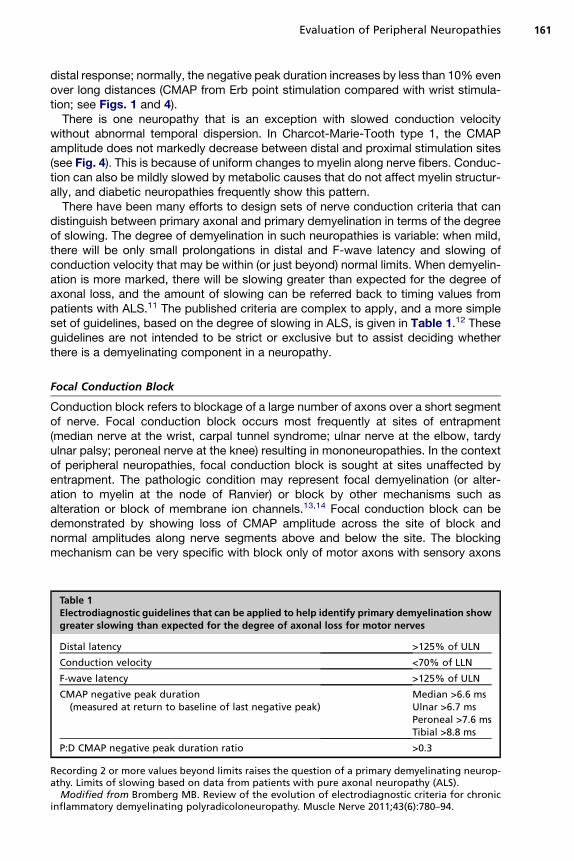
Table 1Electrodiagnostic guidelines that can be applied to help identify primary demyelination showgreater slowing than expected for the degree of axonal loss for motor nerves
Distal latency >125% of ULN
Conduction velocity <70% of LLN
F-wave latency >125% of ULN
CMAP negative peak duration(measured at return to baseline of last negative peak)
Median >6.6 msUlnar >6.7 msPeroneal >7.6 msTibial >8.8 ms
P:D CMAP negative peak duration ratio >0.3
Recording 2 or more values beyond limits raises the question of a primary demyelinating neurop-athy. Limits of slowing based on data from patients with pure axonal neuropathy (ALS).
Modified from Bromberg MB. Review of the evolution of electrodiagnostic criteria for chronicinflammatory demyelinating polyradicoloneuropathy. Muscle Nerve 2011;43(6):780–94.

Bromberg162
unaffected (multifocal motor neuropathy with conduction block)15 or with block ofboth motor and sensory fibers (Lewis-Sumner syndrome).16 Although the electrodiag-nostic hallmark of conduction block is reduction in the CMAP amplitude or areaacross the site of block, the effects of abnormal temporal dispersion can alsoproduce CMAP amplitude reductions as a result of increased phase cancellationthat may be misinterpreted as conduction block. Pure focal conduction block hasbeen defined as a greater than 50% reduction of CMAP area (area is less affectedby abnormal temporal dispersion than is amplitude) across the site of block withoutan increase in negative peak CMAP duration (<20%).17
DESIGNING AN ELECTRODIAGNOSTIC STUDY
Patients may be initially seen in a clinic, where a detailed history is obtained anda neurologic examination is performed. However, patients are frequently referred tothe EMG laboratory for electrodiagnostic tests only. Under these circumstances,a brief and focused history and examination are required to properly design and inter-pret the studies.3
Patient History
A pertinent history and examination allow for a rational selection of electrodiagnostictests.18 Referrals may be for complaints that do not accurately represent the trueclinical issue. Important topics to review and specific points to clarify include (1)time course: acute, chronic, or insidious; (2) symptoms: sensory (including numbnessor pain) or motor (including cramps, fasciculations, or weakness); (3) distribution:distal legs, distal and proximal limbs, or single nerve; (4) medical history: such asdiabetes or collagen vascular diseases; (5) family history: family members withneuropathy, high arches, or hammertoes; and (6) medications: neurotoxic chemo-therapeutic drugs or others. A focused neurologic examination should confirm theclinical suspicion for the presence and distribution of sensory loss and weakness.Tendon reflexes are helpful because they are usually absent, at least distally (Achillesreflex), in axonal neuropathies and more diffusely absent or reduced in demyelinatingneuropathies.
Study Design
At the conclusion of the clinical evaluation, there should be a good notion of thenature of the neuropathy. The electrodiagnostic study should be designed toconfirm what is expected from the history and answer questions that may not beevident from the history and examination. Specific issues include (1) type of nervesinvolved (motor, sensory, or both); (2) degree of involvement (mild, moderate, orsevere); (3) anatomy (which nerves are involved); (4) underlying pathologic condi-tions (axonal, demyelinating, or both); and (5) time course (acute, ongoing, or slowlyprogressive).It is argued here that there is benefit to designing an electrodiagnostic study test by
test for each patient, as opposed to applying a predetermined study protocol. With theformer approach, data from each nerve or muscle should be predicted. If the pre-dicted result is confirmed, the process of confirming the clinical diagnosis isproceeding on course. If there is an unexpected result, a technical error is sought orthe clinical diagnosis is questioned and reviewed. With the latter approach, data arereviewed at the end of the study. As such, it may be hard to make clinical sense ofdisparate findings, and technical errors may escape detection at a time when theycould be corrected.

Evaluation of Peripheral Neuropathies 163
Study Strategies
Sensory nerve conductionAssess the most distal sensory nerve first, usually the sural or superficial peronealnerve. Most peripheral neuropathies have a greater effect on sensory than motornerves and distal than proximal nerves. If the response is absent, to exclude a tech-nical cause, study the contralateral nerve. This will document symmetry, althoughthe history and examination will support symmetry clinically without the need to docu-ment symmetry electrodiagnostically for every nerve. If sensory responses are absentin the legs, a sensory nerve in the arm should be assessed for the overall extent (distri-bution) of axonal loss, and it is reasonable to chose the ulnar instead of the mediansensory nerve, because it will not be affected by common focal conduction abnormal-ities at the wrist.
Motor nerve conductionAssess the longest motor nerve next, usually the peroneal motor recording from theextensor digitorum brevis, before the tibial nerve; the tibial nerve is less informativebecause the amplitude is normally relatively large and small reductions resultingfrom axonal loss will not be detected. If motor responses are absent in the legs, motornerves in the arms should be studied, and when a primary demyelinating neuropathy issuspected, the ulnar nerves should be tested with stimulation up to the axilla. Motornerves will provide information about primary axonal versus primary demyelinatingneuropathies by inspection of the distal latencies, F-wave latencies, and conductionvelocities and the presence of abnormal temporal dispersion. The amplitude of thetibial response stimulating at the knee usually shows a marked loss of amplitude,and the P:D amplitude ratio from this nerve is not a reliable measure of abnormaltemporal dispersion or conduction block. The F-wave response should be assessedafter 10 maximal stimuli for minimum latency. Response amplitudes and timing valuesshould be compared for fulfillment of guidelines for primary demyelination (seeTable 1) when suggested by the history and examination.
Needle EMGThe needle EMG study will provide information on the presence of subtle denervation,proximal extent, and chronicity. Assessing the anterior tibialis muscle is usefulbecause it is usually affected to a mild degree and hence provides full information,whereas the extensor digitorum brevis muscle represents a very distal muscle thatmay be very atrophic. Assessment for abnormal spontaneous activity is to determinewhether there is mild motor nerve axonal loss, especially when the CMAP is mildlyreduced. Assessment of recruitment and MUAP amplitude and complexity is helpfulto determine the time course: moderately reduced recruitment and moderatelyincreased MUAP amplitude and complexity support ongoing denervation, whereasmarkedly reduced recruitment, markedly increased MUAP amplitude, but simplewaveforms support a very longstanding neuropathy.
Electrodiagnostic Report Summary and Interpretation
The electrodiagnostic report should contain sufficient historical and examinationfeatures to provide a context for the interpretation.3 A summary should not repeat inprose nerve conduction and EMG data tables but should present the salient pointsthat will be used in the overall interpretation, in particular technical or physical issuesthat influence the data. The overall interpretation can include information from thehistory and examination and must make sense internally (data internally consistentwith the conclusions) and clinically (consistent with examination findings, past medical
Bromberg164
and family histories, and laboratory findings). Overall, the interpretation can be mostinformative if the study is designed to answer questions formulated from the historyand examination with assessment of each metric to determine consistency.
REVIEWING OUTSIDE ELECTRODIAGNOSTIC STUDIES
When reviewing an outside electrodiagnostic study, one should not rely on the inter-pretation but instead review the data and make your own conclusions and thencompare the 2 interpretations. It must be kept in mind that there may be technicalissues not recognized and inappropriate interpretations and numbers, and waveformsshould be reviewed when provided. In reading the nerve conduction and EMG datatables, it is most efficient and informative if one reads the study as if one were perform-ing the study; that is, look at the data in the order suggested earlier (steps 1–3).
CLINICAL AND ELECTRODIAGNOSTIC FEATURES OF PROTOTYPIC NEUROPATHIESPrimary Axonal Neuropathies
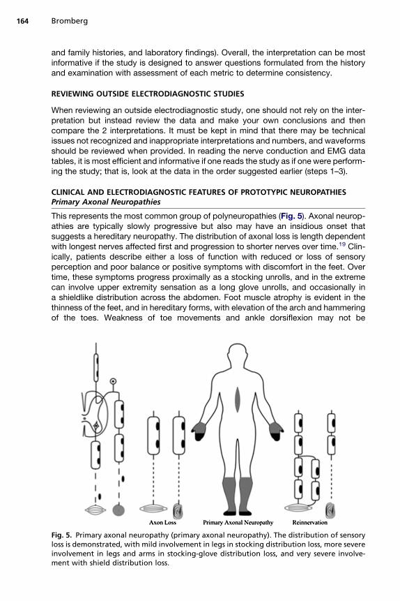
This represents the most common group of polyneuropathies (Fig. 5). Axonal neurop-athies are typically slowly progressive but also may have an insidious onset thatsuggests a hereditary neuropathy. The distribution of axonal loss is length dependentwith longest nerves affected first and progression to shorter nerves over time.19 Clin-ically, patients describe either a loss of function with reduced or loss of sensoryperception and poor balance or positive symptoms with discomfort in the feet. Overtime, these symptoms progress proximally as a stocking unrolls, and in the extremecan involve upper extremity sensation as a long glove unrolls, and occasionally ina shieldlike distribution across the abdomen. Foot muscle atrophy is evident in thethinness of the feet, and in hereditary forms, with elevation of the arch and hammeringof the toes. Weakness of toe movements and ankle dorsiflexion may not be
Fig. 5. Primary axonal neuropathy (primary axonal neuropathy). The distribution of sensoryloss is demonstrated, with mild involvement in legs in stocking distribution loss, more severeinvolvement in legs and arms in stocking-glove distribution loss, and very severe involve-ment with shield distribution loss.

Evaluation of Peripheral Neuropathies 165
appreciated by the patient but can be confirmed on examination. Tendon reflexes areusually absent, at least at the ankle.Electrodiagnostic findings are greatest in distal leg nerves and may not be evident
early on in distal arm nerves.20 SNAP amplitude is decreased earlier and to a greaterdegree than CMAP amplitude. Distal latency, F-wave latency and conduction velocityare mildly affected (see Figs. 3 and 5) and values are within 75% of the LLN or 125% ofthe ULN. Needle EMG will be more sensitive to mild degrees of denervation when theCMAP amplitude is within the LLN, and the greatest EMG changes will be in mostdistal muscles. In very chronic forms, such as hereditary neuropathies, there will beexpected absent SNAP responses and absent or low amplitude CMAP responsesindistinguishable from other causes, but the clue to chronicity will be in the EMG result,which will show markedly reduced motor unit recruitment with very large amplitudesbut simple waveforms in distal muscles. Clinical and electrodiagnostic findings areusually symmetric in axonal neuropathies of any cause.
Primary Demyelinating Neuropathies
This group is less common but potentially treatable.21 The distribution of demyelin-ation is diffuse with multiple foci along nerves and nerve roots, and thus both distaland proximal nerves are affected clinically and electrically (Fig. 6). Clinically, patientsdescribe numbness and tingling with a distal emphasis but also along the proximalportion of the limbs. Weakness also has a distal emphasis but can be demonstratedin proximal muscles. The time course can be rapid (within days), as in acute inflamma-tory demyelinating polyradiculoneuropathy (AIDP), or slow (over months), as in chronicinflammatory demyelinating polyradicoloneuropathy (CIDP). Tendon reflexes areusually uniformly absent, but at least absent in the legs.
Fig. 6. Primary demyelinating neuropathy (primary demyelinating neuropathy). The distri-bution of sensory loss resulting from multifocal demyelination involving nerve and spinalroot fibers is shown. Sensory loss involves both legs and arms, both distally and proximally,with distal predominance.

Bromberg166
Electrodiagnostic findings are greatest when longer lengths of nerve are studied,and abnormalities are found in leg and arm nerves but more severely in leg nerves.SNAP amplitudes may be markedly reduced and frequently absent because of distalabnormal temporal dispersion and conduction block. CMAP amplitude is usuallyreduced to distal stimulation and may be markedly reduced to more proximal stimu-lation caused by multifocal demyelination and conduction block (see Figs. 3 and 4).Distal and F-wave latencies are prolonged and conduction velocities slowed. Withrelatively severe primary demyelinating neuropathies, these values will be markedlyslowed, greater than seen in axonal neuropathies, but with more mild degrees ofseverity, values may overlap with the limits of primary axonal neuropathies.12 Thisrepresents a challenge if electrodiagnostic criteria are not met. A practical approachis to consider the possibility of a demyelinating neuropathy when one or more nerveshas values supporting slowing greater than expected for primary axonal neuropathy(see Table 1). This may result in false positive demyelinating neuropathies with erringon the side of a possibly treatable neuropathy.
Mixed Primary Axonal and Primary Demyelinating Neuropathies
Several neuropathies may contain a mixture of demyelination and axonal loss. Whendemyelination is the primary process, axonal loss occurs frequently as a secondarypathologic process. Thus, absent responses in more severe demyelinating neuropa-thies may be in part the result of secondary axonal loss. Most examples of primarydemyelinating neuropathies, AIDP and CIDP, include varying degrees of secondaryaxonal loss, and responses can be absent in leg nerves. The needle EMG will helpdetermine the degree of axonal loss with the presence of abnormal spontaneousactivity and the degree of reduced MUAP recruitment and complexity. Diabetes anduremia are the most common causes of mixed axonal and demyelinating neuropathy,and findings are reduced SNAP and CMAP amplitudes, similar to findings in primaryaxonal neuropathies, but the slowing of conduction velocities is not as substantialas expected in a primarily demyelinating neuropathy.
Focal Conduction Bock Neuropathies
Multifocal motor neuropathy with conduction block is characterized by focal conduc-tion block over short nerve segments away from entrapment sites and in a mononeur-opathy multiplex pattern. There is a potential technical issue in identify this neuropathyin that there may be inadequate activation of axons at the proximal stimulation siteleading to a falsely low CMAP response. There is a physiologic issue in that mildreductions in amplitude to proximal stimulation can occur from abnormal temporaldispersion leading to greater phase cancellation, mimicking conduction block. Thiscan be assessed by considering the negative peak duration values: in conductionblock, there will be a greater than 50% loss of CMAP amplitude with a less than15% increase in negative peak duration.17 In classic multifocal motor neuropathy,sensory conduction across the site is normal, but in the Lewis-Sumner syndrome,there is involvement of sensory nerves at sites of motor block.
SUMMARY
Electrodiagnostic testing follows from the history and is an extension of the neurologicexamination. When clinical assessment implicates a peripheral neuropathy, the goal ofelectrodiagnostic testing is to more fully characterize the neuropathy in terms of thedistribution (motor, sensory, symmetric, or asymmetric), extent of a neuropathy(symmetric, legs, or arms), and time course (very chronic or ongoing). Of greatest
Evaluation of Peripheral Neuropathies 167
importance is that electrodiagnostic testing should help identify the type of underlyingpathologic condition (primary axonal, primary demyelinating, or conduction block).Once this is accomplished, the differential diagnosis narrows and rational laboratorytesting can be ordered or treatment trials initiated.1
REFERENCES
1. Donofrio P, Albers J. AAEM minimonograph #34: polyneuropathy: classificationby nerve conduction studies and electromyography. Muscle Nerve 1990;13:889–903.
2. Kimura J. Principles and pitfalls of nerve conduction studies. Ann Neurol 1984;16:415–29.
3. Jablecki CK, Busis NA, Brandstater MA, et al. Reporting the results of needleEMG and nerve conduction studies: an educational report [Guideline]. MuscleNerve 2005;32(5):682–5.
4. Kimura J, Machida M, Ishida T, et al. Relation between size of compound sensoryor motor action potentials and length of nerve segment. Neurology 1986;36:647–52.
5. Rutkove SB. Effects of temperature on neuromuscular electrophysiology[Review]. Muscle Nerve 2001;24(7):867–82.
6. Daube JR, Rubin DI. Needle electromyography. Muscle Nerve 2009;39(2):244–70.
7. Dumitru D. Single muscle fiber discharges (insertional activity, end-plate poten-tials, positive sharp waves, and fibrillation potentials): a unifying proposal. MuscleNerve 1996;19:221–6.
8. Bromberg M. Electromyographic (EMG) findings in denervation. Crit Rev PhysRehabil Med 1993;5:83–127.
9. Robinson L, Temkin N, Fujimoto W, et al. Effects of statistical methodology onnormal limits in nerve conduction studies. Muscle Nerve 1991;14:1084–90.
10. Benatar M, Wuu J, Peng L. Reference data for commonly used sensory and motornerve conduction studies. Muscle Nerve 2009;40(5):772–94.
11. Cornblath D, Kuncl R, Mellits E, et al. Nerve conduction studies in amyotrophiclateral sclerosis. Muscle Nerve 1992;15:1111–5.
12. Bromberg MB. Review of the evolution of electrodiagnostic criteria for chronicinflammatory demyelinating polyradicoloneuropathy [Historical Article]. MuscleNerve 2011;43(6):780–94.
13. Feasby TE, Brown WF, Gilbert JJ, et al. The pathological basis of conductionblock in human neuropathies. J Neurol Neurosurg Psychiatry 1985;48(3):239–44.
14. Arasaki K, Kusunoki S, Kudo N, et al. Acute conduction block in vitro followingexposure to antiganglioside sera. Muscle Nerve 1993;16(6):587–93.
15. Lange D, Trojaborg W, Latov N, et al. Multifocal motor neuropathy with conductionblock: is it a distinct clinical entity? Neurology 1992;42:497–505.
16. Lewis R, Sumner A, Brown M, et al. Multifocal demyelinating neuropathy withpersistent conduction block. Neurology 1982;32:958–64.
17. EFNS/PNSR1. European Federation of Neurological Societies/Peripheral NerveSociety Guideline on management of chronic inflammatory demyelinating polyra-diculoneuropathy: report of a joint task force of the European Federation ofNeurological Societies and the Peripheral Nerve Society–First Revision.J Peripher Nerv Syst 2010;15(1):1–9.
18. Bromberg MB. An approach to the evaluation of peripheral neuropathies. SeminNeurol 2005;25(2):153–9.

Bromberg168
19. Sabin T. Classification of peripheral neuropathy: the long and the short of it.Muscle Nerve 1986;9:711–9.
20. Raynor EM, Ross MH, Shefner JM, et al. Differentiation between axonal anddemyelinating neuropathies: identical segments recorded from proximal anddistal muscles. Muscle Nerve 1995;18(4):402–8.
21. Albers J, Kelly J. Acquired inflammatory demyelinating polyneuropathies: clinicaland electrodiagnostic features. Muscle Nerve 1989;12:435–51.
Related Documents


