An approach to ankle x-rays Aric Storck PGY2 (acknowledgement to Dr. Dave Dyck for several slides) September 11, 2003

An approach to ankle x-rays Aric Storck PGY2 (acknowledgement to Dr. Dave Dyck for several slides) September 11, 2003.
Dec 24, 2015
Welcome message from author
This document is posted to help you gain knowledge. Please leave a comment to let me know what you think about it! Share it to your friends and learn new things together.
Transcript
An approach to ankle x-rays
Aric Storck PGY2(acknowledgement to Dr. Dave Dyck for several slides)
September 11, 2003

Objectives
Review basic ankle fracture classification
Review x-rays of common ankle fractures
Discuss management of common ankle fractures
Case 1:
25 year old female• Jumped off roof
• Right ankle pain
• Inability to weight bear on right foot
What else do you want to know on history and physical examination?
Does she need x-rays ?

Ottawa Ankle Rules:
Order ankle x-rays if acute trauma to ankle and one or more of• Age 55 or older
• Inability to weight bear both immediately and in ER (4 steps)
• Bony tenderness over posterior distal 6 cm of lateral or medial malleoli
Sensitivity ~100% Specificity ~40%

You have decided to order an “ankle x-ray.” The nurse entering your orders asks which views you want …
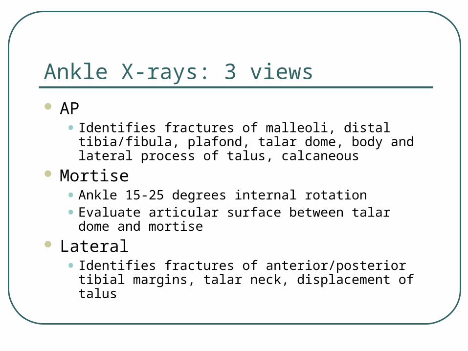
Ankle X-rays: 3 views
AP• Identifies fractures of malleoli, distal tibia/fibula,
plafond, talar dome, body and lateral process of talus, calcaneous
Mortise• Ankle 15-25 degrees internal rotation
• Evaluate articular surface between talar dome and mortise
Lateral • Identifies fractures of anterior/posterior tibial margins,
talar neck, displacement of talus

AP x-ray:
Identifies fractures of • malleoli
• distal tibia/fibula
• plafond
• talar dome
• body and lateral process of talus
• calcaneous
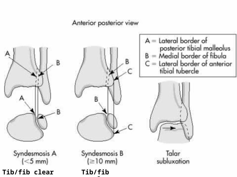
Tib/fib clear space Tib/fib overlap

AP xray

Now apply what you’ve learned …
Lateral malleolar fracture
Tib/fib clear space <5mm
Tib/fib overlap >10 mm
No evidence of syndesmotic injury

Mortise X-Ray
Taken with ankle in 15-25 degrees of internal rotation
Useful in evaluation of articular surface between talar dome and mortise
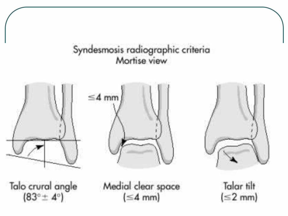

Mortise x-ray:
Medial clear space• Between lateral border of
medial malleous and medial talus
• <4mm is normal
• >4mm suggests lateral shift of talus

Mortise x-ray:
Talar tilt • Normal = -1.5 to +1.5
degrees (ie. Parallel)
• Can go up to 5 degrees in stress views
• <2mm difference between medial and lateral talar/plafond distances
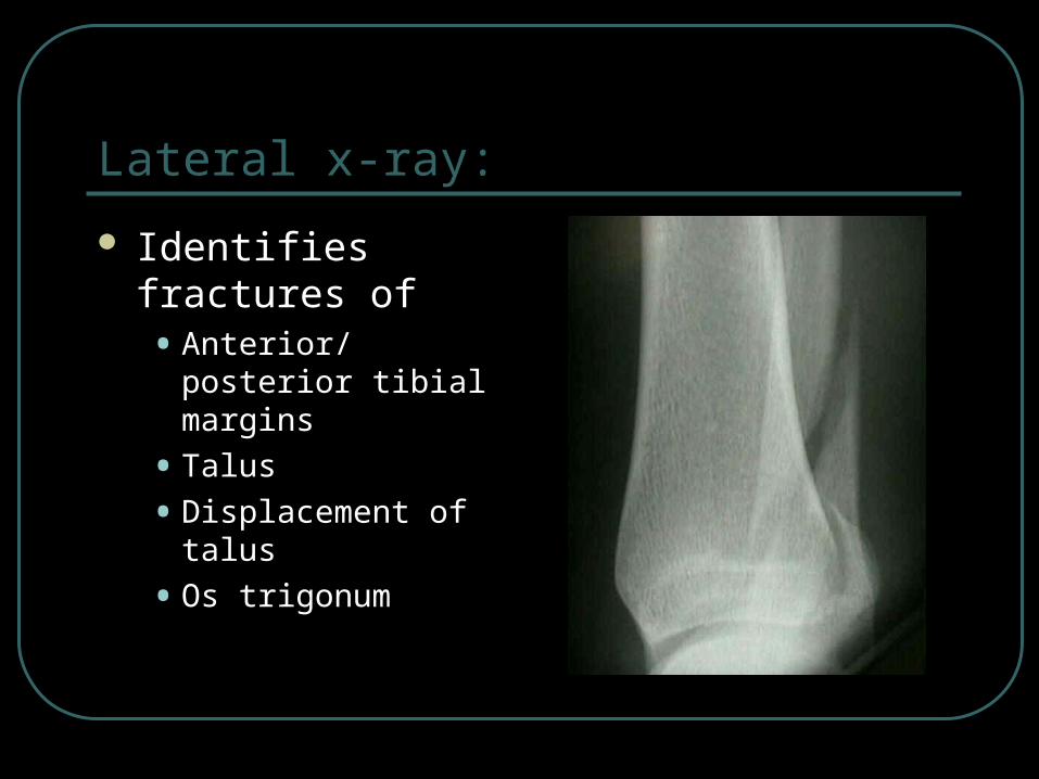
Lateral x-ray:
Identifies fractures of • Anterior/posterior tibial
margins
• Talus
• Displacement of talus
• Os trigonum
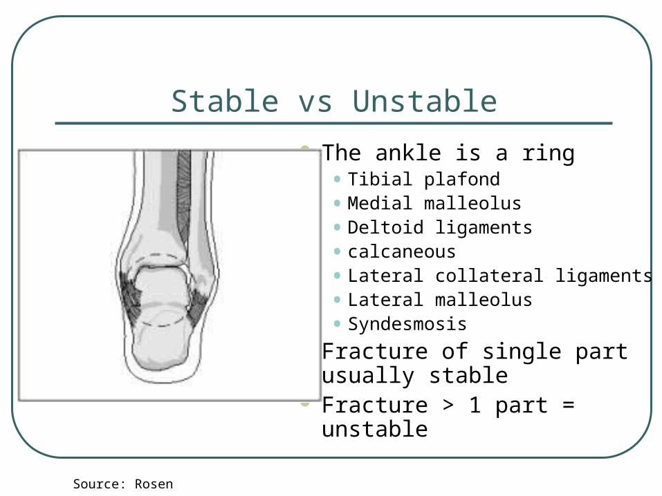
Stable vs Unstable
The ankle is a ring• Tibial plafond
• Medial malleolus
• Deltoid ligaments
• calcaneous
• Lateral collateral ligaments
• Lateral malleolus
• Syndesmosis Fracture of single part usually
stable Fracture > 1 part = unstable
Source: Rosen
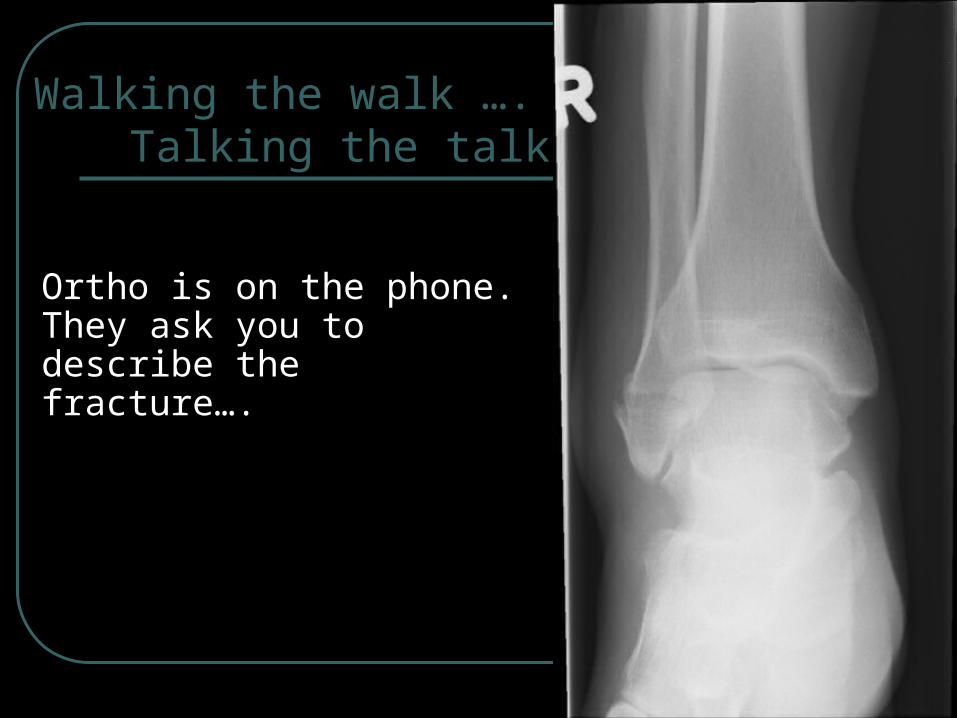
Walking the walk ….Talking the talk
Ortho is on the phone. They ask you to describe the fracture….
Lauge-Hansen:
15 basic types of injury in 5 major categories• Described by two words
1.Position of foot at time of injury
2.Direction of talus within mortise causing fracture
• Eg: supination-external rotation
• Further subdivided into worsening areas of injury
Impossible to remember and clinically useless in the ED

Danis-Weber
• Defines injury based on level of fibular fracture• A=below tibiotalar joint
• No disruption of syndesmosis
• Usually stable
• B=at level of tibiotalar joint
• Partial disruption of syndesmosis
• C=above tibiotalar joint
• Disrupts syndesmosis to level of fracture
• unstable
• THE MORE PROXIMAL THE FIBULAR # THE MORE SEVERE THE INJURY
AO classification:
Similar to Danis-Weber scheme
Takes into account damage to other structures (usually medial malleolous)
~2 pages of classifications• Remember them all for your exam!
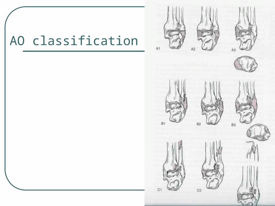
AO classification
Pott’s classification:
Easy to remember
First degree• unimalleolar
Second degree• bimalleolar
Third degree• trimalleolar

Case 2
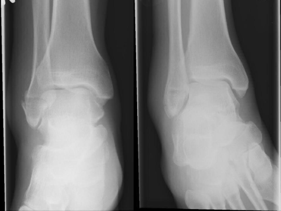
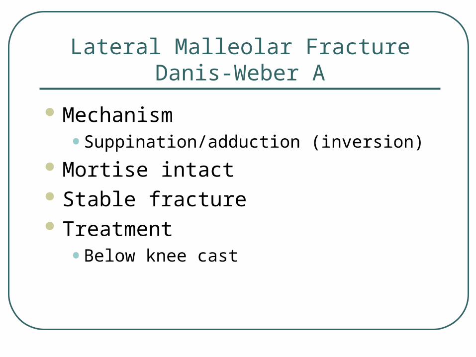
Lateral Malleolar FractureDanis-Weber A
Mechanism• Suppination/adduction (inversion)
Mortise intact Stable fracture Treatment
• Below knee cast
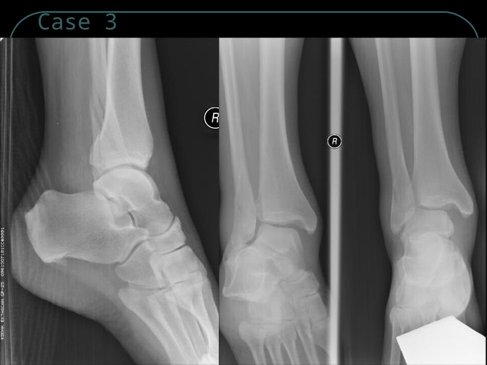
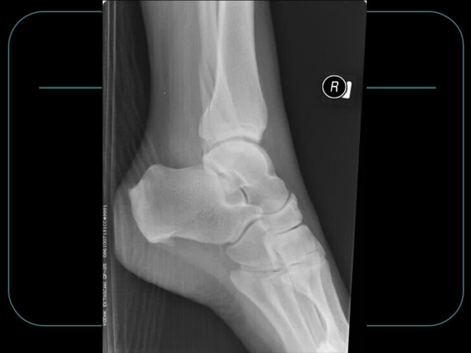
Case 3

Bimalleolar (lat & post malleoli)
Mechanism• Inversion
• Avulsion of posterior malleolus (post tibiofibular ligament)
Medial mortise wide• Suggests instability
Management• Posterior slab
• Orthopedic consult
Source: McRae’s Practical Fracture Treatment

Case 4
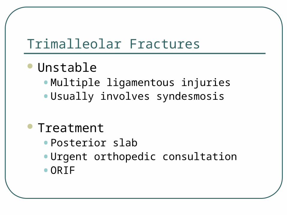
Unstable• Multiple ligamentous injuries
• Usually involves syndesmosis
Treatment• Posterior slab
• Urgent orthopedic consultation
• ORIF
Trimalleolar Fractures
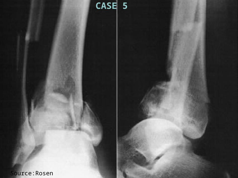
Source:Rosen
CASE 5
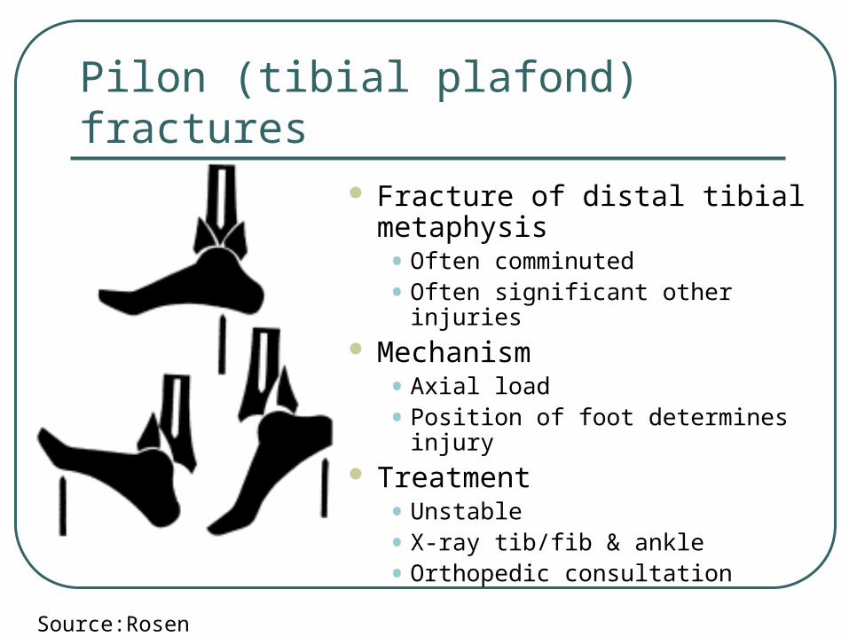
Fracture of distal tibial metaphysis• Often comminuted
• Often significant other injuries Mechanism
• Axial load
• Position of foot determines injury Treatment
• Unstable
• X-ray tib/fib & ankle
• Orthopedic consultation
Pilon (tibial plafond) fractures
Source:Rosen
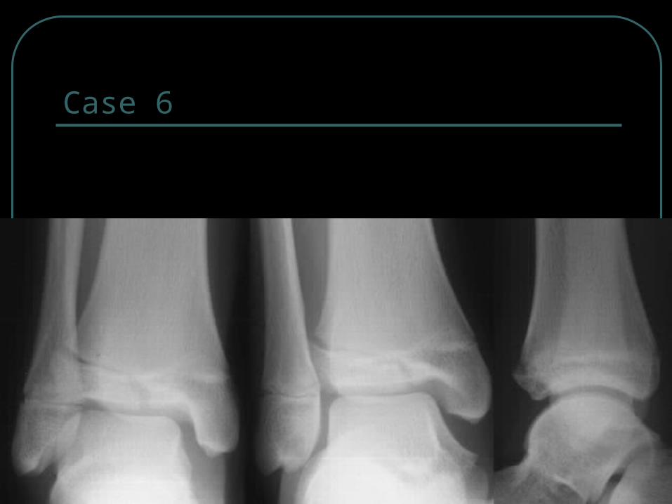
Case 6

Tillaux Fracture
Occurs in 12-14 year olds• 18 month period when epiphysis is closing
Salter-Harris 3 injury• Runs through anterolateral physis until reaches fused part,
then extends inferiorly through epiphysis into joint
• Visible if x-ray parallel to plane of fracture (may require oblique)
Mechanism• External rotation
• Strenth of tibiofibular ligament > unfused epiphysis

Tillaux Fracture
Management• Inadequate reduction of articular surface can lead
to early OA
• Gap >2mm in articular surface is unacceptable
• Advanced imaging techniques may be necessary
• Early orthopedic consultation
• Non-displaced• NWB below knee cast
• Displaced• surgery
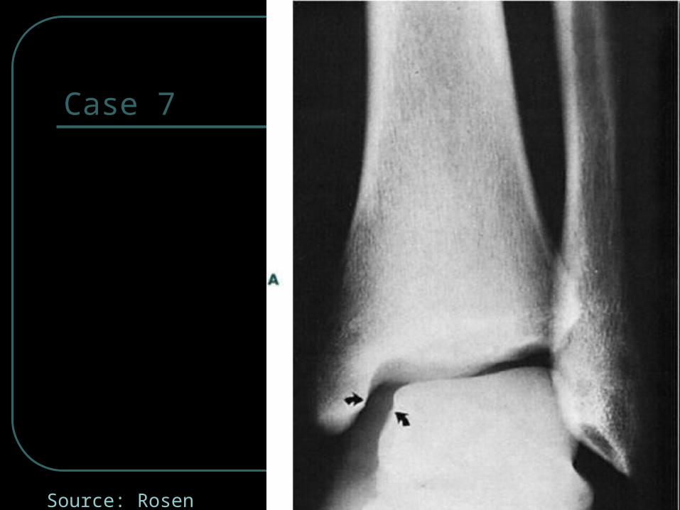
Case 7
Source: Rosen

Maisonneuve Fracture
Mechanism• Eversion + lateral rotation
• May cause medial malleolar fracture or deltoid ligament disruption
• Injury proceeds along syndesmosis and involves proximal fibula
Always rule out Maisonneuve fracture in medial malleolar/ligamentous injury
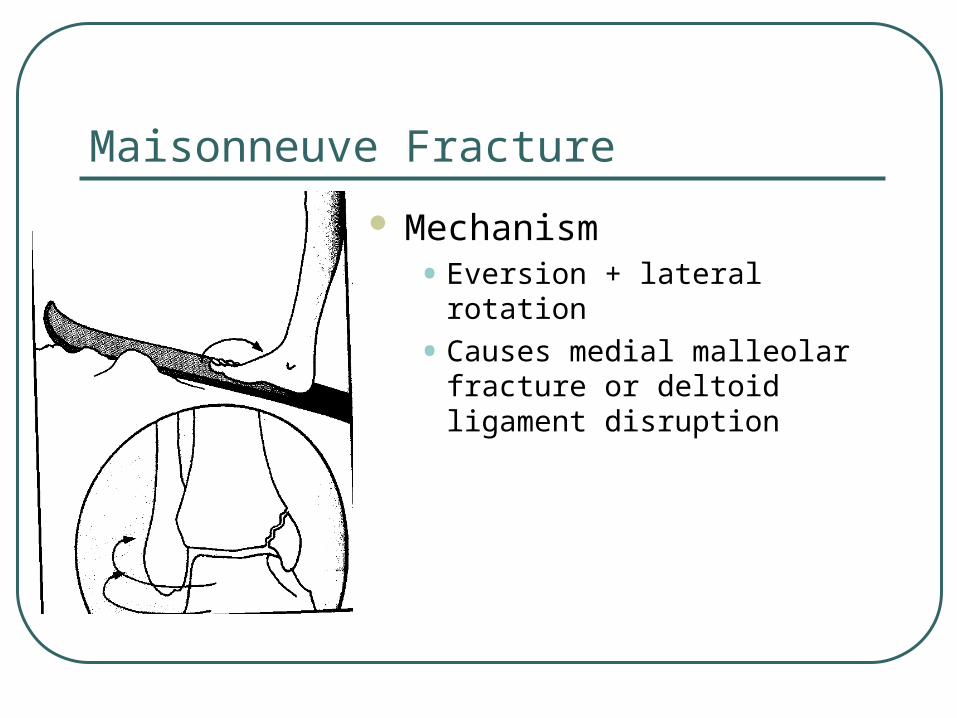
Maisonneuve Fracture
Mechanism• Eversion + lateral rotation
• Causes medial malleolar fracture or deltoid ligament disruption
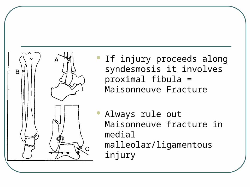
If injury proceeds along syndesmosis it involves proximal fibula = Maisonneuve Fracture
Always rule out Maisonneuve fracture in medial malleolar/ligamentous injury

As talus continues to rotate• Posterior tib-fib ligament ruptures
• Interosseous membrane rips
• Gross diastasis
• Dupuytren fracture – dislocation of the ankle
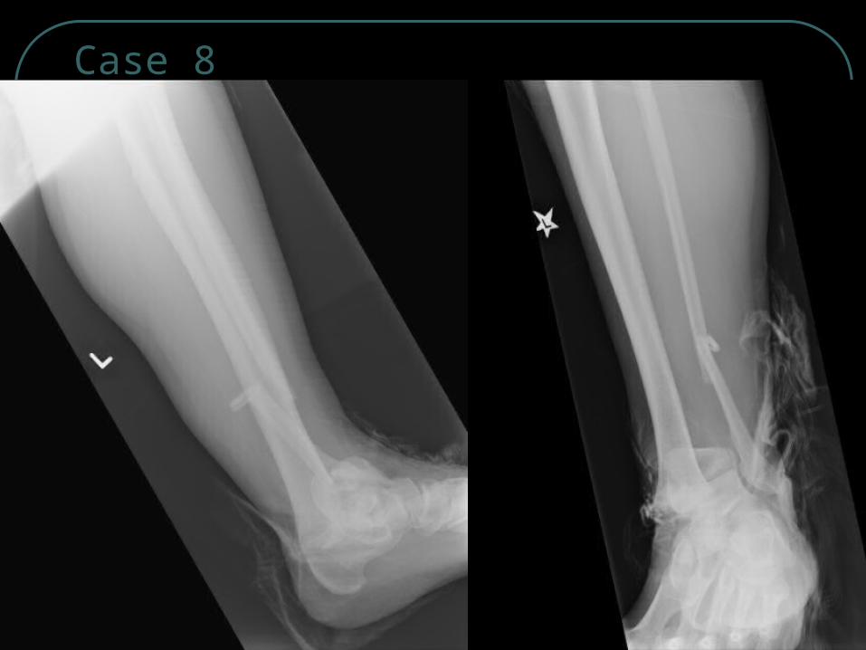
Case 8

the end
Related Documents