The PDF of the article you requested follows this cover page. This is an enhanced PDF from The Journal of Bone and Joint Surgery 2009;91:2144-2150. doi:10.2106/JBJS.H.00744 J Bone Joint Surg Am. Xiaofeng Jia, Jong Hun Ji, Steve A. Petersen, Michael T. Freehill and Edward G. McFarland Surgery An Analysis of Shoulder Laxity in Patients Undergoing Shoulder This information is current as of September 4, 2009 Reprints and Permissions Permissions] link. and click on the [Reprints and jbjs.org article, or locate the article citation on to use material from this order reprints or request permission Click here to Publisher Information www.jbjs.org 20 Pickering Street, Needham, MA 02492-3157 The Journal of Bone and Joint Surgery

Welcome message from author
This document is posted to help you gain knowledge. Please leave a comment to let me know what you think about it! Share it to your friends and learn new things together.
Transcript
The PDF of the article you requested follows this cover page.
This is an enhanced PDF from The Journal of Bone and Joint Surgery
2009;91:2144-2150. doi:10.2106/JBJS.H.00744 J Bone Joint Surg Am.Xiaofeng Jia, Jong Hun Ji, Steve A. Petersen, Michael T. Freehill and Edward G. McFarland
SurgeryAn Analysis of Shoulder Laxity in Patients Undergoing Shoulder
This information is current as of September 4, 2009
Reprints and Permissions
Permissions] link. and click on the [Reprints andjbjs.orgarticle, or locate the article citation on
to use material from thisorder reprints or request permissionClick here to
Publisher Information
www.jbjs.org20 Pickering Street, Needham, MA 02492-3157The Journal of Bone and Joint Surgery

An Analysis of Shoulder Laxity in PatientsUndergoing Shoulder Surgery
By Xiaofeng Jia, MD, PhD, Jong Hun Ji, MD, Steve A. Petersen, MD, Michael T. Freehill, MD, and Edward G. McFarland, MD
Investigation performed at the Division of Shoulder Surgery, Department of Orthopaedic Surgery, The Johns HopkinsUniversity/Johns Hopkins Bayview Medical Center, Baltimore, Maryland
Background: It has been recognized that there is a distinction between shoulder laxity and shoulder instability and thatthere is a wide range of normal shoulder laxities. Our goals were (1) to evaluate if the ability to subluxate the shoulder overthe glenoid rim in patients under anesthesia would be more prevalent than the inability to do so, (2) to determine ifpatients with a diagnosis of instability would have significantly more shoulder laxity in the operatively treated shoulder thanin the contralateral shoulder, and (3) to evaluate the observation that higher grades of shoulder laxity would be related to adiagnosis of shoulder instability. We hypothesized that, on examination with the patient under anesthesia, most shoulderscould be subluxated over the glenoid rim and that the degree of shoulder laxity would be related to diagnosis.
Methods: In the present study of 1206 patients undergoing shoulder surgery, we evaluated the symptomatic andcontralateral shoulders with use of a modified anterior and posterior drawer test and a sulcus sign test, with thepatients under anesthesia. The anterior and posterior translations were graded as no subluxation (Grade I), subluxationover the glenoid rim with spontaneous reduction (Grade II), or subluxation without spontaneous reduction (Grade III).The sulcus sign was graded as <1.0 cm (Grade I), 1.0 to 2.0 cm (Grade II), or >2.0 cm (Grade III).
Results: When the patients were evaluated while under anesthesia, the humeral head could be subluxated over therim anteriorly in 81.6% (984 of 1206) of the patients and posteriorly in 57.5% (693 of 1206) of the patients. When thepatients were evaluated while under anesthesia, there was an increase in the laxity grade anteriorly, posteriorly, andinferiorly in 50.8%, 36.3%, and 15.8% of the patients, respectively, as compared with the preoperative assessment. Forall laxity testing, the higher the grade of laxity in an anterior, posterior, or inferior direction, the greater the chance thatthe patient had a diagnosis of instability. Compared with Grade-I laxity, Grade-III laxity increased the odds of a diagnosisof instability in the anterior (odds ratio, 170), posterior (odds ratio, 32), and inferior (odds ratio, 10.3) directions.Compared with Grade-I laxity, Grade-II laxity increased the odds of a diagnosis of instability in the anterior (odds ratio,9.8), posterior (odds ratio, 4.6), and inferior (odds ratio, 4.4) directions.
Conclusions: The ability to subluxate the humeral head over the glenoid rim in the patient who is undergoing shouldersurgery under anesthesia is common regardless of the diagnosis. Higher grades of shoulder laxity are associated withshoulder instability.
Many physicians have recommended laxity testing ofthe shoulder as a component of the physical exami-nation of patients with shoulder conditions and as a
screening test during routine physical examinations of otherpatients1-6. The evaluation of shoulder laxity has been particu-larly recommended for two sets of patients in the clinical setting:those with symptomatic traumatic shoulder instability (to confirmthe diagnosis)1,2 and those with occult instability associatedwith shoulder pain3. This laxity evaluation has been used es-
pecially for patients who are involved in overhead sports todetermine if shoulder stabilization should be performed4,7.
However, clinicians have increasingly recognized thatthe range of normal shoulder laxity is wide, that high gradesof shoulder laxity may not be pathologic, and that laxity andinstability of the shoulder joint are not the same8-10. Laxity is ameasure of the joint movement within normal limits, whereasinstability is a pathologic condition that results in symptomsbecause of excessive movement of the humeral head on the
Disclosure: The authors did not receive any outside funding or grants in support of their research for or preparation of this work. Neither they nor amember of their immediate families received payments or other benefits or a commitment or agreement to provide such benefits from a commercialentity. No commercial entity paid or directed, or agreed to pay or direct, any benefits to any research fund, foundation, division, center, clinical practice,or other charitable or nonprofit organization with which the authors, or a member of their immediate families, are affiliated or associated.
2144
COPYRIGHT � 2009 BY THE JOURNAL OF BONE AND JOINT SURGERY, INCORPORATED
J Bone Joint Surg Am. 2009;91:2144-50 d doi:10.2106/JBJS.H.00744
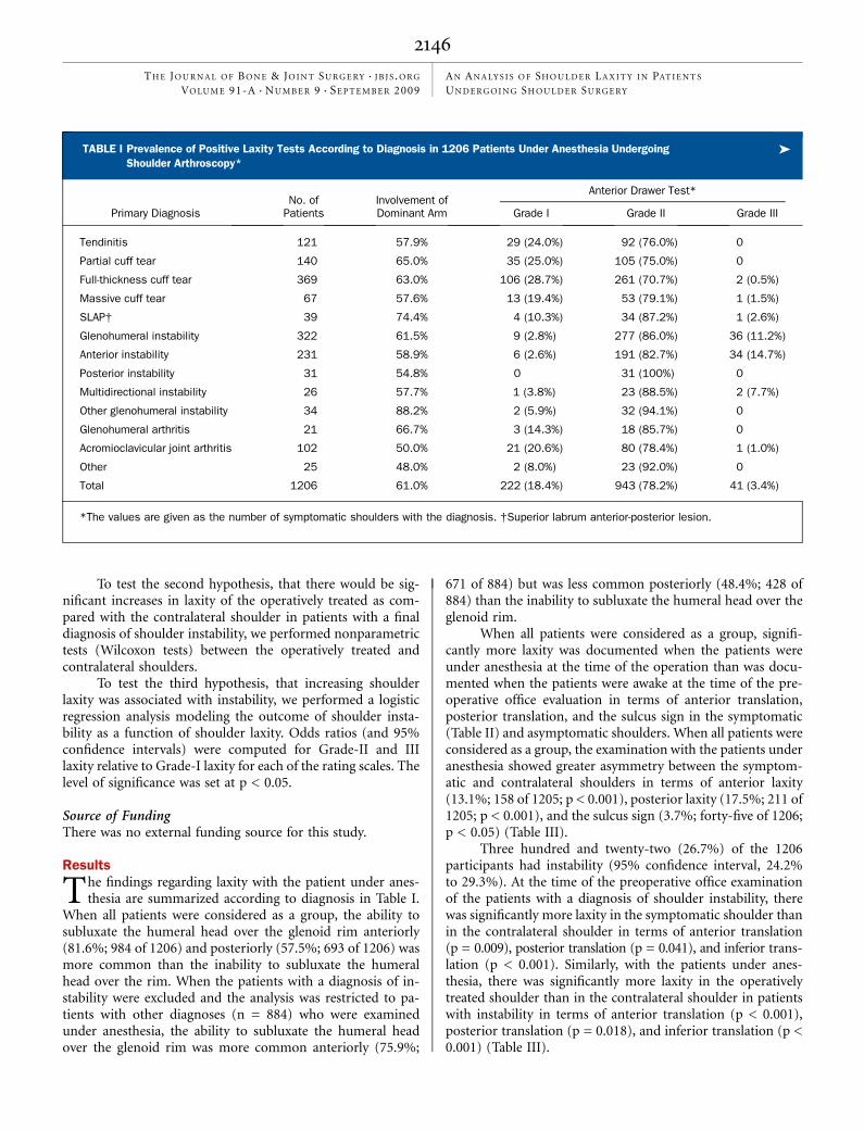
glenoid5,6,11. Several studies have shown that the ability tosubluxate the shoulder on examination is common, especiallyin athletic individuals12-15. However, to our knowledge, therehave been no studies of shoulder laxity in large populations ofpatients with a variety of diagnoses.
Similarly, the performance of laxity testing of the shoul-der during the examination of the shoulder with the patientunder anesthesia has been widely advocated8,11,16. Although anincrease in shoulder laxity with the patient under anesthesiahas been documented11, the role of laxity testing, particularlyfor patients with a diagnosis of shoulder instability, has notbeen thoroughly explored.
It has been our observation that the ability to subluxatethe shoulder over the glenoid rim is essentially a normalfinding on examination of the shoulder, especially with thepatient under anesthesia13. The purpose of the present studywas threefold: (1) to evaluate if the ability to subluxate theshoulder over the glenoid rim in patients under anesthesiawould be more prevalent than the inability to do so, (2) todetermine if patients with a diagnosis of instability would havesignificantly more shoulder laxity in the operatively treatedshoulder than in the contralateral shoulder, and (3) to evaluatethe observation that higher grades of shoulder laxity would berelated to a diagnosis of shoulder instability.
Materials and Methods
The present study was approved by our institutional reviewboard. This retrospective cohort study analyzed informa-
tion from our institution’s shoulder surgery database, whichincludes patients managed with shoulder surgery by only thesenior author (E.G.M.) from 1992 through 200717-26. Of the1836 patients who were initially identified, 630 were excludedbecause no examination was performed with the patient underanesthesia because of a fracture (n = 51), shoulder arthroplasty(n = 330), or infection (n = 20); because of a frozen shoul-der (n = 40); or because no arthroscopic evaluation was per-formed (n = 189). Therefore, the study group consisted of1206 patients who had physical examination of the symptom-atic and asymptomatic shoulders, both while awake and whileunder anesthesia, followed by diagnostic arthroscopy of theshoulder. The study group included 716 male patients and 490female patients with an average age of forty-five years (range,twelve to eighty-six years). The diagnoses included rotator cufftendinitis or tear (697 patients), shoulder instability (322 pa-tients), isolated acromioclavicular arthritis (102 patients), su-perior labrum anterior-posterior (SLAP) lesions (thirty-ninepatients), glenohumeral joint arthritis (twenty-one patients),and other diagnoses (twenty-five patients).
All patients underwent a preoperative assessment, in-cluding a thorough physical examination, and filled out de-tailed questionnaires, as previously reported17-19,21,22,24,25,27,28. Theshoulder examination included an assessment of range ofmotion, an evaluation of strength with use of manual muscletesting, a complete neurologic evaluation of the upper ex-tremities, and shoulder laxity testing. The physical examina-tion was performed by, or under the direct supervision of, the
senior author. There was no attempt to standardize the ex-amination for the amount of force used to translate the hu-meral head over the glenoid rim. Laxity testing of the shoulderincluded a modified anterior and posterior drawer test, whichwas adapted6,23 from the technique described by Gerber andGanz29. The tests were performed with the arm in approxi-mately 60� to 80� of abduction and 10� to 15� of forwardflexion23.
Laxity was measured with use of a modified Hawkinsscale13 in which the humeral head was judged not to subluxateover the glenoid rim (Grade I), to subluxate over the glenoidrim but to spontaneously reduce (Grade II), or to remaindislocated even when the subluxation force exerted by theexaminer’s hands was withdrawn (Grade III). As previouslyreported1, a note was made in the examination record if thepatient was judged to be unable to relax for the examination inthe office because of pain or apprehension.
All patients underwent an evaluation of inferior laxityby testing for a sulcus sign. As previously reported6, with thepatient in a sitting position, an inferiorly directed forcewas applied to the arm and the translation was graded as <1.0cm (Grade I), 1.0 to 2.0 cm (Grade II), or >2.0 cm (GradeIII)2,6,8,18,23,30,31.
Laxity testing was performed with the patient undergeneral anesthesia with or without regional anesthesia with useof a scalene block. The anterior and posterior drawer tests wereperformed and graded with use of the modified Hawkins scaledescribed above. Testing for the sulcus sign was performedwith the patient supine, and the result was also graded with thesystem described above. All examinations with the patientunder anesthesia were performed by the senior author. Allpatients underwent diagnostic arthroscopy in the lateral de-cubitus position, and all intra-articular findings were recordedon a datasheet14,17-19,21-25,32.
The final diagnosis was based on the preoperative his-tory, physical examination, radiographs, and the findings atthe time of diagnostic arthroscopy18,19,21,22,24,25. Patients withrotator cuff disease were subdivided into three categories: thosewith impingement symptoms with no rotator cuff tear, thosewith partial rotator cuff tears, and those with full-thickness ormassive rotator cuff tears (defined as those measuring >5cm)33. Shoulder instability was classified as anterior, posterior,or multidirectional1,23.
Statistical MethodsThe first objective of the current study was to estimate theprevalence of shoulder laxity among individuals presenting forshoulder surgery for the treatment of a variety of diagnoses. Totest the hypothesis that the ability to subluxate the shoulder overthe glenoid rim was more prevalent than the inability to sub-luxate the humeral head over the glenoid rim, we estimated theprevalence of instability as the ratio of the number of patientswith a diagnosis consistent with shoulder instability to thetotal number of patients presenting for shoulder surgery. A95% confidence interval was constructed around our estimateof prevalence with use of Clopper-Pearson exact methods.
2145
TH E J O U R N A L O F B O N E & JO I N T SU R G E RY d J B J S . O R G
VO LU M E 91-A d NU M B E R 9 d S E P T E M B E R 2009AN A N A LY S I S O F SH O U L D E R LA X I T Y I N PAT I E N T S
UN D E R G O I N G SH O U L D E R SU R G E RY
To test the second hypothesis, that there would be sig-nificant increases in laxity of the operatively treated as com-pared with the contralateral shoulder in patients with a finaldiagnosis of shoulder instability, we performed nonparametrictests (Wilcoxon tests) between the operatively treated andcontralateral shoulders.
To test the third hypothesis, that increasing shoulderlaxity was associated with instability, we performed a logisticregression analysis modeling the outcome of shoulder insta-bility as a function of shoulder laxity. Odds ratios (and 95%confidence intervals) were computed for Grade-II and IIIlaxity relative to Grade-I laxity for each of the rating scales. Thelevel of significance was set at p < 0.05.
Source of FundingThere was no external funding source for this study.
Results
The findings regarding laxity with the patient under anes-thesia are summarized according to diagnosis in Table I.
When all patients were considered as a group, the ability tosubluxate the humeral head over the glenoid rim anteriorly(81.6%; 984 of 1206) and posteriorly (57.5%; 693 of 1206) wasmore common than the inability to subluxate the humeralhead over the rim. When the patients with a diagnosis of in-stability were excluded and the analysis was restricted to pa-tients with other diagnoses (n = 884) who were examinedunder anesthesia, the ability to subluxate the humeral headover the glenoid rim was more common anteriorly (75.9%;
671 of 884) but was less common posteriorly (48.4%; 428 of884) than the inability to subluxate the humeral head over theglenoid rim.
When all patients were considered as a group, signifi-cantly more laxity was documented when the patients wereunder anesthesia at the time of the operation than was docu-mented when the patients were awake at the time of the pre-operative office evaluation in terms of anterior translation,posterior translation, and the sulcus sign in the symptomatic(Table II) and asymptomatic shoulders. When all patients wereconsidered as a group, the examination with the patients underanesthesia showed greater asymmetry between the symptom-atic and contralateral shoulders in terms of anterior laxity(13.1%; 158 of 1205; p < 0.001), posterior laxity (17.5%; 211 of1205; p < 0.001), and the sulcus sign (3.7%; forty-five of 1206;p < 0.05) (Table III).
Three hundred and twenty-two (26.7%) of the 1206participants had instability (95% confidence interval, 24.2%to 29.3%). At the time of the preoperative office examinationof the patients with a diagnosis of shoulder instability, therewas significantly more laxity in the symptomatic shoulder thanin the contralateral shoulder in terms of anterior translation(p = 0.009), posterior translation (p = 0.041), and inferior trans-lation (p < 0.001). Similarly, with the patients under anes-thesia, there was significantly more laxity in the operativelytreated shoulder than in the contralateral shoulder in patientswith instability in terms of anterior translation (p < 0.001),posterior translation (p = 0.018), and inferior translation (p <0.001) (Table III).
TABLE I Prevalence of Positive Laxity Tests According to Diagnosis in 1206 Patients Under Anesthesia Undergoing �
Shoulder Arthroscopy*
No. ofPatients
Involvement ofDominant Arm
Anterior Drawer Test*
Primary Diagnosis Grade I Grade II Grade III
Tendinitis 121 57.9% 29 (24.0%) 92 (76.0%) 0
Partial cuff tear 140 65.0% 35 (25.0%) 105 (75.0%) 0
Full-thickness cuff tear 369 63.0% 106 (28.7%) 261 (70.7%) 2 (0.5%)
Massive cuff tear 67 57.6% 13 (19.4%) 53 (79.1%) 1 (1.5%)
SLAP† 39 74.4% 4 (10.3%) 34 (87.2%) 1 (2.6%)
Glenohumeral instability 322 61.5% 9 (2.8%) 277 (86.0%) 36 (11.2%)
Anterior instability 231 58.9% 6 (2.6%) 191 (82.7%) 34 (14.7%)
Posterior instability 31 54.8% 0 31 (100%) 0
Multidirectional instability 26 57.7% 1 (3.8%) 23 (88.5%) 2 (7.7%)
Other glenohumeral instability 34 88.2% 2 (5.9%) 32 (94.1%) 0
Glenohumeral arthritis 21 66.7% 3 (14.3%) 18 (85.7%) 0
Acromioclavicular joint arthritis 102 50.0% 21 (20.6%) 80 (78.4%) 1 (1.0%)
Other 25 48.0% 2 (8.0%) 23 (92.0%) 0
Total 1206 61.0% 222 (18.4%) 943 (78.2%) 41 (3.4%)
*The values are given as the number of symptomatic shoulders with the diagnosis. †Superior labrum anterior-posterior lesion.
2146
TH E J O U R N A L O F B O N E & JO I N T SU R G E RY d J B J S . O R G
VO LU M E 91-A d NU M B E R 9 d S E P T E M B E R 2009AN A N A LY S I S O F SH O U L D E R LA X I T Y I N PAT I E N T S
UN D E R G O I N G SH O U L D E R SU R G E RY
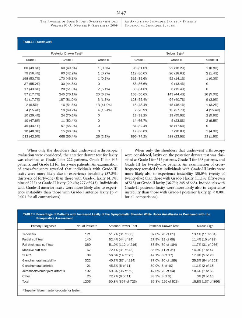
When only the shoulders that underwent arthroscopicevaluation were considered, the anterior drawer test for laxitywas classified as Grade I for 222 patients, Grade II for 943patients, and Grade III for forty-one patients. An examinationof cross-frequency revealed that individuals with Grade-IIIlaxity were more likely also to experience instability (87.8%;thirty-six of forty-one) than those with Grade-I laxity (4.1%;nine of 222) or Grade-II laxity (29.4%; 277 of 943). Individualswith Grade-II anterior laxity were more likely also to experi-ence instability than those with Grade-I anterior laxity (p <0.001 for all comparisons).
When only the shoulders that underwent arthroscopywere considered, laxity on the posterior drawer test was clas-sified as Grade I for 513 patients, Grade II for 668 patients, andGrade III for twenty-five patients. An examination of cross-frequency revealed that individuals with Grade-III laxity weremore likely also to experience instability (80.0%; twenty oftwenty-five) than those with Grade-I laxity (11.1%; fifty-sevenof 513) or Grade-II laxity (36.7%; 245 of 668). Individuals withGrade-II posterior laxity were more likely also to experienceinstability than those with Grade-I posterior laxity (p < 0.001for all comparisons).
Posterior Drawer Test* Sulcus Sign*
Grade I Grade II Grade III Grade I Grade II Grade III
60 (49.6%) 60 (49.6%) 1 (0.8%) 98 (81.0%) 22 (18.2%) 1 (0.8%)
79 (56.4%) 60 (42.9%) 1 (0.7%) 112 (80.0%) 26 (18.6%) 2 (1.4%)
198 (53.7%) 170 (46.1%) 1 (0.3%) 316 (85.6%) 52 (14.1%) 1 (0.3%)
37 (55.2%) 30 (44.8%) 0 58 (86.6%) 9 (13.4%) 0
17 (43.6%) 20 (51.3%) 2 (5.1%) 33 (84.6%) 6 (15.4%) 0
57 (17.7%) 245 (76.1%) 20 (6.2%) 163 (50.6%) 143 (44.4%) 16 (5.0%)
41 (17.7%) 187 (81.0%) 3 (1.3%) 128 (55.4%) 94 (40.7%) 9 (3.9%)
2 (6.5%) 16 (51.6%) 13 (41.9%) 15 (48.4%) 15 (48.1%) 1 (3.2%)
4 (15.4%) 18 (69.2%) 4 (15.4%) 7 (26.9%) 15 (57.7%) 4 (15.4%)
10 (29.4%) 24 (70.6%) 0 13 (38.2%) 19 (55.9%) 2 (5.9%)
10 (47.6%) 11 (52.4%) 0 14 (66.7%) 5 (23.8%) 2 (9.5%)
45 (44.1%) 57 (55.9%) 0 84 (82.4%) 18 (17.6%) 0
10 (40.0%) 15 (60.0%) 0 17 (68.0%) 7 (28.0%) 1 (4.0%)
513 (42.5%) 668 (55.4%) 25 (2.1%) 895 (74.2%) 288 (23.9%) 23 (1.9%)
TABLE I (continued)
TABLE II Percentage of Patients with Increased Laxity of the Symptomatic Shoulder While Under Anesthesia as Compared with the
Preoperative Assessment
Primary Diagnosis No. of Patients Anterior Drawer Test Posterior Drawer Test Sulcus Sign
Tendinitis 121 51.7% (31 of 60) 32.8% (20 of 61) 13.1% (11 of 84)
Partial cuff tear 140 52.4% (44 of 84) 27.9% (19 of 68) 11.4% (10 of 88)
Full-thickness cuff tear 369 51.9% (112 of 216) 37.5% (69 of 184) 11.7% (31 of 266)
Massive cuff tear 67 72.1% (31 of 43) 35.5% (11 of 31) 14.9% (7 of 47)
SLAP* 39 56.0% (14 of 25) 47.1% (8 of 17) 17.9% (5 of 28)
Glenohumeral instability 322 40.7% (87 of 214) 37.0% (70 of 189) 25.3% (64 of 253)
Glenohumeral arthritis 21 45.5% (5 of 11) 30.0% (3 of 10) 11.1% (2 of 18)
Acromioclavicular joint arthritis 102 59.3% (35 of 59) 42.6% (23 of 54) 10.6% (7 of 66)
Other 25 72.7% (8 of 11) 33.3% (3 of 9) 0% (0 of 16)
Total 1206 50.8% (367 of 723) 36.3% (226 of 623) 15.8% (137 of 866)
*Superior labrum anterior-posterior lesion.
2147
TH E J O U R N A L O F B O N E & JO I N T SU R G E RY d J B J S . O R G
VO LU M E 91-A d NU M B E R 9 d S E P T E M B E R 2009AN A N A LY S I S O F SH O U L D E R LA X I T Y I N PAT I E N T S
UN D E R G O I N G SH O U L D E R SU R G E RY
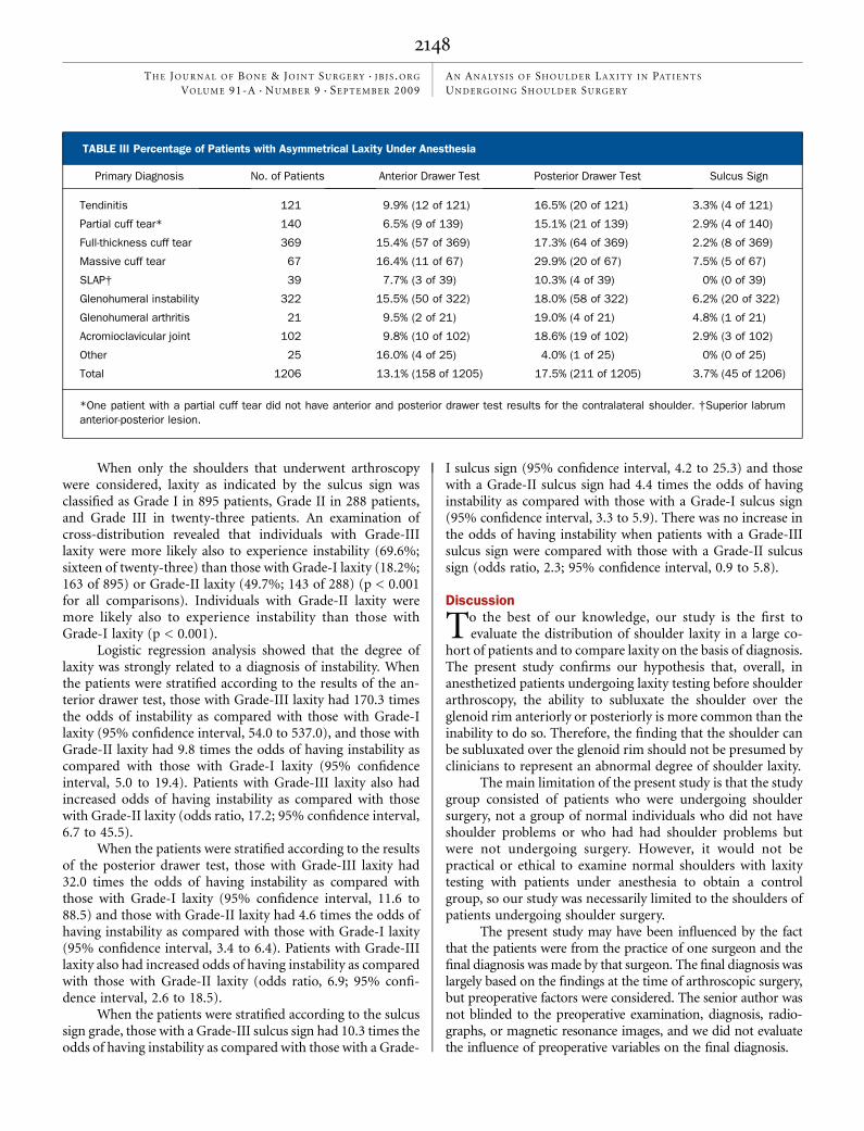
When only the shoulders that underwent arthroscopywere considered, laxity as indicated by the sulcus sign wasclassified as Grade I in 895 patients, Grade II in 288 patients,and Grade III in twenty-three patients. An examination ofcross-distribution revealed that individuals with Grade-IIIlaxity were more likely also to experience instability (69.6%;sixteen of twenty-three) than those with Grade-I laxity (18.2%;163 of 895) or Grade-II laxity (49.7%; 143 of 288) (p < 0.001for all comparisons). Individuals with Grade-II laxity weremore likely also to experience instability than those withGrade-I laxity (p < 0.001).
Logistic regression analysis showed that the degree oflaxity was strongly related to a diagnosis of instability. Whenthe patients were stratified according to the results of the an-terior drawer test, those with Grade-III laxity had 170.3 timesthe odds of instability as compared with those with Grade-Ilaxity (95% confidence interval, 54.0 to 537.0), and those withGrade-II laxity had 9.8 times the odds of having instability ascompared with those with Grade-I laxity (95% confidenceinterval, 5.0 to 19.4). Patients with Grade-III laxity also hadincreased odds of having instability as compared with thosewith Grade-II laxity (odds ratio, 17.2; 95% confidence interval,6.7 to 45.5).
When the patients were stratified according to the resultsof the posterior drawer test, those with Grade-III laxity had32.0 times the odds of having instability as compared withthose with Grade-I laxity (95% confidence interval, 11.6 to88.5) and those with Grade-II laxity had 4.6 times the odds ofhaving instability as compared with those with Grade-I laxity(95% confidence interval, 3.4 to 6.4). Patients with Grade-IIIlaxity also had increased odds of having instability as comparedwith those with Grade-II laxity (odds ratio, 6.9; 95% confi-dence interval, 2.6 to 18.5).
When the patients were stratified according to the sulcussign grade, those with a Grade-III sulcus sign had 10.3 times theodds of having instability as compared with those with a Grade-
I sulcus sign (95% confidence interval, 4.2 to 25.3) and thosewith a Grade-II sulcus sign had 4.4 times the odds of havinginstability as compared with those with a Grade-I sulcus sign(95% confidence interval, 3.3 to 5.9). There was no increase inthe odds of having instability when patients with a Grade-IIIsulcus sign were compared with those with a Grade-II sulcussign (odds ratio, 2.3; 95% confidence interval, 0.9 to 5.8).
Discussion
To the best of our knowledge, our study is the first toevaluate the distribution of shoulder laxity in a large co-
hort of patients and to compare laxity on the basis of diagnosis.The present study confirms our hypothesis that, overall, inanesthetized patients undergoing laxity testing before shoulderarthroscopy, the ability to subluxate the shoulder over theglenoid rim anteriorly or posteriorly is more common than theinability to do so. Therefore, the finding that the shoulder canbe subluxated over the glenoid rim should not be presumed byclinicians to represent an abnormal degree of shoulder laxity.
The main limitation of the present study is that the studygroup consisted of patients who were undergoing shouldersurgery, not a group of normal individuals who did not haveshoulder problems or who had had shoulder problems butwere not undergoing surgery. However, it would not bepractical or ethical to examine normal shoulders with laxitytesting with patients under anesthesia to obtain a controlgroup, so our study was necessarily limited to the shoulders ofpatients undergoing shoulder surgery.
The present study may have been influenced by the factthat the patients were from the practice of one surgeon and thefinal diagnosis was made by that surgeon. The final diagnosis waslargely based on the findings at the time of arthroscopic surgery,but preoperative factors were considered. The senior author wasnot blinded to the preoperative examination, diagnosis, radio-graphs, or magnetic resonance images, and we did not evaluatethe influence of preoperative variables on the final diagnosis.
TABLE III Percentage of Patients with Asymmetrical Laxity Under Anesthesia
Primary Diagnosis No. of Patients Anterior Drawer Test Posterior Drawer Test Sulcus Sign
Tendinitis 121 9.9% (12 of 121) 16.5% (20 of 121) 3.3% (4 of 121)
Partial cuff tear* 140 6.5% (9 of 139) 15.1% (21 of 139) 2.9% (4 of 140)
Full-thickness cuff tear 369 15.4% (57 of 369) 17.3% (64 of 369) 2.2% (8 of 369)
Massive cuff tear 67 16.4% (11 of 67) 29.9% (20 of 67) 7.5% (5 of 67)
SLAP† 39 7.7% (3 of 39) 10.3% (4 of 39) 0% (0 of 39)
Glenohumeral instability 322 15.5% (50 of 322) 18.0% (58 of 322) 6.2% (20 of 322)
Glenohumeral arthritis 21 9.5% (2 of 21) 19.0% (4 of 21) 4.8% (1 of 21)
Acromioclavicular joint 102 9.8% (10 of 102) 18.6% (19 of 102) 2.9% (3 of 102)
Other 25 16.0% (4 of 25) 4.0% (1 of 25) 0% (0 of 25)
Total 1206 13.1% (158 of 1205) 17.5% (211 of 1205) 3.7% (45 of 1206)
*One patient with a partial cuff tear did not have anterior and posterior drawer test results for the contralateral shoulder. †Superior labrumanterior-posterior lesion.
2148
TH E J O U R N A L O F B O N E & JO I N T SU R G E RY d J B J S . O R G
VO LU M E 91-A d NU M B E R 9 d S E P T E M B E R 2009AN A N A LY S I S O F SH O U L D E R LA X I T Y I N PAT I E N T S
UN D E R G O I N G SH O U L D E R SU R G E RY
Our findings may not be transferable to the evaluation ofshoulder laxity in the office setting. The present study andprevious studies11,16,34 documented that laxity in the shoulder isincreased with the patient under anesthesia as compared withthat determined during an office examination. One study dem-onstrated that approximately 80% of patients relax enough foranterior drawer testing in the office1. In the current study, allpatients were examined while under a general anesthetic withor without a regional block, so different distributions of laxityaccording to diagnosis may be explained by the fact that, inother studies, a general anesthetic was not used.
Any study of laxity testing of the shoulder is confoundedby the lack of a gold standard measurement. In the presentstudy, we used a scale for laxity based on what the clinicianfeels when performing the examination, and this measure isnot currently quantifiable. As a result, statistical analysis oflaxity did not allow parametric analysis of measurable dis-tances. To our knowledge, the amount of translation necessaryto create Grade-I, II, or III laxity in an anterior, posterior, orinferior direction has not been established. Similarly, it may bethat subtle differences in shoulder laxity (which possibly couldhave influenced our findings and conclusions) are not mea-surable with use of existing shoulder examination methods. Inaddition, the amount of force used by the senior author tosubluxate the shoulder was not quantified, but this short-coming is inherent to any study involving the use of suchuninstrumented examination techniques for the examinationof shoulder laxity.
Despite these limitations, an important variable of ourstudy was that one clinician performed all of the shoulderexaminations of patients who were under anesthesia for theentire time period with use of the same technique and the samescale for measuring laxity. A previous study showed that theintraobserver reliability of the examination by this one ex-aminer was 100% (twenty-eight of twenty-eight) for anteriortranslation and 86% (twenty-four of twenty-eight) for poste-rior translation23. Similarly, an interobserver study of thismethod of measuring shoulder laxity showed that the agreementbetween observers using this technique was 77% (thirty-fourof forty-four) for anterior drawer testing and 70% (thirty-one
of forty-four) for posterior drawer testing23. However, in thecurrent study, the examiner was not blinded to the patient’sdiagnosis, and it is possible that observer bias influenced ourresults. It may be that the results reported here are not trans-ferable to a wider range of examiners because the reliability ofthe examination for a wide range of examiners has not beenstudied extensively.
The technique used for measuring anterior and posteriorshoulder laxity in the present study differed slightly from thatoriginally described by Gerber and Ganz29, and it is unknownhow that factor might have affected the results.
The present study confirms our observation that theability to subluxate the shoulder over the glenoid rim is morecommon than previously believed. We also found that the dis-tribution of shoulder laxities is related to the diagnosis, withincreasing grades of shoulder laxity associated with a higherlikelihood of shoulder instability. Until instruments for themeasurement of laxity are available, the subjective aspect ofpresent systems to grade shoulder laxity limits the clinician’sability to use laxity testing to make a definitive diagnosis ofshoulder instability. The clinical examination of shoulderlaxity with the patient under anesthesia should be performedwith the knowledge that the ability to subluxate the shoulderover the glenoid rim is common regardless of diagnosis andthat the extent and distribution of shoulder laxities may varyaccording to diagnosis. n
Xiaofeng Jia, MD, PhDJong Hun Ji, MDSteve A. Petersen, MDMichael T. Freehill, MDEdward G. McFarland, MDc/o Elaine P. Henze, BJ, ELS, Medical Editor,Department of Orthopaedic Surgery,Johns Hopkins Bayview Medical Center,4940 Eastern Avenue, #A665, Baltimore,MD 21224-2780.E-mail address for E.P. Henze: [email protected]
References
1. Farber AJ, Castillo R, Clough M, Bahk M, McFarland EG. Clinical assessment ofthree common tests for traumatic anterior shoulder instability. J Bone Joint SurgAm. 2006;88:1467-74.
2. Silliman JF, Hawkins RJ. Classification and physical diagnosis of instability ofthe shoulder. Clin Orthop Relat Res. 1993;291:7-19.
3. Jobe CM, Pink MM, Jobe FW, Shaffer B. Anterior shoulder instability, impinge-ment, and rotator cuff tear: theories and concepts. In: Jobe FW, editor. Operativetechniques in upper extremity sports injuries. St. Louis, MO: Mosby; 1996.p 164-76.
4. Jobe FW, Kvitne RS. Shoulder pain in the overhand or throwing athlete. Therelationship of anterior instability and rotator cuff impingement. Orthop Rev.1989;18:963-75. Erratum in: Orthop Rev. 1989;18:1268.
5. Matsen FA 3rd, Chebli CM, Lippitt SB. Principles for the evaluation and man-agement of shoulder instability. Instr Course Lect. 2007;56:23-34.
6. McFarland EG. Instability and laxity. In: McFarland EG, Kim TK, Park HB, El RassiG, Gill H, Keyurapan E, editors. Examination of the shoulder: the complete guide.New York: Thieme; 2006. p 162-212.
7. Jobe FW, Giangarra CE, Kvitne RS, Glousman RE. Anterior capsulolabral re-construction of the shoulder in athletes in overhand sports. Am J Sports Med.1991;19:428-34.
8. Bahk M, Keyurapan E, Tasaki A, Sauers EL, McFarland EG. Laxity testing ofthe shoulder: a review. Am J Sports Med. 2007;35:131-44.
9. Borsa PA, Jacobson JA, Scibek JS, Dover GC. Comparison of dynamic sonog-raphy to stress radiography for assessing glenohumeral laxity in asymptomaticshoulders. Am J Sports Med. 2005;33:734-41.
10. Harryman DT 2nd, Sidles JA, Clark JM, McQuade KJ, Gibb TD, Matsen FA 3rd.Translation of the humeral head on the glenoid with passive glenohumeral motion.J Bone Joint Surg Am. 1990;72:1334-43.
2149
TH E J O U R N A L O F B O N E & JO I N T SU R G E RY d J B J S . O R G
VO LU M E 91-A d NU M B E R 9 d S E P T E M B E R 2009AN A N A LY S I S O F SH O U L D E R LA X I T Y I N PAT I E N T S
UN D E R G O I N G SH O U L D E R SU R G E RY
11. Hawkins RJ, Schutte JP, Janda DH, Huckell GH. Translation of the glenohu-meral joint with the patient under anesthesia. J Shoulder Elbow Surg. 1996;5:286-92.
12. Lintner SA, Levy A, Kenter K, Speer KP. Glenohumeral translation in theasymptomatic athlete’s shoulder and its relationship to other clinically measurableanthropometric variables. Am J Sports Med. 1996;24:716-20.
13. McFarland EG, Campbell G, McDowell J. Posterior shoulder laxity in asymp-tomatic athletes. Am J Sports Med. 1996;24:468-71.
14. McFarland EG, Hsu CY, Neira C, O’Neil O. Internal impingement of theshoulder: a clinical and arthroscopic analysis. J Shoulder Elbow Surg. 1999;8:458-60.
15. Tibone JE, Lee TQ, Csintalan RP, Dettling J, McMahon PJ. Quantitativeassessment of glenohumeral translation. Clin Orthop Relat Res. 2002;400:93-7.
16. Yoldas EA, Faber KJ, Hawkins RJ. Translation of the glenohumeral jointin patients with multidirectional and posterior instability: awake examinationversus examination under anesthesia. J Shoulder Elbow Surg. 2001;10:416-20.
17. Chronopoulos E, Kim TK, Park HB, Ashenbrenner D, McFarland EG. Diagnosticvalue of physical tests for isolated chronic acromioclavicular lesions. Am J SportsMed. 2004;32:655-61.
18. Kim TK, Queale WS, Cosgarea AJ, McFarland EG. Clinical features of thedifferent types of SLAP lesions: an analysis of one hundred and thirty-nine cases.J Bone Joint Surg Am. 2003;85:66-71.
19. Kim TK, Rauh PB, McFarland EG. Partial tears of the subscapularis tendonfound during arthroscopic procedures on the shoulder: a statistical analysis of sixtycases. Am J Sports Med. 2003;31:744-50.
20. MacDonald PB, Clark P, Sutherland K. An analysis of the diagnostic accuracyof the Hawkins and Neer subacromial impingement signs. J Shoulder Elbow Surg.2000;9:299-301.
21. McFarland EG, Neira CA, Gutierrez MI, Cosgarea AJ, Magee M. Clinical sig-nificance of the arthroscopic drive-through sign in shoulder surgery. Arthroscopy.2001;17:38-43.
22. McFarland EG, Kim TK, Savino RM. Clinical assessment of three commontests for superior labral anterior-posterior lesions. Am J Sports Med. 2002;30:810-5.
23. McFarland EG, Kim TK, Park HB, Neira CA, Gutierrez MI. The effect of variationin definition on the diagnosis of multidirectional instability of the shoulder. J BoneJoint Surg Am. 2003;85:2138-44.
24. Park HB, Yokota A, Gill HS, El Rassi G, McFarland EG. Diagnostic accuracy ofclinical tests for the different degrees of subacromial impingement syndrome.J Bone Joint Surg Am. 2005;87:1446-55.
25. Rao AG, Kim TK, Chronopoulos E, McFarland EG. Anatomical variants in theanterosuperior glenoid labrum: a statistical analysis of seventy-three cases. J BoneJoint Surg Am. 2003;85:653-9.
26. Jia X, Ji JH, Petersen SA, Keefer J, McFarland EG. Clinical evaluation of theshoulder shrug sign. Clin Orthop Relat Res. 2008;466:2813-9.
27. Farmer KW, Sonin A, Kim TK, McFarland EG. Unusual pattern of injuries fol-lowing knee hyperextension: a case report. Clin J Sport Med. 2003;13:53-6.
28. Kim TK, McFarland EG. Internal impingement of the shoulder in flexion. ClinOrthop Relat Res. 2004;421:112-9.
29. Gerber C, Ganz R. Clinical assessment of instability of the shoulder. Withspecial reference to anterior and posterior drawer tests. J Bone Joint Surg Br.1984;66:551-6.
30. Altchek DW, Warren RF, Skyhar MJ, Ortiz G. T-plasty modification of theBankart procedure for multidirectional instability of the anterior and inferior types.J Bone Joint Surg Am. 1991;73:105-12.
31. Allen AA. Clinical evaluation of the unstable shoulder. In: Warren RF, Craig EV,Altchek DW, editors. The unstable shoulder. Philadelphia: Lippincott-Raven; 1999.p 93-106.
32. Rowe CR, Zarins B. Recurrent transient subluxation of the shoulder. J BoneJoint Surg Am. 1981;63:863-72.
33. DeOrio JK, Cofield RH. Results of a second attempt at surgical repair of afailed initial rotator-cuff repair. J Bone Joint Surg Am. 1984;66:563-7.
34. Faber KJ, Homa K, Hawkins RJ. Translation of the glenohumeral joint inpatients with anterior instability: awake examination versus examination withthe patient under anesthesia. J Shoulder Elbow Surg. 1999;8:320-3.
2150
TH E J O U R N A L O F B O N E & JO I N T SU R G E RY d J B J S . O R G
VO LU M E 91-A d NU M B E R 9 d S E P T E M B E R 2009AN A N A LY S I S O F SH O U L D E R LA X I T Y I N PAT I E N T S
UN D E R G O I N G SH O U L D E R SU R G E RY
Related Documents


