An AFM/Rotaxane Molecular Reading Head for Sequence- Dependent DNA Structure ** Brian A. Ashcroft 1 , Biodesign Institute, Arizona State University, PO Box 875601, Tempe, AZ 85287, (USA) Quinn Spadola 1 , Biodesign Institute, Arizona State University, PO Box 875601, Tempe, AZ 85287, (USA) Shahid Qamar, Biodesign Institute, Arizona State University, PO Box 875601, Tempe, AZ 85287, (USA) Peiming Zhang, Biodesign Institute, Arizona State University, PO Box 875601, Tempe, AZ 85287, (USA) Gerald Kada, Agilent Technologies, Nanotechnology Measurements Division Roemerstr. 18, 4020 Linz (Austria) Rouvain Bension, and Neotech Development Co. LLC, 310 Summit Ave., Brighton, MA 02135-7504 (USA) Stuart Lindsay The Biodesign Institute, Department of Physics, Department of Chemistry and Biochemistry, Arizona State University, PO Box 875601, Tempe, AZ 85281 (USA), Fax: 480-727-2378 E-mail: [email protected] Abstract A nanomechanical molecular “tape reader” is assembled and tested by threading a β-cyclodextrin ring onto a DNA oligomer and pulling it along with an AFM tip. The formation and mechanical operation of the system is confirmed by measuring the forces required to unfold secondary structures in the form of hairpins. Unfolding induced by this 0.7 nm aperture requires 40 times more force than that reported for pulling on the ends of the DNA. A kinetic analysis shows that much less strain is required to destabilize the double helix in this geometry. Consequently, much more force is required to provide the free energy needed for opening. DNA secondary structure may prove to be a significant obstacle both for enzymes that process DNA though an orifice, and for the passage through nanopores proposed for some novel sequencing schemes. Keywords AFM; Biophysics; DNA Structure; Force microscopy ** This work was supported by the DNA sequencing technology program of the NHGRI and by DARPA. We thank Jan Liphardt for useful discussions. Correspondence to: Stuart Lindsay. 1 These authors contributed equally to this work. NIH Public Access Author Manuscript Small. Author manuscript; available in PMC 2008 December 8. Published in final edited form as: Small. 2008 September ; 4(9): 1468–1475. doi:10.1002/smll.65476. NIH-PA Author Manuscript NIH-PA Author Manuscript NIH-PA Author Manuscript

Welcome message from author
This document is posted to help you gain knowledge. Please leave a comment to let me know what you think about it! Share it to your friends and learn new things together.
Transcript

An AFM/Rotaxane Molecular Reading Head for Sequence-Dependent DNA Structure**
Brian A. Ashcroft1,Biodesign Institute, Arizona State University, PO Box 875601, Tempe, AZ 85287, (USA)
Quinn Spadola1,Biodesign Institute, Arizona State University, PO Box 875601, Tempe, AZ 85287, (USA)
Shahid Qamar,Biodesign Institute, Arizona State University, PO Box 875601, Tempe, AZ 85287, (USA)
Peiming Zhang,Biodesign Institute, Arizona State University, PO Box 875601, Tempe, AZ 85287, (USA)
Gerald Kada,Agilent Technologies, Nanotechnology Measurements Division Roemerstr. 18, 4020 Linz (Austria)
Rouvain Bension, andNeotech Development Co. LLC, 310 Summit Ave., Brighton, MA 02135-7504 (USA)
Stuart LindsayThe Biodesign Institute, Department of Physics, Department of Chemistry and Biochemistry,Arizona State University, PO Box 875601, Tempe, AZ 85281 (USA), Fax: 480-727-2378 E-mail:[email protected]
AbstractA nanomechanical molecular “tape reader” is assembled and tested by threading a β-cyclodextrinring onto a DNA oligomer and pulling it along with an AFM tip. The formation and mechanicaloperation of the system is confirmed by measuring the forces required to unfold secondary structuresin the form of hairpins. Unfolding induced by this 0.7 nm aperture requires 40 times more force thanthat reported for pulling on the ends of the DNA. A kinetic analysis shows that much less strain isrequired to destabilize the double helix in this geometry. Consequently, much more force is requiredto provide the free energy needed for opening. DNA secondary structure may prove to be a significantobstacle both for enzymes that process DNA though an orifice, and for the passage through nanoporesproposed for some novel sequencing schemes.
KeywordsAFM; Biophysics; DNA Structure; Force microscopy
**This work was supported by the DNA sequencing technology program of the NHGRI and by DARPA. We thank Jan Liphardt foruseful discussions.Correspondence to: Stuart Lindsay.1These authors contributed equally to this work.
NIH Public AccessAuthor ManuscriptSmall. Author manuscript; available in PMC 2008 December 8.
Published in final edited form as:Small. 2008 September ; 4(9): 1468–1475. doi:10.1002/smll.65476.
NIH
-PA Author Manuscript
NIH
-PA Author Manuscript
NIH
-PA Author Manuscript

1. IntroductionIn nature, enzymes encircle biopolymers, forming rotaxanes to avoid falling off as they read,exemplified by the sliding clamp subunit of DNA polymerase.[1] Here we report an AFM-based molecular reading device for determining the force required for sequence related featuresof nucleic acids to pass through a small encircling orifice. The system is sensitive enough tomeasure unfolding of nucleic acid secondary structures. Most current single-moleculemeasurement techniques pull biomolecules apart by applying stress to each end of the polymerchain, a stretching process that might result in an unfolding pathway different from the shearforce imposed by a sliding clamp.[2,3] Optical tweezers have been used to measure theelectrophoretic force on a single DNA molecule translocating through a nanopore.[4] Proteinpores have been used to trap DNA molecules[5] and the force required to open hairpins hasbeen inferred from studies of electrophoretically-driven transit through these pores.[6–8] Theredo not appear to be any direct measurements of the force required to unfold secondary structuresequentially by pulling a polymer through an orifice (or, as is the case here, the orifice overthe polymer). The present construct may prove expandable to a variety of studies that requiresequential processing, serving as a model system for an enzyme processing a nucleic acid, andpotentially providing a method for sequencing heteropolymers like DNA and proteins at themonomer level.[9]
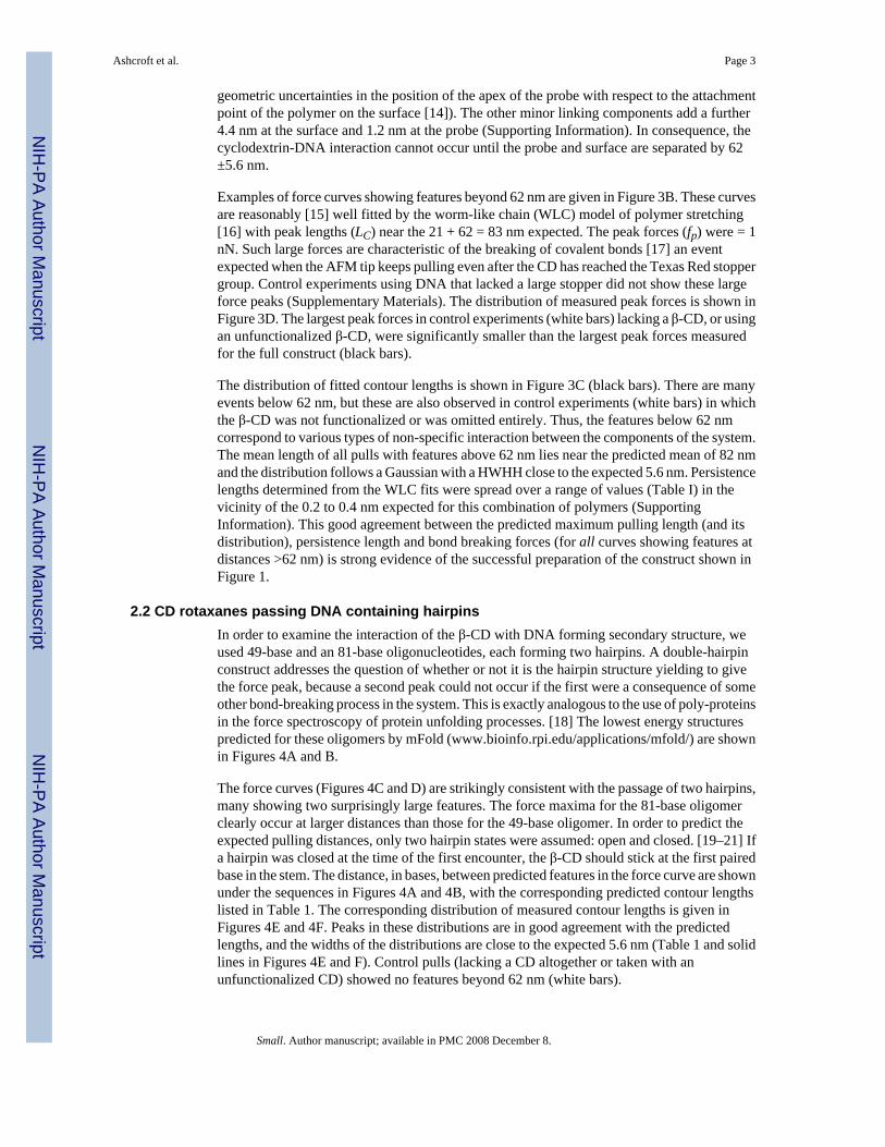
A molecular reading head that resembles the sliding clamp of DNA polymerase was self-assembled on a threading molecule, forming a rotaxane[10,11] by locking each end of thethread with a large stopper[12] In contrast to conventional rotaxanes, one stopper is the surfaceto which the DNA is to be tethered, covered with anchors for the threading molecule scatteredin a non-stick layer (Figure 1A). The assembly sequence is as follows:Adamantanedicarboxylic acid is attached to the anchor, diaminododecane to the adamantane,and the resulting threading molecule (with a free amino end) spontaneously complexesfunctionalized β-cyclodextrin (β-CD), the smallest CD that fits over ssDNA. A poly(ethyleneglycol) molecule (PEG) is attached to the free end of the dodecyl group to function as atemporary stopper. It holds the β-CD in place until it is pulled onto the PEG by an AFM. Finally,a DNA molecule is attached to the free end of the PEG. Translocation of the β-CD, andsimultaneous force read-out, is achieved by fishing with a PEG “line” for a thiol group on theβ-CD, the fishing “rod” being an atomic force microscope (AFM) probe. When the two PEGmolecules are fully extended into a linear structure, the (shorter) DNA is lifted entirely fromthe surface, so that force signals beyond this point must result from interactions with the DNAand not with the surface. The steps required to accomplish this assembly of the CD/DNArotaxane are shown in Figure 2. Full details of the synthesis and characterization of this systemare given in the experimental section and the Supporting Information online.
2. Results2.1 CD/DNA rotaxanes without secondary structure
Silicon AFM probes bearing vinylsulfone functionalized PEG (MW: 3200 Dalton ) were usedto capture freshly deprotected thiolated β-CDs. Force curves were obtained in 50 mMphosphate buffer solution (PBS, pH = 7). using the liquid cell of a Picoplus AFM (MolecularImaging/Agilent).
The formation of surface-tethered rotaxanes was verified using a test sample consisting of a5′-thiolated oligo-T stoppered at its 3′ end with a Texas Red dye molecule (Figure 3A). Fullyextended, this molecule has a length of 21.4 nm (using a stretched base-to-base distance of0.61 nm [13]). Each PEG linker (one on the surface and one on the probe – see Figure 1) hasa Gaussian distribution of fully-stretched lengths with a mean value of 28 nm and a half-widthat half-height (HWHH) of 2.8 nm (arising mainly from polydispersity but also including
Ashcroft et al. Page 2
Small. Author manuscript; available in PMC 2008 December 8.
NIH
-PA Author Manuscript
NIH
-PA Author Manuscript
NIH
-PA Author Manuscript
geometric uncertainties in the position of the apex of the probe with respect to the attachmentpoint of the polymer on the surface [14]). The other minor linking components add a further4.4 nm at the surface and 1.2 nm at the probe (Supporting Information). In consequence, thecyclodextrin-DNA interaction cannot occur until the probe and surface are separated by 62±5.6 nm.
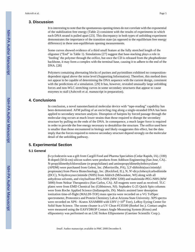
Examples of force curves showing features beyond 62 nm are given in Figure 3B. These curvesare reasonably [15] well fitted by the worm-like chain (WLC) model of polymer stretching[16] with peak lengths (LC) near the 21 + 62 = 83 nm expected. The peak forces (fp) were = 1nN. Such large forces are characteristic of the breaking of covalent bonds [17] an eventexpected when the AFM tip keeps pulling even after the CD has reached the Texas Red stoppergroup. Control experiments using DNA that lacked a large stopper did not show these largeforce peaks (Supplementary Materials). The distribution of measured peak forces is shown inFigure 3D. The largest peak forces in control experiments (white bars) lacking a β-CD, or usingan unfunctionalized β-CD, were significantly smaller than the largest peak forces measuredfor the full construct (black bars).
The distribution of fitted contour lengths is shown in Figure 3C (black bars). There are manyevents below 62 nm, but these are also observed in control experiments (white bars) in whichthe β-CD was not functionalized or was omitted entirely. Thus, the features below 62 nmcorrespond to various types of non-specific interaction between the components of the system.The mean length of all pulls with features above 62 nm lies near the predicted mean of 82 nmand the distribution follows a Gaussian with a HWHH close to the expected 5.6 nm. Persistencelengths determined from the WLC fits were spread over a range of values (Table I) in thevicinity of the 0.2 to 0.4 nm expected for this combination of polymers (SupportingInformation). This good agreement between the predicted maximum pulling length (and itsdistribution), persistence length and bond breaking forces (for all curves showing features atdistances >62 nm) is strong evidence of the successful preparation of the construct shown inFigure 1.
2.2 CD rotaxanes passing DNA containing hairpinsIn order to examine the interaction of the β-CD with DNA forming secondary structure, weused 49-base and an 81-base oligonucleotides, each forming two hairpins. A double-hairpinconstruct addresses the question of whether or not it is the hairpin structure yielding to givethe force peak, because a second peak could not occur if the first were a consequence of someother bond-breaking process in the system. This is exactly analogous to the use of poly-proteinsin the force spectroscopy of protein unfolding processes. [18] The lowest energy structurespredicted for these oligomers by mFold (www.bioinfo.rpi.edu/applications/mfold/) are shownin Figures 4A and B.
The force curves (Figures 4C and D) are strikingly consistent with the passage of two hairpins,many showing two surprisingly large features. The force maxima for the 81-base oligomerclearly occur at larger distances than those for the 49-base oligomer. In order to predict theexpected pulling distances, only two hairpin states were assumed: open and closed. [19–21] Ifa hairpin was closed at the time of the first encounter, the β-CD should stick at the first pairedbase in the stem. The distance, in bases, between predicted features in the force curve are shownunder the sequences in Figures 4A and 4B, with the corresponding predicted contour lengthslisted in Table 1. The corresponding distribution of measured contour lengths is given inFigures 4E and 4F. Peaks in these distributions are in good agreement with the predictedlengths, and the widths of the distributions are close to the expected 5.6 nm (Table 1 and solidlines in Figures 4E and F). Control pulls (lacking a CD altogether or taken with anunfunctionalized CD) showed no features beyond 62 nm (white bars).
Ashcroft et al. Page 3
Small. Author manuscript; available in PMC 2008 December 8.
NIH
-PA Author Manuscript
NIH
-PA Author Manuscript
NIH
-PA Author Manuscript
2.3 Frequency of spontaneous hairpin openingThe free energies stabilizing these hairpins are quite small (Table 2) and the frequency of curveswith only one feature corresponding to the second hairpin might be expected to give a measureof the spontaneous (unforced) opening rate of the first hairpin. It is open in 30% of the pullsfor both the 49 base and 81 oligomer (it has the same stem sequence in both). ΔG = −RT ln([open]/[closed]) [21] yields ΔG = 0.2 kcal/mol, well below the 3.7 kcal/mol predicted bymFold. This underestimate is to be expected, because forced opening events for which the peakforce is lower than the noise floor (ca. 20 pN) are not detected, and hence not counted in thedenominator.
2.4 Magnitude of the opening forceThe force required to open the hairpins by this method (up to 400 pN – Table 2) is remarkablylarge. Pulling apart the two strands that enter the double helical stem requires forces in therange of 10–15 pN, [21,22] consistent with the model of Cocco et al. [23] How could the samestructure sustain a force some 40 times larger in the present geometry? The application of aforce leads to unfolding as the work done on the folded molecule tilts the energy landscapetowards the unfolded state. [21,22,24,25] For a single transition state that remains unchangedduring the application of force, the distribution of opening forces is given by [24,25]
(1)
where τ0 is the mean time between thermal (unloaded) opening, rf is the loading rate in N/sand fB = kBT / xts where xts is the distance from the equilibrium closed position to the positionof the transition state for opening, measured along the reaction coordinate for unfolding.Solving Equation 1 for the modal value of the force (f*) yields
(2)
The modal force is plotted as a function of loading rate for the three different hairpins in Figures5A, C and E. The solid lines are fits to Equation 2 and these fits yield the values of τ0 and xtslisted in Table 2. As a check on the procedure, the parameters obtained by fitting Equation 2were used to calculate the distribution of opening forces [Eq. 1] at a given loading rate for eachof the hairpins. Calculated (solid lines) and measured force distributions are shown in Figures5B, D and F. The thermal opening times at zero force – on the order of 10 to 100 ms – agreewell with those reported by other methods. [6, 7, 22, 26] The key difference in this case lies inthe much smaller distance to the transition state (xts) measured here: 0.05 nm compared to the5 to 26 nm measured when the force is applied via opposing strands. [21, 22] Thus the increasedforce required to open the hairpins by this method is a consequence of the much smaller strainrequired to destabilize the structure. More force is required to produce the destabilizing freeenergy because it acts over a shorter distance. The unfolding pathway is “brittle” compared tothe pathway followed in experiments that pull on opposite ends of the double helical stem.
The kinetics of hairpin opening has been studied in a protein nanopore [6] yielding values forτ0 which are with a factor of 3 to 10 times longer than the values we report here. It may be thatwe fail to sample the slowest events because of the relatively rapid force ramp used here. Inthe nanpore experiments, the quantity that is analogous to fB is VB, the bias that determines thevoltage scale for electric-field driven opening. VB is determined by the ratio of thermal energyto the charge acted on in the pore, kBT / q, and thus does not yield information about the distanceto a transition state. It is therefore not possible to compare the forces that act on the hairpin inthe two experiments without a microscopic model of the nanopore.
Ashcroft et al. Page 4
Small. Author manuscript; available in PMC 2008 December 8.
NIH
-PA Author Manuscript
NIH
-PA Author Manuscript
NIH
-PA Author Manuscript
3. DiscussionIt is interesting to note that the spontaneous opening times do not correlate with the exponentialof the stabilization free energy (Table 2) consistent with the results of experiments in whicheach DNA strand is pulled apart [23]. This discrepancy in both types of unfolding experimentdemonstrates the importance of the transition state (as opposed to the equilibrium free energydifference) in these non-equilibrium opening measurements.
Some curves showed evidence of a third small feature at the fully stretched length of theoligomer (“End” in Table 1). Simulations [27] suggest that base-stacking plays a role in‘feeding’ the polymer through the orifice, but once the CD is released from the phophodiesterbackbone, it may form a complex with the terminal base, causing it to adhere to the end of theDNA. [28]
Polymers containing alternating blocks of purines and pyrimidines exhibited no composition-dependent signal above the noise level (Supporting Information). Therefore, this method doesnot appear to be capable of determining the DNA sequence with the current design, consistentwith the predictions of a simulation. [29] It has, however, revealed unusually large unfoldingforces and non-WLC stretching curves in some secondary structures that appear to causeenzymes to stall (Ashcroft et al. manuscript in preparation).
4. ConclusionsIn conclusion, a novel nanomechanical molecular device with “tape-reading” capability hasbeen demonstrated. AFM pulling of an encircling ring along a single-stranded DNA has beenapplied to secondary structure analysis. Disruption of hairpins by forced passage through themolecular ring occurs at much lower strains than those required to disrupt the secondarystructure by pulling on the ends of the DNA. In consequence, a much larger force is requiredin order to provide the free energy necessary to destabilize the structure. The orifice used hereis smaller than those encountered in biology and likely exaggerates this effect, but the dataimply that the forces required to remove secondary structure depend strongly on the moleculardetail of the unfolding pathway.
5. Experimental Section5.1 General
β-cyclodextrin was a gift from Cargill Food and Pharma Specialties (Cedar Rapids, IA). (100)B-doped (50 Ω-cm) silicon wafers were products from Addison Engineering (San Jose, CA)..N-propyldimethylchlorosilane (n-propylsilane) and aminopropyldimethylethoxysilane(APDM) were purchased from Gelest, Inc. (Morrisville, PA), 3,3′-dithiobis(succinimidylpropionate) from Pierce Biotechnology, Inc. (Rockford, IL), N, N′-dicyclohexylcarbodiimide(DCC), N-hydroxysuccinimide (NHS) from Aldrich (Milwaukee, WI) along with allanhydrous solvents, and vinylsulfone-PEG-NHS (MW 3200) and maleimide-PEG-NHS (MW5000) from Nektar Therapeutics (San Carlos, CA). All reagents were used as received. TLCplates were from EMD Chemical Inc (Gibbstown, NJ). Sephadex G-25 Quick Spin columnswere from Roche Applied Science (Indianapolis, IN). Matrix assisted laser desorptionionization time-of-flight (MALDI-TOF) mass spectra were recorded on a VG TofSpecspectrometer, Proteomics and Protein Chemistry Lab at Arizona State University. XPS spectrawere recorded on XPS - Kratos XSAM800 with UHV (~109 Torr), LeRoy Eyring Center forSolid State Science. The ozone cleaner is a UV Clean #135500 (Boekel Inc.). Contact angleswere measured using the EASYDROP Contact Angle Measuring System (Kruss) andellipsometry was performed on an LSE Stokes Ellipsometer (Gaertner Scientific Corp.).
Ashcroft et al. Page 5
Small. Author manuscript; available in PMC 2008 December 8.
NIH
-PA Author Manuscript
NIH
-PA Author Manuscript
NIH
-PA Author Manuscript

5.2 3, 3′-Dithiopropionic acid mono(N-mono-6-deoxy-β-cyclodextrin)amide (S-CD)Mono-6-deoxy-6-amino-β-cyclodextrin (β-CD-NH2) was prepared following the literature[30] and characterized by MALDI mass spectrometry. β-CD-NH2 (16 mg, 0.014 mmol) wasdissolved in anhydrous DMF (10 mL) and added dropwise to a solution of 3,3′-dithiobis(succinimidyl propionate) (0.011 g, 0.028 mmol) in DMF (10 mL) with stirring under N2.[31] The reaction was monitored by TLC (isopropanol:ethyl acetate: ammoniumhydroxide:water, 5:3:3:3), with the presence of the cyclodextrin product marked by treatmentwith 5% sulfuric acid in ethanol with heating. After completion of the reaction, the productmixture was rotary-evaporated down to ~2 mL of DMF. Acetone was added to the solution,the resulting precipitate gravity filtered, and washed with acetone. The precipitate wasredissolved in the minimum amount of DMF, acetone was again added, and the precipitatecollected. This process was repeated one more time. The final precipitate was dried in vacuoat 40°C overnight. The product was furnished as yellowish powder (73%). MALDI-TOF Mass(m/z): 1348 for [M + Na]+ (calculated mass for (C48H79O37NS2): 1325). Further purificationwas carried out using preparative TLC (5:3:3:3 isopropanol:ethyl acetate:ammoniumhydroxide:water, detected with H2SO4 in ethanol) with N-hydroxysuccinimide byproduct usedas a UV detectable marker and methanol used to extract the cyclodextrin (yield 49%). 1H NMR(500MHz, D2O, δ): 5.24 (s, 1 H), 5.07 (s, 6 H), 3.94-3.61 (m, 42 H), 2.94 (t, 2 H), 2.60 (t, 2H). We verified that this modified CD would form a rotaxane on a dodecyl-amine thread insolution (Figure s1 in the Supporting Information).
5.3 Silicon wafer pretreatmentSilicon wafers were cut into 1-cm square slabs and placed in the ozone cleaner for 10 minutes.The slabs were removed and immediately placed in piranha solution (3:1 sulfuric acid andhydrogen peroxide) for 3 minutes, and then rinsed with 18 MΩ water. Thickness of the oxidizedlayer: (3.3Å). Contact angle of the surface: 28°. Caution: piranha solution is extremelycorrosive and can explode on contact with organics!
5.4 DNA conjugation on the CD-rotaxane surfaceAll oligonucleotides were deprotected with tris (2-carboxyethyl)phosphine hydrochloride(TCEP) (0.02% in PBS) for 15 minutes and run through a Sephadex G-25 size exclusion columnbefore use, leaving a thiol linked to the DNA via a hexane linker. The thiolated DNA (0.75nmol in 17 μL of TCEP/PBS solution) was deposited on the CD-rotaxane surface (see Figure2) and allowed to react for 2 hours. The synthesis of a surface bound CD-rotaxane was verifiedusing FTIR (see Supporting Information, Figures s2 through s6). Sequences of all oligomersare given in the Supporting Information (Table s1).
5.5 Preparation of functionalized AFM probesUltrasharp CSC11/AlBS probes were placed in the ozone cleaner for 10 minutes, immediatelydipped into fresh piranha solution for no more than 30 seconds (to prevent damage to themetallization), rinsed with water and put into a solution of APDM (200 μl, 1.4 μmol ) in 95%ethanol (1 mL). After 5 minutes, the tips were rinsed with water, placed in a clean dessicatorflushed with argon, and placed under vacuum (10 Torr) for 1 hour. The tips were removedfrom the vacuum and placed in solution of vinylsulfone -PEG-NHS (45 mg, 0.01 mmol) inPBS buffer (1 mL) pH 7 for 15 minutes. This produced tethered PEG molecules terminated ina vinylsulfone. The probes were then rinsed in water and used immediately. The maximumnumber of successful pulls with any one probe indicated that there were typically 15 to 45functional groups near enough to the end of a probe to be used in the experiment.
Ashcroft et al. Page 6
Small. Author manuscript; available in PMC 2008 December 8.
NIH
-PA Author Manuscript
NIH
-PA Author Manuscript
NIH
-PA Author Manuscript
5.6 Preparation of the system for pulling experimentsThe final step in preparing the samples was addition of TCEP (0.02% in 50 mM phosphatebuffer, pH 7) for 20 min to deprotect the thiols on the cyclodextrin. Surfaces were rinsed withPBS and force curves were obtained in PBS using the liquid cell of a Picoplus AFM (MolecularImaging/Agilent).
5.7 Acquisition and analysis of force curvesThe AFM was interfaced to a LabView control system via a custom modification of the AFMhead electronics. The AFM approach was controlled with custom software using theMeasurement Studio (National Instruments) in Visual Basic (Microsoft). Once all reagentswere activated, the sample was placed immediately into the liquid cell of the microscope andcovered in PBS buffer. A freshly-functionalized probe was inserted into the scanner andcalibrated after each run using the thermal spectrum method. [32] The probe was lowered ontothe surface using Measurement Studio until a deflection increase of ~100 pN was detected,held for 6–8 s to allow the linkers to react, and then retracted while force-distance curves wererecorded. Pulls showing significant deflection at a distance greater than 50 nm from the surfacewere flagged for further analysis. Typically, 80 retractions from the surface were required tolocate one successful pull because the substrate surface had been sparsely functionalized inorder to present only an individual linker for reaction with the tip for any given pull. Eachprobe lasted ~800 to 1000 pulls before all the active tethers were depleted (through thecollection of ~15 to 45 curves that were indicative of successful attachment to the S-CD). Eachsample survived through 3 probe replacements to give a total of 3000 curves per run. The endof the experiment was signaled by the appearance of spurious features at distances greater thanthe sum of the linker and DNA lengths (indicative of contamination). 2000 data points persecond were collected for each force-distance curve at pulling rates that varied from 30 nm/sto 600 nm/s. Rupture of the rotaxane system occurred close to the surface at higher pullingspeeds, limiting the maximum loading rates (see Supporting Information).
Displacement data were corrected for the tip deflection, and all plots shown here reflect thetrue probe-sample distance.
Supplementary MaterialRefer to Web version on PubMed Central for supplementary material.
References1. Jeruzalmi D, O’Donnell M, Kuriyan J. Curr Opin Struct Biol 2002;12:217–224. [PubMed: 11959500]2. Bustamante C, Smith SB, Liphardt J, Smith D. Current Opinion in Structural Biology 2000;10:279–
285. [PubMed: 10851197]3. Bustamante C, Bryant Z, Smith SB. Nature 2003;421:423–427. [PubMed: 12540915]4. Keyser UF, Koelman BN, van Dorp S, Krapf D, Smeets RMM, Lemay SG, Dekker NH, Dekker C.
Nature Physics 2006;2:473–477.5. Sanchez-Quesada J, Saghatelian A, Cheley S, Bayley H, Ghadiri MR, Reza M. Angew Chem Int Ed
2004;43:3063–3067.6. Mathe J, Visram H, Viasnoff V, Rabin Y, Meller A. Biophysical Journal 2004;87:3205–3212.
[PubMed: 15347593]7. Vercoutere WA, Winters-Hilt S, DeGuzman VS, Deamer D, Ridino SE, Rodgers JT, Olsen HE,
Marziali A, Akeson M. Nucleic Acids Research 2003;31:1311–1318. [PubMed: 12582251]8. Sauer-Budge AF, Nyamwanda JA, Lubensky DK, Branton D. Phys Rev Let 2003;93:238101–238105.
[PubMed: 12857290]9. Bension, RM. U S Patent. 7163658. Jan 17. 2007
Ashcroft et al. Page 7
Small. Author manuscript; available in PMC 2008 December 8.
NIH
-PA Author Manuscript
NIH
-PA Author Manuscript
NIH
-PA Author Manuscript
10. Sauvage, J-P.; Dietrich-Buchecker, C. Molecular Catenanes, Rotaxanes and Knots: A JourneyThrough the World of Molecular Topology. Wiley; New York: 1999.
11. Gibson, HW.; Mahan, EJ. Cyclic Polymers. 2. Semlyen, JA., editor. Springer; Berlin: 2000. p.415-560.
12. Dodziuk, H. Wiley-VCH; New York: 2006.13. Smith SB, Cui Y, Bustamante C. Science 1996;271:795–799. [PubMed: 8628994]14. Timothy, V Ratto; Langry, Kevin C.; Rudd, Robert E.; Balhorn, Rodney L.; Allen, Michael J.;
McElfresh, MW. Biophysical Journal 2004;86:2430–2437. [PubMed: 15041680]15. Oesterhelt F, Rief M, Gaub HE. New Journal of Physics 1999;1:6.1–6.11.16. Bustamante C, Marko JF, Siggia ED, Smith S. Science 1994;265:1599–1600. [PubMed: 8079175]17. Grandbois G, Beyer M, Rief M, Clausen-Schaumann H, Gaub HE. Science 1999;283:1727–1703.
[PubMed: 10073936]18. Carrion-Vazquez M, Oberhauser AF, Fowler SB, Marszalek PE, Broedel SE, Clarke J, Fernandez
JM. Proc Natl Acad Sci U S A 1999;96:3694–3699. [PubMed: 10097099]19. Cuesta-Lopez S, Peyrard M, Graham DJ. The European Physical Journal E 2005;16:235–246.20. Muth J, Williams PM, Williams SJ, Brown MD, Wallace DC, Karger BL. Electrophoresis
1996;17:1875–1883. [PubMed: 9034769]21. Liphardt J, Onoa B, Smith SB, Tinoco I Jr, Bustamante C. Science (Washington, DC, United States)
2001;292:733–737.22. Greenleaf WJ, Woodside MT, Abbondanzieri EA, Block SM. Phys Rev Lett 2005:95.23. Cocco S, Monasson R, Marko JF. PNAS 2001;98:8608–8613. [PubMed: 11447279]24. Evans E, Ritchie K. Biophys J 1997;72:1541–1555. [PubMed: 9083660]25. Evans E. Annual Review of Biophysics and Biomolecular Structure 2001;30:105–128.26. Dornberger U, Leijon M, Fritzsche H. J Biol Chem 1999;274:6957–6962. [PubMed: 10066749]27. Flores S, Echols N, Milburn D, Hespenheide B, Keating K, Lu J, Wells S, Yu EZ, Thorpe MF, Gerstein
M. Nucleic Acid Research 2006;34:D296–D301.28. Spies MA, Schowen RL. J Am Chem Soc 2002;124:14049–14053. [PubMed: 12440903]29. Qamar S, Williams PM, Lindsay SM. Biophysical Journal. 2007submitted30. Petter RC, Salek JS, Sikorski CT, Kumaravel G, Lin FT. J Am Chem SOC 1990;112:3860–3868.31. Henke C, Steinem C, Janshoff A, Steffan G, Luftmann H, Sieber M, Galla HJ. Analytical Chemistry
1996;68:3158–3165.32. Hutter JL, Bechhoefer J. Review of Scientific Instruments 1993;64:1868–1873.
Ashcroft et al. Page 8
Small. Author manuscript; available in PMC 2008 December 8.
NIH
-PA Author Manuscript
NIH
-PA Author Manuscript
NIH
-PA Author Manuscript
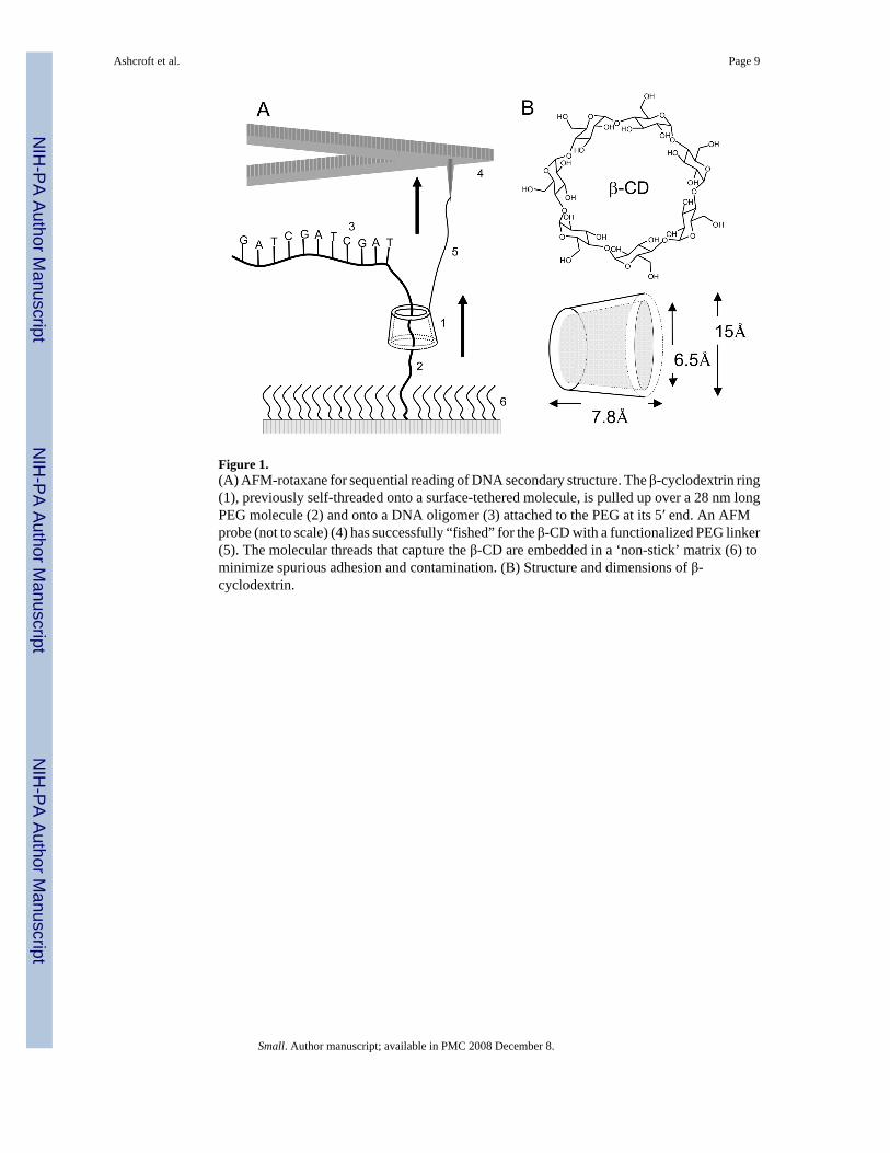
Figure 1.(A) AFM-rotaxane for sequential reading of DNA secondary structure. The β-cyclodextrin ring(1), previously self-threaded onto a surface-tethered molecule, is pulled up over a 28 nm longPEG molecule (2) and onto a DNA oligomer (3) attached to the PEG at its 5′ end. An AFMprobe (not to scale) (4) has successfully “fished” for the β-CD with a functionalized PEG linker(5). The molecular threads that capture the β-CD are embedded in a ‘non-stick’ matrix (6) tominimize spurious adhesion and contamination. (B) Structure and dimensions of β-cyclodextrin.
Ashcroft et al. Page 9
Small. Author manuscript; available in PMC 2008 December 8.
NIH
-PA Author Manuscript
NIH
-PA Author Manuscript
NIH
-PA Author Manuscript
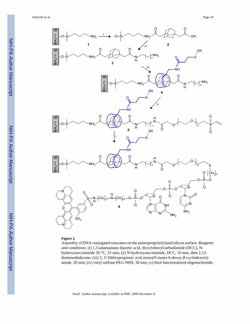
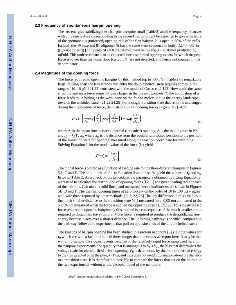
Figure 2.Assembly of DNA conjugated rotaxanes on the aminopropylsilylated silicon surface. Reagentsand conditions: (i) 1,3-adamantane diacetic acid, dicyclohexylcarbodiimide (DCC), N-hydroxysuccinimide 50 °C, 15 min; (ii) N-hydroxysuccinimide, DCC, 10 min, then 1,12-diaminododecane; (iii) 3, 3′-Dithiopropionic acid mono(N-mono-6-deoxy-β-cyclodextrin)amide, 20 min; (iv) vinyl sulfone-PEG-NHS, 30 min; (v) thiol functionalized oligonucleotide.
Ashcroft et al. Page 10
Small. Author manuscript; available in PMC 2008 December 8.
NIH
-PA Author Manuscript
NIH
-PA Author Manuscript
NIH
-PA Author Manuscript
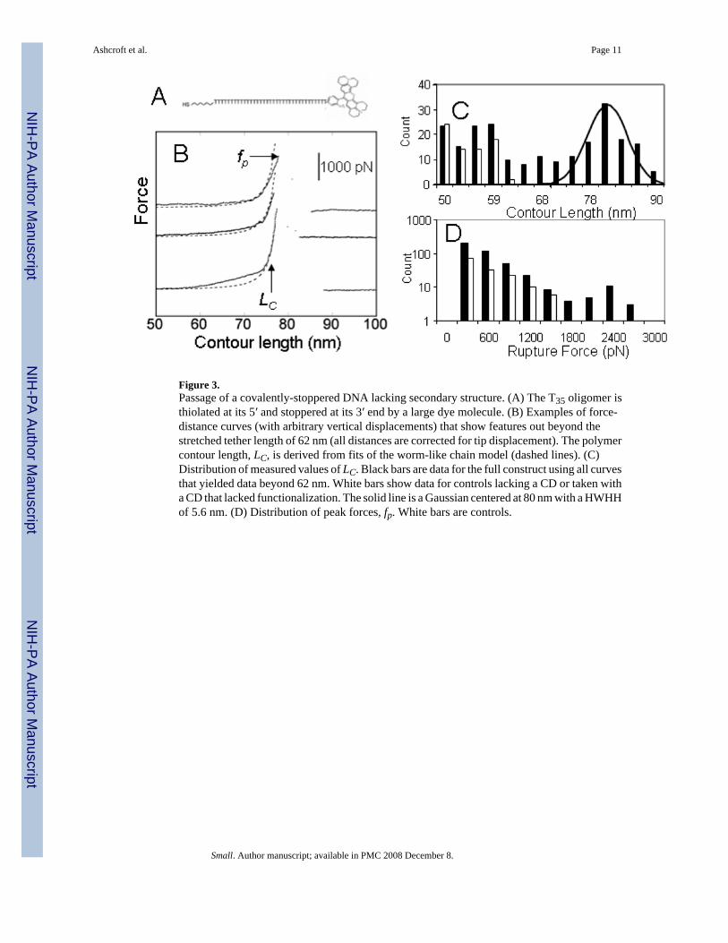
Figure 3.Passage of a covalently-stoppered DNA lacking secondary structure. (A) The T35 oligomer isthiolated at its 5′ and stoppered at its 3′ end by a large dye molecule. (B) Examples of force-distance curves (with arbitrary vertical displacements) that show features out beyond thestretched tether length of 62 nm (all distances are corrected for tip displacement). The polymercontour length, LC, is derived from fits of the worm-like chain model (dashed lines). (C)Distribution of measured values of LC. Black bars are data for the full construct using all curvesthat yielded data beyond 62 nm. White bars show data for controls lacking a CD or taken witha CD that lacked functionalization. The solid line is a Gaussian centered at 80 nm with a HWHHof 5.6 nm. (D) Distribution of peak forces, fp. White bars are controls.
Ashcroft et al. Page 11
Small. Author manuscript; available in PMC 2008 December 8.
NIH
-PA Author Manuscript
NIH
-PA Author Manuscript
NIH
-PA Author Manuscript
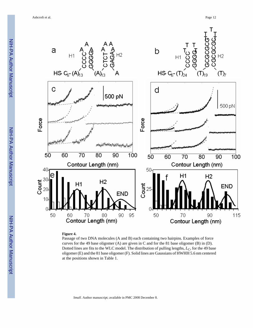
Figure 4.Passage of two DNA molecules (A and B) each containing two hairpins. Examples of forcecurves for the 49 base oligomer (A) are given in C and for the 81 base oligomer (B) in (D).Dotted lines are fits to the WLC model. The distribution of pulling lengths, LC, for the 49 baseoligomer (E) and the 81 base oligomer (F). Solid lines are Gaussians of HWHH 5.6 nm centeredat the positions shown in Table 1.
Ashcroft et al. Page 12
Small. Author manuscript; available in PMC 2008 December 8.
NIH
-PA Author Manuscript
NIH
-PA Author Manuscript
NIH
-PA Author Manuscript

Figure 5.Kinetic analysis of hairpin opening: The left panels plot the modal force (error bars are ±1 sd)as a function of the logarithm of the loading rate for (A) the first hairpins in both oligomers(data were identical and are aggregated here) (C) the second hairpin in the 49 base oligomerand (E) the second hairpin in the 81 base oligomer. The corresponding distribution of measuredforces are plotted in the right panels at loading rates of 10.5 nN/s (B) 21 nN/s (D) and 105 nN/s (F) The solid lines are calculated with equation 1 using the parameters shown in Table 2.
Ashcroft et al. Page 13
Small. Author manuscript; available in PMC 2008 December 8.
NIH
-PA Author Manuscript
NIH
-PA Author Manuscript
NIH
-PA Author Manuscript
NIH
-PA Author Manuscript
NIH
-PA Author Manuscript
NIH
-PA Author Manuscript
Ashcroft et al. Page 14
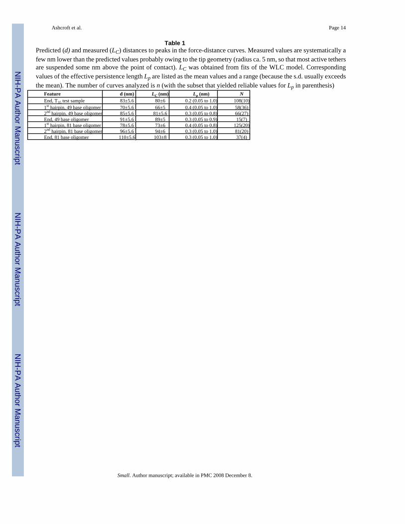
Table 1Predicted (d) and measured (LC) distances to peaks in the force-distance curves. Measured values are systematically afew nm lower than the predicted values probably owing to the tip geometry (radius ca. 5 nm, so that most active tethersare suspended some nm above the point of contact). LC was obtained from fits of the WLC model. Correspondingvalues of the effective persistence length Lp are listed as the mean values and a range (because the s.d. usually exceedsthe mean). The number of curves analyzed is n (with the subset that yielded reliable values for Lp in parenthesis)
Feature d (nm) LC (nm) Lp (nm) NEnd, T35 test sample 83±5.6 80±6 0.2 (0.05 to 1.0) 108(10)1st hairpin, 49 base oligomer 70±5.6 66±5 0.4 (0.05 to 1.0) 58(36)2nd hairpin, 49 base oligomer 85±5.6 81±5.6 0.3 (0.05 to 0.8) 66(27)End, 49 base oligomer 91±5.6 89±5 0.3 (0.05 to 0.9) 15(7)1st hairpin, 81 base oligomer 78±5.6 73±6 0.4 (0.05 to 0.8) 125(20)2nd hairpin, 81 base oligomer 96±5.6 94±6 0.3 (0.05 to 1.0) 81(20)End, 81 base oligomer 110±5.6 103±8 0.3 (0.05 to 1.0) 37(4)
Small. Author manuscript; available in PMC 2008 December 8.

NIH
-PA Author Manuscript
NIH
-PA Author Manuscript
NIH
-PA Author Manuscript
Ashcroft et al. Page 15
Table 2Average rupture forces for the various secondary structures (data for the hairpins with identical stems have beencombined). xts and τopen were derived from fits to equation 2. Stabilization free energies (ΔG) are predicted by mFold
Feature <fp> (pN) xts (nm) τopen(ms)
ΔGcalc(kcal/mole)
1st hairpin, both oligomers 376 0.05±0.01 53±56 −5.952nd hairpin, 49 base oligomer 248 0.06±0.01 100±37 −2.942nd hairpin, 81 base oligomer 371 0.03±0.01 43±26 −12.19
Small. Author manuscript; available in PMC 2008 December 8.
Related Documents


![Model AFM - S Series · 2018. 4. 24. · 예] AFM-SR-1000 X 450 . AFM-S Series Airflow Measuring Stations for Rectangular 1000(L)X450(H) Duct Size. AFM-SC- 600 . AFM-S Series Airflow](https://static.cupdf.com/doc/110x72/60146f16cb4905760706a3f1/model-afm-s-2018-4-24-afm-sr-1000-x-450-afm-s-series-airflow-measuring.jpg)