Cellular accumulations Amyloidosis. Endogenous and exogenous pigments. Pathological calcification.

Welcome message from author
This document is posted to help you gain knowledge. Please leave a comment to let me know what you think about it! Share it to your friends and learn new things together.
Transcript

Cellular accumulations
Amyloidosis. Endogenous and exogenous pigments.
Pathological calcification.

Amyloidosis. Endogenous and exogenous pigments. Pathological calcification.
I. Microspecimens:
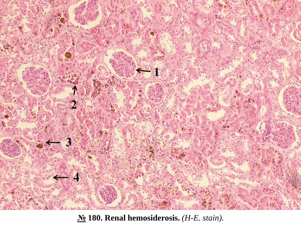
№ 180. Renal hemosiderosis. (H-E. stain).
Indications:
1. Glomerulus.
2. Hemosiderin granules in the cytoplasm of nephrocytes.
3. Hemosiderin granules in the lumen of the tubule.
4. Unchanged tubule.
In the epithelial cells of the convoluted renal tubules, brown hemosiderin granules are observed,
which can be found in some places in the lumen of the tubules.
Renal hemosiderosis is a manifestation of generalized hemosiderosis, characterized by an
increased content of hemosiderin in the body. It is due to massive and prolonged intravascular
hemolysis of erythrocytes and is found in hemolytic anemias, intoxications with hemolytic toxins
(eg, snake venom), infectious diseases (eg, in sepsis), incompatible blood transfusions. From the
products of destruction of hemoglobin in the elements of the reticuloendothelial system (spleen,
bone marrow, lymph nodes) and in some parenchymal organs (liver, kidneys) are formed and
stored excessive amounts of hemosiderin, these organs acquiring a rusted nuance. At the same
time, there is an increase in the synthesis of ferritin and bilirubin. Generalized hemosiderosis is
also observed in hemochromatosis.
1
2
3
4
№ 180. Renal hemosiderosis. (H-E. stain).

№ 135. Obstructive jaundice of the liver. (H-E. stain).
Indications:
1. Dilated bile ducts filled with bile pigments.
2. Accumulation of bile into the lumen of the intralobular bile capillaries.
3. Granules of bile pigment in the cytoplasm of hepatocytes.
The bile ducts and some interlobular ducts are dilated, in their lumen are observed brown bile
coagulates (biliary “thrombi”), and in the cytoplasm of some hepatocytes - bile pigment granules;
in some places in the center of the hepatic lobes there are foci of necrosis of hepatocytes, imbued
with bile.
Bile stasis (mechanical jaundice) can be caused by gallstones, intra- and extrahepatic bile duct
tumors, bile duct malformations or their external compression in pancreatic head cancer,
duodenal papilla tumors, cancer metastases in the lymph nodes in hepatic hilum, adhesions.
Excess of direct (conjugated) bilirubin in the blood, causes yellow-green pigmentation of organs
and tissues, including skin and sclera. Apart from the intense coloration of the skin, in general
obstructive jaundice there is general intoxication due to bile acids, hemorrhagic syndrome,
dystrophic kidney damage, hepato-renal failure. Biliary stasis can be complicated by
inflammation of the bile ducts (cholangitis), and if the process acquires a chronic evolution,
cholestatic biliary cirrhosis can develop.

№ 135. Obstructive jaundice of the liver. (H-E. stain).
3
1
2

№ 197a. Metastatic calcinosis of the myocardium. (H-E. stain).
Indications:
1. Focal deposits of calcium salts.
2. Adjacent myocardium.
In the microspecimen are observed multiple small foci of powdered granular deposits of calcium
salts, stained with hematoxylin in blue-purple. Calcium salts are stored in both the cardiomyocyte
sarcoplasm and the myocardial stroma.
Metastatic calcinosis is caused by excess of calcium in the blood plasma (hypercalcemia) which
is caused by the mobilization of calcium from the bones or by disturbing the processes of calcium
elimination from the body. It is observed in primary hyperparathyroidism (parathyroid adenoma),
bone tumors (multiple myeloma, bone metastases), multiple bone fractures, hypervitaminosis D,
osteoporosis, chronic nephritis. Calcium deposits occur in healthy, unaltered tissues: lungs,
gastric mucosa, kidneys, myocardium, arterial walls (on average) - organs and tissues, where
there is a local alkalosis due to the fact that they eliminate acidic products, which promotes
precipitation calcium salts (stomach - hydrochloric acid, kidneys - uric acid, lungs - carbon
dioxide, and myocardium and arteries are in constant contact with arterial blood and have low
carbon dioxide content). Macroscopically, the calcareous deposits are white, chalky in
appearance.

№ 197a. Metastatic calcinosis of the myocardium. (H-E. stain).
1
2
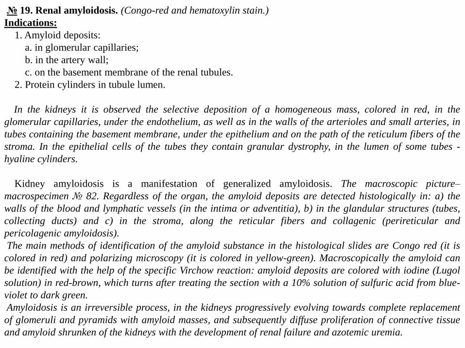
№ 19. Renal amyloidosis. (Congo-red and hematoxylin stain.)
Indications:
1. Amyloid deposits:
a. in glomerular capillaries;
b. in the artery wall;
c. on the basement membrane of the renal tubules.
2. Protein cylinders in tubule lumen.
In the kidneys it is observed the selective deposition of a homogeneous mass, colored in red, in the
glomerular capillaries, under the endothelium, as well as in the walls of the arterioles and small arteries, in
tubes containing the basement membrane, under the epithelium and on the path of the reticulum fibers of the
stroma. In the epithelial cells of the tubes they contain granular dystrophy, in the lumen of some tubes -
hyaline cylinders.
Kidney amyloidosis is a manifestation of generalized amyloidosis. The macroscopic picture–
macrospecimen № 82. Regardless of the organ, the amyloid deposits are detected histologically in: a) the
walls of the blood and lymphatic vessels (in the intima or adventitia), b) in the glandular structures (tubes,
collecting ducts) and c) in the stroma, along the reticular fibers and collagenic (perireticular and
pericolagenic amyloidosis).
The main methods of identification of the amyloid substance in the histological slides are Congo red (it is
colored in red) and polarizing microscopy (it is colored in yellow-green). Macroscopically the amyloid can
be identified with the help of the specific Virchow reaction: amyloid deposits are colored with iodine (Lugol
solution) in red-brown, which turns after treating the section with a 10% solution of sulfuric acid from blue-
violet to dark green.
Amyloidosis is an irreversible process, in the kidneys progressively evolving towards complete replacement
of glomeruli and pyramids with amyloid masses, and subsequently diffuse proliferation of connective tissue
and amyloid shrunken of the kidneys with the development of renal failure and azotemic uremia.

1b
1a
2
№ 19. Renal amyloidosis. (Congo-red and hematoxylin stain.)
1c

II. Macrospecimens:
№ 76. Biliary calculi.
In the gallbladder there are multiple stones of different sizes from a few mm to a few cm, with a
smooth, polished (faceted stones) or rough surface, yellow, olive or black, the bladder wall is
thickened, sclerosed.
Risk factors in the development of gallstones are multiple, the most important: old age, female
sex (in women it is 2 times more common than in men), obesity, oral contraceptives, hereditary
predisposition and others. Among the local factors have an important role biliary stasis
(gallbladder hypomobility) and mucus hypersecretion. According to the chemical composition
there are 2 main types of stones: cholesterol (≈80% of the total number), which contain
cholesterol, are yellow and pigmented, which contain calcium salts of bilirubin, are brown or
black. Possible complications: obstruction of the cystic duct, bile retention and development of
gallbladder hydrops, mucocele, acute or chronic cholecystitis, gallbladder empyema, it is
possible to perforate the bladder wall with bile flow into the peritoneal cavity and biliary
peritonitis. The larger the stones, the lower the risk of obstruction of the cystic duct, and the
smaller the stones ("sand") are more dangerous.

№ 76. Biliary calculi.

№ 87. Renal calculi.
The renal pelvis and calyxes are dilated, contain multiple stones, some are free, others -
overgrown with the pelvic / calyx wall, dimensions from 2-3 mm to 1-2 cm, smooth, polished
surface (faceted stones) or irregular, rough , sometimes with branches, which take the shape of
calyxes - coral-shaped stones, white, yellow or brown depending on the chemical composition.
There are 3 main types of urinary stones:
a) calciform stones (calcium oxalates and phosphates), which are observed in ~ 75% of cases,
have a granular-rough surface, brown color due to hemosiderin, which occurs after mucosal
trauma and repeated hemorrhages;
b) mixed stones, so-called "crushed stones" or "triple phosphates", consisting of ammonia-
magnesium phosphate; are found in ~ 15% of cases, especially in patients with urinary tract
infections (Proteus vulgaris, Klebsiella, Staphylococcus); bacteria produce proteases, which
cleave urea (infection-induced stones); they are white-yellow;
c) stones with uric acid (urates) - occur after hyperuricemia and hyperuricuria, which is
observed in cases of primary or secondary gout in myeloproliferative diseases (eg, in leukosis),
have yellow-gray color.
Complications: pyelonephritis, nephrosclerosis and macronodular shedding of the kidney, if the
process is bilateral - progressive chronic renal failure.

№ 87. Renal calculi.

№ 82. Renal amyloidosis.
The kidney is enlarged in size of dense consistency, gray yellowish color and lardy or waxy appearance, the
surface is slightly wavy, on section layers are poorly delimited - "big white amyloidic kidney".
Renal amyloidosis (amyloid nephropathy) is found in both primary amyloidosis, e.g., multiple myeloma (AL
amyloidosis) and secondary amyloidosis, eg, purulent osteomyelitis, tuberculosis, bronchiectasis, rheumatoid
arthritis (AA amyloidosis).
Clinically manifested by nephrotic syndrome: massive proteinuria (more than 3.5 g in 24 hours,
hypoalbuminemia, generalized edema, hyperlipidemia and lipiduria, azotemia, hypertension (in 50% of
cases).
Complications: renal failure, association of infections due to decreased immunity, cardiovascular
insufficiency, predisposition to thrombosis of vessels due to loss with urine of immunoglobulins and
anticoagulant system proteins.

№ 82. Renal amyloidosis.

Hemosiderin.

Pulmonary hemosiderosis.

Bilirubin

Hydrochloric hematin
on the bottom of gastric
mucosal erosions.
Brain
hemomelanosis
in malaria.

Brown atrophy of
the heart.

Melanin / Melanosis

Tuberculosis (caseous necrosis)
and amyloidosis (Congo red) of
the adrenal glands.
Clinic - Addison's disease.

Pigmented nevus
Melanoma metastases in the
brain.

Dystrophic calcinosis
Lung tissue.
Coronary artery in
atherosclerosis.

Dystrophic calcinosis
Aortic valve calcinosis.
LITHOPEDION - in which the
calcification of a dead fetus is
almost complete.

Metastatic calcinosis
Lung tissue.
Stomach.

Myocardial amyloidosis
Macroscopic
identification.
Virchow macroscopic reaction: on successive application to the section surface of
iodine or Lugol's solution and sulfuric acid (10%) the amyloid stains in blue-violet or
dark green.

Focal spleen amyloidosis (sago spleen).

Diffuse spleen amyloidosis.
Image beneath - norm.

PIGMENTS AND OTHER TISSUE DEPOSITS
many pathologic processes are accompanied by
accumulations of material either within the cell
(intracellular) or within the extracellular space.

Pigments
Pigments are colored substances, some of which are normal constituents of cells whereas others are
abnormal and collect in cells only under special
circumstances Pigments can be either
exogenous or endogenous

LipofuscinAlso known as lipochrome and “wear-
and-tear” or aging pigment
•origin: lysosomal breakdown products of lipids.
•sites: aged or chronically injured cells.
•significance: doesn’t injure cell, but a sign of aging or excess free radical damage.

Lipofuscinlipofuscin in Purkinje cells.
Lipofuscin in myocardial cells.
Electron micrograph showing electron dense pigment (lipofuscin) in myocardial cells.

Lipofuscin
It is particularly
prominent in the
liver and heart of
aging patients or
patients with severe
malnutrition and
cancer cachexia.
It is usually
accompanied by
organ shrinkage
(brown atrophy).

Hemosiderin
A hemoglobin-derived, golden-
yellow to brown, pigment in which form iron is stored in cells Excesses of iron cause hemosiderin to accumulate
within cells

Hemosiderin
• • origin:-represents stored iron (esp from recycled
Hb).• •sites:-normal in MФ’s of spleen, bone
marrow.-anywhere – increased breakdown of
erythrocytes or increased accumulations of iron.
1. areas of congestion or hemorrhage (eg bruise)
2. Kupffer cells in hemolytic anemia3. alveolar macrophages in congestive heart
failure (heart failure cells)

Hemosiderin
Hemosiderin
Deposition
In renal tubules

Hemosiderin granules in liver cells. A, H&E section showing golden-brown, finely granular pigment. B, Prussian blue reaction, specific for iron.
Hemosiderin

Hemosiderin in lung
Hemosiderin in liver

Hemosiderin in lung
Chronic passive congestion, lung. Prussian blue reaction.

Hemosiderosis
An example of localized hemosiderosis is the common bruise
Following local hemorrhage, the area is at first red-blue. With lysis of the erythrocytes, the hemoglobin eventually undergoes transformation to hemosiderin

Hemosiderosis
The original red-blue color of hemoglobin is transformed to varying shades of green-blue, comprising the local formation of biliverdin,then bilirubin and thereafter the iron moiety of hemoglobin is deposited as golden-yellow hemosiderin

Hemochromatosis
The more extreme accumulation
of iron, however, in a disease
called hemochromatosis is
associated with liver and
pancreatic damage, resulting in
liver fibrosis, heart failure, and
diabetes mellitus

Bilirubin
The normal major pigment found
in bile.
It is derived from hemoglobin but
contains no iron.
Jaundice is a common clinical
disorder due to excesses of this
pigment within cells and tissues

Jaundice

Bilirubin•bilirubin is end product of heme degradation (after removal of iron).•mostly from senescent rbc’s via MФ’s.
Each hemoglobin molecule is an assembly of four globular protein subunits. Each subunit is composed of a protein chain tightly associated with a non-protein hemegroup.

Bilirubin metabolism and elimination.
1, Normal bilirubin production from heme (~0.2 to 0.3 gm/day in humans) is derived primarily from the breakdown of senescent circulating erythrocytes, with a minor contribution from degradation of tissue heme-containing proteins. 2, Extrahepatic (unconjugated) bilirubin is bound to serum albumin and delivered to the liver. 3, Hepatocellular uptake and 4, Glucuronidation in the endoplasmic reticulum generate conjugated bilirubin (monoglucuronides and diglucuronides), which are water soluble and readily excreted into bile. 5, Gut bacteria deconjugate the bilirubin and degrade it to colorless urobilinogens. The urobilinogens and the residue of intact pigments are excreted in the feces, with some reabsorption and excretion into urine.

Ilustration of the morphologic features of cholestasis in the liver parenchyma,
Cholestatichepatocytes are enlarged with dilated canalicular spaces.Apoptotic cells may be seen, and Kupffer cells frequently contain regurgitated bile pigments

Bilirubin pigment
prehepatic jaundicehepatic jaundicepost hepatic jaundice

Melanin / Melanosis
• intracellular, brown-black pigment.
• origin: normal pigment found in the epidermis and eye.
• sites: melanin can also occur in other sites & when in excess called melanosis,
• eg leptomeninges (esp ruminants), intestine, kidney, lung, base of aorta, etc.

Melanin / Melanosis
Diagram of a melanocyte. It sends irregular dendritic processes between neighboring keratinocytes for transfer of melanin to those cells.
melanocytes and keratinocytes containing melanin granules.

Melanin / Melanosis

Melanin / Melanosis

Melanin / Melanosis

Calcification
Pathologic calcification implies the abnormal
deposition of calcium salts, together with smaller
amounts of iron, magnesium, and other mineral salts

Dystrophic calcification
Occurs in nonviable or dying tissues. It occurs despite normal serum
levels of calcium and in the absence of
derangements in calcium metabolism

Dystrophic calcification
note degeneration / necrosis and mineralization of myofibers. On H&E staining mineral is seen as basophilic granules and/or clumps.

Metastatic Calcification
Metastatic Calcification• when apparently normal tissue undergoes calcification (with hypercalcemia / altered Ca++metabolism).
•pathogenesis:
•primary hyperparathyroidism
•renal failure / secondary hyperparathyroidism
•hypervitaminosis D
•paraneoplastic syndromes•sites: many sites can be affected, especially gastric and intestinal mucosa, blood vessel walls, BMZ of lung, kidney, etc.
Hypercalcemia >11.0mg/dl

Metastatic Calcification
A band of calcium has been laid down the middle of the gastric mucosa.

Calcification
Metastatic calcification
Lung
Dystrophic calcification
Stomach

Crystals
• significance:
severe renal dysfunction & electrolyte imbalances

Urates and Uric Acid
note clear crystals surrounded by granulomatous inflammation.

Carbon
Accumulations of this pigment blacken the tissues of the lungs (anthracosis)
and the involved lymph nodes.

Anthracosis
Lymph node of the lung showing carbon deposition
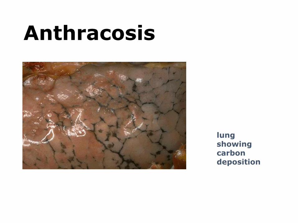
Anthracosis
lung showing carbon deposition

AMYLOIDOSIS• BUILDUP OF AMYLOID “PROTEIN”
• AL (Amyloid Light Chain)
• AA (NON-immunoglobulin protein)
• Aß (Alzheimer’s)
• WHERE? BLOOD VESSEL WALLS, at first
• KIDNEY
• SPLEEN
• LIVER
- HEART
Why is amyloid called amyloid? Because in the early days, it took up STARCH stains applied to GROSS specimens,
e.g., IODINE stains.
But of course now we know it’s a PROTEIN, chiefly immunoglobulin protein chronic buildup.
It is therefore not surprising that diseases which have chronic immunoglobulin buildup over many years are
associated with amyloidosis, i.e., multiple myeloma (also called plasma cell “dyscrasias”), granulomatous diseases,
classically.

CONGO RED STAIN, WITHOUT,
and WITH, POLARIZATION

AMYLOID ASSOCIATIONS
• PLASMA CELL “DYSCRASIAS”, i.e., MULTIPLE MYELOMA
• CHRONIC GRANULOMATOUS DISEASE, e.g., TB
• HEMODIALYSIS
• HEREDOFAMILIAL
• LOCALIZED
• ENDOCRINE MEAs (Multiple Endocrine Adenomas)
• AGING
Diseases in which there is a cumulative buildup of immunoglobulin
would be a setting for amyloidosis, so myelomas and chronic
granulomatous diseases are at the top of the list.
Related Documents


