ASIP Journal CME Program Neurobiology Amyloid- Peptide Remnants in AN-1792-Immunized Alzheimer’s Disease Patients A Biochemical Analysis R. Lyle Patton,* Walter M. Kalback,* Chera L. Esh,* Tyler A. Kokjohn,* † Gregory D. Van Vickle,* Dean C. Luehrs,* Yu-Min Kuo, ‡ John Lopez, § Daniel Brune, § Isidro Ferrer, ¶ Eliezer Masliah, Amanda J. Newel,** Thomas G. Beach,** Eduardo M. Castan ˜o, †† and Alex E. Roher* From The Longtine Center for Molecular Biology and Genetics,* and the W.H. Civin Laboratory for Neuropathology,** Sun Health Research Institute, Sun City, Arizona; the Department of Microbiology, † Midwestern University, Glendale, Arizona; the Department of Cell Biology and Anatomy, ‡ National Cheng Kung University, Tainan, Taiwan; the Department of Chemistry and Biochemistry, § Arizona State University, Tempe, Arizona; the Institut de Neuropatologia, ¶ Hospital Universitari de Bellvitge, Hospitalet de Llobregat, Spain; the Department of Neurosciences, University of California, San Diego, La Jolla, California; and the Fundacion Instituto Leloir, †† Buenos Aires, Argentina Experiments with amyloid- (A)-42-immunized transgenic mouse models of Alzheimer’s disease have revealed amyloid plaque disruption and apparent cognitive function recovery. Neuropathological ex- amination of patients vaccinated against purified A-42 (AN-1792) has demonstrated that senile plaque disruption occurred in immunized humans as well. Here , we examined tissue histology and quantified and biochemically characterized the remnant amy- loid peptides in the gray and white matter and lepto- meningeal/cortical vessels of two AN-1792-vaccinated patients , one of whom developed meningoencepha- litis. Compact core and diffuse amyloid deposits in both vaccinated individuals were focally absent in some regions. Although parenchymal amyloid was focally disaggregated , vascular deposits were rela- tively preserved or even increased. Immunoassay re- vealed that total soluble amyloid levels were sharply elevated in vaccinated patient gray and white matter compared with Alzheimer’s disease cases. Our exper- iments suggest that although immunization disrupted amyloid deposits , vascular capture prevented large- scale egress of A peptides. Trapped , solubilized amy- loid peptides may ultimately have cascading toxic ef- fects on cerebrovascular , gray and white matter tissues. Anti-amyloid immunization may be most effective not as therapeutic or mitigating measures but as a prophy- lactic measure when A deposition is still minimal. This may allow A mobilization under conditions in which drainage and degradation of these toxic peptides is efficient. (Am J Pathol 2006, 169:1048 –1063; DOI: 10.2353/ajpath.2006.060269) Sporadic Alzheimer’s disease (AD) affects the aged with a prevalence approaching 40 to 50% by age 80. At present, 4 million Americans are affected with AD at an estimated annual care cost of almost 100 billion dollars. Because the number of individuals 65 years or older is growing rapidly due to a general average life expectancy increase, it is estimated that the total incidence of AD will quadruple by the year 2050. 1 Therefore, it is urgent to find a means of preventing, delaying the onset, or revers- ing the course of AD. Alzheimer’s disease is characterized by the massive accumulation of extracellular amyloid fibrils in both the brain parenchyma and in the walls of cerebral blood vessels. The deposited fibrillar amyloid is mainly com- posed of amyloid- (A) peptides, 40/42 amino acid- Supported by National Institute on Aging grant RO1 AG-19795, by Ari- zona Alzheimer’s Disease Core Center grant P30 AG-19610, and by grants from the State of Arizona to the Arizona Alzheimer’s Research Consortium. Brain tissue from case 2 was obtained from University of California at San Diego Alzheimer’s Disease Research Center grant NIA AG-05131 and grant RO1 AG-18440. Accepted for publication June 14, 2006. Supplemental material for this article appears on http://ajp. amjpathol.org. Address reprint requests to Alex E. Roher, M.D. Ph.D., Sun Health Research Institute, 10515 W. Santa Fe Dr., Sun City, AZ 85351. E-mail: [email protected]. Related Commentary on page 738 The American Journal of Pathology, Vol. 169, No. 3, September 2006 Copyright © American Society for Investigative Pathology DOI: 10.2353/ajpath.2006.060269 1048

Welcome message from author
This document is posted to help you gain knowledge. Please leave a comment to let me know what you think about it! Share it to your friends and learn new things together.
Transcript
ASIP
Journ
al
CME P
rogra
m
Neurobiology
Amyloid-� Peptide Remnants in AN-1792-ImmunizedAlzheimer’s Disease Patients
A Biochemical Analysis
R. Lyle Patton,* Walter M. Kalback,*Chera L. Esh,* Tyler A. Kokjohn,*†
Gregory D. Van Vickle,* Dean C. Luehrs,*Yu-Min Kuo,‡ John Lopez,§ Daniel Brune,§
Isidro Ferrer,¶ Eliezer Masliah,�
Amanda J. Newel,** Thomas G. Beach,**Eduardo M. Castano,†† and Alex E. Roher*From The Longtine Center for Molecular Biology and Genetics,*
and the W.H. Civin Laboratory for Neuropathology,** Sun Health
Research Institute, Sun City, Arizona; the Department of
Microbiology,† Midwestern University, Glendale, Arizona; the
Department of Cell Biology and Anatomy,‡ National Cheng Kung
University, Tainan, Taiwan; the Department of Chemistry and
Biochemistry,§ Arizona State University, Tempe, Arizona; the
Institut de Neuropatologia,¶ Hospital Universitari de Bellvitge,
Hospitalet de Llobregat, Spain; the Department of Neurosciences,�
University of California, San Diego, La Jolla, California; and the
Fundacion Instituto Leloir,†† Buenos Aires, Argentina
Experiments with amyloid-� (A�)-42-immunizedtransgenic mouse models of Alzheimer’s disease haverevealed amyloid plaque disruption and apparentcognitive function recovery. Neuropathological ex-amination of patients vaccinated against purifiedA�-42 (AN-1792) has demonstrated that senile plaquedisruption occurred in immunized humans as well.Here, we examined tissue histology and quantifiedand biochemically characterized the remnant amy-loid peptides in the gray and white matter and lepto-meningeal/cortical vessels of two AN-1792-vaccinatedpatients, one of whom developed meningoencepha-litis. Compact core and diffuse amyloid deposits inboth vaccinated individuals were focally absent insome regions. Although parenchymal amyloid wasfocally disaggregated, vascular deposits were rela-tively preserved or even increased. Immunoassay re-vealed that total soluble amyloid levels were sharplyelevated in vaccinated patient gray and white mattercompared with Alzheimer’s disease cases. Our exper-
iments suggest that although immunization disruptedamyloid deposits, vascular capture prevented large-scale egress of A� peptides. Trapped, solubilized amy-loid peptides may ultimately have cascading toxic ef-fects on cerebrovascular, gray and white matter tissues.Anti-amyloid immunization may be most effective notas therapeutic or mitigating measures but as a prophy-lactic measure when A� deposition is still minimal.This may allow A� mobilization under conditions inwhich drainage and degradation of these toxic peptidesis efficient. (Am J Pathol 2006, 169:1048–1063; DOI:
10.2353/ajpath.2006.060269)
Sporadic Alzheimer’s disease (AD) affects the aged witha prevalence approaching 40 to 50% by age 80. Atpresent, 4 million Americans are affected with AD at anestimated annual care cost of almost 100 billion dollars.Because the number of individuals 65 years or older isgrowing rapidly due to a general average life expectancyincrease, it is estimated that the total incidence of AD willquadruple by the year 2050.1 Therefore, it is urgent tofind a means of preventing, delaying the onset, or revers-ing the course of AD.
Alzheimer’s disease is characterized by the massiveaccumulation of extracellular amyloid fibrils in both thebrain parenchyma and in the walls of cerebral bloodvessels. The deposited fibrillar amyloid is mainly com-posed of amyloid-� (A�) peptides, 40/42 amino acid-
Supported by National Institute on Aging grant RO1 AG-19795, by Ari-zona Alzheimer’s Disease Core Center grant P30 AG-19610, and bygrants from the State of Arizona to the Arizona Alzheimer’s ResearchConsortium. Brain tissue from case 2 was obtained from University ofCalifornia at San Diego Alzheimer’s Disease Research Center grant NIAAG-05131 and grant RO1 AG-18440.
Accepted for publication June 14, 2006.
Supplemental material for this article appears on http://ajp.amjpathol.org.
Address reprint requests to Alex E. Roher, M.D. Ph.D., Sun HealthResearch Institute, 10515 W. Santa Fe Dr., Sun City, AZ 85351. E-mail:[email protected].
Related Commentary on page 738The American Journal of Pathology, Vol. 169, No. 3, September 2006
Copyright © American Society for Investigative Pathology
DOI: 10.2353/ajpath.2006.060269
1048
residue molecules derived by proteolytic processing oflarger amyloid precursor proteins (APPs) by the con-certed actions of the �- and �-secretases. The relevanceof A� peptides to sporadic AD pathogenesis is stronglysupported by the fact that mutations in the APP andpresenilin genes both result in early-onset familial AD.Moreover, a formally similar suite of pathophysiologicaland cognitive changes is observed in multiple strains oftransgenic (Tg) mice that overexpress APP and/or otherAPP processing genes. The fibrillar and soluble forms ofA� interfere with the normal brain architecture and func-tion, resulting in profound neuroinflammation, gliosis, se-vere neuronal injury, and vascular damage and in theinduction of neurofibrillary tangle (NFT) and protracteddementia development. The clearly preeminent role of A�in AD provides strong experimental support to the amy-loid cascade hypothesis as a mechanism central to ADpathogenesis.
Among the multiple remedial avenues so far explored,immunotherapy promises to be one of the most effectiveinterventions. Several single (APP) and double Tg (APP/presenilin) mouse strains have been generated that pro-duce amyloid structures similar to those observed in AD.Active and passive anti-A� immunization therapies weretested in Tg animals and resulted in amyloid depositdisaggregation and the reversal of cognitive deficits.2–4
Immunotherapy has also been successful in reducingamyloid levels in the brains of aging Caribbean Vervetmonkeys.5
Encouraged by the impressive results observed in an-imal models, active A� vaccination clinical trials wereinitiated in humans. Three hundred individuals were vac-cinated with the AN-1792/QS-21 antigen/adjuvant com-plex, and 72 subjects received placebo treatment. Of the300 vaccinated subjects, 18 (6%) developed asepticmeningoencephalitis, whereas no placebo group sub-jects developed this condition during the same timeframe. In the immunized cohort, a total of 59 individualshad desirable plasma antibody titers �1:2200. Thirteenpatients from this vaccine-responsive subgroup devel-oped meningoencephalitis (22%), whereas only 5 (2%) ofa total pool of 241 “nonresponders” evidenced this ad-verse outcome. No significant differences were observedbetween vaccinated and placebo-treated subjects withrespect to a battery of individual psychometric tests,although neuropsychological test battery z-scores dem-onstrated differences favoring the antibody responders.In addition, significant cerebrospinal fluid (CSF)-tau de-creases were evident in the antibody-responsive patientgroup.6,7 Intriguingly, 45 of the high anti-amyloid anti-body titer responding individuals had, as measured bymagnetic resonance imaging, a greater brain volumereduction with an enhanced ventricular enlargement forwhich there is presently no certain explanation.8 It hasbeen suggested that this reduction may be attributed toamyloid deposit removal. It is also possible that the re-duction of brain volume is due to hydrodynamic changesin CSF and brain interstitial fluid. In addition, cognitivefunctions showed a slower decline in 20 AD patients whogenerated acceptable levels of antibodies after receivingprimer and a booster of aggregated A�.9 There is also
evidence that the administration of intravenous immuno-globulins, a complex mixture of IgG that contains anti-bodies against A�, results in an amelioration of dementiasymptoms in AD patients, supporting the tenet that amy-loid deposits are subject to immunological therapy.10,11
However, larger scale longitudinal studies with completeneuropathological analyses are required to rigorouslyconfirm the results of the pilot studies.
The initial AD patient vaccination study was terminatedbecause of an unanticipated adverse event: the emer-gence of vaccination-associated meningoencephalitis.So far, there are three published complete neuropatho-logical reports concerning AN-1792/QS-21-immunizedindividuals.12–14 Two of these reports describe subjectswho developed meningoencephalitis, whereas the thirdconcerns a vaccinated subject who died without menin-goencephalitis development. We isolated, characterized,and compared the amyloid species present in the graymatter (GM) and white matter (WM) as well as corticaland leptomeningeal vessels of two AN-1792/QS-21-vac-cinated AD patients, one of whom developed meningo-encephalitis. Our analyses reveal that parenchymal amy-loid plaque deposits regressed to a remarkable extent inboth patients, suggesting that simple differences in ulti-mate antibody titers or activity alone do not account forthe adverse outcome of meningoencephalitis in somepatients. A full understanding of the direct and emergenteffects of amyloid vaccination on the AD patient brainnecessitates a larger sample of the AN-1792 experimen-tal cohort undergoing comprehensive biochemical as-sessments and comparisons.
Materials and Methods
Human Subjects
Case 1
Male, age 76, with mild to moderate cognitive impair-ment [mini mental state examination (MMSE) � 18], re-ceived two intramuscular doses of 225 �g of AN-1792.Nine months after the last immunization, the patient de-veloped cerebral edema and neurological symptoms ofmeningoencephalitis. Magnetic resonance imaging re-vealed lesions in the WM. Two months later, the patientevidenced serious global cognitive deterioration and agreatly decreased MMSE score of 7. On neuropatholog-ical examination, the brain weight was 1130 g. There wassoftening of WM, signs of demyelination, enlarged ven-tricles, and multiple hemorrhages in the GM, uncommonin AD (the patient did not have a history of cardiovasculardisease). Amyloid plaques were distributed nonuniformlyand were mostly of the diffuse type with a lesser amountof compact amyloid core plaques presenting multinucle-ated cells loaded with dense aggregates of A�. Manyapparently disaggregated senile plaques with amyloiddeposits were observed in this patient. These apparentsenile plaque debris structures, referred to as “collapsedplaques” by Ferrer et al,13 consisted of extracellularplaques depleted of amyloid. These structures probably
A� Peptides in Immunized Alzheimer’s Patients 1049AJP September 2006, Vol. 169, No. 3
represent the complex nonamyloid glycoproteins andglycolipids that account for 30% of the AD plaque com-position. In addition, severe cerebrovascular amyloid-osis, abundant NFT deposits, marked inflammatory T-lymphocyte infiltration and severe small vessel disease(lipohyalinosis) were present. The apolipoprotein E (ApoE) genotype for this patient was �3/�4. A comprehensiveaccount of the neuropathological changes and detailedimmunocytochemical profile of this patient are given byFerrer et al.13
Case 2
Male, age 71, with moderate to severe cognitive im-pairment (MMSE � 12). This individual received threeintramuscular doses of 225 �g each of AN-1792 and died1 year after the first immunization. The cause of deathwas recorded as “failure to thrive.” The brain weighed1100 g and exhibited a normal macroscopic appear-ance. White matter and ventricles looked normal, andfrontal lobes were almost free of extracellular amyloiddeposits and neuritic plaques. A� immunoassay, per-formed by Masliah et al,14 demonstrated low cortical A�levels. Aggregates of A� were observed within microglial/macrophage cells, and there was severe cerebrovascu-lar amyloidosis in the frontal lobe but less apparent in theparietal and temporal lobes. In addition, there were abun-dant NFTs distributed in the cerebral cortex.14 The Apo Egenotype of this individual was �3/�4.
Biochemical Analyses
The biochemical analyses of A� peptides were per-formed on GM from temporal lobe including the entorhi-nal cortex (case 1) and on parietal and temporal cortex(case 2). The postmortem delay time before freezing ofthe tissue was approximately 4 hours for case 1 and 12hours for case 2. Brain sections were subjected to Camp-bell-Switzer silver stain15 and 1.0% aqueous thioflavine-Sfor 10 minutes. To assess the distribution and relativenumber of senile and diffuse plaques in the cerebralcortex, brain sections were immunocytochemicallystained. This step also permitted the estimation of theamount of tissue required to perform subsequent bio-chemical experiments. Briefly, brain sections weredeparaffinized and treated with 80% formic acid for 20minutes. Sections were incubated overnight with anti-A�antibodies 6E10 and 4G8 diluted 1:1000 (Signet Labora-tories, Dedham, MA). Anti-mouse IgG was used as asecondary antibody (1:10,000), and then developed withdiaminobenzidine in 0.5% nickel ammonium sulfate. He-matoxylin was used as a counterstain.
Water-Soluble Amyloid
The water-soluble material represents soluble interstitialfluid A�, A� monomers and low molecular weight oli-gomers loosely attached to amyloid plaques, and solubleA� free in the cytosol.16 The leptomeninges covering thecerebral cortex were separated and stored at �80°C.
Twenty grams of cerebral cortex was finely minced with arazor blade, suspended in 150 ml of ammonium bicar-bonate buffer (0.1 mol/L NH4HCO3, 5 mmol/L ethylenedi-amine tetraacetic acid, and 0.01% sodium azide), withthree tablets of complete enzyme inhibitors (Roche,Mannheim, Germany), and allowed to stand at 4°C for 48hours. The brain cortex was homogenized with a 40-mlcapacity Tenbroeck glass homogenizer (six manual gen-tle strokes) and centrifuged at 150,000 � g for 3 hours(Beckman SW28 rotor) in a Beckman Optima LE-80Kultracentrifuge (Beckman, Fullerton, CA). The water-sol-uble fraction was lyophilized and analyzed by fast proteinliquid chromatography (FPLC) (Amersham Biosciences,Piscataway, NJ) and high performance liquid chromatog-raphy (HPLC) (Thermo Separation Products, Schaum-burg, IL).
Sodium Dodecyl Sulfate (SDS)-InsolubleAmyloid
The water-insoluble pellets were dispersed and stirred for12 hours in 50 ml of 0.1 mol/L NH4HCO3. Fifty milliliters of30% SDS was added (final SDS concentration 15%) andstirred for an additional 14 hours at room temperature. Toremove the SDS-insoluble tufts of parenchymal bloodvessels, the suspension was filtered through a series of300-, 45-, and 25-�m nylon meshes. The SDS-containingfiltrate was centrifuged at 300,000 � g for 5 hours at 20°Cin an SW41 Ti Beckman rotor. The resulting pellets weresuspended in 60 ml of 0.1 mol/L ammonium bicarbonate,pH 10.0 (titrated with NH4OH), and after 24 hours ofcontinuous stirring, centrifuged at 300,000 � g for 5hours at 20°C. The pellets were stored, and the superna-tant was lyophilized and analyzed by FPLC and HPLC.
Leptomeningeal Vascular Amyloid
The leptomeninges were washed 10 times with 500 ml ofcold water to remove blood cells by hypotonic shock.Vessels larger than 600 �m in diameter were eliminated,and the smaller vessels were finely minced and washedthree times (5 minutes at 1500 � g) with 0.1 mol/L Tris-HCl and 3 mmol/L CaCl2, pH 8.0. Vessels were sus-pended in the wash buffer amended with 0.3 mg/mlClostridium histolyticum collagenase CL3 (WorthingtonBiochemicals Corp., Lakewood, NJ), and 10 �g/mlDNase I (Sigma-Aldrich, St. Louis, MO) was added tocreate a final suspension of 100 ml, which was incubatedin a shaker at 37°C for 3 hours. The preparation was thenfiltered through a 150-�m nylon mesh, and the filtrate wascentrifuged at 3000 � g for 10 minutes. The supernatantwas eliminated, and the pellet was washed with water,suspended in water, and centrifuged at 3000 � g. Thepellet, containing sheets of leptomeningeal vascularamyloid, was dissolved in 4 ml of glass distilled formicacid (GDFA) with the aid of a glass homogenizer, left atroom temperature for 30 minutes, and centrifuged at500,000 � g for 10 minutes in a TL 100 rotor (BeckmanOptima TLX ultracentrifuge). The supernatant was lyoph-ilized and analyzed by FPLC and HPLC.
1050 Patton et alAJP September 2006, Vol. 169, No. 3
Cortico-Vascular Amyloid
The 300-, 45-, and 25-�m nylon mesh-retained vesseltufts from the cortical SDS-insoluble amyloid preparationwere immediately shaken in 500 ml of distilled water tosuspend the retained vessels. The material was allowedto settle, and the clarified liquid was decanted with theremaining volume centrifuged at 1500 � g for 10 minutes.The pellets containing the cortical blood vessels werewashed twice with distilled water to remove remainingSDS and were suspended in 0.1 mol/L Tris-HCl and 3mmol/L CaCl2, pH 8.0. Next, 0.3 mg/ml collagenase CL3(Worthington) and 10 �g/ml DNase I (Sigma) wereadded, and samples were incubated in a shaker at 37°Cfor 3 hours. The preparation was filtered through a 40-�mnylon mesh, and the filtrate fraction was centrifuged at3000 � g. The pellet was treated with 4 ml of GDFA, spunat 500,000 � g as described above, and analyzed byFPLC and HPLC.
Diffuse Amyloid (P3)
This fraction represents the most insoluble of the amyloidaggregates and is composed primarily of A� 17–42 pep-tides recovered after high-speed centrifugation of theSDS/alkaline insoluble pellets. This material was dis-solved in 80% GDFA with the aid of a glass homogenizer,allowed to stand at room temperature for 1 hour, parceledinto 0.7-ml aliquots in thick-walled polyallomer tubes, andcentrifuged at 500,000 � g for 15 minutes in a BeckmanOptima TLA-ultracentrifuge using a 120.2 rotor. The darkbrown pellets containing formic acid-insoluble materialmostly composed of lipofuscin remnants were discarded,and the clear supernatant was submitted to FPLC-GDFAchromatography. Column eluate samples containingamyloid fraction P3 were pooled, reduced in volume bySpeed-Vac centrifugation, and subjected to size-exclu-sion HPLC chromatography on a GPC-100 column, equil-ibrated, and developed with 80% GDFA. The GPC-100column fraction containing the P3 was refined further bychromatography on a reverse-phase Zorbax SB-C8 col-umn as described below.
White Matter A�
The WM was separated from the adjacent GM to avoidcontamination from cortical amyloid. The WM (3 g) wasfinely minced and thoroughly homogenized in 18 ml of80% GDFA. The homogenate was centrifuged at250,000 � g for 1 hour using 11-ml polyallomer tubes ina Beckman SW41 rotor. The fatty material collected at thetop of the tube and the minute bottom pellets were dis-carded. The transparent supernatant was brought to 20ml by the addition of GDFA and centrifuged under thesame conditions. The clear supernatant was submitted toFPLC, and further purification of A� peptides wasachieved by reverse-phase chromatography on a C8column Zorbax RX-C8 (Mac-Mod, Chadds Ford, PA) (seebelow).
Separation of Amyloid Peptides by FPLC
The lyophilized water-soluble fraction, the SDS-insolublefraction, leptomeningeal-vascular amyloid and cortico-vascular A�, the P3 fraction, and the WM A� were allindependently submitted to size-exclusion chromatogra-phy using a Superose 12 column (1 � 30 cm; GeneralElectric, Uppsala, Sweden) equilibrated with 80% GDFA.In the case of the water-soluble and SDS-insoluble frac-tions, aliquots of 40 mg each of the lyophilized materialwere dissolved in 500 �l of 80% GDFA and chromato-graphed at 15 ml/h with column output monitored by UVabsorbance at 280 nm. The 2- to 8-kd fraction was col-lected, acid was removed, and volume was reduced to50 �l by Speed-Vac centrifugation and stored in a ultra-low freezer (�80°C). In some instances to further refinethe separation of the A� peptides from higher and lowermolecular mass flanking contaminants, the FPLC Super-ose 12 chromatographic step was repeated twice.
HPLC Purification of A� Peptides
The FPLC fractions containing 2- to 8-kd molecules werefurther purified by reverse-phase HPLC. Five FPLC runswere pooled and concentrated to 50 �l, 200 �l of 90%GDFA was added, and the final volume was adjusted to500 �l with distilled water. The specimens were loadedonto a reverse-phase Zorbax SB-C8 column (4.6 � 250mm; Mac-Mod) equilibrated with 20% acetonitrile/waterand 0.1% trifluoroacetic acid (TFA). Separations wereperformed using a linear (20 to 40%) acetonitrile gradientdeveloped for a period of 60 minutes at 80°C at 1 ml/minute flow rate. The column eluate was monitored at214-nm UV absorbance. Alternatively, the A� peptideswere further purified in a semipreparative reverse-phasecolumn Zorbax RX-C8 (9.4 � 250 mm; Mac-Mod). Thechromatography was developed with a linear gradientformed by 20% acetonitrile/water, 0.1% TFA and 60%acetonitrile/water, 0.1% TFA, at 1.5 ml/minute at roomtemperature. The chromatography was monitored at 214nm. The FPLC-enriched diffuse plaque A�-related pep-tides were purified by size-exclusion chromatography ona silica-based Syncropack GPC-100 column (5-�m par-ticle; 100-Å pore; 500 � 7.8 mm; Synchrom, Inc., Lafay-ette, IN), equilibrated, and developed at room tempera-ture with 80% GDFA at a flow rate of 0.5 ml/minute. Thechromatography was monitored at 280 nm. All HPLCcolumns were calibrated to establish the retention timesfor A� tetramer, A� trimer, A� dimer, A� monomer, andA�-P3 (residues 17–42 species). The A� monomers weresynthesized by California Peptide Research, Inc. (Napa,CA) and further purified in our laboratory by size-exclu-sion FPLC (Superdex 75) and reverse-phase HPLC (C8).The A� oligomers were generated by dialyzing syntheticmonomer A� 1–42, dissolved in 80% GDFA (3 mg/mlSpectraPor 6, 1000 d cut-off) against 4 L of 50 mmol/LTris-HCl buffer, pH 7.5, followed by incubation at 37°C fora period of 8 weeks. Before the opening of the dialysisbag, the A� was dialyzed for 24 hours against 0.1 mol/Lammonium bicarbonate followed by lyophilization.17
A� Peptides in Immunized Alzheimer’s Patients 1051AJP September 2006, Vol. 169, No. 3
1052 Patton et alAJP September 2006, Vol. 169, No. 3
Automatic Peptide Sequencing
The HPLC-purified A� peptides were dissolved in 50 �l of50% (v/v) acetonitrile and 0.1% TFA. Approximately 30 �lof the sample was dried on a fiberglass disk (BeckmanCoulter, Fullerton, CA). Sequencing was performed on aPorton 2090E gas-phase protein sequencer (BeckmanCoulter) attached to an on-line Hewlett-Packard 1090LHPLC.
A� 40 and 42 Enzyme-Linked ImmunosorbentAssays (ELISAs)
All preparations were performed at 4°C. One hundredmilligrams of tissue was homogenized in 800 �l of 5 mol/Lguanidine HCl and 50 mmol/L Tris-HCl, pH 8.0, with anelectric grinder. The homogenates were shaken for 3hours and then centrifuged in a 50.4-Ti rotor (Beckman)for 30 minutes at 60,000 � g. The supernatants werecollected and stored at �80°C. Tissue extractions werealso performed in 50 mmol/L Tris-HCl, pH 8.0, followingthe above protocol. The ELISA A� 40 (Immuno-BiologicalLaboratories, Minneapolis, MN) and A� 42 (Innogenetics,Belgium, Germany) kits were used according to the man-ufacturers’ instructions.
Mass Spectrometry of A� Peptides
Matrix-assisted laser desorption/ionization time-of-flightmass spectrometry (MALDI-TOF) was performed as de-scribed in detail by Esh et al.18 The complete massspectrometry data are presented as Supplemental Ta-bles 1 to 4 (http://ajp.amjpathol.org).
Western Blot Analysis
A detailed description of the methods used is providedby Esh et al.18 Briefly, formic acid or acetonitrile wasremoved by drying with vacuum centrifugation and re-placed with 200 �l of distilled water. This procedure wasrepeated three times. The pellets were then suspended inNuPage LDS sample buffer (Invitrogen, Carlsbad, CA)containing 50 mmol/L dithiothreitol, heated for 10 minutesat 80°C, and loaded onto 4 to 12% Bis-Tris gels (Invitro-gen) with NuPage MES running buffer. Proteins weretransferred onto nitrocellulose membranes (Invitrogen),and the membranes were blocked in 5% nonfat dry milkin phosphate-buffered saline and 0.5% (v/v) Tween 20(Fluka, St. Louis, MO). A� peptides were detected with
anti-A�40 and anti-A�42 (Biosource, Camarillo, CA) asprimary antibodies and goat anti-rabbit horseradish per-oxidase conjugated as a secondary antibody (Pierce,Rockford, IL). The blots were developed with SuperSignalWest Pico chemiluminescent substrate and CL-Xposefilm (Pierce) and Kodak GBX developer (Sigma). Thefilms were scanned with a GS-800 calibrated densitom-eter (Bio-Rad, Hercules, CA) and analyzed with QuantityOne software (Bio-Rad).
Results
The histopathological studies, immunoassays, chemicalextractions, and FPLC and HPLC purification procedureswere conducted on the brain tissue of case 1 and case 2.However, because of limitations in time and resourcesand because of the remarkable similarities in chromato-graphic profiles, mass spectrometry analyses were per-formed on case 1 only.
Morphological Features
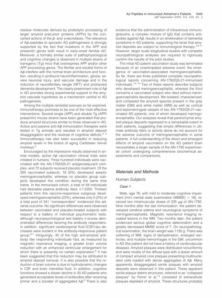
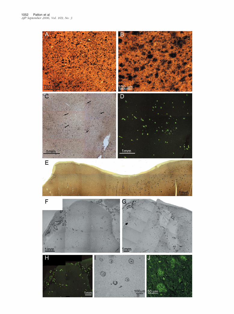
It is evident that the amyloid deposits in the cerebralcortex of AN-1792-vaccinated human cases focally dis-appeared, suggesting that polyclonal anti-A� antibodiescrossed the blood-brain barrier. These antibodies wereinduced using synthetic fibrillar A� 1–42 as an antigen,and the resulting antibodies apparently disassembledfibrillar amyloid plaques. In case 1, there were patchyareas in which the diffuse and compact amyloid depositswere totally absent, although abundant accumulations ofamyloid were present in the cortical vessels (Figure 1, C,D, E, and H). In this study, the term “diffuse amyloiddeposits” describes plaques that are a conglomerate ofnonfibrillar A� peptides organized into amorphous ag-gregates. They stain faintly with thioflavine-S (Figure 1J)or thioflavine-T, are negative for Congo red birefringence,and are very visible and stain intensely with silver dyessuch as Campbell-Switzer (Figure 1, A and B). In otherareas of the cortex, there were focally decreased densi-ties of compact core plaques as revealed by histochem-ical stains and the 6E10 antibody probing, with remainingdiffuse amyloid deposits clearly demonstrated by the4G8 antibody (Figure 1, F and G), respectively. NFTdensity remained high in regions with focally depletedplaques. Earlier neuropathological reports of NFTs in AN-1792 vaccinated cases13,14 found no apparent changesin their abundance and distribution. Previous biochemi-
Figure 1. Brain tissue sections showing cortical areas with and without amyloid deposits. A: Section of frontal cortex from a nonvaccinated sporadic AD patientshowing numerous plaques. Campbell-Switzer stain (�25). B: Campbell-Switzer silver-stained area of the frontal cortex of the same patient shown in A at highermagnification (�100). C: Section of the frontal cortex from case 1, stained by Campbell-Switzer stain demonstrating moderate number of diffuse amyloid deposits.Arrows point to examples of diffuse deposits (�25). D: The same area as in C stained by thioflavine-S showing an abundant number of vascular amyloid deposits(�25). E: A photomontage of the temporal cortex from case 1 demonstrating the transition between an area with no amyloid deposits (left) and an area withnumerous diffuse plaques (right). Campbell-Switzer silver stain (�25). F: A photomontage of the superior frontal gyrus from case 1 stained by the 6E10monoclonal antibody against A� residues 1 to 16. A very low number of amyloid core plaques are observed, whereas deposits of leptomeningeal- andcortico-vascular amyloid are abundant (�25). G: The same region as in F but stained with the monoclonal antibody 4G8 against A� residues 17 to 24, revealingnumerous diffuse plaques, represented as fine powdery material, and vascular amyloid deposits (�25). H: The same area as in F and G, stained with thioflavine-Sillustrating numerous vascular deposits of amyloid in both leptomeningeal and cortical vessels (�25). I: A temporal lobe section from case 1 stained with the 6E10antibody showing a group of amyloid plaques. Four of these plaques contain a small centrally situated core of amyloid with a peripheral halo of immunoreactivematerial (�100). J: Diffuse deposits of A� in the parietal lobe from case 1 stained with thioflavine-S. These deposits were enhanced to delineate their boundariesand increase the contrast against the neuropil background (�200).
A� Peptides in Immunized Alzheimer’s Patients 1053AJP September 2006, Vol. 169, No. 3
1054 Patton et alAJP September 2006, Vol. 169, No. 3
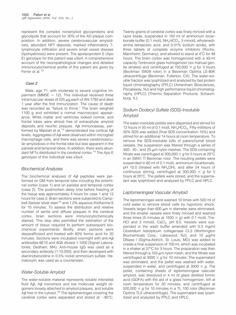
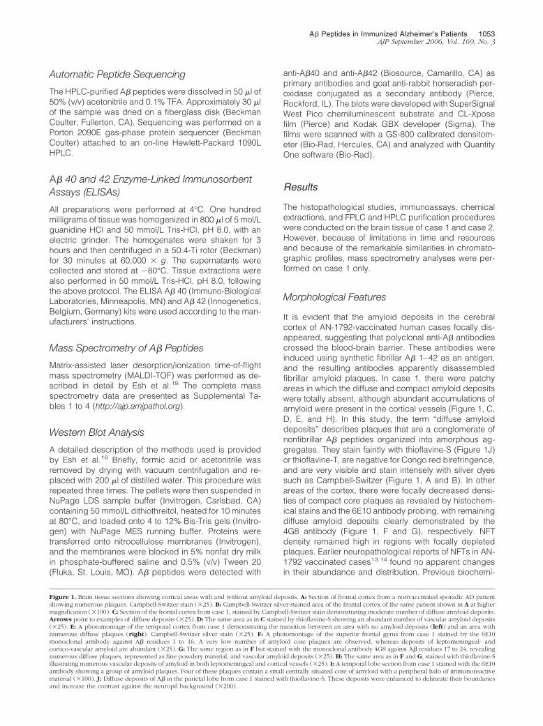
cal studies have revealed that the diffuse deposits of A�in sporadic AD are mainly composed of a mixture of A�17–42 and A� 1–42.19 In some areas, the remainingamyloid plaques exhibited a peculiar morphology, con-sisting of a clearly defined halo of amyloid-positive ma-terial and diminutive centrally situated punctiform core.Between these two structures, there was a clear areadevoid of A� immunoreactivity (Figure 1I). Concerningcase 2, histological sections of the frontal lobe stainedwith Campbell-Switzer and thioflavine-S revealed a com-plete absence of amyloid core plaques and diffuseplaques (Figure 2, A and B) in agreement with previouslypublished data.14 However, although the parietal andtemporal cortex demonstrated sparse core amyloidplaques, diffuse plaques were abundant (Figure 2, C andD). Histological sections of case 1 showed an extensivedeposition of A� in the cortical vessels and in the lepto-meningeal vasculature (Figure 1, D and H; Figure 2, Eand F). However, they were less pronounced in case 2.
Immunoassay Quantification of A� Peptides
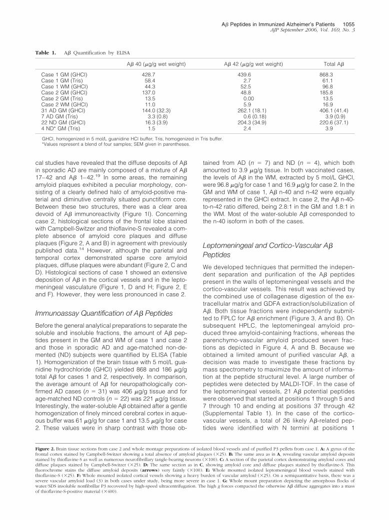
Before the general analytical preparations to separate thesoluble and insoluble fractions, the amount of A� pep-tides present in the GM and WM of case 1 and case 2and those in sporadic AD and age-matched non-de-mented (ND) subjects were quantified by ELISA (Table1). Homogenization of the brain tissue with 5 mol/L gua-nidine hydrochloride (GHCl) yielded 868 and 186 �g/gtotal A� for cases 1 and 2, respectively. In comparison,the average amount of A� for neuropathologically con-firmed AD cases (n � 31) was 406 �g/g tissue and forage-matched ND controls (n � 22) was 221 �g/g tissue.Interestingly, the water-soluble A� obtained after a gentlehomogenization of finely minced cerebral cortex in aque-ous buffer was 61 �g/g for case 1 and 13.5 �g/g for case2. These values were in sharp contrast with those ob-
tained from AD (n � 7) and ND (n � 4), which bothamounted to 3.9 �g/g tissue. In both vaccinated cases,the levels of A� in the WM, extracted by 5 mol/L GHCl,were 96.8 �g/g for case 1 and 16.9 �g/g for case 2. In theGM and WM of case 1, A� n-40 and n-42 were equallyrepresented in the GHCl extract. In case 2, the A� n-40-to-n-42 ratio differed, being 2.8:1 in the GM and 1.8:1 inthe WM. Most of the water-soluble A� corresponded tothe n-40 isoform in both of the cases.
Leptomeningeal and Cortico-Vascular A�
Peptides
We developed techniques that permitted the indepen-dent separation and purification of the A� peptidespresent in the walls of leptomeningeal vessels and thecortico-vascular vessels. This result was achieved bythe combined use of collagenase digestion of the ex-tracellular matrix and GDFA extraction/solubilization ofA�. Both tissue fractions were independently submit-ted to FPLC for A� enrichment (Figure 3, A and B). Onsubsequent HPLC, the leptomeningeal amyloid pro-duced three amyloid-containing fractions, whereas theparenchymo-vascular amyloid produced seven frac-tions as depicted in Figure 4, A and B. Because weobtained a limited amount of purified vascular A�, adecision was made to investigate these fractions bymass spectrometry to maximize the amount of informa-tion at the peptide structural level. A large number ofpeptides were detected by MALDI-TOF. In the case ofthe leptomeningeal vessels, 21 A� potential peptideswere observed that started at positions 1 through 5 and7 through 10 and ending at positions 37 through 42(Supplemental Table 1). In the case of the cortico-vascular vessels, a total of 26 likely A�-related pep-tides were identified with N termini at positions 1
Table 1. A� Quantification by ELISA
A� 40 (�g/g wet weight) A� 42 (�g/g wet weight) Total A�
Case 1 GM (GHCl) 428.7 439.6 868.3Case 1 GM (Tris) 58.4 2.7 61.1Case 1 WM (GHCl) 44.3 52.5 96.8Case 2 GM (GHCl) 137.0 48.8 185.8Case 2 GM (Tris) 13.5 0.00 13.5Case 2 WM (GHCl) 11.0 5.9 16.931 AD GM (GHCl) 144.0 (32.3) 262.1 (18.1) 406.1 (41.4)7 AD GM (Tris) 3.3 (0.8) 0.6 (0.18) 3.9 (0.9)22 ND GM (GHCl) 16.3 (3.9) 204.3 (34.9) 220.6 (37.1)4 ND* GM (Tris) 1.5 2.4 3.9
GHCl, homogenized in 5 mol/L guanidine HCl buffer; Tris, homogenized in Tris buffer.*Values represent a blend of four samples; SEM given in parentheses.
Figure 2. Brain tissue sections from case 2 and whole montage preparations of isolated blood vessels and of purified P3 pellets from case 1. A: A gyrus of thefrontal cortex stained by Campbell-Switzer showing a total absence of amyloid plaques (�25). B: The same area as in A, revealing vascular amyloid depositsstained by thioflavine-S as well as numerous neurofibrillary tangle-bearing neurons (�100). C: A section of the parietal cortex demonstrating amyloid cores anddiffuse plaques stained by Campbell-Switzer (�25). D: The same section as in C, showing amyloid core and diffuse plaques stained by thioflavine-S. Thisfluorochrome stains the diffuse amyloid deposits (arrows) very faintly (�100). E: Whole mounted isolated leptomeningeal blood vessels stained withthioflavine-S (�25). F: Whole mounted isolated cortical vessels showing a heavy burden of vascular amyloid (�25). On a semiquantitative basis, there was asevere vascular amyloid load (3) in both cases under study, being more severe in case 1. G: Whole mount preparation depicting the amorphous flocks ofwater/SDS insoluble nonfibrillar P3 recovered by high-speed ultracentrifugation. The high g forces compacted the otherwise A� diffuse aggregates into a massof thioflavine-S-positive material (�400).
A� Peptides in Immunized Alzheimer’s Patients 1055AJP September 2006, Vol. 169, No. 3
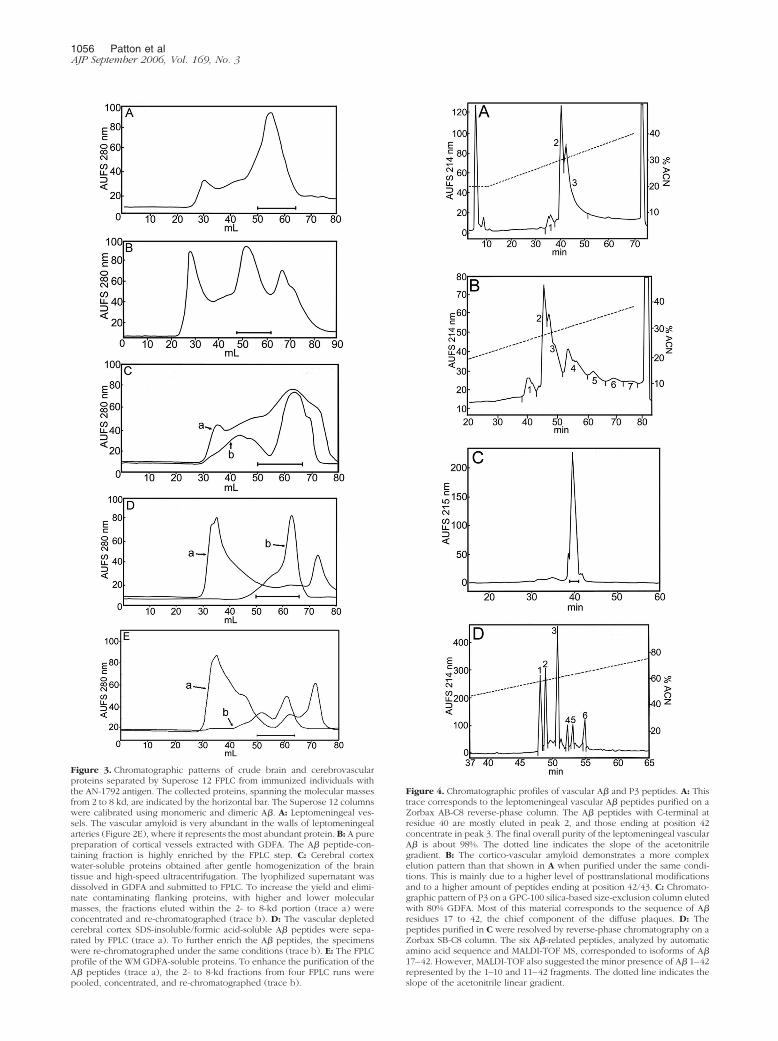
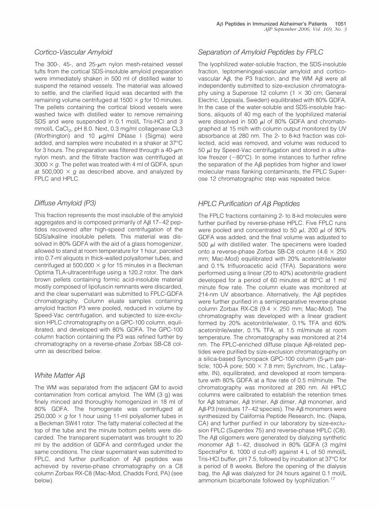
Figure 3. Chromatographic patterns of crude brain and cerebrovascularproteins separated by Superose 12 FPLC from immunized individuals withthe AN-1792 antigen. The collected proteins, spanning the molecular massesfrom 2 to 8 kd, are indicated by the horizontal bar. The Superose 12 columnswere calibrated using monomeric and dimeric A�. A: Leptomeningeal ves-sels. The vascular amyloid is very abundant in the walls of leptomeningealarteries (Figure 2E), where it represents the most abundant protein. B: A purepreparation of cortical vessels extracted with GDFA. The A� peptide-con-taining fraction is highly enriched by the FPLC step. C: Cerebral cortexwater-soluble proteins obtained after gentle homogenization of the braintissue and high-speed ultracentrifugation. The lyophilized supernatant wasdissolved in GDFA and submitted to FPLC. To increase the yield and elimi-nate contaminating flanking proteins, with higher and lower molecularmasses, the fractions eluted within the 2- to 8-kd portion (trace a) wereconcentrated and re-chromatographed (trace b). D: The vascular depletedcerebral cortex SDS-insoluble/formic acid-soluble A� peptides were sepa-rated by FPLC (trace a). To further enrich the A� peptides, the specimenswere re-chromatographed under the same conditions (trace b). E: The FPLCprofile of the WM GDFA-soluble proteins. To enhance the purification of theA� peptides (trace a), the 2- to 8-kd fractions from four FPLC runs werepooled, concentrated, and re-chromatographed (trace b).
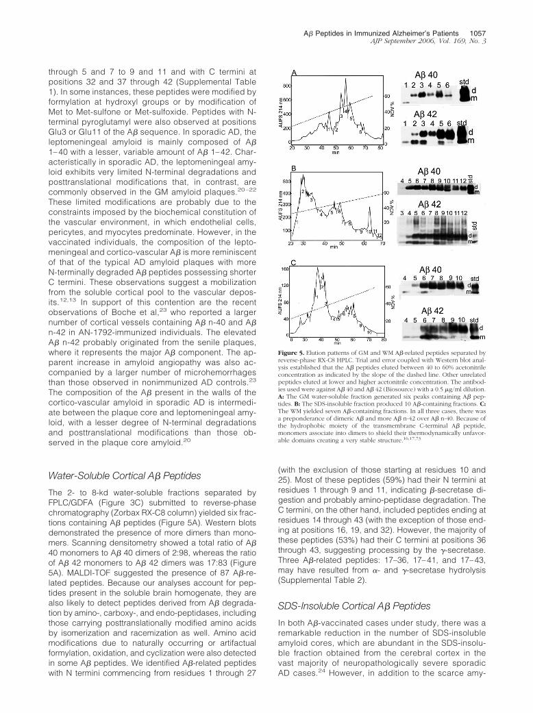
Figure 4. Chromatographic profiles of vascular A� and P3 peptides. A: Thistrace corresponds to the leptomeningeal vascular A� peptides purified on aZorbax AB-C8 reverse-phase column. The A� peptides with C-terminal atresidue 40 are mostly eluted in peak 2, and those ending at position 42concentrate in peak 3. The final overall purity of the leptomeningeal vascularA� is about 98%. The dotted line indicates the slope of the acetonitrilegradient. B: The cortico-vascular amyloid demonstrates a more complexelution pattern than that shown in A when purified under the same condi-tions. This is mainly due to a higher level of posttranslational modificationsand to a higher amount of peptides ending at position 42/43. C: Chromato-graphic pattern of P3 on a GPC-100 silica-based size-exclusion column elutedwith 80% GDFA. Most of this material corresponds to the sequence of A�residues 17 to 42, the chief component of the diffuse plaques. D: Thepeptides purified in C were resolved by reverse-phase chromatography on aZorbax SB-C8 column. The six A�-related peptides, analyzed by automaticamino acid sequence and MALDI-TOF MS, corresponded to isoforms of A�17–42. However, MALDI-TOF also suggested the minor presence of A� 1–42represented by the 1–10 and 11–42 fragments. The dotted line indicates theslope of the acetonitrile linear gradient.
1056 Patton et alAJP September 2006, Vol. 169, No. 3
through 5 and 7 to 9 and 11 and with C termini atpositions 32 and 37 through 42 (Supplemental Table1). In some instances, these peptides were modified byformylation at hydroxyl groups or by modification ofMet to Met-sulfone or Met-sulfoxide. Peptides with N-terminal pyroglutamyl were also observed at positionsGlu3 or Glu11 of the A� sequence. In sporadic AD, theleptomeningeal amyloid is mainly composed of A�1– 40 with a lesser, variable amount of A� 1– 42. Char-acteristically in sporadic AD, the leptomeningeal amy-loid exhibits very limited N-terminal degradations andposttranslational modifications that, in contrast, arecommonly observed in the GM amyloid plaques.20 –22
These limited modifications are probably due to theconstraints imposed by the biochemical constitution ofthe vascular environment, in which endothelial cells,pericytes, and myocytes predominate. However, in thevaccinated individuals, the composition of the lepto-meningeal and cortico-vascular A� is more reminiscentof that of the typical AD amyloid plaques with moreN-terminally degraded A� peptides possessing shorterC termini. These observations suggest a mobilizationfrom the soluble cortical pool to the vascular depos-its.12,13 In support of this contention are the recentobservations of Boche et al,23 who reported a largernumber of cortical vessels containing A� n-40 and A�n-42 in AN-1792-immunized individuals. The elevatedA� n-42 probably originated from the senile plaques,where it represents the major A� component. The ap-parent increase in amyloid angiopathy was also ac-companied by a larger number of microhemorrhagesthan those observed in nonimmunized AD controls.23
The composition of the A� present in the walls of thecortico-vascular amyloid in sporadic AD is intermedi-ate between the plaque core and leptomeningeal amy-loid, with a lesser degree of N-terminal degradationsand posttranslational modifications than those ob-served in the plaque core amyloid.20
Water-Soluble Cortical A� Peptides
The 2- to 8-kd water-soluble fractions separated byFPLC/GDFA (Figure 3C) submitted to reverse-phasechromatography (Zorbax RX-C8 column) yielded six frac-tions containing A� peptides (Figure 5A). Western blotsdemonstrated the presence of more dimers than mono-mers. Scanning densitometry showed a total ratio of A�40 monomers to A� 40 dimers of 2:98, whereas the ratioof A� 42 monomers to A� 42 dimers was 17:83 (Figure5A). MALDI-TOF suggested the presence of 87 A�-re-lated peptides. Because our analyses account for pep-tides present in the soluble brain homogenate, they arealso likely to detect peptides derived from A� degrada-tion by amino-, carboxy-, and endo-peptidases, includingthose carrying posttranslationally modified amino acidsby isomerization and racemization as well. Amino acidmodifications due to naturally occurring or artifactualformylation, oxidation, and cyclization were also detectedin some A� peptides. We identified A�-related peptideswith N termini commencing from residues 1 through 27
(with the exclusion of those starting at residues 10 and25). Most of these peptides (59%) had their N termini atresidues 1 through 9 and 11, indicating �-secretase di-gestion and probably amino-peptidase degradation. TheC termini, on the other hand, included peptides ending atresidues 14 through 43 (with the exception of those end-ing at positions 16, 19, and 32). However, the majority ofthese peptides (53%) had their C termini at positions 36through 43, suggesting processing by the �-secretase.Three A�-related peptides: 17–36, 17–41, and 17–43,may have resulted from �- and �-secretase hydrolysis(Supplemental Table 2).
SDS-Insoluble Cortical A� Peptides
In both A�-vaccinated cases under study, there was aremarkable reduction in the number of SDS-insolubleamyloid cores, which are abundant in the SDS-insolu-ble fraction obtained from the cerebral cortex in thevast majority of neuropathologically severe sporadicAD cases.24 However, in addition to the scarce amy-
Figure 5. Elution patterns of GM and WM A�-related peptides separated byreverse-phase RX-C8 HPLC. Trial and error coupled with Western blot anal-ysis established that the A� peptides eluted between 40 to 60% acetonitrileconcentration as indicated by the slope of the dashed line. Other unrelatedpeptides eluted at lower and higher acetonitrile concentration. The antibod-ies used were against A� 40 and A� 42 (Biosource) with a 0.5 �g/ml dilution.A: The GM water-soluble fraction generated six peaks containing A� pep-tides. B: The SDS-insoluble fraction produced 10 A�-containing fractions. C:The WM yielded seven A�-containing fractions. In all three cases, there wasa preponderance of dimeric A� and more A� n-42 over A� n-40. Because ofthe hydrophobic moiety of the transmembrane C-terminal A� peptide,monomers associate into dimers to shield their thermodynamically unfavor-able domains creating a very stable structure.16,17,73
A� Peptides in Immunized Alzheimer’s Patients 1057AJP September 2006, Vol. 169, No. 3
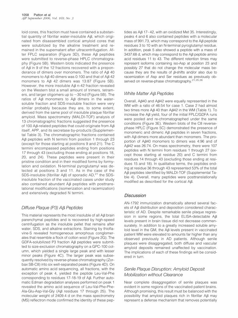
loid cores, this fraction must have contained a substan-tial quantity of fibrillar water-insoluble A�, which origi-nated from disassembled cortical amyloid cores thatwere solubilized by the alkaline treatment and re-mained in the supernatant after ultracentrifugation. Af-ter FPLC separation (Figure 3D), these A� peptideswere submitted to reverse-phase HPLC chromatogra-phy (Figure 5B). Western blots indicated the presenceof A� in 9 of the 12 fractions recovered with a prepon-derance of dimers over monomers. The ratio of A� 40monomers to A� 40 dimers was 0:100 and that of A� 42monomers to A� 42 dimers was 13:87 (Figure 5B).However, the more insoluble A� n-42 fraction revealedon the Western blot a small amount of trimers, tetram-ers, and larger oligomers up to �30 kd (Figure 5B). Theratios of A� monomers to A� dimers in the water-soluble fraction and SDS-insoluble fraction were verysimilar probably because they are, to some extent,derived from the same pool of insoluble plaque fibrillaramyloid. Mass spectrometry (MALDI-TOF) analysis of13 chromatographic fractions suggested the presenceof 100 A�-related peptides that could originate from A�itself, APP, and its secretase by-products (Supplemen-tal Table 3). The chromatographic fractions containedA� peptides with N termini from position 1 through 23(except for those starting at positions 9 and 21). The Ctermini encompassed peptides ending from positions17 through 43 (excluding those ending at positions 18,20, and 24). These peptides were present in theirpristine condition and in their modified forms by formy-lation and oxidation. N-terminal pyroglutamyl was de-tected at positions 3 and 11. As in the case of theSDS-insoluble (fibrillar A�) of sporadic AD,21 the SDS-insoluble fraction of the vaccinated cases under studyalso contained abundant A� peptides with posttrans-lational modifications (isomerization and racemization)and extensively degraded N termini.
Diffuse Plaque (P3) A� Peptides
This material represents the most insoluble of all A� brainparenchymal peptides and is recovered by high-speedcentrifugation as the insoluble pellet that remains afterwater, SDS, and alkaline extractions. Staining by thiofla-vine-S revealed homogeneous amorphous conglomer-ates that resemble a flock of cotton wool (Figure 2G). TheGDFA-solubilized P3 fraction A� peptides were submit-ted to size-exclusion chromatography on a GPC-100 col-umn, which yielded a single large peak and with lesserminor peaks (Figure 4C). The larger peak was subse-quently resolved by reverse-phase chromatography (Zor-bax SB-C8) into six well-separated peaks (Figure 4D). Onautomatic amino acid sequencing, all fractions, with theexception of peak 4, yielded the peptide Leu-Val-Phecorresponding to residues 17-18-19 of A�. Further auto-matic Edman degradation analyses performed on peak 1revealed the amino acid sequence of Leu-Val-Phe-Phe-Ala-Glu-Asp-Val-Gly (A� residues 17 through 25). Themolecular weight of 2409.4 d on the mass spectrometry(MS) reflectron mode confirmed the identity of these pep-
tides as A� 17–42, with an oxidized Met 35. Interestingly,peaks 4 and 6 also contained peptides with a molecularmass of 991.73, which may correspond to the A� peptideresidues 3 to 10 with an N-terminal pyroglutamyl residue.In addition, peak 5 also showed a peptide with a mass of3437.68 d, which may correspond to the A� peptide aminoacid residues 11 to 43. The different retention times mayrepresent isoforms containing iso-Asp at position 23 andpossibly 27 that do not change the molecular mass be-cause they are the results of �-shifts and/or also due toracemization of Asp and Ser residues as previously ob-served on reverse-phase chromatography.21,25,26
White Matter A� Peptides
Overall, A�40 and A�42 were equally represented in theWM with a ratio of 46:54 for case 1. Case 2 had almosttwo times more A� 40 than A�42 in the WM (Table 1). Toincrease the A� yield, four of the initial FPLC/GDFA runswere pooled and re-chromatographed under the sameconditions (Figure 3E). Western blots of the C8 reverse-phase HPLC (Figure 5C) demonstrated the presence ofmonomeric and dimeric A� peptides in seven fractions,with A� dimers more abundant than monomers. The pro-portion of A�40 monomers to dimers was 4:96 and ofA�42 was 26:74. On mass spectrometry, there were 107peptides with N termini from residues 1 through 27 (ex-cept those starting at residue 20) and C termini fromresidues 14 through 43 (excluding those ending at resi-dues 15 and 18). In qualitative terms, the peptides end-ing at residue 36 through 43 represented 53% of the totalA� peptides identified by MALDI-TOF (Supplemental Ta-ble 4). Overall, many peptides were posttranslationallymodified as described for the cortical A�.
Discussion
AN-1792 immunization dramatically altered several fac-ets of A� distribution and deposition considered charac-teristic of AD. Despite remarkable senile plaque regres-sion in some regions, the total ELISA-detectable A�levels present in brain tissue did not decrease commen-surately. In addition to a greatly increased soluble amy-loid level in the GM, the A� levels present in vaccinatedpatient WM were elevated to amounts far higher than anyobserved previously in AD patients. Although senileplaques were disaggregated, both diffuse and vascularamyloid deposits remained unaffected by vaccination.The implications of each of these findings will be consid-ered in turn.
Senile Plaque Disruption: Amyloid DepositMobilization without Clearance
Near complete disaggregation of senile plaques wasevident in some regions of the vaccinated patient brains.Although promising, this result must be balanced with thepossibility that amyloid plaques rich in fibrillar A� mayrepresent a defense mechanism that removes potentially
1058 Patton et alAJP September 2006, Vol. 169, No. 3
neurotoxic soluble amyloid from the neuropil and insu-lates it with a layer of glial cells.17,27,28 This suggests thatplaque disruption in the absence of removal of the solu-bilized amyloid or in situations in which removal is ineffi-cient may inadvertently produce a destructive side effect.Dissolution of fibrillar amyloid plaque cores by immuno-globulins or complement is followed by microglia/macro-phage receptor-mediated uptake, limited degradation ofA� and eventual discharge of soluble A� into the neuro-pil.29,30 The A� HHQK domain, amino acid residues 13 to16, is capable of unleashing neuroinflammation via acti-vation of microglia,31,32 and low molecular weight A�oligomers are recognized to kill neurons and impair cog-nitive function.17,33 The oligomerization of A�-protein be-gins intracellularly in cells derived from human brain,16
and intraneuronal A� accumulation causes early cogni-tive impairment before the extracellular A� deposition intriple Tg mice.34 Whether the antibody-solubilized amy-loid species constrained within the cortex and WM aretoxic and can eventually be eliminated remains to beestablished. In addition to A� n-40 and n-42 residuelength species and their multimers, AD patients havelarge quantities of soluble A� molecules present that areposttranslationally modified and difficult for proteolyticenzymes to degrade.35
In both vaccinated patients, the amount of solubleamyloid was substantially increased compared with spo-radic AD patients (Table 1). Compared with the levelsobserved in the GM of nonimmunized sporadic AD pa-tient pools, case 1 demonstrated a 2.1-fold increase intotal A� (extracted by 5 mol/L guanidine HCl buffer),whereas case 2 showed a reduction (2.2-fold less) (Table1). Case 1 developed meningoencephalitis, and it is pos-sible that the added pathology associated with this ad-verse event increased A� levels above the typical meanfor uncomplicated AD. In addition, AD pathology isknown to exhibit great variation among individuals, andperhaps case 2 simply harbored a comparatively lowamount of senile plaques, vascular amyloid deposits, andsoluble A� before vaccination. However, it is remarkablethat the total amounts of GM soluble amyloid (extractedwith Tris buffer) are substantially increased in both vac-cinated cases. Cases 1 and 2 demonstrated 15- and3.5-fold higher levels of total soluble A� compared withAD and ND individuals, respectively (Table 1).
Despite a marked focal reduction in the number ofamyloid plaques, there was no commensurate decreasein total brain tissue A� levels (measured by ELISA), sim-ilar to previous results obtained with immunized APP Tganimals. Although plaques were disrupted in vaccinatedTg mice, formic acid-extractable A� levels representingthe combined pools of soluble and insoluble plaque andvascular-associated fibrillar A� fractions did not differbetween A� immunized TgCRND8, PDAPP, and tg2576Tg mice and nonimmunized rodents.36–39 These obser-vations suggest that the removal of A� from the brain byimmunization is not complete or occurs at substantiallyslower rates than senile plaque disruption.
Comprehending the ultimate effects of plaque amyloiddispersal on brain vascular function is vital. Soluble intra-and extracellular A� may exert deleterious effects on
vascular endothelial and smooth muscle cell metabolism.Soluble A� is a potent vasoconstrictor capable of alteringregional cerebral blood flow,40,41 and A� peptides arealso powerful angiogenesis inhibitors.42,43 These activi-ties may impact blood-brain barrier selectivity and vas-cular repair. In addition, A� interferes with the productionof vascular relaxing factors and disturbs the balancebetween cerebral energy requirements and cerebralblood flow.44 The soluble and insoluble A� effects onvascular homeostasis may be the most aggressive pa-thology in AD, and it is possible that increased levels ofsoluble A� peptides could have important vascularpathophysiological consequences in immunizedindividuals.
Vascular and Diffuse Amyloid Deposits ResistAN-1792 Vaccine-Induced Disruption
A feature shared by both Tg mice and human AD patientsis the firm recalcitrance of vascular amyloid deposits todisruption by immunization.4,13,14,36–38,45,46 Also com-mon to both is that focal senile plaque density decreaseswere accompanied by increased vascular amyloid dep-osition suggesting an active, antibody-induced remodel-ing of amyloid accumulation patterns. It is possible thatsome antibody-solublilized senile plaque amyloid wasre-deposited in the microvasculature during egress fromthe brain through the periarterial spaces. In support ofthis contention is the observation that the peptide com-position of the water-soluble A� peptides (SupplementalTable 2) resembled that isolated from the vascular com-partments (Supplemental Table 1). Moreover, of the wa-ter-soluble fraction extracted from the cerebral cortex, 96and 100% of the A� peptides measured by ELISA weremostly composed of the more soluble A� n-40 for cases1 and 2, respectively (Table 1). However, extractions withwater-based buffers do not efficiently isolate the moreinsoluble A� n-42. Therefore, a more stringent analysisusing formic acid extraction was used for the water-insoluble pellets. Subsequent MALDI-TOF MS analysesrevealed a substantial amount of A� peptides ending atresidues 42 and 43. Pre-existing impaired brain interstitialfluid drainage or an emerging periarterial occlusion couldact to retain a permanent, antibody-dispersed A� pool inthe cerebral cortex.47–50 In this scenario, the reportedcognitive enhancement in Tg mice,4 rats,51 and immu-nized individuals may be due to the removal of a toxicsoluble amyloid pool. Recent investigations have demon-strated that passive immunotherapy against A� in agedtg2576 Tg mice reversed cognitive deficits and reducedparenchymal plaque burden but resulted in a concomi-tant increase in vascular amyloid load and associatedmicrohemorrhages.52 Moreover, in the 21-month-oldAPP23 Tg mice, passive vaccination with IgG againstresidues 3 to 6 of human A� resulted in a twofold in-crease in cerebral amyloid angiopathy-associated micro-hemorrhages.45 The same phenomenon occurred inaged PDAPP Tg mice in which vaccination at high dosesof the 3D6 antibody, directed against the N-terminalregion of A�, resulted in an increased incidence and
A� Peptides in Immunized Alzheimer’s Patients 1059AJP September 2006, Vol. 169, No. 3
severity of microhemorrhages.46 Intriguingly, all threehuman vaccinated cases, neuropathologically character-ized,12–14 showed the highest score of 3 (severe) forvascular amyloid deposition in those areas in which theapparent removal of amyloid attained its maximal expres-sion. Additional studies with APP23 mice,53 a Tg mousemodel in which there is a remarkable load of vascularamyloid, have confirmed that elevated quantities of sol-uble amyloid (�50 �g/g) are ultimately accompanied bya prompt and overwhelming global leptomeningeal andcortico-vascular amyloid deposition, a phenomenon thathas not been observed in other Tg mice studied.54
Immunization was apparently unable to remove diffusedeposits of A�. In case 1, there were large areas in thefrontal parietal and temporal regions in which there werenumerous diffuse plaques. In case 2, however, therewere abundant diffuse plaques present in the temporaland parietal lobes, although they were absent from thefrontal lobe, a finding consistent with the results ofMasliah et al.14 We were able to isolate from both AN-1792-treated cases substantial quantities of the ex-tremely insoluble nonamyloidogenic P3 peptide, the ma-jor peptide in the diffuse plaques,19 mainly composed ofthe A� sequence of residues 17 to 42. Because P3 de-posits are not fibrillar and are not associated with acti-vated proinflammatory microglia, it has been concludedthat P3 is a “desirable” APP by-product of the �- and�-secretases, the generation of which precludes amyloidformation. However, this peptide has also been observedin dystrophic neurites associated with amyloid plaquesand in pathological cortical vesicular structures.55 Fur-thermore, the A� 17–42 induces apoptosis by activationof c-jun N-terminal kinase and caspases 8 and 3.56 TheTg mice transfected with APP751 cDNA, under the controlof the rat neuron-specific enolase promoter, only developdiffuse A� deposits that correlate with age-dependentsevere learning deficits.57 The C-terminal domain of A�has powerful membrane destabilizing properties that al-ter the permeability of cellular membranes, thus contrib-uting to cytotoxicity.58–60 It has been recently suggestedthat the retention and accumulation of A� in lipid bilayerscould induce severe disturbances in cellular metabolismand play a major role in AD pathology.61 These findingssuggest that diffuse P3 deposits are not benign. Passiveor active immunizations directed exclusively to N-terminalA�-42 sequences, such as AN-1792,62 will not removepotentially neurotoxic A� P3 peptides. However, a newgeneration of antibodies specifically directed against theC-terminal domain of A� and designed to remove bothcompact and diffuse plaques are undergoing experimen-tal evaluation.63
Gross Enlargement of the White MatterAmyloid Pool
White matter lesions are present in two-thirds of patientswith AD. They represent severe alterations in protein,lipid, and cholesterol metabolism; dysmyelination; demy-elination; loss of axons and astrogliosis compounded bya reduced number of microvessels; concurrent ischemia/
hypoxia; and interstitial fluid retention with dilation of peri-arterial spaces.49,64 In elderly ND individuals, the WMcontains a small pool of soluble A� that probably origi-nates in the GM and gradually diffuses into the WM.64 Inthe absence of visible WM A� deposits, such as thoseobserved in the cerebral cortex of AD patients, it can beassumed that the WM A� peptides are either solubleoligomers or associated with structural molecules suchas myelin sheets that may inhibit A� fibrillization. In ad-dition, an amplified A�-mediated WM neuroinflammatoryresponse would increase the complement-mediated pro-duction of membrane attack complexes that will furtherdestroy myelin and axonal integrity.65 Experimentally, A�peptides induce severe oligodendrocyte damage by in-terfering with the sphingomyelinase-ceramide pathway66
as well as alterations in axonal cytoskeleton manifestedby loss of neurofilament immunoreactivity.67 Both guani-dine and formic acid extractions recovered a substantialamount of WM A� n-40 and A� n-42. In sporadic AD WM,the total average soluble A� is 2.2 �g/g tissue64 whereasthe WM of immunized cases 1 and 2 contained 97 and 17�g/g tissue of soluble total A�, representing 44 and 8times more A� peptide load, respectively. The higheramount of total WM A� (� 5.8-fold) of case 1 over case 2may be due to the postvaccination aseptic encephalitisthat gravely affected the WM or to differences in prevac-cination A� levels. The substantial increase in WM amy-loid burden suggests that this compartment acted as theultimate “sink” for A� released from disrupted senileplaques that could not escape through the vasculature.The ultimate consequences of a shifted amyloid tissueburden are unknown.
Amyloid Accumulation Dynamics andVaccination
The cognitive improvement exhibited by vaccinated pa-tients suggests that amyloid plaques play key roles in ADpathophysiology and the development of dementia. Inaddition, the results suggest that a patient with preclinicalstages of AD may have incompletely evolved plaquesthat have not yet undergone extensive chemical modifi-cation, provoked inflammation, or managed to irrevers-ibly damage surrounding neurons. Thus, there may beonly a brief window of opportunity to reverse theseplaques. The problem is that vascular A� deposits in alllikelihood are forming concomitantly with senile plaquesand may interfere with the complete removal of disruptedamyloid. Even with an antibody specific to A� 1–40, thedominant peptide in vascular amyloid, once preclinicalAD has progressed to dementia, it may be too late topreserve the architecture and function of the vessels.49,68
These tenets suggest two quite interesting hypotheses:1) AD plaques might initially be virtually absent butemerge rapidly once mild cognitive impairment appears.2) Plaques from the early clinical stages of AD are sub-stantially less modified chemically than those of full-blown AD, as in the case of the APP Tg mice where theamyloid does not have time to accumulate chemicalmodifications and thereby becomes more resistant to
1060 Patton et alAJP September 2006, Vol. 169, No. 3
disruption. In support of these contentions is the earlyprophylactic immunization of several strains of Tg micethat results in minimal amyloid deposits.4,69 As the Tgmice age, the efficient removal of plaques by vaccinationdecreases.69–71 Arresting A� production in the Tg APPmice carrying the Swedish/Indiana mutations by doxycy-cline-controlled expression demonstrated that pre-exist-ing amyloid deposits become very stable and difficult toclear as the animals age.72 Therefore, although “young”and comparatively unmodified amyloid in Tg mice andpossibly in preclinical AD is more amenable to disruption,such efforts will be substantially less effective in fullydeveloped AD, where modified A� is too abundant anddifficult to eliminate. In addition, immunotherapy cannotremove the vascular amyloid because it is enmeshedwith the extracellular matrix to too great an extent. Wealso hypothesize that impaired regional brain perfusiondue to damaged microvasculature might explain thepatchy plaque removal patterns observed in immunizedindividuals. These assumptions suggest that early inter-vention, well before vascular amyloid deposition is exten-sive and circulatory compromise is severe, may be a keyfeature of successful mitigation approaches.
In summary, examination of two AN-1792 immunizedindividuals revealed areas of the cerebral cortex in whichthe compact amyloid plaques were removed in someregions, leaving behind diffuse A� deposits. A remark-able increase in the soluble A� pool was evident in bothcases and may have contributed to a more severe amy-loid angiopathy and sharply elevated WM-soluble A�
levels. In addition, the increasing deposits of amyloid inthe brain vasculature may intensify blood-brain barrierdistress and hamper brain perfusion. The elevated poolof soluble amyloid in GM and WM may further impairneuronal function and neural transmission and addition-ally promote neuroinflammation. Despite these potentialpitfalls, the morphological and biochemical evidencesupports immunization programs in humans by providingdirect and substantial evidence that amyloid plaque de-posits of mild cognitive impairment patients are amena-ble to disassembly by antibody therapies. However, morecases will need to be studied to verify these preliminaryconclusions and to determine whether immunization mayhave deleterious consequences for AD patients or pa-tients with non-AD dementias. Earlier anti-A� immuniza-tions, combined with treatment strategies that are morepreventative than therapeutic, may be needed to facili-tate drainage and efficient degradation of the brain’s A�
peptides and prevent the accumulation of toxic solubleand insoluble A� and subsequent deleterious neuronaland vascular damage.
Acknowledgment
We express our gratitude to Dr. Douglas Walker (SunHealth Research Institute) for performing Apo Egenotyping.
References
1. Brookmeyer R, Gray S, Kawas C: Projections of Alzheimer’s diseasein the United States and the public health impact of delaying diseaseonset. Am J Public Health 1998, 88:1337–1342
2. Schenk D, Hagen M, Seubert P: Current progress in beta-amyloidimmunotherapy. Curr Opin Immunol 2004, 16:599–606
3. Solomon B: Alzheimer’s disease and immunotherapy. Curr AlzheimerRes 2004, 1:149–163
4. Gelinas DS, Dasilva K, Fenili D, George-Hyslop P, McLaurin J: Immu-notherapy for Alzheimer’s disease. Proc Natl Acad Sci USA 2004,101(Suppl 2):14657–14662
5. Lemere CA, Beierschmitt A, Iglesias M, Spooner ET, Bloom JK, Le-verone JF, Zheng JB, Seabrook TJ, Louard D, Li D, Selkoe DJ,Palmour RM, Ervin FR: Alzheimer’s disease abeta vaccine reducescentral nervous system abeta levels in a non-human primate, theCaribbean vervet. Am J Pathol 2004, 165:283–297
6. Orgogozo JM, Gilman S, Dartigues JF, Laurent B, Puel M, Kirby LC,Jouanny P, Dubois B, Eisner L, Flitman S, Michel BF, Boada M, FrankA, Hock C: Subacute meningoencephalitis in a subset of patients withAD after Abeta42 immunization. Neurology 2003, 61:46–54
7. Gilman S, Koller M, Black RS, Jenkins L, Griffith SG, Fox NC, Eisner L,Kirby L, Rovira MB, Forette F, Orgogozo JM: Clinical effects of Abetaimmunization (AN1792) in patients with AD in an interrupted trial.Neurology 2005, 64:1553–1562
8. Fox NC, Black RS, Gilman S, Rossor MN, Griffith SG, Jenkins L, KollerM: Effects of Abeta immunization (AN1792) on MRI measures ofcerebral volume in Alzheimer disease. Neurology 2005,64:1563–1572
9. Hock C, Konietzko U, Streffer JR, Tracy J, Signorell A, Muller-Till-manns B, Lemke U, Henke K, Moritz E, Garcia E, Wollmer MA,Umbricht D, deq Uervain DJ, Hofmann M, Maddalena A, Papassoti-ropoulos A, Nitsch RM: Antibodies against beta-amyloid slow cogni-tive decline in Alzheimer’s disease. Neuron 2003, 38:547–554
10. Weksler ME, Gouras G, Relkin NR, Szabo P: The immune system,amyloid-beta peptide, and Alzheimer’s disease. Immunol Rev 2005,205:244–256
11. Dodel RC, Du Y, Depboylu C, Hampel H, Frolich L, Haag A, Hem-meter U, Paulsen S, Teipel SJ, Brettschneider S, Spottke A, Nolker C,Moller HJ, Wei X, Farlow M, Sommer N, Oertel WH: Intravenousimmunoglobulins containing antibodies against beta-amyloid for thetreatment of Alzheimer’s disease. J Neurol Neurosurg Psychiatry2004, 75:1472–1474
12. Nicoll JA, Wilkinson D, Holmes C, Steart P, Markham H, Weller RO:Neuropathology of human Alzheimer disease after immunization withamyloid-beta peptide: a case report. Nat Med 2003, 9:448–452
13. Ferrer I, Boada RM, Sanchez Guerra ML, Rey MJ, Costa-Jussa F:Neuropathology and pathogenesis of encephalitis following amyloid-beta immunization in Alzheimer’s disease. Brain Pathol 2004,14:11–20
14. Masliah E, Hansen L, Adame A, Crews L, Bard F, Lee C, Seubert P,Games D, Kirby L, Schenk D: Abeta vaccination effects on plaquepathology in the absence of encephalitis in Alzheimer disease. Neu-rology 2005, 64:129–131
15. Braak H, Braak E: Demonstration of amyloid deposits and neurofibril-lary changes in whole brain sections. Brain Pathol 1991, 1:213–216
16. Walsh DM, Tseng BP, Rydel RE, Podlisny MB, Selkoe DJ: The oli-gomerization of amyloid �-protein begins intracellularly in cells de-rived from human brain. Biochemistry 2000, 39:10831–10839
17. Roher AE, Chaney MO, Kuo YM, Webster SD, Stine WB, HaverkampLJ, Woods AS, Cotter RJ, Tuohy JM, Krafft GA, Bonnell BS, Emmer-ling MR: Morphology and toxicity of Abeta-(1–42) dimer derived fromneuritic and vascular amyloid deposits of Alzheimer’s disease. J BiolChem 1996, 271:20631–20635
18. Esh C, Patton L, Kalback W, Kokjohn TA, Lopez J, Brune D, NewellAJ, Beach T, Schenk D, Games D, Paul S, Bales K, Ghetti B, CastanoEM, Roher AE: Altered APP processing in PDAPP (Val7173Phe)transgenic mice yields extended-length Abeta peptides. Biochemis-try 2005, 44:13807–13819
19. Gowing E, Roher AE, Woods AS, Cotter RJ, Chaney M, Little SP, BallMJ: Chemical characterization of A beta 17–42 peptide, a componentof diffuse amyloid deposits of Alzheimer disease. J Biol Chem 1994,269:10987–10990
20. Roher AE, Lowenson JD, Clarke S, Woods AS, Cotter RJ, Gowing E,
A� Peptides in Immunized Alzheimer’s Patients 1061AJP September 2006, Vol. 169, No. 3
Ball MJ: Beta-amyloid-(1–42) is a major component of cerebrovas-cular amyloid deposits: implications for the pathology of Alzheimerdisease. Proc Natl Acad Sci USA 1993, 90:10836–10840
21. Roher AE, Lowenson JD, Clarke S, Wolkow C, Wang R, Cotter RJ,Reardon IM, Zurcher-Neely HA, Heinrikson RL, Ball MJ: Structuralalterations in the peptide backbone of beta-amyloid core protein mayaccount for its deposition and stability in Alzheimer’s disease. J BiolChem 1993, 268:3072–3083
22. Kuo YM, Emmerling MR, Woods AS, Cotter RJ, Roher AE: Isolation,chemical characterization, and quantitation of A� 3-pyroglutamylpeptides from neuritic plaques and vascular amyloid deposits. Bio-chem Biophys Res Commun 1997, 237:188–191
23. Boche D, Barton E, Neal J, Ferrer I, Wilkinson D, Bayer A, Holmes C,Weller RO, Nicoll JAR: Effects of Abeta immunotherapy on cerebralamyloid angiopathy in human Alzheimer’s disease. Abstracts of the107th meeting of the British Neuropathological Society. NeuropatholAppl Neurobiol 2006, 32(Suppl 1):3–4
24. Roher AE, Palmer KC, Chau V, Ball MJ: Isolation and chemicalcharacterization of Alzheimer’s disease paired helical filamentcytoskeletons: differentiation from amyloid plaque core protein. J CellBiol 1988, 107:2703–2716
25. Lowenson JD, Clarke S, Roher AE: Chemical modification of depos-ited amyloid-� peptides. Methods Enzymol 1999, 309:89–105
26. Shimizu T, Watanabe A, Ogawara M, Mori H, Shirasawa T: Isoaspar-tate formation and neurodegeneration in Alzheimer’s disease. ArchBiochem Biophys 2000, 381:225–234
27. Watson D, Castano E, Kokjohn TA, Kuo YM, Lyubchenko Y, Pinsky D,Connolly ES Jr, Esh C, Luehrs DC, Stine WB, Rowse LM, EmmerlingMR, Roher AE: Physicochemical characteristics of soluble oligomericAbeta and their pathologic role in Alzheimer’s disease. Neurol Res2005, 27:869–881
28. Lee HG, Casadesus G, Zhu X, Takeda A, Perry G, Smith MA: Chal-lenging the amyloid cascade hypothesis: senile plaques and amy-loid-beta as protective adaptations to Alzheimer disease. Ann NYAcad Sci 2004, 1019:1–4
29. Brazil MI, Chung H, Maxfield FR: Effects of incorporation of immuno-globulin G and complement component C1q on uptake and degra-dation of Alzheimer’s disease amyloid fibrils by microglia. J Biol Chem2000, 275:16941–16947
30. Chung H, Brazil MI, Soe TT, Maxfield FR: Uptake, degradation, andrelease of fibrillar and soluble forms of Alzheimer’s amyloid �-peptideby microglial cells. J Biol Chem 1999, 274:32301–32308
31. Giulian D, Haverkamp LJ, Yu J, Karshin W, Tom D, Li J, KazanskaiaA, Kirkpatrick J, Roher AE: The HHQK domain of beta-amyloid pro-vides a structural basis for the immunopathology of Alzheimer’s dis-ease. J Biol Chem 1998, 273:29719–29726
32. Giulian D, Haverkamp LJ, Yu JH, Karshin W, Tom D, Li J, KirkpatrickJ, Kuo LM, Roher AE: Specific domains of beta-amyloid from Alzhei-mer plaque elicit neuron killing in human microglia. J Neurosci 1996,16:6021–6037
33. Cleary JP, Walsh DM, Hofmeister JJ, Shankar GM, Kuskowski MA,Selkoe DJ, Ashe KH: Natural oligomers of the amyloid-beta proteinspecifically disrupt cognitive function. Nat Neurosci 2005, 8:79–84
34. Billings LM, Oddo S, Green KN, McGaugh JL, LaFerla FM: Intraneu-ronal Abeta causes the onset of early Alzheimer’s disease-relatedcognitive deficits in transgenic mice. Neuron 2005, 45:675–688
35. Kuo YM, Webster S, Emmerling MR, De Lima N, Roher AE: Irrevers-ible dimerization/tetramerization and post-translational modificationsinhibit proteolytic degradation of A beta peptides of Alzheimer’sdisease. Biochim Biophys Acta 1998, 1406:291–298
36. Janus C, Pearson J, McLaurin J, Mathews PM, Jiang Y, Schmidt SD,Chishti MA, Horne P, Heslin D, French J, Mount HT, Nixon RA,Mercken M, Bergeron C, Fraser PE, George-Hyslop P, Westaway D:A beta peptide immunization reduces behavioural impairment andplaques in a model of Alzheimer’s disease. Nature 2000,408:979–982
37. Dodart JC, Bales KR, Gannon KS, Greene SJ, DeMattos RB, MathisC, DeLong CA, Wu S, Wu X, Holtzman DM, Paul SM: Immunizationreverses memory deficits without reducing brain Abeta burden inAlzheimer’s disease model. Nat Neurosci 2002, 5:452–457
38. Kotilinek LA, Bacskai B, Westerman M, Kawarabayashi T, Younkin L,Hyman BT, Younkin S, Ashe KH: Reversible memory loss in a mousetransgenic model of Alzheimer’s disease. J Neurosci 2002,22:6331–6335
39. Sigurdsson EM, Knudsen E, Asuni A, Fitzer-Attas C, Sage D, Quar-termain D, Goni F, Frangione B, Wisniewski T: An attenuated immuneresponse is sufficient to enhance cognition in an Alzheimer’s diseasemouse model immunized with amyloid-beta derivatives. J Neurosci2004, 24:6277–6282
40. Thomas T, Thomas G, McLendon C, Sutton T, Mullan M: Beta-amy-loid-mediated vasoactivity and vascular endothelial damage. Nature1996, 380:168–171
41. Crawford F, Suo Z, Fang C, Sawar A, Su G, Arendash G, Mullan M:The vasoactivity of A� peptides. Ann NY Acad Sci 1997, 826:35–46
42. Paris D, Townsend K, Quadros A, Humphrey J, Sun J, Brem S,Wotoczek-Obadia M, DelleDonne A, Patel N, Obregon DF, Crescen-tini R, Abdullah L, Coppola D, Rojiani AM, Crawford F, Sebti S, MullanM: Inhibition of angiogenesis by A� peptides. Angiogenesis 2004,7:75–85
43. Paris D, Town T, Parker T, Humphrey J, Mullan M: A� vasoactivity: aninflammatory reaction. Ann NY Acad Sci 2000, 903:97–109
44. Iadecola C: Cerebrovascular effects of amyloid-� peptides: mecha-nisms and implications for Alzheimer’s dementia. Cell Mol Neurobiol2003, 23:681–689
45. Pfeifer M, Boncristiano S, Bondolfi L, Stalder A, Deller T, StaufenbielM, Mathews PM, Jucker M: Cerebral hemorrhage after passiveanti-A� immunotherapy. Science 2002, 298:1379
46. Racke MM, Boone LI, Hepburn DL, Parsadainian M, Bryan MT, NessDK, Piroozi KS, Jordan WH, Brown DD, Hoffman WP, Holtzman DM,Bales KR, Gitter BD, May PC, Paul SM, DeMattos RB: Exacerbation ofcerebral amyloid angiopathy-associated microhemorrhage in amy-loid precursor protein transgenic mice by immunotherapy is depen-dent on antibody recognition of deposited forms of amyloid beta.J Neurosci 2005, 25:629–636
47. Weller RO, Massey A, Newman TA, Hutchings M, Kuo YM, Roher AE:Cerebral amyloid angiopathy: amyloid-� accumulates in putative in-terstitial fluid drainage pathways in Alzheimer’s disease. Am J Pathol1998, 153:725–733
48. Preston SD, Steart PV, Wilkinson A, Nicoll JA, Weller RO: Capillaryand arterial cerebral amyloid angiopathy in Alzheimer’s disease: de-fining the perivascular route for the elimination of amyloid beta fromthe human brain. Neuropathol Appl Neurobiol 2003, 29:106–117
49. Roher AE, Kuo YM, Esh C, Knebel C, Weiss N, Kalback W, Luehrs DC,Childress JL, Beach TG, Weller RO, Kokjohn TA: Cortical and lepto-meningeal cerebrovascular amyloid and white matter pathology inAlzheimer’s disease. Mol Med 2003, 9:112–122
50. Weller RO, Cohen NR, Nicoll JA: Cerebrovascular disease and thepathophysiology of Alzheimer’s disease: implications for therapy.Panminerva Med 2004, 46:239–251
51. Klyubin I, Walsh DM, Lemere CA, Cullen WK, Shankar GM, Betts V,Spooner ET, Jiang L, Anwyl R, Selkoe DJ, Rowan MJ: Amyloid betaprotein immunotherapy neutralizes Abeta oligomers that disrupt syn-aptic plasticity in vivo. Nat Med 2005, 11:556–561
52. Wilcock DM, Rojiani A, Rosenthal A, Subbarao S, Freeman MJ, Gor-don MN, Morgan D: Passive immunotherapy against Abeta in agedAPP-transgenic mice reverses cognitive deficits and depletes paren-chymal amyloid deposits in spite of increased vascular amyloid andmicrohemorrhage. J Neuroinflammation 2004, 1:24
53. Kuo YM, Beach TG, Sue LI, Scott S, Layne KJ, Kokjohn TA, KalbackWM, Luehrs DC, Vishnivetskaya TA, Abramowski D, Sturchler-PierratC, Staufenbiel M, Weller RO, Roher AE: The evolution of A betapeptide burden in the APP23 transgenic mice: implications for A betadeposition in Alzheimer disease. Mol Med 2001, 7:609–618
54. Kuo YM, Crawford F, Mullan M, Kokjohn TA, Emmerling MR, WellerRO, Roher AE: Elevated A� and apolipoprotein E in A�PP transgenicmice and its relationship to amyloid accumulation in Alzheimer’sdisease. Mol Med 2000, 6:430–439
55. Higgins LS, Murphy GM Jr, Forno LS, Catalano R, Cordell B: P3beta-amyloid peptide has a unique and potentially pathogenic immu-nohistochemical profile in Alzheimer’s disease brain. Am J Pathol1996, 149:585–596
56. Wei W, Norton DD, Wang X, Kusiak JW: Abeta 17–42 in Alzheimer’sdisease activates JNK and caspase-8 leading to neuronal apoptosis.Brain 2002, 125:2036–2043
57. Koistinaho M, Ort M, Cimadevilla JM, Vondrous R, Cordell B, Koisti-naho J, Bures J, Higgins LS: Specific spatial learning deficits becomesevere with age in �-amyloid precursor protein transgenic mice that
1062 Patton et alAJP September 2006, Vol. 169, No. 3
harbor diffuse �-amyloid deposits but do not form plaques. Proc NatAcad Sci USA 2001, 98:14675–14680
58. Pillot T, Goethals M, Vanloo B, Talussot C, Brasseur R, Vandekerck-hove J, Rosseneu M, Lins L: Fusogenic properties of the C-terminaldomain of the Alzheimer �-amyloid peptide. J Biol Chem 1996,271:28757–28765
59. McLaurin J, Chakrabartty A: Membrane disruption by Alzheimer beta-amyloid peptides mediated through specific binding to either phos-pholipids or gangliosides: implications for neurotoxicity. J Biol Chem1996, 271:26482–26489
60. Demuro A, Mina E, Kayed R, Milton SC, Parker I, Glabe CG: Calciumdysregulation and membrane disruption as a ubiquitous neurotoxicmechanism of soluble amyloid oligomers. J Biol Chem 2005,280:17294–17300
61. Marchesi VT: An alternative interpretation of the amyloid Abeta hy-pothesis with regard to the pathogenesis of Alzheimer’s disease. ProcNatl Acad Sci USA 2005, 102:9093–9098
62. Lee M, Bard F, Johnson-Wood K, Lee C, Hu K, Griffith SG, Black RS,Schenk D, Seubert P: Abeta42 immunization in Alzheimer’s diseasegenerates Abeta N-terminal antibodies. Ann Neurol 2005, 58:430–435
63. Carty NC, Wilcock DM, Rosenthal A, Grimm J, Pons J, Ronan V,Gottschall PE, Gordon MN, Morgan D: Intracranial administration ofdeglycosylated modified C-terminal-specific anti-A antibody effi-ciently clears amyloid plaques without activating microglia in amyloiddepositing transgenic mice. J Neuroinflammation 2006, 3:11
64. Roher AE, Weiss N, Kokjohn TA, Kuo Y: M., Kalback W, Anthony J,Watson D, Luehrs DC, Sue L, Walker D, Emmerling M, Goux W,Beach T: Increased A� peptides and reduced cholesterol and myelinproteins characterize white matter degeneration in Alzheimer’s dis-ease. Biochemistry 2002, 41:11080–11090
65. Webster S, Lue LF, Brachova L, Tenner AJ, McGeer PL, Terai K,Walker DG, Bradt B, Cooper NR, Rogers J: Molecular and cellular
characterization of the membrane attack complex, C5b-9, in Alzhei-mer’s disease. Neurobiol Aging 1997, 18:415–421
66. Lee JT, Xu J, Lee JM, Ku G, Han X, Yang DI, Chen S, Hsu CY:Amyloid-� peptide induces oligodendrocyte death by activating theneutral sphingomyelinase-ceramide pathway. J Cell Biol 2004,164:123–131
67. Jantaratnotai N, Ryu JK, Kim SU, McLarnon JG: Amyloid beta pep-tide-induced corpus callosum damage and glial activation in vivo.Neuroreport 2003, 14:1429–1433
68. Nicoll JA, Yamada M, Frackowiak J, Mazur-Kolecka B, Weller RO:Cerebral amyloid angiopathy plays a direct role in the pathogenesisof Alzheimer’s disease: pro-CAA position statement. Neurobiol Aging2004, 25:589–597
69. Levites Y, Das P, Price RW, Rochette MJ, Kostura LA, McGowan EM,Murphy MP, Golde TE: Anti-Abeta(42)- and anti-Abeta(40)-specificmAbs attenuate amyloid deposition in an Alzheimer disease mousemodel. J Clin Invest 2006, 116:193–201
70. Das P, Murphy MP, Younkin LH, Younkin SG, Golde TE: Reducedeffectiveness of A� 1–42 immunization in APP transgenic mice withsignificant amyloid deposition. Neurobiol Aging 2001, 22:721–727
71. Zhou J, Fonseca MI, Kayed R, Hernandez I, Webster SD, Yazan O,Cribbs DH, Glabe CG, Tenner AJ: Novel Abeta peptide immunogensmodulate plaque pathology and inflammation in a murine model ofAlzheimer’s disease. J Neuroinflammation 2005, 2:28
72. Jankowsky JL, Slunt HH, Gonzales V, Savonenko AV, Wen JC, Jen-kins NA, Copeland NG, Younkin LH, Lester HA, Younkin SG, BorcheltDR: Persistent amyloidosis following suppression of Abeta productionin a transgenic model of Alzheimer disease. PLoS Med 2005, 2:e355
73. Chaney MO, Webster SD, Kuo YM, Roher AE: Molecular modeling ofthe A�-42 peptide from Alzheimer’s disease. Protein Eng 1998,11:761–767
A� Peptides in Immunized Alzheimer’s Patients 1063AJP September 2006, Vol. 169, No. 3
Related Documents